EP3117012B1 - Verfahren zur überwachung immunsuppressiver therapien in einem transplantatempfänger - Google Patents
Verfahren zur überwachung immunsuppressiver therapien in einem transplantatempfänger Download PDFInfo
- Publication number
- EP3117012B1 EP3117012B1 EP15761889.3A EP15761889A EP3117012B1 EP 3117012 B1 EP3117012 B1 EP 3117012B1 EP 15761889 A EP15761889 A EP 15761889A EP 3117012 B1 EP3117012 B1 EP 3117012B1
- Authority
- EP
- European Patent Office
- Prior art keywords
- free dna
- donor
- cell
- transplant
- recipient
- Prior art date
- Legal status (The legal status is an assumption and is not a legal conclusion. Google has not performed a legal analysis and makes no representation as to the accuracy of the status listed.)
- Active
Links
- 238000000034 method Methods 0.000 title claims description 161
- 238000002650 immunosuppressive therapy Methods 0.000 title claims description 107
- 238000012544 monitoring process Methods 0.000 title claims description 16
- 238000012163 sequencing technique Methods 0.000 claims description 114
- 108700028369 Alleles Proteins 0.000 claims description 97
- 210000000056 organ Anatomy 0.000 claims description 75
- 238000009826 distribution Methods 0.000 claims description 38
- 206010052779 Transplant rejections Diseases 0.000 claims description 31
- 238000012360 testing method Methods 0.000 claims description 29
- 238000003556 assay Methods 0.000 claims description 23
- 230000003247 decreasing effect Effects 0.000 claims description 18
- 230000008859 change Effects 0.000 claims description 16
- 238000011835 investigation Methods 0.000 claims description 12
- 210000003734 kidney Anatomy 0.000 claims description 12
- 239000002773 nucleotide Substances 0.000 claims description 11
- 125000003729 nucleotide group Chemical group 0.000 claims description 11
- 102000054765 polymorphisms of proteins Human genes 0.000 claims description 11
- 241000700605 Viruses Species 0.000 claims description 10
- 238000011223 gene expression profiling Methods 0.000 claims description 10
- 238000003205 genotyping method Methods 0.000 claims description 8
- 230000007704 transition Effects 0.000 claims description 8
- 239000012678 infectious agent Substances 0.000 claims description 6
- 241001339993 Anelloviridae Species 0.000 claims description 4
- 241000829111 Human polyomavirus 1 Species 0.000 claims description 4
- 241000894006 Bacteria Species 0.000 claims description 3
- 241000701022 Cytomegalovirus Species 0.000 claims description 3
- 241000233866 Fungi Species 0.000 claims description 3
- 241000701044 Human gammaherpesvirus 4 Species 0.000 claims description 3
- 230000006378 damage Effects 0.000 claims description 3
- 210000004185 liver Anatomy 0.000 claims description 3
- 210000004072 lung Anatomy 0.000 claims description 3
- 210000000496 pancreas Anatomy 0.000 claims description 3
- 244000045947 parasite Species 0.000 claims description 3
- 208000027418 Wounds and injury Diseases 0.000 claims description 2
- 208000014674 injury Diseases 0.000 claims description 2
- 108020004414 DNA Proteins 0.000 description 455
- 239000000523 sample Substances 0.000 description 131
- 210000002381 plasma Anatomy 0.000 description 104
- 238000004458 analytical method Methods 0.000 description 96
- 238000003199 nucleic acid amplification method Methods 0.000 description 54
- 230000003321 amplification Effects 0.000 description 48
- 210000004369 blood Anatomy 0.000 description 44
- 239000008280 blood Substances 0.000 description 44
- 239000003550 marker Substances 0.000 description 44
- 239000000463 material Substances 0.000 description 29
- 230000008569 process Effects 0.000 description 22
- 238000007405 data analysis Methods 0.000 description 21
- 238000003752 polymerase chain reaction Methods 0.000 description 21
- 230000014509 gene expression Effects 0.000 description 19
- 108020004707 nucleic acids Proteins 0.000 description 19
- 102000039446 nucleic acids Human genes 0.000 description 19
- 150000007523 nucleic acids Chemical class 0.000 description 19
- 238000011282 treatment Methods 0.000 description 19
- 238000001574 biopsy Methods 0.000 description 15
- 108010007577 Exodeoxyribonuclease I Proteins 0.000 description 14
- 210000004027 cell Anatomy 0.000 description 14
- 239000003153 chemical reaction reagent Substances 0.000 description 14
- 102000016928 DNA-directed DNA polymerase Human genes 0.000 description 12
- 108010014303 DNA-directed DNA polymerase Proteins 0.000 description 12
- 238000012986 modification Methods 0.000 description 12
- 230000004048 modification Effects 0.000 description 12
- 238000002360 preparation method Methods 0.000 description 12
- 238000007400 DNA extraction Methods 0.000 description 11
- 238000000605 extraction Methods 0.000 description 11
- 238000012545 processing Methods 0.000 description 11
- 230000001506 immunosuppresive effect Effects 0.000 description 10
- 230000004544 DNA amplification Effects 0.000 description 9
- 108091093088 Amplicon Proteins 0.000 description 8
- 210000001124 body fluid Anatomy 0.000 description 8
- 239000006228 supernatant Substances 0.000 description 8
- 238000004364 calculation method Methods 0.000 description 7
- 238000013461 design Methods 0.000 description 7
- 238000003306 harvesting Methods 0.000 description 7
- 238000011068 loading method Methods 0.000 description 7
- 238000003908 quality control method Methods 0.000 description 7
- 108091032973 (ribonucleotides)n+m Proteins 0.000 description 6
- 230000006870 function Effects 0.000 description 6
- 239000003018 immunosuppressive agent Substances 0.000 description 6
- 208000015181 infectious disease Diseases 0.000 description 6
- 238000005259 measurement Methods 0.000 description 6
- 239000013610 patient sample Substances 0.000 description 6
- 210000003819 peripheral blood mononuclear cell Anatomy 0.000 description 6
- 238000009966 trimming Methods 0.000 description 6
- 230000006907 apoptotic process Effects 0.000 description 5
- 150000002500 ions Chemical class 0.000 description 5
- 238000012423 maintenance Methods 0.000 description 5
- 238000005070 sampling Methods 0.000 description 5
- 210000002966 serum Anatomy 0.000 description 5
- GUAHPAJOXVYFON-ZETCQYMHSA-N (8S)-8-amino-7-oxononanoic acid zwitterion Chemical compound C[C@H](N)C(=O)CCCCCC(O)=O GUAHPAJOXVYFON-ZETCQYMHSA-N 0.000 description 4
- HEDRZPFGACZZDS-UHFFFAOYSA-N Chloroform Chemical compound ClC(Cl)Cl HEDRZPFGACZZDS-UHFFFAOYSA-N 0.000 description 4
- 108091092878 Microsatellite Proteins 0.000 description 4
- 239000003814 drug Substances 0.000 description 4
- 239000012530 fluid Substances 0.000 description 4
- -1 for example Chemical class 0.000 description 4
- 230000002068 genetic effect Effects 0.000 description 4
- 210000000987 immune system Anatomy 0.000 description 4
- 229960003444 immunosuppressant agent Drugs 0.000 description 4
- 238000011534 incubation Methods 0.000 description 4
- 210000004698 lymphocyte Anatomy 0.000 description 4
- 230000017074 necrotic cell death Effects 0.000 description 4
- 238000007481 next generation sequencing Methods 0.000 description 4
- 238000000926 separation method Methods 0.000 description 4
- 238000002054 transplantation Methods 0.000 description 4
- 238000001712 DNA sequencing Methods 0.000 description 3
- 206010062016 Immunosuppression Diseases 0.000 description 3
- 206010028980 Neoplasm Diseases 0.000 description 3
- 238000011529 RT qPCR Methods 0.000 description 3
- 239000000090 biomarker Substances 0.000 description 3
- 238000004590 computer program Methods 0.000 description 3
- 229940079593 drug Drugs 0.000 description 3
- 230000000694 effects Effects 0.000 description 3
- 230000036541 health Effects 0.000 description 3
- 230000001861 immunosuppressant effect Effects 0.000 description 3
- 230000007774 longterm Effects 0.000 description 3
- 108090000623 proteins and genes Proteins 0.000 description 3
- 239000007787 solid Substances 0.000 description 3
- 210000001519 tissue Anatomy 0.000 description 3
- 230000003612 virological effect Effects 0.000 description 3
- 238000013382 DNA quantification Methods 0.000 description 2
- RPTUSVTUFVMDQK-UHFFFAOYSA-N Hidralazin Chemical compound C1=CC=C2C(NN)=NN=CC2=C1 RPTUSVTUFVMDQK-UHFFFAOYSA-N 0.000 description 2
- ISWSIDIOOBJBQZ-UHFFFAOYSA-N Phenol Chemical compound OC1=CC=CC=C1 ISWSIDIOOBJBQZ-UHFFFAOYSA-N 0.000 description 2
- QJJXYPPXXYFBGM-LFZNUXCKSA-N Tacrolimus Chemical compound C1C[C@@H](O)[C@H](OC)C[C@@H]1\C=C(/C)[C@@H]1[C@H](C)[C@@H](O)CC(=O)[C@H](CC=C)/C=C(C)/C[C@H](C)C[C@H](OC)[C@H]([C@H](C[C@H]2C)OC)O[C@@]2(O)C(=O)C(=O)N2CCCC[C@H]2C(=O)O1 QJJXYPPXXYFBGM-LFZNUXCKSA-N 0.000 description 2
- 210000001185 bone marrow Anatomy 0.000 description 2
- 238000006243 chemical reaction Methods 0.000 description 2
- 239000003795 chemical substances by application Substances 0.000 description 2
- 239000002299 complementary DNA Substances 0.000 description 2
- 239000003246 corticosteroid Substances 0.000 description 2
- 229960001334 corticosteroids Drugs 0.000 description 2
- DDRJAANPRJIHGJ-UHFFFAOYSA-N creatinine Chemical compound CN1CC(=O)NC1=N DDRJAANPRJIHGJ-UHFFFAOYSA-N 0.000 description 2
- 230000034994 death Effects 0.000 description 2
- 238000011161 development Methods 0.000 description 2
- 208000037265 diseases, disorders, signs and symptoms Diseases 0.000 description 2
- 238000005516 engineering process Methods 0.000 description 2
- 230000001605 fetal effect Effects 0.000 description 2
- 238000009093 first-line therapy Methods 0.000 description 2
- 238000012252 genetic analysis Methods 0.000 description 2
- XXSMGPRMXLTPCZ-UHFFFAOYSA-N hydroxychloroquine Chemical compound ClC1=CC=C2C(NC(C)CCCN(CCO)CC)=CC=NC2=C1 XXSMGPRMXLTPCZ-UHFFFAOYSA-N 0.000 description 2
- 230000028993 immune response Effects 0.000 description 2
- 229940124589 immunosuppressive drug Drugs 0.000 description 2
- CGIGDMFJXJATDK-UHFFFAOYSA-N indomethacin Chemical compound CC1=C(CC(O)=O)C2=CC(OC)=CC=C2N1C(=O)C1=CC=C(Cl)C=C1 CGIGDMFJXJATDK-UHFFFAOYSA-N 0.000 description 2
- 238000003780 insertion Methods 0.000 description 2
- 230000037431 insertion Effects 0.000 description 2
- 230000008774 maternal effect Effects 0.000 description 2
- GLVAUDGFNGKCSF-UHFFFAOYSA-N mercaptopurine Chemical compound S=C1NC=NC2=C1NC=N2 GLVAUDGFNGKCSF-UHFFFAOYSA-N 0.000 description 2
- 239000000203 mixture Substances 0.000 description 2
- 210000005087 mononuclear cell Anatomy 0.000 description 2
- 230000035772 mutation Effects 0.000 description 2
- RTGDFNSFWBGLEC-SYZQJQIISA-N mycophenolate mofetil Chemical compound COC1=C(C)C=2COC(=O)C=2C(O)=C1C\C=C(/C)CCC(=O)OCCN1CCOCC1 RTGDFNSFWBGLEC-SYZQJQIISA-N 0.000 description 2
- 229960004866 mycophenolate mofetil Drugs 0.000 description 2
- 229940021182 non-steroidal anti-inflammatory drug Drugs 0.000 description 2
- XOFYZVNMUHMLCC-ZPOLXVRWSA-N prednisone Chemical compound O=C1C=C[C@]2(C)[C@H]3C(=O)C[C@](C)([C@@](CC4)(O)C(=O)CO)[C@@H]4[C@@H]3CCC2=C1 XOFYZVNMUHMLCC-ZPOLXVRWSA-N 0.000 description 2
- 229960004618 prednisone Drugs 0.000 description 2
- 230000010076 replication Effects 0.000 description 2
- 238000007894 restriction fragment length polymorphism technique Methods 0.000 description 2
- 210000003296 saliva Anatomy 0.000 description 2
- 150000003431 steroids Chemical class 0.000 description 2
- 238000003860 storage Methods 0.000 description 2
- 230000003442 weekly effect Effects 0.000 description 2
- RDJGLLICXDHJDY-NSHDSACASA-N (2s)-2-(3-phenoxyphenyl)propanoic acid Chemical compound OC(=O)[C@@H](C)C1=CC=CC(OC=2C=CC=CC=2)=C1 RDJGLLICXDHJDY-NSHDSACASA-N 0.000 description 1
- WHTVZRBIWZFKQO-AWEZNQCLSA-N (S)-chloroquine Chemical compound ClC1=CC=C2C(N[C@@H](C)CCCN(CC)CC)=CC=NC2=C1 WHTVZRBIWZFKQO-AWEZNQCLSA-N 0.000 description 1
- OQUXVPJVBSENGK-QXMHVHEDSA-N (z)-2,5-dicyano-3-hydroxy-n-[4-(trifluoromethyl)phenyl]pent-2-enamide Chemical compound N#CCCC(/O)=C(\C#N)C(=O)NC1=CC=C(C(F)(F)F)C=C1 OQUXVPJVBSENGK-QXMHVHEDSA-N 0.000 description 1
- 239000005541 ACE inhibitor Substances 0.000 description 1
- 206010003445 Ascites Diseases 0.000 description 1
- 241000228212 Aspergillus Species 0.000 description 1
- BSYNRYMUTXBXSQ-UHFFFAOYSA-N Aspirin Chemical compound CC(=O)OC1=CC=CC=C1C(O)=O BSYNRYMUTXBXSQ-UHFFFAOYSA-N 0.000 description 1
- 108010021064 CTLA-4 Antigen Proteins 0.000 description 1
- 102000008203 CTLA-4 Antigen Human genes 0.000 description 1
- 229940045513 CTLA4 antagonist Drugs 0.000 description 1
- 229940122739 Calcineurin inhibitor Drugs 0.000 description 1
- 101710192106 Calcineurin-binding protein cabin-1 Proteins 0.000 description 1
- 102100024123 Calcineurin-binding protein cabin-1 Human genes 0.000 description 1
- 241000222120 Candida <Saccharomycetales> Species 0.000 description 1
- 208000035473 Communicable disease Diseases 0.000 description 1
- 241001337994 Cryptococcus <scale insect> Species 0.000 description 1
- CMSMOCZEIVJLDB-UHFFFAOYSA-N Cyclophosphamide Chemical compound ClCCN(CCCl)P1(=O)NCCCO1 CMSMOCZEIVJLDB-UHFFFAOYSA-N 0.000 description 1
- 229930105110 Cyclosporin A Natural products 0.000 description 1
- PMATZTZNYRCHOR-CGLBZJNRSA-N Cyclosporin A Chemical compound CC[C@@H]1NC(=O)[C@H]([C@H](O)[C@H](C)C\C=C\C)N(C)C(=O)[C@H](C(C)C)N(C)C(=O)[C@H](CC(C)C)N(C)C(=O)[C@H](CC(C)C)N(C)C(=O)[C@@H](C)NC(=O)[C@H](C)NC(=O)[C@H](CC(C)C)N(C)C(=O)[C@H](C(C)C)NC(=O)[C@H](CC(C)C)N(C)C(=O)CN(C)C1=O PMATZTZNYRCHOR-CGLBZJNRSA-N 0.000 description 1
- 108010036949 Cyclosporine Proteins 0.000 description 1
- 102000053602 DNA Human genes 0.000 description 1
- 241000588921 Enterobacteriaceae Species 0.000 description 1
- 102000004190 Enzymes Human genes 0.000 description 1
- 108090000790 Enzymes Proteins 0.000 description 1
- 108010008165 Etanercept Proteins 0.000 description 1
- HKVAMNSJSFKALM-GKUWKFKPSA-N Everolimus Chemical compound C1C[C@@H](OCCO)[C@H](OC)C[C@@H]1C[C@@H](C)[C@H]1OC(=O)[C@@H]2CCCCN2C(=O)C(=O)[C@](O)(O2)[C@H](C)CC[C@H]2C[C@H](OC)/C(C)=C/C=C/C=C/[C@@H](C)C[C@@H](C)C(=O)[C@H](OC)[C@H](O)/C(C)=C/[C@@H](C)C(=O)C1 HKVAMNSJSFKALM-GKUWKFKPSA-N 0.000 description 1
- 206010019315 Heart transplant rejection Diseases 0.000 description 1
- HTTJABKRGRZYRN-UHFFFAOYSA-N Heparin Chemical compound OC1C(NC(=O)C)C(O)OC(COS(O)(=O)=O)C1OC1C(OS(O)(=O)=O)C(O)C(OC2C(C(OS(O)(=O)=O)C(OC3C(C(O)C(O)C(O3)C(O)=O)OS(O)(=O)=O)C(CO)O2)NS(O)(=O)=O)C(C(O)=O)O1 HTTJABKRGRZYRN-UHFFFAOYSA-N 0.000 description 1
- 101001063392 Homo sapiens Lymphocyte function-associated antigen 3 Proteins 0.000 description 1
- HEFNNWSXXWATRW-UHFFFAOYSA-N Ibuprofen Chemical compound CC(C)CC1=CC=C(C(C)C(O)=O)C=C1 HEFNNWSXXWATRW-UHFFFAOYSA-N 0.000 description 1
- 108060003951 Immunoglobulin Proteins 0.000 description 1
- FBOZXECLQNJBKD-ZDUSSCGKSA-N L-methotrexate Chemical compound C=1N=C2N=C(N)N=C(N)C2=NC=1CN(C)C1=CC=C(C(=O)N[C@@H](CCC(O)=O)C(O)=O)C=C1 FBOZXECLQNJBKD-ZDUSSCGKSA-N 0.000 description 1
- 241000589248 Legionella Species 0.000 description 1
- 208000007764 Legionnaires' Disease Diseases 0.000 description 1
- 102100030984 Lymphocyte function-associated antigen 3 Human genes 0.000 description 1
- 108700012912 MYCN Proteins 0.000 description 1
- 101150022024 MYCN gene Proteins 0.000 description 1
- ZRVUJXDFFKFLMG-UHFFFAOYSA-N Meloxicam Chemical compound OC=1C2=CC=CC=C2S(=O)(=O)N(C)C=1C(=O)NC1=NC=C(C)S1 ZRVUJXDFFKFLMG-UHFFFAOYSA-N 0.000 description 1
- 108020005196 Mitochondrial DNA Proteins 0.000 description 1
- HZQDCMWJEBCWBR-UUOKFMHZSA-N Mizoribine Chemical compound OC1=C(C(=O)N)N=CN1[C@H]1[C@H](O)[C@H](O)[C@@H](CO)O1 HZQDCMWJEBCWBR-UUOKFMHZSA-N 0.000 description 1
- 102000055056 N-Myc Proto-Oncogene Human genes 0.000 description 1
- 108700026495 N-Myc Proto-Oncogene Proteins 0.000 description 1
- CMWTZPSULFXXJA-UHFFFAOYSA-N Naproxen Natural products C1=C(C(C)C(O)=O)C=CC2=CC(OC)=CC=C21 CMWTZPSULFXXJA-UHFFFAOYSA-N 0.000 description 1
- 206010029155 Nephropathy toxic Diseases 0.000 description 1
- 206010029260 Neuroblastoma Diseases 0.000 description 1
- 241000187654 Nocardia Species 0.000 description 1
- 108091093105 Nuclear DNA Proteins 0.000 description 1
- 241000233872 Pneumocystis carinii Species 0.000 description 1
- 206010036790 Productive cough Diseases 0.000 description 1
- 241000589517 Pseudomonas aeruginosa Species 0.000 description 1
- 206010038111 Recurrent cancer Diseases 0.000 description 1
- 206010040047 Sepsis Diseases 0.000 description 1
- 206010040070 Septic Shock Diseases 0.000 description 1
- 201000005010 Streptococcus pneumonia Diseases 0.000 description 1
- 241000193998 Streptococcus pneumoniae Species 0.000 description 1
- 238000000692 Student's t-test Methods 0.000 description 1
- 241000223997 Toxoplasma gondii Species 0.000 description 1
- LVBMFPUTQOHXQE-UHFFFAOYSA-N [2-[6-(diaminomethylideneamino)hexylamino]-2-oxoethyl] n-[4-(3-aminopropylamino)butyl]carbamate Chemical compound NCCCNCCCCNC(=O)OCC(=O)NCCCCCCN=C(N)N LVBMFPUTQOHXQE-UHFFFAOYSA-N 0.000 description 1
- 230000005856 abnormality Effects 0.000 description 1
- 229960001138 acetylsalicylic acid Drugs 0.000 description 1
- 230000004913 activation Effects 0.000 description 1
- 230000001154 acute effect Effects 0.000 description 1
- 229960002964 adalimumab Drugs 0.000 description 1
- 229940044094 angiotensin-converting-enzyme inhibitor Drugs 0.000 description 1
- 239000003430 antimalarial agent Substances 0.000 description 1
- 229940033495 antimalarials Drugs 0.000 description 1
- 238000013459 approach Methods 0.000 description 1
- 229960002170 azathioprine Drugs 0.000 description 1
- LMEKQMALGUDUQG-UHFFFAOYSA-N azathioprine Chemical compound CN1C=NC([N+]([O-])=O)=C1SC1=NC=NC2=C1NC=N2 LMEKQMALGUDUQG-UHFFFAOYSA-N 0.000 description 1
- 229960004669 basiliximab Drugs 0.000 description 1
- 239000011324 bead Substances 0.000 description 1
- 230000008901 benefit Effects 0.000 description 1
- 239000002876 beta blocker Substances 0.000 description 1
- 229940097320 beta blocking agent Drugs 0.000 description 1
- 210000004556 brain Anatomy 0.000 description 1
- PZOHOALJQOFNTB-UHFFFAOYSA-M brequinar sodium Chemical compound [Na+].N1=C2C=CC(F)=CC2=C(C([O-])=O)C(C)=C1C(C=C1)=CC=C1C1=CC=CC=C1F PZOHOALJQOFNTB-UHFFFAOYSA-M 0.000 description 1
- 229940046731 calcineurin inhibitors Drugs 0.000 description 1
- 201000011510 cancer Diseases 0.000 description 1
- 231100000357 carcinogen Toxicity 0.000 description 1
- 239000003183 carcinogenic agent Substances 0.000 description 1
- 230000000747 cardiac effect Effects 0.000 description 1
- 230000015556 catabolic process Effects 0.000 description 1
- RZEKVGVHFLEQIL-UHFFFAOYSA-N celecoxib Chemical compound C1=CC(C)=CC=C1C1=CC(C(F)(F)F)=NN1C1=CC=C(S(N)(=O)=O)C=C1 RZEKVGVHFLEQIL-UHFFFAOYSA-N 0.000 description 1
- 229960000590 celecoxib Drugs 0.000 description 1
- 230000030833 cell death Effects 0.000 description 1
- 239000013592 cell lysate Substances 0.000 description 1
- 108091092259 cell-free RNA Proteins 0.000 description 1
- 210000001175 cerebrospinal fluid Anatomy 0.000 description 1
- 238000012512 characterization method Methods 0.000 description 1
- 229960003677 chloroquine Drugs 0.000 description 1
- WHTVZRBIWZFKQO-UHFFFAOYSA-N chloroquine Natural products ClC1=CC=C2C(NC(C)CCCN(CC)CC)=CC=NC2=C1 WHTVZRBIWZFKQO-UHFFFAOYSA-N 0.000 description 1
- 230000001684 chronic effect Effects 0.000 description 1
- 208000020832 chronic kidney disease Diseases 0.000 description 1
- 229960001265 ciclosporin Drugs 0.000 description 1
- 230000006835 compression Effects 0.000 description 1
- 238000007906 compression Methods 0.000 description 1
- 238000011109 contamination Methods 0.000 description 1
- 230000001276 controlling effect Effects 0.000 description 1
- 210000004087 cornea Anatomy 0.000 description 1
- 230000002596 correlated effect Effects 0.000 description 1
- 229940072645 coumadin Drugs 0.000 description 1
- 229940109239 creatinine Drugs 0.000 description 1
- 229960004397 cyclophosphamide Drugs 0.000 description 1
- 229960002806 daclizumab Drugs 0.000 description 1
- 238000006731 degradation reaction Methods 0.000 description 1
- FMGSKLZLMKYGDP-USOAJAOKSA-N dehydroepiandrosterone Chemical compound C1[C@@H](O)CC[C@]2(C)[C@H]3CC[C@](C)(C(CC4)=O)[C@@H]4[C@@H]3CC=C21 FMGSKLZLMKYGDP-USOAJAOKSA-N 0.000 description 1
- 238000012217 deletion Methods 0.000 description 1
- 230000037430 deletion Effects 0.000 description 1
- 238000001514 detection method Methods 0.000 description 1
- 229960003957 dexamethasone Drugs 0.000 description 1
- UREBDLICKHMUKA-CXSFZGCWSA-N dexamethasone Chemical compound C1CC2=CC(=O)C=C[C@]2(C)[C@]2(F)[C@@H]1[C@@H]1C[C@@H](C)[C@@](C(=O)CO)(O)[C@@]1(C)C[C@@H]2O UREBDLICKHMUKA-CXSFZGCWSA-N 0.000 description 1
- 229960001259 diclofenac Drugs 0.000 description 1
- DCOPUUMXTXDBNB-UHFFFAOYSA-N diclofenac Chemical compound OC(=O)CC1=CC=CC=C1NC1=C(Cl)C=CC=C1Cl DCOPUUMXTXDBNB-UHFFFAOYSA-N 0.000 description 1
- HUPFGZXOMWLGNK-UHFFFAOYSA-N diflunisal Chemical compound C1=C(O)C(C(=O)O)=CC(C=2C(=CC(F)=CC=2)F)=C1 HUPFGZXOMWLGNK-UHFFFAOYSA-N 0.000 description 1
- 238000007847 digital PCR Methods 0.000 description 1
- 238000010252 digital analysis Methods 0.000 description 1
- 201000010099 disease Diseases 0.000 description 1
- 208000035475 disorder Diseases 0.000 description 1
- 229940072701 dolobid Drugs 0.000 description 1
- 230000004064 dysfunction Effects 0.000 description 1
- 229960000403 etanercept Drugs 0.000 description 1
- 229960005293 etodolac Drugs 0.000 description 1
- XFBVBWWRPKNWHW-UHFFFAOYSA-N etodolac Chemical compound C1COC(CC)(CC(O)=O)C2=N[C]3C(CC)=CC=CC3=C21 XFBVBWWRPKNWHW-UHFFFAOYSA-N 0.000 description 1
- 238000011156 evaluation Methods 0.000 description 1
- 229960005167 everolimus Drugs 0.000 description 1
- 230000001747 exhibiting effect Effects 0.000 description 1
- 229940065410 feldene Drugs 0.000 description 1
- 229960001419 fenoprofen Drugs 0.000 description 1
- 229960002390 flurbiprofen Drugs 0.000 description 1
- SYTBZMRGLBWNTM-UHFFFAOYSA-N flurbiprofen Chemical compound FC1=CC(C(C(O)=O)C)=CC=C1C1=CC=CC=C1 SYTBZMRGLBWNTM-UHFFFAOYSA-N 0.000 description 1
- 238000003633 gene expression assay Methods 0.000 description 1
- 229960002706 gusperimus Drugs 0.000 description 1
- 229960002897 heparin Drugs 0.000 description 1
- 229920000669 heparin Polymers 0.000 description 1
- 238000012165 high-throughput sequencing Methods 0.000 description 1
- 230000002962 histologic effect Effects 0.000 description 1
- 229960002474 hydralazine Drugs 0.000 description 1
- 229960004171 hydroxychloroquine Drugs 0.000 description 1
- 229960001680 ibuprofen Drugs 0.000 description 1
- 210000002865 immune cell Anatomy 0.000 description 1
- 230000000899 immune system response Effects 0.000 description 1
- 102000018358 immunoglobulin Human genes 0.000 description 1
- 230000006872 improvement Effects 0.000 description 1
- 238000011337 individualized treatment Methods 0.000 description 1
- 229960000905 indomethacin Drugs 0.000 description 1
- 229960000598 infliximab Drugs 0.000 description 1
- 239000003112 inhibitor Substances 0.000 description 1
- 230000000968 intestinal effect Effects 0.000 description 1
- 208000028867 ischemia Diseases 0.000 description 1
- 210000004153 islets of langerhan Anatomy 0.000 description 1
- DKYWVDODHFEZIM-UHFFFAOYSA-N ketoprofen Chemical compound OC(=O)C(C)C1=CC=CC(C(=O)C=2C=CC=CC=2)=C1 DKYWVDODHFEZIM-UHFFFAOYSA-N 0.000 description 1
- 229960000991 ketoprofen Drugs 0.000 description 1
- 229960004752 ketorolac Drugs 0.000 description 1
- OZWKMVRBQXNZKK-UHFFFAOYSA-N ketorolac Chemical compound OC(=O)C1CCN2C1=CC=C2C(=O)C1=CC=CC=C1 OZWKMVRBQXNZKK-UHFFFAOYSA-N 0.000 description 1
- 238000011862 kidney biopsy Methods 0.000 description 1
- 229960000681 leflunomide Drugs 0.000 description 1
- VHOGYURTWQBHIL-UHFFFAOYSA-N leflunomide Chemical compound O1N=CC(C(=O)NC=2C=CC(=CC=2)C(F)(F)F)=C1C VHOGYURTWQBHIL-UHFFFAOYSA-N 0.000 description 1
- 235000019689 luncheon sausage Nutrition 0.000 description 1
- 210000004880 lymph fluid Anatomy 0.000 description 1
- 230000036210 malignancy Effects 0.000 description 1
- 238000001840 matrix-assisted laser desorption--ionisation time-of-flight mass spectrometry Methods 0.000 description 1
- 230000007246 mechanism Effects 0.000 description 1
- 230000001404 mediated effect Effects 0.000 description 1
- 229960003464 mefenamic acid Drugs 0.000 description 1
- HYYBABOKPJLUIN-UHFFFAOYSA-N mefenamic acid Chemical compound CC1=CC=CC(NC=2C(=CC=CC=2)C(O)=O)=C1C HYYBABOKPJLUIN-UHFFFAOYSA-N 0.000 description 1
- 229960001929 meloxicam Drugs 0.000 description 1
- 229960000901 mepacrine Drugs 0.000 description 1
- 229960001428 mercaptopurine Drugs 0.000 description 1
- 230000001394 metastastic effect Effects 0.000 description 1
- 208000037819 metastatic cancer Diseases 0.000 description 1
- 208000011575 metastatic malignant neoplasm Diseases 0.000 description 1
- 229960000485 methotrexate Drugs 0.000 description 1
- 230000011987 methylation Effects 0.000 description 1
- 238000007069 methylation reaction Methods 0.000 description 1
- 210000004080 milk Anatomy 0.000 description 1
- 239000008267 milk Substances 0.000 description 1
- 235000013336 milk Nutrition 0.000 description 1
- 229950000844 mizoribine Drugs 0.000 description 1
- 239000003471 mutagenic agent Substances 0.000 description 1
- 231100000707 mutagenic chemical Toxicity 0.000 description 1
- 210000004165 myocardium Anatomy 0.000 description 1
- IDINUJSAMVOPCM-INIZCTEOSA-N n-[(1s)-2-[4-(3-aminopropylamino)butylamino]-1-hydroxy-2-oxoethyl]-7-(diaminomethylideneamino)heptanamide Chemical compound NCCCNCCCCNC(=O)[C@H](O)NC(=O)CCCCCCN=C(N)N IDINUJSAMVOPCM-INIZCTEOSA-N 0.000 description 1
- 239000011807 nanoball Substances 0.000 description 1
- 229960002009 naproxen Drugs 0.000 description 1
- CMWTZPSULFXXJA-VIFPVBQESA-N naproxen Chemical compound C1=C([C@H](C)C(O)=O)C=CC2=CC(OC)=CC=C21 CMWTZPSULFXXJA-VIFPVBQESA-N 0.000 description 1
- 229960005027 natalizumab Drugs 0.000 description 1
- 230000007694 nephrotoxicity Effects 0.000 description 1
- 231100000417 nephrotoxicity Toxicity 0.000 description 1
- 210000004789 organ system Anatomy 0.000 description 1
- 229960002739 oxaprozin Drugs 0.000 description 1
- OFPXSFXSNFPTHF-UHFFFAOYSA-N oxaprozin Chemical compound O1C(CCC(=O)O)=NC(C=2C=CC=CC=2)=C1C1=CC=CC=C1 OFPXSFXSNFPTHF-UHFFFAOYSA-N 0.000 description 1
- 239000008188 pellet Substances 0.000 description 1
- 230000000737 periodic effect Effects 0.000 description 1
- 239000002831 pharmacologic agent Substances 0.000 description 1
- 230000035790 physiological processes and functions Effects 0.000 description 1
- QYSPLQLAKJAUJT-UHFFFAOYSA-N piroxicam Chemical compound OC=1C2=CC=CC=C2S(=O)(=O)N(C)C=1C(=O)NC1=CC=CC=N1 QYSPLQLAKJAUJT-UHFFFAOYSA-N 0.000 description 1
- 229940072689 plaquenil Drugs 0.000 description 1
- 210000004180 plasmocyte Anatomy 0.000 description 1
- 230000002250 progressing effect Effects 0.000 description 1
- 230000000069 prophylactic effect Effects 0.000 description 1
- 238000012175 pyrosequencing Methods 0.000 description 1
- 238000011002 quantification Methods 0.000 description 1
- GPKJTRJOBQGKQK-UHFFFAOYSA-N quinacrine Chemical compound C1=C(OC)C=C2C(NC(C)CCCN(CC)CC)=C(C=CC(Cl)=C3)C3=NC2=C1 GPKJTRJOBQGKQK-UHFFFAOYSA-N 0.000 description 1
- 230000005855 radiation Effects 0.000 description 1
- 238000003753 real-time PCR Methods 0.000 description 1
- 230000000241 respiratory effect Effects 0.000 description 1
- 230000000717 retained effect Effects 0.000 description 1
- 210000005241 right ventricle Anatomy 0.000 description 1
- 238000005096 rolling process Methods 0.000 description 1
- 230000028327 secretion Effects 0.000 description 1
- 210000000582 semen Anatomy 0.000 description 1
- 239000004065 semiconductor Substances 0.000 description 1
- 230000036303 septic shock Effects 0.000 description 1
- QFJCIRLUMZQUOT-HPLJOQBZSA-N sirolimus Chemical compound C1C[C@@H](O)[C@H](OC)C[C@@H]1C[C@@H](C)[C@H]1OC(=O)[C@@H]2CCCCN2C(=O)C(=O)[C@](O)(O2)[C@H](C)CC[C@H]2C[C@H](OC)/C(C)=C/C=C/C=C/[C@@H](C)C[C@@H](C)C(=O)[C@H](OC)[C@H](O)/C(C)=C/[C@@H](C)C(=O)C1 QFJCIRLUMZQUOT-HPLJOQBZSA-N 0.000 description 1
- 210000003491 skin Anatomy 0.000 description 1
- 210000004927 skin cell Anatomy 0.000 description 1
- PLQRBFAACWRSKF-LJTMIZJLSA-M sodium;n-methyl-n-[(2s,3r,4r,5r)-2,3,4,5,6-pentahydroxyhexyl]carbamodithioate Chemical compound [Na+].[S-]C(=S)N(C)C[C@H](O)[C@@H](O)[C@H](O)[C@H](O)CO PLQRBFAACWRSKF-LJTMIZJLSA-M 0.000 description 1
- 210000003802 sputum Anatomy 0.000 description 1
- 208000024794 sputum Diseases 0.000 description 1
- 210000000130 stem cell Anatomy 0.000 description 1
- 239000000126 substance Substances 0.000 description 1
- 229960000894 sulindac Drugs 0.000 description 1
- MLKXDPUZXIRXEP-MFOYZWKCSA-N sulindac Chemical compound CC1=C(CC(O)=O)C2=CC(F)=CC=C2\C1=C/C1=CC=C(S(C)=O)C=C1 MLKXDPUZXIRXEP-MFOYZWKCSA-N 0.000 description 1
- 239000000725 suspension Substances 0.000 description 1
- 210000004243 sweat Anatomy 0.000 description 1
- 238000012353 t test Methods 0.000 description 1
- 229960001967 tacrolimus Drugs 0.000 description 1
- QJJXYPPXXYFBGM-SHYZHZOCSA-N tacrolimus Natural products CO[C@H]1C[C@H](CC[C@@H]1O)C=C(C)[C@H]2OC(=O)[C@H]3CCCCN3C(=O)C(=O)[C@@]4(O)O[C@@H]([C@H](C[C@H]4C)OC)[C@@H](C[C@H](C)CC(=C[C@@H](CC=C)C(=O)C[C@H](O)[C@H]2C)C)OC QJJXYPPXXYFBGM-SHYZHZOCSA-N 0.000 description 1
- 210000001138 tear Anatomy 0.000 description 1
- 229940124597 therapeutic agent Drugs 0.000 description 1
- 229940107955 thymoglobulin Drugs 0.000 description 1
- 229960001017 tolmetin Drugs 0.000 description 1
- UPSPUYADGBWSHF-UHFFFAOYSA-N tolmetin Chemical compound C1=CC(C)=CC=C1C(=O)C1=CC=C(CC(O)=O)N1C UPSPUYADGBWSHF-UHFFFAOYSA-N 0.000 description 1
- 229950007229 tresperimus Drugs 0.000 description 1
- 210000002700 urine Anatomy 0.000 description 1
- BICRTLVBTLFLRD-PTWUADNWSA-N voclosporin Chemical compound CC[C@@H]1NC(=O)[C@H]([C@H](O)[C@H](C)C\C=C\C=C)N(C)C(=O)[C@H](C(C)C)N(C)C(=O)[C@H](CC(C)C)N(C)C(=O)[C@H](CC(C)C)N(C)C(=O)[C@@H](C)NC(=O)[C@H](C)NC(=O)[C@H](CC(C)C)N(C)C(=O)[C@H](C(C)C)NC(=O)[C@H](CC(C)C)N(C)C(=O)CN(C)C1=O BICRTLVBTLFLRD-PTWUADNWSA-N 0.000 description 1
- PJVWKTKQMONHTI-UHFFFAOYSA-N warfarin Chemical compound OC=1C2=CC=CC=C2OC(=O)C=1C(CC(=O)C)C1=CC=CC=C1 PJVWKTKQMONHTI-UHFFFAOYSA-N 0.000 description 1
Images

Classifications
-
- C—CHEMISTRY; METALLURGY
- C12—BIOCHEMISTRY; BEER; SPIRITS; WINE; VINEGAR; MICROBIOLOGY; ENZYMOLOGY; MUTATION OR GENETIC ENGINEERING
- C12Q—MEASURING OR TESTING PROCESSES INVOLVING ENZYMES, NUCLEIC ACIDS OR MICROORGANISMS; COMPOSITIONS OR TEST PAPERS THEREFOR; PROCESSES OF PREPARING SUCH COMPOSITIONS; CONDITION-RESPONSIVE CONTROL IN MICROBIOLOGICAL OR ENZYMOLOGICAL PROCESSES
- C12Q1/00—Measuring or testing processes involving enzymes, nucleic acids or microorganisms; Compositions therefor; Processes of preparing such compositions
- C12Q1/68—Measuring or testing processes involving enzymes, nucleic acids or microorganisms; Compositions therefor; Processes of preparing such compositions involving nucleic acids
- C12Q1/6876—Nucleic acid products used in the analysis of nucleic acids, e.g. primers or probes
- C12Q1/6883—Nucleic acid products used in the analysis of nucleic acids, e.g. primers or probes for diseases caused by alterations of genetic material
-
- C—CHEMISTRY; METALLURGY
- C12—BIOCHEMISTRY; BEER; SPIRITS; WINE; VINEGAR; MICROBIOLOGY; ENZYMOLOGY; MUTATION OR GENETIC ENGINEERING
- C12Q—MEASURING OR TESTING PROCESSES INVOLVING ENZYMES, NUCLEIC ACIDS OR MICROORGANISMS; COMPOSITIONS OR TEST PAPERS THEREFOR; PROCESSES OF PREPARING SUCH COMPOSITIONS; CONDITION-RESPONSIVE CONTROL IN MICROBIOLOGICAL OR ENZYMOLOGICAL PROCESSES
- C12Q1/00—Measuring or testing processes involving enzymes, nucleic acids or microorganisms; Compositions therefor; Processes of preparing such compositions
- C12Q1/68—Measuring or testing processes involving enzymes, nucleic acids or microorganisms; Compositions therefor; Processes of preparing such compositions involving nucleic acids
- C12Q1/6876—Nucleic acid products used in the analysis of nucleic acids, e.g. primers or probes
-
- C—CHEMISTRY; METALLURGY
- C12—BIOCHEMISTRY; BEER; SPIRITS; WINE; VINEGAR; MICROBIOLOGY; ENZYMOLOGY; MUTATION OR GENETIC ENGINEERING
- C12Q—MEASURING OR TESTING PROCESSES INVOLVING ENZYMES, NUCLEIC ACIDS OR MICROORGANISMS; COMPOSITIONS OR TEST PAPERS THEREFOR; PROCESSES OF PREPARING SUCH COMPOSITIONS; CONDITION-RESPONSIVE CONTROL IN MICROBIOLOGICAL OR ENZYMOLOGICAL PROCESSES
- C12Q2600/00—Oligonucleotides characterized by their use
- C12Q2600/118—Prognosis of disease development
-
- C—CHEMISTRY; METALLURGY
- C12—BIOCHEMISTRY; BEER; SPIRITS; WINE; VINEGAR; MICROBIOLOGY; ENZYMOLOGY; MUTATION OR GENETIC ENGINEERING
- C12Q—MEASURING OR TESTING PROCESSES INVOLVING ENZYMES, NUCLEIC ACIDS OR MICROORGANISMS; COMPOSITIONS OR TEST PAPERS THEREFOR; PROCESSES OF PREPARING SUCH COMPOSITIONS; CONDITION-RESPONSIVE CONTROL IN MICROBIOLOGICAL OR ENZYMOLOGICAL PROCESSES
- C12Q2600/00—Oligonucleotides characterized by their use
- C12Q2600/156—Polymorphic or mutational markers
-
- C—CHEMISTRY; METALLURGY
- C12—BIOCHEMISTRY; BEER; SPIRITS; WINE; VINEGAR; MICROBIOLOGY; ENZYMOLOGY; MUTATION OR GENETIC ENGINEERING
- C12Q—MEASURING OR TESTING PROCESSES INVOLVING ENZYMES, NUCLEIC ACIDS OR MICROORGANISMS; COMPOSITIONS OR TEST PAPERS THEREFOR; PROCESSES OF PREPARING SUCH COMPOSITIONS; CONDITION-RESPONSIVE CONTROL IN MICROBIOLOGICAL OR ENZYMOLOGICAL PROCESSES
- C12Q2600/00—Oligonucleotides characterized by their use
- C12Q2600/158—Expression markers
-
- G—PHYSICS
- G16—INFORMATION AND COMMUNICATION TECHNOLOGY [ICT] SPECIALLY ADAPTED FOR SPECIFIC APPLICATION FIELDS
- G16H—HEALTHCARE INFORMATICS, i.e. INFORMATION AND COMMUNICATION TECHNOLOGY [ICT] SPECIALLY ADAPTED FOR THE HANDLING OR PROCESSING OF MEDICAL OR HEALTHCARE DATA
- G16H50/00—ICT specially adapted for medical diagnosis, medical simulation or medical data mining; ICT specially adapted for detecting, monitoring or modelling epidemics or pandemics
- G16H50/30—ICT specially adapted for medical diagnosis, medical simulation or medical data mining; ICT specially adapted for detecting, monitoring or modelling epidemics or pandemics for calculating health indices; for individual health risk assessment
-
- Y—GENERAL TAGGING OF NEW TECHNOLOGICAL DEVELOPMENTS; GENERAL TAGGING OF CROSS-SECTIONAL TECHNOLOGIES SPANNING OVER SEVERAL SECTIONS OF THE IPC; TECHNICAL SUBJECTS COVERED BY FORMER USPC CROSS-REFERENCE ART COLLECTIONS [XRACs] AND DIGESTS
- Y02—TECHNOLOGIES OR APPLICATIONS FOR MITIGATION OR ADAPTATION AGAINST CLIMATE CHANGE
- Y02A—TECHNOLOGIES FOR ADAPTATION TO CLIMATE CHANGE
- Y02A90/00—Technologies having an indirect contribution to adaptation to climate change
- Y02A90/10—Information and communication technologies [ICT] supporting adaptation to climate change, e.g. for weather forecasting or climate simulation
Definitions
- the present disclosure relates to methods of monitoring the status of an allograft in a transplant recipient, as well as to methods of monitoring and adjusting immunosuppressive therapies being administered to the transplant recipient.
- the immune system plays a defensive role in subjects, such as a human individual, but can also cause diseases, disorders, and other undesirable conditions.
- subjects such as a human individual
- the recipient's immune system recognizes the allograft to be foreign to the body and activates various mechanisms to reject the allograft. Thus, it is necessary to medically suppress the normal immune system responses to reject the transplant.
- the medical practice of immunosuppression in transplant recipients has evolved to include a regimen of prophylactic pharmacologic agents, typically beginning with induction therapies to deplete lymphocytes, followed by maintenance drugs intended to inhibit activation or replication of lymphocytes such as corticosteroids, calcineurin inhibitors (such as tacrolimus), and additional inhibitors of lymphocyte replication (such as mycophenolate mofetil).
- maintenance drugs intended to inhibit activation or replication of lymphocytes such as corticosteroids, calcineurin inhibitors (such as tacrolimus), and additional inhibitors of lymphocyte replication (such as mycophenolate mofetil).
- Changing or varying the amount of immunosuppressive drugs administered to a transplant recipient has largely been guided by empirical experience. After transplant, the dosage of immunosuppressant(s) are reduced over time to reduce the incidence and severity of side effects, such as increased risk of infectious diseases, while still avoiding immune rejection of the allograft.
- the status of the allograft in the transplant recipient may be monitored for the remainder of his/her lifetime, including assessment function of the allograft and immune-mediated rejection of the allograft.
- surveillance for rejection may include up to 15 scheduled biopsies within the first year of the transplant to provide specimens of the heart muscle for histologic evaluation by a pathologist.
- Each biopsy procedure is invasive (percutaneous passage of a transvenous catheter into the right ventricle of the heart), stressful, inconvenient, and incumbent of procedural risks for the patient, as well as being expensive.
- the biopsy sampling is extremely localized, so histological abnormalities in any non-biopsied areas of the heart are missed.
- biopsies The grading of biopsies is subjective, and discordance of biopsy findings is common between independent pathologists.
- In the standard clinical care of transplant recipients there are a variety of clinical laboratory diagnostics tests, in addition to periodic biopsies, that provide some information relating to the status of the allograft. For example, the serum trough levels of the calcineurin inhibitor drug are measured to estimate adequacy of intended coverage.
- Other assays detect the presence of antibodies directed against the allograft.
- Biopsy is primarily used for surveillance of transplant rejection within the first year, but this invasive method is not well suited or established for guiding individualized immunosuppressive therapy in the longer term (e.g beyond one year after transplant) maintenance care of patients.
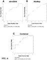
- Non-invasive gene expression methods inform on the status of the immune system by examining the status of genes expressed in immune cells. AlloMap Molecular Expression Testing is an FDA-cleared test available for heart transplant recipients. Tests are in development for monitoring other solid organ transplants.
- compositions and kits relating to detecting donor cell-free DNA in the circulation of an organ transplant recipient for the early identification of transplant rejection are disclosed.
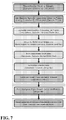
- the present disclosure relates to methods of monitoring immunosuppressive therapy in a subject, the method including: a) providing cell-free DNA from a sample obtained from a subject who is the recipient of an organ transplant from a donor, b) sequencing a panel of single nucleotide polymorphisms (SNPs) from the cell-free DNA, where the panel of SNPs is suitable for differentiating between donor-derived cell-free DNA and recipient-derived cell-free DNA, c) assaying variance in SNP allele distribution patterns in the panel as compared to expected homozygous or heterozygous distribution patterns to determine the level of donor-derived cell-free DNA, and d) diagnosing the status of the transplanted organ in the subject, where a change in levels of the donor-derived cell-free DNA over a time interval is indicative of transplanted organ status and a basis for adjusting immunosuppressive therapy.
- SNPs single nucleotide polymorphisms
- the present disclosure relates to methods of adjusting an immunosuppressive therapy in a subject, the method including: a) providing cell-free DNA from a sample obtained from a subject who is the recipient of an organ transplant from a donor, b) sequencing a panel of single nucleotide polymorphisms (SNPs) from the cell-free DNA, where the panel of SNPs is suitable for differentiating between donor-derived cell-free DNA and recipient-derived cell-free DNA, c) assaying variance in SNP allele distribution patterns in the panel as compared to expected homozygous or heterozygous distribution patterns to determine the level of donor-derived cell-free DNA, d) diagnosing the status of the transplanted organ in the subject, where a change in levels of the donor-derived cell-free DNA over a time interval is indicative of transplanted organ status, e) adjusting immunosuppressive therapy being administered to the subject.
- SNPs single nucleotide polymorphisms
- an increase in the levels of the donor-derived cell-free DNA over the time interval is indicative of transplant rejection, a need for adjusting immunosuppressive therapy, and/or a need for further investigation of the transplanted organ status.
- a decrease in the levels of the donor-derived cell-free DNA over the time interval is indicative of transplant tolerance, a need for adjusting immunosuppressive therapy, and/or a need for further investigation of the transplanted organ status.
- no change in the levels of the donor-derived cell-free DNA over the time interval is indicative of stable transplant rejection status and/or opportunity for adjusting immunosuppressive therapy.
- immunosuppressive therapy being administered to the subject is increased.
- immunosuppressive therapy being administered to the subject is decreased.
- immunosuppressive therapy being administered to the subject is maintained.
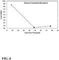
- the organ transplant is a kidney transplant.
- the organ transplant is a heart transplant.
- the sample is a plasma sample.
- the panel of SNPs includes at least 20 independent SNPs.
- the panel of SNPs includes independent SNPs selected from rs1004357, rs10092491, rs1019029, rs1027895, rs10488710, rs10500617, rs1058083, rs10768550, rs10773760, rs10776839, rs1109037, rs12480506, rs1294331, rs12997453, rs13134862, rs13182883, rs13218440, rs1336071, rs1358856, rs1410059, rs1478829, rs1490413, rs1498553, rs1523537, rs1554472, rs159606, rs1736442, rs1821380, rs1872575, rs2046361, rs2073383, rs2149
- sequencing the panel of SNPs is performed using a multiplex sequencing platform.
- the time interval is about 12-14 months after the transplant from the donor to the recipient subject occurred.
- the method further includes testing for viral load.
- the testing includes determining the presence of a virus selected from CMV, EBV, anellovirus, and BKV.
- the method further includes conducting one or more gene expression profiling assays.
- the gene expression profiling assay is an AlloMap test.
- the present disclosure relates to methods of monitoring the status of a transplanted organ in a subject, the method including: a) providing cell-free DNA from a sample obtained from a subject who is the recipient of an organ transplant from a donor, b) sequencing a panel of single nucleotide polymorphisms (SNPs) from the cell-free DNA, where the panel of SNPs is suitable for differentiating between donor-derived cell-free DNA and recipient-derived cell-free DNA, c) assaying variance in SNP allele distribution patterns in the panel as compared to expected homozygous or heterozygous distribution patterns to determine the level of donor-derived cell-free DNA, and d) diagnosing the status of the transplanted organ in the subject, where a change in levels of the donor-derived cell-free DNA over a time interval is indicative of the status of the transplanted organ.
- SNPs single nucleotide polymorphisms
- the present disclosure relates to methods of monitoring immunosuppressive therapy in a subject, the method including: a) providing cell-free DNA from a sample obtained from a subject who is the recipient of an organ transplant from a donor, b) sequencing a panel of single nucleotide polymorphisms (SNPs) from the cell-free DNA, where the panel of SNPs is suitable for differentiating between donor-derived cell-free DNA and recipient-derived cell-free DNA, c) assaying variance in SNP allele distribution patterns in the panel as compared to expected homozygous or heterozygous distribution patterns to determine the level of donor-derived cell-free DNA, and d) diagnosing the status of the transplanted organ in the subject, where a change in levels of the donor-derived cell-free DNA over a time interval is indicative of transplanted organ status and a basis for adjusting immunosuppressive therapy.
- SNPs single nucleotide polymorphisms
- the present disclosure relates to methods of adjusting an immunosuppressive therapy in a subject, the method including: a) providing cell-free DNA from a sample obtained from a subject who is the recipient of an organ transplant from a donor, b) sequencing a panel of single nucleotide polymorphisms (SNPs) from the cell-free DNA, where the panel of SNPs is suitable for differentiating between donor-derived cell-free DNA and recipient-derived cell-free DNA, c) assaying variance in SNP allele distribution patterns in the panel as compared to expected homozygous or heterozygous distribution patterns to determine the level of donor-derived cell-free DNA, d) diagnosing the status of the transplanted organ in the subject, where a change in levels of the donor-derived cell-free DNA over a time interval is indicative of transplanted organ status, and e) adjusting immunosuppressive therapy being administered to the subject.
- SNPs single nucleotide polymorphisms
- an increase in the levels of the donor-derived cell-free DNA over the time interval is indicative of transplant rejection, a need for adjusting immunosuppressive therapy, and/or a need for further investigation of the transplanted organ status.
- a decrease in the levels of the donor-derived cell-free DNA over the time interval is indicative of transplant tolerance, a need for adjusting immunosuppressive therapy, and/or a need for further investigation of the transplanted organ status.
- no change in the levels of the donor-derived cell-free DNA over the time interval is indicative of stable transplant rejection status and/or opportunity for adjusting immunosuppressive therapy.
- immunosuppressive therapy being administered to the subject is increased.
- immunosuppressive therapy being administered to the subject is decreased.
- immunosuppressive therapy being administered to the subject is maintained.
- the organ transplant is a kidney transplant.
- the organ transplant is a heart transplant. In some embodiments that may be combined with any of the preceding embodiments, the organ transplant is selected from a liver transplant, a lung transplant, and a pancreas transplant. In some embodiments that may be combined with any of the preceding embodiments, the sample is a plasma sample.
- the panel of SNPs includes independent SNPs selected fro rs1004357, rs10092491, rs1019029, rs1027895, rs10488710, rs10500617, rs1058083, rs10768550, rs10773760, rs10776839, rs1109037, rs12480506, rs1294331, rs12997453, rs13134862, rs13182883, rs13218440, rs1336071, rs1358856, rs1004357, rs10092491, rs1019029, rs1027895, rs10488710, rs10500617, rs1058083, rs10768550, rs10773760, rs10776839, rs1109037, rs12480506,
- the panel of SNPs includes SNPs that have an overall population minor allele frequency of >0.4, a target population minor allele frequency of >0.4, the lowest polymerase error rate of the 6 potential allele transitions or transversions, and the genomic distance between each independent SNP is >500kb.
- the panel of SNPs includes independent SNPs selected from rs10488710, rs279844, rs1048290, rs1049379, rs1051614, rs1052637, rs1055851, rs1056033, rs1056149, rs1064074, rs1078004, rs10831567, rs6811238, rs11106, rs11210490, rs1126899, rs1127472, rs1127893, rs1130857, rs1049544, rs11547806, rs12237048, rs430046, rs12508837, rs12529, rs12717, rs13184586, rs13295990, rs13428, rs13436, rs1374570, rs14080, r
- the SNP panel includes about 195 to about 200, about 200 to about 205, about 210 to about 215, about 215 to about 220, about 220 to about 225, about 225 to about 230, about 230 to about 235, about 235 to about 240, about 240 to about 245, about 245 to about 250, about 250 to about 255, about 255 to about 260, about 260 to about 265, or about 260 to about 266 of the independent SNPs.
- the SNP panel includes rs10488710, rs279844, rs1048290, rs1049379, rs1051614, rs1052637, rs1055851, rs1056033, rs1056149, rs1064074, rs1078004, rs10831567, rs6811238, rs11106, rs11210490, rs1126899, rs1127472, rs1127893, rs1130857, rs1049544, rs11547806, rs12237048, rs430046, rs12508837, rs12529, rs12717, rs13184586, rs13295990, rs13428, rs13436, rs1374570, rs14080, rs1411271, rs57
- sequencing the panel of SNPs is performed using a multiplex sequencing platform.
- the level of donor-derived cell-free DNA in the sample is determined without using genotype information.
- the method further includes testing for the presence of an infectious agent.
- the infectious agent is selected from viruses, bacteria, fungi, and parasites.
- the viruses are selected from Cytomegalovirus, Epstein-Barr virus, Anelloviridae, and BK virus.
- the method further includes conducting one or more gene expression profiling assays.
- a combination score is calculated based on the results of the level of donor-derived cell-free DNA and the results of the gene expression profiling assay.
- the gene expression profiling assay is an AlloMap test.
- the present disclosure relates to methods of monitoring the status of an allograft in a transplant recipient, as well as to methods of monitoring and adjusting immunosuppressive therapies being administered to the transplant recipient.
- the present disclosure is based, at least in part, on Applicant's development of techniques for probing the status of an allograft in a transplant recipient.
- Transplant recipients contain an allograft that is foreign to the recipient's body. This triggers an immune response from the recipient's immune system, which may lead to acute and/or chronic transplant rejection.
- Applicant's methods involve the analysis of cell-free DNA (cfDNA) from the transplant recipient to diagnose the status of the transplant and inform the need to adjust immunosuppressive therapy being administered to the transplant recipient.
- cfDNA cell-free DNA
- transplant rejection is associated with the death of cells in the transplanted (donor) organ or tissue, which will release donor-derived DNA from the dying donor cells, thus releasing donor-derived cell-free DNA (dd-cfDNA) into the bloodstream of the recipient.
- donor-derived cell-free DNA can be extracted from a sample from the recipient, such as a bodily fluid, and various polymorphic markers, such as single nucleotide polymorphism (SNP) loci, can be sequenced, where the panel of polymorphic markers, such as a panel of SNPs, is suitable for differentiating between donor-derived cell-free DNA and recipient-derived cell-free DNA (rd-cfDNA).
- SNP single nucleotide polymorphism
- the specific polymorphic markers selected to be on the panel include those that are identified as having low probabilities of being identical in any two individuals, thus making them appropriate for differentiating between recipient-derived cell free DNA and donor-derived cell-free DNA.
- the number of polymorphic markers on the panel such as, for example, the number of SNPs on the panel, will be sufficient to discriminate between recipient and donor alleles even in related individuals (excepting identical twins).
- the allele distribution patterns of polymorphic markers in the panel can be assayed to determine variance in the patterns as compared to expected homozygous ( i.e. 0% or 100% of each allele) or heterozygous ( i.e. 50% of each allele) distribution patterns, which can be used to determine the level of donor-derived cell-free DNA.
- the present disclosure provides methods of monitoring the status of an allograft in a transplant recipient, as well as methods of monitoring and/or adjusting an immunosuppressive therapy being or to be administered to a transplant recipient.
- Monitoring the status of an allograft involves analyzing various aspects which provide useful information about the physiological state of the allograft such as, for example, the level of donor-derived cell-free DNA in a sample from the transplant recipient.
- the methods of the present disclosure may be used to predict the risk of future transplant rejection such as, for example, the risk of rejection within the following 3-6 months after analysis of samples from the transplant recipient.
- the methods of the present disclosure may also be used to diagnose or predict the risk of allograft dysfunction, such as chronic renal insufficiency or cardiac allograft vasculopathy (CAV) ( e.g. within the next 1-2 years after analysis of samples from the transplant recipient).
- CAV cardiac allograft vasculopathy
- the methods of the present disclosure may also be used provide an assessment of the immune status of the transplant recipient, which may be used to guide decisions regarding immunosuppressive therapy in the transplant recipient.
- the methods of the present disclosure may also be used to guide decisions related to adjustment of immunosuppressive therapies being administered to the transplant recipient. Additional benefits and/or uses of the methods of the present disclosure will be readily apparent to one of skill in the art.
- Cell-free DNA generally refers to DNA that is present outside of a cell such as, for example, DNA that is present in a bodily fluid (e.g. blood, plasma, serum, etc.) of a subject.
- Cell-free DNA may have originated from various locations within a cell.
- Cell-free DNA may have originated from, for example, nuclear DNA and mitochondrial DNA. Without wishing to be bound by theory, it is believed that cell-free DNA is released from cells via apoptosis or necrosis of the cells ( i.e. cell death).
- transplanted cells will result in donor-derived cell-free DNA being released into the bodily fluid of a transplant recipient.
- Transplant recipients undergoing transplant rejection may then have a cell-free DNA population in their bodily fluids which includes both their own endogenous cell-free DNA (recipient-derived cell-free DNA) as well as cell-free DNA that originated from the donor (donor-derived cell-free DNA). Determining a change in the levels and/ in donor-derived cell-free DNA in a transplant recipient over time according to the methods of the present disclosure may be used to diagnose the status of the allograft and inform a need to adjust immunosuppressive therapy.
- Cell-free RNA may also be collected from a transplant recipient and analyzed by analogous methods as described above and also analysis of recipient RNA levels from specific marker genes to diagnose the status of the transplant and/or to inform a need to adjust immunosuppressive therapy being administered to the transplant recipient.
- the methods of the present disclosure generally relate to analysis of cell-free nucleic acids from a transplant recipient to diagnose the status of the transplant and/or to inform a need to adjust immunosuppressive therapy being administered to the transplant recipient.
- the methods of the present disclosure involve providing cell-free DNA from a sample obtained from a subject who is the recipient of an allograft from a donor.
- the subject is a transplant recipient who contains an allograft from a donor, and is typically a human transplant recipient.
- the transplant recipient may have received one or more of a variety of allografts from a donor.
- Allografts may include transplanted organs.
- Transplanted organs may include, for example, a heart, a kidney, a lung, a liver, a pancreas, a cornea, an organ system, or other solid organs.
- the transplant received by the transplant recipient from the donor may also include other allografts such as, for example, a bone marrow transplant, pancreatic islet cells, stem cells, skin tissue, skin cells, or a xenotransplant.
- the provided sample may include a bodily fluid isolated from the transplant recipient.
- Samples obtained from the transplant recipient contain cell-free DNA, and the total cell-free DNA present in the sample may be entirely recipient-derived cell-free DNA, or the total cell-free DNA present in the sample may include a mixture of recipient-derived cell-free DNA and donor-derived cell-free DNA.
- Samples may include a bodily fluid from the transplant recipient such as, for example, plasma, serum, whole blood, sweat, tears, saliva, ear flow fluid, sputum, fluid from bone marrow suspension, lymph fluid, urine, saliva, semen, vaginal flow, cerebrospinal fluid, brain fluid, ascites, milk, secretions of the respiratory, and intestinal or genitourinary tract fluids.
- plasma plasma derived from the venous blood of the transplant recipient can be obtained.
- a sample Once a sample is obtained, it can be used directly, frozen, or otherwise stored in a condition that maintains the integrity of the cell-free DNA for short periods of time by preventing degradation and/or contamination with genomic DNA or other nucleic acids.
- the amount of a sample that is taken at a particular time may vary, and may depend on additional factors, such as any need to repeat analysis of the sample. In some aspects, up to 50, 40, 30, 20, 10, 9, 8, 7, 6, 5, 4, 3, 2, or 1 mL of a sample is obtained. In some aspects, 1-50, 2-40, 3-30, or 4-20 mL of sample is obtained. In some aspects, more than 5, 10, 15, 20, 25, 30, 35, 40, 45, 50, 55, 60, 65, 70, 75, 80, 85, 90, 95 or 100 mL of a sample is obtained.
- Samples may be taken from a transplant recipient over a period of time (i.e. over a time interval). The time at which samples are taken from the transplant recipient following the transplant event may vary. Samples may be taken from a transplant recipient at various times and over various periods of time for use in determining the status of the allograft according to the methods of the present disclosure. For example, samples may be taken from the transplant recipient within days and weeks after, about three months after, about six months after, about nine months after, or less than one year after the transplant event. Samples may be taken from the transplant recipient at various times before the one year anniversary of the transplant event, at the one year anniversary of the transplant event, or at various times after the one year anniversary of the transplant event.
- samples may begin to be taken from the transplant recipient at month 12 (i.e. the one year anniversary of the transplant event) and continue to be taken for periods of time after this.
- the time period for obtaining samples from a transplant recipient is within the first few days after the transplant from the donor to the recipient occurred. This may be done to monitor induction therapy.
- the time period for obtaining samples from a transplant recipient is during tapering of the immunosuppressive regimen, a period that occurs during the first 12 months after the transplant from the donor to the recipient occurred.
- the time period for obtaining samples from a transplant recipient is during the initial long term immunosuppressive maintenance phase, beginning about 12-14 months after the transplant from the donor to the recipient occurred.
- the time period for obtaining samples from a transplant recipient is during the entire long term maintenance of the immunosuppressive regimen, any time beyond 12 months after the transplant from the donor to the recipient occurred.
- samples may be obtained about once every week, about once every 2 weeks, about once every 3 weeks, about once every month, about once every two months, about once every three months, about once every four months, about once every five months, about once every six months, about once every year, or about once every two years or more after the initial sampling event.
- a transplant recipient has samples of bodily fluid taken for one to three consecutive months, starting at the one year anniversary of the transplant event ( i.e. 12 months after the transplant event), providing a total of 4-6 samples for analysis taken over a three month time period, with samples being collected about every two weeks.
- a transplant recipient has samples of bodily fluid taken once a week for one to three consecutive months, starting at the one year anniversary of the transplant event ( i.e. 12 months after the transplant event), providing a total of twelve samples for analysis taken over a three month time period.
- the total duration of obtaining samples from a transplant recipient, as well as the frequency of obtaining such samples may vary and will depend on a variety of factors, such as clinical progress.
- a transplant recipient may have samples obtained for analysis of cell-free DNA for the duration of their lifetime. Appropriate timing and frequency of sampling will be able to be determined by one of skill in the art for a given transplant recipient.
- the methods of the present disclosure involve the analysis of cell-free DNA from a transplant recipient. After cell-free DNA has been isolated from a transplant recipient, various methods and techniques may be used to analyze the cell-free DNA. Analysis of cell-free DNA according to the methods of the present disclosure involves analysis of a panel of polymorphic markers from the cell-free DNA.
- the polymorphic markers are single nucleotide polymorphisms (SNPs).
- SNPs are selected to be included in the panel at least in part on the basis that the panel of SNPs will be sufficient to differentiate between donor-derived cell-free DNA and recipient-derived cell-free DNA.
- Analysis of cell-free DNA obtained from a transplant recipient involves the analysis of a panel of polymorphic markers from the cell-free DNA.
- Various polymorphic markers may be selected for inclusion in the panel to be analyzed as long as the polymorphic marker panel as a whole is suitable for differentiating between donor-derived cell-free DNA and recipient-derived cell-free DNA.
- the same polymorphic marker panel may be used for each transplant recipient; there is no need to customize polymorphic marker panels to individualize the panel to different transplant recipients.
- polymorphic markers are found at a region of DNA containing a polymorphism.
- a polymorphism generally refers to the occurrence of two or more genetically determined alternative sequences or alleles in a population.
- a polymorphic marker or site is the locus at which divergence, or the polymorphism, occurs.
- a polymorphism may contain, for example, one or more base changes, an insertion, a repeat, or a deletion.
- a polymorphic locus may be as small as one base pair, such as a SNP.
- Polymorphic markers may include, for example, single nucleotide polymorphisms (SNPs), restriction fragment length polymorphisms (RFLPs), short tandem repeats (STRs), variable number of tandem repeats (VNTRs), hypervariable regions, minisatellites, dinucleotide repeats, trinucleotide repeats, tetranucleotide repeats, simple sequence repeats, and insertion elements.
- Polymorphic markers may contain one or more bases modified by methylation. Additional types of polymorphisms and polymorphic markers will be readily apparent to one of skill in the art.
- a polymorphism between two nucleic acids can be naturally occurring, or may be caused by exposure to or contact with chemicals, enzymes, or other agents, or exposure to agents that cause damage to nucleic acids such as, for example, ultraviolet radiation, mutagens, or carcinogens.
- polymorphic marker panel may include both SNPs and short tandem repeats, or any other type of polymorphic marker.
- the polymorphic marker panel is composed entirely of SNPs; thus, the polymorphic marker panel is a SNP panel. Additional combinations of polymorphic markers on polymorphic marker panels will be readily apparent to one of skill in the art.
- the panel of polymorphic markers to be analyzed may include at least 10, at least 15, at least 20, at least 25, at least 30, at least 35, at least 40, at least 45, at least 50, at least 55, at least 60, at least 65, at least 70, at least 75, at least 80, at least 85, at least 90, at least 95, at least 100, at least 105, at least 110, or at least 115, at least 120, at least 150, at least 200, at least 250, at least 300, at least 350, at least 400, at least 450, at least 500, at least 1,000, or at least 1,500 or more independent polymorphic markers.
- the polymorphic marker panel is a panel of SNPs.
- SNPs to be included in the SNP panel, or in any other polymorphic marker panel, may be those previously identified as being suitable for differentiating between any two unrelated individuals (Pakstis et al., 2010).
- the SNP panel may include one or more of the following human SNPs (named according to dbSNP numbering): rs1004357, rs10092491, rs1019029, rs1027895, rs10488710, rs10500617, rs1058083, rs10768550, rs10773760, rs10776839, rs1109037, rs12480506, rs1294331, rs12997453, rs13134862, rs13182883, rs13218440, rs1336071, rs1358856, rs1410059, rs1478829, rs1490413, rs1498553, rs1523537, rs1554472, rs159606, rs1736442, rs1821380, rs1872575, rs2046361, rs2073383, rs100
- SNPs may also be selected on the basis that they have, for example, an overall population minor allele frequency of >0.4, a target population minor allele frequency of >0.4, the lowest polymerase error rate (in the test system) of the 6 potential allele transitions or transversions, and low linkage on the genome such as, for example, >500kb distance between SNPs.
- the SNP panel may include, for example, one or more of the following human SNPs (named according to dbSNP numbering): rs10488710, rs279844, rs1048290, rs1049379, rs1051614, rs1052637, rs1055851, rs1056033, rs1056149, rs1064074, rs1078004, rs10831567, rs6811238, rs11106, rs11210490, rs1126899, rs1127472, rs1127893, rs1130857, rs1049544, rs11547806, rs12237048, rs430046, rs12508837, rs12529, rs12717, rs13184586, rs13295990, rs13428, rs13436, rs1374570, rs
- the SNP panel may include, for example, at least at least 10, at least 15, at least 20, at least 25, at least 30, at least 35, at least 40, at least 45, at least 50, at least 55, at least 60, at least 65, at least 70, at least 75, at least 80, at least 85, at least 90, at least 95, at least 100, at least 105, at least 110, or at least 115, at least 120, at least 150, at least 200, at least 205, at least 210, at least 215, at least 220, at least 225, at least 230, at least 235, at least 240, at least 245, at least 250, at least 255, at least 260, or at least 265 of the 266 independent SNPs identified above.
- the SNP panel may include, for example, about 10 to about 20, about 20 to about 30, about 30 to about 40, about 40 to about 50, about 50 to about 60, about 60 to about 70, about 70 to about 80, about 80 to about 90, about 90 to about 100, about 100 to about 110, about 110 to about 120, about 120 to about 130, about 130 to about 140, about 140 to about 150, about 150 to about 160, about 160 to about 170, about 170 to about 180, about 180 to about 190, about 190 to about 200, about 200 to about 210, about 210 to about 220, about 220 to about 230, about 230 to about 240, about 240 to about 250, about 250 to about 260, or about 250 to about 266 of the 266 independent SNPs identified above.
- the SNP panel may include, for example, about 195 to about 200, about 200 to about 205, about 210 to about 215, about 215 to about 220, about 220 to about 225, about 225 to about 230, about 230 to about 235, about 235 to about 240, about 240 to about 245, about 245 to about 250, about 250 to about 255, about 255 to about 260, about 260 to about 265, or about 260 to about 266 of the 266 independent SNPs identified above.
- Cell-free DNA isolated from a transplant recipient may be amplified for downstream techniques and analysis, such as analysis of a panel of polymorphic markers from the cell-free DNA.
- Various methods and protocols for DNA extraction are well-known in the art and are described herein ( See e.g. Current Protocols in Molecular Biology, latest edition).
- Cell-free DNA may be extracted using the QIAamp circulating nucleic acid kit or other appropriate commercially available kits.
- Other exemplary methods of extracting cell-free DNA are well-known ( See, e.g., Cell-Free Plasma DNA as a Predictor of Outcome in Severe Sepsis and Septic Shock. Clin. Chem. 2008, v.
- Amplification generally refers to any device, method or technique that can generate copies of a nucleic acid.
- Amplification of cell-free DNA may involve, for example, polymerase chain reaction (PCR) techniques such as linear amplification (cf. USPN 6,132,997 ), rolling circle amplification, and the like.
- PCR polymerase chain reaction
- Cell-free DNA may be amplified for use in downstream analysis of the DNA by, for example, digital PCR or sequencing.
- the Fluidigm Access ArrayTM System, the RainDance Technologies RainDrop system, or other technologies for multiplex amplification may be used for multiplex or highly parallel simplex DNA amplification.
- Amplification may involve the use of high-fidelity polymerases such as, for example, FastStart High Fidelity (Roche), Expand High Fidelity (Roche), Phusion Flash II (ThermoFisher Scientific), Phusion Hot Start II (ThermoFisher Scientific), KAPA HiFi (Kapa BioSystems), or KAPA2G (Kapa Biosystems).
- high-fidelity polymerases such as, for example, FastStart High Fidelity (Roche), Expand High Fidelity (Roche), Phusion Flash II (ThermoFisher Scientific), Phusion Hot Start II (ThermoFisher Scientific), KAPA HiFi (Kapa BioSystems), or KAPA2G (Kapa Biosystems).
- Amplification may include an initial PCR cycle that adds a unique sequence to each individual molecule, called molecular indexing.
- Molecular indexing allows for quantitative assessment of the absolute level of both alleles for each SNP amplicon and therefore may improve precision and accuracy of determining the percent donor-derived cell-free DNA.
- Amplified DNA may also be subjected to additional processes, such as indexing (also referred to as barcoding or tagging).
- indexing also referred to as barcoding or tagging.
- Methods of indexing DNA are well-known in the art and are described herein. Indexing will allow for the use of multiplex-sequencing platforms, which are compatible with a variety of sequencing systems, such as Illumina HiSeq, MiSeq, and ThermoFisher Scientific Ion PGM and Ion Proton.
- Multiplex sequencing permits the sequencing of DNA from multiple samples at once through the use of DNA indexing to specifically identify the sample source of the sequenced DNA.
- the amount of DNA that is used for analysis may vary. In some aspects, less than 1 pg, 5 pg, 10 pg, 20 pg, 30 pg, 40 pg, 50 pg, 100 pg, 200 pg, 500 pg, 1 ng, 5 ng, 10 ng, 20 ng, 30 ng, 40 ng, 50 ng, 100 ng, 200 ng, 500 ng, 1 ⁇ g, 5 ⁇ g, 10 ⁇ g, 20 ⁇ g, 30 ⁇ g, 40 ⁇ g, 50 ⁇ g, 100 ⁇ g, 200 ⁇ g, 500 ⁇ g or 1 mg of DNA are obtained from the sample for further genetic analysis.
- DNA sequencing may be accomplished using high-throughput DNA sequencing techniques.
- next generation and high-throughput sequencing include, for example, massively parallel signature sequencing, polony sequencing, 454 pyrosequencing, Illumina (Solexa) sequencing with HiSeq, MiSeq, and other platforms, SOLiD sequencing, ion semiconductor sequencing (Ion Torrent), DNA nanoball sequencing, heliscope single molecule sequencing, single molecule real time (SMRT) sequencing, MassARRAY®, and Digital Analysis of Selected Regions (DANSRTM).
- each sample may be sequenced individually, or multiple samples may be sequenced together using multiplex sequencing.
- the methods of the present disclosure involve assaying variance in polymorphic marker allele distribution patterns in a polymorphic marker panel as compared to expected homozygous or heterozygous distribution patterns. Analysis of these patterns allows for the determination of the level of donor-derived cell-free DNA in a cell-free DNA sample obtained from a transplant recipient.
- an individual contains DNA that is either homozygous or heterozygous for a given polymorphic marker, such as a SNP.
- An individual may be homozygous for one allele of a given polymorphic marker and will contain 100% of one allele of that polymorphic marker and will contain 0% of the other allele of that polymorphic marker (e.g. 100% of allele A for given polymorphic marker, 0% of allele B for given polymorphic marker).
- An individual may also be heterozygous for a given polymorphic marker, and thus will contain 50% of allele A and 50% of allele B of that polymorphic marker.
- any given polymorphic marker in the DNA in that sample will exhibit a homozygous distribution pattern (i.e. 100% of one allele, 0% of the other allele) or a heterozygous distribution pattern (i.e. 50% of one allele and 50% of the other allele).
- a DNA sample contains DNA that originated from more than one individual (e.g . a cell-free DNA sample from a transplant recipient that contains both recipient-derived DNA and donor-derived DNA)
- polymorphic marker allele distributions may vary, for a given polymorphic marker, from expected homozygous or heterozygous distribution patterns.
- variance in polymorphic marker allele distribution patterns in the polymorphic marker panel as compared to expected homozygous or heterozygous distribution patterns is used to determine the level of donor-derived cell-free DNA in the cell-free DNA sample obtained from a transplant recipient.
- a majority signal from an allele of a polymorphic marker may be observed and a minority signal from an allele of a polymorphic marker may be observed.
- cell-free DNA isolated from a transplant recipient as it is assumed that the majority of the DNA in the cell-free DNA sample from the transplant recipient originated from the recipient's own endogenous DNA, it is further assumed that the majority signal represents an allele of a polymorphic marker that originated from recipient-derived cell-free DNA, while the minority signal represents an allele of a polymorphic marker that originated from donor-derived cell-free DNA.
- Polymorphic markers such as SNPs, for example, with an even distribution of both alleles are assumed to have largely both originated from the recipient. Deviations from the even distribution will indicate the influence of alleles from the donor-derived cell-free DNA.
- the methods of the present disclosure may involve calculating various cell-free DNA concentrations, or percents thereof, of a total amount of cell-free DNA.
- this information can be used to calculate a percentage of donor-derived DNA in the cell-free DNA sample.
- the level of donor-derived cell-free DNA in a sample obtained from a transplant recipient may be determined without using genotype information from the transplant recipient, from the transplant donor, and/or any other genotype information from any source.
- genotype information that need not be considered includes, for example, the genotype across the genome as a whole or portions thereof, or the genotype at the particular polymorphic markers being analyzed.
- individual genotyping of the transplant recipient is not performed. In some embodiments, individual genotyping of the transplant donor is not performed. In some embodiments, neither the transplant recipient nor the transplant donor are individually genotyped. In some embodiments, genotype information from the transplant recipient is not considered when determining the levels of donor-derived cell-free DNA in a sample obtained from a transplant recipient. In some embodiments, genotype information from the transplant donor is not considered when determining the levels of donor-derived cell-free DNA in a sample obtained from a transplant recipient. In some embodiments, the levels of donor-derived cell-free DNA in a sample obtained from a transplant recipient are determined without consideration of genotype information from the transplant recipient and without consideration of genotype information of the transplant donor. In some embodiments, the level of donor-derived cell-free DNA in a sample obtained from a transplant recipient may be determined without using genotype information.
- Improvements to the calculations may include estimating and subtracting a level of signal due to amplification or sequencing error to improve accuracy and precision.
- a suitably chosen subset of SNPs may be used to estimate a sum, mean, median or standard deviation of the subset to produce a computation of the overall level of donor-derived cell-free DNA.
- Multiple samples from the same subject at a given time of sampling will all have the same pattern of polymorphic distributions across the SNPs, which can be used to enhance the estimate of donor-derived cell-free DNA in individual samples from that subject.
- the quantity of donor-derived cell-free DNA present in the cell-free DNA sample may be expressed in a variety of ways.
- the amount of one or more DNA molecules from donor-derived cell-free DNA is determined as a percentage of the total the DNA molecules in the sample.
- the amount of one or more DNA molecules from donor-derived cell-free DNA is determined as a ratio of the total DNA in the sample.
- the amount of one or more DNA molecules from donor-derived cell-free DNA is determined as a ratio or percentage compared to one or more reference DNA molecules in the sample.
- the total amount of donor-derived cell-free DNA can be determined to be 10% of the total DNA molecules in the cell-free DNA sample.
- the total amount of donor-derived cell-free DNA can be at a ratio of 1:10 compared to the total DNA molecules in the cell-free DNA sample.
- the amount of one or more DNA molecules from the donor-derived cell-free DNA can be determined as a concentration.
- the total amount of donor-derived cell-free DNA in the cell-free DNA sample can be determined to be 1 ⁇ g/mL.
- the values described here are merely exemplary to illustrate various ways to express quantities of donor-derived cell-free DNA.
- the percentage of donor-derived cell-free DNA in the cell-free DNA sample from a transplant recipient may be extremely low ( e.g. at or below 0.5% of the total DNA content of the cell-free DNA sample).
- the quantity of recipient-derived cell-free DNA in the cell-free DNA sample may also be expressed in the manners as described for donor-derived cell-free DNA. Additional methods of expressing the quantity of a given source, type, or sequence of DNA molecule in a cell-free DNA sample will be readily apparent to one of skill in the art.
- the above-described embodiments and aspects of the present disclosure may be implemented in a variety of ways. For example, some aspects of the embodiments and aspects may be implemented using hardware, software or a combination thereof.
- the software code can be executed on any suitable processor or collection of processors, whether provided in a single computer or distributed among multiple computers. It should be appreciated that any component or collection of components that perform the functions described above can be generically considered as one or more controllers that control the above - discussed functions.
- the one or more controllers can be implemented in numerous ways, such as with dedicated hardware, or with general-purpose hardware ( e.g. , one or more processors) that is programmed using microcode or software to perform the functions recited above.
- implementation of various features of the present disclosure may use at least one non-transitory computer-readable storage medium (e.g ., a computer memory, a floppy disk, a compact disk, a tape, etc.) encoded with a computer program ( i.e., a plurality of instructions), which, when executed on a processor, performs the above - discussed functions.
- the computer-readable storage medium can be transportable such that the program stored thereon can be loaded onto any computer resource to implement certain aspects of the present disclosure discussed herein.
- the reference to a computer program which, when executed, performs the above-discussed functions is not limited to an application program running on a host computer. Rather, the term computer program is used herein in a generic sense to reference any type of computer code (e.g ., software or microcode) that can be employed to program a processor to implement certain aspects of the present disclosure.
- the methods of the present disclosure for determining the levels of donor-derived cell-free DNA in a sample from a transplant recipient can be used to determine the status of the allograft in the transplant recipient.
- changes in the levels of donor-derived cell-free DNA beyond a suitable threshold value in the transplant recipient over time are informative with regard to the status of the allograft.
- a threshold or threshold value generally refers to any predetermined level or range of levels that is indicative of the presence or absence of a condition or the presence or absence of a risk.
- the threshold value can take a variety of forms. It can be single cut-off value, such as a median or mean.
- a threshold value can be determined from baseline values before the presence of a condition or risk or after a course of treatment. Such a baseline can be indicative of a normal or other state in the subject not correlated with the risk or condition that is being tested for.
- the baseline value may be the level of donor-derived cell-free DNA in samples from a transplant recipient prior to the actual transplant event, which would be presumably zero or negligible, but may also indicate baseline error in the system.
- the threshold value can be a baseline value of the subject being tested.
- the threshold value as it pertains to demarcating significant changes in the levels of donor-derived cell-free DNA in a transplant recipient, may vary considerably.
- One of skill in the art would recognize appropriate parameters and means for determining significant changes in the levels of donor-derived cell-free DNA in a transplant recipient over time. Once appropriate analysis parameters are selected, determining changes in the level of donor-derived cell-free DNA in the transplant recipient over a period of time can inform status of the allograft.
- an increase in the levels of the donor-derived cell-free DNA in the transplant recipient over time is indicative of transplant rejection, a need for adjusting immunosuppressive therapy, immunosuppressive treatment nephrotoxicity, infection, and/or a need for further investigation of the allograft status.
- a decrease in the levels of the donor-derived cell-free DNA in the transplant recipient over time is indicative of transplant tolerance, a need for adjusting immunosuppressive therapy, and/or a need for further investigation of the allograft status.
- the level of donor-derived cell-free DNA is decreasing in a transplant recipient over time, then the cells of the allograft are decreasingly experiencing apoptosis and/or necrosis over time, which is indicative of transplant tolerance, overimmunosuppression, or appropriate immunosuppression.
- no change in the levels of the donor-derived cell-free DNA in the transplant recipient over time is indicative of stable transplant rejection status and/or opportunity for adjusting immunosuppressive therapy.
- level of donor-derived cell-free DNA is not changing in a transplant recipient over time, then the cells of the allograft are experiencing a steady-state level of apoptosis over time, which is indicative of a stable transplant rejection status.
- a stable transplant rejection status informs the status of the allograft during the time window analyzed, but does not inform the direction, either towards rejection or tolerance, the allograft is progressing toward.
- the allograft may be undergoing active rejection in the transplant recipient, but a stable transplant rejection status indicates that the rate of allograft rejection is not changing during the time it was analyzed ( i.e. the rate of transplant rejection is not increasing or decreasing).
- the allograft may be undergoing active tolerance in the transplant recipient, but a stable transplant rejection status indicates that the rate of allograft tolerance is not changing during the time it was analyzed ( i.e. the rate of transplant tolerance is not increasing or decreasing).
- the methods of the present disclosure for determining the levels of donor-derived cell-free DNA in a sample from a transplant recipient can be used to inform the need to adjust immunosuppressive therapy being administered to the transplant recipient.
- changes in the levels of donor-derived cell-free DNA beyond a suitable threshold value in the transplant recipient over time are informative with regard to determining a need to adjust immunosuppressive therapy being administered to the transplant recipient.
- Immunosuppressive therapy generally refers to the administration of an immunosuppressant or other therapeutic agent that suppresses immune responses to a subject.
- immunosuppressant agents may include, for example, anticoagulents, antimalarials, heart drugs, non-steroidal anti-inflammatory drugs (NSAIDs), and steroids including, for example, Ace inhibitors, aspirin, azathioprine, B7RP-1-fc, ⁇ -blockers, brequinar sodium, campath-1H, celecoxib, chloroquine, corticosteroids, coumadin, cyclophosphamide, cyclosporin A, DHEA, deoxyspergualin, dexamethasone, diclofenac, dolobid, etodolac, everolimus, FK778, feldene, fenoprofen, flurbiprofen, heparin, hydralazine, hydroxychloroquine, CTLA-4 or LFA3 immuno
- an increase in the levels of the donor-derived cell-free DNA in the transplant recipient over time is indicative of a need to increase immunosuppressive therapy being administered to the transplant recipient.
- the decision to increase immunosuppressive therapy being administered to a transplant recipient may be based on additional clinical factors, such as the health of the transplant recipient.
- immunosuppressive therapy being administered to the subject is increased.
- a decrease in the levels of the donor-derived cell-free DNA in the transplant recipient over time is indicative of a need to decrease immunosuppressive therapy being administered to the transplant recipient.
- the decision to decrease immunosuppressive therapy being administered to a transplant recipient may be based on additional clinical factors, such as the health of the transplant recipient.
- immunosuppressive therapy being administered to the subject is decreased.
- no change in the levels of the donor-derived cell-free DNA in the transplant recipient over time is indicative of no need to adjust immunosuppressive therapy being administered to the transplant recipient, or that the immunosuppressive therapy being administered may be maintained.
- the decision to maintain immunosuppressive therapy being administered to a transplant recipient may be based on additional clinical factors, such as the health of the transplant recipient. In some aspects, immunosuppressive therapy being administered to the subject is maintained.
- adjustment of immunosuppressive therapy includes changing the type or form of immunosuppressant or other immunosuppressive therapy being administered to the transplant recipient.
- the methods of the present disclosure may indicate a need to begin administering immunosuppressive therapy to the transplant recipient.
- the levels of donor-derived cell-free DNA in the transplant recipient may not be the only factor taken into consideration when determining a need or lack thereof to adjust an immunosuppressive therapy being administered to the transplant recipient.
- an immunosuppressive therapy being administered to a transplant recipient may be increased, decreased, or maintained irrespective of the determined levels of donor-derived cell-free DNA in the transplant recipient depending on the presence or absence of other controlling clinical factors.
- the methods of the present disclosure may be performed in addition to or in conjunction with other analyses of samples from a transplant recipient to diagnose the status of the allograft and/or to inform the need to adjust immunosuppressive therapy being administered to the subject.
- the presence or levels of an infectious agent in the transplant recipient is tested.
- Infectious agents which may be tested for include, for example, viruses; bacteria such as Pseudomonas aeruginosa, Enterobacteriaceae, Nocardia, Streptococcus pneumonia, Staphyloccous aureus, and Legionella ; fungi such as Candida, Aspergillus, Cryptococcus, Pneumocystis carinii; or parasites such as Toxoplasma gondii.
- the presence or levels of viral infectious agents in the transplant recipient is tested.
- Viral biomarkers may be analyzed in nucleic acid obtained from a sample from the transplant recipient to determine the presence or levels of viruses in the transplant recipient.
- Viruses which may be tested for include, for example, Cytomegalovirus, Epstein-Barr virus, Anelloviridae, and BK virus. The results of the tests for presence or levels of viruses may be used to classify the immune status of the transplant recipient and to determine the status of infection in the transplant recipient.
- immunosuppressive therapies are decreased, or at least not increased, in transplant recipients that are classified as having a high risk of clinically significant infection.
- immunosuppressive therapies are increased, or at least not decreased, in transplant recipients that are classified as having a low risk of clinically significant infection. It should be noted that as other clinical factors may inform decisions to adjust immunosuppressive therapies, a transplant recipient may have immunosuppressive therapy currently being administered to them increased, decreased, or maintained irrespective of the results of tests for presence or levels of viruses and/or classification for risk of clinically significant infection.
- the methods of the present disclosure also involve performing an AlloMap test to aid in determining the status of the allograft in a transplant recipient.
- AlloMap tests involve performing quantitative real-time polymerase chain reaction (qRT-PCR) assays using RNA that has been isolated from peripheral blood mononuclear cells (PBMC). The expression of a select number of genes is analyzed and this gene expression data is used to provide information relating to the status of an allograft in a transplant recipient.
- qRT-PCR quantitative real-time polymerase chain reaction
- Results of an AlloMap test may be used in conjunction with the methods of the present disclosure, with or without a method to define a single score from the combined tests, to determine the status of an allograft in a transplant recipient and/or inform the need to adjust immunosuppressive therapy being administered to the transplant recipient.
- the methods of the present disclosure involve determining a combination score that may be used to convey the status of an allograft in a transplant recipient.
- Combination scores are generally calculated based on the results of multiple (e.g. two or more) assays used to probe the status of the allograft in the transplant recipient. For example, combination scores may be calculated based on the determined levels of donor-derived cell-free DNA in the transplant recipient and based on the results of a gene expression profiling assay such as, for example, an AlloMap test, which measures select gene expression.
- Combination scores may be calculated based on a single sample taken from a transplant recipient, or they may be based on samples taken from a transplant recipient over a time interval. Combination scores may be used to determine the status of an allograft in a transplant recipient and/or inform the need to adjust immunosuppressive therapy being administered to the transplant recipient.
- Additional biomarker analyses, gene expression assays, and other assays for diagnosing the status of an allograft in a transplant recipient and/or determining need to adjust immunosuppressive therapy may also be used in addition to or in conjunction with the methods of the present disclosure, with or without a method to define a single score from the combined tests, which will be readily apparent to one of skill in the art.
- Primers may be designed to amplify regions where genetic mutations are known to occur to provide an early detection of cancer by identification of known tumor-associated mutations. This may be advantageous at least in part because transplant recipients may be at heightened risk of developing certain malignancies due to overimmunosuppression.
- This Example demonstrates analysis of samples containing cell-free DNA from a transplant recipient to determine the level of donor-derived cell-free DNA in the samples. Changes in the levels of in the donor-derived cell-free DNA over time are used to diagnose the status of the transplanted organ in the transplant recipient, as well as inform the need to adjust or maintain immunosuppressive therapies being administered to the transplant recipient.
- a human patient is selected who was the subject of a kidney transplant 12 months prior to this assay as described in this Example.
- the patient is undergoing treatment with immunosuppressive therapy to prevent rejection of the allograft.
- Separate plasma samples will be collected from this subject on a weekly basis over the course of three consecutive months, starting at month 12 (the one year anniversary) after the transplant event. Accordingly, the transplant recipient will be analyzed during months 12, 13, and 14 after the transplant event.
- the methods described in this Example are applicable to one or more of the samples isolated from the transplant recipient.
- Blood is extracted from the subject so that cell-free DNA can be extracted from plasma isolated from this blood sample.
- the blood sample is collected in a cell-free DNA blood collection tube (Streck) according to the venipuncture method as previously described ( Clinical and Laboratory Standards Institute, 2012 ).
- the Streck tube is filled completely with the blood sample.
- the tube is removed from the adapter and is immediately mixed by gentle inversion about 8 to 10 times. After collection, the tubes are transported and stored within the temperature range of 6-37°C for up to 14 days.
- the Streck tube containing the blood sample is centrifuged at 1600 x g for 20 minutes at room temperature. The resulting plasma layer is carefully removed and is transferred to a 15 mL tube.
- This plasma sample is then centrifuged at 1600 x g for 10 minutes at room temperature. The resulting plasma layer is carefully removed and placed into a new tube, and the plasma sample may then proceed to have cell-free DNA (cf DNA) extracted.
- PPT plasma preparation tubes Becton Dickinson
- CPT pour-off methods may also be used during plasma collection.
- SNPs can be assayed in the cell-free DNA.
- a variety of SNPs are selected for analysis. SNPs may be selected based on those that can provide the highest possible minor allele frequencies (nearest to 50%).
- the location of the SNPs in the subject's genome may vary; genetic linkage may be allowed, but some genetic separation of the SNPs such as, for example, >200 bp separation may also be desirable.
- the number and identity of SNPs to be used should provide sufficient power to accurately estimate the percentage of donor-derived cfDNA present in the subject's plasma sample.
- the SNPs to be used in this assay include those previously identified (Pakstis et al., 2010). To amplify the SNPs, 92 primer pairs are designed (Fluidigm).
- the cell-free DNA is amplified according to the Fluidigm Access Array process and protocols (See page 63 of Access Array System for Illumina Sequencing System).
- Materials to be used for this amplification protocol include a Fluidigm Access Array, or chip, FastStart High Fidelity PCR system (Roche), 1X Access Array Harvest Solution (Fluidigm, PN 100-1031), 20X Access Array Loading reagent (Fluidigm), and the 92 primer pairs designed as described above.
- Instruments to be used for this amplification protocol include two IFC Controller AX (Fluidigm) and one FC1 cycler (Fluidigm).
- the amplified DNA is barcoded. Barcoding may be done, for example, to uniquely identify which of the three samples any detected amplified DNA originated from if cell-free DNA molecules from all of the samples are to be sequenced together.
- the amplified cell-free DNA is barcoded according to the Fluidigm Access Array process and protocols (See page 70 of Access Array System for Illumina Sequencing System). Materials to be used for this barcoding protocol include a FastStart High Fidelity PCR system (Roche), and an Access Array Barcode Library for Illumina Sequencers (Fluidigm). Instruments that will be used include a PCR machine, a plate centrifuge, and a vortexer.
- the barcoded cell-free DNA is sequenced according to Fluidigm/Illumina sequencing protocols for multiplex sequencing (See page 134 of Access Array System for Illumina Sequencing System).
- Materials to be used for this sequencing protocol include FL1 and FL2 sequencing primers (Fluidigm), HT1 buffer (Illumina), and a MiSeq Reagent Kit v2 (Illumina). Instruments that will be used include a MiSeq sequencing instrument (Illumina).
- the cell-free DNA is sequenced, it is analyzed to determine the presence and/or quantity of various SNP alleles as described above.
- Primary analysis involves the generation of FASTQ output files from the MiSeq instrument.
- Secondary analysis involves alignment of the output sequences sequenced by MiSeq to a reference sequence, which in this case will be sequences from the human genome.
- the alignment software "Bowtie” is used to conduct the alignment to the whole genome using default settings. If desired, modifications can be made so that only the intended amplicons are aligned. Allele-aware aligners may also be used to achieve better alignment to non-reference alleles (50% of alignments). After alignment is complete, variant frequencies are assigned using the "LoFreq" software program (Wilm et al., 2012).
- Tertiary analysis involves quality control aspects of the analysis. Data is analyzed to ensure that the minimum number of reads have been reached to achieve sufficient counting. Data is also analyzed to ensure that the minimum number of SNP loci above background and below the heterozygous call level has been reached. These lower (background) and upper (transplant recipient heterozygous loci) limits may vary. Further, genomic DNA may be determined and a cutoff assigned.
- Methods used in the tertiary data will determine the levels of donor-derived cell-free DNA in a given sample.
- the analysis will include determining low and high cutoff values, assigning values to homozygous and heterozygous SNPs, multiplying heterozygous SNPs by 2, calculating the median, and calculating the confidence interval (CI).
- Additional analysis methods and/or methods to improve analysis quality include likelihood-based determinations of the contribution of each allele, use of control DNA in each sequencing reaction with defined noise levels per locus, assigning homozygous or heterozygous status based on likelihood or ⁇ -distributions, and assigning calls that are potentially weighted to deal with compression artifacts at the low end. It is noted that each additional sample from a subject may improve the confidence in the accuracy of determining the percentage of donor-derived cell-free DNA.
- the data analysis methods described above are used to determine the level of donor-derived cell-free DNA in each of the cell-free DNA samples obtained from the transplant recipient.
- the data analysis involves comparison of the levels of donor-derived cell-free DNA in each of the three samples to determine if the levels of donor-derived cell-free DNA is increasing, decreasing, or is being maintained at relatively constant levels in cell-free DNA isolated from the transplant recipient over time.
- An increase in the levels of the donor-derived cell-free DNA over time is indicative of transplant rejection, a need for adjusting immunosuppressive therapy, and/or a need for further investigation of the transplanted organ status.
- a decrease in the levels of the donor-derived cell-free DNA over time is indicative of transplant tolerance, a need for adjusting immunosuppressive therapy, and/or a need for further investigation of the transplanted organ status.
- No change in the levels of the donor-derived cell-free DNA over time is indicative of stable transplant rejection status and/or an opportunity for adjusting immunosuppressive therapy.
- This Example demonstrates additional analysis of samples containing cell-free DNA from a transplant recipient to determine the level of donor-derived cell-free DNA in the samples that builds upon the analysis described in Example 1. Changes in the levels of in the donor-derived cell-free DNA over time are used to diagnose the status of the transplanted organ in the transplant recipient, as well as inform the need to adjust or maintain immunosuppressive therapies being administered to the transplant recipient.
- a human patient is selected who was the subject of a kidney transplant 12 months prior to this assay as described in this Example.
- the patient is undergoing treatment with immunosuppressive therapy to prevent rejection of the allograft.
- Separate plasma samples will be collected from this subject on a weekly basis over the course of three consecutive months, starting at month 12 (the one year anniversary) after the transplant event. Accordingly, the transplant recipient will be analyzed during months 12, 13, and 14 after the transplant event.
- the methods described in this Example are applicable to one or more of the samples isolated from the transplant recipient.
- Blood is extracted from the subject so that cell-free DNA can be extracted from plasma isolated from this blood sample.
- the blood sample is collected in a cell-free DNA blood collection tube (Streck cell-free DNA BCT) according to the venipuncture method as previously described ( Clinical and Laboratory Standards Institute, 2012 ).
- the Streck tube is filled completely with the blood sample.
- the tube is removed from the adapter and is immediately mixed by gentle inversion about 8 to 10 times. After collection, the tubes are transported and stored within the temperature range of 6-37°C for up to 14 days.
- the Streck tube containing the blood sample is centrifuged at 1600 x g for 20 minutes at room temperature. The resulting plasma layer is carefully removed and is transferred to a 15 mL tube.
- This plasma sample is then centrifuged at 1600 x g for 10 minutes at room temperature, the supernatant removed and placed into a new tube and centrifuged at 16,000xg for 10 minutes at room temperature. The resulting plasma layer is carefully removed and placed into a new tube, and the plasma sample may then proceed to have cell-free DNA (cfDNA) extracted.
- PPT plasma preparation tubes Becton Dickinson
- CPT pour-off methods may also be used during plasma collection.
- SNPs can be assayed in the cell-free DNA.
- a variety of SNPs are selected for analysis. SNPs may be selected based on those that can provide the highest possible minor allele frequencies (nearest to 50%).
- the location of the SNPs in the subject's genome may vary; genetic linkage may be allowed, but some genetic separation of the SNPs such as, for example, >200 bp separation may also be desirable.
- the number and identity of SNPs to be used should provide sufficient power to accurately estimate the percentage of donor-derived cfDNA present in the subject's plasma sample.
- the SNPs to be used in this assay include those previously identified (Pakstis et al., 2010), as well as others selected to meet the greater than 0.4 minor allele frequency, an established low rate of DNA polymerase error, and low linkage.
- 266 primer pairs are designed (Fluidigm).
- a high-fidelity polymerase such as FastStart High Fidelity (Roche), Expand High Fidelity (Roche), Phusion Flash II (ThermoFisher Scientific), Phusion Hot Start II (ThermoFisher Scientific), KAPA HiFi (Kapa BioSystems), or KAPA2G (Kapa Biosystems) will be used.
- 266 primer pairs are designed and produced (IDT or Fluidigm per Fluidgim design).
- ExoI and ExoI buffer (New England BioLabs) will be used.
- Methods to follow include the Fluidigm pre-amplification protocol (See Page 152 of Access Array System for Illumina Sequencing System). Instruments that will be used include a PCR machine, a plate centrifuge, and a vortexer.
- the cell-free DNA is amplified according to the Fluidigm Access Array process and protocols (See page 63 of Access Array System for Illumina Sequencing System).
- Materials to be used for this amplification protocol include a Fluidigm Access Array, or chip, a high-fidelity polymerase such as FastStart High Fidelity (Roche), Expand High Fidelity (Roche), Phusion Flash II (ThermoFisher Scientific), Phusion Hot Start II (ThermoFisher Scientific), KAPA HiFi (Kapa BioSystems), or KAPA2G (Kapa Biosystems), 1X Access Array Harvest Solution (Fluidigm, PN 100-1031), 20X Access Array Loading reagent (Fluidigm), and the 226 primer pairs designed as described above.
- Instruments to be used for this amplification protocol include two IFC Controller AX (Fluidigm) and one FC1 cycler (Fluidigm).
- the amplified DNA is indexed using index sequences, also called barcodes or tags. Indexing may be done, for example, to uniquely identify which of the three samples any detected amplified DNA originated from if cell-free DNA molecules from all of the samples are to be sequenced together.
- index sequences also called barcodes or tags. Indexing may be done, for example, to uniquely identify which of the three samples any detected amplified DNA originated from if cell-free DNA molecules from all of the samples are to be sequenced together.
- the amplified cell-free DNA is indexed according to the Fluidigm Access Array process and protocols (See page 70 of Access Array System for Illumina Sequencing System).
- Materials to be used for this indexing protocol include a high-fidelity polymerase such as FastStart High Fidelity (Roche), Expand High Fidelity (Roche), Phusion Flash II (ThermoFisher Scientific), Phusion Hot Start II (ThermoFisher Scientific), KAPA HiFi (Kapa BioSystems), or KAPA2G (Kapa Biosystems), and an Access Array Barcode Library for Illumina Sequencers (Fluidigm - also called an index library). Instruments that will be used include a PCR machine, a plate centrifuge, and a vortexer.
- a high-fidelity polymerase such as FastStart High Fidelity (Roche), Expand High Fidelity (Roche), Phusion Flash II (ThermoFisher Scientific), Phusion Hot Start II (ThermoFisher Scientific), KAPA HiFi (Kapa BioSystems), or KAPA2G (Kapa Biosystems)
- Instruments that will be used include a PCR machine, a plate centr
- the indexed cell-free DNA is sequenced according to Fluidigm/Illumina sequencing protocols for multiplex sequencing (See page 134 of Access Array System for Illumina Sequencing System).
- Materials to be used for this sequencing protocol include FL1 and FL2 sequencing primers (Fluidigm), HT1 buffer (Illumina), and a MiSeq Reagent Kit v3 (Illumina). Instruments that will be used include a MiSeq sequencing instrument (Illumina).
- the cell-free DNA is sequenced, it is analyzed to determine the presence and/or quantity of various SNP alleles as described above (See e.g. FIG. 7 ).
- Primary analysis involves the generation of FASTQ output files from the MiSeq instrument.
- Secondary analysis involves alignment of the output sequences sequenced by MiSeq to a reference sequence, which in this case will be sequences from the human genome. End trimming happens using the "Cutadapt” and "TrimGalore” software packages.
- the alignment software "BWA” is used to conduct the alignment to the genomic regions encompassing the set of amplified amplicons. Allele-aware aligners may also be used to achieve better alignment to non-reference alleles (50% of alignments).
- variant frequencies are assigned using the "SAMtools" software program and settings customized to minimize inclusion of sequencing errors.
- Tertiary analysis involves quality control aspects of the analysis. Data is analyzed to ensure that the minimum number of reads have been reached to achieve sufficient counting for each SNP position and ensure that there are not additional alleles present in the recipient. Data is also analyzed to ensure that the minimum and maximum number of SNP loci above background and below the heterozygous call level has been reached. These lower (background) and upper (transplant recipient heterozygous loci) limits may vary. In addition, there are metrics to ensure sufficient input DNA to achieve accurate measurement by determining the quality of the heterozygous SNP data. Further, genomic DNA may be determined and a cutoff assigned.
- Methods used in the tertiary data will determine the levels of donor-derived cell-free DNA in a given sample.
- the analysis includes determining the set of SNPs that are homozygous in the recipient, determining low and high cutoff values for the allele frequency of the recipient homozygous SNPs to use in estimating the percent donor-derived cell-free DNA, computing the mean of the remaining homozygous recipient SNPs, and assessing a multiplier based on the relationship between the donor and recipient, and calculating the confidence interval (CI).
- Additional analysis methods and/or methods to improve analysis quality include likelihood- or Bayesian-based estimates of the donor or recipient allele distributions for each SNP or of the percent donor-derived cell free DNA, and use of control DNA in each sequencing reaction with defined noise levels per locus. It is noted that each additional sample from a subject may improve the confidence in the accuracy of determining the percentage of donor-derived cell-free DNA.
- the data analysis methods described above are used to determine the level of donor-derived cell-free DNA in each of the cell-free DNA samples obtained from the transplant recipient.
- the data analysis involves comparison of the levels of donor-derived cell-free DNA in each of the three samples to determine if the levels of donor-derived cell-free DNA is increasing, decreasing, or is being maintained at relatively constant levels in cell-free DNA isolated from the transplant recipient over time.
- An increase in the levels of the donor-derived cell-free DNA over time is indicative of transplant rejection, a need for adjusting immunosuppressive therapy, and/or a need for further investigation of the transplanted organ status.
- a decrease in the levels of the donor-derived cell-free DNA over time is indicative of transplant tolerance, a need for adjusting immunosuppressive therapy, and/or a need for further investigation of the transplanted organ status.
- No change in the levels of the donor-derived cell-free DNA over time is indicative of stable transplant rejection status and/or an opportunity for adjusting immunosuppressive therapy.
- This Example demonstrates analysis of samples containing cell-free DNA from a set of 18 transplant recipients to determine the level of donor-derived cell-free DNA in the samples. Changes in the levels of donor-derived cell-free DNA over time are used to diagnose the status of the transplanted organ in the transplant recipient and predict future status of the transplanted organ, as well as inform the need to adjust or maintain immunosuppressive therapies being administered to the transplant recipient.
- Blood was extracted from the subjects so that cell-free DNA could be extracted from plasma isolated from the blood sample.
- the blood sample was collected in PPT tubes according to the venipuncture method as previously described ( Clinical and Laboratory Standards Institute, 2012 ).
- the PPT tube (Plasma Preparation Tube, Becton Dickinson) was filled completely with the blood sample.
- the tube was removed from the adapter and immediately mixed by gentle inversion. After collection, the tubes were centrifuged according to the manufacturer's protocol and stored at -80°C. Upon processing of the sample, the tube was thawed and the plasma layer was carefully removed and transferred to a clean tube.
- the plasma sample was then centrifuged at 1600 x g for 10 minutes at room temperature, the supernatant removed and placed into a new tube and centrifuged at 16,000 x g for 10 minutes at room temperature. The resulting plasma layer was carefully removed and placed into a new tube in preparation to have cell-free DNA (cf DNA) extracted.
- cf DNA cell-free DNA
- SNPs were selected for analysis to estimate the percentage of donor-derived cfDNA present in the subject's plasma sample.
- the SNPs selected for analysis were rs10488710, rs279844, rs1048290, rs1049379, rs1051614, rs1052637, rs1055851, rs1056033, rs1056149, rs1064074, rs1078004, rs10831567, rs6811238, rs11106, rs11210490, rs1126899, rs1127472, rs1127893, rs1130857, rs1049544, rs11547806, rs12237048, rs430046, rs12508837, rs12529, rs12717, rs13184586, rs13295990, rs13428, rs13436, rs1374570
- the cell-free DNA from the pre-amplification was amplified according to the Fluidigm Access Array process and protocols (See page 63 of Access Array System for Illumina Sequencing System).
- Materials used for this amplification protocol included a Fluidigm Access Array, or chip, the high-fidelity DNA polymerase Phusion Flash II (ThermoFisher Scientific), 1X Access Array Harvest Solution (Fluidigm, PN 100-1031), 20X Access Array Loading reagent (Fluidigm), and the 266 primer pairs designed as described above.
- Instruments used for this amplification protocol included two IFC Controller AX (Fluidigm) and one FC1 cycler (Fluidigm).
- index sequences also called barcodes or tags. Indexing may be done, for example, to uniquely identify which of the three samples any detected amplified DNA originated from if cell-free DNA molecules from all of the samples are to be sequenced together.
- the amplified cell-free DNA was indexed according to the Fluidigm Access Array process and protocols (See page 70 of Access Array System for Illumina Sequencing System). Materials used for this indexing protocol included the high-fidelity polymerase Phusion Hot Start II (ThermoFisher Scientific), and an Access Array Barcode Library for Illumina Sequencers (Fluidigm - also called an index library). Instruments used included a PCR machine, a plate centrifuge, and a vortexer.
- the cell-free DNA was amplified and indexed, it was sequenced.
- the indexed cell-free DNA was sequenced according to Fluidigm/Illumina sequencing protocols for multiplex sequencing (See page 134 of Access Array System for Illumina Sequencing System).
- Materials used for this sequencing protocol included FL1 and FL2 sequencing primers (Fluidigm), HT1 buffer (Illumina), and a MiSeq Reagent Kit v3 (Illumina). Cell-free DNA was sequenced using a MiSeq sequencing instrument (Illumina).
- Tertiary analysis of this type of data generally involves quality control aspects of the analysis. Data is analyzed to ensure that the minimum number of reads have been reached to achieve sufficient counting for each SNP position and ensure that there are not additional alleles present in the recipient. Data is also analyzed to ensure that the minimum and maximum number of SNP loci above background and below the heterozygous call level has been reached. These lower (background) and upper (transplant recipient heterozygous loci) limits may vary. In addition, there are metrics to ensure sufficient input DNA to achieve accurate measurement by determining the quality of the heterozygous SNP data. Further, genomic DNA may be determined and a cutoff assigned.
- Methods used in the tertiary data determined the levels of donor-derived cell-free DNA in a given sample.
- the analysis included adjusting the minor allele frequency of the SNPs for sequencing or amplification errors by subtracting an empirically determined error rate for each transition or transversion, determining the set of SNPs that have a minor allele frequency lower than a cutoff between 0.1 to 0.25 as homozygous in the recipient, then using the level of the minor allele in these SNPs for calculation of donor contribution as a percent of the total cell-free DNA. SNPs with values less than 0.0008 minor allele frequency were removed. The median of the lower 55.4% of the remaining SNPs was doubled and averaged with the median of the highest 44.6% of the SNPs to estimate the donor contribution.
- the data analysis methods described above were used to determine the level of donor-derived cell-free DNA in each of the cell-free DNA samples obtained from the transplant recipients.
- the data analysis involved comparison of the levels of donor-derived cell-free DNA in each of the samples to the other samples from that patient to determine if the levels of donor-derived cell-free DNA were increasing, decreasing, or were being maintained at relatively constant levels in cell-free DNA isolated from the transplant recipient over time.
- An increase in the levels of the donor-derived cell-free DNA over time is indicative of transplant rejection as shown in FIG. 1 and FIG. 2 .
- FIG. 1 shows the relationship between well-characterized rejection and high percent donor-derived cell-free DNA.
- FIG. 2 shows the ability of elevated cell-free DNA to predict impending rejection within approximately one month. This suggests that the physician may need to adjust immunosuppressive therapy, and/or further investigate the transplanted organ status.
- This Example demonstrates the analysis of samples containing cell-free DNA from a heart transplant recipient and determination of the level of donor-derived cell-free DNA in the samples.
- the levels of the donor-derived cell-free DNA are used to diagnose the status of the transplanted organ in the transplant recipient, as well as inform the need to adjust or maintain immunosuppressive therapies being administered to the transplant recipient. Ongoing changes in the levels of donor-derived cell-free DNA may be subsequently used to monitor success of the changes in immunosuppressive therapy.
- a human patient was selected who was the subject of a heart transplant prior to this assay as described in this Example.
- the patient was undergoing treatment with immunosuppressive therapy to prevent rejection of the allograft.
- Separate plasma samples were collected at regular visits according to the standard of care within the three months following rejection. Accordingly, the transplant recipient was analyzed during the weeks following a rejection event.
- the methods described in this Example are applicable to one or more of the samples isolated from the transplant recipient.
- Blood was extracted from the subject so that cell-free DNA could be extracted from plasma isolated from the blood sample.
- the blood sample was collected in PPT tubes according to the venipuncture method as previously described (Clinical and Laboratory Standards Institute, 2012).
- the PPT tube (Plasma Preparation Tube, Becton Dickinson) was filled completely with the blood sample.
- the tube was removed from the adapter and was immediately mixed by gentle inversion. After collection, the tubes were centrifuged according to the manufacturer's protocol and stored at -80°C. Upon processing of the sample, the tube was thawed and the plasma layer was carefully removed and transferred to a clean tube.
- This plasma sample was then centrifuged at 1600 x g for 10 minutes at room temperature, the supernatant removed and placed into a new tube and centrifuged at 16,000 x g for 10 minutes at room temperature. The resulting plasma layer was carefully removed and placed into a new tube, and the plasma sample proceeded to have cell-free DNA (cf DNA) extracted.
- cf DNA cell-free DNA
- SNPs were selected for analysis to estimate the percentage of donor-derived cfDNA present in the subject's plasma sample.
- the SNPs selected for analysis were rs10488710, rs279844, rs1048290, rs1049379, rs1051614, rs1052637, rs1055851, rs1056033, rs1056149, rs1064074, rs1078004, rs10831567, rs6811238, rs11106, rs11210490, rs1126899, rs1127472, rs1127893, rs1130857, rs1049544, rs11547806, rs12237048, rs430046, rs12508837, rs12529, rs12717, rs13184586, rs13295990, rs13428, rs13436, rs1374570
- the cell-free DNA from the pre-amplification was amplified according to the Fluidigm Access Array process and protocols (See page 63 of Access Array System for Illumina Sequencing System).
- Materials used for this amplification protocol included a Fluidigm Access Array, or chip, the high-fidelity DNA polymerase Phusion Flash II (ThermoFisher Scientific), 1X Access Array Harvest Solution (Fluidigm, PN 100-1031), 20X Access Array Loading reagent (Fluidigm), and the 266 primer pairs designed as described above.
- Instruments used for this amplification protocol included two IFC Controller AX (Fluidigm) and one FC1 cycler (Fluidigm).
- index sequences also called barcodes or tags. Indexing may be done, for example, to uniquely identify which of the three samples any detected amplified DNA originated from if cell-free DNA molecules from all of the samples are to be sequenced together.
- the amplified cell-free DNA was indexed according to the Fluidigm Access Array process and protocols (See page 70 of Access Array System for Illumina Sequencing System). Materials used for this indexing protocol included the high-fidelity polymerase Phusion Hot Start II (ThermoFisher Scientific), and an Access Array Barcode Library for Illumina Sequencers (Fluidigm - also called an index library). Instruments used included a PCR machine, a plate centrifuge, and a vortexer.
- the cell-free DNA was amplified and indexed, it was sequenced.
- the indexed cell-free DNA was sequenced according to Fluidigm/Illumina sequencing protocols for multiplex sequencing (See page 134 of Access Array System for Illumina Sequencing System).
- Materials used for this sequencing protocol included FL1 and FL2 sequencing primers (Fluidigm), HT1 buffer (Illumina), and a MiSeq Reagent Kit v3 (Illumina). Cell-free DNA was sequenced using a MiSeq sequencing instrument (Illumina).
- Tertiary analysis of this type of data generally involves quality control aspects of the analysis. Data is analyzed to ensure that the minimum number of reads have been reached to achieve sufficient counting for each SNP position and ensure that there are not additional alleles present in the recipient. Data is also analyzed to ensure that the minimum and maximum number of SNP loci above background and below the heterozygous call level has been reached. These lower (background) and upper (transplant recipient heterozygous loci) limits may vary. In addition, there are metrics to ensure sufficient input DNA to achieve accurate measurement by determining the quality of the heterozygous SNP data. Further, genomic DNA may be determined and a cutoff assigned.
- Methods used in the tertiary data determined the levels of donor-derived cell-free DNA in a given sample.
- the analysis included adjusting the minor allele frequency of the SNPs for sequencing or amplification errors by subtracting an empirically determined error rate for each transition or transversion, determining the set of SNPs that have a minor allele frequency lower than a cutoff between 0.1 to 0.25 as homozygous in the recipient, then using the level of the minor allele in these SNPs for calculation of donor contribution as a percent of the total cell-free DNA. SNPs with values less than 0.0008 minor allele frequency were removed. The median of the lower 55.4% of the remaining SNPs was doubled and averaged with the median of the highest 44.6% of the SNPs to estimate the donor contribution.
- FIG. 1 shows the relationship between well-characterized rejection and high percent donor-derived cell-free DNA.
- the patient had experienced rejection (as determined by endomyocardial biopsy) and also had high levels of dd-cfDNA (>5%).
- the patient was given a bolus steroid immunosuppressive treatment. Subsequent samples taken at 20 and 51 days following treatment demonstrate that successful immunosuppressive treatment can be monitored by examination of dd-cfDNA levels for return to levels that indicate no rejection ( FIG. 3 ).
- This Example demonstrates the analysis of samples containing cell-free DNA from a kidney transplant recipient to determine the level of donor-derived cell-free DNA in the samples. Changes in the levels of in the donor-derived cell-free DNA over time were used to diagnose the status of the transplanted organ in the transplant recipient, as well as inform the need to adjust or maintain immunosuppressive therapies being administered to the transplant recipient.
- a human patient was selected who was the subject of a kidney transplant 8 days prior to this assay as described in this Example.
- the patient was undergoing treatment with immunosuppressive therapy to prevent rejection of the allograft.
- Separate plasma samples were collected from this subject for three consecutive months, starting at the first rejection event 8 days post-transplant. Accordingly, the transplant recipient was analyzed during the weeks following a rejection event.
- the methods described in this Example are applicable to one or more of the samples isolated from the transplant recipient.
- Blood was extracted from the subject so that cell-free DNA could be extracted from plasma isolated from the blood sample.
- the blood sample was collected in CPT tubes according to the venipuncture method as previously described ( Clinical and Laboratory Standards Institute, 2012 ).
- the CPT tube (Becton Dickinson) was filled completely with the blood sample.
- the tube was removed from the adapter and was immediately mixed by gentle inversion. After collection, the tubes were centrifuged according to the manufacturer's protocol and the plasma and mononuclear cells fraction was poured off into a tube containing 5ml PBS. This second tube was centrifuged to pellet the cells and the plasma supernatant was retained and stored at -80°C.
- the tube was thawed and the plasma sample was then centrifuged at 1600 x g for 10 minutes at room temperature, the supernatant removed and placed into a new tube and centrifuged at 16,000 x g for 10 minutes at room temperature. The resulting plasma layer was carefully removed and placed into a new tube, and the plasma sample then proceeded to have cell-free DNA (cfDNA) extracted.
- cfDNA cell-free DNA
- SNPs were selected for analysis to estimate the percentage of donor-derived cfDNA present in the subject's plasma sample.
- the SNPs selected for analysis were rs10488710, rs279844, rs1048290, rs1049379, rs1051614, rs1052637, rs1055851, rs1056033, rs1056149, rs1064074, rs1078004, rs10831567, rs6811238, rs11106, rs11210490, rs1126899, rs1127472, rs1127893, rs1130857, rs1049544, rs11547806, rs12237048, rs430046, rs12508837, rs12529, rs12717, rs13184586, rs13295990, rs13428, rs13436, rs1374570
- the cell-free DNA from the pre-amplification was amplified according to the Fluidigm Access Array process and protocols (See page 63 of Access Array System for Illumina Sequencing System).
- Materials used for this amplification protocol included a Fluidigm Access Array, or chip, the high-fidelity DNA polymerase FastStart High Fidelity (Roche), 1X Access Array Harvest Solution (Fluidigm, PN 100-1031), 20X Access Array Loading reagent (Fluidigm), and the 266 primer pairs designed as described above.
- Instruments used for this amplification protocol included two IFC Controller AX (Fluidigm) and one FC1 cycler (Fluidigm).
- index sequences also called barcodes or tags. Indexing may be done, for example, to uniquely identify which of the three samples any detected amplified DNA originated from if cell-free DNA molecules from all of the samples are to be sequenced together.
- the amplified cell-free DNA was indexed according to the Fluidigm Access Array process and protocols (See page 70 of Access Array System for Illumina Sequencing System). Materials used for this indexing protocol included the high-fidelity DNA polymerase FastStart High Fidelity (Roche), and an Access Array Barcode Library for Illumina Sequencers (Fluidigm - also called an index library). Instruments used included a PCR machine, a plate centrifuge, and a vortexer.
- the cell-free DNA was amplified and indexed, it was sequenced.
- the indexed cell-free DNA was sequenced according to Fluidigm/Illumina sequencing protocols for multiplex sequencing (See page 134 of Access Array System for Illumina Sequencing System).
- Materials used for this sequencing protocol included FL1 and FL2 sequencing primers (Fluidigm), HT1 buffer (Illumina), and a MiSeq Reagent Kit v3 (Illumina). Cell-free DNA was sequenced using a MiSeq sequencing instrument (Illumina).
- Tertiary analysis of this type of data generally involves quality control aspects of the analysis. Data is analyzed to ensure that the minimum number of reads have been reached to achieve sufficient counting for each SNP position and ensure that there are not additional alleles present in the recipient. Data is also analyzed to ensure that the minimum and maximum number of SNP loci above background and below the heterozygous call level has been reached. These lower (background) and upper (transplant recipient heterozygous loci) limits may vary. In addition, there are metrics to ensure sufficient input DNA to achieve accurate measurement by determining the quality of the heterozygous SNP data. Further, genomic DNA may be determined and a cutoff assigned.
- Methods used in the tertiary data determined the levels of donor-derived cell-free DNA in a given sample.
- the analysis included adjusting the minor allele frequency of the SNPs for sequencing or amplification errors by subtracting an empirically determined error rate for each transition or transversion, determining the set of SNPs that have a minor allele frequency lower than a cutoff between 0.1 to 0.25 as homozygous in the recipient, then using the level of the minor allele for calculation of donor contribution as a percent of the majority allele. The 5% highest and 5% lowest values were removed and the mean of the remaining SNP minor allele values calculated. This value estimates the heterozygous level for the donor contribution, therefore is multiplied by two to determine the final estimate of donor contribution.
- FIG. 1 shows the relationship between well-characterized rejection and high percent donor-derived cell-free DNA.
- the kidney transplant recipient experienced rejection (as determined by renal biopsy) and also had high levels of dd-cfDNA (>8%).
- the patient was treated by adjustment of immunosuppressive therapy.
- Subsequent samples taken at 40 and 69 days following treatment demonstrate that successful immunosuppressive treatment can be monitored by examination of dd-cfDNA levels for return to levels that indicate no rejection (less than 1% dd-cfDNA, FIG. 4 ).
- This Example demonstrates the analysis of samples containing cell-free DNA from heart transplant recipients to determine the level of donor-derived cell-free DNA in the samples. Changes in the levels of in the donor-derived cell-free DNA over time were used to diagnose the status of the transplanted organ in a transplant recipient, as well as inform the need to adjust or maintain immunosuppressive therapies being administered to a transplant recipient.
- Human patients were selected who were the subject of a heart transplant prior to this assay as described in this Example.
- the patients were undergoing treatment with immunosuppressive therapy to prevent rejection of the allograft, but specific treatment information was blinded at the time of testing.
- Patient selection criteria required that they be stable patients without signs of rejection or other concerns regarding the status of the transplanted organ.
- Separate plasma samples were collected from these subjects during clinical visits according to standard of care prescribed by the physician. The methods described in this Example are applicable to one or more of the samples isolated from the transplant recipient.
- Blood was extracted from the subjects so that cell-free DNA could be extracted from plasma isolated from the blood sample.
- the blood sample was collected in a cell-free DNA blood collection tube (Streck cell-free DNA BCT) according to the venipuncture method as previously described ( Clinical and Laboratory Standards Institute, 2012 ).
- the Streck tube was filled completely with the blood sample.
- the tube was removed from the adapter and was immediately mixed by gentle inversion about 8 to 10 times. After collection, the tubes were transported and stored within the temperature range of 6-37°C for up to 7 days.
- the Streck tube containing the blood sample was centrifuged at 1600 x g for 20 minutes at room temperature. The resulting plasma layer was carefully removed and was transferred to a 15 mL tube.
- This plasma sample was then centrifuged at 1600 x g for 10 minutes at room temperature, the supernatant removed and placed into a new tube and centrifuged at 16,000 x g for 10 minutes at room temperature. The resulting plasma layer was carefully removed and placed into a new tube, and the plasma sample then proceeded to have cell-free DNA (cfDNA) extracted.
- cfDNA cell-free DNA
- SNPs were selected for analysis to estimate the percentage of donor-derived cfDNA present in the subject's plasma sample.
- the SNPs selected for analysis were rs10488710, rs279844, rs1048290, rs1049379, rs1051614, rs1052637, rs1055851, rs1056033, rs1056149, rs1064074, rs1078004, rs10831567, rs6811238, rs11106, rs11210490, rs1126899, rs1127472, rs1127893, rs1130857, rs1049544, rs11547806, rs12237048, rs430046, rs12508837, rs12529, rs12717, rs13184586, rs13295990, rs13428, rs13436, rs1374570
- the cell-free DNA from the pre-amplification was amplified according to the Fluidigm Access Array process and protocols (See page 63 of Access Array System for Illumina Sequencing System).
- Materials used for this amplification protocol included a Fluidigm Access Array, or chip, the high-fidelity DNA polymerase Phusion Flash II (ThermoFisher Scientific), 1X Access Array Harvest Solution (Fluidigm, PN 100-1031), 20X Access Array Loading reagent (Fluidigm), and the 266 primer pairs designed as described above.
- Instruments used for this amplification protocol included two IFC Controller AX (Fluidigm) and one FC1 cycler (Fluidigm).
- index sequences also called barcodes or tags. Indexing may be done, for example, to uniquely identify which of the three samples any detected amplified DNA originated from if cell-free DNA molecules from all of the samples are to be sequenced together.
- the amplified cell-free DNA was indexed according to the Fluidigm Access Array process and protocols (See page 70 of Access Array System for Illumina Sequencing System). Materials used for this indexing protocol included the high-fidelity polymerase Phusion Hot Start II (ThermoFisher Scientific), and an Access Array Barcode Library for Illumina Sequencers (Fluidigm - also called an index library). Instruments used included a PCR machine, a plate centrifuge, and a vortexer.
- the cell-free DNA was amplified and indexed, it was sequenced.
- the indexed cell-free DNA was sequenced according to Fluidigm/Illumina sequencing protocols for multiplex sequencing (See page 134 of Access Array System for Illumina Sequencing System).
- Materials used for this sequencing protocol included FL1 and FL2 sequencing primers (Fluidigm), HT1 buffer (Illumina), and a MiSeq Reagent Kit v3 (Illumina). Cell-free DNA was sequenced using a MiSeq sequencing instrument (Illumina).
- Tertiary analysis of this type of data generally involves quality control aspects of the analysis. Data is analyzed to ensure that the minimum number of reads have been reached to achieve sufficient counting for each SNP position and ensure that there are not additional alleles present in the recipient. Data is also analyzed to ensure that the minimum and maximum number of SNP loci above background and below the heterozygous call level has been reached. These lower (background) and upper (transplant recipient heterozygous loci) limits may vary. In addition, there are metrics to ensure sufficient input DNA to achieve accurate measurement by determining the quality of the heterozygous SNP data. Further, genomic DNA may be determined and a cutoff assigned.
- Methods used in the tertiary data determined the levels of donor-derived cell-free DNA in a given sample.
- the analysis included adjusting the minor allele frequency of the SNPs for sequencing or amplification errors by subtracting an empirically determined error rate for each transition or transversion, determining the set of SNPs that have a minor allele frequency lower than a cutoff between 0.1 to 0.25 as homozygous in the recipient, then using the level of the minor allele in these SNPs for calculation of donor contribution as a percent of the total cell-free DNA. SNPs with values less than 0.0008 minor allele frequency were removed. The median of the lower 55.4% of the remaining SNPs was doubled and averaged with the median of the highest 44.6% of the SNPs to estimate the donor contribution.
- the data analysis methods described above were used to determine the level of donor-derived cell-free DNA in each of the cell-free DNA samples obtained from the transplant recipients.
- the data analysis involved comparison of the levels of donor-derived cell-free DNA in each of the samples to the other samples from that patient to determine if the levels of donor-derived cell-free DNA were increasing, decreasing, or were being maintained at relatively constant levels in cell-free DNA isolated from the transplant recipient over time. An increase in the levels of the donor-derived cell-free DNA over time is indicative of transplant rejection as shown in FIG. 1 .
- FIG. 5 shows the stable nature of percent donor-derived cell-free DNA. This suggests that the physician will not need to adjust immunosuppressive therapy in these stable patients.
- This Example demonstrates the analysis of samples containing cell-free DNA from a set of 55 transplant recipients to determine the level of donor-derived cell-free DNA in the samples.
- samples containing RNA from peripheral blood mononuclear cells were used to determine the levels of gene expression as measured using AlloMap Molecular Expression Testing.
- Levels of donor-derived cell-free DNA and gene expression were used to diagnose the status of the transplanted organ in the transplant recipient and predict future status of the transplanted organ, as well as inform the need to adjust or maintain immunosuppressive therapies being administered to the transplant recipient.
- Human patients were selected who were the subject of a heart transplant prior to this assay as described in this Example. The patients were undergoing treatment with immunosuppressive therapy to prevent rejection of the allograft. Separate plasma samples and peripheral blood mononuclear cell lysates were collected from these subjects at visits dictated by the standard of care at their respective centers. The methods described in this Example are applicable to one or more of the samples isolated from the transplant recipient.
- PBMC Peripheral blood mononuclear cells
- Blood was extracted from the subjects so that cell-free DNA could be extracted from plasma isolated from the blood sample.
- the blood sample was collected in PPT tubes according to the venipuncture method as previously described (Clinical and Laboratory Standards Institute, 2012).
- the PPT tube (Plasma Preparation Tube, Becton Dickinson) was filled completely with the blood sample.
- the tube was removed from the adapter and was immediately mixed by gentle inversion. After collection, the tubes were centrifuged according to the manufacturer's protocol and stored at -80°C. Upon processing of the sample, the tube was thawed and the plasma layer was carefully removed and transferred to a clean tube.
- This plasma sample was then centrifuged at 1600 x g for 10 minutes at room temperature, the supernatant removed and placed into a new tube and centrifuged at 16,000 x g for 10 minutes at room temperature. The resulting plasma layer was carefully removed and placed into a new tube, and the plasma sample then proceeded to have cell-free DNA (cfDNA) extracted.
- cfDNA cell-free DNA
- SNPs were selected for analysis to estimate the percentage of donor-derived cfDNA present in the subject's plasma sample.
- the SNPs selected for analysis were rs10488710, rs279844, rs1048290, rs1049379, rs1051614, rs1052637, rs1055851, rs1056033, rs1056149, rs1064074, rs1078004, rs10831567, rs6811238, rs11106, rs11210490, rs1126899, rs1127472, rs1127893, rs1130857, rs1049544, rs11547806, rs12237048, rs430046, rs12508837, rs12529, rs12717, rs13184586, rs13295990, rs13428, rs13436, rs1374570
- the cell-free DNA from the pre-amplification was amplified according to the Fluidigm Access Array process and protocols (See page 63 of Access Array System for Illumina Sequencing System).
- Materials used for this amplification protocol included a Fluidigm Access Array, or chip, the high-fidelity DNA polymerase Phusion Flash II (ThermoFisher Scientific), 1X Access Array Harvest Solution (Fluidigm, PN 100-1031), 20X Access Array Loading reagent (Fluidigm), and the 266 primer pairs designed as described above.
- Instruments used for this amplification protocol included two IFC Controller AX (Fluidigm) and one FC1 cycler (Fluidigm).
- index sequences also called barcodes or tags. Indexing may be done, for example, to uniquely identify which of the three samples any detected amplified DNA originated from if cell-free DNA molecules from all of the samples are to be sequenced together.
- the amplified cell-free DNA was indexed according to the Fluidigm Access Array process and protocols (See page 70 of Access Array System for Illumina Sequencing System). Materials used for this indexing protocol included the high-fidelity polymerase Phusion Hot Start II (ThermoFisher Scientific), and an Access Array Barcode Library for Illumina Sequencers (Fluidigm - also called an index library). Instruments used included a PCR machine, a plate centrifuge, and a vortexer.
- the cell-free DNA was amplified and indexed, it was sequenced.
- the indexed cell-free DNA was sequenced according to Fluidigm/Illumina sequencing protocols for multiplex sequencing (See page 134 of Access Array System for Illumina Sequencing System).
- Materials used for this sequencing protocol included FL1 and FL2 sequencing primers (Fluidigm), HT1 buffer (Illumina), and a MiSeq Reagent Kit v3 (Illumina). Cell-free DNA was sequenced using a MiSeq sequencing instrument (Illumina).
- Tertiary analysis of this type of data generally involves quality control aspects of the analysis. Data is analyzed to ensure that the minimum number of reads have been reached to achieve sufficient counting for each SNP position and ensure that there are not additional alleles present in the recipient. Data is also analyzed to ensure that the minimum and maximum number of SNP loci above background and below the heterozygous call level has been reached. These lower (background) and upper (transplant recipient heterozygous loci) limits may vary. In addition, there are metrics to ensure sufficient input DNA to achieve accurate measurement by determining the quality of the heterozygous SNP data. Further, genomic DNA may be determined and a cutoff assigned.
- Methods used in the tertiary data determined the levels of donor-derived cell-free DNA in a given sample.
- the analysis included adjusting the minor allele frequency of the SNPs for sequencing or amplification errors by subtracting an empirically determined error rate for each transition or transversion, determining the set of SNPs that have a minor allele frequency lower than a cutoff between 0.1 to 0.25 as homozygous in the recipient, then using the level of the minor allele in these SNPs for calculation of donor contribution as a percent of the total cell-free DNA. SNPs with values less than 0.0008 minor allele frequency were removed. The median of the lower 55.4% of the remaining SNPs was doubled and averaged with the median of the highest 44.6% of the SNPs to estimate the donor contribution.
- the data analysis methods described above were used to determine the level of donor-derived cell-free DNA in each of the cell-free DNA samples obtained from the transplant recipients.
- the data analysis involved comparison of the levels of donor-derived cell-free DNA in each of the samples to the other samples from that patients to determine if the levels of donor-derived cell-free DNA was increasing, decreasing, or was being maintained at relatively constant levels in cell-free DNA isolated from the transplant recipients over time.
- An increase in the levels of the donor-derived cell-free DNA overtime is indicative of transplant rejection as shown in FIG. 1 and FIG. 6A .
- FIG. 6B shows the relationship between well-characterized rejection and the results of AlloMap Molecular Expression Testing for the gene expression signature.
- FIG. 6B shows the relationship between well-characterized rejection and the results of AlloMap Molecular Expression Testing for the gene expression signature.
- 6C shows the ability of a combined result from donor-derived cell-free DNA and gene expression to better discriminate between rejection and non-rejection.
- the two values (percent dd-cfDNA and AlloMap) were scaled to the same range and then additively combined to create a single score. This suggests that the physician will have better information about the status of the transplanted organ if both cfDNA and gene expression are used and combined in this way or similar methods.
Landscapes
- Chemical & Material Sciences (AREA)
- Life Sciences & Earth Sciences (AREA)
- Organic Chemistry (AREA)
- Proteomics, Peptides & Aminoacids (AREA)
- Health & Medical Sciences (AREA)
- Analytical Chemistry (AREA)
- Engineering & Computer Science (AREA)
- Zoology (AREA)
- Wood Science & Technology (AREA)
- Genetics & Genomics (AREA)
- Biochemistry (AREA)
- General Engineering & Computer Science (AREA)
- Microbiology (AREA)
- Immunology (AREA)
- Biotechnology (AREA)
- Biophysics (AREA)
- Bioinformatics & Cheminformatics (AREA)
- Molecular Biology (AREA)
- General Health & Medical Sciences (AREA)
- Physics & Mathematics (AREA)
- Pathology (AREA)
- Measuring Or Testing Involving Enzymes Or Micro-Organisms (AREA)
- Medicines Containing Material From Animals Or Micro-Organisms (AREA)
- Medicines That Contain Protein Lipid Enzymes And Other Medicines (AREA)
- Nitrogen And Oxygen Or Sulfur-Condensed Heterocyclic Ring Systems (AREA)
- Pharmaceuticals Containing Other Organic And Inorganic Compounds (AREA)
Claims (16)
- Verfahren zur Überwachung des Status eines transplantierten Organs in einem Patienten, wobei das Verfahren umfasst:a) Bereitstellen von zellfreier DNA aus einer Probe, die von einem Patienten erhalten wurde, welcher der Empfänger eines Organtransplantats von einem Spender ist;b) Sequenzieren einer Gruppe von Einzelnukleotidpolymorphismen (SNPs) aus der zellfreien DNA, wobei die Gruppe von SNPs geeignet ist, zwischen Spender-abgeleiteter zellfreier DNA und Empfänger-abgeleiteter zellfreier DNA zu unterscheiden;c) Untersuchen der Varianz in SNP-Allelverteilungsmustern in der Gruppe im Vergleich zu erwarteten homozygoten oder heterozygoten Verteilungsmustern zur Bestimmung der Menge an Spender-abgeleiteter zellfreier DNA, wobei die individuelle Genotypisierung des Spenders und des Empfängers, um zu bestimmen, welches Allel des SNP zu dem Spender und zu dem Empfänger gehört, nicht durchgeführt wird, undd) Diagnostizieren des Status des transplantierten Organs bei dem Patienten, wobei eine Änderung in den Mengen der Spender-abgeleiteten zellfreien DNA über ein Zeitintervall den Status des transplantierten Organs anzeigt.
- Verfahren zum Überwachen einer immunsuppressiven Therapie bei einem Patienten, wobei das Verfahren umfasst:a) Bereitstellen von zellfreier DNA aus einer Probe, die von einem Patienten erhalten wurde, welcher der Empfänger eines Organtransplantats von einem Spender ist;b) Sequenzieren einer Gruppe von Einzelnukleotidpolymorphismen (SNPs) aus der zellfreien DNA, wobei die Gruppe von SNPs geeignet ist, zwischen Spender-abgeleiteter zellfreier DNA und Empfänger-abgeleiteter zellfreier DNA zu unterscheiden;c) Untersuchen der Varianz in SNP-Allelverteilungsmustern in der Gruppe im Vergleich zu erwarteten homozygoten oder heterozygoten Verteilungsmustern zur Bestimmung der Menge an Spender-abgeleiteter zellfreier DNA, wobei die individuelle Genotypisierung des Spenders und des Empfängers, um zu bestimmen, welches Allel des SNP zu dem Spender und zu dem Empfänger gehört, nicht durchgeführt wird, undd) Diagnostizieren des Status des transplantierten Organs bei dem Patienten, wobei eine Änderung der Mengen der Spender-abgeleiteten zellfreien DNA über ein Zeitintervall den Status des transplantierten Organs und eine Grundlage zum Einstellen der immunsuppressiven Therapie anzeigt.
- Verfahren zum Einstellen einer immunsuppressiven Therapie bei einem Patienten, wobei das Verfahren umfasst:a) Bereitstellen von zellfreier DNA aus einer Probe, die von einem Patienten erhalten wird, welcher der Empfänger eines Organtransplantats von einem Spender ist;b) Sequenzieren einer Gruppe von Einzelnukleotidpolymorphismen (SNPs) aus der zellfreien DNA, wobei die Gruppe von SNPs geeignet ist, zwischen Spender-abgeleiteter zellfreier DNA und Empfänger-abgeleiteter zellfreier DNA zu unterscheiden;c) Untersuchen der Varianz in SNP-Allelverteilungsmustern in der Gruppe im Vergleich zu erwarteten homozygoten oder heterozygoten Verteilungsmustern zur Bestimmung der Menge an Spender-abgeleiteter zellfreier DNA, wobei die individuelle Genotypisierung des Spenders und des Empfängers, um zu bestimmen, welches Allel des SNP zu dem Spender und zu dem Empfänger gehört, nicht durchgeführt wird;d) Diagnostizieren des Status des transplantierten Organs bei dem Patienten, wobei eine Änderung in den Mengen der Spender-abgeleiteten zellfreien DNA über ein Zeitintervall den Status des transplantierten Organs anzeigt, unde) Abstimmen einer immunsuppressiven Therapie, die dem Patienten verabreicht wird.
- Verfahren nach einem der Ansprüche 1 bis 3, wobeii. eine Erhöhung der Menge der Spender-abgeleiteten zellfreien DNA über das Zeitintervall eine Transplantatabstoßung, ein erforderliches Abstimmen der immunsuppressiven Therapie und/oder eine erforderliche weitere Untersuchung des Status des transplantierten Organs anzeigt;ii. eine Abnahme der Menge der Spender-abgeleiteten zellfreien DNA über das Zeitintervall eine Abnahme der Verletzung oder Toleranz gegenüber dem transplantierten Organ, ein erforderliches Abstimmen der immunsuppressiven Therapie und/oder eine erforderliche weitere Untersuchung des Status des transplantierten Organs anzeigt; undiii. keine Änderung der Menge der Spender-abgeleiteten zellfreien DNA über das Zeitintervall einen stabilen Transplantatabstoßungsstatus und/oder die Gelegenheit zum Abstimmen der immunsuppressiven Therapie anzeigt.
- Verfahren nach einem der Ansprüche 1 bis 3, wobei die dem Patienten verabreichte immunsuppressive Therapie erhöht wird.
- Verfahren nach einem der Ansprüche 1 bis 3, wobei die dem Patienten verabreichte immunsuppressive Therapie verringert wird.
- Verfahren nach einem der Ansprüche 1 oder 2, wobei die dem Patienten verabreichte immunsuppressive Therapie aufrechterhalten wird.
- Verfahren nach einem der Ansprüche 1 bis 7, wobei das Organtransplantat ausgewählt ist aus der Gruppe, bestehend aus einem Nierentransplantat, einem Herztransplantat, einem Lebertransplantat, einem Lungentransplantat und einem Pankreas-Transplantat.
- Verfahren nach einem der Ansprüche 1 bis 8, wobei es sich bei der Probe um eine Plasmaprobe handelt.
- Verfahren nach einem der Ansprüche 1 bis 9, wobei die Gruppe von SNPs unabhängige SNPs umfasst, ausgewählt aus der Gruppe, bestehend aus rs1004357, rs10092491, rs1019029, rs1027895, rs10488710, rs10500617, rs1058083, rs10768550, rs10773760, rs10776839, rs1109037, rs12480506, rs1294331, rs12997453, rs13134862, rs13182883, rs13218440, rs1336071, rs1358856, rs1410059, rs1478829, rs1490413, rs1498553, rs1523537, rs1554472, rs159606, rs1736442, rs1821380, rs1872575, rs2046361, rs2073383, rs214955, rs2175957, rs221956, rs2255301, rs2269355, rs2270529, rs2272998, rs2291395, rs2292972, rs2342747, rs2399332, rs2503107, rs2567608, rs279844, rs2811231, rs2833736, rs2920816, rs315791, rs321198, rs338882, rs3744163, rs3780962, rs4288409, rs430046, rs4364205, rs445251, rs4530059, rs4606077, rs464663, rs4789798, rs4796362, rs4847034, rs521861, rs560681, rs5746846, rs576261, rs590162, rs6444724, rs6591147, rs6811238, rs689512, rs6955448, rs7041158, rs7205345, rs722290, rs7229946, rs740598, rs7520386, rs7704770, rs8070085, rs8078417, rs891700, rs901398, rs9546538, rs9606186, rs985492, rs9866013, rs987640, rs9905977, rs993934, rs9951171, rs10488710, rs279844, rs1048290, rs1049379, rs1051614, rs1052637, rs1055851, rs1056033, rs1056149, rs1064074, rs1078004, rs10831567, rs6811238, rs11106, rs11210490, rs1126899, rs1127472, rs1127893, rs1130857, rs1049544, rs11547806, rs12237048, rs430046, rs12508837, rs12529, rs12717, rs13184586, rs13295990, rs13428, rs13436, rs1374570, rs14080, rs1411271, rs576261, rs14155, rs1151687, rs1565933, rs1600, rs1678690, rs1881421, rs1897820, rs1898882, rs2056844, rs20575, rs10092491, rs2070426, rs2071888, rs2075322, rs2180314, rs2185798, rs2227910, rs2228560, rs2229571, rs2229627, rs2245285, rs2342747, rs2248490, rs2253592, rs2254357, rs2275047, rs2279665, rs2279776, rs2281098, rs2287813, rs4364205, rs2289751, rs2289818, rs2292830, rs2294092, rs2295005, rs2296545, rs2297236, rs2302443, rs2306049, rs1022478, rs445251, rs230898, rs231235, rs2342767, rs236152, rs2362450, rs2384571, rs2455230, rs246703, rs2480345, rs248385, rs2498982, rs2505232, rs2509943, rs2519123, rs2523072, rs2571028, rs2657167, rs28686812, rs2946994, rs1294331, rs10419826, rs3088241, rs3110623, rs3173615, rs3190321, rs3205187, rs344141, rs35596415, rs362124, rs36657, rs1872575, rs159606, rs3731877, rs3734311, rs3735615, rs3740199, rs3748930, rs3751066, rs3790993, rs3802265, rs3803763, rs1004357, rs3803798, rs3809972, rs3810483, rs3812571, rs3813609, rs3814182, rs3816800, rs3826709, rs3829655, rs3951216, rs1019029, rs408600, rs41317515, rs436278, rs448012, rs475002, rs4845480, rs4849167, rs4865615, rs1027895, rs4890012, rs492594, rs4940019, rs4971514, rs523104, rs528557, rs545500, rs561930, rs57010808, rs57285449, rs10500617, rs6061243, rs609521, rs62490396, rs625223, rs638405, rs6459166, rs648802, rs6510057, rs6764714, rs10768550, rs6790129, rs6794, rs6807362, rs6838248, rs713598, rs7161563, rs726009, rs7289, rs7301328, rs7332388, rs10773760, rs743616, rs743852, rs745142, rs7451713, rs7526132, rs7543016, rs7601771, rs7785899, rs7825, rs8009219, rs10776839, rs8025851, rs8058696, rs8076632, rs8097, rs8103906, rs874881, rs9262, rs9289122, rs936019, rs9393728, rs1109037, rs977070, rs9865242, rs12480506, rs560681, rs12997453, rs13134862, rs13218440, rs1358856, rs1410059, rs1478829, rs1498553, rs1523537, rs4606077, rs1554472, rs1736442, rs1821380, rs2046361, rs214955, rs2175957, rs2255301, rs2269355, rs2270529, rs2272998, rs2291395, rs2292972, rs2399332, rs2503107, rs2567608, rs2811231, rs2833736, rs315791, rs321198, rs6955448, rs338882, rs3780962, rs4288409, rs4530059, rs464663, rs4789798, rs4796362, rs4847034, rs521861, rs1058083, rs5746846, rs590162, rs6444724, rs6591147, rs689512, rs7205345, rs722290, rs740598, rs7520386, rs221956, rs7704770, rs8070085, rs8078417, rs891700, rs901398, rs9546538, rs9606186, rs985492, rs9866013, rs987640, rs13182883, rs9905977, rs993934, rs9951171, rs10274334, rs10421285, rs1043413, rs1044010, rs1045248, rs1045644 und rs1047979.
- Verfahren nach einem der Ansprüche 1 bis 9, wobei die Gruppe von SNPs SNPs umfasst, die eine geringe Allelhäufigkeit der Gesamtpopulation von >0,4, eine geringe Allelhäufigkeit der Zielpopulation von >0,4, die niedrigste Polymerasefehlerrate der 6 möglichen Alleltransitionen oder -transversionen aufweisen, und wobei der genomische Abstand zwischen jedem unabhängigen SNP >500 kb beträgt.
- Verfahren nach Anspruch 10, wobei die SNP-Gruppe etwa 195 bis etwa 200, etwa 200 bis etwa 205, etwa 210 bis etwa 215, etwa 215 bis etwa 220, etwa 220 bis etwa 225, etwa 225 bis etwa 230, etwa 230 bis etwa 235, etwa 235 bis etwa 240, etwa 240 bis etwa 245, etwa 245 bis etwa 250, etwa 250 bis etwa 255, etwa 255 bis etwa 260, etwa 260 bis etwa 265 oder etwa 260 bis etwa 266 der unabhängigen SNPs umfasst.
- Verfahren nach einem der Ansprüche 1 bis 12, wobei das Sequenzieren der Gruppe von SNPs mit einer Multiplex-Sequenzierplattform durchgeführt wird.
- Verfahren nach einem der Ansprüche 1 bis 13, wobei das Verfahren ferner das Testen auf die Anwesenheit eines Infektionserregers umfasst, vorzugsweise ausgewählt aus der Gruppe, bestehend aus Viren, Bakterien, Pilzen und Parasiten.
- Verfahren nach Anspruch 14, wobei die Viren ausgewählt sind aus der Gruppe, bestehend aus Cytomegalovirus, Epstein-Barr-Virus, Anelloviridae und BK-Virus.
- Verfahren nach einem der Ansprüche 1 bis 15, wobei das Verfahren ferner das Durchführen eines oder mehrerer Genexpressionsanalyse-Assays umfasst, wobei ein Kombinationswert basierend auf den Ergebnissen der Menge der Spender-abgeleiteten zellfreien DNA und der Ergebnisse des Genexpressionsanalyse-Assays berechnet wird, wobei es sich bei dem Genexpressionsanalyse-Assay vorzugsweise um einen AlloMap-Test handelt.
Priority Applications (3)
| Application Number | Priority Date | Filing Date | Title |
|---|---|---|---|
| SI201530724T SI3117012T1 (sl) | 2014-03-14 | 2015-03-13 | Postopki spremljanja imunosupresivnih terapij v prejemniku presadka |
| PL15761889T PL3117012T3 (pl) | 2014-03-14 | 2015-03-13 | Sposoby monitorowania terapii immunosupresyjnych u biorcy przeszczepu |
| HRP20190759TT HRP20190759T1 (hr) | 2014-03-14 | 2019-04-24 | Postupci praćenja imunosupresivnih terapija kod primatelja transplantata |
Applications Claiming Priority (2)
| Application Number | Priority Date | Filing Date | Title |
|---|---|---|---|
| US201461953582P | 2014-03-14 | 2014-03-14 | |
| PCT/US2015/020603 WO2015138997A1 (en) | 2014-03-14 | 2015-03-13 | Methods of monitoring immunosuppressive therapies in a transplant recipient |
Publications (3)
| Publication Number | Publication Date |
|---|---|
| EP3117012A1 EP3117012A1 (de) | 2017-01-18 |
| EP3117012A4 EP3117012A4 (de) | 2017-08-30 |
| EP3117012B1 true EP3117012B1 (de) | 2019-02-20 |
Family
ID=54072508
Family Applications (1)
| Application Number | Title | Priority Date | Filing Date |
|---|---|---|---|
| EP15761889.3A Active EP3117012B1 (de) | 2014-03-14 | 2015-03-13 | Verfahren zur überwachung immunsuppressiver therapien in einem transplantatempfänger |
Country Status (15)
| Country | Link |
|---|---|
| US (9) | US11767559B2 (de) |
| EP (1) | EP3117012B1 (de) |
| CN (2) | CN113528645A (de) |
| AU (3) | AU2015229083B2 (de) |
| BR (1) | BR112016021038B1 (de) |
| CA (1) | CA2970916A1 (de) |
| DK (1) | DK3117012T3 (de) |
| ES (1) | ES2726650T3 (de) |
| HR (1) | HRP20190759T1 (de) |
| HU (1) | HUE043288T2 (de) |
| PL (1) | PL3117012T3 (de) |
| PT (1) | PT3117012T (de) |
| SI (1) | SI3117012T1 (de) |
| TR (1) | TR201907106T4 (de) |
| WO (1) | WO2015138997A1 (de) |
Families Citing this family (45)
| Publication number | Priority date | Publication date | Assignee | Title |
|---|---|---|---|---|
| US11111544B2 (en) | 2005-07-29 | 2021-09-07 | Natera, Inc. | System and method for cleaning noisy genetic data and determining chromosome copy number |
| US9424392B2 (en) | 2005-11-26 | 2016-08-23 | Natera, Inc. | System and method for cleaning noisy genetic data from target individuals using genetic data from genetically related individuals |
| US11111543B2 (en) | 2005-07-29 | 2021-09-07 | Natera, Inc. | System and method for cleaning noisy genetic data and determining chromosome copy number |
| US8825412B2 (en) | 2010-05-18 | 2014-09-02 | Natera, Inc. | Methods for non-invasive prenatal ploidy calling |
| US20120185176A1 (en) | 2009-09-30 | 2012-07-19 | Natera, Inc. | Methods for Non-Invasive Prenatal Ploidy Calling |
| US11339429B2 (en) * | 2010-05-18 | 2022-05-24 | Natera, Inc. | Methods for non-invasive prenatal ploidy calling |
| US11332785B2 (en) * | 2010-05-18 | 2022-05-17 | Natera, Inc. | Methods for non-invasive prenatal ploidy calling |
| US11939634B2 (en) | 2010-05-18 | 2024-03-26 | Natera, Inc. | Methods for simultaneous amplification of target loci |
| US11326208B2 (en) | 2010-05-18 | 2022-05-10 | Natera, Inc. | Methods for nested PCR amplification of cell-free DNA |
| US20190010543A1 (en) | 2010-05-18 | 2019-01-10 | Natera, Inc. | Methods for simultaneous amplification of target loci |
| US11332793B2 (en) | 2010-05-18 | 2022-05-17 | Natera, Inc. | Methods for simultaneous amplification of target loci |
| US20190284623A1 (en) * | 2010-05-18 | 2019-09-19 | Natera, Inc. | Methods for non-invasive prenatal ploidy calling |
| US11408031B2 (en) | 2010-05-18 | 2022-08-09 | Natera, Inc. | Methods for non-invasive prenatal paternity testing |
| US10316362B2 (en) | 2010-05-18 | 2019-06-11 | Natera, Inc. | Methods for simultaneous amplification of target loci |
| US9677118B2 (en) | 2014-04-21 | 2017-06-13 | Natera, Inc. | Methods for simultaneous amplification of target loci |
| US11322224B2 (en) | 2010-05-18 | 2022-05-03 | Natera, Inc. | Methods for non-invasive prenatal ploidy calling |
| CA2821906C (en) | 2010-12-22 | 2020-08-25 | Natera, Inc. | Methods for non-invasive prenatal paternity testing |
| WO2014194113A2 (en) * | 2013-05-29 | 2014-12-04 | Chronix Biomedical | Detection and quantification of donor cell-free dna in the circulation of organ transplant recipients |
| US10577655B2 (en) | 2013-09-27 | 2020-03-03 | Natera, Inc. | Cell free DNA diagnostic testing standards |
| ES2726650T3 (es) | 2014-03-14 | 2019-10-08 | Caredx Inc | Métodos de monitorización de terapias inmunosupresoras en un receptor de trasplante |
| JP6659575B2 (ja) | 2014-04-21 | 2020-03-04 | ナテラ, インコーポレイテッド | 変異の検出および染色体分節の倍数性 |
| US10435750B2 (en) | 2014-07-03 | 2019-10-08 | Multiplicom N.V. | Methods for non-invasive detection of transplant health or rejection |
| WO2016183106A1 (en) | 2015-05-11 | 2016-11-17 | Natera, Inc. | Methods and compositions for determining ploidy |
| CN106544407B (zh) * | 2015-09-18 | 2019-11-08 | 广州华大基因医学检验所有限公司 | 确定受体cfDNA样本中供体来源cfDNA比例的方法 |
| CN105483123B (zh) * | 2015-11-23 | 2019-02-22 | 元码基因科技(北京)股份有限公司 | 遗传标记组合、个体基因身份证及其用途 |
| WO2018067517A1 (en) | 2016-10-04 | 2018-04-12 | Natera, Inc. | Methods for characterizing copy number variation using proximity-litigation sequencing |
| JP2019534016A (ja) * | 2016-11-02 | 2019-11-28 | ザ メディカル カレッジ オブ ウィスコンシン,インコーポレイテッドThe Medical College of Wisconsin, Inc. | 全および特異的な無細胞dnaを使用しリスクを評価するための方法 |
| CN106520982B (zh) * | 2016-12-05 | 2019-11-08 | 中国人民解放军军事医学科学院放射与辐射医学研究所 | 一种用于身份鉴定的复合分型系统 |
| US10011870B2 (en) | 2016-12-07 | 2018-07-03 | Natera, Inc. | Compositions and methods for identifying nucleic acid molecules |
| CN107058562B (zh) * | 2017-05-17 | 2018-04-10 | 成都仕康美生物科技有限公司 | Snp位点的应用及其检测试剂盒 |
| CA3067635A1 (en) * | 2017-06-20 | 2018-12-27 | The Medical College Of Wisconsin, Inc. | Transplant patient monitoring with cell-free dna |
| WO2018237075A1 (en) | 2017-06-20 | 2018-12-27 | The Medical College Of Wisconsin, Inc. | ASSESSING THE RISK OF GRAFT COMPLICATION WITH TOTAL ACELLULAR DNA |
| US11479819B2 (en) * | 2017-12-15 | 2022-10-25 | Acrannolife Genomics Pvt. Ltd. | Non-invasive method for monitoring transplanted organ status in organ-transplant recipients |
| CN107937571B (zh) * | 2018-01-16 | 2021-12-07 | 良培基因生物科技(武汉)有限公司 | 一种基于信息snp集及其引物的核酸质谱亲权鉴定方法 |
| EP3517629A1 (de) | 2018-01-30 | 2019-07-31 | Myway Genetics S.r.L. | Verwendung von cfdna-fragmenten als biomarker bei patienten nach der organtransplantation |
| US11525159B2 (en) * | 2018-07-03 | 2022-12-13 | Natera, Inc. | Methods for detection of donor-derived cell-free DNA |
| JP7485653B2 (ja) * | 2018-09-07 | 2024-05-16 | セクエノム, インコーポレイテッド | 移植片拒絶を検出する方法およびシステム |
| WO2020076957A1 (en) * | 2018-10-09 | 2020-04-16 | Tai Diagnostics, Inc. | Cell lysis assay for cell-free dna analysis |
| US20220093208A1 (en) * | 2019-02-19 | 2022-03-24 | Sequenom, Inc. | Compositions, methods, and systems to detect hematopoietic stem cell transplantation status |
| US11931674B2 (en) | 2019-04-04 | 2024-03-19 | Natera, Inc. | Materials and methods for processing blood samples |
| MX2022010816A (es) * | 2020-03-06 | 2022-12-13 | Denovo Biopharma Llc | Composiciones y métodos para evaluar la eficacia de los inhibidores de los transportadores de neurotransmisores. |
| EP4168586A1 (de) * | 2020-06-19 | 2023-04-26 | CareDx, Inc. | Verfahren zur überwachung allogener zellen |
| CN113061666A (zh) * | 2020-12-30 | 2021-07-02 | 中南大学湘雅二医院 | 肝移植后巨细胞病毒的检测方法、应用及系统 |
| CA3226436A1 (en) * | 2021-07-23 | 2023-01-26 | Megan E. Mcnamara | Use of circulating cell-free methylated dna to detect tissue damage |
| US20230348985A1 (en) * | 2022-03-21 | 2023-11-02 | CarexDx, Inc. | Methods for non-invasively monitoring organ health in cross-species transplantation |
Citations (7)
| Publication number | Priority date | Publication date | Assignee | Title |
|---|---|---|---|---|
| WO2011057061A1 (en) | 2009-11-06 | 2011-05-12 | The Board Of Trustees Of The Leland Stanford Junior University | Non-invasive diagnosis of graft rejection in organ transplant patients |
| WO2012019200A2 (en) | 2010-08-06 | 2012-02-09 | Tandem Diagnostics, Inc. | Assay systems for determination of source contribution in a sample |
| US20130024127A1 (en) | 2011-07-19 | 2013-01-24 | John Stuelpnagel | Determination of source contributions using binomial probability calculations |
| WO2013043922A1 (en) | 2011-09-22 | 2013-03-28 | ImmuMetrix, LLC | Compositions and methods for analyzing heterogeneous samples |
| WO2013049892A1 (en) | 2011-10-07 | 2013-04-11 | Murdoch Childrens Research Institute | Diagnostic assay for tissue transplantation status |
| WO2013159035A2 (en) | 2012-04-19 | 2013-10-24 | Medical College Of Wisconsin, Inc. | Highly sensitive surveillance using detection of cell free dna |
| WO2014194113A2 (en) | 2013-05-29 | 2014-12-04 | Chronix Biomedical | Detection and quantification of donor cell-free dna in the circulation of organ transplant recipients |
Family Cites Families (12)
| Publication number | Priority date | Publication date | Assignee | Title |
|---|---|---|---|---|
| USRE39920E1 (en) | 1997-05-30 | 2007-11-13 | Xenomics, Inc. | Methods for detection of nucleic acid sequences in urine |
| US6132997A (en) | 1999-05-28 | 2000-10-17 | Agilent Technologies | Method for linear mRNA amplification |
| EP2315850B1 (de) * | 2008-07-18 | 2015-02-25 | TrovaGene, Inc. | Verfahren für den pcr-basierten nachweis ultrakurzer nukleinsäuresequenzen |
| US10131947B2 (en) * | 2011-01-25 | 2018-11-20 | Ariosa Diagnostics, Inc. | Noninvasive detection of fetal aneuploidy in egg donor pregnancies |
| CN102618626B (zh) * | 2011-01-30 | 2014-05-14 | 杭州优思达生物技术有限公司 | 一种快速单核苷酸多态性的检测方法及试剂盒 |
| CN204440396U (zh) * | 2012-04-12 | 2015-07-01 | 维里纳塔健康公司 | 用于确定胎儿分数的试剂盒 |
| ES2726650T3 (es) * | 2014-03-14 | 2019-10-08 | Caredx Inc | Métodos de monitorización de terapias inmunosupresoras en un receptor de trasplante |
| US20210115506A1 (en) | 2017-04-04 | 2021-04-22 | The Board Of Trustees Of The Leland Stanford Junior University | Quantification of transplant-derived circulating cell-free DNA in the absence of a donor genotype |
| JP7009516B2 (ja) | 2017-06-20 | 2022-01-25 | イルミナ インコーポレイテッド | 未知の遺伝子型の寄与体からのdna混合物の正確な計算による分解のための方法 |
| EP3642747A1 (de) | 2017-06-20 | 2020-04-29 | Illumina, Inc. | Verfahren und systeme zur zersetzung und quantifizierung von dna-mischungen von mehreren spendern mit bekannten oder unbekannten genotypen |
| US20220093208A1 (en) | 2019-02-19 | 2022-03-24 | Sequenom, Inc. | Compositions, methods, and systems to detect hematopoietic stem cell transplantation status |
| EP4168586A1 (de) | 2020-06-19 | 2023-04-26 | CareDx, Inc. | Verfahren zur überwachung allogener zellen |
-
2015
- 2015-03-13 ES ES15761889T patent/ES2726650T3/es active Active
- 2015-03-13 US US14/658,061 patent/US11767559B2/en active Active
- 2015-03-13 PL PL15761889T patent/PL3117012T3/pl unknown
- 2015-03-13 AU AU2015229083A patent/AU2015229083B2/en active Active
- 2015-03-13 CA CA2970916A patent/CA2970916A1/en active Pending
- 2015-03-13 EP EP15761889.3A patent/EP3117012B1/de active Active
- 2015-03-13 CN CN202110825684.9A patent/CN113528645A/zh active Pending
- 2015-03-13 WO PCT/US2015/020603 patent/WO2015138997A1/en active Application Filing
- 2015-03-13 HU HUE15761889A patent/HUE043288T2/hu unknown
- 2015-03-13 TR TR2019/07106T patent/TR201907106T4/tr unknown
- 2015-03-13 BR BR112016021038-7A patent/BR112016021038B1/pt active IP Right Grant
- 2015-03-13 SI SI201530724T patent/SI3117012T1/sl unknown
- 2015-03-13 DK DK15761889.3T patent/DK3117012T3/da active
- 2015-03-13 CN CN201580024879.6A patent/CN106536752B/zh active Active
- 2015-03-13 PT PT15761889T patent/PT3117012T/pt unknown
-
2019
- 2019-04-24 HR HRP20190759TT patent/HRP20190759T1/hr unknown
-
2021
- 2021-06-29 AU AU2021204489A patent/AU2021204489B2/en active Active
-
2023
- 2023-07-27 AU AU2023208145A patent/AU2023208145A1/en active Pending
- 2023-08-24 US US18/455,499 patent/US20230407394A1/en active Pending
- 2023-08-24 US US18/455,517 patent/US20230407395A1/en active Pending
- 2023-08-24 US US18/455,537 patent/US20230407396A1/en active Pending
- 2023-08-24 US US18/455,480 patent/US20230407393A1/en active Pending
- 2023-08-24 US US18/455,462 patent/US20230399694A1/en active Pending
- 2023-08-24 US US18/455,488 patent/US20230399695A1/en active Pending
- 2023-08-24 US US18/455,527 patent/US20230399696A1/en active Pending
- 2023-08-24 US US18/455,456 patent/US20230407392A1/en active Pending
Patent Citations (8)
| Publication number | Priority date | Publication date | Assignee | Title |
|---|---|---|---|---|
| WO2011057061A1 (en) | 2009-11-06 | 2011-05-12 | The Board Of Trustees Of The Leland Stanford Junior University | Non-invasive diagnosis of graft rejection in organ transplant patients |
| US20120295810A1 (en) | 2009-11-06 | 2012-11-22 | Quake Stephen R | Non-Invasive Diagnosis of Graft Rejection in Organ Transplant Patients |
| WO2012019200A2 (en) | 2010-08-06 | 2012-02-09 | Tandem Diagnostics, Inc. | Assay systems for determination of source contribution in a sample |
| US20130024127A1 (en) | 2011-07-19 | 2013-01-24 | John Stuelpnagel | Determination of source contributions using binomial probability calculations |
| WO2013043922A1 (en) | 2011-09-22 | 2013-03-28 | ImmuMetrix, LLC | Compositions and methods for analyzing heterogeneous samples |
| WO2013049892A1 (en) | 2011-10-07 | 2013-04-11 | Murdoch Childrens Research Institute | Diagnostic assay for tissue transplantation status |
| WO2013159035A2 (en) | 2012-04-19 | 2013-10-24 | Medical College Of Wisconsin, Inc. | Highly sensitive surveillance using detection of cell free dna |
| WO2014194113A2 (en) | 2013-05-29 | 2014-12-04 | Chronix Biomedical | Detection and quantification of donor cell-free dna in the circulation of organ transplant recipients |
Non-Patent Citations (11)
| Title |
|---|
| BECK ET AL.: "Digital Droplet PCR for Rapid Quantification of Donor DNA in the Circulation of Transplant Recipients as a Potential Universal Biomarker of Graft Injury", CLINICAL CHEMISTRY, vol. 59, no. 12, 12 January 2013 (2013-01-12), pages 1732 - 1741, XP055392164 |
| BECK J ET AL.: "Digital droplet PCR for rapide quantification of donor DNA in the circulation of transplant recipients as a potential universal biomarker of graft injury", CLIN CHEM., vol. 59, no. 12, 1 December 2013 (2013-12-01), pages 1732 - 1741, XP055392164 |
| CRESPO-LEIRO MG ET AL.: "Utility of Gene Expression Profiling test (GEP) score instability to predict future clinical outcomes in heart transplant: results from the CARGO 2 European -based multicentre trial", J HEART LUNG TRANSPL., vol. 32, no. 4, April 2013 (2013-04-01), pages 1136 - 4, XP055392424, DOI: 10.1016/j.healun.2013.01.238 |
| DE VLAMINCK I ET AL.: "Circulating Belongs to cell -free DNA enables noninvasive diagnosis of heart the prior transplant rejection", SCI TRANSPL MED., vol. 6, no. 241, 18 June 2014 (2014-06-18), pages 24tra77, XP008177554 |
| FRAZER KA, BALLINGER DG ET AL.: "A second generation human haplotype map of over 3.1 million SNPs. Nature", INTERNATIONAL HAPMAP CONSORTIUM, vol. 449, no. 7164, October 2007 (2007-10-01), pages 851 - 861, XP055654237 |
| JUN G ET AL.: "Detecting and estimating contamination of human DNA samples in sequencing and array-based genotype data", AM J HUM GENET., vol. 91, no. 5, 2012, pages 839 - 848, XP055442346, DOI: 10.1016/j.ajhg.2012.09.004 |
| MACHER HC ET AL.: "Monitoring of transplanted liver health by quantification the prior of organ-specific genomic marker in circulating DNA from art under receptor", PLOS ONE, vol. 9, no. 12, 9 December 2014 (2014-12-09), pages e113987, XP055654230 |
| NIELSEN R ET AL.: "Genotype and SNP calling from next-generation sequencing data", NAT REV GENET., vol. 12, no. 6, 2011, pages 443 - 451, XP055046801, DOI: 10.1038/nrg2986 |
| PAKSTIS AJ ET AL.: "SNPs for a universal individual identification panel", HUM GENT., vol. 127, no. 3, November 2009 (2009-11-01), pages 315 - 24 24, XP019781746 |
| SNYDER ET AL.: "Universal noninvasive detection of solid organ transplant rejection - supporting information", PNAS, vol. 108, no. 15, 28 March 2011 (2011-03-28), pages 6229 - 6234, XP055524568 |
| SNYDER TM ET AL.: "Universal noninvasive detection of solid organ transplant rejection", PROC NATL ACAD SCI., vol. 108, no. 15, 28 March 2011 (2011-03-28), pages 6229 - 6234, XP055524568, DOI: 10.1073/pnas.1013924108 |
Also Published As
Similar Documents
| Publication | Publication Date | Title |
|---|---|---|
| AU2021204489B2 (en) | Methods of monitoring immunosuppressive therapies in a transplant recipient | |
| US10538813B2 (en) | Biomarker panel for diagnosis and prediction of graft rejection | |
| AU2016202076B2 (en) | Non-invasive diagnosis of graft rejection in organ transplant patients | |
| CN110114477A (zh) | 用于使用总的和特异性无细胞dna评估风险的方法 | |
| ES2824108T3 (es) | Método para identificar receptores de aloinjertos de riñón en riesgo de lesión crónica | |
| EP3825417A2 (de) | Molekulare gewebesignaturen von nierentransplantatabstossungen | |
| WO2017100259A1 (en) | Pretransplant prediction of post-transplant acute rejection | |
| JP2024507536A (ja) | 複数の臓器の移植レシピエントにおけるドナー由来無細胞dnaの検出方法 | |
| US20210230697A1 (en) | Genome-wide classifiers for detection of subacute transplant rejection and other transplant conditions | |
| WO2021257883A1 (en) | Methods for monitoring allogeneic cells | |
| EP3146455A2 (de) | Molekulare signaturen zur unterscheidung von lebertransplantatabstossungen oder -verletzungen | |
| US20230348985A1 (en) | Methods for non-invasively monitoring organ health in cross-species transplantation | |
| US20220356521A1 (en) | Methods for detecting, monitoring, and guiding treatment of allograft rejection using discriminating gene expression signatures | |
| AU2022348442A1 (en) | Methods for treating and diagnosing risk of renal allograft fibrosis and rejection |
Legal Events
| Date | Code | Title | Description |
|---|---|---|---|
| STAA | Information on the status of an ep patent application or granted ep patent |
Free format text: STATUS: THE INTERNATIONAL PUBLICATION HAS BEEN MADE |
|
| PUAI | Public reference made under article 153(3) epc to a published international application that has entered the european phase |
Free format text: ORIGINAL CODE: 0009012 |
|
| STAA | Information on the status of an ep patent application or granted ep patent |
Free format text: STATUS: REQUEST FOR EXAMINATION WAS MADE |
|
| 17P | Request for examination filed |
Effective date: 20160927 |
|
| AK | Designated contracting states |
Kind code of ref document: A1 Designated state(s): AL AT BE BG CH CY CZ DE DK EE ES FI FR GB GR HR HU IE IS IT LI LT LU LV MC MK MT NL NO PL PT RO RS SE SI SK SM TR |
|
| AX | Request for extension of the european patent |
Extension state: BA ME |
|
| DAV | Request for validation of the european patent (deleted) | ||
| DAX | Request for extension of the european patent (deleted) | ||
| A4 | Supplementary search report drawn up and despatched |
Effective date: 20170728 |
|
| RIC1 | Information provided on ipc code assigned before grant |
Ipc: G06F 19/00 20110101ALI20170724BHEP Ipc: C12Q 1/68 20060101AFI20170724BHEP |
|
| STAA | Information on the status of an ep patent application or granted ep patent |
Free format text: STATUS: EXAMINATION IS IN PROGRESS |
|
| 17Q | First examination report despatched |
Effective date: 20180315 |
|
| REG | Reference to a national code |
Ref country code: DE Ref legal event code: R079 Ref document number: 602015024921 Country of ref document: DE Free format text: PREVIOUS MAIN CLASS: C12Q0001680000 Ipc: C12Q0001688300 |
|
| RIC1 | Information provided on ipc code assigned before grant |
Ipc: C12Q 1/6883 20180101AFI20180716BHEP |
|
| GRAP | Despatch of communication of intention to grant a patent |
Free format text: ORIGINAL CODE: EPIDOSNIGR1 |
|
| STAA | Information on the status of an ep patent application or granted ep patent |
Free format text: STATUS: GRANT OF PATENT IS INTENDED |
|
| INTG | Intention to grant announced |
Effective date: 20180913 |
|
| GRAS | Grant fee paid |
Free format text: ORIGINAL CODE: EPIDOSNIGR3 |
|
| GRAA | (expected) grant |
Free format text: ORIGINAL CODE: 0009210 |
|
| STAA | Information on the status of an ep patent application or granted ep patent |
Free format text: STATUS: THE PATENT HAS BEEN GRANTED |
|
| AK | Designated contracting states |
Kind code of ref document: B1 Designated state(s): AL AT BE BG CH CY CZ DE DK EE ES FI FR GB GR HR HU IE IS IT LI LT LU LV MC MK MT NL NO PL PT RO RS SE SI SK SM TR |
|
| REG | Reference to a national code |
Ref country code: GB Ref legal event code: FG4D |
|
| REG | Reference to a national code |
Ref country code: CH Ref legal event code: EP |
|
| REG | Reference to a national code |
Ref country code: DE Ref legal event code: R096 Ref document number: 602015024921 Country of ref document: DE |
|
| REG | Reference to a national code |
Ref country code: AT Ref legal event code: REF Ref document number: 1098239 Country of ref document: AT Kind code of ref document: T Effective date: 20190315 |
|
| REG | Reference to a national code |
Ref country code: IE Ref legal event code: FG4D |
|
| REG | Reference to a national code |
Ref country code: HR Ref legal event code: TUEP Ref document number: P20190759 Country of ref document: HR |
|
| REG | Reference to a national code |
Ref country code: DK Ref legal event code: T3 Effective date: 20190507 |
|
| REG | Reference to a national code |
Ref country code: CH Ref legal event code: NV Representative=s name: ISLER AND PEDRAZZINI AG, CH |
|
| REG | Reference to a national code |
Ref country code: PT Ref legal event code: SC4A Ref document number: 3117012 Country of ref document: PT Date of ref document: 20190530 Kind code of ref document: T Free format text: AVAILABILITY OF NATIONAL TRANSLATION Effective date: 20190510 |
|
| REG | Reference to a national code |
Ref country code: HR Ref legal event code: ODRP Ref document number: P20190759 Country of ref document: HR Payment date: 20190417 Year of fee payment: 5 |
|
| REG | Reference to a national code |
Ref country code: SE Ref legal event code: TRGR |
|
| REG | Reference to a national code |
Ref country code: NL Ref legal event code: FP |
|
| REG | Reference to a national code |
Ref country code: HR Ref legal event code: T1PR Ref document number: P20190759 Country of ref document: HR |
|
| REG | Reference to a national code |
Ref country code: LT Ref legal event code: MG4D |
|
| REG | Reference to a national code |
Ref country code: NO Ref legal event code: T2 Effective date: 20190220 |
|
| PG25 | Lapsed in a contracting state [announced via postgrant information from national office to epo] |
Ref country code: LT Free format text: LAPSE BECAUSE OF FAILURE TO SUBMIT A TRANSLATION OF THE DESCRIPTION OR TO PAY THE FEE WITHIN THE PRESCRIBED TIME-LIMIT Effective date: 20190220 |
|
| REG | Reference to a national code |
Ref country code: SK Ref legal event code: T3 Ref document number: E 30970 Country of ref document: SK |
|
| REG | Reference to a national code |
Ref country code: HU Ref legal event code: AG4A Ref document number: E043288 Country of ref document: HU |
|
| PG25 | Lapsed in a contracting state [announced via postgrant information from national office to epo] |
Ref country code: IS Free format text: LAPSE BECAUSE OF FAILURE TO SUBMIT A TRANSLATION OF THE DESCRIPTION OR TO PAY THE FEE WITHIN THE PRESCRIBED TIME-LIMIT Effective date: 20190620 Ref country code: LV Free format text: LAPSE BECAUSE OF FAILURE TO SUBMIT A TRANSLATION OF THE DESCRIPTION OR TO PAY THE FEE WITHIN THE PRESCRIBED TIME-LIMIT Effective date: 20190220 Ref country code: BG Free format text: LAPSE BECAUSE OF FAILURE TO SUBMIT A TRANSLATION OF THE DESCRIPTION OR TO PAY THE FEE WITHIN THE PRESCRIBED TIME-LIMIT Effective date: 20190520 Ref country code: RS Free format text: LAPSE BECAUSE OF FAILURE TO SUBMIT A TRANSLATION OF THE DESCRIPTION OR TO PAY THE FEE WITHIN THE PRESCRIBED TIME-LIMIT Effective date: 20190220 |
|
| REG | Reference to a national code |
Ref country code: ES Ref legal event code: FG2A Ref document number: 2726650 Country of ref document: ES Kind code of ref document: T3 Effective date: 20191008 |
|
| PG25 | Lapsed in a contracting state [announced via postgrant information from national office to epo] |
Ref country code: RO Free format text: LAPSE BECAUSE OF FAILURE TO SUBMIT A TRANSLATION OF THE DESCRIPTION OR TO PAY THE FEE WITHIN THE PRESCRIBED TIME-LIMIT Effective date: 20190220 Ref country code: EE Free format text: LAPSE BECAUSE OF FAILURE TO SUBMIT A TRANSLATION OF THE DESCRIPTION OR TO PAY THE FEE WITHIN THE PRESCRIBED TIME-LIMIT Effective date: 20190220 Ref country code: AL Free format text: LAPSE BECAUSE OF FAILURE TO SUBMIT A TRANSLATION OF THE DESCRIPTION OR TO PAY THE FEE WITHIN THE PRESCRIBED TIME-LIMIT Effective date: 20190220 |
|
| REG | Reference to a national code |
Ref country code: DE Ref legal event code: R026 Ref document number: 602015024921 Country of ref document: DE |
|
| PLBI | Opposition filed |
Free format text: ORIGINAL CODE: 0009260 |
|
| PG25 | Lapsed in a contracting state [announced via postgrant information from national office to epo] |
Ref country code: SM Free format text: LAPSE BECAUSE OF FAILURE TO SUBMIT A TRANSLATION OF THE DESCRIPTION OR TO PAY THE FEE WITHIN THE PRESCRIBED TIME-LIMIT Effective date: 20190220 Ref country code: LU Free format text: LAPSE BECAUSE OF NON-PAYMENT OF DUE FEES Effective date: 20190313 |
|
| PLAX | Notice of opposition and request to file observation + time limit sent |
Free format text: ORIGINAL CODE: EPIDOSNOBS2 |
|
| PLBI | Opposition filed |
Free format text: ORIGINAL CODE: 0009260 |
|
| PLBB | Reply of patent proprietor to notice(s) of opposition received |
Free format text: ORIGINAL CODE: EPIDOSNOBS3 |
|
| 26 | Opposition filed |
Opponent name: REGIMBEAU SARL Effective date: 20191120 |
|
| PG25 | Lapsed in a contracting state [announced via postgrant information from national office to epo] |
Ref country code: MC Free format text: LAPSE BECAUSE OF FAILURE TO SUBMIT A TRANSLATION OF THE DESCRIPTION OR TO PAY THE FEE WITHIN THE PRESCRIBED TIME-LIMIT Effective date: 20190220 |
|
| 26 | Opposition filed |
Opponent name: J A KEMP LLP Effective date: 20191120 |
|
| REG | Reference to a national code |
Ref country code: FI Ref legal event code: MDE Opponent name: J A KEMP LLP Ref country code: FI Ref legal event code: MDE Opponent name: REGIMBEAU SARL |
|
| REG | Reference to a national code |
Ref country code: GR Ref legal event code: EP Ref document number: 20190401441 Country of ref document: GR Effective date: 20200122 |
|
| PLAS | Information related to reply of patent proprietor to notice(s) of opposition deleted |
Free format text: ORIGINAL CODE: EPIDOSDOBS3 |
|
| REG | Reference to a national code |
Ref country code: HR Ref legal event code: ODRP Ref document number: P20190759 Country of ref document: HR Payment date: 20200204 Year of fee payment: 6 |
|
| PLBB | Reply of patent proprietor to notice(s) of opposition received |
Free format text: ORIGINAL CODE: EPIDOSNOBS3 |
|
| PG25 | Lapsed in a contracting state [announced via postgrant information from national office to epo] |
Ref country code: MT Free format text: LAPSE BECAUSE OF NON-PAYMENT OF DUE FEES Effective date: 20190313 |
|
| PLAB | Opposition data, opponent's data or that of the opponent's representative modified |
Free format text: ORIGINAL CODE: 0009299OPPO |
|
| REG | Reference to a national code |
Ref country code: HR Ref legal event code: ODRP Ref document number: P20190759 Country of ref document: HR Payment date: 20210312 Year of fee payment: 7 |
|
| R26 | Opposition filed (corrected) |
Opponent name: J A KEMP LLP Effective date: 20191120 |
|
| PG25 | Lapsed in a contracting state [announced via postgrant information from national office to epo] |
Ref country code: CY Free format text: LAPSE BECAUSE OF FAILURE TO SUBMIT A TRANSLATION OF THE DESCRIPTION OR TO PAY THE FEE WITHIN THE PRESCRIBED TIME-LIMIT Effective date: 20190220 |
|
| REG | Reference to a national code |
Ref country code: AT Ref legal event code: UEP Ref document number: 1098239 Country of ref document: AT Kind code of ref document: T Effective date: 20190220 |
|
| APAH | Appeal reference modified |
Free format text: ORIGINAL CODE: EPIDOSCREFNO |
|
| APBM | Appeal reference recorded |
Free format text: ORIGINAL CODE: EPIDOSNREFNO |
|
| APBP | Date of receipt of notice of appeal recorded |
Free format text: ORIGINAL CODE: EPIDOSNNOA2O |
|
| APBM | Appeal reference recorded |
Free format text: ORIGINAL CODE: EPIDOSNREFNO |
|
| APBP | Date of receipt of notice of appeal recorded |
Free format text: ORIGINAL CODE: EPIDOSNNOA2O |
|
| APBQ | Date of receipt of statement of grounds of appeal recorded |
Free format text: ORIGINAL CODE: EPIDOSNNOA3O |
|
| PLAB | Opposition data, opponent's data or that of the opponent's representative modified |
Free format text: ORIGINAL CODE: 0009299OPPO |
|
| R26 | Opposition filed (corrected) |
Opponent name: J A KEMP LLP Effective date: 20191120 |
|
| REG | Reference to a national code |
Ref country code: HR Ref legal event code: ODRP Ref document number: P20190759 Country of ref document: HR Payment date: 20220310 Year of fee payment: 8 |
|
| REG | Reference to a national code |
Ref country code: DE Ref legal event code: R082 Ref document number: 602015024921 Country of ref document: DE Representative=s name: ZACCO LEGAL RECHTSANWALTSGESELLSCHAFT MBH, DE |
|
| PG25 | Lapsed in a contracting state [announced via postgrant information from national office to epo] |
Ref country code: MK Free format text: LAPSE BECAUSE OF FAILURE TO SUBMIT A TRANSLATION OF THE DESCRIPTION OR TO PAY THE FEE WITHIN THE PRESCRIBED TIME-LIMIT Effective date: 20190220 |
|
| REG | Reference to a national code |
Ref country code: HR Ref legal event code: ODRP Ref document number: P20190759 Country of ref document: HR Payment date: 20230201 Year of fee payment: 9 |
|
| PGFP | Annual fee paid to national office [announced via postgrant information from national office to epo] |
Ref country code: NO Payment date: 20230309 Year of fee payment: 9 Ref country code: FR Payment date: 20230110 Year of fee payment: 9 Ref country code: DK Payment date: 20230314 Year of fee payment: 9 |
|
| PGFP | Annual fee paid to national office [announced via postgrant information from national office to epo] |
Ref country code: TR Payment date: 20230310 Year of fee payment: 9 Ref country code: SE Payment date: 20230110 Year of fee payment: 9 Ref country code: PL Payment date: 20230220 Year of fee payment: 9 Ref country code: IT Payment date: 20230213 Year of fee payment: 9 Ref country code: HR Payment date: 20230201 Year of fee payment: 9 Ref country code: BE Payment date: 20230216 Year of fee payment: 9 |
|
| P01 | Opt-out of the competence of the unified patent court (upc) registered |
Effective date: 20230526 |
|
| PGFP | Annual fee paid to national office [announced via postgrant information from national office to epo] |
Ref country code: ES Payment date: 20230406 Year of fee payment: 9 Ref country code: CH Payment date: 20230401 Year of fee payment: 9 |
|
| APBU | Appeal procedure closed |
Free format text: ORIGINAL CODE: EPIDOSNNOA9O |
|
| PGFP | Annual fee paid to national office [announced via postgrant information from national office to epo] |
Ref country code: FI Payment date: 20231219 Year of fee payment: 10 |
|
| PGFP | Annual fee paid to national office [announced via postgrant information from national office to epo] |
Ref country code: NL Payment date: 20240108 Year of fee payment: 10 |
|
| REG | Reference to a national code |
Ref country code: HR Ref legal event code: ODRP Ref document number: P20190759 Country of ref document: HR Payment date: 20240130 Year of fee payment: 10 |
|
| PGFP | Annual fee paid to national office [announced via postgrant information from national office to epo] |
Ref country code: GR Payment date: 20240213 Year of fee payment: 10 |
|
| PGFP | Annual fee paid to national office [announced via postgrant information from national office to epo] |
Ref country code: IE Payment date: 20240103 Year of fee payment: 10 |
|
| PGFP | Annual fee paid to national office [announced via postgrant information from national office to epo] |
Ref country code: AT Payment date: 20240226 Year of fee payment: 10 |
|
| PGFP | Annual fee paid to national office [announced via postgrant information from national office to epo] |
Ref country code: HU Payment date: 20240205 Year of fee payment: 10 Ref country code: DE Payment date: 20231229 Year of fee payment: 10 Ref country code: CZ Payment date: 20240216 Year of fee payment: 10 Ref country code: GB Payment date: 20240108 Year of fee payment: 10 Ref country code: PT Payment date: 20240312 Year of fee payment: 10 Ref country code: SK Payment date: 20240213 Year of fee payment: 10 |
|
| PGFP | Annual fee paid to national office [announced via postgrant information from national office to epo] |
Ref country code: SI Payment date: 20240124 Year of fee payment: 10 |