EP3111845B1 - Method and apparatus for acquiring panoramic and cbct volumetric radiographies - Google Patents
Method and apparatus for acquiring panoramic and cbct volumetric radiographies Download PDFInfo
- Publication number
- EP3111845B1 EP3111845B1 EP16177121.7A EP16177121A EP3111845B1 EP 3111845 B1 EP3111845 B1 EP 3111845B1 EP 16177121 A EP16177121 A EP 16177121A EP 3111845 B1 EP3111845 B1 EP 3111845B1
- Authority
- EP
- European Patent Office
- Prior art keywords
- cbct
- pan
- patient
- sensor
- acquiring
- Prior art date
- Legal status (The legal status is an assumption and is not a legal conclusion. Google has not performed a legal analysis and makes no representation as to the accuracy of the status listed.)
- Active
Links
Images


Classifications
-
- A—HUMAN NECESSITIES
- A61—MEDICAL OR VETERINARY SCIENCE; HYGIENE
- A61B—DIAGNOSIS; SURGERY; IDENTIFICATION
- A61B6/00—Apparatus or devices for radiation diagnosis; Apparatus or devices for radiation diagnosis combined with radiation therapy equipment
- A61B6/50—Apparatus or devices for radiation diagnosis; Apparatus or devices for radiation diagnosis combined with radiation therapy equipment specially adapted for specific body parts; specially adapted for specific clinical applications
- A61B6/51—Apparatus or devices for radiation diagnosis; Apparatus or devices for radiation diagnosis combined with radiation therapy equipment specially adapted for specific body parts; specially adapted for specific clinical applications for dentistry
-
- A—HUMAN NECESSITIES
- A61—MEDICAL OR VETERINARY SCIENCE; HYGIENE
- A61B—DIAGNOSIS; SURGERY; IDENTIFICATION
- A61B6/00—Apparatus or devices for radiation diagnosis; Apparatus or devices for radiation diagnosis combined with radiation therapy equipment
- A61B6/02—Arrangements for diagnosis sequentially in different planes; Stereoscopic radiation diagnosis
- A61B6/03—Computed tomography [CT]
- A61B6/032—Transmission computed tomography [CT]
-
- A—HUMAN NECESSITIES
- A61—MEDICAL OR VETERINARY SCIENCE; HYGIENE
- A61B—DIAGNOSIS; SURGERY; IDENTIFICATION
- A61B6/00—Apparatus or devices for radiation diagnosis; Apparatus or devices for radiation diagnosis combined with radiation therapy equipment
- A61B6/02—Arrangements for diagnosis sequentially in different planes; Stereoscopic radiation diagnosis
- A61B6/027—Arrangements for diagnosis sequentially in different planes; Stereoscopic radiation diagnosis characterised by the use of a particular data acquisition trajectory, e.g. helical or spiral
-
- A—HUMAN NECESSITIES
- A61—MEDICAL OR VETERINARY SCIENCE; HYGIENE
- A61B—DIAGNOSIS; SURGERY; IDENTIFICATION
- A61B6/00—Apparatus or devices for radiation diagnosis; Apparatus or devices for radiation diagnosis combined with radiation therapy equipment
- A61B6/02—Arrangements for diagnosis sequentially in different planes; Stereoscopic radiation diagnosis
- A61B6/03—Computed tomography [CT]
- A61B6/032—Transmission computed tomography [CT]
- A61B6/035—Mechanical aspects of CT
-
- A—HUMAN NECESSITIES
- A61—MEDICAL OR VETERINARY SCIENCE; HYGIENE
- A61B—DIAGNOSIS; SURGERY; IDENTIFICATION
- A61B6/00—Apparatus or devices for radiation diagnosis; Apparatus or devices for radiation diagnosis combined with radiation therapy equipment
- A61B6/04—Positioning of patients; Tiltable beds or the like
-
- A—HUMAN NECESSITIES
- A61—MEDICAL OR VETERINARY SCIENCE; HYGIENE
- A61B—DIAGNOSIS; SURGERY; IDENTIFICATION
- A61B6/00—Apparatus or devices for radiation diagnosis; Apparatus or devices for radiation diagnosis combined with radiation therapy equipment
- A61B6/40—Arrangements for generating radiation specially adapted for radiation diagnosis
-
- A—HUMAN NECESSITIES
- A61—MEDICAL OR VETERINARY SCIENCE; HYGIENE
- A61B—DIAGNOSIS; SURGERY; IDENTIFICATION
- A61B6/00—Apparatus or devices for radiation diagnosis; Apparatus or devices for radiation diagnosis combined with radiation therapy equipment
- A61B6/40—Arrangements for generating radiation specially adapted for radiation diagnosis
- A61B6/4064—Arrangements for generating radiation specially adapted for radiation diagnosis specially adapted for producing a particular type of beam
- A61B6/4085—Cone-beams
-
- A—HUMAN NECESSITIES
- A61—MEDICAL OR VETERINARY SCIENCE; HYGIENE
- A61B—DIAGNOSIS; SURGERY; IDENTIFICATION
- A61B6/00—Apparatus or devices for radiation diagnosis; Apparatus or devices for radiation diagnosis combined with radiation therapy equipment
- A61B6/44—Constructional features of apparatus for radiation diagnosis
-
- A—HUMAN NECESSITIES
- A61—MEDICAL OR VETERINARY SCIENCE; HYGIENE
- A61B—DIAGNOSIS; SURGERY; IDENTIFICATION
- A61B6/00—Apparatus or devices for radiation diagnosis; Apparatus or devices for radiation diagnosis combined with radiation therapy equipment
- A61B6/44—Constructional features of apparatus for radiation diagnosis
- A61B6/4429—Constructional features of apparatus for radiation diagnosis related to the mounting of source units and detector units
- A61B6/4435—Constructional features of apparatus for radiation diagnosis related to the mounting of source units and detector units the source unit and the detector unit being coupled by a rigid structure
- A61B6/4441—Constructional features of apparatus for radiation diagnosis related to the mounting of source units and detector units the source unit and the detector unit being coupled by a rigid structure the rigid structure being a C-arm or U-arm
-
- A—HUMAN NECESSITIES
- A61—MEDICAL OR VETERINARY SCIENCE; HYGIENE
- A61B—DIAGNOSIS; SURGERY; IDENTIFICATION
- A61B6/00—Apparatus or devices for radiation diagnosis; Apparatus or devices for radiation diagnosis combined with radiation therapy equipment
- A61B6/50—Apparatus or devices for radiation diagnosis; Apparatus or devices for radiation diagnosis combined with radiation therapy equipment specially adapted for specific body parts; specially adapted for specific clinical applications
- A61B6/501—Apparatus or devices for radiation diagnosis; Apparatus or devices for radiation diagnosis combined with radiation therapy equipment specially adapted for specific body parts; specially adapted for specific clinical applications for diagnosis of the head, e.g. neuroimaging or craniography
-
- A—HUMAN NECESSITIES
- A61—MEDICAL OR VETERINARY SCIENCE; HYGIENE
- A61B—DIAGNOSIS; SURGERY; IDENTIFICATION
- A61B6/00—Apparatus or devices for radiation diagnosis; Apparatus or devices for radiation diagnosis combined with radiation therapy equipment
- A61B6/50—Apparatus or devices for radiation diagnosis; Apparatus or devices for radiation diagnosis combined with radiation therapy equipment specially adapted for specific body parts; specially adapted for specific clinical applications
- A61B6/505—Apparatus or devices for radiation diagnosis; Apparatus or devices for radiation diagnosis combined with radiation therapy equipment specially adapted for specific body parts; specially adapted for specific clinical applications for diagnosis of bone
-
- A—HUMAN NECESSITIES
- A61—MEDICAL OR VETERINARY SCIENCE; HYGIENE
- A61B—DIAGNOSIS; SURGERY; IDENTIFICATION
- A61B6/00—Apparatus or devices for radiation diagnosis; Apparatus or devices for radiation diagnosis combined with radiation therapy equipment
- A61B6/04—Positioning of patients; Tiltable beds or the like
- A61B6/0407—Supports, e.g. tables or beds, for the body or parts of the body
- A61B6/0421—Supports, e.g. tables or beds, for the body or parts of the body with immobilising means
-
- A—HUMAN NECESSITIES
- A61—MEDICAL OR VETERINARY SCIENCE; HYGIENE
- A61B—DIAGNOSIS; SURGERY; IDENTIFICATION
- A61B6/00—Apparatus or devices for radiation diagnosis; Apparatus or devices for radiation diagnosis combined with radiation therapy equipment
- A61B6/10—Safety means specially adapted therefor
- A61B6/102—Protection against mechanical damage, e.g. anti-collision devices
Definitions
- the present invention refers to the technical field of extraoral dental radiography, and in particular to an apparatus capable of acquiring, in an alternate way, panoramic radiographies, volumetric radiographies with cone beam technique of facial bones, and optionally cranial teleradiographies. More particularly, the invention refers to a method and an apparatus for acquiring panoramic acquisitions (PAN) and volumetric acquisitions (CBCT) without the need of a device alternating the PAN sensor and the CBCT sensor.
- PAN panoramic acquisitions
- CBCT volumetric acquisitions
- Panoramic radiography produces a radiographic image of a curved plan approximating patient jaws, with blurring of the anatomical structures laying outside a narrow layer around the predesigned curved plane.
- Cone beam volumetric radiography (also known as CBCT) is the acquisition, from different projection angles, of a series of two-dimensional radiographic images, which will be processed post-acquisition to reconstruct three-dimensional volumes.
- Teleradiography also known as CEPH is a projective radiographic technique, producing radiographic images of the skull or of other anatomical areas from different projections, with minimum magnification and geometrical distortion. Usually two perspectives are represented, latero-lateral and antero-posterior.
- the alternating device is needed in that the sensors for PAN and CBCT acquisitions are different; moreover, the distance X-ray source-sensor is not the same for PAN and CBCT acquisitions.
- the panoramic acquisition PAN sensor is usually at a distance comprised between 500 and 580 mm from the X-ray source
- CBCT sensor in the volumetric CBCT radiography is usually positioned at distances comprised between 600 and 700 mm.
- the distance X-ray source-sensor is around 1400-1600 mm.
- a rotation movement around a rotation axis R and two translational movements along two directional axes, which are not parallel to each other, are preferably orthogonal and are called X and Y axes. Said translational movements allow to move the rotation axis R inside an XY plane.
- apparatuses having only two degrees of freedom of movement are known: generally, these apparatuses allow the rotation around R axis and the translation along a directional axis, typically the Y axis.
- apparatuses having only one degree of freedom are known, i.e. the rotation around R rotation axis.
- R axis is a rotation axis of the arm carrying the X-ray source and sensor, and it is parallel to the vertical axis of the apparatus.
- Translation X and Y axes are in the horizontal plane, or anyway in a plane perpendicular to R rotation axis; they are visible in Figure 2 and 3 .
- Apparatuses for the acquisition of panoramic and/or CBCT and/or CEPH apparatuses having only two degrees of freedom are available on the market, wherein said two degrees of freedom are a first R rotation axis which can be in its turn moved along a circular path around a rotation axis which is parallel and non-coincident.
- bi-dimensional (having two similar dimensions) X-ray sensors for CBCT acquisitions wherein a selection of adjacent pixels, positioned inside a rectangle, which is wide about a few tens of pixels and as high as the entire sensor, or anyway at least about a thousand pixels.
- These new bi-dimensional sensors can be used for acquiring PAN images.
- Such sensors have the peculiarity that the PAN selection of pixels can acquire images at a frequency much higher than that needed for a CBCT acquisition.
- the sensor used in CBCT mode i.e. with its whole surface
- the above-described rectangular PAN selection only reaches an acquisition frequency of 100-200 frames/sec.
- the panoramic image must be reconstructed a posteriori integrating the images obtained by the sensor.
- PAN acquisitions are made on an angle smaller than 360°, generally around 220°; CBCT acquisitions are made normally on 360°, but can be performed even on a smaller angle (typically about 200°).
- Document EP2774542 disclosed a dental radiographic device capable of carrying out CT imaging and PAN imaging.
- revolving arm carrying at the opposite ends the X-ray source and the X-ray sensor is configured to execute a rotation of 360° around the head of a patient.
- the distance between sensor and source must be at least 600mm or more. With a smaller distance, when imaging laterally positioned objects there would exist a risk of collision between the source or the sensor and the head of the patient.
- the problem of reducing the overall dimensions of the apparatus is not addressed by this document.
- the problem of avoiding conditions of risk of collisions between the source or the sensor and the head is suggested or discussed.
- Document EP2210559 discloses a similar radiographic device which also carry out CT imaging by a complete rotation around the patient head of the arm supporting the X-ray source and the X-ray sensor at his ends. The problem of reducing the dimensions of the apparatus by keeping the revolving arm a short as possible and at the same time avoiding potential collisions of the source or of the sensor with the head of the patient.
- the present invention aims to provide a method and an apparatus allowing to perform alternately PAN or CBCT acquisition without the alternating mechanism described in the above-quoted patent, or without alternating devices described in patents of other manufacturers, or without mechanisms for adapting the X-ray source-sensor distance according to the acquisition to be performed reducing as much as possible the length of the revolving arm avoiding at the same time collisions of the source and of the sensor with the patient head and still providing high quality images.
- the apparatus according to the present invention has a single X-ray sensor, capable of performing PAN and CBCT acquisitions.
- the distance X-ray source-sensor is the same for the two acquisitions, differently from what is usual in the known art.
- PAN acquisitions occur on the usual 220°
- the CBCT acquisition occurs on 200°.
- sagittal planes Vertical planes through the body parallel to the median plane or the sagittal suture, dividing the body into unequal left and right portions are called sagittal planes.
- the plane positioned so as to divide the body into two halves having equal mass and approximately symmetrical and specular is called median sagittal plane.
- Radiography apparatus manufacturers tend to provide more and more compact apparatuses in order to save precious space inside the dental practice; this leads also to reducing the distance X-ray source-sensor.
- the problem of the collision X-ray sensor-patient during a CBCT acquisition is not present when the centre of the acquired volume is on the median sagittal plane of the patient (incisor teeth), while is present when lateral anatomic areas have to be acquired (molar teeth and temporomandibular joint).
- the problem is solved using a non-centred, non-symmetrical trajectory with respect to the patient so that the CBCT acquisition trajectory appears:
- the advantages of the present invention consist in the simplification of the mechanical structure of the apparatus, which becomes much cheaper to produce, thanks to the use of one sensor only and the absence of the alternating device.
- the fact that the distance X-ray source-sensor is the same in the PAN and CBCT acquisition is an advantage in the CBCT acquisition, in that, with this distance, shorter than the standard distance, a larger field of view is obtained, because the magnifying factor of the anatomic structures on the sensor is nearer to 1.
- FIG. 1 indicates on the whole an extraoral radiographic apparatus capable of performing panoramic and CBCT radiographies according to the present invention.
- the apparatus 1 comprises a post 2 supporting a support 3.
- the support 3 supports a C-arm 4, which is fixed to the horizontal section of support 3.
- the C-arm 4 in its turn supports at one of its ends an X-ray source 5, and at the opposed end an X-ray sensor 6.
- the support 3 is vertically movable in order to adjust it to the different heights of individual patients.
- a device 7 for the positioning of the (not shown) patient is present, which is also fixed to support 3.
- Figure 2 shows in a stylised way the head 10 of a patient in a top view.
- the trajectory followed by CBCT sensor during an acquisition is shown by line 11; 12 indicates the centre of rotation of trajectory 11.
- the dotted line 13 shows the median sagittal plane of the patient.
- the movement area 15 indicates the area inside which the centre of rotation R can be moved inside the XY plane.
- the centre of rotation 12 is moved according a pre-defined trajectory inside the square/rectangle 15.
- the movement area 15 is reduced to a segment 315 along the Y axis; for the PAN acquisition the rotation R is moved only in Y direction, while during all the CBCT acquisition the rotation axis R is kept steady in the same position.
- the trajectory shown in Figure 3 can be obtained thanks to the fact that the patient can be positioned so that her/his median sagittal plane does not pass in the vicinity of the movement area 15.
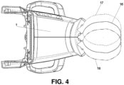
- Figure 4 shows the positioning of patient's head 10 inside the apparatus 1, in different positions.
- the continuous line 16 indicates patient's head, positioned in a symmetric way with respect to the apparatus 1 in all PAN trajectories in apparatuses provided with three and two axes of movement.
- the patient is positioned so that her/his median sagittal plane crosses or passes near the area of movement 15 when the apparatus is provided with three axes of movement (X, Y, R), or when a CBCT acquisition is performed with an apparatus provided with two axes of movement (Y, R) and a central volume must be acquired (incisor teeth).
- the patient is positioned so that her/his median sagittal plane does not pass near the movement area 15 when the apparatus 1 is provided with two axes of movement (Y, R) only, and a CBCT acquisition of posterior teeth or of the temporomandibular joint must be performed.
- the discontinuous lines 17 and 18 indicate said non-symmetric positioning with respect to the apparatus 1.
- a (not shown) arm must be present having a length suitable for positioning the X-ray source at the necessary distance from the sensor (about 1400-1600 mm).
Landscapes
- Health & Medical Sciences (AREA)
- Life Sciences & Earth Sciences (AREA)
- Engineering & Computer Science (AREA)
- Medical Informatics (AREA)
- Pathology (AREA)
- Heart & Thoracic Surgery (AREA)
- Veterinary Medicine (AREA)
- Biophysics (AREA)
- High Energy & Nuclear Physics (AREA)
- Public Health (AREA)
- Nuclear Medicine, Radiotherapy & Molecular Imaging (AREA)
- Optics & Photonics (AREA)
- General Health & Medical Sciences (AREA)
- Radiology & Medical Imaging (AREA)
- Biomedical Technology (AREA)
- Physics & Mathematics (AREA)
- Molecular Biology (AREA)
- Surgery (AREA)
- Animal Behavior & Ethology (AREA)
- Dentistry (AREA)
- Oral & Maxillofacial Surgery (AREA)
- Pulmonology (AREA)
- Theoretical Computer Science (AREA)
- Orthopedic Medicine & Surgery (AREA)
- Neurology (AREA)
- Neurosurgery (AREA)
- Apparatus For Radiation Diagnosis (AREA)
Description
- The present invention refers to the technical field of extraoral dental radiography, and in particular to an apparatus capable of acquiring, in an alternate way, panoramic radiographies, volumetric radiographies with cone beam technique of facial bones, and optionally cranial teleradiographies. More particularly, the invention refers to a method and an apparatus for acquiring panoramic acquisitions (PAN) and volumetric acquisitions (CBCT) without the need of a device alternating the PAN sensor and the CBCT sensor.
- All these types of radiographies are well known in the art.
- Panoramic radiography (PAN; also known as orthopantomography) produces a radiographic image of a curved plan approximating patient jaws, with blurring of the anatomical structures laying outside a narrow layer around the predesigned curved plane.
- Cone beam volumetric radiography (also known as CBCT) is the acquisition, from different projection angles, of a series of two-dimensional radiographic images, which will be processed post-acquisition to reconstruct three-dimensional volumes.
- Teleradiography (also known as CEPH) is a projective radiographic technique, producing radiographic images of the skull or of other anatomical areas from different projections, with minimum magnification and geometrical distortion. Usually two perspectives are represented, latero-lateral and antero-posterior.
- For at least 10 years extraoral apparatuses have been known allowing to obtain PAN and CBCT images using a single source of X-rays and an alternating device allowing to oppose to the X-ray source a PAN sensor or a CBCT sensor in an alternate way. An example of such known apparatus is described in patent
EP2578157B1 of the same applicant. - The alternating device is needed in that the sensors for PAN and CBCT acquisitions are different; moreover, the distance X-ray source-sensor is not the same for PAN and CBCT acquisitions. As a matter of fact, in the panoramic acquisition PAN sensor is usually at a distance comprised between 500 and 580 mm from the X-ray source, while CBCT sensor in the volumetric CBCT radiography is usually positioned at distances comprised between 600 and 700 mm. Generally, for teleradiographic acquisitions the distance X-ray source-sensor is around 1400-1600 mm.
- Many of the apparatuses of the known art have three degrees of freedom or axes of movement: a rotation movement around a rotation axis R, and two translational movements along two directional axes, which are not parallel to each other, are preferably orthogonal and are called X and Y axes. Said translational movements allow to move the rotation axis R inside an XY plane. However, also apparatuses having only two degrees of freedom of movement are known: generally, these apparatuses allow the rotation around R axis and the translation along a directional axis, typically the Y axis. In the field of apparatuses capable of acquiring CBCT images only, apparatuses having only one degree of freedom are known, i.e. the rotation around R rotation axis.
- Typically, with reference to
Figure 1 , R axis is a rotation axis of the arm carrying the X-ray source and sensor, and it is parallel to the vertical axis of the apparatus. Translation X and Y axes are in the horizontal plane, or anyway in a plane perpendicular to R rotation axis; they are visible inFigure 2 and 3 . - Apparatuses for the acquisition of panoramic and/or CBCT and/or CEPH apparatuses having only two degrees of freedom are available on the market, wherein said two degrees of freedom are a first R rotation axis which can be in its turn moved along a circular path around a rotation axis which is parallel and non-coincident.
- Recently a new type of sensor appeared on the market: bi-dimensional (having two similar dimensions) X-ray sensors for CBCT acquisitions, wherein a selection of adjacent pixels, positioned inside a rectangle, which is wide about a few tens of pixels and as high as the entire sensor, or anyway at least about a thousand pixels. These new bi-dimensional sensors can be used for acquiring PAN images. Such sensors have the peculiarity that the PAN selection of pixels can acquire images at a frequency much higher than that needed for a CBCT acquisition. The sensor used in CBCT mode (i.e. with its whole surface) reaches usually an acquisition frequency of about 30 frames/sec, while, when used in PAN mode, the above-described rectangular PAN selection only reaches an acquisition frequency of 100-200 frames/sec.
- Today, when using this sensor, the panoramic image must be reconstructed a posteriori integrating the images obtained by the sensor.
- PAN acquisitions are made on an angle smaller than 360°, generally around 220°; CBCT acquisitions are made normally on 360°, but can be performed even on a smaller angle (typically about 200°).
- Document
EP2774542 disclosed a dental radiographic device capable of carrying out CT imaging and PAN imaging. According to this document revolving arm carrying at the opposite ends the X-ray source and the X-ray sensor is configured to execute a rotation of 360° around the head of a patient. In order to be able to do this the distance between sensor and source must be at least 600mm or more. With a smaller distance, when imaging laterally positioned objects there would exist a risk of collision between the source or the sensor and the head of the patient. The problem of reducing the overall dimensions of the apparatus is not addressed by this document. Similarly, also the problem of avoiding conditions of risk of collisions between the source or the sensor and the head is suggested or discussed. - Document
EP2210559 discloses a similar radiographic device which also carry out CT imaging by a complete rotation around the patient head of the arm supporting the X-ray source and the X-ray sensor at his ends. The problem of reducing the dimensions of the apparatus by keeping the revolving arm a short as possible and at the same time avoiding potential collisions of the source or of the sensor with the head of the patient. - The present invention aims to provide a method and an apparatus allowing to perform alternately PAN or CBCT acquisition without the alternating mechanism described in the above-quoted patent, or without alternating devices described in patents of other manufacturers, or without mechanisms for adapting the X-ray source-sensor distance according to the acquisition to be performed reducing as much as possible the length of the revolving arm avoiding at the same time collisions of the source and of the sensor with the patient head and still providing high quality images.
- This object is achieved by a method and an apparatus having the features of the independent claims. Advantageous embodiment and refinements are specified in the claims dependent thereon.
- The apparatus according to the present invention has a single X-ray sensor, capable of performing PAN and CBCT acquisitions.
- The distance X-ray source-sensor is the same for the two acquisitions, differently from what is usual in the known art. In the present invention, PAN acquisitions occur on the usual 220°, and the CBCT acquisition occurs on 200°.
- Vertical planes through the body parallel to the median plane or the sagittal suture, dividing the body into unequal left and right portions are called sagittal planes. Among all sagittal planes, the plane positioned so as to divide the body into two halves having equal mass and approximately symmetrical and specular is called median sagittal plane.
- Today, radiography apparatus manufacturers tend to provide more and more compact apparatuses in order to save precious space inside the dental practice; this leads also to reducing the distance X-ray source-sensor.
- Having a small distance between X-ray source and sensors is not a problem when acquiring a PAN image, in that the rotation centre of FOV (Field Of View) is on the median sagittal plane of the patient; in this case, the sensor is in front of the patient and never reaches her/his back, where the collision with patient's nape is unavoidable.
- The problem of the collision X-ray sensor-patient during a CBCT acquisition is not present when the centre of the acquired volume is on the median sagittal plane of the patient (incisor teeth), while is present when lateral anatomic areas have to be acquired (molar teeth and temporomandibular joint). The problem is solved using a non-centred, non-symmetrical trajectory with respect to the patient so that the CBCT acquisition trajectory appears:
- Not centred with respect to patient's head (in fact, the actual rotation centre appears very near to a patient's ear, not passing through her/his median sagittal plane)
- Not symmetrical with respect to any of the sagittal plane of the patient:
the acquisition trajectory starts in front of the ear opposed to patient's area to be acquired and ends 200° after, slightly behind the other patient's ear. This trajectory allows to prevent collisions with the patient. - The advantages of the present invention consist in the simplification of the mechanical structure of the apparatus, which becomes much cheaper to produce, thanks to the use of one sensor only and the absence of the alternating device.
- Moreover, the fact that the distance X-ray source-sensor is the same in the PAN and CBCT acquisition is an advantage in the CBCT acquisition, in that, with this distance, shorter than the standard distance, a larger field of view is obtained, because the magnifying factor of the anatomic structures on the sensor is nearer to 1.
- Further advantages and properties of the present invention are disclosed in the following description, in which exemplary embodiments of the present invention are explained in detail based on the drawings:
- Figure 1
- Front view of an apparatus according to the present invention;
- Figure 2
- Exemplary top view of an example of a trajectory performed by the X-sensor during a CBCT acquisition in an apparatus allowing three degrees of freedom: one around the rotation axis of the C-
arm 4, and the other two corresponding to the translation of said rotation axis in the plane perpendicular to said rotation axis and subtended by Cartesian X and Y axes; - Figure 3
- Top view similar to
Figure 2 , wherein the apparatus has two degrees of freedom only, one around the rotation axis of the C-arm, and the other corresponding to the translation of said rotation axis along the Y axis, coincident with the horizontal plane and perpendicular to the rotation axis; - Figure 4
- Top view of a patient positioned inside the apparatus.
- In
Figure 1, 1 indicates on the whole an extraoral radiographic apparatus capable of performing panoramic and CBCT radiographies according to the present invention. Theapparatus 1 comprises apost 2 supporting asupport 3. Thesupport 3 supports a C-arm 4, which is fixed to the horizontal section ofsupport 3. The C-arm 4 in its turn supports at one of its ends anX-ray source 5, and at the opposed end anX-ray sensor 6. Thesupport 3 is vertically movable in order to adjust it to the different heights of individual patients. Moreover, adevice 7 for the positioning of the (not shown) patient is present, which is also fixed to support 3. -
Figure 2 shows in a stylised way thehead 10 of a patient in a top view. The trajectory followed by CBCT sensor during an acquisition is shown byline 11; 12 indicates the centre of rotation oftrajectory 11. The dottedline 13 shows the median sagittal plane of the patient. - In an embodiment having three axes of movement X, Y, and R, the
movement area 15 indicates the area inside which the centre of rotation R can be moved inside the XY plane. During a PAN acquisition the centre ofrotation 12 is moved according a pre-defined trajectory inside the square/rectangle 15. - In an alternative embodiment having movements Y and R only, shown in
Figure 3 , themovement area 15 is reduced to asegment 315 along the Y axis; for the PAN acquisition the rotation R is moved only in Y direction, while during all the CBCT acquisition the rotation axis R is kept steady in the same position. In this alternative embodiment, which is particularly cheap to produce thanks to the absence of the X motor, the trajectory shown inFigure 3 can be obtained thanks to the fact that the patient can be positioned so that her/his median sagittal plane does not pass in the vicinity of themovement area 15. -
Figure 4 shows the positioning of patient'shead 10 inside theapparatus 1, in different positions. Thecontinuous line 16 indicates patient's head, positioned in a symmetric way with respect to theapparatus 1 in all PAN trajectories in apparatuses provided with three and two axes of movement. - The patient is positioned so that her/his median sagittal plane crosses or passes near the area of
movement 15 when the apparatus is provided with three axes of movement (X, Y, R), or when a CBCT acquisition is performed with an apparatus provided with two axes of movement (Y, R) and a central volume must be acquired (incisor teeth). - The patient is positioned so that her/his median sagittal plane does not pass near the
movement area 15 when theapparatus 1 is provided with two axes of movement (Y, R) only, and a CBCT acquisition of posterior teeth or of the temporomandibular joint must be performed. InFigure 4 , thediscontinuous lines apparatus 1. - To the skilled person the fact that the trajectory described in this application can be used also in apparatuses having two sensors, one PAN and one CBCT, or even in apparatuses capable of performing CBCT acquisitions only is apparent.
- Moreover, it is apparent that when the apparatus is used to acquire teleradiographies, a (not shown) arm must be present having a length suitable for positioning the X-ray source at the necessary distance from the sensor (about 1400-1600 mm).
- 1 Extraoral radiographic apparatus
- 2 Post
- 3 Support
- 4 C-arm
- 5 X-ray source
- 6 X-ray sensor
- 7 Device for positioning the patient
- 10 Patient's head
- 11 CBCT sensor trajectory
- 12, 312 Centre of rotation
- 13 Median sagittal plane of the patient
- 14, 314 Centre of the area of
movement 15 - 15, 315 Area of movement
- 16 Symmetrically positioned patient
- 17 Asymmetrically positioned patient
- 18 Asymmetrically positioned patient
Claims (9)
- Method for acquiring volumetric CBCT radiographies and PAN radiographies in an alternate way using an extraoral radiographic apparatus (1), the apparatus (1) comprising rotary supporting arm (4), at whose ends at least an X-ray source (5) and a single X-ray sensor (6) are positioned, said supporting arm (4) having the aim of rotating said at least one source (5) and sensor (6) around a patient; said method comprising the following steps:- setting the distance between the said at least one source (5) and the said sensor (6) to be the distance common for acquiring PAN Images, the said distance being in the range of 500 to 580 mm, and the distance between the X-ray source (5) and the x-ray sensor (6) is the same for the CBCT and the PAN acquisitions, whereby the PAN acquisitions occur with a trajectory having an angular extension of 220°, and the CBCT acquisitions occur with a trajectory having an angular extension of about 200°,- positioning a patient on the positioning device (7), and to prevent collisions with the patient during CBCT acquisitions- setting the rotation centre of the trajectory (11) of said X-ray sensor (6) to be never on the median sagittal plane (13) defined by the patient positioned inside the apparatus (1) and - setting the trajectory to be not symmetrical to any of the possible sagittal planes of the patient.
- Method for acquiring volumetric CBCT and PAN radiographies according to claim 1, wherein said supporting arm is provided with at least three degrees of freedom of movement (R, X, Y) of said couple of at least one X-ray source (5) and sensor (6), said degrees of freedom being a rotation around an axis and the translation of said rotation axis along at least two axes of directions which are perpendicular to each other, said axes being contained in a plane perpendicular to said axis of rotation.
- Method for acquiring volumetric CBCT and PAN radiographies according to claim 1 or 2, wherein said sensor (6) (6) and source (5) have two degrees of freedom of movement (R, Y), corresponding to a rotation around an axis of rotation and to a translation of said axis of rotation along a directional axis perpendicular to said axis of rotation, and wherein the patient is positioned so that her/his median sagittal plane is not in the vicinity of the movement area (315), or is positioned beyond a pre-set minimal distance from the movement area (315) during said CBCT acquisitions performed with a trajectory having an angular extension lower than 360°.
- Method for acquiring volumetric CBCT and PAN radiographies according to one or more of the preceding claims, wherein said acquisition apparatus is a combined apparatus suitable for acquiring and optionally teleradiographic images.
- Extraoral radiographic apparatus (1) for acquiring PAN radiographies and CBCT images in an alternate way, the apparatus (1) comprising a post (2) supporting a support (3), which in its turn supports a C-arm (4) having at one of its ends an X-ray source (5) and on the opposed end a single X-ray sensor (6); the apparatus (1) further comprising a patient positioning device (7), positioning means of said C-arm (4) and means for setting the movement of said C-arm (4), with the C-arm (4) being adapted to rotate around a rotation axis (R), said rotation axis (R) being either not movable in the plane, or movable along an axial direction (Y) coincident with a perpendicular plane to said rotation axis (R), or along two axial directions (X,Y) perpendicular to each other and subtending a plane perpendicular to said rotation axis (R), wherein the C-arm is configured for positioning the said X-ray source (5) and the said X-ray sensor (6) at a fixed distance, which is the distance common for acquiring PAN Images, in the range of about 500 to 580mm; and the same distance between the X-ray source (5) and the X-ray sensor (6) is used for the CBCT and the PAN acquisitions, whereby the PAN acquisitions occur with a trajectory having an angular extension of 220°, and the CBCT acquisitions occur with a trajectory having an angular extension of 200°,
and to prevent collisions with the patient, the apparatus (1) is set to acquire CBCT trajectories with- the rotation centre of the trajectory (11) of said X-ray sensor (6) never being on the median sagittal plane (13) defined by the patient positioned inside the apparatus (1), and- the trajectory not being symmetrical to any of the possible sagittal planes of the patient. - Extraoral radiographic apparatus (1) for acquiring CBCT images and PAN radiographies according to claim 5, having only one sensor for the acquisition of PAN or CBCT images, said sensor having a selection of adjacent pixels with an acquisition frequency higher than the acquisition frequency of the sensor used for its whole surface.
- Extraoral radiographic apparatus (1) for acquiring CBCT images and PAN radiographies according to one or more of the preceding claims 5 or 6, having only one degree of freedom (Y) in addition to the rotation axis (R).
- Extraoral radiographic apparatus (1) for acquiring CBCT images and PAN radiographies according to one or more of the preceding claims 5 to 7, having two degrees of freedom (X, Y) in addition to the rotation axis (R).
- Extraoral radiographic apparatus (1) for acquiring CBCT images and PAN radiographies according to one or more of the preceding claims 5 to 8, suitable for acquiring teleradiographies (CEPH) of patient's skull.
Applications Claiming Priority (1)
| Application Number | Priority Date | Filing Date | Title |
|---|---|---|---|
| ITUB2015A001800A ITUB20151800A1 (en) | 2015-07-02 | 2015-07-02 | METHOD AND APPARATUS FOR THE ACQUISITION OF PANORAMIC RADIOGRAPHIES AND VOLUMETRIC RADIOGRAPHIES CBCT |
Publications (2)
| Publication Number | Publication Date |
|---|---|
| EP3111845A1 EP3111845A1 (en) | 2017-01-04 |
| EP3111845B1 true EP3111845B1 (en) | 2023-07-26 |
Family
ID=54064527
Family Applications (1)
| Application Number | Title | Priority Date | Filing Date |
|---|---|---|---|
| EP16177121.7A Active EP3111845B1 (en) | 2015-07-02 | 2016-06-30 | Method and apparatus for acquiring panoramic and cbct volumetric radiographies |
Country Status (5)
| Country | Link |
|---|---|
| US (1) | US10448902B2 (en) |
| EP (1) | EP3111845B1 (en) |
| CN (1) | CN106308843B (en) |
| FI (1) | FI3111845T3 (en) |
| IT (1) | ITUB20151800A1 (en) |
Families Citing this family (4)
| Publication number | Priority date | Publication date | Assignee | Title |
|---|---|---|---|---|
| USD839427S1 (en) * | 2016-10-14 | 2019-01-29 | J. Morita Mfg. Corp. | Medical X-ray photographing apparatus |
| CN107301622B (en) * | 2017-06-20 | 2018-04-06 | 广州华端科技有限公司 | The panoramic picture acquisition methods and system of oral cavity CBCT images |
| JP7249567B2 (en) * | 2017-10-27 | 2023-03-31 | 学校法人日本大学 | Dental X-ray CT imaging apparatus and X-ray CT imaging condition setting program |
| EP4129191B1 (en) * | 2021-08-06 | 2026-03-04 | DENTSPLY SIRONA Inc. | Optimization of extraoral panorama images by model-based pre-knowledge about the shape of the patient's jaw arch |
Family Cites Families (14)
| Publication number | Priority date | Publication date | Assignee | Title |
|---|---|---|---|---|
| JP4149189B2 (en) * | 2002-04-04 | 2008-09-10 | 株式会社日立メディコ | X-ray CT system |
| DE112006002694B4 (en) * | 2005-10-17 | 2023-02-23 | J. Morita Mfg. Corp. | Medical digital x-ray imaging device and medical digital x-ray sensor |
| US7515679B2 (en) * | 2007-06-01 | 2009-04-07 | Qr (Quantitative Radiography) Srl | Method and system for cone beam x-ray source and detector arrangement in computed tomography systems |
| EP2197547B1 (en) * | 2007-09-13 | 2014-06-11 | Toby D. Henderson | Imaging positioning system having robotically positioned d-arm |
| WO2009063974A1 (en) * | 2007-11-16 | 2009-05-22 | J. Morita Manufacturing Corporation | X-ray ct imaging device |
| FR2924325B1 (en) * | 2007-12-03 | 2010-11-26 | Trophy | DENTAL RADIOLOGY APPARATUS AND ASSOCIATED METHOD. |
| KR101000315B1 (en) * | 2008-07-24 | 2010-12-13 | (주)바텍이우홀딩스 | RPC-driven Dental X-ray Imaging Device |
| JP5757660B2 (en) * | 2010-08-11 | 2015-07-29 | 学校法人日本大学 | X-ray equipment |
| ITBO20110566A1 (en) * | 2011-10-05 | 2013-04-06 | Cefla Coop | DEVICE FOR THE ACQUISITION OF PANORAMIC RADIOGRAPHIES AND VOLUMETRIC CBCT RADIOGRAPHIES |
| JP5863454B2 (en) * | 2011-12-28 | 2016-02-16 | 学校法人日本大学 | X-ray CT imaging apparatus and X-ray CT imaging method |
| ITBO20110764A1 (en) * | 2011-12-28 | 2013-06-29 | Cefla Coop | DEVICE FOR THE ACQUISITION OF PANORAMIC RADIOGRAPHIES, TELERADIOGRAPHIES AND OPTIONAL VOLUMETRIC CBCT RADIOGRAPHIES |
| CN102609965B (en) * | 2012-02-13 | 2014-12-03 | 北京朗视仪器有限公司 | A data processing method for dental CBCT to generate panoramic images |
| JP5805689B2 (en) * | 2013-03-08 | 2015-11-04 | 株式会社モリタ製作所 | X-ray CT imaging apparatus and X-ray CT imaging method |
| CN103690182B (en) * | 2013-12-24 | 2015-09-30 | 杭州美诺瓦医疗科技有限公司 | Digital oral cavity panoramic X-ray imaging device |
-
2015
- 2015-07-02 IT ITUB2015A001800A patent/ITUB20151800A1/en unknown
-
2016
- 2016-06-30 EP EP16177121.7A patent/EP3111845B1/en active Active
- 2016-06-30 US US15/199,134 patent/US10448902B2/en active Active
- 2016-06-30 FI FIEP16177121.7T patent/FI3111845T3/en active
- 2016-07-01 CN CN201610515443.3A patent/CN106308843B/en active Active
Also Published As
| Publication number | Publication date |
|---|---|
| ITUB20151800A1 (en) | 2017-01-02 |
| US10448902B2 (en) | 2019-10-22 |
| CN106308843B (en) | 2020-10-27 |
| US20170007191A1 (en) | 2017-01-12 |
| EP3111845A1 (en) | 2017-01-04 |
| CN106308843A (en) | 2017-01-11 |
| FI3111845T3 (en) | 2023-10-02 |
Similar Documents
| Publication | Publication Date | Title |
|---|---|---|
| EP2609861B1 (en) | Apparatus for acquiring panoramic, teleradiographic and optionally volumetric CBCT radiographies | |
| KR102023549B1 (en) | Apparatus and Method for Digital Radiography | |
| US6496558B2 (en) | X-ray device and medical workplace for diagnostics and surgical interventions in the head and/or jaw of a patient | |
| JP2022106930A (en) | Fixed oral tomosynthesis imaging system, method, and computer-readable medium for 3D dental imaging | |
| EP2578157B1 (en) | Device for the acquisition of panoramic radiographies and CBCT volumetric radiographies | |
| KR20110060899A (en) | Method and apparatus for performing computed tomography X-ray imaging | |
| JP5909278B2 (en) | Patient head support device for imaging | |
| CN105212955A (en) | X-ray imaging unit for medical imaging | |
| US10448902B2 (en) | Method and apparatus for acquiring panoramic and CBCT volumetric radiographies | |
| US10441227B2 (en) | Cephalostat | |
| KR20180073698A (en) | X-ray X-ray image acquisition device capable of acquiring three-dimensional facial optical image and three-ray X-ray image | |
| EP3226250B1 (en) | Beam-limiting device for radiographic apparatuses and method for limiting the beam | |
| EP3393359B1 (en) | Method and device for creating a cephalometric image | |
| KR20150083338A (en) | X-ray image processing apparatus and x-ray photograph system | |
| EP2881040B1 (en) | Method and apparatus for acquiring a scout image in a digital dental radiographic apparatus | |
| US11903748B2 (en) | Method and device for creating a cephalometric image | |
| US20170332985A1 (en) | Cephalometric patient positioning unit extra oral dental imaging devices | |
| KR20170105753A (en) | null | |
| ITBO20110765A1 (en) | APPARATUS FOR DENTAL RADIOGRAPHY WITH IMPROVED ACCURACY | |
| KR20170105756A (en) | 3 Dimensional optical dividing Computerized tomography |
Legal Events
| Date | Code | Title | Description |
|---|---|---|---|
| PUAI | Public reference made under article 153(3) epc to a published international application that has entered the european phase |
Free format text: ORIGINAL CODE: 0009012 |
|
| STAA | Information on the status of an ep patent application or granted ep patent |
Free format text: STATUS: THE APPLICATION HAS BEEN PUBLISHED |
|
| AK | Designated contracting states |
Kind code of ref document: A1 Designated state(s): AL AT BE BG CH CY CZ DE DK EE ES FI FR GB GR HR HU IE IS IT LI LT LU LV MC MK MT NL NO PL PT RO RS SE SI SK SM TR |
|
| AX | Request for extension of the european patent |
Extension state: BA ME |
|
| STAA | Information on the status of an ep patent application or granted ep patent |
Free format text: STATUS: REQUEST FOR EXAMINATION WAS MADE |
|
| 17P | Request for examination filed |
Effective date: 20170629 |
|
| RBV | Designated contracting states (corrected) |
Designated state(s): AL AT BE BG CH CY CZ DE DK EE ES FI FR GB GR HR HU IE IS IT LI LT LU LV MC MK MT NL NO PL PT RO RS SE SI SK SM TR |
|
| STAA | Information on the status of an ep patent application or granted ep patent |
Free format text: STATUS: EXAMINATION IS IN PROGRESS |
|
| 17Q | First examination report despatched |
Effective date: 20200123 |
|
| GRAP | Despatch of communication of intention to grant a patent |
Free format text: ORIGINAL CODE: EPIDOSNIGR1 |
|
| STAA | Information on the status of an ep patent application or granted ep patent |
Free format text: STATUS: GRANT OF PATENT IS INTENDED |
|
| INTG | Intention to grant announced |
Effective date: 20230202 |
|
| GRAS | Grant fee paid |
Free format text: ORIGINAL CODE: EPIDOSNIGR3 |
|
| GRAA | (expected) grant |
Free format text: ORIGINAL CODE: 0009210 |
|
| STAA | Information on the status of an ep patent application or granted ep patent |
Free format text: STATUS: THE PATENT HAS BEEN GRANTED |
|
| AK | Designated contracting states |
Kind code of ref document: B1 Designated state(s): AL AT BE BG CH CY CZ DE DK EE ES FI FR GB GR HR HU IE IS IT LI LT LU LV MC MK MT NL NO PL PT RO RS SE SI SK SM TR |
|
| REG | Reference to a national code |
Ref country code: GB Ref legal event code: FG4D |
|
| REG | Reference to a national code |
Ref country code: CH Ref legal event code: EP |
|
| REG | Reference to a national code |
Ref country code: DE Ref legal event code: R096 Ref document number: 602016081302 Country of ref document: DE |
|
| REG | Reference to a national code |
Ref country code: IE Ref legal event code: FG4D |
|
| REG | Reference to a national code |
Ref country code: FI Ref legal event code: FGE |
|
| REG | Reference to a national code |
Ref country code: LT Ref legal event code: MG9D |
|
| REG | Reference to a national code |
Ref country code: NL Ref legal event code: MP Effective date: 20230726 |
|
| REG | Reference to a national code |
Ref country code: AT Ref legal event code: MK05 Ref document number: 1590990 Country of ref document: AT Kind code of ref document: T Effective date: 20230726 |
|
| PG25 | Lapsed in a contracting state [announced via postgrant information from national office to epo] |
Ref country code: NL Free format text: LAPSE BECAUSE OF FAILURE TO SUBMIT A TRANSLATION OF THE DESCRIPTION OR TO PAY THE FEE WITHIN THE PRESCRIBED TIME-LIMIT Effective date: 20230726 |
|
| PG25 | Lapsed in a contracting state [announced via postgrant information from national office to epo] |
Ref country code: GR Free format text: LAPSE BECAUSE OF FAILURE TO SUBMIT A TRANSLATION OF THE DESCRIPTION OR TO PAY THE FEE WITHIN THE PRESCRIBED TIME-LIMIT Effective date: 20231027 |
|
| PG25 | Lapsed in a contracting state [announced via postgrant information from national office to epo] |
Ref country code: IS Free format text: LAPSE BECAUSE OF FAILURE TO SUBMIT A TRANSLATION OF THE DESCRIPTION OR TO PAY THE FEE WITHIN THE PRESCRIBED TIME-LIMIT Effective date: 20231126 |
|
| PG25 | Lapsed in a contracting state [announced via postgrant information from national office to epo] |
Ref country code: SE Free format text: LAPSE BECAUSE OF FAILURE TO SUBMIT A TRANSLATION OF THE DESCRIPTION OR TO PAY THE FEE WITHIN THE PRESCRIBED TIME-LIMIT Effective date: 20230726 Ref country code: RS Free format text: LAPSE BECAUSE OF FAILURE TO SUBMIT A TRANSLATION OF THE DESCRIPTION OR TO PAY THE FEE WITHIN THE PRESCRIBED TIME-LIMIT Effective date: 20230726 Ref country code: PT Free format text: LAPSE BECAUSE OF FAILURE TO SUBMIT A TRANSLATION OF THE DESCRIPTION OR TO PAY THE FEE WITHIN THE PRESCRIBED TIME-LIMIT Effective date: 20231127 Ref country code: NO Free format text: LAPSE BECAUSE OF FAILURE TO SUBMIT A TRANSLATION OF THE DESCRIPTION OR TO PAY THE FEE WITHIN THE PRESCRIBED TIME-LIMIT Effective date: 20231026 Ref country code: LV Free format text: LAPSE BECAUSE OF FAILURE TO SUBMIT A TRANSLATION OF THE DESCRIPTION OR TO PAY THE FEE WITHIN THE PRESCRIBED TIME-LIMIT Effective date: 20230726 Ref country code: LT Free format text: LAPSE BECAUSE OF FAILURE TO SUBMIT A TRANSLATION OF THE DESCRIPTION OR TO PAY THE FEE WITHIN THE PRESCRIBED TIME-LIMIT Effective date: 20230726 Ref country code: IS Free format text: LAPSE BECAUSE OF FAILURE TO SUBMIT A TRANSLATION OF THE DESCRIPTION OR TO PAY THE FEE WITHIN THE PRESCRIBED TIME-LIMIT Effective date: 20231126 Ref country code: HR Free format text: LAPSE BECAUSE OF FAILURE TO SUBMIT A TRANSLATION OF THE DESCRIPTION OR TO PAY THE FEE WITHIN THE PRESCRIBED TIME-LIMIT Effective date: 20230726 Ref country code: GR Free format text: LAPSE BECAUSE OF FAILURE TO SUBMIT A TRANSLATION OF THE DESCRIPTION OR TO PAY THE FEE WITHIN THE PRESCRIBED TIME-LIMIT Effective date: 20231027 Ref country code: AT Free format text: LAPSE BECAUSE OF FAILURE TO SUBMIT A TRANSLATION OF THE DESCRIPTION OR TO PAY THE FEE WITHIN THE PRESCRIBED TIME-LIMIT Effective date: 20230726 |
|
| PG25 | Lapsed in a contracting state [announced via postgrant information from national office to epo] |
Ref country code: PL Free format text: LAPSE BECAUSE OF FAILURE TO SUBMIT A TRANSLATION OF THE DESCRIPTION OR TO PAY THE FEE WITHIN THE PRESCRIBED TIME-LIMIT Effective date: 20230726 |
|
| PG25 | Lapsed in a contracting state [announced via postgrant information from national office to epo] |
Ref country code: ES Free format text: LAPSE BECAUSE OF FAILURE TO SUBMIT A TRANSLATION OF THE DESCRIPTION OR TO PAY THE FEE WITHIN THE PRESCRIBED TIME-LIMIT Effective date: 20230726 |
|
| REG | Reference to a national code |
Ref country code: DE Ref legal event code: R097 Ref document number: 602016081302 Country of ref document: DE |
|
| PG25 | Lapsed in a contracting state [announced via postgrant information from national office to epo] |
Ref country code: SM Free format text: LAPSE BECAUSE OF FAILURE TO SUBMIT A TRANSLATION OF THE DESCRIPTION OR TO PAY THE FEE WITHIN THE PRESCRIBED TIME-LIMIT Effective date: 20230726 Ref country code: RO Free format text: LAPSE BECAUSE OF FAILURE TO SUBMIT A TRANSLATION OF THE DESCRIPTION OR TO PAY THE FEE WITHIN THE PRESCRIBED TIME-LIMIT Effective date: 20230726 Ref country code: ES Free format text: LAPSE BECAUSE OF FAILURE TO SUBMIT A TRANSLATION OF THE DESCRIPTION OR TO PAY THE FEE WITHIN THE PRESCRIBED TIME-LIMIT Effective date: 20230726 Ref country code: EE Free format text: LAPSE BECAUSE OF FAILURE TO SUBMIT A TRANSLATION OF THE DESCRIPTION OR TO PAY THE FEE WITHIN THE PRESCRIBED TIME-LIMIT Effective date: 20230726 Ref country code: DK Free format text: LAPSE BECAUSE OF FAILURE TO SUBMIT A TRANSLATION OF THE DESCRIPTION OR TO PAY THE FEE WITHIN THE PRESCRIBED TIME-LIMIT Effective date: 20230726 Ref country code: CZ Free format text: LAPSE BECAUSE OF FAILURE TO SUBMIT A TRANSLATION OF THE DESCRIPTION OR TO PAY THE FEE WITHIN THE PRESCRIBED TIME-LIMIT Effective date: 20230726 Ref country code: SK Free format text: LAPSE BECAUSE OF FAILURE TO SUBMIT A TRANSLATION OF THE DESCRIPTION OR TO PAY THE FEE WITHIN THE PRESCRIBED TIME-LIMIT Effective date: 20230726 |
|
| PLBE | No opposition filed within time limit |
Free format text: ORIGINAL CODE: 0009261 |
|
| STAA | Information on the status of an ep patent application or granted ep patent |
Free format text: STATUS: NO OPPOSITION FILED WITHIN TIME LIMIT |
|
| 26N | No opposition filed |
Effective date: 20240429 |
|
| PG25 | Lapsed in a contracting state [announced via postgrant information from national office to epo] |
Ref country code: SI Free format text: LAPSE BECAUSE OF FAILURE TO SUBMIT A TRANSLATION OF THE DESCRIPTION OR TO PAY THE FEE WITHIN THE PRESCRIBED TIME-LIMIT Effective date: 20230726 |
|
| PG25 | Lapsed in a contracting state [announced via postgrant information from national office to epo] |
Ref country code: BG Free format text: LAPSE BECAUSE OF FAILURE TO SUBMIT A TRANSLATION OF THE DESCRIPTION OR TO PAY THE FEE WITHIN THE PRESCRIBED TIME-LIMIT Effective date: 20230726 |
|
| PG25 | Lapsed in a contracting state [announced via postgrant information from national office to epo] |
Ref country code: BG Free format text: LAPSE BECAUSE OF FAILURE TO SUBMIT A TRANSLATION OF THE DESCRIPTION OR TO PAY THE FEE WITHIN THE PRESCRIBED TIME-LIMIT Effective date: 20230726 |
|
| PG25 | Lapsed in a contracting state [announced via postgrant information from national office to epo] |
Ref country code: MC Free format text: LAPSE BECAUSE OF FAILURE TO SUBMIT A TRANSLATION OF THE DESCRIPTION OR TO PAY THE FEE WITHIN THE PRESCRIBED TIME-LIMIT Effective date: 20230726 |
|
| REG | Reference to a national code |
Ref country code: CH Ref legal event code: PL |
|
| PG25 | Lapsed in a contracting state [announced via postgrant information from national office to epo] |
Ref country code: LU Free format text: LAPSE BECAUSE OF NON-PAYMENT OF DUE FEES Effective date: 20240630 |
|
| GBPC | Gb: european patent ceased through non-payment of renewal fee |
Effective date: 20240630 |
|
| PG25 | Lapsed in a contracting state [announced via postgrant information from national office to epo] |
Ref country code: IE Free format text: LAPSE BECAUSE OF NON-PAYMENT OF DUE FEES Effective date: 20240630 |
|
| PG25 | Lapsed in a contracting state [announced via postgrant information from national office to epo] |
Ref country code: BE Free format text: LAPSE BECAUSE OF NON-PAYMENT OF DUE FEES Effective date: 20240630 Ref country code: CH Free format text: LAPSE BECAUSE OF NON-PAYMENT OF DUE FEES Effective date: 20240630 |
|
| PG25 | Lapsed in a contracting state [announced via postgrant information from national office to epo] |
Ref country code: GB Free format text: LAPSE BECAUSE OF NON-PAYMENT OF DUE FEES Effective date: 20240630 |
|
| REG | Reference to a national code |
Ref country code: BE Ref legal event code: MM Effective date: 20240630 |
|
| PG25 | Lapsed in a contracting state [announced via postgrant information from national office to epo] |
Ref country code: CY Free format text: LAPSE BECAUSE OF FAILURE TO SUBMIT A TRANSLATION OF THE DESCRIPTION OR TO PAY THE FEE WITHIN THE PRESCRIBED TIME-LIMIT; INVALID AB INITIO Effective date: 20160630 |
|
| PG25 | Lapsed in a contracting state [announced via postgrant information from national office to epo] |
Ref country code: HU Free format text: LAPSE BECAUSE OF FAILURE TO SUBMIT A TRANSLATION OF THE DESCRIPTION OR TO PAY THE FEE WITHIN THE PRESCRIBED TIME-LIMIT; INVALID AB INITIO Effective date: 20160630 |
|
| PG25 | Lapsed in a contracting state [announced via postgrant information from national office to epo] |
Ref country code: TR Free format text: LAPSE BECAUSE OF FAILURE TO SUBMIT A TRANSLATION OF THE DESCRIPTION OR TO PAY THE FEE WITHIN THE PRESCRIBED TIME-LIMIT Effective date: 20230726 |
|
| PGFP | Annual fee paid to national office [announced via postgrant information from national office to epo] |
Ref country code: IT Payment date: 20260520 Year of fee payment: 11 |
|
| PGFP | Annual fee paid to national office [announced via postgrant information from national office to epo] |
Ref country code: DE Payment date: 20260520 Year of fee payment: 11 |
|
| PGFP | Annual fee paid to national office [announced via postgrant information from national office to epo] |
Ref country code: FI Payment date: 20260520 Year of fee payment: 11 |
|
| PGFP | Annual fee paid to national office [announced via postgrant information from national office to epo] |
Ref country code: FR Payment date: 20260521 Year of fee payment: 11 |