EP3102108B1 - Generating a breast parameter map - Google Patents
Generating a breast parameter map Download PDFInfo
- Publication number
- EP3102108B1 EP3102108B1 EP15702498.5A EP15702498A EP3102108B1 EP 3102108 B1 EP3102108 B1 EP 3102108B1 EP 15702498 A EP15702498 A EP 15702498A EP 3102108 B1 EP3102108 B1 EP 3102108B1
- Authority
- EP
- European Patent Office
- Prior art keywords
- breast
- raster
- unit
- medical imaging
- image data
- Prior art date
- Legal status (The legal status is an assumption and is not a legal conclusion. Google has not performed a legal analysis and makes no representation as to the accuracy of the status listed.)
- Not-in-force
Links
Images


Classifications
-
- A—HUMAN NECESSITIES
- A61—MEDICAL OR VETERINARY SCIENCE; HYGIENE
- A61B—DIAGNOSIS; SURGERY; IDENTIFICATION
- A61B6/00—Apparatus or devices for radiation diagnosis; Apparatus or devices for radiation diagnosis combined with radiation therapy equipment
- A61B6/50—Apparatus or devices for radiation diagnosis; Apparatus or devices for radiation diagnosis combined with radiation therapy equipment specially adapted for specific body parts; specially adapted for specific clinical applications
- A61B6/502—Apparatus or devices for radiation diagnosis; Apparatus or devices for radiation diagnosis combined with radiation therapy equipment specially adapted for specific body parts; specially adapted for specific clinical applications for diagnosis of breast, i.e. mammography
-
- A—HUMAN NECESSITIES
- A61—MEDICAL OR VETERINARY SCIENCE; HYGIENE
- A61B—DIAGNOSIS; SURGERY; IDENTIFICATION
- A61B6/00—Apparatus or devices for radiation diagnosis; Apparatus or devices for radiation diagnosis combined with radiation therapy equipment
- A61B6/42—Arrangements for detecting radiation specially adapted for radiation diagnosis
- A61B6/4208—Arrangements for detecting radiation specially adapted for radiation diagnosis characterised by using a particular type of detector
- A61B6/4241—Arrangements for detecting radiation specially adapted for radiation diagnosis characterised by using a particular type of detector using energy resolving detectors, e.g. photon counting
-
- A—HUMAN NECESSITIES
- A61—MEDICAL OR VETERINARY SCIENCE; HYGIENE
- A61B—DIAGNOSIS; SURGERY; IDENTIFICATION
- A61B6/00—Apparatus or devices for radiation diagnosis; Apparatus or devices for radiation diagnosis combined with radiation therapy equipment
- A61B6/46—Arrangements for interfacing with the operator or the patient
- A61B6/461—Displaying means of special interest
-
- A—HUMAN NECESSITIES
- A61—MEDICAL OR VETERINARY SCIENCE; HYGIENE
- A61B—DIAGNOSIS; SURGERY; IDENTIFICATION
- A61B6/00—Apparatus or devices for radiation diagnosis; Apparatus or devices for radiation diagnosis combined with radiation therapy equipment
- A61B6/52—Devices using data or image processing specially adapted for radiation diagnosis
- A61B6/5211—Devices using data or image processing specially adapted for radiation diagnosis involving processing of medical diagnostic data
- A61B6/5217—Devices using data or image processing specially adapted for radiation diagnosis involving processing of medical diagnostic data extracting a diagnostic or physiological parameter from medical diagnostic data
-
- G—PHYSICS
- G06—COMPUTING OR CALCULATING; COUNTING
- G06T—IMAGE DATA PROCESSING OR GENERATION, IN GENERAL
- G06T7/00—Image analysis
- G06T7/0002—Inspection of images, e.g. flaw detection
- G06T7/0012—Biomedical image inspection
-
- G—PHYSICS
- G16—INFORMATION AND COMMUNICATION TECHNOLOGY [ICT] SPECIALLY ADAPTED FOR SPECIFIC APPLICATION FIELDS
- G16H—HEALTHCARE INFORMATICS, i.e. INFORMATION AND COMMUNICATION TECHNOLOGY [ICT] SPECIALLY ADAPTED FOR THE HANDLING OR PROCESSING OF MEDICAL OR HEALTHCARE DATA
- G16H50/00—ICT specially adapted for medical diagnosis, medical simulation or medical data mining; ICT specially adapted for detecting, monitoring or modelling epidemics or pandemics
- G16H50/30—ICT specially adapted for medical diagnosis, medical simulation or medical data mining; ICT specially adapted for detecting, monitoring or modelling epidemics or pandemics for calculating health indices; for individual health risk assessment
-
- G—PHYSICS
- G06—COMPUTING OR CALCULATING; COUNTING
- G06T—IMAGE DATA PROCESSING OR GENERATION, IN GENERAL
- G06T2207/00—Indexing scheme for image analysis or image enhancement
- G06T2207/10—Image acquisition modality
- G06T2207/10072—Tomographic images
- G06T2207/10081—Computed x-ray tomography [CT]
-
- G—PHYSICS
- G06—COMPUTING OR CALCULATING; COUNTING
- G06T—IMAGE DATA PROCESSING OR GENERATION, IN GENERAL
- G06T2207/00—Indexing scheme for image analysis or image enhancement
- G06T2207/10—Image acquisition modality
- G06T2207/10072—Tomographic images
- G06T2207/10088—Magnetic resonance imaging [MRI]
-
- G—PHYSICS
- G06—COMPUTING OR CALCULATING; COUNTING
- G06T—IMAGE DATA PROCESSING OR GENERATION, IN GENERAL
- G06T2207/00—Indexing scheme for image analysis or image enhancement
- G06T2207/10—Image acquisition modality
- G06T2207/10072—Tomographic images
- G06T2207/10104—Positron emission tomography [PET]
-
- G—PHYSICS
- G06—COMPUTING OR CALCULATING; COUNTING
- G06T—IMAGE DATA PROCESSING OR GENERATION, IN GENERAL
- G06T2207/00—Indexing scheme for image analysis or image enhancement
- G06T2207/10—Image acquisition modality
- G06T2207/10072—Tomographic images
- G06T2207/10108—Single photon emission computed tomography [SPECT]
-
- G—PHYSICS
- G06—COMPUTING OR CALCULATING; COUNTING
- G06T—IMAGE DATA PROCESSING OR GENERATION, IN GENERAL
- G06T2207/00—Indexing scheme for image analysis or image enhancement
- G06T2207/10—Image acquisition modality
- G06T2207/10116—X-ray image
-
- G—PHYSICS
- G06—COMPUTING OR CALCULATING; COUNTING
- G06T—IMAGE DATA PROCESSING OR GENERATION, IN GENERAL
- G06T2207/00—Indexing scheme for image analysis or image enhancement
- G06T2207/10—Image acquisition modality
- G06T2207/10132—Ultrasound image
-
- G—PHYSICS
- G06—COMPUTING OR CALCULATING; COUNTING
- G06T—IMAGE DATA PROCESSING OR GENERATION, IN GENERAL
- G06T2207/00—Indexing scheme for image analysis or image enhancement
- G06T2207/30—Subject of image; Context of image processing
- G06T2207/30004—Biomedical image processing
- G06T2207/30068—Mammography; Breast
Definitions
- the present invention relates to mammography.
- the present invention relates to a medical imaging device for generating a breast parameter map, a medical imaging method for generating a breast parameter map, a computer program element for controlling such device and a computer readable medium having stored such computer program element.
- Mammography information is used for example for breast cancer screening.
- a breast under examination is mechanically compressed and subsequently a radiographic image of the flattened breast tissue is acquired.
- WO 2012/080914 A1 discloses a method for providing mammography information about an object of interest with the following steps: acquiring first and second image data, performing a dual energy basis material decomposition and deriving a density information of the tissue structure of the region of interest from the decomposed basis material image data, and providing the density information to a user. It has been shown that the evaluation of mammographic images is generally difficult and leads to unclear results.
- CHEN B ET AL "Cone-Beam Volume CT Breast Imaging: Wavelet Analysis-based Multi-resolution Reconstruction and De-noising Technique", PROCEEDINGS OF SPIE, SPIE - INTERNATIONAL SOCIETY FOR OPTICAL ENGINEERING, US, vol. 4682, 1 May 2002 (2002-05-01 ), discloses a wavelet analysis-based multi-resolution cone-beam volume CT breast imaging technique that is adaptive for high-resolution and ultra-high resolution reconstructions. Wavelet analysis-based de-noising techniques are employed to improve image quality and further reduce the required absorbed dose.
- US 2012/157819 A1 discloses a method which visualizes a tissue region.
- the method includes the following steps: inserting the tissue region into the capturing region of a first imaging modality, with the tissue region assuming a first shape; capturing the interior of the tissue region by the first imaging modality; establishing a first image volume of the interior of the tissue region when it assumes the first shape; and first transforming of the first image volume into a second image volume, which represents a surface and interior regions of the tissue when the tissue region assumes a second shape.
- FREDENBERG E ET AL "Measurement of breast-tissue x-ray attenuation by spectral mammography: first results on cyst fluid"
- PHYSICS IN MEDICINE AND BIOLOGY, INSTITUTE OF PHYSICS PUBLISHING, BRISTOL GB, vol. 58, no. 24, 20 November 2013 (2013-11-20 ) discloses a method to measure x-ray attenuation of tissue samples using a prototype photon-counting spectral mammography unit. The method was applied to measure the attenuation of 50 samples of breast cyst fluid and 50 samples of water. Spectral (energy-resolved) images of the samples were acquired and the image signal was mapped to equivalent thicknesses of two known reference materials, which can be used to derive the x-ray attenuation as a function of energy.
- a medical imaging device for generating a breast parameter map.
- the medical imaging device comprises an image unit, a raster unit, a definition unit, a generating unit, and a provision unit.
- the image unit is configured to provide image data of a breast.
- the image unit may be an X-ray device, an ultrasound device, a MR device, a CT device, a PET device, a SPECT device and/or else and/or combinations thereof.
- the image unit is an X-ray device with an X-ray source and an X-ray detector.
- the image data of the breast might be a mammogram in cranio-caudal (CC) view, in medio-lateral oblique (MLO) view and/or another view.
- the image data of the breast is acquired in at least two angles of view.
- a 3D breast volume examined by the X-ray device appears as 2D projection in a mammogram.
- the raster unit is configured to provide a predefined spatial raster with several subportions.
- the raster might be radial, grid-shaped or shaped as a pie chart.
- the raster is preferably predefined based on a predefined coordinate system relative to predefined body characteristics of a standard breast.
- predefined points of the raster might relate to the original and intersection of the axes, the position or direction of the axes or else.
- predefined body characteristics might relate to a mammilla, a pectoralis muscle, a pectoralis/chest wall, a breast contour, an axilla and/or combinations thereof.
- the definition unit is configured to define several subvolumes in the breast according to the subportions of the raster and is configured to define an identification of body characteristics in the image data and a segmentation of the image data according to raster subportions and identified body characteristics.
- the subportions of the raster segment the breast volume into several subvolumes.
- the defining comprises an identification of body characteristics in the image data and a segmentation of the image data according to raster subportions and identified body characteristics.
- the generating unit is configured to generate a breast parameter per breast subvolume.
- the subvolumes of the breast are then used to generate at least one breast parameter per breast subvolume.
- the breast parameters can be computed from the image data or be evaluated by other means and devices.
- the breast parameter can be one or more of the following properties: breast density, glandular volume fraction, glandular tissue volume, breast tissue volume, adipose tissue volume, adipose volume fraction, water content or combinations thereof.
- the breast parameter(s) can also be stiffness or elasticity parameters.
- the breast parameter(s) can further be related to a material decomposition of the breast into e.g. aluminium and polymethyl methacrylate (PMMA) from a mammogram, which is acquired by spectral imaging.
- PMMA polymethyl methacrylate
- a spectral mammogram can be acquired for example on a mammography unit with a photon-counting detector with at least two energy bins such as the Philips MicroDose SI system, which enables the separation of a spectral mammogram into a high-energy and a low-energy mammogram for subsequent material decomposition.
- the provision unit is configured to provide a breast parameter per breast subvolume in a breast parameter map, wherein each breast parameter is allocated to its breast subvolume.
- the provision unit preferably comprises a display to show the breast parameter map.
- the breast parameter(s) per region can be visualized by colour coding, grey shades, patterns, symbols, numbers, letters and/or in text form.
- a medical imaging method for generating a breast parameter map comprises the following steps:
- a medical imaging computer program for generating a breast parameter map comprises program code means for causing a medical imaging device as defined in the independent device claim to carry out the steps of the medical imaging method as defined in the independent method claim, when the computer program is run on a computer controlling the object tracking device.
- the subportions of the predefined raster segment the breast volume into several subvolumes.
- the raster is preferably based on a predefined coordinate system relative to predefined body characteristics.
- a breast parameter is generated for each subvolume and provided in a breast parameter map, wherein each breast parameter is allocated to its breast subvolume.
- a breast parameter visualization is created, which makes it easier and clearer to assess the information and in particular to assess the breast condition per breast subvolume. Further, this information combination of condition and location is extremely valuable and still also easy to store, to report and to handle.
- a standardized report is enabled which allow an automatic evaluation of large data amounts for e.g. comparisons over the time (longitudinal parameter tracking) or the population (epidemiological studies).
- this invention proposes to combine the information of the spatial distribution of dense tissue acquired from ipsilateral 2D mammograms (CC+MLO views) to generate estimates of average density values in 3D subvolumes of the breast, which are annotated as subregions in a breast parameter map.
- the density (percentage) or glandular volume (ml) values can be presented colour-coded on a continuous scale with an additional overlay of the local ACR density category (I-IV).
- the corresponding tissue areas have to be identified prior to computing averaged values by using for example a breast coordinate system given by the radial distance to the nipple and the orthogonal distance to the pectoralis/chest wall in each view.
- a reporting of the spatial distribution of the glandular tissue volume and the local breast density in a breast parameter map is provided.
- the medical imaging device for generating a breast parameter map the medical imaging method for generating a breast parameter map, the computer program element for controlling such device and the computer readable medium having stored such computer program element according to the independent claims have similar and/or identical preferred embodiments, in particular, as defined in the dependent claims. It shall be understood further that a preferred embodiment of the invention can also be any combination of the dependent claims with the respective independent claim.
- Figs. 1 to 3 show schematically and exemplarily a medical imaging method for generating a breast parameter map.
- Step-by-step Fig. 1 shows schematically and exemplarily a predefined spatial raster 50 for a breast in a top view and a side view.
- the raster 50 is predefined based on a predefined coordinate system relative to predefined body characteristics of a breast.
- the predefined raster 50 is based on a coordinate system with having the origin of both axes in the mammilla or nipple 56.
- the axes of the coordinate system resemble to crosshairs with the nipple 56 as centre.
- the axes form four subportions of the raster 50. These subportions are characterised by numbers from 1 to 4 starting above right and going clockwise.
- the raster 50 is manually or automatically correctly set into the image data relative to predefined body characteristics. This means, a predefined body characteristic as e.g. the nipple 56 is detected in the image data and the e.g. centre of the raster 50 is set on the nipple 56.
- a predefined body characteristic as e.g. the nipple 56 is detected in the image data and the e.g. centre of the raster 50 is set on the nipple 56.
- the body characteristics for the coordinate system may also be the axilla, the pectoralis muscle itself, the breast contour, which means the skin line and/or combinations thereof. Therefore, the coordinate system may also be otherwise arranged relative to the body characteristics.
- the predefined raster 50 can comprise more or other subportions, which can also be otherwisly formed and/or arranged. For example, circular subportions can be arranged radially around the nipple 56 or the subportions can be shaped as a pie chart around the nipple 56. Also a pixel-by-pixel mapping using the nipple-pectoralis coordinate system could be used to generate an approximate 3D glandularity map on a reference breast diagram.
- the predefined spatial raster 50 is based on a coordinate system with having the pectoralis muscle/chest wall 54 as vertical axis intersecting the horizontal axis through the nipple 56.
- the axes form six subportions of the raster 50. These subportions are characterised by letters A to F starting above right and going above to the left, and down from left to right.
- the raster 50 is a standard raster 50 for a standard breast, which can be adapted to a particular breast shown in the image data. Therefore, predefined points of the standard raster 50 are mapped to predefined body characteristics shown in the image data.
- the raster 50 is not only manually or automatically correctly set into the image data relative to predefined body characteristics, but also manually or automatically adapted to the particular breast shown in the image data, which means the raster subportions are adapted and/or deformed to match this particular breast.
- Fig. 2 shows image data of an actual breast acquired and/or provided by an image unit, here a drawing of a mammography image of an X-ray device.
- the image data of the breast are acquired and provided in two angles of view, namely cranio-caudal (CC) and mediolateral-oblique (MLO).
- Fig. 2 also shows a projection 50' of the raster 50 into the mammograms with corresponding subportions, which define several subvolumens being represented by the projected areas CC 1 to CC 6 and MLO 1 to MLO 6 in the mammograms.
- the subvolumes of the breast are used to generate at least one breast parameter per breast subvolume.
- the breast parameter can be one or more of the following quantitities: breast density, glandular volume fraction, glandular tissue volume, breast tissue volume, adipose tissue volume, adipose volume fraction, water content or combinations thereof.
- the breast parameter can also be a number and/or size of certain features in the subvolume such as the number and/or size of calcifications, lesions, cysts, architectural distortions, asymmetries, spiculated masses or combinations thereof.
- the breast parameter(s) can also be related to a material decomposition of the breast into two base materials as e.g. aluminium and polymethyl methacrylate (PMMA) from a mammogram, which is acquired by spectral imaging.
- a spectral mammogram can be acquired for example on a mammography unit with a photon-counting detector with at least two energy bins such as the Philips MicroDose SI system.
- the breast parameter per breast subvolume is then provided in a breast parameter map, wherein each breast parameter is allocated to its breast subvolume.
- the breast parameter map resembles to the view according to Fig. 1 , but now, the breast parameters for each subvolume are graphically allocated to the respective breast subvolume.
- the breast parameter(s) can be visualized e.g. by colour coding, grey shades, patterns, symbols, numbers, letters, text form and/or combinations thereof.
- the breast subvolumes according to Fig. 1 are characterized by grey shades and roman numbers representing two different breast parameters per breast subvolume.
- Fig. 4 shows a schematic overview of method steps for generating a breast parameter map. It comprises the following steps, not necessarily in this order:
- the method may comprise an identification of body characteristics in the image data and a segmentation of the image data according to raster subportions and identified body characteristics as explained in the following.
- the predefined raster 50 of step 104 is an ideal or a standard raster 50 for a standard breast, which can be adapted in or after step 106 to a particular breast shown in the image data.
- predefined points of the standard raster 50 are mapped to predefined body characteristics shown in the image data.
- the raster 50 is not only manually or automatically correctly set into the image data relative to predefined body characteristics, but also manually or automatically adapted to the particular breast shown in the image data, which means the raster subportions are adapted and/or deformed to match this particular breast.
- the adaption of the raster 50 can further be based on information concerning the acquisition geometry of the image data, as e.g. the projection geometry of a mammography examination.
- the projection area of the selected annotation areas A-F and 1-4 in Fig. 1 have to be identified in the CC and MLO mammograms of Fig. 2 .
- a breast-coordinate system is used, which is defined by the breast nipple 56 and the pectoralis muscle/chest wall 54 as indicated with the broken lines in Fig 2 . Both, pectoralis muscle/chest wall 54 and nipple 56 can be detected automatically in the mammogram.
- the corresponding areas in the mammograms are denoted with CC 1 -CC 6 and MLO 1 -MLO 6 , respectively.
- the glandular volume in subvolume A of the raster equals the measured glandular volume in area ML 1 , and similar equalities hold for subvolumes B-F of the raster.
- these equations yield an approximation for the unknown glandular volumes in the raster subvolumes A-F.
- G 1 + G 4 ML O 1 + ML O 2 + ML O 3
- a breast parameter map with either glandular volume or breast density annotation can be generated by colour-coding the annotation areas with the computed values as depicted in Fig. 3 .
- the spatial distribution of the glandular tissue volume and the local breast density is reported in a breast parameter map.
- the distribution of the glandular volume can be easily depicted and tracked over time in a standardized breast parameter map.
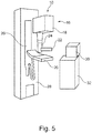
- FIG. 5 shows a schematic drawing of an example of an X-ray imaging system 10 for providing mammography information.
- the X-ray imaging system 10 comprises an image unit 16 with an X-ray source 18 and an X-ray detector 20.
- the X-ray image unit 16 is adapted to provide mammography image data of a breast.
- the example shown is a so-called stand-up investigation system where, for example, a patient in an upright position can stand while, for example, the breast is examined. Therefore, the X-ray detector 20 is provided as a sort of a paddle or small table upon which a breast can be received.
- a moveable compression paddle 22 is provided with an adaptable distance to the detector 20 in order to be able to act with a desired pressing force on the breast arranged detector 20 and the compression paddle 22. Therefore, the compression paddle 22 is attached to an adjusting mechanism 24 allowing the necessary movement of the compression paddle.
- the X-ray source 18 is generating X-ray radiation emanating towards the detector 20. Further, the X-ray source 18 and the detector 20 are attached to an adjustable support 26 allowing for a vertical adjustment such that the height of the detector can be adapted to different sizes of the patient. Further, a rotational movement is possible to acquire X-ray images not only in a vertical direction, but also in a direction with an angle to the vertical direction, such as 30° or any freely chooseable angulation as well as an X-ray viewing direction in a horizontal way. Further, a base 28 is provided which is securely fixed to a floor of an examination room, for example.
- the X-ray imaging system 10 also comprises a provision unit 30 with a display 34 provided on a separate base 32. It is noted that any data connection is not further shown, which data connection can be provided as wire connection or wireless connection between the respective parts of the system 10. It must be further noted that besides the shown stand-up investigation system, also other types for X-ray imaging are possible, for example moveable or stationary X-ray imaging systems or X-ray imaging systems with a table upon which a patient can be received in order to acquire X-ray images while the patient is lying on the table, for example facing downwards.
- the image data of the breast can also be acquired by an ultrasound device, a MR device, a CT device, a PET device, a SPECT device and/or else.
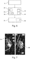
- Fig. 6 shows a schematic drawing of an example of an image unit 16 and a processing unit 40.
- the exemplary processing unit 40 comprises a raster unit 42, a definition unit 44 and a generating unit 46.
- An exemplary provision unit 30 can be arranged either in the processing unit 40 or separate from the processing unit 40.
- the provision unit 30 comprises e.g. a display 34.
- the image unit 16 is configured to provide image data of a breast
- the raster unit 42 is configured to provide a predefined raster with several subportions.
- the definition unit 44 is configured to define several subvolumes in the breast according to the subportions of the raster 50.
- the subvolumes are preferably adapted to the actual image data.
- the generating unit 46 is configured to generate a breast parameter per breast subvolume
- the provision unit 30 is configured to provide a breast parameter per breast subvolume in a breast parameter map, wherein each breast parameter is allocated to its breast subvolume.
- Fig. 7 shows image data of an actual breast acquired and/or provided by an image unit, here a mammography image of an X-ray device.
- the image data of the breast are acquired and provided in two angles of view, namely cranio-caudal (CC) and mediolateral-oblique (MLO).
- Fig. 7 also shows the projection 50' of the raster 50 with subportions, which define several subvolumes of the breast and are projected onto the area segments CC 1 -CC 6 and MLO 1 -MLO 6 in the CC and MLO mammogram, respectively.
- a computer program or a computer program element is provided that is characterized by being adapted to execute the method steps of the method according to one of the preceding embodiments, on an appropriate system.
- the computer program element might therefore be stored on a computer unit, which might also be part of an embodiment of the present invention.
- This computing unit may be adapted to perform or induce a performing of the steps of the method described above. Moreover, it may be adapted to operate the components of the above described apparatus.
- the computing unit can be adapted to operate automatically and/or to execute the orders of a user.
- a computer program may be loaded into a working memory of a data processor. The data processor may thus be equipped to carry out the method of the invention.
- This exemplary embodiment of the invention covers both, a computer program that right from the beginning uses the invention and a computer program that by means of an up-date turns an existing program into a program that uses the invention. Further on, the computer program element might be able to provide all necessary steps to fulfil the procedure of an exemplary embodiment of the method as described above.
- a computer readable medium such as a CD-ROM
- the computer readable medium has a computer program element stored on it, which computer program element is described by the preceding section.
- a computer program may be stored and/or distributed on a suitable medium, such as an optical storage medium or a solid state medium supplied together with or as part of other hardware, but may also be distributed in other forms, such as via the internet or other wired or wireless telecommunication systems.
- the computer program may also be presented over a network like the World Wide Web and can be downloaded into the working memory of a data processor from such a network.
- a medium for making a computer program element available for downloading is provided, which computer program element is arranged to perform a method according to one of the previously described embodiments of the invention.
Landscapes
- Health & Medical Sciences (AREA)
- Engineering & Computer Science (AREA)
- Life Sciences & Earth Sciences (AREA)
- Medical Informatics (AREA)
- General Health & Medical Sciences (AREA)
- Physics & Mathematics (AREA)
- Radiology & Medical Imaging (AREA)
- Nuclear Medicine, Radiotherapy & Molecular Imaging (AREA)
- Public Health (AREA)
- Biomedical Technology (AREA)
- Pathology (AREA)
- Veterinary Medicine (AREA)
- Surgery (AREA)
- Biophysics (AREA)
- High Energy & Nuclear Physics (AREA)
- Animal Behavior & Ethology (AREA)
- Optics & Photonics (AREA)
- Molecular Biology (AREA)
- Heart & Thoracic Surgery (AREA)
- Computer Vision & Pattern Recognition (AREA)
- Theoretical Computer Science (AREA)
- Quality & Reliability (AREA)
- General Physics & Mathematics (AREA)
- Dentistry (AREA)
- Oral & Maxillofacial Surgery (AREA)
- Physiology (AREA)
- Human Computer Interaction (AREA)
- Epidemiology (AREA)
- Databases & Information Systems (AREA)
- Data Mining & Analysis (AREA)
- Primary Health Care (AREA)
- Apparatus For Radiation Diagnosis (AREA)
- Ultra Sonic Daignosis Equipment (AREA)
Applications Claiming Priority (2)
| Application Number | Priority Date | Filing Date | Title |
|---|---|---|---|
| EP14153740 | 2014-02-04 | ||
| PCT/EP2015/052344 WO2015118033A1 (en) | 2014-02-04 | 2015-02-04 | Generating a breast parameter map |
Publications (2)
| Publication Number | Publication Date |
|---|---|
| EP3102108A1 EP3102108A1 (en) | 2016-12-14 |
| EP3102108B1 true EP3102108B1 (en) | 2017-12-13 |
Family
ID=50101693
Family Applications (1)
| Application Number | Title | Priority Date | Filing Date |
|---|---|---|---|
| EP15702498.5A Not-in-force EP3102108B1 (en) | 2014-02-04 | 2015-02-04 | Generating a breast parameter map |
Country Status (5)
| Country | Link |
|---|---|
| US (1) | US10238354B2 (enExample) |
| EP (1) | EP3102108B1 (enExample) |
| JP (1) | JP6262869B2 (enExample) |
| CN (1) | CN105979875B (enExample) |
| WO (1) | WO2015118033A1 (enExample) |
Families Citing this family (6)
| Publication number | Priority date | Publication date | Assignee | Title |
|---|---|---|---|---|
| JP6448859B2 (ja) * | 2015-10-05 | 2019-01-09 | コーニンクレッカ フィリップス エヌ ヴェKoninklijke Philips N.V. | 身体部位の特徴のキャラクタリゼーションのための装置 |
| JP2017164312A (ja) * | 2016-03-16 | 2017-09-21 | キヤノン株式会社 | 放射線撮影装置、挿入状態判定方法、及びプログラム |
| CN109993170A (zh) * | 2019-05-10 | 2019-07-09 | 图兮深维医疗科技(苏州)有限公司 | 一种乳腺病灶钟形图展示装置及设备 |
| CN110459319B (zh) * | 2019-05-16 | 2021-05-25 | 腾讯科技(深圳)有限公司 | 基于人工智能的乳腺钼靶图像的辅助诊断系统 |
| CN110136809B (zh) | 2019-05-22 | 2022-12-27 | 腾讯科技(深圳)有限公司 | 一种医疗图像处理方法、装置、电子医疗设备和存储介质 |
| CN120549537A (zh) * | 2024-02-29 | 2025-08-29 | 通用电气精准医疗有限责任公司 | 乳房密度的量化方法和超声成像系统 |
Family Cites Families (24)
| Publication number | Priority date | Publication date | Assignee | Title |
|---|---|---|---|---|
| US5579360A (en) * | 1994-12-30 | 1996-11-26 | Philips Electronics North America Corporation | Mass detection by computer using digital mammograms of the same breast taken from different viewing directions |
| US5572565A (en) | 1994-12-30 | 1996-11-05 | Philips Electronics North America Corporation | Automatic segmentation, skinline and nipple detection in digital mammograms |
| US20030211036A1 (en) * | 2002-05-07 | 2003-11-13 | Hadassa Degani | Method and apparatus for monitoring and quantitatively evaluating tumor perfusion |
| US7177453B2 (en) * | 2002-11-26 | 2007-02-13 | General Electric Company | Method and apparatus for partitioning a volume |
| US20040102699A1 (en) * | 2002-11-26 | 2004-05-27 | Ge Medical Systems Information Technologies, Inc. | Tool and method to produce orientation marker on a subject of interest |
| US20060251301A1 (en) | 2004-11-02 | 2006-11-09 | Mcnamara Michael P Jr | Method and apparatus for determining correlation between spatial coordinates in breast |
| US10492749B2 (en) * | 2005-05-03 | 2019-12-03 | The Regents Of The University Of California | Biopsy systems for breast computed tomography |
| US10008184B2 (en) * | 2005-11-10 | 2018-06-26 | Hologic, Inc. | System and method for generating a 2D image using mammography and/or tomosynthesis image data |
| GB0602739D0 (en) * | 2006-02-10 | 2006-03-22 | Ccbr As | Breast tissue density measure |
| US8285019B2 (en) | 2006-02-10 | 2012-10-09 | Synarc Inc. | Breast tissue density measure |
| CN1846616A (zh) * | 2006-03-13 | 2006-10-18 | 华中科技大学 | 一种计算机辅助预测乳腺癌风险的方法 |
| FR2919747B1 (fr) * | 2007-08-02 | 2009-11-06 | Gen Electric | Procede et systeme d'affichage d'images de tomosynthese |
| US8537159B2 (en) * | 2007-09-03 | 2013-09-17 | Koninklijke Philips N.V. | Visualization of voxel data |
| JP5399278B2 (ja) | 2009-03-31 | 2014-01-29 | 富士フイルム株式会社 | 乳腺含有率推定装置及び方法 |
| EP2535829A3 (en) * | 2009-10-07 | 2013-07-10 | Hologic, Inc. | Processing and displaying computer-aided detection information associated with breast x-ray images |
| JP5820383B2 (ja) * | 2009-10-30 | 2015-11-24 | コーニンクレッカ フィリップス エヌ ヴェKoninklijke Philips N.V. | 画像データにより表される病巣の3次元解析 |
| US8675933B2 (en) | 2010-04-30 | 2014-03-18 | Vucomp, Inc. | Breast segmentation in radiographic images |
| BR112013014567A2 (pt) | 2010-12-13 | 2017-07-04 | Koninl Philips Electronics Nv | sistema de geração de imagens por raios x, método para a provisão de informações da mamografia sobre um objeto de interesse, elemento de programa de computador para o controle de um aparelho e meio legível por computador |
| DE102010063810B4 (de) | 2010-12-21 | 2019-06-06 | Siemens Healthcare Gmbh | Bildgebendes Verfahren und bildgebende Vorrichtung zum Darstellen dekomprimierter Ansichten eines Gewebebereiches |
| WO2012107057A1 (en) | 2011-02-10 | 2012-08-16 | Mevis Medical Solutions Ag | Image processing device |
| WO2012116746A1 (en) | 2011-03-02 | 2012-09-07 | Mevis Medical Solutions Ag | Image processing device for finding corresponding regions in two image data sets of an object |
| US9805507B2 (en) * | 2012-02-13 | 2017-10-31 | Hologic, Inc | System and method for navigating a tomosynthesis stack using synthesized image data |
| US9047664B2 (en) * | 2013-05-31 | 2015-06-02 | Kathryn Pearson Peyton | Apparatus and method for utilizing mammogram images for verification |
| EP4278977A3 (en) * | 2013-10-24 | 2024-02-21 | Hologic, Inc. | System and method for navigating x-ray guided breast biopsy |
-
2015
- 2015-02-04 WO PCT/EP2015/052344 patent/WO2015118033A1/en not_active Ceased
- 2015-02-04 CN CN201580007187.0A patent/CN105979875B/zh not_active Expired - Fee Related
- 2015-02-04 JP JP2016549136A patent/JP6262869B2/ja not_active Expired - Fee Related
- 2015-02-04 EP EP15702498.5A patent/EP3102108B1/en not_active Not-in-force
- 2015-02-04 US US15/114,438 patent/US10238354B2/en not_active Expired - Fee Related
Also Published As
| Publication number | Publication date |
|---|---|
| WO2015118033A1 (en) | 2015-08-13 |
| CN105979875A (zh) | 2016-09-28 |
| US10238354B2 (en) | 2019-03-26 |
| JP2017504432A (ja) | 2017-02-09 |
| US20160338660A1 (en) | 2016-11-24 |
| EP3102108A1 (en) | 2016-12-14 |
| JP6262869B2 (ja) | 2018-01-17 |
| CN105979875B (zh) | 2019-12-31 |
Similar Documents
| Publication | Publication Date | Title |
|---|---|---|
| US9836872B2 (en) | Methods for generation of edge=preserving synthetic mammograms from tomosynthesis data | |
| EP3102108B1 (en) | Generating a breast parameter map | |
| JP6766045B2 (ja) | トモシンセシスデータから合成マンモグラムを生成する方法 | |
| US10448911B2 (en) | Method and device for displaying medical images | |
| EP2602743B1 (en) | Matching geometry generation and display of mammograms and tomosynthesis images | |
| EP1750584B1 (en) | System and method for diagnosing breast cancer | |
| US9401019B2 (en) | Imaging tomosynthesis system, in particular mammography system | |
| CN100573588C (zh) | 使用截短的投影和在先采集的3d ct图像的锥形束ct设备 | |
| US8798353B2 (en) | Apparatus and method for two-view tomosynthesis imaging | |
| US9168013B2 (en) | Breast density assessment | |
| EP3219261A1 (en) | Breast tomosynthesis with flexible compression paddle | |
| US8660329B2 (en) | Method for reconstruction of a three-dimensional model of a body structure | |
| CN109419526A (zh) | 用于数字乳房断层合成中的运动评估和校正的方法和系统 | |
| US7978886B2 (en) | System and method for anatomy based reconstruction | |
| KR102387403B1 (ko) | 잘림 아티팩트 저감 프로젝션 데이터 보정방법 | |
| EP3076872B1 (en) | Device and method for tomosynthesis imaging | |
| Montúfar et al. | Perspective and orthogonal CBCT/CT digitally reconstructed radiographs compared to conventional cephalograms | |
| US7116808B2 (en) | Method for producing an image sequence from volume datasets | |
| WO2012085818A1 (en) | Mammography calcium score | |
| EP4190245A1 (en) | Quantitative radiographic imaging using a 3d camera |
Legal Events
| Date | Code | Title | Description |
|---|---|---|---|
| PUAI | Public reference made under article 153(3) epc to a published international application that has entered the european phase |
Free format text: ORIGINAL CODE: 0009012 |
|
| 17P | Request for examination filed |
Effective date: 20160905 |
|
| AK | Designated contracting states |
Kind code of ref document: A1 Designated state(s): AL AT BE BG CH CY CZ DE DK EE ES FI FR GB GR HR HU IE IS IT LI LT LU LV MC MK MT NL NO PL PT RO RS SE SI SK SM TR |
|
| AX | Request for extension of the european patent |
Extension state: BA ME |
|
| DAX | Request for extension of the european patent (deleted) | ||
| GRAP | Despatch of communication of intention to grant a patent |
Free format text: ORIGINAL CODE: EPIDOSNIGR1 |
|
| INTG | Intention to grant announced |
Effective date: 20170710 |
|
| GRAS | Grant fee paid |
Free format text: ORIGINAL CODE: EPIDOSNIGR3 |
|
| GRAA | (expected) grant |
Free format text: ORIGINAL CODE: 0009210 |
|
| REG | Reference to a national code |
Ref country code: GB Ref legal event code: FG4D |
|
| REG | Reference to a national code |
Ref country code: AT Ref legal event code: REF Ref document number: 953610 Country of ref document: AT Kind code of ref document: T Effective date: 20171215 Ref country code: CH Ref legal event code: EP |
|
| REG | Reference to a national code |
Ref country code: IE Ref legal event code: FG4D |
|
| REG | Reference to a national code |
Ref country code: DE Ref legal event code: R096 Ref document number: 602015006665 Country of ref document: DE |
|
| REG | Reference to a national code |
Ref country code: DE Ref legal event code: R084 Ref document number: 602015006665 Country of ref document: DE |
|
| REG | Reference to a national code |
Ref country code: NL Ref legal event code: MP Effective date: 20171213 |
|
| PG25 | Lapsed in a contracting state [announced via postgrant information from national office to epo] |
Ref country code: FI Free format text: LAPSE BECAUSE OF FAILURE TO SUBMIT A TRANSLATION OF THE DESCRIPTION OR TO PAY THE FEE WITHIN THE PRESCRIBED TIME-LIMIT Effective date: 20171213 Ref country code: SE Free format text: LAPSE BECAUSE OF FAILURE TO SUBMIT A TRANSLATION OF THE DESCRIPTION OR TO PAY THE FEE WITHIN THE PRESCRIBED TIME-LIMIT Effective date: 20171213 Ref country code: NO Free format text: LAPSE BECAUSE OF FAILURE TO SUBMIT A TRANSLATION OF THE DESCRIPTION OR TO PAY THE FEE WITHIN THE PRESCRIBED TIME-LIMIT Effective date: 20180313 |
|
| REG | Reference to a national code |
Ref country code: AT Ref legal event code: MK05 Ref document number: 953610 Country of ref document: AT Kind code of ref document: T Effective date: 20171213 |
|
| PG25 | Lapsed in a contracting state [announced via postgrant information from national office to epo] |
Ref country code: BG Free format text: LAPSE BECAUSE OF FAILURE TO SUBMIT A TRANSLATION OF THE DESCRIPTION OR TO PAY THE FEE WITHIN THE PRESCRIBED TIME-LIMIT Effective date: 20180313 Ref country code: HR Free format text: LAPSE BECAUSE OF FAILURE TO SUBMIT A TRANSLATION OF THE DESCRIPTION OR TO PAY THE FEE WITHIN THE PRESCRIBED TIME-LIMIT Effective date: 20171213 Ref country code: RS Free format text: LAPSE BECAUSE OF FAILURE TO SUBMIT A TRANSLATION OF THE DESCRIPTION OR TO PAY THE FEE WITHIN THE PRESCRIBED TIME-LIMIT Effective date: 20171213 Ref country code: LV Free format text: LAPSE BECAUSE OF FAILURE TO SUBMIT A TRANSLATION OF THE DESCRIPTION OR TO PAY THE FEE WITHIN THE PRESCRIBED TIME-LIMIT Effective date: 20171213 Ref country code: GR Free format text: LAPSE BECAUSE OF FAILURE TO SUBMIT A TRANSLATION OF THE DESCRIPTION OR TO PAY THE FEE WITHIN THE PRESCRIBED TIME-LIMIT Effective date: 20180314 |
|
| PG25 | Lapsed in a contracting state [announced via postgrant information from national office to epo] |
Ref country code: NL Free format text: LAPSE BECAUSE OF FAILURE TO SUBMIT A TRANSLATION OF THE DESCRIPTION OR TO PAY THE FEE WITHIN THE PRESCRIBED TIME-LIMIT Effective date: 20171213 |
|
| PG25 | Lapsed in a contracting state [announced via postgrant information from national office to epo] |
Ref country code: EE Free format text: LAPSE BECAUSE OF FAILURE TO SUBMIT A TRANSLATION OF THE DESCRIPTION OR TO PAY THE FEE WITHIN THE PRESCRIBED TIME-LIMIT Effective date: 20171213 Ref country code: CY Free format text: LAPSE BECAUSE OF FAILURE TO SUBMIT A TRANSLATION OF THE DESCRIPTION OR TO PAY THE FEE WITHIN THE PRESCRIBED TIME-LIMIT Effective date: 20171213 Ref country code: CZ Free format text: LAPSE BECAUSE OF FAILURE TO SUBMIT A TRANSLATION OF THE DESCRIPTION OR TO PAY THE FEE WITHIN THE PRESCRIBED TIME-LIMIT Effective date: 20171213 Ref country code: ES Free format text: LAPSE BECAUSE OF FAILURE TO SUBMIT A TRANSLATION OF THE DESCRIPTION OR TO PAY THE FEE WITHIN THE PRESCRIBED TIME-LIMIT Effective date: 20171213 Ref country code: SK Free format text: LAPSE BECAUSE OF FAILURE TO SUBMIT A TRANSLATION OF THE DESCRIPTION OR TO PAY THE FEE WITHIN THE PRESCRIBED TIME-LIMIT Effective date: 20171213 |
|
| PG25 | Lapsed in a contracting state [announced via postgrant information from national office to epo] |
Ref country code: PL Free format text: LAPSE BECAUSE OF FAILURE TO SUBMIT A TRANSLATION OF THE DESCRIPTION OR TO PAY THE FEE WITHIN THE PRESCRIBED TIME-LIMIT Effective date: 20171213 Ref country code: AT Free format text: LAPSE BECAUSE OF FAILURE TO SUBMIT A TRANSLATION OF THE DESCRIPTION OR TO PAY THE FEE WITHIN THE PRESCRIBED TIME-LIMIT Effective date: 20171213 Ref country code: IS Free format text: LAPSE BECAUSE OF FAILURE TO SUBMIT A TRANSLATION OF THE DESCRIPTION OR TO PAY THE FEE WITHIN THE PRESCRIBED TIME-LIMIT Effective date: 20180413 Ref country code: SM Free format text: LAPSE BECAUSE OF FAILURE TO SUBMIT A TRANSLATION OF THE DESCRIPTION OR TO PAY THE FEE WITHIN THE PRESCRIBED TIME-LIMIT Effective date: 20171213 Ref country code: IT Free format text: LAPSE BECAUSE OF FAILURE TO SUBMIT A TRANSLATION OF THE DESCRIPTION OR TO PAY THE FEE WITHIN THE PRESCRIBED TIME-LIMIT Effective date: 20171213 |
|
| REG | Reference to a national code |
Ref country code: CH Ref legal event code: PL Ref country code: DE Ref legal event code: R097 Ref document number: 602015006665 Country of ref document: DE |
|
| PG25 | Lapsed in a contracting state [announced via postgrant information from national office to epo] |
Ref country code: MC Free format text: LAPSE BECAUSE OF FAILURE TO SUBMIT A TRANSLATION OF THE DESCRIPTION OR TO PAY THE FEE WITHIN THE PRESCRIBED TIME-LIMIT Effective date: 20171213 |
|
| PLBE | No opposition filed within time limit |
Free format text: ORIGINAL CODE: 0009261 |
|
| STAA | Information on the status of an ep patent application or granted ep patent |
Free format text: STATUS: NO OPPOSITION FILED WITHIN TIME LIMIT |
|
| 26N | No opposition filed |
Effective date: 20180914 |
|
| REG | Reference to a national code |
Ref country code: IE Ref legal event code: MM4A |
|
| REG | Reference to a national code |
Ref country code: BE Ref legal event code: MM Effective date: 20180228 |
|
| PG25 | Lapsed in a contracting state [announced via postgrant information from national office to epo] |
Ref country code: CH Free format text: LAPSE BECAUSE OF NON-PAYMENT OF DUE FEES Effective date: 20180228 Ref country code: DK Free format text: LAPSE BECAUSE OF FAILURE TO SUBMIT A TRANSLATION OF THE DESCRIPTION OR TO PAY THE FEE WITHIN THE PRESCRIBED TIME-LIMIT Effective date: 20171213 Ref country code: LU Free format text: LAPSE BECAUSE OF NON-PAYMENT OF DUE FEES Effective date: 20180204 Ref country code: LI Free format text: LAPSE BECAUSE OF NON-PAYMENT OF DUE FEES Effective date: 20180228 |
|
| REG | Reference to a national code |
Ref country code: FR Ref legal event code: ST Effective date: 20181031 |
|
| PG25 | Lapsed in a contracting state [announced via postgrant information from national office to epo] |
Ref country code: IE Free format text: LAPSE BECAUSE OF NON-PAYMENT OF DUE FEES Effective date: 20180204 |
|
| PG25 | Lapsed in a contracting state [announced via postgrant information from national office to epo] |
Ref country code: SI Free format text: LAPSE BECAUSE OF FAILURE TO SUBMIT A TRANSLATION OF THE DESCRIPTION OR TO PAY THE FEE WITHIN THE PRESCRIBED TIME-LIMIT Effective date: 20171213 Ref country code: FR Free format text: LAPSE BECAUSE OF NON-PAYMENT OF DUE FEES Effective date: 20180228 Ref country code: BE Free format text: LAPSE BECAUSE OF NON-PAYMENT OF DUE FEES Effective date: 20180228 |
|
| GBPC | Gb: european patent ceased through non-payment of renewal fee |
Effective date: 20190204 |
|
| PG25 | Lapsed in a contracting state [announced via postgrant information from national office to epo] |
Ref country code: GB Free format text: LAPSE BECAUSE OF NON-PAYMENT OF DUE FEES Effective date: 20190204 Ref country code: MT Free format text: LAPSE BECAUSE OF NON-PAYMENT OF DUE FEES Effective date: 20180204 |
|
| PG25 | Lapsed in a contracting state [announced via postgrant information from national office to epo] |
Ref country code: TR Free format text: LAPSE BECAUSE OF FAILURE TO SUBMIT A TRANSLATION OF THE DESCRIPTION OR TO PAY THE FEE WITHIN THE PRESCRIBED TIME-LIMIT Effective date: 20171213 |
|
| PG25 | Lapsed in a contracting state [announced via postgrant information from national office to epo] |
Ref country code: PT Free format text: LAPSE BECAUSE OF FAILURE TO SUBMIT A TRANSLATION OF THE DESCRIPTION OR TO PAY THE FEE WITHIN THE PRESCRIBED TIME-LIMIT Effective date: 20171213 |
|
| PG25 | Lapsed in a contracting state [announced via postgrant information from national office to epo] |
Ref country code: MK Free format text: LAPSE BECAUSE OF NON-PAYMENT OF DUE FEES Effective date: 20171213 Ref country code: HU Free format text: LAPSE BECAUSE OF FAILURE TO SUBMIT A TRANSLATION OF THE DESCRIPTION OR TO PAY THE FEE WITHIN THE PRESCRIBED TIME-LIMIT; INVALID AB INITIO Effective date: 20150204 Ref country code: RO Free format text: LAPSE BECAUSE OF FAILURE TO SUBMIT A TRANSLATION OF THE DESCRIPTION OR TO PAY THE FEE WITHIN THE PRESCRIBED TIME-LIMIT Effective date: 20171213 Ref country code: LT Free format text: LAPSE BECAUSE OF FAILURE TO SUBMIT A TRANSLATION OF THE DESCRIPTION OR TO PAY THE FEE WITHIN THE PRESCRIBED TIME-LIMIT Effective date: 20171213 |
|
| PG25 | Lapsed in a contracting state [announced via postgrant information from national office to epo] |
Ref country code: AL Free format text: LAPSE BECAUSE OF FAILURE TO SUBMIT A TRANSLATION OF THE DESCRIPTION OR TO PAY THE FEE WITHIN THE PRESCRIBED TIME-LIMIT Effective date: 20171213 |
|
| PGFP | Annual fee paid to national office [announced via postgrant information from national office to epo] |
Ref country code: DE Payment date: 20210225 Year of fee payment: 7 |
|
| REG | Reference to a national code |
Ref country code: DE Ref legal event code: R119 Ref document number: 602015006665 Country of ref document: DE |
|
| PG25 | Lapsed in a contracting state [announced via postgrant information from national office to epo] |
Ref country code: DE Free format text: LAPSE BECAUSE OF NON-PAYMENT OF DUE FEES Effective date: 20220901 |