EP3095380A2 - Waage mit erweiterten funktionen - Google Patents
Waage mit erweiterten funktionen Download PDFInfo
- Publication number
- EP3095380A2 EP3095380A2 EP16167797.6A EP16167797A EP3095380A2 EP 3095380 A2 EP3095380 A2 EP 3095380A2 EP 16167797 A EP16167797 A EP 16167797A EP 3095380 A2 EP3095380 A2 EP 3095380A2
- Authority
- EP
- European Patent Office
- Prior art keywords
- user
- heart
- signal
- deltahb
- amplitude
- Prior art date
- Legal status (The legal status is an assumption and is not a legal conclusion. Google has not performed a legal analysis and makes no representation as to the accuracy of the status listed.)
- Withdrawn
Links
- 238000005303 weighing Methods 0.000 title description 5
- 238000000034 method Methods 0.000 claims abstract description 43
- 230000004872 arterial blood pressure Effects 0.000 claims abstract description 23
- 230000036772 blood pressure Effects 0.000 claims abstract description 23
- 230000029058 respiratory gaseous exchange Effects 0.000 claims description 31
- 210000004369 blood Anatomy 0.000 claims description 25
- 239000008280 blood Substances 0.000 claims description 25
- 230000007423 decrease Effects 0.000 claims description 15
- 238000005259 measurement Methods 0.000 claims description 10
- 230000000747 cardiac effect Effects 0.000 claims description 9
- 230000036581 peripheral resistance Effects 0.000 claims description 9
- 230000008602 contraction Effects 0.000 claims description 7
- 230000002526 effect on cardiovascular system Effects 0.000 claims description 4
- 102100028379 Methionine aminopeptidase 1 Human genes 0.000 claims description 3
- 101710161855 Methionine aminopeptidase 1 Proteins 0.000 claims description 3
- 102100023174 Methionine aminopeptidase 2 Human genes 0.000 claims description 3
- 108090000192 Methionyl aminopeptidases Proteins 0.000 claims description 3
- 230000001747 exhibiting effect Effects 0.000 claims description 3
- 238000007620 mathematical function Methods 0.000 claims description 2
- 210000001367 artery Anatomy 0.000 description 16
- 230000002861 ventricular Effects 0.000 description 7
- 230000008859 change Effects 0.000 description 6
- 238000004458 analytical method Methods 0.000 description 5
- 210000001765 aortic valve Anatomy 0.000 description 4
- 238000004364 calculation method Methods 0.000 description 4
- 210000000748 cardiovascular system Anatomy 0.000 description 4
- 238000004891 communication Methods 0.000 description 4
- 230000000694 effects Effects 0.000 description 4
- 230000000737 periodic effect Effects 0.000 description 4
- 230000000541 pulsatile effect Effects 0.000 description 4
- 210000000709 aorta Anatomy 0.000 description 3
- 230000017531 blood circulation Effects 0.000 description 3
- 230000002354 daily effect Effects 0.000 description 3
- 238000001914 filtration Methods 0.000 description 3
- 238000002347 injection Methods 0.000 description 3
- 239000007924 injection Substances 0.000 description 3
- 230000003750 conditioning effect Effects 0.000 description 2
- 230000001934 delay Effects 0.000 description 2
- 230000035487 diastolic blood pressure Effects 0.000 description 2
- 230000002996 emotional effect Effects 0.000 description 2
- 230000036541 health Effects 0.000 description 2
- 210000000056 organ Anatomy 0.000 description 2
- 230000008569 process Effects 0.000 description 2
- 239000000243 solution Substances 0.000 description 2
- 230000001360 synchronised effect Effects 0.000 description 2
- 206010003210 Arteriosclerosis Diseases 0.000 description 1
- 201000001320 Atherosclerosis Diseases 0.000 description 1
- 238000012935 Averaging Methods 0.000 description 1
- 210000000577 adipose tissue Anatomy 0.000 description 1
- 230000003321 amplification Effects 0.000 description 1
- 210000002376 aorta thoracic Anatomy 0.000 description 1
- 206010003119 arrhythmia Diseases 0.000 description 1
- 238000009610 ballistocardiography Methods 0.000 description 1
- 230000005800 cardiovascular problem Effects 0.000 description 1
- 230000001413 cellular effect Effects 0.000 description 1
- 238000012937 correction Methods 0.000 description 1
- 230000003247 decreasing effect Effects 0.000 description 1
- 230000001419 dependent effect Effects 0.000 description 1
- 238000010586 diagram Methods 0.000 description 1
- 230000003203 everyday effect Effects 0.000 description 1
- 238000000605 extraction Methods 0.000 description 1
- 210000001105 femoral artery Anatomy 0.000 description 1
- 230000001939 inductive effect Effects 0.000 description 1
- 230000010354 integration Effects 0.000 description 1
- 210000004072 lung Anatomy 0.000 description 1
- 210000004115 mitral valve Anatomy 0.000 description 1
- 238000003199 nucleic acid amplification method Methods 0.000 description 1
- 230000001766 physiological effect Effects 0.000 description 1
- 238000012545 processing Methods 0.000 description 1
- 238000003672 processing method Methods 0.000 description 1
- 238000005070 sampling Methods 0.000 description 1
- 230000035488 systolic blood pressure Effects 0.000 description 1
- 210000002465 tibial artery Anatomy 0.000 description 1
- 210000001519 tissue Anatomy 0.000 description 1
- 238000011282 treatment Methods 0.000 description 1
- 210000003462 vein Anatomy 0.000 description 1
- 230000036642 wellbeing Effects 0.000 description 1
Images


Classifications
-
- A—HUMAN NECESSITIES
- A61—MEDICAL OR VETERINARY SCIENCE; HYGIENE
- A61B—DIAGNOSIS; SURGERY; IDENTIFICATION
- A61B5/00—Measuring for diagnostic purposes; Identification of persons
- A61B5/02—Detecting, measuring or recording pulse, heart rate, blood pressure or blood flow; Combined pulse/heart-rate/blood pressure determination; Evaluating a cardiovascular condition not otherwise provided for, e.g. using combinations of techniques provided for in this group with electrocardiography or electroauscultation; Heart catheters for measuring blood pressure
- A61B5/0205—Simultaneously evaluating both cardiovascular conditions and different types of body conditions, e.g. heart and respiratory condition
-
- A—HUMAN NECESSITIES
- A61—MEDICAL OR VETERINARY SCIENCE; HYGIENE
- A61B—DIAGNOSIS; SURGERY; IDENTIFICATION
- A61B5/00—Measuring for diagnostic purposes; Identification of persons
- A61B5/02—Detecting, measuring or recording pulse, heart rate, blood pressure or blood flow; Combined pulse/heart-rate/blood pressure determination; Evaluating a cardiovascular condition not otherwise provided for, e.g. using combinations of techniques provided for in this group with electrocardiography or electroauscultation; Heart catheters for measuring blood pressure
- A61B5/02007—Evaluating blood vessel condition, e.g. elasticity, compliance
-
- A—HUMAN NECESSITIES
- A61—MEDICAL OR VETERINARY SCIENCE; HYGIENE
- A61B—DIAGNOSIS; SURGERY; IDENTIFICATION
- A61B5/00—Measuring for diagnostic purposes; Identification of persons
- A61B5/02—Detecting, measuring or recording pulse, heart rate, blood pressure or blood flow; Combined pulse/heart-rate/blood pressure determination; Evaluating a cardiovascular condition not otherwise provided for, e.g. using combinations of techniques provided for in this group with electrocardiography or electroauscultation; Heart catheters for measuring blood pressure
- A61B5/02028—Determining haemodynamic parameters not otherwise provided for, e.g. cardiac contractility or left ventricular ejection fraction
-
- A—HUMAN NECESSITIES
- A61—MEDICAL OR VETERINARY SCIENCE; HYGIENE
- A61B—DIAGNOSIS; SURGERY; IDENTIFICATION
- A61B5/00—Measuring for diagnostic purposes; Identification of persons
- A61B5/02—Detecting, measuring or recording pulse, heart rate, blood pressure or blood flow; Combined pulse/heart-rate/blood pressure determination; Evaluating a cardiovascular condition not otherwise provided for, e.g. using combinations of techniques provided for in this group with electrocardiography or electroauscultation; Heart catheters for measuring blood pressure
- A61B5/021—Measuring pressure in heart or blood vessels
- A61B5/02108—Measuring pressure in heart or blood vessels from analysis of pulse wave characteristics
- A61B5/02125—Measuring pressure in heart or blood vessels from analysis of pulse wave characteristics of pulse wave propagation time
-
- A—HUMAN NECESSITIES
- A61—MEDICAL OR VETERINARY SCIENCE; HYGIENE
- A61B—DIAGNOSIS; SURGERY; IDENTIFICATION
- A61B5/00—Measuring for diagnostic purposes; Identification of persons
- A61B5/02—Detecting, measuring or recording pulse, heart rate, blood pressure or blood flow; Combined pulse/heart-rate/blood pressure determination; Evaluating a cardiovascular condition not otherwise provided for, e.g. using combinations of techniques provided for in this group with electrocardiography or electroauscultation; Heart catheters for measuring blood pressure
- A61B5/021—Measuring pressure in heart or blood vessels
- A61B5/022—Measuring pressure in heart or blood vessels by applying pressure to close blood vessels, e.g. against the skin; Ophthalmodynamometers
-
- A—HUMAN NECESSITIES
- A61—MEDICAL OR VETERINARY SCIENCE; HYGIENE
- A61B—DIAGNOSIS; SURGERY; IDENTIFICATION
- A61B5/00—Measuring for diagnostic purposes; Identification of persons
- A61B5/02—Detecting, measuring or recording pulse, heart rate, blood pressure or blood flow; Combined pulse/heart-rate/blood pressure determination; Evaluating a cardiovascular condition not otherwise provided for, e.g. using combinations of techniques provided for in this group with electrocardiography or electroauscultation; Heart catheters for measuring blood pressure
- A61B5/026—Measuring blood flow
- A61B5/0295—Measuring blood flow using plethysmography, i.e. measuring the variations in the volume of a body part as modified by the circulation of blood therethrough, e.g. impedance plethysmography
-
- A—HUMAN NECESSITIES
- A61—MEDICAL OR VETERINARY SCIENCE; HYGIENE
- A61B—DIAGNOSIS; SURGERY; IDENTIFICATION
- A61B5/00—Measuring for diagnostic purposes; Identification of persons
- A61B5/05—Detecting, measuring or recording for diagnosis by means of electric currents or magnetic fields; Measuring using microwaves or radio waves
- A61B5/053—Measuring electrical impedance or conductance of a portion of the body
- A61B5/0535—Impedance plethysmography
-
- A—HUMAN NECESSITIES
- A61—MEDICAL OR VETERINARY SCIENCE; HYGIENE
- A61B—DIAGNOSIS; SURGERY; IDENTIFICATION
- A61B5/00—Measuring for diagnostic purposes; Identification of persons
- A61B5/103—Detecting, measuring or recording devices for testing the shape, pattern, colour, size or movement of the body or parts thereof, for diagnostic purposes
- A61B5/11—Measuring movement of the entire body or parts thereof, e.g. head or hand tremor, mobility of a limb
- A61B5/1102—Ballistocardiography
-
- A—HUMAN NECESSITIES
- A61—MEDICAL OR VETERINARY SCIENCE; HYGIENE
- A61B—DIAGNOSIS; SURGERY; IDENTIFICATION
- A61B5/00—Measuring for diagnostic purposes; Identification of persons
- A61B5/68—Arrangements of detecting, measuring or recording means, e.g. sensors, in relation to patient
- A61B5/6801—Arrangements of detecting, measuring or recording means, e.g. sensors, in relation to patient specially adapted to be attached to or worn on the body surface
-
- A—HUMAN NECESSITIES
- A61—MEDICAL OR VETERINARY SCIENCE; HYGIENE
- A61B—DIAGNOSIS; SURGERY; IDENTIFICATION
- A61B5/00—Measuring for diagnostic purposes; Identification of persons
- A61B5/68—Arrangements of detecting, measuring or recording means, e.g. sensors, in relation to patient
- A61B5/6887—Arrangements of detecting, measuring or recording means, e.g. sensors, in relation to patient mounted on external non-worn devices, e.g. non-medical devices
- A61B5/6892—Mats
-
- G—PHYSICS
- G01—MEASURING; TESTING
- G01G—WEIGHING
- G01G19/00—Weighing apparatus or methods adapted for special purposes not provided for in the preceding groups
- G01G19/44—Weighing apparatus or methods adapted for special purposes not provided for in the preceding groups for weighing persons
- G01G19/50—Weighing apparatus or methods adapted for special purposes not provided for in the preceding groups for weighing persons having additional measuring devices, e.g. for height
Definitions
- the present invention relates to weighing scale with extended functions, especially scales that provide, additionally to weight, information about some cardiovascular parameters.
- a method to determine a blood arterial pressure of an individual user (U) in a system comprising a smartphone (2), a cuff pressure monitor device (6) and a personal electronic scale (1) having a top surface with conductive pads, the method comprising the steps of :
- the individual user is able to know his/her mean arterial pressure (for example shown in the display of the smartphone), just by standing on the scale, without the necessity to use frequently the cuff pressure monitor device.
- the user may measure its arterial pressure every month with the cuff pressure monitor device, and the user measures its weight every day with the bathroom scale, and obtains therefrom daily up-to-date values of its mean arterial pressure.
- a stroke volume of the heart systolic contraction of an individual user (U) standing on a personal electronic scale (1) comprising:
- the one or more characteristic value WCV can be defined by an integration of absolute signal of a portion of the wave signal PW (i), and/or by measuring one or more peak-to-peak amplitudes (A1,A2,A3) of the wave signal PW (i), and extracting the stroke volume value SV therefrom.
- a method to assess a state of stress and/or relaxation of an individual user (U) standing on a personal electronic scale (1) comprising :
- the method may further comprise :
- the characteristic amplitude WA (i) of the pulse wave PW (i) may be defined by the amplitude A JK i , measured from the second positive apex Ji to the second negative apex Ki or the amplitude A IJ i measured from the first negative apex Ii to the second positive apex Ji.
- a method to assess a state of fatigue of an individual user (U) standing on a personal electronic scale (1) comprising :
- the heart rate variability index HRVI can be expressed by : Max [DeltaHB(i)] - Min [DeltaHB(i)], where indicia i is ranging from 1 to i0 , i0 being the number of monitored heart beats when the user is standing on the scale, i0 being at least 6.
- i0 may be defined such that at least one complete respiration cycle is recorded during i0 heart beats, preferably more one complete respiration cycle are recorded, whereby the respiration cycle is retrieved by the following steps :
- the heart rate variability index HRVI can be expressed as a Root Mean Squared of the Successive Differences of DeltaHB (i) over several successive heart beats.
- Figure 1 shows an individual user U standing on a weighing scale 1 (also often called 'bathroom scale').
- the body of the user is shown translucent, the heart 7 produces a pressure pulse in the arterial network causing the user's blood to circulate in arteries toward lungs, head and all other organs, blood coming back to the heart via veins.
- the left ventricular contraction periodically imparts a pressure pulse in the arteries responsible for the pulsatile movement blood in the arteries from the heart towards the other organs. More particularly, the pressure pulse and the blood move toward the feet 81,82 via the descending aorta 70, the femoral artery 72, and the tibial artery 74.
- the pulsatile movement of blood in the arteries is accompanied by a recoil effect of the body which reflects into a small change in weight sensed by the weight sensors of the scale.
- each ventricular contraction induces pressure pulse through the aorta 70 and the leg arteries 72,74 down to the feet.
- This pressure pulse sets in motion the blood in the arteries.
- the resulting change of volume of the blood in the feet arteries can be measured by the method known as impedancemetry.
- the pressure pulse travels for a certain time from the heart to the feet.
- This travel time is somehow representative of the health state of the circulatory system of the user. More precisely, this travel time is representative of the arterial stiffness of the circulatory system of the user.
- the velocity of the blood pressure pulse is usually comprised between 5 m/s and 15 m/s.
- the scale has a controller 4, a battery 8 and a display 5, and comprises as known per se weight sensing element(s) 31,32,33,34, for example four strain gauges as described in WO2014106716 the content of which is incorporated here by reference.
- the main function of the scale 1 is to determine the weight of a person standing on the scale. Also, the small variations over time of the sensed weight can be used to extract signals representative of certain physiological activity of the human body, in particular regarding the heart, this technique is called ballistocardiography.
- the heart beat activity reflects in small variations over time of the sensed weight, which are reflected in a ballistocardiogram (in short ' BCG '), as shown at ref 21 in Fig 4 .
- the extraction can be performed as explained with a comprehensive manner in WO2014106716 .
- the four strain gauges are arranged two by two, in two Wheastone bridges 35,36, either in a right-left logic or in a front-rear logic.
- Each Wheastone bridge ouputs a respective signal 78,79, forwarded to the controller 4, where they enter into a sum-device and then further into an analog-to-digital converter or first into analog-to-digital converters and then further into a sum-device (not shown) to calculate the weight W therefrom, as known per se.
- step / a / of the disclosed method it is possible, conversely, to perform summing before filtering, in order to issue a ballistogram signal 21. This is referred to as step / a / of the disclosed method.
- Band pass filters 37,38 can have the following cut off frequencies [0.5 Hz - 25 Hz], which discards continuous and low frequency components and also eliminates noise.
- the scale comprises, on its top surface 50, at the right side of the scale, four conductive pads 11-14, intended to come in contact with the right foot of a person standing on the scale.
- right and left sides of the scale are separated by a medial sagittal axis X, and front and rear portions of the scale are separated by a medial transverse axis Y.
- the user can stand preferably barefoot on the scale; however, even if the user bears socks, it does not prevent the disclosed method to operate properly.
- the first conductive pad 11 is coupled to a first electrode 41 which is coupled to a current output of the scale, controlled by a current or voltage control signal of the controller 4 (via for instance a Digital Analog Converter 54, or another method (not shown), and adequate signal conditioning (not shown), cf. Fig 5 ).
- the first conductive pad 11 is located at a front portion of the top surface of the scale and is conventionally the place where current is entered into the foot of the user ('+' terminal).
- the second pad 12 is coupled with a second electrode 42 which is coupled to a current input (also called 'current return') of the scale reference.
- the second pad 12 is located at a rear portion of the top surface of the scale and is conventionally the place where current comes out of the foot of the user ('-' terminal).
- the injected current is a sine alternating current.
- the applied frequency F1 is in the range [10kHz-200kHz], preferably about 50kHz, such that the current injection is not harmful to the user and unnoticed by him.
- the injected current has a predefined fixed frequency F1 and a steady amplitude, and is generated by a current source or a voltage source.
- Blood arriving in the foot produces a modulation (at the frequency of the heart rate) of the impedance.
- the amplitude of the modulation is rather small, it accounts for about 1/1000 of the impedance of the body (foot to foot).
- a resulting voltage is measured across a third pad 13 and a fourth pad 14. Since the voltage is modulated at the same frequency as per the injected current, demodulation is required to extract the baseband frequency voltage exhibiting only the low frequency modulation induced by the blood volume variation, as detailed below.
- the third pad 13 is coupled with a third electrode 43 which is coupled to a first voltage input of the controller.
- the third pad 13 is located at the front portion of the top surface of the scale, close to the first pad.
- the fourth pad 14 is coupled with a fourth electrode 44 which is coupled to a second voltage input of the controller.
- a differential measuring technique is carried out.
- the fourth pad 14 is located at the rear portion of the top surface of the scale close to the second pad.
- the third pad 13 and the fourth pad 14 are interposed between the first pad 11 and the second pad 12; in other words, measured voltage is picked up inside the current injection area in the foot.
- the first pad 11 and the third pad 13 could be arranged at the rear portion (instead of front portion), and the second pad 12 and the fourth pad 14 could be are arranged at the front portion (instead of rear portion).
- the periodic heart beat induces a small periodic blood volume variation in the foot; and since the blood volume variations in the foot results in corresponding electrical impedance, impedance variations are representative of the blood volume variations which are resulting in turn from the blood flow pulse arriving at the foot from the heart.
- impedance plethysmography This is also known as “impedance plethysmography" ( 'IPG' in short).
- the scale controller 4 acquires impedance plethysmography signals across the foot of the user resulting from a blood flow pulse at the foot, in particular a variation of the impedance, resulting from a corresponding variation of the blood volume at the foot.
- circuit 45 is an amplifier which amplifies the voltage difference between electrodes 44 and 43.
- Circuit 46 is an amplitude demodulator, to issue a baseband frequency signal.
- Circuit 47 is a band pass filter and circuit 48 is another amplifier to result in a ready-to-use impedance plethysmography signal 22.
- the thus demodulated and filtered analog voltage is digitally handled by the controller 4. This is referred to as step / b / of the disclosed method.
- the stages of the electronic chain can be exchanged. For instance demodulation can be done before amplification.
- the current input 12 and the second voltage input 14 are distinct and separate, as illustrated, to enhance accuracy and signal decoupling.
- the current input 12 and the second voltage input 14 can be electrical-wise common (chain-dotted line 124 at figure 5 ).
- the second and fourth pads 12,14 are formed as a single pad, such that only three conductive pads (instead of 4) are sufficient to measure the impedance of the foot.
- current input 11 and the second voltage input 13 can be electrical-wise common.
- the first and third pads 11, 13 are formed as a single pad.
- the impedance plethysmography signal 22 resulting from the above described signal conditioning is shown at Fig 4 , with other signals.
- Signal 19 shows an indicative heart electrocardiogram (ECG) reflecting the heart electrical activity, as known per se.
- ECG heart electrocardiogram
- Signals 20A and 20B show superposed respectively the ventricular (20B) and aortic (20A) pressures during cardiac cycles. The mechanical contraction of the heart causes the rise of the ventricular pressure.
- T10 denotes the closing of the mitral valve, inducing the beginning of the pressure rise in ventricle (isovolumic contraction); at the instant T11 , when the ventricular pressure 20B equals the diastolic pressure in the aorta, the aortic valve opens and blood is ejected from the ventricle into the aorta, this phase lasts until the instant T12 when the ventricular pressure 20B becomes lower than the aortic pressure, with the closure of the aortic valve.
- T13 denotes the return of the ventricle to an idle state.
- BCG signal 21 shows the corresponding ballistocardiogram (responsive to heart beat), which exhibits a periodic occurrence of a pulse-like wave having negative apexes I,K,M and positive apexes H,J,L,N.
- Instant T1 is defined to be the first positive apex H.
- Instant T1 ' is defined to be the first negative apex I .
- Either T1 and T1 ' can be used to estimates of the opening of the aortic valve at T11 .
- other markers of the BCG could be used to estimate the opening of the aortic valve at T11, for instance the instant of the maximum of the time derivative of the BCG between H and I.
- the impedance plethysmography signal 22 is responsive to an increase of the blood volume.
- Instant T2 is defined to be the first detected significant rise in the signal.
- the time difference T2-T1 is related to the pulse transit time (PTT) of the pressure pulse from the heart to the foot.
- PTT pulse transit time
- DT T2-T1
- this time delay calculation is referred to as step / c / of the disclosed method.
- DT can be the averaged result of three or more consecutive calculations, for more accuracy and/or reliability.
- DT can typically be comprised between 50ms and 300ms, generally between 80ms and 200ms.
- the arteries are flexible, and the time delay DT is rather long, typically 120ms or more depending on his height.
- the arteries are more rigid, and the time delay DT is shorter, typically 110ms or less depending on his height. Of course these values are indicative only. Certain young individuals may have time delays shorter than 120ms, as well as certain old individuals may have time delays longer than 110ms.
- the user can read the weight W, the heart rate HR and a value of arterial stiffness AS .
- the arterial stiffness AS stands for the flexibility of arteries wall tissues. HR can be determined from the BCG signal 21 and/or from IPG signal 22.
- the path length L from the heart to the foot is calculated with a function of the height of the user.
- DT as explained above is related to the pulse transit time of the blood pressure pulse.
- PWV can therefore be expressed in m/s.
- the PWV of the user can be compared to a normal range given the age and gender of the user and optionally also the blood pressure type.
- Another way to express Arterial Stiffness is as an arterial equivalent age, or an arterial range of age, reflecting the state of the arteries compared to a normal state given the chronological age and gender of the user. Therefore, the display 5 can write for example an interval [23 y/o - 26 y/o].
- a value for the arterial stiffness can be given either at each measurement, or can be profitably averaged over several subsequent measurements to smooth out daily variations.
- An arterial stiffness value found outside the expected range for an individual may denote some cardiovascular problem, an atherosclerosis or atheromatosis. It is noted here that an image of the cardiovascular system compliance C can also be inferred from the above process; Compliance C is generally a ratio between arterial volume change and pressure change and is proportional to 1/PWV 2 according to Moens-Korteweg equation known per se.
- the scale 1 is used preferably in a system comprising a smartphone 2 or the like and a remote server 3 (or cloud service).
- the scale 1 and the smartphone 2 are able to be in communication through a wireless short-range communication link 28, preferably BluetoothTM 53 interface. However, instead of BluetoothTM, any wireless remote short-range communication link can be used.
- the smartphone 2 is able to be in communication through cellular wireless network 29 with generally speaking internet, and particularly the remote server 3 (or the cloud service). It is not excluded to have a direct link 27 from scale 1 to the remote server 3 (or cloud service).
- Each individual which may use the scale can be defined at least by a user profile which comprises the height, the age and the gender of the individual. This data can be entered via the graphic tactile interface of the smartphone, and can be stored in the server 3. Also, the scale 1 can recognize automatically which user is currently standing on it, thanks to US20140309541 weight expected intervals, as taught in US8639226 .
- the height, the age and the gender and optionally also the blood pressure type of the individual are used to adjust the interpretation of the value of DT (or PWV ) with regard to normally expected values, i.e. min-max normal interval for a particular type of individual.
- the height, the age and the gender of each known individual can be sent from the smartphone 2 down to the scale 1, for example, at the first use.
- abacus or regression curves in the server 3 to which the user measured values are compared.
- individual storage with past measurements which constitutes a personal history data, stored either in the smartphone and/or in the server 3.
- the system can also comprise a cuff blood pressure monitor device 6, such a device is known for example from US20140309541 . From time to time (typically every month or every fortnight), the user measures his/her blood pressure with the help of the cuff blood pressure monitor device 6.
- blood pressure or "arterial pressure” of an individual usually comprises two values : a systolic pressure P syst (higher value) and a diastolic pressure P diast (lower value); they may be expressed in the following units : kPa or mmHg.
- mean arterial pressure 1 / 3 P syst + 2 / 3 P diast P syst , P diast (optionnaly together with the mean arterial pressure) can be displayed locally on a display of the cuff pressure monitor device 6 and/or sent to the smartphone 2 for storage and further data processing (personal history, ..).
- the user U measures a first mean arterial pressure MAP1 with the help of the cuff pressure monitor device 6, at a first instant GT1 (step denoted 'S1').
- a second arterial pulse wave velocity value PWV2 is determined (step 'S3' including steps / a /,/ b /,/ c /,/ d1 /).
- Fcorr is the correction function. Fcorr gives a positive output if PWV2 is greater than PWV1 and a negative output if PWV2 is smaller than PWV1; Fcorr may rely on an abacus, or may be expressed as a function of PWV1 and PWV2.
- MAP2 MAP1 + KZ x ( PWV2 - PWV1), where KZ is a parameter. This calculation is performed every time the user weights herself/himself, typically on a daily basis. Indeed, it is known that an increase of blood pressure causes an increase of PWV, and a decrease of blood pressure causes a decrease of PWV.
- the short term variations of blood pressure are determined through the change of pulse wave velocity, knowing that the arterial stiffness (flexibility of arteries wall) evolves very slowly over time.
- the mean arterial pressure MAP1 measured with the help of the cuff pressure monitor device 6 can be used to adjust the arterial equivalent age of the user.
- the second, short term, blood pressure data MAP2 can also be used to adjust the arterial equivalent age from the values of PWV.
- MAP2 can be sent to the smartphone 2 and to the server to enhance the personal history.
- successive measurements of the MAP can be averaged in order to smooth the short term variabitity of the PWV caused by the variations of blood pressure, thus making the measurement of the arterial compliance more accurate.
- Recalibration of the baseline pressure with the cuff device 6 is necessary only when the PWV/average arterial stiffness changes significantly.
- the need to proceed to a measurement with the cuff blood pressure monitor 6 can be signaled to the user with a notification send via a relevant application on the smartphone 2.
- each pad can be a trapezoidal shape with two long sides (segments) 94 and two short sides 93, the long sides extending substantially radially from the center portion 52 (where axis X and Y cross) of the top surface 50 of the scale.
- FIG. 7 which is independent from the impedance plethysmography signal analysis, there may be provided a further analysis of the ballistocardiogram signal 21. More precisely, said signal exhibits a periodic occurrence of a pulse-like wave PW (i) having a first negative apex I and a second negative apex K and a first positive apex H and a second positive apex J.
- PW pulse-like wave
- the controller can measure a first amplitude A1 , from the first positive apex H to the first negative apex I, a second amplitude A2 , from the first negative apex I to the second positive apex J, a third amplitude A3 , from the second positive apex J to the second negative apex K.
- the three resulting values of amplitudes A1 , A2 , A3 are known to be related to various aspects of systole, for instance the force of ejection of the blood by the heart (the systolic ejection force), or the work of the heart at systole, or the volume of blood ejected at systole (the stroke volume).
- the three values A1 , A2 , A3 can be called characteristic amplitudes WA .
- the three values A1 , A2 , A3 are thus used to assess these quantities describing systole.
- G K ⁇ ⁇ 1 A 1 + ⁇ 2 A 2 + ⁇ 3 A 3 ⁇ HR BR
- K, ⁇ 1, ⁇ 2, ⁇ 3 and BR are either predefined coefficients or parameters depending on user profile (age, gender, height, mean arterial pressure).
- HR is the user's heart rate.
- WCV ⁇ ⁇ Q PW i * t dt , Q being either apex I or apex J, H being the first positive apex. SV is then inferred from such WCV.
- auxiliary BCG signal is obtained from base BCG signal 21 after filtering operations conveniently chosen to enhance certain features of the pulse wave PW (i).
- the ballistocardiogram signal BCG 21 and its amplitudes is analyzed over a longer period, at least six heart beats in the illustrated case.
- each heart beat is denoted HB(i) and generates a corresponding pulse wave PW (i) as already mentioned.
- characteristic amplitude WA (i) for each PW (i) which gives a plurality of consecutive characteristic amplitudes WA (i).
- Such series of WA (i) are compared from one beat to another, in order to retrieve a modulation caused by the respiration of the user standing on the scale.
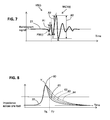
- WA (i) can be defined from the highest positive apex J and the deepest negative apex K. More precisely, WA(i) can be defined by the peak to peak amplitude A JK between points J and K. Notably as shown, there is provided an array of values J1-J9, K1-K9; A JK 1- A JK 9 which are analyzed to extract a low frequency amplitude modulation reflecting the respiration rate.
- WA (i) can also be defined in another manner from an auxiliary BCG signal.
- auxiliary BCG signal is obtained from base BCG signal 21 after filtering operations conveniently chosen to enhance certain features of the pulse wave.
- the beat-to-beat time intervals are measured from the base ballistocardiogram BCG or from the impedance plethysmography signal 22 measured at the user's foot.
- DeltaHB i Time Delay from HB i ⁇ 1 to HB i , likewise denoted D i ⁇ 1 i at Figures 9 and 10.
- beat-to-beat time intervals are known to be modulated by the respiration, which is known as respiration sinus arrythmia.
- the time intervals DeltaHB(i) (shown as D 12 , D 23 , ..., D 89 ) between successive J apexes on BCG signal 21 (respectively on successive Y apexes on impedance plethysmography signal 22) tend to be shorter during inspiration and longer during expiration.
- a user inspiration phase is assumed whenever characteristic amplitude WA (i) increases and/or beat time intervals DeltaHB(i) decreases.
- the expiration phase has a length Texp, starting at T_ie and ending at T_ei .
- the inspiration phase has a length Tinsp, starting at T_ei and ending at T _ ie .
- a state of stress and/or relaxation of the user can be assessed as a function of synchronization index between the inspiration/expiration phases and the user heart beats.
- the phase between the heart cycle and its modulation by the respiration can be calculated by standard signal processing methods of sampling and reconstruction, interpolation, or curve fitting, for instance of a cosine.
- the respiration cycle can be reconstructed from steps above, as a cosine-like respiration cycle, with null phase for instance at a time of switch between inspiration and expiration (namely T _ ie ) or at a time of switch between expiration and inspiration (namely T _ ei ).
- a possible method of reconstruction is a minimal least squares regression on the wave amplitudes WA (i) and heart beats HB(i).
- phase difference of the cosine DeltaPhi(j) can be defined as a phase difference which separates the null phase of the respiration cycle j and the nearest heart beat HB(i).
- respiration cycles and the heart cycles are synchronous if DeltaPhi(j) is constant over several respiration cycles.
- the modulation of the heart periods by the respiration can be reconstructed from the heart beats HB(i) obtained from the feet or the apexes Y of the IPG.
- the phase difference DeltaPhi(j) is calculated at the beginning of each respiration cycle.
- DeltaPhi is constant, the user is thus relaxed.
- DeltaPhi is the same close to HB(2), HB(5) and HB(8)
- DeltaPhi is not constant, and in this case, the user U is subject to stress. More precisely it is apparent that DeltaPhi-1at HB(2) is rather small, DeltaPhi-2 at HB(5) is larger and DeltaPhi-3 at HB(8) is even larger.
- the synchronization index can be taken from a derivative over time of DeltaPhi; in other words the synchronization index is defined from an evolution over time of a phase difference DeltaPhi,
- the variability of the heart rate could be calculated with other fiducial points than J, for instance with the apexes I, or the average values of JJ or II intervals.
- the variability of the heart rate could also be calculated with fiducial points of the IPG, for instance the foot of the beat or its apex Y1-Y9.
- the time intervals DeltaHB(i) between successive J apexes are also modulated by the general state of fatigue of the person. This state can be determined with the help of a heart rate variability index denoted HRVI.
- beat time intervals DeltaHB(i) between successive heart beats are defined by the measured time intervals between the second positive apexes J of each heart beat of a couple of successive heart beats, from the ballistocardiogram signal 21.
- time intervals between the successive apexes Y of the impedance plethysmography signal 22 are measured.
- HRVI heart rate variability index
- HRVI can be expressed by Max [DeltaHB(i)] - Min [DeltaHB(i)], where indicia i is ranging from 1 to i0 , i0 being the number of monitor heart beats when the user is standing on the scale, i0 being at least 6.
- such a heart rate variability index HRVI can be expressed by the average over several complete respiration cycles of the differences Max [DeltaHB(i)] - Min [DeltaHB(i)] calculated over each respiration cycle (detected as explained above), namely where the index i ranges over the indicia of the heart beats in the given respiration cycle.
- the heart rate variability index HRVI is expressed by Max [DeltaHB(i)] - Min [DeltaHB(i)] where the index i ranges over the heart beats of the complete respiration cycle.
- RMSSD Root Mean Squared of the Successive Differences
- HRVI ⁇ k 1 k 2 ⁇ 2 ( DeltaHB i + 2 ⁇ DeltaHB i + 1 2 k 2 ⁇ k 1 ⁇ 1
- HRVI Heart Rate Variability index
- a level of the general state of fatigue can be given back to the user, this level being relative to the past state of fatigue that has been recorded. For instance, the feed back indicates to the user that he is more tired (or much more tired, or more rested, etc) than the previous day, or the previous week.
- averaging over several measurements permits to smooth out the variability introduced to the different emotional states of the person during the measurements in order to get a value more representative of the general, mid-term state of fatigue of the user.
- the user can be asked on at least one occasion to assess himself his state of fatigue and give that information via the smartphone application. This datum is stored on the server and used improve the precision of the feedback to the user.
- the impedance plethysmogram is produced by the pulsatile volume of blood in the arteries which is caused by, and follows closely, the pulsatile blood pressure in the arteries. It is known that during diastole the blood pressure decays approximately according to an exponential as follows: P t ⁇ Pmax . e ⁇ t RP . C
Landscapes
- Health & Medical Sciences (AREA)
- Life Sciences & Earth Sciences (AREA)
- Physics & Mathematics (AREA)
- Cardiology (AREA)
- Animal Behavior & Ethology (AREA)
- Veterinary Medicine (AREA)
- Engineering & Computer Science (AREA)
- Biomedical Technology (AREA)
- Heart & Thoracic Surgery (AREA)
- Medical Informatics (AREA)
- Molecular Biology (AREA)
- Surgery (AREA)
- Biophysics (AREA)
- General Health & Medical Sciences (AREA)
- Public Health (AREA)
- Pathology (AREA)
- Physiology (AREA)
- Vascular Medicine (AREA)
- Hematology (AREA)
- General Physics & Mathematics (AREA)
- Dentistry (AREA)
- Oral & Maxillofacial Surgery (AREA)
- Ophthalmology & Optometry (AREA)
- Pulmonology (AREA)
- Nuclear Medicine, Radiotherapy & Molecular Imaging (AREA)
- Radiology & Medical Imaging (AREA)
- Measuring Pulse, Heart Rate, Blood Pressure Or Blood Flow (AREA)
Applications Claiming Priority (1)
| Application Number | Priority Date | Filing Date | Title |
|---|---|---|---|
| US14/701,054 US20160317043A1 (en) | 2015-04-30 | 2015-04-30 | Weighing scale with extended functions |
Publications (2)
| Publication Number | Publication Date |
|---|---|
| EP3095380A2 true EP3095380A2 (de) | 2016-11-23 |
| EP3095380A3 EP3095380A3 (de) | 2017-02-15 |
Family
ID=55862662
Family Applications (1)
| Application Number | Title | Priority Date | Filing Date |
|---|---|---|---|
| EP16167797.6A Withdrawn EP3095380A3 (de) | 2015-04-30 | 2016-04-29 | Waage mit erweiterten funktionen |
Country Status (2)
| Country | Link |
|---|---|
| US (1) | US20160317043A1 (de) |
| EP (1) | EP3095380A3 (de) |
Cited By (5)
| Publication number | Priority date | Publication date | Assignee | Title |
|---|---|---|---|---|
| EP3375358A1 (de) | 2017-03-15 | 2018-09-19 | Nokia Technologies Oy | Vorrichtung zur analyse kardiovaskulärer parameter einer person |
| WO2023126220A1 (fr) | 2021-12-31 | 2023-07-06 | Withings | Station de mesure avec mesure de l'activité sudorale |
| FR3131524A1 (fr) | 2021-12-31 | 2023-07-07 | Withings | Station de mesure avec mesure d’électrocardiogramme |
| FR3131522A1 (fr) | 2021-12-31 | 2023-07-07 | Withings | Station de mesure avec poignée |
| FR3131523A1 (fr) | 2022-12-29 | 2023-07-07 | Withings | Station de mesure avec mesure d’électrocardiogramme |
Families Citing this family (30)
| Publication number | Priority date | Publication date | Assignee | Title |
|---|---|---|---|---|
| FR3000544B1 (fr) * | 2013-01-02 | 2015-11-27 | Withings | Dispositif de pesage multi-fonction |
| US20220155134A1 (en) * | 2014-05-09 | 2022-05-19 | Daniel Lin | Method and System to Track Weight |
| US9949662B2 (en) | 2014-06-12 | 2018-04-24 | PhysioWave, Inc. | Device and method having automatic user recognition and obtaining impedance-measurement signals |
| US9943241B2 (en) | 2014-06-12 | 2018-04-17 | PhysioWave, Inc. | Impedance measurement devices, systems, and methods |
| US10130273B2 (en) | 2014-06-12 | 2018-11-20 | PhysioWave, Inc. | Device and method having automatic user-responsive and user-specific physiological-meter platform |
| US9546898B2 (en) | 2014-06-12 | 2017-01-17 | PhysioWave, Inc. | Fitness testing scale |
| US9693696B2 (en) | 2014-08-07 | 2017-07-04 | PhysioWave, Inc. | System with user-physiological data updates |
| US10292658B2 (en) | 2015-06-23 | 2019-05-21 | Rochester Institute Of Technology | Apparatus, system and method for medical analyses of seated individual |
| US10945671B2 (en) | 2015-06-23 | 2021-03-16 | PhysioWave, Inc. | Determining physiological parameters using movement detection |
| US20190046069A1 (en) * | 2015-07-10 | 2019-02-14 | Bodyport Inc. | Cardiovascular signal acquisition, fusion, and noise mitigation |
| US20170148240A1 (en) * | 2015-11-20 | 2017-05-25 | PhysioWave, Inc. | Scale-based biometric authorization of communication between scale and a remote user-physiologic device |
| US11561126B2 (en) | 2015-11-20 | 2023-01-24 | PhysioWave, Inc. | Scale-based user-physiological heuristic systems |
| US10395055B2 (en) | 2015-11-20 | 2019-08-27 | PhysioWave, Inc. | Scale-based data access control methods and apparatuses |
| US10553306B2 (en) | 2015-11-20 | 2020-02-04 | PhysioWave, Inc. | Scaled-based methods and apparatuses for automatically updating patient profiles |
| US10436630B2 (en) | 2015-11-20 | 2019-10-08 | PhysioWave, Inc. | Scale-based user-physiological data hierarchy service apparatuses and methods |
| US10980483B2 (en) | 2015-11-20 | 2021-04-20 | PhysioWave, Inc. | Remote physiologic parameter determination methods and platform apparatuses |
| US10923217B2 (en) | 2015-11-20 | 2021-02-16 | PhysioWave, Inc. | Condition or treatment assessment methods and platform apparatuses |
| US20170188845A1 (en) * | 2016-01-05 | 2017-07-06 | Tosense, Inc. | Physiological monitoring system featuring floormat and wired handheld sensor |
| US20170188885A1 (en) * | 2016-01-05 | 2017-07-06 | Tosense, Inc. | Floormat physiological sensor |
| US10390772B1 (en) | 2016-05-04 | 2019-08-27 | PhysioWave, Inc. | Scale-based on-demand care system |
| US10215619B1 (en) | 2016-09-06 | 2019-02-26 | PhysioWave, Inc. | Scale-based time synchrony |
| US11123022B2 (en) * | 2017-10-18 | 2021-09-21 | Samsung Electronics Co., Ltd. | Blood pressure estimating apparatus and blood pressure estimating method |
| CN109009062A (zh) * | 2018-07-06 | 2018-12-18 | 苏州小蓝医疗科技有限公司 | 一种新型体重秤及其测量血流速度的方法 |
| DE102018213350A1 (de) * | 2018-08-08 | 2020-02-13 | Kardion Gmbh | Vorrichtung und Verfahren zur Überwachung eines Gesundheitszustands des Patienten |
| KR20200078795A (ko) | 2018-12-21 | 2020-07-02 | 삼성전자주식회사 | 혈압 추정 장치 및 방법 |
| AU2022253067A1 (en) | 2021-04-09 | 2023-09-07 | Casana Care, Inc. | Systems, devices, and methods for monitoring loads and forces on a seat |
| US20220336104A1 (en) | 2021-04-16 | 2022-10-20 | Withings | Devices, Systems and Processes to Compute A Vascular Health Related Score |
| EP4337087A1 (de) | 2021-05-11 | 2024-03-20 | Casana Care, Inc. | Systeme, vorrichtungen und verfahren zur messung von lasten und kräften einer sitzenden person unter verwendung von skalenvorrichtungen |
| AU2022275847A1 (en) | 2021-05-17 | 2023-10-05 | Casana Care, Inc. | Systems, devices, and methods for measuring body temperature of a subject using characterization of feces and/or urine |
| CN115944737B (zh) * | 2022-12-14 | 2023-08-01 | 江苏省人民医院(南京医科大学第一附属医院) | Map-2抑制剂在制备治疗高血压疾病的药物中的应用 |
Citations (4)
| Publication number | Priority date | Publication date | Assignee | Title |
|---|---|---|---|---|
| US20130310700A1 (en) | 2011-01-27 | 2013-11-21 | The Board Of Trustees Of The Leland Stanford Junior University | Systems and methods for monitoring the circulatory system |
| US8639226B2 (en) | 2009-04-21 | 2014-01-28 | Withings | Weighing device and method |
| WO2014106716A1 (fr) | 2013-01-02 | 2014-07-10 | Withings | Dispositif de pesage multi-fonction |
| US20140309541A1 (en) | 2012-01-16 | 2014-10-16 | Omron Healthcare Co., Ltd. | Blood pressure measurement device and control method for blood pressure measurement device |
Family Cites Families (6)
| Publication number | Priority date | Publication date | Assignee | Title |
|---|---|---|---|---|
| US5810734A (en) * | 1994-04-15 | 1998-09-22 | Vital Insite, Inc. | Apparatus and method for measuring an induced perturbation to determine a physiological parameter |
| US8602997B2 (en) * | 2007-06-12 | 2013-12-10 | Sotera Wireless, Inc. | Body-worn system for measuring continuous non-invasive blood pressure (cNIBP) |
| ES2385898A1 (es) * | 2010-07-30 | 2012-08-02 | Universitat Politècnica De Catalunya | Método y aparato para monitorizar parámetros cardio-respiratorios a partir de las variaciones de la impedancia eléctrica en un solo pie. |
| EP2816950A4 (de) * | 2012-02-22 | 2015-10-28 | Aclaris Medical Llc | Vorrichtung und system zur erkennung physiologischer signale |
| CN106028918B (zh) * | 2014-02-06 | 2020-08-04 | 索泰拉无线公司 | 用于生命体征的连续无创测量的身体佩带式系统 |
| US9568354B2 (en) * | 2014-06-12 | 2017-02-14 | PhysioWave, Inc. | Multifunction scale with large-area display |
-
2015
- 2015-04-30 US US14/701,054 patent/US20160317043A1/en not_active Abandoned
-
2016
- 2016-04-29 EP EP16167797.6A patent/EP3095380A3/de not_active Withdrawn
Patent Citations (4)
| Publication number | Priority date | Publication date | Assignee | Title |
|---|---|---|---|---|
| US8639226B2 (en) | 2009-04-21 | 2014-01-28 | Withings | Weighing device and method |
| US20130310700A1 (en) | 2011-01-27 | 2013-11-21 | The Board Of Trustees Of The Leland Stanford Junior University | Systems and methods for monitoring the circulatory system |
| US20140309541A1 (en) | 2012-01-16 | 2014-10-16 | Omron Healthcare Co., Ltd. | Blood pressure measurement device and control method for blood pressure measurement device |
| WO2014106716A1 (fr) | 2013-01-02 | 2014-07-10 | Withings | Dispositif de pesage multi-fonction |
Cited By (9)
| Publication number | Priority date | Publication date | Assignee | Title |
|---|---|---|---|---|
| EP3375358A1 (de) | 2017-03-15 | 2018-09-19 | Nokia Technologies Oy | Vorrichtung zur analyse kardiovaskulärer parameter einer person |
| WO2018167362A1 (en) | 2017-03-15 | 2018-09-20 | Nokia Technologies Oy | Device for analysing cardiovascular parameters of an individual |
| WO2023126220A1 (fr) | 2021-12-31 | 2023-07-06 | Withings | Station de mesure avec mesure de l'activité sudorale |
| FR3131524A1 (fr) | 2021-12-31 | 2023-07-07 | Withings | Station de mesure avec mesure d’électrocardiogramme |
| FR3131522A1 (fr) | 2021-12-31 | 2023-07-07 | Withings | Station de mesure avec poignée |
| FR3131521A1 (fr) | 2021-12-31 | 2023-07-07 | Withings | Station de mesure avec mesure de l’activité sudorale |
| EP4248854A1 (de) | 2021-12-31 | 2023-09-27 | Withings | Messstation mit griff |
| EP4248855A1 (de) | 2021-12-31 | 2023-09-27 | Withings | Messstation mit elektrokardiogrammmessung |
| FR3131523A1 (fr) | 2022-12-29 | 2023-07-07 | Withings | Station de mesure avec mesure d’électrocardiogramme |
Also Published As
| Publication number | Publication date |
|---|---|
| US20160317043A1 (en) | 2016-11-03 |
| EP3095380A3 (de) | 2017-02-15 |
Similar Documents
| Publication | Publication Date | Title |
|---|---|---|
| EP3095380A2 (de) | Waage mit erweiterten funktionen | |
| US12036044B2 (en) | Apparatus, system and method for medical analyses of seated individual | |
| JP6130474B2 (ja) | 体重計装置及び脈波速度取得方法 | |
| US20240023898A1 (en) | Pulse Wave Velocity, Arterial Compliance, and Blood Pressure | |
| US9943241B2 (en) | Impedance measurement devices, systems, and methods | |
| EP3087914A1 (de) | Waage mit erweiterten funktionen | |
| JP6669409B2 (ja) | 血圧値を測定するための方法、機器及びコンピュータプログラム | |
| US9808168B2 (en) | Method and system for non-invasive measurement of cardiac parameters | |
| EP3551060B1 (de) | Pulswellengeschwindigkeitsbestimmung zum beispiel zur blutdrucküberwachung | |
| US9949662B2 (en) | Device and method having automatic user recognition and obtaining impedance-measurement signals | |
| JP2018517528A5 (de) | ||
| US20130274620A1 (en) | Method and device for long-term monitoring of arterial vascular stiffness and vascular calcification of a patient | |
| EP2598022B1 (de) | Diagnoseunterstützungsvorrichtung | |
| Rajala et al. | Pulse arrival time (PAT) measurement based on arm ECG and finger PPG signals-comparison of PPG feature detection methods for PAT calculation | |
| KR102193284B1 (ko) | 사지의 말단 영역 측정으로부터 동맥 펄스 전달 시간을 추정하는 방법 및 장치 | |
| US10925516B2 (en) | Method and apparatus for estimating the aortic pulse transit time from time intervals measured between fiducial points of the ballistocardiogram | |
| EP3154427A1 (de) | Impedanzmessvorrichtungen, -systeme und -verfahren | |
| Baek et al. | Validation of cuffless blood pressure monitoring using wearable device | |
| Park et al. | Development of blood pressure estimation methods using the PPG and ECG sensors |
Legal Events
| Date | Code | Title | Description |
|---|---|---|---|
| PUAI | Public reference made under article 153(3) epc to a published international application that has entered the european phase |
Free format text: ORIGINAL CODE: 0009012 |
|
| AK | Designated contracting states |
Kind code of ref document: A2 Designated state(s): AL AT BE BG CH CY CZ DE DK EE ES FI FR GB GR HR HU IE IS IT LI LT LU LV MC MK MT NL NO PL PT RO RS SE SI SK SM TR |
|
| AX | Request for extension of the european patent |
Extension state: BA ME |
|
| PUAL | Search report despatched |
Free format text: ORIGINAL CODE: 0009013 |
|
| AK | Designated contracting states |
Kind code of ref document: A3 Designated state(s): AL AT BE BG CH CY CZ DE DK EE ES FI FR GB GR HR HU IE IS IT LI LT LU LV MC MK MT NL NO PL PT RO RS SE SI SK SM TR |
|
| AX | Request for extension of the european patent |
Extension state: BA ME |
|
| RIC1 | Information provided on ipc code assigned before grant |
Ipc: G01G 19/50 20060101ALI20170112BHEP Ipc: A61B 5/021 20060101ALI20170112BHEP Ipc: A61B 5/022 20060101ALI20170112BHEP Ipc: A61B 5/00 20060101AFI20170112BHEP Ipc: A61B 5/0295 20060101ALI20170112BHEP Ipc: A61B 5/053 20060101ALI20170112BHEP Ipc: A61B 5/11 20060101ALI20170112BHEP |
|
| STAA | Information on the status of an ep patent application or granted ep patent |
Free format text: STATUS: REQUEST FOR EXAMINATION WAS MADE |
|
| 17P | Request for examination filed |
Effective date: 20170721 |
|
| RBV | Designated contracting states (corrected) |
Designated state(s): AL AT BE BG CH CY CZ DE DK EE ES FI FR GB GR HR HU IE IS IT LI LT LU LV MC MK MT NL NO PL PT RO RS SE SI SK SM TR |
|
| REG | Reference to a national code |
Ref country code: HK Ref legal event code: DE Ref document number: 1231710 Country of ref document: HK |
|
| RAP1 | Party data changed (applicant data changed or rights of an application transferred) |
Owner name: NOKIA TECHNOLOGIES (FRANCE ) |
|
| RAP1 | Party data changed (applicant data changed or rights of an application transferred) |
Owner name: NOKIA TECHNOLOGIES OY |
|
| STAA | Information on the status of an ep patent application or granted ep patent |
Free format text: STATUS: EXAMINATION IS IN PROGRESS |
|
| 17Q | First examination report despatched |
Effective date: 20190304 |
|
| RAP1 | Party data changed (applicant data changed or rights of an application transferred) |
Owner name: WITHINGS |
|
| STAA | Information on the status of an ep patent application or granted ep patent |
Free format text: STATUS: THE APPLICATION IS DEEMED TO BE WITHDRAWN |
|
| 18D | Application deemed to be withdrawn |
Effective date: 20190917 |
|
| REG | Reference to a national code |
Ref country code: HK Ref legal event code: WD Ref document number: 1231710 Country of ref document: HK |