EP3092014B1 - Procédé de fabrication de dispositifs d'ostéosynthèse, dispositifs d'ostéosynthèse et implants en matériau hybride semi-synthétique obtenu par modification structurale des composants d'un biomatériau naturel marin - Google Patents
Procédé de fabrication de dispositifs d'ostéosynthèse, dispositifs d'ostéosynthèse et implants en matériau hybride semi-synthétique obtenu par modification structurale des composants d'un biomatériau naturel marin Download PDFInfo
- Publication number
- EP3092014B1 EP3092014B1 EP15702540.4A EP15702540A EP3092014B1 EP 3092014 B1 EP3092014 B1 EP 3092014B1 EP 15702540 A EP15702540 A EP 15702540A EP 3092014 B1 EP3092014 B1 EP 3092014B1
- Authority
- EP
- European Patent Office
- Prior art keywords
- osteosynthesis
- pinctada
- bone
- semi
- maxima
- Prior art date
- Legal status (The legal status is an assumption and is not a legal conclusion. Google has not performed a legal analysis and makes no representation as to the accuracy of the status listed.)
- Active
Links
Images


Classifications
-
- A—HUMAN NECESSITIES
- A61—MEDICAL OR VETERINARY SCIENCE; HYGIENE
- A61L—METHODS OR APPARATUS FOR STERILISING MATERIALS OR OBJECTS IN GENERAL; DISINFECTION, STERILISATION OR DEODORISATION OF AIR; CHEMICAL ASPECTS OF BANDAGES, DRESSINGS, ABSORBENT PADS OR SURGICAL ARTICLES; MATERIALS FOR BANDAGES, DRESSINGS, ABSORBENT PADS OR SURGICAL ARTICLES
- A61L27/00—Materials for grafts or prostheses or for coating grafts or prostheses
- A61L27/36—Materials for grafts or prostheses or for coating grafts or prostheses containing ingredients of undetermined constitution or reaction products thereof, e.g. transplant tissue, natural bone, extracellular matrix
- A61L27/3604—Materials for grafts or prostheses or for coating grafts or prostheses containing ingredients of undetermined constitution or reaction products thereof, e.g. transplant tissue, natural bone, extracellular matrix characterised by the human or animal origin of the biological material, e.g. hair, fascia, fish scales, silk, shellac, pericardium, pleura, renal tissue, amniotic membrane, parenchymal tissue, fetal tissue, muscle tissue, fat tissue, enamel
- A61L27/3625—Vascular tissue, e.g. heart valves
-
- A—HUMAN NECESSITIES
- A61—MEDICAL OR VETERINARY SCIENCE; HYGIENE
- A61K—PREPARATIONS FOR MEDICAL, DENTAL OR TOILETRY PURPOSES
- A61K35/00—Medicinal preparations containing materials or reaction products thereof with undetermined constitution
- A61K35/56—Materials from animals other than mammals
- A61K35/618—Molluscs, e.g. fresh-water molluscs, oysters, clams, squids, octopus, cuttlefish, snails or slugs
-
- A—HUMAN NECESSITIES
- A61—MEDICAL OR VETERINARY SCIENCE; HYGIENE
- A61L—METHODS OR APPARATUS FOR STERILISING MATERIALS OR OBJECTS IN GENERAL; DISINFECTION, STERILISATION OR DEODORISATION OF AIR; CHEMICAL ASPECTS OF BANDAGES, DRESSINGS, ABSORBENT PADS OR SURGICAL ARTICLES; MATERIALS FOR BANDAGES, DRESSINGS, ABSORBENT PADS OR SURGICAL ARTICLES
- A61L27/00—Materials for grafts or prostheses or for coating grafts or prostheses
- A61L27/02—Inorganic materials
- A61L27/10—Ceramics or glasses
-
- A—HUMAN NECESSITIES
- A61—MEDICAL OR VETERINARY SCIENCE; HYGIENE
- A61B—DIAGNOSIS; SURGERY; IDENTIFICATION
- A61B17/00—Surgical instruments, devices or methods
- A61B17/56—Surgical instruments or methods for treatment of bones or joints; Devices specially adapted therefor
- A61B17/58—Surgical instruments or methods for treatment of bones or joints; Devices specially adapted therefor for osteosynthesis, e.g. bone plates, screws or setting implements
- A61B17/68—Internal fixation devices, including fasteners and spinal fixators, even if a part thereof projects from the skin
- A61B17/80—Cortical plates, i.e. bone plates; Instruments for holding or positioning cortical plates, or for compressing bones attached to cortical plates
-
- A—HUMAN NECESSITIES
- A61—MEDICAL OR VETERINARY SCIENCE; HYGIENE
- A61B—DIAGNOSIS; SURGERY; IDENTIFICATION
- A61B17/00—Surgical instruments, devices or methods
- A61B17/56—Surgical instruments or methods for treatment of bones or joints; Devices specially adapted therefor
- A61B17/58—Surgical instruments or methods for treatment of bones or joints; Devices specially adapted therefor for osteosynthesis, e.g. bone plates, screws or setting implements
- A61B17/68—Internal fixation devices, including fasteners and spinal fixators, even if a part thereof projects from the skin
- A61B17/84—Fasteners therefor or fasteners being internal fixation devices
- A61B17/86—Pins or screws or threaded wires; nuts therefor
-
- A—HUMAN NECESSITIES
- A61—MEDICAL OR VETERINARY SCIENCE; HYGIENE
- A61C—DENTISTRY; APPARATUS OR METHODS FOR ORAL OR DENTAL HYGIENE
- A61C8/00—Means to be fixed to the jaw-bone for consolidating natural teeth or for fixing dental prostheses thereon; Dental implants; Implanting tools
- A61C8/0012—Means to be fixed to the jaw-bone for consolidating natural teeth or for fixing dental prostheses thereon; Dental implants; Implanting tools characterised by the material or composition, e.g. ceramics, surface layer, metal alloy
-
- A—HUMAN NECESSITIES
- A61—MEDICAL OR VETERINARY SCIENCE; HYGIENE
- A61F—FILTERS IMPLANTABLE INTO BLOOD VESSELS; PROSTHESES; DEVICES PROVIDING PATENCY TO, OR PREVENTING COLLAPSING OF, TUBULAR STRUCTURES OF THE BODY, e.g. STENTS; ORTHOPAEDIC, NURSING OR CONTRACEPTIVE DEVICES; FOMENTATION; TREATMENT OR PROTECTION OF EYES OR EARS; BANDAGES, DRESSINGS OR ABSORBENT PADS; FIRST-AID KITS
- A61F5/00—Orthopaedic methods or devices for non-surgical treatment of bones or joints; Nursing devices ; Anti-rape devices
- A61F5/01—Orthopaedic devices, e.g. long-term immobilising or pressure directing devices for treating broken or deformed bones such as splints, casts or braces
-
- A—HUMAN NECESSITIES
- A61—MEDICAL OR VETERINARY SCIENCE; HYGIENE
- A61L—METHODS OR APPARATUS FOR STERILISING MATERIALS OR OBJECTS IN GENERAL; DISINFECTION, STERILISATION OR DEODORISATION OF AIR; CHEMICAL ASPECTS OF BANDAGES, DRESSINGS, ABSORBENT PADS OR SURGICAL ARTICLES; MATERIALS FOR BANDAGES, DRESSINGS, ABSORBENT PADS OR SURGICAL ARTICLES
- A61L27/00—Materials for grafts or prostheses or for coating grafts or prostheses
- A61L27/36—Materials for grafts or prostheses or for coating grafts or prostheses containing ingredients of undetermined constitution or reaction products thereof, e.g. transplant tissue, natural bone, extracellular matrix
-
- A—HUMAN NECESSITIES
- A61—MEDICAL OR VETERINARY SCIENCE; HYGIENE
- A61L—METHODS OR APPARATUS FOR STERILISING MATERIALS OR OBJECTS IN GENERAL; DISINFECTION, STERILISATION OR DEODORISATION OF AIR; CHEMICAL ASPECTS OF BANDAGES, DRESSINGS, ABSORBENT PADS OR SURGICAL ARTICLES; MATERIALS FOR BANDAGES, DRESSINGS, ABSORBENT PADS OR SURGICAL ARTICLES
- A61L27/00—Materials for grafts or prostheses or for coating grafts or prostheses
- A61L27/50—Materials characterised by their function or physical properties, e.g. injectable or lubricating compositions, shape-memory materials, surface modified materials
- A61L27/54—Biologically active materials, e.g. therapeutic substances
-
- A—HUMAN NECESSITIES
- A61—MEDICAL OR VETERINARY SCIENCE; HYGIENE
- A61L—METHODS OR APPARATUS FOR STERILISING MATERIALS OR OBJECTS IN GENERAL; DISINFECTION, STERILISATION OR DEODORISATION OF AIR; CHEMICAL ASPECTS OF BANDAGES, DRESSINGS, ABSORBENT PADS OR SURGICAL ARTICLES; MATERIALS FOR BANDAGES, DRESSINGS, ABSORBENT PADS OR SURGICAL ARTICLES
- A61L2400/00—Materials characterised by their function or physical properties
- A61L2400/18—Modification of implant surfaces in order to improve biocompatibility, cell growth, fixation of biomolecules, e.g. plasma treatment
-
- A—HUMAN NECESSITIES
- A61—MEDICAL OR VETERINARY SCIENCE; HYGIENE
- A61L—METHODS OR APPARATUS FOR STERILISING MATERIALS OR OBJECTS IN GENERAL; DISINFECTION, STERILISATION OR DEODORISATION OF AIR; CHEMICAL ASPECTS OF BANDAGES, DRESSINGS, ABSORBENT PADS OR SURGICAL ARTICLES; MATERIALS FOR BANDAGES, DRESSINGS, ABSORBENT PADS OR SURGICAL ARTICLES
- A61L2430/00—Materials or treatment for tissue regeneration
- A61L2430/02—Materials or treatment for tissue regeneration for reconstruction of bones; weight-bearing implants
Definitions
- the present invention relates to a semi-synthetic hybrid material obtained by structural modification of the components of a marine natural biomaterial, in particular the pearly aragonitic layer of marine mollusc valves such as Pinctada Maxima, Pinctada Margaritifera, Tridacnae Maxima, Tridacnae Gigas and other Pinctadines .
- the present invention also relates to the manufacture of osteosynthesis devices and implants from said semisynthetic hybrid material.
- osteosynthesis which consists in realigning the fractured segments by the use of plates, screws, nails and fixators. external, made of stainless steel or alloys titanium, cobalt, etc., so as to immobilize the fractured fragments to allow the formation of a callus healing.
- the osteosynthesis material does not have the same mechanical and physical properties as the bone , in particular Young's modulus, resistance to bending, elasticity, hardness, density, and that, on the other hand, it undergoes the corrosive action of the saline environment of the circulating liquids of the human body, which action releases metallic microparticles, ions, as well as various metal salts.
- any change in the surface state of the metal can make it vulnerable to corrosion and alter its mechanical properties.
- they In order to improve the tribological properties of osteosynthesis equipment and metal implants, they have been coated with a hydroxyapatite coating by a plasma torch sintering process, in order to obtain bone adhesion. metaplastic at the interface, during bone healing.
- a plasma torch sintering process in order to obtain bone adhesion. metaplastic at the interface, during bone healing.
- there is a detachment of the coating the formation of a fibrous tissue, with the corollary the mobilization of osteosynthesis material or implants.
- the osteosynthesis material was to be deposited as soon as the consolidation of the fracture was clinically and radiologically confirmed.
- the osteosynthesis material is left in place, because a re-intervention to deposit it would require a new hospitalization, an intervention almost identical to the first, with the usual complications related to any intervention. surgical, especially on the bone.
- the removal of osteosynthesis material leaves a bone with drill holes, a cortical thinned due to a lack of vascularization caused by the pressure of the plate. This results in a secondary weakening of the operating site with the possibility of fracture.
- Ablation is also almost mandatory in clavicle fractures requiring surgical management, given the subcutaneous situation and the presence of vasculo-nervous elements, such as the brachial plexus and the artery. and the risk of pseudarthrosis resulting from ischemia caused by pressure of the plate on a flat bone.
- Ablation is also mandatory in pediatric orthopedic surgery, which does not obey the same rules as those of adults; in particular, when the fracture concerns the epiphyso-diaphyseal region of a long bone, it includes in the child the metaphysis, seat of the cartilage conjugation, responsible for the growth of the bone. Persistence of osteosynthesis material at this level for too long a period of time will compromise the growth of the limb. It is for these reasons that, in the child, the orthopedic material is removed early in order to avoid this inconvenience. In doing so, non-union fractures and bone fragility are sometimes observed, as well as complications associated with removal of the material.
- This hematoma also contains the osteoinductive factors BMP, FGF, IGF, as well as the important elements that are pericytes which, released from the basal lamina of the endothelium of the injured capillaries during the fracture, enter the process of stimulation of angiogenesis, collagen synthesis, proteoglycans, osteocalcin, and in the initiation of phagocytosis.
- the inventors have demonstrated that it is possible to obtain a material satisfying these requirements, from a natural hybrid biomaterial which is the pearlescent aragonitic layer of bivalve molluscs selected from the group comprising Pinctada Maxima, Pinctada Margaritifera, Tridacnae Maxima, Tridacnae Gigas and other Pinctadines.
- this biomaterial can be used for the manufacture of osteosynthesis devices and implants, such as: osteosynthesis plates and screws, osteotomy wedges, diabolos, wedges and inter-somatic cages, intramedullary nails, femoral and femoral heads, glenoid cavities, tibial plateaus, femoral condyles, vertebral bodies, hemi-maxillary bones, ossicles bone, surgical anchors for ligament and tendinous reinsertion, gutting sheaths for osteosynthesized reduction of comminuted fractures with small fragments, membranous compression screws and dental implants behaving like perennial autologous grafts.
- osteosynthesis plates and screws osteotomy wedges, diabolos, wedges and inter-somatic cages
- intramedullary nails femoral and femoral heads
- glenoid cavities glenoid cavities
- tibial plateaus femoral condyles
- vertebral bodies hemi-maxill
- the pearly aragonitic layer of bivalve molluscs selected from the group comprising Pinctada Maxima, Pinctada Margaritifera, Tridacnae Maxima, Tridacnae Gigas and other Pinctadines is an organic and inorganic composite material of biogenic origin, of hybrid structure.
- the aragonitic layer The mother-of-pearl of these bivalve molluscs is in the form of a laminated architecture, alternating a mineral component consisting of nano-crystals of aragonite, calcium carbonate crystallized in the orthorhombic system, organized in tablets, and an organic component consisting of Linear and branched polymers, organized into a three-dimensional lattice.
- This set gives the biomaterial lamellar architecture particularly suitable for the absorption and distribution of forces and shocks opposing the rupture.
- valves including the pearly aragonitic layer
- endogenous factors such as physiology and physiopathology, different from one mollusc to another as well as exogenous factors such as biotope, changes in the marine environment, water temperature, zoo- and phytoplankton composition, pathogen and predator aggression.
- the particularity of the organic lattice, separating and welding the inorganic aragonite tablets lies in the fact that it has interconnected pores of different diameters, communicating throughout its thickness, giving it a continuous porosity and porosity open with open pores.
- Bone is a viscoelastic material whose viscous nature is due to the presence of the interstitial fluids which permeate it, and especially to that of the biopolymers such as collagens, glycosaminoglycans and proteoglycans that come into its composition.
- the viscoelastic properties are more important in fresh cortical bone, that is to say impregnated with interstitial fluids (plasma, serum, etc.) than in dry bone.
- This organic fraction also contains biopolymers composed largely of type I and II collagens, low molecular weight glycoproteins, some of which are related to growth factors, cytokines and other osteo-competent molecules involved in bone and / or cartilaginous regeneration.
- This organic fraction also contains almost all the amino acids and in particular arginine, glycine, aspartic acid, molecules with chemo-tactical properties promoting cell adhesion, as well as metalloenzymes, metalloporphyrins, metalloproteins, molecules involved. in many metabolic reactions during osteogenesis.
- the mineral fraction besides calcium carbonate, also contains many minerals, as well as metals involved in the biosynthesis of calcified tissues.
- the inventors have demonstrated ( CR Acad. Sc. Paris 1988 , CR Acad. Sc. Paris 1989 , CLINICAL MATERIAL 0267-6605 / 90 / S03-50 1990 ), that the biomaterial perennialized in endo-osseous site, realizing a weld without transition with the receiving bone.
- the active molecules contained in the aragonite of molluscs mentioned above have no cytotoxic effect, no mutagenic or systemic effect, their interaction only having an impact of potentiation and stimulation of local factors of healing and bone regeneration.
- the mechanical properties of the bones are therefore different according to their shapes, their functions and their belonging to the various musculoskeletal stages.
- the values of the various types of resistance measurement such as Young's modulus, bending stress or compression, and breaking strain, vary according to whether they are applied to long bones, such as the femur. , tibia, fibula, humerus, radius and ulna, with short bones such as carp, metacarpus, tarsus and metatarsal bone, such as vertebral bodies, pelvic bones, and bones dishes such as those of the face and skull, as well as the clavicle and scapula.
- the inventors have demonstrated that the osteoclastic activity, at the origin of bone remodeling, was exerted in the same way against the biomaterial, at the bone-biomaterial interface, but was limited in time and coupled to the concomitant activity of apposition of bone neoformed by osteoblasts.
- this biological phenomenon confirms the osteomimetic character of the biomaterial and explains that in endo-osseous site, it is welded to the recipient bone.
- the technique consists in using membranes resorbable or not, which requires compression screws, most of the time in titanium or stainless steel, in order to maintain them and to create a space necessary for the protection of the sub-membranous bone substitute. It is known that the maintenance of a bone filling material on the site is difficult, especially if it is in the form of granules, as it is expelled through the incision line or the fibro-mucosa. In order to keep the filling material and the membrane in place, titanium mini-screws are used to create a space above the area to be filled. The protocol goes through a removal of these screws after healing and bone regeneration with ablation of the membrane if not resorbable.
- the inventors have developed a new semisynthetic hybrid material for the realization of osteosynthesis devices and implants intended to be maintained in a permanent manner on the fracture site, such as: plates and screws of osteosynthesis, osteotomy wedges, diabolos, intersomatic wedges and cages, intramedullary nails, humeral and femoral heads, glenoid cavities, tibial plateaus, femoral condyles, vertebral bodies, hemi-maxillary, ossicles bone, surgical anchors for reintegration ligamentous and / or tendinous, guttering gutters for the osteosynthesized reduction of fractures comminuted with small fragments, as well as membranous compression screws and dental implants behaving as perennial autologous grafts.
- the invention relates to a semisynthetic hybrid material which is the pearlescent aragonitic layer of bivalve molluscs selected from the group comprising Pinctada Maxima, Pinctada Margaritifera, Tridacnae Maxima, Tridacnae Gigas and other Pinctadines, said semi-synthetic hybrid material comprising a fraction inorganic and a crosslinked organic fraction and having a pH of 7 to 7.4.
- the present invention also relates to the process for obtaining this material by structural modification of a natural hybrid biomaterial which is the pearlescent aragonitic layer of bivalve molluscs selected from the group comprising Pinctada Maxima, Pinctada Margaritifera, Tridacnae Maxima, Tridacnae Gigas and other Pinctadines .
- the present invention also relates to methods of manufacturing osteosynthesis devices and implants.
- the invention relates to a semi-synthetic hybrid material which is the pearlescent aragonitic layer of bivalve molluscs selected from the group comprising Pinctada Maxima, Pinctada Margaritifera, Tridacnae Maxima, Tridacnae Gigas and other Pinctadines, said semi-hybrid material.
- synthetic composition comprising an inorganic fraction and a crosslinked organic fraction and having a pH of 7 to 7.4.
- the semi-synthetic hybrid material of the invention Due to its chemical composition, its structural properties which vary according to the characteristics of its place of origin and culture, the semi-synthetic hybrid material of the invention has, in particular with respect to those of titanium or steel , adapted mechanical properties, compatible with the different types of cortical bone.
- the semi-synthetic hybrid material has a laminated architecture, i.e. a stepwise superposition of inorganic fractions and organic moieties.
- the organic fraction does not include mineral inclusions.
- the semi-synthetic hybrid material is not obtained by chemical synthesis but by modifying the textural and structural properties of the pearlescent aragonitic layer of bivalve molluscs selected from the group comprising Pinctada Maxima, Pinctada Margaritifera, Tridacnae Maxima, Tridacnae Gigas and other Pinctadines, in particular a modification of the pH and a crosslinking of the organic fraction.
- the pH of the semisynthetic hybrid material of the invention is measured on an aliquot of the reduced powder material and dispersed in water.
- the pH of the semi-synthetic hybrid material of the invention is close to the pH of the biological fluids and the internal medium (which is around 7.4), which will allow osteosynthesis devices and / or implants made from of this material to be well tolerated and perfectly bio-integrated. Moreover, such a pH creates favorable conditions for the induction of a crosslinking of the biopolymer chains of the organic fraction of the hybrid material.
- the crosslinking is characterized by a multidirectional interconnection of the chains of linear and branched biopolymers constituting said organic fraction.
- the crosslinking induces a high cohesion energy, an increase in surface energy and, therefore, an increase in the adhesiveness and hydrophilicity of the semi-synthetic hybrid material of the invention. It also aims to increase the resistance of the organic fraction of the biomaterial to the corrosive action of biological fluids by decreasing its solubility and by opposing its aging and the action of the mechanical stresses that the biomaterial undergoes.
- the invention also relates to a process for producing a semi-synthetic hybrid material from a natural hybrid biomaterial, which comprises an inorganic fraction and an organic fraction, said process comprising a step of modifying the pH and a step crosslinking the organic fraction of said hybrid biomaterial.
- the natural hybrid biomaterial is the pearly aragonitic layer of bivalve molluscs selected from the group comprising Pinctada Maxima, Pinctada Margaritifera, Tridacnae Maxima, Tridacnae Gigas and other Pinctadines.
- the inventors propose, in view of the laminated hybrid nature of the aragonite of the molluscs mentioned in reference, to apply to its biopolymeric component a polymer-specific treatment intended to modify the structure thereof, with the aim of corollary a strengthening of its mechanical properties.
- crosslinking has been shown to consolidate the mechanical properties. This is why the inventors propose a crosslinking process, in particular by riboflavin, collagenic fractions, proteoglycans and glycosaminoglycans of the organic fraction of the valves of the molluscs mentioned, with reference to the crosslinking of corneal collagen, which is a fibrillar biochemical bridging by covalent bonds.
- the step of modifying the pH is carried out by immersion in a water bath of the microbiologically controlled network, brought to boiling, for example in a mixture of equal parts water of the microbiologically controlled network and osmosis water, until the desired pH is obtained.
- the treatment is for example 60 to 180 minutes, and preferably 60 to 120 minutes.
- the pH can be measured on an aliquot of the reduced biomaterial powder and dispersed in water.
- the crosslinking step is carried out using a crosslinking agent such as riboflavin, vitamin C, a polyol such as mannitol, and / or with the aid of physical agents such as as ionizing radiation.
- a crosslinking agent such as riboflavin, vitamin C, a polyol such as mannitol, and / or with the aid of physical agents such as as ionizing radiation.
- Step a) consists in selecting, according to its origin and its culture conditions but also according to the destination of the osteosynthesis material envisaged, a mollusk valve which is of satisfactory thickness and physical and structural integrity to allow cut into the previously exposed pearly aragonitic layer, a preform of size adapted to the device envisaged.
- the periostracum and the external calcitic prismatic layer are ground by abrasion, in particular using a fine-grained diamond grinding wheel, for example at a speed of 3000 rpm under a current of water.
- the thickness is measured using a thickness compass.
- the physical and structural integrity of the aragonitic layer of the selected valve is controlled in an optical chamber, for example using a halogen light source of 500 watts
- valve is then brushed, washed with running water from the microbiologically controlled network at a temperature of 55 ° C.
- the step b) of cutting and manufacturing consists, in a first phase, of cutting preforms intended to produce osteosynthesis devices according to previously printed outlines, on one or other of the faces of the valve, to the dimensions said devices.
- this cutting step of step b) is carried out with the jet of water loaded with abrasive.
- Cutting with a charged water jet has the advantage of not generating vibrations that can generate micro-fracture primers or exothermic reactions that can cause degradation of the hybrid material.
- the abrasive will consist of aragonite grains, thus preventing any contamination of the material by a material of another nature.
- the cutting can be performed in the following manner: said valve is placed in appropriate compression frames, fixed on the carpet of a cutting machine, for example 5-axis, water jet loaded abrasive.
- the abrasive may consist of aragonite grains with a particle size of between 0.1 and 200 ⁇ m.
- the abrasive grain-laden water is pulsed at a pressure of 4,000 to 6,200 bar, using 0.50 to 1.2 mm focusing guns and 0.12 to 0 cutting nozzles. , 40 mm in diameter.
- the second phase of step b) relates to the manufacture by grinding or machining of osteosynthesis devices and / or implants, possibly after numerical modeling allowing the exploitation of these data by CNC machine tools.
- numerical modeling of the possible insertion zone is carried out according to anatomical parts, so that the geometry of the internal face of the osteosynthesis device or implant to be manufactured can be adapted as closely as possible to the topography of this area.
- the osteosynthesis device or implant is therefore made in a homologous manner for right or left limb.
- drawings and sketches of devices are digitized to allow their manufacture by bar turning or grinding.
- step b) The manufacturing phase by grinding or bar turning of step b) can be carried out, according to a particular mode, by means of numerically controlled machine tools using abrasive, diamond or ceramic instruments.
- the planar grinding phase of step b) can be performed using a micro-cutting machine with a charged jet of water.
- Step c) of pH modification and crosslinking consists in modifying the physical and chemical properties of the hybrid material.
- the modification of the pH during step c) is carried out by immersion in a water bath of the microbiologically controlled network, brought to boiling, for example in a mixture of equal parts of water of the microbiologically network. mastered and osmosis water, until the desired pH.
- the treatment may for example be 60 to 180 minutes, and preferably 60 to 120 minutes.
- the crosslinking during step c) is carried out using a crosslinking agent such as riboflavin, vitamin C or a polyol such as mannitol.
- a crosslinking agent such as riboflavin, vitamin C or a polyol such as mannitol.
- the crosslinking is carried out at a temperature greater than 20 ° C.
- the impregnation of the hybrid biomaterial with riboflavin may advantageously be followed by actinic exposure to UVA.
- Crosslinking can also be achieved by the use of physical agents such as ionizing radiation.
- riboflavin or vitamin B2
- riboflavin tolerates sterilization and freezing very well, and stimulates cellular metabolism.
- a cell growth factor it is involved in the synthesis of proteins, carbohydrates and lipids and has powerful anti-oxidant properties, which oppose the action of free radicals produced by actinic action during the crosslinking.
- water-soluble riboflavin has the effect of not only modifying the structure thereof, but of imparting to the semi-synthetic material new pharmacological properties which are of interest for bone regeneration and healing.
- Step d) consists in modifying the surface state of said osteosynthesis device or implant by applying four successive treatments of sandblasting, ultrasonic cleaning, cryogenics and application of nanoparticles.
- the objective is on the one hand to improve the tribological properties, especially the covalent strong bonds at the origin of the aggregation of the nanoparticles and their interaction with the receiving medium, on the other hand to increase the ratio surface / volume between the device and the fractured cortical bone to ensure its stability, finally, to promote the release of soluble osteogenitrice molecules, activating the local factors of osteogenesis contained therein.
- the sanding treatment of step d) consists in modifying the surface condition of the osteosynthesis device or implant in order to improve the anchoring thereof on the bone.
- said device is placed in a sander with handle and treated by successive projections, using a pressure system, preferably aragonite grains of 25 to 70 microns propelled by means of 0.8 mm round blast nozzles and 70 to 250 ⁇ m aragonite grains propelled by means of 1.2 mm nozzles at a pressure of 6 bar.
- a pressure system preferably aragonite grains of 25 to 70 microns propelled by means of 0.8 mm round blast nozzles and 70 to 250 ⁇ m aragonite grains propelled by means of 1.2 mm nozzles at a pressure of 6 bar.
- the ultrasonic cleaning of step d) is as follows: an ultrasonic tank is filled with hot water of the microbiologically controlled network, at 55 ° C., maximum efficiency temperature, up to to a mark indicating the desired volume of water. This is added a cleaning solution and disinfectant at a dilution of 1: 128, 1 part solution for 127 parts of water. After 15 minutes of degassing to eliminate air bubbles, the osteosynthesis device or implant is placed in the tank for a period of 30 minutes at a frequency of 40 kHz for cavitation inducing optimum particulate shrinkage.
- Said device is then rinsed under a stream of water of the microbiologically controlled network, for 20 minutes, and then immersed for 20 minutes in a demineralized water bath at a temperature of 90 ° C., added with 2% of bleach to 2 6% active chlorine for 30 minutes, then rinsed again with deionized water at 90 ° C.
- the device is soaked in deionized water at 50 ° C, supplemented with liquid Calbismeum®, or any other biocidal, virucidal, surfactant agent, diluted to 2% for 30 minutes, rinsed and then dried.
- the non-abrasive cryogenic treatment of step d), intended to prepare the faces of the device in contact with the cortical bone consists in projecting thereon small dry ice balls of liquid nitrogen at -80 ° C, 1 mm in diameter.
- the objective is to optimize the surface state by mechanical effect associated with a thermal shock, due to the difference in temperature between the surface to be treated and the liquid nitrogen balls during the sublimation thereof to the 'impact.
- the implementation consists in projecting into a dedicated space on the surface or surfaces to be treated a mixture of compressed air and ice pellets, under a pressure allowing a non-abrasive treatment optimized by the low hardness of dry nitrogen ice which is 2 Mohs.
- the nanoparticle application phase of step d) of modifying the surface state is a coating of mechano-structured nanoparticles obtained from the hybrid biomaterial, according to the patent FR N ° 09 54066 and the patent U.S. Patent No. 8,485,458 .
- This phase of step d) of modifying the surface state is carried out, either by immersing, in an emulsion of variable viscosity, said mechano-structured nanoparticles, either by centrifugation or by spraying and, preferably, by electrodeposition, which consists of immersing the osteosynthesis devices and the implants in an electrolytic bath of said nanoparticles so as to initiate an electrodeposition thereof on the surface thereof.
- the method of manufacturing an osteosynthesis device and / or implant further comprises a step e) of impregnation thereof with biological fluids and / or compositions containing pharmaceutically active substances.
- the inventors propose thus to impregnate the biopolymers which enter into their composition with plasma or serums of the different blood group antigenic systems, by dipping and / or imbibition, at atmospheric pressure or under vacuum, which, in addition, increases their bio- acceptance.
- the devices and implants may be impregnated with drug substances such as nonsteroidal anti-inflammatory drugs, analgesics, antibiotics and antimitotics or any other substance with a therapeutic effect.
- drug substances such as nonsteroidal anti-inflammatory drugs, analgesics, antibiotics and antimitotics or any other substance with a therapeutic effect.
- the method of manufacturing a device further comprises packaging phases, for example, under double packaging, sterilization, under a protective atmosphere, carried out using ionizing radiation at 25 KGy, as well as a storage step, either at a temperature of 0 ° to 4 ° C or by freezing at -15 ° C, or, for devices that have not been imbibed by biological fluids, a storage at room temperature
- the devices can also be impregnated extemporaneously with whole blood or autologous plasma at the time of the procedure.
- the invention also relates to a device made of semi-synthetic hybrid material, or obtained according to the manufacturing method of said device.
- Said device is chosen from osteosynthesis plates, such as right osteosynthesis plate, epiphyso-diaphyseal osteosynthesis plate, osteosynthesis plate for malleolar fractures, ruptive epiphyso-diaphyseal osteosynthesis plate, osteosynthesis screw, screw Membrane compression, osteotomy wedges, diabolos, inter-somatic wedges and cages, intramedullary nails, humeral and femoral heads, glenoid cavities, tibial trays, femoral condyles, vertebral bodies, hemi-maxillary, ossicles chain, surgical anchors for the ligament and tendinous reinsertion, gutting gutters for the osteosynthesized reduction of comminuted fractures with small fragments and dental implants.
- osteosynthesis plates such as right osteosynthesis plate, epiphyso-diaphyseal osteo
- the device comprises at least one means of contention which, during the installation of said device opposes its displacement, said means being chosen from lugs, notches, a harpoon keyed and flattened, and a Thread opposing unscrewing.
- the restraining means is constituted by lugs
- the latter diametrically opposed, make it possible to lock said device on the bone fragments on either side of the fracture line after fitting into the cortical bone.
- phantom plate aid, insertion wells pins, thereby achieving immediate primary fixation in key and a secondary fixation, once the bone healing achieved.
- the restraining means is constituted by transverse rectilinear notches in stair step, these, by their geometry, oppose the sliding forward or rearward of said device.
- the restraining means is a flattened keyed harpoon, the characteristics of the latter oppose the sliding and rotation of said device.
- the restraining means is a thread
- the latter has a geometry of non-metric pitch, and trapezoidal shape, which opposes its unscrewing.
- this threading promotes a total filling of the turns by the neoformed bone tissue.
- the device further comprises fixing holes.
- the number, size and location of these fixing holes will of course be adapted by those skilled in the art depending on the shape and dimensions of the device.
- Said fixing holes may be milled, partially or entirely and possibly be tapped with a metric thread iso standard pitch or fine pitch.
- said fixing holes may be round or oblong and / or make an acute angle with the vertical.
- the device further comprises a means which, during the installation of said device allows its adjustment, so as to best adapt to the morphology of the fracture site.
- the means which, during the installation of the device, allows its adjustment is a bulge positioned on the inner face of said device of height between 0.1 and 0.5 mm, preferably included in 0, 15 and 0.25 mm. This bulge can be ground on demand just before installation.
- the device which is a ruptive epiphyseal-diaphyseal osteosynthesis plate, further comprises two V-shaped rupture notches, one on the outer face, the other on the inner face.
- the notch of rupture of the external face of the device is perfectly identifiable on palpation and indicates the seat of the cutaneous incision to be made. Said device can then be broken with the aid of a hammer and a surgical chisel, allowing the limb to continue its development, the two parts of said device moving away as the evolution of the conjugation cartilage .
- all the osteosynthesis equipment manufactured according to the invention are placed using ancillaries, comprising ghost plates made to the dimensions and characteristics of the osteosynthesis devices and comprising among others the following accessories: a or several drilling guns, depth and hold gauges, screw-size forests and taps, screw holders and plate holders, fragment holding forceps, screwdrivers and wrench heads. hexagonal screws, distractors, opening wedges, hold wedges, ..
- Mollusk valves are chosen, in this example, Pinctada Maxima, the thickness of which, measured using a compass of thickness, is sufficient for the production of the desired osteosynthesis devices.
- the periostracum and the outer prismatic calcitic layer are milled by abrasion using a fine-grained diamond grinding wheel, at a speed of 3000 rpm under a stream of water, which exposes the pearly aragonitic layer. .
- the physical and structural integrity of the selected valves is monitored in an optical chamber using a 500 watt halogen light source.
- valves are then brushed, washed with running water from the microbiologically controlled network at a temperature of 55 ° C.
- the contours are printed on one or other of the faces of the selected valves.
- the valves are then cut according to the drawn outlines.
- the valves are placed in appropriate compression frames, fixed on the carpet of a 5-axis macro-cutting machine, water jet loaded with abrasive, for example aragonite grains with a particle size distribution. approximately 150 ⁇ m, pulsed under a pressure of 4 135 to 6 150 bar, using focusing guns of 0.50 to 1.2 mm and cutting nozzles of 0.20 to 0.40 mm in diameter .
- the numerical modeling of the latter is performed on an anatomical part, which makes it possible to decline homologous osteosynthesis devices for right or left limb.
- the shapes and dimensions of the osteosynthesis devices are also the subject of drafts, numerical modeling. These numerical data are then used to allow the manufacture by grinding or bar turning of the osteosynthesis devices using numerically controlled machines.
- the grinding of osteosynthesis parts can be carried out by applying a grinding process under water flow, by diamond or ceramic abrasive rotary instruments.
- the osteosynthesis screws they can be obtained by a cutting process under water flow using diamond or ceramic abrasive rotary instruments.
- the osteosynthesis devices are then immersed in a mixture of equal parts water of the microbiologically controlled network, and osmosis water, boiled at 100 ° C, for a variable duration depending on their thickness, for example 60 minutes, in order to bring the pH of the constituent hybrid biomaterial to a value between 7 and 7.4.
- the osteosynthesis devices are immersed in a solution of 5% riboflavin for 48 hours, at a temperature above 20 ° C., in order to carry out the crosslinking protocol of the biopolymer chains of the fraction. organic hybrid biomaterial constitutive.
- the devices are then rinsed, then placed in a glass enclosure, provided with UVA lamps with a wavelength of 365 nm, at an intensity of 2300 ⁇ J / cm 2 , for 20 minutes.
- the osteosynthesis devices are then dried by hot air flow at 40 ° C.
- the scanning electron microscope shows a densification of the network of the biopolymeric lattice of the constituent material.
- the surface condition of the parts intended to be in contact with the bone is modified.
- Sandblasting of the faces and edges is carried out in a sandblaster with a handle, by means of an overpressure system, successively with round blast nozzles of 0.8 mm, by aragonite grains of 25 to 70 ⁇ m, and then 1.2 mm nozzles, with 70 to 250 ⁇ m aragonite grains, at a pressure of 6 bar.
- the osteosynthesis devices are then subjected to sonication as follows.
- An ultrasonic tank is filled with hot water from the microbiologically controlled network, at 55 ° C, maximum efficiency temperature, up to a mark indicating the desired volume of water. This is added a cleaning solution and disinfectant at a dilution of 1: 128, 1 part solution for 127 parts of water. After 15 minutes of degassing to eliminate air bubbles, the devices are placed in the tank for a period of 30 minutes at a frequency of 40 kHz for cavitation inducing optimum particulate shrinkage.
- the osteosynthesis devices are rinsed under a stream of water of the microbiologically controlled network, for 20 minutes, and then immersed for 20 minutes in a demineralized water bath at a temperature of 90 ° C, supplemented with 2% of water. 2.6% active chlorine bleach for 30 minutes, then rinsed again with deionized water at 90 ° C.
- the osteosynthesis devices are soaked in deionized water at 50 ° C, supplemented with a biocidal agent, for example liquid Calbismeum®, or any other virucidal or surfactant agent, diluted to 2%, during 30 minutes, rinsed and then dried.
- a biocidal agent for example liquid Calbismeum®, or any other virucidal or surfactant agent, diluted to 2%, during 30 minutes, rinsed and then dried.
- the faces of the devices that are intended to be in contact with the cortical bone undergo a non-abrasive cryogenic treatment.
- This treatment consists in projecting on these small balls of dry ice of liquid nitrogen at - 80 ° C, 1 mm in diameter, so as to optimize the surface state by mechanical effect associated with a thermal shock due to the temperature difference between the surface to be treated and the liquid nitrogen balls during sublimation from these to the impact.
- the implementation consists in projecting into a dedicated space on the surface (s) to be treated, a mixture of compressed air and ice pellets, under a pressure allowing a non-abrasive treatment optimized by the low hardness of the liquid nitrogen dry ice which is 2 Mohs.
- the devices are then subjected to a mechano-structured nanoparticle coating treatment, obtained from the hybrid biomaterial, according to the patents FR N ° 09 54066 and U.S. Patent No. 8,485,458 , for example by electrodeposition, which consists of immersing the osteosynthesis devices in an electrolytic bath of the mechano-structured nanoparticles so as to initiate an electrodeposition of these on the surface of the osteosynthesis devices.
- a mechano-structured nanoparticle coating treatment obtained from the hybrid biomaterial, according to the patents FR N ° 09 54066 and U.S. Patent No. 8,485,458 , for example by electrodeposition, which consists of immersing the osteosynthesis devices in an electrolytic bath of the mechano-structured nanoparticles so as to initiate an electrodeposition of these on the surface of the osteosynthesis devices.
- the osteosynthesis devices are finally dried in a stream of hot air at 40 ° C for 30 minutes, packaged under double packaging, sterilized, under a protective atmosphere using ionizing radiation at 25 KGy, and stored at room temperature .
- a right osteosynthesis plate is obtained according to the method described in Example 1.
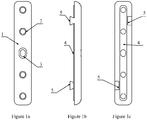
- the Figures 1a, 1b and 1c respectively represent a view of the external face, a side view and a view of the internal face of the right osteosynthesis plate 1.
- the right osteosynthesis plate 1 is in the form of a parallelepiped of variable length, width and thickness. Its external face, flat, radiated all around its periphery, is pierced with several through-fixing holes 2, 3, of variable diameters, of which the most central 3 is oblong, to allow a translation of the plate according to the topography of the site fracture.
- the through fastening holes 2, 3 are tapped on the lower half of their height, a metric thread iso standard pitch or fine pitch, corresponding to the head thread of the fastening screws, and milled to their upper half.
- the outer edges of the plate are radiated around the perimeter.
- the inner face in contact with the bone comprises along its length a bulge 4 rounded with a maximum thickness of 0.2 mm, including the fixing holes of the screws 2, 3.
- This bulge is intended to allow the adjustment of the plate closer to the anatomical variations of the insertion site.
- osteology shows that bone anatomy is reproducible from one individual to another; the differences relating to the anatomical accidents and landmarks which are the tuberosities, the tubercles, the apophyses, the gutters, the lines, the dimples, whose shapes and volumes can vary from a few tenths of a millimeter.
- the right osteosynthesis plate 1 further comprises, on its internal face, along its larger dimensions, two pins 5, of trapezoidal shape, of variable dimensions, one to the upper third, the other to the lower third, and diametrically opposed.
- the function of these pins is to lock the plate on the bone fragments on both sides of the fracture line after fitting into the cortical of the insertion wells of the pins with the help of the phantom plate, which realizes that makes an immediate primary fixation in a key and a secondary fixation, once the bone healing is carried out.
- An epiphyso-diaphyseal osteosynthesis plate is obtained according to the method described in Example 1.
- the Figures 2a, 2b and 2c represent, respectively, a view of the external face, a lateral view and a view of the internal face of the epiphyso-diaphyseal osteosynthesis plate 6.
- the epiphyseal-diaphyseal T osteosynthesis plate 6 has a horizontal epiphyseal part of variable length, height and thickness, curved backwards and forwards, so as to follow the meta-epiphyseal topography.
- the central fixing hole 8 of variable dimensions is oblong, so as to allow a translation as required.
- the diaphyseal vertical bar of variable dimensions, slightly convex from top to bottom and inwards, and from back to front, marrying the topography of the diaphyseal relief, is pierced with opening fixing holes 9, 10 in variable number and diameter (three on the Figure 2a ), whose central hole 10 is oblong to allow translation if necessary, before tightening the screws. They are milled in their upper half and tapped at the iso, standard or fine metric on the lower half.
- the outer edges of the plate are radiated around the perimeter.
- the outer faces of the epiphyseal and diaphyseal bars are flat.
- the inner faces comprise a bulge 11 rounded with a maximum height of 0.2 mm, including the fixing holes 7, 8, 9, 10. They also comprise two lugs 12, 13 acting as keys in the cortex, diametrically opposed. , of trapezoidal shape, of variable dimensions, one 12, on the posterior vertical edge of the epiphyseal bar, the other 13, the lower third of the anterior edge of the diaphyseal bar, thus opposing the rotation and displacement distal and proximal fragments of the fractured bone.
- An epiphyso-diaphyseal ruptive osteosynthesis plate is obtained according to the method described in Example 1.
- the Figures 3a, 3b and 3c represent respectively a view of the external face, a side view and a view of the internal face of the epiphyseal-diaphyseal ruptive osteosynthesis plate 14.
- This ruptive plaque is intended for use in pediatric surgery.
- the epiphyso-diaphyseal ruptive osteosynthesis plate 14 is in the form of a T of variable dimensions, similar in all its characteristics to the adult plate of Example 3, and further comprises on both sides, at the junction between the horizontal bar and the vertical bar, two V-shaped notches 15 interesting edges.
- the horizontal and vertical bars are pierced with through fastening holes 16, 17 in variable number and diameter, milled on their upper half and tapped on their lower half iso metric standard or fine pitch.
- the central fixing hole 17, horizontal and vertical bars, of variable dimensions, is oblong, so as to allow a translation as required.
- the outer edges of the plate are radiated around the perimeter.
- the outer faces of the horizontal and vertical bars are flat.
- the internal faces (represented on the Figure 3c ) of the horizontal bar and the vertical bar each comprise a bulge 18 rounded, a maximum height of 0.2 mm, including the fixing holes 16, 17. They also comprise two lugs 19, 20 acting as keys in the cortex, diametrically opposed, of trapezoidal shape, of variable dimensions, one 19 on the vertical rear edge of the horizontal bar, the other 20 to the bottom third of the front edge of the vertical bar, thus opposing the rotation and displacement of the distal and proximal fragments of the fractured bone.
- V-shaped notches located at the junction of the horizontal bar and the vertical bar, allow the breakage of the plate into two parts. These notches will be perfectly marked on palpation and will indicate the seat of the skin incision to be performed.
- the plate can then be broken using a hammer and a surgical burin, allowing the limb to continue its development, the two parts of the osteosynthesis material moving away as the evolution of the cartilage conjugation.
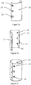
- FIG. 4a, 4b and 4c respectively represent a view of the external face, an isometric view of the inner face and a side view of the osteosynthesis plate for malleolar fractures 21.
- the osteosynthesis plate for malleolar fracture 21 is in the general form of a parallelepiped of variable length and width, the lower end of which is convex from front to back and from bottom to top over half of its length, and radiated all around.
- Its inner face is concave from rear to front and has at each end lugs 22 of trapezoidal shape of variable dimensions.
- the lug at the lower end is positioned to attach to the lower end of the styloid process, once the fracture is reduced.
- the plate is pierced with fixing holes 23, 24, 25, in number depending on its length, and radiated around its entire periphery.
- the most central hole 24 is oblong in shape to allow possible translation.
- the last lower fixation hole 25 makes an angle of 15 ° with the vertical to allow fixation of the styloid process on the distal portion of the fibula with bi-cortical support.
- Example 6 Gutter for the osteosynthesis of diaphyseal comminuted fractures
- a gutter for the osteosynthesis of comminuted diaphyseal fractures is obtained according to the method described in Example 1. It is represented on the Figures 5a, 5b and 5c . It consists of two parts which are two half-sleeves 26, 27, machined according to modeling of the possible areas of insertion of the osteosynthesis device. The outer face of the half-sleeve 26 is represented on the Figure 5a and the inner face of the half-sleeve 27 on the Figure 5b . The assembly of the gutter assembled with the two half-sleeves is represented on the Figure 5c .
- the half-sleeve 26 is in the form of a half-cylinder of variable diameter and length, its external face is convex over its entire height, pierced with through holes 28 of variable number and diameter, aligned with the longitudinal edges of the gutter and offset relative to each other so that the fastening screws intersect.
- the fixing holes 28 are tapped to the iso metric pitch on the inner half of their height and milled on the other half.
- the inner face is concave over its entire height; it presents along the longitudinal edges, two notches 29 diametrically opposed, one to the upper third, the other to the lower third.
- the other half-sleeve 27 is machined according to numerical modeling of the homologous zone of the half-sleeve 26, of generally triangular section, adapting to the bone morphology of the diaphyseal zone considered. It is of length and thickness comparable to those of the half-sleeve 26 and is pierced with through holes 30, threaded over their entire height to the metric pitch iso corresponding to the thread of the end of the fixing screws. These holes are paired with those 28 of the half-sleeve 26 and diametrically opposed. It presents along its longitudinal and diametrically opposite edges two lugs 31, of variable dimensions, and corresponding to the notches 29 of the half-sleeve 26.
- the Figure 5c represents the two half-sleeves assembled, the lugs 31 of the half-sleeve 27 being fixed in the notches 29 of the half-sleeve 26.
- the two half-sleeves 26, 27 are bevelled, so as to adapt during osteosynthesis, their upper and lower edges are radiated.
- An osteosynthesis screw is obtained according to the method equivalent to that described in Example 1.
- the Figure 6a represents a view of the osteosynthesis screw 32.
- the osteosynthesis screw 32 is in a generally cylindrical form composed of a thread and a head of different diameters and of variable dimensions.
- the upper end 39 of the screw is milled, and surmounted by a hexagonal device 40 for screwing with a suitable ancillary.
- the under head portion 33 is threaded in an iso metric pitch, standard pitch or fine pitch, corresponding to the pitch of the inner portion of the plate fixing holes.
- the portion of the screw 34 under the metric thread iso is threaded in a particular step, and a clean geometry on the one hand to oppose the unscrewing and secondly to promote a total filling of the turns by the fabric neoformed bone.
- This step is represented on the Figure 6b .
- it is in the form of any trapezium, whose oblique upper side 36, from outside to inside and from top to bottom, presents with the upper end of the bottom of the net
- Example 8 Surgical anchor for ligament and / or tendinous reinsertion
- a surgical anchor for ligament and / or tendinous reinsertion is obtained according to the method described in Example 1.
- the Figure 7 represents an overview of the surgical anchor for ligament and / or tendon reinsertion 41.
- the surgical anchor for ligament and / or tendinous reinsertion 41 is generally cylindrical in shape and comprises on the lower two-thirds a threading 42, as shown in FIG. Figure 6b , opposing unscrewing.
- the upper third from the upper limit of the threading 42, comprises a circular constriction 43 of variable depth and height, surmounted by a quadrilateral 44 at the radiated corners having on its four faces a rounded depression 45 of variable diameter.
- the quadrilateral 44 is surmounted by a hexagonal structure 46 of variable height, inscribed in the perimeter of the latter.
- FIG. 8a, 8b and 8c respectively represent the lower face, a sagittal section, and the upper face of the inter-somatic cage 47.
- the inter-somatic cage 47 is in the general form of a parallelepiped with a trapezoidal base, with variable dimensions, the central portion of which is pierced by an orifice 48 opening, of variable diameter, in the shape of a dovetail, in the direction transverse.
- the upper face, flat comprises two notches 49, 50 rectilinear cross staircase, one 49 at a distance and in front of the front edge of the central orifice, the other 50 at a distance and behind its edge posterior. These notches are opposed to sliding forward of the cage.
- the lower face, flat also comprises two notches 51, 52 in an inclined plane from top to bottom and front to back, one 51 away from the front edge of the central orifice, the other 52 away from its posterior edge, opposing the sliding towards the rear of the cage.
- the upper side is machined at a predetermined angle, depending on the seat of the discectomy, and to restore cervical or lumbar lordosis.
- the anterior face, plane, of variable dimensions is pierced at its center with a fixing hole 53 threaded with iso metric pitch, of variable depth and diameter, intended to receive at first the cage screwdriver and in a second time the fixation screw of the osteosynthesis plate.
- the rear face of variable height, has a rounded central depression 54, intended to be positioned opposite the yellow ligament.
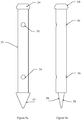
- FIG. 9a and 9b represent respectively a front view and a side view of the intramedullary nail 55.
- the intramedullary nail 55 is in the form of a cylinder of variable diameter and length, having at both ends, two or more orifices 56 opening to key it with bi-cortical support screws.
- the lower end 57 is in the form of a harpoon and has two flats 58 diametrically opposed.
- the upper end 59 is rounded, flattened in the center, to allow impaction, and has, over its entire diameter a flange 60, radially around.
- a membrane retention screw is obtained according to the method described in Example 1.
- the Figure 10 represents a front view of the membrane containment screw 61.
- the membrane retaining screw 61 is in the form of a cylindrical screw of variable length and diameter, comprising a threading 62 as described in FIG. Figure 6b , surmounted by a hexagonal head 63, of variable height and diameter.
- the thread allows to resist tearing when the screw is placed in a cortex or a deficient alveolar bone.
- a replacement dental implant is obtained according to the method described in Example 1.
- the Figures 11a and 11b represent respectively a sagittal section and an exploded view of the substitute dental implant 64.
- the replacement dental implant 64 comprises a cylindrical body 65 of variable length and diameter, comprising a thread opposing the unscrewing as described in FIG. Figure 6b .
- the body 65 is completed at its upper third, a Dacron® 66 felt ring intended to promote fibroblastic colonization and realizing a real gingival crimp all around its periphery, delimiting an area of gingival attachment and a free pseudo-gingiva, opposing the migration of oral fluids and food particles.
- This device also comprises a resilience disk 67, a cap 68 and a pre-prosthetic abutment 69, biocompatible polymer elements, intended to receive the prosthetic restoration.
- the animals are anesthetized according to the usual protocol: thiopental sodium (I V) 1g / animal approximately, maintenance isoflurane 1,7-6-1,8% and ketamine before the painful phases. After cutaneous incision and musculo-aponeurotic and periosteal dissection, the surface of the cortical bone is exposed.
- the screw fixation holes of the osteosynthesis material and lugs are drilled. After removal of the phantom material, the fixation holes are tapped from cortical to cortical, open.
- the plate is positioned and held in place by holding gauges, fixed by the screws in place of the holding gauges.
- the deep planes are sutured with resorbable sutures, the cutaneous plane with non-absorbable threads. These are removed after 10 days of healing.
- Radios are performed at D + 30 and D + 60, the anatomical parts are taken at D + 60 on three ewes and D + 120 on the other three ewes.
- the plates and the head of the screws appear opalescent, soaked with plasma, and adhere to the cortex of the bone.
- the macroscopic examination shows a change in the hue of the osteosynthesis plates, as well as the presence of a suffusion of an amber yellow liquid, similar to the plasma, during the section of the plate.
- Histomorphometric examination of the cortical and vis-endosteal bone-bone interfaces shows an anfractuous lacunar erosion of the inner face of the plate and screw turns, colonized by osteoblasts, of subperiosteal metaplastic bone apposition, and that there is a significant thickening of the endosteum.
- cortical bone-screw-medullary cavity-bone interface shows the same images, namely, surface lacunae erosion along the screw turns and over their entire length, with the appearance of newly formed bone, suggesting that the Mesenchymal stem cells of the bone marrow are at the origin, with the induction of an osteo-promoter activity.
- osteoclastic giant cells found near the screws in the medullary cavity, formed by fusion, up to 100 microns, and derived from the same precursor cells as monocytes, shows that they are responsible for the formation of gaps in the form of crevices on the surface of the screws.
Landscapes
- Health & Medical Sciences (AREA)
- Life Sciences & Earth Sciences (AREA)
- Chemical & Material Sciences (AREA)
- Engineering & Computer Science (AREA)
- Animal Behavior & Ethology (AREA)
- Veterinary Medicine (AREA)
- Public Health (AREA)
- General Health & Medical Sciences (AREA)
- Biomedical Technology (AREA)
- Medicinal Chemistry (AREA)
- Epidemiology (AREA)
- Orthopedic Medicine & Surgery (AREA)
- Oral & Maxillofacial Surgery (AREA)
- Dermatology (AREA)
- Transplantation (AREA)
- Heart & Thoracic Surgery (AREA)
- Surgery (AREA)
- Molecular Biology (AREA)
- Botany (AREA)
- Chemical Kinetics & Catalysis (AREA)
- Zoology (AREA)
- Ceramic Engineering (AREA)
- Nuclear Medicine, Radiotherapy & Molecular Imaging (AREA)
- Medical Informatics (AREA)
- Vascular Medicine (AREA)
- Neurology (AREA)
- Pharmacology & Pharmacy (AREA)
- Marine Sciences & Fisheries (AREA)
- Inorganic Chemistry (AREA)
- Nursing (AREA)
- Dentistry (AREA)
- Urology & Nephrology (AREA)
- Cardiology (AREA)
- Materials For Medical Uses (AREA)
- Prostheses (AREA)
- Dental Preparations (AREA)
- Surgical Instruments (AREA)
Priority Applications (2)
| Application Number | Priority Date | Filing Date | Title |
|---|---|---|---|
| HRP20181603TT HRP20181603T1 (hr) | 2014-01-10 | 2015-01-08 | Postupak proizvodnje uređaja za ostrosintezu, uređaji za ostrosintezu i usatci načinjeni od polusintetskog, hibridnog materijala, dobiveni modificiranjem strukture komponenata od prirodnog morskog biomaterijala |
| PL15702540T PL3092014T3 (pl) | 2014-01-10 | 2015-01-08 | Sposób wytwarzania urządzeń do osteosyntezy, urządzenia do osteosyntezy i implanty wykonane z półsyntetycznego materiału hybrydowego uzyskanego przez modyfikację strukturalną składników naturalnego biomateriału morskiego |
Applications Claiming Priority (2)
| Application Number | Priority Date | Filing Date | Title |
|---|---|---|---|
| FR1450204A FR3016293B1 (fr) | 2014-01-10 | 2014-01-10 | Procede de fabrication de dispositifs d'osteosynthese, dispositifs d'osteosynthese et implants en materiau hybride semi-synthetique obtenu par modification structurale des composants d'un biomateriau naturel marin |
| PCT/FR2015/050037 WO2015104502A1 (fr) | 2014-01-10 | 2015-01-08 | Procédé de fabrication de dispositifs d'ostéosynthèse, dispositifs d'ostéosynthèse et implants en matériau hybride semi-synthètique obtenu par modification structurale des composants d'un biomatériau naturel marin |
Publications (2)
| Publication Number | Publication Date |
|---|---|
| EP3092014A1 EP3092014A1 (fr) | 2016-11-16 |
| EP3092014B1 true EP3092014B1 (fr) | 2018-07-11 |
Family
ID=50290130
Family Applications (1)
| Application Number | Title | Priority Date | Filing Date |
|---|---|---|---|
| EP15702540.4A Active EP3092014B1 (fr) | 2014-01-10 | 2015-01-08 | Procédé de fabrication de dispositifs d'ostéosynthèse, dispositifs d'ostéosynthèse et implants en matériau hybride semi-synthétique obtenu par modification structurale des composants d'un biomatériau naturel marin |
Country Status (19)
| Country | Link |
|---|---|
| EP (1) | EP3092014B1 (pl) |
| JP (2) | JP6785662B2 (pl) |
| KR (1) | KR102313540B1 (pl) |
| CN (1) | CN106132449B (pl) |
| AU (1) | AU2015205476B2 (pl) |
| BR (1) | BR112016015989B1 (pl) |
| CA (1) | CA2936475C (pl) |
| DK (1) | DK3092014T3 (pl) |
| ES (1) | ES2690093T3 (pl) |
| FR (1) | FR3016293B1 (pl) |
| HR (1) | HRP20181603T1 (pl) |
| HU (1) | HUE039518T2 (pl) |
| IL (1) | IL246649B (pl) |
| MX (1) | MX365734B (pl) |
| PH (1) | PH12016501346B1 (pl) |
| PL (1) | PL3092014T3 (pl) |
| PT (1) | PT3092014T (pl) |
| RU (1) | RU2669926C2 (pl) |
| WO (1) | WO2015104502A1 (pl) |
Families Citing this family (8)
| Publication number | Priority date | Publication date | Assignee | Title |
|---|---|---|---|---|
| CN106421926A (zh) * | 2016-09-28 | 2017-02-22 | 范玉梅 | 纯天然中药海洋生物活性免疫骨 |
| CN107837131B (zh) * | 2017-12-05 | 2024-04-16 | 运怡(北京)医疗器械有限公司 | 一种带袢固定板 |
| FR3095947B1 (fr) * | 2019-05-13 | 2022-05-13 | Mbp Mauritius Ltd | Procédé d'isolation des molécules contenues dans les couches organo-minérales des coquilles de mollusques marins bivalves |
| CN112807066B (zh) * | 2020-12-30 | 2023-05-12 | 杨润松 | 桡骨头重建复位髓内髓外组合式固定装置 |
| RU2758570C1 (ru) * | 2021-01-13 | 2021-10-29 | Общество с ограниченной ответственностью "Медлайн Компани" | Способ дегазации гранулированного остеокондуктивного костнопластического материала |
| RU2758551C1 (ru) * | 2021-01-13 | 2021-10-29 | Общество с ограниченной ответственностью "Медлайн Компани" | Способ определения адсорбционной ёмкости гранулированного остеокондуктивного костнопластического материала |
| CN112957161B (zh) * | 2021-01-29 | 2023-03-31 | 四川大学华西医院 | 颈椎侧方植骨撑开翼状固定钛网 |
| CN119925041B (zh) * | 2024-12-20 | 2025-10-14 | 北京力达康科技有限公司 | 一种股骨颈骨折修复系统 |
Family Cites Families (13)
| Publication number | Priority date | Publication date | Assignee | Title |
|---|---|---|---|---|
| FR2647334A1 (fr) * | 1989-05-23 | 1990-11-30 | Cazprasse Serge | Produits de remplacement de structures osseuses et radiculo-dentaires en nacre |
| CA2022480C (en) * | 1989-08-02 | 2001-02-27 | Gerald L. Mechanic | Process for cross-linking collagenous materials and resulting product |
| FR2706308B1 (fr) * | 1993-02-05 | 1995-09-08 | Camprasse Georges | Prothèses orthopédiques et ciment de scellement biologique en nacré. |
| US6093530A (en) * | 1998-02-06 | 2000-07-25 | Sulzer Carbomedics Inc. | Non-calcific biomaterial by glutaraldehyde followed by oxidative fixation |
| US6630153B2 (en) * | 2001-02-23 | 2003-10-07 | Smith & Nephew, Inc. | Manufacture of bone graft substitutes |
| FR2808657B1 (fr) * | 2000-05-11 | 2003-06-13 | Pharma Futura | Composition commestible comprenant de la poudre de nacre micronisee |
| FR2827478B1 (fr) * | 2001-07-18 | 2003-10-24 | Jean Louis Montero | Procede de preparation d'une poudre a base de nacre, proteine isolee de ladite poudre et leurs utilisations en chirurgie osseuse et dans diverses pathologies osteoarticulaires |
| JP2009120449A (ja) * | 2007-11-16 | 2009-06-04 | Amino Up Chemical Co Ltd | バイオアパタイトの製造方法及び生物学的な活性を有する物質の分離方法 |
| CN101496908B (zh) * | 2009-02-20 | 2012-10-31 | 杭州电子科技大学 | 一种具有多级微纳结构的珍珠粉人工骨支架材料及其生产工艺 |
| FR2946888B1 (fr) * | 2009-06-17 | 2011-07-29 | Mega Bio Pharma | Procede de preparation de nacre mecanostructuree par mecano-synthese, nacre mecanostructuree ainsi obtenue et ses applications |
| CN101856515B (zh) * | 2010-06-04 | 2013-03-13 | 西南大学 | 以壳聚糖和贝壳粉末为原料制备人工骨的方法 |
| US20120207839A1 (en) * | 2011-02-14 | 2012-08-16 | Maxigen Biotech Inc. | Mineralized Collagen/Bioceramic Composite and Manufacturing Method Thereof |
| CN102160900B (zh) * | 2011-04-14 | 2014-06-04 | 暨南大学 | 一种骨生长因子控释型骨修复材料及其制备方法与应用 |
-
2014
- 2014-01-10 FR FR1450204A patent/FR3016293B1/fr active Active
-
2015
- 2015-01-08 PL PL15702540T patent/PL3092014T3/pl unknown
- 2015-01-08 CN CN201580004180.3A patent/CN106132449B/zh active Active
- 2015-01-08 DK DK15702540.4T patent/DK3092014T3/en active
- 2015-01-08 HR HRP20181603TT patent/HRP20181603T1/hr unknown
- 2015-01-08 RU RU2016132785A patent/RU2669926C2/ru active
- 2015-01-08 WO PCT/FR2015/050037 patent/WO2015104502A1/fr not_active Ceased
- 2015-01-08 PT PT15702540T patent/PT3092014T/pt unknown
- 2015-01-08 EP EP15702540.4A patent/EP3092014B1/fr active Active
- 2015-01-08 CA CA2936475A patent/CA2936475C/fr active Active
- 2015-01-08 HU HUE15702540A patent/HUE039518T2/hu unknown
- 2015-01-08 ES ES15702540.4T patent/ES2690093T3/es active Active
- 2015-01-08 JP JP2016563268A patent/JP6785662B2/ja active Active
- 2015-01-08 MX MX2016009028A patent/MX365734B/es active IP Right Grant
- 2015-01-08 KR KR1020167021812A patent/KR102313540B1/ko active Active
- 2015-01-08 AU AU2015205476A patent/AU2015205476B2/en active Active
- 2015-01-08 BR BR112016015989-6A patent/BR112016015989B1/pt active IP Right Grant
-
2016
- 2016-07-07 PH PH12016501346A patent/PH12016501346B1/en unknown
- 2016-07-07 IL IL246649A patent/IL246649B/en active IP Right Grant
-
2019
- 2019-11-14 JP JP2019206218A patent/JP2020044344A/ja active Pending
Non-Patent Citations (1)
| Title |
|---|
| None * |
Also Published As
| Publication number | Publication date |
|---|---|
| HK1225658B (zh) | 2017-09-15 |
| CN106132449A (zh) | 2016-11-16 |
| MX2016009028A (es) | 2017-03-10 |
| RU2016132785A (ru) | 2018-02-14 |
| PH12016501346A1 (en) | 2017-02-06 |
| PH12016501346B1 (en) | 2020-01-31 |
| IL246649A0 (en) | 2016-08-31 |
| JP6785662B2 (ja) | 2020-11-18 |
| CA2936475A1 (fr) | 2015-07-16 |
| DK3092014T3 (en) | 2018-10-22 |
| JP2020044344A (ja) | 2020-03-26 |
| WO2015104502A1 (fr) | 2015-07-16 |
| KR102313540B1 (ko) | 2021-10-15 |
| RU2669926C2 (ru) | 2018-10-17 |
| CN106132449B (zh) | 2019-08-20 |
| PL3092014T3 (pl) | 2019-01-31 |
| HRP20181603T1 (hr) | 2018-11-30 |
| KR20160130221A (ko) | 2016-11-10 |
| FR3016293B1 (fr) | 2019-12-20 |
| BR112016015989B1 (pt) | 2021-02-09 |
| BR112016015989A2 (pt) | 2017-08-08 |
| IL246649B (en) | 2020-07-30 |
| FR3016293A1 (fr) | 2015-07-17 |
| JP2017503627A (ja) | 2017-02-02 |
| PT3092014T (pt) | 2018-11-05 |
| ES2690093T3 (es) | 2018-11-19 |
| HUE039518T2 (hu) | 2019-01-28 |
| CA2936475C (fr) | 2022-10-18 |
| AU2015205476B2 (en) | 2017-10-05 |
| MX365734B (es) | 2019-06-12 |
| AU2015205476A1 (en) | 2016-07-21 |
| EP3092014A1 (fr) | 2016-11-16 |
| RU2016132785A3 (pl) | 2018-06-28 |
Similar Documents
| Publication | Publication Date | Title |
|---|---|---|
| EP3092014B1 (fr) | Procédé de fabrication de dispositifs d'ostéosynthèse, dispositifs d'ostéosynthèse et implants en matériau hybride semi-synthétique obtenu par modification structurale des composants d'un biomatériau naturel marin | |
| EP0022724B1 (fr) | Implant biodégradable utilisable comme pièce de prothèse osseuse | |
| ES2247113T3 (es) | Implante oseo desmineralizado en la superficie y metodo para fabricarlo. | |
| US10932916B2 (en) | Method for producing osteosynthesis devices, osteosynthesis devices and implants made of semi-synthetic hybrid material obtained by structural modification of the components of a natural marine biomaterial | |
| Kujala et al. | Effect of porosity on the osteointegration and bone ingrowth of a weight-bearing nickel–titanium bone graft substitute | |
| Cunha et al. | Surface modification of zirconia dental implants by laser texturing | |
| ES2215913T3 (es) | Implantes osteogenicos derivados de hueso. | |
| CA2765930C (fr) | Procede de preparation de nacre mecanostructuree par mecano-synthese, nacre mecanostructuree ainsi obtenue et ses applications | |
| US20050152987A1 (en) | Transplantable particulate bone composition having high osteoinductive capacity and methods for making and using same | |
| US20100010632A1 (en) | Sand-blasting method using biocompatible polymers | |
| KR20030036620A (ko) | 골이식물 및 이의 제조방법 | |
| NZ513080A (en) | Implants with modified surfaces for increased biocompatibility, and method for production thereof | |
| US9968704B2 (en) | Method for regenerating alveolar bone and calcium-containing microparticles used to regenerate alveolar bone | |
| WO2009046517A1 (en) | Method and device for bone regeneration | |
| Li et al. | In vivo histological evaluation of bioactive NiTi alloy after two years implantation | |
| KR102639266B1 (ko) | 표면 처리된 임플란트 구조체 | |
| US20100076501A1 (en) | Method and device for bone regeneration | |
| HK1225658A1 (en) | Method for manufacturing osteosynthesis devices, osteosynthesis devices and implants made of semi-synthetic hybrid material obtained by structural modification of the components of a natural marine biomaterial | |
| DE MASSIS | Biochemical analysis of titanium screws covered with decellularized heterologous bone tissue |
Legal Events
| Date | Code | Title | Description |
|---|---|---|---|
| PUAI | Public reference made under article 153(3) epc to a published international application that has entered the european phase |
Free format text: ORIGINAL CODE: 0009012 |
|
| 17P | Request for examination filed |
Effective date: 20160706 |
|
| AK | Designated contracting states |
Kind code of ref document: A1 Designated state(s): AL AT BE BG CH CY CZ DE DK EE ES FI FR GB GR HR HU IE IS IT LI LT LU LV MC MK MT NL NO PL PT RO RS SE SI SK SM TR |
|
| AX | Request for extension of the european patent |
Extension state: BA ME |
|
| DAX | Request for extension of the european patent (deleted) | ||
| STAA | Information on the status of an ep patent application or granted ep patent |
Free format text: STATUS: EXAMINATION IS IN PROGRESS |
|
| 17Q | First examination report despatched |
Effective date: 20170524 |
|
| REG | Reference to a national code |
Ref country code: HK Ref legal event code: DE Ref document number: 1225658 Country of ref document: HK |
|
| GRAP | Despatch of communication of intention to grant a patent |
Free format text: ORIGINAL CODE: EPIDOSNIGR1 |
|
| STAA | Information on the status of an ep patent application or granted ep patent |
Free format text: STATUS: GRANT OF PATENT IS INTENDED |
|
| INTG | Intention to grant announced |
Effective date: 20180417 |
|
| GRAA | (expected) grant |
Free format text: ORIGINAL CODE: 0009210 |
|
| STAA | Information on the status of an ep patent application or granted ep patent |
Free format text: STATUS: THE PATENT HAS BEEN GRANTED |
|
| AK | Designated contracting states |
Kind code of ref document: B1 Designated state(s): AL AT BE BG CH CY CZ DE DK EE ES FI FR GB GR HR HU IE IS IT LI LT LU LV MC MK MT NL NO PL PT RO RS SE SI SK SM TR |
|
| REG | Reference to a national code |
Ref country code: GB Ref legal event code: FG4D Free format text: NOT ENGLISH |
|
| REG | Reference to a national code |
Ref country code: CH Ref legal event code: EP |
|
| REG | Reference to a national code |
Ref country code: AT Ref legal event code: REF Ref document number: 1016231 Country of ref document: AT Kind code of ref document: T Effective date: 20180715 |
|
| GRAS | Grant fee paid |
Free format text: ORIGINAL CODE: EPIDOSNIGR3 |
|
| REG | Reference to a national code |
Ref country code: IE Ref legal event code: FG4D Free format text: LANGUAGE OF EP DOCUMENT: FRENCH |
|
| REG | Reference to a national code |
Ref country code: DE Ref legal event code: R096 Ref document number: 602015013361 Country of ref document: DE |
|
| REG | Reference to a national code |
Ref country code: HR Ref legal event code: TUEP Ref document number: P20181603 Country of ref document: HR |
|
| REG | Reference to a national code |
Ref country code: RO Ref legal event code: EPE |
|
| REG | Reference to a national code |
Ref country code: DK Ref legal event code: T3 Effective date: 20181015 |
|
| REG | Reference to a national code |
Ref country code: NL Ref legal event code: FP |
|
| REG | Reference to a national code |
Ref country code: SE Ref legal event code: TRGR |
|
| REG | Reference to a national code |
Ref country code: PT Ref legal event code: SC4A Ref document number: 3092014 Country of ref document: PT Date of ref document: 20181105 Kind code of ref document: T Free format text: AVAILABILITY OF NATIONAL TRANSLATION Effective date: 20181008 |
|
| REG | Reference to a national code |
Ref country code: ES Ref legal event code: FG2A Ref document number: 2690093 Country of ref document: ES Kind code of ref document: T3 Effective date: 20181119 |
|
| REG | Reference to a national code |
Ref country code: LT Ref legal event code: MG4D |
|
| REG | Reference to a national code |
Ref country code: HR Ref legal event code: T1PR Ref document number: P20181603 Country of ref document: HR |
|
| REG | Reference to a national code |
Ref country code: HR Ref legal event code: ODRP Ref document number: P20181603 Country of ref document: HR Payment date: 20181214 Year of fee payment: 5 |
|
| REG | Reference to a national code |
Ref country code: HU Ref legal event code: AG4A Ref document number: E039518 Country of ref document: HU |
|
| PG25 | Lapsed in a contracting state [announced via postgrant information from national office to epo] |
Ref country code: RS Free format text: LAPSE BECAUSE OF FAILURE TO SUBMIT A TRANSLATION OF THE DESCRIPTION OR TO PAY THE FEE WITHIN THE PRESCRIBED TIME-LIMIT Effective date: 20180711 Ref country code: IS Free format text: LAPSE BECAUSE OF FAILURE TO SUBMIT A TRANSLATION OF THE DESCRIPTION OR TO PAY THE FEE WITHIN THE PRESCRIBED TIME-LIMIT Effective date: 20181111 Ref country code: NO Free format text: LAPSE BECAUSE OF FAILURE TO SUBMIT A TRANSLATION OF THE DESCRIPTION OR TO PAY THE FEE WITHIN THE PRESCRIBED TIME-LIMIT Effective date: 20181011 Ref country code: FI Free format text: LAPSE BECAUSE OF FAILURE TO SUBMIT A TRANSLATION OF THE DESCRIPTION OR TO PAY THE FEE WITHIN THE PRESCRIBED TIME-LIMIT Effective date: 20180711 Ref country code: BG Free format text: LAPSE BECAUSE OF FAILURE TO SUBMIT A TRANSLATION OF THE DESCRIPTION OR TO PAY THE FEE WITHIN THE PRESCRIBED TIME-LIMIT Effective date: 20181011 Ref country code: LT Free format text: LAPSE BECAUSE OF FAILURE TO SUBMIT A TRANSLATION OF THE DESCRIPTION OR TO PAY THE FEE WITHIN THE PRESCRIBED TIME-LIMIT Effective date: 20180711 |
|
| REG | Reference to a national code |
Ref country code: SK Ref legal event code: T3 Ref document number: E 28795 Country of ref document: SK |
|
| PG25 | Lapsed in a contracting state [announced via postgrant information from national office to epo] |
Ref country code: AL Free format text: LAPSE BECAUSE OF FAILURE TO SUBMIT A TRANSLATION OF THE DESCRIPTION OR TO PAY THE FEE WITHIN THE PRESCRIBED TIME-LIMIT Effective date: 20180711 Ref country code: LV Free format text: LAPSE BECAUSE OF FAILURE TO SUBMIT A TRANSLATION OF THE DESCRIPTION OR TO PAY THE FEE WITHIN THE PRESCRIBED TIME-LIMIT Effective date: 20180711 |
|
| REG | Reference to a national code |
Ref country code: GR Ref legal event code: EP Ref document number: 20180402849 Country of ref document: GR Effective date: 20190225 |
|
| REG | Reference to a national code |
Ref country code: DE Ref legal event code: R097 Ref document number: 602015013361 Country of ref document: DE |
|
| PG25 | Lapsed in a contracting state [announced via postgrant information from national office to epo] |
Ref country code: EE Free format text: LAPSE BECAUSE OF FAILURE TO SUBMIT A TRANSLATION OF THE DESCRIPTION OR TO PAY THE FEE WITHIN THE PRESCRIBED TIME-LIMIT Effective date: 20180711 |
|
| PLBE | No opposition filed within time limit |
Free format text: ORIGINAL CODE: 0009261 |
|
| STAA | Information on the status of an ep patent application or granted ep patent |
Free format text: STATUS: NO OPPOSITION FILED WITHIN TIME LIMIT |
|
| PG25 | Lapsed in a contracting state [announced via postgrant information from national office to epo] |
Ref country code: SM Free format text: LAPSE BECAUSE OF FAILURE TO SUBMIT A TRANSLATION OF THE DESCRIPTION OR TO PAY THE FEE WITHIN THE PRESCRIBED TIME-LIMIT Effective date: 20180711 |
|
| 26N | No opposition filed |
Effective date: 20190412 |
|
| PG25 | Lapsed in a contracting state [announced via postgrant information from national office to epo] |
Ref country code: SI Free format text: LAPSE BECAUSE OF FAILURE TO SUBMIT A TRANSLATION OF THE DESCRIPTION OR TO PAY THE FEE WITHIN THE PRESCRIBED TIME-LIMIT Effective date: 20180711 |
|
| REG | Reference to a national code |
Ref country code: HR Ref legal event code: ODRP Ref document number: P20181603 Country of ref document: HR Payment date: 20191220 Year of fee payment: 6 |
|
| PG25 | Lapsed in a contracting state [announced via postgrant information from national office to epo] |
Ref country code: MT Free format text: LAPSE BECAUSE OF FAILURE TO SUBMIT A TRANSLATION OF THE DESCRIPTION OR TO PAY THE FEE WITHIN THE PRESCRIBED TIME-LIMIT Effective date: 20180711 |
|
| REG | Reference to a national code |
Ref country code: HR Ref legal event code: ODRP Ref document number: P20181603 Country of ref document: HR Payment date: 20201217 Year of fee payment: 7 |
|
| REG | Reference to a national code |
Ref country code: AT Ref legal event code: UEP Ref document number: 1016231 Country of ref document: AT Kind code of ref document: T Effective date: 20180711 |
|
| PG25 | Lapsed in a contracting state [announced via postgrant information from national office to epo] |
Ref country code: CY Free format text: LAPSE BECAUSE OF FAILURE TO SUBMIT A TRANSLATION OF THE DESCRIPTION OR TO PAY THE FEE WITHIN THE PRESCRIBED TIME-LIMIT Effective date: 20180711 |
|
| REG | Reference to a national code |
Ref country code: HR Ref legal event code: ODRP Ref document number: P20181603 Country of ref document: HR Payment date: 20211228 Year of fee payment: 8 |
|
| PG25 | Lapsed in a contracting state [announced via postgrant information from national office to epo] |
Ref country code: MK Free format text: LAPSE BECAUSE OF FAILURE TO SUBMIT A TRANSLATION OF THE DESCRIPTION OR TO PAY THE FEE WITHIN THE PRESCRIBED TIME-LIMIT Effective date: 20180711 |
|
| REG | Reference to a national code |
Ref country code: HR Ref legal event code: ODRP Ref document number: P20181603 Country of ref document: HR Payment date: 20221229 Year of fee payment: 9 |
|
| PGFP | Annual fee paid to national office [announced via postgrant information from national office to epo] |
Ref country code: SK Payment date: 20231220 Year of fee payment: 10 |
|
| PGFP | Annual fee paid to national office [announced via postgrant information from national office to epo] |
Ref country code: GR Payment date: 20231219 Year of fee payment: 10 |
|
| PGFP | Annual fee paid to national office [announced via postgrant information from national office to epo] |
Ref country code: MC Payment date: 20231228 Year of fee payment: 10 |
|
| REG | Reference to a national code |
Ref country code: HR Ref legal event code: ODRP Ref document number: P20181603 Country of ref document: HR Payment date: 20231221 Year of fee payment: 10 |
|
| PGFP | Annual fee paid to national office [announced via postgrant information from national office to epo] |
Ref country code: PT Payment date: 20231218 Year of fee payment: 10 Ref country code: HR Payment date: 20231221 Year of fee payment: 10 Ref country code: DK Payment date: 20231228 Year of fee payment: 10 Ref country code: CZ Payment date: 20231219 Year of fee payment: 10 |
|
| PGFP | Annual fee paid to national office [announced via postgrant information from national office to epo] |
Ref country code: PL Payment date: 20231228 Year of fee payment: 10 |
|
| PGFP | Annual fee paid to national office [announced via postgrant information from national office to epo] |
Ref country code: AT Payment date: 20231222 Year of fee payment: 10 |
|
| PGFP | Annual fee paid to national office [announced via postgrant information from national office to epo] |
Ref country code: RO Payment date: 20240103 Year of fee payment: 10 Ref country code: HU Payment date: 20231219 Year of fee payment: 10 |
|
| PGFP | Annual fee paid to national office [announced via postgrant information from national office to epo] |
Ref country code: SE Payment date: 20240119 Year of fee payment: 10 |
|
| PGFP | Annual fee paid to national office [announced via postgrant information from national office to epo] |
Ref country code: NL Payment date: 20241224 Year of fee payment: 11 Ref country code: LU Payment date: 20241224 Year of fee payment: 11 |
|
| PGFP | Annual fee paid to national office [announced via postgrant information from national office to epo] |
Ref country code: FR Payment date: 20241220 Year of fee payment: 11 |
|
| PGFP | Annual fee paid to national office [announced via postgrant information from national office to epo] |
Ref country code: IE Payment date: 20241219 Year of fee payment: 11 |
|
| PGFP | Annual fee paid to national office [announced via postgrant information from national office to epo] |
Ref country code: DE Payment date: 20250116 Year of fee payment: 11 |
|
| PGFP | Annual fee paid to national office [announced via postgrant information from national office to epo] |
Ref country code: ES Payment date: 20250213 Year of fee payment: 11 |
|
| PGFP | Annual fee paid to national office [announced via postgrant information from national office to epo] |
Ref country code: CH Payment date: 20250201 Year of fee payment: 11 Ref country code: BE Payment date: 20250128 Year of fee payment: 11 |
|
| PGFP | Annual fee paid to national office [announced via postgrant information from national office to epo] |
Ref country code: IT Payment date: 20250109 Year of fee payment: 11 Ref country code: GB Payment date: 20250123 Year of fee payment: 11 |
|
| PGFP | Annual fee paid to national office [announced via postgrant information from national office to epo] |
Ref country code: TR Payment date: 20250106 Year of fee payment: 11 |
|
| REG | Reference to a national code |
Ref country code: HR Ref legal event code: PBON Ref document number: P20181603 Country of ref document: HR Effective date: 20250108 |
|
| PG25 | Lapsed in a contracting state [announced via postgrant information from national office to epo] |
Ref country code: CZ Free format text: LAPSE BECAUSE OF NON-PAYMENT OF DUE FEES Effective date: 20250108 |
|
| REG | Reference to a national code |
Ref country code: DK Ref legal event code: EBP Effective date: 20250131 |
|
| REG | Reference to a national code |
Ref country code: SK Ref legal event code: MM4A Ref document number: E 28795 Country of ref document: SK Effective date: 20250108 |
|
| REG | Reference to a national code |
Ref country code: SE Ref legal event code: EUG |
|
| PG25 | Lapsed in a contracting state [announced via postgrant information from national office to epo] |
Ref country code: MC Free format text: LAPSE BECAUSE OF NON-PAYMENT OF DUE FEES Effective date: 20250131 |
|
| REG | Reference to a national code |
Ref country code: AT Ref legal event code: MM01 Ref document number: 1016231 Country of ref document: AT Kind code of ref document: T Effective date: 20250108 |
|
| PG25 | Lapsed in a contracting state [announced via postgrant information from national office to epo] |
Ref country code: HU Free format text: LAPSE BECAUSE OF NON-PAYMENT OF DUE FEES Effective date: 20250109 |
|
| PG25 | Lapsed in a contracting state [announced via postgrant information from national office to epo] |
Ref country code: PT Free format text: LAPSE BECAUSE OF NON-PAYMENT OF DUE FEES Effective date: 20250708 |
|
| PG25 | Lapsed in a contracting state [announced via postgrant information from national office to epo] |
Ref country code: GR Free format text: LAPSE BECAUSE OF NON-PAYMENT OF DUE FEES Effective date: 20250805 |
|
| PG25 | Lapsed in a contracting state [announced via postgrant information from national office to epo] |
Ref country code: HR Free format text: LAPSE BECAUSE OF NON-PAYMENT OF DUE FEES Effective date: 20250108 |
|
| PG25 | Lapsed in a contracting state [announced via postgrant information from national office to epo] |
Ref country code: AT Free format text: LAPSE BECAUSE OF NON-PAYMENT OF DUE FEES Effective date: 20250108 |
|
| PG25 | Lapsed in a contracting state [announced via postgrant information from national office to epo] |
Ref country code: RO Free format text: LAPSE BECAUSE OF NON-PAYMENT OF DUE FEES Effective date: 20250108 |
|
| PG25 | Lapsed in a contracting state [announced via postgrant information from national office to epo] |
Ref country code: SK Free format text: LAPSE BECAUSE OF NON-PAYMENT OF DUE FEES Effective date: 20250108 |