EP3088511A1 - Image processing device, program, information storage medium, and image processing method - Google Patents
Image processing device, program, information storage medium, and image processing method Download PDFInfo
- Publication number
- EP3088511A1 EP3088511A1 EP14873584.8A EP14873584A EP3088511A1 EP 3088511 A1 EP3088511 A1 EP 3088511A1 EP 14873584 A EP14873584 A EP 14873584A EP 3088511 A1 EP3088511 A1 EP 3088511A1
- Authority
- EP
- European Patent Office
- Prior art keywords
- target
- specimen
- image processing
- detection
- sample
- Prior art date
- Legal status (The legal status is an assumption and is not a legal conclusion. Google has not performed a legal analysis and makes no representation as to the accuracy of the status listed.)
- Granted
Links
- 238000003672 processing method Methods 0.000 title claims description 4
- 238000001514 detection method Methods 0.000 claims abstract description 174
- 238000000034 method Methods 0.000 claims abstract description 58
- 210000004027 cell Anatomy 0.000 claims description 210
- 210000004369 blood Anatomy 0.000 claims description 23
- 239000008280 blood Substances 0.000 claims description 23
- 210000003743 erythrocyte Anatomy 0.000 claims description 14
- 230000008774 maternal effect Effects 0.000 claims description 11
- 230000035935 pregnancy Effects 0.000 claims description 7
- 210000004940 nucleus Anatomy 0.000 description 42
- 230000003287 optical effect Effects 0.000 description 19
- 238000002360 preparation method Methods 0.000 description 14
- 210000000265 leukocyte Anatomy 0.000 description 8
- 238000010186 staining Methods 0.000 description 8
- 239000000284 extract Substances 0.000 description 7
- 230000006870 function Effects 0.000 description 7
- 239000013598 vector Substances 0.000 description 7
- 210000003855 cell nucleus Anatomy 0.000 description 6
- 238000010586 diagram Methods 0.000 description 6
- 239000003086 colorant Substances 0.000 description 2
- 230000010354 integration Effects 0.000 description 2
- 238000012706 support-vector machine Methods 0.000 description 2
- 238000004364 calculation method Methods 0.000 description 1
- 238000004891 communication Methods 0.000 description 1
- 230000000694 effects Effects 0.000 description 1
- 238000000605 extraction Methods 0.000 description 1
- 230000001605 fetal effect Effects 0.000 description 1
- 239000004973 liquid crystal related substance Substances 0.000 description 1
- 238000001000 micrograph Methods 0.000 description 1
- 238000010606 normalization Methods 0.000 description 1
Images


Classifications
-
- G—PHYSICS
- G01—MEASURING; TESTING
- G01N—INVESTIGATING OR ANALYSING MATERIALS BY DETERMINING THEIR CHEMICAL OR PHYSICAL PROPERTIES
- G01N15/00—Investigating characteristics of particles; Investigating permeability, pore-volume or surface-area of porous materials
- G01N15/10—Investigating individual particles
- G01N15/14—Optical investigation techniques, e.g. flow cytometry
- G01N15/1468—Optical investigation techniques, e.g. flow cytometry with spatial resolution of the texture or inner structure of the particle
-
- G—PHYSICS
- G01—MEASURING; TESTING
- G01N—INVESTIGATING OR ANALYSING MATERIALS BY DETERMINING THEIR CHEMICAL OR PHYSICAL PROPERTIES
- G01N33/00—Investigating or analysing materials by specific methods not covered by groups G01N1/00 - G01N31/00
- G01N33/48—Biological material, e.g. blood, urine; Haemocytometers
- G01N33/483—Physical analysis of biological material
- G01N33/487—Physical analysis of biological material of liquid biological material
- G01N33/49—Blood
-
- G—PHYSICS
- G06—COMPUTING; CALCULATING OR COUNTING
- G06F—ELECTRIC DIGITAL DATA PROCESSING
- G06F18/00—Pattern recognition
- G06F18/20—Analysing
- G06F18/24—Classification techniques
-
- G—PHYSICS
- G06—COMPUTING; CALCULATING OR COUNTING
- G06T—IMAGE DATA PROCESSING OR GENERATION, IN GENERAL
- G06T7/00—Image analysis
- G06T7/0002—Inspection of images, e.g. flaw detection
- G06T7/0012—Biomedical image inspection
-
- G—PHYSICS
- G06—COMPUTING; CALCULATING OR COUNTING
- G06T—IMAGE DATA PROCESSING OR GENERATION, IN GENERAL
- G06T7/00—Image analysis
- G06T7/70—Determining position or orientation of objects or cameras
- G06T7/73—Determining position or orientation of objects or cameras using feature-based methods
-
- G—PHYSICS
- G06—COMPUTING; CALCULATING OR COUNTING
- G06V—IMAGE OR VIDEO RECOGNITION OR UNDERSTANDING
- G06V20/00—Scenes; Scene-specific elements
- G06V20/60—Type of objects
- G06V20/69—Microscopic objects, e.g. biological cells or cellular parts
-
- C—CHEMISTRY; METALLURGY
- C12—BIOCHEMISTRY; BEER; SPIRITS; WINE; VINEGAR; MICROBIOLOGY; ENZYMOLOGY; MUTATION OR GENETIC ENGINEERING
- C12M—APPARATUS FOR ENZYMOLOGY OR MICROBIOLOGY; APPARATUS FOR CULTURING MICROORGANISMS FOR PRODUCING BIOMASS, FOR GROWING CELLS OR FOR OBTAINING FERMENTATION OR METABOLIC PRODUCTS, i.e. BIOREACTORS OR FERMENTERS
- C12M41/00—Means for regulation, monitoring, measurement or control, e.g. flow regulation
- C12M41/30—Means for regulation, monitoring, measurement or control, e.g. flow regulation of concentration
- C12M41/36—Means for regulation, monitoring, measurement or control, e.g. flow regulation of concentration of biomass, e.g. colony counters or by turbidity measurements
-
- G—PHYSICS
- G01—MEASURING; TESTING
- G01N—INVESTIGATING OR ANALYSING MATERIALS BY DETERMINING THEIR CHEMICAL OR PHYSICAL PROPERTIES
- G01N15/00—Investigating characteristics of particles; Investigating permeability, pore-volume or surface-area of porous materials
- G01N15/01—Investigating characteristics of particles; Investigating permeability, pore-volume or surface-area of porous materials specially adapted for biological cells, e.g. blood cells
- G01N2015/012—Red blood cells
-
- G—PHYSICS
- G01—MEASURING; TESTING
- G01N—INVESTIGATING OR ANALYSING MATERIALS BY DETERMINING THEIR CHEMICAL OR PHYSICAL PROPERTIES
- G01N15/00—Investigating characteristics of particles; Investigating permeability, pore-volume or surface-area of porous materials
- G01N15/10—Investigating individual particles
- G01N15/14—Optical investigation techniques, e.g. flow cytometry
- G01N2015/1486—Counting the particles
-
- G—PHYSICS
- G06—COMPUTING; CALCULATING OR COUNTING
- G06T—IMAGE DATA PROCESSING OR GENERATION, IN GENERAL
- G06T2207/00—Indexing scheme for image analysis or image enhancement
- G06T2207/10—Image acquisition modality
- G06T2207/10056—Microscopic image
-
- G—PHYSICS
- G06—COMPUTING; CALCULATING OR COUNTING
- G06T—IMAGE DATA PROCESSING OR GENERATION, IN GENERAL
- G06T2207/00—Indexing scheme for image analysis or image enhancement
- G06T2207/30—Subject of image; Context of image processing
- G06T2207/30004—Biomedical image processing
- G06T2207/30024—Cell structures in vitro; Tissue sections in vitro
-
- G—PHYSICS
- G06—COMPUTING; CALCULATING OR COUNTING
- G06T—IMAGE DATA PROCESSING OR GENERATION, IN GENERAL
- G06T2207/00—Indexing scheme for image analysis or image enhancement
- G06T2207/30—Subject of image; Context of image processing
- G06T2207/30242—Counting objects in image
Definitions
- the present invention relates to an image processing apparatus, a program, an information storage medium, and an image processing method.
- maternal blood serves as a specimen
- nucleated red blood cells in the maternal blood serve as target cells.
- a determination target region is set, and whether target cells are included in the set determination target region is determined.
- An invention according to Claim 1 is an image processing apparatus including: estimation means for estimating, based on a feature of a target specimen, a number of target cells included in the target specimen; and setting means for setting, based on the estimated number of target cells, a detection parameter regarding a process of detecting the target cells in a captured image of the target specimen.
- An invention according to Claim 2 is the image processing apparatus described in Claim 1, in which the detection parameter includes information defining whether each of a plurality of samples generated from the target specimen serves as a detection target of detecting whether the target cells are included, and information defining a condition for setting a determination target region for determining whether the target cell region is included in the captured image.
- the image processing apparatus further includes determination means for determining, for a captured image of a sample serving as a detection target based on the detection parameter, whether the target cells are included in a determination target region set based on the detection parameter.
- An invention according to Claim 3 is the image processing apparatus described in Claim 2, further including: deciding means for deciding on a number of samples including target cells, a number of which is greater than or equal to a predetermined threshold, based on the number of target cells, estimated by the estimation means, and a number of target cells per sample, calculated based on a number of a plurality of samples generated from the target specimen; and selection means for selecting samples, a number of which is decided by the deciding means, from among the plurality of samples.
- the setting means sets the detection parameter based on information on the samples selected by the selection means.
- An invention according to Claim 4 is the image processing apparatus described in Claim 3, in which the setting means sets the setting condition such that more determination target regions are set as a difference of the threshold with respect to a product of the number of samples, decided by the deciding means, and the calculated number of target cells per sample is smaller.
- An invention according to Claim 5 is the image processing apparatus described in any one of Claims 2 to 4, further including detection difficulty calculating means for calculating detection difficulty indicating difficulty in detecting a number of target cells included in the sample, based on a thickness of the sample, an intensity of the sample, and a density of the sample.
- the setting means sets the detection parameter based on the calculated detection difficulty.
- An invention according to Claim 6 is the image processing apparatus described in Claim 5, in which the setting means sets the setting condition such that more determination target regions are set as the detection difficulty is higher.
- An invention according to Claim 7 is the image processing apparatus described in Claim 5 or 6, in which the detection difficulty calculating means calculates the detection difficulty to be lower as thickness evenness of the sample is higher, calculates the detection difficulty to be lower as an intensity of a nucleus included in the sample is lower, and calculates the detection difficulty to be higher as a proportion of pixels included in the sample that are lighter than a predetermined density is higher.
- An invention according to Claim 8 is the image processing apparatus described in any one of Claims 2 to 7, further including integrating means for integrating, in a case where there is a plurality of determination target regions that at least partially overlap each other and that are determined to include the target cells, the plurality of determination target regions with a determination target region determined by the determination means to be most likely to include the target cells.
- An invention according to Claim 9 is the image processing apparatus described in any one of Claims 1 to 8, in which the specimen is maternal blood; the target cells are nucleated red blood cell; the feature of the specimen includes at least one of age, medical history, and weeks of pregnancy of a mother; a reference number of nucleated red blood cells per unit blood volume is defined for each category based on the feature of the specimen; and the estimation means estimates a number of nucleated red blood cells included in the target specimen, based on a reference number of nucleated red blood cells, defined for a category in accordance with the feature of the target specimen, and a volume of blood of the target specimen.
- An invention according to Claim 10 is a program for causing a computer to function as: estimation means for estimating, based on a feature of a target specimen, a number of target cells included in the target specimen; and setting means for setting, based on the estimated number of target cells, a detection parameter regarding a process of detecting the target cells in a captured image of the target specimen.
- parameters of a process of detecting target cells in a target specimen can be set in accordance with a feature of the target specimen.
- the number of samples serving as processing targets, among a plurality of samples generated from a target specimen, and a determination target region set for each sample can be set in accordance with the estimated number of target cells, which is estimated for the target specimen.
- selection can be made so as to include target cells, the number of which is greater than or equal to a threshold, in a sample serving as a processing target.
- parameters of a process of detecting target cells in a target specimen can be set in accordance with the difficulty in detecting target cells in a specimen serving as a processing target.
- the difficulty in detecting target cells in a specimen serving as a processing target can be accurately calculated, compared with the case where the present configuration is not provided.
- an accurate determination result based on the determination results of a plurality of determination target regions for the same target can be obtained, compared with the case of not integrating a plurality of determination target region.
- the number of nucleated red blood cells included in target maternal blood can be accurately estimated, compared with the case where the present configuration is not provided.
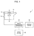
- Fig. 1 illustrates an exemplary system configuration of an image processing system 1 according to the embodiment.
- the image processing system 1 includes an optical microscope 10, an image processing apparatus 20, an input device 50, and a display device 60.
- the image processing apparatus 20 is connected to the optical microscope 10, the input device 50, and the display device 60 so as to be capable of communicating data.
- the optical microscope 10 captures, with a CCD camera 14, an image of a preparation on a slide 11 placed on a stage, via an optical system such as an objective lens(es) 12.
- the optical microscope 10 includes a focusing mechanism 13 that changes the distance between the slide 11 and the objective lens 12, and the optical microscope 10 is configured to capture an image of the preparation on the slide 11 at different focusing distances.
- maternal blood is applied to the slide 11, and the slide 11 is stained with May-Giemsa, which is used as a preparation. This stains fetal nucleated red blood cells (NRBCs) in the maternal blood blue-purple.
- NRBCs fetal nucleated red blood cells
- NRBCs are referred to as target cells.
- the image processing apparatus 20 obtains a captured image, captured by the optical microscope 10, and detects target cells in the obtained captured image. For example, the image processing apparatus 20 may determine a score (such as a probability) indicating the probability that target cells are included in a determination target region set in the captured image, captured by the optical microscope 10, on the basis of a discriminator that has learned conditions for discriminating target cells. Note that a process of detecting target cells, performed by the image processing apparatus 20, will be described in detail later.
- the input device 50 is a device such as a keyboard or a mouse, and inputs an operation accepted from a user to the image processing apparatus 20.
- the image processing apparatus 20 obtains information on an image region specified, regarding an image displayed on the display device 60, by the user by using the input device 50 as learning information for learning positive and negative examples of target cells, or image features of other particular cells, and may have the discriminator learn conditions (discrimination parameters) for discriminating target cells on the basis of the learning information.
- the display device 60 is, for example, a liquid crystal display device 60, and displays a screen on the basis of the result of a process performed by the image processing apparatus 20.
- the display device 60 displays a captured image captured by the optical microscope 10, the result of detecting target cells by the image processing apparatus 20, or the like.
- Fig. 2 is a functional block diagram illustrating an example of functions included in the image processing apparatus 20.
- the image processing apparatus 20 includes a specimen information obtaining unit 21, a specimen feature information memory 22, a number-of-target-cells estimating unit 23, a captured image obtaining unit 24, a sample feature extracting unit 25, a detection difficulty determining unit 26, a detection target sample setting unit 27, a detection parameter setting unit 28, a nucleus candidate region extracting unit 29, a determination target region setting unit 30, an image feature generating unit 31, a target cell determining unit 32, a target cell region setting unit 33, a target cell region integrating unit 34, and a determination result output unit 35.
- the functions of the above-mentioned units included in the image processing apparatus 20 may be realized by reading and executing a program stored in a computer-readable information storage medium, by the image processing apparatus 20, which is a computer including control means such as a CPU, storage means such as a memory, and input-output means that transmits/receives data to/from an external device.
- the program may be supplied to the image processing apparatus 20 through an information storage medium such as an optical disk, a magnetic disk, a magnetic tape, a magneto-optical disk, or a flash memory, or may be supplied to the image processing apparatus 20 via a data communication network such as the Internet.
- a data communication network such as the Internet.
- the specimen information obtaining unit 21 obtains information on a specimen (mother) serving as a test target (specimen information).
- the specimen information obtaining unit 21 may obtain specimen information on the basis of data input via the input device 50.
- the specimen information may include the age, medical history, weeks of pregnancy, or the like of a person who is a test object from which maternal blood has been taken.
- the specimen feature information memory 22 stores information for use in estimating target cells included in a specimen, on the basis of a feature of the specimen. For example, with respect to a category in accordance with a feature of a specimen, the specimen feature information memory 22 may store the number of target cells per unit blood volume according to each category, for example.
- the image processing apparatus 20 may generate and cluster a feature vector for each specimen on the basis of each item of information, that is, the age, medical history, and weeks of pregnancy, and may associatively store a premeasured representative value (such as an average value) of the number of target cells (nucleated red blood cells) per unit blood volume for a specimen belonging to each cluster, in the specimen feature information memory 22.
- the specimen feature information memory 22 may store a table or an equation defining the relationship between the age, medical history, and weeks of pregnancy with a corresponding number of nucleated red blood cells.
- the number-of-target-cells estimating unit 23 estimates the number of target cells included in a specimen, on the basis of specimen information (information representing a feature of the specimen) regarding the specimen, obtained by the specimen information obtaining unit 21, and information stored in the specimen feature information memory 22. For example, for a category corresponding to the specimen information obtained by the specimen information obtaining unit 21, the number-of-target-cells estimating unit 23 estimates the number of target cells included in a preparation (maternal blood) obtained from the specimen, on the basis of a representative number of target cells, stored in the specimen feature information memory 22.
- the number-of-target-cells estimating unit 23 may obtain the estimated number of target cells by multiplying the number of nucleated red blood cells per unit blood volume, stored in the specimen feature information memory 22, and the blood volume of the preparation.
- the method of estimating the number of target cells is not limited to that described above.
- the specimen feature information memory 22 may store in advance, for one or more specimens, a feature vector(s) of the specimen(s) and the number of target cells in association with each another.
- the number-of-target-cells estimating unit 23 identifies a feature vector that is similar to a feature vector of a target specimen (for example, the distance between the feature vectors is shortest) among the feature vector(s) of the specimen(s), stored in the specimen feature information memory 22, and may regard the number of target cells associated with the identified feature vector as the number of target cells in the target specimen.
- the captured image obtaining unit 24 obtains a captured image of a preparation (maternal blood) on the slide, obtained from a specimen.
- the captured image obtaining unit 24 obtains, from the optical microscope 102, a captured image of a preparation (maternal blood), captured with the CCD camera 5 included in the optical microscope 102.
- the captured image obtaining unit 24 may obtain a captured image in the case where a detection target flag defined for a target specimen is true (T) on the basis of later-described detection parameters, and may not obtain a captured image in the case where a detection target flag defined for a target specimen is false (F).
- the sample feature extracting unit 25 extracts features of a target sample.
- Features of a sample include information indicating a generation state of the sample.
- the sample feature extracting unit 25 may extract, as sample features, the thickness evenness of a sample generated from a specimen, the staining intensity of the cell nucleus, the content by percentage of white blood cells, and the like.
- the sample feature extracting unit 25 may extract sample features of each of all or some of samples (such that samples are limited to those serving as detection targets) generated from a target specimen.
- the sample feature extracting unit 25 may calculate, for example, the thickness evenness (A) of a sample by measuring the depth from the surface of the sample to the slide at a plurality of points in the sample by using the optical microscope 10, and obtaining the reciprocal of a variance of the measured depths.
- the sample feature extracting unit 25 may calculate, for example, the staining intensity (B) of the cell nucleus in a sample as a value obtained by dividing a predetermined threshold by the average intensity value of a captured image of the sample (may be captured with the optical microscope 10) (that is, threshold/average intensity value), or as the proportion of pixels with intensity values lower than a predetermined threshold in a captured image of the sample.
- the staining intensity (B) of the cell nucleus in a sample as a value obtained by dividing a predetermined threshold by the average intensity value of a captured image of the sample (may be captured with the optical microscope 10) (that is, threshold/average intensity value), or as the proportion of pixels with intensity values lower than a predetermined threshold in a captured image of the sample.
- the sample feature extracting unit 25 may calculate, for example, the content by percentage (C) of white blood cells in a sample as the proportion of pixels of colors that are lighter than a predetermined density in a captured image of the sample (may be captured with the optical microscope 10).
- the detection difficulty determining unit 26 determines the difficulty (Df) in detecting target cells in a sample, on the basis of sample features extracted by the sample feature extracting unit 25. For example, the detection difficulty determining unit 26 may calculate the detection difficulty Df of a sample by using the following equation (1) on the basis of the thickness evenness A of the sample, the staining intensity B of the cell nucleus in the sample, and the content by percentage C of white blood cells in the sample, which are extracted by the sample feature extracting unit 25.
- A0 may be a reference value of the thickness evenness of the sample;
- B0 may be a reference value of the staining intensity of the cell nucleus in the sample;
- C0 may be a reference value of the content by percentage of white blood cells in the sample.
- Df w 1 ⁇ A 0 / A + w 2 ⁇ B 0 / B + w 3 ⁇ C / C 0
- the detection difficulty Df is calculated to be lower as the value of the thickness evenness A of the sample becomes greater; the detection difficulty Df is calculated to be lower as the value of the staining intensity B of the cell nucleus in the sample becomes greater; and the detection difficulty Df is calculated to be greater as the value of the content by percentage C of white blood cells in the sample becomes greater.
- the detection difficulty determining unit 26 may correct the detection difficulty Df, calculated by the above-mentioned equation (1), to take a value from 0 to 1.
- the detection difficulty determining unit 26 may have 0 for Df when Df is less than a lower threshold and may have 1 for Df when Df is greater than an upper threshold (such as a value greater than 1).
- the detection target sample setting unit 27 sets samples serving as detection targets from among a plurality of samples generated from a target specimen, on the basis of at least one of the estimated number (Cb) of target cells estimated by the number-of-target-cells estimating unit 23 and the detection difficulty (Df) determined by the detection difficulty determining unit 26.
- the detection target sample setting unit 27 determines an integer X that satisfies a X ⁇ Z as the number of samples serving as detection targets, and, when Cb ⁇ Z, the detection target sample setting unit 27 determines the number of samples serving as detection targets as N (all samples).
- the detection target sample setting unit 27 may select, from among N samples generated from the preparation, samples the number of which is equivalent to the number of samples serving as detection targets (for example, in the order of identification number or in the order of generation date and time), and may set the detection target flags of the selected samples to true (T) and the detection target flags of unselected samples to false (F).
- the detection target sample setting unit 27 may set, for example, among a plurality of samples generated from a target specimen, samples whose detection difficulties are less than or equal to a threshold as detection targets.
- the detection parameter setting unit 28 sets detection parameters for use in a detection process for samples generated from a preparation.
- the detection parameter setting unit 28 sets detection parameters on the basis of at least one of detection target samples set by the detection target sample setting unit 27 and the detection difficulty determined by the detection difficulty determining unit 26.
- the detection parameters may include a detection target flag indicating whether the sample serves as a detection target, nucleus candidate region parameters indicating conditions of an image region extracted as a nucleus candidate region from a captured image of the sample, and determination target region parameters indicating conditions for setting a determination target region set for a captured image of the sample.
- Fig. 3 illustrates an example of a detection parameter management table storing detection parameters set by the detection parameter setting unit 28.
- the detection parameter management table associatively stores a specimen ID for identifying a specimen, a sample ID for identifying each of a plurality of samples obtained from the specimen, a detection target flag indicating whether each sample serves as a detection target, nucleus candidate region parameters, and determination target region parameters.
- the nucleus candidate region parameters include the color range of target pixels extracted as a nucleus candidate region, and a threshold of the number of connected pixels.
- the determination target region parameters include a step width indicating the amount of shift between pixels serving as the base points of a determination target region in a nucleus candidate region, the maximum magnification indicating, regarding determination target regions of a plurality of sizes set for pixels serving as the base points of the determination target regions, a size ratio between the minimum determination target region and the maximum determination target region, and magnification levels indicating the number of levels of magnification executed from the minimum determination target region to the maximum determination target region.
- the detection parameter setting unit 28 may preliminarily determine detection parameters for each of M levels (L1 to LM) (M is an integer greater than or equal to 2) (note that, for the same image, the number of nucleus candidate regions and determination target regions extracted/set in accordance with detection parameters at Li+1 is greater than the number of nucleus candidate regions and determination target regions extracted/set in accordance with detection parameters at Li), determine the level on the basis of the value of at least one of the margin Y and the detection difficulty Df, and set detection parameters on the basis of the determined level.
- the detection parameter setting unit 28 may preliminarily define the range of the margin for each of M levels, and may determine the level on the basis of to which level's range the above-calculated Y belongs.
- the detection parameter setting unit 28 may preliminarily define the range of the detection difficulty for each of M levels, and may determine the level on the basis of to which level's range the above-calculated Df belongs.
- the detection parameter setting unit 28 may preliminarily define the range of the sum of the margin and the detection difficulty for each of M levels, and may determine the level on the basis of to which level's range the above-calculated sum of Y and Df belongs.
- the detection parameter setting unit 28 may set, for each sample set by the detection target sample setting unit 27, detection parameters that are different for each sample on the basis of the detection difficulty calculated for each sample.
- the nucleus candidate region extracting unit 29 extracts, from a captured image, obtained by the captured image obtaining unit 24, of a sample serving as a processing target, a nucleus candidate region on the basis of nucleus candidate region parameters set by the detection parameter setting unit 28 for the sample.
- the nucleus candidate region extracting unit 29 may perform binarization by regarding pixels included in a color range included in the nucleus candidate region parameters as black pixels and pixels not included in the color range as white pixels, and, among connected pixel groups of connected adjacent black pixels, may extract, as a nucleus candidate region, a connected pixel group having connected pixels whose number is greater than or equal to the number of connected pixel groups included in the nucleus candidate region parameters.
- the determination target region setting unit 30 sets, for a captured image, obtained by the captured image obtaining unit 24, of a sample serving as a processing target, a determination target region subjected to determination of whether there are target cells, on the basis of determination target region parameters set by the detection parameter setting unit 28 for the sample and a nucleus candidate region extracted by the nucleus candidate region extracting unit 29. For example, the determination target region setting unit 30 sets, for each of one or more pixels included in a nucleus candidate region extracted by the nucleus candidate region extracting unit 29, a rectangular region around that pixel (or with reference to that pixel, which serves as a base point), as a determination target region.
- the determination target region setting unit 30 may sequentially set determination target regions by sequentially moving, on the basis of a step width (shift amount) included in determination target region parameters set for a sample serving as a processing target, a pixel that is in a nucleus candidate region and that serves as the base point of a determination target region, by the step width.
- the determination target region setting unit 30 may set, regarding a pixel serving as the base point of a determination target region, determination target regions of different sizes by changing, on the basis of the maximum magnification and the magnification levels included in the determination target region parameters set for a sample serving as a processing target, the size of a determination target region from 1 to the maximum magnification, over the number of levels defined by the magnification levels.
- the image feature generating unit 31 generates an image feature amount of a determination target region set by the determination target region setting unit 30. For example, the image feature generating unit 31 may calculate an HOG feature amount of a determination target region, and obtain the HOG feature amount as an image feature amount.
- a process of calculating two types of HOG feature amounts will be specifically described.
- the image feature generating unit 31 obtains the intensity gradient orientation and the intensity gradient magnitude at each pixel in a determination target region, splits a target image into Y blocks each having X cells, obtains a histogram of oriented gradients ([a first gradient orientation value, a second gradient orientation value, ..., and a ninth gradient orientation value]) from the intensity gradient orientations and the intensity gradient magnitudes for each cell included in each block, and performs normalization in units of blocks so that the mean square thereof becomes 1.
- the image feature generating unit 31 obtains X ⁇ 9, which is a value generated by combining the above-mentioned normalized histograms of oriented gradients in each block, as a feature value of that block, and obtains Y ⁇ X ⁇ 9, which is a value generated by combining all the blocks in the target image, as an HOG feature amount of the determination target region.
- the image feature generating unit 31 obtains the intensity gradient orientation and the intensity gradient magnitude at each pixel in a determination target region, splits a target image into Y blocks each having X cells, and obtains a histogram of oriented gradients ([a first gradient orientation value, a second gradient orientation value, ..., and an eighteenth gradient orientation value]) from the intensity gradient orientations and the intensity gradient magnitudes for each cell included in each block.
- the image feature generating unit 31 obtains X ⁇ 18, which is a value generated by combining the above-mentioned histograms of oriented gradients in each block, as a feature value of that block, and obtains Y ⁇ X ⁇ 18, which is a value generated by combining all the blocks in the target image, as a Cell-HOG feature amount of the determination target region.
- the target cell determining unit 32 determines, on the basis of the image feature amount of a determination target region, the probability (reliability) that target cells are included in the determination target region.
- a discriminator may be caused in advance to learn conditions for discriminating target cells (discrimination parameters) on the basis of the image feature amount of an image region where target cells are shown, and the result of discriminating the image feature amount of a determination target region, obtained by the discriminator, may be obtained.
- AdaBoost AdaBoost, SVM (support vector machine), or the like may be used as the discriminator.
- the discriminator outputs, on the basis of the image feature amount of a determination target region, a score indicating the probability (reliability) that target cells are included in the determination target region.
- the discriminator may output a score having a positive value when cells included in a determination target region are target cells, and may output a score having a negative value when the cells are not target cells.
- the target cell region setting unit 33 sets a candidate region (target cell region) including target cells. For example, the target cell region setting unit 33 may set a determination target region as a target cell candidate region when the reliability output by the discriminator is greater than or equal to 0.
- the target cell region integrating unit 34 integrates, among candidate regions set by the target cell region setting unit 33, candidate regions that at least partially overlap each other into one region. For example, when a plurality of candidate regions overlap each other, the target cell region integrating unit 34 may integrate the plurality of candidate regions into one of these regions (a region with the maximum reliability, for example). Here, the target cell region integrating unit 34 may determine that a plurality of candidate regions overlap each other when these regions are determination target regions set from the same nucleus candidate region, or may determine that a plurality of candidate regions overlap each other when the plurality of candidate regions have an overlapping portion whose area is greater than or equal to a predetermined threshold.
- the target cell region integrating unit 34 may leave only a candidate region with the highest reliability, or may have a region including all the overlapping candidate regions as an integrated candidate region. In addition, the target cell region integrating unit 34 may obtain the reliability of an integrated candidate region by adding the reliabilities of the candidate regions that have been integrated, or may obtain the reliability of an integrated candidate region by multiplying that reliability by the number of overlapping candidate regions.
- Fig. 4 is a diagram describing exemplary integration of target cell candidate regions.
- a captured image I includes a nucleus candidate region 70.
- two target cell regions 71 and 72 which partially overlap each other, are set. If the score for the target cell region 71 is 30 and the score for the target cell region 72 is 10, the target cell region integrating unit 34 may integrate the target cell region 72 with the target cell region 71 whose score is maximum out of the target cell regions 71 and 72, add the score of the target cell region 72 to the score of the target cell region 71, and update the score.
- the determination result output unit 35 outputs information on candidate regions integrated by the target cell region integrating unit 34 as a target cell detection result for a preparation.
- the determination result output unit 35 may display a display screen displaying candidate regions integrated by the target cell region integrating unit 34, which are sorted according to the reliability and displayed in a list, on the display device 60. Note that the determination result output unit 35 may not include a candidate region whose reliability is less than a specified or predetermined threshold in the displayed list.
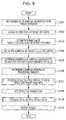
- the image processing apparatus 20 sets the number (N (N is an integer greater than or equal to 1)) of samples generated from a target specimen (S101), and obtains information on the target specimen (specimen information) (S102).
- the specimen information may include at least one of information on the age, medical history, and weeks of pregnancy.
- the image processing apparatus 20 estimates the number (a) of target cells included in the target specimen (S103). For example, the image processing apparatus 20 may identify a category to which the specimen information obtained in S102 belongs, and, on the basis of a representative number of target cells (a reference number of target cells) per unit blood, which is predetermined for the identified category, and the volume of blood of a preparation serving as the specimen, may calculate the estimated number of target cells included in the specimen.
- the image processing apparatus 20 may identify a category to which the specimen information obtained in S102 belongs, and, on the basis of a representative number of target cells (a reference number of target cells) per unit blood, which is predetermined for the identified category, and the volume of blood of a preparation serving as the specimen, may calculate the estimated number of target cells included in the specimen.
- the image processing apparatus 20 calculates the number (b) of target cells per sample (S104). On the basis of the number (b) of target cells per sample, calculated in S104, the image processing apparatus 20 determines the number (d) of detection target samples equivalent to or greater than a necessary number (c) of cells needed for detection (S105).
- the image processing apparatus 20 selects and determines samples serving as processing targets, the number of which is the number (d) of detection target samples, determined in S105, from among a plurality of samples generated from the target specimen (S106).
- the image processing apparatus 20 executes a process of calculating the detection difficulty indicating the difficulty in detecting target cells in each sample serving as a processing target, determined in S106 (S107).
- the process of calculating the detection difficulty will be described in detail on the basis of the flowchart illustrated in Fig. 6 .
- the image processing apparatus 20 initializes a variable i to 1 (S201), and measures depths (A) of a sample Si (S202). For example, the depth from the surface of the sample Si to the slide may be measured at a plurality of points in the sample Si by using the optical microscope 10.
- the image processing apparatus 20 obtains a captured image of the sample Si, captured using the optical microscope 10 (S203), and obtains the staining intensity (B) of the sample Si on the basis of the obtained captured image (S204).
- the image processing apparatus 20 may calculate the staining intensity (B) of the cell nucleus in the sample Si as a value obtained by dividing a predetermined threshold by the average intensity value of the captured image of the sample Si (that is, threshold/average intensity value), or as the proportion of pixels with intensity values lower than a predetermined threshold in the captured image of the sample.
- the image processing apparatus 20 obtains the proportion (C) of white blood cells in the sample Si (S205). For example, the image processing apparatus 20 may calculate the proportion (C) of white blood cells in the sample Si as the proportion of pixels of colors lighter than a predetermined density in the captured image of the sample Si.
- the image processing apparatus 20 calculates the detection difficulty Df (Dfi) in detecting target cells in the sample Si, on the basis of, for example, the above-described equation (1) and on the basis of the variance of the depths (A), the staining intensity (B) of the preparation, and the proportion (C) of white blood cells (S206).
- the image processing apparatus 20 increments the variable i (adds 1) (S208) and returns to S202; and, when the variable i has reached d (S207: Y), the image processing apparatus 20 returns.
- the image processing apparatus 20 increments the variable i (adds 1) (S208) and returns to S202; and, when the variable i has reached d (S207: Y), the image processing apparatus 20 returns.
- the image processing apparatus 20 sets detection parameters on the basis of at least one of the detection difficulty Df, calculated in S107, and the information on each sample serving as a processing target, determined in S106 (S108).
- the detection parameters may include a detection target flag indicating whether the sample serves as a detection target, nucleus candidate region parameters indicating conditions of an image region extracted as a nucleus candidate region from the sample, and determination target region parameters indicating conditions for setting a determination target region set for an image of the sample.
- the image processing apparatus 20 may set, for example, the detection target flag on the basis of the information on each sample serving as a processing target, determined in S106, and may set the nucleus candidate region parameters and the determination target region parameters on the basis of the detection difficulty Dfi calculated for the sample Si.
- the image processing apparatus 20 executes a process of detecting target cells in the specimen (S109).
- the process of detecting target cells will be described in detail on the basis of the flowchart illustrated in Fig. 7 .
- the image processing apparatus 20 initializes the values of variables i, j, and k to 1 (S301), and determines whether a sample Si of a target specimen is a detection target (S302). For example, the image processing apparatus 20 may determine that the sample Si is a detection target when the detection target flag of the sample Si is true (T), and may determine that the sample Si is not a detection target when the detection target flag is false (F).
- the image processing apparatus 20 causes, for example, the optical microscope 10 to capture an image of the sample Si, and obtains a captured image Ti of the sample Si (S303).
- the image processing apparatus 20 extracts nucleus candidate regions (Ai1 to AiM) from the captured image Ti, on the basis of the nucleus candidate region parameters included in the detection parameters set for the sample Si (S304).
- M is the number of nucleus candidate regions included in the captured image Ti. Note that extraction of nucleus candidate regions may be executed by the above-described nucleus candidate region extracting unit 29.
- the image processing apparatus 20 sets determination target regions (Bij1 to BijL) for a nucleus candidate region Aij extracted from the captured image Ti, on the basis of the determination target region parameters included in the detection parameters set for the sample Si (S305).
- L is the number of determination target regions set for the nucleus candidate region Aij, and setting of determination target regions may be executed by the above-described determination target region setting unit 30.
- the image processing apparatus 20 calculates an image feature amount Vijk of a determination target region Bijk (S306). Note that calculation of an image feature amount may be executed by the above-described image feature generating unit 31.
- the image processing apparatus 20 calculates a score Pijk indicating the probability that target cells are included in the determination target region Bijk (S307).
- the image processing apparatus 20 sets the determination target region Bijk as a target cell region (S309).
- the image processing apparatus 20 determines whether the variable k has reached L (S310), and, when the variable k has not reached L (S310: N), the image processing apparatus 20 increments k (adds 1 to k) (S311), and returns to S306. Alternatively, when the variable k has reached L in S310 (S310: Y), the image processing apparatus 20 determines whether the variable j has reached M (S312).
- the image processing apparatus 20 increments j (adds 1 to j), initializes k to 1 (S313), and returns to S305.
- the image processing apparatus 20 executes a process of integrating target cell regions set for the sample Si (S314).
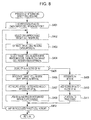
- the process of integrating target cell regions will be described in detail on the basis of the flowchart illustrated in Fig. 8 .
- the image processing apparatus 20 gives a processed flag to each of target cell regions set for the captured image Ti of the sample Si (S401). Note that the processed flag is a true/false value. It is assumed that processed is true (T) and unprocessed is false (F), and the initial value of the processed flag given in S401 is false (F).
- the image processing apparatus 20 selects one target cell region whose processed flag value indicates unprocessed (S402).
- the target cell region selected in S402 will be referred to as As.
- the image processing apparatus 20 extracts other target cell regions overlapping the target cell region As selected in S402, from among the target cell regions set for the captured image Ti (S403).
- the image processing apparatus 20 selects a target cell region with the maximum score from among the target cell regions extracted in S403 (S404).
- the target cell region selected in S404 will be referred to as At.
- the image processing apparatus 20 integrates the target cell region extracted in S403 with the target cell region As (S406), and adds the score of this other target cell region being integrated to the score of the target cell region As (S407).
- the target cell region integrated with the target cell region As may be deleted, or the state of the target cell region integrated with the target cell region As may be updated to integrated.
- the image processing apparatus 20 updates the processed flag of each of the target cell region As and the other target cell region integrated with the target cell region As to true (T) (S408).
- the image processing apparatus 20 integrates the target cell region As with the target cell region At (S409), and adds the score of the target cell region As to the score of the target cell region At (S410).
- the image processing apparatus 20 updates the processed flag of the target cell region As to true (T) (S411).
- the image processing apparatus 20 When there remains an unprocessed target cell region after S408 or S411 (S412: Y), the image processing apparatus 20 returns to S402. When there remains no unprocessed target cell region (S412: N), the image processing apparatus 20 ends the process of integrating target cells, and returns.
- the image processing apparatus 20 increments i (adds 1), initializes j and k to 1 (S316), and returns to S302; and, when the variable i has reached d (S317: Y), the image processing apparatus 20 returns.
- the image processing apparatus 20 outputs, on the basis of integrated target cell regions for each specimen, information on the target cell regions (S110), and ends the process. For example, for each specimen, the image processing apparatus 20 may sort the integrated target cell regions in descending order of the score of the target cell regions, and may display the sorted target cell regions on the display device 60.
- parameters for use in a detection process can be set on the basis of the estimated number of target cells included in a specimen serving as a processing target, and the difficulty in detecting target cells in the specimen serving as a processing target.
- the second example is different from the first example in the point that detection parameters are set without using the detection difficulty.
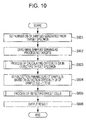
- the image processing apparatus 20 sets the number (N (N is an integer greater than or equal to 1)) of samples generated from a target specimen (S501), and obtains information on the target specimen (specimen information) (S502).
- the specimen information may include at least one of information on the age, medical history, and weeks of pregnancy.
- the image processing apparatus 20 estimates the number (a) of target cells included in the target specimen (S503). For example, the image processing apparatus 20 may identify a category to which the specimen information obtained in S502 belongs, and, on the basis of a representative number of target cells per unit blood, which is predetermined for the identified category, and the volume of blood of a preparation serving as a specimen, may calculate the estimated number of target cells included in the specimen.
- the image processing apparatus 20 calculates the number (b) of target cells per sample (S504). On the basis of the number (b) of target cells per sample, calculated in S504, the image processing apparatus 20 determines the number (d) of detection target samples equivalent to or greater than a necessary number (c) of cells needed for detection (S505).
- the image processing apparatus 20 selects and determines samples serving as processing targets, the number of which is the number (d) of detection target samples, determined in S505, from among a plurality of samples generated from the target specimen (S506).
- the image processing apparatus 20 sets detection parameters on the basis of information on each of the samples serving as processing targets, determined in S506, and the number (b) of target cells per sample, calculated in S504 (S507).
- the detection parameters may include a detection target flag indicating whether the sample serves as a detection target, nucleus candidate region parameters indicating conditions of an image region extracted as a nucleus candidate region from the sample, and determination target region parameters indicating conditions for setting a determination target region set for an image of the sample.
- the image processing apparatus 20 may set, for example, the detection target flag on the basis of the information on each sample serving as a processing target, determined in S506, and may set the nucleus candidate region parameters and the determination target region parameters on the basis of a margin (b ⁇ d-c) obtained by subtracting the necessary number (c) of cells from b ⁇ d, which is multiplication of the number (d) of detection target samples and the number (c) of target cells per sample.
- a margin (b ⁇ d-c) obtained by subtracting the necessary number (c) of cells from b ⁇ d, which is multiplication of the number (d) of detection target samples and the number (c) of target cells per sample.
- the image processing apparatus 20 executes a process of detecting target cells in the specimen (S508).
- the process of detecting target cells is as illustrated in the flowchart illustrated in Fig. 7 and is common to the first example, a description of which will be omitted here.
- the image processing apparatus 20 Having finished the process of detecting target cells in each specimen serving as a processing target, the image processing apparatus 20 outputs, on the basis of integrated target cell regions for each specimen, information on the target cell regions (S509), and ends the process.
- parameters for use in a detection process can be set on the basis of the estimated number of target cells included in a specimen serving as a processing target.
- the third example is different from the first example in the point that detection parameters are set without using the number of target cells, which is estimated for a target specimen.
- the image processing apparatus 20 sets the number (N (N is an integer greater than or equal to 1)) of samples generated from a target specimen (S601).
- the image processing apparatus 20 determines samples serving as processing targets, from among a plurality of samples generated from the target specimen (S602). For example, the image processing apparatus 20 may regard all the samples generated from the target specimen as processing targets, or may determine samples serving as processing targets on the basis of the detection difficulty Df calculated for the target specimen. For example, the image processing apparatus 20 may define a proportion in accordance with the range of the detection difficulty (specifically, the higher the detection difficulty, the higher the proportion), determines the number of samples serving as processing targets on the basis of the product of a proportion corresponding to the detection difficulty Df and the number of all the samples (specifically, the obtained product may be made an integer by rounding up), and determines samples serving as processing targets on the basis of the determined number of samples.
- the number of samples serving as processing targets is represented as d.
- the image processing apparatus 20 executes a process of calculating the detection difficulty indicating the difficulty in detecting target cells in each sample serving as a processing target (S603).
- the process of detecting target cells is as illustrated in the flowchart illustrated in Fig. 6 and is common to the first example, a description of which will be omitted here.
- the image processing apparatus 20 sets detection parameters of the sample Si on the basis of the detection difficulty Dfi calculated in S603.
- the detection parameters may include a detection target flag indicating whether the sample serves as a detection target, nucleus candidate region parameters indicating conditions of an image region extracted as a nucleus candidate region from the sample, and determination target region parameters indicating conditions for setting a determination target region set for an image of the sample.
- the image processing apparatus 20 may set, for example, the detection target flag on the basis of the information on each sample serving as a processing target, determined in S603, and may set the nucleus candidate region parameters and the determination target region parameters on the basis of the detection difficulty Dfi calculated for the sample Si.
- the image processing apparatus 20 executes a process of detecting target cells in the specimen (S605).
- the process of detecting target cells is as illustrated in the flowchart illustrated in Fig. 7 and is common to the first example, a description of which will be omitted here.
- the image processing apparatus 20 Having finished the process of detecting target cells in each specimen serving as a processing target, the image processing apparatus 20 outputs, on the basis of integrated target cell regions for each specimen, information on the target cell regions (S606), and ends the process.
- parameters for use in a detection process can be set on the basis of the difficulty in detecting target cells in a specimen serving as a processing target.
- the present invention is not construed as being limited to the above-described embodiment. For example, there is no problem in displaying target cell regions in order of score, without integrating the target cell regions.
- the image processing apparatus 20 is not construed as being limited to the case of obtaining a captured image of a specimen from the optical microscope 10, and the image processing apparatus 20 may obtain a captured image of a specimen from another computer.
Landscapes
- Engineering & Computer Science (AREA)
- Health & Medical Sciences (AREA)
- Life Sciences & Earth Sciences (AREA)
- Physics & Mathematics (AREA)
- General Physics & Mathematics (AREA)
- Chemical & Material Sciences (AREA)
- Biomedical Technology (AREA)
- General Health & Medical Sciences (AREA)
- Theoretical Computer Science (AREA)
- Computer Vision & Pattern Recognition (AREA)
- Pathology (AREA)
- Immunology (AREA)
- Biochemistry (AREA)
- Analytical Chemistry (AREA)
- Hematology (AREA)
- Molecular Biology (AREA)
- Medicinal Chemistry (AREA)
- Food Science & Technology (AREA)
- Urology & Nephrology (AREA)
- Biophysics (AREA)
- Dispersion Chemistry (AREA)
- Ecology (AREA)
- Data Mining & Analysis (AREA)
- Multimedia (AREA)
- Radiology & Medical Imaging (AREA)
- Nuclear Medicine, Radiotherapy & Molecular Imaging (AREA)
- Medical Informatics (AREA)
- Quality & Reliability (AREA)
- Bioinformatics & Cheminformatics (AREA)
- Artificial Intelligence (AREA)
- Evolutionary Biology (AREA)
- Evolutionary Computation (AREA)
- General Engineering & Computer Science (AREA)
- Bioinformatics & Computational Biology (AREA)
- Apparatus Associated With Microorganisms And Enzymes (AREA)
- Investigating Or Analysing Biological Materials (AREA)
- Investigating Or Analysing Materials By Optical Means (AREA)
- Image Analysis (AREA)
- Image Processing (AREA)
Abstract
Description
- The present invention relates to an image processing apparatus, a program, an information storage medium, and an image processing method.
- For example, in
PTL 1, maternal blood serves as a specimen, and nucleated red blood cells in the maternal blood serve as target cells. On the basis of a nucleus candidate region extracted from a microscopy image of the specimen, a determination target region is set, and whether target cells are included in the set determination target region is determined. - PTL 1: Japanese Unexamined Patent Application Publication No.
2012-254042 - It is an object of the present invention to provide an image processing apparatus, a program, an information storage medium, and an image processing method capable of setting parameters of a process of detecting target cells in a target specimen, in accordance with a feature of the target specimen.
- An invention according to
Claim 1 is an image processing apparatus including: estimation means for estimating, based on a feature of a target specimen, a number of target cells included in the target specimen; and setting means for setting, based on the estimated number of target cells, a detection parameter regarding a process of detecting the target cells in a captured image of the target specimen. - An invention according to
Claim 2 is the image processing apparatus described inClaim 1, in which the detection parameter includes information defining whether each of a plurality of samples generated from the target specimen serves as a detection target of detecting whether the target cells are included, and information defining a condition for setting a determination target region for determining whether the target cell region is included in the captured image. The image processing apparatus further includes determination means for determining, for a captured image of a sample serving as a detection target based on the detection parameter, whether the target cells are included in a determination target region set based on the detection parameter. - An invention according to
Claim 3 is the image processing apparatus described inClaim 2, further including: deciding means for deciding on a number of samples including target cells, a number of which is greater than or equal to a predetermined threshold, based on the number of target cells, estimated by the estimation means, and a number of target cells per sample, calculated based on a number of a plurality of samples generated from the target specimen; and selection means for selecting samples, a number of which is decided by the deciding means, from among the plurality of samples. The setting means sets the detection parameter based on information on the samples selected by the selection means. - An invention according to
Claim 4 is the image processing apparatus described inClaim 3, in which the setting means sets the setting condition such that more determination target regions are set as a difference of the threshold with respect to a product of the number of samples, decided by the deciding means, and the calculated number of target cells per sample is smaller. - An invention according to Claim 5 is the image processing apparatus described in any one of
Claims 2 to 4, further including detection difficulty calculating means for calculating detection difficulty indicating difficulty in detecting a number of target cells included in the sample, based on a thickness of the sample, an intensity of the sample, and a density of the sample. The setting means sets the detection parameter based on the calculated detection difficulty. - An invention according to
Claim 6 is the image processing apparatus described in Claim 5, in which the setting means sets the setting condition such that more determination target regions are set as the detection difficulty is higher. - An invention according to Claim 7 is the image processing apparatus described in
Claim 5 or 6, in which the detection difficulty calculating means calculates the detection difficulty to be lower as thickness evenness of the sample is higher, calculates the detection difficulty to be lower as an intensity of a nucleus included in the sample is lower, and calculates the detection difficulty to be higher as a proportion of pixels included in the sample that are lighter than a predetermined density is higher. - An invention according to Claim 8 is the image processing apparatus described in any one of
Claims 2 to 7, further including integrating means for integrating, in a case where there is a plurality of determination target regions that at least partially overlap each other and that are determined to include the target cells, the plurality of determination target regions with a determination target region determined by the determination means to be most likely to include the target cells. - An invention according to Claim 9 is the image processing apparatus described in any one of
Claims 1 to 8, in which the specimen is maternal blood; the target cells are nucleated red blood cell; the feature of the specimen includes at least one of age, medical history, and weeks of pregnancy of a mother; a reference number of nucleated red blood cells per unit blood volume is defined for each category based on the feature of the specimen; and the estimation means estimates a number of nucleated red blood cells included in the target specimen, based on a reference number of nucleated red blood cells, defined for a category in accordance with the feature of the target specimen, and a volume of blood of the target specimen. - An invention according to
Claim 10 is a program for causing a computer to function as: estimation means for estimating, based on a feature of a target specimen, a number of target cells included in the target specimen; and setting means for setting, based on the estimated number of target cells, a detection parameter regarding a process of detecting the target cells in a captured image of the target specimen. - According to the inventions described in
Claims 1 to 10, parameters of a process of detecting target cells in a target specimen can be set in accordance with a feature of the target specimen. - According to the invention described in
Claim 2, the number of samples serving as processing targets, among a plurality of samples generated from a target specimen, and a determination target region set for each sample can be set in accordance with the estimated number of target cells, which is estimated for the target specimen. - According to the invention described in
Claim 3, selection can be made so as to include target cells, the number of which is greater than or equal to a threshold, in a sample serving as a processing target. - According to the fourth invention described in
Claim 4, when the difference between the number of target cells included in a sample serving as a processing target and the threshold is small, the number of regions serving as determination targets is increased, thereby preventing the occurrence of detection evasion. - According to the invention described in Claim 5, parameters of a process of detecting target cells in a target specimen can be set in accordance with the difficulty in detecting target cells in a specimen serving as a processing target.
- According to the invention described in
Claim 6, when the difficulty in detecting target cells in a specimen serving as a processing target is high, the number of regions serving as determination targets is increased, thereby preventing the occurrence of detection evasion. - According to the invention described in Claim 7, the difficulty in detecting target cells in a specimen serving as a processing target can be accurately calculated, compared with the case where the present configuration is not provided.
- According to the invention described in Claim 8, an accurate determination result based on the determination results of a plurality of determination target regions for the same target can be obtained, compared with the case of not integrating a plurality of determination target region.
- According to the invention described in Claim 9, the number of nucleated red blood cells included in target maternal blood can be accurately estimated, compared with the case where the present configuration is not provided.
-
- [
Fig. 1] Fig. 1 is a diagram illustrating an exemplary system configuration of an image processing system according to an embodiment. - [
Fig. 2] Fig. 2 is a functional block diagram illustrating an example of functions included in an image processing apparatus. - [
Fig. 3] Fig. 3 is a diagram illustrating an example of a detection parameter management table. - [
Fig. 4] Fig. 4 is a diagram describing exemplary integration of target cell candidate regions. - [
Fig. 5] Fig. 5 is a flowchart of a process according to a first example. - [
Fig. 6] Fig. 6 is a flowchart of a process of calculating the detection difficulty. - [
Fig. 7] Fig. 7 is a flowchart of a process of detecting target cells. - [
Fig. 8] Fig. 8 is a flowchart of a process of integrating target cell regions. - [
Fig. 9] Fig. 9 is a flowchart of a process according to a second example. - [
Fig. 10] Fig. 10 is a flowchart of a process according to a third example. - Hereinafter, an embodiment for implementing the present invention (hereinafter referred to as an embodiment) will be described in accordance with the drawings.
-
Fig. 1 illustrates an exemplary system configuration of animage processing system 1 according to the embodiment. As illustrated inFig. 1 , theimage processing system 1 includes anoptical microscope 10, animage processing apparatus 20, aninput device 50, and adisplay device 60. Theimage processing apparatus 20 is connected to theoptical microscope 10, theinput device 50, and thedisplay device 60 so as to be capable of communicating data. - The
optical microscope 10 captures, with aCCD camera 14, an image of a preparation on aslide 11 placed on a stage, via an optical system such as an objective lens(es) 12. Theoptical microscope 10 includes afocusing mechanism 13 that changes the distance between theslide 11 and theobjective lens 12, and theoptical microscope 10 is configured to capture an image of the preparation on theslide 11 at different focusing distances. In the embodiment, maternal blood is applied to theslide 11, and theslide 11 is stained with May-Giemsa, which is used as a preparation. This stains fetal nucleated red blood cells (NRBCs) in the maternal blood blue-purple. - The
image processing apparatus 20 obtains a captured image, captured by theoptical microscope 10, and detects target cells in the obtained captured image. For example, theimage processing apparatus 20 may determine a score (such as a probability) indicating the probability that target cells are included in a determination target region set in the captured image, captured by theoptical microscope 10, on the basis of a discriminator that has learned conditions for discriminating target cells. Note that a process of detecting target cells, performed by theimage processing apparatus 20, will be described in detail later. - The
input device 50 is a device such as a keyboard or a mouse, and inputs an operation accepted from a user to theimage processing apparatus 20. For example, theimage processing apparatus 20 obtains information on an image region specified, regarding an image displayed on thedisplay device 60, by the user by using theinput device 50 as learning information for learning positive and negative examples of target cells, or image features of other particular cells, and may have the discriminator learn conditions (discrimination parameters) for discriminating target cells on the basis of the learning information. - The
display device 60 is, for example, a liquidcrystal display device 60, and displays a screen on the basis of the result of a process performed by theimage processing apparatus 20. For example, thedisplay device 60 displays a captured image captured by theoptical microscope 10, the result of detecting target cells by theimage processing apparatus 20, or the like. - Next, functions included in the
image processing apparatus 20 according to the embodiment will be described. -
Fig. 2 is a functional block diagram illustrating an example of functions included in theimage processing apparatus 20. As illustrated inFig. 2 , theimage processing apparatus 20 includes a specimeninformation obtaining unit 21, a specimenfeature information memory 22, a number-of-target-cells estimating unit 23, a capturedimage obtaining unit 24, a samplefeature extracting unit 25, a detectiondifficulty determining unit 26, a detection targetsample setting unit 27, a detectionparameter setting unit 28, a nucleus candidateregion extracting unit 29, a determination targetregion setting unit 30, an imagefeature generating unit 31, a targetcell determining unit 32, a target cellregion setting unit 33, a target cellregion integrating unit 34, and a determinationresult output unit 35. - The functions of the above-mentioned units included in the
image processing apparatus 20 may be realized by reading and executing a program stored in a computer-readable information storage medium, by theimage processing apparatus 20, which is a computer including control means such as a CPU, storage means such as a memory, and input-output means that transmits/receives data to/from an external device. Note that the program may be supplied to theimage processing apparatus 20 through an information storage medium such as an optical disk, a magnetic disk, a magnetic tape, a magneto-optical disk, or a flash memory, or may be supplied to theimage processing apparatus 20 via a data communication network such as the Internet. Hereinafter, the functions of the units included in theimage processing apparatus 20 will be described in detail. - The specimen
information obtaining unit 21 obtains information on a specimen (mother) serving as a test target (specimen information). For example, the specimeninformation obtaining unit 21 may obtain specimen information on the basis of data input via theinput device 50. For example, the specimen information may include the age, medical history, weeks of pregnancy, or the like of a person who is a test object from which maternal blood has been taken. - The specimen
feature information memory 22 stores information for use in estimating target cells included in a specimen, on the basis of a feature of the specimen. For example, with respect to a category in accordance with a feature of a specimen, the specimenfeature information memory 22 may store the number of target cells per unit blood volume according to each category, for example. For example, theimage processing apparatus 20 may generate and cluster a feature vector for each specimen on the basis of each item of information, that is, the age, medical history, and weeks of pregnancy, and may associatively store a premeasured representative value (such as an average value) of the number of target cells (nucleated red blood cells) per unit blood volume for a specimen belonging to each cluster, in the specimenfeature information memory 22. In addition, the specimenfeature information memory 22 may store a table or an equation defining the relationship between the age, medical history, and weeks of pregnancy with a corresponding number of nucleated red blood cells. - The number-of-target-
cells estimating unit 23 estimates the number of target cells included in a specimen, on the basis of specimen information (information representing a feature of the specimen) regarding the specimen, obtained by the specimeninformation obtaining unit 21, and information stored in the specimenfeature information memory 22. For example, for a category corresponding to the specimen information obtained by the specimeninformation obtaining unit 21, the number-of-target-cells estimating unit 23 estimates the number of target cells included in a preparation (maternal blood) obtained from the specimen, on the basis of a representative number of target cells, stored in the specimenfeature information memory 22. For example, for a category corresponding to the specimen information obtained by the specimeninformation obtaining unit 21, the number-of-target-cells estimating unit 23 may obtain the estimated number of target cells by multiplying the number of nucleated red blood cells per unit blood volume, stored in the specimenfeature information memory 22, and the blood volume of the preparation. - In addition, the method of estimating the number of target cells is not limited to that described above. For example, the specimen
feature information memory 22 may store in advance, for one or more specimens, a feature vector(s) of the specimen(s) and the number of target cells in association with each another. The number-of-target-cells estimating unit 23 identifies a feature vector that is similar to a feature vector of a target specimen (for example, the distance between the feature vectors is shortest) among the feature vector(s) of the specimen(s), stored in the specimenfeature information memory 22, and may regard the number of target cells associated with the identified feature vector as the number of target cells in the target specimen. - The captured
image obtaining unit 24 obtains a captured image of a preparation (maternal blood) on the slide, obtained from a specimen. For example, the capturedimage obtaining unit 24 obtains, from the optical microscope 102, a captured image of a preparation (maternal blood), captured with the CCD camera 5 included in the optical microscope 102. In addition, the capturedimage obtaining unit 24 may obtain a captured image in the case where a detection target flag defined for a target specimen is true (T) on the basis of later-described detection parameters, and may not obtain a captured image in the case where a detection target flag defined for a target specimen is false (F). - The sample
feature extracting unit 25 extracts features of a target sample. Features of a sample include information indicating a generation state of the sample. For example, the samplefeature extracting unit 25 may extract, as sample features, the thickness evenness of a sample generated from a specimen, the staining intensity of the cell nucleus, the content by percentage of white blood cells, and the like. The samplefeature extracting unit 25 may extract sample features of each of all or some of samples (such that samples are limited to those serving as detection targets) generated from a target specimen. - The sample
feature extracting unit 25 may calculate, for example, the thickness evenness (A) of a sample by measuring the depth from the surface of the sample to the slide at a plurality of points in the sample by using theoptical microscope 10, and obtaining the reciprocal of a variance of the measured depths. - The sample
feature extracting unit 25 may calculate, for example, the staining intensity (B) of the cell nucleus in a sample as a value obtained by dividing a predetermined threshold by the average intensity value of a captured image of the sample (may be captured with the optical microscope 10) (that is, threshold/average intensity value), or as the proportion of pixels with intensity values lower than a predetermined threshold in a captured image of the sample. - The sample
feature extracting unit 25 may calculate, for example, the content by percentage (C) of white blood cells in a sample as the proportion of pixels of colors that are lighter than a predetermined density in a captured image of the sample (may be captured with the optical microscope 10). - The detection
difficulty determining unit 26 determines the difficulty (Df) in detecting target cells in a sample, on the basis of sample features extracted by the samplefeature extracting unit 25. For example, the detectiondifficulty determining unit 26 may calculate the detection difficulty Df of a sample by using the following equation (1) on the basis of the thickness evenness A of the sample, the staining intensity B of the cell nucleus in the sample, and the content by percentage C of white blood cells in the sample, which are extracted by the samplefeature extracting unit 25. Note that w1, w2, and w3 may be predetermined coefficients that satisfy w1+w2+w3=1 and that are greater than or equal to 0; A0 may be a reference value of the thickness evenness of the sample; B0 may be a reference value of the staining intensity of the cell nucleus in the sample; and C0 may be a reference value of the content by percentage of white blood cells in the sample.
- According to the above-mentioned equation (1), the detection difficulty Df is calculated to be lower as the value of the thickness evenness A of the sample becomes greater; the detection difficulty Df is calculated to be lower as the value of the staining intensity B of the cell nucleus in the sample becomes greater; and the detection difficulty Df is calculated to be greater as the value of the content by percentage C of white blood cells in the sample becomes greater. Note that the detection
difficulty determining unit 26 may correct the detection difficulty Df, calculated by the above-mentioned equation (1), to take a value from 0 to 1. For example, the detectiondifficulty determining unit 26 may have 0 for Df when Df is less than a lower threshold and may have 1 for Df when Df is greater than an upper threshold (such as a value greater than 1). - The detection target
sample setting unit 27 sets samples serving as detection targets from among a plurality of samples generated from a target specimen, on the basis of at least one of the estimated number (Cb) of target cells estimated by the number-of-target-cells estimating unit 23 and the detection difficulty (Df) determined by the detectiondifficulty determining unit 26. - The detection target
sample setting unit 27 calculates the number of target cells per sample (a=Cb/N) by, for example, dividing the estimated number (Cb) of target cells estimated for a target specimen by the number (N) of samples generated from the preparation. Here, when Cb>Z for the necessary number (Z) of target cells, the detection targetsample setting unit 27 determines an integer X that satisfies a X≥Z as the number of samples serving as detection targets, and, when Cb≤Z, the detection targetsample setting unit 27 determines the number of samples serving as detection targets as N (all samples). The detection targetsample setting unit 27 may select, from among N samples generated from the preparation, samples the number of which is equivalent to the number of samples serving as detection targets (for example, in the order of identification number or in the order of generation date and time), and may set the detection target flags of the selected samples to true (T) and the detection target flags of unselected samples to false (F). - In addition, in the case of setting samples serving as detection targets on the basis of the detection difficulty calculated by the detection
difficulty determining unit 26, the detection targetsample setting unit 27 may set, for example, among a plurality of samples generated from a target specimen, samples whose detection difficulties are less than or equal to a threshold as detection targets. - The detection
parameter setting unit 28 sets detection parameters for use in a detection process for samples generated from a preparation. For example, the detectionparameter setting unit 28 sets detection parameters on the basis of at least one of detection target samples set by the detection targetsample setting unit 27 and the detection difficulty determined by the detectiondifficulty determining unit 26. For example, the detection parameters may include a detection target flag indicating whether the sample serves as a detection target, nucleus candidate region parameters indicating conditions of an image region extracted as a nucleus candidate region from a captured image of the sample, and determination target region parameters indicating conditions for setting a determination target region set for a captured image of the sample. -
Fig. 3 illustrates an example of a detection parameter management table storing detection parameters set by the detectionparameter setting unit 28. As illustrated inFig. 3 , the detection parameter management table associatively stores a specimen ID for identifying a specimen, a sample ID for identifying each of a plurality of samples obtained from the specimen, a detection target flag indicating whether each sample serves as a detection target, nucleus candidate region parameters, and determination target region parameters. Note that the nucleus candidate region parameters include the color range of target pixels extracted as a nucleus candidate region, and a threshold of the number of connected pixels. In addition, the determination target region parameters include a step width indicating the amount of shift between pixels serving as the base points of a determination target region in a nucleus candidate region, the maximum magnification indicating, regarding determination target regions of a plurality of sizes set for pixels serving as the base points of the determination target regions, a size ratio between the minimum determination target region and the maximum determination target region, and magnification levels indicating the number of levels of magnification executed from the minimum determination target region to the maximum determination target region. - The detection
parameter setting unit 28 may set detection parameters on the basis of at least one of a margin Y, which is the difference between the product of the number X of samples serving as detection targets and the number a of target cells per sample, set by the detection targetsample setting unit 27, and the necessary number Z of target cells (Y=a·X-Z), and the detection difficulty Df determined by the detectiondifficulty determining unit 26. More specifically, the detectionparameter setting unit 28 may preliminarily determine detection parameters for each of M levels (L1 to LM) (M is an integer greater than or equal to 2) (note that, for the same image, the number of nucleus candidate regions and determination target regions extracted/set in accordance with detection parameters at Li+1 is greater than the number of nucleus candidate regions and determination target regions extracted/set in accordance with detection parameters at Li), determine the level on the basis of the value of at least one of the margin Y and the detection difficulty Df, and set detection parameters on the basis of the determined level. For example, the detectionparameter setting unit 28 may preliminarily define the range of the margin for each of M levels, and may determine the level on the basis of to which level's range the above-calculated Y belongs. Alternatively, for example, the detectionparameter setting unit 28 may preliminarily define the range of the detection difficulty for each of M levels, and may determine the level on the basis of to which level's range the above-calculated Df belongs. Alternatively, for example, the detectionparameter setting unit 28 may preliminarily define the range of the sum of the margin and the detection difficulty for each of M levels, and may determine the level on the basis of to which level's range the above-calculated sum of Y and Df belongs. - Note that the detection
parameter setting unit 28 may set, for each sample set by the detection targetsample setting unit 27, detection parameters that are different for each sample on the basis of the detection difficulty calculated for each sample. - The nucleus candidate
region extracting unit 29 extracts, from a captured image, obtained by the capturedimage obtaining unit 24, of a sample serving as a processing target, a nucleus candidate region on the basis of nucleus candidate region parameters set by the detectionparameter setting unit 28 for the sample. For example, the nucleus candidateregion extracting unit 29 may perform binarization by regarding pixels included in a color range included in the nucleus candidate region parameters as black pixels and pixels not included in the color range as white pixels, and, among connected pixel groups of connected adjacent black pixels, may extract, as a nucleus candidate region, a connected pixel group having connected pixels whose number is greater than or equal to the number of connected pixel groups included in the nucleus candidate region parameters. - The determination target
region setting unit 30 sets, for a captured image, obtained by the capturedimage obtaining unit 24, of a sample serving as a processing target, a determination target region subjected to determination of whether there are target cells, on the basis of determination target region parameters set by the detectionparameter setting unit 28 for the sample and a nucleus candidate region extracted by the nucleus candidateregion extracting unit 29. For example, the determination targetregion setting unit 30 sets, for each of one or more pixels included in a nucleus candidate region extracted by the nucleus candidateregion extracting unit 29, a rectangular region around that pixel (or with reference to that pixel, which serves as a base point), as a determination target region. Here, the determination targetregion setting unit 30 may sequentially set determination target regions by sequentially moving, on the basis of a step width (shift amount) included in determination target region parameters set for a sample serving as a processing target, a pixel that is in a nucleus candidate region and that serves as the base point of a determination target region, by the step width. In addition, the determination targetregion setting unit 30 may set, regarding a pixel serving as the base point of a determination target region, determination target regions of different sizes by changing, on the basis of the maximum magnification and the magnification levels included in the determination target region parameters set for a sample serving as a processing target, the size of a determination target region from 1 to the maximum magnification, over the number of levels defined by the magnification levels. - The image
feature generating unit 31 generates an image feature amount of a determination target region set by the determination targetregion setting unit 30. For example, the imagefeature generating unit 31 may calculate an HOG feature amount of a determination target region, and obtain the HOG feature amount as an image feature amount. Hereinafter, a process of calculating two types of HOG feature amounts will be specifically described. - The image
feature generating unit 31 obtains the intensity gradient orientation and the intensity gradient magnitude at each pixel in a determination target region, splits a target image into Y blocks each having X cells, obtains a histogram of oriented gradients ([a first gradient orientation value, a second gradient orientation value, ..., and a ninth gradient orientation value]) from the intensity gradient orientations and the intensity gradient magnitudes for each cell included in each block, and performs normalization in units of blocks so that the mean square thereof becomes 1. The imagefeature generating unit 31 obtains X×9, which is a value generated by combining the above-mentioned normalized histograms of oriented gradients in each block, as a feature value of that block, and obtains Y×X×9, which is a value generated by combining all the blocks in the target image, as an HOG feature amount of the determination target region. - In addition, the image
feature generating unit 31 obtains the intensity gradient orientation and the intensity gradient magnitude at each pixel in a determination target region, splits a target image into Y blocks each having X cells, and obtains a histogram of oriented gradients ([a first gradient orientation value, a second gradient orientation value, ..., and an eighteenth gradient orientation value]) from the intensity gradient orientations and the intensity gradient magnitudes for each cell included in each block. The imagefeature generating unit 31 obtains X×18, which is a value generated by combining the above-mentioned histograms of oriented gradients in each block, as a feature value of that block, and obtains Y×X×18, which is a value generated by combining all the blocks in the target image, as a Cell-HOG feature amount of the determination target region. - The target
cell determining unit 32 determines, on the basis of the image feature amount of a determination target region, the probability (reliability) that target cells are included in the determination target region. For example, a discriminator may be caused in advance to learn conditions for discriminating target cells (discrimination parameters) on the basis of the image feature amount of an image region where target cells are shown, and the result of discriminating the image feature amount of a determination target region, obtained by the discriminator, may be obtained. Note that AdaBoost, SVM (support vector machine), or the like may be used as the discriminator. The discriminator outputs, on the basis of the image feature amount of a determination target region, a score indicating the probability (reliability) that target cells are included in the determination target region. For example, the discriminator may output a score having a positive value when cells included in a determination target region are target cells, and may output a score having a negative value when the cells are not target cells. - On the basis of the determination result obtained by the target
cell determining unit 32 for a determination target region, the target cellregion setting unit 33 sets a candidate region (target cell region) including target cells. For example, the target cellregion setting unit 33 may set a determination target region as a target cell candidate region when the reliability output by the discriminator is greater than or equal to 0. - The target cell
region integrating unit 34 integrates, among candidate regions set by the target cellregion setting unit 33, candidate regions that at least partially overlap each other into one region. For example, when a plurality of candidate regions overlap each other, the target cellregion integrating unit 34 may integrate the plurality of candidate regions into one of these regions (a region with the maximum reliability, for example). Here, the target cellregion integrating unit 34 may determine that a plurality of candidate regions overlap each other when these regions are determination target regions set from the same nucleus candidate region, or may determine that a plurality of candidate regions overlap each other when the plurality of candidate regions have an overlapping portion whose area is greater than or equal to a predetermined threshold. In addition, regarding the integrated candidate regions, the target cellregion integrating unit 34 may leave only a candidate region with the highest reliability, or may have a region including all the overlapping candidate regions as an integrated candidate region. In addition, the target cellregion integrating unit 34 may obtain the reliability of an integrated candidate region by adding the reliabilities of the candidate regions that have been integrated, or may obtain the reliability of an integrated candidate region by multiplying that reliability by the number of overlapping candidate regions. -
Fig. 4 is a diagram describing exemplary integration of target cell candidate regions. As illustrated inFig. 4 , a captured image I includes anucleus candidate region 70. For thenucleus candidate region 70, twotarget cell regions target cell region 71 is 30 and the score for thetarget cell region 72 is 10, the target cellregion integrating unit 34 may integrate thetarget cell region 72 with thetarget cell region 71 whose score is maximum out of thetarget cell regions target cell region 72 to the score of thetarget cell region 71, and update the score. - The determination
result output unit 35 outputs information on candidate regions integrated by the target cellregion integrating unit 34 as a target cell detection result for a preparation. For example, the determinationresult output unit 35 may display a display screen displaying candidate regions integrated by the target cellregion integrating unit 34, which are sorted according to the reliability and displayed in a list, on thedisplay device 60. Note that the determinationresult output unit 35 may not include a candidate region whose reliability is less than a specified or predetermined threshold in the displayed list. - Next, an exemplary process performed in the
image processing system 1 according to the embodiment will be described in detail on the basis ofFigs. 5 to 10 . - First, a process according to a first example, executed by the
image processing system 1, will be described in detail on the basis of the flowcharts illustrated inFigs. 5 to 8 . - As illustrated in
Fig. 5 , on the basis of data input from theinput device 50, theimage processing apparatus 20 sets the number (N (N is an integer greater than or equal to 1)) of samples generated from a target specimen (S101), and obtains information on the target specimen (specimen information) (S102). Here, the specimen information may include at least one of information on the age, medical history, and weeks of pregnancy. - Next, on the basis of the specimen information obtained in S102, the
image processing apparatus 20 estimates the number (a) of target cells included in the target specimen (S103). For example, theimage processing apparatus 20 may identify a category to which the specimen information obtained in S102 belongs, and, on the basis of a representative number of target cells (a reference number of target cells) per unit blood, which is predetermined for the identified category, and the volume of blood of a preparation serving as the specimen, may calculate the estimated number of target cells included in the specimen. - On the basis of the number (a) of target cells, calculated in S103, and the number (N) of samples, set in S101, the
image processing apparatus 20 calculates the number (b) of target cells per sample (S104). On the basis of the number (b) of target cells per sample, calculated in S104, theimage processing apparatus 20 determines the number (d) of detection target samples equivalent to or greater than a necessary number (c) of cells needed for detection (S105). - Here, the
image processing apparatus 20 selects and determines samples serving as processing targets, the number of which is the number (d) of detection target samples, determined in S105, from among a plurality of samples generated from the target specimen (S106). - Next, the
image processing apparatus 20 executes a process of calculating the detection difficulty indicating the difficulty in detecting target cells in each sample serving as a processing target, determined in S106 (S107). The process of calculating the detection difficulty will be described in detail on the basis of the flowchart illustrated inFig. 6 . - As illustrated in
Fig. 6 , theimage processing apparatus 20 initializes a variable i to 1 (S201), and measures depths (A) of a sample Si (S202). For example, the depth from the surface of the sample Si to the slide may be measured at a plurality of points in the sample Si by using theoptical microscope 10. - Next, the
image processing apparatus 20 obtains a captured image of the sample Si, captured using the optical microscope 10 (S203), and obtains the staining intensity (B) of the sample Si on the basis of the obtained captured image (S204). For example, theimage processing apparatus 20 may calculate the staining intensity (B) of the cell nucleus in the sample Si as a value obtained by dividing a predetermined threshold by the average intensity value of the captured image of the sample Si (that is, threshold/average intensity value), or as the proportion of pixels with intensity values lower than a predetermined threshold in the captured image of the sample. - Further, on the basis of the captured image obtained in S203, the
image processing apparatus 20 obtains the proportion (C) of white blood cells in the sample Si (S205). For example, theimage processing apparatus 20 may calculate the proportion (C) of white blood cells in the sample Si as the proportion of pixels of colors lighter than a predetermined density in the captured image of the sample Si. - The
image processing apparatus 20 calculates the detection difficulty Df (Dfi) in detecting target cells in the sample Si, on the basis of, for example, the above-described equation (1) and on the basis of the variance of the depths (A), the staining intensity (B) of the preparation, and the proportion (C) of white blood cells (S206). - Here, when the variable i has not reached d (S207: N), the
image processing apparatus 20 increments the variable i (adds 1) (S208) and returns to S202; and, when the variable i has reached d (S207: Y), theimage processing apparatus 20 returns. Next, referring back to the flowchart inFig. 5 , a description will be continuously given. - As illustrated in
Fig. 5 , having finished calculating the detection difficulty Df in detecting target cells in a sample serving as a processing target, theimage processing apparatus 20 sets detection parameters on the basis of at least one of the detection difficulty Df, calculated in S107, and the information on each sample serving as a processing target, determined in S106 (S108). For example, the detection parameters may include a detection target flag indicating whether the sample serves as a detection target, nucleus candidate region parameters indicating conditions of an image region extracted as a nucleus candidate region from the sample, and determination target region parameters indicating conditions for setting a determination target region set for an image of the sample. Here, theimage processing apparatus 20 may set, for example, the detection target flag on the basis of the information on each sample serving as a processing target, determined in S106, and may set the nucleus candidate region parameters and the determination target region parameters on the basis of the detection difficulty Dfi calculated for the sample Si. - On the basis of the detection parameters set in S108, the
image processing apparatus 20 executes a process of detecting target cells in the specimen (S109). The process of detecting target cells will be described in detail on the basis of the flowchart illustrated inFig. 7 . - As illustrated in
Fig. 7 , theimage processing apparatus 20 initializes the values of variables i, j, and k to 1 (S301), and determines whether a sample Si of a target specimen is a detection target (S302). For example, theimage processing apparatus 20 may determine that the sample Si is a detection target when the detection target flag of the sample Si is true (T), and may determine that the sample Si is not a detection target when the detection target flag is false (F). - When the sample Si is a detection target (S302: Y), the
image processing apparatus 20 causes, for example, theoptical microscope 10 to capture an image of the sample Si, and obtains a captured image Ti of the sample Si (S303). - Next, the
image processing apparatus 20 extracts nucleus candidate regions (Ai1 to AiM) from the captured image Ti, on the basis of the nucleus candidate region parameters included in the detection parameters set for the sample Si (S304). Note that M is the number of nucleus candidate regions included in the captured image Ti. Note that extraction of nucleus candidate regions may be executed by the above-described nucleus candidateregion extracting unit 29. - The
image processing apparatus 20 sets determination target regions (Bij1 to BijL) for a nucleus candidate region Aij extracted from the captured image Ti, on the basis of the determination target region parameters included in the detection parameters set for the sample Si (S305). Note that L is the number of determination target regions set for the nucleus candidate region Aij, and setting of determination target regions may be executed by the above-described determination targetregion setting unit 30. - The
image processing apparatus 20 calculates an image feature amount Vijk of a determination target region Bijk (S306). Note that calculation of an image feature amount may be executed by the above-described imagefeature generating unit 31. - On the basis of the image feature amount Vijk calculated in S306, the
image processing apparatus 20 calculates a score Pijk indicating the probability that target cells are included in the determination target region Bijk (S307). - Here, when the score Pijk calculated in S307 is greater than or equal to a threshold Th (S308: Y), the
image processing apparatus 20 sets the determination target region Bijk as a target cell region (S309). - After S309 or when the score Pijk calculated in S307 is less than the threshold Th (S308: N), the
image processing apparatus 20 determines whether the variable k has reached L (S310), and, when the variable k has not reached L (S310: N), theimage processing apparatus 20 increments k (adds 1 to k) (S311), and returns to S306. Alternatively, when the variable k has reached L in S310 (S310: Y), theimage processing apparatus 20 determines whether the variable j has reached M (S312). - When the variable j has not reached M in S312 (S312: N), the
image processing apparatus 20 increments j (adds 1 to j), initializes k to 1 (S313), and returns to S305. Alternatively, when the variable j has reached M in S312 (S312: Y), theimage processing apparatus 20 executes a process of integrating target cell regions set for the sample Si (S314). Here, the process of integrating target cell regions will be described in detail on the basis of the flowchart illustrated inFig. 8 . - As illustrated in
Fig. 8 , theimage processing apparatus 20 gives a processed flag to each of target cell regions set for the captured image Ti of the sample Si (S401). Note that the processed flag is a true/false value. It is assumed that processed is true (T) and unprocessed is false (F), and the initial value of the processed flag given in S401 is false (F). - Next, the
image processing apparatus 20 selects one target cell region whose processed flag value indicates unprocessed (S402). Hereinafter, the target cell region selected in S402 will be referred to as As. - The
image processing apparatus 20 extracts other target cell regions overlapping the target cell region As selected in S402, from among the target cell regions set for the captured image Ti (S403). Theimage processing apparatus 20 selects a target cell region with the maximum score from among the target cell regions extracted in S403 (S404). Hereinafter, the target cell region selected in S404 will be referred to as At. - When the score of the target cell region As is greater than or equal to the score of the target cell region At (S405: Y), the
image processing apparatus 20 integrates the target cell region extracted in S403 with the target cell region As (S406), and adds the score of this other target cell region being integrated to the score of the target cell region As (S407). For example, the target cell region integrated with the target cell region As may be deleted, or the state of the target cell region integrated with the target cell region As may be updated to integrated. Theimage processing apparatus 20 updates the processed flag of each of the target cell region As and the other target cell region integrated with the target cell region As to true (T) (S408). - Alternatively, when the score of the target cell region As is less than the score of the target cell region At (S405: N), the
image processing apparatus 20 integrates the target cell region As with the target cell region At (S409), and adds the score of the target cell region As to the score of the target cell region At (S410). Theimage processing apparatus 20 updates the processed flag of the target cell region As to true (T) (S411). - When there remains an unprocessed target cell region after S408 or S411 (S412: Y), the
image processing apparatus 20 returns to S402. When there remains no unprocessed target cell region (S412: N), theimage processing apparatus 20 ends the process of integrating target cells, and returns. - Here, referring back to
Fig. 7 , a description will be continuously given. Having finished the process of integrating target cell regions set for the sample Si, when the variable i has not reached d (the number of detection target samples or the number of all samples) (S315: N), theimage processing apparatus 20 increments i (adds 1), initializes j and k to 1 (S316), and returns to S302. Alternatively, when the variable i has reached d in S315 (S315: Y), theimage processing apparatus 20 returns. - Alternatively, when the sample Si is not a detection target in S302 (S302: N) and the variable i has not reached d (S317: N), the
image processing apparatus 20 increments i (adds 1), initializes j and k to 1 (S316), and returns to S302; and, when the variable i has reached d (S317: Y), theimage processing apparatus 20 returns. - Here, referring back to the flowchart in
Fig. 5 , a description will be continuously given. Having finished the process of detecting target cells in each specimen serving as a processing target, theimage processing apparatus 20 outputs, on the basis of integrated target cell regions for each specimen, information on the target cell regions (S110), and ends the process. For example, for each specimen, theimage processing apparatus 20 may sort the integrated target cell regions in descending order of the score of the target cell regions, and may display the sorted target cell regions on thedisplay device 60. - According to the process according to the above-described first example, parameters for use in a detection process can be set on the basis of the estimated number of target cells included in a specimen serving as a processing target, and the difficulty in detecting target cells in the specimen serving as a processing target.
- Next, on the basis of the flowchart illustrated in
Fig. 9 , a process according to a second example, executed by theimage processing system 1, will be described in detail. The second example is different from the first example in the point that detection parameters are set without using the detection difficulty. - As illustrated in
Fig. 9 , on the basis of data input from theinput device 50, theimage processing apparatus 20 sets the number (N (N is an integer greater than or equal to 1)) of samples generated from a target specimen (S501), and obtains information on the target specimen (specimen information) (S502). Here, the specimen information may include at least one of information on the age, medical history, and weeks of pregnancy. - Next, on the basis of the specimen information obtained in S502, the
image processing apparatus 20 estimates the number (a) of target cells included in the target specimen (S503). For example, theimage processing apparatus 20 may identify a category to which the specimen information obtained in S502 belongs, and, on the basis of a representative number of target cells per unit blood, which is predetermined for the identified category, and the volume of blood of a preparation serving as a specimen, may calculate the estimated number of target cells included in the specimen. - On the basis of the number (a) of target cells, calculated in S503, and the number (N) of samples, set in S501, the
image processing apparatus 20 calculates the number (b) of target cells per sample (S504). On the basis of the number (b) of target cells per sample, calculated in S504, theimage processing apparatus 20 determines the number (d) of detection target samples equivalent to or greater than a necessary number (c) of cells needed for detection (S505). - Here, the
image processing apparatus 20 selects and determines samples serving as processing targets, the number of which is the number (d) of detection target samples, determined in S505, from among a plurality of samples generated from the target specimen (S506). - Next, the
image processing apparatus 20 sets detection parameters on the basis of information on each of the samples serving as processing targets, determined in S506, and the number (b) of target cells per sample, calculated in S504 (S507). For example, the detection parameters may include a detection target flag indicating whether the sample serves as a detection target, nucleus candidate region parameters indicating conditions of an image region extracted as a nucleus candidate region from the sample, and determination target region parameters indicating conditions for setting a determination target region set for an image of the sample. Here, theimage processing apparatus 20 may set, for example, the detection target flag on the basis of the information on each sample serving as a processing target, determined in S506, and may set the nucleus candidate region parameters and the determination target region parameters on the basis of a margin (b·d-c) obtained by subtracting the necessary number (c) of cells from b·d, which is multiplication of the number (d) of detection target samples and the number (c) of target cells per sample. - On the basis of the detection parameters set in S507, the
image processing apparatus 20 executes a process of detecting target cells in the specimen (S508). The process of detecting target cells is as illustrated in the flowchart illustrated inFig. 7 and is common to the first example, a description of which will be omitted here. - Having finished the process of detecting target cells in each specimen serving as a processing target, the
image processing apparatus 20 outputs, on the basis of integrated target cell regions for each specimen, information on the target cell regions (S509), and ends the process. - According to the process according to the above-described second example, parameters for use in a detection process can be set on the basis of the estimated number of target cells included in a specimen serving as a processing target.
- Next, on the basis of the flowchart illustrated in
Fig. 10 , a process according to a third example, executed by theimage processing system 1, will be described in detail. The third example is different from the first example in the point that detection parameters are set without using the number of target cells, which is estimated for a target specimen. - As illustrated in
Fig. 10 , on the basis of data input from theinput device 50, theimage processing apparatus 20 sets the number (N (N is an integer greater than or equal to 1)) of samples generated from a target specimen (S601). - Here, the
image processing apparatus 20 determines samples serving as processing targets, from among a plurality of samples generated from the target specimen (S602). For example, theimage processing apparatus 20 may regard all the samples generated from the target specimen as processing targets, or may determine samples serving as processing targets on the basis of the detection difficulty Df calculated for the target specimen. For example, theimage processing apparatus 20 may define a proportion in accordance with the range of the detection difficulty (specifically, the higher the detection difficulty, the higher the proportion), determines the number of samples serving as processing targets on the basis of the product of a proportion corresponding to the detection difficulty Df and the number of all the samples (specifically, the obtained product may be made an integer by rounding up), and determines samples serving as processing targets on the basis of the determined number of samples. Here, the number of samples serving as processing targets is represented as d. - Next, the
image processing apparatus 20 executes a process of calculating the detection difficulty indicating the difficulty in detecting target cells in each sample serving as a processing target (S603). The process of detecting target cells is as illustrated in the flowchart illustrated inFig. 6 and is common to the first example, a description of which will be omitted here. - Next, for the sample Si (i = 1 to d) serving as a processing target, the
image processing apparatus 20 sets detection parameters of the sample Si on the basis of the detection difficulty Dfi calculated in S603. For example, the detection parameters may include a detection target flag indicating whether the sample serves as a detection target, nucleus candidate region parameters indicating conditions of an image region extracted as a nucleus candidate region from the sample, and determination target region parameters indicating conditions for setting a determination target region set for an image of the sample. Here, theimage processing apparatus 20 may set, for example, the detection target flag on the basis of the information on each sample serving as a processing target, determined in S603, and may set the nucleus candidate region parameters and the determination target region parameters on the basis of the detection difficulty Dfi calculated for the sample Si. - On the basis of the detection parameters set in S604, the
image processing apparatus 20 executes a process of detecting target cells in the specimen (S605). The process of detecting target cells is as illustrated in the flowchart illustrated inFig. 7 and is common to the first example, a description of which will be omitted here. - Having finished the process of detecting target cells in each specimen serving as a processing target, the
image processing apparatus 20 outputs, on the basis of integrated target cell regions for each specimen, information on the target cell regions (S606), and ends the process. - According to the process according to the above-described third example, parameters for use in a detection process can be set on the basis of the difficulty in detecting target cells in a specimen serving as a processing target.
- The present invention is not construed as being limited to the above-described embodiment. For example, there is no problem in displaying target cell regions in order of score, without integrating the target cell regions. In addition, the
image processing apparatus 20 is not construed as being limited to the case of obtaining a captured image of a specimen from theoptical microscope 10, and theimage processing apparatus 20 may obtain a captured image of a specimen from another computer.
Claims (12)
- An image processing apparatus comprising:estimation means for estimating, based on a feature of a target specimen, a number of target cells included in the target specimen; andsetting means for setting, based on the estimated number of target cells, a detection parameter regarding a process of detecting the target cells in a captured image of the target specimen.
- The image processing apparatus according to Claim 1,
wherein the detection parameter includes information defining whether each of a plurality of samples generated from the target specimen serves as a detection target of detecting whether the target cells are included, and
information defining a condition for setting a determination target region for determining whether the target cell region is included in the captured image, and
wherein the image processing apparatus further comprises determination means for determining, for a captured image of a sample serving as a detection target based on the detection parameter, whether the target cells are included in a determination target region set based on the detection parameter. - The image processing apparatus according to Claim 2, further comprising
deciding means for deciding on a number of samples including target cells, a number of which is greater than or equal to a predetermined threshold, based on the number of target cells, estimated by the estimation means, and a number of target cells per sample, calculated based on a number of a plurality of samples generated from the target specimen, and
selection means for selecting samples, a number of which is decided by the deciding means, from among the plurality of samples,
wherein the setting means sets the detection parameter based on information on the samples selected by the selection means. - The image processing apparatus according to Claim 3,
wherein the setting means sets the setting condition such that more determination target regions are set as a difference of the threshold with respect to a product of the number of samples, decided by the deciding means, and the calculated number of target cells per sample is smaller. - The image processing apparatus according to any one of Claims 2 to 4, further comprising:detection difficulty calculating means for calculating detection difficulty indicating difficulty in detecting a number of target cells included in the sample, based on a thickness of the sample, an intensity of the sample, and a density of the sample,wherein the setting means sets the detection parameter based on the calculated detection difficulty.
- The image processing apparatus according to Claim 5,
wherein the setting means sets the setting condition such that more determination target regions are set as the detection difficulty is higher. - The image processing apparatus according to Claim 5 or 6,
wherein the detection difficulty calculating means calculates the detection difficulty to be lower as thickness evenness of the sample is higher, calculates the detection difficulty to be lower as an intensity of a nucleus included in the sample is lower, and
calculates the detection difficulty to be higher as a proportion of pixels included in the sample that are lighter than a predetermined density is higher. - The image processing apparatus according to any one of Claims 2 to 7, further comprising:integrating means for integrating, in a case where there is a plurality of determination target regions that at least partially overlap each other and that are determined to include the target cells, the plurality of determination target regions with a determination target region determined by the determination means to be most likely to include the target cells.
- The image processing apparatus according to any one of Claims 1 to 8,
wherein the specimen is maternal blood,
wherein the target cells are nucleated red blood cell,
wherein the feature of the specimen includes at least one of age, medical history, and weeks of pregnancy of a mother,
wherein a reference number of nucleated red blood cells per unit blood volume is defined for each category based on the feature of the specimen, and
wherein the estimation means estimates a number of nucleated red blood cells included in the target specimen, based on a reference number of nucleated red blood cells, defined for a category in accordance with the feature of the target specimen, and a volume of blood of the target specimen. - A program for causing a computer to function as:estimation means for estimating, based on a feature of a target specimen, a number of target cells included in the target specimen; andsetting means for setting, based on the estimated number of target cells, a detection parameter regarding a process of detecting the target cells in a captured image of the target specimen.
- An information storage medium having recorded thereon a program for causing a computer to function as:estimation means for estimating, based on a feature of a target specimen, a number of target cells included in the target specimen; andsetting means for setting, based on the estimated number of target cells, a detection parameter regarding a process of detecting the target cells in a captured image of the target specimen.
- An image processing method comprising:an estimation step of estimating, based on a feature of a target specimen, a number of target cells included in the target specimen; anda setting step of setting, based on the estimated number of target cells, a detection parameter regarding a process of detecting the target cells in a captured image of the target specimen.
Applications Claiming Priority (2)
| Application Number | Priority Date | Filing Date | Title |
|---|---|---|---|
| JP2013271140A JP6194791B2 (en) | 2013-12-27 | 2013-12-27 | Image processing apparatus and program |
| PCT/JP2014/071596 WO2015098177A1 (en) | 2013-12-27 | 2014-08-19 | Image processing device, program, information storage medium, and image processing method |
Publications (3)
| Publication Number | Publication Date |
|---|---|
| EP3088511A1 true EP3088511A1 (en) | 2016-11-02 |
| EP3088511A4 EP3088511A4 (en) | 2017-09-13 |
| EP3088511B1 EP3088511B1 (en) | 2020-12-30 |
Family
ID=53478066
Family Applications (1)
| Application Number | Title | Priority Date | Filing Date |
|---|---|---|---|
| EP14873584.8A Active EP3088511B1 (en) | 2013-12-27 | 2014-08-19 | Image processing device, program, information storage medium, and image processing method |
Country Status (5)
| Country | Link |
|---|---|
| US (1) | US10169861B2 (en) |
| EP (1) | EP3088511B1 (en) |
| JP (1) | JP6194791B2 (en) |
| CN (1) | CN105612249B (en) |
| WO (1) | WO2015098177A1 (en) |
Families Citing this family (16)
| Publication number | Priority date | Publication date | Assignee | Title |
|---|---|---|---|---|
| US9522396B2 (en) | 2010-12-29 | 2016-12-20 | S.D. Sight Diagnostics Ltd. | Apparatus and method for automatic detection of pathogens |
| CN106840812B (en) | 2011-12-29 | 2019-12-17 | 思迪赛特诊断有限公司 | Methods and systems for detecting pathogens in biological samples |
| EP3869257B1 (en) | 2013-05-23 | 2024-05-08 | S.D. Sight Diagnostics Ltd. | Method and system for imaging a cell sample |
| IL227276A0 (en) | 2013-07-01 | 2014-03-06 | Parasight Ltd | A method and system for preparing a monolayer of cells, particularly suitable for diagnosis |
| US10831013B2 (en) | 2013-08-26 | 2020-11-10 | S.D. Sight Diagnostics Ltd. | Digital microscopy systems, methods and computer program products |
| US10482595B2 (en) | 2014-08-27 | 2019-11-19 | S.D. Sight Diagnostics Ltd. | System and method for calculating focus variation for a digital microscope |
| US10488644B2 (en) | 2015-09-17 | 2019-11-26 | S.D. Sight Diagnostics Ltd. | Methods and apparatus for detecting an entity in a bodily sample |
| CA3018536A1 (en) | 2016-03-30 | 2017-10-05 | S.D. Sight Diagnostics Ltd | Distinguishing between blood sample components |
| EP4177593A1 (en) | 2016-05-11 | 2023-05-10 | S.D. Sight Diagnostics Ltd. | Sample carrier for optical measurements |
| WO2017195208A1 (en) | 2016-05-11 | 2017-11-16 | S.D. Sight Diagnostics Ltd | Performing optical measurements on a sample |
| JP6938270B2 (en) * | 2017-08-09 | 2021-09-22 | キヤノン株式会社 | Information processing device and information processing method |
| US11004209B2 (en) * | 2017-10-26 | 2021-05-11 | Qualcomm Incorporated | Methods and systems for applying complex object detection in a video analytics system |
| JP7214729B2 (en) | 2017-11-14 | 2023-01-30 | エス.ディー.サイト ダイアグノスティクス リミテッド | Sample container for optical measurement |
| JP7337557B2 (en) * | 2019-06-14 | 2023-09-04 | キヤノン株式会社 | Information processing device, system, information processing method and program |
| CN114729924A (en) * | 2019-12-04 | 2022-07-08 | 深圳迈瑞生物医疗电子股份有限公司 | Target cell counting method, device, system and storage medium |
| US20240161280A1 (en) * | 2021-03-25 | 2024-05-16 | Nce Corporation | Slide number estimation apparatus, control method, and non-transitory computer readable medium |
Family Cites Families (26)
| Publication number | Priority date | Publication date | Assignee | Title |
|---|---|---|---|---|
| CA2236268A1 (en) | 1995-11-30 | 1997-06-05 | Chromavision Medical Systems, Inc. | Method and apparatus for automated image analysis of biological specimens |
| US6929953B1 (en) * | 1998-03-07 | 2005-08-16 | Robert A. Levine | Apparatus for analyzing biologic fluids |
| US20080241848A1 (en) * | 1998-05-09 | 2008-10-02 | Ikonisys, Inc. | Methods for prenatal diagnosis of aneuploidy |
| JP2003504627A (en) * | 1999-07-13 | 2003-02-04 | クロマビジョン メディカル システムズ インコーポレイテッド | Automatic detection of objects in biological samples |
| US6320174B1 (en) * | 1999-11-16 | 2001-11-20 | Ikonisys Inc. | Composing microscope |
| EP1122661A1 (en) * | 2000-01-31 | 2001-08-08 | Brainpower S.A. | Improvements relating to data distribution |
| JP2002214228A (en) * | 2001-01-12 | 2002-07-31 | Matsushita Electric Ind Co Ltd | Apparatus and method for evaluating biological specimen |
| US7113624B2 (en) * | 2002-10-15 | 2006-09-26 | Palo Alto Research Center Incorporated | Imaging apparatus and method employing a large linear aperture |
| US7050613B2 (en) * | 2002-11-07 | 2006-05-23 | Fujitsu Limited | Method for supporting cell image analysis |
| JP4346923B2 (en) * | 2003-02-21 | 2009-10-21 | 晴夫 高林 | Target cell automatic search system |
| JP4515332B2 (en) * | 2005-05-30 | 2010-07-28 | オリンパス株式会社 | Image processing apparatus and target area tracking program |
| JP4921858B2 (en) * | 2006-06-07 | 2012-04-25 | オリンパス株式会社 | Image processing apparatus and image processing program |
| JP4911172B2 (en) * | 2006-07-12 | 2012-04-04 | 東洋紡績株式会社 | Analytical apparatus and use thereof |
| JP2008124885A (en) * | 2006-11-14 | 2008-05-29 | Sony Corp | Imaging system, and imaging method |
| JP5430188B2 (en) * | 2009-03-16 | 2014-02-26 | オリンパス株式会社 | Cell image analysis apparatus and method for capturing cell image |
| US20100267059A1 (en) * | 2009-04-20 | 2010-10-21 | Jacobson James W | Immunodeficiency screening at point of care |
| EP2453005B1 (en) * | 2009-07-08 | 2019-09-04 | Nikon Corporation | Cell picking-assisting device |
| JP2011177063A (en) * | 2010-02-26 | 2011-09-15 | Hitachi Plant Technologies Ltd | Method and apparatus for counting cell |
| CN101949819B (en) * | 2010-09-16 | 2013-05-01 | 北京优纳科技有限公司 | Cell counting method based on image identification |
| CN102411715A (en) * | 2010-09-21 | 2012-04-11 | 张云超 | Automatic cell image classification method and system with learning monitoring function |
| US9354155B2 (en) * | 2011-05-31 | 2016-05-31 | Bio-Rad Laboratories, Inc. | Cell counting systems and methods |
| JP5413408B2 (en) * | 2011-06-09 | 2014-02-12 | 富士ゼロックス株式会社 | Image processing apparatus, program, and image processing system |
| JP5849498B2 (en) * | 2011-07-29 | 2016-01-27 | 株式会社ニコン | Culture state evaluation apparatus, cell culture method and program |
| JP5333570B2 (en) * | 2011-12-21 | 2013-11-06 | 富士ゼロックス株式会社 | Image processing apparatus, program, and image processing system |
| JP5413501B1 (en) * | 2012-12-07 | 2014-02-12 | 富士ゼロックス株式会社 | Image processing apparatus, image processing system, and program |
| JP6156137B2 (en) * | 2013-12-27 | 2017-07-05 | 富士ゼロックス株式会社 | Image processing apparatus and program |
-
2013
- 2013-12-27 JP JP2013271140A patent/JP6194791B2/en not_active Expired - Fee Related
-
2014
- 2014-08-19 WO PCT/JP2014/071596 patent/WO2015098177A1/en active Application Filing
- 2014-08-19 CN CN201480055887.2A patent/CN105612249B/en not_active Expired - Fee Related
- 2014-08-19 EP EP14873584.8A patent/EP3088511B1/en active Active
-
2016
- 2016-03-07 US US15/062,866 patent/US10169861B2/en not_active Expired - Fee Related
Also Published As
| Publication number | Publication date |
|---|---|
| CN105612249A (en) | 2016-05-25 |
| EP3088511B1 (en) | 2020-12-30 |
| JP6194791B2 (en) | 2017-09-13 |
| US20160189378A1 (en) | 2016-06-30 |
| JP2015123047A (en) | 2015-07-06 |
| CN105612249B (en) | 2017-12-19 |
| WO2015098177A1 (en) | 2015-07-02 |
| EP3088511A4 (en) | 2017-09-13 |
| US10169861B2 (en) | 2019-01-01 |
Similar Documents
| Publication | Publication Date | Title |
|---|---|---|
| EP3088511A1 (en) | Image processing device, program, information storage medium, and image processing method | |
| EP2889619B1 (en) | Image processing device, image processing method, computer-readable medium, and image processing system | |
| EP2889367B1 (en) | Image processing device, program, image processing method, computer-readable medium, and image processing system | |
| US9684958B2 (en) | Image processing device, program, image processing method, computer-readable medium, and image processing system | |
| JP5333570B2 (en) | Image processing apparatus, program, and image processing system | |
| EP3098591A1 (en) | Detection control device, program, detection system, storage medium and detection control method | |
| EP3006551B1 (en) | Image processing device, image processing method, program, and storage medium | |
| EP3006550B1 (en) | Image processing device, image processing method, computer-implemented method and storage medium | |
| EP3091348B1 (en) | Image processing device, program, storage medium, and image processing method | |
| JP2013224966A (en) | Image processing device, program, and image processing system |
Legal Events
| Date | Code | Title | Description |
|---|---|---|---|
| PUAI | Public reference made under article 153(3) epc to a published international application that has entered the european phase |
Free format text: ORIGINAL CODE: 0009012 |
|
| 17P | Request for examination filed |
Effective date: 20160405 |
|
| AK | Designated contracting states |
Kind code of ref document: A1 Designated state(s): AL AT BE BG CH CY CZ DE DK EE ES FI FR GB GR HR HU IE IS IT LI LT LU LV MC MK MT NL NO PL PT RO RS SE SI SK SM TR |
|
| AX | Request for extension of the european patent |
Extension state: BA ME |
|
| DAX | Request for extension of the european patent (deleted) | ||
| REG | Reference to a national code |
Ref country code: DE Ref legal event code: R079 Ref document number: 602014073886 Country of ref document: DE Free format text: PREVIOUS MAIN CLASS: C12M0001340000 Ipc: G01N0015140000 |
|
| A4 | Supplementary search report drawn up and despatched |
Effective date: 20170811 |
|
| RIC1 | Information provided on ipc code assigned before grant |
Ipc: G01N 21/27 20060101ALI20170807BHEP Ipc: G01N 15/14 20060101AFI20170807BHEP Ipc: G01N 33/48 20060101ALI20170807BHEP Ipc: C12M 1/34 20060101ALI20170807BHEP |
|
| RAP1 | Party data changed (applicant data changed or rights of an application transferred) |
Owner name: FUJIFILM CORPORATION |
|
| STAA | Information on the status of an ep patent application or granted ep patent |
Free format text: STATUS: EXAMINATION IS IN PROGRESS |
|
| STAA | Information on the status of an ep patent application or granted ep patent |
Free format text: STATUS: REQUEST FOR EXAMINATION WAS MADE |
|
| STAA | Information on the status of an ep patent application or granted ep patent |
Free format text: STATUS: EXAMINATION IS IN PROGRESS |
|
| 17Q | First examination report despatched |
Effective date: 20200527 |
|
| GRAP | Despatch of communication of intention to grant a patent |
Free format text: ORIGINAL CODE: EPIDOSNIGR1 |
|
| STAA | Information on the status of an ep patent application or granted ep patent |
Free format text: STATUS: GRANT OF PATENT IS INTENDED |
|
| INTG | Intention to grant announced |
Effective date: 20200916 |
|
| RIN1 | Information on inventor provided before grant (corrected) |
Inventor name: KATO, NORIJI Inventor name: OZAKI, RYOTA Inventor name: ODA, HIDETO |
|
| GRAS | Grant fee paid |
Free format text: ORIGINAL CODE: EPIDOSNIGR3 |
|
| GRAA | (expected) grant |
Free format text: ORIGINAL CODE: 0009210 |
|
| STAA | Information on the status of an ep patent application or granted ep patent |
Free format text: STATUS: THE PATENT HAS BEEN GRANTED |
|
| AK | Designated contracting states |
Kind code of ref document: B1 Designated state(s): AL AT BE BG CH CY CZ DE DK EE ES FI FR GB GR HR HU IE IS IT LI LT LU LV MC MK MT NL NO PL PT RO RS SE SI SK SM TR |
|
| REG | Reference to a national code |
Ref country code: GB Ref legal event code: FG4D |
|
| REG | Reference to a national code |
Ref country code: DE Ref legal event code: R096 Ref document number: 602014073886 Country of ref document: DE |
|
| REG | Reference to a national code |
Ref country code: AT Ref legal event code: REF Ref document number: 1350414 Country of ref document: AT Kind code of ref document: T Effective date: 20210115 |
|
| REG | Reference to a national code |
Ref country code: IE Ref legal event code: FG4D |
|
| REG | Reference to a national code |
Ref country code: CH Ref legal event code: NV Representative=s name: SERVOPATENT GMBH, CH |
|
| RAP4 | Party data changed (patent owner data changed or rights of a patent transferred) |
Owner name: FUJIFILM CORPORATION |
|
| PG25 | Lapsed in a contracting state [announced via postgrant information from national office to epo] |
Ref country code: RS Free format text: LAPSE BECAUSE OF FAILURE TO SUBMIT A TRANSLATION OF THE DESCRIPTION OR TO PAY THE FEE WITHIN THE PRESCRIBED TIME-LIMIT Effective date: 20201230 Ref country code: FI Free format text: LAPSE BECAUSE OF FAILURE TO SUBMIT A TRANSLATION OF THE DESCRIPTION OR TO PAY THE FEE WITHIN THE PRESCRIBED TIME-LIMIT Effective date: 20201230 Ref country code: NO Free format text: LAPSE BECAUSE OF FAILURE TO SUBMIT A TRANSLATION OF THE DESCRIPTION OR TO PAY THE FEE WITHIN THE PRESCRIBED TIME-LIMIT Effective date: 20210330 Ref country code: GR Free format text: LAPSE BECAUSE OF FAILURE TO SUBMIT A TRANSLATION OF THE DESCRIPTION OR TO PAY THE FEE WITHIN THE PRESCRIBED TIME-LIMIT Effective date: 20210331 |
|
| REG | Reference to a national code |
Ref country code: AT Ref legal event code: MK05 Ref document number: 1350414 Country of ref document: AT Kind code of ref document: T Effective date: 20201230 |
|
| PG25 | Lapsed in a contracting state [announced via postgrant information from national office to epo] |
Ref country code: LV Free format text: LAPSE BECAUSE OF FAILURE TO SUBMIT A TRANSLATION OF THE DESCRIPTION OR TO PAY THE FEE WITHIN THE PRESCRIBED TIME-LIMIT Effective date: 20201230 Ref country code: SE Free format text: LAPSE BECAUSE OF FAILURE TO SUBMIT A TRANSLATION OF THE DESCRIPTION OR TO PAY THE FEE WITHIN THE PRESCRIBED TIME-LIMIT Effective date: 20201230 Ref country code: BG Free format text: LAPSE BECAUSE OF FAILURE TO SUBMIT A TRANSLATION OF THE DESCRIPTION OR TO PAY THE FEE WITHIN THE PRESCRIBED TIME-LIMIT Effective date: 20210330 |
|
| REG | Reference to a national code |
Ref country code: NL Ref legal event code: MP Effective date: 20201230 |
|
| PG25 | Lapsed in a contracting state [announced via postgrant information from national office to epo] |
Ref country code: HR Free format text: LAPSE BECAUSE OF FAILURE TO SUBMIT A TRANSLATION OF THE DESCRIPTION OR TO PAY THE FEE WITHIN THE PRESCRIBED TIME-LIMIT Effective date: 20201230 |
|
| REG | Reference to a national code |
Ref country code: LT Ref legal event code: MG9D |
|
| PG25 | Lapsed in a contracting state [announced via postgrant information from national office to epo] |
Ref country code: CZ Free format text: LAPSE BECAUSE OF FAILURE TO SUBMIT A TRANSLATION OF THE DESCRIPTION OR TO PAY THE FEE WITHIN THE PRESCRIBED TIME-LIMIT Effective date: 20201230 Ref country code: EE Free format text: LAPSE BECAUSE OF FAILURE TO SUBMIT A TRANSLATION OF THE DESCRIPTION OR TO PAY THE FEE WITHIN THE PRESCRIBED TIME-LIMIT Effective date: 20201230 Ref country code: NL Free format text: LAPSE BECAUSE OF FAILURE TO SUBMIT A TRANSLATION OF THE DESCRIPTION OR TO PAY THE FEE WITHIN THE PRESCRIBED TIME-LIMIT Effective date: 20201230 Ref country code: SK Free format text: LAPSE BECAUSE OF FAILURE TO SUBMIT A TRANSLATION OF THE DESCRIPTION OR TO PAY THE FEE WITHIN THE PRESCRIBED TIME-LIMIT Effective date: 20201230 Ref country code: RO Free format text: LAPSE BECAUSE OF FAILURE TO SUBMIT A TRANSLATION OF THE DESCRIPTION OR TO PAY THE FEE WITHIN THE PRESCRIBED TIME-LIMIT Effective date: 20201230 Ref country code: PT Free format text: LAPSE BECAUSE OF FAILURE TO SUBMIT A TRANSLATION OF THE DESCRIPTION OR TO PAY THE FEE WITHIN THE PRESCRIBED TIME-LIMIT Effective date: 20210430 Ref country code: LT Free format text: LAPSE BECAUSE OF FAILURE TO SUBMIT A TRANSLATION OF THE DESCRIPTION OR TO PAY THE FEE WITHIN THE PRESCRIBED TIME-LIMIT Effective date: 20201230 |
|
| PG25 | Lapsed in a contracting state [announced via postgrant information from national office to epo] |
Ref country code: PL Free format text: LAPSE BECAUSE OF FAILURE TO SUBMIT A TRANSLATION OF THE DESCRIPTION OR TO PAY THE FEE WITHIN THE PRESCRIBED TIME-LIMIT Effective date: 20201230 Ref country code: AT Free format text: LAPSE BECAUSE OF FAILURE TO SUBMIT A TRANSLATION OF THE DESCRIPTION OR TO PAY THE FEE WITHIN THE PRESCRIBED TIME-LIMIT Effective date: 20201230 |
|
| PG25 | Lapsed in a contracting state [announced via postgrant information from national office to epo] |
Ref country code: IS Free format text: LAPSE BECAUSE OF FAILURE TO SUBMIT A TRANSLATION OF THE DESCRIPTION OR TO PAY THE FEE WITHIN THE PRESCRIBED TIME-LIMIT Effective date: 20210430 |
|
| REG | Reference to a national code |
Ref country code: DE Ref legal event code: R097 Ref document number: 602014073886 Country of ref document: DE |
|
| PG25 | Lapsed in a contracting state [announced via postgrant information from national office to epo] |
Ref country code: AL Free format text: LAPSE BECAUSE OF FAILURE TO SUBMIT A TRANSLATION OF THE DESCRIPTION OR TO PAY THE FEE WITHIN THE PRESCRIBED TIME-LIMIT Effective date: 20201230 Ref country code: IT Free format text: LAPSE BECAUSE OF FAILURE TO SUBMIT A TRANSLATION OF THE DESCRIPTION OR TO PAY THE FEE WITHIN THE PRESCRIBED TIME-LIMIT Effective date: 20201230 |
|
| PLBE | No opposition filed within time limit |
Free format text: ORIGINAL CODE: 0009261 |
|
| STAA | Information on the status of an ep patent application or granted ep patent |
Free format text: STATUS: NO OPPOSITION FILED WITHIN TIME LIMIT |
|
| PG25 | Lapsed in a contracting state [announced via postgrant information from national office to epo] |
Ref country code: DK Free format text: LAPSE BECAUSE OF FAILURE TO SUBMIT A TRANSLATION OF THE DESCRIPTION OR TO PAY THE FEE WITHIN THE PRESCRIBED TIME-LIMIT Effective date: 20201230 Ref country code: ES Free format text: LAPSE BECAUSE OF FAILURE TO SUBMIT A TRANSLATION OF THE DESCRIPTION OR TO PAY THE FEE WITHIN THE PRESCRIBED TIME-LIMIT Effective date: 20201230 |
|
| 26N | No opposition filed |
Effective date: 20211001 |
|
| PG25 | Lapsed in a contracting state [announced via postgrant information from national office to epo] |
Ref country code: SI Free format text: LAPSE BECAUSE OF FAILURE TO SUBMIT A TRANSLATION OF THE DESCRIPTION OR TO PAY THE FEE WITHIN THE PRESCRIBED TIME-LIMIT Effective date: 20201230 |
|
| REG | Reference to a national code |
Ref country code: DE Ref legal event code: R119 Ref document number: 602014073886 Country of ref document: DE |
|
| REG | Reference to a national code |
Ref country code: CH Ref legal event code: PL |
|
| PG25 | Lapsed in a contracting state [announced via postgrant information from national office to epo] |
Ref country code: MC Free format text: LAPSE BECAUSE OF FAILURE TO SUBMIT A TRANSLATION OF THE DESCRIPTION OR TO PAY THE FEE WITHIN THE PRESCRIBED TIME-LIMIT Effective date: 20201230 |
|
| REG | Reference to a national code |
Ref country code: BE Ref legal event code: MM Effective date: 20210831 |
|
| GBPC | Gb: european patent ceased through non-payment of renewal fee |
Effective date: 20210819 |
|
| PG25 | Lapsed in a contracting state [announced via postgrant information from national office to epo] |
Ref country code: LI Free format text: LAPSE BECAUSE OF NON-PAYMENT OF DUE FEES Effective date: 20210831 Ref country code: CH Free format text: LAPSE BECAUSE OF NON-PAYMENT OF DUE FEES Effective date: 20210831 |
|
| PG25 | Lapsed in a contracting state [announced via postgrant information from national office to epo] |
Ref country code: IS Free format text: LAPSE BECAUSE OF FAILURE TO SUBMIT A TRANSLATION OF THE DESCRIPTION OR TO PAY THE FEE WITHIN THE PRESCRIBED TIME-LIMIT Effective date: 20210430 Ref country code: LU Free format text: LAPSE BECAUSE OF NON-PAYMENT OF DUE FEES Effective date: 20210819 |
|
| PG25 | Lapsed in a contracting state [announced via postgrant information from national office to epo] |
Ref country code: IE Free format text: LAPSE BECAUSE OF NON-PAYMENT OF DUE FEES Effective date: 20210819 Ref country code: GB Free format text: LAPSE BECAUSE OF NON-PAYMENT OF DUE FEES Effective date: 20210819 Ref country code: FR Free format text: LAPSE BECAUSE OF NON-PAYMENT OF DUE FEES Effective date: 20210831 Ref country code: DE Free format text: LAPSE BECAUSE OF NON-PAYMENT OF DUE FEES Effective date: 20220301 Ref country code: BE Free format text: LAPSE BECAUSE OF NON-PAYMENT OF DUE FEES Effective date: 20210831 |
|
| PG25 | Lapsed in a contracting state [announced via postgrant information from national office to epo] |
Ref country code: HU Free format text: LAPSE BECAUSE OF FAILURE TO SUBMIT A TRANSLATION OF THE DESCRIPTION OR TO PAY THE FEE WITHIN THE PRESCRIBED TIME-LIMIT; INVALID AB INITIO Effective date: 20140819 |
|
| PG25 | Lapsed in a contracting state [announced via postgrant information from national office to epo] |
Ref country code: CY Free format text: LAPSE BECAUSE OF FAILURE TO SUBMIT A TRANSLATION OF THE DESCRIPTION OR TO PAY THE FEE WITHIN THE PRESCRIBED TIME-LIMIT Effective date: 20201230 |
|
| PG25 | Lapsed in a contracting state [announced via postgrant information from national office to epo] |
Ref country code: SM Free format text: LAPSE BECAUSE OF FAILURE TO SUBMIT A TRANSLATION OF THE DESCRIPTION OR TO PAY THE FEE WITHIN THE PRESCRIBED TIME-LIMIT Effective date: 20201230 |
|
| PG25 | Lapsed in a contracting state [announced via postgrant information from national office to epo] |
Ref country code: MK Free format text: LAPSE BECAUSE OF FAILURE TO SUBMIT A TRANSLATION OF THE DESCRIPTION OR TO PAY THE FEE WITHIN THE PRESCRIBED TIME-LIMIT Effective date: 20201230 |