EP3088421B1 - Monoclonal antibodies for use in diagnosis and therapy of cancers and autoimmune disease - Google Patents
Monoclonal antibodies for use in diagnosis and therapy of cancers and autoimmune disease Download PDFInfo
- Publication number
- EP3088421B1 EP3088421B1 EP16152641.3A EP16152641A EP3088421B1 EP 3088421 B1 EP3088421 B1 EP 3088421B1 EP 16152641 A EP16152641 A EP 16152641A EP 3088421 B1 EP3088421 B1 EP 3088421B1
- Authority
- EP
- European Patent Office
- Prior art keywords
- antibody
- tumor
- hla
- cells
- cancer
- Prior art date
- Legal status (The legal status is an assumption and is not a legal conclusion. Google has not performed a legal analysis and makes no representation as to the accuracy of the status listed.)
- Active
Links
Images


Classifications
-
- C—CHEMISTRY; METALLURGY
- C07—ORGANIC CHEMISTRY
- C07K—PEPTIDES
- C07K16/00—Immunoglobulins [IG], e.g. monoclonal or polyclonal antibodies
- C07K16/40—Immunoglobulins [IG], e.g. monoclonal or polyclonal antibodies against enzymes
-
- C—CHEMISTRY; METALLURGY
- C07—ORGANIC CHEMISTRY
- C07K—PEPTIDES
- C07K16/00—Immunoglobulins [IG], e.g. monoclonal or polyclonal antibodies
- C07K16/18—Immunoglobulins [IG], e.g. monoclonal or polyclonal antibodies against material from animals or humans
- C07K16/28—Immunoglobulins [IG], e.g. monoclonal or polyclonal antibodies against material from animals or humans against receptors, cell surface antigens or cell surface determinants
- C07K16/2803—Immunoglobulins [IG], e.g. monoclonal or polyclonal antibodies against material from animals or humans against receptors, cell surface antigens or cell surface determinants against the immunoglobulin superfamily
- C07K16/2833—Immunoglobulins [IG], e.g. monoclonal or polyclonal antibodies against material from animals or humans against receptors, cell surface antigens or cell surface determinants against the immunoglobulin superfamily against MHC-molecules, e.g. HLA-molecules
-
- A—HUMAN NECESSITIES
- A61—MEDICAL OR VETERINARY SCIENCE; HYGIENE
- A61K—PREPARATIONS FOR MEDICAL, DENTAL OR TOILETRY PURPOSES
- A61K39/00—Medicinal preparations containing antigens or antibodies
- A61K39/395—Antibodies; Immunoglobulins; Immune serum, e.g. antilymphocytic serum
-
- A—HUMAN NECESSITIES
- A61—MEDICAL OR VETERINARY SCIENCE; HYGIENE
- A61K—PREPARATIONS FOR MEDICAL, DENTAL OR TOILETRY PURPOSES
- A61K45/00—Medicinal preparations containing active ingredients not provided for in groups A61K31/00 - A61K41/00
- A61K45/06—Mixtures of active ingredients without chemical characterisation, e.g. antiphlogistics and cardiaca
-
- A—HUMAN NECESSITIES
- A61—MEDICAL OR VETERINARY SCIENCE; HYGIENE
- A61K—PREPARATIONS FOR MEDICAL, DENTAL OR TOILETRY PURPOSES
- A61K47/00—Medicinal preparations characterised by the non-active ingredients used, e.g. carriers or inert additives; Targeting or modifying agents chemically bound to the active ingredient
- A61K47/50—Medicinal preparations characterised by the non-active ingredients used, e.g. carriers or inert additives; Targeting or modifying agents chemically bound to the active ingredient the non-active ingredient being chemically bound to the active ingredient, e.g. polymer-drug conjugates
- A61K47/51—Medicinal preparations characterised by the non-active ingredients used, e.g. carriers or inert additives; Targeting or modifying agents chemically bound to the active ingredient the non-active ingredient being chemically bound to the active ingredient, e.g. polymer-drug conjugates the non-active ingredient being a modifying agent
- A61K47/68—Medicinal preparations characterised by the non-active ingredients used, e.g. carriers or inert additives; Targeting or modifying agents chemically bound to the active ingredient the non-active ingredient being chemically bound to the active ingredient, e.g. polymer-drug conjugates the non-active ingredient being a modifying agent the modifying agent being an antibody, an immunoglobulin or a fragment thereof, e.g. an Fc-fragment
- A61K47/6835—Medicinal preparations characterised by the non-active ingredients used, e.g. carriers or inert additives; Targeting or modifying agents chemically bound to the active ingredient the non-active ingredient being chemically bound to the active ingredient, e.g. polymer-drug conjugates the non-active ingredient being a modifying agent the modifying agent being an antibody, an immunoglobulin or a fragment thereof, e.g. an Fc-fragment the modifying agent being an antibody or an immunoglobulin bearing at least one antigen-binding site
- A61K47/6851—Medicinal preparations characterised by the non-active ingredients used, e.g. carriers or inert additives; Targeting or modifying agents chemically bound to the active ingredient the non-active ingredient being chemically bound to the active ingredient, e.g. polymer-drug conjugates the non-active ingredient being a modifying agent the modifying agent being an antibody, an immunoglobulin or a fragment thereof, e.g. an Fc-fragment the modifying agent being an antibody or an immunoglobulin bearing at least one antigen-binding site the antibody targeting a determinant of a tumour cell
-
- A—HUMAN NECESSITIES
- A61—MEDICAL OR VETERINARY SCIENCE; HYGIENE
- A61K—PREPARATIONS FOR MEDICAL, DENTAL OR TOILETRY PURPOSES
- A61K47/00—Medicinal preparations characterised by the non-active ingredients used, e.g. carriers or inert additives; Targeting or modifying agents chemically bound to the active ingredient
- A61K47/50—Medicinal preparations characterised by the non-active ingredients used, e.g. carriers or inert additives; Targeting or modifying agents chemically bound to the active ingredient the non-active ingredient being chemically bound to the active ingredient, e.g. polymer-drug conjugates
- A61K47/51—Medicinal preparations characterised by the non-active ingredients used, e.g. carriers or inert additives; Targeting or modifying agents chemically bound to the active ingredient the non-active ingredient being chemically bound to the active ingredient, e.g. polymer-drug conjugates the non-active ingredient being a modifying agent
- A61K47/68—Medicinal preparations characterised by the non-active ingredients used, e.g. carriers or inert additives; Targeting or modifying agents chemically bound to the active ingredient the non-active ingredient being chemically bound to the active ingredient, e.g. polymer-drug conjugates the non-active ingredient being a modifying agent the modifying agent being an antibody, an immunoglobulin or a fragment thereof, e.g. an Fc-fragment
- A61K47/6835—Medicinal preparations characterised by the non-active ingredients used, e.g. carriers or inert additives; Targeting or modifying agents chemically bound to the active ingredient the non-active ingredient being chemically bound to the active ingredient, e.g. polymer-drug conjugates the non-active ingredient being a modifying agent the modifying agent being an antibody, an immunoglobulin or a fragment thereof, e.g. an Fc-fragment the modifying agent being an antibody or an immunoglobulin bearing at least one antigen-binding site
- A61K47/6871—Medicinal preparations characterised by the non-active ingredients used, e.g. carriers or inert additives; Targeting or modifying agents chemically bound to the active ingredient the non-active ingredient being chemically bound to the active ingredient, e.g. polymer-drug conjugates the non-active ingredient being a modifying agent the modifying agent being an antibody, an immunoglobulin or a fragment thereof, e.g. an Fc-fragment the modifying agent being an antibody or an immunoglobulin bearing at least one antigen-binding site the antibody targeting an enzyme
-
- A—HUMAN NECESSITIES
- A61—MEDICAL OR VETERINARY SCIENCE; HYGIENE
- A61K—PREPARATIONS FOR MEDICAL, DENTAL OR TOILETRY PURPOSES
- A61K49/00—Preparations for testing in vivo
- A61K49/001—Preparation for luminescence or biological staining
- A61K49/0013—Luminescence
- A61K49/0017—Fluorescence in vivo
- A61K49/005—Fluorescence in vivo characterised by the carrier molecule carrying the fluorescent agent
- A61K49/0058—Antibodies
-
- A—HUMAN NECESSITIES
- A61—MEDICAL OR VETERINARY SCIENCE; HYGIENE
- A61P—SPECIFIC THERAPEUTIC ACTIVITY OF CHEMICAL COMPOUNDS OR MEDICINAL PREPARATIONS
- A61P29/00—Non-central analgesic, antipyretic or antiinflammatory agents, e.g. antirheumatic agents; Non-steroidal antiinflammatory drugs [NSAID]
-
- A—HUMAN NECESSITIES
- A61—MEDICAL OR VETERINARY SCIENCE; HYGIENE
- A61P—SPECIFIC THERAPEUTIC ACTIVITY OF CHEMICAL COMPOUNDS OR MEDICINAL PREPARATIONS
- A61P35/00—Antineoplastic agents
-
- A—HUMAN NECESSITIES
- A61—MEDICAL OR VETERINARY SCIENCE; HYGIENE
- A61P—SPECIFIC THERAPEUTIC ACTIVITY OF CHEMICAL COMPOUNDS OR MEDICINAL PREPARATIONS
- A61P35/00—Antineoplastic agents
- A61P35/02—Antineoplastic agents specific for leukemia
-
- A—HUMAN NECESSITIES
- A61—MEDICAL OR VETERINARY SCIENCE; HYGIENE
- A61P—SPECIFIC THERAPEUTIC ACTIVITY OF CHEMICAL COMPOUNDS OR MEDICINAL PREPARATIONS
- A61P35/00—Antineoplastic agents
- A61P35/04—Antineoplastic agents specific for metastasis
-
- A—HUMAN NECESSITIES
- A61—MEDICAL OR VETERINARY SCIENCE; HYGIENE
- A61P—SPECIFIC THERAPEUTIC ACTIVITY OF CHEMICAL COMPOUNDS OR MEDICINAL PREPARATIONS
- A61P37/00—Drugs for immunological or allergic disorders
-
- A—HUMAN NECESSITIES
- A61—MEDICAL OR VETERINARY SCIENCE; HYGIENE
- A61P—SPECIFIC THERAPEUTIC ACTIVITY OF CHEMICAL COMPOUNDS OR MEDICINAL PREPARATIONS
- A61P37/00—Drugs for immunological or allergic disorders
- A61P37/02—Immunomodulators
-
- A—HUMAN NECESSITIES
- A61—MEDICAL OR VETERINARY SCIENCE; HYGIENE
- A61P—SPECIFIC THERAPEUTIC ACTIVITY OF CHEMICAL COMPOUNDS OR MEDICINAL PREPARATIONS
- A61P37/00—Drugs for immunological or allergic disorders
- A61P37/02—Immunomodulators
- A61P37/06—Immunosuppressants, e.g. drugs for graft rejection
-
- A—HUMAN NECESSITIES
- A61—MEDICAL OR VETERINARY SCIENCE; HYGIENE
- A61P—SPECIFIC THERAPEUTIC ACTIVITY OF CHEMICAL COMPOUNDS OR MEDICINAL PREPARATIONS
- A61P43/00—Drugs for specific purposes, not provided for in groups A61P1/00-A61P41/00
-
- C—CHEMISTRY; METALLURGY
- C07—ORGANIC CHEMISTRY
- C07K—PEPTIDES
- C07K16/00—Immunoglobulins [IG], e.g. monoclonal or polyclonal antibodies
- C07K16/18—Immunoglobulins [IG], e.g. monoclonal or polyclonal antibodies against material from animals or humans
- C07K16/28—Immunoglobulins [IG], e.g. monoclonal or polyclonal antibodies against material from animals or humans against receptors, cell surface antigens or cell surface determinants
- C07K16/30—Immunoglobulins [IG], e.g. monoclonal or polyclonal antibodies against material from animals or humans against receptors, cell surface antigens or cell surface determinants from tumour cells
- C07K16/3061—Blood cells
-
- G—PHYSICS
- G01—MEASURING; TESTING
- G01N—INVESTIGATING OR ANALYSING MATERIALS BY DETERMINING THEIR CHEMICAL OR PHYSICAL PROPERTIES
- G01N33/00—Investigating or analysing materials by specific methods not covered by groups G01N1/00 - G01N31/00
- G01N33/48—Biological material, e.g. blood, urine; Haemocytometers
- G01N33/50—Chemical analysis of biological material, e.g. blood, urine; Testing involving biospecific ligand binding methods; Immunological testing
- G01N33/53—Immunoassay; Biospecific binding assay; Materials therefor
- G01N33/575—Immunoassay; Biospecific binding assay; Materials therefor for cancer
- G01N33/5758—Immunoassay; Biospecific binding assay; Materials therefor for cancer involving compounds serving as markers for tumours, cancers or neoplasias, e.g. cellular determinants, receptors, heat shock/stress proteins, A-protein, oligosaccharides or metabolites
- G01N33/57585—Immunoassay; Biospecific binding assay; Materials therefor for cancer involving compounds serving as markers for tumours, cancers or neoplasias, e.g. cellular determinants, receptors, heat shock/stress proteins, A-protein, oligosaccharides or metabolites involving compounds identifiable in body fluids
-
- A—HUMAN NECESSITIES
- A61—MEDICAL OR VETERINARY SCIENCE; HYGIENE
- A61K—PREPARATIONS FOR MEDICAL, DENTAL OR TOILETRY PURPOSES
- A61K39/00—Medicinal preparations containing antigens or antibodies
-
- C—CHEMISTRY; METALLURGY
- C07—ORGANIC CHEMISTRY
- C07K—PEPTIDES
- C07K2317/00—Immunoglobulins specific features
- C07K2317/20—Immunoglobulins specific features characterized by taxonomic origin
- C07K2317/24—Immunoglobulins specific features characterized by taxonomic origin containing regions, domains or residues from different species, e.g. chimeric, humanized or veneered
-
- C—CHEMISTRY; METALLURGY
- C07—ORGANIC CHEMISTRY
- C07K—PEPTIDES
- C07K2317/00—Immunoglobulins specific features
- C07K2317/30—Immunoglobulins specific features characterized by aspects of specificity or valency
- C07K2317/32—Immunoglobulins specific features characterized by aspects of specificity or valency specific for a neo-epitope on a complex, e.g. antibody-antigen or ligand-receptor
-
- C—CHEMISTRY; METALLURGY
- C07—ORGANIC CHEMISTRY
- C07K—PEPTIDES
- C07K2317/00—Immunoglobulins specific features
- C07K2317/30—Immunoglobulins specific features characterized by aspects of specificity or valency
- C07K2317/34—Identification of a linear epitope shorter than 20 amino acid residues or of a conformational epitope defined by amino acid residues
-
- C—CHEMISTRY; METALLURGY
- C07—ORGANIC CHEMISTRY
- C07K—PEPTIDES
- C07K2317/00—Immunoglobulins specific features
- C07K2317/50—Immunoglobulins specific features characterized by immunoglobulin fragments
- C07K2317/56—Immunoglobulins specific features characterized by immunoglobulin fragments variable (Fv) region, i.e. VH and/or VL
- C07K2317/567—Framework region [FR]
-
- C—CHEMISTRY; METALLURGY
- C07—ORGANIC CHEMISTRY
- C07K—PEPTIDES
- C07K2317/00—Immunoglobulins specific features
- C07K2317/70—Immunoglobulins specific features characterized by effect upon binding to a cell or to an antigen
- C07K2317/71—Decreased effector function due to an Fc-modification
-
- C—CHEMISTRY; METALLURGY
- C07—ORGANIC CHEMISTRY
- C07K—PEPTIDES
- C07K2317/00—Immunoglobulins specific features
- C07K2317/70—Immunoglobulins specific features characterized by effect upon binding to a cell or to an antigen
- C07K2317/73—Inducing cell death, e.g. apoptosis, necrosis or inhibition of cell proliferation
-
- C—CHEMISTRY; METALLURGY
- C07—ORGANIC CHEMISTRY
- C07K—PEPTIDES
- C07K2317/00—Immunoglobulins specific features
- C07K2317/90—Immunoglobulins specific features characterized by (pharmaco)kinetic aspects or by stability of the immunoglobulin
- C07K2317/92—Affinity (KD), association rate (Ka), dissociation rate (Kd) or EC50 value
-
- G—PHYSICS
- G01—MEASURING; TESTING
- G01N—INVESTIGATING OR ANALYSING MATERIALS BY DETERMINING THEIR CHEMICAL OR PHYSICAL PROPERTIES
- G01N2333/00—Assays involving biological materials from specific organisms or of a specific nature
- G01N2333/90—Enzymes; Proenzymes
- G01N2333/914—Hydrolases (3)
- G01N2333/948—Hydrolases (3) acting on peptide bonds (3.4)
- G01N2333/95—Proteinases, i.e. endopeptidases (3.4.21-3.4.99)
- G01N2333/964—Proteinases, i.e. endopeptidases (3.4.21-3.4.99) derived from animal tissue
- G01N2333/96425—Proteinases, i.e. endopeptidases (3.4.21-3.4.99) derived from animal tissue from mammals
- G01N2333/96427—Proteinases, i.e. endopeptidases (3.4.21-3.4.99) derived from animal tissue from mammals in general
- G01N2333/9643—Proteinases, i.e. endopeptidases (3.4.21-3.4.99) derived from animal tissue from mammals in general with EC number
- G01N2333/96433—Serine endopeptidases (3.4.21)
Definitions
- the present invention relates generally to the fields of cancer and immunotherapy. More particularly, it concerns immunodiagnositic and immunotherapeutic antibodies for the treatment and prevention of cancer and autoimmune disease.
- CML chronic myelogenous leukemia
- BMT allogeneic bone marrow transplant
- IFN- ⁇ 2b interferon- ⁇ 2b
- lymphocytes play a role in meditating an antileukemia effect.
- DMI allogeneic donor lymphocyte infusions
- BM bone marrow
- CML chronic myelocytic leukemia
- CP chronic phase
- Remissions after DLI for AML are generally not as durable as those obtained in chronic phase CML, which may reflect the rapid kinetics of tumor growth outpacing the kinetics of the developing immune response. Additionally, most patients with myeloid forms of leukemia will die from the disease unless they can be treated with allogeneic bone marrow transplant, where the associated graft versus leukemia (GVL) effect cures patients. However, graft-versus-host disease (GVHD) and transplant-related toxicity limit this treatment. It is believed that GVL may be separable from GHVD, and that targeting the immune response toward leukemia-associated antigens will allow for the transfer of GVL to patients without GVHD.
- GVL graft-versus-host disease
- antigens i.e., leukemia antigens or antigens aganist other cancers
- CTLs cytotoxic T lymphocytes
- PR1 an HLAA2.1-restricted nonamer derived from proteinase 3 (P3) and elastase, was identified as a leukemia-associated antigen (Molldrem et al., 2000; Molldrem et al., 1996; Molldrem et al., 1997; Molldrem et al., 1999; Molldrem et al., 2003).
- the finding that PR1 is a leukemia-associated antigen has been independently confirmed by Burchert et al. (2002) and Scheibenbogen et al. (2002).
- WO 2010/065962 describes a murine antibody, 8F4, that recognizes PR1 in the context of HLA presentation on the surface of cancer cells, and the use of antibodies in the diagnosis and treatment of cancer and immune-related diseases.
- 8F4 mediates complement-dependent cytotoxicity against acute myeloid leukemia progenitor cells.
- CTLs that are specific for PR1 kill AML, CML and MDS cells, but not normal bone marrow cells.
- the PR1 peptide has been administered to patients with AML, CML and MDS, and PR1-specific CTL immunity has been elicited in 47% of patients, and clincial responses have been observed in 26%.
- this antigen provides an interesting platform for further investigation into anti-cancer immune responses as well as for the development of new therapeutic agents.
- a humanized antibody that binds to VLQELNVTV (SEQ ID NO: 45) when bound by an HLA-A2 receptor, said antibody comprising a heavy chain variable region consisting of amino acid sequence shown by SEQ ID NO: 16, and a light chain variable region consisting of amino acid sequences shown by SEQ ID NOS: 19 or 20.
- the antibody may be a mouse antibody, a single chain antibody, a bispecific antibody, fused to a non-antibody peptide or polypeptide segment, linked to a diagnostic reagent (such as a fluorophore, a chromophore, a dye, a radioisotope, a chemilluminescent molecule, a paramagnetic ion, or a spin-trapping reagent), linked to a therapeutic reagent (such as a cytokine, a chemotherapeutic, a radiotherapeutic, a hormone, an antibody Fc fragment, a TLR agonist, a CpG-containing molecule, or an immune co-stimulatory molecule), a humanized antibody, or combinations of the above.
- the bispecific antibody may have, in addition to binding affinity for SEQ ID NO: 45, binding affinity for B cells (CD19, CD20), NK cells, phagocytes (CD16), or monocytes (CD14).
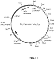
- an expression vector comprising a nucleic acid encoding a heavy chain variable region of the antibody of Claim 1 and a nucleic acid encoding a light chain variable region of the antibody of Claim 1.
- a host cell wherein a nucleic acid encoding the heavy chain variable region of the antibody of Claim 1 and a nucleic acid encoding the light chain variable region of the antibody of Claim 1 have been introduced into the host cell.
- the host cell may be used in a method of producing a humanized antibody comprising culturing the host cell under conditions supporting expression of a humanized antibody that binds to VLQELNVTV (SEQ ID NO: 45) when bound by an HLA-A2 receptor.
- the method may further comprise isolating said antibody.
- the method may further comprising the step of linking said antibody to a diagnostic or therapeutic agent.
- a method of detecting abnormal cells in a sample suspected of containing abnormal cells comprising contacting said sample with an antibody or an artificial antibody as described above.
- the antibody or artificial antibody may be conjugated to a diagnostic agent (such as a fluorophore, a chromophore, a dye, a radioisotope, a chemilluminescent molecule, a paramagnetic ion, or a spin-trapping reagent).
- the antibody or artificial antibody may be detected using a secondary binding agent, such as an anti-Fc receptor antibody.
- the sample may be (a) a tumor tissue from head & neck, brain, esophagus, breast, lung, liver, spleen, stomach, small intestine, large intestine, rectum, ovary, uterus, cervix, prostate, testicle or skin tissue, or (b) a fluid such as blood, lymph, urine, bone marrow aspirate or nipple aspirate.
- the sample may be from a resected tumor bed.
- the method may further comprising making a treatment decision based on the presence, absence or degree of detection, such as a decision to treat said subject with a PR-1-based peptide vaccine.
- the method may detect primary cancer cells, metastatic cancer cells or myeloid dysplastic cells.
- a humanized antibody as defined above for use in a method of treating a subject with cancer, wherein said method comprises administering to the antibody to said subject.
- the antibody or artificial antibody may be conjugated to a therapeutic agent.
- the cancer may be a solid tumor, such as a head & neck tumor, a brain tumor, an esophageal tumor, a breast tumor, a lung tumor, a liver tumor, a spleen tumor, and stomach tumor, a small intestinal tumor, a large intestinal tumor, a rectal tumor, an ovarian tumor, a uterine tumor, a cervical tumor, a prostate tumor, a testicular tumor or a skin tumor.
- the cancer may be a blood cancer, such as a leukemia or lymphoma.
- the therapeutic agent may be a cytokine, a toxin, a chemotherapeutic, a radiotherapeutic, a hormone, an antibody Fc fragment, a TLR agonist, a CpG-containing molecule, or an immune co-stimulatory molecule.
- the method may further comprise providing said subject with a second anti-cancer therapy, such as a gene therapy, a chemotherapy, a radiotherapy, a hormone therapy, a toxin therapy or surgery.
- the humanized antibody may be administered to said subject more than once.
- the cancer may be recurrent or metastatic cancer.
- the present application also describes a method of treating a subject with an autoimmune disease comprising administering to said subject an antibody or an artificial antibody as described above.
- the autoimmune disease may be Wegener's granulomatosis, Churg-Strauss Syndrome, or systemic small vessel vasculitis.
- the antibody or artificial antibody may be conjugated to a therapeutic agent, such as a toxin or apoptosis-inducing agent.
- the method may further comprise providing said subject with a second anti-autoimmune therapy.
- the second anti-autoimmune therapy may be an anti-inflammatory agent.
- the antibody may be administered to said subject more than once.
- the present application also describes a method of inducing complement-mediated cytotoxicity of an HLA-A2 cancer cell comprising contacting said cancer cell with an antibody or an artificial antibody as described above.
- the present application also describes a method of detecting abnormal cells in a sample suspected of containing abnormal cells comprising contacting said sample with an antibody or artificial antibody as described above.
- the antibody or artificial antibody may be conjugated to a diagnostic agent, such as a a fluorophore, a chromophore, a dye, a radioisotope, a chemilluminescent molecule, a paramagnetic ion, or a spin-trapping reagent.
- the antibody or artificial antibody may be detected using a secondary binding agent, such as an anti-Fc receptor antibody.
- the sample may be (a) a tumor tissue from head & neck, brain, esophagus, breast, lung, liver, spleen, stomach, small intestine, large intestine, rectum, ovary, uterus, cervix, prostate, testicle or skin tissue, or (b) a fluid such as blood, lymph, urine, bone marrow aspirate or nipple aspirate.
- the sample may be from a resected tumor bed.
- the method may further comprise making a treatment decision based on the presence, absence or degree of detection, such as deciding to treat said subject with a PR-1-based peptide vaccine.
- the method may detect primary cancer cells, metastatic cancer cells or myeloid dysplastic cells are detected.
- the humanized antibody for use in a method of treating a subject with cancer, then optionally:
- the humanized antibody may be conjugated to a therapeutic agent, such a cytokine, a toxin, a chemotherapeutic, a radiotherapeutic, a hormone, an antibody Fc fragment, a TLR agonist, a CpG-containing molecule, or an immune co-stimulatory molecule.
- a therapeutic agent such as a cytokine, a toxin, a chemotherapeutic, a radiotherapeutic, a hormone, an antibody Fc fragment, a TLR agonist, a CpG-containing molecule, or an immune co-stimulatory molecule.
- the cancer may be a solid tumor, such as a head & neck tumor, a brain tumor, an esophageal tumor, a breast tumor, a lung tumor, a liver tumor, a spleen tumor, and stomach tumor, a small intestinal tumor, a large intestinal tumor, a rectal tumor, an ovarian tumor, a uterine tumor, a cervical tumor, a prostate tumor, a testicular tumor or a skin tumor.
- the cancer may be a blood cancer, such as a leukemia or lymphoma.
- the cancer may be recurrent or metastatic cancer.
- the method may further comprise providing said subject with a second anti-cancer therapy, such as a gene therapy, a chemotherapy, a radiotherapy, a hormone therapy, a toxin therapy or surgery.
- the humanized antibody may be administered to said subject more than once.
- Additional methods described herein include (i) treating a subject with a myeloid dysplastic disease comprising administering to said subject the antibody or artificial antibody described above; and (ii) inducing complement-mediated cytotoxicity of an HLA-A2 cancer cell comprising contacting said cancer cell with the antibody or artificial antibody described above.
- Hu1-8F4 and Hu2-8F4 refer to Hu8F4-1 and Hu8F4-2, respectively.
- the term "Hu8F4" in this document refers generally to both humanized forms of 8F4 (Hu8F4-1 and Hu8F4-2).
- a or “an” may mean one or more.
- the words “a” or “an” when used in conjunction with the word “comprising,” the words “a” or “an” may mean one or more than one.
- another may mean at least a second or more.
- the PR-1 self-peptide (VLQELNVTV; SEQ ID NO:45) has been shown to be recognized on leukemia cell membrane-expressed HLA-A*0201 by CD8+ cytotoxic T lymphocytes (CTL), and PR1-specific CTL specifically lyse myeloid leukemia but not normal bone marrow cells.
- CTL cytotoxic T lymphocytes
- PR1-specific CTL specifically lyse myeloid leukemia but not normal bone marrow cells.
- Vaccination of HLA-A2+ patients with AML, CML, and MDS with PR1 peptide induced PR-1-CTL immunity in 58% of patients and objective clinical responses in 13 of 66 (20%) patients. While these results are encouraging, high tumor burden remains a barrier to successful vaccination.
- PR1 peptide is expressed in sufficient quantity only on the surface of myeloid leukemia cells and not on normal bone marrow cells, the inventors sought to develop an antibody targeted to PR1/HLA-A*0201 that might be used therapeutically to treat patients with myeloid leukemia or that could be used to identify which patients might be susceptible to PR1-based immunotherapy, such as vaccines or adoptive T-cell transfer.
- HLA-A2+ is the most commonly expressed HLA allele (40% of the general Caucasian population), antibody-based therapy for a T-cell epitope therefore would be novel and it might be widely applied.
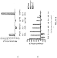
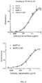
- an IgG2a-kappa monoclonal antibody (8F4) with specificity for the combined PR1/HLA-A*0201 epitope.
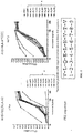
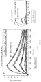
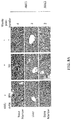
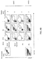
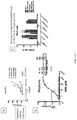
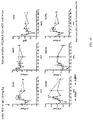
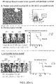
- 8F4 binds to HLA-A2+ AML using both FACS and confocal microscopy to label the cells, but not to normal HLA-A2+ peripheral blood cells.
- 8F4 induced dose-dependent complement-mediated cytotoxicity (CDC) of HLA-A2+ primary human leukemia but not normal bone marrow cells.
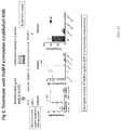
- 8F4 antibody specifically prevented engraftment of human AML in an HLA-A2 transgenic NOD/SCID animal model with only a single exposure to antibody at the time of adoptive transfer into the animal.
- 8F4 delayed breast cancer tumor growth and prolonged survival despite the fact that P3 and NE are not expressed in breast cancer cells.
- isolated or “biologically pure” refer to material which is substantially or essentially free from components which normally accompany the material as it is found in its native state.
- isolated peptides in accordance with the invention preferably do not contain materials normally associated with the peptides in their in situ environment.
- MHC Major histocompatibility complex
- HLA complex HLA complex
- Human leukocyte antigen or "HLA” is a human class I or class II major histocompatibility complex (MHC) protein (see, e.g., Stites, 1994).
- MHC major histocompatibility complex
- HLA supertype or family describes sets of HLA molecules grouped on the basis of shared peptide-binding specificities. HLA class I molecules that share somewhat similar binding affinity for peptides bearing certain amino acid motifs are grouped into HLA supertypes.
- HLA superfamily, HLA supertype family, HLA family, and HLA xx-like supertype molecules are synonyms.
- motif' refers to the pattern of residues in a peptide of defined length, usually a peptide of from about 8 to about 13 amino acids for a class I HLA motif and from about 6 to about 25 amino acids for a class II HLA motif, which is recognized by a particular HLA molecule.
- Peptide motifs are typically different for each protein encoded by each human HLA allele and differ in the pattern of the primary and secondary anchor residues.
- Abnormal cell is any cell that is considered to have a characteristic a typical for that cell type, including atypical growth, typical growth in an atypical location or typical action against an atypical target. Such cells include cancer cells, benign hyperplastic or dysplastic cells, inflammatory cells or autoimmune cells.
- the PR-1 self-peptide (VLQELNVTV; SEQ ID NO:45) is derived from proteinase 3 (P3) and neutrophil elastase (NE), both aberrantly expressed in leukemia. It has been shown to be recognized on leukemia cell membrane-expressed HLA-A*0201 by CD8+ cytotoxic T lymphocytes (CTL).
- CTL cytotoxic T lymphocytes
- PR-1-specific CTL specifically lyse myeloid leukemia, including acute myelogenous leukemia (AML), chronic myelogenous leukemia (CML), and myelodysplastic syndrome (MDS) but not normal bone marrow cells.
- HLA human leukocyte antigen system
- MHC major histocompatibility complex
- the superlocus contains a large number of genes related to immune system function in humans. This group of genes resides on chromosome 6, and encode cell-surface antigen-presenting proteins and many other genes. The proteins encoded by certain genes are also known as antigens, as a result of their historic discovery as factors in organ transplantations.
- the major HLA antigens are essential elements in immune function. Different classes have different functions.
- HLA class I antigens (A, B & C) present peptides from inside the cell (including viral peptides if present). These peptides are produced from digested proteins that are broken down in the lysozomes. The peptides are generally small polymers, about 9 amino acids in length.
- Foreign antigens attract killer T-cells (also called CD8 + cells) that destroy cells.
- HLA class II antigens (DR, DP & DQ) present antigens from outside of the cell to T-lymphocytes. These particular antigens stimulate T-helper cells to reproduce and these T-helper cells then stimulate antibody producing B-cells, self-antigens are suppressed by suppressor T-cells.
- HLA-A2 (A2) is a human leukocyte antigen serotype within the HLA-A "A" serotype group.
- the serotype identifies the gene products of many HLA-A*02 alleles, including HLA-A*0201, *0202, *0203, *0206, and *0207 gene products.
- A*02 is globally common, but A*0201 is at high frequencies in Northern Asia and North America.
- A2 is the most diverse serotype, showing diversity in Eastern Africa and Southwest Asia. While the frequency of A*0201 in Northern Asia is high, its diversity is limited to A*0201 the less common Asian variants A*0203, A*0206.
- the serotype is determined by the antibody recognition of ⁇ 2 subset of HLA-A ⁇ -chains.
- the ⁇ "A" chain are encoded by the HLA-A*02 allele group and the ⁇ -chain are encoded by B2M locus.
- A2 and A*02 are almost synonymous in meaning.
- A2 is more common in Northern Asia and North America than elsewhere, and it is part of a several long haplotypes.
- the present invention concerns the production and use of antibodies that bind to PR1 in the context of HLA-A2 presentation.
- Antibodies are capable of "specific binding" to a particular target or series of antigenically related targets.
- an antibody is said to be capable of "specific binding” to a antigen if it discriminates from antigenically distinct molecules based on binding to the variable region of the antibody. Such interactions are in contrast to non-specific binding that involve classes of compounds, irrespective of their chemical structure (such as the binding of proteins to nitrocellulose, etc .).
- an antibody of the present invention can exhibit "highly specific binding" such that they will be incapable or substantially incapable of binding to even closely related molecules
- Monoclonal antibodies can be readily prepared through use of well-known techniques such as those exemplified in U.S. Patent 4,196,265 .
- a technique involves first immunizing a suitable animal with a selected antigen (e.g ., a polypeptide or polynucleotide of the present invention) in a manner sufficient to provide an immune response. Rodents such as mice and rats are preferred animals. Spleen cells from the immunized animal are then fused with cells of an immortal myeloma cell. Successful fusions are then screened for production of appropriate antibodies.
- a selected antigen e.g ., a polypeptide or polynucleotide of the present invention
- antibody molecules will comprise fragments (such as (F(ab'), F(ab')2) that are produced, for example, by the proteolytic cleavage of the mAbs, or single-chain immunoglobulins producible, for example, via recombinant means. Such antibody derivatives are monovalent. In one embodiment, such fragments can be combined with one another, or with other antibody fragments or receptor ligands to form "chimeric" binding molecules. Significantly, such chimeric molecules may contain substituents capable of binding to different epitopes of the same molecule, or they may be capable of binding to an activated protein C epitope and a "non-activated protein C" epitope.
- humanized antibodies may be studied in an in vitro or an in vivo context. Humanized antibodies may be produced, for example by replacing an immunogenic portion of an antibody with a corresponding, but non-immunogenic portion ( i.e., chimeric antibodies).
- PCT Application WO 87/02671 EP Application 184,187 ; EP Application 171,496 ; EP Application 173,494 ; PCT Application WO 86/01533 ; EP Application 125,023 ; Sun et al. (1987); Wood et al. (1985); and Shaw et al. (1988.
- Humanized chimeric antibodies can alternatively be produced by CDR or CEA substitution. Jones et al. (1986); Verhoeyan et al. (1988); Beidler et al. (1988).
- hydropathic index of amino acids may be considered.
- the importance of the hydropathic amino acid index in conferring interactive biologic function on a protein is generally understood in the art (Kyte and Doolittle, 1982). It is accepted that the relative hydropathic character of the amino acid contributes to the secondary structure of the resultant protein, which in turn defines the interaction of the protein with other molecules, for example, enzymes, substrates, receptors, DNA, antibodies, antigens, and the like.
- Patent 4,554,101 the following hydrophilicity values have been assigned to amino acid residues: basic amino acids: arginine (+3.0), lysine (+3.0), and histidine (-0.5); acidic amino acids: aspartate (+3.0 ⁇ 1), glutamate (+3.0 ⁇ 1), asparagine (+0.2), and glutamine (+0.2); hydrophilic, nonionic amino acids: serine (+0.3), asparagine (+0.2), glutamine (+0.2), and threonine (-0.4), sulfur containing amino acids: cysteine (-1.0) and methionine (-1.3); hydrophobic, nonaromatic amino acids: valine (-1.5), leucine (-1.8), isoleucine (-1.8), proline (-0.5 ⁇ 1), alanine (-0.5), and glycine (0); hydrophobic, aromatic amino acids: tryptophan (-3.4), phenylalanine (-2.5), and tyrosine (-2.3).
- an amino acid can be substituted for another having a similar hydrophilicity and produce a biologically or immunologically modified protein.
- substitution of amino acids whose hydrophilicity values are within ⁇ 2 is preferred, those that are within ⁇ 1 are particularly preferred, and those within ⁇ 0.5 are even more particularly preferred.
- amino acid substitutions generally are based on the relative similarity of the amino acid side-chain substituents, for example, their hydrophobicity, hydrophilicity, charge, size, and the like.
- Exemplary substitutions that take into consideration the various foregoing characteristics are well known to those of skill in the art and include: arginine and lysine; glutamate and aspartate; serine and threonine; glutamine and asparagine; and valine, leucine and isoleucine.
- the present invention may also employ the use of peptide mimetics for the preparation of polypeptides ( see e.g., Johnson, 1993) having many of the natural properties of an antibody, but with altered and/or improved characteristics.
- peptide mimetics for the preparation of polypeptides ( see e.g., Johnson, 1993) having many of the natural properties of an antibody, but with altered and/or improved characteristics.
- the underlying rationale behind the use of mimetics is that the peptide backbone of proteins exists chiefly to orient amino acid side chains in such a way as to facilitate molecular interactions, such as those of antibody and antigen.
- These principles may be used, in conjunction with the principles outline above, to engineer second generation molecules having many of the natural properties of an antibody but with altered and even improved characteristics.
- sequence variants such as insertional or deletion variants.
- Deletion variants lack one or more residues of the native protein. Insertional mutants typically involve the addition of material at a non-terminal point in the polypeptide. It also will be understood that insertional sequence variants may include N- or C-terminal amino acids, and yet still be essentially as set forth in one of the sequences disclosed herein, so long as the sequence meets the criteria set forth above, including the maintenance of biological activity.
- the present invention also contemplates isotype modification.
- antibody 8F4 was determined to be an IgG2a- ⁇ .
- modifying the Fc region to have a different isotype different functionalities can be achieved. For example, changing to IgG1 can increase antibody dependent cell cytotoxicity, switching to class A can improve tissue distribution, and switching to class M can improve valency.
- Modified antibodies may be made by any technique known to those of skill in the art, including expression through standard molecular biological techniques, or the chemical synthesis of polypeptides. Methods for recombinant expression are addressed elsewhere in this document.
- a Single Chain Variable Fragment is a fusion of the variable regions of the heavy and light chains of immunoglobulins, linked together with a short (usually serine, glycine) linker.
- This chimeric molecule retains the specificity of the original immunoglobulin, despite removal of the constant regions and the introduction of a linker peptide. The image to the right shows how this modification usually leaves the specificity unaltered.
- These molecules were created historically to facilitate phage display where it is highly convenient to express the antigen binding domain as a single peptide.
- scFv can be created directly from subcloned heavy and light chains derived from a hybridoma.
- Single chain variable fragments lack the constant Fc region found in complete antibody molecules, and thus, the common binding sites (e.g ., protein A/G) used to purify antibodies. These fragments can often be purified/immobilized using Protein L since Protein L interacts with the variable region of kappa light chains.
- Flexible linkers generally are comprised of helix- and turn-promoting amino acid residues such as alaine, serine and glycine. However, other residues can function as well.
- Tang et al. (1996) used phage display as a means of rapidly selecting tailored linkers for single-chain antibodies (scFvs) from protein linker libraries.
- a random linker library was constructed in which the genes for the heavy and light chain variable domains were linked by a segment encoding an 18-amino acid polypeptide of variable composition.
- the scFv repertoire (approx. 5 ⁇ 10 6 different members) was displayed on filamentous phage and subjected to affinity selection with hapten. The population of selected variants exhibited significant increases in binding activity but retained considerable sequence diversity.
- the recombinant antibodies of the present invention may also involve sequences or moieties that permit dimerization or multimerization of the receptors.
- sequences include those derived from IgA, which permit formation of multimers in conjunction with the J-chain.
- Another multimerization domain is the Gal4 dimerization domain.
- the chains may be modified with agents such as biotin/avidin, which permit the combination of two antibodies.
- a single-chain antibody can be created by joining receptor light and heavy chains using a non-peptide linker or chemical unit.
- the light and heavy chains will be produced in distinct cells, purified, and subsequently linked together in an appropriate fashion (i.e., the N-terminus of the heavy chain being attached to the C-terminus of the light chain via an appropriate chemical bridge).
- Cross-linking reagents are used to form molecular bridges that tie functional groups of two different molecules, e.g., a stablizing and coagulating agent.
- dimers or multimers of the same analog or heteromeric complexes comprised of different analogs can be created.
- hetero-bifunctional cross-linkers can be used that eliminate unwanted homopolymer formation. Table 3 illustrates several cross-linkers.
- An exemplary hetero-bifunctional cross-linker contains two reactive groups: one reacting with primary amine group (e.g ., N-hydroxy succinimide) and the other reacting with a thiol group (e.g., pyridyl disulfide, maleimides, halogens, etc .).
- primary amine group e.g ., N-hydroxy succinimide
- a thiol group e.g., pyridyl disulfide, maleimides, halogens, etc .
- the cross-linker may react with the lysine residue(s) of one protein (e.g., the selected antibody or fragment) and through the thiol reactive group, the cross-linker, already tied up to the first protein, reacts with the cysteine residue (free sulfhydryl group) of the other protein (e.g., the selective agent).
- cross-linker having reasonable stability in blood will be employed.
- Numerous types of disulfide-bond containing linkers are known that can be successfully employed to conjugate targeting and therapeutic/preventative agents. Linkers that contain a disulfide bond that is sterically hindered may prove to give greater stability in vivo, preventing release of the targeting peptide prior to reaching the site of action. These linkers are thus one group of linking agents.
- SMPT cross-linking reagent
- Another cross-linking reagent is SMPT, which is a bifunctional cross-linker containing a disulfide bond that is "sterically hindered" by an adjacent benzene ring and methyl groups. It is believed that steric hindrance of the disulfide bond serves a function of protecting the bond from attack by thiolate anions such as glutathione which can be present in tissues and blood, and thereby help in preventing decoupling of the conjugate prior to the delivery of the attached agent to the target site.
- thiolate anions such as glutathione which can be present in tissues and blood
- the SMPT cross-linking reagent lends the ability to cross-link functional groups such as the SH of cysteine or primary amines (e.g., the epsilon amino group of lysine).
- Another possible type of cross-linker includes the hetero-bifunctional photoreactive phenylazides containing a cleavable disulfide bond such as sulfosuccinimidyl-2-(p-azido salicylamido) ethyl-1,3'-dithiopropionate.
- the N-hydroxy-succinimidyl group reacts with primary amino groups and the phenylazide (upon photolysis) reacts non-selectively with any amino acid residue.
- non-hindered linkers also can be employed in accordance herewith.
- Other useful cross-linkers include SATA, SPDP and 2-iminothiolane (Wawrzynczak & Thorpe, 1987). The use of such cross-linkers is well understood in the art. Another embodiment involves the use of flexible linkers.
- U.S. Patent 4,680,338 describes bifunctional linkers useful for producing conjugates of ligands with amine-containing polymers and/or proteins, especially for forming antibody conjugates with chelators, drugs, enzymes, detectable labels and the like.
- U.S. Patents 5,141,648 and 5,563,250 disclose cleavable conjugates containing a labile bond that is cleavable under a variety of mild conditions. This linker is particularly useful in that the agent of interest may be bonded directly to the linker, with cleavage resulting in release of the active agent. Particular uses include adding a free amino or free sulfhydryl group to a protein, such as an antibody, or a drug.
- U.S. Patent 5,856,456 provides peptide linkers for use in connecting polypeptide constituents to make fusion proteins, e.g., single chain antibodies.
- the linker is up to about 50 amino acids in length, contains at least one occurrence of a charged amino acid (preferably arginine or lysine) followed by a proline, and is characterized by greater stability and reduced aggregation.
- U.S. Patent 5,880,270 discloses aminooxy-containing linkers useful in a variety of immunodiagnostic and separative techniques.
- the antibodies of the present invention may be purified.
- purified is intended to refer to a composition, isolatable from other components, wherein the protein is purified to any degree relative to its naturally-obtainable state.
- a purified protein therefore also refers to a protein, free from the environment in which it may naturally occur.
- substantially purified this designation will refer to a composition in which the protein or peptide forms the major component of the composition, such as constituting about 50%, about 60%, about 70%, about 80%, about 90%, about 95% or more of the proteins in the composition.
- Protein purification techniques are well known to those of skill in the art. These techniques involve, at one level, the crude fractionation of the cellular milieu to polypeptide and non-polypeptide fractions. Having separated the polypeptide from other proteins, the polypeptide of interest may be further purified using chromatographic and electrophoretic techniques to achieve partial or complete purification (or purification to homogeneity). Analytical methods particularly suited to the preparation of a pure peptide are ion-exchange chromatography, exclusion chromatography; polyacrylamide gel electrophoresis; isoelectric focusing.
- protein purification include, precipitation with ammonium sulfate, PEG, antibodies and the like or by heat denaturation, followed by centrifugation; gel filtration, reverse phase, hydroxylapatite and affinity chromatography; and combinations of such and other techniques.
- polypeptide In purifying an antibody of the present invention, it may be desirable to express the polypeptide in a prokaryotic or eukaryotic expression system and extract the protein using denaturing conditions.
- the polypeptide may be purified from other cellular components using an affinity column, which binds to a tagged portion of the polypeptide.
- affinity column which binds to a tagged portion of the polypeptide.
- antibodies are fractionated utilizing agents (i.e., protein A) that bind the Fc portion of the antibody.
- agents i.e., protein A
- antigens my be used to simultaneously purify and select appropriate antibodies.
- Such methods often utilize the selection agent bound to a support, such as a column, filter or bead.
- the antibodies is bound to a support, contaminants removed ( e.g ., washed away), and the antibodies released by applying conditions (salt, heat, etc .).
- the antibodies of the present invention may be linked to various reagents for use in diagnosis and therapy of disease. Linking may be performed using a variety of well known chemical reactions and agents, some of which are described elsewhere in this document.
- imaging moieties can be paramagnetic ions, radioactive isotopes, fluorochromes, NMR-detectable substances, and X-ray imaging agents.
- paramagnetic ions such as chromium (III), manganese (II), iron (III), iron (II), cobalt (II), nickel (II), copper (II), neodymium (III), samarium (III), ytterbium (III), gadolinium (III), vanadium (II), terbium (III), dysprosium (III), holmium (III) and/or erbium (III), with gadolinium being particularly preferred.
- Ions useful in other contexts, such as X-ray imaging include but are not limited to lanthanum (III), gold (III), lead (II), and especially bismuth (III).
- radioactive isotopes for therapeutic and/or diagnostic application, one might mention astatine 211 , 14 carbon, 51 chromium, 36 chlorine, 57 cobalt, 58 cobalt, copper 67 , 152 Eu, gallium 67 , 3 hydrogen, iodine 123 , iodine 125 , iodine 131 , indium 111 , 59 iron, 32 phosphorus, rhenium 186 , rhenium 188 , 75 selenium, 35 sulphur, technicium 99m and/or yttrium 90 .
- Radioactively labeled receptors of the present invention may be produced according to well-known methods in the art. For instance, receptors can be iodinated by contact with sodium and/or potassium iodide and a chemical oxidizing agent such as sodium hypochlorite, or an enzymatic oxidizing agent, such as lactoperoxidase.
- TcRs according to the invention may be labeled with technetium 99m by ligand exchange process, for example, by reducing pertechnate with stannous solution, chelating the reduced technetium onto a Sephadex column and applying the antibody to this column.
- direct labeling techniques may be used, e.g., by incubating pertechnate, a reducing agent such as SNCl 2 , a buffer solution such as sodium-potassium phthalate solution, and the antibody.
- Intermediary functional groups which are often used to bind radioisotopes, which exist as metallic ions to antibody are diethylenetriaminepentaacetic acid (DTPA) or ethylene diaminetetracetic acid (EDTA).
- DTPA diethylenetriaminepentaacetic acid
- EDTA ethylene diaminetetracetic acid
- fluorescent labels contemplated for use as conjugates are Alexa 350, Alexa 430, AMCA, BODIPY 630/650, BODIPY 650/665, BODIPY-FL, BODIPY-R6G, BODIPY-TMR, BODIPY-TRX, Cascade Blue, Cy3, Cy5,6-FAM, Fluorescein Isothiocyanate, HEX, 6-JOE, Oregon Green 488, Oregon Green 500, Oregon Green 514, Pacific Blue, REG, Rhodamine Green, Rhodamine Red, Renographin, ROX, TAMRA, TET, Tetramethylrhodamine, and/or Texas Red.
- Chemotherapeutics may also be conjugated to antibodies, and include cisplatin (CDDP), carboplatin, procarbazine, mechlorethamine, cyclophosphamide, camptothecin, ifosfamide, melphalan, chlorambucil, busulfan, nitrosurea, dactinomycin, daunorubicin, doxorubicin, bleomycin, plicomycin, mitomycin, etoposide (VP16), tamoxifen, raloxifene, estrogen receptor binding agents, taxol, gemcitabien, navelbine, farnesyl-protein transferase inhibitors, transplatinum, 5-fluorouracil, vincristine, vinblastine and methotrexate.
- CDDP cisplatin
- carboplatin carboplatin
- procarbazine mechlorethamine
- cyclophosphamide camptothecin
- Another class of therapeutic agent is the toxins. Cholera toxin, botulism toxin, pertussis toxin, ricin A and B chains, as well as other natural or synthetic toxins are contemplated.
- Cytokines and lymphokines are yet another class of therapeutic agents than can be coupled to the TcR of the present invention, and include IL-1, IL-2, IL-3, IL-4, IL-5, IL-6, IL-7, IL-8, IL-9, IL-10, IL-11, IL-12, IL-13, IL-14, IL-15, IL-16, IL-17, IL-18, IL-19, IL-20, IL-21, IL-22, IL-23, TNF ⁇ , GM-CSF, INF ⁇ , IFN ⁇ , and IFN ⁇ .
- anti-inflammatory agents are contemplated as therapeutic agents that may be conjugated to antibodies.
- Anti-inflammatories include NSAIDs, steroids, rapamycin, infliximab, and ontak.
- Immunosuppressive agents include FK-506 and cyclosporin A.
- TLR agonist may be linked to the antibody, e.g., through the Fc portion of the molecule.
- Agonists of TLRs are compounds that stimulate, or "turn on,” the immune system.
- Natural agonists for TLR9 are components of DNA that are common to bacteria and viruses.
- Natural agonists for TLRs 7 and 8 are patterns of RNA found in viruses. Following recognition of their natural DNA and RNA agonists, TLRs 7, 8, and 9 each initiate a different cascade of protective immune responses.
- TLR agonists include oligodeoxynucleotides, hyaluronic acid fragments, imiquimod, lavendustin C, lipid A, loroxibine, LPS, monophosphoryl lipda A, myristicin, resiquimod, S. typhimurium flagellin, HKLM, PAM3CSK4, and polyI:C.
- nucleic acid encode various portions of antibody heavy and light chain, variable and constant domains.
- a nucleic acid segment may be derived from genomic DNA, complementary DNA (cDNA) or synthetic DNA. Where incorporation into an expression vector is desired, the nucleic acid may also comprise a natural intron or an intron derived from another gene, as well as other non-coding (e.g ., regulatory) and coding regions ( e.g ., linkers).
- cDNA is intended to refer to DNA prepared using messenger RNA (mRNA) as template. The advantage of using a cDNA, as opposed to genomic DNA or DNA polymerized from a genomic, non- or partially-processed RNA template, is that the cDNA primarily contains coding sequences of the corresponding protein.
- Recombinant may be used in conjunction with a polypeptide or the name of a specific polypeptide, and generally refers to a polypeptide produced from a nucleic acid molecule that has been manipulated in vitro or that is the replicated product of such a molecule.
- Recombinant vectors and isolated nucleic acid segments may variously include the antibody-coding regions themselves, coding regions bearing selected alterations or modifications in the basic coding region, or they may encode larger polypeptides that include non-antibody regions.
- nucleic acid as used herein includes single-stranded and double-stranded molecules, as well as DNA, RNA, chemically modified nucleic acids and nucleic acid analogs. It is contemplated that a nucleic acid within the scope of the present invention may be of about 10, about 20, about 30, about 40, about 50, about 60, about 70, about 80, about 90, about 100, about 110, about 120, about 130, about 140, about 150, about 160, about 170, about 180, about 190, about 200, about 210, about 220, about 230, about 240, about 250, about 275, about 300, about 325, about 350, about 375, about 400, about 425, about 450, about 475, about 500, about 525, about 550, about 575, about 600, about 625, about 650, about 675, about 700, about 725, about 750, about 775, about 800, about 825, about 850, about 875, about 900, about 925, about 950, about
- antibody may be encoded by any nucleic acid sequence that encodes the appropriate amino acid sequence, such as those in SEQ ID NOS: 3, 60, 5, 8, 9, 10 (heavy CDRs 1, 2 and 3; light CDRs 1 and 2, 3/JK), and SEQ ID NOS: 9 or 25, which includes the heavy CDRs and framework regions 1, 2 and 3, which flank upstream of heavy CDRs 1, 2 and 3, respectively, and SEQ ID NO: 24, which includes the light CDRs and framework regions 1, 2 and 3, which flank upstream of light CDRs 1, 2 and 3, respectively.
- SEQ ID NOS: 3 60, 5, 8, 9, 10 (heavy CDRs 1, 2 and 3; light CDRs 1 and 2, 3/JK)
- SEQ ID NOS: 9 or 25 which includes the heavy CDRs and framework regions 1, 2 and 3, which flank upstream of heavy CDRs 1, 2 and 3, respectively
- SEQ ID NO: 24 which includes the light CDRs and framework regions 1, 2 and 3, which flank upstream of light CDRs 1, 2 and 3, respectively.
- the codons selected for encoding each amino acid may be modified to optimize expression of the nucleic acid in the host cell of interest.
- the term "functionally equivalent codon” is used herein to refer to codons that encode the same amino acid, such as the six codons for arginine or serine, and also refers to codons that encode biologically equivalent amino acids. Codon preferences for various species of host cell are well known in the art. Codons preferred for use in humans, are well known to those of skill in the art (Wada et al., 1990). Codon preferences for other organisms also are well known to those of skill in the art (Wada et al., 1990, included herein in its entirety by reference).
- Prokaryote- and/or eukaryote-based systems can be used to produce nucleic acid sequences, or their cognate polypeptides, proteins and peptides.
- the present invention contemplates the use of such an expression system to produce the antibodies that bind PR-1/HLA-A2.
- One powerful expression technology employs the insect-cell/baculovirus system.
- the insect-cell/baculovirus system can produce a high level of protein expression of a heterologous nucleic acid segment, such as described in U.S.
- Patents 5,871,986 , 4,879,236 which can be bought, for example, under the name MAXBAC® 2.0 from INVITROGEN® and BACPACKTM BACULOVIRUS EXPRESSION SYSTEM FROM CLONTECH®.
- INVITROGEN® also provides a yeast expression system called the Pichia methanolica Expression System, which is designed for high-level production of recombinant proteins in the methylotrophic yeast Pichia methanolica.
- a vector such as an expression construct, to produce a nucleic acid sequence or its cognate polypeptide, protein, or peptide.
- the expression vector comprises a virus or engineered vector derived from a viral genome.
- the first viruses used as gene vectors were DNA viruses including the papovaviruses (simian virus 40, bovine papilloma virus, and polyoma) (Ridgeway, 1988; Baichwal and Sugden, 1986) and adenoviruses (Ridgeway, 1988; Baichwal and Sugden, 1986).
- papovaviruses simian virus 40, bovine papilloma virus, and polyoma
- adenoviruses Rosgeway, 1988; Baichwal and Sugden, 1986.
- Adenoviral Vectors A particular method for delivery of the nucleic acid involves the use of an adenovirus expression vector. Although adenovirus vectors are known to have a low capacity for integration into genomic DNA, this feature is counterbalanced by the high efficiency of gene transfer afforded by these vectors. "Adenovirus expression vector” is meant to include those constructs containing adenovirus sequences sufficient to (a) support packaging of the construct and (b) to ultimately express a tissue or cell-specific construct that has been cloned therein. Knowledge of the genetic organization or adenovirus, a 36 kb, linear, double-stranded DNA virus, allows substitution of large pieces of adenoviral DNA with foreign sequences up to 7 kb (Grunhaus and Horwitz, 1992).
- AAV Vectors The nucleic acid may be introduced into the cell using adenovirus assisted transfection. Increased transfection efficiencies have been reported in cell systems using adenovirus coupled systems (Kelleher and Vos, 1994; Cotten et al., 1992; Curiel, 1994).
- Adeno-associated virus (AAV) is an attractive vector system for use in the vaccines of the present invention (Muzyczka, 1992).
- AAV has a broad host range for infectivity (Tratschin et al., 1984; Laughlin et al., 1986; Lebkowski et al., 1988; McLaughlin et al., 1988). Details concerning the generation and use of rAAV vectors are described in U.S. Patents 5,139,941 and 4,797,368 .
- Retroviral Vectors have promise as gene delivery vectors in vaccines due to their ability to integrate their genes into the host genome, transferring a large amount of foreign genetic material, infecting a broad spectrum of species and cell types and of being packaged in special cell-lines (Miller, 1992).
- a nucleic acid e.g., one encoding an antigen of interest
- a packaging cell line containing the gag, pol, and env genes but without the LTR and packaging components is constructed (Mann et al., 1983).
- Retroviral vectors are able to infect a broad variety of cell types. However, integration and stable expression require the division of host cells (Paskind et al., 1975).
- Lentiviruses are complex retroviruses, which, in addition to the common retroviral genes gag, pol, and env, contain other genes with regulatory or structural function. Lentiviral vectors are well known in the art (see, for example, Naldini et al., 1996; Zufferey et al., 1997; Blomer et al., 1997; U.S. Patents 6,013,516 and 5,994,136 ). Some examples of lentivirus include the Human Immunodeficiency Viruses: HIV-1, HIV-2 and the Simian Immunodeficiency Virus: SIV. Lentiviral vectors have been generated by multiply attenuating the HIV virulence genes, for example, the genes env, vif, vpr, vpu and nef are deleted making the vector biologically safe.
- Recombinant lentiviral vectors are capable of infecting non-dividing cells and can be used for both in vivo and ex vivo gene transfer and expression of nucleic acid sequences.
- recombinant lentivirus capable of infecting a non-dividing cell wherein a suitable host cell is transfected with two or more vectors carrying the packaging functions, namely gag, pol and env, as well as rev and tat is described in U.S. Patent 5,994,136 .
- One may target the recombinant virus by linkage of the envelope protein with an antibody or a particular ligand for targeting to a receptor of a particular cell-type.
- a sequence (including a regulatory region) of interest into the viral vector, along with another gene which encodes the ligand for a receptor on a specific target cell, for example, the vector is now target-specific.
- viral vectors may be employed as vaccine constructs in the present invention.
- Vectors derived from viruses such as vaccinia virus (Ridgeway, 1988; Baichwal and Sugden, 1986; Coupar et al., 1988), Sindbis virus, cytomegalovirus and herpes simplex virus may be employed. They offer several attractive features for various mammalian cells (Friedmann, 1989; Ridgeway, 1988; Baichwal and Sugden, 1986; Coupar et al., 1988; Horwich et al., 1990). Lentiviruses also have been explored as vaccine vectors (VandenDriessche et al., 2002).
- a nucleic acid to be delivered may be housed within an infective virus that has been engineered to express a specific binding ligand.
- the virus particle will thus bind specifically to the cognate receptors of the target cell and deliver the contents to the cell.
- a novel approach designed to allow specific targeting of retrovirus vectors was developed based on the chemical modification of a retrovirus by the chemical addition of lactose residues to the viral envelope. This modification can permit the specific infection of hepatocytes via sialoglycoprotein receptors.
- Suitable non-viral methods for nucleic acid delivery to effect expression of compositions of the present invention are believed to include virtually any method by which a nucleic acid (e.g., DNA) can be introduced into an organelle, a cell, a tissue or an organism, as described herein or as would be known to one of ordinary skill in the art.
- a nucleic acid e.g., DNA
- Such methods include, but are not limited to, direct delivery of DNA such as by injection ( U.S. Patents 5,994,624 , 5,981,274 , 5,945,100 , 5,780,448 , 5,736,524 , 5,702,932 , 5,656,610 , 5,589,466 and 5,580,859 ), including microinjection ( Harland and Weintraub, 1985; U.S.
- Patent 5,789,215 by electroporation ( U.S. Patent 5,384,253 ); by calcium phosphate precipitation (Graham and Van Der Eb, 1973; Chen and Okayama, 1987; Rippe et al., 1990); by using DEAE-dextran followed by polyethylene glycol (Gopal, 1985); by direct sonic loading (Fechheimer et al., 1987); by liposome mediated transfection (Nicolau and Sene, 1982; Fraley et al., 1979; Nicolau et al., 1987; Wong et al., 1980; Kaneda et al., 1989; Kato et al., 1991); by microprojectile bombardment ( PCT Application Nos.
- MDS myelodysplasias
- MDS myelodysplasia
- refractory anemia refractory anemia with ring sideroblasts
- refractory anemia with excess blasts refractory anemia with excess blasts in transformation
- chronic myelomonocytic leukemia The remaining 50% typically present with isolated or combined cytopenias such as anemia, leucopenia and/or thrombocytopenia (low platelet count).
- cytopenias such as anemia, leucopenia and/or thrombocytopenia (low platelet count).
- AML acute myeloid leukemia
- solid tumor cancers Such cancer lung cancer, head and neck cancer, breast cancer, pancreatic cancer, prostate cancer, renal cancer, bone cancer, testicular cancer, cervical cancer, gastrointestinal cancer, lymphomas, pre-neoplastic lesions in the lung, colon cancer, melanoma, and bladder cancer.
- Other hyperplastic, neoplastic and dysplastic diseases, including benign hyperprolifertative diseases are also with the scope of the diagnostic procedures described herein.
- Adminstration of diagnostic reagents is well known in the art and will vary depending on diagnosis to be achieved. For example, where a discrete tumor mass or masses is/are to be imagined, local or regional administration (e.g ., in the the tumor vasculature, local lymph system or local arteries or veins) may be utilized. Alternatively, one may provide diagnostic reagents regionally or systemically. This may be the route of choice where imaging of an entire limb or organism is desired, where know specific tumor mass has been identified, or when metastasis is suspected.
- compositions disclosed herein may alternatively be administered parenterally, intravenously, intradermally, intramuscularly, transdermally or even intraperitoneally as described in U.S. Patent 5,543,158 ; U.S. Patent 5,641,515 and U.S. Patent 5,399,363 .
- Injection of pharmaceuticals may be by syringe or any other method used for injection of a solution, as long as the agent can pass through the particular gauge of needle required for injection.
- a novel needleless injection system has been described ( U.S. Patent 5,846,233 ) having a nozzle defining an ampule chamber for holding the solution and an energy device for pushing the solution out of the nozzle to the site of delivery.
- a syringe system has also been described for use in gene therapy that permits multiple injections of predetermined quantities of a solution precisely at any depth ( U.S. Patent 5,846,225 ).
- Solutions of the active compounds as free base or pharmacologically acceptable salts may be prepared in water suitably mixed with a surfactant, such as hydroxypropylcellulose. Dispersions may also be prepared in glycerol, liquid polyethylene glycols, and mixtures thereof and in oils. Under ordinary conditions of storage and use, these preparations contain a preservative to prevent the growth of microorganisms.
- the pharmaceutical forms suitable for injectable use include sterile aqueous solutions or dispersions and sterile powders for the extemporaneous preparation of sterile injectable solutions or dispersions ( U.S. Patent 5,466,468 ). In all cases the form must be sterile and must be fluid to the extent that easy syringability exists.
- the carrier can be a solvent or dispersion medium containing, for example, water, ethanol, polyol (e.g., glycerol, propylene glycol, and liquid polyethylene glycol, and the like), suitable mixtures thereof, and/or vegetable oils.
- a coating such as lecithin
- surfactants for example, water, alcohol, glycol, and water.
- the prevention of the action of microorganisms can be brought about by various antibacterial and antifungal agents, for example, parabens, chlorobutanol, phenol, sorbic acid, thimerosal, and the like.
- isotonic agents for example, sugars or sodium chloride.
- Prolonged absorption of the injectable compositions can be brought about by the use in the compositions of agents delaying absorption, for example, aluminum monostearate and gelatin.
- aqueous solutions For parenteral administration in an aqueous solution, for example, the solution should be suitably buffered if necessary and the liquid diluent first rendered isotonic with sufficient saline or glucose.
- aqueous solutions are especially suitable for intravenous, intramuscular, subcutaneous, intratumoral and intraperitoneal administration.
- sterile aqueous media that can be employed will be known to those of skill in the art in light of the present disclosure.
- one dosage may be dissolved in 1 ml of isotonic NaCl solution and either added to 1000 ml of hypodermolysis fluid or injected at the proposed site of infusion, (see for example, " Remington's Pharmaceutical Sciences” 15th Edition, pages 1035-1038 and 1570-1580 ).
- Some variation in dosage will necessarily occur depending on the condition of the subject being treated.
- the person responsible for administration will, in any event, determine the appropriate dose for the individual subject.
- preparations should meet sterility, pyrogenicity, general safety and purity standards as required by FDA Office of Biologics standards.
- Sterile injectable solutions are prepared by incorporating the active compounds in the required amount in the appropriate solvent with various of the other ingredients enumerated above, as required, followed by filtered sterilization.
- dispersions are prepared by incorporating the various sterilized active ingredients into a sterile vehicle which contains the basic dispersion medium and the required other ingredients from those enumerated above.
- the preferred methods of preparation are vacuum-drying and freeze-drying techniques which yield a powder of the active ingredient plus any additional desired ingredient from a previously sterile-filtered solution thereof.
- compositions disclosed herein may be formulated in a neutral or salt form.
- Pharmaceutically-acceptable salts include the acid addition salts (formed with the free amino groups of the protein) and which are formed with inorganic acids such as, for example, hydrochloric or phosphoric acids, or such organic acids as acetic, oxalic, tartaric, mandelic, and the like. Salts formed with the free carboxyl groups can also be derived from inorganic bases such as, for example, sodium, potassium, ammonium, calcium, or ferric hydroxides, and such organic bases as isopropylamine, trimethylamine, histidine, procaine and the like.
- solutions Upon formulation, solutions will be administered in a manner compatible with the dosage formulation and in such amount as is therapeutically effective.
- the formulations are easily administered in a variety of dosage forms such as injectable solutions, drug release capsules and the like.
- carrier includes any and all solvents, dispersion media, vehicles, coatings, diluents, antibacterial and antifungal agents, isotonic and absorption delaying agents, buffers, carrier solutions, suspensions, colloids, and the like.
- carrier includes any and all solvents, dispersion media, vehicles, coatings, diluents, antibacterial and antifungal agents, isotonic and absorption delaying agents, buffers, carrier solutions, suspensions, colloids, and the like.
- the use of such media and agents for pharmaceutical active substances is well known in the art. Except insofar as any conventional media or agent is incompatible with the active ingredient, its use in the therapeutic compositions is contemplated. Supplementary active ingredients can also be incorporated into the compositions.
- phrases "pharmaceutically-acceptable” or “pharmacologically-acceptable” refers to molecular entities and compositions that do not produce an allergic or similar untoward reaction when administered to a human.
- the preparation of an aqueous composition that contains a protein as an active ingredient is well understood in the art.
- such compositions are prepared as injectables, either as liquid solutions or suspensions; solid forms suitable for solution in, or suspension in, liquid prior to injection can also be prepared.
- the antibodies of the present invention may be used in the methods of treating hyperplastic/dysplastic/neoplastic diseases/conditions including cancer.
- Types of diseases/conditions contemplated to be treated with the peptides of the present invention include, but are not limited to leukemias such as, AML, MDS and CML, as well as myelodysplasias.
- Other types of cancers may include lung cancer, head and neck cancer, breast cancer, pancreatic cancer, prostate cancer, renal cancer, bone cancer, testicular cancer, cervical cancer, gastrointestinal cancer, lymphomas, pre-neoplastic lesions in the lung, colon cancer, melanoma, bladder cancer and any other neoplastic diseases.
- a hyperplastic/neoplastic/cancer cell with the uses and compositions of the present invention, one would generally contact a hyperplastic/neoplastic/cancer cell with the therapeutic compound such as a polypeptide or an expression construct encoding an antibody of the present invention, normally dispersed in a pharmacetically acceptable buffer or carrier (see above in the discussion of diagnostic agents).
- the therapeutic compound such as a polypeptide or an expression construct encoding an antibody of the present invention, normally dispersed in a pharmacetically acceptable buffer or carrier (see above in the discussion of diagnostic agents).
- the routes of administration will vary, naturally, with the location and nature of the lesion, and include, e.g ., intradermal, transdermal, parenteral, intravenous, intramuscular, intranasal, subcutaneous, percutaneous, intratracheal, intraperitoneal, intratumoral, perfusion, lavage, direct injection, and oral administration and formulation. Any of the formulations and routes of administration discussed with respect to the treatment or diagnosis of cancer may also be employed with respect to neoplastic diseases and conditions. Ex vivo embodiments, where tumor cells are treated/transduced outside a patient's body (either specifically or as part of a larger cell population) are contemplated.
- Intratumoral injection, or injection into the tumor vasculature is specifically contemplated for discrete, solid, accessible tumors. Local, regional or systemic administration also may be appropriate. For tumors of >4 cm, the volume to be administered will be about 4-10 ml), while for tumors of ⁇ 4 cm, a volume of about 1-3 ml will be used. Multiple injections delivered as single dose comprise about 0.1 to about 0.5 ml volumes. The viral particles may advantageously be contacted by administering multiple injections to the tumor, spaced at approximately 1 cm intervals.
- the present invention may be used at the time of surgery, and/or thereafter, to treat residual or metastatic disease.
- a resected tumor bed may be injected or perfused with a formulation comprising antibodies.
- the perfusion may be continued post-resection, for example, by leaving a catheter implanted at the site of the surgery. Periodic post-surgical treatment also is envisioned.
- Continuous administration also may be applied where appropriate, for example, where a tumor is excised and the tumor bed is treated to eliminate residual, microscopic disease. Delivery via syringe or catherization is preferred. Such continuous perfusion may take place for a period from about 1-2 hr, to about 2-6 hr, to about 6-12 hr, to about 12-24 hr, to about 1-2 days, to about 1-2 wk or longer following the initiation of treatment. Generally, the dose of the therapeutic composition via continuous perfusion will be equivalent to that given by a single or multiple injections, adjusted over a period of time during which the perfusion occurs. It is further contemplated that limb perfusion may be used to administer therapeutic compositions of the present invention, particularly in the treatment of melanomas and sarcomas.
- Treatment regimens may vary as well, and often depend on tumor type, tumor location, disease progression, and health and age of the patient. Obviously, certain types of tumor will require more aggressive treatment, while at the same time, certain patients cannot tolerate more taxing protocols. The clinician will be best suited to make such decisions based on the known efficacy and toxicity (if any) of the therapeutic formulations.
- the tumor being treated may not, at least initially, be resectable. Treatments may increase the resectability of the tumor due to shrinkage at the margins or by elimination of certain particularly invasive portions. Following treatments, resection may be possible. Additional treatments subsequent to resection will serve to eliminate microscopic residual disease at the tumor site.
- a typical course of treatment, for a primary tumor or a post-excision tumor bed, will involve multiple doses.
- Typical primary tumor treatment involves a 6-dose application over a two-week period.
- the two-week regimen may be repeated one, two, three, four, five, six or more times.
- the need to complete the planned dosings may be re-evaluated.
- an anti-cancer agent is capable of negatively affecting cancer in a subject, for example, by killing cancer cells, inducing apoptosis in cancer cells, reducing the growth rate of cancer cells, reducing the incidence or number of metastases, reducing tumor size, inhibiting tumor growth, reducing the blood supply to a tumor or cancer cells, promoting an immune response against cancer cells or a tumor, preventing or inhibiting the progression of cancer, or increasing the lifespan of a subject with cancer.
- Anti-cancer agents include biological agents (biotherapy), chemotherapy agents, and radiotherapy agents.
- these other compositions would be provided with a therapy according to the present invention in a combined amount effective to kill or inhibit proliferation of the cell.
- This process may involve contacting the cells with the both agent(s) at the same time. This may be achieved by contacting the cell with a single composition or pharmacological formulation that includes both agents, or by contacting the cell with two distinct compositions or formulations at the same time.
- the antibody therapy may precede or follow the other agent treatment by intervals ranging from minutes to weeks.
- the other agent and antibodies are applied separately to the cell, one would generally ensure that a significant period of time did not expire between the time of each delivery, such that the agent and expression construct would still be able to exert an advantageously combined effect on the cell.
- the antibody therapy (with or without a conjugated therapeutic agent) is "A” and the secondary anti-cancer therapy is "B": A/B/A B/A/B B/B/A A/A/B A/B/B B/A/A A/B/B/B B/A/B/B B/B/B/A B/B/A/B A/A/B/B A/B/A/B A/B/B/A B/B/A/A B/A/B/A B/A/A/B A/A/A/B B/A/A/A A/B/A/A A/B/A/A A/B/A/A A/B/A/A A/B/A/A A/B/A/A A/B/A/A A/B/A/A A/B/A/A A/B/A/A A/B/A/A A/B/A/A Administration of the therapeutic agents of the present invention to a patient will follow general protocols for the administration of that particular secondary therapy
- Cancer therapies also include a variety of combination therapies with both chemical and radiation based treatments.
- Combination chemotherapies include, for example, cisplatin (CDDP), carboplatin, procarbazine, mechlorethamine, cyclophosphamide, camptothecin, ifosfamide, melphalan, chlorambucil, busulfan, nitrosurea, dactinomycin, daunorubicin, doxorubicin, bleomycin, plicomycin, mitomycin, etoposide (VP16), tamoxifen, raloxifene, estrogen receptor binding agents, taxol, gemcitabien, navelbine, farnesyl-protein transferase inhibitors, transplatinum, 5-fluorouracil, vincristine, vinblastine and methotrexate, Temazolomide (an aqueous form of DTIC), or any analog or derivative variant of the foregoing.
- Dosage ranges for X-rays range from daily doses of 50 to 200 roentgens for prolonged periods of time (3 to 4 wk), to single doses of 2000 to 6000 roentgens.
- Dosage ranges for radioisotopes vary widely, and depend on the half-life of the isotope, the strength and type of radiation emitted, and the uptake by the neoplastic cells.
- contacted and “exposed,” when applied to a cell are used herein to describe the process by which a therapeutic construct and a chemotherapeutic or radiotherapeutic agent are delivered to a target cell or are placed in direct juxtaposition with the target cell.
- both agents are delivered to a cell in a combined amount effective to kill the cell or prevent it from dividing.
- Immunotherapeutics generally, rely on the use of immune effector cells and molecules to target and destroy cancer cells.
- the immune effector may be, for example, an antibody specific for some marker on the surface of a tumor cell.
- the antibody alone may serve as an effector of therapy or it may recruit other cells to actually effect cell killing.
- the antibody also may be conjugated to a drug or toxin (chemotherapeutic, radionuclide, ricin A chain, cholera toxin, pertussis toxin, etc .) and serve merely as a targeting agent.
- the effector may be a lymphocyte carrying a surface molecule that interacts, either directly or indirectly, with a tumor cell target.
- Various effector cells include cytotoxic T-cells and NK cells. The combination of therapeutic modalities, i.e., direct cytotoxic activity and inhibition or reduction of Fortilin would provide therapeutic benefit in the treatment of cancer.
- Immunotherapy could also be used as part of a combined therapy.
- the general approach for combined therapy is discussed below.
- the tumor cell must bear some marker that is amenable to targeting, i.e., is not present on the majority of other cells.
- Common tumor markers include carcinoembryonic antigen, prostate specific antigen, urinary tumor associated antigen, fetal antigen, tyrosinase (p97), gp68, TAG-72, HMFG, Sialyl Lewis Antigen, MucA, MucB, PLAP, estrogen receptor, laminin receptor, erb B and p155.
- Immune stimulating molecules also exist including: cytokines such as IL-2, IL-4, IL-12, GM-CSF, gamma-IFN, chemokines such as MIP-1, MCP-1, IL-8 and growth factors such as FLT3 ligand.
- cytokines such as IL-2, IL-4, IL-12, GM-CSF, gamma-IFN
- chemokines such as MIP-1, MCP-1, IL-8 and growth factors such as FLT3 ligand.
- Combining immune stimulating molecules, either as proteins or using gene delivery in combination with a tumor suppressor such as mda-7 has been shown to enhance anti-tumor effects (Ju et al., 2000).
- immune adjuvants e.g., Mycobacterium bovis, Plasmodium falciparum, dinitrochlorobenzene and aromatic compounds
- U.S. Patent 5,801,005 U.S.
- Patent 5,739,169 Hui and Hashimoto, 1998; Christodoulides et al., 1998), cytokine therapy (e.g., interferons, and; IL-1, GM-CSF and TNF) (Bukowski et al., 1998; Davidson et al., 1998; Hellstrand et al., 1998) gene therapy (e.g., TNF, IL-1, IL-2, p53) (Qin et al., 1998; Austin-Ward and Villaseca, 1998; U.S. Patent 5,830,880 and U.S.
- cytokine therapy e.g., interferons, and; IL-1, GM-CSF and TNF
- gene therapy e.g., TNF, IL-1, IL-2, p53
- Patent 5,846,945 and monoclonal antibodies (e.g., anti-ganglioside GM2, anti-HER-2, anti-p185) (Pietras et al., 1998; Hanibuchi et al., 1998; U.S. Patent 5,824,311 ).
- Herceptin trastuzumab
- Herceptin is a chimeric (mouse-human) monoclonal antibody that blocks the HER2-neu receptor. It possesses anti-tumor activity and has been approved for use in the treatment of malignant tumors (Dillman, 1999). Combination therapy of cancer with herceptin and chemotherapy has been shown to be more effective than the individual therapies.
- one or more anti-cancer therapies may be employed with the tumor-associated HLA-restricted peptide therapies described herein.
- adoptive immunotherapy the patient's circulating lymphocytes, or tumor infiltrated lymphocytes, are isolated in vitro, activated by lymphokines such as IL-2 or transduced with genes for tumor necrosis, and readministered (Rosenberg et al., 1988; 1989). To achieve this, one would administer to an animal, or human patient, an immunologically effective amount of activated lymphocytes in combination with an adjuvant-incorporated antigenic peptide composition as described herein.
- the activated lymphocytes will most preferably be the patient's own cells that were earlier isolated from a blood or tumor sample and activated (or "expanded") in vitro.
- This form of immunotherapy has produced several cases of regression of melanoma and renal carcinoma, but the percentage of responders was few compared to those who did not respond.
- Passive Immunotherapy A number of different approaches for passive immunotherapy of cancer exist. They may be broadly categorized into the following: injection of antibodies alone; injection of antibodies coupled to toxins or chemotherapeutic agents; injection of antibodies coupled to radioactive isotopes; injection of anti-idiotype antibodies; and finally, purging of tumor cells in bone marrow.
- human monoclonal antibodies are employed in passive immunotherapy, as they produce few or no side effects in the patient.
- their application is somewhat limited by their scarcity and have so far only been administered intralesionally.
- Human monoclonal antibodies to ganglioside antigens have been administered intralesionally to patients suffering from cutaneous recurrent melanoma (Irie & Morton, 1986). Regression was observed in six out of ten patients, following, daily or weekly, intralesional injections. In another study, moderate success was achieved from intralesional injections of two human monoclonal antibodies (Irie et al., 1989).
- Possible therapeutic antibodies include anti-TNF, anti-CD25, anti-CD3, anti-CD20, CTLA-4-IG, and anti-CD28.
- Treatment protocols may include administration of lymphokines or other immune enhancers as described by Bajorin et al. (1988). The development of human monoclonal antibodies is described in further detail elsewhere in the specification.
- the secondary treatment is a gene therapy in which a therapeutic polynucleotide is administered before, after, or at the same time as the tumor-associated HLA-restricted peptide is administered.
- Delivery of a vector encoding a the tumor-associated HLA-restricted peptide in conjunction with a second vector encoding one of the following gene products will have a combined anti-hyperproliferative effect on target tissues.
- a single vector encoding both genes may be used.
- proteins are encompassed within the invention, some of which are described below.
- Various genes that may be targeted for gene therapy of some form in combination with the present invention are will known to one of ordinary skill in the art and may comprise any gene involved in cancers.
- Inducers of Cellular Proliferation The proteins that induce cellular proliferation further fall into various categories dependent on function. The commonality of all of these proteins is their ability to regulate cellular proliferation.
- a form of PDGF the sis oncogene
- Oncogenes rarely arise from genes encoding growth factors, and at the present, sis is the only known naturally-occurring oncogenic growth factor.
- anti-sense mRNA directed to a particular inducer of cellular proliferation is used to prevent expression of the inducer of cellular proliferation.
- the proteins FMS, ErbA, ErbB and neu are growth factor receptors. Mutations to these receptors result in loss of regulatable function. For example, a point mutation affecting the transmembrane domain of the Neu receptor protein results in the neu oncogene.
- the erbA oncogene is derived from the intracellular receptor for thyroid hormone. The modified oncogenic ErbA receptor is believed to compete with the endogenous thyroid hormone receptor, causing uncontrolled growth.
- the largest class of oncogenes includes the signal transducing proteins (e.g ., Src, Abl and Ras).
- Src is a cytoplasmic protein-tyrosine kinase, and its transformation from proto-oncogene to oncogene in some cases, results via mutations at tyrosine residue 527.
- transformation of GTPase protein ras from proto-oncogene to oncogene results from a valine to glycine mutation at amino acid 12 in the sequence, reducing ras GTPase activity.
- Jun, Fos and Myc are proteins that directly exert their effects on nuclear functions as transcription factors.
- the tumor suppressor oncogenes function to inhibit excessive cellular proliferation.
- the inactivation of these genes destroys their inhibitory activity, resulting in unregulated proliferation.
- the most common tumor suppressors are Rb, p53, p21 and p16.
- Other genes that may be employed according to the present invention include APC, DCC, NF-1, NF-2, WT-1, MEN-I, MEN-II, zac1, p73, VHL, C-CAM, MMAC1/PTEN, DBCCR-1, FCC, rsk-3, p27, p27/p16 fusions, and p21/p27 fusions.
- Apoptosis or programmed cell death, is an essential process for normal embryonic development, maintaining homeostasis in adult tissues, and suppressing carcinogenesis (Kerr et al., 1972).
- the Bcl-2 family of proteins and ICE-like proteases have been demonstrated to be important regulators and effectors of apoptosis in other systems.
- the Bcl-2 protein plays a prominent role in controlling apoptosis and enhancing cell survival in response to diverse apoptotic stimuli (Bakhshi et al., 1985; Cleary and Sklar, 1985; Cleary et al., 1986; Tsujimoto et al., 1985; Tsujimoto and Croce, 1986).
- the evolutionarily conserved Bcl-2 protein now is recognized to be a member of a family of related proteins, which can be categorized as death agonists or death antagonists.
- Bcl-2 acts to suppress cell death triggered by a variety of stimuli. Also, it now is apparent that there is a family of Bcl-2 cell death regulatory proteins that share in common structural and sequence homologies. These different family members have been shown to either possess similar functions to Bcl-2 (e.g., Bcl XL , Bcl W , Bcl S , Mcl-1, A1, Bfl-1) or counteract Bcl-2 function and promote cell death (e.g., Bax, Bak, Bik, Bim, Bid, Bad, Harakiri).
- Curative surgery is a cancer treatment that may be used in conjunction with other therapies, such as the treatment of the present invention, chemotherapy, radiotherapy, hormonal therapy, gene therapy, immunotherapy and/or alternative therapies.
- Curative surgery includes resection in which all or part of cancerous tissue is physically removed, excised, and/or destroyed.
- Tumor resection refers to physical removal of at least part of a tumor.
- treatment by surgery includes laser surgery, cryosurgery, electrosurgery, and microscopically controlled surgery (Mohs' surgery). It is further contemplated that the present invention may be used in conjunction with removal of superficial cancers, precancers, or incidental amounts of normal tissue.
- a cavity may be formed in the body.
- Treatment may be accomplished by perfusion, direct injection or local application of the area with an additional anti-cancer therapy.
- Such treatment may be repeated, for example, every 1, 2, 3, 4, 5, 6, or 7 days, or every 1, 2, 3, 4, and 5 weeks or every 1, 2, 3, 4, 5, 6, 7, 8, 9, 10, 11, or 12 months.
- These treatments may be of varying dosages as well.
- the present invention also contemplates the treatment of autoimmune disease usig the antibodies of the present invention.
- PR1 is derived from myeloid self-proteins. Proteinase 3 (Pr3), which contains PR1, is the target of autoimmune attack in Wegener's granulomatosis.
- Pr3 Proteinase 3
- MPO Myeloperoxidase
- MPO is the target antigen in small vessel vasculitis (Franssen et al., 1996; Brouwer et al., 1994; Molldrem et al., 1996), with evidence for both T-cell and antibody immunity in patients with these diseases.
- cANCA cytoplasmic anti-neutrophil cytoplasmic antibodies
- pANCA perinuclear ANCA
- MPO perinuclear ANCA
- inhibiting immune cell recognition of PR1 may be therapeutic for autoimmune disease.
- antibodies of the present invention will be administered to subjects suffering from autoimmune disease to neutralize effects of other autoantibodies (e.g ., pANCA against proteinase 3).
- an antibody will be engineered to be "bi-specific," i.e., to have immunologic specificity for two antigen, where one is PR1/HLA-A2, and the other is a dendritic cell surface antigens like DEC-205, LOX-1, RAGE, thereby blocking dendritic cell function in antigen presentation.
- Vasculitis is a process caused by inflammation of blood vessel walls and results in a variety of disorders. An accepted classification system for vasculitis has not emerged, although it may be categorized by the size or type of the involved blood vessel as large-, medium-, or small-vessel vasculitis. Small-vessel vasculitis is defined as vasculitis that affects vessels smaller than arteries ( i.e., arterioles, venules, and capillaries); however, small-vessel vasculitis can also involve medium-sized arteries.
- Anti-neutrophil cytoplasmic antibodies (ANCA)-associated vasculitis is the most common cause of small-vessel vasculitis and includes microscopic polyangiitis, Wegener's granulomatosis, Churg-Strauss syndrome, and certain types of drug-induced vasculitis.
- Wegener's Granulomatosis is a rare disorder which causes inflammation of blood vessels in the upper respiratory tract (nose, sinuses, ears), lungs, and kidneys. Many other areas of the body may also be affected, with arthritis (joint inflammation) occurring in almost half of all cases. The eyes and skin may also be affected. The cause is unknown, but Wegener's Granulomatosis is thought to be an autoimmune disorder and is often classified as one of the rheumatic diseases. Destructive lesions develop in the upper and lower respiratory tract and the kidney. In the kidney, these lesions cause glomerulonephritis that may result in hematuria (blood in the urine) and kidney failure.
- kidney disease It occurs most often between the ages of 30 and 50, and men are affected twice as often as women. It is rare in children, but has been seen in infants as young as 3 months old. The kidney disease can progress rapidly, with kidney failure occurring within months of the initial diagnosis. If untreated, kidney failure and death occur in more than 90% of all patients with Wegener's granulomatosis.
- Eye problems develop in a significant number of patients and may range from a mild conjunctivitis to severe inflammation of the eyeball and the tissues around the eyeball. Additional symptoms include weakness, loss of appetite, weight loss, bloody discharge from the nose, pain over the sinuses, sinusitis, lesions in and around the opening of the nose, cough, coughing up blood, bloody sputum, shortness of breath, wheezing, chest pain, blood in the urine, rashes, and joint pain.
- Diagnosis as made by take a biopsy of abnormal tissue which may include open lung biopsy, upper airway biopsy, nasal mucosal biopsy, bronchoscopy witn transtracheal biopsy, kidney biopsy, urinalysis, chest x-ray, bone marrow aspiration, blood test (for autoantibodies).
- Treatment includes corticosteroids, cyclophosphamide, methotrexate, or azathioprine, which can produce long-term remission in over 90% of affected people.
- Churg-Strauss Syndrome also known as allergic granulomatosis, is a form of systemic vasculitis. CSS is similar to polyarteritis nodosa, but the abundance of eosinophils distinguished this disease. Most CSS patients are middle-aged, with a history of new or increased severity asthma - asthma being one of the cardinal features of CSS. The symptoms of asthma may begin long before the onset of vasculitis. Other early symptoms include nasal polyps and allergic rhinitis. The disease often transitions into eosinophilia, with counts reaching as high as 60%.
- vasculitis The next phase of disease is overt vasculitis, which can involve the skin, lungs, nerves, kidneys, and other organs. Peripheral nerve involvement can be particularly debilitating and includes pain, numbness, or tingling in extremities (neuropathy/mononeuritis multiplex). Prior to the advent of therapies, CSS was often a fatal disease. The majority of patients died from rampant, uncontrolled disease.
- a patient In order to be classified as a CSS patient, a patient should have at least 4 of the following 6 criteria: 1) asthma; 2) eosinophilia [>10% on differential WBC count]; 3) mononeuropathy; 4) transient pulmonary infiltrates on chest X-rays; 5) paranasal sinus abnormalities; and 6) biopsy containing a blood vessel with extravascular eosinophils.
- CSS usually responds to prednisone. Initially, high doses of oral prednisone are used, but after the first month or so, this high dose of prednisone is gradually tapered down over the ensuing months.
- Other immunosuppressive drugs such as azathioprine, cellcept, methotrexate, or cyclophosphamide may be used in addition to prednisone.
- High doses of intravenous steroids maybe useful for those patients with severe disease, or for those who are unresponsive to other treatments. With proper therapy, symptoms begin to resolve quickly, with gradual improvement in cardiac and renal function, as well as improvement in the pain that results from peripheral nerve involvement. Therapy may last for 1 to 2 years, depending on patient response and continuation of disease.
- Crohn's disease symptoms include intestinal inflammation and the development of intestinal stenosis and fistulas; neuropathy often accompanies these symptoms.
- Anti-inflammatory drugs such as 5-aminosalicylates (e.g ., mesalamine) or corticosteroids, are typically prescribed, but are not always effective (reviewed in V. A. Botoman et al., 1998). Immunosuppression with cyclosporine is sometimes beneficial for patients resistant to or intolerant of corticosteroids (Brynskov et al., 1989).
- Another hypothesis is that persistent intestinal infection by pathogens such as Mycobacterium paratuberculosis, Listeria monocytogenes, abnormal Escherichia coli, or paramyxovirus, stimulates the immune response; or alternatively, symptoms result from a dysregulated immune response to ubiquitous antigens, such as normal intestinal microflora and the metabolites and toxins they produce (Sartor, 1997).
- pathogens such as Mycobacterium paratuberculosis, Listeria monocytogenes, abnormal Escherichia coli, or paramyxovirus
- ASCA IgA and IgG anti-Sacccharomyces cerevisiae antibodies
- immunosuppressive drugs such as cyclosporine, tacrolimus, and mesalamine have been used to treat corticosteroid-resistant cases of Crohn's disease with mixed success (Brynskov et al., 1989; Fellerman et al., 1998).
- Cytokines are small, secreted proteins or factors (5 to 20 kD) that have specific effects on cell-to-cell interactions, intercellular communication, or the behavior of other cells. Cytokines are produced by lymphocytes, especially T H 1 and T H 2 lymphocytes, monocytes, intestinal macrophages, granulocytes, epithelial cells, and fibroblasts (reviewed in Rogler & Andus, 1998; Galley & Webster, 1996).
- cytokines are pro-inflammatory (e.g ., TNF- ⁇ , IL-1( ⁇ and ⁇ ), IL-6, IL-8, IL-12, or leukemia inhibitory factor (LIF)); others are anti-inflammatory (e.g ., IL-1 receptor antagonist, IL-4, IL-10, IL-11, and TGF- ⁇ ).
- pro-inflammatory e.g ., TNF- ⁇ , IL-1( ⁇ and ⁇ ), IL-6, IL-8, IL-12, or leukemia inhibitory factor (LIF)
- anti-inflammatory e.g IL-1 receptor antagonist, IL-4, IL-10, IL-11, and TGF- ⁇ .
- TNF- ⁇ and IL-6 are secreted into the blood circulation, and TNF- ⁇ , IL-1, IL-6, and IL-8 are produced in excess locally by mucosal cells (Funakoshi et al., 1998).
- These cytokines can have far-ranging effects on physiological systems including bone development, hematopoiesis, and liver, thyroid, and neuropsychiatric function.
- an imbalance of the IL-1 ⁇ /IL-1ra ratio in favor of pro-inflammatory IL-1 ⁇ , has been observed in patients with Crohn's disease (Rogler & Andus, 1998; Saiki et al., 1998; Dionne et al., 1998; but see S. Kuboyama, 1998).
- cytokine profiles in stool samples could be a useful diagnostic tool for Crohn's disease (Saiki et al., 1998).
- Treatments that have been proposed for Crohn's disease include the use of various cytokine antagonists (e.g., IL-1ra), inhibitors (e.g., of IL-1 ⁇ converting enzyme and antioxidants) and anti-cytokine antibodies (Rogler and Andus, 1998; van Hogezand & Verspaget, 1998; Reimund et al., 1998; N. Lugering et al., 1998; McAlindon et al., 1998).
- monoclonal antibodies against TNF- ⁇ have been tried with some success in the treatment of Crohn's disease (Targan et al., 1997; Stack et al., 1997; van Dullemen et al., 1995).
- cytokine antagonists e.g., IL-1ra
- inhibitors e.g., of IL-1 ⁇ converting enzyme and antioxidants
- anti-cytokine antibodies e.g., anti-cytokine antibodies
- U.S. Patent 5,599,795 discloses a method for the prevention and treatment of Crohn's disease in human patients. Their method was directed to sterilizing the intestinal tract with at least one antibiotic and at least one anti-fungal agent to kill off the existing flora and replacing them with different, select, well-characterized bacteria taken from normal humans.
- IL-1 concentration of IL-1 in plasma is significantly higher in patients with RA than in healthy individuals and, notably, plasma IL-1 levels correlate with RA disease activity (Eastgate et al., 1988). Moreover, synovial fluid levels of IL-1 are correlated with various radiographic and histologic features of RA (Kahle et al., 1992; Rooney et al., 1990).
- IL-1ra IL-1 receptor antagonist
- IL-1ra is a natural receptor antagonist that competes with IL-1 for binding to type I IL-1 receptors and, as a result, blocks the effects of IL-1 (Arend et al., 1998). A 10- to 100-fold excess of IL-1ra may be needed to block IL-1 effectively; however, synovial cells isolated from patients with RA do not appear to produce enough IL-1ra to counteract the effects of IL-1 (Firestein et al., 1994; Fujikawa et al., 1995).
- SLE Systemic lupus erythematosus
- MS and type 1 diabetes mellitus SLE
- SLE potentially involves multiple organ systems directly, and its clinical manifestations are diverse and variable (reviewed by Kotzin & O'Dell, 1995). For example, some patients may demonstrate primarily skin rash and joint pain, show spontaneous remissions, and require little medication. At the other end of the spectrum are patients who demonstrate severe and progressive kidney involvement that requires therapy with high doses of steroids and cytotoxic drugs such as cyclophosphamide (Kotzin, 1996).
- IgG anti-dsDNA antibodies play a major role in the development of lupus glomerulonephritis (G N) (Hahn & Tsao, 1993; Ohnishi et al., 1994).
- Glomerulonephritis is a serious condition in which the capillary walls of the kidney's blood purifying glomeruli become thickened by accretions on the epithelial side of glomerular basement membranes. The disease is often chronic and progressive and may lead to eventual renal failure.
- Juvenile rheumatoid arthritis a term for the most prevalent form of arthritis in children, is applied to a family of illnesses characterized by chronic inflammation and hypertrophy of the synovial membranes. The term overlaps, but is not completely synonymous, with the family of illnesses referred to as juvenile chronic arthritis and/or juvenile idiopathic arthritis in Europe.
- Jarvis (1998) and others (Arend, 2001) have proposed that the pathogenesis of rheumatoid disease in adults and children involves complex interactions between innate and adaptive immunity. This complexity lies at the core of the difficulty of unraveling disease pathogenesis.
- Polyarticular JRA is a distinct clinical subtype characterized by inflammation and synovial proliferation in multiple joints (four or more), including the small joints of the hands (Jarvis, 2002). This subtype of JRA may be severe, because of both its multiple joint involvement and its capacity to progress rapidly over time. Although clinically distinct, polyarticular JRA is not homogeneous, and patients vary in disease manifestations, age of onset, prognosis, and therapeutic response. These differences very likely reflect a spectrum of variation in the nature of the immune and inflammatory attack that can occur in this disease (Jarvis, 1998).
- SS Primary Sjögren's syndrome
- ectopic lymphoid microstructures in the salivary glands denoted as ectopic germinal centers
- ectopic GCs are defined as T and B cell aggregates of proliferating cells with a network of follicular dendritic cells and activated endothelial cells.
- Psoriasis is a chronic skin disease of scaling and inflammation that affects 2 to 2.6 percent of the United States population, or between 5.8 and 7.5 million people. Although the disease occurs in all age groups, it primarily affects adults. It appears about equally in males and females. Psoriasis occurs when skin cells quickly rise from their origin below the surface of the skin and pile up on the surface before they have a chance to mature. Usually this movement (also called turnover) takes about a month, but in psoriasis it may occur in only a few days. In its typical form, psoriasis results in patches of thick, red (inflamed) skin covered with silvery scales. These patches, which are sometimes referred to as plaques, usually itch or feel sore.
- Psoriasis is a skin disorder driven by the immune system, especially involving T-cells.
- T-cells are put into action by mistake and become so active that they trigger other immune responses, which lead to inflammation and to rapid turnover of skin cells.
- researchers have studied a large number of families affected by psoriasis and identified genes linked to the disease. People with psoriasis may notice that there are times when their skin worsens, then improves. Conditions that may cause flareups include infections, stress, and changes in climate that dry the skin.
- certain medicines, including lithium and betablockers which are prescribed for high blood pressure, may trigger an outbreak or worsen the disease.
- MS Multiple sclerosis
- myelination a fatty material that insulates nerves, acts as insulator in allowing nerves to transmit impulses from one point to another.
- the loss of myelin is accompanied by a disruption in the ability of the nerves to conduct electrical impulses to and from the brain and this produces the various symptoms of MS, such as impairments in vision, muscle coordination, strength, sensation, speech and swallowing, bladder control, sexuality and cognitive function.
- the plaques or lesions where myelin is lost appear as hardened, scar-like areas. These scars appear at different times and in different areas of the brain and spinal cord, hence the term "multiple" sclerosis, literally meaning many scars.
- MS encephalomyelitis
- Lyme disease HIV-associated myelopathy
- HTLV-I-associated myelopathy neurosyphilis
- progressive multifocal leukoencephalopathy systemic lupus erythematosus
- polyarteritis nodosa polyarteritis nodosa
- Sjögren's syndrome Behçet's disease
- sarcoidosis paraneoplastic syndromes
- subacute combined degeneration of cord subacute myelo-optic neuropathy
- adrenomyeloneuropathy spinocerebellar syndromes
- hereditary spastic paraparesis/primary lateral sclerosis strokes, tumors, arteriovenous malformations, arachnoid cysts, Arnold-Chiari malformations
- MS Generally, the diagnosis of MS relies on two criteria. First, there must have been two attacks at least one month apart. An attack, also known as an exacerbation, flare, or relapse, is a sudden appearance of or worsening of an MS symptom or symptoms which lasts at least 24 hours. Second, there must be more than one area of damage to central nervous system myelin sheath. Damage to sheath must have occurred at more than one point in time and not have been caused by any other disease that can cause demyelination or similar neurologic symptoms. MRI (magnetic resonance imaging) currently is the preferred method of imaging the brain to detect the presence of plaques or scarring caused by MS.
- MRI magnetic resonance imaging
- MS The diagnosis of MS cannot be made, however, solely on the basis of MRI.
- Other diseases can cause comparable lesions in the brain that resemble those caused by MS.
- the appearance of brain lesions by MRI can be quite heterogeneous in different patients, even resembling brain or spinal cord tumors in some.
- a normal MRI scan does not rule out a diagnosis of MS, as a small number of patients with confirmed MS do not show any lesions in the brain on MRI.
- These individuals often have spinal cord lesions or lesions which cannot be detected by MRI.
- it is critical that a thorough clinical exam also include a patient history and functional testing. This should cover mental, emotional, and language functions, movement and coordination, vision, balance, and the functions of the five senses.
- Sex, birthplace, family history, and age of the person when symptoms first began are also important considerations.
- Other tests, including evoked potentials (electrical diagnostic studies that may reveal delays in central nervous system conduction times), cerebrospinal fluid (seeking the presence of clonally-expanded immunoglobulin genes, referred to as oligoclonal bands), and blood (to rule out other causes), may be required in certain cases.
- Such therapies would include standard therapies such as anti-inflammatories and immunosuppressive agents, used in conjunction with the therapeutic methods, in accordance with the medical uses of the present invention.
- standard therapies would be capable of negatively affecting an immune cell causing disease in a subject or to alleviate the symptoms of such disease. This process may involve contacting the cells or subject with the both agent(s) at the same time. This may be achieved with a single composition or pharmacological formulation that includes both agents, or with two distinct compositions or formulations at the same time.
- the antibody therapy may precede or follow the other agent treatment by intervals ranging from minutes to weeks.
- the antibody therapy (with or without a conjugated therapeutic agent) is "A” and the secondary immune disease therapy is "B": A/B/A B/A/B B/B/A A/A/B A/B/B B/A/A A/B/B/B B/A/B/B B/B/B/A B/B/A/B A/A/B/B A/B/A/B A/B/B/A B/B/A/A B/A/B/A B/A/A/B A/A/A/B B/A/A/A A/B/A/A A/B/A/A A/B/A/A A/B/A/A A/B/A/A A/B/A/A A/B/A/A A/B/A/A A/B/A/A A/B/A/A Administration of the therapeutic agents of the present invention to a patient will follow general protocols for the administration of that particular secondary therapy, taking into account the toxicity, if any, of the antibody therapy
- the inventors immunized BALB/c mice with recombinant PR1/HLA-A*0201 monomers via subcutaneous (SQ) and intraperitoneal (IP) routes, three times spaced two weeks apart.
- Splenocytes were isolated from the immunized animal and B cells were fused with HGPRT negative, immortalized myeloma cells using polyethylene glycol (PEG).
- PEG polyethylene glycol
- Monoclonal cell lines ( ⁇ 20,000) were screened with PR1/HLA-A*0201 monomers by ELISA to identify a positive antibody-secreting hybridoma.
- the 8F4 hybridoma was identified by ELISA with specificity for PR1/HLA-A*0201 and was characterized using isotype-specific antibodies and immunoglobulin light chain antibodies.
- 8F4 heavy chain was cloned from hybridoma cDNA and primary sequence was obtained. Epitope mapping was performed by folding altered PR1 peptides, containing an Ala substitution at each PI to P9 position, with the HLA-A*0201 heavy chain plus ⁇ -2 micro globulin. Binding affinity of 8F4 to PR1/HLA-A*0201 was determined by surface Plasmon resonance on a Biacore instrument with immobilized 8F4 and increasing concentration of soluble PR1/HLA-A*0201. FACS analysis and confocal imaging were utilized to study the binding of 8F4 to normal and abnormal cells.
- ADCC antibody-dependent cellular cytotoxicity
- CDC complement-dependent cytotoxicity
- RNA loading buffer was made as follows: add 1 ⁇ l ethidium bromide (EtBr) (10 mg/ml) to 100 ⁇ l, 10x DNA loading dye, 1% agarose in IX TAE, RNAse free H 2 O (in kit). Instructions are "Frozen vial of hybridoma cells or 1-5 x 10 6 live cells. If active cultures are available, pellet 1-5 x 10 6 live cells in 15 ml conical tube as in step 4 and proceed to step 5. If only frozen cells are available, thaw 1 vial hybridoma cells at 37C, remove from water bath promptly after thaw, mix gently.
- EtBr ethidium bromide
- b-mercaptoethanol added to small aliquot of RLT buffer.
- RNA from minelut column with 2X of 15 ⁇ l RNAse free dH20 dpending on starting cell amount (if from 6-well, elute with 1 x 13 ⁇ l). RNA should remain on ICE throughout following procedures. Pour 1% agarose minigel containing 1 ⁇ g/ml EtBr and during 15 min solidification time quantitate RNA. Quantitate 2 ⁇ l RNA using spectrophotometer, using same RNAse free H 2 0 above as a blank. Calculate RNA concentration: (A 260 )(40) - ⁇ g/ml. A260/280 ratio should be >1.6.
- RNA Check quality of RNA by running 1 ug on 1% agarose minigel in 10 ⁇ l total volume of IX RNA loading buffer. Run ⁇ 1 inch into the gel. Analyze gel on photo documentation system. The banding pattern of high quality RNA is characterized by distinct 28s and 18s ribosomal RNA bands at an ideal ratio of 2:1 in intensity. A 1:1 ratio may be acceptable; however no bands or a smear at the bottom of the gel is indicative of RNA degradation and indicates that this RNA should not be used.”
- V Ig variable region
- VH V heavy chain
- VL V light chain
- RACE rapid amplification of cDNA ends
- 5'RACE cDNA amplification was performed with the BD Smart TM RACE cDNA amplification kit (BD Bioscience) and followed the instructions provided therewith.
- PFU ultra Stratagene
- UPM Universla primer A mix
- GSP gene specific primers
- Cloning and DNA sequencing of 5' RACE PCR products used the TOPO cloning kit (Invitrogen) and gel extraction kit (Qiagen). For IgG L, 8 colonies were islated for miniprep and screen by EcoRI digest. Six positive clones were sequenced with M13 rev and T7 primers. For IgG, 8 colonies were isolated for miniprep and screen by EcoRI digest. Six positive clones were sequenced with M13 rev and T7 primers.
- the inventors immunized BALB/c mice with recombinant PR1/HLA-A*0201 monomers via subcutaneous (SQ) and intraperitoneal (IP) routes, three times spaced two weeks apart.
- Splenocytes were isolated from the immunized animal and B cells were fused with HGPRT negative, immortalized myeloma cells using polyethylene glycol (PEG).
- PEG polyethylene glycol
- Monoclonal cell lines were screened with PR1/HLA-A*0201 monomers by ELISA to identify a positive antibody-secreting hybridoma. Nearly 2,000 hybridomas were screened and one, dubbed 8F4, was identified by ELISA with specificity for PR1/HLA-A*0201. The 8F4 hybridoma was characterized using isotype-specific antibodies and immunoglobulin light chain antibodies and shown to secrete a single IgG2a- ⁇ PR1/HLA-A*0201-specific antibody.
- 8F4 K D is 9.9 nM, compared to K D of 162 nM for a commercially available BB7.2 murine monoclonal antibody that recognizes a distinct allele-specific site on HLA-A*0201.
- Using confocal microscopy direct fluorescence conjugates of 8F4 only bound to PR1 peptide-pulsed T2 cells (that express HLA-A*0201), but not to irrelevant pp65-pulsed or to non-pulsed T2 cells.
- 8F4 specificity for, and high 8F4 binding affinity to the combined PR1/HLA-A*0201 was confirmed.
- 8F4 was shown to bind to circulating blasts from HLA-A2+ patients with AML but not to PBMC from HLA-A2+ healthy donors nor to HLA-A2 negative AML blasts ( FIGS. 3 and 5 ).
- Mouse 8F4 hybridoma cells were grown in RPMI-1640 media (HyClone, Logan, UT) containing 10% fetal bovine serum (FBS; HyClone) and 1 mM sodium pyruvate at 37 °C in a 7.5% CO 2 incubator.
- Total RNA was extracted from approximately 10 7 hybridoma cells using TRIzol reagent (Invitrogen, Carlsbad, CA) according to the supplier's protocol.
- Oligo dT-primed cDNA was synthesized using the SMARTer RACE cDNA Amplification Kit (Clontech, Mountain View, CA) following the supplier's protocol.
- variable region cDNAs for 8F4 heavy and light chains were amplified by polymerase chain reaction (PCR) with Phusion DNA polymerase (New England Biolabs, Beverly, MA) using 3' primers that anneal respectively to the mouse ⁇ -2a and ⁇ chain constant regions, and a Universal Primer A Mix or Nested Universal Primer A provided in the SMARTer RACE cDNA Amplification Kit as a 5' primer.
- PCR polymerase chain reaction
- Phusion DNA polymerase New England Biolabs, Beverly, MA
- 3' primers that anneal respectively to the mouse ⁇ -2a and ⁇ chain constant regions
- a Universal Primer A Mix or Nested Universal Primer A provided in the SMARTer RACE cDNA Amplification Kit
- the 3' primer has the sequence 5'-GCCAGTGGATAGACCGATGG-3' (SEQ ID NO:46).
- VL light chain variable region
- the 3' primer has the sequence 5'-GATGGATACAGTTGGTGCAGC-3' (SEQ ID NO:47).
- the amplified VH and VL cDNAs were cloned into the pCR4Blunt-TOPO vector (Invitrogen) for sequence determination. DNA sequencing of the variable regions was carried out at Tocore (Menlo Park, CA). Several heavy and light chain clones were sequenced and unique sequences homologous to typical mouse heavy and light chain variable regions were identified.
- the deduced amino acid sequences of 8F4 VH and VL are shown in Figs. 12 and 13 , respectively. No unusual features were noticed in the mature 8F4 VH and VL amino acid sequences.
- 8F4 VH Construction of chimeric 8F4 IgG1/ ⁇ antibody.
- a gene encoding 8F4 VH was generated as an exon including a splice donor signal and appropriate flanking restriction enzyme sites by PCR using 8F4 VH cDNA as a template, 5'-GCAACTAGTACCACCATGAACTTCGGGCTCAGC-3' (SEQ ID NO:48; SpeI site is underlined) as a 5' primer, and 5'-CGAAAGCTTGAAGTTAGGACTCACCTGCAGAGAGAGTGACCAGAG-3' (SEQ ID NO:49; HindIII site is underlined) as a 3' primer.
- a gene encoding 8F4 VL was generated as an exon including a splice donor signal and appropriate flanking restriction enzyme sites by PCR using 8F4 VL cDNA as a template, 5'-GCAGCTAGCACCACCATGGAGTCACAGATTCAG-3' (SEQ ID NO:50; NheI site is underlined) as a 5' primer, and 5'-CGAGAATTCTTTGGATTCTACTTACGTTTGATTTCCAGCTTGGTG-3' (SEQ ID NO:51; EcoRI site is underlined) as a 3' primer.
- the splice donor signals of the 8F4 VH and VL exons were derived from the mouse germline JH3 and J ⁇ 1 sequences, respectively.
- PCR-amplified fragments were gel-purified using NucleoSpin Extraction II Kit (Macherey-Nagel, Bethlehem, PA) and cloned into the pCR4Blunt-TOPO vector (Invitrogen) for sequence confirmation.
- the correct V fragments were digested with SpeI and HindIII (for VH) or NheI and EcoRI (for VL), gel-purified and cloned into a mammalian expression vector carrying human ⁇ -1 and ⁇ constant regions for production of chimeric 8F4 IgG1/ ⁇ antibody.
- the schematic structure of the resulting expression vector, pCh8F4 is shown in FIG. 11 .
- Human VH sequences homologous to the 8F4 VH frameworks were searched for within the GenBank database, and the VH sequence encoded by the human U96282 cDNA (U96282 VH) (GenBank accession number; Rassenti and Kipps, J. Exp. Med. 185: 1435, 1997 ) was chosen as an acceptor for humanization.
- the CDR sequences of 8F4 VH were first transferred to the corresponding positions of U96282 VH. No substitution of human framework amino acids was predicted to be necessary to maintain the CDR structure.
- the amino acid sequence of the resulting humanized VH, Hu8F4 VH, alongside with 8F4 and U96282 VH sequences, is shown in FIG. 12 .
- the human V ⁇ region encoded by the AY043146 cDNA (AY043146 VL) (GenBank accession number; Ghiotto et al., submitted to GenBank on June 29, 2001) was chosen as an acceptor for humanization.
- CDR sequences of 8F4 VL were first transferred to the corresponding positions of AY043146 VL.
- framework position 70 where the analysis of the three-dimensional model of the 8F4 variable regions indicated contact with the CDRs, an amino acid residue from mouse 8F4 VL was substituted for the corresponding human residue.
- the amino acid sequence of the resulting humanized VL, Hu8F4 VL1 is shown alongside with 8F4 and AY043146 VL sequences in FIG. 13 .
- Hu8F4 VH A gene encoding Hu8F4 VH was designed as an exon including a signal peptide, a splice donor signal, and appropriate restriction enzyme sites for subsequent cloning into a mammalian expression vector.
- the splice donor signal of the Hu8F4 VH exon was derived from the human germline JH3 sequence.
- the signal peptide sequence in the humanized Hu8F4 VH exon was derived from the corresponding mouse 8F4 VH sequence.
- Each of the genes encoding Hu8F4 VL1 and VL2 was designed as an exon including a signal peptide, a splice donor signal, and appropriate restriction enzyme sites for subsequent cloning into a mammalian expression vector.
- the splice donor signal of the Hu8F4 VL1 and VL2 exons was derived from the human germline J ⁇ 4 sequence.
- the signal peptide sequence in the humanized Hu8F4 VL1 and VL2 exons was derived from the corresponding mouse 8F4 VL sequence.
- Hu8F4 VH, VL1 and VL2 genes were constructed by GenScript USA (Piscataway, NJ) under a confidentiality non-disclosure agreement. After digestion with SpeI and HindIII (for VH) or NheI and EcoRI (for VL), Hu8F4 VH, VL1 and VL2 genes were subcloned into corresponding sites in a mammalian expression vector for production in the human IgG1/ ⁇ form. The resulting expression vector, pHu8F4-1, expresses humanized 8F4 IgG1/ ⁇ antibody containing Hu8F4 VH and VL1 (Hu8F4-1).
- pHu8F4-2 expresses humanized 8F4 IgG1/ ⁇ antibody containing Hu8F4 VH and VL2 (Hu8F4-2).
- the schematic structure of pHu8F4-1 and pHu8F4-2 is shown in FIG. 11 .
- the nucleotide sequences of the Hu8F4 VH, VL1 and VL2 genes along with deduced amino acid sequences are shown as SEQ ID NOS: 22/23, 24/25 and 26/27, respectively.
- the Hu8F4 VH and VL2 genes were also cloned into another mammalian expression vector for production of a variant human IgG1/ ⁇ form termed IgG1-AA.
- the IgG1-AA form carries two amino acid substitutions in the ⁇ -1 heavy chain from Leu to Ala at position 234 and Leu to Ala at position 235 (Eu numbering; Kabat et al., 1991), resulting in severely reduced binding to Fc ⁇ receptors ( U.S. Patent 6,491,916 ).
- the schematic structure of the resultant plasmid, pHu8F4-2-AA, is shown in FIG. 11 .
- NS0 stable transfectants producing chimeric and humanized 8F4 IgG1/ ⁇ antibodies.
- the expression vectors pCh8F4, pHu8F4-1, pHu8F4-2 and pHu8F4-2-AA, respectively were introduced into the chromosome of a mouse myeloma cell line NS0 (European Collection of Animal Cell Cultures, Salisbury, Wiltshire, UK).
- NS0 cells were grown in DME medium containing 10% FBS at 37 °C in a 7.5% CO 2 incubator.
- Stable transfection into NS0 was carried out by electroporation as described in Bebbington et al. (Bio/Technology 10: 169-175, 1992 ). Before transfection, each expression vector was linearized using FspI. Approximately 10 7 cells were transfected with 20 ⁇ g of linearized plasmid, suspended in DME medium containing 10% FBS, and plated into several 96-well plates. After 48 hr, selection media (DME medium containing 10% FBS, HT media supplement (Sigma, St. Louis, MO), 0.25 mg/ml xanthine and 1 ⁇ g/ml mycophenolic acid) was applied. Approximately 10 days after the initiation of selection, culture supernatants were assayed for antibody production.
- NS0 stable transfectants producing a high level of Ch8F4, Hu8F4-1, Hu8F4-2 and Hu8F4-2-AA antibodies (NS0-Ch8F4 1-G8, NS0-Hu8F4-1 1-D2, NS0-Hu8F4-2 1-F5 and NS0-Hu8F4-2-AA 1D3, respectively) were adapted to growth in serum-free media using Hybridoma-SFM (Invitrogen).
- RNA sequencing The authenticity of heavy and light chains produced in NS0-Ch8F4 1-G8, NS0-Hu8F4-1 1-D2, NS0-Hu8F4-2 1-F5 and NS0-Hu8F4-2-AA 1D3 was confirmed by cDNA sequencing.
- Total RNA was extracted from cells using TRIzol reagent (Invitrogen) and oligo dT-primed cDNA was synthesized using the SuperScript III First-Strand Synthesis System for RT-PCR (Invitrogen) following supplier's protocols.
- the coding region of ⁇ -1 heavy chain was amplified by PCR using CMV2 and JNT098 as primers ( FIG. 11 ) and Phusion DNA polymerase.
- PCR fragments were gel-purified and subjected to sequencing with CMV2, JNT082, JNT097 and JNT098 as primers shown as SEQ ID NOS: 26 and 28-30.
- the coding region of ⁇ light chain was amplified using CMV2 and JNT026 (SEQ ID NOS: 26 and 27). Gel-purified DNA fragments were subjected to sequencing with CMV2 and JNT026 as primers.
- Hybridoma 8F4-4 (provided by Dr. Molldrem) was cultured in RPMI media (Hyclone) containing 10% FBS, and adapted to growth in Hybridoma-SFM.
- NS0-Ch8F4 1-G8, NS0-Hu8F4-1 1-D2, NS0-Hu8F4-2 1-F5 and NS0-Hu8F4-2-AA 1D3 cells were grown in Hybridoma-SFM in a roller bottle to the density of about 10 6 /ml, fed with 1/10 th volume of 60 mg/ml of Ultrafiltered Soy Hydrolysate (Irvine Scientific, Santa Ana, CA) dissolved in SFM4MAb media (HyClone), and grown further until the cell viability became less than 50%. After centrifugation and filtration, culture supernatant was loaded onto a Protein-A Sepharose column (HiTrap MABSelect SuRe, GE Healthcare, Piscataway, NJ).
- Purified 8F4-4, Ch8F4, Hu8F4-1, Hu8F4-2 and Hu8F4-2-AA were characterized by SDS-PAGE according to standard procedures. Analysis under reducing conditions indicated that each of the antibodies is comprised of a heavy chain with a molecular weight of about 50 kDa and a light chain with a molecular weight of about 25 kDa ( FIG. 14 ). The purity of each antibody appeared to be more than 95%.
- Ch8F4 and Hu8F4 antibodies Characterization of Ch8F4 and Hu8F4 antibodies. Antigen binding of Ch8F4, Hu8F4-1, Hu8F4-2 and Hu8F4-2-AA was examined by ELISA using the complex of PR1 peptide (VLQELNVTV (SEQ ID NO:45)) with HLA-A2 (PR1/HLA-A2). An ELISA plate was first coated with 100 ⁇ l/well of 5 ⁇ g/ml streptavidin (Jackson ImmunoResearch, West Grove, PA) in PBS.
- PR1 peptide VLQELNVTV (SEQ ID NO:45)
- HLA-A2 PR1/HLA-A2
- EC 50 values calculated using GraphPad Prism were 0.054 ⁇ g/ml for Ch8F4, 0.050 ⁇ g/ml for Hu8F4-1, 0.07 ⁇ g/ml for Hu8F4-2, and 0.07 ⁇ g/ml for Hu8F4-2-AA . This result indicates that Hu8F4-1, Hu8F4-2 and Hu8F4-2-AA all retain the antigen binding affinity of mouse 8F4 antibody.
- ADCC antibody-dependent cellular cytotoxicity
- CDC complement-dependent cytotoxicity
- 8F4 monoclonal antibody (1) specifically binds with high affinity to the combined PR1/HLA-A*0201 epitope; (2) specifically binds to, and can be used to identify PR1 peptide-occupied HLA-A*0201 molecules on the surface of human cells, including myeloid leukemia; (3) causes specific lysis of HLA-A2+ AML in the presence of complement; (4) can prevent engraftment of AML in an immunodeficient mouse model.
- FIGS. 8A-B AML infiltration in tissues of experimental mice following injection with AML cells plus 8F4 was measured and is shown in FIGS. 8A-B .
- AML cells were not detected in the bone marrow and peripheral blood of no transfer control and experimental 8F4-treated mice.
- Mice that received AML cells mixed with isotype matched control antibody (iso) showed engraftment of AML1 and AML5 two or four weeks after AML transfer.
- An extended panel including a mouse cell specific marker (mCD45), 3-6 human markers (CD45, CD13, CD33, CD34, CD38, HLA-DR), and Live/Dead Fixable Aqua (Invitrogen) was used for flow cytometric analysis of AML engraftment. All plots show viable mCD45- cells.
- HLA-A2 Tg NOD/SCID shown to present endogenous PR1, were injected with 8F4 or control Ab. Bone marrow cells were harvested and stained with mAb directed to mouse antigens. Reduced granulocytes were evident in scatter profiles of bone marrow ( FIG. 9A ; left panels). Gr-1lo immature neutrophils were present, but Gr-1hi mature neutrophils were less numerous in the bone marrow of 8F4-treated mice ( FIG. 9A ; center panels). Additionally, monocytes (SSClo CD11b+; FIG. 9A ; lower right gate of right panels) were reduced in 8F4-treated animals.
- Intravenous injection of 8F4 induced transient reduction in absolute numbers of circulating mature granulocytes, macrophages and monocytes in HLA-A2 Tg NOD/SCID mice ( FIG 9B ). Three weeks after treatment, all populations remain. No significant pathological changes were evident in liver, lung, spleen, kidney, heart or brains of HLA-A2 Tg NOD/SCID mice 7 days after injection of 200 ⁇ g (10 mg/kg) 8F4 ( FIG. 9C ).
- 8F4 Induces Transient Leukopenia of Established Human Hematopoietic Cells.
- Peripheral blood from mice was taken to monitor cord blood engraftment, and 9-12 weeks after transfer mice were injected with 8F4.
- Mice were subsequently sacrificed and blood, spleen and bone marrow were analyzed for engraftment of human cells ( FIG. 10B ).
- the antibody injection transiently reduces the % engraftment of trnasferred cells ( FIG. 10A ).
- the inventors used recombinant PR1/HLA-A2 monomer-coated plates to capture the antibody, and anti-human antibodies were used in a colorimetric assay to determine the bound fraction by optical density (OD).
- OD optical density
- Hu8F4 treated PR1 - pulsed T2 target cells with Hu8F4 or isotype control antibody (IgG1) in the presence of rabbit complement and determined complement-mediated lysis using a standard assay.
- Hu8F4 nor the chimeric Ch8F4 (human Fc from IgG1 and mouse F(ab) 2 from 8F4) mediated complement-dependent cytotoxicity (CDC). Therefore, unlike mouse 8F4 (IgG2), Hu8F4 does not lyse target cells by complement fixation.
- the inventors is conducting further studies to determine whether Hu8F4 mediates ADCC, direct apoptosis, or suppression of mitosis and proliferation in ongoing experiments.
- Hu8F4 to treat established primary human AML xenografts in NSG mice.
- Mice were first engrafted with AML for two weeks and then treated with Hu8F4, Ch8F4, or isotype IgG1 3x/week for 2 weeks with 10 mg/kg of antibody.
- BM and peripheral blood chimerism were analyzed after treatment.
- FIG. 17 three separate experiments with 3 different AML specimens were either eliminated or their growth was significantly inhibited by Hu8F4 and Ch8F4 compared to isotype control. Therefore, these data establish that Hu8F4 is highly biologically active against primary human AML from patients with treatment-refractory relapsed disease, and that the mechanism of action of Hu8F4 is complement-independent because NSG mice lack expression of key complement proteins.
- HLA-A2 transgenic immune-competent B6 and immune-deficient mice.
- the inventors has established three mouse models for pre-clinical studies of Hu8F4: HLA-A2 transgenic B6 immune-competent mice, HLA-A2 transgenic NOD/scid mice, and NSG (lacking the IL-2 common ⁇ chain) mice.
- PR1 is expressed on HLA-A2 on 5 and 6% of hematopoietic stem cells and granulocytes, respectively, of the HLA-A2 transgenic animals.
- mice have been engrafted with CD34-selected human cord blood to establish long-term stable human chimerism, which will be treated with single and multiple doses of Hu8F4 to determine effects of the mAb against PR1/HLA-A2+ human hematopoietic stem cells.
- H8F4 an anti-PRl/HLA-A2 mAb, delays tumor growth of triple-negative breast cancer xenografts and prolongs survival.
- the inventors have shown that the PR1 9-mer peptide, derived from the hematopoietic-restricted serine proteases neutrophil elastase (NE) and proteinase 3 (P3), can also be cross-presented on HLA-A2 on many non-hematopoietic tumors that do not express endogenous P3 or NE, including melanoma, non-small cell lung cancer, and breast cancer (Alatrash et al., 2012).
- neutrophil elastase neutrophil elastase
- P3 proteinase 3
- the triple-negative breast cancer cell line MB-MDA-231 (referred to as 231 cells) does not express P3 and NE but does express HLA-A2. However, these cells take up soluble P3 and NE and cross-present PR1, which subsequently makes the 231 cells susceptible to 8F4-mediated lysis.
- PR1/HLA-A2 is expressed on breast cancer cells from patient biopsies (Alatrash et al., 2012), including patients with triple-negative breast cancer (TNBC). Therefore, PR1/HLA-A2 might be a target antigen on breast cancer and the inventors reasoned that 8F4 might have biological activity against HLA-A2+ breast cancer.
- h8F4 in (a) a primary tumor and (b) a metastatic tumor xenograft model in NSG mice.
- 231 TNBC cells were injected into the mammary fat pad of NSG mice, followed by injection of a single dose of h8F4, isotype control antibody, or PBS.
- 231 cells were transfected with the ffluc gene so that tumor growth could be monitored over time with bioluminescence imaging (BLI).
- H&E stains of tumor site biopsies 1-2 days after implantation showed tumor infiltration by neutrophils and macrophages, which are cells that naturally express P3 and NE ( FIG. 20A ).
- FIG. 20B tumor growth was delayed in the mice that received h8F4 compared to mice that received either isotype control or PBS.
- h8F4 prolonged survival compared to control mice (p ⁇ 0.01).
- ffluc gene modified 231 cells (2 x 10 5 ) were injected into the tail vein of NSG mice and on day 7 mice received 10 mg/kg of h8F4 or isotype control antibody 3x/week.
- 231 cells injected IV rapidly metastasize to the lungs (confirmed with BLI) and subsequently to other tissues, including the spleen, GI tract, and liver.
- h8F4 is biologically active against TNBC and our results strongly suggest that h8F4 mAb has potential as a therapeutic mAb to treat nonhematopoietic HLA-A2+ tumors, including breast cancer.
- Patient tissues, cells, and cell culture were purchased from Origene.
- Patient and healthy donor samples were collected after informed consent was obtained to participate in a study approved by the institutional review board at MDAnderson Cancer Center (Houston, TX).MDA-MB-231,MCF-7,MDAMB-453, and T47D breast cancer cell lines, and SW-620 (colorectal adenocarcinoma), OVCAR-3 (ovarian adenocarcinoma), MIA PaCa-2 (pancreatic carcinoma), Jurkat (acute T cell leukemia), T2 (B-cell/T-cell hybridoma), HL-60 (acute promyelocytic leukemia), and U-937 (histiocytic leukemia) cell lines were obtained from American Type Culture Collection.
- MCF-HER-18 cell line was provided by M.-C. Hung (MD Anderson Cancer Center). Mel 526, Mel 624, MT 2019, and MT 2333 melanoma cell lines were provided by L. Radvanyi (MD Anderson Cancer Center). Cell lines were authenticated by DNA fingerprinting at MD Anderson Cancer Center within 6 mo of use in experiments.
- RNA Stat 60 kit (TelTest). Synthesis of cDNA was performed using the Gene Amp RNA kit (PerkinElmer). The following primers were used: P3, forward primer, 59-GACCCCACCATGGCTCAC-39 [SEQ ID NO: 52] and reverse primer, 59-ATGGGAAGGACAGACAGGAG-39 [SEQ ID NO: 53]; mammaglobin-1, forward primer, 59-AGCACTGCTACGCAGGCTCT-39 [SEQ ID NO: 54] and reverse primer, 59-ATAAGAAAGAGAAGGTGTGG-39 [SEQ ID NO: 55]; actin, forward primer, 59-CCAGAGCAAGAGCTATCC-39 [SEQ ID NO: 56] and reverse primer, 59-CTGTGGTGGTGAAGCTGTAG-39 [SEQ ID NO: 57]; and GAPDH, forward primer, 59-TAGACG
- WCL Whole-cell lysates
- lysis buffer (10 mM/L HEPES [pH 7.9], 10 mM/L KCl, 0.1 mM/L EGTA, 0.1 mM/L EDTA, and 1 mM/L DTT) containing protease inhibitors and subsequent freeze-thaw cycles for 15 min.
- WCL were separated by electrophoresis on 10% SDS gels under reducing conditions, transferred onto polyvinylidene difluoride membranes, blocked with 5% milk, and stained with anti-NE (Santa Cruz Biotechnology), anti-P3 (NeoMarkers), antitubulin (Sigma-Aldrich), or anti-GAPDH (Sigma-Aldrich) Abs. Chemiluminescence was captured on Kodak film.
- PR1-specific CTL lines were expanded by stimulating PBMCs from healthy HLA-A2 donors with PR1 peptide in vitro, as previously described (Molldrem et al., 2000 and Molldrem et al., 1999). Briefly, T2 cells were washed in serum-free RPMI 1640 medium and incubated with PR1 peptide at 20 mg/ml for 90 min at 37°C. Peptide-loaded T2 cells were irradiated with 7500 cGy, washed, and cultured with freshly isolated PBMCs at a 1:1 ratio in RPMI 1640 medium supplemented with 10% human AB serum. Cultures were restimulated with peptide-pulsed T2 cells on days 7, 14, and 21, and, on the following day, 20 IU/ml human rIL-2 (BioSource International) was added.
- cytotoxicity assay A standard cytotoxicity assay was used to determine specific lysis, as described previously (Molldrem et al., 1996 and Molldrem et al., 1997). Briefly, 1000 target cells in 10 ml (1.0 3 10 5 cells/ml) were stained with calcein-AM (Invitrogen) for 90 min at 37°C, washed three times with RPMI 1640, and then coincubated with 10 ml peptide-specific CTLs at varying E:T ratios.
- calcein-AM Invitrogen
- Complement-mediated cytotoxicity assay To determine whether cross-presentation increases breast cancer susceptibility to 8F4, we performed complement-mediated cytotoxicity assay, as previously described (Sergeeva et al., 2011 and Prang et al., 2005). MDA-MB-231 cells were cultured in NE (10 mg/ml) or P3 (10 mg/ml) containing media for 24 h. Cells were incubated with calcein-AM (Invitrogen), washed three times, and resuspended in serum-free RPMI 1640.
- LCM and RNA extraction from breast tumor tissue LCM was performed to isolate breast cancer cells from breast tumor biopsy tissue with an Arcturus PixCell laser capture microscope with an IR diode laser (Life Technologies, Applied Biosystems). Tissue was sectioned (5 mm thickness), placed on noncharged glass slides, and fixed in 75% ethanol and diethyl pyrocarbonate water. Hematoxylin was used to stain nuclei after tissue hydration. Samples were stored in xylene after graded alcohol dehydration until ready for LCM. The areas used for microdissection were identified using H&E staining. Tissue was pulsed with a laser beam with power adjusted between 30 and 70 mW to maintain a 10 mm diameter.
- PR1-CTLs were acquired on Canto flow cytometer (BD Biosciences) and analyzed using FlowJo software (Tree Star). The frequency of PR1-CTLs was determined as the percentage of live cells that were lineage - , CD4 - , CD3+, CD8+, and PR1-dextramer + .
- tissue sections were fixed with cold acetone, permeabilized with 0.5% Triton X-100 (Sigma-Aldrich) for 15 min, and blocked with 5% normal goat serum (Jackson ImmunoResearch Laboratories). Sections were then incubated with the melanoma marker mouse anti-MITF (Thermo Scientific) for 1 h, washed with PBS, and then incubated with Alexa-488-conjugated goat anti-mouse IgG (Jackson ImmunoResearch Laboratories). Slides were then washed, blocked with 5% normal mouse serum (Jackson ImmunoResearch Laboratories), and incubated with Alexa-647-conjugated 8F4 Ab.
- melanoma marker mouse anti-MITF Thermo Scientific
- ProLong Gold antifade reagent with DAPI (Invitrogen) was added. Confocal imaging was performed using Leica Microsystems SP2 SE confocal microscope with 310/25 air, 363/1.4 oil objectives. Leica LCS software (version 2.61) was used for image analysis.
- Solid tumors take up NE and P3.
- the inventors cocultured multiple solid tumor cell lines with 10 mg/ml NE or P3 and then used flow cytometry to assess for intracellular uptake.
- the inventors showed that not all tumor types take up NE and P3, and, furthermore, the degree of uptake varies among different tumor types ( FIG. 21 ).
- NE uptake appears to plateau over time and is much lower than P3 uptake, indicating different uptake mechanisms and suggesting a receptor-mediated process that may be involved in NE uptake.
- P3 is absent in breast cancer. Because the inventors have previously shown that NE is absent in breast cancer and is taken up by breast cancer cells (Mittendorf et al., 2012) and to differentiate P3 uptake from endogenous expression, we analyzed breast cancer cell lines and primary tumor tissues for P3 expression at the mRNA and protein levels. PCR shows that the breast cancer cell lines MDA-MB-231, MCF-7, MCF-7-HER18 (HER18), and MDAMB-453 all lack P3 mRNA ( FIG. 22A ). Similarly, breast cancer cells extracted from three different breast tumors ( FIG. 22B , Table 6) also lack P3 mRNA. Immunoblots of WCL from cell lines confirmed the absence of P3 protein in breast cancer cells ( FIG. 22C ).
- P3 is taken up by breast cancer cells. Because we showed that P3 is not expressed endogenously by breast cancer cells, the inventors hypothesized that P3 may be taken up by breast cancer cells, as they have previously shown for NE (Mittendorf et al., 2012).
- the HLA-A2-positive cell lines MDA-MB-231, MCF-7, and HER18 were cocultured with 10 mg P3 at 1, 4, and 24 h and then analyzed using flow cytometry for intracellular uptake of P3 ( FIG. 23A ). The inventors detected a time-dependent increase in P3 uptake in all three cell lines. They also demonstrated a dose-dependent uptake of P3 that does not appear to plateau, suggesting a nonreceptor-mediated process for P3 uptake ( FIG. 23B ).
- P3 uptake As it relates to Ag cross-presentation, which occurs in distinct cellular compartments (Cresswell et al., 2005), the inventors performed laser confocal microscopy and showed that, following uptake, P3 localizes within lysosomes, as shown by P3 costaining with LAMP-2 ( FIG. 23C ). Uptake into lysosomal compartments occurred at early time points (1-4 h) and may be the initial step in Ag degradation as it is being processed for cross-presentation on HLA class-I molecules (Basha et al., 2008).
- MDA-MB-231 cells were cocultured for 4 h with soluble P3 (10 mg/ml) or with irradiated PMNs or PBMCs at a 1:1 ratio ( FIG. 24A ; data not shown). Data demonstrated that breast cancer cells can take up both soluble P3 as well as cell-associated P3.
- P3 and NE are cross-presented by breast cancer cells. Because the inventors have shown that NE is also taken up by breast cancer (Mittendorf et al., 2012) and because PR1 is derived from both of the neutrophil azurophil granule proteases NE and P3, they investigated whether NE and P3 are cross-presented by breast cancer cells following uptake.
- the HLA-A2 + MDA-MB-231 cells were cocultured with soluble P3 or NE at increasing time points and subsequently analyzed for PR1/HLA-A2 expression using the mouse anti-PR1/HLA-A2 Ab 8F4 (Sergeeva et al., 2011). These data show that breast cancer cells can cross-present PR1 from both NE and P3.
- PR1 cross-presentation renders breast cancer susceptible to PR1-targeting therapies. Because PR1 has been effectively targeted in leukemia using a PR1 peptide vaccine (Rezvani et al., 2008), PR1-CTLs (Rezvani et al., 2007 and Ma et al., 2010), and anti-PR1/HLA-A2 Ab (8F4) (Sergeeva et al., 2011), the inventors investigated whether PR1/HLA-A2 expression on breast cancer cells following cross-presentation would render these cells susceptible to killing by PR1-CTLs and 8F4 Ab.
- the HLA-A2 + MDA-MB-231 cells were cultured in media containing 10 mg/ml NE or P3 for 24 h and then incubated with healthy donor-expanded PR1-CTLs for 4 h in a standard calcein-AM cytotoxicity assay (Molldrem et al., 1996; Jiang et al., 1996) ( FIG. 24E ).
- the data demonstrate that cross-presentation of NE and P3 increased the susceptibility of MDA-MB-231 cells to killing by PR1-CTLs after NE or P3 pulsing, in comparison with unpulsed MDA-MB-231 cells.
- 8F4 Ab in a complement-dependent cytotoxicity assay ( FIG.
- PR1/HLA-A2 and PR1-CTL are detected in patients with breast cancer. Because the inventors showed that cultured breast cancer cell lines and tumor tissues lack endogenous NE and P3, and because they observed in vitro evidence of NE and P3 cross-presentation by breast cancer cells and subsequent susceptibility to PR1-targeting therapies, the inventors investigated whether PR1 could be detected in primary beast cancer patient tissues and whether PR1-CTLs could be detected in peripheral blood from patients with breast cancer. Laser confocal microscopy of two HLA-A2-positive breast cancer tissues demonstrated 8F4 in both tumor tissues ( FIG. 25A ). The 8F4 staining was absent in HLA-A2-negative tissue (data not shown).
- the inventors stained consecutive breast cancer tissue sections with the leukocyte marker CD45.
- the inventors showed the absence of CD45 staining in the areas of the breast cancer tissue that costained with 8F4 and CK7, further confirming that the PR1/HLA-A2 expression was by breast cancer cells, not by adjacent inflammatory cells ( FIG. 25B ).
- PR1-CTL PR1/HLA-A2 dextramer staining of 11 peripheral blood samples from early-stage breast cancer patients ( FIG. 25C ).
- melanoma tissues were also shown to have inflammatory cells that may be a source for NE and P3 (Jensen et al., 2012), and because melanoma is known to be susceptible to immunotherapy (Dudley et al., 2002 and Schwartzentruber et al., 2011), the inventors next investigated whether cross-presentation of NE and P3 could also be detected in melanoma.
- the inventors stained PBMC from melanoma patients with PR1/HLA-A2 dextramer and detected PR1-CTLs in all seven patients at a median frequency of 0.014% of CD8 + T cells (range, 0.0053-0.019%) ( FIG. 25C ), similar to what was seen in blood from normal donors.
- the inventors also detected PR1/HLA-A2 expression in one HLA-A2 + (melanoma 1), but not HLA-A2 2 (melanoma 2) melanoma tissue ( FIG. 25D ).
- FIGS. 26A, 26B The inventors stained melanoma tissue obtained from patients for NE and P3 and showed the absence of NE and P3 ( FIGS. 26A, 26B ).
- the inventors also analyzed NE and P3 expression in four melanoma cell lines, MEL526, MEL624, MT2019, and MT2333.
- Western blot analysis shows absence of NE and P3 in melanoma cell lines ( FIGS. 26C ). Similar to breast cancer, the inventors demonstrate uptake and cross-presentation of NE and P3 by the HLA-A2 + Mel 526 cell line ( FIGS.
- compositions and/or medical uses disclosed and claimed herein can be made and executed without undue experimentation in light of the present disclosure the compositions and medical uses of this invention have been described in terms of preferred embodiments
Landscapes
- Health & Medical Sciences (AREA)
- Chemical & Material Sciences (AREA)
- Life Sciences & Earth Sciences (AREA)
- Immunology (AREA)
- Medicinal Chemistry (AREA)
- Organic Chemistry (AREA)
- General Health & Medical Sciences (AREA)
- Engineering & Computer Science (AREA)
- Public Health (AREA)
- Veterinary Medicine (AREA)
- Animal Behavior & Ethology (AREA)
- Pharmacology & Pharmacy (AREA)
- Molecular Biology (AREA)
- Biochemistry (AREA)
- Cell Biology (AREA)
- Proteomics, Peptides & Aminoacids (AREA)
- Genetics & Genomics (AREA)
- Biophysics (AREA)
- Bioinformatics & Cheminformatics (AREA)
- Epidemiology (AREA)
- Hematology (AREA)
- Chemical Kinetics & Catalysis (AREA)
- General Chemical & Material Sciences (AREA)
- Nuclear Medicine, Radiotherapy & Molecular Imaging (AREA)
- Biomedical Technology (AREA)
- Urology & Nephrology (AREA)
- Microbiology (AREA)
- Oncology (AREA)
- Food Science & Technology (AREA)
- Physics & Mathematics (AREA)
- Analytical Chemistry (AREA)
- Biotechnology (AREA)
- General Physics & Mathematics (AREA)
- Pathology (AREA)
- Mycology (AREA)
- Rheumatology (AREA)
- Pain & Pain Management (AREA)
- Transplantation (AREA)
- Peptides Or Proteins (AREA)
- Medicines Containing Antibodies Or Antigens For Use As Internal Diagnostic Agents (AREA)
Priority Applications (4)
| Application Number | Priority Date | Filing Date | Title |
|---|---|---|---|
| RS20190609A RS58755B1 (sr) | 2012-07-10 | 2013-07-03 | Monoklonska antitela za upotrebu u dijagnozi i terapiji kancera i autoimunskih bolesti |
| SI201331371T SI3088421T1 (sl) | 2012-07-10 | 2013-07-03 | Monoklonska protitelesa za uporabo v diagnozi in terapiji rakov in avtoimune bolezni |
| PL16152641T PL3088421T3 (pl) | 2012-07-10 | 2013-07-03 | Przeciwciała monoklonalne do zastosowania w diagnostyce i leczeniu nowotworów i choroby autoimmunologicznej |
| HRP20190939TT HRP20190939T1 (hr) | 2012-07-10 | 2019-05-21 | Monoklonska antitijela za upotrebu u dijagnozi i terapiji raka i autoimunih bolesti |
Applications Claiming Priority (4)
| Application Number | Priority Date | Filing Date | Title |
|---|---|---|---|
| US201261669967P | 2012-07-10 | 2012-07-10 | |
| US201261702916P | 2012-09-19 | 2012-09-19 | |
| PCT/US2013/049368 WO2014011489A2 (en) | 2012-07-10 | 2013-07-03 | Monoclonal antibodies for use in diagnosis and therapy of cancers and automimmune disease |
| EP13816335.7A EP2872174A2 (en) | 2012-07-10 | 2013-07-03 | Monoclonal antibodies for use in diagnosis and therapy of cancers and autoimmune disease |
Related Parent Applications (1)
| Application Number | Title | Priority Date | Filing Date |
|---|---|---|---|
| EP13816335.7A Division EP2872174A2 (en) | 2012-07-10 | 2013-07-03 | Monoclonal antibodies for use in diagnosis and therapy of cancers and autoimmune disease |
Publications (2)
| Publication Number | Publication Date |
|---|---|
| EP3088421A1 EP3088421A1 (en) | 2016-11-02 |
| EP3088421B1 true EP3088421B1 (en) | 2019-02-27 |
Family
ID=49916655
Family Applications (2)
| Application Number | Title | Priority Date | Filing Date |
|---|---|---|---|
| EP16152641.3A Active EP3088421B1 (en) | 2012-07-10 | 2013-07-03 | Monoclonal antibodies for use in diagnosis and therapy of cancers and autoimmune disease |
| EP13816335.7A Withdrawn EP2872174A2 (en) | 2012-07-10 | 2013-07-03 | Monoclonal antibodies for use in diagnosis and therapy of cancers and autoimmune disease |
Family Applications After (1)
| Application Number | Title | Priority Date | Filing Date |
|---|---|---|---|
| EP13816335.7A Withdrawn EP2872174A2 (en) | 2012-07-10 | 2013-07-03 | Monoclonal antibodies for use in diagnosis and therapy of cancers and autoimmune disease |
Country Status (25)
| Country | Link |
|---|---|
| US (2) | US9926380B2 (enExample) |
| EP (2) | EP3088421B1 (enExample) |
| JP (1) | JP6336696B2 (enExample) |
| KR (1) | KR102049222B1 (enExample) |
| CN (1) | CN104540522B (enExample) |
| AU (1) | AU2013288982B2 (enExample) |
| CA (1) | CA2883056C (enExample) |
| CY (1) | CY1121743T1 (enExample) |
| DK (1) | DK3088421T3 (enExample) |
| EA (1) | EA034033B1 (enExample) |
| ES (1) | ES2720291T3 (enExample) |
| HR (1) | HRP20190939T1 (enExample) |
| HU (1) | HUE043443T2 (enExample) |
| IL (1) | IL236664B (enExample) |
| IN (1) | IN2015DN00263A (enExample) |
| LT (1) | LT3088421T (enExample) |
| MX (1) | MX360984B (enExample) |
| PH (1) | PH12015500074B1 (enExample) |
| PL (1) | PL3088421T3 (enExample) |
| PT (1) | PT3088421T (enExample) |
| RS (1) | RS58755B1 (enExample) |
| SI (1) | SI3088421T1 (enExample) |
| UA (1) | UA114108C2 (enExample) |
| WO (1) | WO2014011489A2 (enExample) |
| ZA (1) | ZA201500247B (enExample) |
Families Citing this family (20)
| Publication number | Priority date | Publication date | Assignee | Title |
|---|---|---|---|---|
| ES3011307T3 (en) | 2012-02-23 | 2025-04-07 | Juno Therapeutics Gmbh | Chromatographic isolation of cells and other complex biological materials |
| SI3132247T1 (sl) * | 2014-04-16 | 2021-12-31 | Juno Therapeutics Gmbh | Postopki, kompleti in naprava za povečanje populacije celic |
| NL2014935B1 (en) | 2015-06-08 | 2017-02-03 | Applied Immune Tech Ltd | T cell receptor like antibodies having fine specificity. |
| EP3365453A2 (en) | 2015-10-22 | 2018-08-29 | Juno Therapeutics GmbH | Methods, kits, agents and apparatuses for transduction |
| AU2016341529B2 (en) | 2015-10-22 | 2023-03-30 | Juno Therapeutics Gmbh | Methods for culturing cells and kits and apparatus for same |
| MX2018004875A (es) | 2015-10-22 | 2018-08-01 | Juno Therapeutics Gmbh | Metodos para cultivar celulas y kits y aparatos para ello. |
| EP3368092B9 (en) | 2015-10-29 | 2020-07-29 | Novartis AG | Antibody conjugates comprising toll-like receptor agonist |
| CN109843327B (zh) | 2016-07-07 | 2022-05-13 | 小利兰·斯坦福大学托管委员会 | 抗体佐剂缀合物 |
| JP7339160B2 (ja) | 2017-04-27 | 2023-09-05 | ジュノ セラピューティクス ゲーエムベーハー | オリゴマー粒子試薬およびその使用方法 |
| US12290564B2 (en) | 2017-05-18 | 2025-05-06 | Renovorx, Inc. | Methods and apparatuses for treating tumors |
| CN111032076A (zh) | 2017-06-14 | 2020-04-17 | 阿迪塞特生物公司 | 能够以hla限制性方式结合hla-a2/tyrd的抗体及其用途 |
| CN108611322B (zh) * | 2018-05-09 | 2020-02-11 | 邹畅 | 乳腺癌循环肿瘤细胞系ctc-3、培养基及ctc-3的建立方法和应用 |
| US12516099B2 (en) | 2018-08-09 | 2026-01-06 | Juno Therapeutics, Inc. | Processes for generating engineered cells and compositions thereof |
| SG11202104355SA (en) | 2018-10-31 | 2021-05-28 | Juno Therapeutics Gmbh | Methods for selection and stimulation of cells and apparatus for same |
| WO2020190725A1 (en) | 2019-03-15 | 2020-09-24 | Bolt Biotherapeutics, Inc. | Immunoconjugates targeting her2 |
| WO2021202798A1 (en) * | 2020-04-02 | 2021-10-07 | The United States Of America As Represented By The Secretary Of The Navy | Antigen binding proteins to class 5 etec adhesins |
| GB202008860D0 (en) * | 2020-06-11 | 2020-07-29 | Univ Oxford Innovation Ltd | BTLA antibodies |
| US20240425583A1 (en) * | 2021-07-26 | 2024-12-26 | Michael H. Cardone | Methods and compositions related to mcl-1 and bim heterodimer antibodies |
| WO2024102497A2 (en) * | 2022-11-11 | 2024-05-16 | Renovorx, Inc. | Methods and apparatuses for delivering an agent through the vasa vasorum |
| WO2025049921A1 (en) * | 2023-08-30 | 2025-03-06 | University Of Louisville Research Foundation, Inc. | Methods for single molecule resolution of near full-length immunoglobulin heavy and light chain repertoires |
Family Cites Families (42)
| Publication number | Priority date | Publication date | Assignee | Title |
|---|---|---|---|---|
| US4680338A (en) | 1985-10-17 | 1987-07-14 | Immunomedics, Inc. | Bifunctional linker |
| US4843092A (en) | 1986-04-25 | 1989-06-27 | Hitoshi Shichi | Immunosuppressive agent |
| US5824311A (en) | 1987-11-30 | 1998-10-20 | Trustees Of The University Of Pennsylvania | Treatment of tumors with monoclonal antibodies against oncogene antigens |
| US5141648A (en) | 1987-12-02 | 1992-08-25 | Neorx Corporation | Methods for isolating compounds using cleavable linker bound matrices |
| US5563250A (en) | 1987-12-02 | 1996-10-08 | Neorx Corporation | Cleavable conjugates for the delivery and release of agents in native form |
| US5443826A (en) | 1988-08-02 | 1995-08-22 | Borody; Thomas J. | Treatment of gastro-intestinal disorders with a fecal composition or a composition of bacteroides and E. Coli |
| FR2658197B1 (fr) | 1990-02-14 | 1992-05-22 | Inst Nat Sante Rech Med | Anticorps monoclonaux restreints reconnaissant un peptide associe a un antigene du complexe majeur d'histocompatibilite - applications au diagnostic et au traitement. |
| WO1994012520A1 (en) | 1992-11-20 | 1994-06-09 | Enzon, Inc. | Linker for linked fusion polypeptides |
| US5801029A (en) | 1993-02-16 | 1998-09-01 | Onyx Pharmaceuticals, Inc. | Cytopathic viruses for therapy and prophylaxis of neoplasia |
| US5801005A (en) | 1993-03-17 | 1998-09-01 | University Of Washington | Immune reactivity to HER-2/neu protein for diagnosis of malignancies in which the HER-2/neu oncogene is associated |
| US6491916B1 (en) | 1994-06-01 | 2002-12-10 | Tolerance Therapeutics, Inc. | Methods and materials for modulation of the immunosuppresive activity and toxicity of monoclonal antibodies |
| US7074904B2 (en) | 1994-07-29 | 2006-07-11 | Altor Bioscience Corporation | MHC complexes and uses thereof |
| US5599795A (en) | 1994-08-19 | 1997-02-04 | Mccann; Michael | Method for treatment of idiopathic inflammatory bowel disease (IIBD) |
| GB9506466D0 (en) | 1994-08-26 | 1995-05-17 | Prolifix Ltd | Cell cycle regulated repressor and dna element |
| US5599302A (en) | 1995-01-09 | 1997-02-04 | Medi-Ject Corporation | Medical injection system and method, gas spring thereof and launching device using gas spring |
| WO1996040662A2 (en) | 1995-06-07 | 1996-12-19 | Cellpro, Incorporated | Aminooxy-containing linker compounds and their application in conjugates |
| US6013516A (en) | 1995-10-06 | 2000-01-11 | The Salk Institute For Biological Studies | Vector and method of use for nucleic acid delivery to non-dividing cells |
| US5739169A (en) | 1996-05-31 | 1998-04-14 | Procept, Incorporated | Aromatic compounds for inhibiting immune response |
| US5846225A (en) | 1997-02-19 | 1998-12-08 | Cornell Research Foundation, Inc. | Gene transfer therapy delivery device and method |
| US5994136A (en) | 1997-12-12 | 1999-11-30 | Cell Genesys, Inc. | Method and means for producing high titer, safe, recombinant lentivirus vectors |
| DE69936927T2 (de) | 1998-10-21 | 2008-05-15 | Altor Bioscience Corp., Miramar | Polyspezifische bindemoleküle und deren verwendung |
| CA2487779C (en) | 2001-06-05 | 2012-10-02 | Altor Bioscience Corporation | P53 binding t cell receptor molecules and uses thereof |
| CA2452986C (en) | 2001-07-02 | 2011-11-01 | Aimsco Limited | Use of polyclonal anti-hiv goat serum as a therapeutic agent |
| US6992176B2 (en) | 2002-02-13 | 2006-01-31 | Technion Research & Development Foundation Ltd. | Antibody having a T-cell receptor-like specificity, yet higher affinity, and the use of same in the detection and treatment of cancer, viral infection and autoimmune disease |
| AU2003271175A1 (en) | 2002-10-11 | 2004-05-04 | Masahiro Abe | Cell death-inducing agent |
| EP1598368A4 (en) * | 2003-01-20 | 2007-07-25 | Chugai Pharmaceutical Co Ltd | ANTI-PCI neutralizing ANTIBODY |
| FR2858235B1 (fr) | 2003-07-31 | 2006-02-17 | Lab Francais Du Fractionnement | Utilisation d'anticorps optimises en adcc pour traiter les patients faibles repondeurs |
| US20070280951A1 (en) | 2003-12-12 | 2007-12-06 | Naoki Kimura | Cell Death Inducing Agents |
| US20090304679A1 (en) | 2004-05-27 | 2009-12-10 | Weidanz Jon A | Antibodies as T cell receptor mimics, methods of production and uses thereof |
| US20090042285A1 (en) | 2004-05-27 | 2009-02-12 | Weidanz Jon A | Antibodies at T cell receptor mimics, methods of production and uses thereof |
| AU2005247950B2 (en) | 2004-05-27 | 2012-02-02 | Receptor Logic, Inc. | Antibodies as T cell receptor mimics, methods of production and uses thereof |
| US20090233318A1 (en) | 2004-12-28 | 2009-09-17 | Weidanz Jon A | Methods of assaying vaccine potency |
| US20060045884A1 (en) | 2004-08-26 | 2006-03-02 | Board Of Regents | Vaccines for autoimmune and infectious disease |
| US20060045881A1 (en) | 2004-08-26 | 2006-03-02 | Board Of Regents, The University Of Texas System | Anti-cancer vaccines |
| PT1876236E (pt) * | 2005-04-08 | 2014-10-22 | Chugai Pharmaceutical Co Ltd | Anticorpos para substituição da função do factor de coagulação sanguínea viii |
| US20090022687A1 (en) | 2005-05-18 | 2009-01-22 | Chugai Seiyaku Kabushiki Kaisha | Novel Pharmaceuticals That Use Anti-HLA Antibodies |
| CA2662798A1 (en) | 2005-09-07 | 2007-03-15 | Receptor Logic, Ltd. | Antibodies as t cell receptor mimics, methods of production and uses thereof |
| US20100150927A1 (en) | 2006-07-13 | 2010-06-17 | Chugai Seiyaku Kabushiki Kaisha | Cell death inducer |
| CL2008000719A1 (es) | 2007-03-12 | 2008-09-05 | Univ Tokushima Chugai Seiyaku | Agente terapeutico para cancer resistente a agentes quimioterapeuticos que comprende un anticuerpo que reconoce hla de clase i como ingrediente activo; composicion farmaceutica que comprende dicho anticuerpo; y metodo para tratar cancer resistente a |
| WO2009073163A1 (en) * | 2007-12-03 | 2009-06-11 | American Type Culture Collection (Atcc) | Avian influenza antibodies, compositions, and methods thereof |
| US20110293620A1 (en) * | 2008-12-05 | 2011-12-01 | Molldrem Jeffrey J | Monoclonal Antibodies For Use In Diagnosis and Therapy of Cancers and Autoimmune Disease |
| TW201118166A (en) | 2009-09-24 | 2011-06-01 | Chugai Pharmaceutical Co Ltd | HLA class I-recognizing antibodies |
-
2013
- 2013-03-07 UA UAA201501025A patent/UA114108C2/uk unknown
- 2013-07-03 RS RS20190609A patent/RS58755B1/sr unknown
- 2013-07-03 LT LTEP16152641.3T patent/LT3088421T/lt unknown
- 2013-07-03 ES ES16152641T patent/ES2720291T3/es active Active
- 2013-07-03 DK DK16152641.3T patent/DK3088421T3/da active
- 2013-07-03 AU AU2013288982A patent/AU2013288982B2/en active Active
- 2013-07-03 CA CA2883056A patent/CA2883056C/en active Active
- 2013-07-03 SI SI201331371T patent/SI3088421T1/sl unknown
- 2013-07-03 US US14/413,833 patent/US9926380B2/en active Active
- 2013-07-03 IN IN263DEN2015 patent/IN2015DN00263A/en unknown
- 2013-07-03 EP EP16152641.3A patent/EP3088421B1/en active Active
- 2013-07-03 KR KR1020157003610A patent/KR102049222B1/ko active Active
- 2013-07-03 CN CN201380042522.1A patent/CN104540522B/zh active Active
- 2013-07-03 PT PT16152641T patent/PT3088421T/pt unknown
- 2013-07-03 MX MX2015000450A patent/MX360984B/es active IP Right Grant
- 2013-07-03 JP JP2015521668A patent/JP6336696B2/ja active Active
- 2013-07-03 HU HUE16152641A patent/HUE043443T2/hu unknown
- 2013-07-03 EP EP13816335.7A patent/EP2872174A2/en not_active Withdrawn
- 2013-07-03 PL PL16152641T patent/PL3088421T3/pl unknown
- 2013-07-03 EA EA201590187A patent/EA034033B1/ru not_active IP Right Cessation
- 2013-07-03 WO PCT/US2013/049368 patent/WO2014011489A2/en not_active Ceased
-
2015
- 2015-01-11 IL IL236664A patent/IL236664B/en active IP Right Grant
- 2015-01-12 PH PH12015500074A patent/PH12015500074B1/en unknown
- 2015-01-14 ZA ZA2015/00247A patent/ZA201500247B/en unknown
-
2018
- 2018-02-13 US US15/895,782 patent/US11192958B2/en active Active
-
2019
- 2019-05-07 CY CY20191100490T patent/CY1121743T1/el unknown
- 2019-05-21 HR HRP20190939TT patent/HRP20190939T1/hr unknown
Non-Patent Citations (1)
| Title |
|---|
| None * |
Also Published As
Similar Documents
| Publication | Publication Date | Title |
|---|---|---|
| US11192958B2 (en) | Monoclonal antibodies for use in diagnosis and therapy of cancers and autoimmune disease | |
| CN111511765B (zh) | 抗半乳凝素-9抗体及其用途 | |
| AU2020215576A1 (en) | Antibodies and chimeric antigen receptors specific for receptor tyrosine kinase like orphan receptor 1 (ROR1) | |
| BR112016028755B1 (pt) | Anticorpo ou fragmento de ligação ao antígeno do mesmo,macromolécula, molécula de ácido nucleico, composição farmacêutica, uso de uma macromolécula, e, métodos para fabricar um anticorpo e para selecionar um anticorpo | |
| BR112016013338B1 (pt) | Anticorpo pd-1 ou fragmento de ligação a antígeno do mesmo, seus usos e composição farmacêutica | |
| CN109414500A (zh) | 治疗和诊断用pd-l1特异性单克隆抗体 | |
| TWI756621B (zh) | 新型雙特異性抗體分子以及同時結合pd-l1和lag-3的雙特異性抗體 | |
| CN111378043B (zh) | 一种具有中和作用的人鼠嵌合抗Siglec-15全分子IgG及其制备方法和应用 | |
| EP3491017A2 (en) | Methods for identifying lilrb-blocking antibodies | |
| Zhong et al. | Development of a bispecific antibody targeting PD-L1 and TIGIT with optimal cytotoxicity | |
| CN114641310A (zh) | 治疗癌症和其他疾病的靶向α3β1整合素 | |
| IL294019A (en) | New ddr1 antibodies for their uses | |
| US20210070845A1 (en) | Anti-cct5 binding molecules and methods of use thereof | |
| US20080064631A1 (en) | T-cell receptors for use in diagnosis and therapy of cancers and autoimmune disease | |
| US20110293620A1 (en) | Monoclonal Antibodies For Use In Diagnosis and Therapy of Cancers and Autoimmune Disease | |
| HK1208168B (zh) | 用於诊断和治疗癌症以及自身免疫病的单克隆抗体 | |
| KR20260061237A (ko) | 이중 특이성 항체 및 그 응용 | |
| HK40000286B (en) | Gene products differentially expressed in tumors and their uses | |
| HK40000286A (en) | Gene products differentially expressed in tumors and their uses |
Legal Events
| Date | Code | Title | Description |
|---|---|---|---|
| PUAI | Public reference made under article 153(3) epc to a published international application that has entered the european phase |
Free format text: ORIGINAL CODE: 0009012 |
|
| 17P | Request for examination filed |
Effective date: 20160203 |
|
| AC | Divisional application: reference to earlier application |
Ref document number: 2872174 Country of ref document: EP Kind code of ref document: P |
|
| AK | Designated contracting states |
Kind code of ref document: A1 Designated state(s): AL AT BE BG CH CY CZ DE DK EE ES FI FR GB GR HR HU IE IS IT LI LT LU LV MC MK MT NL NO PL PT RO RS SE SI SK SM TR |
|
| AX | Request for extension of the european patent |
Extension state: BA ME |
|
| STAA | Information on the status of an ep patent application or granted ep patent |
Free format text: STATUS: EXAMINATION IS IN PROGRESS |
|
| 17Q | First examination report despatched |
Effective date: 20170530 |
|
| RIC1 | Information provided on ipc code assigned before grant |
Ipc: A61K 39/395 20060101ALI20180201BHEP Ipc: C07K 16/28 20060101ALI20180201BHEP Ipc: A61P 35/00 20060101ALI20180201BHEP Ipc: C07K 16/30 20060101AFI20180201BHEP Ipc: C07K 16/40 20060101ALN20180201BHEP Ipc: A61K 39/00 20060101ALI20180201BHEP Ipc: A61P 37/00 20060101ALI20180201BHEP Ipc: A61K 45/06 20060101ALN20180201BHEP Ipc: G01N 33/574 20060101ALN20180201BHEP |
|
| GRAP | Despatch of communication of intention to grant a patent |
Free format text: ORIGINAL CODE: EPIDOSNIGR1 |
|
| STAA | Information on the status of an ep patent application or granted ep patent |
Free format text: STATUS: GRANT OF PATENT IS INTENDED |
|
| INTG | Intention to grant announced |
Effective date: 20180313 |
|
| GRAJ | Information related to disapproval of communication of intention to grant by the applicant or resumption of examination proceedings by the epo deleted |
Free format text: ORIGINAL CODE: EPIDOSDIGR1 |
|
| STAA | Information on the status of an ep patent application or granted ep patent |
Free format text: STATUS: EXAMINATION IS IN PROGRESS |
|
| INTC | Intention to grant announced (deleted) | ||
| GRAP | Despatch of communication of intention to grant a patent |
Free format text: ORIGINAL CODE: EPIDOSNIGR1 |
|
| STAA | Information on the status of an ep patent application or granted ep patent |
Free format text: STATUS: GRANT OF PATENT IS INTENDED |
|
| GRAJ | Information related to disapproval of communication of intention to grant by the applicant or resumption of examination proceedings by the epo deleted |
Free format text: ORIGINAL CODE: EPIDOSDIGR1 |
|
| STAA | Information on the status of an ep patent application or granted ep patent |
Free format text: STATUS: EXAMINATION IS IN PROGRESS |
|
| RIC1 | Information provided on ipc code assigned before grant |
Ipc: A61P 35/00 20060101ALI20180618BHEP Ipc: A61K 39/00 20060101ALI20180618BHEP Ipc: C07K 16/30 20060101AFI20180618BHEP Ipc: C07K 16/40 20060101ALN20180618BHEP Ipc: A61K 45/06 20060101ALN20180618BHEP Ipc: A61K 39/395 20060101ALI20180618BHEP Ipc: A61P 37/00 20060101ALI20180618BHEP Ipc: C07K 16/28 20060101ALI20180618BHEP Ipc: G01N 33/574 20060101ALN20180618BHEP |
|
| INTG | Intention to grant announced |
Effective date: 20180712 |
|
| INTG | Intention to grant announced |
Effective date: 20180712 |
|
| INTC | Intention to grant announced (deleted) | ||
| GRAP | Despatch of communication of intention to grant a patent |
Free format text: ORIGINAL CODE: EPIDOSNIGR1 |
|
| STAA | Information on the status of an ep patent application or granted ep patent |
Free format text: STATUS: GRANT OF PATENT IS INTENDED |
|
| RIC1 | Information provided on ipc code assigned before grant |
Ipc: A61P 35/00 20060101ALI20180906BHEP Ipc: A61K 39/00 20060101ALI20180906BHEP Ipc: C07K 16/40 20060101ALN20180906BHEP Ipc: C07K 16/30 20060101AFI20180906BHEP Ipc: C07K 16/28 20060101ALI20180906BHEP Ipc: A61K 45/06 20060101ALN20180906BHEP Ipc: A61K 39/395 20060101ALI20180906BHEP Ipc: G01N 33/574 20060101ALN20180906BHEP Ipc: A61P 37/00 20060101ALI20180906BHEP |
|
| INTG | Intention to grant announced |
Effective date: 20180921 |
|
| RAP1 | Party data changed (applicant data changed or rights of an application transferred) |
Owner name: BOARD OF REGENTS, THE UNIVERSITY OF TEXAS SYSTEM |
|
| GRAS | Grant fee paid |
Free format text: ORIGINAL CODE: EPIDOSNIGR3 |
|
| GRAA | (expected) grant |
Free format text: ORIGINAL CODE: 0009210 |
|
| STAA | Information on the status of an ep patent application or granted ep patent |
Free format text: STATUS: THE PATENT HAS BEEN GRANTED |
|
| AC | Divisional application: reference to earlier application |
Ref document number: 2872174 Country of ref document: EP Kind code of ref document: P |
|
| AK | Designated contracting states |
Kind code of ref document: B1 Designated state(s): AL AT BE BG CH CY CZ DE DK EE ES FI FR GB GR HR HU IE IS IT LI LT LU LV MC MK MT NL NO PL PT RO RS SE SI SK SM TR |
|
| AX | Request for extension of the european patent |
Extension state: BA ME |
|
| REG | Reference to a national code |
Ref country code: GB Ref legal event code: FG4D |
|
| REG | Reference to a national code |
Ref country code: CH Ref legal event code: EP |
|
| REG | Reference to a national code |
Ref country code: AT Ref legal event code: REF Ref document number: 1101180 Country of ref document: AT Kind code of ref document: T Effective date: 20190315 Ref country code: CH Ref legal event code: NV Representative=s name: ISLER AND PEDRAZZINI AG, CH |
|
| REG | Reference to a national code |
Ref country code: IE Ref legal event code: FG4D |
|
| REG | Reference to a national code |
Ref country code: DE Ref legal event code: R096 Ref document number: 602013051667 Country of ref document: DE |
|
| REG | Reference to a national code |
Ref country code: NL Ref legal event code: FP |
|
| REG | Reference to a national code |
Ref country code: RO Ref legal event code: EPE |
|
| REG | Reference to a national code |
Ref country code: PT Ref legal event code: SC4A Ref document number: 3088421 Country of ref document: PT Date of ref document: 20190510 Kind code of ref document: T Free format text: AVAILABILITY OF NATIONAL TRANSLATION Effective date: 20190411 |
|
| REG | Reference to a national code |
Ref country code: DK Ref legal event code: T3 Effective date: 20190508 |
|
| REG | Reference to a national code |
Ref country code: HR Ref legal event code: TUEP Ref document number: P20190939 Country of ref document: HR Ref country code: SE Ref legal event code: TRGR |
|
| REG | Reference to a national code |
Ref country code: EE Ref legal event code: FG4A Ref document number: E017305 Country of ref document: EE Effective date: 20190415 |
|
| REG | Reference to a national code |
Ref country code: NO Ref legal event code: T2 Effective date: 20190227 |
|
| REG | Reference to a national code |
Ref country code: HR Ref legal event code: ODRP Ref document number: P20190939 Country of ref document: HR Payment date: 20190624 Year of fee payment: 7 |
|
| REG | Reference to a national code |
Ref country code: ES Ref legal event code: FG2A Ref document number: 2720291 Country of ref document: ES Kind code of ref document: T3 Effective date: 20190719 |
|
| REG | Reference to a national code |
Ref country code: HR Ref legal event code: T1PR Ref document number: P20190939 Country of ref document: HR |
|
| REG | Reference to a national code |
Ref country code: GR Ref legal event code: EP Ref document number: 20190401391 Country of ref document: GR Effective date: 20190708 |
|
| REG | Reference to a national code |
Ref country code: HU Ref legal event code: AG4A Ref document number: E043443 Country of ref document: HU |
|
| REG | Reference to a national code |
Ref country code: SK Ref legal event code: T3 Ref document number: E 31161 Country of ref document: SK |
|
| REG | Reference to a national code |
Ref country code: DE Ref legal event code: R097 Ref document number: 602013051667 Country of ref document: DE |
|
| PG25 | Lapsed in a contracting state [announced via postgrant information from national office to epo] |
Ref country code: SM Free format text: LAPSE BECAUSE OF FAILURE TO SUBMIT A TRANSLATION OF THE DESCRIPTION OR TO PAY THE FEE WITHIN THE PRESCRIBED TIME-LIMIT Effective date: 20190227 |
|
| PLBE | No opposition filed within time limit |
Free format text: ORIGINAL CODE: 0009261 |
|
| STAA | Information on the status of an ep patent application or granted ep patent |
Free format text: STATUS: NO OPPOSITION FILED WITHIN TIME LIMIT |
|
| 26N | No opposition filed |
Effective date: 20191128 |
|
| REG | Reference to a national code |
Ref country code: HR Ref legal event code: ODRP Ref document number: P20190939 Country of ref document: HR Payment date: 20200629 Year of fee payment: 8 |
|
| REG | Reference to a national code |
Ref country code: AT Ref legal event code: UEP Ref document number: 1101180 Country of ref document: AT Kind code of ref document: T Effective date: 20190227 |
|
| REG | Reference to a national code |
Ref country code: HR Ref legal event code: ODRP Ref document number: P20190939 Country of ref document: HR Payment date: 20210630 Year of fee payment: 9 |
|
| PGFP | Annual fee paid to national office [announced via postgrant information from national office to epo] |
Ref country code: SK Payment date: 20220614 Year of fee payment: 10 Ref country code: SE Payment date: 20220615 Year of fee payment: 10 Ref country code: RS Payment date: 20220628 Year of fee payment: 10 Ref country code: RO Payment date: 20220630 Year of fee payment: 10 Ref country code: PT Payment date: 20220630 Year of fee payment: 10 Ref country code: NL Payment date: 20220615 Year of fee payment: 10 Ref country code: MC Payment date: 20220628 Year of fee payment: 10 Ref country code: LU Payment date: 20220627 Year of fee payment: 10 Ref country code: IT Payment date: 20220628 Year of fee payment: 10 Ref country code: IE Payment date: 20220624 Year of fee payment: 10 Ref country code: EE Payment date: 20220621 Year of fee payment: 10 Ref country code: CZ Payment date: 20220616 Year of fee payment: 10 Ref country code: BG Payment date: 20220629 Year of fee payment: 10 |
|
| REG | Reference to a national code |
Ref country code: HR Ref legal event code: ODRP Ref document number: P20190939 Country of ref document: HR Payment date: 20220701 Year of fee payment: 10 |
|
| PGFP | Annual fee paid to national office [announced via postgrant information from national office to epo] |
Ref country code: TR Payment date: 20220629 Year of fee payment: 10 Ref country code: PL Payment date: 20220621 Year of fee payment: 10 Ref country code: LV Payment date: 20220621 Year of fee payment: 10 Ref country code: GR Payment date: 20220630 Year of fee payment: 10 Ref country code: BE Payment date: 20220615 Year of fee payment: 10 |
|
| PGFP | Annual fee paid to national office [announced via postgrant information from national office to epo] |
Ref country code: IS Payment date: 20220629 Year of fee payment: 10 |
|
| PGFP | Annual fee paid to national office [announced via postgrant information from national office to epo] |
Ref country code: NO Payment date: 20220708 Year of fee payment: 10 Ref country code: LT Payment date: 20220701 Year of fee payment: 10 Ref country code: HR Payment date: 20220701 Year of fee payment: 10 Ref country code: FI Payment date: 20220712 Year of fee payment: 10 Ref country code: ES Payment date: 20220802 Year of fee payment: 10 Ref country code: DK Payment date: 20220714 Year of fee payment: 10 Ref country code: CY Payment date: 20220704 Year of fee payment: 10 Ref country code: AT Payment date: 20220627 Year of fee payment: 10 |
|
| PGFP | Annual fee paid to national office [announced via postgrant information from national office to epo] |
Ref country code: SI Payment date: 20220615 Year of fee payment: 10 Ref country code: HU Payment date: 20220706 Year of fee payment: 10 |
|
| PGFP | Annual fee paid to national office [announced via postgrant information from national office to epo] |
Ref country code: CH Payment date: 20220801 Year of fee payment: 10 |
|
| PGFP | Annual fee paid to national office [announced via postgrant information from national office to epo] |
Ref country code: MT Payment date: 20220728 Year of fee payment: 10 Ref country code: MK Payment date: 20220627 Year of fee payment: 10 Ref country code: AL Payment date: 20220902 Year of fee payment: 10 |
|
| P01 | Opt-out of the competence of the unified patent court (upc) registered |
Effective date: 20230529 |
|
| REG | Reference to a national code |
Ref country code: HR Ref legal event code: PBON Ref document number: P20190939 Country of ref document: HR Effective date: 20230703 |
|
| REG | Reference to a national code |
Ref country code: LT Ref legal event code: MM4D Effective date: 20230703 |
|
| PG25 | Lapsed in a contracting state [announced via postgrant information from national office to epo] |
Ref country code: CZ Free format text: LAPSE BECAUSE OF NON-PAYMENT OF DUE FEES Effective date: 20230703 |
|
| REG | Reference to a national code |
Ref country code: EE Ref legal event code: MM4A Ref document number: E017305 Country of ref document: EE Effective date: 20230731 |
|
| REG | Reference to a national code |
Ref country code: NO Ref legal event code: MMEP Ref country code: DK Ref legal event code: EBP Effective date: 20230731 |
|
| PG25 | Lapsed in a contracting state [announced via postgrant information from national office to epo] |
Ref country code: MC Free format text: LAPSE BECAUSE OF NON-PAYMENT OF DUE FEES Effective date: 20230731 |
|
| REG | Reference to a national code |
Ref country code: SK Ref legal event code: MM4A Ref document number: E 31161 Country of ref document: SK Effective date: 20230703 |
|
| PG25 | Lapsed in a contracting state [announced via postgrant information from national office to epo] |
Ref country code: MC Free format text: LAPSE BECAUSE OF NON-PAYMENT OF DUE FEES Effective date: 20230731 |
|
| REG | Reference to a national code |
Ref country code: CH Ref legal event code: PL |
|
| REG | Reference to a national code |
Ref country code: SE Ref legal event code: EUG |
|
| REG | Reference to a national code |
Ref country code: NL Ref legal event code: MM Effective date: 20230801 |
|
| REG | Reference to a national code |
Ref country code: AT Ref legal event code: MM01 Ref document number: 1101180 Country of ref document: AT Kind code of ref document: T Effective date: 20230703 |
|
| REG | Reference to a national code |
Ref country code: BE Ref legal event code: MM Effective date: 20230731 |
|
| PG25 | Lapsed in a contracting state [announced via postgrant information from national office to epo] |
Ref country code: LU Free format text: LAPSE BECAUSE OF NON-PAYMENT OF DUE FEES Effective date: 20230703 |
|
| PG25 | Lapsed in a contracting state [announced via postgrant information from national office to epo] |
Ref country code: LU Free format text: LAPSE BECAUSE OF NON-PAYMENT OF DUE FEES Effective date: 20230703 |
|
| PG25 | Lapsed in a contracting state [announced via postgrant information from national office to epo] |
Ref country code: GR Free format text: LAPSE BECAUSE OF NON-PAYMENT OF DUE FEES Effective date: 20240207 |
|
| PG25 | Lapsed in a contracting state [announced via postgrant information from national office to epo] |
Ref country code: LT Free format text: LAPSE BECAUSE OF NON-PAYMENT OF DUE FEES Effective date: 20230703 |
|
| PG25 | Lapsed in a contracting state [announced via postgrant information from national office to epo] |
Ref country code: NL Free format text: LAPSE BECAUSE OF NON-PAYMENT OF DUE FEES Effective date: 20230801 |
|
| PG25 | Lapsed in a contracting state [announced via postgrant information from national office to epo] |
Ref country code: AT Free format text: LAPSE BECAUSE OF NON-PAYMENT OF DUE FEES Effective date: 20230703 |
|
| REG | Reference to a national code |
Ref country code: IE Ref legal event code: MM4A |
|
| PG25 | Lapsed in a contracting state [announced via postgrant information from national office to epo] |
Ref country code: RO Free format text: LAPSE BECAUSE OF NON-PAYMENT OF DUE FEES Effective date: 20230703 Ref country code: NL Free format text: LAPSE BECAUSE OF NON-PAYMENT OF DUE FEES Effective date: 20230801 Ref country code: LT Free format text: LAPSE BECAUSE OF NON-PAYMENT OF DUE FEES Effective date: 20230703 Ref country code: HU Free format text: LAPSE BECAUSE OF NON-PAYMENT OF DUE FEES Effective date: 20230704 Ref country code: GR Free format text: LAPSE BECAUSE OF NON-PAYMENT OF DUE FEES Effective date: 20240207 Ref country code: FI Free format text: LAPSE BECAUSE OF NON-PAYMENT OF DUE FEES Effective date: 20230703 Ref country code: EE Free format text: LAPSE BECAUSE OF NON-PAYMENT OF DUE FEES Effective date: 20230731 Ref country code: CY Free format text: LAPSE BECAUSE OF NON-PAYMENT OF DUE FEES Effective date: 20230703 Ref country code: BG Free format text: LAPSE BECAUSE OF NON-PAYMENT OF DUE FEES Effective date: 20240131 Ref country code: AT Free format text: LAPSE BECAUSE OF NON-PAYMENT OF DUE FEES Effective date: 20230703 Ref country code: PT Free format text: LAPSE BECAUSE OF NON-PAYMENT OF DUE FEES Effective date: 20240103 Ref country code: SK Free format text: LAPSE BECAUSE OF NON-PAYMENT OF DUE FEES Effective date: 20230703 Ref country code: CH Free format text: LAPSE BECAUSE OF NON-PAYMENT OF DUE FEES Effective date: 20230731 |
|
| PG25 | Lapsed in a contracting state [announced via postgrant information from national office to epo] |
Ref country code: SI Free format text: LAPSE BECAUSE OF NON-PAYMENT OF DUE FEES Effective date: 20230704 |
|
| PG25 | Lapsed in a contracting state [announced via postgrant information from national office to epo] |
Ref country code: SI Free format text: LAPSE BECAUSE OF NON-PAYMENT OF DUE FEES Effective date: 20230704 Ref country code: SE Free format text: LAPSE BECAUSE OF NON-PAYMENT OF DUE FEES Effective date: 20230704 Ref country code: RS Free format text: LAPSE BECAUSE OF NON-PAYMENT OF DUE FEES Effective date: 20230703 Ref country code: NO Free format text: LAPSE BECAUSE OF NON-PAYMENT OF DUE FEES Effective date: 20230731 Ref country code: LV Free format text: LAPSE BECAUSE OF NON-PAYMENT OF DUE FEES Effective date: 20230703 Ref country code: HR Free format text: LAPSE BECAUSE OF NON-PAYMENT OF DUE FEES Effective date: 20230703 Ref country code: BE Free format text: LAPSE BECAUSE OF NON-PAYMENT OF DUE FEES Effective date: 20230731 |
|
| REG | Reference to a national code |
Ref country code: SI Ref legal event code: KO00 Effective date: 20240423 |
|
| PG25 | Lapsed in a contracting state [announced via postgrant information from national office to epo] |
Ref country code: IE Free format text: LAPSE BECAUSE OF NON-PAYMENT OF DUE FEES Effective date: 20230703 |
|
| PG25 | Lapsed in a contracting state [announced via postgrant information from national office to epo] |
Ref country code: DK Free format text: LAPSE BECAUSE OF NON-PAYMENT OF DUE FEES Effective date: 20230731 |
|
| PG25 | Lapsed in a contracting state [announced via postgrant information from national office to epo] |
Ref country code: IT Free format text: LAPSE BECAUSE OF NON-PAYMENT OF DUE FEES Effective date: 20230703 Ref country code: IE Free format text: LAPSE BECAUSE OF NON-PAYMENT OF DUE FEES Effective date: 20230703 Ref country code: DK Free format text: LAPSE BECAUSE OF NON-PAYMENT OF DUE FEES Effective date: 20230731 |
|
| REG | Reference to a national code |
Ref country code: ES Ref legal event code: FD2A Effective date: 20240827 |
|
| PG25 | Lapsed in a contracting state [announced via postgrant information from national office to epo] |
Ref country code: ES Free format text: LAPSE BECAUSE OF NON-PAYMENT OF DUE FEES Effective date: 20230704 |
|
| PG25 | Lapsed in a contracting state [announced via postgrant information from national office to epo] |
Ref country code: ES Free format text: LAPSE BECAUSE OF NON-PAYMENT OF DUE FEES Effective date: 20230704 |
|
| PG25 | Lapsed in a contracting state [announced via postgrant information from national office to epo] |
Ref country code: PL Free format text: LAPSE BECAUSE OF NON-PAYMENT OF DUE FEES Effective date: 20230703 |
|
| PG25 | Lapsed in a contracting state [announced via postgrant information from national office to epo] |
Ref country code: PL Free format text: LAPSE BECAUSE OF NON-PAYMENT OF DUE FEES Effective date: 20230703 |
|
| PGFP | Annual fee paid to national office [announced via postgrant information from national office to epo] |
Ref country code: DE Payment date: 20250806 Year of fee payment: 13 |
|
| PG25 | Lapsed in a contracting state [announced via postgrant information from national office to epo] |
Ref country code: IS Free format text: LAPSE BECAUSE OF NON-PAYMENT OF DUE FEES Effective date: 20240205 |
|
| PGFP | Annual fee paid to national office [announced via postgrant information from national office to epo] |
Ref country code: GB Payment date: 20250808 Year of fee payment: 13 |
|
| PGFP | Annual fee paid to national office [announced via postgrant information from national office to epo] |
Ref country code: FR Payment date: 20250808 Year of fee payment: 13 |