EP3081939B1 - Marker for early diagnosis of kidney failure - Google Patents
Marker for early diagnosis of kidney failure Download PDFInfo
- Publication number
- EP3081939B1 EP3081939B1 EP14868998.7A EP14868998A EP3081939B1 EP 3081939 B1 EP3081939 B1 EP 3081939B1 EP 14868998 A EP14868998 A EP 14868998A EP 3081939 B1 EP3081939 B1 EP 3081939B1
- Authority
- EP
- European Patent Office
- Prior art keywords
- concentration
- subject
- renal failure
- serine
- pathological index
- Prior art date
- Legal status (The legal status is an assumption and is not a legal conclusion. Google has not performed a legal analysis and makes no representation as to the accuracy of the status listed.)
- Active
Links
- 208000001647 Renal Insufficiency Diseases 0.000 title claims description 94
- 201000006370 kidney failure Diseases 0.000 title claims description 94
- 239000003550 marker Substances 0.000 title description 13
- 238000013399 early diagnosis Methods 0.000 title description 4
- 230000001575 pathological effect Effects 0.000 claims description 208
- 210000002700 urine Anatomy 0.000 claims description 136
- MTCFGRXMJLQNBG-REOHCLBHSA-N (2S)-2-Amino-3-hydroxypropansäure Chemical compound OC[C@H](N)C(O)=O MTCFGRXMJLQNBG-REOHCLBHSA-N 0.000 claims description 113
- 229960001153 serine Drugs 0.000 claims description 61
- MTCFGRXMJLQNBG-UWTATZPHSA-N D-Serine Chemical compound OC[C@@H](N)C(O)=O MTCFGRXMJLQNBG-UWTATZPHSA-N 0.000 claims description 58
- 229930195711 D-Serine Natural products 0.000 claims description 58
- 208000020832 chronic kidney disease Diseases 0.000 claims description 52
- 208000009304 Acute Kidney Injury Diseases 0.000 claims description 37
- 208000033626 Renal failure acute Diseases 0.000 claims description 37
- 201000011040 acute kidney failure Diseases 0.000 claims description 37
- 208000022831 chronic renal failure syndrome Diseases 0.000 claims description 36
- 238000004458 analytical method Methods 0.000 claims description 32
- 208000012998 acute renal failure Diseases 0.000 claims description 26
- 238000000034 method Methods 0.000 claims description 18
- 238000012545 processing Methods 0.000 claims description 13
- 238000005259 measurement Methods 0.000 claims description 11
- 230000010410 reperfusion Effects 0.000 description 112
- 241000699670 Mus sp. Species 0.000 description 89
- AYFVYJQAPQTCCC-GBXIJSLDSA-N L-threonine Chemical compound C[C@@H](O)[C@H](N)C(O)=O AYFVYJQAPQTCCC-GBXIJSLDSA-N 0.000 description 74
- 229940024606 amino acid Drugs 0.000 description 68
- 235000001014 amino acid Nutrition 0.000 description 68
- DDRJAANPRJIHGJ-UHFFFAOYSA-N creatinine Chemical compound CN1CC(=O)NC1=N DDRJAANPRJIHGJ-UHFFFAOYSA-N 0.000 description 68
- 150000001413 amino acids Chemical class 0.000 description 64
- 210000002966 serum Anatomy 0.000 description 62
- 206010063897 Renal ischaemia Diseases 0.000 description 61
- UQDJGEHQDNVPGU-UHFFFAOYSA-N serine phosphoethanolamine Chemical compound [NH3+]CCOP([O-])(=O)OCC([NH3+])C([O-])=O UQDJGEHQDNVPGU-UHFFFAOYSA-N 0.000 description 61
- WHUUTDBJXJRKMK-GSVOUGTGSA-N D-glutamic acid Chemical compound OC(=O)[C@H](N)CCC(O)=O WHUUTDBJXJRKMK-GSVOUGTGSA-N 0.000 description 51
- HNDVDQJCIGZPNO-YFKPBYRVSA-N L-histidine Chemical compound OC(=O)[C@@H](N)CC1=CN=CN1 HNDVDQJCIGZPNO-YFKPBYRVSA-N 0.000 description 50
- KDXKERNSBIXSRK-YFKPBYRVSA-N L-lysine Chemical compound NCCCC[C@H](N)C(O)=O KDXKERNSBIXSRK-YFKPBYRVSA-N 0.000 description 48
- COLNVLDHVKWLRT-QMMMGPOBSA-N L-phenylalanine Chemical compound OC(=O)[C@@H](N)CC1=CC=CC=C1 COLNVLDHVKWLRT-QMMMGPOBSA-N 0.000 description 46
- KZSNJWFQEVHDMF-BYPYZUCNSA-N L-valine Chemical compound CC(C)[C@H](N)C(O)=O KZSNJWFQEVHDMF-BYPYZUCNSA-N 0.000 description 44
- DCXYFEDJOCDNAF-REOHCLBHSA-N L-asparagine Chemical compound OC(=O)[C@@H](N)CC(N)=O DCXYFEDJOCDNAF-REOHCLBHSA-N 0.000 description 43
- QNAYBMKLOCPYGJ-UWTATZPHSA-N D-alanine Chemical compound C[C@@H](N)C(O)=O QNAYBMKLOCPYGJ-UWTATZPHSA-N 0.000 description 42
- QNAYBMKLOCPYGJ-UHFFFAOYSA-N D-alpha-Ala Natural products CC([NH3+])C([O-])=O QNAYBMKLOCPYGJ-UHFFFAOYSA-N 0.000 description 42
- 239000004473 Threonine Substances 0.000 description 38
- 229960002898 threonine Drugs 0.000 description 38
- 210000004369 blood Anatomy 0.000 description 37
- 239000008280 blood Substances 0.000 description 37
- AYFVYJQAPQTCCC-PWNYCUMCSA-N D-Allothreonine Chemical compound C[C@@H](O)[C@@H](N)C(O)=O AYFVYJQAPQTCCC-PWNYCUMCSA-N 0.000 description 35
- 229940109239 creatinine Drugs 0.000 description 34
- ONIBWKKTOPOVIA-BYPYZUCNSA-N L-Proline Chemical compound OC(=O)[C@@H]1CCCN1 ONIBWKKTOPOVIA-BYPYZUCNSA-N 0.000 description 29
- ROHFNLRQFUQHCH-YFKPBYRVSA-N L-leucine Chemical compound CC(C)C[C@H](N)C(O)=O ROHFNLRQFUQHCH-YFKPBYRVSA-N 0.000 description 29
- 229960002429 proline Drugs 0.000 description 29
- ONIBWKKTOPOVIA-SCSAIBSYSA-N D-Proline Chemical compound OC(=O)[C@H]1CCCN1 ONIBWKKTOPOVIA-SCSAIBSYSA-N 0.000 description 28
- 229930182820 D-proline Natural products 0.000 description 28
- AGPKZVBTJJNPAG-CRCLSJGQSA-N D-allo-isoleucine Chemical compound CC[C@H](C)[C@@H](N)C(O)=O AGPKZVBTJJNPAG-CRCLSJGQSA-N 0.000 description 27
- HNDVDQJCIGZPNO-RXMQYKEDSA-N D-histidine Chemical compound OC(=O)[C@H](N)CC1=CN=CN1 HNDVDQJCIGZPNO-RXMQYKEDSA-N 0.000 description 27
- 229930195721 D-histidine Natural products 0.000 description 27
- KDXKERNSBIXSRK-RXMQYKEDSA-N D-lysine Chemical compound NCCCC[C@@H](N)C(O)=O KDXKERNSBIXSRK-RXMQYKEDSA-N 0.000 description 27
- COLNVLDHVKWLRT-MRVPVSSYSA-N D-phenylalanine Chemical compound OC(=O)[C@H](N)CC1=CC=CC=C1 COLNVLDHVKWLRT-MRVPVSSYSA-N 0.000 description 27
- 229930182832 D-phenylalanine Natural products 0.000 description 27
- 230000003907 kidney function Effects 0.000 description 27
- 238000001356 surgical procedure Methods 0.000 description 27
- 229930182847 D-glutamic acid Natural products 0.000 description 26
- 229960002885 histidine Drugs 0.000 description 26
- DCXYFEDJOCDNAF-UWTATZPHSA-N D-Asparagine Chemical compound OC(=O)[C@H](N)CC(N)=O DCXYFEDJOCDNAF-UWTATZPHSA-N 0.000 description 25
- 229930182846 D-asparagine Natural products 0.000 description 25
- AGPKZVBTJJNPAG-WHFBIAKZSA-N L-isoleucine Chemical compound CC[C@H](C)[C@H](N)C(O)=O AGPKZVBTJJNPAG-WHFBIAKZSA-N 0.000 description 25
- 229930182844 L-isoleucine Natural products 0.000 description 25
- 239000004472 Lysine Substances 0.000 description 25
- 229960002989 glutamic acid Drugs 0.000 description 25
- 229960000310 isoleucine Drugs 0.000 description 25
- 229930182821 L-proline Natural products 0.000 description 24
- 229960005190 phenylalanine Drugs 0.000 description 24
- 210000002381 plasma Anatomy 0.000 description 24
- 229960004295 valine Drugs 0.000 description 24
- KZSNJWFQEVHDMF-SCSAIBSYSA-N D-valine Chemical compound CC(C)[C@@H](N)C(O)=O KZSNJWFQEVHDMF-SCSAIBSYSA-N 0.000 description 23
- 229930182831 D-valine Natural products 0.000 description 23
- WHUUTDBJXJRKMK-VKHMYHEASA-N L-Glutamic acid Natural products OC(=O)[C@@H](N)CCC(O)=O WHUUTDBJXJRKMK-VKHMYHEASA-N 0.000 description 23
- 235000019766 L-Lysine Nutrition 0.000 description 23
- 229960001230 asparagine Drugs 0.000 description 23
- QNAYBMKLOCPYGJ-REOHCLBHSA-N L-alanine Chemical compound C[C@H](N)C(O)=O QNAYBMKLOCPYGJ-REOHCLBHSA-N 0.000 description 22
- 229960003767 alanine Drugs 0.000 description 22
- ODKSFYDXXFIFQN-SCSAIBSYSA-N D-arginine Chemical compound OC(=O)[C@H](N)CCCNC(N)=N ODKSFYDXXFIFQN-SCSAIBSYSA-N 0.000 description 20
- 229930028154 D-arginine Natural products 0.000 description 20
- 230000007423 decrease Effects 0.000 description 18
- ODKSFYDXXFIFQN-BYPYZUCNSA-N L-arginine Chemical compound OC(=O)[C@@H](N)CCCN=C(N)N ODKSFYDXXFIFQN-BYPYZUCNSA-N 0.000 description 17
- 229930064664 L-arginine Natural products 0.000 description 17
- 235000014852 L-arginine Nutrition 0.000 description 17
- 230000035945 sensitivity Effects 0.000 description 17
- ZDXPYRJPNDTMRX-GSVOUGTGSA-N D-glutamine Chemical compound OC(=O)[C@H](N)CCC(N)=O ZDXPYRJPNDTMRX-GSVOUGTGSA-N 0.000 description 15
- 229930195715 D-glutamine Natural products 0.000 description 15
- ROHFNLRQFUQHCH-RXMQYKEDSA-N D-leucine Chemical compound CC(C)C[C@@H](N)C(O)=O ROHFNLRQFUQHCH-RXMQYKEDSA-N 0.000 description 15
- 229930182819 D-leucine Natural products 0.000 description 15
- AYFVYJQAPQTCCC-STHAYSLISA-N D-threonine Chemical compound C[C@H](O)[C@@H](N)C(O)=O AYFVYJQAPQTCCC-STHAYSLISA-N 0.000 description 15
- 229930182822 D-threonine Natural products 0.000 description 15
- 229960003136 leucine Drugs 0.000 description 15
- ZDXPYRJPNDTMRX-VKHMYHEASA-N L-glutamine Chemical compound OC(=O)[C@@H](N)CCC(N)=O ZDXPYRJPNDTMRX-VKHMYHEASA-N 0.000 description 14
- 229930182816 L-glutamine Natural products 0.000 description 14
- 239000004395 L-leucine Substances 0.000 description 14
- 235000019454 L-leucine Nutrition 0.000 description 14
- 210000003734 kidney Anatomy 0.000 description 14
- 230000003247 decreasing effect Effects 0.000 description 13
- 208000028867 ischemia Diseases 0.000 description 13
- CKLJMWTZIZZHCS-UWTATZPHSA-N D-aspartic acid Chemical compound OC(=O)[C@H](N)CC(O)=O CKLJMWTZIZZHCS-UWTATZPHSA-N 0.000 description 10
- MTCFGRXMJLQNBG-UHFFFAOYSA-N Serine Natural products OCC(N)C(O)=O MTCFGRXMJLQNBG-UHFFFAOYSA-N 0.000 description 10
- 235000004400 serine Nutrition 0.000 description 10
- AYFVYJQAPQTCCC-HRFVKAFMSA-N L-allothreonine Chemical compound C[C@H](O)[C@H](N)C(O)=O AYFVYJQAPQTCCC-HRFVKAFMSA-N 0.000 description 9
- 150000008574 D-amino acids Chemical class 0.000 description 8
- 102100034459 Hepatitis A virus cellular receptor 1 Human genes 0.000 description 8
- 101710185991 Hepatitis A virus cellular receptor 1 homolog Proteins 0.000 description 8
- 102000013519 Lipocalin-2 Human genes 0.000 description 8
- 108010051335 Lipocalin-2 Proteins 0.000 description 8
- 201000010099 disease Diseases 0.000 description 8
- 208000037265 diseases, disorders, signs and symptoms Diseases 0.000 description 8
- 230000003287 optical effect Effects 0.000 description 8
- 241001465754 Metazoa Species 0.000 description 7
- 239000000090 biomarker Substances 0.000 description 7
- 238000002405 diagnostic procedure Methods 0.000 description 7
- 230000024924 glomerular filtration Effects 0.000 description 7
- WEVYAHXRMPXWCK-UHFFFAOYSA-N Acetonitrile Chemical compound CC#N WEVYAHXRMPXWCK-UHFFFAOYSA-N 0.000 description 6
- -1 but not limited to Substances 0.000 description 6
- 230000000994 depressogenic effect Effects 0.000 description 6
- 229940079593 drug Drugs 0.000 description 6
- 239000003814 drug Substances 0.000 description 6
- 238000002474 experimental method Methods 0.000 description 6
- 238000004128 high performance liquid chromatography Methods 0.000 description 6
- 208000017169 kidney disease Diseases 0.000 description 6
- 239000000203 mixture Substances 0.000 description 6
- 229940126585 therapeutic drug Drugs 0.000 description 6
- 102000012192 Cystatin C Human genes 0.000 description 5
- 108010061642 Cystatin C Proteins 0.000 description 5
- ONIBWKKTOPOVIA-UHFFFAOYSA-N Proline Natural products OC(=O)C1CCCN1 ONIBWKKTOPOVIA-UHFFFAOYSA-N 0.000 description 5
- 206010063837 Reperfusion injury Diseases 0.000 description 5
- 235000004279 alanine Nutrition 0.000 description 5
- 125000000539 amino acid group Chemical group 0.000 description 5
- 229960005261 aspartic acid Drugs 0.000 description 5
- 230000002596 correlated effect Effects 0.000 description 5
- 230000006378 damage Effects 0.000 description 5
- 239000000463 material Substances 0.000 description 5
- 238000001543 one-way ANOVA Methods 0.000 description 5
- 235000013930 proline Nutrition 0.000 description 5
- 239000000126 substance Substances 0.000 description 5
- PGZIDERTDJHJFY-UHFFFAOYSA-N 4-fluoro-7-nitro-2,1,3-benzoxadiazole Chemical compound [O-][N+](=O)C1=CC=C(F)C2=NON=C12 PGZIDERTDJHJFY-UHFFFAOYSA-N 0.000 description 4
- 150000008575 L-amino acids Chemical class 0.000 description 4
- 238000010162 Tukey test Methods 0.000 description 4
- KZSNJWFQEVHDMF-UHFFFAOYSA-N Valine Natural products CC(C)C(N)C(O)=O KZSNJWFQEVHDMF-UHFFFAOYSA-N 0.000 description 4
- 238000001514 detection method Methods 0.000 description 4
- 238000003745 diagnosis Methods 0.000 description 4
- 208000012947 ischemia reperfusion injury Diseases 0.000 description 4
- 239000007788 liquid Substances 0.000 description 4
- 230000007170 pathology Effects 0.000 description 4
- 235000014393 valine Nutrition 0.000 description 4
- 239000004474 valine Substances 0.000 description 4
- 239000004475 Arginine Substances 0.000 description 3
- DCXYFEDJOCDNAF-UHFFFAOYSA-N Asparagine Natural products OC(=O)C(N)CC(N)=O DCXYFEDJOCDNAF-UHFFFAOYSA-N 0.000 description 3
- 238000011746 C57BL/6J (JAX™ mouse strain) Methods 0.000 description 3
- 102000004674 D-amino-acid oxidase Human genes 0.000 description 3
- 108010003989 D-amino-acid oxidase Proteins 0.000 description 3
- RTZKZFJDLAIYFH-UHFFFAOYSA-N Diethyl ether Chemical compound CCOCC RTZKZFJDLAIYFH-UHFFFAOYSA-N 0.000 description 3
- 230000005526 G1 to G0 transition Effects 0.000 description 3
- FFEARJCKVFRZRR-BYPYZUCNSA-N L-methionine Chemical compound CSCC[C@H](N)C(O)=O FFEARJCKVFRZRR-BYPYZUCNSA-N 0.000 description 3
- OKKJLVBELUTLKV-UHFFFAOYSA-N Methanol Chemical compound OC OKKJLVBELUTLKV-UHFFFAOYSA-N 0.000 description 3
- 238000010171 animal model Methods 0.000 description 3
- ODKSFYDXXFIFQN-UHFFFAOYSA-N arginine Natural products OC(=O)C(N)CCCNC(N)=N ODKSFYDXXFIFQN-UHFFFAOYSA-N 0.000 description 3
- 235000009697 arginine Nutrition 0.000 description 3
- 235000009582 asparagine Nutrition 0.000 description 3
- 235000003704 aspartic acid Nutrition 0.000 description 3
- OQFSQFPPLPISGP-UHFFFAOYSA-N beta-carboxyaspartic acid Natural products OC(=O)C(N)C(C(O)=O)C(O)=O OQFSQFPPLPISGP-UHFFFAOYSA-N 0.000 description 3
- 208000028208 end stage renal disease Diseases 0.000 description 3
- 201000000523 end stage renal failure Diseases 0.000 description 3
- 229960004452 methionine Drugs 0.000 description 3
- 230000002085 persistent effect Effects 0.000 description 3
- 238000000926 separation method Methods 0.000 description 3
- VTYYLEPIZMXCLO-UHFFFAOYSA-L Calcium carbonate Chemical compound [Ca+2].[O-]C([O-])=O VTYYLEPIZMXCLO-UHFFFAOYSA-L 0.000 description 2
- CKLJMWTZIZZHCS-UHFFFAOYSA-N D-OH-Asp Natural products OC(=O)C(N)CC(O)=O CKLJMWTZIZZHCS-UHFFFAOYSA-N 0.000 description 2
- FFEARJCKVFRZRR-SCSAIBSYSA-N D-methionine Chemical compound CSCC[C@@H](N)C(O)=O FFEARJCKVFRZRR-SCSAIBSYSA-N 0.000 description 2
- 229930182818 D-methionine Natural products 0.000 description 2
- 102000004190 Enzymes Human genes 0.000 description 2
- 108090000790 Enzymes Proteins 0.000 description 2
- WHUUTDBJXJRKMK-UHFFFAOYSA-N Glutamic acid Natural products OC(=O)C(N)CCC(O)=O WHUUTDBJXJRKMK-UHFFFAOYSA-N 0.000 description 2
- 102000003810 Interleukin-18 Human genes 0.000 description 2
- 108090000171 Interleukin-18 Proteins 0.000 description 2
- FFEARJCKVFRZRR-UHFFFAOYSA-N L-Methionine Natural products CSCCC(N)C(O)=O FFEARJCKVFRZRR-UHFFFAOYSA-N 0.000 description 2
- 229930195722 L-methionine Natural products 0.000 description 2
- KDXKERNSBIXSRK-UHFFFAOYSA-N Lysine Natural products NCCCCC(N)C(O)=O KDXKERNSBIXSRK-UHFFFAOYSA-N 0.000 description 2
- 241000700159 Rattus Species 0.000 description 2
- 238000000692 Student's t-test Methods 0.000 description 2
- AYFVYJQAPQTCCC-UHFFFAOYSA-N Threonine Natural products CC(O)C(N)C(O)=O AYFVYJQAPQTCCC-UHFFFAOYSA-N 0.000 description 2
- DTQVDTLACAAQTR-UHFFFAOYSA-N Trifluoroacetic acid Chemical compound OC(=O)C(F)(F)F DTQVDTLACAAQTR-UHFFFAOYSA-N 0.000 description 2
- 208000027418 Wounds and injury Diseases 0.000 description 2
- 230000017531 blood circulation Effects 0.000 description 2
- 210000001124 body fluid Anatomy 0.000 description 2
- 239000010839 body fluid Substances 0.000 description 2
- 244000309464 bull Species 0.000 description 2
- 239000003153 chemical reaction reagent Substances 0.000 description 2
- 230000001684 chronic effect Effects 0.000 description 2
- 230000000875 corresponding effect Effects 0.000 description 2
- 238000012631 diagnostic technique Methods 0.000 description 2
- 235000013922 glutamic acid Nutrition 0.000 description 2
- 239000004220 glutamic acid Substances 0.000 description 2
- 230000036541 health Effects 0.000 description 2
- HNDVDQJCIGZPNO-UHFFFAOYSA-N histidine Natural products OC(=O)C(N)CC1=CN=CN1 HNDVDQJCIGZPNO-UHFFFAOYSA-N 0.000 description 2
- 208000014674 injury Diseases 0.000 description 2
- NOESYZHRGYRDHS-UHFFFAOYSA-N insulin Chemical compound N1C(=O)C(NC(=O)C(CCC(N)=O)NC(=O)C(CCC(O)=O)NC(=O)C(C(C)C)NC(=O)C(NC(=O)CN)C(C)CC)CSSCC(C(NC(CO)C(=O)NC(CC(C)C)C(=O)NC(CC=2C=CC(O)=CC=2)C(=O)NC(CCC(N)=O)C(=O)NC(CC(C)C)C(=O)NC(CCC(O)=O)C(=O)NC(CC(N)=O)C(=O)NC(CC=2C=CC(O)=CC=2)C(=O)NC(CSSCC(NC(=O)C(C(C)C)NC(=O)C(CC(C)C)NC(=O)C(CC=2C=CC(O)=CC=2)NC(=O)C(CC(C)C)NC(=O)C(C)NC(=O)C(CCC(O)=O)NC(=O)C(C(C)C)NC(=O)C(CC(C)C)NC(=O)C(CC=2NC=NC=2)NC(=O)C(CO)NC(=O)CNC2=O)C(=O)NCC(=O)NC(CCC(O)=O)C(=O)NC(CCCNC(N)=N)C(=O)NCC(=O)NC(CC=3C=CC=CC=3)C(=O)NC(CC=3C=CC=CC=3)C(=O)NC(CC=3C=CC(O)=CC=3)C(=O)NC(C(C)O)C(=O)N3C(CCC3)C(=O)NC(CCCCN)C(=O)NC(C)C(O)=O)C(=O)NC(CC(N)=O)C(O)=O)=O)NC(=O)C(C(C)CC)NC(=O)C(CO)NC(=O)C(C(C)O)NC(=O)C1CSSCC2NC(=O)C(CC(C)C)NC(=O)C(NC(=O)C(CCC(N)=O)NC(=O)C(CC(N)=O)NC(=O)C(NC(=O)C(N)CC=1C=CC=CC=1)C(C)C)CC1=CN=CN1 NOESYZHRGYRDHS-UHFFFAOYSA-N 0.000 description 2
- 235000018977 lysine Nutrition 0.000 description 2
- 238000002483 medication Methods 0.000 description 2
- 230000035772 mutation Effects 0.000 description 2
- 238000012856 packing Methods 0.000 description 2
- WEXRUCMBJFQVBZ-UHFFFAOYSA-N pentobarbital Chemical compound CCCC(C)C1(CC)C(=O)NC(=O)NC1=O WEXRUCMBJFQVBZ-UHFFFAOYSA-N 0.000 description 2
- 238000005191 phase separation Methods 0.000 description 2
- COLNVLDHVKWLRT-UHFFFAOYSA-N phenylalanine Natural products OC(=O)C(N)CC1=CC=CC=C1 COLNVLDHVKWLRT-UHFFFAOYSA-N 0.000 description 2
- ZWLUXSQADUDCSB-UHFFFAOYSA-N phthalaldehyde Chemical compound O=CC1=CC=CC=C1C=O ZWLUXSQADUDCSB-UHFFFAOYSA-N 0.000 description 2
- 230000036470 plasma concentration Effects 0.000 description 2
- 238000002360 preparation method Methods 0.000 description 2
- 230000008085 renal dysfunction Effects 0.000 description 2
- 238000012360 testing method Methods 0.000 description 2
- 235000008521 threonine Nutrition 0.000 description 2
- XLYOFNOQVPJJNP-UHFFFAOYSA-N water Substances O XLYOFNOQVPJJNP-UHFFFAOYSA-N 0.000 description 2
- VZSRBBMJRBPUNF-UHFFFAOYSA-N 2-(2,3-dihydro-1H-inden-2-ylamino)-N-[3-oxo-3-(2,4,6,7-tetrahydrotriazolo[4,5-c]pyridin-5-yl)propyl]pyrimidine-5-carboxamide Chemical compound C1C(CC2=CC=CC=C12)NC1=NC=C(C=N1)C(=O)NCCC(N1CC2=C(CC1)NN=N2)=O VZSRBBMJRBPUNF-UHFFFAOYSA-N 0.000 description 1
- QDGAVODICPCDMU-UHFFFAOYSA-N 2-amino-3-[3-[bis(2-chloroethyl)amino]phenyl]propanoic acid Chemical compound OC(=O)C(N)CC1=CC=CC(N(CCCl)CCCl)=C1 QDGAVODICPCDMU-UHFFFAOYSA-N 0.000 description 1
- 229940077274 Alpha glucosidase inhibitor Drugs 0.000 description 1
- 206010002091 Anaesthesia Diseases 0.000 description 1
- 208000037157 Azotemia Diseases 0.000 description 1
- 241000282472 Canis lupus familiaris Species 0.000 description 1
- OKTJSMMVPCPJKN-UHFFFAOYSA-N Carbon Chemical compound [C] OKTJSMMVPCPJKN-UHFFFAOYSA-N 0.000 description 1
- 241000282693 Cercopithecidae Species 0.000 description 1
- 108091007403 Cholesterol transporters Proteins 0.000 description 1
- 241000725101 Clea Species 0.000 description 1
- 208000007342 Diabetic Nephropathies Diseases 0.000 description 1
- 206010013700 Drug hypersensitivity Diseases 0.000 description 1
- 238000008157 ELISA kit Methods 0.000 description 1
- 102000030914 Fatty Acid-Binding Human genes 0.000 description 1
- 206010018372 Glomerulonephritis membranous Diseases 0.000 description 1
- DHMQDGOQFOQNFH-UHFFFAOYSA-N Glycine Natural products NCC(O)=O DHMQDGOQFOQNFH-UHFFFAOYSA-N 0.000 description 1
- 239000004471 Glycine Substances 0.000 description 1
- 229940121710 HMGCoA reductase inhibitor Drugs 0.000 description 1
- 208000032843 Hemorrhage Diseases 0.000 description 1
- 101000987586 Homo sapiens Eosinophil peroxidase Proteins 0.000 description 1
- 101000920686 Homo sapiens Erythropoietin Proteins 0.000 description 1
- IACOAKYXFIWAQN-UHFFFAOYSA-N Homovanillic acid sulfate Chemical compound COC1=CC(CC(O)=O)=CC=C1OS(O)(=O)=O IACOAKYXFIWAQN-UHFFFAOYSA-N 0.000 description 1
- 208000002682 Hyperkalemia Diseases 0.000 description 1
- 206010020772 Hypertension Diseases 0.000 description 1
- 201000001431 Hyperuricemia Diseases 0.000 description 1
- 208000010159 IgA glomerulonephritis Diseases 0.000 description 1
- 206010021263 IgA nephropathy Diseases 0.000 description 1
- 102000004877 Insulin Human genes 0.000 description 1
- 108090001061 Insulin Proteins 0.000 description 1
- 150000008550 L-serines Chemical class 0.000 description 1
- ROHFNLRQFUQHCH-UHFFFAOYSA-N Leucine Natural products CC(C)CC(N)C(O)=O ROHFNLRQFUQHCH-UHFFFAOYSA-N 0.000 description 1
- 241000699666 Mus <mouse, genus> Species 0.000 description 1
- 241000283973 Oryctolagus cuniculus Species 0.000 description 1
- 206010061481 Renal injury Diseases 0.000 description 1
- 108010006152 Serine racemase Proteins 0.000 description 1
- 102100035717 Serine racemase Human genes 0.000 description 1
- VYPSYNLAJGMNEJ-UHFFFAOYSA-N Silicium dioxide Chemical compound O=[Si]=O VYPSYNLAJGMNEJ-UHFFFAOYSA-N 0.000 description 1
- 208000007536 Thrombosis Diseases 0.000 description 1
- PNNCWTXUWKENPE-UHFFFAOYSA-N [N].NC(N)=O Chemical compound [N].NC(N)=O PNNCWTXUWKENPE-UHFFFAOYSA-N 0.000 description 1
- 230000005856 abnormality Effects 0.000 description 1
- 239000008351 acetate buffer Substances 0.000 description 1
- 230000001154 acute effect Effects 0.000 description 1
- 239000003463 adsorbent Substances 0.000 description 1
- 239000003888 alpha glucosidase inhibitor Substances 0.000 description 1
- 230000037005 anaesthesia Effects 0.000 description 1
- 239000002333 angiotensin II receptor antagonist Substances 0.000 description 1
- 230000000567 anti-anemic effect Effects 0.000 description 1
- 229940127003 anti-diabetic drug Drugs 0.000 description 1
- 230000001708 anti-dyslipidemic effect Effects 0.000 description 1
- 239000003472 antidiabetic agent Substances 0.000 description 1
- 239000002220 antihypertensive agent Substances 0.000 description 1
- 229940127088 antihypertensive drug Drugs 0.000 description 1
- 125000000637 arginyl group Chemical group N[C@@H](CCCNC(N)=N)C(=O)* 0.000 description 1
- 210000001367 artery Anatomy 0.000 description 1
- 230000008901 benefit Effects 0.000 description 1
- 238000001574 biopsy Methods 0.000 description 1
- 210000000601 blood cell Anatomy 0.000 description 1
- 230000036772 blood pressure Effects 0.000 description 1
- 210000000988 bone and bone Anatomy 0.000 description 1
- KGBXLFKZBHKPEV-UHFFFAOYSA-N boric acid Chemical compound OB(O)O KGBXLFKZBHKPEV-UHFFFAOYSA-N 0.000 description 1
- 239000004327 boric acid Substances 0.000 description 1
- VSGNNIFQASZAOI-UHFFFAOYSA-L calcium acetate Chemical compound [Ca+2].CC([O-])=O.CC([O-])=O VSGNNIFQASZAOI-UHFFFAOYSA-L 0.000 description 1
- 239000001639 calcium acetate Substances 0.000 description 1
- 229960005147 calcium acetate Drugs 0.000 description 1
- 235000011092 calcium acetate Nutrition 0.000 description 1
- 229910000019 calcium carbonate Inorganic materials 0.000 description 1
- 229910052799 carbon Inorganic materials 0.000 description 1
- 230000008859 change Effects 0.000 description 1
- 238000004587 chromatography analysis Methods 0.000 description 1
- 239000000470 constituent Substances 0.000 description 1
- 230000001276 controlling effect Effects 0.000 description 1
- 238000012937 correction Methods 0.000 description 1
- 238000000354 decomposition reaction Methods 0.000 description 1
- 230000018044 dehydration Effects 0.000 description 1
- 238000006297 dehydration reaction Methods 0.000 description 1
- 206010012601 diabetes mellitus Diseases 0.000 description 1
- 208000033679 diabetic kidney disease Diseases 0.000 description 1
- 238000000502 dialysis Methods 0.000 description 1
- 230000004064 dysfunction Effects 0.000 description 1
- 230000000694 effects Effects 0.000 description 1
- 238000010828 elution Methods 0.000 description 1
- 238000005516 engineering process Methods 0.000 description 1
- 238000011156 evaluation Methods 0.000 description 1
- 230000005284 excitation Effects 0.000 description 1
- 108091022862 fatty acid binding Proteins 0.000 description 1
- 238000001914 filtration Methods 0.000 description 1
- 230000006870 function Effects 0.000 description 1
- 125000003630 glycyl group Chemical group [H]N([H])C([H])([H])C(*)=O 0.000 description 1
- 102000044890 human EPO Human genes 0.000 description 1
- 239000002471 hydroxymethylglutaryl coenzyme A reductase inhibitor Substances 0.000 description 1
- 201000005991 hyperphosphatemia Diseases 0.000 description 1
- 210000000987 immune system Anatomy 0.000 description 1
- 230000000984 immunochemical effect Effects 0.000 description 1
- 208000015181 infectious disease Diseases 0.000 description 1
- 230000010365 information processing Effects 0.000 description 1
- 239000003112 inhibitor Substances 0.000 description 1
- 230000005764 inhibitory process Effects 0.000 description 1
- 229910052500 inorganic mineral Inorganic materials 0.000 description 1
- 229940125396 insulin Drugs 0.000 description 1
- 230000000968 intestinal effect Effects 0.000 description 1
- 230000000302 ischemic effect Effects 0.000 description 1
- 208000037806 kidney injury Diseases 0.000 description 1
- 238000011813 knockout mouse model Methods 0.000 description 1
- 239000004973 liquid crystal related substance Substances 0.000 description 1
- 238000004519 manufacturing process Methods 0.000 description 1
- 201000008350 membranous glomerulonephritis Diseases 0.000 description 1
- 231100000855 membranous nephropathy Toxicity 0.000 description 1
- 208000030159 metabolic disease Diseases 0.000 description 1
- 230000002503 metabolic effect Effects 0.000 description 1
- 229930182817 methionine Natural products 0.000 description 1
- 230000027939 micturition Effects 0.000 description 1
- 239000011707 mineral Substances 0.000 description 1
- 238000012986 modification Methods 0.000 description 1
- 230000004048 modification Effects 0.000 description 1
- 210000003205 muscle Anatomy 0.000 description 1
- 201000008383 nephritis Diseases 0.000 description 1
- 201000009925 nephrosclerosis Diseases 0.000 description 1
- 229960001412 pentobarbital Drugs 0.000 description 1
- 230000010412 perfusion Effects 0.000 description 1
- 230000004962 physiological condition Effects 0.000 description 1
- 229940096701 plain lipid modifying drug hmg coa reductase inhibitors Drugs 0.000 description 1
- 229920001467 poly(styrenesulfonates) Polymers 0.000 description 1
- 230000002250 progressing effect Effects 0.000 description 1
- 235000018102 proteins Nutrition 0.000 description 1
- 102000004169 proteins and genes Human genes 0.000 description 1
- 108090000623 proteins and genes Proteins 0.000 description 1
- 210000000512 proximal kidney tubule Anatomy 0.000 description 1
- 230000009467 reduction Effects 0.000 description 1
- 238000011160 research Methods 0.000 description 1
- 230000004044 response Effects 0.000 description 1
- 230000000717 retained effect Effects 0.000 description 1
- 238000010206 sensitivity analysis Methods 0.000 description 1
- 150000003384 small molecules Chemical class 0.000 description 1
- 229940006186 sodium polystyrene sulfonate Drugs 0.000 description 1
- 238000007619 statistical method Methods 0.000 description 1
- 238000003860 storage Methods 0.000 description 1
- WPLOVIFNBMNBPD-ATHMIXSHSA-N subtilin Chemical compound CC1SCC(NC2=O)C(=O)NC(CC(N)=O)C(=O)NC(C(=O)NC(CCCCN)C(=O)NC(C(C)CC)C(=O)NC(=C)C(=O)NC(CCCCN)C(O)=O)CSC(C)C2NC(=O)C(CC(C)C)NC(=O)C1NC(=O)C(CCC(N)=O)NC(=O)C(CC(C)C)NC(=O)C(NC(=O)C1NC(=O)C(=C/C)/NC(=O)C(CCC(N)=O)NC(=O)C(CC(C)C)NC(=O)C(C)NC(=O)CNC(=O)C(NC(=O)C(NC(=O)C2NC(=O)CNC(=O)C3CCCN3C(=O)C(NC(=O)C3NC(=O)C(CC(C)C)NC(=O)C(=C)NC(=O)C(CCC(O)=O)NC(=O)C(NC(=O)C(CCCCN)NC(=O)C(N)CC=4C5=CC=CC=C5NC=4)CSC3)C(C)SC2)C(C)C)C(C)SC1)CC1=CC=CC=C1 WPLOVIFNBMNBPD-ATHMIXSHSA-N 0.000 description 1
- 239000013589 supplement Substances 0.000 description 1
- 208000024891 symptom Diseases 0.000 description 1
- 238000010998 test method Methods 0.000 description 1
- UYPYRKYUKCHHIB-UHFFFAOYSA-N trimethylamine N-oxide Chemical compound C[N+](C)(C)[O-] UYPYRKYUKCHHIB-UHFFFAOYSA-N 0.000 description 1
- 208000009852 uremia Diseases 0.000 description 1
- 239000002441 uremic toxin Substances 0.000 description 1
- 210000003932 urinary bladder Anatomy 0.000 description 1
- 230000002485 urinary effect Effects 0.000 description 1
- 210000003462 vein Anatomy 0.000 description 1
- 239000002699 waste material Substances 0.000 description 1
Images


Classifications
-
- G—PHYSICS
- G01—MEASURING; TESTING
- G01N—INVESTIGATING OR ANALYSING MATERIALS BY DETERMINING THEIR CHEMICAL OR PHYSICAL PROPERTIES
- G01N33/00—Investigating or analysing materials by specific methods not covered by groups G01N1/00 - G01N31/00
- G01N33/48—Biological material, e.g. blood, urine; Haemocytometers
- G01N33/50—Chemical analysis of biological material, e.g. blood, urine; Testing involving biospecific ligand binding methods; Immunological testing
- G01N33/68—Chemical analysis of biological material, e.g. blood, urine; Testing involving biospecific ligand binding methods; Immunological testing involving proteins, peptides or amino acids
- G01N33/6803—General methods of protein analysis not limited to specific proteins or families of proteins
- G01N33/6806—Determination of free amino acids
- G01N33/6812—Assays for specific amino acids
-
- G—PHYSICS
- G01—MEASURING; TESTING
- G01N—INVESTIGATING OR ANALYSING MATERIALS BY DETERMINING THEIR CHEMICAL OR PHYSICAL PROPERTIES
- G01N30/00—Investigating or analysing materials by separation into components using adsorption, absorption or similar phenomena or using ion-exchange, e.g. chromatography or field flow fractionation
- G01N30/02—Column chromatography
- G01N30/88—Integrated analysis systems specially adapted therefor, not covered by a single one of the groups G01N30/04 - G01N30/86
- G01N2030/8809—Integrated analysis systems specially adapted therefor, not covered by a single one of the groups G01N30/04 - G01N30/86 analysis specially adapted for the sample
- G01N2030/8877—Integrated analysis systems specially adapted therefor, not covered by a single one of the groups G01N30/04 - G01N30/86 analysis specially adapted for the sample optical isomers
-
- G—PHYSICS
- G01—MEASURING; TESTING
- G01N—INVESTIGATING OR ANALYSING MATERIALS BY DETERMINING THEIR CHEMICAL OR PHYSICAL PROPERTIES
- G01N2800/00—Detection or diagnosis of diseases
- G01N2800/34—Genitourinary disorders
- G01N2800/347—Renal failures; Glomerular diseases; Tubulointerstitial diseases, e.g. nephritic syndrome, glomerulonephritis; Renovascular diseases, e.g. renal artery occlusion, nephropathy
-
- G—PHYSICS
- G01—MEASURING; TESTING
- G01N—INVESTIGATING OR ANALYSING MATERIALS BY DETERMINING THEIR CHEMICAL OR PHYSICAL PROPERTIES
- G01N30/00—Investigating or analysing materials by separation into components using adsorption, absorption or similar phenomena or using ion-exchange, e.g. chromatography or field flow fractionation
- G01N30/02—Column chromatography
- G01N30/26—Conditioning of the fluid carrier; Flow patterns
- G01N30/38—Flow patterns
- G01N30/46—Flow patterns using more than one column
- G01N30/461—Flow patterns using more than one column with serial coupling of separation columns
- G01N30/463—Flow patterns using more than one column with serial coupling of separation columns for multidimensional chromatography
-
- G—PHYSICS
- G16—INFORMATION AND COMMUNICATION TECHNOLOGY [ICT] SPECIALLY ADAPTED FOR SPECIFIC APPLICATION FIELDS
- G16H—HEALTHCARE INFORMATICS, i.e. INFORMATION AND COMMUNICATION TECHNOLOGY [ICT] SPECIALLY ADAPTED FOR THE HANDLING OR PROCESSING OF MEDICAL OR HEALTHCARE DATA
- G16H50/00—ICT specially adapted for medical diagnosis, medical simulation or medical data mining; ICT specially adapted for detecting, monitoring or modelling epidemics or pandemics
- G16H50/30—ICT specially adapted for medical diagnosis, medical simulation or medical data mining; ICT specially adapted for detecting, monitoring or modelling epidemics or pandemics for calculating health indices; for individual health risk assessment
Definitions
- the present invention relates to a method for analyzing the blood or urine of a subject suspected of renal failure, and a system for analyzing a blood or urine sample of a subject suspected of renal failure. More particularly, the present invention relates to a method for analyzing the blood, plasma, serum or urine of a subject that comprises a step for measuring the concentration of a pair of D-form and L-form of at least one amino acid selected from the group consisting of [D-serine] and [L-serine] and the like in the blood, plasma, serum or urine of the subject, and to a system for analyzing a blood, plasma, serum or urine sample of the subject for performing the aforementioned analysis method, comprising a memory unit, an analysis and measurement unit, a data processing unit and a pathological information output unit.
- Chronic kidney disease is a disease that affects 13.3 million Japanese, corresponding to roughly 13% of the Japanese adult population, and threatens the health of Japanese citizens due to the risk of progressing to end stage kidney disease (ESKD).
- Chronic kidney disease includes all pathological states in which depressed renal function as represented by glomerular filtration rate is found, or findings suggesting kidney damage persist in a chronic state (three months or longer).
- There is no effective treatment method for chronic kidney disease and if chronic kidney disease progresses resulting in further depression of renal function, there is the risk of uremia, resulting in the need for artificial dialysis or kidney transplant, which will place a considerable burden on the patient in terms of health care costs (Non-Patent Document 1).
- Chronic kidney disease does not exhibit subjective symptoms.
- Acute kidney injury is a disease in which renal function decreases rapidly over several weeks or several days.
- a known model of this disease is an acute kidney injury experimental model which is induced by a surgical procedure or administration of a drug.
- the "gold standards" for diagnosing acute kidney injury are urine production volume and serum creatinine concentration. Serum creatinine concentration is superior in that it can be evaluated without performing a biopsy regardless of the presence or absence of urination.
- glomerular filtration rate is required to be in a steady state.
- changes in glomerular filtration rate become apparent at a comparatively late stage.
- Non-Patent Document 2 Reported examples of markers for acute kidney injury include neutrophil gelatinase-associated lipocalin (NGAL), interleukin-18 (IL-18), kidney injury molecule-1 (KIM-1), proteins such as fatty acid binding proteins or cystatin C, and metabolic low molecular weight compounds such as homovanillic acid sulfate or trimethylamine-N-oxide.
- NGAL neutrophil gelatinase-associated lipocalin
- IL-18 interleukin-18
- KIM-1 kidney injury molecule-1
- proteins such as fatty acid binding proteins or cystatin C
- metabolic low molecular weight compounds such as homovanillic acid sulfate or trimethylamine-N-oxide.
- Non-Patent Document 3 D-amino acids (Ala, Pro, Ser) in the serum of nephritis patients tend to be elevated and have a correlation with creatinine level (Non-Patent Document 9).
- Non-Patent Document 5 D-serine and D-alanine concentration in the urine along with the ratio of the D-form to the total of the D-form and L-form were investigated in healthy individuals of various age groups, and it was suggested that processing of D-serine in the kidneys is different (Non-Patent Document 6).
- Non-Patent Document 7 Although the ratio of the D-form concentration/(D-form concentration + L-form concentration) of each amino acid is calculated in renal failure patients for which pathological assessment criteria have not been indicated, increases in these ratios are merely indicated randomly (or vaguely) irrespective of disease stage, and there are no descriptions or suggestions as to the fact that fluctuations in these ratios correlate with pathological or other biomarkers of renal failure.
- urinary D/L-serine ratio was suggested to be able to be used as a biomarker capable of detecting early stage ischemic renal failure and classifying pathological stage since it decreases over time following ischemia reperfusion injury (Non-Patent Document 8).
- D-amino acid oxidase which is involved in the decomposition of D-amino acids, is expressed in renal proximal tubules, and the enzyme activity of D-amino acid oxidase is known to decrease in ischemia reperfusion model rats (Non-Patent Document 4).
- L-serine is reabsorbed while D-serine is hardly reabsorbed at all under physiological conditions.
- the manner in which D-serine and other D-amino acids fluctuate in the early stage of renal failure is unknown.
- Patent Document 1 International Publication No. WO 2013/140785
- the present invention provides a method for analyzing the blood or urine of a subject suspected of renal failure.
- the analysis method of the present invention comprises a step for measuring the concentration of a pair of [D-serine] and [L-serine] and a step for calculating a pathological index value from the concentration of a pair of D-form and L-form of serine.
- the pathological index value calculated from a pair of D-serine and L-serine enables to correlate a subject with the pathology of kidney disease in the case where a significant decrease in the proportion of the D-form is indicated.
- the ratio of the concentration of the D-form to the concentration of the L-form of serine, or the ratio or percentage of the concentration of the D-form to the sum of the concentrations of the D-form and the L-form can be calculated as the pathological index value of the subject.
- amino acids in the urine of renal failure patients whose pathological assessment criteria have not been indicated are analyzed in Non-Patent Document 7, it is disclosed that the D-amino acid ratio for alanine, valine, proline, threonine and aspartic acid is significantly increased.
- a decrease in the proportion of D-amino acids in the urine of renal failure patients is a surprising finding.
- [D-glutamine] and [L-glutamine], [D-threonine] and [L-threonine], [D-allo-threonine] and [L-allo-threonine] and [D-leucine] and [L-leucine] may be included in the aforementioned amino acid group.
- the analysis method may comprise a step for measuring the concentration of a pair of D-form and L-form of at least one amino acid selected from the amino acid group consisting of [D-histidine] and [L-histidine], [D-asparagine] and [L-asparagine], [D-arginine] and [L-arginine], [D-allo-threonine] and [L-threonine], [D-glutamic acid] and [L-glutamic acid], [D-alanine] and [L-alanine], [D-proline] and [L-proline], [D-valine] and [L-valine], [D-allo-isoleucine] and [L-isoleucine], [D-phenylalanine] and [L-phenylalanine], [D-lysine] and [L-lysine], [D-glutamine] and [L-glutamine], [D-threonine] and [L-threonine], [
- the pathological index value calculated from a pair of D-form and L-form of at least one amino acid enables to correlate a subject with the pathology of kidney disease, when a significant decrease in the proportion of the D-form is indicated.
- the ratio of the concentration of the D-form to the concentration of the L-form of at least one amino acids, or the ratio or percentage of the concentration of the D-form to the sum of the concentrations of the D-form and the L-form can be calculated as the pathological index value of the subject.
- Another mode of the present invention may also relate to a detection method for detecting renal failure, comprising a step for measuring the concentration of a pair of D-form and L-form of at least one amino acid selected from the amino acid group consisting of [D-serine] and [L-serine] in the urine of a subject, and a step for calculating a pathological index value from the concentration of a pair of D-form and L-form of at least one amino acid that correlates a decrease in the proportion of a D-form with renal failure of the subject.
- the detection method relates to a method that may be performed by a non-physician such as a medical assistant or may be performed by an analysis facility.
- pathological index values have been indicated to decrease following renal ischemia reperfusion in the same manner as urine creatinine concentration. Since a decrease in urine creatinine concentration indicates depression of renal function, it can be used as a marker of renal failure, and a pathological index value of the present application can also be used as a marker of renal failure in the same manner as urine creatinine concentration. Furthermore, urine renal failure markers in the form of KIM-1 and NGAL demonstrate increases in urine concentration in response to depressed renal function. In an experimental model of ischemia reperfusion, although urine creatinine concentration decreased significantly 8 hours after ischemia reperfusion, urine KIM-1 increases significantly 20 hours after ischemia reperfusion and urine NGAL increased significantly 8 hours after ischemia reperfusion ( FIGS.
- Another pathological index value which does not form part of the present invention, calculated using the concentration of a pair of D-form and L-form of one or more amino acids selected from the group consisting of [D-histidine] and [L-histidine], [D-asparagine] and [L-asparagine], [D-proline] and [L-proline], and [D-lysine] and [L-lysine] decreases significantly at a level of significance of P ⁇ 0.01 4 hours after renal ischemia reperfusion, this pathological index value can be used as a marker having higher sensitivity.
- another pathological index value which does not form part of the present invention, calculated from concentration of a pair of D-form and L-form of one or more amino acids selected from the group consisting of [D-histidine] and [L-histidine], [D-proline] and [L-proline], and [D-lysine] and [L-lysine] decreases significantly at a level of significance of P ⁇ 0.001 4 hours after renal ischemia reperfusion, this pathological index value can be used as a marker having extremely high sensitivity.
- the pathological index value of the present invention has sensitivity that is equal to or higher than that of conventional renal failure markers such as serum creatinine, and can be used for early diagnosis of renal failure.
- Examples of early stage renal failure include a state in which serum creatinine has increased by 1.5 to 2.0 times, which is the criterion for the early stage of each of the RIFLE, AKIN and KDIGO classifications of acute kidney injury (AKI), a state wherein renal function reduction in chronic kidney disease (CKD) indicated by GFR is started, or an extremely early state prior to the appearance of changes in serum creatinine or GFR even though renal failure is present.
- AKI acute kidney injury
- CKD chronic kidney disease
- another pathological index value which does not form part of the present invention, calculated using the concentration of a pair of D-form and L-form of one or more amino acids selected from the group consisting of [D-glutamic acid] and [L-glutamic acid], [D-phenylalanine] and [L-phenylalanine], [D-valine] and [L-valine], [D-glutamine] and [L-glutamine], [D-threonine] and [L-threonine], [D-leucine] and [L-leucine], [D-allo-isoleucine] and [L-isoleucine], and [D-allo-threonine] and [L-threonine], which do not exhibit a significant difference 4 hours after renal ischemia reperfusion but exhibit a significant difference 8 hours after renal ischemia reperfusion, can be used as a marker having sensitivity equal to that of conventional markers such as creatinine.
- the pathological index value calculated from the concentration of D-form and L-form of one amino acid may be used, or the pathological index value calculated from the concentration of another D-form and L-form of another amino acid can be used in combination therewith.
- a pathological index value having extremely high sensitivity calculated from the concentration of D-form and L-form of one or more amino acids selected from the group consisting of [D-histidine] and [L-histidine], [D-proline] and [L-proline], and [D-lysine] and [L-lysine] may be combined with another pathological index value having extremely high sensitivity calculated from the concentration of a D-form and L-form of an amino acid selected from the same group.
- the aforementioned pathological index value having extremely high sensitivity can be used in combination with a pathological index value having a lower sensitivity, for example, a pathological index value calculated from the concentration of a D-form and L-form of one amino acid selected from the group consisting of [D-glutamic acid] and [L-glutamic acid], [D-phenylalanine] and [L-phenylalanine], [D-valine] and [L-valine], [D-glutamine] and [L-glutamine], [D-threonine] and [L-threonine], [D-leucine] and [L-leucine], [D-allo-isoleucine] and [L-isoleucine], and [D-allo-threonine] and [L-threonine], which enables sensitively detecting kidney injury in comparison with urine creatinine, urine KIM-1 or urine NGAL in a state in which de
- the analysis method of the present invention may further comprise a step for determining whether the pathological index value of a subject is similar to the pathological index reference value of a healthy individual, whether the pathological index value of a subject is similar to the pathological index reference value of an acute renal failure patient or chronic renal failure patient, or whether the pathological index value of a subject is between the pathological index reference value of a healthy individual and the pathological index reference value of a chronic renal failure patient, by comparing the pathological index value of the subject with the pathological index reference value of a healthy individual and the pathological index reference value of an acute renal failure patient and/or chronic renal failure patient.
- the present invention makes it possible to determine whether a subject suffers renal failure by preliminarily setting a threshold value based on the pathological index values of a healthy individual group and/or renal failure patient group, and then comparing the pathological index value of a subject with the threshold value.
- a threshold value is able to suitably set from the pathological index values of a healthy individual group and renal failure patient group.
- the average value, median value or X percentile value of the healthy individual group or renal failure patient group can be used for the threshold value, the threshold value is not limited thereto.
- an arbitrary numerical value can be selected for X, and a value of 3, 5, 10, 15, 20, 30, 40, 60, 70, 80, 85, 90, 95 or 97 can be suitably used. Only one threshold value may be used or a plurality of threshold values can be set depending on the type of renal failure (acute or chronic), the cause thereof (such as drug-induced nephropathy, diabetic nephropathy, IgA nephropathy, membranous nephropathy or nephrosclerosis), disease state (early, intermediate or late) and the amino acids or combination thereof used.
- the pathology of the renal failure of a subject can be assessed, determined or diagnosed by comparing the pathological index value of the subject with a preset threshold value.
- the analysis method of the present invention may further comprise a step for determining whether a first pathological index value of a subject, calculated from the concentration of [D-serine] and [L-serine] in the blood, plasma, serum or urine of the subject, is between the pathological index reference value of a healthy individual and the pathological index reference value of a chronic renal failure patient, and whether the second pathological index value of the subject, calculated from the concentration of a pair of D-form and L-form of at least one amino acid among [D-glutamic] and [L-glutamic acid], [D-allo-isoleucine] and [L-isoleucine] and [D-phenylalanine] and [L-phenylalanine] in the blood, plasma, serum or urine of the subject, is between a pathological index reference value of the healthy individual and the pathological index reference value of the chronic renal failure patient.
- the pathological index value of a subject calculated from the concentration of [D-serine] and [L-serine] can be used as a first pathological index value
- the pathological index value of the subject calculated from the concentration of [D-serine] and [L-serine] can be used for the first pathological index value.
- the present invention provides a system for analyzing a blood or urine sample of a subject suspected of renal failure.
- the sample analysis system of the present invention comprises a memory unit, an analysis and measurement unit, a data processing unit and a pathological information output unit.
- the aforementioned memory unit stores the pathological index reference values for the blood, plasma, serum or urine of healthy individuals, and the pathological index reference values for the blood, plasma, serum or urine of acute renal failure patients and/or chronic renal failure patients.
- the aforementioned analysis and measurement unit separates and quantifies D-serine and L-serine stereoisomers present in the blood, plasma, serum or urine of the aforementioned subject.
- the aforementioned data processing unit performs a step for calculating the pathological index value of the aforementioned subject from the concentration of [D-serine] and [L-serine].
- the pathological index value calculated from the concentration of a pair of D-form and L-form of serine enables a subject to be correlated with renal failure in the case of indicating a decrease in the composition ratio of the D-form.
- D-glutamine and L-glutamine D-threonine and L-threonine]
- [D-allo-threonine and L-allo-threonine and D-leucine and L-leucine may be included in addition to the aforementioned amino acid pairs.
- the aforementioned analysis and measurement unit separates and quantifies at least one pair of amino acid stereoisomers present in the blood, plasma, serum or urine of the aforementioned subject, selected from the group consisting of D-histidine and L-histidine, D-asparagine and L-asparagine, D-arginine and L-arginine, D-allo-threonine and L-threonine, D-glutamic acid and L-glutamic acid, D-alanine and L-alanine, D-proline and L-proline, D-valine and L-valine, D-allo-isoleucine and L-isoleucine, D-phenylalanine and L-phenylalanine, D-lysine and L-lysine, D-glutamine and L-glutamine, D-threonine and L-threonine, D-allo-threonine and L-allo-threonine and D-leucine
- the aforementioned data processing unit performs a step for calculating the pathological index value of the aforementioned subject from the concentration of a pair of D-form and L-form of at least one amino acid selected from the group consisting of [D-histidine] and [L-histidine], [D-asparagine] and [L-asparagine], [D-arginine] and [L-arginine], [D-allo-threonine] and [L-threonine], [D-glutamic acid] and [L-glutamic acid], [D-alanine] and [L-alanine], [D-proline] and [L-proline], [D-valine] and [L-valine], [D-allo-isoleucine] and [L-isoleucine], [D-phenylalanine] and [L-phenylalanine], [D-lysine] and [L-lysine], [D-glutamine] and [L-glutamine], [D-th
- the pathological index value refers to a value that can be calculated from the concentration of a D-form and L-form of serine, and enables a subject to be correlated with renal failure in the case of indicating a decrease in the composition ratio of the D-form.
- the ratio of the concentration of the D-form to the concentration of the L-form, and the ratio or percentage of the concentration of a D-form to the sum of the concentrations of the D-form and L-form is defined as the pathological index value of a subject.
- pathological index value of the subject When the pathological index value of the subject is compared with the pathological index reference value of a healthy individual and the pathological index reference value of a patient with acute renal failure and/or chronic renal failure, and the pathological index value of the subject is similar to the pathological index reference value of the healthy individual, information that the aforementioned subject is hardly suspected of renal failure is defined as pathological information of the aforementioned subject.
- pathological index value of the aforementioned subject is similar to the pathological index reference value of the aforementioned chronic renal failure patient, information that the aforementioned subject is suspected of renal failure is defined as pathological information of the aforementioned subject.
- pathological information of the aforementioned subject When the pathological index value of the aforementioned subject is between the pathological index reference value of the aforementioned healthy individual and the pathological index reference value of the aforementioned chronic renal failure patient, information that the aforementioned subject is suspected of early stage renal failure is defined as pathological information of the aforementioned subject.
- the aforementioned pathological information output unit outputs the pathological information of the aforementioned subject.
- the present invention provides a method for diagnosing renal failure.
- the diagnostic method of the present invention comprises a step for measuring the concentration in the blood, plasma, serum or urine of a subject suspected of renal failure of a pair of [D-serine] and [L-serine] and a step for calculating a pathological index value from the aforementioned concentration of the pair of D-form and L-form of aforementioned serine.
- [D-glutamine] and [L-glutamine], [D-threonine] and [L-threonine], and [D-leucine] and [L-leucine] may be included in the aforementioned amino acid group.
- the diagnostic method comprises a step for measuring the concentration in the blood, plasma, serum or urine of a subject suspected of renal failure of a pair of D-form and L-form of at least one amino acid selected from the amino acid group consisting of [D-histidine] and [L-histidine], [D-asparagine] and [L-asparagine], [D-arginine] and [L-arginine], [D-allo-threonine] and [L-threonine], [D-glutamic acid] and [L-glutamic acid], [D-alanine] and [L-alanine], [D-proline] and [L-proline], [D-valine] and [L-valine], [D-allo-isoleucine] and [L-isoleucine], [D-phenylalanine] and [L-phenylalanine], [D-lysine] and [L-lysine], [D-glutamine] and [L-glutamine], [D
- the pathological index value calculated from the concentration of a pair of D-form and L-form of at least one amino acid enables a subject to be correlated with renal failure in the case of indicating a decrease in the composition ratio of the D-form.
- the ratio of the concentration of the D-form to the concentration of the L-form of at least one amino acid pair, or the ratio or percentage of the concentration of the D-form to the sum of the concentrations of D-form and L-form, can be used as the pathological index value of the aforementioned subject.
- the diagnostic method of the present invention may further comprise a step for diagnosing that the aforementioned subject has a high likelihood of being a healthy individual or is hardly suspected of renal failure when the pathological index value of the aforementioned subject is compared with the pathological index reference value of the healthy individual and the pathological index reference value of a patient with acute renal failure and/or chronic renal failure, and the pathological index value of the aforementioned subject is similar to the pathological index reference value of the healthy individual; the aforementioned subject is diagnosed as being strongly suspected of renal failure when the pathological index value of the aforementioned subject is similar to the pathological index reference value of the aforementioned patient having acute renal failure or chronic renal failure; and the aforementioned subject is diagnosed as being suspected of early stage renal failure when the pathological index value of the aforementioned subject is between the pathological index reference value of the healthy individual and the pathological index reference value of the patient having acute renal failure or chronic renal failure.
- the diagnostic method of the present invention comprises a step for diagnosing that a subject is suspected of extremely early stage renal failure when a first pathological index value of a subject calculated from the concentration in the blood, plasma, serum or urine of the aforementioned subject of [D-serine] and [L-serine] is between the pathological index reference value of the aforementioned healthy individual and the pathological index reference value of the aforementioned chronic renal failure patient, and a second pathological index value of the aforementioned subject calculated from the concentration in the blood, plasma, serum or urine of the aforementioned subject of a pair of D-form and L-form of at least one amino acid selected from the group consisting of [D-glutamic acid] and [L-glutamic acid], [D-allo-isoleucine] and [L-isoleucine], and [D-phenylalanine] and [L-phenylalanine] is between the pathological index reference value of the aforementioned healthy individual and the pathological index reference value of the aforementioned chronic renal failure patient.
- the present invention provides a method for treating renal failure.
- the treatment method of the present invention comprises a step for measuring the concentration in the blood, plasma, serum or urine of a subject suspected of renal failure of a pair of D-form and L-form of at least one amino acid selected from the group consisting of [D-serine] and [L-serine], [D-histidine] and [L-histidine], [D-asparagine] and [L-asparagine], [D-arginine] and [L-arginine], [D-allo-threonine] and [L-threonine], [D-glutamic acid] and [L-glutamic acid], [D-alanine] and [L-alanine], [D-proline] and [L-proline], [D-valine] and [L-valine], [D-allo-isoleucine] and [L-isoleucine], [D-phenylalanine] and [L-phenylalanine], and [
- the treatment method comprises a step for diagnosing that a subject is suspected of extremely early stage renal failure when a first pathological index value of the aforementioned subject calculated from the concentration in the blood, plasma, serum or urine of the aforementioned subject of a pair of D-form and L-form of at least one amino acid selected from the group consisting of [D-serine] and [L-serine], [D-histidine] and [L-histidine], [D-asparagine] and [L-asparagine], [D-arginine] and [L-arginine], [D-allo-threonine] and [L-threonine], [D-alanine] and [L-alanine], [D-proline] and [L-proline], [D-valine] and [L-valine], and [D-lysine] and [L-lysine] is between the pathological index reference value of the aforementioned healthy individual and the pathological index reference value of the aforementioned chronic renal failure patient, and
- Measurement of D-amino acid concentration in blood, plasma, serum or urine in the present invention may be performed using any method commonly known among persons with ordinary skill in the art.
- a method consisting of preliminarily stereospecifically derivatizing D- and L-amino acids with o-phthaldehyde (OPA), N-tert-butyloxycarbonyl-L-cysteine (Boc-L-Cys) or other modifying reagent, followed by separating a mixture of 100 mM acetate buffer (pH 6.0) and acetonitrile by gradient elution using an analytical column in the manner of ODS-80TsQA can be used to simultaneous measure the D-forms and L-forms of aspartic acid, serine and alanine.
- OPA o-phthaldehyde
- Boc-L-Cys N-tert-butyloxycarbonyl-L-cysteine
- ODS-80TsQA acetonit
- a method consisting of preliminarily derivatizing D- and L-amino acids with a fluorescent reagent in the manner of 4-fluoro-7-nitro-2,1,3-benzoxadiazole (NBD-F) and then stereospecifically separating each amino acid using an analytical column in the manner of ODS-80TsQA or Mightysil RP-18GP, followed by stereospecifically separating by optically resolving using a Pirkle chiral stationary phase column (such as the Sumichiral OA-2500S or R), can be used to measure trace amounts of proline, leucine and other amino acids ( Hamase, K. and Zaitsu, K.: Analytical Chemistry, Vol. 53, 677-690 (2004 )).
- An optical resolution column system in the present description refers to a separation analysis system that at least uses an optical resolution column, and may include separation analyses using an analytical column other than an optical resolution column. More specifically, the concentrations of D- and L-amino acids in a sample can be measured by using an optical isomer analysis method comprising a step for passing a sample containing components having optical isomers through a stationary phase in the form of a first column packing material together with a mobile phase in the form of a first liquid to separate the aforementioned components of the aforementioned sample, a step for individually retaining each of the aforementioned components of the aforementioned sample in a multi-loop unit, a step for supplying each of the aforementioned components of the aforementioned sample individually retained in the aforementioned multi-loop unit by passing the flow path through a stationary phase in the form of a second column packing material having an optically active center together with a mobile phase in the form of a second liquid to resolve the aforementioned optical isomers contained in each component of the aforementioned
- D-amino acids can be quantified by an immunochemical method that uses monoclonal antibodies that identify optical isomers of amino acids, such as a monoclonal antibody that specifically binds to D-leucine or D-asparagine and the like (Japanese Patent Application No. 2008-27650 ).
- notations of amino acids enclosed in brackets ([]) refer to the concentration of that amino acid.
- the ratio of the concentration of a D-form to the concentration of an L-form and the percentage of the concentration of a D-form to the sum of the concentrations of a D-form and an L-form are used as parameters (pathological index values) based on the concentration of an amino acid.
- the volume of a liquid in the manner of blood, serum, plasma or urine is reduced in order to divide the concentration of a certain substance by the concentration of another substance. Consequently, differing from the case of concentration, these parameters offer the advantage of eliminating the need for correction for liquid volume.
- the function of the kidneys which produce urine by specifically removing only a portion of the components in blood by filtering and reabsorbing blood, play an important role. Consequently, although amino acid concentrations in the present invention may differ greatly between blood and urine, the differences between blood, serum and plasma are not that large. This is because amino acids are not known to be specifically concentrated in blood cells or blood clots and the like.
- serum concentrations are measured in examples of the present invention and parameters based on serum concentrations are used for the pathological index values of subjects or the pathological index reference values of healthy individuals and renal failure patients
- blood concentrations or plasma concentrations may be measured instead of serum concentrations
- parameters based on blood concentrations or plasma concentrations may be used for the pathological index values of subjects and the pathological index reference values of healthy individuals and renal failure patients.
- a pathological index value refers to a numerical value able to be calculated based on the concentrations of a plurality of biomarkers and not on the concentration of individual biomarker molecules.
- a pathological index value used in the present description can be calculated from the concentration of a certain D-form and L-form of at least one amino acid//, and refers to a value that enables a subject to be correlated with renal failure in the case of indicating a decrease in the composition ratio of the D-form.
- Pathological index values include, but are not limited to, the concentration ratio between a certain amino acid and an enantiomer thereof, such as the ratio of the concentration of a D-form to the concentration of the L-form of a certain amino acid, and the percentage of the concentration of a D-form to the sum of the concentrations of the D-form and L-form.
- a pathological index reference value refers to the average value or median value of the pathological index value of a biomarker molecule obtained for healthy individuals and acute renal failure and/or chronic renal failure patients diagnosed by a known diagnostic technique.
- a pathological index value is a numerical value of a specific subject at a specific point in time
- a pathological index reference value is a numerical value obtained by statistical processing from a plurality of healthy individuals and patients with acute renal failure and/or chronic renal failure.
- a certain pathological index value being similar to a certain pathological index reference value refers to the absence of a significant difference between the pathological index value and the aforementioned pathological index reference value.
- a statistical technique such as the two-tailed Student's t-test, one-way analysis of variance or Tukey's multiple comparison test can be used.
- a significance threshold P value of less than 0.05 constitutes significance in these tests.
- the system for analyzing the blood, plasma, serum or urine of a subject in the present invention comprises a memory unit, analysis and measurement unit, data processing unit and pathological information output unit.
- the memory unit contains memory that stores pathological index reference values, obtained from parameters based on enantiomer concentration data of amino acids present in blood, plasma, serum or urine, obtained for healthy individuals and patients with acute renal failure and/or chronic renal failure diagnosed by a known diagnostic technique.
- the aforementioned storage unit may contain significance threshold P data obtained by statistical processing from the number of the aforementioned healthy individuals and patients and individual data.
- the aforementioned analysis and measurement unit contains an automated analyzer, capable of automatically operating a two-dimensional HPLC system by remote control that measures amino acid enantiomer concentration explained in the present description, and a central control device for controlling the automated analyzer.
- the aforementioned data processing unit calculates parameters explained in the present description from amino acid enantiomer concentrations obtained with the aforementioned analysis and measurement unit.
- This unit also compares a pathological index value of a subject obtained from these parameters with pathological index reference values of healthy individuals and patients recalled from the aforementioned memory unit.
- pathological index value of the aforementioned subject is similar to the pathological index reference value of the aforementioned healthy individuals, information that the aforementioned subject is weakly suspected of renal failure is defined as pathological information of the aforementioned subject.
- pathological information of the aforementioned subject When the pathological index value of the aforementioned subject is similar to the pathological index reference value of the aforementioned chronic renal failure patients, information that the aforementioned subject is suspected of renal failure is defined as pathological information of the aforementioned subject.
- pathological index value of the aforementioned subject When the pathological index value of the aforementioned subject is between the pathological index reference value of the aforementioned healthy individuals and the pathological index reference value of the aforementioned chronic renal failure patients, information that the aforementioned subject is suspected of early stage renal failure is defined as pathological information of the aforementioned subject.
- the aforementioned significance threshold P value may be recalled from the aforementioned memory unit and used to determine the degree to which the pathological index value of the aforementioned subject is similar to the pathological index reference value of the aforementioned healthy individuals or patients.
- the data processing unit contains a computer of the prior art and pathological information processing software stored in the computer.
- the aforementioned pathological information output unit may display the pathological information of the aforementioned subject on a liquid crystal or other display screen, output the information to a printer for printout, or transmit the pathological information of the subject via the Internet or LAN and the like as data.
- analyzed or detected renal failure refers to a state in which renal function is depressed below that when normal, and includes all forms of kidney damage used in the normal sense of the word.
- renal failure generally refers to a state in which renal function is below 30% of normal renal function, and is broadly classified into acute renal failure and chronic renal failure. Examples of the causes of depressed renal function include multiple factors such as immune system abnormalities or drug allergies, hypertension, diabetes, hemorrhage or sudden drop in blood pressure, infection or dehydration accompanying burns.
- AKI acute renal failure
- stage 1 risk
- stage 2 injury
- stage 3 loss and end-stage kidney disease corresponding to the duration thereof.
- stage 1 all use serum creatinine level and urine volume as indicators, and the diagnostic criterion in the case of risk (stage 1), for example, consists of a 1.5-fold to 2.0-fold increase in serum creatinine from the baseline or urine volume of less than 0.5 ml/kg/hr persisting for 6 hours or more, that in the case of injury (stage 2) consists of a 2.0-fold to 3.0-fold increase in serum creatinine from the baseline or urine volume of less than 0.5 ml/kg/hr persisting for 12 hours or more, while that in the case of failure (stage 3) consists of a 3.0-fold or more increase in serum creatinine from the baseline or urine volume of less than 0.3 ml/kg/hr persisting for 24 hours or more.
- these classifications are able to more accurately classify acute renal failure by using in combination with other indicators such as the amount of change in GFR.
- Diagnostic criteria for disease stage 1 normal renal function although kidney damage is present, eGFR ⁇ 90) to disease stage 5 (renal failure, eGFR ⁇ 15) are indicated for chronic kidney disease (CKD) in guidelines of the Japanese Society of Nephrology (2009 ).
- estimated glomerular filtration rate (eGFR) used as an indicator, is calculated with serum creatinine level based on age and gender, and indicates the capacity of the kidney to discharge body waste into urine.
- depression of renal function can be detected with higher sensitivity than conventional renal function markers.
- subjects can be classified into a risk group that was not classified as renal failure using conventional markers, and for example, depression of renal function can even be detected in a high risk group having risk factors for AKI or CKD in the manner of the previously described causes, but for which there are no well-defined fluctuations observed for serum creatinine or GFR.
- the subject is not limited to a human, but rather can include experimental animals such as mice, rats, rabbits, dogs or monkeys.
- a subject may also be represented as a subject.
- the analysis method of the present invention can be used to gather preliminary data for a method for diagnosing chronic renal failure and/or acute renal failure. Although a physician can diagnose chronic renal failure and/or acute renal failure using such preliminary data, this analysis method may also be performed by a non-physician such as a medical assistant, or can be performed by an analysis facility and the like. Thus, the analysis method of the present invention can also be said to be a preliminary diagnostic method.
- Amino acid enantiomers and HPLC-grade acetonitrile were purchased from Nacalai Tesque, Inc., Kyoto, Japan.
- HPLC-grade methanol, trifluoroacetic acid and boric acid were purchased from Wako Pure Chemical Industries, Ltd., Osaka, Japan. Water was purified using the Milli-Q Gradient A10 System.
- mice were purchased from CLEA Japan, Inc., Tokyo, Japan. Mice having a point mutation of D-amino acid oxidase used in the examples were the result of a mutation in which glycine at position 181 was replaced with arginine and obtained by backcrossing strain ddY mice to strain C57BL/6J mice ( Sasabe, J. et al, Proc. Natl. Acad. Sci. U.S.A., 109:627 (2012 )). Serine racemase knockout mice were produced according to Miyoshi, Y. et al (Amino Acids 43:1919 (2012 )).
- mice were anesthetized with diethyl ether, blood was collected from the vena cava, and urine was collected from the urinary bladder after reperfusing for 4, 8, 20 and 24 hours. Following excision, the kidneys were perfused and fixed as necessary. Serum was separated by centrifuging at 1500 ⁇ g for 10 minutes in a Becton Dickinson (BD) Microtainer. Serum and urine creatinine levels and blood urea nitrogen (BUN) levels were measured using the Fuji DRI-CHEM4000 System (Fujifilm Corp., Tokyo, Japan).
- Serum cystatin C levels and urine KIM-1 and NGAL levels were measured using a mouse ELISA kit available from R&D Systems, Inc.
- NBD-F 4-fluoro-7-nitro-2,1,3-benzoxadiazole, Tokyo Chemical Industry Co., Ltd.
- HPLC HPLC system
- NBD-F 4-fluoro-7-nitro-2,1,3-benzoxadiazole
- HPLC HPLC system
- NBD-F 4-fluoro-7-nitro-2,1,3-benzoxadiazole
- HPLC HPLC system
- NBD-F 4-fluoro-7-nitro-2,1,3-benzoxadiazole
- HPLC HPLC system
- an in-house manufactured monolithic ODS column internal diameter: 1.5 mm ⁇ 250 mm, installed in quartz glass capillary tube
- Fluorescence was detected at an excitation wavelength of 470 nm and detection wavelength of 530 nm.
- the samples were transferred to an enantiomer selective column following reversed-phase separation.
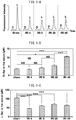
- FIG. 1-A is a typical chromatogram obtained by two-dimensional HPLC of D-/L-serine in the serum of C57BL/6J wild-type mice that underwent sham surgery or ischemia reperfusion treatment.
- markers were measured for 8 animals in a sham group and for 5, 9, 6 and 7 animals at 4, 8, 20 and 40 hours, respectively, after reperfusion.
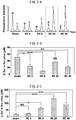
- the bar graphs of FIGS. 1-A to 1-F represent average values, while the error bars represent the standard error of the sample mean (SEM). Data of the examples was tested statistically by one way analysis of variance followed by Tukey's multiple comparison test.
- SEM standard error of the sample mean
- one asterisk (*) indicates a P value of less than 0.05
- two asterisks (**) indicate a P value of less than 0.01
- three asterisks (***) indicate a P value of less than 0.001.
- NS stands for not significant.
- the word "sham” in the drawings indicates concentrations in mice that underwent sham surgery, while IRI4, IRI8, IRI20 and IRI40 indicate concentrations in mice at 4, 8, 20 and 40 hours after reperfusion, respectively. Although there were significant fluctuations in serum D-serine concentrations at 4 and 8 hours after reperfusion in the C57BL/6J mice, concentrations increased at 20 hours and increased further at 40 hours ( FIG. 1-B ). Furthermore, the values of D-serine concentration indicated in FIG.
- a certain value is only indicated at a certain point in time after reperfusion in the case of a monotonically changing marker
- a certain value is not only indicated at a single point in time, but may also increase another time or a plurality of times more. Consequently, the stage of progression of renal failure cannot be uniquely estimated by the value of a marker.
- FIGS. 2-A to 2-J represent average values, while the error bars represent the standard error of the sample mean (SEM). Data of the examples was tested statistically by one way analysis of variance followed by Tukey's multiple comparison test.
- one asterisk (*) indicates a P value of less than 0.05
- two asterisks (**) indicate a P value of less than 0.01
- three asterisks (***) indicate a P value of less than 0.001.
- NS stands for not significant.
- FIGS. 3-A to 3-L indicate bar graphs of the ratio of the average value of the concentration of a D-form to the average value of the concentration of an L-form of individual mice for the mice at 4, 8, 20 and 40 hours after ischemia reperfusion treatment.
- [D-glutamic acid] and [L-glutamic acid] FIG. 3-F
- [D-allo-isoleucine] and [L-isoleucine] FIG. 3-J
- [D-phenylalanine] and [L-phenylalanine] FIG.
- FIGS. 4-A to 4-L indicate bar graphs of the percentages of the average values of concentrations of a D-form to the sum of the average values of the concentrations of an L-form and the average values of the concentrations of the D-form of individual mice for sham surgery mice and mice 4, 8, 20 and 40 hours after ischemia reperfusion treatment.
- FIG. 4-F [D-allo-isoleucine] and [L-isoleucine]
- FIG. 4-J [D-phenylalanine] and [L-phenylalanine]
- FIGS. 5-A to 5-R indicate bar graphs of the ratios of the average value of the concentration of a D-form to the average value of the concentration of an L-form in individual mice in sham surgery mice and mice at 4, 8, 20 and 40 hours after ischemia reperfusion treatment along with the results of investigating for the presence or absence of statistically significant differences.
- [D-allo-isoleucine] and [L-isoleucine] FIG. 5-J
- [D-phenylalanine] and [L-phenylalanine] FIG.
- FIG. 5-B [D-serine] and [L-serine]
- FIG. 5-C [D-arginine] and [L-arginine]
- FIG. 5-E [D-allo-threonine] and [L-threonine]
- FIG. 5-G [D-proline] and [L-proline]
- FIG. 5-H [D-lysine] and [L-lysine]
- FIG. 5-H and [D-lysine] and [L-lysine] ( FIG. 5-L ) can be used as a marker for renal failure having sensitivity that is equal to or greater than that of urine creatinine.
- one or a plurality of pathological index values selected from the group consisting of [D-histidine]/[L-histidine], [D-proline]/[L-proline] and [D-lysine]/[L-lysine] which demonstrate a significant difference of P ⁇ 0.001 with the sham surgery group at 4 hours after reperfusion, is capable of diagnosing renal failure with even higher sensitivity.
- pathological index values may be used alone, combining a plurality of pathological index values enables diagnoses having a
- [D-lysine] and [L-lysine] can be used as markers of renal failure having sensitivity that is equal to or greater than that of urine creatinine.
- a pathological index value of a subject is statistically significantly different from the pathological index reference value of a healthy individual group and statistically significantly different from the pathological index reference value of a renal failure patient group, and is between the pathological index reference value of a healthy individual group and the pathological index reference value of a renal failure patient group, the subject can be diagnosed as being suspected of early renal failure.
- renal failure can be diagnosed at an even earlier stage by using a pathological index value calculated from the concentration of one or a plurality of a pair of D-form and L-form of one or more amino acids selected from the group consisting of [D-histidine] and [L-histidine], [D-proline] and [L-proline] and [D-lysine] and [L-lysine], which exhibit a significant difference of p ⁇ 0.001 with a sham surgery group at 4 hours after reperfusion.
Description
- The present invention relates to a method for analyzing the blood or urine of a subject suspected of renal failure, and a system for analyzing a blood or urine sample of a subject suspected of renal failure. More particularly, the present invention relates to a method for analyzing the blood, plasma, serum or urine of a subject that comprises a step for measuring the concentration of a pair of D-form and L-form of at least one amino acid selected from the group consisting of [D-serine] and [L-serine] and the like in the blood, plasma, serum or urine of the subject, and to a system for analyzing a blood, plasma, serum or urine sample of the subject for performing the aforementioned analysis method, comprising a memory unit, an analysis and measurement unit, a data processing unit and a pathological information output unit.
- Chronic kidney disease (CKD) is a disease that affects 13.3 million Japanese, corresponding to roughly 13% of the Japanese adult population, and threatens the health of Japanese citizens due to the risk of progressing to end stage kidney disease (ESKD). Chronic kidney disease includes all pathological states in which depressed renal function as represented by glomerular filtration rate is found, or findings suggesting kidney damage persist in a chronic state (three months or longer). There is no effective treatment method for chronic kidney disease, and if chronic kidney disease progresses resulting in further depression of renal function, there is the risk of uremia, resulting in the need for artificial dialysis or kidney transplant, which will place a considerable burden on the patient in terms of health care costs (Non-Patent Document 1). Chronic kidney disease does not exhibit subjective symptoms. Diagnosis using markers for early diagnosis of renal failure is necessary for early diagnosis of chronic kidney disease and inhibition of its progression. However, there is currently no biomarker that is satisfactory in terms of accurately reflecting the progression of renal dysfunction at an earlier stage than the occurrence of changes in renal function as represented by glomerular filtration rate.
- An experimental animal model of acute kidney injury (AKI) is able to reproduce the early stages of renal dysfunction. Acute kidney injury is a disease in which renal function decreases rapidly over several weeks or several days. A known model of this disease is an acute kidney injury experimental model which is induced by a surgical procedure or administration of a drug. The "gold standards" for diagnosing acute kidney injury are urine production volume and serum creatinine concentration. Serum creatinine concentration is superior in that it can be evaluated without performing a biopsy regardless of the presence or absence of urination. However, glomerular filtration rate is required to be in a steady state. In a experimental animal model of acute kidney injury, which is not sensitive to small fluctuations in glomerular filtration rate, changes in glomerular filtration rate become apparent at a comparatively late stage. Since serum creatinine concentration also fluctuates due to conditions such as age, gender, muscle mass or medications being taken at the time, it cannot be a specific marker (Non-Patent Document 2). Reported examples of markers for acute kidney injury include neutrophil gelatinase-associated lipocalin (NGAL), interleukin-18 (IL-18), kidney injury molecule-1 (KIM-1), proteins such as fatty acid binding proteins or cystatin C, and metabolic low molecular weight compounds such as homovanillic acid sulfate or trimethylamine-N-oxide. However, none of these markers are detected in the early stage of renal failure.
- Since concentrations of D-serine and D-alanine in the serum of renal failure patients are higher than serum concentrations in normal individuals, and both the D-form concentrations and ratio of D-form concentration/(D-form concentration + L-form concentration) correlate with creatinine, these amino acids have been suggested to be candidates for markers of renal proximal tubular dysfunction (Non-Patent Document 3). It is also disclosed that D-amino acids (Ala, Pro, Ser) in the serum of nephritis patients tend to be elevated and have a correlation with creatinine level (Non-Patent Document 9). In addition, D-alanine, D-serine, D-glutamic acid and D-aspartic acid are observed in the serum of renal failure patients, and because of this, measurement of serum D-alanine concentration has been suggested to be useful in the diagnosis of renal failure (Non-Patent Document 5). D-serine and D-alanine concentration in the urine along with the ratio of the D-form to the total of the D-form and L-form were investigated in healthy individuals of various age groups, and it was suggested that processing of D-serine in the kidneys is different (Non-Patent Document 6). Although one or more amino acids selected from the group consisting of D-serine, D-threonine, D-alanine, D-asparagine, D-allo-threonine, D-glutamine, D-proline and D-phenylalanine were disclosed as being able to be used as pathological indicators of kidney disease (Patent Document 1). However, even though it is described in this document that a body fluid such as blood, plasma or urine is used as a specimen, blood is the only specimen used to determine pathological indicators for kidney disease in the examples, while there is nothing disclosed as to whether or not amino acids present in urine can be used as pathological indicators of kidney disease. It is also disclosed that the D-form ratios of alanine, valine, proline, threonine, aspartic acid and asparagine increase significantly in the urine of renal failure patients, while there is no significant differences with respect to methionine and serine (Non-Patent Document 7). However, in this document, although the ratio of the D-form concentration/(D-form concentration + L-form concentration) of each amino acid is calculated in renal failure patients for which pathological assessment criteria have not been indicated, increases in these ratios are merely indicated randomly (or vaguely) irrespective of disease stage, and there are no descriptions or suggestions as to the fact that fluctuations in these ratios correlate with pathological or other biomarkers of renal failure. After the priority date of the present application, urinary D/L-serine ratio was suggested to be able to be used as a biomarker capable of detecting early stage ischemic renal failure and classifying pathological stage since it decreases over time following ischemia reperfusion injury (Non-Patent Document 8). D-amino acid oxidase, which is involved in the decomposition of D-amino acids, is expressed in renal proximal tubules, and the enzyme activity of D-amino acid oxidase is known to decrease in ischemia reperfusion model rats (Non-Patent Document 4). L-serine is reabsorbed while D-serine is hardly reabsorbed at all under physiological conditions. However, the manner in which D-serine and other D-amino acids fluctuate in the early stage of renal failure is unknown.
- Patent Document 1: International Publication No.
WO 2013/140785 -
- Non-Patent Document 1: KDIGO 2012, Clinical Practice Guideline for the Evaluation and Management of Chronic Kidney Disease, Kidney International Supplements 1 (2013)
- Non-Patent Document 2: Slocum, J. L., et al, Transl. Res. 159: 277 (2012)
- Non-Patent Document 3: Fukushima, T., et al, Biol. Pharm. Bull. 18: 1130 (1995)
- Non-Patent Document 4: Zhang, H., et al, Amino Acids 42: 337 (2012)
- Non-Patent Document 5: Ishida, et al, Kitasato Medical Journal, 23: 51-62 (1993)
- Non-Patent Document 6: Yong Huang, et al, Biol. Pharm. Bull. 21: (2)156-162 (1998)
- Non-Patent Document 7: Magdalena C. Waldhier et al, Chromatography B (2010) 1103-1112
- Non-Patent Document 8: Jumpeji Sasabe et al, PLOS ONE, Vol. 9,
- Non-Patent Document 9: Nagata, Y., Viva Origino Vol. 18 (No. 2) (1990), 15th Academic Symposium, Collection of Abstracts
- There is a need to develop a technology for diagnosis of early stage renal failure by identifying a biomarker of renal failure that can be collected from urine or blood and fluctuates at an earlier stage than glomerular filtration rate and serum creatinine concentration.
- The present invention provides a method for analyzing the blood or urine of a subject suspected of renal failure. The analysis method of the present invention comprises a step for measuring the concentration of a pair of [D-serine] and [L-serine] and a step for calculating a pathological index value from the concentration of a pair of D-form and L-form of serine. Here, the pathological index value calculated from a pair of D-serine and L-serine enables to correlate a subject with the pathology of kidney disease in the case where a significant decrease in the proportion of the D-form is indicated. For example, the ratio of the concentration of the D-form to the concentration of the L-form of serine, or the ratio or percentage of the concentration of the D-form to the sum of the concentrations of the D-form and the L-form, can be calculated as the pathological index value of the subject. Although amino acids in the urine of renal failure patients whose pathological assessment criteria have not been indicated are analyzed in Non-Patent Document 7, it is disclosed that the D-amino acid ratio for alanine, valine, proline, threonine and aspartic acid is significantly increased. Thus, a decrease in the proportion of D-amino acids in the urine of renal failure patients is a surprising finding.
- In another instance described herein, which does not form part of the present invention, [D-glutamine] and [L-glutamine], [D-threonine] and [L-threonine], [D-allo-threonine] and [L-allo-threonine] and [D-leucine] and [L-leucine] may be included in the aforementioned amino acid group. Thus, the analysis method may comprise a step for measuring the concentration of a pair of D-form and L-form of at least one amino acid selected from the amino acid group consisting of [D-histidine] and [L-histidine], [D-asparagine] and [L-asparagine], [D-arginine] and [L-arginine], [D-allo-threonine] and [L-threonine], [D-glutamic acid] and [L-glutamic acid], [D-alanine] and [L-alanine], [D-proline] and [L-proline], [D-valine] and [L-valine], [D-allo-isoleucine] and [L-isoleucine], [D-phenylalanine] and [L-phenylalanine], [D-lysine] and [L-lysine], [D-glutamine] and [L-glutamine], [D-threonine] and [L-threonine], [D-allo-threonine] and [L-allo-threonine], and [D-leucine] and [L-leucine], and a step for calculating a pathological index value from the concentration of a pair of D-form and L-form of the at least one amino acid. Here, the pathological index value calculated from a pair of D-form and L-form of at least one amino acid enables to correlate a subject with the pathology of kidney disease, when a significant decrease in the proportion of the D-form is indicated. For example, the ratio of the concentration of the D-form to the concentration of the L-form of at least one amino acids, or the ratio or percentage of the concentration of the D-form to the sum of the concentrations of the D-form and the L-form, can be calculated as the pathological index value of the subject.
- Another mode of the present invention may also relate to a detection method for detecting renal failure, comprising a step for measuring the concentration of a pair of D-form and L-form of at least one amino acid selected from the amino acid group consisting of [D-serine] and [L-serine] in the urine of a subject, and
a step for calculating a pathological index value from the concentration of a pair of D-form and L-form of at least one amino acid that correlates a decrease in the proportion of a D-form with renal failure of the subject. - Here, the detection method relates to a method that may be performed by a non-physician such as a medical assistant or may be performed by an analysis facility.
- These pathological index values have been indicated to decrease following renal ischemia reperfusion in the same manner as urine creatinine concentration. Since a decrease in urine creatinine concentration indicates depression of renal function, it can be used as a marker of renal failure, and a pathological index value of the present application can also be used as a marker of renal failure in the same manner as urine creatinine concentration. Furthermore, urine renal failure markers in the form of KIM-1 and NGAL demonstrate increases in urine concentration in response to depressed renal function. In an experimental model of ischemia reperfusion, although urine creatinine concentration decreased significantly 8 hours after ischemia reperfusion, urine KIM-1 increases significantly 20 hours after ischemia reperfusion and urine NGAL increased significantly 8 hours after ischemia reperfusion (
FIGS. 2-D, 2-F and2-G ). Since a pathological index value calculated using the concentration of a pair of [D-serine] and [L-serine] decreases significantly at a level of significance of P<0.05 4 hours after renal ischemia reperfusion, it can be said to have higher sensitivity for renal failure. Thus, this pathological index value is preferably used from the viewpoint of having higher sensitivity for early stage renal failure. Another pathological index value, which does not form part of the present invention, calculated using the concentration of a pair of D-form and L-form of one or more amino acids selected from the group consisting of [D-histidine] and [L-histidine], [D-asparagine] and [L-asparagine], [D-proline] and [L-proline], and [D-lysine] and [L-lysine] decreases significantly at a level of significance of P<0.01 4 hours after renal ischemia reperfusion, this pathological index value can be used as a marker having higher sensitivity. Furthermore, another pathological index value, which does not form part of the present invention, calculated from concentration of a pair of D-form and L-form of one or more amino acids selected from the group consisting of [D-histidine] and [L-histidine], [D-proline] and [L-proline], and [D-lysine] and [L-lysine] decreases significantly at a level of significance of P<0.001 4 hours after renal ischemia reperfusion, this pathological index value can be used as a marker having extremely high sensitivity. Thus, the pathological index value of the present invention has sensitivity that is equal to or higher than that of conventional renal failure markers such as serum creatinine, and can be used for early diagnosis of renal failure. Examples of early stage renal failure include a state in which serum creatinine has increased by 1.5 to 2.0 times, which is the criterion for the early stage of each of the RIFLE, AKIN and KDIGO classifications of acute kidney injury (AKI), a state wherein renal function reduction in chronic kidney disease (CKD) indicated by GFR is started, or an extremely early state prior to the appearance of changes in serum creatinine or GFR even though renal failure is present. In addition, another pathological index value, which does not form part of the present invention, calculated using the concentration of a pair of D-form and L-form of one or more amino acids selected from the group consisting of [D-glutamic acid] and [L-glutamic acid], [D-phenylalanine] and [L-phenylalanine], [D-valine] and [L-valine], [D-glutamine] and [L-glutamine], [D-threonine] and [L-threonine], [D-leucine] and [L-leucine], [D-allo-isoleucine] and [L-isoleucine], and [D-allo-threonine] and [L-threonine], which do not exhibit asignificant difference 4 hours after renal ischemia reperfusion but exhibit asignificant difference 8 hours after renal ischemia reperfusion, can be used as a marker having sensitivity equal to that of conventional markers such as creatinine. - According to the present disclosure, which does not form part of the present invention, the pathological index value calculated from the concentration of D-form and L-form of one amino acid may be used, or the pathological index value calculated from the concentration of another D-form and L-form of another amino acid can be used in combination therewith. For example, a pathological index value having extremely high sensitivity calculated from the concentration of D-form and L-form of one or more amino acids selected from the group consisting of [D-histidine] and [L-histidine], [D-proline] and [L-proline], and [D-lysine] and [L-lysine] may be combined with another pathological index value having extremely high sensitivity calculated from the concentration of a D-form and L-form of an amino acid selected from the same group. In addition, in a different mode, which does not form part of the present invention, the aforementioned pathological index value having extremely high sensitivity can be used in combination with a pathological index value having a lower sensitivity, for example, a pathological index value calculated from the concentration of a D-form and L-form of one amino acid selected from the group consisting of [D-glutamic acid] and [L-glutamic acid], [D-phenylalanine] and [L-phenylalanine], [D-valine] and [L-valine], [D-glutamine] and [L-glutamine], [D-threonine] and [L-threonine], [D-leucine] and [L-leucine], [D-allo-isoleucine] and [L-isoleucine], and [D-allo-threonine] and [L-threonine], which enables sensitively detecting kidney injury in comparison with urine creatinine, urine KIM-1 or urine NGAL in a state in which depressed renal function, in which serum creatinine has increased by a factor of 1.5 to 2.0, has occurred or a state prior thereto.
- The analysis method of the present invention may further comprise a step for determining whether the pathological index value of a subject is similar to the pathological index reference value of a healthy individual, whether the pathological index value of a subject is similar to the pathological index reference value of an acute renal failure patient or chronic renal failure patient, or whether the pathological index value of a subject is between the pathological index reference value of a healthy individual and the pathological index reference value of a chronic renal failure patient, by comparing the pathological index value of the subject with the pathological index reference value of a healthy individual and the pathological index reference value of an acute renal failure patient and/or chronic renal failure patient.
- In still another mode, the present invention makes it possible to determine whether a subject suffers renal failure by preliminarily setting a threshold value based on the pathological index values of a healthy individual group and/or renal failure patient group, and then comparing the pathological index value of a subject with the threshold value. A person with ordinary skill in the art is able to suitably set a threshold value from the pathological index values of a healthy individual group and renal failure patient group. Although the average value, median value or X percentile value of the healthy individual group or renal failure patient group can be used for the threshold value, the threshold value is not limited thereto. Here, an arbitrary numerical value can be selected for X, and a value of 3, 5, 10, 15, 20, 30, 40, 60, 70, 80, 85, 90, 95 or 97 can be suitably used. Only one threshold value may be used or a plurality of threshold values can be set depending on the type of renal failure (acute or chronic), the cause thereof (such as drug-induced nephropathy, diabetic nephropathy, IgA nephropathy, membranous nephropathy or nephrosclerosis), disease state (early, intermediate or late) and the amino acids or combination thereof used. The pathology of the renal failure of a subject can be assessed, determined or diagnosed by comparing the pathological index value of the subject with a preset threshold value.
- The analysis method of the present invention may further comprise a step for determining whether a first pathological index value of a subject, calculated from the concentration of [D-serine] and [L-serine] in the blood, plasma, serum or urine of the subject, is between the pathological index reference value of a healthy individual and the pathological index reference value of a chronic renal failure patient, and whether the second pathological index value of the subject, calculated from the concentration of a pair of D-form and L-form of at least one amino acid among [D-glutamic] and [L-glutamic acid], [D-allo-isoleucine] and [L-isoleucine] and [D-phenylalanine] and [L-phenylalanine] in the blood, plasma, serum or urine of the subject, is between a pathological index reference value of the healthy individual and the pathological index reference value of the chronic renal failure patient.
- In the present invention, the pathological index value of a subject calculated from the concentration of [D-serine] and [L-serine] can be used as a first pathological index value, while the pathological index value of the subject calculated from the concentration of a pair of D-form and L-form of at least one amino acid selected from the group consisting of [D-glutamic acid] and [L-glutamic acid], [D-allo-isoleucine] and [L-isoleucine], [D-phenylalanine] and [L-phenylalanine], [D-valine] and [L-valine], and [D-allo-threonine] and [L-threonine] can be used as a second pathological index value.
- The pathological index value of the subject calculated from the concentration of [D-serine] and [L-serine] can be used for the first pathological index value. On the other hand, the pathological index value calculated from the concentration of a pair of D-form and L-form of at least one amino acid selected from the group consisting of [D-glutamine] and [L-glutamine], [D-threonine] and [L-threonine], and [D-leucine] and [L-leucine] in addition to [D-glutamic acid] and [L-glutamic acid], [D-allo-isoleucine] and [L-isoleucine], [D-phenylalanine] and [L-phenylalanine], and [D-valine and L-valine] can be used for the second pathological index value.
- The present invention provides a system for analyzing a blood or urine sample of a subject suspected of renal failure. The sample analysis system of the present invention comprises a memory unit, an analysis and measurement unit, a data processing unit and a pathological information output unit. The aforementioned memory unit stores the pathological index reference values for the blood, plasma, serum or urine of healthy individuals, and the pathological index reference values for the blood, plasma, serum or urine of acute renal failure patients and/or chronic renal failure patients. The aforementioned analysis and measurement unit separates and quantifies D-serine and L-serine stereoisomers present in the blood, plasma, serum or urine of the aforementioned subject. The aforementioned data processing unit performs a step for calculating the pathological index value of the aforementioned subject from the concentration of [D-serine] and [L-serine]. Here, the pathological index value calculated from the concentration of a pair of D-form and L-form of serine enables a subject to be correlated with renal failure in the case of indicating a decrease in the composition ratio of the D-form.
- In a different instance described herein, which does not form part of the present invention, D-glutamine and L-glutamine], D-threonine and L-threonine], [D-allo-threonine and L-allo-threonine and D-leucine and L-leucine may be included in addition to the aforementioned amino acid pairs. Thus, in a different mode, the aforementioned analysis and measurement unit separates and quantifies at least one pair of amino acid stereoisomers present in the blood, plasma, serum or urine of the aforementioned subject, selected from the group consisting of D-histidine and L-histidine, D-asparagine and L-asparagine, D-arginine and L-arginine, D-allo-threonine and L-threonine, D-glutamic acid and L-glutamic acid, D-alanine and L-alanine, D-proline and L-proline, D-valine and L-valine, D-allo-isoleucine and L-isoleucine, D-phenylalanine and L-phenylalanine, D-lysine and L-lysine, D-glutamine and L-glutamine, D-threonine and L-threonine, D-allo-threonine and L-allo-threonine and D-leucine and L-leucine. The aforementioned data processing unit performs a step for calculating the pathological index value of the aforementioned subject from the concentration of a pair of D-form and L-form of at least one amino acid selected from the group consisting of [D-histidine] and [L-histidine], [D-asparagine] and [L-asparagine], [D-arginine] and [L-arginine], [D-allo-threonine] and [L-threonine], [D-glutamic acid] and [L-glutamic acid], [D-alanine] and [L-alanine], [D-proline] and [L-proline], [D-valine] and [L-valine], [D-allo-isoleucine] and [L-isoleucine], [D-phenylalanine] and [L-phenylalanine], [D-lysine] and [L-lysine], [D-glutamine] and [L-glutamine], [D-threonine] and [L-threonine], [D-allo-threonine] and [L-allo-threonine] and [D-leucine] and [L-leucine]. Here, the pathological index value calculated from the concentration of a pair of D-form and L-form of at least one amino acid enables a subject to be correlated with renal failure in the case of indicating a decrease in the composition ratio of the D-form.
- The pathological index value refers to a value that can be calculated from the concentration of a D-form and L-form of serine, and enables a subject to be correlated with renal failure in the case of indicating a decrease in the composition ratio of the D-form. In the sample analysis system of the present invention, for example, in relation to a D-form and L-form pair, the ratio of the concentration of the D-form to the concentration of the L-form, and the ratio or percentage of the concentration of a D-form to the sum of the concentrations of the D-form and L-form, is defined as the pathological index value of a subject. When the pathological index value of the subject is compared with the pathological index reference value of a healthy individual and the pathological index reference value of a patient with acute renal failure and/or chronic renal failure, and the pathological index value of the subject is similar to the pathological index reference value of the healthy individual, information that the aforementioned subject is hardly suspected of renal failure is defined as pathological information of the aforementioned subject. When the pathological index value of the aforementioned subject is similar to the pathological index reference value of the aforementioned chronic renal failure patient, information that the aforementioned subject is suspected of renal failure is defined as pathological information of the aforementioned subject. When the pathological index value of the aforementioned subject is between the pathological index reference value of the aforementioned healthy individual and the pathological index reference value of the aforementioned chronic renal failure patient, information that the aforementioned subject is suspected of early stage renal failure is defined as pathological information of the aforementioned subject. The aforementioned pathological information output unit outputs the pathological information of the aforementioned subject.
- In the sample analysis system of the present invention, when a first pathological index value of a subject calculated from the concentration in the blood, plasma, serum or urine of the aforementioned subject of [D-serine] and [L-serine] is between the pathological index reference value of the aforementioned healthy individual and the pathological index reference value of the aforementioned chronic renal failure patient, and a second pathological index value of the aforementioned subject calculated from the concentration in the blood, plasma, serum or urine of the aforementioned subject of a pair of D-form and L-form of at least one amino acid selected from the group consisting of[D-glutamic acid] and [L-glutamic acid], [D-allo-isoleucine] and [L-isoleucine], and [D-phenylalanine] and [L-phenylalanine] is between the pathological index reference value of the aforementioned healthy individual and the pathological index reference value of the aforementioned chronic renal failure patient, information that the aforementioned subject is suspected of extremely early stage renal failure is defined as pathological information of the aforementioned subject.
- The present invention provides a method for diagnosing renal failure. The diagnostic method of the present invention comprises a step for measuring the concentration in the blood, plasma, serum or urine of a subject suspected of renal failure of a pair of [D-serine] and [L-serine] and a step for calculating a pathological index value from the aforementioned concentration of the pair of D-form and L-form of aforementioned serine.
- In a different instance of the diagnostic method described herein, which does not form part of the diagnostic method of the present invention, [D-glutamine] and [L-glutamine], [D-threonine] and [L-threonine], and [D-leucine] and [L-leucine] may be included in the aforementioned amino acid group. Thus, the diagnostic method comprises a step for measuring the concentration in the blood, plasma, serum or urine of a subject suspected of renal failure of a pair of D-form and L-form of at least one amino acid selected from the amino acid group consisting of [D-histidine] and [L-histidine], [D-asparagine] and [L-asparagine], [D-arginine] and [L-arginine], [D-allo-threonine] and [L-threonine], [D-glutamic acid] and [L-glutamic acid], [D-alanine] and [L-alanine], [D-proline] and [L-proline], [D-valine] and [L-valine], [D-allo-isoleucine] and [L-isoleucine], [D-phenylalanine] and [L-phenylalanine], [D-lysine] and [L-lysine], [D-glutamine] and [L-glutamine], [D-threonine] and [L-threonine], and [D-leucine] and [L-leucine], and a step for calculating a pathological index value from the aforementioned concentration of the pair of D-form and L-form of aforementioned at least one amino acid.
- Here, the pathological index value calculated from the concentration of a pair of D-form and L-form of at least one amino acid enables a subject to be correlated with renal failure in the case of indicating a decrease in the composition ratio of the D-form. The ratio of the concentration of the D-form to the concentration of the L-form of at least one amino acid pair, or the ratio or percentage of the concentration of the D-form to the sum of the concentrations of D-form and L-form, can be used as the pathological index value of the aforementioned subject.
- The diagnostic method of the present invention may further comprise a step for diagnosing that the aforementioned subject has a high likelihood of being a healthy individual or is hardly suspected of renal failure when the pathological index value of the aforementioned subject is compared with the pathological index reference value of the healthy individual and the pathological index reference value of a patient with acute renal failure and/or chronic renal failure, and the pathological index value of the aforementioned subject is similar to the pathological index reference value of the healthy individual; the aforementioned subject is diagnosed as being strongly suspected of renal failure when the pathological index value of the aforementioned subject is similar to the pathological index reference value of the aforementioned patient having acute renal failure or chronic renal failure; and the aforementioned subject is diagnosed as being suspected of early stage renal failure when the pathological index value of the aforementioned subject is between the pathological index reference value of the healthy individual and the pathological index reference value of the patient having acute renal failure or chronic renal failure.
- The diagnostic method of the present invention comprises a step for diagnosing that a subject is suspected of extremely early stage renal failure when a first pathological index value of a subject calculated from the concentration in the blood, plasma, serum or urine of the aforementioned subject of [D-serine] and [L-serine] is between the pathological index reference value of the aforementioned healthy individual and the pathological index reference value of the aforementioned chronic renal failure patient, and a second pathological index value of the aforementioned subject calculated from the concentration in the blood, plasma, serum or urine of the aforementioned subject of a pair of D-form and L-form of at least one amino acid selected from the group consisting of [D-glutamic acid] and [L-glutamic acid], [D-allo-isoleucine] and [L-isoleucine], and [D-phenylalanine] and [L-phenylalanine] is between the pathological index reference value of the aforementioned healthy individual and the pathological index reference value of the aforementioned chronic renal failure patient.
- The present invention provides a method for treating renal failure. The treatment method of the present invention comprises a step for measuring the concentration in the blood, plasma, serum or urine of a subject suspected of renal failure of a pair of D-form and L-form of at least one amino acid selected from the group consisting of [D-serine] and [L-serine], [D-histidine] and [L-histidine], [D-asparagine] and [L-asparagine], [D-arginine] and [L-arginine], [D-allo-threonine] and [L-threonine], [D-glutamic acid] and [L-glutamic acid], [D-alanine] and [L-alanine], [D-proline] and [L-proline], [D-valine] and [L-valine], [D-allo-isoleucine] and [L-isoleucine], [D-phenylalanine] and [L-phenylalanine], and [D-lysine] and [L-lysine], a step for calculating a pathological index value from the aforementioned concentration of a pair of D-form and L-form of at least one amino acid, a step for diagnosing that the aforementioned subject has a high likelihood of being a healthy individual or is hardly suspected of renal failure when the pathological index value of the aforementioned subject is compared with the pathological index reference value of the healthy individual and the pathological index reference value of a patient with acute renal failure and/or chronic renal failure and the pathological index value of the aforementioned subject is similar to the pathological index reference value of the healthy individual, the aforementioned subject is diagnosed as being strongly suspected of renal failure when the pathological index value of the aforementioned subject is similar to the pathological index reference value of the aforementioned patient having acute renal failure or chronic renal failure, and the aforementioned subject is diagnosed has being suspected of early stage renal failure when the pathological index value of the aforementioned subject is between the pathological index reference value of the healthy individual and the pathological index reference value of the patient having acute renal failure or chronic renal failure, and a step for treating the aforementioned subject when the aforementioned subject has been diagnosed as being strongly suspected of renal failure by administering a therapeutic drug that inhibits the progression or improves renal failure, including antihypertensive drugs including, but not limited to, angiotensin-converting enzymes and angiotensin II receptor antagonists, antidiabetic drugs including, but not limited to, α-glucosidase inhibitors and insulin preparations, antidyslipidemic drugs including, but not limited to, HMG-CoA reductase inhibitors and intestinal cholesterol transporter inhibitors, antianemic drugs including, but not limited to, recombinant human erythropoietin preparations, therapeutic drugs for bone and mineral metabolic disorders, therapeutic drugs for hyperuricemia, and therapeutic drugs for uremic toxins including, but not limited to, spherical adsorbent carbon medications.
- The treatment method comprises a step for diagnosing that a subject is suspected of extremely early stage renal failure when a first pathological index value of the aforementioned subject calculated from the concentration in the blood, plasma, serum or urine of the aforementioned subject of a pair of D-form and L-form of at least one amino acid selected from the group consisting of [D-serine] and [L-serine], [D-histidine] and [L-histidine], [D-asparagine] and [L-asparagine], [D-arginine] and [L-arginine], [D-allo-threonine] and [L-threonine], [D-alanine] and [L-alanine], [D-proline] and [L-proline], [D-valine] and [L-valine], and [D-lysine] and [L-lysine] is between the pathological index reference value of the aforementioned healthy individual and the pathological index reference value of the aforementioned chronic renal failure patient, and a second pathological index value of the aforementioned subject calculated from the concentration in the blood, plasma, serum or urine of the aforementioned subject of a pair of D-form and L-form of at least one amino acid selected from [D-glutamic acid] and [L-glutamic acid], [D-allo-isoleucine] and [L-isoleucine], and [D-phenylalanine] and [L-phenylalanine] is between the pathological index reference value of the aforementioned healthy individual and the pathological index reference value of the aforementioned chronic renal failure patient, and a step for treating the aforementioned subject by administering a therapeutic drug for hyperkalemia, including, but not limited to, sodium polystyrene sulfonate, and a therapeutic drug for hyperphosphatemia, including, but not limited to, calcium carbonate and calcium acetate.
- Measurement of D-amino acid concentration in blood, plasma, serum or urine in the present invention may be performed using any method commonly known among persons with ordinary skill in the art. For example, a method consisting of preliminarily stereospecifically derivatizing D- and L-amino acids with o-phthaldehyde (OPA), N-tert-butyloxycarbonyl-L-cysteine (Boc-L-Cys) or other modifying reagent, followed by separating a mixture of 100 mM acetate buffer (pH 6.0) and acetonitrile by gradient elution using an analytical column in the manner of ODS-80TsQA can be used to simultaneous measure the D-forms and L-forms of aspartic acid, serine and alanine. In addition, a method consisting of preliminarily derivatizing D- and L-amino acids with a fluorescent reagent in the manner of 4-fluoro-7-nitro-2,1,3-benzoxadiazole (NBD-F) and then stereospecifically separating each amino acid using an analytical column in the manner of ODS-80TsQA or Mightysil RP-18GP, followed by stereospecifically separating by optically resolving using a Pirkle chiral stationary phase column (such as the Sumichiral OA-2500S or R), can be used to measure trace amounts of proline, leucine and other amino acids (Hamase, K. and Zaitsu, K.: Analytical Chemistry, Vol. 53, 677-690 (2004)). An optical resolution column system in the present description refers to a separation analysis system that at least uses an optical resolution column, and may include separation analyses using an analytical column other than an optical resolution column. More specifically, the concentrations of D- and L-amino acids in a sample can be measured by using an optical isomer analysis method comprising a step for passing a sample containing components having optical isomers through a stationary phase in the form of a first column packing material together with a mobile phase in the form of a first liquid to separate the aforementioned components of the aforementioned sample, a step for individually retaining each of the aforementioned components of the aforementioned sample in a multi-loop unit, a step for supplying each of the aforementioned components of the aforementioned sample individually retained in the aforementioned multi-loop unit by passing the flow path through a stationary phase in the form of a second column packing material having an optically active center together with a mobile phase in the form of a second liquid to resolve the aforementioned optical isomers contained in each component of the aforementioned sample, and a step for detecting the aforementioned optical isomers contained in each component of the aforementioned sample (Japanese Patent No.
4291628 2008-27650 - In the present description, notations of amino acids enclosed in brackets ([]) (such as [D-serine]) refer to the concentration of that amino acid. In the present invention, the ratio of the concentration of a D-form to the concentration of an L-form and the percentage of the concentration of a D-form to the sum of the concentrations of a D-form and an L-form, for example, are used as parameters (pathological index values) based on the concentration of an amino acid. The volume of a liquid in the manner of blood, serum, plasma or urine is reduced in order to divide the concentration of a certain substance by the concentration of another substance. Consequently, differing from the case of concentration, these parameters offer the advantage of eliminating the need for correction for liquid volume.
- In the present invention, the function of the kidneys, which produce urine by specifically removing only a portion of the components in blood by filtering and reabsorbing blood, play an important role. Consequently, although amino acid concentrations in the present invention may differ greatly between blood and urine, the differences between blood, serum and plasma are not that large. This is because amino acids are not known to be specifically concentrated in blood cells or blood clots and the like. Therefore, although serum concentrations are measured in examples of the present invention and parameters based on serum concentrations are used for the pathological index values of subjects or the pathological index reference values of healthy individuals and renal failure patients, blood concentrations or plasma concentrations may be measured instead of serum concentrations, and parameters based on blood concentrations or plasma concentrations may be used for the pathological index values of subjects and the pathological index reference values of healthy individuals and renal failure patients.
- In the present description, a "pathological index value" refers to a numerical value able to be calculated based on the concentrations of a plurality of biomarkers and not on the concentration of individual biomarker molecules. A pathological index value used in the present description can be calculated from the concentration of a certain D-form and L-form of at least one amino acid//, and refers to a value that enables a subject to be correlated with renal failure in the case of indicating a decrease in the composition ratio of the D-form. Pathological index values include, but are not limited to, the concentration ratio between a certain amino acid and an enantiomer thereof, such as the ratio of the concentration of a D-form to the concentration of the L-form of a certain amino acid, and the percentage of the concentration of a D-form to the sum of the concentrations of the D-form and L-form.
- In the present description, a "pathological index reference value" refers to the average value or median value of the pathological index value of a biomarker molecule obtained for healthy individuals and acute renal failure and/or chronic renal failure patients diagnosed by a known diagnostic technique. Although a pathological index value is a numerical value of a specific subject at a specific point in time, a pathological index reference value is a numerical value obtained by statistical processing from a plurality of healthy individuals and patients with acute renal failure and/or chronic renal failure. Thus, a certain pathological index value being similar to a certain pathological index reference value refers to the absence of a significant difference between the pathological index value and the aforementioned pathological index reference value. For example, a statistical technique such as the two-tailed Student's t-test, one-way analysis of variance or Tukey's multiple comparison test can be used. In addition, a significance threshold P value of less than 0.05 constitutes significance in these tests.
- The system for analyzing the blood, plasma, serum or urine of a subject in the present invention comprises a memory unit, analysis and measurement unit, data processing unit and pathological information output unit. Here, the memory unit contains memory that stores pathological index reference values, obtained from parameters based on enantiomer concentration data of amino acids present in blood, plasma, serum or urine, obtained for healthy individuals and patients with acute renal failure and/or chronic renal failure diagnosed by a known diagnostic technique. The aforementioned storage unit may contain significance threshold P data obtained by statistical processing from the number of the aforementioned healthy individuals and patients and individual data. The aforementioned analysis and measurement unit contains an automated analyzer, capable of automatically operating a two-dimensional HPLC system by remote control that measures amino acid enantiomer concentration explained in the present description, and a central control device for controlling the automated analyzer. The aforementioned data processing unit calculates parameters explained in the present description from amino acid enantiomer concentrations obtained with the aforementioned analysis and measurement unit. This unit also compares a pathological index value of a subject obtained from these parameters with pathological index reference values of healthy individuals and patients recalled from the aforementioned memory unit. When the pathological index value of the aforementioned subject is similar to the pathological index reference value of the aforementioned healthy individuals, information that the aforementioned subject is weakly suspected of renal failure is defined as pathological information of the aforementioned subject. When the pathological index value of the aforementioned subject is similar to the pathological index reference value of the aforementioned chronic renal failure patients, information that the aforementioned subject is suspected of renal failure is defined as pathological information of the aforementioned subject. When the pathological index value of the aforementioned subject is between the pathological index reference value of the aforementioned healthy individuals and the pathological index reference value of the aforementioned chronic renal failure patients, information that the aforementioned subject is suspected of early stage renal failure is defined as pathological information of the aforementioned subject. At this time, the aforementioned significance threshold P value may be recalled from the aforementioned memory unit and used to determine the degree to which the pathological index value of the aforementioned subject is similar to the pathological index reference value of the aforementioned healthy individuals or patients. The data processing unit contains a computer of the prior art and pathological information processing software stored in the computer. The aforementioned pathological information output unit may display the pathological information of the aforementioned subject on a liquid crystal or other display screen, output the information to a printer for printout, or transmit the pathological information of the subject via the Internet or LAN and the like as data.
- In the present invention, analyzed or detected renal failure refers to a state in which renal function is depressed below that when normal, and includes all forms of kidney damage used in the normal sense of the word. Although not limited thereto, renal failure generally refers to a state in which renal function is below 30% of normal renal function, and is broadly classified into acute renal failure and chronic renal failure. Examples of the causes of depressed renal function include multiple factors such as immune system abnormalities or drug allergies, hypertension, diabetes, hemorrhage or sudden drop in blood pressure, infection or dehydration accompanying burns. Classifications of disease stage in the manner of the RIFLE classification, AKIN classification or KDIGO classification have been advocated for acute renal failure (AKI), and acute renal failure has been classified as risk (stage 1), injury (stage 2) and failure (stage 3), and as loss and end-stage kidney disease corresponding to the duration thereof. These classifications all use serum creatinine level and urine volume as indicators, and the diagnostic criterion in the case of risk (stage 1), for example, consists of a 1.5-fold to 2.0-fold increase in serum creatinine from the baseline or urine volume of less than 0.5 ml/kg/hr persisting for 6 hours or more, that in the case of injury (stage 2) consists of a 2.0-fold to 3.0-fold increase in serum creatinine from the baseline or urine volume of less than 0.5 ml/kg/hr persisting for 12 hours or more, while that in the case of failure (stage 3) consists of a 3.0-fold or more increase in serum creatinine from the baseline or urine volume of less than 0.3 ml/kg/hr persisting for 24 hours or more. On the other hand, these classifications are able to more accurately classify acute renal failure by using in combination with other indicators such as the amount of change in GFR. Diagnostic criteria for disease stage 1 (normal renal function although kidney damage is present, eGFR ≥ 90) to disease stage 5 (renal failure, eGFR < 15) are indicated for chronic kidney disease (CKD) in guidelines of the Japanese Society of Nephrology (2009). Here, estimated glomerular filtration rate (eGFR), used as an indicator, is calculated with serum creatinine level based on age and gender, and indicates the capacity of the kidney to discharge body waste into urine. In the analysis and test method of the present invention, depression of renal function can be detected with higher sensitivity than conventional renal function markers. Thus, subjects can be classified into a risk group that was not classified as renal failure using conventional markers, and for example, depression of renal function can even be detected in a high risk group having risk factors for AKI or CKD in the manner of the previously described causes, but for which there are no well-defined fluctuations observed for serum creatinine or GFR.
- In the present invention, the subject is not limited to a human, but rather can include experimental animals such as mice, rats, rabbits, dogs or monkeys. Thus, a subject may also be represented as a subject.
- The analysis method of the present invention can be used to gather preliminary data for a method for diagnosing chronic renal failure and/or acute renal failure. Although a physician can diagnose chronic renal failure and/or acute renal failure using such preliminary data, this analysis method may also be performed by a non-physician such as a medical assistant, or can be performed by an analysis facility and the like. Thus, the analysis method of the present invention can also be said to be a preliminary diagnostic method.
-
-
FIG. 1-A is a typical chromatogram obtained by two-dimensional HPLC of D-/L-serine in the serum of C57BL/6J wild-type mice that underwent sham surgery or ischemia reperfusion treatment. -
FIG. 1-B is a graph showing changes in D-serine concentration in the serum of C57BL/6J wild-type mice that underwent renal ischemia reperfusion treatment. -
FIG. 1-C is a graph showing changes in L-serine concentration in the serum of C57BL/6J wild-type mice that underwent renal ischemia reperfusion treatment. -
FIG. 1-D is a graph showing changes in the ratio of D-serine concentration to L-serine concentration in the serum of C57BL/6J wild-type mice that underwent renal ischemia reperfusion treatment. -
FIG. 1-E is a graph showing changes in creatinine concentration in the serum of C57BL/6J wild-type mice that underwent renal ischemia reperfusion treatment. -
FIG. 1-F is a graph showing changes in cystatin C concentration in the serum of C57BL/6J wild-type mice that underwent renal ischemia reperfusion treatment. -
FIG. 2-A is a typical chromatogram obtained by two-dimensional HPLC of D-/L-serine concentration in the urine of C57BL/6J wild-type mice that underwent sham surgery and ischemia reperfusion treatment. -
FIG. 2-B is a graph showing changes in D-serine concentration in the urine of C57BL/6J wild-type mice that underwent renal ischemia reperfusion treatment. -
FIG. 2-C is a graph showing changes in L-serine concentration urine of C57BL/6J wild-type mice that underwent renal ischemia reperfusion treatment. -
FIG. 2-D is a graph showing changes in creatinine concentration urine of C57BL/6J wild-type mice that underwent renal ischemia reperfusion treatment. -
FIG. 2-E is a graph showing changes in the ratio of D-serine concentration to L-serine concentration urine of C57BL/6J wild-type mice that underwent renal ischemia reperfusion treatment. -
FIG. 2-F is a graph showing changes in KIM-1 concentration urine of C57BL/6J wild-type mice that underwent renal ischemia reperfusion treatment. -
FIG. 2-G is a graph showing changes in NGAL concentration urine of C57BL/6J wild-type mice that underwent renal ischemia reperfusion treatment. -
FIG. 3-A is a graph showing changes in the ratio of D-histidine concentration to L-histidine concentration in the urine of C57BL/6J wild-type mice that underwent renal ischemia reperfusion treatment. -
FIG. 3-B is a graph showing changes in the ratio of D-asparagine concentration to L-asparagine concentration in the urine of C57BL/6J wild-type mice that underwent renal ischemia reperfusion treatment. -
FIG. 3-C is a graph showing changes in the ratio of D-serine concentration to L-serine concentration in the urine of C57BL/6J wild-type mice that underwent renal ischemia reperfusion treatment. -
FIG. 3-D is a graph showing changes in the ratio of D-arginine concentration to L-arginine concentration in the urine of C57BL/6J wild-type mice that underwent renal ischemia reperfusion treatment. -
FIG. 3-E is a graph showing changes in the ratio of D-allo-threonine concentration to L-threonine concentration in the urine of C57BL/6J wild-type mice that underwent renal ischemia reperfusion treatment. -
FIG. 3-F is a graph showing changes in the ratio of D-glutamic acid concentration to L-glutamic acid concentration in the urine of C57BL/6J wild-type mice that underwent renal ischemia reperfusion treatment. -
FIG. 3-G is a graph showing changes in the ratio of D-alanine concentration to L-alanine concentration in the urine of C57BL/6J wild-type mice that underwent renal ischemia reperfusion treatment. -
FIG. 3-H is a graph showing changes in the ratio of D-proline concentration to L-proline concentration in the urine of C57BL/6J wild-type mice that underwent renal ischemia reperfusion treatment. -
FIG. 3-I is a graph showing changes in the ratio of D-valine concentration to L-valine concentration in the urine of C57BL/6J wild-type mice that underwent renal ischemia reperfusion treatment. -
FIG. 3-J is a graph showing changes in the ratio of D-allo-isoleucine concentration to L-isoleucine concentration in the urine of C57BL/6J wild-type mice that underwent renal ischemia reperfusion treatment. -
FIG. 3-K is a graph showing changes in the ratio of D-phenylalanine concentration to L-phenylalanine concentration in the urine of C57BL/6J wild-type mice that underwent renal ischemia reperfusion treatment. -
FIG. 3-L is a graph showing changes in the ratio of D-lysine concentration to L-lysine concentration in the urine of C57BL/6J wild-type mice that underwent renal ischemia reperfusion treatment. -
FIG. 4-A is a graph showing changes in the percentage of D-histidine concentration to the sum of L-histidine concentration and D-histidine concentration in the urine of C57BL/6J wild-type mice that underwent renal ischemia reperfusion treatment. -
FIG. 4-B is a graph showing changes in the percentage of D-asparagine concentration to the sum of L-asparagine concentration and D-asparagine concentration in the urine of C57BL/6J wild-type mice that underwent renal ischemia reperfusion treatment. -
FIG. 4-C is a graph showing changes in the percentage of D-serine concentration to the sum of L-serine concentration and D-serine concentration in the urine of C57BL/6J wild-type mice that underwent renal ischemia reperfusion treatment. -
FIG. 4-D is a graph showing changes in the percentage of D-arginine concentration to the sum of L-arginine concentration and D-arginine concentration in the urine of C57BL/6J wild-type mice that underwent renal ischemia reperfusion treatment. -
FIG. 4-E is a graph showing changes in the percentage of D-allo-threonine concentration to the sum of L-threonine concentration and D-allo-threonine concentration in the urine of C57BL/6J wild-type mice that underwent renal ischemia reperfusion treatment. -
FIG. 4-F is a graph showing changes in the percentage of D-glutamic acid concentration to the sum of L-glutamic acid concentration and D-glutamic acid concentration in the urine of C57BL/6J wild-type mice that underwent renal ischemia reperfusion treatment. -
FIG. 4-G is a graph showing changes in the percentage of D-alanine concentration to the sum of L-alanine concentration and D-alanine concentration in the urine of C57BL/6J wild-type mice that underwent renal ischemia reperfusion treatment. -
FIG. 4-H is a graph showing changes in the percentage of D-proline concentration to the sum of L-proline concentration and D-proline concentration in the urine of C57BL/6J wild-type mice that underwent renal ischemia reperfusion treatment. -
FIG. 4-I is a graph showing changes in the percentage of D-valine concentration to the sum of L-valine concentration and D-valine concentration in the urine of C57BL/6J wild-type mice that underwent renal ischemia reperfusion treatment. -
FIG. 4-J is a graph showing changes in the percentage of D-allo-isoleucine concentration to the sum of L-isoleucine concentration and D-allo-isoleucine concentration in the urine of C57BL/6J wild-type mice that underwent renal ischemia reperfusion treatment. -
FIG. 4-K is a graph showing changes in the percentage of D-phenylalanine concentration to the sum of L-phenylalanine concentration and D-phenylalanine concentration in the urine of C57BL/6J wild-type mice that underwent renal ischemia reperfusion treatment. -
FIG. 4-L is a graph showing changes in the percentage of D-lysine concentration to the sum of L-lysine concentration and D-lysine concentration in the urine of C57BL/6J wild-type mice that underwent renal ischemia reperfusion treatment. -
FIG. 5-A is a graph showing changes in the ratio of D-histidine concentration to L-histidine concentration in the urine of C57BL/6J wild-type mice that underwent renal ischemia reperfusion treatment. -
FIG. 5-B is a graph showing changes in the ratio of D-asparagine concentration to L-asparagine concentration in the urine of C57BL/6J wild-type mice that underwent renal ischemia reperfusion treatment. -
FIG. 5-C is a graph showing changes in the ratio of D-serine concentration to L-serine concentration in the urine of C57BL/6J wild-type mice that underwent renal ischemia reperfusion treatment. -
FIG. 5-D is a graph showing changes in the ratio of D-arginine concentration to L-arginine concentration in the urine of C57BL/6J wild-type mice that underwent renal ischemia reperfusion treatment. -
FIG. 5-E is a graph showing changes in the ratio of D-allo-threonine concentration to L-threonine concentration in the urine of C57BL/6J wild-type mice that underwent renal ischemia reperfusion treatment. -
FIG. 5-F is a graph showing changes in the ratio of D-glutamic acid concentration to L-glutamic acid concentration in the urine of C57BL/6J wild-type mice that underwent renal ischemia reperfusion treatment. -
FIG. 5-G is a graph showing changes in the ratio of D-alanine concentration to L-alanine concentration in the urine of C57BL/6J wild-type mice that underwent renal ischemia reperfusion treatment. -
FIG. 5-H is a graph showing changes in the ratio of D-proline concentration to L-proline concentration in the urine of C57BL/6J wild-type mice that underwent renal ischemia reperfusion treatment. -
FIG. 5-I is a graph showing changes in the ratio of D-valine concentration to L-valine concentration in the urine of C57BL/6J wild-type mice that underwent renal ischemia reperfusion treatment. -
FIG. 5-J is a graph showing changes in the ratio of D-allo-isoleucine concentration to L-isoleucine concentration in the urine of C57BL/6J wild-type mice that underwent renal ischemia reperfusion treatment. -
FIG. 5-K is a graph showing changes in the ratio of D-phenylalanine concentration to L-phenylalanine concentration in the urine of C57BL/6J wild-type mice that underwent renal ischemia reperfusion treatment. -
FIG. 5-L is a graph showing changes in the ratio of D-lysine concentration to L-lysine concentration in the urine of C57BL/6J wild-type mice that underwent renal ischemia reperfusion treatment. -
FIG. 5-M is a graph showing changes in the ratio of D-glutamine concentration to L-glutamine concentration in the urine of C57BL/6J wild-type mice that underwent renal ischemia reperfusion treatment. -
FIG. 5-N is a graph showing changes in the ratio of D-threonine concentration to L-threonine concentration in the urine of C57BL/6J wild-type mice that underwent renal ischemia reperfusion treatment. -
FIG. 5-O is a graph showing changes in the ratio of D-methionine concentration to L-methionine concentration in the urine of C57BL/6J wild-type mice that underwent renal ischemia reperfusion treatment. -
FIG. 5-P is a graph showing changes in the ratio of D-aspartic acid concentration to L-aspartic acid concentration in the urine of C57BL/6J wild-type mice that underwent renal ischemia reperfusion treatment. -
FIG. 5-Q is a graph showing changes in the ratio of D-allo-threonine concentration to L-allo-threonine concentration in the urine of C57BL/6J wild-type mice that underwent renal ischemia reperfusion treatment. -
FIG. 5-R is a graph showing changes in the ratio of D-leucine concentration to L-leucine concentration in the urine of C57BL/6J wild-type mice that underwent renal ischemia reperfusion treatment. - Examples of the present invention explained below are only intended to be exemplary and do not limit the technical scope of the present invention. The technical scope of the present invention is limited only by the description of the scope of claim for patent. The present invention can be modified, such as by adding, deleting or substituting constituent features of the present invention, under the condition that such modifications do not deviate from the gist of the present invention.
- All experiments were performed in accordance with facility guidelines and were approved by the animal experimentation ethics committee of the facility.
- Amino acid enantiomers and HPLC-grade acetonitrile were purchased from Nacalai Tesque, Inc., Kyoto, Japan. HPLC-grade methanol, trifluoroacetic acid and boric acid were purchased from Wako Pure Chemical Industries, Ltd., Osaka, Japan. Water was purified using the Milli-Q Gradient A10 System.
- Animals were housed in an SPF environment under a light/dark cycle of 12 hours each while allowing unrestricted access to water and feed. C57BL/6J mice were purchased from CLEA Japan, Inc., Tokyo, Japan. Mice having a point mutation of D-amino acid oxidase used in the examples were the result of a mutation in which glycine at position 181 was replaced with arginine and obtained by backcrossing strain ddY mice to strain C57BL/6J mice (Sasabe, J. et al, Proc. Natl. Acad. Sci. U.S.A., 109:627 (2012)). Serine racemase knockout mice were produced according to Miyoshi, Y. et al (Amino Acids 43:1919 (2012)).
- 12- to 16-week old male mice were subjected to renal ischemia reperfusion injury (IRI). The right kidney was removed under pentobarbital anesthesia prior to IRI treatment. Mice were randomly selected after 12 days and subjected to a sham surgery or IRI treatment. The left kidney was placed outside the body and the arteries and veins were occluded with clamps (Schwartz Micro Serrefines, Fine Science Tools Inc., Vancouver, Canada). Blood circulation was resumed 45 minutes later and the clamps were removed. The return of the surface of the kidney to its original color was confirmed visually after which the kidney was returned to the body. Although the left kidney was placed outside the body in the sham surgery, occlusion of blood flow by clamping was not performed. The mice were anesthetized with diethyl ether, blood was collected from the vena cava, and urine was collected from the urinary bladder after reperfusing for 4, 8, 20 and 24 hours. Following excision, the kidneys were perfused and fixed as necessary. Serum was separated by centrifuging at 1500×g for 10 minutes in a Becton Dickinson (BD) Microtainer. Serum and urine creatinine levels and blood urea nitrogen (BUN) levels were measured using the Fuji DRI-CHEM4000 System (Fujifilm Corp., Tokyo, Japan).
- Serum cystatin C levels and urine KIM-1 and NGAL levels were measured using a mouse ELISA kit available from R&D Systems, Inc.
- The aforementioned samples were subjected to complete analysis of amino acid stereoisomers using the D/L-Amino Acid Simultaneous High-Sensitivity Analysis System developed by Zaitsu et al. Details of the analysis conditions for each amino acid are explained in Miyoshi, Y. et al, J. Chromatogr. B, 879:3194 (2011) and Sasabe, J. et al, Proc. Natl. Acad. Sci. U.S.A., 109:627 (2012). Briefly speaking, amino acids present in serum and urine were derivatized with NBD-F (4-fluoro-7-nitro-2,1,3-benzoxadiazole, Tokyo Chemical Industry Co., Ltd.) and applied to an HPLC system (refer to supplementary information provided with Nanospace SI-2, Shiseido Japan Co., Ltd.). Briefly speaking, an in-house manufactured monolithic ODS column (internal diameter: 1.5 mm × 250 mm, installed in quartz glass capillary tube) was used for the reversed-phase separation analytical column. Fluorescence was detected at an excitation wavelength of 470 nm and detection wavelength of 530 nm. The samples were transferred to an enantiomer selective column following reversed-phase separation. The Sumichiral OA-2500S column (250 mm × 1.5 mm, packed in-house, material manufactured by Sumika Chemical Analysis Service, Ltd.) using (S)-naphthylglycine for the chiral center was used for enantiomer separation. Concentrations of D-amino acids in body fluids were maintained on the physiological micromole order. The two-dimensional HPLC system explained in the examples is able to quantitatively measure within a range of 1 fmol to 100 pmol by distinguishing stereoisomers of serine, for example. This sensitivity was sufficient for identifying changes in the concentrations of the D-form and L-form of serine in healthy individuals and renal failure patients (not shown in the drawings).
- All numerical values described in the present description and drawings are indicated as the standard error of the mean±sample mean (SEM). Statistical techniques such as the two-tailed Student's t-test, one-way analysis of variance (one way ANOVA) or Tukey's multiple comparison test were used for statistical analysis of experiment results. In addition, P values of less than 0.05 were evaluated as constituting a significant difference in these tests. Prism5 (GraphPad Software, La Hoya, California) was used for all analyses.
-
FIG. 1-A is a typical chromatogram obtained by two-dimensional HPLC of D-/L-serine in the serum of C57BL/6J wild-type mice that underwent sham surgery or ischemia reperfusion treatment. In the following experiment, markers were measured for 8 animals in a sham group and for 5, 9, 6 and 7 animals at 4, 8, 20 and 40 hours, respectively, after reperfusion. The bar graphs ofFIGS. 1-A to 1-F represent average values, while the error bars represent the standard error of the sample mean (SEM). Data of the examples was tested statistically by one way analysis of variance followed by Tukey's multiple comparison test. InFIGS. 1-A to 1-F , one asterisk (*) indicates a P value of less than 0.05, two asterisks (**) indicate a P value of less than 0.01, and three asterisks (***) indicate a P value of less than 0.001. NS stands for not significant. The word "sham" in the drawings indicates concentrations in mice that underwent sham surgery, while IRI4, IRI8, IRI20 and IRI40 indicate concentrations in mice at 4, 8, 20 and 40 hours after reperfusion, respectively. Although there were significant fluctuations in serum D-serine concentrations at 4 and 8 hours after reperfusion in the C57BL/6J mice, concentrations increased at 20 hours and increased further at 40 hours (FIG. 1-B ). Furthermore, the values of D-serine concentration indicated inFIG. 1-B were 3.7±0.4 µM in the sham surgery mice, 3.4±0.3 µM for IRI4, 4.3±0.4 µM for IRI8, 5.5±0.5 µM for IRI20 and 10.6±0.4 µM for IRI40. Serum L-serine concentrations decreased 4 hours after reperfusion and subsequently remained at a low value (FIG. 1-C ). The values of L-serine concentration indicated inFIG. 1-C were 106.1±5.6 µM in the sham surgery mice, 46.9±0.6 µM for IRI4, 61.5±5.6 µM for IRI8, 70.6±7.5 µM for IRI20 and 64.7±2.2 µM for IRI40. Consequently, the ratio of [D-serine]/[L-serine] increased accompanying the decrease in L-serine concentration and increased further after 40 hours (FIG. 1-D ). The values of [D-serine]/[L-serine] shown inFIG. 1-D were 0.036±0.004 in the sham surgery mice, 0.074±0.005 for IRI4, 0.073±0.009 for IRI8, 0.082±0.009 for IRI20 and 0.164±0.008 for IRI40. Serum creatinine concentrations increased starting 4 hours after reperfusion and increased further 40 hours after reperfusion (FIG. 1-E ). The values of creatinine concentration indicated inFIG. 1-E were 0.59±0.05 mg/dl in the sham surgery mice, 1.108±0.04 mg/dl for IRI4, 1.89±0.09 mg/dl for IRI8, 1.14±0.22 mg/dl for IRI20 and 3.73±0.09 mg/dl for IRI40. However, serum cystatin C concentrations gradually decreased after 40 hours after having initially increased at 4 hours after reperfusion (FIG. 1-F ). The values of cystatin C concentration indicated inFIG. 1-F were 0.84±0.01 µg/ml in the sham surgery mice, 1.63±0.08 µg/ml for IRI4, 1.39±0.09 µg/ml for IRI8, 1.19±0.05 µg/ml for IRI20 and 1.06±0.10 µg/ml for IRI40. On the basis of these experiments, the ratio of [D-serine]/[L-serine] began to increase 4 hours after reperfusion and then exhibited a monotonic increase until 40 hours after reperfusion, thereby clearly indicating that it is useful as a marker of renal failure. Here, although a certain value is only indicated at a certain point in time after reperfusion in the case of a monotonically changing marker, in the case of fluctuations having a peak and trough, a certain value is not only indicated at a single point in time, but may also increase another time or a plurality of times more. Consequently, the stage of progression of renal failure cannot be uniquely estimated by the value of a marker. - In the following experiment, markers were measured for 7 animals in a sham group and for 5 animals each at 4, 8, 20 and 40 hours after reperfusion. The bar graphs of
FIGS. 2-A to 2-J represent average values, while the error bars represent the standard error of the sample mean (SEM). Data of the examples was tested statistically by one way analysis of variance followed by Tukey's multiple comparison test. InFIGS. 2-A to 2-G , one asterisk (*) indicates a P value of less than 0.05, two asterisks (**) indicate a P value of less than 0.01, and three asterisks (***) indicate a P value of less than 0.001. NS stands for not significant. Although serum D-serine concentrations increased with the passage of time after reperfusion, L-serine concentrations decreased. In urine, however, D-serine concentrations conversely decreased with the passage of time after reperfusion (FIG. 2-B ), while L-serine concentrations increased (FIG. 2-C ). The values of D-serine concentration indicated inFIG. 2-B were 52.0±7.6 µM in the sham surgery mice, 24.5±5.7 µM for IRI4, 9.9±1.1 µM for IRI8, 36.9±3.3 µM for IRI20 and 22.4±3.8 µM for IRI40. The values of L-serine indicated inFIG. 2-C were 19.0±3.0 µM in the sham surgery mice, 23.6±2.7 µM for IRI4, 62.6±9.9 µM for IRI8, 136.1±14.9 µM for IRI20 and 93.8±12.1 µM for IRI40. Furthermore, urine creatinine levels decreased starting 8 hours after reperfusion (FIG. 2-D ). This is due to outflow of creatinine into the urine having been inhibited by depressed renal function. Although depression of renal function was not remarkable until 4 hours after reperfusion since creatinine levels did not differ that much from the sham surgery mice at 4 hours after reperfusion, the ratio of [D-serine]/[L-serine] in urine decreased to nearly one-third of that of the sham surgery mice after 4 hours (FIG. 2-E ). Therefore, the ratio of [D-serine]/[L-serine] in urine fluctuated prior to depression of renal function, and since it decreased monotonically, it was indicated to be useful as an early marker of renal failure. The values of the ratio of [D-serine]/[L-serine] indicated inFIG. 2-E were 2.82±0.18 in the sham surgery mice, 1.10±0.26 for IRI4, 0.16±0.01 for IRI8, 0.28±0.02 for IRI20 and 0.25±0.04 for IRI40. Although urine KIM-1 concentrations increased through 20 hours after reperfusion, they decreased at 40 hours after reperfusion (FIG. 2-F ). Urine NGAL concentrations did not differ significantly from the sham surgery mice at 4 hours after reperfusion, increased at 8 hours and subsequently remained substantially unchanged (FIG. 2-G ). Thus, parameters based on urine serine concentration demonstrated fluctuations accompanying renal failure that started earlier than any of the known markers, and since those fluctuations changed monotonically, they are useful for determining the stage of the progression of renal failure of a subject. - The concentrations of various amino acid enantiomer pairs were measured for the urine of two of the mice used in the experiment of
FIG. 2-A to 2-J in which urine markers were measured.FIGS. 3-A to 3-L indicate bar graphs of the ratio of the average value of the concentration of a D-form to the average value of the concentration of an L-form of individual mice for the mice at 4, 8, 20 and 40 hours after ischemia reperfusion treatment. As a result, in the case of [D-glutamic acid] and [L-glutamic acid] (FIG. 3-F ), [D-allo-isoleucine] and [L-isoleucine] (FIG. 3-J ) and [D-phenylalanine] and [L-phenylalanine] (FIG. 3-K ) in urine, the ratio of the concentration of the D-form to the concentration of the L-form did not fluctuate 4 hours after reperfusion, decreased considerably starting 8 hours after reperfusion. In contrast, in the combinations of [D-histidine] and [L-histidine] (FIG. 3-A ), [D-asparagine] and [L-asparagine] (FIG. 3-B ), [D-serine] and [L-serine] (FIG. 3-C ), [D-arginine] and [L-arginine] (FIG. 3-D ), [D-allo-threonine] and [L-threonine] (FIG. 3-E ), [D-alanine] and [L-alanine] (FIG. 3-G ), [D-proline] and [L-proline] (FIG. 3-H ), [D-valine] and [L-valine] (FIG. 3-I ) and [D-lysine] and [L-lysine] (FIG. 3-L ), the ratio of the concentration of the D-form to the concentration of the L-form fluctuated considerably at 4 hours after reperfusion, demonstrating values intermediate to the sham surgery mice and values starting 8 hours after reperfusion. Therefore, if at least one of any of [D-histidine]/[L-histidine], [D-asparagine]/[L-asparagine], [D-arginine]/[L-arginine], [D-allo-threonine]/ [L-threonine], [D-alanine]/[L-alanine], [D-proline]/ [L-proline], [D-valine]/ [L-valine] and [D-lysine]/[L-lysine] in a certain individual is lower than the value of a healthy individual, even if at least any one of [D-glutamic acid]/[L-glutamic acid], [D-allo-i-allo-isoleucine]/[L-isoleucine] and [D-phenylalanine]/[L-phenylalanine] has a value that is no different from the value of a healthy individual, an extremely early state prior to the onset of depression of renal function can be detected. In addition, when at least one of any of [D-histidine]/[L-histidine], [D-asparagine]/[L-asparagine], [D-arginine]/[L-arginine], [D-allo-threonine]/ [L-threonine], [D-alanine]/[L-alanine], [D-proline]/ [L-proline], [D-valine]/ [L-valine] and [D-lysine]/[L-lysine] in a certain individual is lower than the value of a healthy individual, and at least any one of [D-glutamic acid]/[L-glutamic acid], [D-allo-isoleucine]/[L-isoleucine] and [D-phenylalanine]/[L-phenylalanine] is lower than the value of a healthy individual, a state at a time when depression of renal function has begun is detected. In this manner, not only whether or not a subject is in the early stage of renal failure, but also an extremely early stage prior to the onset of depression of renal function, or even a state at the time depression of renal function has begun, can be distinguished by parameters based on the urine concentrations of the D-forms and L-forms of different groups of amino acids. - Concentrations of Various Amino Acid Enantiomers in Urine
-
FIGS. 4-A to 4-L indicate bar graphs of the percentages of the average values of concentrations of a D-form to the sum of the average values of the concentrations of an L-form and the average values of the concentrations of the D-form of individual mice for sham surgery mice andmice FIG. 4-F ), [D-allo-isoleucine] and [L-isoleucine] (FIG. 4-J ) and [D-phenylalanine] and [L-phenylalanine] (FIG. 4-K ) in urine, the percentage of the average value of the concentration of the D-form to the sum of the average value of the concentration of the L-form and the average value of the concentration of D-form did not fluctuate even at 4 hours after reperfusion, and decreased considerably starting 8 hours after reperfusion. In contrast, in the combinations of [D-histidine] and [L-histidine] (FIG. 4-A ), [D-asparagine] and [L-asparagine] (FIG. 4-B ), [D-serine] and [L-serine] (FIG. 4-C ), [D-arginine] and [L-arginine] (FIG. 4-D ), [D-allo-threonine] and [L-threonine] (FIG. 4-E ), [D-alanine] and [L-alanine] (FIG. 4-G ), [D-proline] and [L-proline] (FIG. 4-H ), [D-valine] and [L-valine] (FIG. 4-I ) and [D-lysine] and [L-lysine] (FIG. 4-L ), the percentage of the average value of the concentration of the D-form to the sum of the average value of the concentration of the L-form and the average value of the concentration of the D-form fluctuated considerably 4 hours after reperfusion, demonstrating values intermediate to the sham surgery mice and values starting 8 hours after reperfusion. Therefore, if at least one of any of the percentage of [D-histidine] to [total histidine], the percentage of [D-asparagine] to [total asparagine], the percentage of [D-arginine] to [total arginine], the percentage of [D-allo-threonine] to the sum of [D-allo-threonine] and [L-threonine], the percentage of [D-alanine] to [total alanine], the percentage of [D-proline] to [total proline], the percentage of [D-valine] to [total valine], and the percentage of [D-lysine] to [total lysine] in a certain individual is lower than the value of a healthy individual, even if at least any one of the percentage of [D-glutamic acid] to [total glutamic acid], the percentage of [D-allo-isoleucine] to the sum of [D-allo-isoleucine] and [L-isoleucine] and the percentage of [D-phenylalanine] to [total phenylalanine] is no different from the value of a healthy individual, an extremely early state prior to the onset of depression of renal function can be detected. - In addition, when at least one of any of the percentage of [D-histidine] to [total histidine], the percentage of [D-asparagine] to [total asparagine], the percentage of [D-arginine] to [total arginine], the percentage of [D-allo-threonine] to the sum of [D-allo-threonine] and [L-threonine], the percentage of [D-alanine] to [total alanine], the percentage of [D-proline] to [total proline], the percentage of [D-valine] to [total valine], and the percentage of [D-lysine] to [total lysine] in a certain individual is lower than the value of a healthy individual, and at least one of any of the percentages of [D-glutamic acid] to [total glutamic acid], the percentage of [D-allo-isoleucine] to the sum of [D-allo-isoleucine] and [L-isoleucine] and the percentage of [D-phenylalanine] to [total phenylalanine] is also lower than the value of a healthy individual, a state at a time when depression of renal function has begun is detected. In this manner, not only whether or not a subject is in the early stage of renal failure, but also an extremely early stage prior to the onset of depression of renal function, or even a state at the time depression of renal function has begun, can be distinguished by parameters based on the urine concentrations of the D-forms and L-forms of different groups of amino acids.
- Ischemia reperfusion treatment was performed on 3 to 7 mice and the concentrations of various amino acid enantiomer pairs were measured in the acquired urine.
FIGS. 5-A to 5-R indicate bar graphs of the ratios of the average value of the concentration of a D-form to the average value of the concentration of an L-form in individual mice in sham surgery mice and mice at 4, 8, 20 and 40 hours after ischemia reperfusion treatment along with the results of investigating for the presence or absence of statistically significant differences. As a result, in the case of [D-allo-isoleucine] and [L-isoleucine] (FIG. 5-J ), [D-phenylalanine] and [L-phenylalanine] (FIG. 5-K ) and [D-leucine] and [L-leucine] (FIG. 5-R ) in urine, the ratio of the concentration of the D-form to the concentration of the L-form did not fluctuate even at 4 hours after reperfusion (absence of significant difference) and decreased considerably starting 8 hours after reperfusion (presence of significant difference). In the case of [D-glutamic acid] and [L-glutamic acid] (FIG. 5-F ), [D-valine] and [L-valine] (FIG. 5-I ), [D-glutamine] and [L-glutamine] (FIG. 5-M ), [D-threonine] and [L-threonine] (FIG. 5-N ) and [D-allo-threonine] and [L-allo-threonine] (FIG. 5-Q ), although values fluctuated even at 4 hours after reperfusion, there were no statistically significant differences, while values decreased considerably starting at 8 hours after reperfusion (presence of significant difference). In the case of [D-methionine] and [L-methionine] (FIG. 5-O ) and [D-aspartic acid] and [L-aspartic acid] (FIG. 5-P ), there were no fluctuating tendencies observed. In contrast, in the combinations of [D-histidine] and [L-histidine] (FIG. 5-A ), [D-asparagine] and [L-asparagine] (FIG. 5-B ), [D-serine] and [L-serine] (FIG. 5-C ), [D-arginine] and [L-arginine] (FIG. 5-D ), [D-allo-threonine] and [L-threonine] (FIG. 5-E ), [D-alanine] and [L-alanine] (FIG. 5-G ), [D-proline] and [L-proline] (FIG. 5-H ) and [D-lysine] and [L-lysine] (FIG. 5-L ), the ratio of the concentration of the D-form to the concentration of the L-form fluctuated considerably at 4 hours after perfusion (presence of statistically significant difference), demonstrating values intermediate to the sham surgery mice and values starting at 8 hours after reperfusion. In the case of urine creatinine conventionally used as a diagnostic marker of renal failure, since renal failure was unable to be detected at 4 hours after reperfusion and was only able to be detected starting at 8 hours after reperfusion in a ischemia reperfusion model using mice (FIG. 2-D ), any of the [D-allo-isoleucine] and [L-isoleucine], [D-phenylalanine] and [L-phenylalanine], [D-leucine] and [L-leucine], [D-glutamic acid] and [L-glutamic acid], [D-valine] and [L-valine], [D-glutamine] and [L-glutamine], [D-threonine] and [L-threonine], [D-allo-threonine] and [L-allo-threonine], [D-histidine] and [L-histidine], [D-asparagine] and [L-asparagine], [D-serine] and [L-serine], [D-arginine] and [L-arginine], [D-allo-threonine] and [L-threonine], [D-alanine] and [L-alanine], [D-proline] and [L-proline] (FIG. 5-H ) and [D-lysine] and [L-lysine] (FIG. 5-L ) can be used as a marker for renal failure having sensitivity that is equal to or greater than that of urine creatinine. If one or a plurality of pathological index values selected from the group consisting of [D-histidine]/[L-histidine], [D-asparagine]/[L-asparagine], [D-arginine]/[L-arginine], [D-allo-threonine]/[L-threonine], [D-alanine]/[L-alanine], [D-proline]/[L-proline] and [D-lysine]/[L-lysine], which are capable of detection at 4 hours after reperfusion with a significant difference in particular, is used, renal failure can be diagnosed with higher sensitivity than urine creatinine. Among these, one or a plurality of pathology index values selected from the group consisting of [D-histidine]/[L-histidine], [D-asparagine]/[L-asparagine], [D-proline]/[L-proline] and [D-lysine]/[L-lysine], which demonstrate a significant difference of P<0.01 with the sham surgery group at 4 hours after reperfusion, in particular is capable of diagnosing renal failure with higher sensitivity, while one or a plurality of pathological index values selected from the group consisting of [D-histidine]/[L-histidine], [D-proline]/[L-proline] and [D-lysine]/[L-lysine], which demonstrate a significant difference of P<0.001 with the sham surgery group at 4 hours after reperfusion, is capable of diagnosing renal failure with even higher sensitivity. Although pathological index values may be used alone, combining a plurality of pathological index values enables diagnoses having a higher level of reliability. - In addition, among the pathological index values of the present invention, pathological index values calculated using the concentrations of any of the pairs of D-forms and L-forms among [D-allo-isoleucine] and [L-isoleucine], [D-phenylalanine] and [L-phenylalanine], [D-leucine] and [L-leucine], [D-glutamic acid] and [L-glutamic acid], [D-valine] and [L-valine], [D-glutamine] and [L-glutamine], [D-threonine] and [L-threonine], [D-allo-threonine] and [L-allo-threonine], [D-histidine] and [L-histidine], [D-asparagine] and [L-asparagine], [D-serine] and [L-serine], [D-arginine] and [L-arginine], [D-allo-threonine] and [L-threonine], [D-alanine] and [L-alanine], [D-proline] and [L-proline] (
FIG. 5-H ) and [D-lysine] and [L-lysine] can be used as markers of renal failure having sensitivity that is equal to or greater than that of urine creatinine. Thus, in the case a pathological index value of a subject is statistically significantly different from the pathological index reference value of a healthy individual group and statistically significantly different from the pathological index reference value of a renal failure patient group, and is between the pathological index reference value of a healthy individual group and the pathological index reference value of a renal failure patient group, the subject can be diagnosed as being suspected of early renal failure. In particular, the use of a pathological index value calculated using the concentration of one or a plurality of a pair of D-form and L-form of one or more amino acids selected from the group consisting of [D-histidine] and [L-histidine], [D-asparagine] and [L-asparagine], [D-proline] and [L-proline] and [D-lysine] and [L-lysine], which exhibit a significant difference of p<0.01 with a sham surgery group at 4 hours after reperfusion, enables diagnosis of renal failure at an earlier stage. Moreover, renal failure can be diagnosed at an even earlier stage by using a pathological index value calculated from the concentration of one or a plurality of a pair of D-form and L-form of one or more amino acids selected from the group consisting of [D-histidine] and [L-histidine], [D-proline] and [L-proline] and [D-lysine] and [L-lysine], which exhibit a significant difference of p<0.001 with a sham surgery group at 4 hours after reperfusion.
Claims (3)
- A method for analyzing urine of a subject who is suspected to be renal failure, comprising:measuring the concentration of a pair of [D-serine] and [L-serine] in the urine of the subject,calculating ratio of the concentration of [D-serine] to the concentration of [L-serine] or percentage of the concentration of [D-serine] to the sum of the concentrations of [D-serine] and [L-serine] as a pathological index value of said subject.
- The analysis method according to claim 1, further comprising: comparing the pathological index value of the subject with a pathological index reference value of a healthy individual group and a pathological index reference value of a patient group of an acute renal failure and/or chronic renal failure, determining whether the pathological index value of the subject is similar to the pathological index reference value of the healthy individuals, whether the pathological index value of the subject is similar to the pathological index reference value of the acute renal failure patient and/or chronic renal failure patient group, or whether the pathological index value of the subject is between the pathological index reference value of the healthy individual group and the pathological index reference value of the acute renal failure and/or chronic renal failure patient group.
- A system for analyzing a urine sample of a subject suspected of renal failure, comprising: a memory unit which comprises a memory, an analysis and measurement unit which comprises an automated analyzer, a data processing unit which comprises a computer, and a pathological information output unit; wherein
the memory unit stores the pathological index reference values for urine of a healthy individual group, and the pathological index reference values for urine of patient group of an acute renal failure and/or chronic renal failure,
the analysis and measurement unit separates and quantifies a pair of D-serine and L-serine present in the urine of the subject,
the data processing unit calculates ratio of the concentration of [D-serine] to the concentration of [L-serine] or percentage of the concentration of [D-serine] to the sum of the concentrations of [D-serine] and [L-serine] as a pathological index value of the subject, compares the pathological index value of the subject with the pathological index reference values of the healthy individual group and the pathological index reference values of the patient group of an acute renal failure and/or chronic renal failure so as to define pathological information of the subject that the subject is weakly suspected of renal failure when the pathological index value of the subject is similar to the pathological index reference value of the healthy individual group, so as to define pathological information of the subject that the subject is suspected of renal failure when the pathological index value of the subject is similar to the pathological index reference value of the patient group of an acute renal failure and/or chronic renal failure, and so as to define pathological information of the subject that the subject is suspected of early stage renal failure when the pathological index value of the subject is between the pathological index reference value of the healthy individual group and the pathological index reference value of the patient group of an acute renal failure and/or chronic renal failure, and
the pathological information output unit outputs the pathological information of the subject.
Priority Applications (1)
| Application Number | Priority Date | Filing Date | Title |
|---|---|---|---|
| EP21155414.2A EP3842806A1 (en) | 2013-12-11 | 2014-12-11 | Marker for early diagnosis of kidney failure |
Applications Claiming Priority (2)
| Application Number | Priority Date | Filing Date | Title |
|---|---|---|---|
| JP2013256224 | 2013-12-11 | ||
| PCT/JP2014/082899 WO2015087985A1 (en) | 2013-12-11 | 2014-12-11 | Marker for early diagnosis of kidney failure |
Related Child Applications (1)
| Application Number | Title | Priority Date | Filing Date |
|---|---|---|---|
| EP21155414.2A Division EP3842806A1 (en) | 2013-12-11 | 2014-12-11 | Marker for early diagnosis of kidney failure |
Publications (3)
| Publication Number | Publication Date |
|---|---|
| EP3081939A1 EP3081939A1 (en) | 2016-10-19 |
| EP3081939A4 EP3081939A4 (en) | 2017-06-21 |
| EP3081939B1 true EP3081939B1 (en) | 2021-02-17 |
Family
ID=53371283
Family Applications (2)
| Application Number | Title | Priority Date | Filing Date |
|---|---|---|---|
| EP21155414.2A Withdrawn EP3842806A1 (en) | 2013-12-11 | 2014-12-11 | Marker for early diagnosis of kidney failure |
| EP14868998.7A Active EP3081939B1 (en) | 2013-12-11 | 2014-12-11 | Marker for early diagnosis of kidney failure |
Family Applications Before (1)
| Application Number | Title | Priority Date | Filing Date |
|---|---|---|---|
| EP21155414.2A Withdrawn EP3842806A1 (en) | 2013-12-11 | 2014-12-11 | Marker for early diagnosis of kidney failure |
Country Status (5)
| Country | Link |
|---|---|
| US (3) | US10393749B2 (en) |
| EP (2) | EP3842806A1 (en) |
| JP (1) | JP5740523B1 (en) |
| CN (1) | CN105793712B (en) |
| WO (1) | WO2015087985A1 (en) |
Families Citing this family (12)
| Publication number | Priority date | Publication date | Assignee | Title |
|---|---|---|---|---|
| EP3460478A4 (en) * | 2016-05-17 | 2020-01-08 | Osaka University | Blood sample analysis method and system, for determining diabetes |
| WO2017200024A1 (en) * | 2016-05-17 | 2017-11-23 | 国立大学法人大阪大学 | Kidney disease prognosis prediction method and system |
| CN109563127B (en) * | 2016-07-28 | 2024-01-05 | 麦特博隆股份有限公司 | Compounds, agents and uses thereof |
| US11026625B2 (en) | 2017-08-08 | 2021-06-08 | Fresenius Medical Care Holdings, Inc. | Systems and methods for treating and estimating progression of chronic kidney disease |
| TW202011835A (en) * | 2018-06-07 | 2020-04-01 | 國立大學法人金澤大學 | Pharmaceutical composition for prevention or treatment of kidney damage |
| JPWO2020080494A1 (en) * | 2018-10-17 | 2021-09-09 | Kagami株式会社 | Markers for determining critical stage renal damage |
| CN113196063A (en) * | 2018-10-17 | 2021-07-30 | 镜株式会社 | Marker for renal injury determination in risk stage |
| WO2020080484A1 (en) * | 2018-10-17 | 2020-04-23 | 株式会社 資生堂 | Method for determining glomerular filtration ability |
| CN113631923A (en) * | 2019-03-22 | 2021-11-09 | 镜株式会社 | Method for assisting evaluation of renal condition, renal condition evaluation system, and renal condition evaluation program |
| US20220252610A1 (en) * | 2019-03-22 | 2022-08-11 | Kagami Inc. | Method for assisting evaluation of condition of kidneys, system for evaluating condition of kidneys, and program for evaluating condition of kidneys |
| EP4083628A4 (en) * | 2019-12-27 | 2024-02-21 | Kagami Inc | Method and system for estimating renal function |
| JP7390921B2 (en) | 2020-02-17 | 2023-12-04 | 花王株式会社 | How to measure higher brain function |
Family Cites Families (9)
| Publication number | Priority date | Publication date | Assignee | Title |
|---|---|---|---|---|
| CA2373314C (en) | 1999-05-14 | 2010-09-14 | Brandeis University | Nucleic acid-based detection |
| WO2004081527A2 (en) | 2003-03-10 | 2004-09-23 | Sionex Corporation | Systems for differential ion mobility analysis |
| JP4291628B2 (en) | 2003-06-12 | 2009-07-08 | 株式会社資生堂 | Liquid chromatograph apparatus and method for analyzing optical isomers contained in sample |
| BRPI0509331B8 (en) * | 2004-03-30 | 2021-05-25 | Ilypsa Inc | use of a sodium binding composition |
| US20060046268A1 (en) * | 2004-08-26 | 2006-03-02 | Australian Stem Cell Centre Ltd. | Diagnosis of anemia and optimizing treatment for heart repair |
| JP2008027650A (en) | 2006-07-19 | 2008-02-07 | Koito Mfg Co Ltd | Lamp tool for vehicle |
| US20090075284A1 (en) | 2006-09-19 | 2009-03-19 | The Regents Of The University Of Michigan | Metabolomic profiling of prostate cancer |
| EP2249161B1 (en) | 2009-05-05 | 2020-05-13 | InfanDx AG | Method of diagnosing asphyxia |
| CN107449923B (en) | 2012-03-18 | 2020-07-24 | 株式会社资生堂 | Disease sample analysis device, disease sample analysis system, and disease sample analysis method |
-
2014
- 2014-12-11 EP EP21155414.2A patent/EP3842806A1/en not_active Withdrawn
- 2014-12-11 JP JP2014251322A patent/JP5740523B1/en active Active
- 2014-12-11 US US15/103,539 patent/US10393749B2/en active Active
- 2014-12-11 CN CN201480065113.8A patent/CN105793712B/en active Active
- 2014-12-11 WO PCT/JP2014/082899 patent/WO2015087985A1/en active Application Filing
- 2014-12-11 EP EP14868998.7A patent/EP3081939B1/en active Active
-
2018
- 2018-12-21 US US16/231,381 patent/US20190339281A1/en not_active Abandoned
-
2019
- 2019-07-15 US US16/511,097 patent/US20190339282A1/en not_active Abandoned
Non-Patent Citations (1)
| Title |
|---|
| YANG J.H. ET AL.: "Brain-specific Phgdh deletion reveals a pivotal role for L-serine biosynthesis in controlling the level of D-serine, an N-methyl-D-aspartate receptor co-agonist, in adult brain", J. BIOL. CHEM., vol. 285, no. 53, 31 December 2010 (2010-12-31), pages 41380 - 41390 * |
Also Published As
| Publication number | Publication date |
|---|---|
| JP5740523B1 (en) | 2015-06-24 |
| US20190339282A1 (en) | 2019-11-07 |
| CN105793712B (en) | 2018-01-05 |
| JP2015132598A (en) | 2015-07-23 |
| US20160313342A1 (en) | 2016-10-27 |
| CN105793712A (en) | 2016-07-20 |
| US20190339281A1 (en) | 2019-11-07 |
| EP3842806A1 (en) | 2021-06-30 |
| EP3081939A4 (en) | 2017-06-21 |
| WO2015087985A1 (en) | 2015-06-18 |
| US10393749B2 (en) | 2019-08-27 |
| EP3081939A1 (en) | 2016-10-19 |
Similar Documents
| Publication | Publication Date | Title |
|---|---|---|
| EP3081939B1 (en) | Marker for early diagnosis of kidney failure | |
| US20220003774A1 (en) | Kidney disease prognosis prediction method and system | |
| JP6609282B2 (en) | Blood sample analysis method and system for determining diabetes | |
| US20180252730A1 (en) | Disease-state biomarker for renal disease | |
| Furman et al. | Recent developments in separation methods for enantiomeric ratio determination of amino acids specifically involved in cataract and Alzheimer's disease | |
| JP6993654B2 (en) | Methods and systems for estimating renal function | |
| JP7471581B2 (en) | Pathological biomarkers for kidney disease | |
| JP5832429B2 (en) | Method or apparatus for determining the stage of chronic kidney disease or method for operating the same | |
| JPWO2020080482A1 (en) | A method for validating renal function test results based on the amount of cystatin C in blood | |
| JPWO2004005935A1 (en) | Testing and diagnosis methods for schizophrenia | |
| TW202028747A (en) | Method for determining glomerular filtration ability | |
| TW202028746A (en) | Marker for determining critical stage kidney disease |
Legal Events
| Date | Code | Title | Description |
|---|---|---|---|
| PUAI | Public reference made under article 153(3) epc to a published international application that has entered the european phase |
Free format text: ORIGINAL CODE: 0009012 |
|
| 17P | Request for examination filed |
Effective date: 20160707 |
|
| AK | Designated contracting states |
Kind code of ref document: A1 Designated state(s): AL AT BE BG CH CY CZ DE DK EE ES FI FR GB GR HR HU IE IS IT LI LT LU LV MC MK MT NL NO PL PT RO RS SE SI SK SM TR |
|
| AX | Request for extension of the european patent |
Extension state: BA ME |
|
| DAX | Request for extension of the european patent (deleted) | ||
| A4 | Supplementary search report drawn up and despatched |
Effective date: 20170523 |
|
| RIC1 | Information provided on ipc code assigned before grant |
Ipc: G01N 33/68 20060101AFI20170517BHEP |
|
| REG | Reference to a national code |
Ref country code: HK Ref legal event code: DE Ref document number: 1226808 Country of ref document: HK |
|
| STAA | Information on the status of an ep patent application or granted ep patent |
Free format text: STATUS: REQUEST FOR EXAMINATION WAS MADE |
|
| RAP1 | Party data changed (applicant data changed or rights of an application transferred) |
Owner name: SHISEIDO COMPANY LTD. |
|
| STAA | Information on the status of an ep patent application or granted ep patent |
Free format text: STATUS: EXAMINATION IS IN PROGRESS |
|
| 17Q | First examination report despatched |
Effective date: 20180912 |
|
| GRAP | Despatch of communication of intention to grant a patent |
Free format text: ORIGINAL CODE: EPIDOSNIGR1 |
|
| STAA | Information on the status of an ep patent application or granted ep patent |
Free format text: STATUS: GRANT OF PATENT IS INTENDED |
|
| INTG | Intention to grant announced |
Effective date: 20200123 |
|
| GRAJ | Information related to disapproval of communication of intention to grant by the applicant or resumption of examination proceedings by the epo deleted |
Free format text: ORIGINAL CODE: EPIDOSDIGR1 |
|
| STAA | Information on the status of an ep patent application or granted ep patent |
Free format text: STATUS: EXAMINATION IS IN PROGRESS |
|
| INTC | Intention to grant announced (deleted) | ||
| RAP1 | Party data changed (applicant data changed or rights of an application transferred) |
Owner name: KAGAMI INC. |
|
| GRAP | Despatch of communication of intention to grant a patent |
Free format text: ORIGINAL CODE: EPIDOSNIGR1 |
|
| STAA | Information on the status of an ep patent application or granted ep patent |
Free format text: STATUS: GRANT OF PATENT IS INTENDED |
|
| INTG | Intention to grant announced |
Effective date: 20200717 |
|
| GRAS | Grant fee paid |
Free format text: ORIGINAL CODE: EPIDOSNIGR3 |
|
| GRAA | (expected) grant |
Free format text: ORIGINAL CODE: 0009210 |
|
| STAA | Information on the status of an ep patent application or granted ep patent |
Free format text: STATUS: THE PATENT HAS BEEN GRANTED |
|
| AK | Designated contracting states |
Kind code of ref document: B1 Designated state(s): AL AT BE BG CH CY CZ DE DK EE ES FI FR GB GR HR HU IE IS IT LI LT LU LV MC MK MT NL NO PL PT RO RS SE SI SK SM TR |
|
| REG | Reference to a national code |
Ref country code: GB Ref legal event code: FG4D |
|
| REG | Reference to a national code |
Ref country code: CH Ref legal event code: EP |
|
| REG | Reference to a national code |
Ref country code: DE Ref legal event code: R096 Ref document number: 602014074982 Country of ref document: DE |
|
| REG | Reference to a national code |
Ref country code: AT Ref legal event code: REF Ref document number: 1362131 Country of ref document: AT Kind code of ref document: T Effective date: 20210315 |
|
| REG | Reference to a national code |
Ref country code: IE Ref legal event code: FG4D |
|
| REG | Reference to a national code |
Ref country code: CH Ref legal event code: NV Representative=s name: BUGNION S.A., CH |
|
| REG | Reference to a national code |
Ref country code: LT Ref legal event code: MG9D |
|
| REG | Reference to a national code |
Ref country code: NL Ref legal event code: MP Effective date: 20210217 |
|
| PG25 | Lapsed in a contracting state [announced via postgrant information from national office to epo] |
Ref country code: NO Free format text: LAPSE BECAUSE OF FAILURE TO SUBMIT A TRANSLATION OF THE DESCRIPTION OR TO PAY THE FEE WITHIN THE PRESCRIBED TIME-LIMIT Effective date: 20210517 Ref country code: BG Free format text: LAPSE BECAUSE OF FAILURE TO SUBMIT A TRANSLATION OF THE DESCRIPTION OR TO PAY THE FEE WITHIN THE PRESCRIBED TIME-LIMIT Effective date: 20210517 Ref country code: HR Free format text: LAPSE BECAUSE OF FAILURE TO SUBMIT A TRANSLATION OF THE DESCRIPTION OR TO PAY THE FEE WITHIN THE PRESCRIBED TIME-LIMIT Effective date: 20210217 Ref country code: GR Free format text: LAPSE BECAUSE OF FAILURE TO SUBMIT A TRANSLATION OF THE DESCRIPTION OR TO PAY THE FEE WITHIN THE PRESCRIBED TIME-LIMIT Effective date: 20210518 Ref country code: FI Free format text: LAPSE BECAUSE OF FAILURE TO SUBMIT A TRANSLATION OF THE DESCRIPTION OR TO PAY THE FEE WITHIN THE PRESCRIBED TIME-LIMIT Effective date: 20210217 Ref country code: LT Free format text: LAPSE BECAUSE OF FAILURE TO SUBMIT A TRANSLATION OF THE DESCRIPTION OR TO PAY THE FEE WITHIN THE PRESCRIBED TIME-LIMIT Effective date: 20210217 Ref country code: PT Free format text: LAPSE BECAUSE OF FAILURE TO SUBMIT A TRANSLATION OF THE DESCRIPTION OR TO PAY THE FEE WITHIN THE PRESCRIBED TIME-LIMIT Effective date: 20210617 |
|
| REG | Reference to a national code |
Ref country code: AT Ref legal event code: MK05 Ref document number: 1362131 Country of ref document: AT Kind code of ref document: T Effective date: 20210217 |
|
| PG25 | Lapsed in a contracting state [announced via postgrant information from national office to epo] |
Ref country code: SE Free format text: LAPSE BECAUSE OF FAILURE TO SUBMIT A TRANSLATION OF THE DESCRIPTION OR TO PAY THE FEE WITHIN THE PRESCRIBED TIME-LIMIT Effective date: 20210217 Ref country code: RS Free format text: LAPSE BECAUSE OF FAILURE TO SUBMIT A TRANSLATION OF THE DESCRIPTION OR TO PAY THE FEE WITHIN THE PRESCRIBED TIME-LIMIT Effective date: 20210217 Ref country code: LV Free format text: LAPSE BECAUSE OF FAILURE TO SUBMIT A TRANSLATION OF THE DESCRIPTION OR TO PAY THE FEE WITHIN THE PRESCRIBED TIME-LIMIT Effective date: 20210217 Ref country code: NL Free format text: LAPSE BECAUSE OF FAILURE TO SUBMIT A TRANSLATION OF THE DESCRIPTION OR TO PAY THE FEE WITHIN THE PRESCRIBED TIME-LIMIT Effective date: 20210217 Ref country code: PL Free format text: LAPSE BECAUSE OF FAILURE TO SUBMIT A TRANSLATION OF THE DESCRIPTION OR TO PAY THE FEE WITHIN THE PRESCRIBED TIME-LIMIT Effective date: 20210217 |
|
| PG25 | Lapsed in a contracting state [announced via postgrant information from national office to epo] |
Ref country code: IS Free format text: LAPSE BECAUSE OF FAILURE TO SUBMIT A TRANSLATION OF THE DESCRIPTION OR TO PAY THE FEE WITHIN THE PRESCRIBED TIME-LIMIT Effective date: 20210617 |
|
| PG25 | Lapsed in a contracting state [announced via postgrant information from national office to epo] |
Ref country code: SM Free format text: LAPSE BECAUSE OF FAILURE TO SUBMIT A TRANSLATION OF THE DESCRIPTION OR TO PAY THE FEE WITHIN THE PRESCRIBED TIME-LIMIT Effective date: 20210217 Ref country code: EE Free format text: LAPSE BECAUSE OF FAILURE TO SUBMIT A TRANSLATION OF THE DESCRIPTION OR TO PAY THE FEE WITHIN THE PRESCRIBED TIME-LIMIT Effective date: 20210217 Ref country code: CZ Free format text: LAPSE BECAUSE OF FAILURE TO SUBMIT A TRANSLATION OF THE DESCRIPTION OR TO PAY THE FEE WITHIN THE PRESCRIBED TIME-LIMIT Effective date: 20210217 Ref country code: AT Free format text: LAPSE BECAUSE OF FAILURE TO SUBMIT A TRANSLATION OF THE DESCRIPTION OR TO PAY THE FEE WITHIN THE PRESCRIBED TIME-LIMIT Effective date: 20210217 |
|
| REG | Reference to a national code |
Ref country code: DE Ref legal event code: R097 Ref document number: 602014074982 Country of ref document: DE |
|
| PG25 | Lapsed in a contracting state [announced via postgrant information from national office to epo] |
Ref country code: DK Free format text: LAPSE BECAUSE OF FAILURE TO SUBMIT A TRANSLATION OF THE DESCRIPTION OR TO PAY THE FEE WITHIN THE PRESCRIBED TIME-LIMIT Effective date: 20210217 Ref country code: ES Free format text: LAPSE BECAUSE OF FAILURE TO SUBMIT A TRANSLATION OF THE DESCRIPTION OR TO PAY THE FEE WITHIN THE PRESCRIBED TIME-LIMIT Effective date: 20210217 Ref country code: SK Free format text: LAPSE BECAUSE OF FAILURE TO SUBMIT A TRANSLATION OF THE DESCRIPTION OR TO PAY THE FEE WITHIN THE PRESCRIBED TIME-LIMIT Effective date: 20210217 Ref country code: RO Free format text: LAPSE BECAUSE OF FAILURE TO SUBMIT A TRANSLATION OF THE DESCRIPTION OR TO PAY THE FEE WITHIN THE PRESCRIBED TIME-LIMIT Effective date: 20210217 |
|
| PLBE | No opposition filed within time limit |
Free format text: ORIGINAL CODE: 0009261 |
|
| STAA | Information on the status of an ep patent application or granted ep patent |
Free format text: STATUS: NO OPPOSITION FILED WITHIN TIME LIMIT |
|
| 26N | No opposition filed |
Effective date: 20211118 |
|
| PG25 | Lapsed in a contracting state [announced via postgrant information from national office to epo] |
Ref country code: AL Free format text: LAPSE BECAUSE OF FAILURE TO SUBMIT A TRANSLATION OF THE DESCRIPTION OR TO PAY THE FEE WITHIN THE PRESCRIBED TIME-LIMIT Effective date: 20210217 |
|
| PG25 | Lapsed in a contracting state [announced via postgrant information from national office to epo] |
Ref country code: SI Free format text: LAPSE BECAUSE OF FAILURE TO SUBMIT A TRANSLATION OF THE DESCRIPTION OR TO PAY THE FEE WITHIN THE PRESCRIBED TIME-LIMIT Effective date: 20210217 |
|
| PG25 | Lapsed in a contracting state [announced via postgrant information from national office to epo] |
Ref country code: IT Free format text: LAPSE BECAUSE OF FAILURE TO SUBMIT A TRANSLATION OF THE DESCRIPTION OR TO PAY THE FEE WITHIN THE PRESCRIBED TIME-LIMIT Effective date: 20210217 |
|
| PG25 | Lapsed in a contracting state [announced via postgrant information from national office to epo] |
Ref country code: IS Free format text: LAPSE BECAUSE OF FAILURE TO SUBMIT A TRANSLATION OF THE DESCRIPTION OR TO PAY THE FEE WITHIN THE PRESCRIBED TIME-LIMIT Effective date: 20210617 |
|
| PG25 | Lapsed in a contracting state [announced via postgrant information from national office to epo] |
Ref country code: MC Free format text: LAPSE BECAUSE OF FAILURE TO SUBMIT A TRANSLATION OF THE DESCRIPTION OR TO PAY THE FEE WITHIN THE PRESCRIBED TIME-LIMIT Effective date: 20210217 |
|
| REG | Reference to a national code |
Ref country code: BE Ref legal event code: MM Effective date: 20211231 |
|
| PG25 | Lapsed in a contracting state [announced via postgrant information from national office to epo] |
Ref country code: LU Free format text: LAPSE BECAUSE OF NON-PAYMENT OF DUE FEES Effective date: 20211211 Ref country code: IE Free format text: LAPSE BECAUSE OF NON-PAYMENT OF DUE FEES Effective date: 20211211 |
|
| PG25 | Lapsed in a contracting state [announced via postgrant information from national office to epo] |
Ref country code: BE Free format text: LAPSE BECAUSE OF NON-PAYMENT OF DUE FEES Effective date: 20211231 |
|
| PGFP | Annual fee paid to national office [announced via postgrant information from national office to epo] |
Ref country code: FR Payment date: 20221208 Year of fee payment: 9 |
|
| PGFP | Annual fee paid to national office [announced via postgrant information from national office to epo] |
Ref country code: CH Payment date: 20230101 Year of fee payment: 9 |
|
| PG25 | Lapsed in a contracting state [announced via postgrant information from national office to epo] |
Ref country code: HU Free format text: LAPSE BECAUSE OF FAILURE TO SUBMIT A TRANSLATION OF THE DESCRIPTION OR TO PAY THE FEE WITHIN THE PRESCRIBED TIME-LIMIT; INVALID AB INITIO Effective date: 20141211 |
|
| PGFP | Annual fee paid to national office [announced via postgrant information from national office to epo] |
Ref country code: DE Payment date: 20221230 Year of fee payment: 9 |
|
| PG25 | Lapsed in a contracting state [announced via postgrant information from national office to epo] |
Ref country code: CY Free format text: LAPSE BECAUSE OF FAILURE TO SUBMIT A TRANSLATION OF THE DESCRIPTION OR TO PAY THE FEE WITHIN THE PRESCRIBED TIME-LIMIT Effective date: 20210217 |
|
| REG | Reference to a national code |
Ref country code: HK Ref legal event code: WD Ref document number: 1226808 Country of ref document: HK |
|
| PGFP | Annual fee paid to national office [announced via postgrant information from national office to epo] |
Ref country code: GB Payment date: 20231207 Year of fee payment: 10 |