EP3058372B1 - Detektoren und verfahren zum nachweis eines zielanalyten - Google Patents
Detektoren und verfahren zum nachweis eines zielanalyten Download PDFInfo
- Publication number
- EP3058372B1 EP3058372B1 EP15746242.5A EP15746242A EP3058372B1 EP 3058372 B1 EP3058372 B1 EP 3058372B1 EP 15746242 A EP15746242 A EP 15746242A EP 3058372 B1 EP3058372 B1 EP 3058372B1
- Authority
- EP
- European Patent Office
- Prior art keywords
- probe
- particle
- target analyte
- sample
- solid support
- Prior art date
- Legal status (The legal status is an assumption and is not a legal conclusion. Google has not performed a legal analysis and makes no representation as to the accuracy of the status listed.)
- Active
Links
- 239000012491 analyte Substances 0.000 title claims description 192
- 238000000034 method Methods 0.000 title claims description 69
- 238000001514 detection method Methods 0.000 title description 27
- 239000000523 sample Substances 0.000 claims description 375
- 239000002245 particle Substances 0.000 claims description 193
- 239000007787 solid Substances 0.000 claims description 121
- 102000039446 nucleic acids Human genes 0.000 claims description 71
- 108020004707 nucleic acids Proteins 0.000 claims description 71
- 150000007523 nucleic acids Chemical class 0.000 claims description 71
- 230000027455 binding Effects 0.000 claims description 57
- 238000006073 displacement reaction Methods 0.000 claims description 42
- 230000008878 coupling Effects 0.000 claims description 22
- 238000010168 coupling process Methods 0.000 claims description 22
- 238000005859 coupling reaction Methods 0.000 claims description 22
- 239000012634 fragment Substances 0.000 claims description 20
- 239000012530 fluid Substances 0.000 claims description 17
- 239000000427 antigen Substances 0.000 claims description 15
- 108091007433 antigens Proteins 0.000 claims description 15
- 102000036639 antigens Human genes 0.000 claims description 15
- 238000003556 assay Methods 0.000 claims description 12
- 230000005653 Brownian motion process Effects 0.000 claims description 8
- 238000005537 brownian motion Methods 0.000 claims description 8
- 238000005406 washing Methods 0.000 claims description 4
- 239000011324 bead Substances 0.000 description 86
- 108020004414 DNA Proteins 0.000 description 38
- 108091034117 Oligonucleotide Proteins 0.000 description 24
- 229920000642 polymer Polymers 0.000 description 24
- 230000000295 complement effect Effects 0.000 description 23
- VYPSYNLAJGMNEJ-UHFFFAOYSA-N Silicium dioxide Chemical compound O=[Si]=O VYPSYNLAJGMNEJ-UHFFFAOYSA-N 0.000 description 21
- 230000003993 interaction Effects 0.000 description 21
- 230000005291 magnetic effect Effects 0.000 description 20
- 102000053602 DNA Human genes 0.000 description 18
- 230000008859 change Effects 0.000 description 16
- 108090000765 processed proteins & peptides Proteins 0.000 description 16
- JLCPHMBAVCMARE-UHFFFAOYSA-N [3-[[3-[[3-[[3-[[3-[[3-[[3-[[3-[[3-[[3-[[3-[[5-(2-amino-6-oxo-1H-purin-9-yl)-3-[[3-[[3-[[3-[[3-[[3-[[5-(2-amino-6-oxo-1H-purin-9-yl)-3-[[5-(2-amino-6-oxo-1H-purin-9-yl)-3-hydroxyoxolan-2-yl]methoxy-hydroxyphosphoryl]oxyoxolan-2-yl]methoxy-hydroxyphosphoryl]oxy-5-(5-methyl-2,4-dioxopyrimidin-1-yl)oxolan-2-yl]methoxy-hydroxyphosphoryl]oxy-5-(6-aminopurin-9-yl)oxolan-2-yl]methoxy-hydroxyphosphoryl]oxy-5-(6-aminopurin-9-yl)oxolan-2-yl]methoxy-hydroxyphosphoryl]oxy-5-(6-aminopurin-9-yl)oxolan-2-yl]methoxy-hydroxyphosphoryl]oxy-5-(6-aminopurin-9-yl)oxolan-2-yl]methoxy-hydroxyphosphoryl]oxyoxolan-2-yl]methoxy-hydroxyphosphoryl]oxy-5-(5-methyl-2,4-dioxopyrimidin-1-yl)oxolan-2-yl]methoxy-hydroxyphosphoryl]oxy-5-(4-amino-2-oxopyrimidin-1-yl)oxolan-2-yl]methoxy-hydroxyphosphoryl]oxy-5-(5-methyl-2,4-dioxopyrimidin-1-yl)oxolan-2-yl]methoxy-hydroxyphosphoryl]oxy-5-(5-methyl-2,4-dioxopyrimidin-1-yl)oxolan-2-yl]methoxy-hydroxyphosphoryl]oxy-5-(6-aminopurin-9-yl)oxolan-2-yl]methoxy-hydroxyphosphoryl]oxy-5-(6-aminopurin-9-yl)oxolan-2-yl]methoxy-hydroxyphosphoryl]oxy-5-(4-amino-2-oxopyrimidin-1-yl)oxolan-2-yl]methoxy-hydroxyphosphoryl]oxy-5-(4-amino-2-oxopyrimidin-1-yl)oxolan-2-yl]methoxy-hydroxyphosphoryl]oxy-5-(4-amino-2-oxopyrimidin-1-yl)oxolan-2-yl]methoxy-hydroxyphosphoryl]oxy-5-(6-aminopurin-9-yl)oxolan-2-yl]methoxy-hydroxyphosphoryl]oxy-5-(4-amino-2-oxopyrimidin-1-yl)oxolan-2-yl]methyl [5-(6-aminopurin-9-yl)-2-(hydroxymethyl)oxolan-3-yl] hydrogen phosphate Polymers Cc1cn(C2CC(OP(O)(=O)OCC3OC(CC3OP(O)(=O)OCC3OC(CC3O)n3cnc4c3nc(N)[nH]c4=O)n3cnc4c3nc(N)[nH]c4=O)C(COP(O)(=O)OC3CC(OC3COP(O)(=O)OC3CC(OC3COP(O)(=O)OC3CC(OC3COP(O)(=O)OC3CC(OC3COP(O)(=O)OC3CC(OC3COP(O)(=O)OC3CC(OC3COP(O)(=O)OC3CC(OC3COP(O)(=O)OC3CC(OC3COP(O)(=O)OC3CC(OC3COP(O)(=O)OC3CC(OC3COP(O)(=O)OC3CC(OC3COP(O)(=O)OC3CC(OC3COP(O)(=O)OC3CC(OC3COP(O)(=O)OC3CC(OC3COP(O)(=O)OC3CC(OC3COP(O)(=O)OC3CC(OC3COP(O)(=O)OC3CC(OC3CO)n3cnc4c(N)ncnc34)n3ccc(N)nc3=O)n3cnc4c(N)ncnc34)n3ccc(N)nc3=O)n3ccc(N)nc3=O)n3ccc(N)nc3=O)n3cnc4c(N)ncnc34)n3cnc4c(N)ncnc34)n3cc(C)c(=O)[nH]c3=O)n3cc(C)c(=O)[nH]c3=O)n3ccc(N)nc3=O)n3cc(C)c(=O)[nH]c3=O)n3cnc4c3nc(N)[nH]c4=O)n3cnc4c(N)ncnc34)n3cnc4c(N)ncnc34)n3cnc4c(N)ncnc34)n3cnc4c(N)ncnc34)O2)c(=O)[nH]c1=O JLCPHMBAVCMARE-UHFFFAOYSA-N 0.000 description 15
- 238000003384 imaging method Methods 0.000 description 14
- 239000000758 substrate Substances 0.000 description 14
- -1 antibody Chemical class 0.000 description 13
- 235000018102 proteins Nutrition 0.000 description 13
- 108090000623 proteins and genes Proteins 0.000 description 13
- 102000004169 proteins and genes Human genes 0.000 description 13
- 108091032973 (ribonucleotides)n+m Proteins 0.000 description 12
- 239000011521 glass Substances 0.000 description 12
- 102000004196 processed proteins & peptides Human genes 0.000 description 12
- 239000000126 substance Substances 0.000 description 12
- 150000004676 glycans Chemical class 0.000 description 11
- 239000002773 nucleotide Substances 0.000 description 11
- 125000003729 nucleotide group Chemical group 0.000 description 11
- 229920001184 polypeptide Polymers 0.000 description 11
- 229920001282 polysaccharide Polymers 0.000 description 11
- 239000005017 polysaccharide Substances 0.000 description 11
- 239000000243 solution Substances 0.000 description 11
- 239000003795 chemical substances by application Substances 0.000 description 10
- 239000000377 silicon dioxide Substances 0.000 description 10
- 239000000463 material Substances 0.000 description 9
- 239000000178 monomer Substances 0.000 description 9
- 239000002073 nanorod Substances 0.000 description 9
- 239000013612 plasmid Substances 0.000 description 9
- BASFCYQUMIYNBI-UHFFFAOYSA-N platinum Chemical compound [Pt] BASFCYQUMIYNBI-UHFFFAOYSA-N 0.000 description 9
- YBJHBAHKTGYVGT-ZKWXMUAHSA-N (+)-Biotin Chemical compound N1C(=O)N[C@@H]2[C@H](CCCCC(=O)O)SC[C@@H]21 YBJHBAHKTGYVGT-ZKWXMUAHSA-N 0.000 description 8
- 230000003287 optical effect Effects 0.000 description 8
- 238000009396 hybridization Methods 0.000 description 7
- 239000007788 liquid Substances 0.000 description 7
- 229910052751 metal Inorganic materials 0.000 description 7
- 239000002184 metal Substances 0.000 description 7
- 230000009870 specific binding Effects 0.000 description 7
- 210000004027 cell Anatomy 0.000 description 6
- 239000002096 quantum dot Substances 0.000 description 6
- 229910001868 water Inorganic materials 0.000 description 6
- 108020004459 Small interfering RNA Proteins 0.000 description 5
- 239000010931 gold Substances 0.000 description 5
- 239000006249 magnetic particle Substances 0.000 description 5
- 150000002739 metals Chemical class 0.000 description 5
- 239000002679 microRNA Substances 0.000 description 5
- 230000004048 modification Effects 0.000 description 5
- 238000012986 modification Methods 0.000 description 5
- 239000003607 modifier Substances 0.000 description 5
- 230000009871 nonspecific binding Effects 0.000 description 5
- 229920002401 polyacrylamide Polymers 0.000 description 5
- 102000040430 polynucleotide Human genes 0.000 description 5
- 108091033319 polynucleotide Proteins 0.000 description 5
- 239000002157 polynucleotide Substances 0.000 description 5
- 239000000376 reactant Substances 0.000 description 5
- 239000004055 small Interfering RNA Substances 0.000 description 5
- 241000894006 Bacteria Species 0.000 description 4
- 241001465754 Metazoa Species 0.000 description 4
- 229920003171 Poly (ethylene oxide) Polymers 0.000 description 4
- 239000002202 Polyethylene glycol Substances 0.000 description 4
- 229920002873 Polyethylenimine Polymers 0.000 description 4
- 239000004372 Polyvinyl alcohol Substances 0.000 description 4
- FAPWRFPIFSIZLT-UHFFFAOYSA-M Sodium chloride Chemical compound [Na+].[Cl-] FAPWRFPIFSIZLT-UHFFFAOYSA-M 0.000 description 4
- 108010090804 Streptavidin Proteins 0.000 description 4
- 235000001014 amino acid Nutrition 0.000 description 4
- 150000001413 amino acids Chemical class 0.000 description 4
- 229960002685 biotin Drugs 0.000 description 4
- 235000020958 biotin Nutrition 0.000 description 4
- 239000011616 biotin Substances 0.000 description 4
- 150000001735 carboxylic acids Chemical class 0.000 description 4
- 238000005516 engineering process Methods 0.000 description 4
- PCHJSUWPFVWCPO-UHFFFAOYSA-N gold Chemical compound [Au] PCHJSUWPFVWCPO-UHFFFAOYSA-N 0.000 description 4
- 229910052737 gold Inorganic materials 0.000 description 4
- 238000005286 illumination Methods 0.000 description 4
- 229910052697 platinum Inorganic materials 0.000 description 4
- 229920001223 polyethylene glycol Polymers 0.000 description 4
- 229920002451 polyvinyl alcohol Polymers 0.000 description 4
- 229920000036 polyvinylpyrrolidone Polymers 0.000 description 4
- 239000001267 polyvinylpyrrolidone Substances 0.000 description 4
- 235000013855 polyvinylpyrrolidone Nutrition 0.000 description 4
- 230000008569 process Effects 0.000 description 4
- 239000000047 product Substances 0.000 description 4
- 238000011160 research Methods 0.000 description 4
- 241000894007 species Species 0.000 description 4
- 238000012360 testing method Methods 0.000 description 4
- SHIBSTMRCDJXLN-UHFFFAOYSA-N Digoxigenin Natural products C1CC(C2C(C3(C)CCC(O)CC3CC2)CC2O)(O)C2(C)C1C1=CC(=O)OC1 SHIBSTMRCDJXLN-UHFFFAOYSA-N 0.000 description 3
- PXHVJJICTQNCMI-UHFFFAOYSA-N Nickel Chemical compound [Ni] PXHVJJICTQNCMI-UHFFFAOYSA-N 0.000 description 3
- 108091093037 Peptide nucleic acid Proteins 0.000 description 3
- 239000004793 Polystyrene Substances 0.000 description 3
- XUIMIQQOPSSXEZ-UHFFFAOYSA-N Silicon Chemical compound [Si] XUIMIQQOPSSXEZ-UHFFFAOYSA-N 0.000 description 3
- 241000700605 Viruses Species 0.000 description 3
- 230000029936 alkylation Effects 0.000 description 3
- 238000005804 alkylation reaction Methods 0.000 description 3
- 210000004369 blood Anatomy 0.000 description 3
- 239000008280 blood Substances 0.000 description 3
- 239000007853 buffer solution Substances 0.000 description 3
- 230000007423 decrease Effects 0.000 description 3
- QONQRTHLHBTMGP-UHFFFAOYSA-N digitoxigenin Natural products CC12CCC(C3(CCC(O)CC3CC3)C)C3C11OC1CC2C1=CC(=O)OC1 QONQRTHLHBTMGP-UHFFFAOYSA-N 0.000 description 3
- SHIBSTMRCDJXLN-KCZCNTNESA-N digoxigenin Chemical compound C1([C@@H]2[C@@]3([C@@](CC2)(O)[C@H]2[C@@H]([C@@]4(C)CC[C@H](O)C[C@H]4CC2)C[C@H]3O)C)=CC(=O)OC1 SHIBSTMRCDJXLN-KCZCNTNESA-N 0.000 description 3
- 238000002474 experimental method Methods 0.000 description 3
- 239000006193 liquid solution Substances 0.000 description 3
- 108020004999 messenger RNA Proteins 0.000 description 3
- 108091070501 miRNA Proteins 0.000 description 3
- 239000000203 mixture Substances 0.000 description 3
- 229920002223 polystyrene Polymers 0.000 description 3
- 239000004065 semiconductor Substances 0.000 description 3
- 150000004756 silanes Chemical class 0.000 description 3
- 229910052710 silicon Inorganic materials 0.000 description 3
- 239000010703 silicon Substances 0.000 description 3
- 238000001179 sorption measurement Methods 0.000 description 3
- 230000002194 synthesizing effect Effects 0.000 description 3
- 210000001519 tissue Anatomy 0.000 description 3
- 230000001960 triggered effect Effects 0.000 description 3
- XLYOFNOQVPJJNP-UHFFFAOYSA-N water Substances O XLYOFNOQVPJJNP-UHFFFAOYSA-N 0.000 description 3
- QGZKDVFQNNGYKY-UHFFFAOYSA-N Ammonia Chemical compound N QGZKDVFQNNGYKY-UHFFFAOYSA-N 0.000 description 2
- IJGRMHOSHXDMSA-UHFFFAOYSA-N Atomic nitrogen Chemical compound N#N IJGRMHOSHXDMSA-UHFFFAOYSA-N 0.000 description 2
- 108091003079 Bovine Serum Albumin Proteins 0.000 description 2
- 238000002965 ELISA Methods 0.000 description 2
- 108090000790 Enzymes Proteins 0.000 description 2
- 102000004190 Enzymes Human genes 0.000 description 2
- 108060002716 Exonuclease Proteins 0.000 description 2
- ZHNUHDYFZUAESO-UHFFFAOYSA-N Formamide Chemical compound NC=O ZHNUHDYFZUAESO-UHFFFAOYSA-N 0.000 description 2
- 241000282412 Homo Species 0.000 description 2
- 102000001706 Immunoglobulin Fab Fragments Human genes 0.000 description 2
- 108010054477 Immunoglobulin Fab Fragments Proteins 0.000 description 2
- 102000008394 Immunoglobulin Fragments Human genes 0.000 description 2
- 108010021625 Immunoglobulin Fragments Proteins 0.000 description 2
- 102000017727 Immunoglobulin Variable Region Human genes 0.000 description 2
- 108010067060 Immunoglobulin Variable Region Proteins 0.000 description 2
- XEEYBQQBJWHFJM-UHFFFAOYSA-N Iron Chemical compound [Fe] XEEYBQQBJWHFJM-UHFFFAOYSA-N 0.000 description 2
- 229910019142 PO4 Inorganic materials 0.000 description 2
- 239000004952 Polyamide Substances 0.000 description 2
- 239000004698 Polyethylene Substances 0.000 description 2
- 239000004743 Polypropylene Substances 0.000 description 2
- 108020004682 Single-Stranded DNA Proteins 0.000 description 2
- 229920002125 Sokalan® Polymers 0.000 description 2
- PPBRXRYQALVLMV-UHFFFAOYSA-N Styrene Chemical compound C=CC1=CC=CC=C1 PPBRXRYQALVLMV-UHFFFAOYSA-N 0.000 description 2
- RYYWUUFWQRZTIU-UHFFFAOYSA-N Thiophosphoric acid Chemical group OP(O)(S)=O RYYWUUFWQRZTIU-UHFFFAOYSA-N 0.000 description 2
- GWEVSGVZZGPLCZ-UHFFFAOYSA-N Titan oxide Chemical compound O=[Ti]=O GWEVSGVZZGPLCZ-UHFFFAOYSA-N 0.000 description 2
- 230000021736 acetylation Effects 0.000 description 2
- 238000006640 acetylation reaction Methods 0.000 description 2
- DZBUGLKDJFMEHC-UHFFFAOYSA-N acridine Chemical compound C1=CC=CC2=CC3=CC=CC=C3N=C21 DZBUGLKDJFMEHC-UHFFFAOYSA-N 0.000 description 2
- PYMYPHUHKUWMLA-LMVFSUKVSA-N aldehydo-D-ribose Chemical compound OC[C@@H](O)[C@@H](O)[C@@H](O)C=O PYMYPHUHKUWMLA-LMVFSUKVSA-N 0.000 description 2
- 230000000890 antigenic effect Effects 0.000 description 2
- 239000012620 biological material Substances 0.000 description 2
- 239000012472 biological sample Substances 0.000 description 2
- 230000015572 biosynthetic process Effects 0.000 description 2
- 210000001124 body fluid Anatomy 0.000 description 2
- 239000010839 body fluid Substances 0.000 description 2
- 229940098773 bovine serum albumin Drugs 0.000 description 2
- 239000006227 byproduct Substances 0.000 description 2
- 150000001720 carbohydrates Chemical class 0.000 description 2
- 229910052799 carbon Inorganic materials 0.000 description 2
- 239000002894 chemical waste Substances 0.000 description 2
- 230000001427 coherent effect Effects 0.000 description 2
- 150000001875 compounds Chemical class 0.000 description 2
- CVSVTCORWBXHQV-UHFFFAOYSA-N creatine Chemical compound NC(=[NH2+])N(C)CC([O-])=O CVSVTCORWBXHQV-UHFFFAOYSA-N 0.000 description 2
- 239000004205 dimethyl polysiloxane Substances 0.000 description 2
- 208000037265 diseases, disorders, signs and symptoms Diseases 0.000 description 2
- 229940079593 drug Drugs 0.000 description 2
- 239000003814 drug Substances 0.000 description 2
- 230000000694 effects Effects 0.000 description 2
- 102000013165 exonuclease Human genes 0.000 description 2
- 230000005294 ferromagnetic effect Effects 0.000 description 2
- 238000001093 holography Methods 0.000 description 2
- 238000003018 immunoassay Methods 0.000 description 2
- 239000013462 industrial intermediate Substances 0.000 description 2
- 239000002440 industrial waste Substances 0.000 description 2
- 239000000138 intercalating agent Substances 0.000 description 2
- 238000005259 measurement Methods 0.000 description 2
- 230000000813 microbial effect Effects 0.000 description 2
- 244000005700 microbiome Species 0.000 description 2
- 238000010369 molecular cloning Methods 0.000 description 2
- 150000002772 monosaccharides Chemical class 0.000 description 2
- 229940124276 oligodeoxyribonucleotide Drugs 0.000 description 2
- 235000021317 phosphate Nutrition 0.000 description 2
- 150000003013 phosphoric acid derivatives Chemical class 0.000 description 2
- 229920000435 poly(dimethylsiloxane) Polymers 0.000 description 2
- 229920003229 poly(methyl methacrylate) Polymers 0.000 description 2
- 239000004584 polyacrylic acid Substances 0.000 description 2
- 229920002647 polyamide Polymers 0.000 description 2
- 229920000573 polyethylene Polymers 0.000 description 2
- 238000006116 polymerization reaction Methods 0.000 description 2
- 229920000193 polymethacrylate Polymers 0.000 description 2
- 229920001155 polypropylene Polymers 0.000 description 2
- 229920002635 polyurethane Polymers 0.000 description 2
- 239000004814 polyurethane Substances 0.000 description 2
- 239000002243 precursor Substances 0.000 description 2
- ZCCUUQDIBDJBTK-UHFFFAOYSA-N psoralen Chemical compound C1=C2OC(=O)C=CC2=CC2=C1OC=C2 ZCCUUQDIBDJBTK-UHFFFAOYSA-N 0.000 description 2
- 230000009467 reduction Effects 0.000 description 2
- 108091008146 restriction endonucleases Proteins 0.000 description 2
- 239000013049 sediment Substances 0.000 description 2
- 210000002966 serum Anatomy 0.000 description 2
- 239000011780 sodium chloride Substances 0.000 description 2
- 238000006557 surface reaction Methods 0.000 description 2
- 239000005450 thionucleoside Substances 0.000 description 2
- 239000003053 toxin Substances 0.000 description 2
- 231100000765 toxin Toxicity 0.000 description 2
- 108700012359 toxins Proteins 0.000 description 2
- 210000002700 urine Anatomy 0.000 description 2
- ASJSAQIRZKANQN-CRCLSJGQSA-N 2-deoxy-D-ribose Chemical compound OC[C@@H](O)[C@@H](O)CC=O ASJSAQIRZKANQN-CRCLSJGQSA-N 0.000 description 1
- 108020005097 23S Ribosomal RNA Proteins 0.000 description 1
- VXGRJERITKFWPL-UHFFFAOYSA-N 4',5'-Dihydropsoralen Natural products C1=C2OC(=O)C=CC2=CC2=C1OCC2 VXGRJERITKFWPL-UHFFFAOYSA-N 0.000 description 1
- 241000251468 Actinopterygii Species 0.000 description 1
- 229920001817 Agar Polymers 0.000 description 1
- 108010017384 Blood Proteins Proteins 0.000 description 1
- 102000004506 Blood Proteins Human genes 0.000 description 1
- ZOXJGFHDIHLPTG-UHFFFAOYSA-N Boron Chemical compound [B] ZOXJGFHDIHLPTG-UHFFFAOYSA-N 0.000 description 1
- 229930182476 C-glycoside Natural products 0.000 description 1
- 150000000700 C-glycosides Chemical class 0.000 description 1
- QCMYYKRYFNMIEC-UHFFFAOYSA-N COP(O)=O Chemical class COP(O)=O QCMYYKRYFNMIEC-UHFFFAOYSA-N 0.000 description 1
- OKTJSMMVPCPJKN-UHFFFAOYSA-N Carbon Chemical compound [C] OKTJSMMVPCPJKN-UHFFFAOYSA-N 0.000 description 1
- 229920002101 Chitin Polymers 0.000 description 1
- 108020004998 Chloroplast DNA Proteins 0.000 description 1
- 108020005133 Chloroplast RNA Proteins 0.000 description 1
- RYGMFSIKBFXOCR-UHFFFAOYSA-N Copper Chemical compound [Cu] RYGMFSIKBFXOCR-UHFFFAOYSA-N 0.000 description 1
- 150000008574 D-amino acids Chemical class 0.000 description 1
- 239000003298 DNA probe Substances 0.000 description 1
- RWSOTUBLDIXVET-UHFFFAOYSA-N Dihydrogen sulfide Chemical compound S RWSOTUBLDIXVET-UHFFFAOYSA-N 0.000 description 1
- 238000012286 ELISA Assay Methods 0.000 description 1
- 241000196324 Embryophyta Species 0.000 description 1
- 241000233866 Fungi Species 0.000 description 1
- 108010010803 Gelatin Proteins 0.000 description 1
- 229920002527 Glycogen Polymers 0.000 description 1
- 229920000869 Homopolysaccharide Polymers 0.000 description 1
- 108060003951 Immunoglobulin Proteins 0.000 description 1
- AHLPHDHHMVZTML-BYPYZUCNSA-N L-Ornithine Chemical compound NCCC[C@H](N)C(O)=O AHLPHDHHMVZTML-BYPYZUCNSA-N 0.000 description 1
- 150000008575 L-amino acids Chemical class 0.000 description 1
- FFFHZYDWPBMWHY-VKHMYHEASA-N L-homocysteine Chemical compound OC(=O)[C@@H](N)CCS FFFHZYDWPBMWHY-VKHMYHEASA-N 0.000 description 1
- 102000003960 Ligases Human genes 0.000 description 1
- 108090000364 Ligases Proteins 0.000 description 1
- 229920000877 Melamine resin Polymers 0.000 description 1
- 239000004640 Melamine resin Substances 0.000 description 1
- 102000014171 Milk Proteins Human genes 0.000 description 1
- 108010011756 Milk Proteins Proteins 0.000 description 1
- 108020005196 Mitochondrial DNA Proteins 0.000 description 1
- 241000187479 Mycobacterium tuberculosis Species 0.000 description 1
- 239000000020 Nitrocellulose Substances 0.000 description 1
- 101710163270 Nuclease Proteins 0.000 description 1
- 108020004711 Nucleic Acid Probes Proteins 0.000 description 1
- 108091005461 Nucleic proteins Proteins 0.000 description 1
- 239000004677 Nylon Substances 0.000 description 1
- 108010038807 Oligopeptides Proteins 0.000 description 1
- 102000015636 Oligopeptides Human genes 0.000 description 1
- AHLPHDHHMVZTML-UHFFFAOYSA-N Orn-delta-NH2 Natural products NCCCC(N)C(O)=O AHLPHDHHMVZTML-UHFFFAOYSA-N 0.000 description 1
- UTJLXEIPEHZYQJ-UHFFFAOYSA-N Ornithine Natural products OC(=O)C(C)CCCN UTJLXEIPEHZYQJ-UHFFFAOYSA-N 0.000 description 1
- 238000012408 PCR amplification Methods 0.000 description 1
- 206010035226 Plasma cell myeloma Diseases 0.000 description 1
- 239000005062 Polybutadiene Substances 0.000 description 1
- 206010036790 Productive cough Diseases 0.000 description 1
- 108010076504 Protein Sorting Signals Proteins 0.000 description 1
- KDCGOANMDULRCW-UHFFFAOYSA-N Purine Natural products N1=CNC2=NC=NC2=C1 KDCGOANMDULRCW-UHFFFAOYSA-N 0.000 description 1
- CZPWVGJYEJSRLH-UHFFFAOYSA-N Pyrimidine Chemical compound C1=CN=CN=C1 CZPWVGJYEJSRLH-UHFFFAOYSA-N 0.000 description 1
- 238000012181 QIAquick gel extraction kit Methods 0.000 description 1
- 108020004511 Recombinant DNA Proteins 0.000 description 1
- 108091028664 Ribonucleotide Proteins 0.000 description 1
- 240000004808 Saccharomyces cerevisiae Species 0.000 description 1
- 229920002472 Starch Polymers 0.000 description 1
- NINIDFKCEFEMDL-UHFFFAOYSA-N Sulfur Chemical compound [S] NINIDFKCEFEMDL-UHFFFAOYSA-N 0.000 description 1
- 239000004809 Teflon Substances 0.000 description 1
- 229920006362 Teflon® Polymers 0.000 description 1
- 241000726445 Viroids Species 0.000 description 1
- GRRMZXFOOGQMFA-UHFFFAOYSA-J YoYo-1 Chemical compound [I-].[I-].[I-].[I-].C12=CC=CC=C2C(C=C2N(C3=CC=CC=C3O2)C)=CC=[N+]1CCC[N+](C)(C)CCC[N+](C)(C)CCC[N+](C1=CC=CC=C11)=CC=C1C=C1N(C)C2=CC=CC=C2O1 GRRMZXFOOGQMFA-UHFFFAOYSA-J 0.000 description 1
- QCWXUUIWCKQGHC-UHFFFAOYSA-N Zirconium Chemical compound [Zr] QCWXUUIWCKQGHC-UHFFFAOYSA-N 0.000 description 1
- 238000010521 absorption reaction Methods 0.000 description 1
- 239000002253 acid Substances 0.000 description 1
- 150000007513 acids Chemical class 0.000 description 1
- 229920006397 acrylic thermoplastic Polymers 0.000 description 1
- 239000008272 agar Substances 0.000 description 1
- 239000011543 agarose gel Substances 0.000 description 1
- 150000001298 alcohols Chemical class 0.000 description 1
- 150000001299 aldehydes Chemical class 0.000 description 1
- 125000005376 alkyl siloxane group Chemical group 0.000 description 1
- 239000002168 alkylating agent Substances 0.000 description 1
- 150000001371 alpha-amino acids Chemical class 0.000 description 1
- 235000008206 alpha-amino acids Nutrition 0.000 description 1
- 229910052782 aluminium Inorganic materials 0.000 description 1
- XAGFODPZIPBFFR-UHFFFAOYSA-N aluminium Chemical compound [Al] XAGFODPZIPBFFR-UHFFFAOYSA-N 0.000 description 1
- 239000012080 ambient air Substances 0.000 description 1
- 150000001412 amines Chemical class 0.000 description 1
- 125000000539 amino acid group Chemical group 0.000 description 1
- 125000003277 amino group Chemical group 0.000 description 1
- 229910021529 ammonia Inorganic materials 0.000 description 1
- 230000003321 amplification Effects 0.000 description 1
- 238000004458 analytical method Methods 0.000 description 1
- 230000000840 anti-viral effect Effects 0.000 description 1
- 239000003443 antiviral agent Substances 0.000 description 1
- 229940121357 antivirals Drugs 0.000 description 1
- 238000013459 approach Methods 0.000 description 1
- 239000007864 aqueous solution Substances 0.000 description 1
- PYMYPHUHKUWMLA-UHFFFAOYSA-N arabinose Natural products OCC(O)C(O)C(O)C=O PYMYPHUHKUWMLA-UHFFFAOYSA-N 0.000 description 1
- 125000003118 aryl group Chemical group 0.000 description 1
- 230000001580 bacterial effect Effects 0.000 description 1
- SRBFZHDQGSBBOR-UHFFFAOYSA-N beta-D-Pyranose-Lyxose Natural products OC1COC(O)C(O)C1O SRBFZHDQGSBBOR-UHFFFAOYSA-N 0.000 description 1
- 235000013361 beverage Nutrition 0.000 description 1
- 239000011230 binding agent Substances 0.000 description 1
- 238000004166 bioassay Methods 0.000 description 1
- 239000013060 biological fluid Substances 0.000 description 1
- JCXGWMGPZLAOME-UHFFFAOYSA-N bismuth atom Chemical compound [Bi] JCXGWMGPZLAOME-UHFFFAOYSA-N 0.000 description 1
- 229910052796 boron Inorganic materials 0.000 description 1
- 239000000872 buffer Substances 0.000 description 1
- 150000004657 carbamic acid derivatives Chemical class 0.000 description 1
- 235000014633 carbohydrates Nutrition 0.000 description 1
- 125000003178 carboxy group Chemical group [H]OC(*)=O 0.000 description 1
- 239000005018 casein Substances 0.000 description 1
- BECPQYXYKAMYBN-UHFFFAOYSA-N casein, tech. Chemical compound NCCCCC(C(O)=O)N=C(O)C(CC(O)=O)N=C(O)C(CCC(O)=N)N=C(O)C(CC(C)C)N=C(O)C(CCC(O)=O)N=C(O)C(CC(O)=O)N=C(O)C(CCC(O)=O)N=C(O)C(C(C)O)N=C(O)C(CCC(O)=N)N=C(O)C(CCC(O)=N)N=C(O)C(CCC(O)=N)N=C(O)C(CCC(O)=O)N=C(O)C(CCC(O)=O)N=C(O)C(COP(O)(O)=O)N=C(O)C(CCC(O)=N)N=C(O)C(N)CC1=CC=CC=C1 BECPQYXYKAMYBN-UHFFFAOYSA-N 0.000 description 1
- 235000021240 caseins Nutrition 0.000 description 1
- 238000004113 cell culture Methods 0.000 description 1
- 230000030833 cell death Effects 0.000 description 1
- 230000001413 cellular effect Effects 0.000 description 1
- 239000001913 cellulose Substances 0.000 description 1
- 229920002678 cellulose Polymers 0.000 description 1
- 239000000919 ceramic Substances 0.000 description 1
- 210000001175 cerebrospinal fluid Anatomy 0.000 description 1
- 125000003636 chemical group Chemical group 0.000 description 1
- 238000006243 chemical reaction Methods 0.000 description 1
- 239000013626 chemical specie Substances 0.000 description 1
- 210000000349 chromosome Anatomy 0.000 description 1
- 239000011248 coating agent Substances 0.000 description 1
- 238000000576 coating method Methods 0.000 description 1
- 239000000084 colloidal system Substances 0.000 description 1
- 239000003086 colorant Substances 0.000 description 1
- 238000002485 combustion reaction Methods 0.000 description 1
- 230000009918 complex formation Effects 0.000 description 1
- 239000002131 composite material Substances 0.000 description 1
- 238000006482 condensation reaction Methods 0.000 description 1
- 238000007796 conventional method Methods 0.000 description 1
- 229920001577 copolymer Polymers 0.000 description 1
- 229910052802 copper Inorganic materials 0.000 description 1
- 239000010949 copper Substances 0.000 description 1
- 239000002537 cosmetic Substances 0.000 description 1
- 229960003624 creatine Drugs 0.000 description 1
- 239000006046 creatine Substances 0.000 description 1
- 239000013078 crystal Substances 0.000 description 1
- 239000012228 culture supernatant Substances 0.000 description 1
- SUYVUBYJARFZHO-RRKCRQDMSA-N dATP Chemical compound C1=NC=2C(N)=NC=NC=2N1[C@H]1C[C@H](O)[C@@H](COP(O)(=O)OP(O)(=O)OP(O)(O)=O)O1 SUYVUBYJARFZHO-RRKCRQDMSA-N 0.000 description 1
- 230000018044 dehydration Effects 0.000 description 1
- 238000006297 dehydration reaction Methods 0.000 description 1
- 238000004925 denaturation Methods 0.000 description 1
- 230000036425 denaturation Effects 0.000 description 1
- 239000005547 deoxyribonucleotide Substances 0.000 description 1
- 125000002637 deoxyribonucleotide group Chemical group 0.000 description 1
- 230000001419 dependent effect Effects 0.000 description 1
- 239000003599 detergent Substances 0.000 description 1
- 201000010099 disease Diseases 0.000 description 1
- 208000035475 disorder Diseases 0.000 description 1
- 150000002019 disulfides Chemical class 0.000 description 1
- NAGJZTKCGNOGPW-UHFFFAOYSA-N dithiophosphoric acid Chemical class OP(O)(S)=S NAGJZTKCGNOGPW-UHFFFAOYSA-N 0.000 description 1
- 239000000839 emulsion Substances 0.000 description 1
- 108700020302 erbB-2 Genes Proteins 0.000 description 1
- 230000032050 esterification Effects 0.000 description 1
- 238000005886 esterification reaction Methods 0.000 description 1
- 108010052305 exodeoxyribonuclease III Proteins 0.000 description 1
- 239000003302 ferromagnetic material Substances 0.000 description 1
- 238000001914 filtration Methods 0.000 description 1
- GNBHRKFJIUUOQI-UHFFFAOYSA-N fluorescein Chemical compound O1C(=O)C2=CC=CC=C2C21C1=CC=C(O)C=C1OC1=CC(O)=CC=C21 GNBHRKFJIUUOQI-UHFFFAOYSA-N 0.000 description 1
- 239000007850 fluorescent dye Substances 0.000 description 1
- 229920002313 fluoropolymer Polymers 0.000 description 1
- 239000004811 fluoropolymer Substances 0.000 description 1
- 235000013305 food Nutrition 0.000 description 1
- 125000000524 functional group Chemical group 0.000 description 1
- 238000007306 functionalization reaction Methods 0.000 description 1
- 108020001507 fusion proteins Proteins 0.000 description 1
- 102000037865 fusion proteins Human genes 0.000 description 1
- 229920000159 gelatin Polymers 0.000 description 1
- 239000008273 gelatin Substances 0.000 description 1
- 235000019322 gelatine Nutrition 0.000 description 1
- 235000011852 gelatine desserts Nutrition 0.000 description 1
- 230000002068 genetic effect Effects 0.000 description 1
- 229940096919 glycogen Drugs 0.000 description 1
- 230000013595 glycosylation Effects 0.000 description 1
- 238000006206 glycosylation reaction Methods 0.000 description 1
- 229910001385 heavy metal Inorganic materials 0.000 description 1
- 239000001257 hydrogen Substances 0.000 description 1
- 229910052739 hydrogen Inorganic materials 0.000 description 1
- 125000004435 hydrogen atom Chemical group [H]* 0.000 description 1
- 229910000037 hydrogen sulfide Inorganic materials 0.000 description 1
- 229920001477 hydrophilic polymer Polymers 0.000 description 1
- 125000001165 hydrophobic group Chemical group 0.000 description 1
- 125000002887 hydroxy group Chemical group [H]O* 0.000 description 1
- 102000018358 immunoglobulin Human genes 0.000 description 1
- PJXISJQVUVHSOJ-UHFFFAOYSA-N indium(III) oxide Inorganic materials [O-2].[O-2].[O-2].[In+3].[In+3] PJXISJQVUVHSOJ-UHFFFAOYSA-N 0.000 description 1
- 239000003317 industrial substance Substances 0.000 description 1
- 239000000543 intermediate Substances 0.000 description 1
- 230000000968 intestinal effect Effects 0.000 description 1
- 229910052742 iron Inorganic materials 0.000 description 1
- 150000002527 isonitriles Chemical class 0.000 description 1
- 239000010410 layer Substances 0.000 description 1
- 210000000265 leukocyte Anatomy 0.000 description 1
- 150000002632 lipids Chemical class 0.000 description 1
- 210000004880 lymph fluid Anatomy 0.000 description 1
- 239000003550 marker Substances 0.000 description 1
- 239000011159 matrix material Substances 0.000 description 1
- 230000005499 meniscus Effects 0.000 description 1
- 239000002207 metabolite Substances 0.000 description 1
- 229910044991 metal oxide Inorganic materials 0.000 description 1
- 150000004706 metal oxides Chemical class 0.000 description 1
- 125000000956 methoxy group Chemical group [H]C([H])([H])O* 0.000 description 1
- 238000002493 microarray Methods 0.000 description 1
- 235000013336 milk Nutrition 0.000 description 1
- 210000004080 milk Anatomy 0.000 description 1
- 239000008267 milk Substances 0.000 description 1
- 235000021239 milk protein Nutrition 0.000 description 1
- 108091064355 mitochondrial RNA Proteins 0.000 description 1
- 238000012544 monitoring process Methods 0.000 description 1
- 238000002703 mutagenesis Methods 0.000 description 1
- 231100000350 mutagenesis Toxicity 0.000 description 1
- 201000000050 myeloid neoplasm Diseases 0.000 description 1
- 239000002105 nanoparticle Substances 0.000 description 1
- 239000002070 nanowire Substances 0.000 description 1
- 150000002823 nitrates Chemical class 0.000 description 1
- 229920001220 nitrocellulos Polymers 0.000 description 1
- 229910052757 nitrogen Inorganic materials 0.000 description 1
- 238000003199 nucleic acid amplification method Methods 0.000 description 1
- 239000002853 nucleic acid probe Substances 0.000 description 1
- 229920001778 nylon Polymers 0.000 description 1
- 239000003921 oil Substances 0.000 description 1
- 230000005693 optoelectronics Effects 0.000 description 1
- 229920000620 organic polymer Polymers 0.000 description 1
- 229960003104 ornithine Drugs 0.000 description 1
- 230000001590 oxidative effect Effects 0.000 description 1
- 239000003209 petroleum derivative Substances 0.000 description 1
- 239000012071 phase Substances 0.000 description 1
- 150000008298 phosphoramidates Chemical class 0.000 description 1
- 230000026731 phosphorylation Effects 0.000 description 1
- 238000006366 phosphorylation reaction Methods 0.000 description 1
- 210000002381 plasma Anatomy 0.000 description 1
- 229920003023 plastic Polymers 0.000 description 1
- 239000004033 plastic Substances 0.000 description 1
- 229920000729 poly(L-lysine) polymer Polymers 0.000 description 1
- 229920002239 polyacrylonitrile Polymers 0.000 description 1
- 229920002857 polybutadiene Polymers 0.000 description 1
- 229920001748 polybutylene Polymers 0.000 description 1
- 229920000515 polycarbonate Polymers 0.000 description 1
- 239000004417 polycarbonate Substances 0.000 description 1
- 229920000728 polyester Polymers 0.000 description 1
- 239000004926 polymethyl methacrylate Substances 0.000 description 1
- 229920001296 polysiloxane Polymers 0.000 description 1
- 239000004810 polytetrafluoroethylene Substances 0.000 description 1
- 229920001343 polytetrafluoroethylene Polymers 0.000 description 1
- 239000004800 polyvinyl chloride Substances 0.000 description 1
- 230000004481 post-translational protein modification Effects 0.000 description 1
- 108091007428 primary miRNA Proteins 0.000 description 1
- 230000000750 progressive effect Effects 0.000 description 1
- 238000000746 purification Methods 0.000 description 1
- 239000012264 purified product Substances 0.000 description 1
- IGFXRKMLLMBKSA-UHFFFAOYSA-N purine Chemical compound N1=C[N]C2=NC=NC2=C1 IGFXRKMLLMBKSA-UHFFFAOYSA-N 0.000 description 1
- 239000010453 quartz Substances 0.000 description 1
- 230000002285 radioactive effect Effects 0.000 description 1
- 229910052761 rare earth metal Inorganic materials 0.000 description 1
- 150000002910 rare earth metals Chemical class 0.000 description 1
- 239000011541 reaction mixture Substances 0.000 description 1
- 230000003252 repetitive effect Effects 0.000 description 1
- 229920005989 resin Polymers 0.000 description 1
- 239000011347 resin Substances 0.000 description 1
- 230000000241 respiratory effect Effects 0.000 description 1
- 230000004044 response Effects 0.000 description 1
- 230000000717 retained effect Effects 0.000 description 1
- 230000002441 reversible effect Effects 0.000 description 1
- 239000002336 ribonucleotide Substances 0.000 description 1
- 125000002652 ribonucleotide group Chemical group 0.000 description 1
- 229920002477 rna polymer Polymers 0.000 description 1
- 210000003296 saliva Anatomy 0.000 description 1
- 150000003839 salts Chemical class 0.000 description 1
- 238000012216 screening Methods 0.000 description 1
- 230000028327 secretion Effects 0.000 description 1
- 239000013545 self-assembled monolayer Substances 0.000 description 1
- 230000035945 sensitivity Effects 0.000 description 1
- 150000003376 silicon Chemical class 0.000 description 1
- 239000002356 single layer Substances 0.000 description 1
- 239000011343 solid material Substances 0.000 description 1
- 238000010532 solid phase synthesis reaction Methods 0.000 description 1
- 239000002904 solvent Substances 0.000 description 1
- 239000012798 spherical particle Substances 0.000 description 1
- 210000003802 sputum Anatomy 0.000 description 1
- 208000024794 sputum Diseases 0.000 description 1
- 235000019698 starch Nutrition 0.000 description 1
- 239000008107 starch Substances 0.000 description 1
- 239000003270 steroid hormone Substances 0.000 description 1
- 238000006467 substitution reaction Methods 0.000 description 1
- HXJUTPCZVOIRIF-UHFFFAOYSA-N sulfolane Chemical class O=S1(=O)CCCC1 HXJUTPCZVOIRIF-UHFFFAOYSA-N 0.000 description 1
- 150000003462 sulfoxides Chemical class 0.000 description 1
- 229910052717 sulfur Inorganic materials 0.000 description 1
- 239000011593 sulfur Substances 0.000 description 1
- 210000001138 tear Anatomy 0.000 description 1
- ISXSCDLOGDJUNJ-UHFFFAOYSA-N tert-butyl prop-2-enoate Chemical compound CC(C)(C)OC(=O)C=C ISXSCDLOGDJUNJ-UHFFFAOYSA-N 0.000 description 1
- 230000001225 therapeutic effect Effects 0.000 description 1
- 150000007944 thiolates Chemical class 0.000 description 1
- 150000003573 thiols Chemical class 0.000 description 1
- 239000004408 titanium dioxide Substances 0.000 description 1
- 201000008827 tuberculosis Diseases 0.000 description 1
- 229920002554 vinyl polymer Polymers 0.000 description 1
- 238000012800 visualization Methods 0.000 description 1
- 239000011782 vitamin Substances 0.000 description 1
- 235000013343 vitamin Nutrition 0.000 description 1
- 229940088594 vitamin Drugs 0.000 description 1
- 229930003231 vitamin Natural products 0.000 description 1
- 150000003722 vitamin derivatives Chemical class 0.000 description 1
- 235000012431 wafers Nutrition 0.000 description 1
- 239000003403 water pollutant Substances 0.000 description 1
- 238000004804 winding Methods 0.000 description 1
- 229910052726 zirconium Inorganic materials 0.000 description 1
Images


Classifications
-
- C—CHEMISTRY; METALLURGY
- C12—BIOCHEMISTRY; BEER; SPIRITS; WINE; VINEGAR; MICROBIOLOGY; ENZYMOLOGY; MUTATION OR GENETIC ENGINEERING
- C12Q—MEASURING OR TESTING PROCESSES INVOLVING ENZYMES, NUCLEIC ACIDS OR MICROORGANISMS; COMPOSITIONS OR TEST PAPERS THEREFOR; PROCESSES OF PREPARING SUCH COMPOSITIONS; CONDITION-RESPONSIVE CONTROL IN MICROBIOLOGICAL OR ENZYMOLOGICAL PROCESSES
- C12Q1/00—Measuring or testing processes involving enzymes, nucleic acids or microorganisms; Compositions therefor; Processes of preparing such compositions
- C12Q1/68—Measuring or testing processes involving enzymes, nucleic acids or microorganisms; Compositions therefor; Processes of preparing such compositions involving nucleic acids
- C12Q1/6813—Hybridisation assays
- C12Q1/6816—Hybridisation assays characterised by the detection means
-
- G—PHYSICS
- G01—MEASURING; TESTING
- G01N—INVESTIGATING OR ANALYSING MATERIALS BY DETERMINING THEIR CHEMICAL OR PHYSICAL PROPERTIES
- G01N33/00—Investigating or analysing materials by specific methods not covered by groups G01N1/00 - G01N31/00
- G01N33/48—Biological material, e.g. blood, urine; Haemocytometers
- G01N33/50—Chemical analysis of biological material, e.g. blood, urine; Testing involving biospecific ligand binding methods; Immunological testing
- G01N33/53—Immunoassay; Biospecific binding assay; Materials therefor
- G01N33/543—Immunoassay; Biospecific binding assay; Materials therefor with an insoluble carrier for immobilising immunochemicals
- G01N33/54306—Solid-phase reaction mechanisms
-
- G—PHYSICS
- G01—MEASURING; TESTING
- G01N—INVESTIGATING OR ANALYSING MATERIALS BY DETERMINING THEIR CHEMICAL OR PHYSICAL PROPERTIES
- G01N27/00—Investigating or analysing materials by the use of electric, electrochemical, or magnetic means
- G01N27/26—Investigating or analysing materials by the use of electric, electrochemical, or magnetic means by investigating electrochemical variables; by using electrolysis or electrophoresis
- G01N27/28—Electrolytic cell components
- G01N27/30—Electrodes, e.g. test electrodes; Half-cells
- G01N27/327—Biochemical electrodes, e.g. electrical or mechanical details for in vitro measurements
- G01N27/3271—Amperometric enzyme electrodes for analytes in body fluids, e.g. glucose in blood
-
- G—PHYSICS
- G01—MEASURING; TESTING
- G01N—INVESTIGATING OR ANALYSING MATERIALS BY DETERMINING THEIR CHEMICAL OR PHYSICAL PROPERTIES
- G01N27/00—Investigating or analysing materials by the use of electric, electrochemical, or magnetic means
- G01N27/26—Investigating or analysing materials by the use of electric, electrochemical, or magnetic means by investigating electrochemical variables; by using electrolysis or electrophoresis
- G01N27/28—Electrolytic cell components
- G01N27/30—Electrodes, e.g. test electrodes; Half-cells
- G01N27/327—Biochemical electrodes, e.g. electrical or mechanical details for in vitro measurements
- G01N27/3275—Sensing specific biomolecules, e.g. nucleic acid strands, based on an electrode surface reaction
- G01N27/3276—Sensing specific biomolecules, e.g. nucleic acid strands, based on an electrode surface reaction being a hybridisation with immobilised receptors
-
- G—PHYSICS
- G01—MEASURING; TESTING
- G01N—INVESTIGATING OR ANALYSING MATERIALS BY DETERMINING THEIR CHEMICAL OR PHYSICAL PROPERTIES
- G01N27/00—Investigating or analysing materials by the use of electric, electrochemical, or magnetic means
- G01N27/26—Investigating or analysing materials by the use of electric, electrochemical, or magnetic means by investigating electrochemical variables; by using electrolysis or electrophoresis
- G01N27/403—Cells and electrode assemblies
- G01N27/414—Ion-sensitive or chemical field-effect transistors, i.e. ISFETS or CHEMFETS
- G01N27/4145—Ion-sensitive or chemical field-effect transistors, i.e. ISFETS or CHEMFETS specially adapted for biomolecules, e.g. gate electrode with immobilised receptors
-
- G—PHYSICS
- G01—MEASURING; TESTING
- G01N—INVESTIGATING OR ANALYSING MATERIALS BY DETERMINING THEIR CHEMICAL OR PHYSICAL PROPERTIES
- G01N27/00—Investigating or analysing materials by the use of electric, electrochemical, or magnetic means
- G01N27/72—Investigating or analysing materials by the use of electric, electrochemical, or magnetic means by investigating magnetic variables
- G01N27/74—Investigating or analysing materials by the use of electric, electrochemical, or magnetic means by investigating magnetic variables of fluids
- G01N27/745—Investigating or analysing materials by the use of electric, electrochemical, or magnetic means by investigating magnetic variables of fluids for detecting magnetic beads used in biochemical assays
-
- G—PHYSICS
- G01—MEASURING; TESTING
- G01N—INVESTIGATING OR ANALYSING MATERIALS BY DETERMINING THEIR CHEMICAL OR PHYSICAL PROPERTIES
- G01N33/00—Investigating or analysing materials by specific methods not covered by groups G01N1/00 - G01N31/00
- G01N33/48—Biological material, e.g. blood, urine; Haemocytometers
- G01N33/50—Chemical analysis of biological material, e.g. blood, urine; Testing involving biospecific ligand binding methods; Immunological testing
- G01N33/53—Immunoassay; Biospecific binding assay; Materials therefor
- G01N33/543—Immunoassay; Biospecific binding assay; Materials therefor with an insoluble carrier for immobilising immunochemicals
- G01N33/54313—Immunoassay; Biospecific binding assay; Materials therefor with an insoluble carrier for immobilising immunochemicals the carrier being characterised by its particulate form
- G01N33/54326—Magnetic particles
-
- G—PHYSICS
- G01—MEASURING; TESTING
- G01N—INVESTIGATING OR ANALYSING MATERIALS BY DETERMINING THEIR CHEMICAL OR PHYSICAL PROPERTIES
- G01N33/00—Investigating or analysing materials by specific methods not covered by groups G01N1/00 - G01N31/00
- G01N33/48—Biological material, e.g. blood, urine; Haemocytometers
- G01N33/50—Chemical analysis of biological material, e.g. blood, urine; Testing involving biospecific ligand binding methods; Immunological testing
- G01N33/53—Immunoassay; Biospecific binding assay; Materials therefor
- G01N33/543—Immunoassay; Biospecific binding assay; Materials therefor with an insoluble carrier for immobilising immunochemicals
- G01N33/54366—Apparatus specially adapted for solid-phase testing
- G01N33/54373—Apparatus specially adapted for solid-phase testing involving physiochemical end-point determination, e.g. wave-guides, FETS, gratings
Definitions
- the present invention relates generally to detection units and methods for detecting a target analyte such as natural, synthetic, modified or unmodified nucleic acids or proteins in a sample.
- detection systems for determining the presence or absence of a particular target analyte in a sample are known.
- detection systems for detecting analytes include immunoassays, such as an enzyme linked immunosorbent assays (ELISAs), which are used in numerous diagnostic, research and screening applications.
- ELISAs enzyme linked immunosorbent assays
- these detection systems detect the target analyte when it binds to a specific binding agent or probe resulting in a measurable signal.
- the ability to detect a target analyte is often limited by the low concentration of the target analyte in the sample and by non-specific interactions, such as non-specific binding of signal producing molecules and non-specific binding of sample molecules.
- the ability to detect a target analyte in a biological sample is often limited by these two factors.
- the signal generated by detection systems is normally proportional to the number of target analytes that bind to the specific binding probe. Therefore, when the concentration of target is low, the signal is low.
- the total signal can be increased by increasing the signal associated with each bound target analyte.
- detection systems use a solid support and reporter markers, such as fluorescent molecules, to generate the signal.
- reporter markers such as fluorescent molecules
- Several strategies that use reporter markers have been designed to increase the signal associated with each bound target, such as in branched-DNA ( Hendricks et al., Am J Clin Pathol. 1995, 104(5):537 ) and hybrid capture ( WO 2003078966 A2 ). While these strategies increase the total signal, they often also increase the background noise resulting from the non-specific interaction between the reporter marker and the solid support. These strategies do not offer an effective method of discriminating reporter markers non-specifically bound to the solid support.
- micrometer scale particles as reporter markers, described in PCT/GB2010/001913 , offers a method to remove particles non-specifically bound to the solid support by applying a controlled fluid drag force on the particles.
- the drag force significantly reduces the signal as well as the background noise because the disrupting force experienced by the target containing tethers is as high as the force experienced by the non-specific tethers.
- Another strategy uses a magnetic bead tethered to a solid support by an elongated molecule as a sensing apparatus to detect, for example a signal from an ELISA assay.
- the bead is tethered to the solid support independently of the presence or absence of target molecules and the signal is amplified be releasing manipulating agents that act on the elongated molecule. This strategy does not provide a simple method to discriminate non-specific interactions.
- WO 2013/059044 discloses detection units and methods for detecting one or more target analytes in a sample.
- the detection unit provides a first and second surface connected by a filament which is capable of binding the target analyte in the sample.
- the methods provide for the detection of the target analyte through the generation of a detectable signal following the binding of the target analyte to the filament.
- WO 2010/021639 discloses a semi-homogenous fluidic force discrimination assay method wherein micron- sized beads and any required intermediate receptors (e.g. secondary antibodies) are first mixed directly with a sample, allowing target analytes to be efficiently captured onto the beads. The target- loaded beads are then specifically captured onto a microarray surface, with nonspecifically bound beads removed by controlled, laminar fluidic forces.
- any required intermediate receptors e.g. secondary antibodies
- WO 00/61803 discloses a bead array counter system that combines strand displacement amplification with magnetoresistive micro sensor chips and magnetic beads. The system allows for detection of target nucleic acids in highly dilute samples.
- WO 99/36577 discloses a sensor for a selected target species having (1) a substrate which has been chemically modified by attachment of substrate modifiers; (2) one or more magnetically active beads which have been chemically modified by attachment of bead modifiers, where these bead modifiers will have a binding affinity for the substrate modifiers in the presence of the target species, and a measurably different binding affinity for the substrate modifiers in the absence of the target species; (3) an adjustable source of a magnetic field for exerting a force on the beads; and (4) an imaging system, for observing and counting beads bound to said substrate.
- the invention further has a system for identifying clusters of beads, and for removing the effect of such clusters from measurements of the target analyte.
- the senor relies on the ability of certain molecules to bind with specific target (analyte) molecules.
- the beads By coating the beads and the substrate with appropriate molecules, the beads will (or will not) bind specifically to the substrate in the presence (or absence) of the target molecule.
- the magnetic beads When a magnetic field is applied to the substrate, the magnetic beads will be pulled away from the substrate. If the beads are specifically bound to the substrate, however, the beads will be retained on the substrate, indicating the presence (or absence) of the target species.
- WO 2011/087916 discloses a sensitive and specific method of detecting chemical species, viruses and microorganisms to improve performance of molecular-recognition-based assays utilizing particles decorated with molecular recognition agents such as antibodies and DNA probes, and observing analyte-dependent changes in the response of the particles to forces such as magnetic or gravitational forces or Brownian thermal fluctuations.
- Mulvaney et al (“Rapid, femtomolar bioassays in complex matrices combining microfluidics and magnetoelecctronics” Bionsensors and Bioelectronics, 23 (2007), 191-200 ) and Mulvaney et al (“Direct detection of genomic DNA with fluidic force discrimination assays” Analytical Biochemistry 392 (2009) 139-144 ) both disclose assay techniques utilising fluidic force discrimination.
- the present invention provides a method of detecting a target analyte in a sample according to claim 1 and according to claim 14.
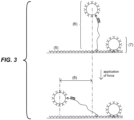
- While certain particles can be used to increase the sensitivity of detection systems to generate a measurable signal, these particles are prone to non-specific interactions with the solid support to which the probe is attached, creating background noise.
- a particle indirectly coupled to the solid support via a complex formed by the target analyte and the first and second probes, and wherein the complex comprises an elongated region specific binding to a target analyte can be distinguished from non-specific binding by measuring the displacement of the particle.
- Particles that are coupled to the solid support via the complex are displaced by a distance that is a function of the length of the elongated region.
- Particles that are non-specifically bound to the solid support are displaced by a distance less than the particles that are specifically coupled to the solid support via the complex. In this way, displacement of the particles can be used to distinguish specific from non-specific binding, particularly in samples with low concentrations of target analyte.
- Other non-specific interactions that can produce the non-specific attachment of a particle to the solid support can also be distinguished, such as particles that non-specifically bind to a probe, or probes that non-specifically bind to the solid support, because those particles are also displaced by a distance less than the particles that are specifically coupled to the solid support via a complex.
- Using a particle indirectly coupled to the solid support via a complex formed by the target analyte and the first and second probes, and wherein the complex comprises an elongated region in embodiments of this invention has at least three additional advantages over other systems that use reporter markers.
- the application of force can increase the target selectivity of the technique by removing particles that are bound via molecules in the sample that are similar to but not exactly the same as the target molecule.
- force can remove non-specifically bound particles while not significantly reducing the signal because being part of a complex with an elongated region reduces the force experienced by the target analyte.
- non-specific interactions which are normally tethers about 10 nm long, experience tensions that are significantly higher than the tension that a target bound in an elongated complex experience.
- This property of long tethers allows in embodiments of the present invention the removal of non-specifically bound particles without significantly affecting specifically bound particles.
- the method comprises: a) providing a probe and a particle, wherein the probe comprises a first end for coupling to a solid support, a second end comprising a first analyte binding region, and an elongated region disposed between the first and second end, and wherein the particle comprises a second analyte binding region; b) exposing the probe to the sample, wherein if the target analyte is present in the sample, the target analyte binds to the first analyte binding region of the probe; c) exposing the particle to the sample, wherein if the target analyte is present in the sample, the target analyte binds to the second analyte binding region of the particle; d) exposing the probe to the solid support, under conditions that permit the coupling of the first end of the probe to the solid support, so that if the target analyte is present in the sample, the particle is indirectly coupled to the solid support via the target analy
- the method for detecting a target analyte in a sample comprises: a) providing a probe and a solid support, wherein the probe comprises a first end for coupling to a particle, a second end comprising a first analyte binding region, and an elongated region disposed between the first and second ends, and wherein the solid support comprises a second analyte binding region; b) exposing the probe to the sample, wherein if the target analyte is present in the sample, the target analyte binds to the first analyte binding region of the probe; c) exposing the probe to the particle, under conditions that permit the coupling of the first end of the probe to the particle d) exposing the solid support to the sample, wherein if the target analyte is present in the sample, the target analyte binds to the second analyte binding region of the solid support, so that the particle is indirectly coupled to the solid support via the target analy
- the first analyte binding molecule of the probe comprises a first nucleic acid that hybridizes to a first region of the target analyte and the second analyte binding region comprises a second nucleic acid that hybridizes to a second region of the target analyte.
- the first analyte binding region of the probe comprises a first antibody that binds to the target analyte and the second analyte binding region comprises a second antibody that binds to the target analyte.
- the method for detecting a target analyte in a sample comprises: a) providing 1) a first probe comprising a first end for coupling to a particle and a second end comprising a first analyte binding region and 2) a second probe comprising a first end for coupling to a solid support and a second end comprising a second analyte binding region, wherein at least one of the first or second probes comprise an elongated region disposed between the first and the second end; b) exposing the first probe and the second probe to the sample, wherein if the target analyte is present in the sample, a first region of the target analyte binds to the first analyte binding region of the first probe and a second region of the target analyte binds to the second analyte binding region of the second probe; c) exposing the first probe to a particle under conditions that permit the coupling of the first end of the first probe to the particle;
- the first probe and second probes bind to locations in the target analyte that are separated by an elongated region in the target analyte.
- the elongated region in the complex may coincide in part or totally with the elongated region of the target analyte.
- the first analyte binding region of the first probe comprises a first nucleic acid that hybridizes to a first region of the target analyte and the second analyte binding region of the second probe comprises a second nucleic acid that hybridizes to a second region of the target analyte.
- the first analyte binding region of the first probe comprises a first antibody that binds to the target analyte and the second analyte binding region of the second probe comprises a second antibody that binds to the target analyte.
- the first probe comprises an elongated nucleic acid disposed between its first and second end
- the second probe comprises an elongated nucleic acid disposed between its first and second end
- the first probe comprises a first elongated nucleic acid disposed between its first and second end
- the second probe comprises a second elongated nucleic acid disposed between its first and second end.
- the exposing steps (b), (c), and (d), can be performed in any order, or simultaneously.
- the order of the steps b), c) and d) is b-d-c; c-b-d; c-d-b; d-b-c; or d-c-b.
- two out of the three steps b), c) and d) are conducted simultaneously.
- the exposure of the sample to the first probe is conducted before, after, or simultaneously with the exposure of the sample to the second probe.
- steps b), c) and d) are conducted simultaneously.
- the methods comprise a washing step after step b) and/or step c) and/or step d).
- the force applied to the particle is a magnetic force, fluid drag, fluid buoyancy, mechanical force, electrical force, centrifugal force, gravitational force, or a combination thereof.
- the elongated region ranges from about 0.15 ⁇ m to about 20 ⁇ m in length. In other embodiments, the elongated nucleic acid ranges from about 0.5 ⁇ m to about 5 ⁇ m in length.
- the diameter of the particle ranges from about 0.3 ⁇ m to about 20 ⁇ m.
- the particle is a magnetic particle, including, but not limited to a superparamagnetic particle.
- the displacement of the particle is measured using an imaging system with a lens,or with a lens-free microscope or with a coherent imaging technique.
- the target analyte is a nucleic acid molecule selected from, single stranded DNA or single stranded RNA such as, messenger RNA, small interfering RNA, micro-RNA and its precursors or circulating RNA.
- the temperature of the sample is controlled to produce denaturation of double stranded nucleic acids in the sample and/or specific hybridization of nucleic acids in the sample to the first and second analyte binding region.
- the sample is initially treated with an exonuclease enzyme to convert double stranded nucleic acids into single stranded nucleic acids.
- the target analyte is a protein.
- target analyte or “analyte,” are used herein to denote the molecule to be detected in the test sample. According to the invention, there can be any number of different target analytes in the test sample (from one to one thousand, or even more).
- the target analyte can be any molecule for which there exists a naturally or artificially prepared specific binding member.
- target analytes include, but are not limited to, a nucleic acid, oligonucleotide, DNA, RNA, protein, peptide, polypeptide, amino acid, antibody, carbohydrate, lipid, hormone, steroid, toxin, vitamin, any drug administered for therapeutic and illicit purposes, a bacterium, a virus, cell, as well as any antigenic substances, haptens, antibodies, metabolites, water pollutants (such as nitrates, phosphates, heavy metals, etc.) and molecules having an odor, such as compounds containing sulfur and/or nitrogen, for example hydrogen sulfide, ammonia, amines, etc., and combinations thereof.
- the target analyte is a nucleic acid.
- the nucleic acid can be from any source in purified or unpurified form including DNA (dsDNA and ssDNA) and RNA, including tRNA, mRNA, rRNA, siRNA, mitochondrial DNA and RNA, chloroplast DNA and RNA, DNA-RNA hybrids, or mixtures thereof, genes, chromosomes, plasmids, the genomes of biological materials, including microorganisms such as bacteria, yeast, viruses, viroids, molds, fungi, plants, animals, humans, and fragments thereof.
- the nucleic acid can be single stranded DNA obtained by exposing double stranded DNA to an exonuclease enzyme, such as exonuclease III.
- the target analyte can be obtained from various biological materials by procedures well known in the art.
- the target analyte is a short nucleic acid containing less than about 200 base pairs or less than about 200 nucleotides.
- such molecules are difficult to detect using PCR-based techniques because suitable primers often cannot be found in such a short sequence.
- a particular case of small DNA molecules are molecules of less than about 40 nucleotides. These molecules are smaller than the combined size of standard PCR primers (each primer about 20 nucleotides).
- Short nucleic acid molecules are common in nature, exemplary cases are small interfering RNA (siRNA), micro-RNA (miRNA) and its precursors, pri-miRNA and pre-miRNA, and fragmented DNA molecules produced after cell death and present in blood, urine and other body fluids.
- probe is understood herein to mean one or more molecules that are capable of binding to the target analyte and also being coupled to, depending on the context, either a solid support or a particle. Probes have a region capable of binding to the target analyte.
- first probe is understood herein to mean the probe that is capable of coupling to a particle.
- second probe is understood herein to mean the probe that is capable of coupling to the solid support.
- the region capable of binding the target analyte in both the first and second probe may comprise a nucleic acid, oligonucleotide, DNA, or RNA molecule having a sequence complementary to the target analyte and capable of hybridizing thereto.
- the target analyte is a protein, peptide, polypeptide, or amino acid
- the region capable of binding the target analyte in both the first and second probe may comprise an antibody or an antigen-binding fragment that specifically binds to the target analyte.
- Coupled refers to a covalent or non-covalent bond between a probe and a surface, or between a probe and another molecule covalently or non-covalently linked to the surface.
- binding refers to a covalent or non-covalent interaction between a probe and a target analyte. In either case, non-covalent interactions could be, for example, ionic, via hydrogen bonding, etc.
- antibody refers to an immunoglobulin or an antigen-binding fragment thereof.
- antigen-binding fragment refers to a part of an antibody molecule that comprises amino acids responsible for the specific binding between antibody and antigen.
- the antigen-binding domain or antigen-binding fragment may only bind to a part of the antigen.
- Antigen-binding domains and antigen-binding fragments include Fab (Fragment antigen-binding); a F(ab') 2 fragment, a bivalent fragment having two Fab fragments linked by a disulfide bridge at the hinge region; Fv fragment; a single chain Fv fragment (scFv) see e.g., Bird et al. (1988) Science 242:423-426 ; and Huston et al. (1988) Proc. Natl. Acad. Sci.
- Fab fragment antigen-binding
- F(ab') 2 fragment a bivalent fragment having two Fab fragments linked by a disulfide bridge at the hinge region
- Fv fragment a single chain Fv fragment
- a Fd fragment having the two V H and C H 1 domains dAb ( Ward et al., (1989) Nature 341:544-546 ), and other antibody fragments that retain antigen-binding function.
- the Fab fragment has V H -C H 1 and V L -C L domains covalently linked by a disulfide bond between the constant regions.
- the F v fragment is smaller and has V H and V L domains non-covalently linked. To overcome the tendency of non-covalently linked domains to dissociate, a scF v can be constructed.
- the scF v contains a flexible polypeptide that links (1) the C-terminus of V H to the N-terminus of V L , or (2) the C-terminus of V L to the N-terminus of V H .
- a 15-mer (Gly 4 Ser) 3 peptide may be used as a linker, but other linkers are known in the art.
- the term "elongated region” refers to a section of the complex formed by the target analyte and the first and second probes that is sufficiently long such that when the complex tethers a particle to a solid support the displacement of the particle can be detected and differentiated from the displacement of particles that are non-specifically attached to the solid support.
- the elongated region is a biomolecule, such as a polysaccharide, polypeptide or nucleic acid, between about 0.15 and about 20 ⁇ m long. In even more preferred embodiments, the elongated region is a double stranded nucleic acid, between about 0.5 and about 5 ⁇ m long.
- test sample or “sample” are used interchangeably herein and include, but are not limited to, biological samples that can be tested by the methods of the present invention described herein and include human and animal body fluids such as whole blood, serum, plasma, cerebrospinal fluid, urine, lymph fluids, and various external secretions of the respiratory, intestinal and genitourinary tracts, tears, saliva, milk, white blood cells, myelomas and the like, biological fluids such as cell culture supernatants, fixed tissue specimens and fixed cell specimens, PCR amplification products or a purified product of one of the above samples.
- biological samples such as whole blood, serum, plasma, cerebrospinal fluid, urine, lymph fluids, and various external secretions of the respiratory, intestinal and genitourinary tracts, tears, saliva, milk, white blood cells, myelomas and the like
- biological fluids such as cell culture supernatants, fixed tissue specimens and fixed cell specimens, PCR amplification products or a purified product of one of the above
- sample may include gaseous mediums, such as ambient air, chemical or industrial intermediates, chemical or industrial products, chemical or industrial byproducts, chemical or industrial waste, exhaled vapor, internal combustion engine exhaust, or headspace vapor such as vapor surrounding foods, beverages, cosmetics, vapor surrounding plant or animal tissue and vapor surrounding a microbial sample.
- gaseous mediums such as ambient air, chemical or industrial intermediates, chemical or industrial products, chemical or industrial byproducts, chemical or industrial waste, exhaled vapor, internal combustion engine exhaust, or headspace vapor such as vapor surrounding foods, beverages, cosmetics, vapor surrounding plant or animal tissue and vapor surrounding a microbial sample.
- sample relevant to this invention is a liquid solution produced by dissolving material collected by filtering a gaseous sample or a liquid solution produced by exposing the liquid to a gaseous sample.
- Additional sample mediums include supercritical fluids such as supercritical CO 2 extricate.
- exemplary mediums include liquids such as water or aqueous solutions, oil or petroleum products, oil-water emulsions, liquid chemical or industrial intermediates, liquid chemical or industrial products, liquid chemical or industrial byproducts, and liquid chemical or industrial waste.
- Additional exemplary sample mediums include semisolid mediums such as animal or plant tissues, microbial samples, or samples containing gelatin, agar or polyacrylamide.
- solid support is used herein to denote any solid material suitable for coupling to a probe and which is amenable to the detection methods disclosed herein.
- the number of possible suitable materials is large and would be readily known by one of ordinary skill in the art.
- particle is used to indicate any solid object or fluorescent molecule suitable for coupling to a probe and which is amenable to the detection methods disclosed herein.
- the solid support or the particles may be composed of modified or functionalized glasses, inorganic glasses, plastics, including acrylics, polystyrene and copolymers of styrene, polypropylene, polyethylene, polybutylene, polyurethanes, Teflon, polysaccharides, nylon or nitrocellulose, resins, and other polymers, carbon, metals, ceramics, silica or silica-based materials including silicon and modified silicon and silicon wafers.
- the surface can be a composite material.
- Surfaces can be functionalized with molecules by physical or chemical adsorption.
- the surfaces are functionalized with probes or with molecules capable of coupling to probes.
- Such methods of functionalization are known in the art.
- a gold surface can be functionalized with nucleic acids that have been modified with alkanethiols at their 3'-termini or 5'-termini. See, for example, Whitesides, Proceedings of the Robert A. Welch Foundation 39th Conference On Chemical Research Nanophase Chemistry, Houston, Tex., pages 109-121 (1995 ). See also Mucic et al., Chem. Commun.
- 555-557 (1996 ) (describes a method of attaching 3' thiol DNA to flat gold surfaces; this method can be used to attach oligonucleotides to nanoparticles).
- the alkanethiol method can also be used to attach oligonucleotides to other metal and semiconductors.
- Other functional groups for attaching oligonucleotides to solid surfaces include phosphorothioate groups (see, e.g., U.S. Pat. No. 5,472,881 for the binding of oligonucleotide-phosphorothioates to gold surfaces), substituted alkylsiloxanes (see, e.g.
- Oligonucleotides terminated with a 5' thionucleoside or a 3' thionucleoside may also be used for attaching oligonucleotides to solid surfaces.
- surfaces can be functionalized by chemically linking streptavidin molecules to them, which are capable of coupling to probes comprising one or more biotin molecules.
- streptavidin molecules capable of coupling to probes comprising one or more biotin molecules.
- the following reference describes the attachment of biotin labeled oligonucleotides to a streptavidin functionalized surface. Shaiu et al., Nucleic Acids Research, 21, 99 (1993 ). Digoxigenin and anti-Digoxigenin antibodies can also be used to attach probes to surfaces.
- the surfaces can be functionalized by a monolayer of one or more molecules.
- Methods of producing self-assembled monolayers are well known in the art.
- the surface functionalization methods described above can be used to couple molecules that prevent or reduce non-specific interactions with the surface. For instance, after immobilization on to the surface of an analyte binding molecule, such as a ssDNA or an antibody, physical adsorption on the surface of a protein that blocks non-specific interactions is often conducted.
- an analyte binding molecule such as a ssDNA or an antibody
- Common proteins used as blockers are: bovine serum albumin (BSA), fish serum and milk proteins, such as casein.
- a "detectable signal" which can be generated according to the invention includes, but is not limited to, an electrical, mechanical, optical, acoustic or thermal signal. In preferred embodiments, the detectable signal is optical or electrical.
- a "polymer” is a molecule formed by monomers in which each monomer is covalently linked to other monomers.
- the term "monomer” is used herein to refer to a molecule that has the ability to combine with identical or other molecules in a process known as polymerization.
- the polymerization reaction may be a dehydration or condensation reaction (due to the formation of water (H 2 O) as one of the products) where a hydrogen atom and a hydroxyl (--OH) group are lost to form H 2 O and an oxygen molecule bonds between each monomer unit.
- a polymer of the invention may be composed of monomers that have hydrophilic groups, and/or hydrophobic groups pendant from their backbones. Accordingly, a polymer may include side chains "R" pendant from a structurally repetitive backbone.
- Exemplary backbones with side chains include: --(CO--N(--R)--CH 2 )--; --(O--Si(--CH 3 )(--R))--; --(CH 2 --CH(--R)--CO--NH)--; --(CH 2 --CH(--R)--O)--; and --(CH 2 -C 6 H 4 --CO--N(--R))--. --(CH 2 --CHR)--, or --(CH 2 --CH 2 --CHR)--; --(CF 2 --CFR), or --(CF 2 --CF 2 --CFR)--; and --(CH 2 --CH(--Co--NHR))--.
- polymers suitable for use in this invention are polyethylene oxide (PEO), polyethylene glycol (PEG), polyisopropylacrylamide (PNIPAM), polyacrylamide (PAM), polyvinyl alcohol (PVA), polyethylenimine (PEI), polyacrylic acid, polymethacrylate and polyvinylpyrrolidone (PVP) polyvinyls, polyesters, polysiloxanes, polyamides, polyurethanes, polycarbonates, fluoropolymers, polyethylene, polystyrene, polybutadiene, polydimethylsiloxane (PDMS), polypropylene, polymethylmethacrylate, polytetrafluoroethylene and polyvinyl chloride (PVC).
- PEO polyethylene oxide
- PEG polyethylene glycol
- PIPAM polyisopropylacrylamide
- PAM polyacrylamide
- PVA polyvinyl alcohol
- PEI polyethylenimine
- PVP polyvinylpyrrolidone
- suitable polymers include, but are not limited to, those described in the references cited in this written description. Nomenclature pertinent to molecular structures, as well as description of monomers and side chain structures useful for the present invention can be found in U.S. Patent Publication No. U.S. 2009/0011946 .
- polysaccharides refers to polymeric carbohydrate structures, formed of repeating units (either mono- or di-saccharides) joined together by glycosidic bonds. Polysaccharides of the invention are preferably linear, but may contain various degrees of branching. Additionally, polysaccharides are generally heterogeneous, containing slight modifications of the repeating unit. Examples of polysaccharides suitable for the invention include homopolysaccharides or homoglycans, where all of the monosaccharides in a polysaccharide are the same type, and heteropolysaccharies or heteroglycans, where more than one type of monosaccharide is present. In exemplary embodiments, the polysaccharide is a starch, glycogen, cellulose, or chitin.
- Polysaccharides of the invention have the general formula of C x (H 2 0) y .
- X is about 100 to about 100,000, about 200 to about 10,000, about 500 to about 5,000, or about 1,000 to about 2,000.
- polysaccharides have repeating units in the polymer backbone of about six-carbon monosaccharides and can be represented by the general formula of (C 6 H 10 0 5 ) n where n is about 30 to about 100,000, about 200 to about 10,000, about 500 to about 5,000, or about 1,000 to about 2,000.
- polynucleotide As used herein, the terms “polynucleotide,” “oligonucleotide,” “nucleic acid” and “nucleic acid molecule” are used interchangeably herein to refer to a polymeric form of nucleotides of any length, and may comprise ribonucleotides, deoxyribonucleotides, analogs thereof, or mixtures thereof. This term refers only to the primary structure of the molecule. Thus, the term includes triple-, double- and single-stranded deoxyribonucleic acid (“DNA”), as well as triple-, double- and single-stranded ribonucleic acid (“RNA”) or RNA/DNA hybrids.
- DNA triple-, double- and single-stranded deoxyribonucleic acid
- RNA triple-, double- and single-stranded ribonucleic acid
- polynucleotide oligonucleotide
- nucleic acid containing D-ribose
- polyribonucleotides including tRNA, rRNA, siRNA, and mRNA, whether spliced or unspliced, any other type of polynucleotide which is an N- or C-glycoside of a purine or pyrimidine base, and other polymers containing nonnucleotidic backbones, for example, polyamide (e.g., peptide nucleic acids (PNAs)) and polymorpholino (commercially available from the Anti-Virals, Inc., Corvallis, Oreg., as Neugene
- PNAs peptide nucleic acids
- nucleotides include hybrids thereof, for example between PNAs and DNA or RNA, and also include known types of modifications, for example, labels, alkylation, "caps," substitution of one or more of the nucleotides with an analog, internucleotide modifications such as, for example, those with uncharged linkages (e.g., methyl phosphonates, phosphotriesters, phosphoramidates, carbamates, etc.), with negatively charged linkages (e.g., phosphorothioates, phosphorodithioates, etc.), and with positively charged linkages (e.g., aminoalkylphosphoramidates, aminoalkylphosphotriesters), those containing pendant moieties, such as, for example, proteins (including enzymes (e.g.
- nucleases nucleases
- toxins antibodies
- signal peptides poly-L-lysine, etc.
- intercalators e.g., acridine and psoralen
- chelates e.g., metals, radioactive metals, boron, oxidative metals, etc.
- alkylators those with modified linkages (e.g., alpha anomeric nucleic acids, etc.).
- Oligonucleotides of defined sequences are used for a variety of purposes in the practice of the invention. Methods of making oligonucleotides of a predetermined sequence are well-known. See, e.g., Sambrook et al., Molecular Cloning: A Laboratory Manual (2nd ed. 1989 ) and F. Eckstein (ed.) Oligonucleotides and Analogues, 1st Ed. (Oxford University Press, New York, 1991 ). Solid-phase synthesis methods are preferred for both oligoribonucleotides and oligodeoxyribonucleotides (the well-known methods of synthesizing DNA are also useful for synthesizing RNA). Oligoribonucleotides and oligodeoxyribonucleotides can also be prepared enzymatically.
- polypeptides refers to a polymer formed from the linking, in a defined order, of preferably, ⁇ -amino acids, D-, L-amino acids and combinations thereof.
- peptides oligopeptides
- proteins are included within the definition of polypeptide.
- the term includes polypeptides containing post-translational modifications of the polypeptide, for example, glycosylations, acetylations, phosphorylations, and sulphations.
- protein fragments, analogs including amino acids not encoded by the genetic code, e.g.
- homocysteine, ornithine, D-amino acids, and creatine), natural or artificial mutants or variants or combinations thereof, fusion proteins, derivatized residues (e.g. alkylation of amine groups, acetylations or esterifications of carboxyl groups) and the like are included within the meaning of polypeptide.
- the link between one amino acid residue and the next is referred to as an amide bond or a peptide bond. The terms do not refer to a specific length of the polypeptide.
- the elongated region of one or both probes comprise a non-biological hydrophilic polymer, such as polyethylene oxide (PEO), polyethylene glycol (PEG), polyisopropylacrylamide (PNIPAM), polyacrylamide (PAM), polyvinyl alcohol (PVA), polyethylenimine (PEI), polyacrylic acid, polymethacrylate and polyvinylpyrrolidone (PVP), or a combination thereof.
- a non-biological hydrophilic polymer such as polyethylene oxide (PEO), polyethylene glycol (PEG), polyisopropylacrylamide (PNIPAM), polyacrylamide (PAM), polyvinyl alcohol (PVA), polyethylenimine (PEI), polyacrylic acid, polymethacrylate and polyvinylpyrrolidone (PVP), or a combination thereof.
- PEO polyethylene oxide
- PEG polyethylene glycol
- PNIPAM polyisopropylacrylamide
- PAM polyacrylamide
- PVA polyvinyl
- the elongated region of one or both probes and/or the target analyte comprises a biomolecule, such as a polysaccharide, polynucleotide, or a polypeptide, or a combination thereof.
- a preferred embodiment is a method of detecting a target analyte in a sample.
- a complex is provided, the complex formed from: a first probe coupled to a particle and bound to said analyte if present, and a second probe coupled to a solid support and bound to said analyte if present, so that if the target analyte is present in the sample, the particle is indirectly coupled to the solid support via a complex formed by the target analyte and the first and second probes, and wherein the complex comprises an elongated region.
- the provided complex may have been formed in any possible manner and order of steps. It will also be clear that the person(s) who carries out any of the complex-formation steps may, but need not be, the person who performs the subsequent steps in the process, i.e., applying the force or measuring the Brownian motion.
- the first probe, the second probe, and the target analyte comprise nucleic acids or oligonucleotides. More preferably, the first probe and second probe each comprise a region for binding the target analyte.
- the first probe couples to a particle at a different location than where the target analyte binds to the first probe.
- the second probe couples to the solid support at a different location than where the target analyte binds to the second probe.
- the complex formed by the target analyte and the first and the second probe comprises an elongated region.
- the elongated region of the complex formed by the target analyte and the first and the second probe comprises double-stranded DNA having a total length ranging from about 500 base pairs to about 60,000 base pairs, for example, from about 1,500 base pairs to about 15,000 base pairs.
- the nucleic acid target can form base pairs with unpaired nucleotides in the first probe and with unpaired nucleotides in the second probe.
- the first probe further comprises a region for coupling to the particle.
- the first probe can comprise a protein, a peptide, or an antigen covalently attached to the 5' or 3' end of a nucleic acid.
- the target analyte is not a nucleic acid.
- the first and second probes preferably comprise an antibody.
- the target analyte is a nucleic acid and the first and second probes bind to locations on the target that are at least 500 nucleotides from each other.
- the target can be either double or single stranded.
- the force required to extend it is significantly higher than the force required to extend a double stranded nucleic acid ( Current Opinion in Structural Biology 2000, 10:279 ; Nucleic Acids Research 2014 (42), 3:2064 ).
- the force required to extend the single stranded nucleic acid can be modified by changing solution properties, such as ionic strength and temperature, and/or adding a molecule that bind to the single strand .
- the target analyte is a nucleic acid molecule
- exposure of the target analyte to the first and/or second probe is preferably conducted under high stringency conditions.
- High stringency conditions favor the hybridization of nucleic acid molecules which are perfectly complementary or substantially perfectly complementary to single stranded nucleic acids in the probe and make more unlikely the binding of targets which are not perfectly complementary or substantially perfectly complementary.
- washing or exposing the probes to a medium with high stringency can remove non-perfectly complementary molecules as well.
- High stringency conditions occur at high temperature, low salt concentration and high pH. Also the presence of certain chemicals, such as formamide, can increase the stringency of the solution.
- exposure of the target to probes and washing, when performed are conducted preferably at temperatures between 20°C and 70°C, ionic strength between 0.01 M and 1 M, and pH between 7 and 8.
- Some methods of this invention contain "exposing" steps where the probe(s), particles, and/or solid support are exposed to the sample or one another. These exposing steps can occur in any order, or even simultaneously. For example, in one embodiment, reactants are exposed in the following order, before applying force to the particle and measuring displacement: a) the first and second probe are exposed to the target analyte, b) the second probe which comprises a first end for coupling to the solid support, is exposed to the solid support, c) the first probe which comprises a first end for coupling to the particle is exposed to the particle.
- the particle is indirectly coupled to the solid support via a complex formed by the target, and the first and second probes.
- the steps are conducted in reverse order (c-b-a). If these steps are conducted under conditions that allow reactants to bind or couple and if the target analyte is present in the sample, the particle is indirectly coupled to the solid support as before: via the first probe, the target analyte and the second probe. In another exemplary embodiment all the steps can be conducted simultaneously.
- the particle is indirectly coupled to the solid support in the same manner as the previous two examples: via the first probe, the target analyte and the second probe. Any order and/or combination of steps that are conducted simultaneously will have the same result, if each step is conducted under conditions that allow reactants to bind or couple.
- the particle is a solid bead with a diameter between about 0.1 ⁇ m and about 20 ⁇ m, or between about 0.3 ⁇ m and about 5 ⁇ m.
- Preferred bead materials are: silica-based glasses, such as quartz and borosilicate; zirconium and organic polymers, such as polystyrene, melamine resin and polyacrylonitrile.
- the particle is a superparamagnetic bead with a diameter between about 0.5 ⁇ m and about 5 ⁇ m.
- Superparamagnetic beads are commonly used in biotechnological applications. They consist of a polymer matrix that contains small ( ⁇ about 10 nm) particles of a ferromagnetic material in it. The small size of the ferromagnetic particles makes them superparamagnetic. As a result, the beads are magnetic only under the influence of an external magnetic field.
- the particle is a quantum dot.
- a quantum dot is typically less than about 10 nm and made of semiconductor materials that display quantum mechanical properties. As a result of these properties, the electronic characteristics of quantum dots are related to the size and shape of the individual crystal. Quantum dots are fluorescent and the emission frequency increases as the size of the quantum dot decreases. Therefore the color of quantum dots can be controlled by their size.
- the particle is a nanorod.
- the length of the nanorods is at least about 0.5 ⁇ m.
- Methods of making nanorods or nanowires are known in the art. See for example, Hahm and Mieber, Nano Lett, 4, 51-54 (2004 ) (silicon nanorods); Li et al., Appl. Phys. Lett. 4, 4014-1016 (2003 ) (In2O3 nanorods); Liu et al., Phys. Ev. B. 58, 14681-14684 (1998 ) (Bismuth nanorods); Sun et al., Appl. Phys. Lett.
- the particle comprises a fluorescent molecule.
- fluorescent nucleic acid can be created in a PCR reaction where one of the deoxynucleotides in the reaction mix has a florescent label.
- a commonly used labeled deoxyadenosine triphosphate for this procedure is Fluorescein-12-dATP. Protocols to label nucleic acid molecules are readily available ( Nucl. Acids Res. (1994) 22 (16):3418 ; Nat Biotechnol. (2008) 26(3):317 ; Nat Biotechnol. (2000) 18 (2):233 ).
- Another example of fluorescent molecule is a single fluorophore, such as Cy3 and other cyanines, and fluorescein.
- the probes and/or target may be labeled before or after the application of force with at least two particles, one particle at one end of the probe-target complex, the other at the other end.
- the elongated region of the complex may be labeled substantially along its length with fluorescent molecules.
- the elongated region may be a double stranded DNA that is labeled with a nucleic acid fluorescent dye, such as YOYO-1.
- the approximate length of the elongated region can be determined from the position of said particles after the application of force.
- the discrimination of non-specific interaction is based on the length of the elongated region. If the full length of the elongated region is observed, it means that the target analyte is present. Instead, if a fraction of the length of the elongated region is observed, it means that the attachment to the solid support is via non-specific interactions.
- the first probe and second probes are exposed to a sample comprising the target analyte.
- particles are introduced into the sample, which couples to the first probe.
- the sample is exposed to a solid support, where the second probe is coupled to the solid support.
- the order of exposing the probe to the sample, the solid support and/or the particles can vary.
- the particle can be exposed to the first probe prior to exposure of the target analyte to the first and second probe.
- the second probe is exposed to a sample comprising the target analyte.
- the second probe comprises an antibody specific for the target analyte.
- particles are introduced into the sample.
- the particles are functionalized with a first probe that comprises an antibody specific for the target analyte.
- the sample is exposed to a solid support, where the second probe is coupled to the solid support.
- the order of exposing the probe to the sample, the solid support and/or the particles can vary.
- the first probe can be exposed to the target analyte prior to exposure of the target analyte to the second probe.
- the analyte comprises a nucleic acid and the probes comprise a nucleic acid.
- One probe comprises a first end or region for coupling to either the particle or the solid support and a second end or region for binding to the analyte, and either the particles or the solid support comprise a second analyte binding region.
- the first probe (1) is covalently attached to the surface of a particle (4). After the target analyte (3) binds to the first (1) and second (2) probes, the sample is exposed to a solid support (5), where the second probe (2) is coupled to the solid support.
- the second probe (2) is a nucleic acid coupled to the solid support (5).
- the target analyte (3) can be exposed to the first probe (1) prior to exposure of the target analyte (3) to the second probe (2) or, alternatively, the target analyte can be exposed to the second probe (2) prior to exposure of the target analyte (3) to the first probe (1).
- the target analyte can be exposed to the probe and solid support simultaneously. After the target analyte binds to both the first and second probes, particles are introduced to the sample, where a particle couples to the first probe.
- the first probe can be coupled to the particle prior to exposure to the sample and/or prior to exposure to the second probe.
- the first (1) and second (2) probes are exposed to a sample comprising the target analyte (3).
- the target analyte can be exposed to the first probe prior to exposure of the target analyte to the second probe or, alternatively, the target analyte can be exposed to the second probe prior to exposure of the target analyte to the first probe.
- the target analyte can be exposed to the first and second probes simultaneously.
- particles (4) are introduced into the sample, which couple to the first probe.
- the sample is exposed to a solid support (5), where the second probe is coupled to the solid support.
- the particle can be exposed to the first probe prior to exposure of the target analyte to the first and second probe.
- a force is applied to the particles.
- the force field acts on the particle and pulls it away from its initial position (e.g. a magnetic field acting on a magnetic particle or a flow exerting a drag on a particle).
- the device is exposed to a sample in conditions such that if the target analyte is present, then a complex is formed by the target analyte and the first and second probes. Consequently, when a force is applied, the particle that is associated with the complex will move a distance that is a function of the length of the complex. As shown in FIG. 3 , this amount of displacement (8) indicates specific binding.
- the particles that are non-specifically bound (7) to the solid support will move a shorter distance, if at all, than the specifically-bound particles.
- the detection device is exposed to the sample in conditions such that the number of beads tethered by probes is proportional to the concentration of the target analyte. In this manner, the detectable signal is proportional to the concentration of the target analyte, thereby permitting the concentration of the target analyte in the sample to be determined.
- Preferred embodiments use probes having elongated regions of multiple different lengths, with each probe of a certain length having a region capable of binding a specific target analyte, in such a manner that each probe length is associated with a different target analyte.
- the approximate concentration of multiple target analytes in a sample can be determined in a single assay by measuring different displacements of particles after application of force, grouping them based on the amount of displacement and counting the number of particles associated with each possible displacement.
- the multiplexing capability is further increased by modifying the probes having elongated regions in such a manner that each of them may have more than one length and the change of length is triggered by an external agent.
- external agents that can trigger a change of length are temperature, ionic strength, pH, force, an auxiliary molecule, such as a nucleic acid, an enzyme a detergent, etc.
- the identity of a target is determined after measuring the displacement of the particle before and after triggering the change of length. The change of length can be triggered multiple times.
- each probe specific to a different target analyte can be created by a set of probes that have 10 different lengths before triggering the length change and wherein each probe experiences one out of 10 possible different length changes upon triggering the length change.
- An example of a probe with an elongated region having a length that can be changed by an external agent is a probe in which two remote positions in the probe interact in such a manner that an internal loop is formed. In this case, the external agent can trigger the release or the formation of an internal loop. The characteristics of the external agent required to trigger the change of length are controlled by the characteristics of the interaction.
- an external agent that can trigger the release of an internal loop formed by hybridization between nucleic acid molecules is an auxiliary nucleic acid complementary to one of the molecules in the hybridization region that holds an internal loop. When the probe is exposed to this auxiliary nucleic acid, the auxiliary nucleic acid can displace one of the strands in the hybridization region, thereby releasing the loop.
- an external agent is an auxiliary nucleic acid that has a first and a second region, wherein the first region is complementary to a first region in a nucleic acid probe and the second region is complementary to a second region in the probe.
- the nucleic acid When the probe is exposed to this nucleic acid, the nucleic acid binds to the two regions in the probe which produces an internal loop.
- the auxiliary molecules are proteins that can be used similarly to the nucleic acid described above with the purpose of releasing or forming internal loops.
- the surfaces and probes of the present invention may have a plurality of different analyte binding molecules attached to them, and as a result, the tethering of beads to the solid support could be triggered by a plurality of target analytes.
- a solid support may have an array of regions, each region comprising a second probe specific to a unique target molecule.
- exposure of the solid support to the sample captures different targets at different locations in the array. Therefore, detection of particle displacement in a specific array region indicates that the corresponding target molecule is present in the sample.
- a method is provided for creating a unique profile or fingerprint of a sample having any number of different target analytes (e.g., any of two through one thousand, or even more).
- profiles from different samples can be stored in a database and/or compared for diagnostic purposes for the detection of diseases or disorders.
- Another embodiment uses multiple distinguishable particles, wherein each different particle comprises a different first probe specific to a different molecule.
- fluorescent beads of different colors are functionalized with different antibodies, one antibody kind for each bead color. In this manner, the specific target molecules present in the sample are identified by detecting the color of the tethered beads that are displaced under a force.
- beads of different sizes or distinguishable strings of fluorescent molecules or particles can be used ( Nat. Biotech. 26, 317-325, 2008 ).
- the applied force is fluid drag.
- This type of force is generated by the flow of the liquid solution in which the particle and/or molecule is immersed. More precisely, this force is applied when there is a difference between the speed of the liquid and the speed of the particle and/or molecule.
- This force is normally parallel to the solid support, but it can have a component perpendicular to the solid support if the solid support is porous.
- the particles are in proximity to the surface of the solid support and the flow is substantially parallel to the surface of the solid support. In these embodiments, the speed of the fluid increases away from the surface of the solid support and not only produces a linear force substantially parallel to the surface of the solid support but also a torque.
- the particles have a diameter or length less than about 20 micrometers and the flow is laminar, with a Reynolds number less than about 1.
- the particles and/or molecule are inside a capillary tube and flow can be generated using a pump, such as a syringe pump, connected to the capillary by a hose.
- a pump such as a syringe pump
- Another way of generating a suitable flow is to exert an external pressure on the hose connected to the capillary. The pressure moves a small amount of fluid into the capillary, which displaces the particles away from the side of the capillary where the pressure was applied. Releasing the pressure in the hose displaces the beads in the opposite direction.
- the applied force on the particle is fluid buoyancy. This type of force is equal to the weight of the fluid displaced by the particle and in the direction opposite to the gravitational force.
- the applied force is a magnetic force.
- the particles are magnetic, such as superparamagnetic beads.
- the force is a consequence of the presence of a magnetic field which can be generated with permanent magnets, such as iron or rare earth magnets, or electromagnets.
- the magnetic force can be used to pull the particles away from the glass surface, in such a way that particles tethered via an elongated region are displaced to a plane higher than the particles non-specifically attached to the surface.
- This type of displacement can be detected optically using an imaging system able to image planes parallel to the solid support. In this imaging system, this type of displacement produces a change in diffraction pattern when the particle moves to a different focal plane.
- the magnetic force can be used to pull the particles in a direction parallel to the surface of the solid support. This type of displacement is easily detected as a change of position of the particles in the image using an optical system.
- the applied force is gravitational.
- the direction of the force is always toward the center of the earth and therefore its direction with respect to the solid support is determined by the orientation in space of the solid support.
- the applied force is centrifugal.
- the particles are subjected to a motion that changes direction.
- the motion is a rotational motion.
- the applied force is electrical.
- An electrical force is generated when at least two electrodes having different voltage are introduced in the solution generating a voltage gradient.
- the force is applied to the target-probe complex using flow, a receding meniscus, or a voltage gradient.
- the formed complex is coupled to a substantially flat solid support, and is subjected to a force substantially parallel to the solid support.
- the force is selected such that it removes non-specifically bound particles from the support. Then, using suitable detection methods, the presence of remaining particles indicates the presence of the target analyte in the sample.
- force can remove non-specifically bound particles while not significantly reducing the signal because being part of a complex with an elongated region reduces the force experienced by the target analyte.
- force substantially parallel to the solid support is applied on particles bound to the solid support, the tension on the tether decreases with tether length ( Langmuir 1996, 12(9): 2271 ).. Therefore, non-specific interactions, which are normally tethers about 10 nanometers long, experience tensions that are significantly higher than the tension that a target bound in an elongated complex experience.
- Figure 8B shows the value of the ratio of tension and horizontal force ( T / F ) as a function of the ratio of tether length and particle radius ( L / a ).
- the tension in the tether dramatically increases for tether lengths that are less than half the radius of the particle. For example, for a tether that is 0.01 times the radius of the particle, the tension is 7 times higher than the horizontal force, while for a tether that is 2 times the radius of the particle, the tension is only 6% higher than the horizontal force.
- the torque applied on the particle by the drag further increases the difference in the tension experienced by short versus long tethers.
- the application of force to the complex formed by the target analyte and the first and second probes either directly or indirectly through force applied to the particle can increase the target specificity of the detection system by removing complexes where the target is not the exact binding partner of the binding regions in the probes.
- the target is a nucleic acid
- the application of force is a novel form of hybridization stringency. This stringency can be modulated by the configuration of the structure formed by probes when they bind to the target.
- a probe can hybridize to a target in two main types of configurations.
- the axis of the duplex In a first configuration, the axis of the duplex is in the direction of the force and the application of force tends to disrupt all the base pairs simultaneously. In a second configuration, the axis of the duplex is perpendicular to the direction of the force and the application of force tends to disrupt base pair in a progressive order, starting with the ones closer to the point of force application.
- the stringency can be modulated by the amount of force applied ( Current Opinion in Chemical Biology 2008, 12: 640 , PNAS 2006 (103),16:6190 ).
- coiling refers to a conformational change suffered by bundles of two or more polymers subjected to a rotational force.
- a simple example of “coiling” takes place when two linear polymers are held together by holding a first end from each polymer together at one location and a second end of the polymers together at a second location, if the second end of one polymer rotates around the second end of the other polymer, the two polymers will wind around each other. As rotations accumulate the two polymers will start presenting a more compact conformation. This phenomenon is referred in the present invention as “coiling”. Another example of coiling is "supercoiling".
- nucleic acid strands are connected by base pairing and in the absence of torsional stress wind around each other.
- Supercoiling takes place when the strands are either winded or unwinded from their natural amount of winding.
- Supercoiled structures have writhe ( PNAS 1978, 75 (8) 3557 ).
- the present invention may be incorporated into an assay as described in: International Patent Publication WO 2013/059044 published April 25, 2013 and entitled "Detection Units and Methods for Detecting a Target Analyte", United States Patent Application Publication US 2014/0099635 A1 published April 10, 2014 and United States Provisional Application 61/983,684, filed on April 24 2014 .
- the particle may be a magnetic particle (4) and both probes may be double stranded DNA molecules, the first probe (1) couples to the particle with at least two attachment points and the second probe (2) couples to the solid support with at least two attachment points.
- first and second probes have regions to bind a second molecule (9) different from the target analyte (3), in such a manner that when both target and second molecule bind to the probes the entire complex is made of two strands, one strand contains the target and the other the second molecule.
- a rotational force is applied to the magnetic particle by rotating a magnetic field, torsional stress is accumulated in the complex, which forms supercoils that dramatically shorten the end-to-end distance of the complex after a certain level of torsional stress is reached. Therefore, supercoiling can be readily detected as a reduction in the displacement of the particle (4) under an applied force or as a reduction in the Brownian motion of the particle.
- This strategy can be used to increase the specificity and the discrimination of non-specific interactions in the system.
- the torsional stress in the complex tends to disrupt mismatched targets faster than perfectly matched ones. Therefore, requiring for target detection that a bead displace first, and then the complex supercoils, imposes a second condition that increases target specificity.
- non-specific interactions can be discriminated because non-specifically bound beads cannot supercoil.
- the first probe is a circular double stranded DNA molecule, wherein one of the strands has a discontinuity in a region designated as "active segment" (10).
- the circular molecule is not capable of supercoiling because of this discontinuity.
- the discontinued strand has flaps that have sequence complementary to different regions of the target analyte (3). When the target analyte binds (3) to the active segment (10), it bridges the two sides of the discontinued strand, which enables the circular molecule to supercoil. If a rotational force is applied to the first probe, for example by an intercalator molecule, torsional stress is accumulated in the probe, which forms supercoils that dramatically change the conformation of the probe.
- the first probe comprises a circular double stranded DNA molecule wherein one of the strands has a discontinuity and the particle comprises a fluorescent molecule (11).
- the first probe may have multiple fluorescent labels.
- the discontinuous strand has flaps that have sequence complementary to different regions of the target analyte (3).
- the target analyte binds (3) to the active segment (10), it bridges the two sides of the discontinuous strand, which enables the circular molecule to supercoil.
- a rotational force is applied to the first probe, for example by an intercalator molecule, torsional stress is accumulated in the probe, which forms supercoils that dramatically change the conformation of the probe. Therefore, supercoiling can be readily detected as a change in the displacement of the particle under an applied force or a change in the length of the complex under an applied force.
- this strategy can be used to increase the specificity and the discrimination of non-specific interactions in the system.
- the displacement of the particles is detected using an imaging system, wherein the imaging system generates an image of the particles and/or the probes-target complex that is detected by a sensing device.
- the image can be a regular image or a transformed representation of the object such as a shadow.
- the imaging system consists of four main components: illumination, specimen, image forming part, and a detector, which are sequentially positioned on the spatial path.
- An example of an imaging system is the optical microscope, and in this case the image forming part is the lens/lenses.
- Optical microscopes are well known by those of skill in the art. Optical microscopes can visualize unstained samples using image contrast of scattering, absorption or phase contrast, or stained samples with fluorescence or other scheme of light emission.
- the light source employed in a microscope can be coherence light source (such as laser) or incoherent source (such as LED or white light source).
- the lenses of a microscope can be a single lens, a series of lenses, or a compound lens which is usually called an objective.
- a macroscope is another example of imaging system. The main difference between a macroscope and microscope is the lens/objective they use.
- the microscope lens usually has magnification equal or larger than 1X, meaning the size of image is larger than the object. That results in a small field of view.
- the macroscope lens can have magnification smaller than 1X, which allows for visualization of a large area.
- a lens-free imaging system is another example of an imaging system.
- This type of system uses a digital optoelectronic sensor array, such as a charged coupled device (CCD) or a CMOS chip to directly sample the light transmitted through a specimen without the use of imaging lenses between the object and the sensor plane ( Greenbaum, Nat. Methods 2012, 9, 9, 889 - 895 ; Gurkan, U.A., et al., Biotechnol. J. 2011, 6, 138-149 ).
- the lensless ultra wide-field cell monitoring array platform (LUCAS) ( Ozcan, A. and Demirci, U. Lab Chip, 2008, 8, 98-106 ) is an example of this type of microscopes.
- the LUCAS platform is based on recording the "shadow images" of microscopic objects onto a sensor array plane.
- Microscopic objects are uniformly illuminated with an incoherent light source or a laser.
- the cell shadow pattern is digitally recorded using a CCD or CMOS sensor array.
- a coherent imaging system is another example of image system. This type of system uses the object to modulate the illumination laser beam and makes the modulated beam interfere with a reference laser beam or the same illumination beam, then the interferential information is recorded to reconstruct the information of the object.
- Digital holography Javidi, Opt. Lett., 2000, 25, 9, 610-612
- in-line holography Xu, PNAS, 2001, 98, 20, 11301-11305
- the displacement of the magnetic particles is detected from the induced current in a solenoid.
- the first probe is generated in the following manner: A plasmid (approximately 10 kilo base pairs) is linearized using a restriction enzyme that cuts it once. Two types of short DNA molecules capable of being ligated to the linearized plasmid are purchased (IDT): the first short DNA molecule is labeled with Digoxigenin and the second has single stranded region complementary to part of the target. The two short DNA molecules are ligated to the linearized plasmid. The second probe, a biotin labeled single stranded molecule, is purchased (IDT) with a sequence complementary to part of the target.
- Super-paramagnetic beads (Life Technologies) coated with Streptavidin are purchased. The first and second probes are mixed at high concentration (>0.01 ⁇ M) with the sample and incubated for few minutes. Magnetic beads are added to the solution and incubated few minutes. In order to wash unbound molecules, the solution is exposed to an external magnet that attracts the magnetic beads removing them from the solution. The solution is extracted from the container and discarded. The external magnet is taken away and the beads are suspended in buffer solution. Beads are then flowed into a capillary tube previously functionalized with anti-digoxigenin proteins (Roche) and let sediment and interact with the surface of the tube. In order to wash unbound beads, new buffer solution is flowed while a magnet is passed over the capillary.
- a large magnet is placed over the capillary to lift remnant unbound beads.
- the beads that remain attached to the bottom of the capillary are imaged using an image sensor array directly in contact with the bottom of the capillary tube.
- the solution in the tube is flowed in one direction to drag the beads and then the direction of the flow is reversed to drag the beads in the opposite direction.
- Successive images of the beads during this process show that some beads move about 3 micrometers in each direction while the rest of the beads bound to the surface do not move or move significantly less.
- Beads that move about 3 micrometers in each direction are bound to the glass surface via the complex of probes and target. The initial concentration of target molecules in the sample determines the number of these beads.
- This example demonstrates detection of a 60 nucleotide (nt) synthetic DNA oligonucleotide target having the sequence of a section of Mycobacterium tuberculosis rRNA.
- the first probe was purchased from IDT and consisted of a 24 nt single stranded oligonucleotide having a sequence complementary to the 3' end of the target and a 5' biotin modification.
- the second probe was generated in the following manner: A plasmid (8.5 kbps) was linearized using the restriction enzyme BsmB I (New England Biolabs), which cut the plasmid twice generating a large fragment (approximately 8.4 kbps/2.8 micrometers) with different 4 nt overhangs at each end, and a small fragment which was separated and discarded by agarose gel purification (QIAquick Gel Extraction Kit, Qiagen). The linearized plasmid was ligated using T4 ligase to two double stranded DNA fragments generated by hybridizing synthetic oligonucleotides.
- BsmB I New England Biolabs
- the first fragment had one end with an overhang compatible to one of the overhangs of the plasmid and the other end had a 30 nt overhang complementary to the 5' end of the target.
- the second fragment had one end with an overhang compatible to the other overhang of the plasmid, and the other end of the fragment had a 5' digoxigenin modification.
- the first probe was mixed with a solution containing the target and the temperature was raised to 65°C for 1 minute and then incubated at room temperature for 10 minutes.
- the buffer contained 800 mM NaCl.
- a blocker oligonucleotide complementary to the first probe was added and incubated for 5 minutes.
- the second probe was added and incubated for 10 minutes.
- the optical system used to image the beads was composed of a LED ring light, a telecentric lens and a camera.
- the LED ring light provided a dark field illumination.
- the telecentric lens which is popularly employed in machine vision, exhibited the same magnification for objects at different distances.
- the lens had a large depth of field of around 500 micrometers.
- the magnification of the system was 1:1, which helped to achieve a large field of view of 6.14 mm by 4.6 mm.
- the image size of each particle was about 4 micrometers, which was sufficient to investigate the displacement of the particles.
- the camera used in the system had a 1 ⁇ 2. 3 inch complementary metal oxide semiconductor (CMOS) chip, which had 4384 by 3288 pixels with pixel size of 1.4 micrometer.
- CMOS complementary metal oxide semiconductor
- a custom code written in Matlab was used to analyze the images and determine the displacement of most of the beads present in the field of view. Beads that were too close to each other (less than about 6 micrometers) were not included in the analysis. The position of a bead was defined as the average of its position in the 45 images. This system allowed measurement of the displacement of over 3,000 beads in each experiment with sub-micrometer resolution.
- the code generated a histogram of bead displacement ( Figure 6 ). When the first set of images was taken without flow ( Figure 6A ), beads that moved more than 2.5 micrometers where considered bound to a target-probe complex (right peak in the histogram). Beads that moved less than 2.5 micrometers were considered non-specifically attached to the capillary surface.
Landscapes
- Health & Medical Sciences (AREA)
- Life Sciences & Earth Sciences (AREA)
- Immunology (AREA)
- Chemical & Material Sciences (AREA)
- Engineering & Computer Science (AREA)
- Molecular Biology (AREA)
- Hematology (AREA)
- Physics & Mathematics (AREA)
- Biomedical Technology (AREA)
- Urology & Nephrology (AREA)
- Analytical Chemistry (AREA)
- General Health & Medical Sciences (AREA)
- Biochemistry (AREA)
- Pathology (AREA)
- General Physics & Mathematics (AREA)
- Microbiology (AREA)
- Biotechnology (AREA)
- Food Science & Technology (AREA)
- Medicinal Chemistry (AREA)
- Cell Biology (AREA)
- Chemical Kinetics & Catalysis (AREA)
- Electrochemistry (AREA)
- Organic Chemistry (AREA)
- Wood Science & Technology (AREA)
- Zoology (AREA)
- Proteomics, Peptides & Aminoacids (AREA)
- Spectroscopy & Molecular Physics (AREA)
- General Engineering & Computer Science (AREA)
- Biophysics (AREA)
- Bioinformatics & Cheminformatics (AREA)
- Genetics & Genomics (AREA)
- Microelectronics & Electronic Packaging (AREA)
- Measuring Or Testing Involving Enzymes Or Micro-Organisms (AREA)
Claims (15)
- Verfahren zum Nachweis eines Zielanalyten in einer Probe, wobei das Verfahren aufweist:a) Bereitstellung eines Komplexes, gebildet aus:i) einer erste Sonde, die an ein Teilchen gekoppelt und an den Analyten, falls vorhanden, gebunden ist, undii) einer zweite Sonde, die an einen festen Träger gekoppelt ist und an den Analyten, falls vorhanden, gebunden ist,so dass, wenn der Zielanalyt in der Probe vorhanden ist, das Teilchen nur indirekt an den festen Träger über einen Komplex gekoppelt ist, der durch den Zielanalyten und die erste und zweite Sonde gebildet wird, und wobei der Komplex einen länglichen Bereich aufweist, der sich an der ersten Sonde, der zweiten Sonde oder dem Zielanalyten oder einer beliebigen Kombination davon befindet; und wobei der längliche Bereich von etwa 0,15 µm bis etwa 20 µm in der Länge reicht; undb) entweder i) Anwenden einer Kraft auf das indirekt gekoppelte Teilchen oder auf den Komplex, der den länglichen Bereich aufweist, und Messen des Ausmaßes der Teilchenverschiebung oder der Länge des Komplexes, wobei das Ausmaß der Verschiebung oder die Länge des Komplexes anzeigt, ob der Zielanalyt in der Probe vorhanden ist oder nicht, oder ii) Messen der Brownschen Bewegung des indirekt gekoppelten Teilchens, wobei das Ausmaß der Brownschen Bewegung anzeigt, ob der Zielanalyt in der Probe vorhanden ist oder nicht.
- Verfahren nach Anspruch 1, wobei die erste Sonde einen ersten Antikörper aufweist, der sich an den Zielanalyten bindet, und die zweite Sonde einen zweiten Antikörper aufweist, der sich an den Zielanalyten bindet.
- Verfahren nach Anspruch 1, wobei die erste Sonde eine erste Nukleinsäure aufweist, die mit einer ersten Region des Zielanalyten hybridisiert, und die zweite Sonde eine zweite Nukleinsäure aufweist, die mit einer zweiten Region des Zielanalyten hybridisiert.
- Verfahren nach Anspruch 1, wobei der längliche Bereich ein Biomolekül aufweist.
- Verfahren nach einem der vorhergehenden Ansprüche, das ferner den Schritt des Ankoppelns der ersten Sonde an das Teilchen und den Schritt des Ankoppelns der zweiten Sonde an den festen Träger aufweist.
- Verfahren nach einem der vorhergehenden Ansprüche, das ferner die Schritte des Aussetzens der Probe der ersten Sonde und des Aussetzens der Probe der zweiten Sonde aufweist.
- Verfahren nach Anspruch 6, wobei die weiteren Schritte in beliebiger Reihenfolge oder gleichzeitig durchgeführt werden.
- Verfahren nach Anspruch 7, das ferner einen Waschschritt nach einem oder mehreren der Schritte aufweist.
- Verfahren nach einem der vorhergehenden Ansprüche, wobei die Kraft, die auf den Komplex mit dem länglichen Bereich ausgeübt wird, mit einen Flüssigkeitsstrom erzeugt wird.
- Verfahren nach einem der vorhergehenden Ansprüche, wobei mehr als ein Target in einem einzigen Assay nachgewiesen wird und wobei die Targets auf der Grundlage der unterschiedlichen Teilchenverschiebung oder des Ausmaßes der Brownschen Teilchenbewegung, die sie erzeugen, sobald der Komplex an den festen Träger gebunden ist, unterschieden werden.
- Verfahren nach einem der vorhergehenden Ansprüche, wobei mindestens eine Sonde einen länglichen Bereich aufweist, der mindestens eine interne Schleife aufweist, die gelöst oder gebildet werden kann, und wobei das Lösen oder Bilden der Schleife das Ausmaß der Teilchenverschiebung verändert.
- Verfahren nach einem der vorhergehenden Ansprüche, wobei die Länge oder der Durchmesser des Teilchens im Bereich von etwa 0,3 µm bis etwa 20 µm liegt.
- Verfahren nach einem der vorhergehenden Ansprüche, wobei die erste und die zweite Sonde der Probe ausgesetzt werden, um eine Lösung zu bilden; das Teilchen dann zu der Lösung gegeben wird; die Lösung, die das Teilchen enthält, dann zu einer Kapillare gegeben wird, an die sich die zweite Sonde bindet, um einen gebundenen Komplex zu bilden; der gebundene Komplex gewaschen wird; eine erste Position des Teilchens in Gegenwart einer aufgebrachten Kraft oder in Abwesenheit einer aufgebrachten Kraft bestimmt wird; eine zweite Position des Teilchens wird dann in Anwesenheit einer in einer anderen Richtung aufgebrachten Kraft oder optional in Abwesenheit einer aufgebrachten Kraft bestimmt, falls die Teilchenposition zuvor in Anwesenheit einer aufgebrachten Kraft bestimmt wurde; und die Teilchenverschiebung wird aus der ersten und der zweiten Position bestimmt.
- Verfahren zum Nachweis eines Zielanalyten in einer Probe, wobei das Verfahren aufweist:a) Bereitstellung eines Komplexes, gebildet aus:i) einer erste Sonde, die an ein Partikel gekoppelt und an den Analyten gebunden ist, falls vorhanden, undii) einer zweite Sonde, die an einen im Wesentlichen flachen festen Träger gekoppelt ist und an den Analyten, falls vorhanden, gebunden ist,so dass, wenn der Zielanalyt in der Probe vorhanden ist, das Teilchen nur indirekt an den festen Träger über einen Komplex gekoppelt ist, der durch den Zielanalyten und die erste und zweite Sonde gebildet wird, und wobei der Komplex einen länglichen Bereich aufweist, der sich auf der ersten Sonde, der zweiten Sonde, dem Bereich des Zielanalyten zwischen der Stelle, an der die erste Sonde und die zweite Sonde sich an den Zielanalyten binden, oder beliebigen Kombinationen davon befindet, wobei der längliche Bereich in der Länge von etwa 0,15 µm bis etwa 20 µm reicht; undb) Aufbringen einer Kraft auf das indirekt gekoppelte Teilchen unter Verwendung einer Kraft mit einer Komponente parallel zu dem festen Träger, die nicht spezifisch gebundene Teilchen entfernt, wobei das Vorhandensein des Teilchens nach dem Aufbringen der Kraft das Vorhandensein des Zielanalyten in der Probe anzeigt.
- Verfahren nach Anspruch 1 oder Anspruch 14, wobei die erste und die zweite Sonde einen Antikörper oder ein Antigen-bindendes Fragment davon aufweisen; und wobei der Zielanalyt keine Nukleinsäure ist.
Applications Claiming Priority (2)
| Application Number | Priority Date | Filing Date | Title |
|---|---|---|---|
| US201461936863P | 2014-02-06 | 2014-02-06 | |
| PCT/US2015/014616 WO2015120147A1 (en) | 2014-02-06 | 2015-02-05 | Detection units and methods for detecting a target analyte |
Publications (3)
| Publication Number | Publication Date |
|---|---|
| EP3058372A1 EP3058372A1 (de) | 2016-08-24 |
| EP3058372A4 EP3058372A4 (de) | 2017-06-07 |
| EP3058372B1 true EP3058372B1 (de) | 2023-07-05 |
Family
ID=53778441
Family Applications (1)
| Application Number | Title | Priority Date | Filing Date |
|---|---|---|---|
| EP15746242.5A Active EP3058372B1 (de) | 2014-02-06 | 2015-02-05 | Detektoren und verfahren zum nachweis eines zielanalyten |
Country Status (4)
| Country | Link |
|---|---|
| US (3) | US10179930B2 (de) |
| EP (1) | EP3058372B1 (de) |
| CN (1) | CN105980854A (de) |
| WO (1) | WO2015120147A1 (de) |
Families Citing this family (8)
| Publication number | Priority date | Publication date | Assignee | Title |
|---|---|---|---|---|
| CN105980854A (zh) | 2014-02-06 | 2016-09-28 | 思甘诺基公司 | 用于检测靶标分析物的检测单元和方法 |
| US10768406B2 (en) | 2016-06-20 | 2020-09-08 | Scanogen Inc. | Imaging system |
| US11435371B2 (en) * | 2016-11-24 | 2022-09-06 | Siemens Healthineers Nederland B.V. | Device, system method and kit for isolating an analyte from a body fluid sample |
| CN110234994A (zh) * | 2016-12-28 | 2019-09-13 | 埃因霍温科技大学 | 用于检测切换事件带有不同捕获分子的系留粒子生物传感器 |
| CN107570482A (zh) * | 2017-07-06 | 2018-01-12 | 天津大学 | 界面的非特异性吸附物的去除装置及方法 |
| GB201807409D0 (en) * | 2018-05-04 | 2018-06-20 | Oxford Nanoimaging Ltd | Assay |
| WO2019226841A1 (en) * | 2018-05-25 | 2019-11-28 | Scanogen Inc. | Detection units and methods for detecting a target analyte |
| JP7203532B2 (ja) | 2018-08-10 | 2023-01-13 | シスメックス株式会社 | 被検物質の検出方法 |
Family Cites Families (24)
| Publication number | Priority date | Publication date | Assignee | Title |
|---|---|---|---|---|
| US5472881A (en) | 1992-11-12 | 1995-12-05 | University Of Utah Research Foundation | Thiol labeling of DNA for attachment to gold surfaces |
| US7169556B2 (en) * | 1996-07-29 | 2007-01-30 | Nanosphere, Inc. | Nanoparticles having oligonucleotides attached thereto and uses therefor |
| US6180418B1 (en) | 1998-01-20 | 2001-01-30 | The United States Of America As Represented By The Secretary Of The Navy | Force discrimination assay |
| AU4244300A (en) | 1999-04-13 | 2000-11-14 | Nanogen, Inc. | Magnetic bead-based array for genetic detection |
| JP4076681B2 (ja) * | 1999-08-24 | 2008-04-16 | 富士ゼロックス株式会社 | 静電潜像現像用トナーの製造方法 |
| AU2002213096B2 (en) * | 2000-10-06 | 2007-03-29 | Nugen Technologies, Inc. | Methods and probes for detection and/or quantification of nucleic acid sequences |
| KR20020063359A (ko) * | 2001-01-27 | 2002-08-03 | 일렉트론 바이오 (주) | 핵산 및 올리거뉴클레오티드의 상보적 이중결합의 특정서열에 특이적으로 반응하는 절단기법을 이용한 핵산 혼성분석 방법 및 장치 |
| US20030175828A1 (en) | 2002-03-15 | 2003-09-18 | Lazar James G. | Signal amplification by Hybrid Capture |
| US7736889B2 (en) | 2003-06-10 | 2010-06-15 | The United States Of America As Represented By The Secretary Of The Navy | Fluidic force discrimination |
| EP1593745B1 (de) * | 2004-05-04 | 2007-02-14 | Eppendorf Ag | Personalisierte Mikroarrays and ihre Verwendung zum Nachweis von Zielmolekülen |
| US20090011946A1 (en) | 2004-12-20 | 2009-01-08 | Kalinex | Use of Sequence Specific Polymers in Chemical Detection |
| US20100234234A1 (en) | 2006-09-16 | 2010-09-16 | University Of Leeds | Methods and Devices for Detecting Structural Changes in a Molecule Measuring Electrochemical Impedance |
| US7892739B2 (en) | 2007-03-27 | 2011-02-22 | Lehigh University | Systems, compositions and methods for nucleic acid detection |
| WO2010021639A1 (en) * | 2008-08-19 | 2010-02-25 | The Government Of The United States Of America, As Represented By The Secretary Of The Navy | Attomolar protein detection in complex damle matrices with semi-homogeneous fluidic force discrimination assays |
| US8993236B2 (en) | 2009-04-17 | 2015-03-31 | California Institute Of Technology | Electromagnetic molecular sensors and methods of using same |
| US8350564B2 (en) | 2009-03-09 | 2013-01-08 | The Johns Hopkins University | Magnetic tweezers method to measure single molecule torque |
| US8986958B2 (en) * | 2009-03-30 | 2015-03-24 | Life Technologies Corporation | Methods for generating target specific probes for solution based capture |
| GB0918016D0 (en) | 2009-10-14 | 2009-12-02 | Univ Portsmouth | Biosensor |
| US20120288852A1 (en) * | 2010-01-15 | 2012-11-15 | Richard Willson | Force Mediated Assays |
| EP2643480A4 (de) | 2010-11-22 | 2014-05-14 | Solulink Inc | Verfahren und/oder verwendung von oligonukleotidkonjugaten für assays und nachweise |
| CA2851913A1 (en) * | 2011-10-18 | 2013-04-25 | Twistnostics Llc | Detection units and methods for detecting a target analyte |
| CN105980854A (zh) * | 2014-02-06 | 2016-09-28 | 思甘诺基公司 | 用于检测靶标分析物的检测单元和方法 |
| WO2015195404A1 (en) | 2014-06-20 | 2015-12-23 | The United States Of America, As Represented By The Secretary, Department Of Health & Human Services | Methods for detection of an analyte by movement of tethered microparticles |
| WO2019226841A1 (en) * | 2018-05-25 | 2019-11-28 | Scanogen Inc. | Detection units and methods for detecting a target analyte |
-
2015
- 2015-02-05 CN CN201580007600.3A patent/CN105980854A/zh active Pending
- 2015-02-05 US US15/033,629 patent/US10179930B2/en active Active
- 2015-02-05 WO PCT/US2015/014616 patent/WO2015120147A1/en active Application Filing
- 2015-02-05 EP EP15746242.5A patent/EP3058372B1/de active Active
-
2018
- 2018-12-12 US US16/217,253 patent/US11505818B2/en active Active
-
2022
- 2022-10-25 US US17/973,055 patent/US20230039492A1/en active Pending
Non-Patent Citations (2)
| Title |
|---|
| MULVANEY ET AL: "Rapid, femtomolar bioassays in complex matrices combining microfluidics and magnetoelectronics", BIOSENSORS AND BIOELECTRONICS, ELSEVIER BV, NL, vol. 23, no. 2, 16 September 2007 (2007-09-16), pages 191 - 200, XP022250735, ISSN: 0956-5663, DOI: 10.1016/J.BIOS.2007.03.029 * |
| MULVANEY S P ET AL: "Direct detection of genomic DNA with fluidic force discrimination assays", ANALYTICAL BIOCHEMISTRY, ELSEVIER, AMSTERDAM, NL, vol. 392, no. 2, 15 September 2009 (2009-09-15), pages 139 - 144, XP026322896, ISSN: 0003-2697, [retrieved on 20090602], DOI: 10.1016/J.AB.2009.05.042 * |
Also Published As
| Publication number | Publication date |
|---|---|
| US20230039492A1 (en) | 2023-02-09 |
| US20190112641A1 (en) | 2019-04-18 |
| US10179930B2 (en) | 2019-01-15 |
| US20160258003A1 (en) | 2016-09-08 |
| EP3058372A1 (de) | 2016-08-24 |
| EP3058372A4 (de) | 2017-06-07 |
| CN105980854A (zh) | 2016-09-28 |
| WO2015120147A1 (en) | 2015-08-13 |
| US11505818B2 (en) | 2022-11-22 |
Similar Documents
| Publication | Publication Date | Title |
|---|---|---|
| US20230039492A1 (en) | Detection units and methods for detecting a target analyte | |
| US20220291212A1 (en) | Detection units and methods for detecting a target analyte | |
| CA2720247C (en) | Single molecule loading methods and compositions | |
| US10731208B2 (en) | Microfluidic device for detecting target gene, method for manufacturing same, and method for detecting using same | |
| US20120288852A1 (en) | Force Mediated Assays | |
| NZ279861A (en) | Specific surface for biological reactions comprising a support having a surface compact layer of an organic compound with an exposed affinity group containing an ethylenic double bond | |
| CN108291910B (zh) | 用于数字计数的流动系统和方法 | |
| US9382578B2 (en) | Detection units and methods for detecting a target analyte | |
| CN101523156A (zh) | 用于分析物检测的比色生物条形码扩增测定 | |
| CN101503734A (zh) | 生物分子高灵敏度检测方法 | |
| US20150307926A1 (en) | Detection units and methods for detecting a target analyte | |
| CN109661580A (zh) | 数字计数方法的改进 | |
| US7456028B2 (en) | Electrochemical method for detecting water born pathogens | |
| Shlyapnikov et al. | Improving immunoassay performance with cleavable blocking of microarrays | |
| US8765369B2 (en) | Ultrasensitive detection of target using target-ready particles | |
| Krizkova et al. | Microchip Capillary Electrophoresis: Quantum Dots and Paramagnetic Particles for Bacteria Immunoseparation: Rapid Superparamagnetic-Beads-Based Automated Immunoseparation of Zn-Proteins from Staphylococcus aureus with Nanogram Yield | |
| Chylewska et al. | Modern approach to medical diagnostics-the use of separation techniques in microorganisms detection | |
| CN101932730A (zh) | 浓集核酸分子的方法 | |
| JP2024512158A (ja) | マイクロ流体デバイス上へのタンパク質共局在デバイス(pcd)の組み込み | |
| JP5233296B2 (ja) | ターゲット評価方法および装置 | |
| AU739219B2 (en) | Highly specific surfaces for biological reactions, method of preparation and utilization | |
| Hao | Detection of foodborne pathogens using microfluidic channels |
Legal Events
| Date | Code | Title | Description |
|---|---|---|---|
| PUAI | Public reference made under article 153(3) epc to a published international application that has entered the european phase |
Free format text: ORIGINAL CODE: 0009012 |
|
| 17P | Request for examination filed |
Effective date: 20160519 |
|
| AK | Designated contracting states |
Kind code of ref document: A1 Designated state(s): AL AT BE BG CH CY CZ DE DK EE ES FI FR GB GR HR HU IE IS IT LI LT LU LV MC MK MT NL NO PL PT RO RS SE SI SK SM TR |
|
| AX | Request for extension of the european patent |
Extension state: BA ME |
|
| RIN1 | Information on inventor provided before grant (corrected) |
Inventor name: CELEDON, ALFREDO ANDRES Inventor name: MURTHY, SARAVANA RADHA KRISHNA Inventor name: SCHULTZ, DANIELLE ELISE Inventor name: HORN, TROY, ALLEN Inventor name: XU, ZHIGUANG |
|
| RIN1 | Information on inventor provided before grant (corrected) |
Inventor name: XU, ZHIGUANG Inventor name: CELEDON, ALFREDO ANDRES Inventor name: SCHULTZ, DANIELLE ELISE Inventor name: MURTHY, SARAVANA RADHA KRISHNA Inventor name: HORN, TROY, ALLEN |
|
| DAX | Request for extension of the european patent (deleted) | ||
| A4 | Supplementary search report drawn up and despatched |
Effective date: 20170511 |
|
| RIC1 | Information provided on ipc code assigned before grant |
Ipc: G01N 33/543 20060101AFI20170504BHEP Ipc: C12Q 1/68 20060101ALI20170504BHEP |
|
| STAA | Information on the status of an ep patent application or granted ep patent |
Free format text: STATUS: EXAMINATION IS IN PROGRESS |
|
| 17Q | First examination report despatched |
Effective date: 20180523 |
|
| STAA | Information on the status of an ep patent application or granted ep patent |
Free format text: STATUS: EXAMINATION IS IN PROGRESS |
|
| STAA | Information on the status of an ep patent application or granted ep patent |
Free format text: STATUS: EXAMINATION IS IN PROGRESS |
|
| GRAP | Despatch of communication of intention to grant a patent |
Free format text: ORIGINAL CODE: EPIDOSNIGR1 |
|
| STAA | Information on the status of an ep patent application or granted ep patent |
Free format text: STATUS: GRANT OF PATENT IS INTENDED |
|
| INTG | Intention to grant announced |
Effective date: 20230302 |
|
| GRAS | Grant fee paid |
Free format text: ORIGINAL CODE: EPIDOSNIGR3 |
|
| GRAA | (expected) grant |
Free format text: ORIGINAL CODE: 0009210 |
|
| STAA | Information on the status of an ep patent application or granted ep patent |
Free format text: STATUS: THE PATENT HAS BEEN GRANTED |
|
| AK | Designated contracting states |
Kind code of ref document: B1 Designated state(s): AL AT BE BG CH CY CZ DE DK EE ES FI FR GB GR HR HU IE IS IT LI LT LU LV MC MK MT NL NO PL PT RO RS SE SI SK SM TR |
|
| P01 | Opt-out of the competence of the unified patent court (upc) registered |
Effective date: 20230601 |
|
| REG | Reference to a national code |
Ref country code: CH Ref legal event code: EP |
|
| REG | Reference to a national code |
Ref country code: AT Ref legal event code: REF Ref document number: 1585302 Country of ref document: AT Kind code of ref document: T Effective date: 20230715 |
|
| REG | Reference to a national code |
Ref country code: DE Ref legal event code: R096 Ref document number: 602015084440 Country of ref document: DE |
|
| REG | Reference to a national code |
Ref country code: IE Ref legal event code: FG4D |
|
| REG | Reference to a national code |
Ref country code: LT Ref legal event code: MG9D |
|
| REG | Reference to a national code |
Ref country code: NL Ref legal event code: MP Effective date: 20230705 |
|
| REG | Reference to a national code |
Ref country code: AT Ref legal event code: MK05 Ref document number: 1585302 Country of ref document: AT Kind code of ref document: T Effective date: 20230705 |
|
| PG25 | Lapsed in a contracting state [announced via postgrant information from national office to epo] |
Ref country code: NL Free format text: LAPSE BECAUSE OF FAILURE TO SUBMIT A TRANSLATION OF THE DESCRIPTION OR TO PAY THE FEE WITHIN THE PRESCRIBED TIME-LIMIT Effective date: 20230705 |
|
| PG25 | Lapsed in a contracting state [announced via postgrant information from national office to epo] |
Ref country code: GR Free format text: LAPSE BECAUSE OF FAILURE TO SUBMIT A TRANSLATION OF THE DESCRIPTION OR TO PAY THE FEE WITHIN THE PRESCRIBED TIME-LIMIT Effective date: 20231006 |
|
| PG25 | Lapsed in a contracting state [announced via postgrant information from national office to epo] |
Ref country code: ES Free format text: LAPSE BECAUSE OF FAILURE TO SUBMIT A TRANSLATION OF THE DESCRIPTION OR TO PAY THE FEE WITHIN THE PRESCRIBED TIME-LIMIT Effective date: 20230705 |
|
| PG25 | Lapsed in a contracting state [announced via postgrant information from national office to epo] |
Ref country code: IS Free format text: LAPSE BECAUSE OF FAILURE TO SUBMIT A TRANSLATION OF THE DESCRIPTION OR TO PAY THE FEE WITHIN THE PRESCRIBED TIME-LIMIT Effective date: 20231105 |
|
| PG25 | Lapsed in a contracting state [announced via postgrant information from national office to epo] |
Ref country code: SE Free format text: LAPSE BECAUSE OF FAILURE TO SUBMIT A TRANSLATION OF THE DESCRIPTION OR TO PAY THE FEE WITHIN THE PRESCRIBED TIME-LIMIT Effective date: 20230705 Ref country code: RS Free format text: LAPSE BECAUSE OF FAILURE TO SUBMIT A TRANSLATION OF THE DESCRIPTION OR TO PAY THE FEE WITHIN THE PRESCRIBED TIME-LIMIT Effective date: 20230705 Ref country code: PT Free format text: LAPSE BECAUSE OF FAILURE TO SUBMIT A TRANSLATION OF THE DESCRIPTION OR TO PAY THE FEE WITHIN THE PRESCRIBED TIME-LIMIT Effective date: 20231106 Ref country code: NO Free format text: LAPSE BECAUSE OF FAILURE TO SUBMIT A TRANSLATION OF THE DESCRIPTION OR TO PAY THE FEE WITHIN THE PRESCRIBED TIME-LIMIT Effective date: 20231005 Ref country code: LV Free format text: LAPSE BECAUSE OF FAILURE TO SUBMIT A TRANSLATION OF THE DESCRIPTION OR TO PAY THE FEE WITHIN THE PRESCRIBED TIME-LIMIT Effective date: 20230705 Ref country code: LT Free format text: LAPSE BECAUSE OF FAILURE TO SUBMIT A TRANSLATION OF THE DESCRIPTION OR TO PAY THE FEE WITHIN THE PRESCRIBED TIME-LIMIT Effective date: 20230705 Ref country code: IS Free format text: LAPSE BECAUSE OF FAILURE TO SUBMIT A TRANSLATION OF THE DESCRIPTION OR TO PAY THE FEE WITHIN THE PRESCRIBED TIME-LIMIT Effective date: 20231105 Ref country code: HR Free format text: LAPSE BECAUSE OF FAILURE TO SUBMIT A TRANSLATION OF THE DESCRIPTION OR TO PAY THE FEE WITHIN THE PRESCRIBED TIME-LIMIT Effective date: 20230705 Ref country code: GR Free format text: LAPSE BECAUSE OF FAILURE TO SUBMIT A TRANSLATION OF THE DESCRIPTION OR TO PAY THE FEE WITHIN THE PRESCRIBED TIME-LIMIT Effective date: 20231006 Ref country code: FI Free format text: LAPSE BECAUSE OF FAILURE TO SUBMIT A TRANSLATION OF THE DESCRIPTION OR TO PAY THE FEE WITHIN THE PRESCRIBED TIME-LIMIT Effective date: 20230705 Ref country code: ES Free format text: LAPSE BECAUSE OF FAILURE TO SUBMIT A TRANSLATION OF THE DESCRIPTION OR TO PAY THE FEE WITHIN THE PRESCRIBED TIME-LIMIT Effective date: 20230705 Ref country code: AT Free format text: LAPSE BECAUSE OF FAILURE TO SUBMIT A TRANSLATION OF THE DESCRIPTION OR TO PAY THE FEE WITHIN THE PRESCRIBED TIME-LIMIT Effective date: 20230705 |
|
| PG25 | Lapsed in a contracting state [announced via postgrant information from national office to epo] |
Ref country code: PL Free format text: LAPSE BECAUSE OF FAILURE TO SUBMIT A TRANSLATION OF THE DESCRIPTION OR TO PAY THE FEE WITHIN THE PRESCRIBED TIME-LIMIT Effective date: 20230705 |
|
| REG | Reference to a national code |
Ref country code: DE Ref legal event code: R097 Ref document number: 602015084440 Country of ref document: DE |
|
| PG25 | Lapsed in a contracting state [announced via postgrant information from national office to epo] |
Ref country code: SM Free format text: LAPSE BECAUSE OF FAILURE TO SUBMIT A TRANSLATION OF THE DESCRIPTION OR TO PAY THE FEE WITHIN THE PRESCRIBED TIME-LIMIT Effective date: 20230705 Ref country code: RO Free format text: LAPSE BECAUSE OF FAILURE TO SUBMIT A TRANSLATION OF THE DESCRIPTION OR TO PAY THE FEE WITHIN THE PRESCRIBED TIME-LIMIT Effective date: 20230705 Ref country code: EE Free format text: LAPSE BECAUSE OF FAILURE TO SUBMIT A TRANSLATION OF THE DESCRIPTION OR TO PAY THE FEE WITHIN THE PRESCRIBED TIME-LIMIT Effective date: 20230705 Ref country code: DK Free format text: LAPSE BECAUSE OF FAILURE TO SUBMIT A TRANSLATION OF THE DESCRIPTION OR TO PAY THE FEE WITHIN THE PRESCRIBED TIME-LIMIT Effective date: 20230705 Ref country code: CZ Free format text: LAPSE BECAUSE OF FAILURE TO SUBMIT A TRANSLATION OF THE DESCRIPTION OR TO PAY THE FEE WITHIN THE PRESCRIBED TIME-LIMIT Effective date: 20230705 Ref country code: SK Free format text: LAPSE BECAUSE OF FAILURE TO SUBMIT A TRANSLATION OF THE DESCRIPTION OR TO PAY THE FEE WITHIN THE PRESCRIBED TIME-LIMIT Effective date: 20230705 |
|
| PGFP | Annual fee paid to national office [announced via postgrant information from national office to epo] |
Ref country code: DE Payment date: 20240228 Year of fee payment: 10 Ref country code: GB Payment date: 20240227 Year of fee payment: 10 |
|
| PLBE | No opposition filed within time limit |
Free format text: ORIGINAL CODE: 0009261 |
|
| STAA | Information on the status of an ep patent application or granted ep patent |
Free format text: STATUS: NO OPPOSITION FILED WITHIN TIME LIMIT |
|
| PG25 | Lapsed in a contracting state [announced via postgrant information from national office to epo] |
Ref country code: IT Free format text: LAPSE BECAUSE OF FAILURE TO SUBMIT A TRANSLATION OF THE DESCRIPTION OR TO PAY THE FEE WITHIN THE PRESCRIBED TIME-LIMIT Effective date: 20230705 |
|
| PGFP | Annual fee paid to national office [announced via postgrant information from national office to epo] |
Ref country code: FR Payment date: 20240226 Year of fee payment: 10 |
|
| 26N | No opposition filed |
Effective date: 20240408 |
|
| PG25 | Lapsed in a contracting state [announced via postgrant information from national office to epo] |
Ref country code: SI Free format text: LAPSE BECAUSE OF FAILURE TO SUBMIT A TRANSLATION OF THE DESCRIPTION OR TO PAY THE FEE WITHIN THE PRESCRIBED TIME-LIMIT Effective date: 20230705 |