EP3052131B1 - Methods for treating cancer in patients with elevated levels of bim - Google Patents
Methods for treating cancer in patients with elevated levels of bim Download PDFInfo
- Publication number
- EP3052131B1 EP3052131B1 EP14850189.3A EP14850189A EP3052131B1 EP 3052131 B1 EP3052131 B1 EP 3052131B1 EP 14850189 A EP14850189 A EP 14850189A EP 3052131 B1 EP3052131 B1 EP 3052131B1
- Authority
- EP
- European Patent Office
- Prior art keywords
- cells
- bim
- cancer
- mice
- antibody
- Prior art date
- Legal status (The legal status is an assumption and is not a legal conclusion. Google has not performed a legal analysis and makes no representation as to the accuracy of the status listed.)
- Active
Links
Images


Classifications
-
- C—CHEMISTRY; METALLURGY
- C07—ORGANIC CHEMISTRY
- C07K—PEPTIDES
- C07K16/00—Immunoglobulins [IGs], e.g. monoclonal or polyclonal antibodies
- C07K16/18—Immunoglobulins [IGs], e.g. monoclonal or polyclonal antibodies against material from animals or humans
- C07K16/28—Immunoglobulins [IGs], e.g. monoclonal or polyclonal antibodies against material from animals or humans against receptors, cell surface antigens or cell surface determinants
- C07K16/2803—Immunoglobulins [IGs], e.g. monoclonal or polyclonal antibodies against material from animals or humans against receptors, cell surface antigens or cell surface determinants against the immunoglobulin superfamily
- C07K16/2827—Immunoglobulins [IGs], e.g. monoclonal or polyclonal antibodies against material from animals or humans against receptors, cell surface antigens or cell surface determinants against the immunoglobulin superfamily against B7 molecules, e.g. CD80, CD86
-
- A—HUMAN NECESSITIES
- A61—MEDICAL OR VETERINARY SCIENCE; HYGIENE
- A61P—SPECIFIC THERAPEUTIC ACTIVITY OF CHEMICAL COMPOUNDS OR MEDICINAL PREPARATIONS
- A61P35/00—Antineoplastic agents
-
- C—CHEMISTRY; METALLURGY
- C07—ORGANIC CHEMISTRY
- C07K—PEPTIDES
- C07K16/00—Immunoglobulins [IGs], e.g. monoclonal or polyclonal antibodies
- C07K16/18—Immunoglobulins [IGs], e.g. monoclonal or polyclonal antibodies against material from animals or humans
- C07K16/28—Immunoglobulins [IGs], e.g. monoclonal or polyclonal antibodies against material from animals or humans against receptors, cell surface antigens or cell surface determinants
- C07K16/2803—Immunoglobulins [IGs], e.g. monoclonal or polyclonal antibodies against material from animals or humans against receptors, cell surface antigens or cell surface determinants against the immunoglobulin superfamily
- C07K16/2818—Immunoglobulins [IGs], e.g. monoclonal or polyclonal antibodies against material from animals or humans against receptors, cell surface antigens or cell surface determinants against the immunoglobulin superfamily against CD28 or CD152
-
- C—CHEMISTRY; METALLURGY
- C12—BIOCHEMISTRY; BEER; SPIRITS; WINE; VINEGAR; MICROBIOLOGY; ENZYMOLOGY; MUTATION OR GENETIC ENGINEERING
- C12Q—MEASURING OR TESTING PROCESSES INVOLVING ENZYMES, NUCLEIC ACIDS OR MICROORGANISMS; COMPOSITIONS OR TEST PAPERS THEREFOR; PROCESSES OF PREPARING SUCH COMPOSITIONS; CONDITION-RESPONSIVE CONTROL IN MICROBIOLOGICAL OR ENZYMOLOGICAL PROCESSES
- C12Q1/00—Measuring or testing processes involving enzymes, nucleic acids or microorganisms; Compositions therefor; Processes of preparing such compositions
- C12Q1/68—Measuring or testing processes involving enzymes, nucleic acids or microorganisms; Compositions therefor; Processes of preparing such compositions involving nucleic acids
- C12Q1/6876—Nucleic acid products used in the analysis of nucleic acids, e.g. primers or probes
- C12Q1/6883—Nucleic acid products used in the analysis of nucleic acids, e.g. primers or probes for diseases caused by alterations of genetic material
- C12Q1/6886—Nucleic acid products used in the analysis of nucleic acids, e.g. primers or probes for diseases caused by alterations of genetic material for cancer
-
- G01N33/57557—
-
- G01N33/5758—
-
- A—HUMAN NECESSITIES
- A61—MEDICAL OR VETERINARY SCIENCE; HYGIENE
- A61K—PREPARATIONS FOR MEDICAL, DENTAL OR TOILETRY PURPOSES
- A61K39/00—Medicinal preparations containing antigens or antibodies
- A61K2039/505—Medicinal preparations containing antigens or antibodies comprising antibodies
-
- C—CHEMISTRY; METALLURGY
- C07—ORGANIC CHEMISTRY
- C07K—PEPTIDES
- C07K2317/00—Immunoglobulins specific features
- C07K2317/70—Immunoglobulins specific features characterized by effect upon binding to a cell or to an antigen
- C07K2317/76—Antagonist effect on antigen, e.g. neutralization or inhibition of binding
-
- C—CHEMISTRY; METALLURGY
- C12—BIOCHEMISTRY; BEER; SPIRITS; WINE; VINEGAR; MICROBIOLOGY; ENZYMOLOGY; MUTATION OR GENETIC ENGINEERING
- C12Q—MEASURING OR TESTING PROCESSES INVOLVING ENZYMES, NUCLEIC ACIDS OR MICROORGANISMS; COMPOSITIONS OR TEST PAPERS THEREFOR; PROCESSES OF PREPARING SUCH COMPOSITIONS; CONDITION-RESPONSIVE CONTROL IN MICROBIOLOGICAL OR ENZYMOLOGICAL PROCESSES
- C12Q2600/00—Oligonucleotides characterized by their use
- C12Q2600/106—Pharmacogenomics, i.e. genetic variability in individual responses to drugs and drug metabolism
-
- C—CHEMISTRY; METALLURGY
- C12—BIOCHEMISTRY; BEER; SPIRITS; WINE; VINEGAR; MICROBIOLOGY; ENZYMOLOGY; MUTATION OR GENETIC ENGINEERING
- C12Q—MEASURING OR TESTING PROCESSES INVOLVING ENZYMES, NUCLEIC ACIDS OR MICROORGANISMS; COMPOSITIONS OR TEST PAPERS THEREFOR; PROCESSES OF PREPARING SUCH COMPOSITIONS; CONDITION-RESPONSIVE CONTROL IN MICROBIOLOGICAL OR ENZYMOLOGICAL PROCESSES
- C12Q2600/00—Oligonucleotides characterized by their use
- C12Q2600/158—Expression markers
-
- G—PHYSICS
- G01—MEASURING; TESTING
- G01N—INVESTIGATING OR ANALYSING MATERIALS BY DETERMINING THEIR CHEMICAL OR PHYSICAL PROPERTIES
- G01N2333/00—Assays involving biological materials from specific organisms or of a specific nature
- G01N2333/435—Assays involving biological materials from specific organisms or of a specific nature from animals; from humans
- G01N2333/46—Assays involving biological materials from specific organisms or of a specific nature from animals; from humans from vertebrates
- G01N2333/47—Assays involving proteins of known structure or function as defined in the subgroups
-
- G—PHYSICS
- G01—MEASURING; TESTING
- G01N—INVESTIGATING OR ANALYSING MATERIALS BY DETERMINING THEIR CHEMICAL OR PHYSICAL PROPERTIES
- G01N2333/00—Assays involving biological materials from specific organisms or of a specific nature
- G01N2333/435—Assays involving biological materials from specific organisms or of a specific nature from animals; from humans
- G01N2333/705—Assays involving receptors, cell surface antigens or cell surface determinants
- G01N2333/70503—Immunoglobulin superfamily, e.g. VCAMs, PECAM, LFA-3
- G01N2333/70532—B7 molecules, e.g. CD80, CD86
-
- G—PHYSICS
- G01—MEASURING; TESTING
- G01N—INVESTIGATING OR ANALYSING MATERIALS BY DETERMINING THEIR CHEMICAL OR PHYSICAL PROPERTIES
- G01N2800/00—Detection or diagnosis of diseases
- G01N2800/52—Predicting or monitoring the response to treatment, e.g. for selection of therapy based on assay results in personalised medicine; Prognosis
Definitions
- This document relates to materials and methods for treating cancer, and more particularly to materials and methods for treating cancer in patients identified as having elevated levels of Bim.
- the metastatic spread of tumor cells is the primary cause of cancer related mortality, indicating a need for therapeutic approaches capable of controlling or preventing metastasis ( Gibbons et al. (2012) OncoImmunology 1(7):1061-1073 ; and Grivennikov et al. (2010) Cell 140:883-899 ).
- the presence of tumor-infiltrating effector and memory T cells is correlated with decreased metastatic spread, consistent with a role for T cells in preventing metastasis of primary tumors.
- B7-H1 (also referred to as PD-L1) is a polypeptide expressed by a variety of tumor cells. It also is constitutively expressed by macrophages and dendritic cells, and its expression is up-regulated upon cell activation. PD-1 is expressed on the surface of activated T cells, B cells, and macrophages, and is a receptor for B7-H1. CD80 is found on activated B cells and monocytes, and provides a costimulatory signal necessary for T cell activation and survival; CD80 also binds B7-H1.
- WO 2013/090552 A1 describes compositions and methods for the therapeutic intervention of signaling through EP2 and EP4, in combination with the therapeutic intervention of signaling through PD-1, by inhibiting at least one of EP2, EP4, PGE2, or combinations thereof, in combination with inhibiting at least one of PD-1, PD-L1, PD-L2, and combinations thereof.
- This document provides, inter alia , a method for determining whether PD-1 on T cells has engaged its ligand, B7-H1.
- the method is based in part on the discovery that engagement of PD-1 by B7-H1 results in up-regulation of Bim, a pro-apoptotic molecule, and is correlated with B7-H1-mediated T cell death.
- This discovery suggests that intracellular levels of Bim among PD-1 positive cells is a barometer of the extent to which PD-1 has been triggered by B7-H1, with lower levels of Bim identifying activated PD-1 positive T cells whose PD-1 molecules have not yet been extensively engaged, and higher levels of Bim reflecting chronic engagement of PD-1 with B7-H1.
- Stratifying Bim levels among PD-1 positive CD8 T cells may be a biomarker for gauging (1) whether PD-1 molecules on CD8 T cells have been engaged by B7-H1 tumor associated ligands; and (2) the efficacy of an anti-PD-1 or anti-B7-H1 blockade regimen in reducing PD-1 engagement.
- Bim as a signaling biomarker for PD-1 function, it may be possible to select patients more likely to benefit from checkpoint blockade therapy and to identify optimal therapeutic timing and dosing schedules.
- this document features a composition for use in a method for treating a mammal having cancer, wherein said method comprises: (a) identifying said mammal as containing an elevated level of Bim, and (b) administering to said mammal an anti-B7-H1 antibody, an anti-PD-1 antibody, an anti-CD80 antibody, a fusion protein comprising a portion of PD-1 linked to an immunoglobulin Fc sequence, or a fusion protein comprising a portion of CD80 linked to an Ig Fc sequence, under conditions wherein the interaction of naturally-occurring B7-H1 with PD-1 or CD80 in said mammal is reduced after said administering.
- the mammal can be a human.
- the cancer can be a melanoma cancer, a breast cancer, a lung cancer, a renal cell carcinoma cancer, a pancreas cancer, a prostate cancer, a colon cancer, a brain cancer, a liver cancer, or an ovarian cancer.
- This document provides materials and methods for identifying patients as being more likely to benefit from checkpoint blockade therapy, materials and methods for determining optimal therapeutic timing and dosing schedules, and methods and materials for treating cancer.
- this document provides methods and materials for identifying a mammal (e.g., a human) as having an elevated level of Bim, and treating the mammal with a molecule that can interfere with the interaction between B7-H1 and PD-1, and/or the interaction between B7-H1 and CD80 (e.g., an antibody against B7-H1, PD-1, or CD80, or with a fusion protein containing a portion of PD-1 or a portion of CD80 fused to an immunoglobulin (Ig) Fc domain).
- a mammal e.g., a human
- B7-H1 and CD80 e.g., an antibody against B7-H1, PD-1, or CD80, or with a fusion protein containing a portion of PD-1 or a portion
- elevated levels of Bim can be related to increased apoptosis of antigen-primed CD8+ T cells, but inhibiting the interaction of B7-H1 with PD-1 or CD80 can lead to reduced levels of Bim and reduced T cell apoptosis.
- the term "elevated level” as used herein with respect to a level of Bim refers to a level that is greater (e.g., 50% greater, 2-fold greater, 3-fold greater, or more than 3-fold greater) than a reference level of Bim.
- the term "reference level” as used herein with respect to Bim refers to the level of Bim typically observed in healthy mammals without cancer. For example, a reference level of Bim can be the average level of Bim present in samples obtained from a random sampling of 50 humans free of cancer.
- the presence of an elevated level of Bim can be determined by measuring, for example, protein levels or nucleic acid levels.
- the level of Bim protein can be measured in a sample of blood (e.g., a peripheral blood sample) or another bodily fluid from a mammal with cancer or from a control mammal, using cell staining, western blotting, or other immunological techniques.
- the level of Bim expression also can be measured at the nucleic acid level, using Northern blotting, or any other method suitable for determining mRNA levels of Bcl2l11 , which encodes the Bim protein.
- Bim protein or nucleic acid levels can be measured in tumor tissue samples, ascites samples, or lymphoid organ samples. It will be appreciated that levels from comparable samples are used when determining whether or not a particular level is an elevated level.
- a representative example of a human B7-H1 nucleic acid has the sequence set forth in GENBANK® Accession No. AF177937 (GI No. 6708118) (SEQ ID NO:1; Figure 10 ), and a representative human B7-H1 polypeptide has the sequence set forth in GENBANK® Accession No. AAF25807 (GI No. 6708119) (SEQ ID NO:2; Figure 10 ).
- a representative example of a human PD-1 nucleic acid can have the sequence set forth in GENBANK® Accession No. BC074740.2 (GI No. 50960296) (SEQ ID NO:3; Figure 11 ), and representative example of a human PD-1 polypeptide has the sequence set forth in GENBANK® Accession No. AAH74740.1 (GI No. 49902307) (SEQ ID NO:4; Figure 11 ).
- a representative example of a human CD80 nucleic acid has the sequence set forth in NCBI Reference No. NM_005191.3 (GI No. 113722122) (SEQ ID NO:5; Figure 12A ), and a representative example of a human CD80 polypeptide has the sequence set forth in NCBI Reference No. NP_005182.1 (GI No. 4885123) (SEQ ID NO:6; Figure 12B ).
- the level of Bim within a sample from a mammal is determined, the level can be compared to a reference level and used to classify the mammal as having or lacking an elevated level of Bim.
- the mammal can be administered a molecule that inhibits the interaction between B7-H1 and PD-1 and/or the interaction between B7-H1 and CD80.
- molecules include, without limitation, antibodies (e.g., anti-B7-H1 antibodies, anti-PD-1 antibodies, or anti-CD80 antibodies), and fusion proteins (e.g., PD-1 fusion proteins or CD80 fusion proteins).
- fusion proteins can contain, for example, the extracellular domain of PD-1 fused to an IgG Fc domain, or the extracellular domain of CD80 fused to an IgG Fc domain.
- the antibody/ies or fusion protein(s) can bind B7-H1, thus reducing or blocking B7-H1's action in inducing Bim up regulation.
- antibody includes monoclonal antibodies, polyclonal antibodies, recombinant antibodies, humanized antibodies ( Jones et al. (1986) Nature 321:522-525 ; Riechmann et al. (1988) Nature 332:323-329 ; and Presta (1992) Curr. Op. Struct. Biol. 2:593-596 ), chimeric antibodies ( Morrison et al. (1984) Proc. Natl. Acad. Sci. USA 81:6851-6855 ), multispecific antibodies (e.g., bispecific antibodies) formed from at least two antibodies, and antibody fragments.
- antibody fragment comprises any portion of the afore-mentioned antibodies, such as their antigen binding or variable regions.
- antibody fragments include Fab fragments, Fab' fragments, F(ab')2 fragments, Fv fragments, diabodies ( Hollinger et al. (1993) Proc. Natl. Acad. Sci. USA 90:6444-6448 ), single chain antibody molecules (Plückthun in: The Pharmacology of Monoclonal Antibodies 113, Rosenburg and Moore, eds., Springer Verlag, N.Y. (1994), 269-315 ) and other fragments as long as they exhibit the desired capability of binding to B7-H1, PD-1, or CD80.
- anti-human B7-H1 antibodies include, without limitation, anti-human B7-H1 antibodies commercially available from Biolegend (e.g., Catalog No. 329701 or 329702; San Diego, CA) or eBioscience (e.g., Catalog No. 14-5983-80 or 14-5983-82).
- anti-human PD-1 antibodies examples include, without limitation, anti-human PD-1 antibodies commercially available from Biolegend (e.g., Catalog No. 329904 or 329905) or eBioscience (Catalog No. 12-2799-42; San Diego, CA).
- anti-human CD80 antibodies include, without limitation, anti-human CD8 antibodies commercially available from Biolegend (e.g., Catalog No. 305201 or 305202) or eBioscience (e.g., Catalog No. 14-0809-80 or 14-0809-82).
- antibody also includes antibody-like molecules that contain engineered sub-domains of antibodies or naturally occurring antibody variants. These antibody-like molecules may be single-domain antibodies such as V H -only or V L -only domains derived either from natural sources such as camelids ( Muyldermans et al. (2001) Rev. Mol. Biotechnol. 74:277-302 ) or through in vitro display of libraries from humans, camelids or other species ( Holt et al. (2003) Trends Biotechnol. 21:484-90 ).
- the polypeptide structure of the antigen binding proteins can be based on antibodies, including, but not limited to, minibodies, synthetic antibodies (sometimes referred to as “antibody mimetics”), human antibodies, antibody fusions (sometimes referred to as “antibody conjugates”), and fragments thereof, respectively.
- an “Fv fragment” is the minimum antibody fragment that contains a complete antigen-recognition and -binding site. This region consists of a dimer of one heavy chain variable domain and one light chain variable domain in tight, non-covalent association. It is in this configuration that the three CDR's of each variable domain interact to define an antigen-binding site on the surface of the VH-VL dimer. Collectively, the six CDR's confer antigen-binding specificity to the antibody. However, even a single variable domain (or half of an Fv comprising only three CDR's specific for an antigen) has the ability to recognize and bind the antigen, although usually at a lower affinity than the entire binding site.
- the “Fab fragment” also contains the constant domain of the light chain and the first constant domain (C H 1) of the heavy chain.
- the “Fab fragment” differs from the “Fab' fragment” by the addition of a few residues at the carboxy terminus of the heavy chain C H 1 domain, including one or more cysteines from the antibody hinge region.
- the “F(ab')2 fragment” originally is produced as a pair of “Fab' fragments” which have hinge cysteines between them. Methods of preparing such antibody fragments, such as papain or pepsin digestion, are known to those skilled in the art.
- An antibody can be of the IgA-, IgD-, IgE, IgG- or IgM-type, including IgG- or IgM-types such as, without limitation, IgG1-, IgG2-, IgG3-, IgG4-, IgM1- and IgM2-types.
- the antibody is of the IgG1-, IgG2- or IgG4- type.
- antibodies as used in the methods described herein can be fully human or humanized antibodies.
- Human antibodies can avoid certain problems associated with xenogeneic antibodies, such as antibodies that possess murine or rat variable and/or constant regions.
- the effector portion is human, it can interact better with other parts of the human immune system, e.g., to destroy target cells more efficiently by complement-dependent cytotoxicity or antibody-dependent cellular cytotoxicity.
- the human immune system should not recognize the antibody as foreign.
- half-life in human circulation will be similar to naturally occurring human antibodies, allowing smaller and less frequent doses to be given. Methods for preparing human antibodies are known in the art.
- Humanized antibodies can have many advantages.
- Humanized antibodies generally are chimeric or mutant monoclonal antibodies from mouse, rat, hamster, rabbit or other species, bearing human constant and/or variable region domains or specific changes.
- Techniques for generating humanized antibodies are well known to those of skill in the art. For example, controlled rearrangement of antibody domains joined through protein disulfide bonds to form new, artificial protein molecules or "chimeric" antibodies can be utilized ( Konieczny et al. (1981) Haematologia (Budap.) 14:95 ).
- Recombinant DNA technology can be used to construct gene fusions between DNA sequences encoding mouse antibody variable light and heavy chain domains and human antibody light and heavy chain constant domains ( Morrison et al. (1984) Proc. Natl. Acad. Sci. USA 81:6851 ).
- DNA sequences encoding antigen binding portions or complementarity determining regions (CDR's) of murine monoclonal antibodies can be grafted by molecular means into DNA sequences encoding frameworks of human antibody heavy and light chains ( Jones et al. (1986) Nature 321:522 ; Riechmann et al. (1988) Nature 332:323 ). Expressed recombinant products are called "reshaped" or humanized antibodies, and comprise the framework of a human antibody light or heavy chain and antigen recognition portions, CDR's, of a murine monoclonal antibody.
- Molecules that interfere with the interaction between B7-H1 and PD-1, and/or the interaction between B7-H1 and CD80, as described herein can be incorporated into pharmaceutical compositions for treatment of cancer.
- compositions can further include one or more pharmaceutically acceptable carriers, diluents and/or adjuvants.
- the potency of the pharmaceutical compositions provided herein typically is based on the binding of the antibody or fusion protein to B7-H1.
- a “pharmaceutically acceptable carrier” (also referred to as an “excipient” or a “carrier”) is a pharmaceutically acceptable solvent, suspending agent, stabilizing agent, or any other pharmacologically inert vehicle for delivering one or more therapeutic compounds to a subject, which is nontoxic to the cell or mammal being exposed thereto at the dosages and concentrations employed.
- Pharmaceutically acceptable carriers can be liquid or solid, and can be selected with the planned manner of administration in mind so as to provide for the desired bulk, consistency, and other pertinent transport and chemical properties, when combined with one or more of therapeutic compounds and any other components of a given pharmaceutical composition.
- Typical pharmaceutically acceptable carriers that do not deleteriously react with amino acids include, by way of example and not limitation: water, saline solution, binding agents (e.g., polyvinylpyrrolidone or hydroxypropyl methylcellulose), fillers (e.g., lactose and other sugars, gelatin, or calcium sulfate), lubricants (e.g., starch, polyethylene glycol, or sodium acetate), disintegrates (e.g., starch or sodium starch glycolate), and wetting agents (e.g., sodium lauryl sulfate).
- binding agents e.g., polyvinylpyrrolidone or hydroxypropyl methylcellulose
- fillers e.g., lactose and other sugars, gelatin, or calcium sulfate
- lubricants e.g., starch, polyethylene glycol, or sodium acetate
- disintegrates e.g., starch or sodium starch glycolate
- Pharmaceutically acceptable carriers also include aqueous pH buffered solutions or liposomes (small vesicles composed of various types of lipids, phospholipids and/or surfactants which are useful for delivery of a drug to a mammal).
- Further examples of pharmaceutically acceptable carriers include buffers such as phosphate, citrate, and other organic acids, antioxidants such as ascorbic acid, low molecular weight (less than about 10 residues) polypeptides, proteins such as serum albumin, gelatin, or immunoglobulins, hydrophilic polymers such as polyvinylpyrrolidone, amino acids such as glycine, glutamine, asparagine, arginine or lysine, monosaccharides, disaccharides, and other carbohydrates including glucose, mannose or dextrins, chelating agents such as EDTA, sugar alcohols such as mannitol or sorbitol, salt-forming counterions such as sodium, and/or nonionic surfactants such as TWEENTM,
- compositions can be formulated by mixing one or more active agents with one or more physiologically acceptable carriers, diluents, and/or adjuvants, and optionally other agents that are usually incorporated into formulations to provide improved transfer, delivery, tolerance, and the like.
- a pharmaceutical composition can be formulated, e.g., in lyophilized formulations, aqueous solutions, dispersions, or solid preparations, such as tablets, dragees or capsules.
- a multitude of appropriate formulations can be found in the formulary known to all pharmaceutical chemists: Remington's Pharmaceutical Sciences (18th ed, Mack Publishing Company, Easton, PA (1990 )), particularly Chapter 87 by Block, Lawrence, therein.
- formulations include, for example, powders, pastes, ointments, jellies, waxes, oils, lipids, lipid (cationic or anionic) containing vesicles (such as LIPOFECTINTM), DNA conjugates, anhydrous absorption pastes, oil-in-water and water-in-oil emulsions, emulsions carbowax (polyethylene glycols of various molecular weights), semi-solid gels, and semi-solid mixtures containing carbowax. Any of the foregoing mixtures may be appropriate in treatments and therapies as described herein, provided that the active agent in the formulation is not inactivated by the formulation and the formulation is physiologically compatible and tolerable with the route of administration.
- compositions include, without limitation, solutions, emulsions, aqueous suspensions, and liposome-containing formulations. These compositions can be generated from a variety of components that include, for example, preformed liquids, self-emulsifying solids and self-emulsifying semisolids.
- Emulsions are often biphasic systems comprising of two immiscible liquid phases intimately mixed and dispersed with each other; in general, emulsions are either of the water-in-oil (w/o) or oil-in-water (o/w) variety.
- Emulsion formulations have been widely used for oral delivery of therapeutics due to their ease of formulation and efficacy of solubilization, absorption, and bioavailability.
- compositions and formulations can include sterile aqueous solutions, which also can contain buffers, diluents and other suitable additives (e.g., penetration enhancers, carrier compounds and other pharmaceutically acceptable carriers). Compositions additionally can contain other adjunct components conventionally found in pharmaceutical compositions. Thus, the compositions also can include compatible, pharmaceutically active materials such as, for example, antipruritics, astringents, local anesthetics or anti-inflammatory agents, or additional materials useful in physically formulating various dosage forms of the compositions provided herein, such as dyes, flavoring agents, preservatives, antioxidants, opacifiers, thickening agents and stabilizers.
- suitable additives e.g., penetration enhancers, carrier compounds and other pharmaceutically acceptable carriers.
- Compositions additionally can contain other adjunct components conventionally found in pharmaceutical compositions.
- the compositions also can include compatible, pharmaceutically active materials such as, for example, antipruritics, astringents, local anesthetics or anti-inflammatory agents,
- compositions can be mixed with auxiliary agents, e.g., lubricants, preservatives, stabilizers, wetting agents, emulsifiers, salts for influencing osmotic pressure, buffers, colorings, flavorings, and aromatic substances.
- auxiliary agents e.g., lubricants, preservatives, stabilizers, wetting agents, emulsifiers, salts for influencing osmotic pressure, buffers, colorings, flavorings, and aromatic substances.
- auxiliary agents e.g., lubricants, preservatives, stabilizers, wetting agents, emulsifiers, salts for influencing osmotic pressure, buffers, colorings, flavorings, and aromatic substances.
- a composition containing an antibody or fusion protein as provided herein can be in the form of a solution or powder with or without a diluent to make an injectable suspension.
- the composition may contain additional ingredients including, without limitation, pharmaceutically acceptable vehicles, such as saline, water, lactic acid, mannitol, or combinations thereof, for example.
- Administration can be, for example, parenteral (e.g., by subcutaneous, intrathecal, intraventricular, intramuscular, or intraperitoneal injection, or by intravenous drip). Administration can be rapid (e.g., by injection) or can occur over a period of time (e.g., by slow infusion or administration of slow release formulations). In some embodiments, administration can be topical (e.g., transdermal, sublingual, ophthalmic, or intranasal), pulmonary (e.g., by inhalation or insufflation of powders or aerosols), or oral.
- a composition containing an antibody or fusion protein as described herein can be administered prior to, after, or in lieu of surgical resection of a tumor.
- a composition containing an antibody e.g., an anti-B7-H1 antibody, anti-PD-1 antibody, or anti-CD80 antibody
- a fusion protein e.g., a PD-1 Fc fusion or a CD80 Fc fusion
- a composition containing an antibody or fusion protein as described herein can be administered to a mammal having cancer to reduce the progression rate of the cancer by 5, 10, 25, 50, 75, 100, or more percent. For example, the progression rate can be reduced such that no additional cancer progression is detected.
- the progression rate can be assessed by imaging tissue at different time points and determining the amount of cancer cells present. The amounts of cancer cells determined within tissue at different times can be compared to determine the progression rate. After treatment as described herein, the progression rate can be determined again over another time interval. In some cases, the stage of cancer after treatment can be determined and compared to the stage before treatment to determine whether or not the progression rate has been reduced.
- skin cancer e.g., melanoma
- the progression rate can be assessed by imaging tissue at different time points and determining the amount of cancer cells present. The amounts of cancer cells determined within tissue at different times can be compared to determine the progression rate. After treatment as described herein, the progression rate can be determined again over another time interval. In some cases, the stage of cancer after treatment can be determined and compared to the stage before treatment to determine whether or not the progression rate has been reduced.
- a composition containing an antibody or a fusion protein as described herein can be administered to a mammal having cancer under conditions where progression-free survival is increased (e.g., by 5, 10, 25, 50, 75, 100, or more percent) as compared to the median progression-free survival of corresponding mammals having untreated cancer or the median progression-free survival of corresponding mammals having cancer and treated with other therapies (e.g., chemotherapeutic agents).
- progression-free survival can be measured over any length of time (e.g., one month, two months, three months, four months, five months, six months, or longer).
- Administration to a mammal of a molecule as set forth herein can result in increased numbers of naturally-occurring tumor-reactive CD8+ T cells, which can exert anti-cancer effects against cancer cells present within the mammal.
- An effective amount of a composition containing a molecule as provided herein can be any amount that reduces the progression rate of cancer, increases the progression-free survival rate, or increases the median time to progression without producing significant toxicity to the mammal.
- Optimum dosages can vary depending on the relative potency of individual polypeptides (e.g., antibodies and fusion proteins), and can generally be estimated based on EC 50 found to be effective in in vitro and in vivo animal models. Typically, dosage is from 0.01 ⁇ g to 100 g per kg of body weight.
- an effective amount of an antibody or fusion protein can be from about 1 mg/kg to about 100 mg/kg (e.g., about 5 mg/kg, about 10 mg/kg, about 20 mg/kg, about 50 mg/kg, or about 75 mg/kg). If a particular mammal fails to respond to a particular amount, then the amount of the antibody or fusion protein can be increased by, for example, two fold. After receiving this higher concentration, the mammal can be monitored for both responsiveness to the treatment and toxicity symptoms, and adjustments made accordingly. The effective amount can remain constant or can be adjusted as a sliding scale or variable dose depending on the mammal's response to treatment. Various factors can influence the actual effective amount used for a particular application. For example, the frequency of administration, duration of treatment, use of multiple treatment agents, route of administration, and severity of the cancer may require an increase or decrease in the actual effective amount administered.
- the frequency of administration can be any frequency that reduces the progression rate of cancer, increases the progression-free survival rate, or increases the median time to progression without producing significant toxicity to the mammal.
- the frequency of administration can be once or more daily, biweekly, weekly, monthly, or even less.
- the frequency of administration can remain constant or can be variable during the duration of treatment.
- a course of treatment can include rest periods.
- a composition containing an antibody or fusion protein as provided herein can be administered over a two week period followed by a two week rest period, and such a regimen can be repeated multiple times.
- the effective amount various factors can influence the actual frequency of administration used for a particular application.
- the effective amount, duration of treatment, use of multiple treatment agents, route of administration, and severity of the cancer may require an increase or decrease in administration frequency.
- An effective duration for administering a composition provided herein can be any duration that reduces the progression rate of cancer, increases the progression-free survival rate, or increases the median time to progression without producing significant toxicity to the mammal.
- the effective duration can vary from several days to several weeks, months, or years.
- the effective duration for the treatment of cancer can range in duration from several weeks to several months.
- an effective duration can be for as long as an individual mammal is alive. Multiple factors can influence the actual effective duration used for a particular treatment.
- an effective duration can vary with the frequency of administration, effective amount, use of multiple treatment agents, route of administration, and severity of the cancer.
- the mammal After administering a composition provided herein to a mammal, the mammal can be monitored to determine whether or not the cancer was treated. For example, a mammal can be assessed after treatment to determine whether or not the progression rate of the cancer has been reduced (e.g., stopped). Any method, including those that are standard in the art, can be used to assess progression and survival rates.
- mice Female CD45.2+ C57BL/6 mice were purchased from Taconic Farms and CD45.1+ congenic C57BL/6-Ly5.1 mice were purchased from National Cancer Institute.
- OT-1 TCR (Thy 1.1+) transgenic mice were provided by T. Tian (Harvard University, Boston, MA).
- B7-H1-deficient C57BL/6 mice were provided by L. Chen ( Yale University, New Haven, CT; Dong et al., Immunity 20:327-336, 2004 ).
- Bcl211- / - mice and Cd80- / - mice were purchased from Jackson Laboratory.
- Cd80- /-mice were crossbred into WT OT-1 mice and produced Cd80-/- OT-1 mice.
- Bcl-2 transgenic mice were provided by V. Shapiro (Mayo Clinic, Rochester). Mice were maintained under pathogen-free conditions and used at 8-12 weeks of age.
- B16-OVA murine melanoma cells were provided by R. Vile (Mayo Clinic, Rochester, MN), and were cultured in RPMI 1640 medium (Cellgro) with 10% FBS (Life Technologies), 1 U/mL penicillin, 1 ⁇ g/mL streptomycin and 20 mM HEPES buffer (all from Mediatech).
- Hamster anti-mouse B7-H1 mAb (10B5) and PD-1 (G4) was obtained from hybridoma cells provided by L. Chen. Hamster anti-mouse B7-H1 mAb (43H12) was provided by K. Tamada (John Hopkins University).
- Class I MHC (K b OVA peptide SIINFEKL; SEQ ID NO:1) tetramer and negative control tetramer were purchased from Beckman Coulter. Fluorochrome-conjugated Abs against CD8, CD11a, Fas (CD95), Fas ligand, CD90.1 (Thy 1.1), CD90.2 (Thy 1.2), CD107a, IPN ⁇ , IL-2 and TNF ⁇ were purchased from BD Biosciences, BioLegend, or eBiosciences. To detect intracellular cytokine levels, cells were incubated with GolgiPlug (BD Biosciences) for 4 hours prior to analysis.
- GolgiPlug BD Biosciences
- T cells were first stained for surface antigens, then fixed with 2% paraformaldehyde for 10 minutes at 37°C, followed by permeabilization with ice-cold methanol for 30 minutes. After blocking with 15% rat serum for 15 minutes, cells were stained with Abs for 1 hour at room temperature. After staining, cells were washed three times with incubation buffer before analysis. At least 100,000 viable cells were live gated on FACScan or FACSCailbur (BD Biosciences) instrumentation. Flow cytometry analysis was performed using FlowJo software (Tree Star).
- T-cell immunization , activation , apoptosis assay and proliferation assay Mice were immunized by i.p. injection of 0.5 mg ovalbumin (OVA, from Sigma-Aldrich) and 50 ⁇ g poly (I:C) (Sigma Aldrich).
- OVA ovalbumin
- I:C poly
- purified CD8+ T cells were labeled with CFSE (Invitrogen-Molecular Probes) and incubated with OVA peptide 257-264 (Mayo Clinic Core Facilities) at 0.2 ⁇ g/mL for 72 hours.
- Apoptosis of CD8+ T cells was analyzed by staining using Annexin V (BD Biosciences) and TMRE (tetramethylrhodamine ethyl ester, Invitrogen/Molecular Probes T-669). Proliferation was also measured by detection of BrdU incorporation and Ki67 staining. Immunized mice were injected i.p. with 0.8 mg/mL BrdU (BD Biosciences) on days 4 through 6 following immunization. On day 7 after immunization BrdU incorporation was determined by intra-nuclear staining with anti-BrdU (B9285, Sigma-Aldrich) and anti-Ki67 (556027, BD Biosciences).
- In vivo CTL assay OVA 257-264 peptide-pulsed or control peptide-pulsed spleen cells (as target cells) from syngeneic mice were labeled with a high dose of CFSE (5 ⁇ M) or low dose of CFSE (0.5 ⁇ M), mixed at 1:1 (2.5 ⁇ 10 6 of each) before injection.
- Target cells were i.v. injected into immunized mice on day 4 after re-challenge with cognate antigen protein.
- mice were inoculated i.v. with 5 ⁇ 10 5 B16-OVA tumor cells on day 25 after immunization. On day 21-post tumor injection, mice were sacrificed and the lung tissue was perfused with PBS. The number of tumor foci on the lung tissue was counted.
- T-cell transfer experiments Purified CD8+ T cells (1 ⁇ 10 6 ) from Thy1.1+ OT-1 transgenic mice were i.v. injected into Thy 1.2+ WT or B7-H1-deficient recipient mice, followed by immunization with OVA plus poly I:C. On day 7 after immunization, transferred CD8+ T cells were identified by their expression of Thy1.1 and used for detection of intracellular expression of Bim, Bcl-2 and Bcl-xL. Equal numbers of Cd80-/- (CD45.2+) and WT OT-1 (Thy1.1+, CD45.2+) CD8+ T cells (10 6 of each) were i.v. injected into CD45.1+ mice followed with immunization of OVA and poly I:C. The transferred OT-1 CD8+ T cells in the spleen were identified by flow cytometry.
- T-cell activation and culturing with fusion proteins Spleen cells were harvested from naive mice and pre-activated with ConA (5 ⁇ g/mL, L7647, Sigma-Aldrich) for 48 hours. Following activation, CD8+ T cells were purified (EasySep CD8+ T-cell negative selection kit, Stem Cell Technologies) and incubated with plate-bound anti-CD3 (BD Biosciences) and B7-H1 Fc fusion protein or control Fc protein (R&D Systems). Cultures were maintained for indicated time periods, and then cells were harvested for analysis.
- ConA 5 ⁇ g/mL, L7647, Sigma-Aldrich
- CD8+ T cells were purified (EasySep CD8+ T-cell negative selection kit, Stem Cell Technologies) and incubated with plate-bound anti-CD3 (BD Biosciences) and B7-H1 Fc fusion protein or control Fc protein (R&D Systems). Cultures were maintained for indicated time periods, and
- Example 2 More memory T cells are generated in the absence of B7-H1
- CD8+ T-cell responses in the spleen and liver of WT and B7-H1-deficient C57BL/6 mice were compared following immunization with ovalbumin (OVA) protein and polyinosinic:polycytidylic acid (poly (I:C)) as adjuvant.
- OVA ovalbumin
- polyinosinic:polycytidylic acid poly (I:C)
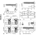
- OVA peptide- or control peptide-pulsed target cells (syngeneic splenocytes labeled with either high or low CFSE) were injected into immunized WT and B7-H1-deficient mice. Four hours following cell injection, the remaining CFSE positive cells in the spleen were analyzed. Memory CD8+ T cells in the B7-H1-deficient mice lysed more OVA-peptide pulsed target cells (33.5%) than those in WT mice (9.3%, p ⁇ 0.01; FIG. 2D ). Collectively, these data suggest that B7-H1 negatively regulates the generation of memory CD8+ T cells in immunized mice.
- B16-OVA melanoma tumor cells (engineered to express OVA) were injected into immunized WT and B7-H1-deficient mice. Intravenously injected B16-OVA tumor cells form metastases in the lung, and antitumor immunity can be monitored by counting the number of tumor foci.
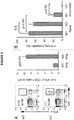
- the frequency of functional memory CD8+ T cells in the lungs of WT and B7-H1-deficient mice was determined by intracellular staining for IFN ⁇ . About 4 to 5-fold more IFN ⁇ + CD8+ T cells were detected in the lungs of B7-H1-deficient mice as compared with WT mice (p ⁇ 0.01; FIG. 3A ).
- CD11a was used as a surrogate activation marker.
- An advantage of this method is that CD11a high CD8+ T cells represent antigen-primed CD8+ T cells that are responsive to undefined antigen epitopes not recognized by tetramers. CD11a high CD8+ T cells were detected at low levels in the spleens of naive WT and B7-H1-deficient mice ( FIG. 4A ).
- CD11a high CD8+ T cells Nearly 80-90% of OVA-induced CD11a high CD8+ T cells were reactive against undefined antigen epitopes of the OVA protein ( FIG. 4C ). Therefore, the CD11a high CD8+ T-cell population could be used to represent a majority of the antigen-primed CD8+ T cells during primary T-cell responses. In the following studies, CD11a high was used as a marker to track antigen-specific CD8+ T cells.
- TMRE tetramethylrhodamine ethyl ester
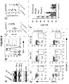
- OT-1 CD8+ T cells transferred into B7-H1-deficient hosts expressed lower levels of Bim in both the spleen and liver as compared with the OT-1 CD8+ T cells transferred into WT hosts ( FIG. 7 ).
- the expression of Bcl-2 and Bcl-x L in OT-1 CD8+ T cells transferred into WT or B7-H1-deficient mice was comparable ( FIG. 7 ).
- CD80 signaling in the regulation of memory generation was addressed by transferring equal numbers of CD80-deficient OT-1 and WT OT-1 naive CD8+ T cells into CD45.1+ mice.
- host mice were immunized with OVA plus poly I:C.
- the frequency and phenotype of the transferred CD80-deficient and WT OT-1 CD8+ T cells was analyzed.
- B7-H1 might regulate Bim levels in activated CD8+ T cells.
- Pre-activated WT CD8+ T cells were incubated with platebound B7-H1 fusion protein for 48 hours in the presence of TCR stimulation (anti-CD3 antibody).
- Bim expression was analyzed by western blotting, and increased expression levels were observed in CD8+ T cells cultured in the presence of B7-H1 fusion protein, as compared with a control fusion protein ( FIG. 8A ).
- Bim expression also was analyzed by intracellular flow cytometry, revealing that the B7-H1 fusion protein dramatically increased the levels of Bim protein in CD8+ T cells compared with a control fusion protein (p ⁇ 0.02; FIGS. 8B and 8C ).
- pre-activated WT CD8+ T cells were incubated with plate-bound B7-H1 fusion protein pre-blocked with anti-B7-H1 (10B5 or 43H12) or anti-PDl (G4) antibodies.
- the 10B5 antibody blocks the interaction of B7-H1 with both PD-1 and CD80.
- Both 10B5 and G4 antibodies completely blocked Bim upregulation induced by B7-H1 fusion protein, while 43H12 only partially, but significantly, did so ( FIG. 8F ).
- B7-H1 regulates Bim expression levels was then examined.
- mRNA levels of Bcl211 which encodes the Bim protein, were examined by quantitative real-time PCR analysis using mRNA isolated from pre-activated CD8+ T cells that were exposed to B7-H1 fusion protein or to a control fusion protein and anti-CD3 for 24 hours. Incubation of pre-activated CD8+ T cells with B7-H1 fusion protein did not increase the levels of Bcl211 ( FIG. 9A ), indicating that the B7-H1-mediated upregulation of Bim does not result from transcriptional regulation.
- the degradation of Bim is tightly regulated, at least in part via the activation of Akt followed by Akt-mediated Bim phosphorylation and degradation ( Qi et al. (2006) J. Biol. Chem. 281:813-823 ).
- the level of Akt activation in CD8+ T cells after B7-H1 engagement was measured by intracellular flow cytometry for phosphorylated-Akt (Ser473).
- CD8+ T cells cultured with B7-H1 fusion protein exhibited decreased levels of phosphorylated Akt as compared with CD8+ T cells cultured with a control fusion protein (p ⁇ 0.01; FIGS. 9B and 9C ).
- Example 5 - Bim is increased in tumor-reactive CD8+ T cells in peripheral blood of melanoma and prostate cancer patients
- Peripheral blood lymphocytes were isolated from 26 patients with stage IV (advanced) melanoma, and from 11 healthy blood donors. Lymphocytes were stained with CD8, CD11a and PD-1 followed with intracellular staining for Bim. High expression of CD11a by CD8 T cells was used to identify antigen-primed T cells. Tumor-reactive CD8+ T cells were defined by their expression of CD11a high and PD-1+ ( FIG. 13 , left panel). The histograms shown in the right panel of FIG. 13 indicate expression of Bim by subsets of CD8+ T cells (Tn: T naive cells; PD-1-, PD-1 negative primed cells; PD-1 +, PD-1 positive primed cells).
- PD-1+ primed cells (tumor-reactive) CD8+ T cells expressed high levels of Bim.
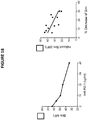
- Bim expression was increased in tumor-reactive CD8+ T cells in peripheral blood of melanoma patients as compared to the healthy controls, and also was increased in tumor-reactive CD8+ T cells in peripheral blood of prostate cancer patients as compared to healthy controls ( FIG. 14 ).
- the Bim upregulation in melanoma patients was PD-1 dependent, as depicted in FIG. 15 .
- Bim expression was not increased in PD-1+ T cells in healthy donors, suggesting that Bim upregulation is dependent on PD-1 expression and is cancer-related.
- Example 6 B7-H1 protein induces high expression of Bim in human pre-activated CD8+ T cells
- Example 7 Anti-PD-1 treatment reduced the frequency of Bim+ PD-1+ tumor-reactive CD8 T cells
- Example 8 B7-H1 expressed by tumor cells induces Bim up-regulation in human pre-activated CD8 T cells
- Example 9 - Bim expression is associated with B7-H1 expression in human RCC
- B7-H1 The ability of B7-H1 to up-regulate Bim in pre-activated, but not newly activated, CD8+ T cells, implied that reactivation of tumor-reactive CD8+ T cells at tumor sites could be dampened through this mechanism by B7-H1 positive tumor cells.
- human cancer tissues stained for B7-H1 and Bim were evaluated. The hypothesis was that B7-H1 positive human cancer tissues would be associated with more Bim positive tumor-infiltrating lymphocytes (TILs). As shown in FIG. 21 (left panel), human renal cell carcinoma tissues were stained with anti-B7-H1 and anti-Bim antibodies in immunohistochemistry assays.
- B7-H1 reactivity was identified on the surface of cancer cells, while Bim positive staining was identified on cancer cells and also on TILs ( FIG. 21 , left panel). Bim reactivity was determined by an arbitrary scoring system: 0 (absence), 1(focal), 2 (moderate), and 3 (marked). The association between B7-H1 positive or negative tumors and the frequency of Bim reactivity at different levels is demonstrated in the right panel of FIG. 21 , and was analyzed using Fisher's exact test. B7-H1 positive tumors were found in general to have a higher degree (2-3 scores) of Bim positive TILs than B7-H1 negative tumors (Fisher's exact test, p ⁇ 0.01). These results suggest that B7-H1 positive tumors can induce more death in tumor-reactive T cells at tumor sites via up-regulation of Bim when these T cells are re-activated with tumor antigen stimulation.
- Example 10 - Bim expression is correlated with Granzyme B and T-bet expressed by cancer-related PD-1+ CD11a high CD8+ T cells
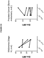
- Bim levels were measured in PD-1+ CD11a high CD8 T cells from the peripheral blood of patients with melanoma and prostate cancers before and post radiotherapy. As shown in FIG. 23 (left panel), decreased levels of Bim were observed in melanoma patients post radiotherapy. In contrast, increased levels of Bim were observed in prostate cancer patients post radiotherapy (right panel). Due to the limited numbers of patients in this study, these changes did not reach statistical significance.
Landscapes
- Health & Medical Sciences (AREA)
- Chemical & Material Sciences (AREA)
- Immunology (AREA)
- Organic Chemistry (AREA)
- Life Sciences & Earth Sciences (AREA)
- Proteomics, Peptides & Aminoacids (AREA)
- Genetics & Genomics (AREA)
- General Health & Medical Sciences (AREA)
- Molecular Biology (AREA)
- Biochemistry (AREA)
- Biophysics (AREA)
- Medicinal Chemistry (AREA)
- Engineering & Computer Science (AREA)
- Pathology (AREA)
- Analytical Chemistry (AREA)
- Zoology (AREA)
- Wood Science & Technology (AREA)
- Biotechnology (AREA)
- Physics & Mathematics (AREA)
- Microbiology (AREA)
- Oncology (AREA)
- Hospice & Palliative Care (AREA)
- Bioinformatics & Cheminformatics (AREA)
- General Engineering & Computer Science (AREA)
- Animal Behavior & Ethology (AREA)
- Nuclear Medicine, Radiotherapy & Molecular Imaging (AREA)
- Pharmacology & Pharmacy (AREA)
- Chemical Kinetics & Catalysis (AREA)
- Public Health (AREA)
- Veterinary Medicine (AREA)
- General Chemical & Material Sciences (AREA)
- Medicines Containing Antibodies Or Antigens For Use As Internal Diagnostic Agents (AREA)
- Cell Biology (AREA)
- Hematology (AREA)
- Urology & Nephrology (AREA)
- Biomedical Technology (AREA)
- Peptides Or Proteins (AREA)
- Food Science & Technology (AREA)
- General Physics & Mathematics (AREA)
Description
- This document relates to materials and methods for treating cancer, and more particularly to materials and methods for treating cancer in patients identified as having elevated levels of Bim.
- The metastatic spread of tumor cells is the primary cause of cancer related mortality, indicating a need for therapeutic approaches capable of controlling or preventing metastasis (Gibbons et al. (2012) OncoImmunology 1(7):1061-1073; and Grivennikov et al. (2010) Cell 140:883-899). The presence of tumor-infiltrating effector and memory T cells is correlated with decreased metastatic spread, consistent with a role for T cells in preventing metastasis of primary tumors.
- B7-H1 (also referred to as PD-L1) is a polypeptide expressed by a variety of tumor cells. It also is constitutively expressed by macrophages and dendritic cells, and its expression is up-regulated upon cell activation. PD-1 is expressed on the surface of activated T cells, B cells, and macrophages, and is a receptor for B7-H1. CD80 is found on activated B cells and monocytes, and provides a costimulatory signal necessary for T cell activation and survival; CD80 also binds B7-H1.
- Topalian SL et al. describe in The New England Journal of Medicine, vol. 366, no. 26, 28 June 2012, on pages 2443-2454 the safety, activity, and immune correlates of an anti-PD-1 antibody in cancer.
WO 2013/090552 A1 describes compositions and methods for the therapeutic intervention of signaling through EP2 and EP4, in combination with the therapeutic intervention of signaling through PD-1, by inhibiting at least one of EP2, EP4, PGE2, or combinations thereof, in combination with inhibiting at least one of PD-1, PD-L1, PD-L2, and combinations thereof. - Berrien-Elliot MM et al. describe in Cancer Research, vol. 73, no. 2, 15 January 2013, on pages 605-616, a durable adoptive immunotherapy for leukemia produced by manipulation of multiple regulatory pathways of CD8+ T-cell tolerance.
- Gibbons RM et al. describe in Oncoimmunology vol. 1, no. 7, 1 October 2012, on pages 1061-1073 that B7-H1 limits the entry of effector CD8+ T cells to the memory pool by upregulating Bim.
- The present invention is defined by
independent claim 1. The dependent claims depict other embodiments of the invention. - This document provides, inter alia, a method for determining whether PD-1 on T cells has engaged its ligand, B7-H1. The method is based in part on the discovery that engagement of PD-1 by B7-H1 results in up-regulation of Bim, a pro-apoptotic molecule, and is correlated with B7-H1-mediated T cell death. This discovery suggests that intracellular levels of Bim among PD-1 positive cells is a barometer of the extent to which PD-1 has been triggered by B7-H1, with lower levels of Bim identifying activated PD-1 positive T cells whose PD-1 molecules have not yet been extensively engaged, and higher levels of Bim reflecting chronic engagement of PD-1 with B7-H1. Stratifying Bim levels among PD-1 positive CD8 T cells may be a biomarker for gauging (1) whether PD-1 molecules on CD8 T cells have been engaged by B7-H1 tumor associated ligands; and (2) the efficacy of an anti-PD-1 or anti-B7-H1 blockade regimen in reducing PD-1 engagement. Thus, using Bim as a signaling biomarker for PD-1 function, it may be possible to select patients more likely to benefit from checkpoint blockade therapy and to identify optimal therapeutic timing and dosing schedules.
- In one aspect, this document features a composition for use in a method for treating a mammal having cancer, wherein said method comprises: (a) identifying said mammal as containing an elevated level of Bim, and (b) administering to said mammal an anti-B7-H1 antibody, an anti-PD-1 antibody, an anti-CD80 antibody, a fusion protein comprising a portion of PD-1 linked to an immunoglobulin Fc sequence, or a fusion protein comprising a portion of CD80 linked to an Ig Fc sequence, under conditions wherein the interaction of naturally-occurring B7-H1 with PD-1 or CD80 in said mammal is reduced after said administering. The mammal can be a human. The cancer can be a melanoma cancer, a breast cancer, a lung cancer, a renal cell carcinoma cancer, a pancreas cancer, a prostate cancer, a colon cancer, a brain cancer, a liver cancer, or an ovarian cancer.
- Unless otherwise defined, all technical and scientific terms used herein have the same meaning as commonly understood by one of ordinary skill in the art to which this invention pertains. Although methods and materials similar or equivalent to those described herein can be used to practice the invention, suitable methods and materials are described below. In case of conflict, the present specification, including definitions, will control. In addition, the materials, methods, and examples are illustrative only and not intended to be limiting.
- The details of one or more embodiments of the invention are set forth in the accompanying drawings and the description below. Other features, objects, and advantages of the invention will be apparent from the description and drawings, and from the claims.
-
-
FIG. 1 contains a pair of graphs plotting the kinetics of CD8+ T-cell responses to antigen stimulation. Wild type (WT) and B7-H1-deficient (KO) mice were immunized (i.p.) with OVA plus poly I:C, and Kb/OVA tetramer was used to identify antigen-specific CD8+ T cells in spleen (top panel) and liver (bottom panel) at the indicated times after immunization. Data show the percentage of tetramer+ CD8+ T cells (mean ± SD of three mice per time point). One of two independent experiments is shown. *p < 0.05 compared with WT mice. -
FIGS. 2A-2D contain a series of FACS scans and graphs showing enhanced memory CD8+ T-cell population in the absence of B7-H1. Mice were immunized with OVA plus poly I:C, and were re-stimulated with OVA onday 40 after immunization. Onday 4 after re-stimulation, spleen cells were isolated from naive or immunized WT and B7-H1-deficient mice for analysis.FIG. 2A , FACS scans showing the percentage of OVA- specific tetramer+ CD8+ T cells; *p < 0.05 compared with WT mice.FIG. 2B graph plotting the absolute number of OVA- specific tetramer+ CD8+ T cells (mean ± SD, n = 3).FIG. 2C , FACS analysis of intracellular production of cytokines in CD8+ T cells from immunized mice (mean ± SD, n = 3).FIG. 2D , graphs plotting in vivo cytolytic activity in immunized mice. OVA- peptide or control-peptide pulsed target cells (syngeneic splenocytes) were labeled with high or low dose CFSE (5 µM for OVA-peptide pulsed cells; 0.5 µM for control-peptide pulsed cells) and mixed (1:1, 2.5 × 10⁶ of each) and injected i.v. into WT or B7-H1-deficient mice. Histogram plots (left) show the percentage of remaining target cells in thespleen 4 hours after target cell transfer. Bar graph (right) shows percentage of specific lysis in the spleen (mean ± SD, n = 3). -
FIGS. 3A and 3B contain FACS scans and graphs showing enhanced memory CD8+ T-cell recall responses and improved antitumor immunity in the lung in the absence of B7-H1. Onday 35 after immunization, immunized or naive WT and B7-H1-deficient mice were injected (i.v.) with 5 × 10⁵ B16-OVA tumor cells.FIG. 3A , percentage and absolute numbers of IFNy+ CD8+ T cells in the lung of immunized mice (mean ± SD, n = 3) onday 4 after tumor injection. *p < 0.01 compared with WT mice.FIG. 3B , metastatic tumor foci in the lung tissue were identified and counted onday 20 after tumor injection (mean ± SD, n = 5). N.S.: not significant. -
FIGS. 4A-4D contain a series of FACS scans and a graph showing that CD11ahigh CD8+ T cells represent antigen-primed effector T cells. Spleen cells from naive or immunized WT and B7-H1-deficient mice were analyzed by co-staining with anti-C D11a and Kb/OVA tetramer or functional markers.FIG. 4A , FACS scans showing the percentage of CD11ahigh CD8+ T cells from WT and B7-H1-deficient immunized mice.FIG. 4B , graph plotting average percentage of CD11ahigh CD8+ T cells from WT and B7-H1-deficient immunized mice (mean ± SD, n = 4).FIG. 4C , FACS scans showing the percentage of antigen-specific tetramer+ (Kb/OVA-tet) cells in CD11ahigh and CD11alow CD8+ T-cell population.FIG. 4D , FACS scans showing CTL functional assay of CD11ahigh and CD11alow CD8+ T cells after a brief re-stimulation in vitro. Degranulation of CTLs was analyzed by CD107a mobilization, followed by intracellular staining for IPNγ. Numbers indicate percentages of gated areas. -
FIGS. 5A-5D contain a series of FACS scans and a graph showing fewer apoptotic antigen-primed CD8+ T cells in B7-H1-deficient mice. On day 7 after immunization, spleen cells were analyzed for proliferation and apoptosis.FIGS. 5A and 5B , FACS scans showing Ki67 expression and BrdU incorporation, respectively, analyzed in CD11ahigh or CD11alow CD8+ T cells. Numbers are percentages of gated area in total CD8+ T cells.FIG. 5C , FACS scans of TMRElow Annexin V+ apoptotic cells measured in CD11ahigh and CD11alow CD8+ T cells.FIG. 5D , graph plotting the percentage of apoptotic cells (TMRElow Annexin V+) in CD11ahigh CD8+ T cells (mean ± SD, n = 4). -
FIGS. 6A-6D contain a series of histograms and a graph showing lower Bim levels in antigen-primed CD8+ T cells in B7-H1-deficient mice.FIG. 6A , flow cytometry assay of the intracellular expression of Bim, Bcl-2 and Bcl-xL in gated CD11ahigh CD8+ T cells in the spleen of WT (red) and B7-H1-deficient (blue) mice on day 7 after immunization. Numbers are mean fluorescence intensity (MFI) of Bim expression.FIG. 6B , graph showing average MFI of Bim expressed by CD11ahigh CD8+ T cells (mean ± SD, n = 9).FIG. 6C , intracellular expression of Bim in CD11ahigh CD8+ T cells in the liver of immunized mice. Numbers are MFI.FIG. 6D , Bim expression in total CD8+ T cells in the spleen of naive WT (red) and B7-H1-deficient (blue) mice. -
FIG. 7 contains a series of histograms showing the extrinsic role of B7-H1 in regulation of Bim. WT OT-1 CD8+ T cells (Thy1.1+) were transferred in WT (red) or B7-H1-deficient (blue) host mice one day before immunization with OVA plus poly I:C. On day 7 after immunization, the OT-1 CD8+ T cells in the spleen and liver were identified by the Thy1.1 marker and analyzed for intracellular expression of Bim. Numbers are MFI. -
FIGS. 8A-8F show that B7-H1 co-stimulation induces upregulation of Bim protein levels in activated T cells. Pre-activated CD8+ T cells were incubated with platebound B7-H1 or control fusion protein (Fc) for 48 hours in the presence of anti-CD3.FIG. 8A , Western blot showing Bim isoform expression in CD8+ T cells.FIG. 8B , histogram showing expression of total Bim in CD8+ T cells co-stimulated with B7-H1 (blue) or control protein (red). Numbers are MFI.FIG. 8C , graph plotting average MFI of Bim expressed by activated CD8+ T cells (mean ± SD, n = 5).FIG. 8D , graph plotting the percentage of live (trypan blue exclusive) CD8+ T cells in culture (mean ± SD, n = 5).FIG. 8E , FACS scans indicating apoptosis of CD8+ T cells isolated from WT, Bim-deficient, and Bcl-2 transgenic (Tg) mice. Numbers show percentage of TMRElow Annexin V+ apoptotic T cells in total CD8+ T cells.FIG. 8F , graph plotting average MFI of Bim expressed by CD8+ T cells in culture with anti-B7-H1 Ab (10B5, blocking B7-H1 binding to both PD-1 and CD80; 43H12, blocking B7-H1 binding to CD80 only), anti-PD-1 Ab (G4), or control Ab (10 µg/mL of each) (mean ± SD, n = 3). -
FIGS. 9A-9C contain a series of plots showing that B7-H1 co-stimulation inhibits activation of Akt. Pre-activated CD8+ T cells were stimulated with plate-bound B7-H1 or control fusion protein (Fc). After 24 hours of stimulation, CD8+ T cells were harvested and used for analysis.FIG. 9A , graph plotting analysis of Bcl2l11 transcript levels by real-time qPCR using the comparative CT method. GAPDH served as the internal control gene. Graph shows fold change (mean ± SD, n = 4).FIG. 9B , histograms plotting phosphorylation of Akt (left) and mTOR (right), analyzed by intracellular staining of CD8+ T cells with anti-phospho-Akt and anti-phospho-mTOR Abs. Numbers show percentage of positive stained cells.FIG. 9C , bar graph plotting average MFI of phospho-Akt and phospho-mTOR expression (mean ± SD, n = 3). N.S.: not significant. -
FIG. 10 contains representative nucleic acid (top) and amino acid (bottom) sequences for human B7-H1 (SEQ ID NOS:1 and 2, respectively). -
FIG. 11 contains representative nucleic acid (top) and amino acid (bottom) sequences for human PD-1 (SEQ ID NOS:3 and 4, respectively). -
FIGS. 12A and12B contain representative nucleic acid (12A) and amino acid (12B) sequences for human CD80 (SEQ ID NOS:5 and 6, respectively). -
FIG. 13 contains a plot (left) showing the identification of CD8+ T cells based on their expression of CD11ahigh and PD-1+ (left panel). Lymphocytes were stained with CD8, CD11a and PD-1, followed by intracellular staining for Bim.FIG. 13 also contains a histogram plotting expression of Bim by subsets of CD8+ T cells (Tn: T naive cells; PD-1- , PD-1 negative primed cells; PD-1 +, PD-1 positive primed cells). Only the PD-1+ primed cells CD8+ T cells expressed high levels of Bim. -
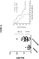
FIG. 14 is a pair of graphs plotting the level of Bim expression in tumor-reactive PD-1+ CD11ahigh CD8+ T cells in the peripheral blood of 26 melanoma patients as compared to 11 normal, healthy controls (left panel, P=0.007 by unpaired Student T test), and in tumor-reactive PD-1+ CD11ahigh CD8+ T cells in the peripheral blood of 11 prostate cancer patients as compared to 11 normal, healthy controls (right panel, P=0.001 by unpaired Student T test). -
FIG. 15 is a pair of graphs plotting Bim expression in PD-1 negative (PD-1-) and PD-1 positive (PD-1+) CD11ahigh CD8+ T cells from melanoma patients (left) and healthy controls (right). Bim was significantly increased in the PD-1+ populations (p=0.0081) in melanoma patients. -
FIG. 16 is a pair of graphs plotting the level of Bim expression in tumor-reactive PD-1+ CD11ahigh CD8+ T cells in the peripheral blood of 26 melanoma patients as compared to 11 normal, healthy controls (left panel, indicating "Bim low" and "Bim high" samples), and plotting the survival rate for "Bim low" vs. "Bim high" patients (right panel). -
FIG. 17A is a pair of graphs showing that B7-H1 protein induced expression of Bim in human pre-activated CD8+ T cells.FIG. 17B is a picture of a Western blot showing Bim levels in the cells. -
FIG. 18 is a pair of graphs showing that an anti-PD-1 antibody significantly blocked B7-H1-induced Bim up-regulation in a dose dependent fashion (left panel), and that the blocking effects of the anti-PD-1 antibody were inversely correlated with the higher levels of Bim induced by B7-H1 (right panel). -
FIG. 19 is a pair of graphs showing that the frequency of Bim+PD-1+ CD8 T cells was significantly higher in the peripheral blood of melanoma patients before treatment than in a healthy control group (left panel), and that after anti-PD-1 antibody therapy, about 67% of the melanoma patients demonstrated a significant reduction in the frequency of Bim+PD-1+ CD8 T cells (right panel). -
FIG. 20 is a pair of graphs showing that B7-H1 expressed by tumor cells induced Bim up-regulation in human pre-activated CD8 T cells. -
FIG. 21 is a pair of photographs (left) and a graph (right) showing that Bim expression was associated with B7-H1 expression in human renal cell carcinoma (RCC). In particular, the graph in the right panel shows that Bim+ tumor infiltrating lymphocytes (TILs) were increased in B7-H1 positive tumor tissues. Bim reactivity scores: 0, absence; 1, focal; 2, moderate; 3, marked. Contingency analysis using Fisher's exact test (p<0.01). -
FIG. 22 is a pair of graphs showing that Bim expression was correlated with Granzyme B and T-bet (a transcription factor of effector T cells) expressed by cancer-related PD-1+ CD11ahigh CD8+ T cells, suggesting that Bim expression is associated with effector CD8+ T cell differentiation. -
FIG. 23 is a pair of graphs showing that Bim expression declined in PD-1+ CD11ahigh CD8 T cells following radiotherapy in some melanoma (left panel) and prostate (right panel) cancer patients. - This document provides materials and methods for identifying patients as being more likely to benefit from checkpoint blockade therapy, materials and methods for determining optimal therapeutic timing and dosing schedules, and methods and materials for treating cancer. For example, this document provides methods and materials for identifying a mammal (e.g., a human) as having an elevated level of Bim, and treating the mammal with a molecule that can interfere with the interaction between B7-H1 and PD-1, and/or the interaction between B7-H1 and CD80 (e.g., an antibody against B7-H1, PD-1, or CD80, or with a fusion protein containing a portion of PD-1 or a portion of CD80 fused to an immunoglobulin (Ig) Fc domain). As described herein, elevated levels of Bim can be related to increased apoptosis of antigen-primed CD8+ T cells, but inhibiting the interaction of B7-H1 with PD-1 or CD80 can lead to reduced levels of Bim and reduced T cell apoptosis.
- The term "elevated level" as used herein with respect to a level of Bim refers to a level that is greater (e.g., 50% greater, 2-fold greater, 3-fold greater, or more than 3-fold greater) than a reference level of Bim. The term "reference level" as used herein with respect to Bim refers to the level of Bim typically observed in healthy mammals without cancer. For example, a reference level of Bim can be the average level of Bim present in samples obtained from a random sampling of 50 humans free of cancer.
- The presence of an elevated level of Bim can be determined by measuring, for example, protein levels or nucleic acid levels. For example, the level of Bim protein can be measured in a sample of blood (e.g., a peripheral blood sample) or another bodily fluid from a mammal with cancer or from a control mammal, using cell staining, western blotting, or other immunological techniques. The level of Bim expression also can be measured at the nucleic acid level, using Northern blotting, or any other method suitable for determining mRNA levels of Bcl2l11, which encodes the Bim protein. In some cases, Bim protein or nucleic acid levels can be measured in tumor tissue samples, ascites samples, or lymphoid organ samples. It will be appreciated that levels from comparable samples are used when determining whether or not a particular level is an elevated level.
- A representative example of a human B7-H1 nucleic acid has the sequence set forth in GENBANK® Accession No. AF177937 (GI No. 6708118) (SEQ ID NO:1;
Figure 10 ), and a representative human B7-H1 polypeptide has the sequence set forth in GENBANK® Accession No. AAF25807 (GI No. 6708119) (SEQ ID NO:2;Figure 10 ). - A representative example of a human PD-1 nucleic acid can have the sequence set forth in GENBANK® Accession No. BC074740.2 (GI No. 50960296) (SEQ ID NO:3;
Figure 11 ), and representative example of a human PD-1 polypeptide has the sequence set forth in GENBANK® Accession No. AAH74740.1 (GI No. 49902307) (SEQ ID NO:4;Figure 11 ). - A representative example of a human CD80 nucleic acid has the sequence set forth in NCBI Reference No. NM_005191.3 (GI No. 113722122) (SEQ ID NO:5;
Figure 12A ), and a representative example of a human CD80 polypeptide has the sequence set forth in NCBI Reference No. NP_005182.1 (GI No. 4885123) (SEQ ID NO:6;Figure 12B ). - Once the level of Bim within a sample from a mammal is determined, the level can be compared to a reference level and used to classify the mammal as having or lacking an elevated level of Bim.
- Once a mammal has been identified as having an elevated level of Bim as described herein, the mammal can be administered a molecule that inhibits the interaction between B7-H1 and PD-1 and/or the interaction between B7-H1 and CD80. Examples of such molecules include, without limitation, antibodies (e.g., anti-B7-H1 antibodies, anti-PD-1 antibodies, or anti-CD80 antibodies), and fusion proteins (e.g., PD-1 fusion proteins or CD80 fusion proteins). Such fusion proteins can contain, for example, the extracellular domain of PD-1 fused to an IgG Fc domain, or the extracellular domain of CD80 fused to an IgG Fc domain. After administration, the antibody/ies or fusion protein(s) can bind B7-H1, thus reducing or blocking B7-H1's action in inducing Bim up regulation.
- The term "antibody" includes monoclonal antibodies, polyclonal antibodies, recombinant antibodies, humanized antibodies (Jones et al. (1986) Nature 321:522-525; Riechmann et al. (1988) Nature 332:323-329; and Presta (1992) Curr. Op. Struct. Biol. 2:593-596), chimeric antibodies (Morrison et al. (1984) Proc. Natl. Acad. Sci. USA 81:6851-6855), multispecific antibodies (e.g., bispecific antibodies) formed from at least two antibodies, and antibody fragments. The term "antibody fragment" comprises any portion of the afore-mentioned antibodies, such as their antigen binding or variable regions. Examples of antibody fragments include Fab fragments, Fab' fragments, F(ab')2 fragments, Fv fragments, diabodies (Hollinger et al. (1993) Proc. Natl. Acad. Sci. USA 90:6444-6448), single chain antibody molecules (Plückthun in: The Pharmacology of Monoclonal Antibodies 113, Rosenburg and Moore, eds., Springer Verlag, N.Y. (1994), 269-315) and other fragments as long as they exhibit the desired capability of binding to B7-H1, PD-1, or CD80.
- Examples of anti-human B7-H1 antibodies include, without limitation, anti-human B7-H1 antibodies commercially available from Biolegend (e.g., Catalog No. 329701 or 329702; San Diego, CA) or eBioscience (e.g., Catalog No. 14-5983-80 or 14-5983-82).
- Examples of anti-human PD-1 antibodies include, without limitation, anti-human PD-1 antibodies commercially available from Biolegend (e.g., Catalog No. 329904 or 329905) or eBioscience (Catalog No. 12-2799-42; San Diego, CA).
- Examples of anti-human CD80 antibodies include, without limitation, anti-human CD8 antibodies commercially available from Biolegend (e.g., Catalog No. 305201 or 305202) or eBioscience (e.g., Catalog No. 14-0809-80 or 14-0809-82).
- The term "antibody," as used herein, also includes antibody-like molecules that contain engineered sub-domains of antibodies or naturally occurring antibody variants. These antibody-like molecules may be single-domain antibodies such as VH-only or VL-only domains derived either from natural sources such as camelids (Muyldermans et al. (2001) Rev. Mol. Biotechnol. 74:277-302) or through in vitro display of libraries from humans, camelids or other species (Holt et al. (2003) Trends Biotechnol. 21:484-90). In certain embodiments, the polypeptide structure of the antigen binding proteins can be based on antibodies, including, but not limited to, minibodies, synthetic antibodies (sometimes referred to as "antibody mimetics"), human antibodies, antibody fusions (sometimes referred to as "antibody conjugates"), and fragments thereof, respectively.
- An "Fv fragment" is the minimum antibody fragment that contains a complete antigen-recognition and -binding site. This region consists of a dimer of one heavy chain variable domain and one light chain variable domain in tight, non-covalent association. It is in this configuration that the three CDR's of each variable domain interact to define an antigen-binding site on the surface of the VH-VL dimer. Collectively, the six CDR's confer antigen-binding specificity to the antibody. However, even a single variable domain (or half of an Fv comprising only three CDR's specific for an antigen) has the ability to recognize and bind the antigen, although usually at a lower affinity than the entire binding site. The "Fab fragment" also contains the constant domain of the light chain and the first constant domain (CH1) of the heavy chain. The "Fab fragment" differs from the "Fab' fragment" by the addition of a few residues at the carboxy terminus of the
heavy chain C H1 domain, including one or more cysteines from the antibody hinge region. The "F(ab')2 fragment" originally is produced as a pair of "Fab' fragments" which have hinge cysteines between them. Methods of preparing such antibody fragments, such as papain or pepsin digestion, are known to those skilled in the art. - An antibody can be of the IgA-, IgD-, IgE, IgG- or IgM-type, including IgG- or IgM-types such as, without limitation, IgG1-, IgG2-, IgG3-, IgG4-, IgM1- and IgM2-types. For example, in some cases, the antibody is of the IgG1-, IgG2- or IgG4- type.
- In some embodiments, antibodies as used in the methods described herein can be fully human or humanized antibodies. Human antibodies can avoid certain problems associated with xenogeneic antibodies, such as antibodies that possess murine or rat variable and/or constant regions. First, because the effector portion is human, it can interact better with other parts of the human immune system, e.g., to destroy target cells more efficiently by complement-dependent cytotoxicity or antibody-dependent cellular cytotoxicity. Second, the human immune system should not recognize the antibody as foreign. Third, half-life in human circulation will be similar to naturally occurring human antibodies, allowing smaller and less frequent doses to be given. Methods for preparing human antibodies are known in the art.
- In addition to human antibodies, "humanized" antibodies can have many advantages. Humanized antibodies generally are chimeric or mutant monoclonal antibodies from mouse, rat, hamster, rabbit or other species, bearing human constant and/or variable region domains or specific changes. Techniques for generating humanized antibodies are well known to those of skill in the art. For example, controlled rearrangement of antibody domains joined through protein disulfide bonds to form new, artificial protein molecules or "chimeric" antibodies can be utilized (Konieczny et al. (1981) Haematologia (Budap.) 14:95). Recombinant DNA technology can be used to construct gene fusions between DNA sequences encoding mouse antibody variable light and heavy chain domains and human antibody light and heavy chain constant domains (Morrison et al. (1984) Proc. Natl. Acad. Sci. USA 81:6851).
- DNA sequences encoding antigen binding portions or complementarity determining regions (CDR's) of murine monoclonal antibodies can be grafted by molecular means into DNA sequences encoding frameworks of human antibody heavy and light chains (Jones et al. (1986) Nature 321:522; Riechmann et al. (1988) Nature 332:323). Expressed recombinant products are called "reshaped" or humanized antibodies, and comprise the framework of a human antibody light or heavy chain and antigen recognition portions, CDR's, of a murine monoclonal antibody.
- Other methods for designing heavy and light chains and for producing humanized antibodies are described in, for example,
U.S. Patent Nos. 5,530,101 ;5,565,332 ;5,585,089 ;5,639,641 ;5,693,761 ;5,693,762 ; and5,733,743 . Yet additional methods for humanizing antibodies are described inU.S. Patent Nos. 4,816,567 ;4,935,496 ;5,502,167 ;5,558,864 ;5,693,493 ;5,698,417 ;5,705,154 ;5,750,078 ; and5,770,403 , for example. - Molecules that interfere with the interaction between B7-H1 and PD-1, and/or the interaction between B7-H1 and CD80, as described herein (e.g., antibodies against B7-H1, PD-1, and CD80, as well as fusion proteins containing portions of PD-1 or CD80 linked to an Ig Fc domain), can be incorporated into pharmaceutical compositions for treatment of cancer. Thus, this document also provides the use of such molecules in the manufacture of medicaments for treating cancer. The compositions can further include one or more pharmaceutically acceptable carriers, diluents and/or adjuvants. The potency of the pharmaceutical compositions provided herein typically is based on the binding of the antibody or fusion protein to B7-H1.
- A "pharmaceutically acceptable carrier" (also referred to as an "excipient" or a "carrier") is a pharmaceutically acceptable solvent, suspending agent, stabilizing agent, or any other pharmacologically inert vehicle for delivering one or more therapeutic compounds to a subject, which is nontoxic to the cell or mammal being exposed thereto at the dosages and concentrations employed. Pharmaceutically acceptable carriers can be liquid or solid, and can be selected with the planned manner of administration in mind so as to provide for the desired bulk, consistency, and other pertinent transport and chemical properties, when combined with one or more of therapeutic compounds and any other components of a given pharmaceutical composition. Typical pharmaceutically acceptable carriers that do not deleteriously react with amino acids include, by way of example and not limitation: water, saline solution, binding agents (e.g., polyvinylpyrrolidone or hydroxypropyl methylcellulose), fillers (e.g., lactose and other sugars, gelatin, or calcium sulfate), lubricants (e.g., starch, polyethylene glycol, or sodium acetate), disintegrates (e.g., starch or sodium starch glycolate), and wetting agents (e.g., sodium lauryl sulfate). Pharmaceutically acceptable carriers also include aqueous pH buffered solutions or liposomes (small vesicles composed of various types of lipids, phospholipids and/or surfactants which are useful for delivery of a drug to a mammal). Further examples of pharmaceutically acceptable carriers include buffers such as phosphate, citrate, and other organic acids, antioxidants such as ascorbic acid, low molecular weight (less than about 10 residues) polypeptides, proteins such as serum albumin, gelatin, or immunoglobulins, hydrophilic polymers such as polyvinylpyrrolidone, amino acids such as glycine, glutamine, asparagine, arginine or lysine, monosaccharides, disaccharides, and other carbohydrates including glucose, mannose or dextrins, chelating agents such as EDTA, sugar alcohols such as mannitol or sorbitol, salt-forming counterions such as sodium, and/or nonionic surfactants such as TWEEN™, polyethylene glycol (PEG), and PLURONICS™.
- Pharmaceutical compositions can be formulated by mixing one or more active agents with one or more physiologically acceptable carriers, diluents, and/or adjuvants, and optionally other agents that are usually incorporated into formulations to provide improved transfer, delivery, tolerance, and the like. A pharmaceutical composition can be formulated, e.g., in lyophilized formulations, aqueous solutions, dispersions, or solid preparations, such as tablets, dragees or capsules. A multitude of appropriate formulations can be found in the formulary known to all pharmaceutical chemists: Remington's Pharmaceutical Sciences (18th ed, Mack Publishing Company, Easton, PA (1990)), particularly Chapter 87 by Block, Lawrence, therein. These formulations include, for example, powders, pastes, ointments, jellies, waxes, oils, lipids, lipid (cationic or anionic) containing vesicles (such as LIPOFECTIN™), DNA conjugates, anhydrous absorption pastes, oil-in-water and water-in-oil emulsions, emulsions carbowax (polyethylene glycols of various molecular weights), semi-solid gels, and semi-solid mixtures containing carbowax. Any of the foregoing mixtures may be appropriate in treatments and therapies as described herein, provided that the active agent in the formulation is not inactivated by the formulation and the formulation is physiologically compatible and tolerable with the route of administration. See, also, Baldrick (2000) Regul. Toxicol. Pharmacol. 32:210-218; Wang (2000) Int. J. Pharm. 203:1-60; Charman (2000) J. Pharm. Sci. 89:967-978; and Powell et al. (1998) PDA J. Pharm. Sci. Technol. 52:238-311), and the citations therein for additional information related to formulations, excipients and carriers well known to pharmaceutical chemists.
- Pharmaceutical compositions include, without limitation, solutions, emulsions, aqueous suspensions, and liposome-containing formulations. These compositions can be generated from a variety of components that include, for example, preformed liquids, self-emulsifying solids and self-emulsifying semisolids. Emulsions are often biphasic systems comprising of two immiscible liquid phases intimately mixed and dispersed with each other; in general, emulsions are either of the water-in-oil (w/o) or oil-in-water (o/w) variety. Emulsion formulations have been widely used for oral delivery of therapeutics due to their ease of formulation and efficacy of solubilization, absorption, and bioavailability.
- Compositions and formulations can include sterile aqueous solutions, which also can contain buffers, diluents and other suitable additives (e.g., penetration enhancers, carrier compounds and other pharmaceutically acceptable carriers). Compositions additionally can contain other adjunct components conventionally found in pharmaceutical compositions. Thus, the compositions also can include compatible, pharmaceutically active materials such as, for example, antipruritics, astringents, local anesthetics or anti-inflammatory agents, or additional materials useful in physically formulating various dosage forms of the compositions provided herein, such as dyes, flavoring agents, preservatives, antioxidants, opacifiers, thickening agents and stabilizers. Furthermore, the composition can be mixed with auxiliary agents, e.g., lubricants, preservatives, stabilizers, wetting agents, emulsifiers, salts for influencing osmotic pressure, buffers, colorings, flavorings, and aromatic substances. When added, however, such materials should not unduly interfere with the biological activities of the polypeptide components within the compositions provided herein. The formulations can be sterilized if desired.
- In some embodiments, a composition containing an antibody or fusion protein as provided herein (e.g., an anti-B7-H7, anti-PD-1, or anti-CD80 antibody, or a PD-1 FC or CD80 Fc fusion protein) can be in the form of a solution or powder with or without a diluent to make an injectable suspension. The composition may contain additional ingredients including, without limitation, pharmaceutically acceptable vehicles, such as saline, water, lactic acid, mannitol, or combinations thereof, for example.
- Any appropriate method can be used to administer a molecule as described herein to a mammal. Administration can be, for example, parenteral (e.g., by subcutaneous, intrathecal, intraventricular, intramuscular, or intraperitoneal injection, or by intravenous drip). Administration can be rapid (e.g., by injection) or can occur over a period of time (e.g., by slow infusion or administration of slow release formulations). In some embodiments, administration can be topical (e.g., transdermal, sublingual, ophthalmic, or intranasal), pulmonary (e.g., by inhalation or insufflation of powders or aerosols), or oral. In addition, a composition containing an antibody or fusion protein as described herein can be administered prior to, after, or in lieu of surgical resection of a tumor.
- A composition containing an antibody (e.g., an anti-B7-H1 antibody, anti-PD-1 antibody, or anti-CD80 antibody) or a fusion protein (e.g., a PD-1 Fc fusion or a CD80 Fc fusion) can be administered to a mammal in any appropriate amount, at any appropriate frequency, and for any appropriate duration effective to achieve a desired outcome (e.g., to increase progression-free survival). In some cases, a composition containing an antibody or fusion protein as described herein can be administered to a mammal having cancer to reduce the progression rate of the cancer by 5, 10, 25, 50, 75, 100, or more percent. For example, the progression rate can be reduced such that no additional cancer progression is detected. Any appropriate method can be used to determine whether or not the progression rate of cancer is reduced. For skin cancer (e.g., melanoma), for example, the progression rate can be assessed by imaging tissue at different time points and determining the amount of cancer cells present. The amounts of cancer cells determined within tissue at different times can be compared to determine the progression rate. After treatment as described herein, the progression rate can be determined again over another time interval. In some cases, the stage of cancer after treatment can be determined and compared to the stage before treatment to determine whether or not the progression rate has been reduced.
- In some cases, a composition containing an antibody or a fusion protein as described herein can be administered to a mammal having cancer under conditions where progression-free survival is increased (e.g., by 5, 10, 25, 50, 75, 100, or more percent) as compared to the median progression-free survival of corresponding mammals having untreated cancer or the median progression-free survival of corresponding mammals having cancer and treated with other therapies (e.g., chemotherapeutic agents). Progression-free survival can be measured over any length of time (e.g., one month, two months, three months, four months, five months, six months, or longer).
- Administration to a mammal of a molecule as set forth herein can result in increased numbers of naturally-occurring tumor-reactive CD8+ T cells, which can exert anti-cancer effects against cancer cells present within the mammal.
- An effective amount of a composition containing a molecule as provided herein can be any amount that reduces the progression rate of cancer, increases the progression-free survival rate, or increases the median time to progression without producing significant toxicity to the mammal. Optimum dosages can vary depending on the relative potency of individual polypeptides (e.g., antibodies and fusion proteins), and can generally be estimated based on EC50 found to be effective in in vitro and in vivo animal models. Typically, dosage is from 0.01 µg to 100 g per kg of body weight. For example, an effective amount of an antibody or fusion protein can be from about 1 mg/kg to about 100 mg/kg (e.g., about 5 mg/kg, about 10 mg/kg, about 20 mg/kg, about 50 mg/kg, or about 75 mg/kg). If a particular mammal fails to respond to a particular amount, then the amount of the antibody or fusion protein can be increased by, for example, two fold. After receiving this higher concentration, the mammal can be monitored for both responsiveness to the treatment and toxicity symptoms, and adjustments made accordingly. The effective amount can remain constant or can be adjusted as a sliding scale or variable dose depending on the mammal's response to treatment. Various factors can influence the actual effective amount used for a particular application. For example, the frequency of administration, duration of treatment, use of multiple treatment agents, route of administration, and severity of the cancer may require an increase or decrease in the actual effective amount administered.
- The frequency of administration can be any frequency that reduces the progression rate of cancer, increases the progression-free survival rate, or increases the median time to progression without producing significant toxicity to the mammal. For example, the frequency of administration can be once or more daily, biweekly, weekly, monthly, or even less. The frequency of administration can remain constant or can be variable during the duration of treatment. A course of treatment can include rest periods. For example, a composition containing an antibody or fusion protein as provided herein can be administered over a two week period followed by a two week rest period, and such a regimen can be repeated multiple times. As with the effective amount, various factors can influence the actual frequency of administration used for a particular application. For example, the effective amount, duration of treatment, use of multiple treatment agents, route of administration, and severity of the cancer may require an increase or decrease in administration frequency.
- An effective duration for administering a composition provided herein can be any duration that reduces the progression rate of cancer, increases the progression-free survival rate, or increases the median time to progression without producing significant toxicity to the mammal. Thus, the effective duration can vary from several days to several weeks, months, or years. In general, the effective duration for the treatment of cancer can range in duration from several weeks to several months. In some cases, an effective duration can be for as long as an individual mammal is alive. Multiple factors can influence the actual effective duration used for a particular treatment. For example, an effective duration can vary with the frequency of administration, effective amount, use of multiple treatment agents, route of administration, and severity of the cancer.
- After administering a composition provided herein to a mammal, the mammal can be monitored to determine whether or not the cancer was treated. For example, a mammal can be assessed after treatment to determine whether or not the progression rate of the cancer has been reduced (e.g., stopped). Any method, including those that are standard in the art, can be used to assess progression and survival rates.
- The invention will be further described in the following examples, which do not limit the scope of the invention described in the claims.
- Mice, cell lines and reagents: Female CD45.2+ C57BL/6 mice were purchased from Taconic Farms and CD45.1+ congenic C57BL/6-Ly5.1 mice were purchased from National Cancer Institute. OT-1 TCR (Thy 1.1+) transgenic mice were provided by T. Tian (Harvard University, Boston, MA). B7-H1-deficient C57BL/6 mice were provided by L. Chen (Yale University, New Haven, CT; Dong et al., Immunity 20:327-336, 2004). Bcl211-/- mice and Cd80-/- mice were purchased from Jackson Laboratory. Cd80-/-mice were crossbred into WT OT-1 mice and produced Cd80-/- OT-1 mice. Bcl-2 transgenic mice were provided by V. Shapiro (Mayo Clinic, Rochester). Mice were maintained under pathogen-free conditions and used at 8-12 weeks of age. B16-OVA murine melanoma cells were provided by R. Vile (Mayo Clinic, Rochester, MN), and were cultured in RPMI 1640 medium (Cellgro) with 10% FBS (Life Technologies), 1 U/mL penicillin, 1 µg/mL streptomycin and 20 mM HEPES buffer (all from Mediatech). Hamster anti-mouse B7-H1 mAb (10B5) and PD-1 (G4) was obtained from hybridoma cells provided by L. Chen. Hamster anti-mouse B7-H1 mAb (43H12) was provided by K. Tamada (John Hopkins University).
- Flow cytometry analysis: Class I MHC (KbOVA peptide SIINFEKL; SEQ ID NO:1) tetramer and negative control tetramer were purchased from Beckman Coulter. Fluorochrome-conjugated Abs against CD8, CD11a, Fas (CD95), Fas ligand, CD90.1 (Thy 1.1), CD90.2 (Thy 1.2), CD107a, IPNγ, IL-2 and TNFα were purchased from BD Biosciences, BioLegend, or eBiosciences. To detect intracellular cytokine levels, cells were incubated with GolgiPlug (BD Biosciences) for 4 hours prior to analysis. Cells were stained for surface antigens, and then incubated in Fixation Buffer (BioLegend) for 20 minutes at room temperature, followed by permeabilization in Permeabilization Wash Buffer (BioLegend). Fixed and permeabilized cells were then stained with Abs for 20 minutes at room temperature. Abs to Akt, Bcl-xL, Bcl-2, Bim and mTOR and fluorochrome-conjugated secondary Abs were purchased from Cell Signaling (Danvers, MA). To detect intracellular levels of Akt, Bcl-xL, Bcl-2, Bim and mTOR, T cells were first stained for surface antigens, then fixed with 2% paraformaldehyde for 10 minutes at 37°C, followed by permeabilization with ice-cold methanol for 30 minutes. After blocking with 15% rat serum for 15 minutes, cells were stained with Abs for 1 hour at room temperature. After staining, cells were washed three times with incubation buffer before analysis. At least 100,000 viable cells were live gated on FACScan or FACSCailbur (BD Biosciences) instrumentation. Flow cytometry analysis was performed using FlowJo software (Tree Star).
- T-cell immunization, activation, apoptosis assay and proliferation assay: Mice were immunized by i.p. injection of 0.5 mg ovalbumin (OVA, from Sigma-Aldrich) and 50 µg poly (I:C) (Sigma Aldrich). For in vitro T-cell activation and apoptosis assay, purified CD8+ T cells were labeled with CFSE (Invitrogen-Molecular Probes) and incubated with OVA peptide257-264 (Mayo Clinic Core Facilities) at 0.2 µg/mL for 72 hours. Apoptosis of CD8+ T cells was analyzed by staining using Annexin V (BD Biosciences) and TMRE (tetramethylrhodamine ethyl ester, Invitrogen/Molecular Probes T-669). Proliferation was also measured by detection of BrdU incorporation and Ki67 staining. Immunized mice were injected i.p. with 0.8 mg/mL BrdU (BD Biosciences) on
days 4 through 6 following immunization. On day 7 after immunization BrdU incorporation was determined by intra-nuclear staining with anti-BrdU (B9285, Sigma-Aldrich) and anti-Ki67 (556027, BD Biosciences). - In vivo CTL assay. For the in vivo CTL assay, OVA257-264 peptide-pulsed or control peptide-pulsed spleen cells (as target cells) from syngeneic mice were labeled with a high dose of CFSE (5 µM) or low dose of CFSE (0.5 µM), mixed at 1:1 (2.5 × 10⁶ of each) before injection. Target cells were i.v. injected into immunized mice on
day 4 after re-challenge with cognate antigen protein. The CTL activity was determined 4 hours after target cell transfer. Specific lysis is calculated using the following formulas:
- Tumor studies: Mice were inoculated i.v. with 5 × 10⁵ B16-OVA tumor cells on
day 25 after immunization. On day 21-post tumor injection, mice were sacrificed and the lung tissue was perfused with PBS. The number of tumor foci on the lung tissue was counted. - T-cell transfer experiments: Purified CD8+ T cells (1 × 10⁶) from Thy1.1+ OT-1 transgenic mice were i.v. injected into Thy 1.2+ WT or B7-H1-deficient recipient mice, followed by immunization with OVA plus poly I:C. On day 7 after immunization, transferred CD8+ T cells were identified by their expression of Thy1.1 and used for detection of intracellular expression of Bim, Bcl-2 and Bcl-xL. Equal numbers of Cd80-/- (CD45.2+) and WT OT-1 (Thy1.1+, CD45.2+) CD8+ T cells (106 of each) were i.v. injected into CD45.1+ mice followed with immunization of OVA and poly I:C. The transferred OT-1 CD8+ T cells in the spleen were identified by flow cytometry.
- In vitro T-cell activation and culturing with fusion proteins: Spleen cells were harvested from naive mice and pre-activated with ConA (5 µg/mL, L7647, Sigma-Aldrich) for 48 hours. Following activation, CD8+ T cells were purified (EasySep CD8+ T-cell negative selection kit, Stem Cell Technologies) and incubated with plate-bound anti-CD3 (BD Biosciences) and B7-H1 Fc fusion protein or control Fc protein (R&D Systems). Cultures were maintained for indicated time periods, and then cells were harvested for analysis.
- Western blotting: Cells were lysed with NETN buffer (0.5% NP40, 150 mM NaCl, 50 mM Tris and 1 mM EDTA). Cell lysates were boiled and run on SDS-PAGE gels (BioRad), transferred to nitrocellulose membrane (Millipore), and blotted using standard procedures.
- Quantitative RT-PCR: Total RNA was isolated from purified CD8+ T cells (RNeasy Kit, Qiagen), and reverse transcribed (iScriptcDNA synthesis kit, BioRad). Samples were analyzed for Bim transcript levels using Bcl211 primers (Qiagen) and QuantiFast SYBR Green PCR Master Mix (Qiagen) on an iCycler (BioRad). GAPDH levels were used to normalize data by the comparative CT method.
- Statistical analysis: A two-sided, unpaired or paired Student's t-test was used to assess statistical differences in experimental groups. A p value < 0.05 was considered statistically significant.
- The kinetics of CD8+ T-cell responses in the spleen and liver of WT and B7-H1-deficient C57BL/6 mice were compared following immunization with ovalbumin (OVA) protein and polyinosinic:polycytidylic acid (poly (I:C)) as adjuvant. An increased number of CD8+ T cells was observed at the peak of the immune response (day 7-post immunization) in the spleen and liver of B7-H1-deficient mice as compared with WT mice. During the contraction phase (days 7 to 14 post-immunization), there was a significant delay in the reduction of antigen-specific CD8+ T cells in the spleen and liver of B7-H1-deficient mice as compared with WT mice. On
day 40 following immunization, more antigen-specific memory CD8+ T cells were detected in B7-H1-deficient mice as compared with WT mice (FIG. 1 ). These data suggested that host B7-H1 may regulate the extent of expansion and contraction of effector CD8+ T cells, thus influencing the size of the memory CD8+ T-cell pool in both lymphoid and non-lymphoid tissues. - Studies were conducted to examine the extent to which B7-H1 regulates the generation of memory CD8+ T cells in immunized mice, using KbOVA257-264 tetramer (KbOVA-tet) to detect antigen-primed memory CD8+ T cells in the spleen on
day 4 after in vivo restimulation (OVA protein, administered onday 40 after primary immunization).Day 4 was selected for analysis because at this time point it is possible to distinguish a recall response from the primary response (which takes 7 days to establish). Thus, naive mice did not show a significant increase of antigen-specific CD8+ T cells onday 4 after immunization (FIG. 2A ). The frequency of KbOVA-tet+ CD8+ T cells increased more than 2-fold in immunized B7-H1-deficient mice (0.38%) as compared with WT mice (0.16%; p < 0.05;FIG. 2A ). This increase was reflected in the absolute cell numbers (p = 0.001;FIG. 2B ). In addition to having increased numbers of memory CD8+ T cells, an increased percentage of memory CD8+ T cells capable of producing multiple cytokines was detected in the spleens of B7-H1-deficient mice (0.73% IFNγ+/TNFα+, 0.17% IFNγ+/IL-2+) as compared with WT mice (0.24% IFNγ+/TNFα+, 0.07% IFNγ+/IL-2+; p < 0.05;FIG. 2C ). An in vivo CTL assay to measure cytolytic activity of the memory CD8+ T cells also was performed. Onday 4 after in vivo re-stimulation, OVA peptide- or control peptide-pulsed target cells (syngeneic splenocytes labeled with either high or low CFSE) were injected into immunized WT and B7-H1-deficient mice. Four hours following cell injection, the remaining CFSE positive cells in the spleen were analyzed. Memory CD8+ T cells in the B7-H1-deficient mice lysed more OVA-peptide pulsed target cells (33.5%) than those in WT mice (9.3%, p < 0.01;FIG. 2D ). Collectively, these data suggest that B7-H1 negatively regulates the generation of memory CD8+ T cells in immunized mice. - A hallmark of memory CD8+ T cells is their rapid recall response to cognate antigens, so studies were conducted to determine whether the increased memory pool in B7-H1-deficient mice would lead to a more protective recall response. B16-OVA melanoma tumor cells (engineered to express OVA) were injected into immunized WT and B7-H1-deficient mice. Intravenously injected B16-OVA tumor cells form metastases in the lung, and antitumor immunity can be monitored by counting the number of tumor foci. On
day 4 following intravenous injection of 5 × 10⁵ B16-OVA tumor cells, the frequency of functional memory CD8+ T cells in the lungs of WT and B7-H1-deficient mice was determined by intracellular staining for IFNγ. About 4 to 5-fold more IFNγ+ CD8+ T cells were detected in the lungs of B7-H1-deficient mice as compared with WT mice (p < 0.01;FIG. 3A ). On day 21-post tumor injection, the number of tumor metastases in the lungs of naive B7-H1-deficient mice was comparable to that of naive WT mice (p = 0.43;FIG. 3B ). Fewer tumor metastases formed in the lungs of immunized WT mice as compared with naive WT mice (p = 0.001). Significantly, tumor metastases were completely rejected in the lungs of immunized B7-H1-deficient mice (FIG. 3B ), suggesting that a more efficient CD8+ T-cell memory population is established in the absence of B7-H1. - Studies were conducted to determine which mechanisms could be responsible for the increased population of memory CD8+ T cells in B7-H1-deficient mice by examining the proliferation and apoptosis of antigen-primed CD8+ T cells following immunization. CD11a was used as a surrogate activation marker. An advantage of this method is that CD11ahigh CD8+ T cells represent antigen-primed CD8+ T cells that are responsive to undefined antigen epitopes not recognized by tetramers. CD11ahigh CD8+ T cells were detected at low levels in the spleens of naive WT and B7-H1-deficient mice (
FIG. 4A ). On day 7 after immunization, the percentage of CD11ahigh CD8+ T cells increased more than 2-fold in the spleens of B7-H1-deficient mice (41.5%) as compared with immunized WT mice (17.2%; p < 0.01;FIGS. 4A and 4B ), consistent with the results obtained by tetramer staining (FIG. 1 ). Seven to 15% of CD11ahigh CD8+ T cells from WT and B7-H1-deficient mice were specific for the known H-2Kb-restricted OVA257-264 epitope based on tetramer staining, and CD11alow CD8+ T cells did not contain tetramer+ cells (FIG. 4C ), suggesting that all antigen-specific CD8+ T cells are found in the CD11ahigh CD8+ T-cell population. In addition, the CD11ahigh CD8+ population from both WT and B7-H1-deficient mice, but not the CD11alow CD8+ T-cell population, produced IFNγ and underwent degranulation (indicated by CD107a surface expression) following ex vivo re-stimulation (FIG. 4D ). As T-cell responses against diverse epitopes are coordinately regulated, these data further support the concept that the CD11ahigh CD8+ T-cell population represents true OVA-specific CD8+ T cells. Nearly 80-90% of OVA-induced CD11ahigh CD8+ T cells were reactive against undefined antigen epitopes of the OVA protein (FIG. 4C ). Therefore, the CD11ahigh CD8+ T-cell population could be used to represent a majority of the antigen-primed CD8+ T cells during primary T-cell responses. In the following studies, CD11ahigh was used as a marker to track antigen-specific CD8+ T cells. - The proliferation of effector CD8+ T cells following immunization was examined by staining cells for Ki67, a nuclear protein associated with cell proliferation (Gerdes et al. (1984) J. Immunol. 133:1710-1715). The percent of Ki67+ cells increased in CD11ahigh CD8+ T cells from B7-H1-deficient mice (9.32%) as compared with WT mice (7.5%), but this increase was not statistically significant (
FIG. 5A ). Proliferation also was monitored by performing a BrdU incorporation assay to measure the ongoing proliferation of CD8+ T cells following immunization. In this assay, the percentage of BrdU+ CD11ahigh CD8+ T cells also was similar between WT (6.05%) and B7-H1-deficient mice (5.59%;FIG. 5B ). Ki67+ or BrdU+ cells were mainly detected in the CD11ahigh CD8+ T cells but not in CD11alow CD8+ T cells, suggesting that CD11ahigh CD8+ T cells are proliferating following antigen-stimulation (FIGS. 5A and 5B ). These results suggested that the observed increased population of antigen-primed CD8+ T cells in B7-H1-deficient mice is not due to an increased proliferation of this cell compartment, as compared with WT mice. - Studies were then conducted to evaluate whether decreased apoptosis of antigen-primed CD8+ T cells could contribute to the observed increased population of antigen-primed CD8+ T cells in immunized B7-H1-deficient mice. As discussed above, the Fas/Fas ligand death receptor pathway is implicated in regulation of T-cell contraction, so the surface expression levels of Fas and Fas ligand on effector CD8+ T cells were measured on day 7 after immunization. Expression of Fas and Fas ligand was detected at similar levels in WT and B7-H1-deficient mice. These results suggest that the observed increased population of effector CD8+ T cells is not due to a change in Fas-induced apoptosis in B7-H1-deficient mice. The mitochondrial pathway for apoptosis was investigated by analyzing levels of Annexin V and tetramethylrhodamine ethyl ester (TMRE) staining. TMRE is a fluorescent marker that is incorporated into intact mitochondria, and cells undergoing apoptosis show reduced TMRE staining as compared with live cells (Jayaraman, J. Immunol. Methods 306:68-79, 2005). These studies revealed that fewer antigen-primed CD11ahigh CD8+ T cells were undergoing apoptosis (TMRElow Annexin V+) in B7-H1-deficient mice (3.4%) as compared with WT mice (6.7%, p < 0.05;
FIGS. 5C and 5D ). These results suggested that decreased levels of mitochondrial apoptosis may contribute to the observed increased population of antigen-primed CD8+ T cells in B7-H1-deficient mice. - Experiments were conducted to look for alterations in the expression of apoptosis-regulating molecules in effector CD8+ T cells. Intracellular levels of the pro-apoptotic molecule Bim and the anti-apoptotic molecules Bcl-2 and Bcl-xL were measured in CD11ahigh CD8+ T cells freshly isolated from the spleen on day 7 after immunization of naive mice. Lower intracellular expression levels of Bim were observed in CD11ahigh CD8+ T cells from B7-H1-deficient mice than in the same cells obtained from WT mice (p < 0.001;
FIGS. 6A and 6B ), while the expression levels of Bcl-2 and Bcl-xL were comparable in WT and B7-H1-deficient mice (FIG. 6A ). The expression of Bim, Bcl-2 and Bcl-xL were comparable in CD11alow CD8+ T cells from B7-H1-deficient and WT mice (FIG. 6A ). Intracellular expression levels of Bim also were analyzed in CD11ahigh CD8+ T cells isolated from the liver on day 7 after immunization of naive mice. Again, lower intracellular expression levels of Bim were observed in CD11ahigh CD8+ T cells from B7-H1-deficient mice as compared with WT mice (FIG. 6C ). Finally, intracellular expression levels of these proteins were examined in CD11ahigh CD8+ T cells isolated from the spleen of naive mice, and no significant differences were observed in B7-H1-deficient vs. WT mice in the expression levels of Bim (FIG. 6D ), Bcl-2 or Bcl-xL. These data suggested that the downregulation of the pro-apoptotic molecule Bim may contribute to the observed increased population of antigen-primed effector CD8+ T cells in B7-H1-deficient mice. - To exclude the possibility that the downregulation of Bim in B7-H1-deficient mice would be due to an intrinsic change in B7-H1-deficient T cells, transfer experiments were performed in which naive OT-1 CD8+ T cells (Thy1.1+) were injected into WT or B7-H1-deficient mice (Thy1.2+). Following transfer of the OT-1 CD8+ T cells, host mice were immunized with OVA plus poly I:C. On day 7 after immunization, the intracellular levels of Bim, Bcl-2, and Bcl-xL were measured in transferred OT-1 CD8+ T cells freshly isolated from spleen and liver. OT-1 CD8+ T cells transferred into B7-H1-deficient hosts expressed lower levels of Bim in both the spleen and liver as compared with the OT-1 CD8+ T cells transferred into WT hosts (
FIG. 7 ). The expression of Bcl-2 and Bcl-xL in OT-1 CD8+ T cells transferred into WT or B7-H1-deficient mice was comparable (FIG. 7 ). These data suggested that the downregulation of Bim in B7-H1-deficient mice is not due to an intrinsic change in B7-H1-deficient T cells, but rather to host B7-H1 interacting with one of its binding partners on CD8+ T cells. - Next, antibodies that block the interaction between B7-H1 and PD-1 or between B7-H1 and CD80 were used to examine if blocking either of these pathways would impact Bim expression levels. On
days - After an acute viral infection, more central memory T cells accumulate in the lymphoid organs of PD-1-deficient mice as compared with WT mice, indicating that PD-1 signaling negatively regulates memory T-cell generation (Allie et al. (2011) J. Immunol. 186:6280-6286). The relevance of CD80 signaling in the regulation of memory generation was addressed by transferring equal numbers of CD80-deficient OT-1 and WT OT-1 naive CD8+ T cells into CD45.1+ mice. One day after T-cell transfer, host mice were immunized with OVA plus poly I:C. On day 21 after immunization, the frequency and phenotype of the transferred CD80-deficient and WT OT-1 CD8+ T cells was analyzed. On day 21 after immunization, a 2-fold increased percentage of CD80-deficient OT-1 CD8+ T cells as compared with WT OT-1 CD8+ T cells was detected in the spleen, indicating that the transferred CD80-deficient OT-1 CD8+ T cells generated more memory T cells as compared with WT OT-1 CD8+ T cells. Surface staining confirmed that these cells had a central memory phenotype (CD44hi CD62Lhi). The recall response of the memory population generated from transferred cells was investigated by injecting the hosts with OVA plus poly I:C on
day 30 after the initial immunization, and 3 days later the frequency and phenotype of the transferred cells was analyzed. An increased percentage of CD80-deficient OT-1 CD8+ T cells as compared with WT OT-1 CD8+ T cells was detected in the spleen (p = 0.013). Surface staining confirmed that these cells had an effector memory phenotype (CD44hi CD62Llo). Taken together, these data demonstrated that Cd80-/- OT-1 CD8+ T cells generated more memory T cells as compared their WT counterparts, indicating that CD80 expressed by CD8+ T cells may negatively regulate memory T-cell generation. - Studies were conducted to investigate how B7-H1 might regulate Bim levels in activated CD8+ T cells. Pre-activated WT CD8+ T cells were incubated with platebound B7-H1 fusion protein for 48 hours in the presence of TCR stimulation (anti-CD3 antibody). Bim expression was analyzed by western blotting, and increased expression levels were observed in CD8+ T cells cultured in the presence of B7-H1 fusion protein, as compared with a control fusion protein (
FIG. 8A ). Bim expression also was analyzed by intracellular flow cytometry, revealing that the B7-H1 fusion protein dramatically increased the levels of Bim protein in CD8+ T cells compared with a control fusion protein (p < 0.02;FIGS. 8B and 8C ). In the absence of anti-CD3 antibodies, Bim levels did not increase upon incubation with B7-H1 fusion protein, suggesting that B7-H1 provides a co-stimulatory signal for Bim upregulation. Accordingly, the absolute number of live cells was also reduced in CD8+ T cells cultured in the presence of B7-H1 fusion protein compared with a control protein (p < 0.01;FIG. 8D ). Increased levels of cells undergoing apoptosis (TMRElow Annexin V+) were observed in cultures of activated CD8+ T cells exposed to the B7-H1 fusion protein and anti-CD3 (12.4%) as compared with cells cultured with control fusion protein and anti-CD3 (4.1%,FIG. 8E ). The induction of apoptosis by B7-H1 fusion protein was lost in CD8+ T cells isolated from Bim-deficient and Bcl-2 transgenic mice (FIG. 8E ), suggesting that B7-H1-induced T-cell apoptosis may be dependent on the Bim-mediated mitochondrial pathway of apoptosis. - To examine which receptor of B7-H1 is involved in mediating Bim upregulation, pre-activated WT CD8+ T cells were incubated with plate-bound B7-H1 fusion protein pre-blocked with anti-B7-H1 (10B5 or 43H12) or anti-PDl (G4) antibodies. The 10B5 antibody blocks the interaction of B7-H1 with both PD-1 and CD80. Both 10B5 and G4 antibodies completely blocked Bim upregulation induced by B7-H1 fusion protein, while 43H12 only partially, but significantly, did so (
FIG. 8F ). None of the antibodies used in this experiment had effects on Bim expression levels in cells cultured with control fusion protein, indicating that their effect on Bim expression levels is due to blocking the interaction between B7-H1/PD-1 or B7-H1/CD80, and not due to a non-specific effect. These results suggest that B7-H1 may use PD-1 or CD80 on CD8+ T cells to deliver co-stimulatory signals for the upregulation of Bim. - The mechanism by which B7-H1 regulates Bim expression levels was then examined. mRNA levels of Bcl211, which encodes the Bim protein, were examined by quantitative real-time PCR analysis using mRNA isolated from pre-activated CD8+ T cells that were exposed to B7-H1 fusion protein or to a control fusion protein and anti-CD3 for 24 hours. Incubation of pre-activated CD8+ T cells with B7-H1 fusion protein did not increase the levels of Bcl211 (
FIG. 9A ), indicating that the B7-H1-mediated upregulation of Bim does not result from transcriptional regulation. The degradation of Bim is tightly regulated, at least in part via the activation of Akt followed by Akt-mediated Bim phosphorylation and degradation (Qi et al. (2006) J. Biol. Chem. 281:813-823). The level of Akt activation in CD8+ T cells after B7-H1 engagement was measured by intracellular flow cytometry for phosphorylated-Akt (Ser473). CD8+ T cells cultured with B7-H1 fusion protein exhibited decreased levels of phosphorylated Akt as compared with CD8+ T cells cultured with a control fusion protein (p < 0.01;FIGS. 9B and 9C ). As phosphorylation of Akt at Ser473 is regulated by activation of mTOR (Sarbassov et al. (2005) Science 307:1098-1101; and Jacinto et al. (2006) Cell 127:125-137), studies were conducted to examine whether B7-H1 regulates phosphorylation of mTOR in vitro. Unexpectedly, there was no difference in levels of phospho-mTOR in CD8+ T cells cultured with B7-H1 fusion protein and cells cultured with control fusion protein (FIGS. 9B and 9C ). These results suggested that CD8+ T-cell engagement with B7-H1 inhibits the activation of Akt, resulting in decreased degradation of Bim. - Peripheral blood lymphocytes were isolated from 26 patients with stage IV (advanced) melanoma, and from 11 healthy blood donors. Lymphocytes were stained with CD8, CD11a and PD-1 followed with intracellular staining for Bim. High expression of CD11a by CD8 T cells was used to identify antigen-primed T cells. Tumor-reactive CD8+ T cells were defined by their expression of CD11ahigh and PD-1+ (
FIG. 13 , left panel). The histograms shown in the right panel ofFIG. 13 indicate expression of Bim by subsets of CD8+ T cells (Tn: T naive cells; PD-1-, PD-1 negative primed cells; PD-1 +, PD-1 positive primed cells). Of note, only PD-1+ primed cells (tumor-reactive) CD8+ T cells expressed high levels of Bim. Bim expression was increased in tumor-reactive CD8+ T cells in peripheral blood of melanoma patients as compared to the healthy controls, and also was increased in tumor-reactive CD8+ T cells in peripheral blood of prostate cancer patients as compared to healthy controls (FIG. 14 ). The Bim upregulation in melanoma patients was PD-1 dependent, as depicted inFIG. 15 . When levels of Bim were compared between PD-1 negative (PD-1-) and PD-1 positive (PD-1+) CD11ahigh CD8+ T cells, Bim was found to be significantly increased in the PD-1+ populations (p=0.0081) in melanoma patients. In contrast, Bim expression was not increased in PD-1+ T cells in healthy donors, suggesting that Bim upregulation is dependent on PD-1 expression and is cancer-related. - Further, when melanoma patients were broken into "Bim low" vs. "Bim high" categories based on the level of Bim expression in tumor-reactive PD-1+ CD11ahigh CD8+ T cells in the peripheral blood (
FIG. 16 , left panel), the survival rate for patients with Bimhigh PD-1+ CD8+ T cells was reduced as compared to the survival rate for patients with. Bimlow PD-1+ CD8+ T cells (FIG. 16 , right panel). - Since Bim up-regulation is a consequence of interaction between B7-H1 and PD-1, experiments were conducted to test whether an anti-PD-1 blocking antibody can reduce B7-H1-induced Bim up-regulation in T cells. An in vitro system was established in which pre-activated human primary CD8+ T cells were incubated with a B7-H1/PD-L1 fusion protein to induce Bim up-regulation. As shown in
FIG. 17A (right panel), a significant up-regulation of Bim (presented as MFI) was induced the B7-H1/PD-L1 fusion (P<0.05, n=6). The increased Bim expression is further demonstrated by a flow cytometry histogram (FIG. 17A , left panel) and a Western blotting assay (FIG. 17B ). - Using this system, several commercially available anti-human PD-1 antibodies were screened for their blocking effects, and one anti-PD-1 antibody (clone MIH4) was identified that significantly blocked B7-H1-induced Bim up-regulation in a dose dependent fashion (
FIG. 18 , left panel). Since B7-H1 induced different degrees of Bim up-regulation in individual healthy donors, experiments were conducted to examine whether different degrees of Bim up-regulation would affect the blocking effects of the anti-PD-1 antibody. Interestingly, it was observed that higher levels of Bim induced by B7-H1 had a negative correlation with Bim reduction by anti-PD-1 blocking antibody (FIG. 18 , right panel; Pearson R = - 0.71, n = 12, P < 0.05). These results suggested that pre-existing Bim levels in CD8+ T cells might affect the efficiency of anti-PD-1 blockade. Thus, measuring Bim levels before treatment could help to determine the degree to which an anti-PD-1 antibody might block the impact of PD-1 signals on antitumor T cell responses. - Next, studies were conducted to evaluate the impact of the anti-PD-1 antibody on Bim expression by tumor-reactive CD8 T cells in cancer patients. Peripheral blood lymphocytes were collected from patients with advanced melanoma (Stage IV) before and 12 weeks post anti-PD-1 treatment. Tumor-reactive CD8 T cells were identified by their high expression of CD11a and expression of PD-1. Bim expression was analyzed by intracellular staining. The percentage of Bim+PD-1+ in CD11ahigh CD8+ T cells was compared between healthy people and melanoma patients, and between melanoma patients before and after treatment with anti-PD-1 antibody. As shown in
FIG. 19 , the frequency of Bim+PD-1+ CD8 T cells in the peripheral blood of melanoma patients (before treatment, n = 29) was significantly higher than in the healthy control group (p = 0.0012, n = 14), suggesting that more PD-1+ CD8 T cells are under the influence of the PD-1/B7-H1 interaction that leads to up-regulation of Bim. Importantly, twelve weeks after anti-PD-1 antibody therapy (2 mg/kg, one cycle), about 67% (6/9) of the melanoma patients demonstrated a significant reduction in the frequency of Bim+PD-1+ CD8 T cells (p = 0.023, n=6). These results indicated that measurement of Bim expressed by PD-1+ CD8 T cells could be used to monitor the responses of cancer patients to anti-PD-1 therapy, which may block B7-H1-induced Bim up-regulation. A reduced frequency or level of Bim expressed by PD-1+ CD8 T cells in cancer patients after anti-PD-1 therapy may be used to assess which patients are responsive to the therapy. - Since most human solid tumor cells express elevated levels of B7-H1, the function of tumor cell-expressed B7-H1 in T cell Bim expression was examined. Pre-activated human primary CD8 T cells were incubated with cells from a human melanoma line (624mel) that were transfected with B7-H1 cDNA or with control mock cDNA, for 24 hours. As shown in
FIG. 20 , intracellular expression of Bim was dramatically increased in CD8 T cells cultured with B7-H1/624mel cells, as compared to mock/624mel cells (p < 0.01). This result suggested that B7-H1 expressed by human tumor cells has the potential to up-regulate Bim in pre-activated CD8 T cells. - The ability of B7-H1 to up-regulate Bim in pre-activated, but not newly activated, CD8+ T cells, implied that reactivation of tumor-reactive CD8+ T cells at tumor sites could be dampened through this mechanism by B7-H1 positive tumor cells. To test this possibility, human cancer tissues stained for B7-H1 and Bim were evaluated. The hypothesis was that B7-H1 positive human cancer tissues would be associated with more Bim positive tumor-infiltrating lymphocytes (TILs). As shown in
FIG. 21 (left panel), human renal cell carcinoma tissues were stained with anti-B7-H1 and anti-Bim antibodies in immunohistochemistry assays. B7-H1 reactivity was identified on the surface of cancer cells, while Bim positive staining was identified on cancer cells and also on TILs (FIG. 21 , left panel). Bim reactivity was determined by an arbitrary scoring system: 0 (absence), 1(focal), 2 (moderate), and 3 (marked). The association between B7-H1 positive or negative tumors and the frequency of Bim reactivity at different levels is demonstrated in the right panel ofFIG. 21 , and was analyzed using Fisher's exact test. B7-H1 positive tumors were found in general to have a higher degree (2-3 scores) of Bim positive TILs than B7-H1 negative tumors (Fisher's exact test, p<0.01). These results suggest that B7-H1 positive tumors can induce more death in tumor-reactive T cells at tumor sites via up-regulation of Bim when these T cells are re-activated with tumor antigen stimulation. - To examine whether up-regulation of Bim is associated with effector T cells, the levels of Granzyme B (an executive molecule of cytotoxic T lymphocytes, CTL) and T-bet (a transcription factor of CTL) were measured in PD-1+ CD11ahigh CD8 T cells from the blood of melanoma patients, and their correlation to Bim levels was analyzed. As shown in
FIG. 22 , positive correlations between levels of Bim and Granzyme B (left panel; r = 0.51, p < 0.05) and between levels of Bim and T-bet (right panel; r = 0.62, p < 0.01) were observed. These results suggested that higher levels of Bim expression are associated with effector T cell differentiation or function. These data also imply that up-regulation of Bim may be used by B7-H1 positive tumor cells to induce apoptosis of tumor-reactive CD8 T cells, especially of CD8 T cells with effector function. - To observe how the levels of Bim in tumor-reactive CD8 T cells respond to therapy, Bim levels were measured in PD-1+ CD11ahigh CD8 T cells from the peripheral blood of patients with melanoma and prostate cancers before and post radiotherapy. As shown in
FIG. 23 (left panel), decreased levels of Bim were observed in melanoma patients post radiotherapy. In contrast, increased levels of Bim were observed in prostate cancer patients post radiotherapy (right panel). Due to the limited numbers of patients in this study, these changes did not reach statistical significance. However, these changes in Bim levels after tumor cytotoxic therapy suggested that destruction of tumor tissues could alter the antigen stimulation and B7-H1 expression that would result in alterations in Bim expression, which is dependent on both antigen stimulation and B7-H1 engagement with PD-1 on CD8 T cells. Taken together, these studies indicate that measurement of Bim levels in tumor-reactive PD-1 + CD8 T cells could be used as a biomarker to monitor T cell responses to antigens and PD-1 ligands (e.g., B7-H1) expressed by human tumor cells. - It is to be understood that while the invention has been described in conjunction with the detailed description thereof, the foregoing description is intended to illustrate and not limit the scope of the invention, which is defined by the scope of the appended claims. Other aspects, advantages, and modifications are within the scope of the following claims.
-
- <110> Mayo Foundation for Medical Education and Research
- <120> METHODS FOR TREATING CANCER IN PATIENTS
WITH ELEVATED LEVELS OF BIM - <130> 07039-1283WO1
- <150>
US 61/885,218
<151> 2013-10-01 - <160> 6
- <170> FastSEQ for Windows Version 4.0
- <210> 1
<211> 873
<212> DNA
<213> Homo sapiens - <400> 1

- <210> 2
<211> 290
<212> PRT
<213> Homo sapiens - <400> 2


- <210> 3
<211> 948
<212> DNA
<213> Homo sapiens - <400> 3

- <210> 4
<211> 288
<212> PRT
<213> Homo sapiens - <400> 4


- <210> 5
<211> 2757
<212> DNA
<213> Homo sapiens - <400> 5


- <210> 6
<211> 288
<212> PRT
<213> Homo sapiens - <400> 6
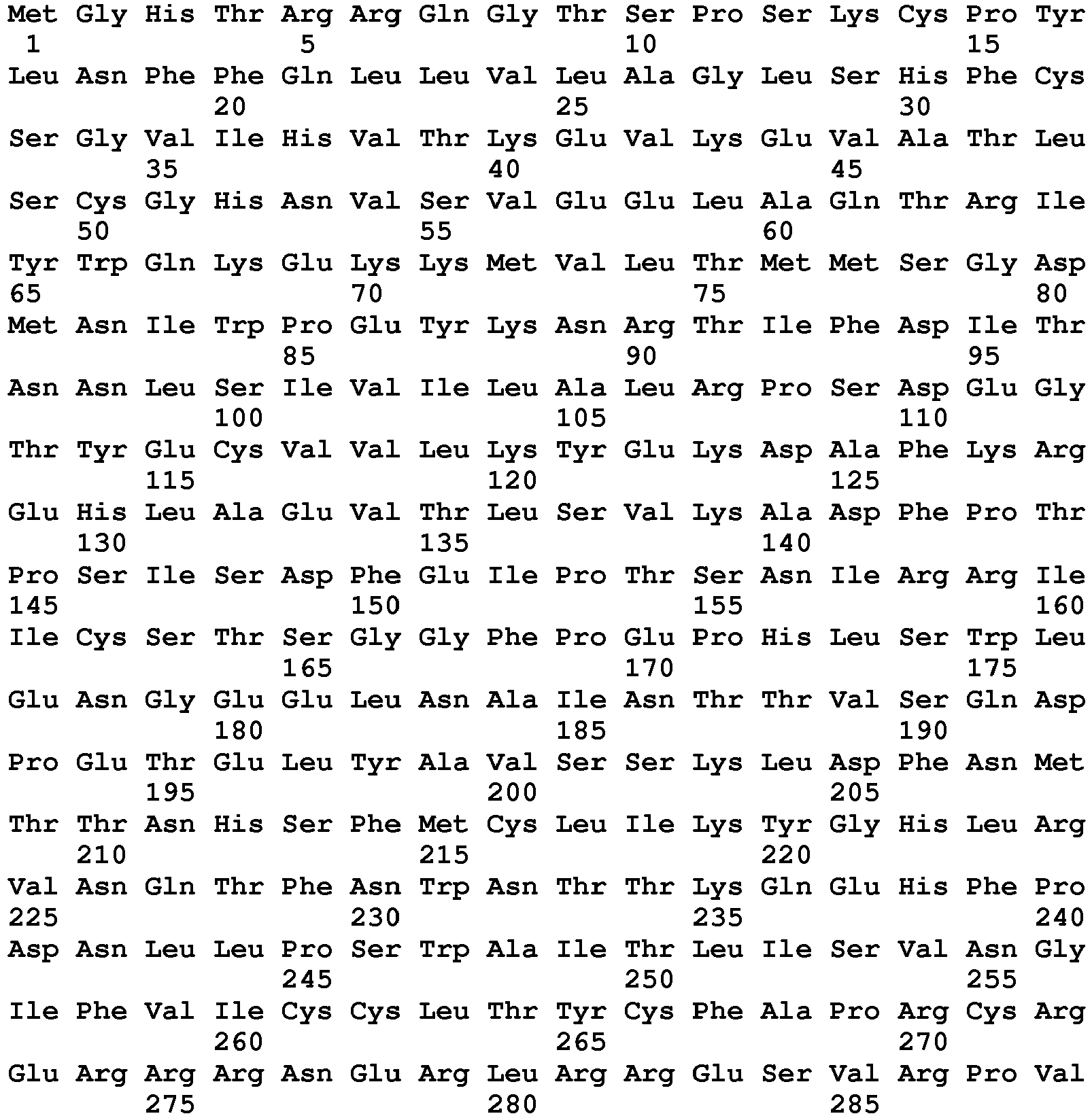
Claims (3)
- Composition comprising
an anti-B7-H1 antibody, an anti-PD-1 antibody, an anti-CD80 antibody, a fusion protein comprising a portion of PD-1 linked to an immunoglobulin Fc sequence, or a fusion protein comprising a portion of CD80 linked to an immunoglobulin Fc sequence
for use in a method for treating a mammal having cancer,
wherein said method comprises(i) determining the level of Bim in a peripheral blood sample from said mammal, and identifying said mammal as containing an elevated level of Bim, wherein said elevated level of Bim is based on Bim protein levels,
wherein said elevated level of Bim refers to a level that is at least 50% greater than a reference level of Bim, wherein said reference level of Bim refers to the level of Bim typically observed in healthy corresponding mammals without cancer, and(ii) administering said composition to said mammal under conditions wherein the interaction of naturally-occurring B7-H1 with PD-1 or CD80 in said mammal is reduced after administering said composition to said mammal. - The composition for the use according to claim 1, wherein said mammal is a human.
- The composition for the use according to claim 1, wherein said cancer is a melanoma cancer, a breast cancer, a lung cancer, a renal cell carcinoma cancer, a pancreas cancer, a prostate cancer, a colon cancer, a brain cancer, a liver cancer, or an ovarian cancer.
Priority Applications (1)
| Application Number | Priority Date | Filing Date | Title |
|---|---|---|---|
| EP18210199.8A EP3470081A1 (en) | 2013-10-01 | 2014-09-03 | Methods for treating cancer in patients with elevated levels of bim |
Applications Claiming Priority (2)
| Application Number | Priority Date | Filing Date | Title |
|---|---|---|---|
| US201361885218P | 2013-10-01 | 2013-10-01 | |
| PCT/US2014/053870 WO2015050663A1 (en) | 2013-10-01 | 2014-09-03 | Methods for treating cancer in patients with elevated levels of bim |
Related Child Applications (1)
| Application Number | Title | Priority Date | Filing Date |
|---|---|---|---|
| EP18210199.8A Division EP3470081A1 (en) | 2013-10-01 | 2014-09-03 | Methods for treating cancer in patients with elevated levels of bim |
Publications (3)
| Publication Number | Publication Date |
|---|---|
| EP3052131A1 EP3052131A1 (en) | 2016-08-10 |
| EP3052131A4 EP3052131A4 (en) | 2017-03-29 |
| EP3052131B1 true EP3052131B1 (en) | 2018-12-05 |
Family
ID=52779026
Family Applications (2)
| Application Number | Title | Priority Date | Filing Date |
|---|---|---|---|
| EP18210199.8A Withdrawn EP3470081A1 (en) | 2013-10-01 | 2014-09-03 | Methods for treating cancer in patients with elevated levels of bim |
| EP14850189.3A Active EP3052131B1 (en) | 2013-10-01 | 2014-09-03 | Methods for treating cancer in patients with elevated levels of bim |
Family Applications Before (1)
| Application Number | Title | Priority Date | Filing Date |
|---|---|---|---|
| EP18210199.8A Withdrawn EP3470081A1 (en) | 2013-10-01 | 2014-09-03 | Methods for treating cancer in patients with elevated levels of bim |
Country Status (4)
| Country | Link |
|---|---|
| US (2) | US10259875B2 (en) |
| EP (2) | EP3470081A1 (en) |
| ES (1) | ES2714708T3 (en) |
| WO (1) | WO2015050663A1 (en) |
Families Citing this family (20)
| Publication number | Priority date | Publication date | Assignee | Title |
|---|---|---|---|---|
| BRPI0917592B1 (en) | 2008-12-09 | 2021-08-17 | Genentech, Inc | ANTI-PD-L1 ANTIBODY, COMPOSITION, MANUFACTURED ARTICLES AND USES OF A COMPOSITION |
| US9302005B2 (en) | 2013-03-14 | 2016-04-05 | Mayo Foundation For Medical Education And Research | Methods and materials for treating cancer |
| ES2714708T3 (en) | 2013-10-01 | 2019-05-29 | Mayo Found Medical Education & Res | Procedures for the treatment of cancer in patients with elevated levels of Bim |
| SG10201804945WA (en) | 2013-12-12 | 2018-07-30 | Shanghai hengrui pharmaceutical co ltd | Pd-1 antibody, antigen-binding fragment thereof, and medical application thereof |
| WO2015179654A1 (en) | 2014-05-22 | 2015-11-26 | Mayo Foundation For Medical Education And Research | Distinguishing antagonistic and agonistic anti b7-h1 antibodies |
| US10517875B2 (en) | 2014-07-23 | 2019-12-31 | Mayo Foundation for Medical Engineering and Research | Targeting DNA-PKcs and B7-H1 to treat cancer |
| SG10201913297TA (en) | 2015-03-13 | 2020-02-27 | Cytomx Therapeutics Inc | Anti-pdl1 antibodies, activatable anti-pdl1 antibodies, and methods of use thereof |
| BR112018000768A2 (en) | 2015-07-13 | 2018-09-25 | Cytomx Therapeutics Inc | anti-pd-1 antibodies, activatable anti-pd-1 antibodies and methods of use |
| EP3337496B1 (en) * | 2015-08-20 | 2020-11-25 | Fraunhofer Gesellschaft zur Förderung der angewandten Forschung e.V. | B7-h1 fusion polypeptides for use in treating and preventing organ failure |
| AU2016332725A1 (en) | 2015-09-29 | 2018-03-22 | Celgene Corporation | PD-1 binding proteins and methods of use thereof |
| US10875923B2 (en) | 2015-10-30 | 2020-12-29 | Mayo Foundation For Medical Education And Research | Antibodies to B7-H1 |
| EP3371208B8 (en) | 2015-11-02 | 2024-10-23 | Five Prime Therapeutics, Inc. | Cd80 extracellular domain polypeptides and their use in cancer treatment |
| JP2019534859A (en) | 2016-09-19 | 2019-12-05 | セルジーン コーポレイション | Method for treating vitiligo using PD-1 binding protein |
| SG11201901950TA (en) | 2016-09-19 | 2019-04-29 | Celgene Corp | Methods of treating immune disorders using pd-1 binding proteins |
| EP3628070B1 (en) | 2017-04-28 | 2021-09-08 | Five Prime Therapeutics, Inc. | Cd80 extracellular domain polypeptides for use in increasing central memory t cells |
| JP2020522486A (en) | 2017-06-01 | 2020-07-30 | サイトメックス セラピューティクス インコーポレイテッド | Activatable anti-PDL1 antibody and method of using the same |
| CN109721657B (en) * | 2017-10-27 | 2021-11-02 | 北京比洋生物技术有限公司 | Fusion protein for blocking PD-1/PD-L1 signal transduction pathway and activating T cells and application thereof |
| EP3873500A4 (en) | 2018-10-31 | 2023-01-11 | Mayo Foundation for Medical Education and Research | CANCER TREATMENT METHODS AND MATERIALS |
| EP3873540A4 (en) | 2018-10-31 | 2022-07-27 | Mayo Foundation for Medical Education and Research | METHODS AND MATERIALS FOR THE TREATMENT OF CANCER |
| CA3203105A1 (en) * | 2020-12-22 | 2022-06-30 | Michael H. Cardone | Methods and compositions related to bcl2 and bim heterodimer antibodies |
Citations (1)
| Publication number | Priority date | Publication date | Assignee | Title |
|---|---|---|---|---|
| WO2014144666A2 (en) | 2013-03-15 | 2014-09-18 | The University Of Chicago | Methods and compositions related to t-cell activity |
Family Cites Families (175)
| Publication number | Priority date | Publication date | Assignee | Title |
|---|---|---|---|---|
| US3687808A (en) | 1969-08-14 | 1972-08-29 | Univ Leland Stanford Junior | Synthetic polynucleotides |
| US4034074A (en) | 1974-09-19 | 1977-07-05 | The Board Of Trustees Of Leland Stanford Junior University | Universal reagent 2-site immunoradiometric assay using labelled anti (IgG) |
| US4036945A (en) | 1976-05-03 | 1977-07-19 | The Massachusetts General Hospital | Composition and method for determining the size and location of myocardial infarcts |
| US4098876A (en) | 1976-10-26 | 1978-07-04 | Corning Glass Works | Reverse sandwich immunoassay |
| US4233402A (en) | 1978-04-05 | 1980-11-11 | Syva Company | Reagents and method employing channeling |
| US4272398A (en) | 1978-08-17 | 1981-06-09 | The United States Of America As Represented By The Secretary Of Agriculture | Microencapsulation process |
| US4257774A (en) | 1979-07-16 | 1981-03-24 | Meloy Laboratories, Inc. | Intercalation inhibition assay for compounds that interact with DNA or RNA |
| US4331647A (en) | 1980-03-03 | 1982-05-25 | Goldenberg Milton David | Tumor localization and therapy with labeled antibody fragments specific to tumor-associated markers |
| US4376110A (en) | 1980-08-04 | 1983-03-08 | Hybritech, Incorporated | Immunometric assays using monoclonal antibodies |
| US4469863A (en) | 1980-11-12 | 1984-09-04 | Ts O Paul O P | Nonionic nucleic acid alkyl and aryl phosphonates and processes for manufacture and use thereof |
| US4769330A (en) | 1981-12-24 | 1988-09-06 | Health Research, Incorporated | Modified vaccinia virus and methods for making and using the same |
| US5155020A (en) | 1989-03-08 | 1992-10-13 | Health Research Inc. | Recombinant poxvirus host range selection system |
| US4816567A (en) | 1983-04-08 | 1989-03-28 | Genentech, Inc. | Recombinant immunoglobin preparations |
| US4650764A (en) | 1983-04-12 | 1987-03-17 | Wisconsin Alumni Research Foundation | Helper cell |
| JPS61134325A (en) | 1984-12-04 | 1986-06-21 | Teijin Ltd | Expression of hybrid antibody gene |
| US5235033A (en) | 1985-03-15 | 1993-08-10 | Anti-Gene Development Group | Alpha-morpholino ribonucleoside derivatives and polymers thereof |
| US5618920A (en) | 1985-11-01 | 1997-04-08 | Xoma Corporation | Modular assembly of antibody genes, antibodies prepared thereby and use |
| US4861719A (en) | 1986-04-25 | 1989-08-29 | Fred Hutchinson Cancer Research Center | DNA constructs for retrovirus packaging cell lines |
| WO1988001213A1 (en) | 1986-08-18 | 1988-02-25 | Clinical Technologies Associates, Inc. | Delivery systems for pharmacological agents |
| US4946778A (en) | 1987-09-21 | 1990-08-07 | Genex Corporation | Single polypeptide chain binding molecules |
| US4987071A (en) | 1986-12-03 | 1991-01-22 | University Patents, Inc. | RNA ribozyme polymerases, dephosphorylases, restriction endoribonucleases and methods |
| US4980289A (en) | 1987-04-27 | 1990-12-25 | Wisconsin Alumni Research Foundation | Promoter deficient retroviral vector |
| US4861627A (en) | 1987-05-01 | 1989-08-29 | Massachusetts Institute Of Technology | Preparation of multiwall polymeric microcapsules |
| US5254678A (en) | 1987-12-15 | 1993-10-19 | Gene Shears Pty. Limited | Ribozymes |
| ATE140963T1 (en) | 1988-01-22 | 1996-08-15 | Zymogenetics Inc | METHOD FOR PRODUCING SECRETED RECEPTOR ANALOGUES |
| US5567584A (en) | 1988-01-22 | 1996-10-22 | Zymogenetics, Inc. | Methods of using biologically active dimerized polypeptide fusions to detect PDGF |
| US6018026A (en) | 1988-01-22 | 2000-01-25 | Zymogenetics, Inc. | Biologically active dimerized and multimerized polypeptide fusions |
| US5750375A (en) | 1988-01-22 | 1998-05-12 | Zymogenetics, Inc. | Methods of producing secreted receptor analogs and biologically active dimerized polypeptide fusions |
| US5278056A (en) | 1988-02-05 | 1994-01-11 | The Trustees Of Columbia University In The City Of New York | Retroviral packaging cell lines and process of using same |
| US5750666A (en) | 1988-05-26 | 1998-05-12 | Competitve Technologies, Inc. | Polynucleotide phosphorodithioate compounds |
| US6303121B1 (en) | 1992-07-30 | 2001-10-16 | Advanced Research And Technology | Method of using human receptor protein 4-1BB |
| US5530101A (en) | 1988-12-28 | 1996-06-25 | Protein Design Labs, Inc. | Humanized immunoglobulins |
| IL162181A (en) | 1988-12-28 | 2006-04-10 | Pdl Biopharma Inc | A method of producing humanized immunoglubulin, and polynucleotides encoding the same |
| US5124263A (en) | 1989-01-12 | 1992-06-23 | Wisconsin Alumni Research Foundation | Recombination resistant retroviral helper cell and products produced thereby |
| US5225538A (en) | 1989-02-23 | 1993-07-06 | Genentech, Inc. | Lymphocyte homing receptor/immunoglobulin fusion proteins |
| US5225336A (en) | 1989-03-08 | 1993-07-06 | Health Research Incorporated | Recombinant poxvirus host range selection system |
| US5175099A (en) | 1989-05-17 | 1992-12-29 | Research Corporation Technologies, Inc. | Retrovirus-mediated secretion of recombinant products |
| US5240846A (en) | 1989-08-22 | 1993-08-31 | The Regents Of The University Of Michigan | Gene therapy vector for cystic fibrosis |
| US5013556A (en) | 1989-10-20 | 1991-05-07 | Liposome Technology, Inc. | Liposomes with enhanced circulation time |
| DK0463151T3 (en) | 1990-01-12 | 1996-07-01 | Cell Genesys Inc | Generation of xenogenic antibodies |
| DE69030172T2 (en) | 1990-01-26 | 1997-06-19 | Immunomedics Inc | Vaccines against cancer and infectious diseases |
| US5204243A (en) | 1990-02-14 | 1993-04-20 | Health Research Incorporated | Recombinant poxvirus internal cores |
| US5214136A (en) | 1990-02-20 | 1993-05-25 | Gilead Sciences, Inc. | Anthraquinone-derivatives oligonucleotides |
| US6641809B1 (en) | 1990-03-26 | 2003-11-04 | Bristol-Myers Squibb Company | Method of regulating cellular processes mediated by B7 and CD28 |
| US5427908A (en) | 1990-05-01 | 1995-06-27 | Affymax Technologies N.V. | Recombinant library screening methods |
| JPH06508501A (en) | 1990-07-02 | 1994-09-29 | ブリストル―マイアーズ スクイブ カンパニー | Ligand for CD28 receptor on B cells and method for regulating cell-cell interaction using the ligand |
| GB9015198D0 (en) | 1990-07-10 | 1990-08-29 | Brien Caroline J O | Binding substance |
| EP0585287B1 (en) | 1990-07-10 | 1999-10-13 | Cambridge Antibody Technology Limited | Methods for producing members of specific binding pairs |
| US5218105A (en) | 1990-07-27 | 1993-06-08 | Isis Pharmaceuticals | Polyamine conjugated oligonucleotides |
| WO1992005186A1 (en) | 1990-09-20 | 1992-04-02 | Gilead Sciences | Modified internucleoside linkages |
| US5296347A (en) | 1991-02-08 | 1994-03-22 | Ciba Corning Diagnostics Corp. | Bridge immunoassay |
| CZ333796A3 (en) | 1991-03-06 | 1998-06-17 | MERCK Patent Gesellschaft mit beschränkter Haftung | Chimeric monoclonal antibody, expression vectors and pharmaceutical preparation |
| AU643141B2 (en) | 1991-03-15 | 1993-11-04 | Amgen, Inc. | Pulmonary administration of granulocyte colony stimulating factor |
| EP0592668B1 (en) | 1991-07-03 | 1999-03-10 | Kanebo, Ltd. | Method and device for the manufacture of a thermoplastic polyurethane elastomer |
| CA2078539C (en) | 1991-09-18 | 2005-08-02 | Kenya Shitara | Process for producing humanized chimera antibody |
| ES2136092T3 (en) | 1991-09-23 | 1999-11-16 | Medical Res Council | PROCEDURES FOR THE PRODUCTION OF HUMANIZED ANTIBODIES. |
| GB9125979D0 (en) | 1991-12-06 | 1992-02-05 | Wellcome Found | Antibody |
| US5863537A (en) | 1992-02-19 | 1999-01-26 | Schering Corporation | Humanized monoclonal antibodies against human interleukin-4 |
| US5733743A (en) | 1992-03-24 | 1998-03-31 | Cambridge Antibody Technology Limited | Methods for producing members of specific binding pairs |
| US5639641A (en) | 1992-09-09 | 1997-06-17 | Immunogen Inc. | Resurfacing of rodent antibodies |
| US7211259B1 (en) | 1993-05-07 | 2007-05-01 | Immunex Corporation | 4-1BB polypeptides and DNA encoding 4-1BB polypeptides |
| WO1994026290A1 (en) | 1993-05-07 | 1994-11-24 | Immunex Corporation | Cytokine designated 4-1bb ligand and human receptor that binds thereto |
| US5942607A (en) | 1993-07-26 | 1999-08-24 | Dana-Farber Cancer Institute | B7-2: a CTLA4/CD28 ligand |
| US5861310A (en) | 1993-11-03 | 1999-01-19 | Dana-Farber Cancer Institute | Tumor cells modified to express B7-2 with increased immunogenicity and uses therefor |
| AU7526894A (en) | 1993-08-16 | 1995-03-14 | Arch Development Corporation | B7-2: ctla4/cd28 counter receptor |
| ES2318848T3 (en) | 1993-09-14 | 2009-05-01 | Pharmexa Inc. | PEPTIDES THAT JOIN PAN DR TO POWER THE IMMUNE RESPONSE. |
| CA2429027C (en) | 1993-09-16 | 2011-04-05 | Indiana University Foundation | Antibodies against human receptor h4-1bb |
| US5451569A (en) | 1994-04-19 | 1995-09-19 | Hong Kong University Of Science And Technology R & D Corporation Limited | Pulmonary drug delivery system |
| US5972703A (en) | 1994-08-12 | 1999-10-26 | The Regents Of The University Of Michigan | Bone precursor cells: compositions and methods |
| US5705154A (en) | 1995-03-08 | 1998-01-06 | Schering Corporation | Humanized monoclonal antibodies against human interleukin-4 |
| AU5369996A (en) | 1995-03-23 | 1996-10-08 | Indiana University Foundation | Monoclonal antibody against human receptor protein 4-1bb and methods of its use for treatment of diseases |
| ES2185770T3 (en) | 1995-04-08 | 2003-05-01 | Lg Chemical Ltd | SPECIFIC MONOCLONAL ANTIBODY FOR 4-1BB HUMAN AND CELL LINE THAT PRODUCES IT. |
| US5675848A (en) | 1995-10-18 | 1997-10-14 | Mallinckrodt Medical, Inc. | Inflatable blanket having perforations of different sizes |
| AU705688B2 (en) | 1995-11-10 | 1999-05-27 | Cytogen Corporation | Peptides which enhance transport across tissues and methods of identifying and using the same |
| EP0871747A1 (en) | 1996-01-02 | 1998-10-21 | Chiron Viagene, Inc. | Immunostimulation mediated by gene-modified dendritic cells |
| US5874240A (en) | 1996-03-15 | 1999-02-23 | Human Genome Sciences, Inc. | Human 4-1BB receptor splicing variant |
| ATE230801T1 (en) | 1996-10-03 | 2003-01-15 | Canon Kk | METHOD FOR DETECTING TARGET NUCLEIC ACID, METHOD FOR QUANTIFYING IT AND PYRYLIUM COMPOUNDS FOR CHEMILUMINESCENCE ANALYSIS |
| WO1998016249A1 (en) | 1996-10-11 | 1998-04-23 | Bristol-Myers Squibb Company | Methods and compositions for immunomodulation |
| JP4672092B2 (en) | 1996-11-20 | 2011-04-20 | イェール ユニバーシティ | Survivin, a protein that inhibits cell apoptosis, and its regulation |
| AUPO390396A0 (en) | 1996-11-29 | 1996-12-19 | Csl Limited | Novel promiscuous T helper cell epitopes |
| EP1012275A1 (en) | 1997-01-31 | 2000-06-28 | University Of Rochester | Chimeric antibody fusion proteins for the recruitment and stimulation of an antitumor immune response |
| AU6534898A (en) | 1997-02-14 | 1998-09-08 | E.I. Du Pont De Nemours And Company | Detection of double-stranded dna in a homogeneous solution |
| US20070224663A1 (en) | 1997-03-07 | 2007-09-27 | Human Genome Sciences, Inc. | Human Secreted Proteins |
| US20060223088A1 (en) | 1997-03-07 | 2006-10-05 | Rosen Craig A | Human secreted proteins |
| US7368531B2 (en) | 1997-03-07 | 2008-05-06 | Human Genome Sciences, Inc. | Human secreted proteins |
| US7411051B2 (en) | 1997-03-07 | 2008-08-12 | Human Genome Sciences, Inc. | Antibodies to HDPPA04 polypeptide |
| US7064193B1 (en) | 1997-09-17 | 2006-06-20 | The Walter And Eliza Hall Institute Of Medical Research | Therapeutic molecules |
| ATE321859T1 (en) | 1998-06-10 | 2006-04-15 | Us Gov Health & Human Serv | B2MICROGLOBULIN FUSION PROTEINS AND VARIANTS WITH HIGH AFFINITY |
| AU1329700A (en) | 1998-10-30 | 2000-05-22 | Northwestern University | Method of interfering with cell proliferation |
| KR20000034847A (en) | 1998-11-17 | 2000-06-26 | 성재갑 | Humanized Antibody Specific for Human 4-1BB Molecule and Pharmaceutical Composition Comprising Same |
| US6734172B2 (en) | 1998-11-18 | 2004-05-11 | Pacific Northwest Research Institute | Surface receptor antigen vaccines |
| AU3209300A (en) | 1999-01-15 | 2000-08-01 | Mount Sinai School Of Medicine Of The City University Of New York, The | Combination therapy of cancer by the activation of co-stimulatory molecules expressed by immune cells and cytokines |
| AU3899700A (en) | 1999-03-17 | 2000-10-04 | Alphagene, Inc. | Secreted proteins and polynucleotides encoding them |
| BR0009505A (en) | 1999-04-02 | 2002-06-11 | Corixa Corp | Lung cancer therapy and diagnosis compounds and processes |
| EP1074617A3 (en) | 1999-07-29 | 2004-04-21 | Research Association for Biotechnology | Primers for synthesising full-length cDNA and their use |
| US6423885B1 (en) | 1999-08-13 | 2002-07-23 | Commonwealth Scientific And Industrial Research Organization (Csiro) | Methods for obtaining modified phenotypes in plant cells |
| US20020177551A1 (en) | 2000-05-31 | 2002-11-28 | Terman David S. | Compositions and methods for treatment of neoplastic disease |
| CA2387959A1 (en) | 1999-11-12 | 2001-05-17 | Human Genome Sciences, Inc. | 21 human secreted proteins |
| PT1234031T (en) | 1999-11-30 | 2017-06-26 | Mayo Foundation | B7-h1, a novel immunoregulatory molecule |
| US6803192B1 (en) | 1999-11-30 | 2004-10-12 | Mayo Foundation For Medical Education And Research | B7-H1, a novel immunoregulatory molecule |
| NZ521437A (en) | 2000-02-25 | 2004-04-30 | Immunex Corp | Integrin antagonists suitable as inhibitors of angiogenesis |
| US20030165831A1 (en) | 2000-03-21 | 2003-09-04 | John Lee | Novel genes, compositions, kits, and methods for identification, assessment, prevention, and therapy of ovarian cancer |
| ES2529300T3 (en) | 2000-04-12 | 2015-02-18 | Novozymes Biopharma Dk A/S | Albumin fusion proteins |
| US7030219B2 (en) | 2000-04-28 | 2006-04-18 | Johns Hopkins University | B7-DC, Dendritic cell co-stimulatory molecules |
| MXPA02012106A (en) | 2000-06-06 | 2003-06-06 | Bristol Myers Squibb Co | B7-related nucleic acids and polypeptides useful for immunomodulation. |
| US20030031675A1 (en) | 2000-06-06 | 2003-02-13 | Mikesell Glen E. | B7-related nucleic acids and polypeptides useful for immunomodulation |
| AU7309601A (en) | 2000-06-28 | 2002-01-08 | Genetics Inst | Pd-l2 molecules: novel pd-1 ligands and uses therefor |
| WO2002002587A1 (en) | 2000-06-30 | 2002-01-10 | Human Genome Sciences, Inc. | B7-like polynucleotides, polypeptides, and antibodies |
| WO2002002891A1 (en) | 2000-07-05 | 2002-01-10 | Vogue Pool Products | Above ground swimming pool support structure |
| US20020076409A1 (en) | 2000-07-12 | 2002-06-20 | March Carl J. | Method for treating cancer |
| US6635750B1 (en) | 2000-07-20 | 2003-10-21 | Millennium Pharmaceuticals, Inc. | B7-H2 nucleic acids, members of the B7 family |
| US6891030B2 (en) | 2000-07-27 | 2005-05-10 | Mayo Foundation For Medical Education And Research | T-cell immunoregulatory molecule |
| PT1326961E (en) | 2000-09-15 | 2007-10-02 | Ortho Mcneil Pharm Inc | COMPOSITIONS AND METHODS TO INDUCE SPECIFIC T-CELL CITOLITIC RESPONSES |
| MXPA03002413A (en) | 2000-09-20 | 2003-06-19 | Amgen Inc | B7-like molecules and uses thereof. |
| US20040247563A1 (en) | 2000-11-02 | 2004-12-09 | Lynch David H. | Method of enhancing lymphocyte-mediated immune responses |
| CA2436519A1 (en) | 2000-12-07 | 2002-06-13 | The Penn State Research Foundation | Selection of catalytic nucleic acids targeted to infectious agents |
| WO2002057453A2 (en) | 2000-12-19 | 2002-07-25 | Curagen Corporation | Polypetides and nucleic acids encoding same |
| WO2002058535A2 (en) | 2000-12-26 | 2002-08-01 | Oxigene, Inc. | Use of combretastatin a4 and its prodrugs as an immune enhancing therapy |
| US20030180309A1 (en) | 2001-01-08 | 2003-09-25 | Baum Peter R. | Human B7 polypeptides |
| AU2002246970B2 (en) | 2001-01-12 | 2007-06-14 | Yale University | Detection of survivin in the biological fluids of cancer patients |
| US6743619B1 (en) | 2001-01-30 | 2004-06-01 | Nuvelo | Nucleic acids and polypeptides |
| WO2003008583A2 (en) | 2001-03-02 | 2003-01-30 | Sagres Discovery | Novel compositions and methods for cancer |
| AR036993A1 (en) | 2001-04-02 | 2004-10-20 | Wyeth Corp | USE OF AGENTS THAT MODULATE THE INTERACTION BETWEEN PD-1 AND ITS LINKS IN THE SUBMODULATION OF IMMUNOLOGICAL ANSWERS |
| US20060084794A1 (en) | 2001-04-12 | 2006-04-20 | Human Genome Sciences, Inc. | Albumin fusion proteins |
| AU2002258941A1 (en) | 2001-04-20 | 2002-11-05 | Mayo Foundation For Medical Education And Research | Methods of enhancing cell responsiveness |
| US20060276422A1 (en) | 2001-05-18 | 2006-12-07 | Nassim Usman | RNA interference mediated inhibition of B7-H1 gene expression using short interfering nucleic acid (siNA) |
| WO2003006632A2 (en) | 2001-07-12 | 2003-01-23 | Canvac | Methods and compisitions for activation human t cells in vitro |
| EP1434596B1 (en) | 2001-10-09 | 2009-07-08 | Mayo Foundation For Medical Education And Research | Enhancement of immune responses by agonist 4-1bb-antibodies |
| DE60224822T2 (en) | 2001-10-19 | 2009-01-22 | Zymogenetics, Inc., Seattle | DIMERIZED GROWTH FACTOR AND MATERIALS AND METHOD FOR THE PRODUCTION THEREOF |
| EP1456652A4 (en) | 2001-11-13 | 2005-11-02 | Dana Farber Cancer Inst Inc | AGENTS MODULATING IMMUNE CELL ACTIVITY AND METHODS OF USE THEREOF |
| US7164500B2 (en) | 2002-01-29 | 2007-01-16 | Hewlett-Packard Development Company, L.P. | Method and apparatus for the automatic generation of image capture device control marks |
| WO2003076575A2 (en) | 2002-03-04 | 2003-09-18 | Fidelity Systems, Inc., Et Al. | The complete genome and protein sequence of the hyperthermophile methanopyrus kandleri av19 and monophyly of archael methanogens and methods of use thereof |
| US20030223989A1 (en) | 2002-04-18 | 2003-12-04 | Pluenneke John D. | CD137 agonists to treat patients with IgE-mediated conditions |
| EP2243493A1 (en) | 2002-07-03 | 2010-10-27 | Ono Pharmaceutical Co., Ltd. | Immunopotentiative composition |
| US20040109847A1 (en) | 2002-07-15 | 2004-06-10 | Lieping Chen | Treatment and prophylaxis with 4-1BB-binding agents |
| US7432351B1 (en) | 2002-10-04 | 2008-10-07 | Mayo Foundation For Medical Education And Research | B7-H1 variants |
| US7449300B2 (en) | 2002-11-21 | 2008-11-11 | Mayo Foundation For Medical Education And Research | Detection of antibodies specific for B7-H1 in subjects with diseases or pathological conditions mediated by activated T cells |
| KR20050107399A (en) | 2003-01-23 | 2005-11-11 | 오노 야꾸힝 고교 가부시키가이샤 | Substance specific to human pd-1 |
| JP2006523681A (en) | 2003-03-24 | 2006-10-19 | ルイトポルド・ファーマシューティカルズ・インコーポレーテッド | Xanthone, thioxanthone and acridinone as DNA-PK inhibitors |
| US7381794B2 (en) | 2004-03-08 | 2008-06-03 | Zymogenetics, Inc. | Dimeric fusion proteins and materials and methods for producing them |
| SI1810026T1 (en) | 2004-10-06 | 2018-08-31 | Mayo Foundation For Medical Education And Research | B7-H1 and PD-1 in the treatment of renal cell carcinoma |
| CN101072578A (en) | 2004-10-29 | 2007-11-14 | 南加州大学 | Combination cancer immunotherapy with co-stimulatory molecules |
| NZ593388A (en) | 2005-06-08 | 2012-08-31 | Dana Farber Cancer Inst Inc | Methods and compositions for the treatment of persistent infections and cancer by inhibiting the programmed cell death 1 (pd-1) pathway |
| US20070231344A1 (en) | 2005-10-28 | 2007-10-04 | The Brigham And Women's Hospital, Inc. | Conjugate vaccines for non-proteinaceous antigens |
| WO2007082144A2 (en) | 2006-01-05 | 2007-07-19 | Mayo Foundation For Medical Education And Research | B7-h1 and survivin in cancer |
| WO2007082154A2 (en) | 2006-01-05 | 2007-07-19 | Mayo Foundation For Medical Education And Research | B7-h1 and b7-h4 in cancer |
| WO2007124361A2 (en) | 2006-04-20 | 2007-11-01 | Mayo Foundation For Medical Education And Research | Soluble b7-h1 |
| AU2007252295B2 (en) | 2006-05-19 | 2013-07-25 | Central Adelaide Local Health Network Incorporated | Selective modulation of receptor signalling |
| EP2061504A4 (en) * | 2006-09-20 | 2010-01-27 | Univ Johns Hopkins | COMBINATORY THERAPY AGAINST CANCER AND INFECTIOUS DISEASES USING VACCINE AND ANTI-B7-H1 |
| WO2008037080A1 (en) | 2006-09-29 | 2008-04-03 | Universite De Montreal | Methods and compositions for immune response modulation and uses thereof |
| CN103536915A (en) | 2006-12-27 | 2014-01-29 | 埃默里大学 | Compositions and methods for the treatment of infections and tumors |
| EP2514762B1 (en) | 2007-07-13 | 2015-04-08 | The Johns Hopkins University | B7-DC variants |
| AU2008287063B2 (en) | 2007-08-09 | 2013-10-24 | Genzyme Corporation | Method of treating autoimmune disease with mesenchymal stem cells |
| WO2009089149A1 (en) * | 2008-01-03 | 2009-07-16 | The Johns Hopkins University | B7-h1 (cd274) antagonists induce apoptosis of tumor cells |
| WO2009111315A2 (en) | 2008-02-29 | 2009-09-11 | Mayo Foundation For Medical Education And Research | Methods for reducing granulomatous inflammation |
| WO2009114110A1 (en) | 2008-03-08 | 2009-09-17 | Immungene, Inc. | Engineered fusion molecules immunotherapy in cancer and inflammatory diseases |
| WO2010027827A2 (en) | 2008-08-25 | 2010-03-11 | Amplimmune, Inc. | Targeted costimulatory polypeptides and methods of use to treat cancer |
| NZ591130A (en) | 2008-08-25 | 2012-09-28 | Amplimmune Inc | Compositions comprising a PD-1 antagonists and cyclophosphamide and methods of use thereof |
| US8577949B2 (en) | 2009-07-07 | 2013-11-05 | L-3 Communications Integrated Systems, L.P. | System for conjugate gradient linear iterative solvers |
| US20130017199A1 (en) | 2009-11-24 | 2013-01-17 | AMPLIMMUNE ,Inc. a corporation | Simultaneous inhibition of pd-l1/pd-l2 |
| RS60033B1 (en) | 2009-11-24 | 2020-04-30 | Medimmune Ltd | Targeted binding agents against b7-h1 |
| WO2012012710A2 (en) | 2010-07-22 | 2012-01-26 | The Johns Hopkins University | Radiation sensitization agents for prostate cancer |
| BR112013006207A2 (en) | 2010-09-15 | 2017-10-24 | Almac Diagnostics Ltd | molecular diagnostic test for cancer |
| EP2625278A1 (en) | 2010-10-08 | 2013-08-14 | Regents of the University of Minnesota | A method to increase gene targeting frequency |
| CN103765212A (en) | 2011-06-27 | 2014-04-30 | 杰克逊实验室 | Methods and compositions for treating cancer and autoimmune diseases |
| SG10201606284UA (en) | 2011-08-01 | 2016-09-29 | Genentech Inc | Methods Of Treating Cancer Using Pd-1 Axis Binding Antagonists And Mek Inhibitors |
| EP2747781B1 (en) | 2011-08-23 | 2017-11-15 | Roche Glycart AG | Bispecific antibodies specific for t-cell activating antigens and a tumor antigen and methods of use |
| WO2013090552A1 (en) * | 2011-12-13 | 2013-06-20 | Yale University | Compositions and methods for reducing ctl exhaustion |
| AR090263A1 (en) | 2012-03-08 | 2014-10-29 | Hoffmann La Roche | COMBINED ANTIBODY THERAPY AGAINST HUMAN CSF-1R AND USES OF THE SAME |
| CA2896359A1 (en) | 2013-02-26 | 2014-09-04 | Roche Glycart Ag | Bispecific t cell activating antigen binding molecules |
| US9302005B2 (en) | 2013-03-14 | 2016-04-05 | Mayo Foundation For Medical Education And Research | Methods and materials for treating cancer |
| ES2714708T3 (en) | 2013-10-01 | 2019-05-29 | Mayo Found Medical Education & Res | Procedures for the treatment of cancer in patients with elevated levels of Bim |
| WO2015179654A1 (en) | 2014-05-22 | 2015-11-26 | Mayo Foundation For Medical Education And Research | Distinguishing antagonistic and agonistic anti b7-h1 antibodies |
| US10517875B2 (en) | 2014-07-23 | 2019-12-31 | Mayo Foundation for Medical Engineering and Research | Targeting DNA-PKcs and B7-H1 to treat cancer |
| US10875923B2 (en) | 2015-10-30 | 2020-12-29 | Mayo Foundation For Medical Education And Research | Antibodies to B7-H1 |
-
2014
- 2014-09-03 ES ES14850189T patent/ES2714708T3/en active Active
- 2014-09-03 WO PCT/US2014/053870 patent/WO2015050663A1/en not_active Ceased
- 2014-09-03 EP EP18210199.8A patent/EP3470081A1/en not_active Withdrawn
- 2014-09-03 EP EP14850189.3A patent/EP3052131B1/en active Active
- 2014-09-03 US US15/026,461 patent/US10259875B2/en active Active
-
2019
- 2019-04-15 US US16/384,313 patent/US11136393B2/en active Active
Patent Citations (1)
| Publication number | Priority date | Publication date | Assignee | Title |
|---|---|---|---|---|
| WO2014144666A2 (en) | 2013-03-15 | 2014-09-18 | The University Of Chicago | Methods and compositions related to t-cell activity |
Non-Patent Citations (13)
| Title |
|---|
| DONG ET AL.: "a novel method for identifying downstream signals in tumor-reactive T cells following PD-1 engagement and monitoring endogenous tumor immunity and immunotherapy", JOURNAL OF CLINICAL ONCOLOGY, vol. 32, no. 15, 20 May 2014 (2014-05-20), XP055638824 |
| DRONCA ET AL.: "BCL-2-interacting mediator of cell death (Bim) is a novel biomarker for response to anti-PD-1 therapy in patients with advanced melanoma", IMMUNOTHERAPY, vol. 8, no. 12, December 2016 (2016-12-01), pages 1351 - 1353, XP002788155, DOI: 10.2217/imt-2016-0100 |
| DRONCA ET AL.: "T cell Bim levels reflect responses to anti-PD-1 cancer therapy", JO INSIGHT, vol. 1, no. 6, 2016, XP002788154 |
| DRONCA ROXANA S ET AL: "BCL-2-interacting mediator of cell death (Bim) is a novel biomarker for response to anti-PD-1 therapy in patients with advanced melanoma.", IMMUNOTHERAPY DEC 2016, vol. 8, no. 12, December 2016 (2016-12-01), pages 1351 - 1353, ISSN: 1750-7448 * |
| DRONCA ROXANA S ET AL: "T cell Bim levels reflect responses to anti-PD-1 cancer therapy.", JCI INSIGHT 05 MAY 2016, vol. 1, no. 6, 5 May 2016 (2016-05-05) * |
| DRONCA, R. ET AL.: "T cell Bim levels reflect responses to anti-PD-1 cancer therapy", JCI INSIGHT, vol. 1, no. 6, May 2016 (2016-05-01), pages e86014 - 1 - 14, XP002788154, DOI: 10.1172/jci.insight.86014 |
| GIBBONS ET AL.: "B7-H1 limits the entry of effector CD 8+ T cells to the memory pool by upregulating Bim", ONCOIMMUNOLOGY, vol. 1, no. 7, 2012, pages 1061 - 1073, XP055332328 |
| GIBBONS R. ET AL.: "B7-H1 limits the entry of effector CD 8+ T cells to the memory pool by upregulating Bim", ONCOLMMUNOLOGY, vol. 1, no. 7, October 2012 (2012-10-01), pages 1061 - 1073, XP055332328, DOI: 10.4161/onci.20850 |
| JOHNSON ET AL., N ENGL J MED., vol. 375, no. 18, 3 November 2016 (2016-11-03), pages 1749 - 1755 |
| LIU ET AL.: "Endogenous tumor-reactive CD 8+ T cells are differentiated effector cells expressing high levels of CD 11 a and PD-1 but are unable to control tumor growth", ONCOLMMUNOLOGY, vol. 2, no. 6, 1 June 2013 (2013-06-01), pages 1 - 12, XP055638478 |
| LIU ET AL.: "Endogenous tumor-reactive CD 8+ T cells are differentiated effector cells expressing high levels of CD 11 a and PD-1 but are unable to control tumor growth", ONCOLMMUNOLOGY, vol. 2, no. 6, June 2013 (2013-06-01), pages 1 - 12, XP055638478 |
| TOPALIAN ET AL.: "Safety, Activity, and Immune Correlates of Anti-PD-1 Antibody in Cancer", N ENGL J MED, vol. 366, no. 26, June 2012 (2012-06-01), pages 2443 - 2454, XP055098235 |
| TOPALIAN, S. ET AL.: "Safety, Activity, and Immune Correlates of Anti-PD-1 Antibody in Cancer", NEJM, vol. 366, no. 26, June 2012 (2012-06-01), pages 2443 - 2454, XP055098235, DOI: 10.1056/NEJMoa1200690 |
Also Published As
| Publication number | Publication date |
|---|---|
| US11136393B2 (en) | 2021-10-05 |
| US10259875B2 (en) | 2019-04-16 |
| US20190315869A1 (en) | 2019-10-17 |
| ES2714708T3 (en) | 2019-05-29 |
| EP3052131A4 (en) | 2017-03-29 |
| EP3052131A1 (en) | 2016-08-10 |
| WO2015050663A1 (en) | 2015-04-09 |
| US20160251437A1 (en) | 2016-09-01 |
| EP3470081A1 (en) | 2019-04-17 |
Similar Documents
| Publication | Publication Date | Title |
|---|---|---|
| US11136393B2 (en) | Methods for treating cancer in patients with elevated levels of Bim | |
| US10302653B2 (en) | Distinguishing antagonistic and agonistic anti B7-H1 antibodies | |
| AU2017210224B2 (en) | Treatment of cancer with combinations of immunoregulatory agents | |
| EP3177321B1 (en) | Tumor antigen specific antibodies and tlr3 stimulation to enhance the performance of checkpoint interference therapy of cancer | |
| AU2018351007B2 (en) | Treatment of ovarian cancer with anti-CD47 and anti-PD-L1 | |
| JP2018512397A (en) | Compositions and methods for enhancing the effectiveness of cancer treatment | |
| KR20190098995A (en) | Oxabicycloheptane for the regulation of immune responses | |
| KR102423631B1 (en) | Compositions and Combination Therapies for Preventing or Treating Cancer comprising Chemokine Inhibitor, Colony Stimulating Factor Inhibitor, and Immune Checkpoint Inhibitor | |
| Nair et al. | IL-12+ GM-CSF microsphere therapy induces eradication of advanced spontaneous tumors in her-2/neu transgenic mice but fails to achieve long-term cure due to the inability to maintain effector T-cell activity | |
| US20200055947A1 (en) | Bi-specific activators for tumor therapy | |
| KR20230044315A (en) | Methods and compositions for evaluating and treating pancreatic cancer | |
| AU2012361321B2 (en) | Pharmaceutical compositions for the treatment of tumours that express EGFR and ganglioside N-glycolyl GM3 (NEUGCGM3) | |
| HK40092424A (en) | Treatment of ovarian cancer with anti-cd47 and anti-pd-l1 | |
| EP3675877A1 (en) | Hsp70 based combination therapy | |
| US20230023174A1 (en) | Cd80 extracellular domain fc fusion protein regimens | |
| HK40077720A (en) | Treatment of cancer with combinations of immunoregulatory agents | |
| HK40036031B (en) | Treatment of ovarian cancer with anti-cd47 and anti-pd-l1 | |
| HK40036031A (en) | Treatment of ovarian cancer with anti-cd47 and anti-pd-l1 | |
| HK40032879A (en) | Treatment of ovarian cancer with anti-cd47 and anti-pd-l1 | |
| NZ627012B2 (en) | Pharmaceutical compositions for the treatment of tumours that express egfr and ganglioside n-glycolyl gm3 (neugcgm3) |
Legal Events
| Date | Code | Title | Description |
|---|---|---|---|
| PUAI | Public reference made under article 153(3) epc to a published international application that has entered the european phase |
Free format text: ORIGINAL CODE: 0009012 |
|
| 17P | Request for examination filed |
Effective date: 20160426 |
|
| AK | Designated contracting states |
Kind code of ref document: A1 Designated state(s): AL AT BE BG CH CY CZ DE DK EE ES FI FR GB GR HR HU IE IS IT LI LT LU LV MC MK MT NL NO PL PT RO RS SE SI SK SM TR |
|
| AX | Request for extension of the european patent |
Extension state: BA ME |
|
| DAX | Request for extension of the european patent (deleted) | ||
| STAA | Information on the status of an ep patent application or granted ep patent |
Free format text: STATUS: EXAMINATION IS IN PROGRESS |
|
| A4 | Supplementary search report drawn up and despatched |
Effective date: 20170227 |
|
| RIC1 | Information provided on ipc code assigned before grant |
Ipc: A61K 39/00 20060101ALI20170220BHEP Ipc: G01N 33/53 20060101ALI20170220BHEP Ipc: C12Q 1/68 20060101ALI20170220BHEP Ipc: C07K 16/28 20060101ALI20170220BHEP Ipc: A61K 39/395 20060101AFI20170220BHEP |
|
| 17Q | First examination report despatched |
Effective date: 20170324 |
|
| REG | Reference to a national code |
Ref country code: DE Ref legal event code: R079 Ref document number: 602014037583 Country of ref document: DE Free format text: PREVIOUS MAIN CLASS: A61K0039395000 Ipc: C07K0016280000 |
|
| RIC1 | Information provided on ipc code assigned before grant |
Ipc: G01N 33/574 20060101ALI20180516BHEP Ipc: A61K 35/00 20060101ALI20180516BHEP Ipc: C07K 16/28 20060101AFI20180516BHEP Ipc: C12Q 1/6886 20180101ALI20180516BHEP Ipc: A61K 39/395 20060101ALI20180516BHEP |
|
| GRAP | Despatch of communication of intention to grant a patent |
Free format text: ORIGINAL CODE: EPIDOSNIGR1 |
|
| STAA | Information on the status of an ep patent application or granted ep patent |
Free format text: STATUS: GRANT OF PATENT IS INTENDED |
|
| INTG | Intention to grant announced |
Effective date: 20180628 |
|
| GRAS | Grant fee paid |
Free format text: ORIGINAL CODE: EPIDOSNIGR3 |
|
| GRAA | (expected) grant |
Free format text: ORIGINAL CODE: 0009210 |
|
| STAA | Information on the status of an ep patent application or granted ep patent |
Free format text: STATUS: THE PATENT HAS BEEN GRANTED |
|
| AK | Designated contracting states |
Kind code of ref document: B1 Designated state(s): AL AT BE BG CH CY CZ DE DK EE ES FI FR GB GR HR HU IE IS IT LI LT LU LV MC MK MT NL NO PL PT RO RS SE SI SK SM TR |
|
| REG | Reference to a national code |
Ref country code: GB Ref legal event code: FG4D |
|
| REG | Reference to a national code |
Ref country code: CH Ref legal event code: EP |
|
| REG | Reference to a national code |
Ref country code: AT Ref legal event code: REF Ref document number: 1072927 Country of ref document: AT Kind code of ref document: T Effective date: 20181215 |
|
| REG | Reference to a national code |
Ref country code: IE Ref legal event code: FG4D |
|
| REG | Reference to a national code |
Ref country code: DE Ref legal event code: R096 Ref document number: 602014037583 Country of ref document: DE |
|
| REG | Reference to a national code |
Ref country code: NL Ref legal event code: MP Effective date: 20181205 |
|
| REG | Reference to a national code |
Ref country code: AT Ref legal event code: MK05 Ref document number: 1072927 Country of ref document: AT Kind code of ref document: T Effective date: 20181205 |
|
| REG | Reference to a national code |
Ref country code: LT Ref legal event code: MG4D |
|
| PG25 | Lapsed in a contracting state [announced via postgrant information from national office to epo] |
Ref country code: AT Free format text: LAPSE BECAUSE OF FAILURE TO SUBMIT A TRANSLATION OF THE DESCRIPTION OR TO PAY THE FEE WITHIN THE PRESCRIBED TIME-LIMIT Effective date: 20181205 Ref country code: NO Free format text: LAPSE BECAUSE OF FAILURE TO SUBMIT A TRANSLATION OF THE DESCRIPTION OR TO PAY THE FEE WITHIN THE PRESCRIBED TIME-LIMIT Effective date: 20190305 Ref country code: LV Free format text: LAPSE BECAUSE OF FAILURE TO SUBMIT A TRANSLATION OF THE DESCRIPTION OR TO PAY THE FEE WITHIN THE PRESCRIBED TIME-LIMIT Effective date: 20181205 Ref country code: HR Free format text: LAPSE BECAUSE OF FAILURE TO SUBMIT A TRANSLATION OF THE DESCRIPTION OR TO PAY THE FEE WITHIN THE PRESCRIBED TIME-LIMIT Effective date: 20181205 Ref country code: LT Free format text: LAPSE BECAUSE OF FAILURE TO SUBMIT A TRANSLATION OF THE DESCRIPTION OR TO PAY THE FEE WITHIN THE PRESCRIBED TIME-LIMIT Effective date: 20181205 Ref country code: BG Free format text: LAPSE BECAUSE OF FAILURE TO SUBMIT A TRANSLATION OF THE DESCRIPTION OR TO PAY THE FEE WITHIN THE PRESCRIBED TIME-LIMIT Effective date: 20190305 Ref country code: FI Free format text: LAPSE BECAUSE OF FAILURE TO SUBMIT A TRANSLATION OF THE DESCRIPTION OR TO PAY THE FEE WITHIN THE PRESCRIBED TIME-LIMIT Effective date: 20181205 |
|
| REG | Reference to a national code |
Ref country code: ES Ref legal event code: FG2A Ref document number: 2714708 Country of ref document: ES Kind code of ref document: T3 Effective date: 20190529 |
|
| PG25 | Lapsed in a contracting state [announced via postgrant information from national office to epo] |
Ref country code: AL Free format text: LAPSE BECAUSE OF FAILURE TO SUBMIT A TRANSLATION OF THE DESCRIPTION OR TO PAY THE FEE WITHIN THE PRESCRIBED TIME-LIMIT Effective date: 20181205 Ref country code: SE Free format text: LAPSE BECAUSE OF FAILURE TO SUBMIT A TRANSLATION OF THE DESCRIPTION OR TO PAY THE FEE WITHIN THE PRESCRIBED TIME-LIMIT Effective date: 20181205 Ref country code: RS Free format text: LAPSE BECAUSE OF FAILURE TO SUBMIT A TRANSLATION OF THE DESCRIPTION OR TO PAY THE FEE WITHIN THE PRESCRIBED TIME-LIMIT Effective date: 20181205 Ref country code: GR Free format text: LAPSE BECAUSE OF FAILURE TO SUBMIT A TRANSLATION OF THE DESCRIPTION OR TO PAY THE FEE WITHIN THE PRESCRIBED TIME-LIMIT Effective date: 20190306 |
|
| PG25 | Lapsed in a contracting state [announced via postgrant information from national office to epo] |
Ref country code: NL Free format text: LAPSE BECAUSE OF FAILURE TO SUBMIT A TRANSLATION OF THE DESCRIPTION OR TO PAY THE FEE WITHIN THE PRESCRIBED TIME-LIMIT Effective date: 20181205 |
|
| PG25 | Lapsed in a contracting state [announced via postgrant information from national office to epo] |
Ref country code: PL Free format text: LAPSE BECAUSE OF FAILURE TO SUBMIT A TRANSLATION OF THE DESCRIPTION OR TO PAY THE FEE WITHIN THE PRESCRIBED TIME-LIMIT Effective date: 20181205 Ref country code: PT Free format text: LAPSE BECAUSE OF FAILURE TO SUBMIT A TRANSLATION OF THE DESCRIPTION OR TO PAY THE FEE WITHIN THE PRESCRIBED TIME-LIMIT Effective date: 20190405 Ref country code: CZ Free format text: LAPSE BECAUSE OF FAILURE TO SUBMIT A TRANSLATION OF THE DESCRIPTION OR TO PAY THE FEE WITHIN THE PRESCRIBED TIME-LIMIT Effective date: 20181205 |
|
| PG25 | Lapsed in a contracting state [announced via postgrant information from national office to epo] |
Ref country code: IS Free format text: LAPSE BECAUSE OF FAILURE TO SUBMIT A TRANSLATION OF THE DESCRIPTION OR TO PAY THE FEE WITHIN THE PRESCRIBED TIME-LIMIT Effective date: 20190405 Ref country code: EE Free format text: LAPSE BECAUSE OF FAILURE TO SUBMIT A TRANSLATION OF THE DESCRIPTION OR TO PAY THE FEE WITHIN THE PRESCRIBED TIME-LIMIT Effective date: 20181205 Ref country code: SM Free format text: LAPSE BECAUSE OF FAILURE TO SUBMIT A TRANSLATION OF THE DESCRIPTION OR TO PAY THE FEE WITHIN THE PRESCRIBED TIME-LIMIT Effective date: 20181205 Ref country code: RO Free format text: LAPSE BECAUSE OF FAILURE TO SUBMIT A TRANSLATION OF THE DESCRIPTION OR TO PAY THE FEE WITHIN THE PRESCRIBED TIME-LIMIT Effective date: 20181205 Ref country code: SK Free format text: LAPSE BECAUSE OF FAILURE TO SUBMIT A TRANSLATION OF THE DESCRIPTION OR TO PAY THE FEE WITHIN THE PRESCRIBED TIME-LIMIT Effective date: 20181205 |
|
| REG | Reference to a national code |
Ref country code: DE Ref legal event code: R026 Ref document number: 602014037583 Country of ref document: DE |
|
| PLBI | Opposition filed |
Free format text: ORIGINAL CODE: 0009260 |
|
| PLBI | Opposition filed |
Free format text: ORIGINAL CODE: 0009260 |
|
| PLAX | Notice of opposition and request to file observation + time limit sent |
Free format text: ORIGINAL CODE: EPIDOSNOBS2 |
|
| 26 | Opposition filed |
Opponent name: MERCK PATENT GMBH Effective date: 20190904 |
|
| 26 | Opposition filed |
Opponent name: PFIZER INC. Effective date: 20190905 |
|
| PG25 | Lapsed in a contracting state [announced via postgrant information from national office to epo] |
Ref country code: SI Free format text: LAPSE BECAUSE OF FAILURE TO SUBMIT A TRANSLATION OF THE DESCRIPTION OR TO PAY THE FEE WITHIN THE PRESCRIBED TIME-LIMIT Effective date: 20181205 Ref country code: DK Free format text: LAPSE BECAUSE OF FAILURE TO SUBMIT A TRANSLATION OF THE DESCRIPTION OR TO PAY THE FEE WITHIN THE PRESCRIBED TIME-LIMIT Effective date: 20181205 |
|
| PLBB | Reply of patent proprietor to notice(s) of opposition received |
Free format text: ORIGINAL CODE: EPIDOSNOBS3 |
|
| PG25 | Lapsed in a contracting state [announced via postgrant information from national office to epo] |
Ref country code: TR Free format text: LAPSE BECAUSE OF FAILURE TO SUBMIT A TRANSLATION OF THE DESCRIPTION OR TO PAY THE FEE WITHIN THE PRESCRIBED TIME-LIMIT Effective date: 20181205 |
|
| PG25 | Lapsed in a contracting state [announced via postgrant information from national office to epo] |
Ref country code: MC Free format text: LAPSE BECAUSE OF FAILURE TO SUBMIT A TRANSLATION OF THE DESCRIPTION OR TO PAY THE FEE WITHIN THE PRESCRIBED TIME-LIMIT Effective date: 20181205 |
|
| REG | Reference to a national code |
Ref country code: CH Ref legal event code: PL |
|
| PG25 | Lapsed in a contracting state [announced via postgrant information from national office to epo] |
Ref country code: LI Free format text: LAPSE BECAUSE OF NON-PAYMENT OF DUE FEES Effective date: 20190930 Ref country code: CH Free format text: LAPSE BECAUSE OF NON-PAYMENT OF DUE FEES Effective date: 20190930 Ref country code: LU Free format text: LAPSE BECAUSE OF NON-PAYMENT OF DUE FEES Effective date: 20190903 Ref country code: IE Free format text: LAPSE BECAUSE OF NON-PAYMENT OF DUE FEES Effective date: 20190903 |
|
| REG | Reference to a national code |
Ref country code: BE Ref legal event code: MM Effective date: 20190930 |
|
| PG25 | Lapsed in a contracting state [announced via postgrant information from national office to epo] |
Ref country code: BE Free format text: LAPSE BECAUSE OF NON-PAYMENT OF DUE FEES Effective date: 20190930 |
|
| REG | Reference to a national code |
Ref country code: DE Ref legal event code: R100 Ref document number: 602014037583 Country of ref document: DE |
|
| PLCK | Communication despatched that opposition was rejected |
Free format text: ORIGINAL CODE: EPIDOSNREJ1 |
|
| PG25 | Lapsed in a contracting state [announced via postgrant information from national office to epo] |
Ref country code: CY Free format text: LAPSE BECAUSE OF FAILURE TO SUBMIT A TRANSLATION OF THE DESCRIPTION OR TO PAY THE FEE WITHIN THE PRESCRIBED TIME-LIMIT Effective date: 20181205 |
|
| PG25 | Lapsed in a contracting state [announced via postgrant information from national office to epo] |
Ref country code: HU Free format text: LAPSE BECAUSE OF FAILURE TO SUBMIT A TRANSLATION OF THE DESCRIPTION OR TO PAY THE FEE WITHIN THE PRESCRIBED TIME-LIMIT; INVALID AB INITIO Effective date: 20140903 Ref country code: MT Free format text: LAPSE BECAUSE OF FAILURE TO SUBMIT A TRANSLATION OF THE DESCRIPTION OR TO PAY THE FEE WITHIN THE PRESCRIBED TIME-LIMIT Effective date: 20181205 |
|
| PLBN | Opposition rejected |
Free format text: ORIGINAL CODE: 0009273 |
|
| STAA | Information on the status of an ep patent application or granted ep patent |
Free format text: STATUS: OPPOSITION REJECTED |
|
| 27O | Opposition rejected |
Effective date: 20210217 |
|
| PG25 | Lapsed in a contracting state [announced via postgrant information from national office to epo] |
Ref country code: MK Free format text: LAPSE BECAUSE OF FAILURE TO SUBMIT A TRANSLATION OF THE DESCRIPTION OR TO PAY THE FEE WITHIN THE PRESCRIBED TIME-LIMIT Effective date: 20181205 |
|
| P01 | Opt-out of the competence of the unified patent court (upc) registered |
Effective date: 20230612 |
|
| PGFP | Annual fee paid to national office [announced via postgrant information from national office to epo] |
Ref country code: GB Payment date: 20240905 Year of fee payment: 11 |
|
| PGFP | Annual fee paid to national office [announced via postgrant information from national office to epo] |
Ref country code: FR Payment date: 20240909 Year of fee payment: 11 |
|
| PGFP | Annual fee paid to national office [announced via postgrant information from national office to epo] |
Ref country code: IT Payment date: 20240910 Year of fee payment: 11 |
|
| PGFP | Annual fee paid to national office [announced via postgrant information from national office to epo] |
Ref country code: ES Payment date: 20241004 Year of fee payment: 11 |
|
| PGFP | Annual fee paid to national office [announced via postgrant information from national office to epo] |
Ref country code: DE Payment date: 20250813 Year of fee payment: 12 |