EP3043701B1 - Devices and methods for determination of electrical dipole densities on a cardiac surface - Google Patents
Devices and methods for determination of electrical dipole densities on a cardiac surface Download PDFInfo
- Publication number
- EP3043701B1 EP3043701B1 EP14843283.4A EP14843283A EP3043701B1 EP 3043701 B1 EP3043701 B1 EP 3043701B1 EP 14843283 A EP14843283 A EP 14843283A EP 3043701 B1 EP3043701 B1 EP 3043701B1
- Authority
- EP
- European Patent Office
- Prior art keywords
- electrodes
- ultrasound
- dipole
- wearable garment
- sensors
- Prior art date
- Legal status (The legal status is an assumption and is not a legal conclusion. Google has not performed a legal analysis and makes no representation as to the accuracy of the status listed.)
- Active
Links
- 238000000034 method Methods 0.000 title claims description 73
- 230000000747 cardiac effect Effects 0.000 title description 51
- 238000002604 ultrasonography Methods 0.000 claims description 233
- 238000013507 mapping Methods 0.000 claims description 35
- 206010003119 arrhythmia Diseases 0.000 claims description 33
- 238000005259 measurement Methods 0.000 claims description 29
- 230000033001 locomotion Effects 0.000 claims description 23
- 230000006793 arrhythmia Effects 0.000 claims description 22
- 230000001010 compromised effect Effects 0.000 claims description 19
- 208000028867 ischemia Diseases 0.000 claims description 19
- 230000010016 myocardial function Effects 0.000 claims description 19
- 239000012528 membrane Substances 0.000 claims description 16
- 230000001413 cellular effect Effects 0.000 claims description 15
- 238000004422 calculation algorithm Methods 0.000 claims description 14
- 238000000926 separation method Methods 0.000 claims description 11
- 238000004590 computer program Methods 0.000 claims description 10
- 239000011159 matrix material Substances 0.000 claims description 7
- 230000002708 enhancing effect Effects 0.000 claims 1
- 210000001519 tissue Anatomy 0.000 description 40
- 230000000694 effects Effects 0.000 description 34
- 238000003384 imaging method Methods 0.000 description 25
- 230000004913 activation Effects 0.000 description 17
- 238000002679 ablation Methods 0.000 description 12
- QVGXLLKOCUKJST-UHFFFAOYSA-N atomic oxygen Chemical compound [O] QVGXLLKOCUKJST-UHFFFAOYSA-N 0.000 description 12
- 238000002595 magnetic resonance imaging Methods 0.000 description 12
- 229910052760 oxygen Inorganic materials 0.000 description 12
- 239000001301 oxygen Substances 0.000 description 12
- 238000004891 communication Methods 0.000 description 10
- WQZGKKKJIJFFOK-GASJEMHNSA-N Glucose Natural products OC[C@H]1OC(O)[C@H](O)[C@@H](O)[C@@H]1O WQZGKKKJIJFFOK-GASJEMHNSA-N 0.000 description 9
- 230000036772 blood pressure Effects 0.000 description 9
- 239000008103 glucose Substances 0.000 description 9
- 210000005003 heart tissue Anatomy 0.000 description 9
- 230000029058 respiratory gaseous exchange Effects 0.000 description 9
- 210000004243 sweat Anatomy 0.000 description 9
- 239000013078 crystal Substances 0.000 description 7
- 239000008280 blood Substances 0.000 description 6
- 210000004369 blood Anatomy 0.000 description 6
- 230000007177 brain activity Effects 0.000 description 6
- 238000004364 calculation method Methods 0.000 description 6
- 238000012545 processing Methods 0.000 description 6
- 238000003860 storage Methods 0.000 description 6
- 210000004027 cell Anatomy 0.000 description 5
- 230000000875 corresponding effect Effects 0.000 description 5
- 230000003287 optical effect Effects 0.000 description 5
- 238000002591 computed tomography Methods 0.000 description 4
- 238000009826 distribution Methods 0.000 description 4
- 230000004807 localization Effects 0.000 description 4
- 238000012544 monitoring process Methods 0.000 description 4
- 230000036962 time dependent Effects 0.000 description 4
- 210000005242 cardiac chamber Anatomy 0.000 description 3
- 238000010276 construction Methods 0.000 description 3
- 230000036541 health Effects 0.000 description 3
- 210000002064 heart cell Anatomy 0.000 description 3
- 230000003902 lesion Effects 0.000 description 3
- 239000000463 material Substances 0.000 description 3
- 230000008569 process Effects 0.000 description 3
- 210000004761 scalp Anatomy 0.000 description 3
- 230000000007 visual effect Effects 0.000 description 3
- 206010003658 Atrial Fibrillation Diseases 0.000 description 2
- 206010019280 Heart failures Diseases 0.000 description 2
- 230000002159 abnormal effect Effects 0.000 description 2
- 230000036982 action potential Effects 0.000 description 2
- 238000004458 analytical method Methods 0.000 description 2
- 230000015572 biosynthetic process Effects 0.000 description 2
- 238000013153 catheter ablation Methods 0.000 description 2
- 230000008602 contraction Effects 0.000 description 2
- 238000003745 diagnosis Methods 0.000 description 2
- 210000001174 endocardium Anatomy 0.000 description 2
- 230000006870 function Effects 0.000 description 2
- 150000002500 ions Chemical class 0.000 description 2
- 210000005246 left atrium Anatomy 0.000 description 2
- 238000004519 manufacturing process Methods 0.000 description 2
- 210000004165 myocardium Anatomy 0.000 description 2
- 230000001360 synchronised effect Effects 0.000 description 2
- 238000003786 synthesis reaction Methods 0.000 description 2
- 208000020446 Cardiac disease Diseases 0.000 description 1
- 230000001594 aberrant effect Effects 0.000 description 1
- 238000003491 array Methods 0.000 description 1
- 230000003126 arrythmogenic effect Effects 0.000 description 1
- 230000001746 atrial effect Effects 0.000 description 1
- 230000008901 benefit Effects 0.000 description 1
- 238000009125 cardiac resynchronization therapy Methods 0.000 description 1
- 210000000038 chest Anatomy 0.000 description 1
- 230000006835 compression Effects 0.000 description 1
- 238000007906 compression Methods 0.000 description 1
- 230000002596 correlated effect Effects 0.000 description 1
- 230000001419 dependent effect Effects 0.000 description 1
- 238000001514 detection method Methods 0.000 description 1
- 230000005284 excitation Effects 0.000 description 1
- 238000013213 extrapolation Methods 0.000 description 1
- 239000000835 fiber Substances 0.000 description 1
- 208000019622 heart disease Diseases 0.000 description 1
- 230000000977 initiatory effect Effects 0.000 description 1
- 230000010354 integration Effects 0.000 description 1
- 239000003550 marker Substances 0.000 description 1
- 238000012067 mathematical method Methods 0.000 description 1
- 230000007246 mechanism Effects 0.000 description 1
- 230000007935 neutral effect Effects 0.000 description 1
- 230000037361 pathway Effects 0.000 description 1
- 230000000750 progressive effect Effects 0.000 description 1
- 102000004169 proteins and genes Human genes 0.000 description 1
- 108090000623 proteins and genes Proteins 0.000 description 1
- 238000007674 radiofrequency ablation Methods 0.000 description 1
- 238000011160 research Methods 0.000 description 1
- 230000033764 rhythmic process Effects 0.000 description 1
- 230000004936 stimulating effect Effects 0.000 description 1
- 230000001225 therapeutic effect Effects 0.000 description 1
- 238000002560 therapeutic procedure Methods 0.000 description 1
- 210000000115 thoracic cavity Anatomy 0.000 description 1
- 230000001131 transforming effect Effects 0.000 description 1
- 230000002861 ventricular Effects 0.000 description 1
- 238000012800 visualization Methods 0.000 description 1
Images


Classifications
-
- A—HUMAN NECESSITIES
- A61—MEDICAL OR VETERINARY SCIENCE; HYGIENE
- A61B—DIAGNOSIS; SURGERY; IDENTIFICATION
- A61B5/00—Measuring for diagnostic purposes; Identification of persons
- A61B5/0033—Features or image-related aspects of imaging apparatus classified in A61B5/00, e.g. for MRI, optical tomography or impedance tomography apparatus; arrangements of imaging apparatus in a room
- A61B5/0036—Features or image-related aspects of imaging apparatus classified in A61B5/00, e.g. for MRI, optical tomography or impedance tomography apparatus; arrangements of imaging apparatus in a room including treatment, e.g., using an implantable medical device, ablating, ventilating
-
- A—HUMAN NECESSITIES
- A61—MEDICAL OR VETERINARY SCIENCE; HYGIENE
- A61B—DIAGNOSIS; SURGERY; IDENTIFICATION
- A61B5/00—Measuring for diagnostic purposes; Identification of persons
- A61B5/48—Other medical applications
- A61B5/4836—Diagnosis combined with treatment in closed-loop systems or methods
-
- A—HUMAN NECESSITIES
- A61—MEDICAL OR VETERINARY SCIENCE; HYGIENE
- A61B—DIAGNOSIS; SURGERY; IDENTIFICATION
- A61B18/00—Surgical instruments, devices or methods for transferring non-mechanical forms of energy to or from the body
- A61B18/02—Surgical instruments, devices or methods for transferring non-mechanical forms of energy to or from the body by cooling, e.g. cryogenic techniques
-
- A—HUMAN NECESSITIES
- A61—MEDICAL OR VETERINARY SCIENCE; HYGIENE
- A61B—DIAGNOSIS; SURGERY; IDENTIFICATION
- A61B18/00—Surgical instruments, devices or methods for transferring non-mechanical forms of energy to or from the body
- A61B18/04—Surgical instruments, devices or methods for transferring non-mechanical forms of energy to or from the body by heating
- A61B18/12—Surgical instruments, devices or methods for transferring non-mechanical forms of energy to or from the body by heating by passing a current through the tissue to be heated, e.g. high-frequency current
- A61B18/14—Probes or electrodes therefor
- A61B18/1492—Probes or electrodes therefor having a flexible, catheter-like structure, e.g. for heart ablation
-
- A—HUMAN NECESSITIES
- A61—MEDICAL OR VETERINARY SCIENCE; HYGIENE
- A61B—DIAGNOSIS; SURGERY; IDENTIFICATION
- A61B18/00—Surgical instruments, devices or methods for transferring non-mechanical forms of energy to or from the body
- A61B18/18—Surgical instruments, devices or methods for transferring non-mechanical forms of energy to or from the body by applying electromagnetic radiation, e.g. microwaves
- A61B18/1815—Surgical instruments, devices or methods for transferring non-mechanical forms of energy to or from the body by applying electromagnetic radiation, e.g. microwaves using microwaves
-
- A—HUMAN NECESSITIES
- A61—MEDICAL OR VETERINARY SCIENCE; HYGIENE
- A61B—DIAGNOSIS; SURGERY; IDENTIFICATION
- A61B18/00—Surgical instruments, devices or methods for transferring non-mechanical forms of energy to or from the body
- A61B18/18—Surgical instruments, devices or methods for transferring non-mechanical forms of energy to or from the body by applying electromagnetic radiation, e.g. microwaves
- A61B18/20—Surgical instruments, devices or methods for transferring non-mechanical forms of energy to or from the body by applying electromagnetic radiation, e.g. microwaves using laser
-
- A—HUMAN NECESSITIES
- A61—MEDICAL OR VETERINARY SCIENCE; HYGIENE
- A61B—DIAGNOSIS; SURGERY; IDENTIFICATION
- A61B3/00—Apparatus for testing the eyes; Instruments for examining the eyes
- A61B3/10—Objective types, i.e. instruments for examining the eyes independent of the patients' perceptions or reactions
- A61B3/113—Objective types, i.e. instruments for examining the eyes independent of the patients' perceptions or reactions for determining or recording eye movement
-
- A—HUMAN NECESSITIES
- A61—MEDICAL OR VETERINARY SCIENCE; HYGIENE
- A61B—DIAGNOSIS; SURGERY; IDENTIFICATION
- A61B5/00—Measuring for diagnostic purposes; Identification of persons
- A61B5/02—Detecting, measuring or recording pulse, heart rate, blood pressure or blood flow; Combined pulse/heart-rate/blood pressure determination; Evaluating a cardiovascular condition not otherwise provided for, e.g. using combinations of techniques provided for in this group with electrocardiography or electroauscultation; Heart catheters for measuring blood pressure
- A61B5/0205—Simultaneously evaluating both cardiovascular conditions and different types of body conditions, e.g. heart and respiratory condition
-
- A—HUMAN NECESSITIES
- A61—MEDICAL OR VETERINARY SCIENCE; HYGIENE
- A61B—DIAGNOSIS; SURGERY; IDENTIFICATION
- A61B5/00—Measuring for diagnostic purposes; Identification of persons
- A61B5/103—Detecting, measuring or recording devices for testing the shape, pattern, colour, size or movement of the body or parts thereof, for diagnostic purposes
- A61B5/107—Measuring physical dimensions, e.g. size of the entire body or parts thereof
- A61B5/1072—Measuring physical dimensions, e.g. size of the entire body or parts thereof measuring distances on the body, e.g. measuring length, height or thickness
-
- A—HUMAN NECESSITIES
- A61—MEDICAL OR VETERINARY SCIENCE; HYGIENE
- A61B—DIAGNOSIS; SURGERY; IDENTIFICATION
- A61B5/00—Measuring for diagnostic purposes; Identification of persons
- A61B5/24—Detecting, measuring or recording bioelectric or biomagnetic signals of the body or parts thereof
- A61B5/25—Bioelectric electrodes therefor
- A61B5/279—Bioelectric electrodes therefor specially adapted for particular uses
- A61B5/28—Bioelectric electrodes therefor specially adapted for particular uses for electrocardiography [ECG]
- A61B5/282—Holders for multiple electrodes
-
- A—HUMAN NECESSITIES
- A61—MEDICAL OR VETERINARY SCIENCE; HYGIENE
- A61B—DIAGNOSIS; SURGERY; IDENTIFICATION
- A61B5/00—Measuring for diagnostic purposes; Identification of persons
- A61B5/24—Detecting, measuring or recording bioelectric or biomagnetic signals of the body or parts thereof
- A61B5/25—Bioelectric electrodes therefor
- A61B5/279—Bioelectric electrodes therefor specially adapted for particular uses
- A61B5/28—Bioelectric electrodes therefor specially adapted for particular uses for electrocardiography [ECG]
- A61B5/283—Invasive
- A61B5/287—Holders for multiple electrodes, e.g. electrode catheters for electrophysiological study [EPS]
-
- A—HUMAN NECESSITIES
- A61—MEDICAL OR VETERINARY SCIENCE; HYGIENE
- A61B—DIAGNOSIS; SURGERY; IDENTIFICATION
- A61B5/00—Measuring for diagnostic purposes; Identification of persons
- A61B5/24—Detecting, measuring or recording bioelectric or biomagnetic signals of the body or parts thereof
- A61B5/25—Bioelectric electrodes therefor
- A61B5/279—Bioelectric electrodes therefor specially adapted for particular uses
- A61B5/291—Bioelectric electrodes therefor specially adapted for particular uses for electroencephalography [EEG]
-
- A—HUMAN NECESSITIES
- A61—MEDICAL OR VETERINARY SCIENCE; HYGIENE
- A61B—DIAGNOSIS; SURGERY; IDENTIFICATION
- A61B5/00—Measuring for diagnostic purposes; Identification of persons
- A61B5/24—Detecting, measuring or recording bioelectric or biomagnetic signals of the body or parts thereof
- A61B5/316—Modalities, i.e. specific diagnostic methods
- A61B5/318—Heart-related electrical modalities, e.g. electrocardiography [ECG]
- A61B5/333—Recording apparatus specially adapted therefor
-
- A—HUMAN NECESSITIES
- A61—MEDICAL OR VETERINARY SCIENCE; HYGIENE
- A61B—DIAGNOSIS; SURGERY; IDENTIFICATION
- A61B5/00—Measuring for diagnostic purposes; Identification of persons
- A61B5/24—Detecting, measuring or recording bioelectric or biomagnetic signals of the body or parts thereof
- A61B5/316—Modalities, i.e. specific diagnostic methods
- A61B5/318—Heart-related electrical modalities, e.g. electrocardiography [ECG]
- A61B5/339—Displays specially adapted therefor
-
- A—HUMAN NECESSITIES
- A61—MEDICAL OR VETERINARY SCIENCE; HYGIENE
- A61B—DIAGNOSIS; SURGERY; IDENTIFICATION
- A61B5/00—Measuring for diagnostic purposes; Identification of persons
- A61B5/24—Detecting, measuring or recording bioelectric or biomagnetic signals of the body or parts thereof
- A61B5/316—Modalities, i.e. specific diagnostic methods
- A61B5/318—Heart-related electrical modalities, e.g. electrocardiography [ECG]
- A61B5/346—Analysis of electrocardiograms
- A61B5/349—Detecting specific parameters of the electrocardiograph cycle
-
- A—HUMAN NECESSITIES
- A61—MEDICAL OR VETERINARY SCIENCE; HYGIENE
- A61B—DIAGNOSIS; SURGERY; IDENTIFICATION
- A61B5/00—Measuring for diagnostic purposes; Identification of persons
- A61B5/68—Arrangements of detecting, measuring or recording means, e.g. sensors, in relation to patient
- A61B5/6801—Arrangements of detecting, measuring or recording means, e.g. sensors, in relation to patient specially adapted to be attached to or worn on the body surface
- A61B5/6802—Sensor mounted on worn items
- A61B5/6804—Garments; Clothes
- A61B5/6805—Vests
-
- A—HUMAN NECESSITIES
- A61—MEDICAL OR VETERINARY SCIENCE; HYGIENE
- A61B—DIAGNOSIS; SURGERY; IDENTIFICATION
- A61B6/00—Apparatus for radiation diagnosis, e.g. combined with radiation therapy equipment
- A61B6/02—Devices for diagnosis sequentially in different planes; Stereoscopic radiation diagnosis
- A61B6/03—Computerised tomographs
- A61B6/032—Transmission computed tomography [CT]
-
- A—HUMAN NECESSITIES
- A61—MEDICAL OR VETERINARY SCIENCE; HYGIENE
- A61B—DIAGNOSIS; SURGERY; IDENTIFICATION
- A61B8/00—Diagnosis using ultrasonic, sonic or infrasonic waves
- A61B8/08—Detecting organic movements or changes, e.g. tumours, cysts, swellings
- A61B8/0883—Detecting organic movements or changes, e.g. tumours, cysts, swellings for diagnosis of the heart
-
- A—HUMAN NECESSITIES
- A61—MEDICAL OR VETERINARY SCIENCE; HYGIENE
- A61B—DIAGNOSIS; SURGERY; IDENTIFICATION
- A61B8/00—Diagnosis using ultrasonic, sonic or infrasonic waves
- A61B8/12—Diagnosis using ultrasonic, sonic or infrasonic waves in body cavities or body tracts, e.g. by using catheters
-
- A—HUMAN NECESSITIES
- A61—MEDICAL OR VETERINARY SCIENCE; HYGIENE
- A61B—DIAGNOSIS; SURGERY; IDENTIFICATION
- A61B8/00—Diagnosis using ultrasonic, sonic or infrasonic waves
- A61B8/44—Constructional features of the ultrasonic, sonic or infrasonic diagnostic device
- A61B8/4416—Constructional features of the ultrasonic, sonic or infrasonic diagnostic device related to combined acquisition of different diagnostic modalities, e.g. combination of ultrasound and X-ray acquisitions
-
- A—HUMAN NECESSITIES
- A61—MEDICAL OR VETERINARY SCIENCE; HYGIENE
- A61B—DIAGNOSIS; SURGERY; IDENTIFICATION
- A61B8/00—Diagnosis using ultrasonic, sonic or infrasonic waves
- A61B8/46—Ultrasonic, sonic or infrasonic diagnostic devices with special arrangements for interfacing with the operator or the patient
- A61B8/461—Displaying means of special interest
- A61B8/464—Displaying means of special interest involving a plurality of displays
-
- A—HUMAN NECESSITIES
- A61—MEDICAL OR VETERINARY SCIENCE; HYGIENE
- A61B—DIAGNOSIS; SURGERY; IDENTIFICATION
- A61B8/00—Diagnosis using ultrasonic, sonic or infrasonic waves
- A61B8/56—Details of data transmission or power supply
- A61B8/565—Details of data transmission or power supply involving data transmission via a network
-
- A—HUMAN NECESSITIES
- A61—MEDICAL OR VETERINARY SCIENCE; HYGIENE
- A61B—DIAGNOSIS; SURGERY; IDENTIFICATION
- A61B18/00—Surgical instruments, devices or methods for transferring non-mechanical forms of energy to or from the body
- A61B2018/00994—Surgical instruments, devices or methods for transferring non-mechanical forms of energy to or from the body combining two or more different kinds of non-mechanical energy or combining one or more non-mechanical energies with ultrasound
-
- A—HUMAN NECESSITIES
- A61—MEDICAL OR VETERINARY SCIENCE; HYGIENE
- A61B—DIAGNOSIS; SURGERY; IDENTIFICATION
- A61B2562/00—Details of sensors; Constructional details of sensor housings or probes; Accessories for sensors
- A61B2562/04—Arrangements of multiple sensors of the same type
- A61B2562/046—Arrangements of multiple sensors of the same type in a matrix array
-
- A—HUMAN NECESSITIES
- A61—MEDICAL OR VETERINARY SCIENCE; HYGIENE
- A61B—DIAGNOSIS; SURGERY; IDENTIFICATION
- A61B2576/00—Medical imaging apparatus involving image processing or analysis
- A61B2576/02—Medical imaging apparatus involving image processing or analysis specially adapted for a particular organ or body part
- A61B2576/023—Medical imaging apparatus involving image processing or analysis specially adapted for a particular organ or body part for the heart
-
- G—PHYSICS
- G16—INFORMATION AND COMMUNICATION TECHNOLOGY [ICT] SPECIALLY ADAPTED FOR SPECIFIC APPLICATION FIELDS
- G16H—HEALTHCARE INFORMATICS, i.e. INFORMATION AND COMMUNICATION TECHNOLOGY [ICT] SPECIALLY ADAPTED FOR THE HANDLING OR PROCESSING OF MEDICAL OR HEALTHCARE DATA
- G16H30/00—ICT specially adapted for the handling or processing of medical images
- G16H30/40—ICT specially adapted for the handling or processing of medical images for processing medical images, e.g. editing
Definitions
- CH2007/000380 entitled “Method and Device for Determining and Presenting Surface Charge and Dipole Densities on Cardiac Walls", filed August 3, 2007 , published as WO 2008/014629 , which claimed priority to Swiss Patent Application No. 1251106 filed August 3, 2006 .
- PCT/IB09/00071 filed January 16, 2009 , entitled “A Device and Method for the Geometric Determination of Electrical Dipole Densities on the Cardiac Wall", published as WO2009/090547 , which claimed priority to Swiss Patent Application 00068/08 filed January 17, 2008 .
- the present invention is generally related to devices and methods for dipole density mapping.
- a method using external sensors measures the electrical activity of the heart from the body surface using electrocardiographic techniques that include, for example, electrocardiograms (ECG) and vectorcardiography (VCG). These external sensor techniques can be limited in their ability to provide information and/or data on regional electrocardiac activity. These methods can also fail to localize bioelectric events in the heart.
- ECG electrocardiograms
- VCG vectorcardiography
- a method using external sensors for the localization of cardiac arrhythmias utilizes body surface mapping.
- multiple electrodes are attached to the entire surface of the thorax and the information of the cardiac electrograms (surface ECG) is measured in voltages that are accumulated into maps of cardiac activation.
- This measurement can be problematic because the electrical activity is time dependent and spatially distributed throughout the myocardium and also fails to localize bioelectric events in the heart.
- Complex mathematical methods are required to determine the electric activation upon the outer surface of a heart model (i.e. epicardium), for instance, one obtained from CT or MRI imaging giving information on cardiac size and orientation within the thoracic cavity.
- recordings of potentials at locations on the torso can provide body surface potential maps (BSPMs) over the torso surface.
- BSPMs can indicate regional cardiac electrical activity in a manner that can be different from conventional ECG techniques
- these BSPM techniques generally provide a comparatively low resolution, smoothed projection of cardiac electrical activity that does not facilitate visual detection or identification of cardiac event locations (e.g., sites of initiation of cardiac arrhythmias) and details of regional activity (e.g., number and location of arrythmogenic foci in the heart).
- WO 2012/110942 A1 describes a system for providing an electrical activity map of the heart by means of electrical signals acquired by a plurality of surface electrodes.
- a surface electrodes positions determination unit determines positions of the plurality of surface electrodes by means of optical shape sensing localization.
- the optical shape sensing element may comprise a wand, or alternatively an optical shape sensing fiber embedded in the vest comprising the surface electrodes.
- the position of a cardiac structure may be determined using ultrasound.
- An electrical activity map determination unit determines the electrical activity map at the cardiac structure based on the measured electrical signals, the determined positions of the plurality of electrodes and the position of the cardiac structure, in particular, of the epicardial surface.
- WO 2012/122517 A2 describes a device for determining the dipole densities on heart walls.
- a triangularization of the heart wall is performed in which the dipole density of each of multiple regions correlate to the potential measured at various located within the associated chamber of the heart.
- US 2011/077526 A1 describes an ultrasound assembly including a garment configured to be affixed to a portion of a living body, and at least one ultrasound transducer having a fixed position on the garment and configured to provide at least one of: produce and receive, ultrasound signals that pass through the living body.
- the claimed invention is defined by the independent claim. Specific embodiments are defined in the dependent claims.
- the present invention includes one or more electrodes configured to record electrical activity of tissue.
- one or more ultrasound transducers, ultrasound sensors, and/or combinations of these can be included.
- the electrodes, transducers and sensors are located proximate the torso surface, and can be coupled to a wearable garment, such as a vest, shirt or bib.
- the device is constructed and arranged to produce continuous, real-time geometries of a patient's tissue, as well as information related to electrical activity present in the tissue.
- the device can also be capable of providing tissue information, for example, tissue movement and tissue thickness. Additionally, the device can be configured to produce distance measurements by analyzing at least one of the sensors recorded angles or amplitudes or frequency changes. Non-limiting examples of distance measurements include: distance between the one or more electrodes and the epicardial surface and distance between the one or more electrodes and the one or more transducers and/or sensors.
- the device can be configured to provide a tissue diagnostic through an analysis of both tissue motion information and cell electrical signals.
- the cell electrical signals can be recorded by the one or more electrodes, while tissue motion information can be gathered by the one or more electrodes and/or sensors.
- the device can be configured to provide exact foci and conduction-gap position information, such that ablation can be performed with an increased level of precision. Small conduction paths, including "gaps" in a line, are equally relevant as foci.
- the device can be used with an ablation device, such as robotic or manually controlled catheter ablation device.
- the device can also be used with a pacing system, such as a system for delivering pacing electrodes into the heart and for stimulating the heart with pacing pulses delivered through the pacing electrodes.
- a device generates a table of dipole densities v(P',t) that embody an ionic nature of cellular membranes across the epicardium of a given heart of a patient.
- the device comprises: a measuring and recording unit that measures and records electric potential data Ve at given positions P proximate the patient's torso surface; an a/d-converter that converts the electric potential data Ve into digital voltage data; a processor that transforms the digital voltage data into cellular membrane dipole density data; and a memory that stores the electric potential data Ve and the transformed cellular membrane dipole density data.
- the measuring and recording unit includes multiple electrodes positioned proximate the patient's torso surface.
- the device can further comprise a wearable garment, and the multiple electrodes can be coupled to the wearable garment.
- the wearable garment can be flexible and conform closely to the patient's torso surface.
- the wearable garment can be configured to urge the multiple electrodes against the torso surface with a consistent position to prevent movement of at least one of the multiple electrodes.
- the wearable garment can be selected from the group consisting of: vest; shirt; bib; arm band; torso band; any patient-attachable assembly capable of maintaining the one or more electrodes in contact with the torso surface or sufficiently close thereto that a monitorable signal is detectable; and/or combinations thereof.
- the processor executes a computer program embodying an algorithm for transforming the digital voltage data into cellular membrane dipole density data.
- the computer program can be stored in a storage device, e.g., an electrical, magnetic, and/or optical storage device.
- the storage device can be a non-transitory storage device.
- the device further comprises one or more ultrasound transducers positioned proximate the patient's torso surface, the one or more ultrasound transducers being configured to emit waves toward an epicardial surface; and one or more ultrasound sensors positioned proximate the patient's torso surface, the one or more ultrasound sensors being configured to receive reflections of the waves from the epicardial surface and produce sensor data.
- the processor can be configured to receive the sensor data from the one or more sensors and generate distance measurements from the epicardial surface.
- the processor can be configured to produce the distance measurements by analyzing at least one of: timing of received signal; recorded signal amplitude; sensor recorded angle; or signal frequency changes.
- the device can further comprise at least one wearable garment, and the at least one of the multiple electrodes, one or more ultrasound transducers, or one or more ultrasound sensors can be coupled to the at least one wearable garment.
- the at least one wearable garment can comprise a first wearable garment and a second wearable garment, and the multiple electrodes can be coupled to the first wearable garment, and the one or more ultrasound transducers and one or more ultrasound sensors can be coupled to the second wearable garment.
- the at least one wearable garment can be selected from the group consisting of: vest; shirt; bib; arm band; torso band; any patient-attachable assembly capable of maintaining the one or more electrodes, one or more ultrasound transducers, and/or one or more ultrasound sensors in contact with the torso surface, or sufficiently close thereto that a monitorable signal is detectable; and/or combinations thereof.
- the device can be configured to diagnose at least one of: an arrhythmia; ischemia; or compromised myocardial function.
- the device can be configured to treat at least one of: an arrhythmia; ischemia; or compromised myocardial function.
- a device for creating a database of dipole densities d(y) at an epicardial surface of the heart of a patient comprises: multiple electrodes positioned proximate the patient's torso surface; a first receiver configured to receive mapping information from the multiple electrodes; a second receiver configured to receive an anatomical depiction of the heart; a dipole density module configured to generate the database of dipole densities d(y) of polygonal shaped projections onto the epicardial surface, wherein the dipole density module computes the dipole density at all vertices of the polygonal shaped projections, wherein if the dipole density is d(y), the total measured potential V(x) at a location x is the sum over all vertices of d(y) times a matrix ⁇ (x,y), and wherein: a) x represents a series of locations on the torso surface; and b) V(x) is a measured potential at point x
- the dipole density module can generates the database of dipole densities d(y) using a finite elements method.
- the polygonal shaped projections can be substantially the same size.
- the dipole density can be determined by a number of polygonal shaped projections, wherein the number can be determined by the size of the epicardial surface.
- the polygonal shaped projections can be selected from the group consisting of: triangles; squares; tetrahedral shapes; hexagonal shapes; any other suitable shape compatible with finite elements method; and/or combinations thereof.
- the device can further comprise a wearable garment, and the multiple electrodes can be coupled to the wearable garment.
- the wearable garment can be flexible and conform closely to the patient's torso surface.
- the wearable garment can be configured to urge the multiple electrodes against the torso surface with a consistent position to prevent movement of the electrodes.
- the wearable garment can be selected from the group consisting of: vest; shirt; bib; arm band; torso band; any patient-attachable assembly capable of maintaining the one or more electrodes in contact with the torso surface or sufficiently close thereto that a monitorable signal is detectable; and/or combinations thereof.
- the anatomical depiction of the heart can comprise previous anatomical imaging and/or real-time anatomical imaging from one or more of CT; MRI; internal ultrasound; external ultrasound; or other imaging apparatus.
- the anatomical depiction of the heart can comprise a generic model of a heart.
- the device can further comprise: one or more ultrasound transducers positioned proximate the patient's torso surface, the one or more ultrasound transducers being configured to emit waves toward the epicardial surface; and one or more ultrasound sensors positioned proximate the patient's torso surface, the one or more ultrasound sensors being configured to receive reflections of the waves from the epicardial surface.
- the device can further comprise at least one wearable garment, and at least one of the multiple electrodes, one or more ultrasound transducers, and/or one or more ultrasound sensors can be coupled to the at least one wearable garment.
- the at least one wearable garment can comprise a first wearable garment and a second wearable garment, and the multiple electrodes can be coupled to the first wearable garment, and the one or more ultrasound transducers and/or one or more ultrasound sensors can be coupled to the second wearable garment.
- the at least one wearable garment can be selected from the group consisting of: vest; shirt; bib; arm band; torso band; any patient-attachable assembly capable of maintaining the one or more electrodes, one or more ultrasound transducers, and/or one or more ultrasound sensors in contact with the torso surface, or sufficiently close thereto that a monitorable signal is detectable; and/or combinations thereof.
- the anatomical depiction of the heart can comprise real-time anatomical imaging from the one or more ultrasound transducers and the one or more ultrasound sensors.
- the device can be configured to diagnose at least one of: anarrhythmia; ischemia; or compromised myocardial function.
- the device can be configured to treat at least one of: an arrhythmia; ischemia; or compromised myocardial function.
- a method of creating a database of dipole densities d(y) at the epicardial surface of the heart of a patient comprises: placing an array of multiple electrodes proximate the patient's torso surface; and calculating dipole densities d(y) by: receiving mapping information from the multiple electrodes; receiving an anatomical depiction of the heart; and generating the database of dipole densities d(y) with a dipole density module, wherein the dipole density module determines dipole densities d(y) of polygonal shaped projections onto the epicardial surface, wherein the dipole density module computes the dipole density at all vertices of the polygonal shaped projections, wherein if the dipole density is d(y), the total measured potential V(x) at a location x is the sum over all vertices of d(y) times a matrix ⁇ (x,y), and wherein: a) x represents a series
- the dipole density module can generate the database of dipole densities d(y) using a finite elements method.
- the method can further comprise providing a wearable garment, and the multiple electrodes can be coupled to the wearable garment.
- the wearable garment can be configured to urge the multiple electrodes against the torso surface with a consistent position to prevent movement of the electrodes.
- the wearable garment can be selected from the group consisting of: vest; shirt; bib; arm band; torso band; any patient-attachable assembly capable of maintaining the one or more electrodes in contact with the torso surface or sufficiently close thereto that a monitorable signal is detectable; and/or combinations thereof.
- the method can include using the dipole densities d(y) to locate an origin of abnormal electrical activity of a heart.
- the method can include using the dipole densities d(y) to diagnose at least one of: an arrhythmia; ischemia; or compromised myocardial function.
- the method can include using the dipole densities d(y) to treat at least one of: an arrhythmia; ischemia; or compromised myocardial function.
- calculating the dipole densities d(y) can include a processor executing a computer program stored in a memory, the computer program embodying an algorithm for generating a table of dipole densities in the memory.
- the memory can be a non-transitory storage device, such as an electrical, magnetic, and/or optical storage device, as examples.
- a device for creating a database of dipole densities d(y) and distance measurements at an epicardial surface of a patient comprises: an array of multiple electrodes positioned proximate the patient's torso surface; one or more ultrasound transducers positioned proximate the patient's torso surface, the one or more ultrasound transducers being configured to emit waves toward the epicardial surface; one or more ultrasound sensors positioned proximate the patient's torso surface, the one or more ultrasound sensors being configured to receive reflections of the waves from the epicardial surface; and a computer coupled to the multiple electrodes, one or more ultrasound transducers, and one or more ultrasound sensors, wherein the computer is configured to receive mapping information from the multiple electrodes and sensor data from the one or more sensors, and generate the database of dipole densities d(y) and distance measurements.
- the device can further comprise at least one wearable garment, and at least one of the multiple electrodes, one or more ultrasound transducers, and/or one or more ultrasound sensors can be coupled to the at least one wearable garment.
- the wearable garment can be flexible and conform closely to the body of the patient.
- the wearable garment can be configured to urge electrodes, sensors and/or transducers against the torso surface with a consistent position to prevent movement of the electrodes, sensors and/or transducers.
- the at least one wearable garment can be selected from the group consisting of: vest; shirt; bib; arm band; torso band; any patient-attachable assembly capable of maintaining the one or more electrodes, one or more ultrasound transducers, and one or more ultrasound sensors in contact with the torso surface, or sufficiently close thereto that a monitorable signal is detectable; and combinations thereof.
- the at least one wearable garment can comprise a first wearable garment and a second wearable garment, and the multiple electrodes can be coupled to the first wearable garment, and the one or more ultrasound transducers and/or one or more ultrasound sensors can be coupled to the second wearable garment.
- the computer can be coupled to the wearable garment.
- the computer can include: a dipole density module configured to generate a three dimensional database of dipole densities d(y), and wherein the dipole density module determines a dipole density for polygonal shaped projections onto the epicardial surface and computes the dipole density at all vertices of the polygonal shaped projections, wherein if the dipole density is d(y), the total measured potential V(x) at a location x is the sum over all vertices of d(y) times a matrix ⁇ (x,y), and wherein: a) x represents a series of locations on the torso surface; and b) V(x) is a measured potential at point x, said measured potential recorded by the multiple electrodes.
- a dipole density module configured to generate a three dimensional database of dipole densities d(y)
- the dipole density module determines a dipole density for polygonal shaped projections onto the epicardial surface and computes the dipole density at all vert
- the dipole density module can generate the database of dipole densities d(y) using a finite elements method.
- the polygonal shaped projections can be substantially the same size.
- the dipole density can be determined by a number of polygonal shaped projections, the number determined by the size of an epicardial surface.
- Such module can include or be embodied in, as examples, hardware, computer program code, firmware, and/or combinations thereof.
- the device can be configured to provide epicardial surface motion information of the heart.
- the device can be configured to provide tissue diagnostic information by analyzing both motion information and cell electrical signals.
- the cell electrical signals can be recorded by the multiple electrodes.
- the device can further include a display configured to display real time motion.
- the computer can be configured to produce a geometrical depiction of the heart.
- the device can be further configured to determine properties of the cardiac wall.
- the properties can include cardiac wall thickness information.
- the properties can include precise foci, conduction-gaps, and/or conduction channels position information.
- the distance measurement can comprise the distance between at least one of the multiple electrodes and at least one epicardial surface.
- the device can be configured to produce the distance measurement by analyzing at least one of: timing of received signal; recorded signal amplitude; sensor recorded angle; or signal frequency changes.
- the device can be configured to provide epicardial surface information during a cardiac ablation procedure.
- the ablation procedure can comprise delivery of RF, ultrasound, microwave, cryogenic and/or laser energy to tissue.
- At least one of the sensors and at least one of the transducers can comprise a single component.
- At least one of the sensors and at least one of the transducers can be integral to at least one electrode of the multiple electrodes.
- the computer can be configured to determine a map of dipole densities d(y) at corresponding time intervals.
- the computer can be configured to generate a synthesis of maps that represents a cascade of activation sequences of each corresponding heart beat from a series of heart beats.
- the device can be configured to diagnose at least one of: an arrhythmia; ischemia; or compromised myocardial function.
- the device can be configured to treat at least one of: an arrhythmia; ischemia; or compromised myocardial function.
- a method of creating a database of dipole densities d(y) and distance measurements at an epicardial surface of a patient comprises: placing an array of multiple electrodes, one or more ultrasound transducers, and one or more ultrasound sensors proximate the patient's torso surface; and calculating dipole densities d(y) by: receiving mapping information from the multiple electrodes; emitting waves toward the epicardial surface with the one or more ultrasound transducers; receiving reflections of the waves from the epicardial surface with the one or more ultrasound sensors; producing a geometrical depiction of the epicardial surface; generating the database of dipole densities d(y) with a dipole density module, wherein the dipole density module determines dipole densities d(y) of polygonal shaped projections onto the epicardial surface, wherein the dipole density module computes the dipole density at all vertices of the polygonal shaped projections, wherein if the dipole density is d
- the dipole density module can be configured to generate the database of dipole densities d(y) using a finite elements method.
- the method can further comprise providing at least one wearable garment, wherein at least one of the multiple electrodes, one or more ultrasound transducers, and one or more ultrasound sensors can be coupled to the at least one wearable garment.
- the at least one wearable garment can be configured to urge the electrodes, sensors and/or transducers against the torso surface with a consistent position to prevent movement of the electrodes, sensors and/or transducers.
- the at least one wearable garment can be selected from the group consisting of: vest; shirt; bib; arm band; torso band; any patient-attachable assembly capable of maintaining the one or more electrodes in contact with the torso surface or sufficiently close thereto that a monitorable signal is detectable; and/or combinations thereof.
- the at least one wearable garment can comprise a first wearable garment and a second wearable garment and the multiple electrodes can be coupled to the first wearable garment, and the one or more ultrasound transducers and one or more ultrasound sensors can be coupled to the second wearable garment.
- calculating distance information can comprise calculating tissue thickness information.
- the method can include using the dipole densities d(y) to locate an origin of abnormal electrical activity of a heart.
- the method can include using the dipole densities d(y) to diagnose at least one of: an arrhythmia; ischemia; or compromised myocardial function.
- the method can include using the dipole densities d(y) to treat at least one of: an arrhythmia; ischemia; or compromised myocardial function.
- calculating the dipole densities d(y) can include a processor executing a computer program stored in a memory, the computer program embodying an algorithm for generating a table of dipole densities in the memory.
- At least one ultrasound transducer can comprise at least one ultrasound sensor.
- a device for creating a database of dipole densities d(y) at the epicardial surface and endocardial surface of the heart of a patient comprises: an external array of multiple electrodes positioned proximate the patient's torso surface; an internal array of multiple electrodes positioned within a chamber of the heart; a first receiver configured to receive mapping information from the external and internal array of multiple electrodes; a second receiver configured to receive an anatomical depiction of the heart; a dipole density module configured to generate the database of dipole densities d(y) of polygonal shaped projections onto the epicardial surface and endocardial surface, wherein the dipole density module computes the dipole density at all vertices of the polygonal shaped projections, wherein if the dipole density is d(y), the total measured potential V(x) at a location x is the sum over all vertices of d(y) times a matrix ⁇ (x,y), and
- the dipole density module can be configured to generate the database of dipole densities d(y) using a finite elements method.
- the polygonal shaped projections can be substantially the same size.
- the dipole density can be determined by a number of polygonal shaped projections, wherein the number can be determined by the size of an epicardial surface and endocardial surface.
- the device can further comprise a wearable garment, and the external array of multiple electrodes can be coupled to the wearable garment.
- the device can further comprise a catheter, and the internal array of multiple electrodes can be coupled to the catheter.
- the anatomical depiction of the heart can comprise a generic model of a heart.
- the device can further comprise: one or more external ultrasound transducers positioned proximate the patient's torso surface, the one or more ultrasound transducers being configured to emit waves toward the epicardial surface; and one or more external ultrasound sensors positioned proximate the patient's torso surface, the one or more ultrasound sensors being configured to receive reflections of the waves from the epicardial surface.
- the device can further comprise at least one wearable garment, and the at least one of the multiple external electrodes, one or more external ultrasound transducers, or one or more external ultrasound sensors can be coupled to at least one wearable garment.
- the anatomical depiction of the epicardial surface of the heart can comprise real-time anatomical imaging from the one or more external ultrasound transducers and the one or more external ultrasound sensors.
- the device can further comprise: one or more internal ultrasound transducers positioned within a chamber of the heart, the one or more ultrasound transducers being configured to emit waves toward the endocardial surface; and one or more internal ultrasound sensors positioned within a chamber of the heart, the one or more ultrasound sensors being configured to receive reflections of the waves from the endocardial surface.
- the at least one of the multiple internal electrodes, one or more internal ultrasound transducers, or one or more internal ultrasound sensors can be coupled to a catheter.
- the anatomical depiction of the endocardial surface of the heart can comprise real-time anatomical imaging from the one or more internal ultrasound transducers and the one or more internal ultrasound sensors.
- the device can be configured to diagnose at least one of: an arrhythmia; ischemia; or compromised myocardial function.
- the device can be configured to treat at least one of: an arrhythmia; ischemia; or compromised myocardial function.
- a portable system for obtaining mapping information at an epicardial surface of the heart of a patient comprises: a wearable garment proximate the patient's torso; an array of multiple electrodes coupled to the wearable garment proximate the patient's torso surface; and a device configured to receive mapping information from the multiple electrodes.
- the multiple electrodes can be wired and/or wirelessly connected to the device.
- the device can include a recording device configured to record the mapping information.
- the device can include a communication system configured to transmit the mapping information to a remote location.
- the device can include a computer configured to receive the mapping information from the multiple electrodes and generate a database of dipole densities d(y).
- the computer can be further configured to transmit the mapping information and/or dipole densities d(y) to a remote location.
- the device can be coupled to the wearable garment.
- the portable system can further comprise: one or more ultrasound transducers coupled to the wearable garment, the one or more ultrasound transducers being configured to emit waves toward the epicardial surface; and one or more ultrasound sensors coupled to the wearable garment, the one or more ultrasound sensors being configured to receive reflections of the waves from the epicardial surface; wherein the portable system is configured to receive information from the ultrasound sensors.
- the portable system can include a recording device coupled to the one or more ultrasound sensors and configured to receive and record sensor data from the one or more ultrasound sensors.
- the portable system can include a communication system coupled to the one or more ultrasound transducers and one or more ultrasound sensors and configured to transmit the sensor data from the one or more sensors to a remote location.
- the portable system can include a computer coupled to the one or more ultrasound transducers and one or more ultrasound sensors, and the computer can be configured to receive sensor data from the one or more sensors and to determine distance measurements to the epicardial surface.
- the portable system can further comprise one or more functional elements, the one or more functional elements comprising one or more elements selected from the group consisting of: a pressure sensor such as a blood pressure sensor; a pH sensor; a glucose sensor; a respiration sensor; a salinity or other sweat level sensor; an EEG sensor such as an EEG sensor placed on the scalp of the patient; an oxygen level sensor such as an oxygen level sensor placed on the finger of the patient; an eye gaze sensor; and/or combinations of these.
- the one or more functional elements can be coupled to the wearable garment.
- the portable system can include a recording device operably coupled to the one or more functional elements and configured to receive and record data from the one or more functional elements.
- the portable system can include a communication system operably coupled to the one or more functional elements and configured to transmit data from the one or more functional elements to a remote location.
- the portable system can include a computer operably coupled to the one or more functional elements, and the computer can be configured to receive data from the one or more functional elements.
- the computer can comprise one or more algorithms constructed and arranged, when executed by at least one computer processor, to analyze one or more of: cardiac geometry; cardiac electrical activity; blood pressure; pH; glucose; respiration; sweat level; brain activity; and/or blood oxygen level.
- the computer can be configured to analyze cardiac electrical activity and at least one physiologic parameter selected from the group consisting of: blood pressure; pH; glucose; respiration; sweat level; brain activity; and/or blood oxygen level.
- the system can be configured to diagnose at least one of: an arrhythmia; ischemia; or compromised myocardial function.
- the system can be configured to treat at least one of: an arrhythmia; ischemia; or compromised myocardial function.
- a portable system for obtaining information at an epicardial surface of the heart of a patient comprises: a wearable garment positioned proximate the patient's torso surface having array of multiple electrodes, one or more transducers, one or more sensors and/or one or more functional elements coupled to the wearable garment; and a portable device configured to receive information from the electrodes, transducers, sensors and/or functional elements.
- the wearable garment can be selected from the group consisting of: vest; shirt; bib; arm band; torso band; any patient-attachable assembly capable of maintaining the one or more electrodes, one or more ultrasound transducers, and/or one or more ultrasound sensors in contact with the torso surface, or sufficiently close thereto that a monitorable signal is detectable; and/or combinations thereof.
- the functional elements can include an element selected from the group consisting of: a pressure sensor such as a blood pressure sensor; a pH sensor; a glucose sensor; a respiration sensor; a salinity or other sweat level sensor; an EEG sensor such as an EEG sensor placed on the scalp of the patient; an oxygen level sensor such as an oxygen level sensor placed on the finger of the patient; an eye gaze sensor; and/or combinations of these.
- the portable system can include a computer, and the computer can comprise one or more algorithms constructed and arranged to, when executed by at least one computer processor, analyze one or more of: cardiac geometry; cardiac electrical activity; blood pressure; pH; glucose; respiration; sweat level; brain activity; and blood oxygen level.
- the computer can be configured to analyze cardiac electrical activity and at least one physiologic parameter selected from the group consisting of: blood pressure; pH; glucose; respiration; sweat level; brain activity; and/or blood oxygen level.
- the wearable garment includes multiple wearable garments, and the array of multiple electrodes, one or more transducers, one or more sensors and/or one or more functional elements can be coupled to one or more of the multiple wearable garments.
- the portable system includes a computer coupled to the multiple electrodes and the computer can include one or more algorithms constructed and arranged to analyze mapping information from the multiple electrodes and generate the database of dipole densities d(y).
- the portable system includes a computer coupled to the one or more ultrasound transducers and one or more ultrasound sensors: the one or more ultrasound transducers being configured to emit waves toward the epicardial surface; the one or more ultrasound sensors being configured to receive reflections of the waves from the epicardial surface; and wherein the computer includes one or more algorithms constructed and arranged to receive sensor data from the one or more sensors to determine distance measurements to the epicardial surface.
- the system can be configured to diagnose at least one of: an arrhythmia; ischemia; or compromised myocardial function.
- the system can be configured to treat at least one of: an arrhythmia; ischemia; or compromised myocardial function.
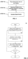
- spatially relative terms such as “beneath,” “below,” “lower,” “above,” “upper” and the like can be used to describe an element and/or feature's relationship to another element(s) and/or feature(s) as, for example, illustrated in the figures. It will be understood that the spatially relative terms are intended to encompass different orientations of the device in use and/or operation in addition to the orientation depicted in the figures. For example, if the device in the figures is turned over, elements described as “below” and/or “beneath” other elements or features would then be oriented “above” the other elements or features. The device can be otherwise oriented (e.g., rotated 90 degrees or at other orientations) and the spatially relative descriptors used herein interpreted accordingly.
- exemplary embodiments are described herein with reference illustrations of idealized or representative structures and intermediate structures. As such, variations from the shapes of the illustrations as a result, for example, of manufacturing techniques and/or tolerances, are to be expected. Thus, exemplary embodiments should not be construed as limited to the particular shapes of regions illustrated herein but are to include deviations in shapes that result, for example, from manufacturing.
- catheters and other devices described in accordance with aspects of the present invention can include numerous forms of diagnostic catheters, such as catheters including one or more electrodes, or therapeutic catheters such as tissue ablation catheters.
- Catheters can be introduced percutaneously into a patient's heart, such as to record electrical activity, measure distances between structures, or deliver energy.
- External devices and systems can be included, such as body surface electrodes used to record electrical activity or deliver an electric signal, or visualization devices such as external ultrasound or fluoroscopic imaging systems.
- Any of these catheters or other devices can include one or more electrodes and/or one or more ultrasound elements (e.g. one or more ultrasound sensors and/or ultrasound transducers).
- the electrodes and/or ultrasound elements of the present invention can be positioned at any location on the device, for example at a distal or proximal portion of the device, and can be positioned internal or external to a patient's body.
- any or all of the ultrasound elements (e.g. ultrasound transducers and/or ultrasound sensors) of the present invention can be used to measure a distance between a sensor and/or a transducer and a surface, as is known in the art.
- One example includes measuring the distance between an ultrasound element comprising a sensor-transducer pair and a wall of a chamber of the heart.
- any or all of the electrodes of the present invention can be used to record electric "signals" (e.g. voltages and/or currents) at or between one or more electrode locations. Recorded electric signals can be used to map electrical activity of tissue. The mapped electrical activity can be further processed (e.g. in terms of sources of charge and charge density and correlated with various physiologic parameters related to the function of the heart) and the mapped electrical activity and other recorded and calculated information can be provided visually to one or more operators of the system of the present invention.
- signals e.g. voltages and/or currents
- Recorded electric signals can be used to map electrical activity of tissue.
- the mapped electrical activity can be further processed (e.g. in terms of sources of charge and charge density and correlated with various physiologic parameters related to the function of the heart) and the mapped electrical activity and other recorded and calculated information can be provided visually to one or more operators of the system of the present invention.
- any or all of the electrodes of the present invention can be used to deliver and/or record electric signals that are generated by the system. Such delivered signals can be emitted from any one or more electrodes, and can be delivered between any two or more electrodes.
- Recorded signals can comprise a signal present at a single electrode location or at multiple electrode locations (e.g. a signal representing a comparison of two or more signals present at two or more electrode locations). Recorded signals can be measured, for example, synchronously or asynchronously in terms of voltage and/or current.
- Recorded signals can be further processed in terms of, for example, resistive and reactive components of impedance and/or the combined magnitude of impedance with any original or processed signal "values" (e.g. those represented by a parameter selected from the group consisting of: instantaneous amplitude; phase; peak; Root-Mean-Square (rms); demodulated magnitude; and combinations of these).
- map and “mapping” shall include, but need not be limited to, “electrical map”, “electrical mapping”, “anatomical map”, “anatomical mapping”, “device map” and “device mapping”, each of which is defined herein below.
- electrical map and “electrical mapping” shall include, but need not be limited to, recording, processing and/or displaying electrical information, such as electrical information recorded by one or more electrodes described or understood in accordance with the present invention.
- This electrical information includes, but is not limited to: cardiac or other tissue voltage measurements; cardiac or other tissue bipolar and/or unipolar electrograms; cardiac or other tissue surface charge data; cardiac or other tissue dipole density data; cardiac or other tissue monophasic action potentials; and combinations of these.
- anatomical map and “anatomical mapping” shall include, but need not be limited to, recording, processing and/or displaying anatomical information, such as anatomical information provided by one or more ultrasound elements of the present invention and/or one or more electrodes described or understood in accordance with the present invention.
- This anatomical information includes, but is not limited to: two-dimensional (2D) or three-dimensional (3D) representations of tissue, such as one or more chambers of a heart; tissue wall thicknesses such as the thickness of an atrial or ventricular wall; distance between two tissue surfaces; and combinations of these.
- a dipole density map and/or surface charge map (hereinafter singly or collectively dipole density map) is provided by using information provided by multiple electrodes and multiple ultrasound elements, such as is described in Applicant's co-pending international application, Serial Number PCT/US2012/028593 , entitled “Device and Method For the Geometric Determination of Electrical Dipole Densities on the Cardiac Wall”.
- device map and “device mapping” shall include, but need not be limited to, recording, processing and/or displaying of device distance information, such as information comprising the distance between a device or device component and another object, such as tissue or another device or device component.
- any pair of electrodes described or understood in accordance with the present invention can be constructed and arranged to provide distance information, such as the distance between that pair of electrodes, or the distance between one of the electrodes and one or more proximate components (e.g. a component at a known distance from one or both of the electrodes in the pair).
- the signal can by processed and/or calibrated according to one or more known separation distances (e.g. the separation distance between two electrodes fixedly mounted to a rigid structure at a pre-determined distance). Calibrated signal values can be combined across adjacent sets of electrode pairs to accurately estimate the distance between any pair (e.g.
- Known and calculated separation distances can be used as "reference" electrodes and combined to triangulate the unknown position of one or more "marker” electrodes, such as an electrode positioned on the present invention or on a separate or external device and positioned proximate the present invention.
- the process of triangulation can be used to dynamically localize the three-dimensional position of any or all of the electrodes either individually and/or as a combined entity in three-dimensional space.
- any or all electrodes described or understood in accordance with the present invention can be used to deliver electric energy, such as radiofrequency energy.
- the dipole densities can be determined by a finite elements method, avoiding the errors encountered using previous extrapolation algorithms.
- the surface charge density means surface charge (Coulombs) per unit area (cm 2 ).
- a dipole as such, is a neutral element, wherein a part comprises a positive charge and the other part comprises the same but negative charge.
- a dipole can better represent the electric nature of cellular membranes, because in a biological environment ion charges are not macroscopically separated.
- the '517 publication disclosed devices, systems, and methods for determining the dipole densities on heart walls using one or more catheters placed into the heart chamber.
- a triangularization of the heart wall is performed in which the dipole density at each vertex correlate to the potential measured at various locations within the associated chamber of the heart.
- mapping information recorded by one or more electrodes located on one or more catheters and anatomical information is used.
- one or more ultrasound elements are provided on the catheter.
- '313 patent, '690 publication and '517 publication disclose devices, systems, and methods for creating an image of the heart based on information recorded from one or more internal electrodes (e.g. creating an anatomical and/or electrical representation of the heart)
- some embodiments of the present invention disclose devices, systems, and methods for creating a heart image with external sensors (i.e. external sensors only), while other embodiments disclose devices, systems, and methods using both internal and external sensors to create the heart image.
- one or more electrodes outside the body can be positioned proximate the surface of the patient's torso.
- one or more ultrasound elements e.g. one or more ultrasound transducers, sensors or combined transducer-sensors, hereinafter “ultrasound element”
- ultrasound element can also be used with the one or more electrodes outside the body, such as one or more ultrasound elements also positioned proximate the surface of the patient's torso.
- the external one or more electrodes disclosed in the present invention are used with internal (inside the body) electrodes disclosed in the internal sensor-based devices, systems, and methods of the '313 patent, '690 publication and '517, combining heart chamber geometry with internal and external sensor (voltage) readings, such that dipole densities can be depicted as an animated color map of activation for each heart beat across the epicardial and/or endocardium surface.
- the information can be used to diagnose and/or treat a patient with a cardiac arrhythmia, such as atrial fibrillation, or an inadequately synchronized activation sequence, such as in heart failure.
- Other information obtained can include precise location of foci, conduction-gaps, and/or position of conduction channels.
- a dipole density library can be created in computer memory by combining the electrode voltage readings from one or more electrodes proximate the surface of the patient's torso with anatomical imaging from an imaging instrument, such as CT; MRI; ultrasound; and/or a generic model of a heart.
- This anatomical imaging can be generated in real-time and/or imported from previous imaging from one or more of CT, MRI, ultrasound (internal or external), or other imaging apparatus.
- the dipole density library is created by combining the electrode voltage readings from one or more electrodes with ultrasound readings recorded by the one or more ultrasound elements proximate the surface of the patient's torso.
- the dipole density library is created by combining the electrode voltage readings from one or more electrodes with ultrasound readings recorded by one or more ultrasound elements positioned within the patient's body, such as one or more ultrasound elements positioned within one or more chambers of the patient's heart.
- the system of the present invention comprises an external device, for example a vest, having one or more electrodes, and optionally, one or more ultrasound elements.
- FIG. 1 shows an example embodiment of a mapping system 100 that can be used for real time dipole density mapping of a heart 12 of a human 10.
- System 100 can include a computer 110 having known types of input devices and output devices, such as a display 120 and printer 130, coupled to a patient attachment device, vest 140, having one or more electrodes 142.
- vest 140 can further include one or more ultrasound elements 144.
- Ultrasound elements 144 can include one or more ultrasound transducers configured to transmit ultrasound waves, such as sound waves configured to reflect off of one or more structures of the heart, and be recorded or otherwise received by one or more ultrasound sensors.
- ultrasound elements 144 can include one or more ultrasound sensors, such as one or more ultrasound sensors which receive the reflected sound waves.
- one or more ultrasound elements 144 can include both an ultrasound transmitter and an ultrasound sensor, such as a single element that both transmits and receives ultrasound waves.
- a vest is shown, numerous alternative patient attachment device types can be used, including, for example, shirts, bibs, arm bands, torso bands and/or any other patient-attachable assembly capable of maintaining the one or more electrodes 142 and/or ultrasound elements 144 in contact with the wearer's body, or sufficiently close thereto, such that a signal can be detected and/or transmitted by each signal-detecting element.
- the one or more electrodes 142 and/or ultrasound elements 144 can be attached directly to the skin.
- multiple discrete attachments can be used with a combination of garments, (e.g. shirt plus armband or torso band plus armband), or a combination of a garment with direct skin attachment(s).
- vest 140 can only include one or more electrodes 142, with no ultrasound elements. In other embodiments, vest 140 can include one or more ultrasound elements 144, and not have any electrodes. In still other embodiments a combination of garments can be used with different elements being positioned on different garments. For example, in a combination of shirt plus armband, the shirt can have one or more electrodes 142 while the armband can have one or more ultrasound elements 144.
- vest 140 is flexible and conforms closely to the body of the patient and can be made of any suitable materials. Vest 140 can be configured so that the one or more electrodes 142 and/or ultrasound elements 144 are urged against the skin at a consistent position, such as to prevent movement of the element across the skin. In some embodiments, the one or more electrodes 142 and/or ultrasound elements 144 can be positioned on both the front and the back of the patient. In other embodiments, the one or more electrodes 142 and/or ultrasound elements 144 can be positioned only on the front or back of the patient, depending on application.
- the one or more electrodes 142 and/or ultrasound elements 144 can be connected to computer 110, such as via a wired and/or wireless connection (see FIG. 8 ).
- Computer 110 can control the operation of the one or more electrodes 142 and/or ultrasound elements 144.
- computer 110 can shut off selected electrodes 142 and/or ultrasound elements 144, leaving only the associated electrodes 142 and/or ultrasound elements 144 that cover one or more areas of interest being turned on.
- System 100 can be used to create a three-dimensional database of dipole densities d(y) and distance measurements at the epicardial surface of the heart.
- the distance measurements can include, but are not limited to: the distance between at least one of the electrodes 142 and the epicardial surface, the distance between at least one of the electrodes 142 and an ultrasound element 144, and the distance between the epicardial surface and an ultrasound element 144. Knowing the speed of sound in the particular environment, as well as the timing of the delivery of sound waves by the transducer, the distance between an ultrasound transducer, a reflected surface, and an ultrasound sensor can be calculated, as described herein below.
- the distance measurements can be calculated by analyzing the received signal amplitude, a shift in frequency between transmitted and received signals, and/or an ultrasound sensor recorded angle.
- System 100 can also be configured to produce continuous, real time geometries of the tissue of a patient.
- System 100 can provide one or more of: tissue geometry information such as tissue position, tissue thickness (e.g. cardiac wall thickness) and tissue motion (e.g. cardiac wall motion) information; distance information such as distance between two tissue locations, and distance between a tissue location and a device component location; tissue electrical activity information; status of ablation of a portion of tissue; status of resynchronization pacing, and/or combinations of these.
- the present invention incorporates one or more ultrasound elements 144 comprising both an ultrasound transducer and an ultrasound sensor, each preferably contained in a single component.
- the ultrasound sensor is configured to record or otherwise detect the wave reflections that result from the ultrasound waves emitted from one or more ultrasound transducers.
- the detected wave reflections can be used to determine real-time continuous measurements of the position of at least one of the electrodes 142 and/or at least one ultrasound element 144. This information is used to enhance one or more dipole density d(y) calculations. Measurements can be taken to determine the thickness of an object, such as the thickness of cardiac tissue, which can be used to determine an ablation parameter such as power or time of energy delivery.
- an ultrasound element 144 comprising a piezo crystal transmits acoustic waves and receives the reflections of those waves.
- the timing between transmitting and receiving can be used to determine the distance between the transmitting and receiving surfaces, and one or more reflective surfaces (e.g. reflective tissue surfaces).
- precise distances and dimensions of target cardiac tissue is determined, resulting in a more precise and effective diagnosis and/or therapy.
- one or more ultrasound elements 144 are constructed and arranged to produce sound waves in at least one of either constant or pulsed excitation, such as sounds waves between 3 megahertz and 18 megahertz.
- the waves emitted by one or more ultrasound elements 144 can be at constant frequency and/or produced by a chirp of changing frequency (to allow pulse compression or demodulation on reception).
- the precision in dipole density calculations along with the distance measurements will allow for the precise detailing of the electrical activity in the cardiac cells and will allow for the precise identification of which cells are the earliest sites of activation.
- one or more ultrasound elements 144 can be configured to automatically detect the distance from one or more ultrasound elements 144 to the epicardial surface via a first reflection and further detect the cardiac wall thickness via a second reflection.
- one or more ultrasound elements 144 integrate multiple reflections to construct an anatomical geometry including an epicardial surface of the heart and the thickness of the associated myocardium.
- one or more ultrasound elements 144 include at least one crystal, typically comprised of a piezoelectric material, which is positioned proximate to the center of each electrode 142 within an electrode array.
- one or more ultrasound elements 144 include at least one crystal positioned between two or more electrodes 142, such as to create a device with a ratio of mapping electrodes 142 to ultrasound elements 144 of 1:1, 2:1, 5:2, 3:1, 4:1 or another ratio.
- the at least one crystal can be constructed and arranged to transmit ultrasonic signals and/or to receive ultrasonic signals (e.g. receive ultrasonic signals transmitted by the same or different crystals and/or the reflections of those signals).
- one or more ultrasound elements 144 comprise a plurality of crystals, such as a plurality of crystals arranged in an array.
- one or more ultrasound elements 144 comprise a piezoelectric film covering one or more electrodes 142, such as one or more electrodes 142 within an array. In some embodiments, one or more ultrasound elements 144 can be constructed as part of an electrode 142.
- system 100 can comprise a sensor/electrode combination.
- FIG. 2 provides an example embodiment of a computer architecture 200 that can form part of mapping system 100.
- Architecture 200 includes interface module 210 for interfacing with the vest 140, interface module 220 for interfacing with output devices 120, 130, and at least one processor 240.
- the computer 110 includes at least one computer memory 250.
- the foregoing are generally known, however the present invention further includes an electric-potential to surface-charge-density and/or dipole-density converter module 230.
- Converter module 230 includes instructions necessary for carrying out the methods described herein, when executed by processor 240, wherein the results of such processing are stored in memory 250, as would be understood by one skilled in the art having the benefit of this disclosure.
- the 3D geometry can be accommodated by integrating anatomical data from CT/MRI scans with the epicardial geometry determined from analysis of the received acoustic signals.
- the CT/MRI scans can include data to determine torso geometry.
- the CT/MRI scans can also provide data associated with an epicardial surface surrounding the heart, where those of ordinary skill would understand that the epicardial surface can be used to register the CT/MRI data with data calculated from the devices of the present invention. Further, locating the epicardial surface can include determining or otherwise providing data to be associated with the location of the heart within the torso.
- system 100 is configured to generate a table of dipole densities v(P', t) that embody an ionic nature of cellular membranes across the epicardium of a given heart of a patient, comprising:
- architecture 200 includes a measuring and recording unit, such as interface module 210 which is configured to obtain electric potential data V e at given positions P proximate the patient's torso surface, the converter module 230 includes an aid-converter that converts the electric potentials V e into digital voltage data, the processor 240 transforms the digital voltage data into dipole charge density data, and the memory 250 stores the electric potential data V e and the transformed cellular membrane dipole density data.
- a measuring and recording unit such as interface module 210 which is configured to obtain electric potential data V e at given positions P proximate the patient's torso surface
- the converter module 230 includes an aid-converter that converts the electric potentials V e into digital voltage data
- the processor 240 transforms the digital voltage data into dipole charge density data
- the memory 250 stores the electric potential data V e and the transformed cellular membrane dipole density data.
- the measuring and recording unit includes multiple electrodes positioned proximate the patient's torso surface.
- the system can further include a wearable garment and at least one of the multiple electrodes can be coupled to the wearable garment.
- the wearable garment is flexible and conforms closely to the patient's torso surface and can urge one or more electrodes against the torso surface with a consistent position to prevent movement of the one or more electrodes.
- the wearable garment can be selected from the group consisting of: vest; shirt; bib; arm band; torso band; any patient-attachable assembly capable of maintaining the one or more electrodes in contact with the torso surface or sufficiently close thereto; and/or combinations thereof.
- the processor includes a computer program embodying an algorithm that, when executed, transforms the digital voltage data into cellular membrane dipole density data.
- the system further includes one or more ultrasound transducers positioned proximate the patient's torso surface, the one or more ultrasound transducers being configured to emit waves toward an epicardial surface, and one or more ultrasound sensors positioned proximate the patient's torso surface, the one or more ultrasound sensors being configured to receive reflections of the waves from the epicardial surface, wherein the measuring and recording unit further measures and records the sensor information.
- one or more ultrasound transducers are further configured to function as an ultrasound sensor.
- the processor is configured to receive sensor data from the one or more sensors and generate distance measurements from the epicardial surface.
- the distance measurement can be produced by analyzing at least one of: timing of received signal; recorded signal amplitude; sensor recorded angle; or signal frequency changes
- the system includes more than one wearable garment and the multiple electrodes, ultrasound transducers, or ultrasound sensors are coupled to different wearable garments.
- the multiple electrodes are coupled to a first wearable garment
- the ultrasound transducers and ultrasound sensors are coupled to a second wearable garment.
- the wearable garments can be selected from the group consisting of: vest; shirt; bib; arm band; torso band; any patient-attachable assembly capable of maintaining the one or more electrodes, one or more ultrasound transducers, and one or more ultrasound sensors in contact with the torso surface, or sufficiently close thereto that a monitorable signal is detectable.
- the system further includes an imaging unit that represents the cellular membrane dipole densities v(P',t) as a two-dimensional image or time-dependent sequence of images
- the system further includes an imaging unit that represents the cellular membrane dipole densities v(P',t) as a three-dimensional image or time-dependent sequence of images.
- FIG. 3 shows a schematic view of some elements of computer 110 used for determining a database table of dipole densities d(y).
- Computer 110 includes a first receiver 310 configured to receive electrical potentials from the one or more electrodes, such as electrodes 142 of Fig. 1 .
- Computer 110 further includes a second receiver 320 configured to receive cardiac geometry information from an imaging instrument, such as CT; MRI; ultrasound; or a generic model of a heart. This anatomical imaging can be generated in real-time and/or imported from previous imaging from one or more of CT, MRI, ultrasound (internal or external), or other imaging apparatus.
- Dipole density processor 330 receives electrical information from first receiver 310 and cardiac geometry information from the second receiver 320.
- Dipole density processor 330 which can comprise converter module 230 and processor 240, includes a mathematical processing element or other electronic module including software and/or hardware for performing mathematical or other calculations.
- Dipole density processor 330 preferably uses one or more algorithms to process the received electrical and geometry information to produce a database table of dipole densities d(y) 350.
- dipole density processor 330 can be configured to produce a database table of surface charge information.
- the vest 140 can further include one or more ultrasound transducers and/or one or more ultrasound sensors to provide cardiac geometry information to the second receiver 320.
- the one or more ultrasound transducers transmit ultrasound waves, such as waves configured to reflect off one or more structures of the heart, and be recorded by the ultrasound sensors (e.g. reflections from the epicardial surface and one or more of the inner surfaces or structures of the heart).
- Dipole density processor 330 receives electrical information from first receiver 310 and ultrasound cardiac geometry information from the second receiver 320.
- Dipole density processor 330 which can comprise converter module 230 and processor 240, includes a mathematical processing element or other electronic module including software and/or hardware for performing mathematical or other calculations.
- Dipole density processor 330 preferably uses one or more algorithms to process the received electrical and geometry information to produce a database table of dipole densities d(y) 350.
- the geometric model of the epicardial surface can be processed by the dipole density processor 330 into multiple small triangles (triangularization) and/or other polygonal shapes (e.g., squares, tetrahedral, hexagonal, and others). When the polygons are sufficiently small, the dipole density has a small variation over the polygon. In a preferred embodiment, the number of triangles is determined by dipole density processor 330. With the electrodes positioned by a clinician, such as an electrophysiologist, the potentials at each electrode are recorded. The dipole density processor 330 computes the dipole density at all vertices of the triangles.
- the total measured potential V(x) at a location x is the sum over all vertices y of d(y) times a matrix W(x,y).
- dipole density processor 330 implements a progressive algorithm that can be modified and/or refined in order to improve spatial and/or time resolution of the database of dipole densities that are produced.
- the dipole densities d(y) can be obtained by solving a linear system of equations. Thereby a map of dipole densities can be created at each corresponding time interval. The synthesis of the maps generates a cascade of the activation sequence of each corresponding heart beat that can be used to diagnose cardiac wall tissue, such as to identify an origin of aberrant electrical activity or otherwise diagnose an arrhythmia.
- the measuring electrodes used in the present invention are placed on or proximate the torso surface. Due to the inhomogeneous structure of the body, it is difficult to localize the actual sources of the skin electrode measured potentials. A highly complicated boundary value problem must be solved with boundary conditions that are poorly known. Prior art attempts at determining the "action potential" from body surface ECG (alone) have not been very successful.
- the present invention calculates the dipole densities using external electrodes on the vest, in combination with cardiac geometry information from an imaging instrument (such as CT; MRI; ultrasound); or the optional external ultrasound transducers and/or ultrasound sensors on the vest.
- an imaging instrument such as CT; MRI; ultrasound
- System 100 shown in FIG. 1 , is configured to create a database table of three-dimensional dipole densities d(y) based on voltage potentials and image information relating to the heart, as has been described above.
- triangle T1 defined by system 100 is at location Y1.
- the contribution of triangle T1 to the potential at location X1 can be computed from the dipole density at the vertices of T1.
- the dipole density processor 330 determines the desired dipole density d(y) from the total measured potential V(x), which is the sum resulting from all the triangles defined by system 100.
- the dipole density d(y) at many equally distributed vertices y on the epicardial surface is calculated (e.g. from 10 to 50,000 with increasing number of calculated potentials providing more detailed results) by solving a system of linear equations.
- the measured and/or calculated potentials e.g. with application of splines
- their number can be increased to a higher number of regions.
- This calculation of dipole density results, such as via an automatic computer program forming at least part of dipole density processor 330.
- the results are presented in a visual, anatomical format, such as depicting the dipole densities on a geometric model of the epicardial surface in relation to time (t).
- This format allows a clinician, such as an electrophysiologist, to determine the activation sequence, or other electrical and mechanical measures, on the epicardial surface, such as to determine treatment locations for a cardiac arrhythmia or other inadequacy in cardiac tissue health, such as force of tissue contraction and motion of the epicardial surface.
- the results can be shown on a display unit 120, or on a separate display not shown, such as a color display.
- the device of the present invention is implemented as, or includes, a software program that is executable by at least one processor.
- the software program can be integrated into one or more of: an ECG system; a cardiac tissue ablation system; an imaging system; a computer; and combinations of these.
- FIG. 5 illustrates one embodiment of a method for determining a database table of dipole densities d(y) of the epicardial surface of a patient's heart.
- a vest having an array of one or more electrodes e.g. vest 140 of system 100 of Fig. 1
- the geometry of the epicardial surface can be obtained in relation to the positions of the one or more electrodes disposed within the electrode array.
- the magnitude and other properties of motion of cardiac wall tissue can be determined.
- the thickness of a patient's heart tissue can be determined. This information will enable a clinician to determine what treatment, (e.g., what ablation parameters) can be appropriate for the patient.
- the geometry of the epicardial surface is obtained in relation to the electrode array position, such as by importing a geometry model from an imaging study (e.g., using computed tomography, MRI and/or ultrasound).
- the surface of the geometry of the corresponding epicardial surface is generally divided into small polygons, such as in the form of at least 1000 triangles of similar size.
- the dipole density d(y) can be calculated at each vertex y from the measured potential values x.
- the measurements can be repeated successively during the cardiac cycle, such as once each millisecond, giving the electrophysiologist a dynamic progression of the activation sequence.
- the information of the time dependent dipole densities can be depicted as an animated color map of activation for each heart beat across the epicardial surface.
- the information can be used to diagnose and/or treat a patient with a cardiac arrhythmia, such as atrial fibrillation, or an inadequately synchronized activation sequence, such as in heart failure.
- Other information obtained can include precise location of foci, conduction-gaps, and/or position of conduction channels.
- the dipole density information can be used to determine cardiac tissue treatment locations for lesion creation, such as a lesion created by a catheter-based ablation system.
- the lesion can be created by an RF, ultrasound, microwave, laser and/or cryogenic energy ablation catheter.
- the information can also be used to determine the location of pacing electrodes for cardiac resynchronization therapy.
- ablating the cardiac tissue can be based upon the tissue diagnosis.
- the anatomical information comprising tissue thickness information and at least one of the magnitude of ablation energy or the time period in which ablation energy is delivered, is adjusted based on the tissue thickness information recorded by one or more ultrasound sensors.
- FIG. 6 summarizes one method 400 for determining and storing surface charge densities and/or dipole densities in accordance with aspects of the present invention, which have been described in detail above.
- mapping system 100 is used to measure and/or calculate one or more electric potential(s) V e in one or more position(s) P at a given time t.
- V e is transformed into a surface charge density ⁇ (P',t) and/or dipole density d(P',t)
- Step 406 the surface charge density ⁇ (P',t) and/or dipole density d(P',t) is stored in a database table. The method is repeated if there is another P, in Step 408.
- FIG. 7 shows an embodiment using both external sensor systems and internal sensor systems together.
- the present systems and methods disclosed above for external sensor-based imaging of the heart can be combined with the devices, systems, and methods using internal sensor-based imaging of the heart disclosed in the '313 patent, '690 publication and '517 publication.
- FIG. 7 shows the present vest system in combination with system 500, described in detail in the '690 publication and '517 publication.
- This combination of internal and external electrodes can be used to augment accuracy, specificity, etc., and combining heart chamber geometry with internal and external sensor (voltage) readings can provide simultaneous maps of the epicardium and endocardium walls.
- System 500 includes a mapping catheter with a shaft 311, which is inserted into a chamber of a patient's heart, such as the Left Atrium (LA).
- a mapping catheter with a shaft 311, which is inserted into a chamber of a patient's heart, such as the Left Atrium (LA).
- LA Left Atrium
- Electrode array 315 including multiple electrodes 316.
- Electrode array 315 is shown in a basket construction, but numerous other constructions can be used including multiple independent arms, spiral arrays, electrode covered balloons, and other constructions configured to place multiple electrodes into a three-dimensional space.
- Any catheter with one or more electrodes can be used to supply mapping information to system 100, which is configured to create a database table of three-dimensional dipole densities d(y) based on voltage potentials and image information relating to the heart, as has been described above.
- triangle T2 is at location Y2 on the endocardial surface and electrode 316a is at location X2.
- the contribution of triangle T2 to the potential at location X2 can be computed from the dipole density at the vertices of T1.
- the dipole density processor 330 determines the desired dipole density d(y) from the total measured potential V(x), which is the sum resulting from all the triangles defined by system 100.
- the dipole density d(y) at many equally distributed vertices y on the endocardial surface can be calculated (e.g. from 10 to 50,000 with increasing number of calculated potentials providing more detailed results) by solving a system of linear equations.
- the measured and/or calculated potentials e.g. with application of splines
- their number can be increased to a higher number of regions.
- the results are presented in a visual, anatomical format, such as on a display depicting the dipole densities on a geometric model of the endocardial surface and epicardial surface in relation to time (t).
- This format allows a clinician, such as an electrophysiologist, to determine the activation sequence, or other electrical and mechanical measures, on the endocardial surface and/or epicardial surface, such as to determine treatment locations for a cardiac arrhythmia or other inadequacy in cardiac tissue health, such as force of tissue contraction and motion of an endocardial surface and/or an epicardial surface.
- the results can be shown on a display unit 120, or on a separate display not shown, such as a color display.
- FIG. 8 shows embodiments for a mapping system 600 for monitoring of a patient at their home or otherwise remote from a clinical setting.
- the system 600 can use many of the elements and methods described above for determination of dipole densities.
- the system 600 includes a vest 640, which can use the same or similar features as vest 140 described above, and a recording device 604a, computer 604b and/or communication system 604c.
- Vest 640 can include one or more electrodes 642.
- vest 640 can further include one or more ultrasound elements 644, such as one or more ultrasound transducers and/or ultrasound sensors.
- Vest 640 can be flexible and conform closely to the body of the patient and can be made of any suitable materials.
- Vest 640 can be configured so that the one or more electrodes 642 and/or ultrasound elements 644 are urged against the torso surface or skin at a consistent position, such as to prevent movement of the element across the skin.
- the one or more electrodes 642 and/or ultrasound elements 644 can be positioned on both the front and the back of the patient.
- the one or more electrodes 642 and/or ultrasound elements 644 can be positioned on only the front or back of the patient, depending on application. Alternatively, the one or more electrodes 642 and/or ultrasound elements 644 can be attached directly to the skin. While the description discloses one or more electrodes 642 and/or one or more ultrasound elements 644 used with the vest, garment, or direct skin attachment, the invention also envisions embodiments that only include electrodes 642 or only ultrasound elements 644.
- vest 640 or another component of system 600 includes one or more additional sensors or transducers, functional element 645.
- Functional elements 645 can comprise an element selected from the group consisting of: a pressure sensor such as a blood pressure sensor; a pH sensor; a glucose sensor; a respiration sensor; a salinity or other sweat level sensor; an EEG sensor such as an EEG sensor placed on the scalp of the patient; an oxygen level sensor such as an oxygen level sensor placed on the finger of the patient; an eye gaze sensor; and combinations of these.
- the one or more electrodes 642, ultrasound elements 644, and/or functional elements 645 can be coupled to the recording device 604a, computer 604b and/or communication system 604c, with either a wired (not shown) or wireless connection (e.g., Bluetooth, Wi-Fi, or other wireless means).
- the recording device 604a, computer 604b and/or communication system 604c can control the operation of the one or more electrodes 642, ultrasound elements 644, and/or functional elements 645. This control feature can be programmed into their systems or can be done remotely via a remote connection (e.g., from a physician's office 608).
- the recording device 604a, computer 604b and/or communication system 604c can turn on or shut off selected electrodes 642, ultrasound elements 644, and/or functional elements 645, leaving only the associated electrodes 642, ultrasound elements 644, and/or functional elements 645 that cover one or more areas of interest being turned on.
- the recording device 604a can be a portable device for monitoring and recording various electrical and/or other signal activities of the one or more electrodes 642, ultrasound elements 644, and/or functional elements 645, similar to a Holter or other mobile-patient monitor.
- the recording device 604a can be configured to continuously monitor and record, or only record on an as needed basis when a recordable event happens. Once the data is recorded, the recording device 604a can be transmitted to the physician's office to be analyzed.
- the recording device 604a can be a smart phone, such as a Galaxy S4, having an application for recording the signal activities. Once recorded, the smart phone can also be capable of transmitting the information, for example, to the physician's office.
- the computer 604b can have the capability of continuously monitoring various signal activities of the one or more electrodes 642, ultrasound elements 644, and/or functional elements 645.
- the computer 604b can also have the capability of analyzing the data from the one or more electrodes 642, ultrasound elements 644, and/or functional elements 645, similar to system 100 described above.
- computer 604b comprises one or more algorithms constructed and arranged to analyze one or more of: cardiac geometry; cardiac electrical activity; blood pressure; pH; glucose; respiration; sweat level; brain activity; or blood oxygen level.
- computer 604b analyzes cardiac electrical activity and at least one physiologic parameter selected from the group consisting of: blood pressure; pH; glucose; respiration; sweat level; brain activity; or blood oxygen level.
- the computer 604b can save the monitored or analyzed data in memory, such as on memory card or flash device card or copy it to a disk.
- the computer 604b can further have the capability of transmitting the analyzed data, for example, to the physician's office, giving the physician real-time feedback as to the health and condition of their patient.
- communication system 604c can include a means of communicating with the physician's office on a real-time basis for remote medical patient monitoring, such as over the internet or other direct communication means (e.g., smart phone).
- the physician can monitor the patient 24 hours a day and/or at any time.
- the system can further include two way communications such that the physician can view the data in real-time while speaking with the patient.
- the physician can also turn on or shut off selected electrodes 642, ultrasound elements 644 and/or functional elements 645, leaving only the associated electrodes 642, ultrasound elements 644 and/or functional elements 645 that cover one or more areas of interest being turned on.
Description
- The present application claims priority under 35 USC 119(e) to
United States Provisional Patent Application Serial No. 61/877,617, entitled "Devices and Methods for Determination of Electrical Dipole Densities on a Cardiac Surface," filed September 13, 2013 - The present application, while not claiming priority to, may be related to
US Patent Application Serial No. 13/858715, entitled "Method and Device for Determining and Presenting Surface Charge and Dipole Densities on Cardiac Walls", filed April 8, 2013 US Patent No. 8,417,313 (hereinafter the '313 patent), entitled "Method and Device for Determining and Presenting Surface Charge and Dipole Densities on Cardiac Walls", issued April 9, 2013 , which was a 35 USC 371 national stage filing ofPCT Application No. CH2007/000380, entitled "Method and Device for Determining and Presenting Surface Charge and Dipole Densities on Cardiac Walls", filed August 3, 2007 WO 2008/014629 , which claimed priority toSwiss Patent Application No. 1251106 filed August 3, 2006 - The present application, while not claiming priority to, may be related to
US Patent Application Serial No. 13/946712, entitled "Device and Method for the Geometric Determination of Electrical Dipole Densities on the Cardiac Wall", filed July 19, 2013 US Patent No. 8,512,255, entitled "Device and Method for the Geometric Determination of Electrical Dipole Densities on the Cardiac Wall", issued August 20, 2013 , published asUS2010/0298690 (hereinafter the '690 publication), which was a 35 USC 371 national stage application of Patent Cooperation Treaty Application No.PCT/IB09/00071 filed January 16, 2009 WO2009/090547 , which claimed priority toSwiss Patent Application 00068/08 filed January 17, 2008 - The present application, while not claiming priority to, may be related to
US Application Serial No. 14/003671, entitled "Device and Method for the Geometric Determination of Electrical Dipole Densities on the Cardiac Wall", filed September 6, 2013 , which is a 35 USC 371 national stage filing of Patent Cooperation Treaty Application No.PCT/US2012/028593 , entitled "Device and Method for the Geometric Determination of Electrical Dipole Densities on the Cardiac Wall",published as WO2012/122517 (hereinafter the '517 publication), which claimed priority toUS Patent Provisional Application Serial No. 61/451,357 - The present application, while not claiming priority to, may be related to Patent Cooperation Treaty Application No.
PCT/US2013/057579, entitled "Catheter System and Methods of Medical Uses of Same, Including Diagnostic and Treatment Uses for the Heart", filed August 30, 2013 , which claims priority toUS Patent Provisional Application Serial No. 61/695,535, entitled "System and Method for Diagnosing and Treating Heart Tissue", filed August 31, 2012 - The present application, while not claiming priority to, may be related to
US Patent Provisional Application Serial No. 61/762,363, entitled "Expandable Catheter Assembly with Flexible Printed Circuit Board (PCB) Electrical Pathways", filed February 8, 2013 - The present invention is generally related to devices and methods for dipole density mapping.
- For localizing the origin(s) of cardiac arrhythmias it is common practice to measure the electric potentials located on the inner surface of the heart by electrophysiological means within the patient's heart. One method is to insert electrode catheters into the heart to record cardiac potentials during normal heart rhythm or cardiac arrhythmia. If the arrhythmia has a regular activation sequence, the timing of the electric activation measured in voltages at the site of the electrode can be accumulated when moving the electrode around during the arrhythmia, to create a three-dimensional map of the electric activation. By doing this, information on the localization of the source of arrhythmia(s) and mechanisms, i.e., re-entry circuits, can be diagnosed to initiate or guide treatment (radiofrequency ablation). The information can also be used to guide the treatment of cardiac resynchronization, in which implantable pacing electrodes are placed in specific locations within the heart wall or chambers to re-establish a normal level of coordinated activation of the heart.
- A method using external sensors measures the electrical activity of the heart from the body surface using electrocardiographic techniques that include, for example, electrocardiograms (ECG) and vectorcardiography (VCG). These external sensor techniques can be limited in their ability to provide information and/or data on regional electrocardiac activity. These methods can also fail to localize bioelectric events in the heart.
- A method using external sensors for the localization of cardiac arrhythmias utilizes body surface mapping. In this technique, multiple electrodes are attached to the entire surface of the thorax and the information of the cardiac electrograms (surface ECG) is measured in voltages that are accumulated into maps of cardiac activation. This measurement can be problematic because the electrical activity is time dependent and spatially distributed throughout the myocardium and also fails to localize bioelectric events in the heart. Complex mathematical methods are required to determine the electric activation upon the outer surface of a heart model (i.e. epicardium), for instance, one obtained from CT or MRI imaging giving information on cardiac size and orientation within the thoracic cavity.
- Alternatively, recordings of potentials at locations on the torso, for example, can provide body surface potential maps (BSPMs) over the torso surface. Although the BSPMs can indicate regional cardiac electrical activity in a manner that can be different from conventional ECG techniques, these BSPM techniques generally provide a comparatively low resolution, smoothed projection of cardiac electrical activity that does not facilitate visual detection or identification of cardiac event locations (e.g., sites of initiation of cardiac arrhythmias) and details of regional activity (e.g., number and location of arrythmogenic foci in the heart).
-
WO 2012/110942 A1 describes a system for providing an electrical activity map of the heart by means of electrical signals acquired by a plurality of surface electrodes. A surface electrodes positions determination unit determines positions of the plurality of surface electrodes by means of optical shape sensing localization. The optical shape sensing element may comprise a wand, or alternatively an optical shape sensing fiber embedded in the vest comprising the surface electrodes. The position of a cardiac structure may be determined using ultrasound. An electrical activity map determination unit determines the electrical activity map at the cardiac structure based on the measured electrical signals, the determined positions of the plurality of electrodes and the position of the cardiac structure, in particular, of the epicardial surface. -
WO 2012/122517 A2 describes a device for determining the dipole densities on heart walls. In particular, a triangularization of the heart wall is performed in which the dipole density of each of multiple regions correlate to the potential measured at various located within the associated chamber of the heart. -
US 2011/077526 A1 describes an ultrasound assembly including a garment configured to be affixed to a portion of a living body, and at least one ultrasound transducer having a fixed position on the garment and configured to provide at least one of: produce and receive, ultrasound signals that pass through the living body. - Since the localization of cardiac arrhythmias by the use of potentials is imprecise, the successful treatment of cardiac arrhythmias has been difficult and has demonstrated limited success and reliability. There is, therefore, a need for improved methods of localizing cardiac arrhythmias.
- The claimed invention is defined by the independent claim. Specific embodiments are defined in the dependent claims. The present invention includes one or more electrodes configured to record electrical activity of tissue. In some embodiments, one or more ultrasound transducers, ultrasound sensors, and/or combinations of these can be included. The electrodes, transducers and sensors are located proximate the torso surface, and can be coupled to a wearable garment, such as a vest, shirt or bib. The device is constructed and arranged to produce continuous, real-time geometries of a patient's tissue, as well as information related to electrical activity present in the tissue.
- The device can also be capable of providing tissue information, for example, tissue movement and tissue thickness. Additionally, the device can be configured to produce distance measurements by analyzing at least one of the sensors recorded angles or amplitudes or frequency changes. Non-limiting examples of distance measurements include: distance between the one or more electrodes and the epicardial surface and distance between the one or more electrodes and the one or more transducers and/or sensors.
- The device can be configured to provide a tissue diagnostic through an analysis of both tissue motion information and cell electrical signals. The cell electrical signals can be recorded by the one or more electrodes, while tissue motion information can be gathered by the one or more electrodes and/or sensors. The device can be configured to provide exact foci and conduction-gap position information, such that ablation can be performed with an increased level of precision. Small conduction paths, including "gaps" in a line, are equally relevant as foci. The device can be used with an ablation device, such as robotic or manually controlled catheter ablation device. The device can also be used with a pacing system, such as a system for delivering pacing electrodes into the heart and for stimulating the heart with pacing pulses delivered through the pacing electrodes.
- In accordance with one aspect of the present disclosure, a device generates a table of dipole densities v(P',t) that embody an ionic nature of cellular membranes across the epicardium of a given heart of a patient. The device comprises: a measuring and recording unit that measures and records electric potential data Ve at given positions P proximate the patient's torso surface; an a/d-converter that converts the electric potential data Ve into digital voltage data; a processor that transforms the digital voltage data into cellular membrane dipole density data; and a memory that stores the electric potential data Ve and the transformed cellular membrane dipole density data.
- In some embodiments, the measuring and recording unit includes multiple electrodes positioned proximate the patient's torso surface. The device can further comprise a wearable garment, and the multiple electrodes can be coupled to the wearable garment. The wearable garment can be flexible and conform closely to the patient's torso surface. The wearable garment can be configured to urge the multiple electrodes against the torso surface with a consistent position to prevent movement of at least one of the multiple electrodes.
- In various embodiments, the wearable garment can be selected from the group consisting of: vest; shirt; bib; arm band; torso band; any patient-attachable assembly capable of maintaining the one or more electrodes in contact with the torso surface or sufficiently close thereto that a monitorable signal is detectable; and/or combinations thereof.
- In some embodiments, the processor executes a computer program embodying an algorithm for transforming the digital voltage data into cellular membrane dipole density data. The computer program can be stored in a storage device, e.g., an electrical, magnetic, and/or optical storage device. The storage device can be a non-transitory storage device.
- In some embodiments, the device further comprises one or more ultrasound transducers positioned proximate the patient's torso surface, the one or more ultrasound transducers being configured to emit waves toward an epicardial surface; and one or more ultrasound sensors positioned proximate the patient's torso surface, the one or more ultrasound sensors being configured to receive reflections of the waves from the epicardial surface and produce sensor data. The processor can be configured to receive the sensor data from the one or more sensors and generate distance measurements from the epicardial surface. The processor can be configured to produce the distance measurements by analyzing at least one of: timing of received signal; recorded signal amplitude; sensor recorded angle; or signal frequency changes.
- The device can further comprise at least one wearable garment, and the at least one of the multiple electrodes, one or more ultrasound transducers, or one or more ultrasound sensors can be coupled to the at least one wearable garment. The at least one wearable garment can comprise a first wearable garment and a second wearable garment, and the multiple electrodes can be coupled to the first wearable garment, and the one or more ultrasound transducers and one or more ultrasound sensors can be coupled to the second wearable garment. In various embodiments, the at least one wearable garment can be selected from the group consisting of: vest; shirt; bib; arm band; torso band; any patient-attachable assembly capable of maintaining the one or more electrodes, one or more ultrasound transducers, and/or one or more ultrasound sensors in contact with the torso surface, or sufficiently close thereto that a monitorable signal is detectable; and/or combinations thereof.
- In some embodiments, the device can be configured to diagnose at least one of: an arrhythmia; ischemia; or compromised myocardial function.
- In some embodiments, the device can be configured to treat at least one of: an arrhythmia; ischemia; or compromised myocardial function.
- In accordance with another aspect of the present disclosure, a device for creating a database of dipole densities d(y) at an epicardial surface of the heart of a patient comprises: multiple electrodes positioned proximate the patient's torso surface; a first receiver configured to receive mapping information from the multiple electrodes; a second receiver configured to receive an anatomical depiction of the heart; a dipole density module configured to generate the database of dipole densities d(y) of polygonal shaped projections onto the epicardial surface, wherein the dipole density module computes the dipole density at all vertices of the polygonal shaped projections, wherein if the dipole density is d(y), the total measured potential V(x) at a location x is the sum over all vertices of d(y) times a matrix ώ(x,y), and wherein: a) x represents a series of locations on the torso surface; and b) V(x) is a measured potential at point x, said measured potential recorded by the multiple electrodes.
- In some embodiments, the dipole density module can generates the database of dipole densities d(y) using a finite elements method.
- In some embodiments, the polygonal shaped projections can be substantially the same size.
- In some embodiments, the dipole density can be determined by a number of polygonal shaped projections, wherein the number can be determined by the size of the epicardial surface.
- In some embodiments, the polygonal shaped projections can be selected from the group consisting of: triangles; squares; tetrahedral shapes; hexagonal shapes; any other suitable shape compatible with finite elements method; and/or combinations thereof.
- In some embodiments, the device can further comprise a wearable garment, and the multiple electrodes can be coupled to the wearable garment. The wearable garment can be flexible and conform closely to the patient's torso surface. The wearable garment can be configured to urge the multiple electrodes against the torso surface with a consistent position to prevent movement of the electrodes. The wearable garment can be selected from the group consisting of: vest; shirt; bib; arm band; torso band; any patient-attachable assembly capable of maintaining the one or more electrodes in contact with the torso surface or sufficiently close thereto that a monitorable signal is detectable; and/or combinations thereof.
- In some embodiments, the anatomical depiction of the heart can comprise previous anatomical imaging and/or real-time anatomical imaging from one or more of CT; MRI; internal ultrasound; external ultrasound; or other imaging apparatus.
- In some embodiments, the anatomical depiction of the heart can comprise a generic model of a heart.
- In some embodiments, the device can further comprise: one or more ultrasound transducers positioned proximate the patient's torso surface, the one or more ultrasound transducers being configured to emit waves toward the epicardial surface; and one or more ultrasound sensors positioned proximate the patient's torso surface, the one or more ultrasound sensors being configured to receive reflections of the waves from the epicardial surface.
- The device can further comprise at least one wearable garment, and at least one of the multiple electrodes, one or more ultrasound transducers, and/or one or more ultrasound sensors can be coupled to the at least one wearable garment. The at least one wearable garment can comprise a first wearable garment and a second wearable garment, and the multiple electrodes can be coupled to the first wearable garment, and the one or more ultrasound transducers and/or one or more ultrasound sensors can be coupled to the second wearable garment. The at least one wearable garment can be selected from the group consisting of: vest; shirt; bib; arm band; torso band; any patient-attachable assembly capable of maintaining the one or more electrodes, one or more ultrasound transducers, and/or one or more ultrasound sensors in contact with the torso surface, or sufficiently close thereto that a monitorable signal is detectable; and/or combinations thereof. The anatomical depiction of the heart can comprise real-time anatomical imaging from the one or more ultrasound transducers and the one or more ultrasound sensors.
- In some embodiments, the device can be configured to diagnose at least one of: anarrhythmia; ischemia; or compromised myocardial function.
- In some embodiments, the device can be configured to treat at least one of: an arrhythmia; ischemia; or compromised myocardial function.
- In accordance with another aspect of the present disclosure, a method of creating a database of dipole densities d(y) at the epicardial surface of the heart of a patient comprises: placing an array of multiple electrodes proximate the patient's torso surface; and calculating dipole densities d(y) by: receiving mapping information from the multiple electrodes; receiving an anatomical depiction of the heart; and generating the database of dipole densities d(y) with a dipole density module, wherein the dipole density module determines dipole densities d(y) of polygonal shaped projections onto the epicardial surface, wherein the dipole density module computes the dipole density at all vertices of the polygonal shaped projections, wherein if the dipole density is d(y), the total measured potential V(x) at a location x is the sum over all vertices of d(y) times a matrix ώ(x,y), and wherein: a) x represents a series of locations on the torso surface; and b) V(x) is a measured potential at point x, said measured potential recorded by the multiple electrodes.
- In some embodiments, the dipole density module can generate the database of dipole densities d(y) using a finite elements method.
- In some embodiments, the method can further comprise providing a wearable garment, and the multiple electrodes can be coupled to the wearable garment. The wearable garment can be configured to urge the multiple electrodes against the torso surface with a consistent position to prevent movement of the electrodes. The wearable garment can be selected from the group consisting of: vest; shirt; bib; arm band; torso band; any patient-attachable assembly capable of maintaining the one or more electrodes in contact with the torso surface or sufficiently close thereto that a monitorable signal is detectable; and/or combinations thereof.
- In some examples not forming part of the claimed invention, the method can include using the dipole densities d(y) to locate an origin of abnormal electrical activity of a heart.
- In some examples not forming part of the claimed invention, the method can include using the dipole densities d(y) to diagnose at least one of: an arrhythmia; ischemia; or compromised myocardial function.
- In some examples not forming part of the claimed invention, the method can include using the dipole densities d(y) to treat at least one of: an arrhythmia; ischemia; or compromised myocardial function.
- In some embodiments, calculating the dipole densities d(y) can include a processor executing a computer program stored in a memory, the computer program embodying an algorithm for generating a table of dipole densities in the memory. The memory can be a non-transitory storage device, such as an electrical, magnetic, and/or optical storage device, as examples.
- In accordance with another aspect of the present disclosure, a device for creating a database of dipole densities d(y) and distance measurements at an epicardial surface of a patient comprises: an array of multiple electrodes positioned proximate the patient's torso surface; one or more ultrasound transducers positioned proximate the patient's torso surface, the one or more ultrasound transducers being configured to emit waves toward the epicardial surface; one or more ultrasound sensors positioned proximate the patient's torso surface, the one or more ultrasound sensors being configured to receive reflections of the waves from the epicardial surface; and a computer coupled to the multiple electrodes, one or more ultrasound transducers, and one or more ultrasound sensors, wherein the computer is configured to receive mapping information from the multiple electrodes and sensor data from the one or more sensors, and generate the database of dipole densities d(y) and distance measurements.
- In some embodiments, the device can further comprise at least one wearable garment, and at least one of the multiple electrodes, one or more ultrasound transducers, and/or one or more ultrasound sensors can be coupled to the at least one wearable garment. The wearable garment can be flexible and conform closely to the body of the patient. The wearable garment can be configured to urge electrodes, sensors and/or transducers against the torso surface with a consistent position to prevent movement of the electrodes, sensors and/or transducers. The at least one wearable garment can be selected from the group consisting of: vest; shirt; bib; arm band; torso band; any patient-attachable assembly capable of maintaining the one or more electrodes, one or more ultrasound transducers, and one or more ultrasound sensors in contact with the torso surface, or sufficiently close thereto that a monitorable signal is detectable; and combinations thereof.
- In various embodiments, the at least one wearable garment can comprise a first wearable garment and a second wearable garment, and the multiple electrodes can be coupled to the first wearable garment, and the one or more ultrasound transducers and/or one or more ultrasound sensors can be coupled to the second wearable garment. The computer can be coupled to the wearable garment.
- In some embodiments, the computer can include: a dipole density module configured to generate a three dimensional database of dipole densities d(y), and wherein the dipole density module determines a dipole density for polygonal shaped projections onto the epicardial surface and computes the dipole density at all vertices of the polygonal shaped projections, wherein if the dipole density is d(y), the total measured potential V(x) at a location x is the sum over all vertices of d(y) times a matrix ώ(x,y), and wherein: a) x represents a series of locations on the torso surface; and b) V(x) is a measured potential at point x, said measured potential recorded by the multiple electrodes. The dipole density module can generate the database of dipole densities d(y) using a finite elements method. The polygonal shaped projections can be substantially the same size. The dipole density can be determined by a number of polygonal shaped projections, the number determined by the size of an epicardial surface. Such module can include or be embodied in, as examples, hardware, computer program code, firmware, and/or combinations thereof.
- In some embodiments, the device can be configured to provide epicardial surface motion information of the heart. The device can be configured to provide tissue diagnostic information by analyzing both motion information and cell electrical signals. The cell electrical signals can be recorded by the multiple electrodes.
- In some embodiments, the device can further include a display configured to display real time motion.
- In some embodiments, the computer can be configured to produce a geometrical depiction of the heart.
- In some embodiments, the device can be further configured to determine properties of the cardiac wall. The properties can include cardiac wall thickness information. The properties can include precise foci, conduction-gaps, and/or conduction channels position information.
- In some embodiments, the distance measurement can comprise the distance between at least one of the multiple electrodes and at least one epicardial surface.
- In some embodiments, the device can be configured to produce the distance measurement by analyzing at least one of: timing of received signal; recorded signal amplitude; sensor recorded angle; or signal frequency changes.
- In some embodiments, the device can be configured to provide epicardial surface information during a cardiac ablation procedure. The ablation procedure can comprise delivery of RF, ultrasound, microwave, cryogenic and/or laser energy to tissue.
- In some embodiments, at least one of the sensors and at least one of the transducers can comprise a single component.
- In some embodiments, at least one of the sensors and at least one of the transducers can be integral to at least one electrode of the multiple electrodes.
- In some embodiments, the computer can be configured to determine a map of dipole densities d(y) at corresponding time intervals.
- In some embodiments, the computer can be configured to generate a synthesis of maps that represents a cascade of activation sequences of each corresponding heart beat from a series of heart beats.
- In some embodiments, the device can be configured to diagnose at least one of: an arrhythmia; ischemia; or compromised myocardial function.
- In some embodiments, the device can be configured to treat at least one of: an arrhythmia; ischemia; or compromised myocardial function.
- In accordance with another aspect of the present disclosure, a method of creating a database of dipole densities d(y) and distance measurements at an epicardial surface of a patient comprises: placing an array of multiple electrodes, one or more ultrasound transducers, and one or more ultrasound sensors proximate the patient's torso surface; and calculating dipole densities d(y) by: receiving mapping information from the multiple electrodes; emitting waves toward the epicardial surface with the one or more ultrasound transducers; receiving reflections of the waves from the epicardial surface with the one or more ultrasound sensors; producing a geometrical depiction of the epicardial surface; generating the database of dipole densities d(y) with a dipole density module, wherein the dipole density module determines dipole densities d(y) of polygonal shaped projections onto the epicardial surface, wherein the dipole density module computes the dipole density at all vertices of the polygonal shaped projections, wherein if the dipole density is d(y), the total measured potential V(x) at a location x is the sum over all vertices of d(y) times a matrix ώ(x,y), and wherein: a) x represents a series of locations on the torso surface; and b) V(x) is a measured potential at point x, said measured potential recorded by the multiple electrodes; and calculating distance or movement information by analyzing signals received from the sensor.
- In some embodiments, the dipole density module can be configured to generate the database of dipole densities d(y) using a finite elements method.
- In some embodiments, the method can further comprise providing at least one wearable garment, wherein at least one of the multiple electrodes, one or more ultrasound transducers, and one or more ultrasound sensors can be coupled to the at least one wearable garment. The at least one wearable garment can be configured to urge the electrodes, sensors and/or transducers against the torso surface with a consistent position to prevent movement of the electrodes, sensors and/or transducers. The at least one wearable garment can be selected from the group consisting of: vest; shirt; bib; arm band; torso band; any patient-attachable assembly capable of maintaining the one or more electrodes in contact with the torso surface or sufficiently close thereto that a monitorable signal is detectable; and/or combinations thereof.
- In various embodiments, the at least one wearable garment can comprise a first wearable garment and a second wearable garment and the multiple electrodes can be coupled to the first wearable garment, and the one or more ultrasound transducers and one or more ultrasound sensors can be coupled to the second wearable garment.
- In some embodiments, calculating distance information can comprise calculating tissue thickness information.
- In some examples not forming part of the claimed invention, the method can include using the dipole densities d(y) to locate an origin of abnormal electrical activity of a heart.
- In some examples not forming part of the claimed invention, the method can include using the dipole densities d(y) to diagnose at least one of: an arrhythmia; ischemia; or compromised myocardial function.
- In some examples not forming part of the claimed invention, , the method can include using the dipole densities d(y) to treat at least one of: an arrhythmia; ischemia; or compromised myocardial function.
- In some embodiments, calculating the dipole densities d(y) can include a processor executing a computer program stored in a memory, the computer program embodying an algorithm for generating a table of dipole densities in the memory.
- In some embodiments, at least one ultrasound transducer can comprise at least one ultrasound sensor.
- In accordance with another aspect of the present disclosure, a device for creating a database of dipole densities d(y) at the epicardial surface and endocardial surface of the heart of a patient comprises: an external array of multiple electrodes positioned proximate the patient's torso surface; an internal array of multiple electrodes positioned within a chamber of the heart; a first receiver configured to receive mapping information from the external and internal array of multiple electrodes; a second receiver configured to receive an anatomical depiction of the heart; a dipole density module configured to generate the database of dipole densities d(y) of polygonal shaped projections onto the epicardial surface and endocardial surface, wherein the dipole density module computes the dipole density at all vertices of the polygonal shaped projections, wherein if the dipole density is d(y), the total measured potential V(x) at a location x is the sum over all vertices of d(y) times a matrix ώ(x,y), and wherein: a) x represents a series of locations on the torso surface; and b) V(x) is a measured potential at point x, said measured potential recorded by the multiple electrodes.
- In some embodiments, the dipole density module can be configured to generate the database of dipole densities d(y) using a finite elements method.
- In some embodiments, the polygonal shaped projections can be substantially the same size.
- In some embodiments, the dipole density can be determined by a number of polygonal shaped projections, wherein the number can be determined by the size of an epicardial surface and endocardial surface.
- In some embodiments, the device can further comprise a wearable garment, and the external array of multiple electrodes can be coupled to the wearable garment.
- In some embodiments, the device can further comprise a catheter, and the internal array of multiple electrodes can be coupled to the catheter.
- In some embodiments, the anatomical depiction of the heart can comprise a generic model of a heart.
- In some embodiments, the device can further comprise: one or more external ultrasound transducers positioned proximate the patient's torso surface, the one or more ultrasound transducers being configured to emit waves toward the epicardial surface; and one or more external ultrasound sensors positioned proximate the patient's torso surface, the one or more ultrasound sensors being configured to receive reflections of the waves from the epicardial surface.
- The device can further comprise at least one wearable garment, and the at least one of the multiple external electrodes, one or more external ultrasound transducers, or one or more external ultrasound sensors can be coupled to at least one wearable garment. The anatomical depiction of the epicardial surface of the heart can comprise real-time anatomical imaging from the one or more external ultrasound transducers and the one or more external ultrasound sensors.
- In some embodiments, the device can further comprise: one or more internal ultrasound transducers positioned within a chamber of the heart, the one or more ultrasound transducers being configured to emit waves toward the endocardial surface; and one or more internal ultrasound sensors positioned within a chamber of the heart, the one or more ultrasound sensors being configured to receive reflections of the waves from the endocardial surface. The at least one of the multiple internal electrodes, one or more internal ultrasound transducers, or one or more internal ultrasound sensors can be coupled to a catheter. The anatomical depiction of the endocardial surface of the heart can comprise real-time anatomical imaging from the one or more internal ultrasound transducers and the one or more internal ultrasound sensors.
- In some embodiments, the device can be configured to diagnose at least one of: an arrhythmia; ischemia; or compromised myocardial function.
- In some embodiments, the device can be configured to treat at least one of: an arrhythmia; ischemia; or compromised myocardial function.
- In accordance with another aspect of the present disclosure, a portable system for obtaining mapping information at an epicardial surface of the heart of a patient comprises: a wearable garment proximate the patient's torso; an array of multiple electrodes coupled to the wearable garment proximate the patient's torso surface; and a device configured to receive mapping information from the multiple electrodes.
- In some embodiments, the multiple electrodes can be wired and/or wirelessly connected to the device.
- In some embodiments, the device can include a recording device configured to record the mapping information.
- In some embodiments, the device can include a communication system configured to transmit the mapping information to a remote location.
- In some embodiments, the device can include a computer configured to receive the mapping information from the multiple electrodes and generate a database of dipole densities d(y). The computer can be further configured to transmit the mapping information and/or dipole densities d(y) to a remote location.
- In some embodiments, the device can be coupled to the wearable garment.
- In some embodiments, the portable system can further comprise: one or more ultrasound transducers coupled to the wearable garment, the one or more ultrasound transducers being configured to emit waves toward the epicardial surface; and one or more ultrasound sensors coupled to the wearable garment, the one or more ultrasound sensors being configured to receive reflections of the waves from the epicardial surface; wherein the portable system is configured to receive information from the ultrasound sensors. The portable system can include a recording device coupled to the one or more ultrasound sensors and configured to receive and record sensor data from the one or more ultrasound sensors. The portable system can include a communication system coupled to the one or more ultrasound transducers and one or more ultrasound sensors and configured to transmit the sensor data from the one or more sensors to a remote location. The portable system can include a computer coupled to the one or more ultrasound transducers and one or more ultrasound sensors, and the computer can be configured to receive sensor data from the one or more sensors and to determine distance measurements to the epicardial surface.
- In some embodiments, the portable system can further comprise one or more functional elements, the one or more functional elements comprising one or more elements selected from the group consisting of: a pressure sensor such as a blood pressure sensor; a pH sensor; a glucose sensor; a respiration sensor; a salinity or other sweat level sensor; an EEG sensor such as an EEG sensor placed on the scalp of the patient; an oxygen level sensor such as an oxygen level sensor placed on the finger of the patient; an eye gaze sensor; and/or combinations of these. The one or more functional elements can be coupled to the wearable garment. The portable system can include a recording device operably coupled to the one or more functional elements and configured to receive and record data from the one or more functional elements. The portable system can include a communication system operably coupled to the one or more functional elements and configured to transmit data from the one or more functional elements to a remote location. The portable system can include a computer operably coupled to the one or more functional elements, and the computer can be configured to receive data from the one or more functional elements. The computer can comprise one or more algorithms constructed and arranged, when executed by at least one computer processor, to analyze one or more of: cardiac geometry; cardiac electrical activity; blood pressure; pH; glucose; respiration; sweat level; brain activity; and/or blood oxygen level. The computer can be configured to analyze cardiac electrical activity and at least one physiologic parameter selected from the group consisting of: blood pressure; pH; glucose; respiration; sweat level; brain activity; and/or blood oxygen level.
- In some embodiments, the system can be configured to diagnose at least one of: an arrhythmia; ischemia; or compromised myocardial function.
- In some embodiments, the system can be configured to treat at least one of: an arrhythmia; ischemia; or compromised myocardial function.
- In accordance with another aspect of the present disclosure, a portable system for obtaining information at an epicardial surface of the heart of a patient comprises: a wearable garment positioned proximate the patient's torso surface having array of multiple electrodes, one or more transducers, one or more sensors and/or one or more functional elements coupled to the wearable garment; and a portable device configured to receive information from the electrodes, transducers, sensors and/or functional elements.
- In some embodiments, the wearable garment can be selected from the group consisting of: vest; shirt; bib; arm band; torso band; any patient-attachable assembly capable of maintaining the one or more electrodes, one or more ultrasound transducers, and/or one or more ultrasound sensors in contact with the torso surface, or sufficiently close thereto that a monitorable signal is detectable; and/or combinations thereof.
- In some embodiments, the functional elements can include an element selected from the group consisting of: a pressure sensor such as a blood pressure sensor; a pH sensor; a glucose sensor; a respiration sensor; a salinity or other sweat level sensor; an EEG sensor such as an EEG sensor placed on the scalp of the patient; an oxygen level sensor such as an oxygen level sensor placed on the finger of the patient; an eye gaze sensor; and/or combinations of these. The portable system can include a computer, and the computer can comprise one or more algorithms constructed and arranged to, when executed by at least one computer processor, analyze one or more of: cardiac geometry; cardiac electrical activity; blood pressure; pH; glucose; respiration; sweat level; brain activity; and blood oxygen level. The computer can be configured to analyze cardiac electrical activity and at least one physiologic parameter selected from the group consisting of: blood pressure; pH; glucose; respiration; sweat level; brain activity; and/or blood oxygen level.
- In some embodiments, the wearable garment includes multiple wearable garments, and the array of multiple electrodes, one or more transducers, one or more sensors and/or one or more functional elements can be coupled to one or more of the multiple wearable garments.
- In some embodiments, the portable system includes a computer coupled to the multiple electrodes and the computer can include one or more algorithms constructed and arranged to analyze mapping information from the multiple electrodes and generate the database of dipole densities d(y).
- In some embodiments, the portable system includes a computer coupled to the one or more ultrasound transducers and one or more ultrasound sensors: the one or more ultrasound transducers being configured to emit waves toward the epicardial surface; the one or more ultrasound sensors being configured to receive reflections of the waves from the epicardial surface; and wherein the computer includes one or more algorithms constructed and arranged to receive sensor data from the one or more sensors to determine distance measurements to the epicardial surface.
- In some embodiments, the system can be configured to diagnose at least one of: an arrhythmia; ischemia; or compromised myocardial function.
- In some embodiments, the system can be configured to treat at least one of: an arrhythmia; ischemia; or compromised myocardial function.
-
-
FIG. 1 illustrates an exemplary embodiment of a mapping system, in accordance with aspects of the present invention. -
FIG. 2 illustrates a computer architecture forming part of the mapping system ofFIG. 1 , in accordance with aspects of the present invention. -
FIG. 3 illustrates a schematic view for determining a database table of dipole densities d(y), in accordance with aspects of the present invention. -
FIG. 4 illustrates a schematic view for determining a database table of dipole densities d(y) using finite elements, in accordance with aspects of the present invention. -
FIG. 5 illustrates a flow chart of a method for determining a database table of dipole densities, in accordance with aspects of the present invention. -
FIG. 6 is an example embodiment of a method of determining and storing dipole densities, in accordance with aspects of the present invention. -
FIG. 7 illustrates a schematic view combining both external and internal systems for determining dipole densities d(y) using finite elements, in accordance with aspects of the present invention. -
FIG. 8 illustrates an exemplary embodiment of a home usable mapping system capable of recording or communicating with the physician, in accordance with aspects of the present invention. - Various exemplary embodiments will be described more fully hereinafter with reference to the accompanying drawings, in which some exemplary embodiments are shown.
- It will be understood that, although the terms first, second, etc. are used herein to describe various elements, these elements should not be limited by these terms. These terms are used to distinguish one element from another, but not to imply a required sequence of elements. For example, a first element can be termed a second element, and, similarly, a second element can be termed a first element, without departing from the scope of the present invention. As used herein, the term "and/or" includes any and all combinations of one or more of the associated listed items.
- It will be understood that when an element is referred to as being "on" or "attached", "connected" or "coupled" to another element, it can be directly on or connected or coupled to the other element or intervening elements can be present. In contrast, when an element is referred to as being "directly on" or "directly connected" or "directly coupled" to another element, there are no intervening elements present. Other words used to describe the relationship between elements should be interpreted in a like fashion (e.g., "between" versus "directly between," "adjacent" versus "directly adjacent," etc.).
- The terminology used herein is for the purpose of describing particular embodiments only and is not intended to be limiting of the invention. As used herein, the singular forms "a," "an" and "the" are intended to include the plural forms as well, unless the context clearly indicates otherwise. It will be further understood that the terms "comprises," "comprising," "includes" and/or "including," when used herein, specify the presence of stated features, steps, operations, elements, and/or components, but do not preclude the presence or addition of one or more other features, steps, operations, elements, components, and/or groups thereof.
- Spatially relative terms, such as "beneath," "below," "lower," "above," "upper" and the like can be used to describe an element and/or feature's relationship to another element(s) and/or feature(s) as, for example, illustrated in the figures. It will be understood that the spatially relative terms are intended to encompass different orientations of the device in use and/or operation in addition to the orientation depicted in the figures. For example, if the device in the figures is turned over, elements described as "below" and/or "beneath" other elements or features would then be oriented "above" the other elements or features. The device can be otherwise oriented (e.g., rotated 90 degrees or at other orientations) and the spatially relative descriptors used herein interpreted accordingly.
- Various exemplary embodiments are described herein with reference illustrations of idealized or representative structures and intermediate structures. As such, variations from the shapes of the illustrations as a result, for example, of manufacturing techniques and/or tolerances, are to be expected. Thus, exemplary embodiments should not be construed as limited to the particular shapes of regions illustrated herein but are to include deviations in shapes that result, for example, from manufacturing.
- The catheters and other devices described in accordance with aspects of the present invention can include numerous forms of diagnostic catheters, such as catheters including one or more electrodes, or therapeutic catheters such as tissue ablation catheters. Catheters can be introduced percutaneously into a patient's heart, such as to record electrical activity, measure distances between structures, or deliver energy. External devices and systems can be included, such as body surface electrodes used to record electrical activity or deliver an electric signal, or visualization devices such as external ultrasound or fluoroscopic imaging systems. Any of these catheters or other devices can include one or more electrodes and/or one or more ultrasound elements (e.g. one or more ultrasound sensors and/or ultrasound transducers). The electrodes and/or ultrasound elements of the present invention can be positioned at any location on the device, for example at a distal or proximal portion of the device, and can be positioned internal or external to a patient's body.
- Any or all of the ultrasound elements (e.g. ultrasound transducers and/or ultrasound sensors) of the present invention can be used to measure a distance between a sensor and/or a transducer and a surface, as is known in the art. One example includes measuring the distance between an ultrasound element comprising a sensor-transducer pair and a wall of a chamber of the heart.
- Any or all of the electrodes of the present invention can be used to record electric "signals" (e.g. voltages and/or currents) at or between one or more electrode locations. Recorded electric signals can be used to map electrical activity of tissue. The mapped electrical activity can be further processed (e.g. in terms of sources of charge and charge density and correlated with various physiologic parameters related to the function of the heart) and the mapped electrical activity and other recorded and calculated information can be provided visually to one or more operators of the system of the present invention.
- Any or all of the electrodes of the present invention can be used to deliver and/or record electric signals that are generated by the system. Such delivered signals can be emitted from any one or more electrodes, and can be delivered between any two or more electrodes. Recorded signals can comprise a signal present at a single electrode location or at multiple electrode locations (e.g. a signal representing a comparison of two or more signals present at two or more electrode locations). Recorded signals can be measured, for example, synchronously or asynchronously in terms of voltage and/or current. Recorded signals can be further processed in terms of, for example, resistive and reactive components of impedance and/or the combined magnitude of impedance with any original or processed signal "values" (e.g. those represented by a parameter selected from the group consisting of: instantaneous amplitude; phase; peak; Root-Mean-Square (rms); demodulated magnitude; and combinations of these).
- The terms "map" and "mapping" shall include, but need not be limited to, "electrical map", "electrical mapping", "anatomical map", "anatomical mapping", "device map" and "device mapping", each of which is defined herein below.
- The terms "electrical map" and "electrical mapping" shall include, but need not be limited to, recording, processing and/or displaying electrical information, such as electrical information recorded by one or more electrodes described or understood in accordance with the present invention. This electrical information includes, but is not limited to: cardiac or other tissue voltage measurements; cardiac or other tissue bipolar and/or unipolar electrograms; cardiac or other tissue surface charge data; cardiac or other tissue dipole density data; cardiac or other tissue monophasic action potentials; and combinations of these.
- The terms "anatomical map" and "anatomical mapping" shall include, but need not be limited to, recording, processing and/or displaying anatomical information, such as anatomical information provided by one or more ultrasound elements of the present invention and/or one or more electrodes described or understood in accordance with the present invention. This anatomical information includes, but is not limited to: two-dimensional (2D) or three-dimensional (3D) representations of tissue, such as one or more chambers of a heart; tissue wall thicknesses such as the thickness of an atrial or ventricular wall; distance between two tissue surfaces; and combinations of these. In some embodiments, a dipole density map and/or surface charge map (hereinafter singly or collectively dipole density map) is provided by using information provided by multiple electrodes and multiple ultrasound elements, such as is described in Applicant's co-pending international application, Serial Number
PCT/US2012/028593 , entitled "Device and Method For the Geometric Determination of Electrical Dipole Densities on the Cardiac Wall". - The terms "device map" and "device mapping" shall include, but need not be limited to, recording, processing and/or displaying of device distance information, such as information comprising the distance between a device or device component and another object, such as tissue or another device or device component.
- Any pair of electrodes described or understood in accordance with the present invention can be constructed and arranged to provide distance information, such as the distance between that pair of electrodes, or the distance between one of the electrodes and one or more proximate components (e.g. a component at a known distance from one or both of the electrodes in the pair). By delivering and recording an electric signal between electrodes of known separation distances, the signal can by processed and/or calibrated according to one or more known separation distances (e.g. the separation distance between two electrodes fixedly mounted to a rigid structure at a pre-determined distance). Calibrated signal values can be combined across adjacent sets of electrode pairs to accurately estimate the distance between any pair (e.g. any arbitrary pair of electrodes on any one or more devices of the system) of electrodes for which the separation distance is not known. Known and calculated separation distances can be used as "reference" electrodes and combined to triangulate the unknown position of one or more "marker" electrodes, such as an electrode positioned on the present invention or on a separate or external device and positioned proximate the present invention. The process of triangulation can be used to dynamically localize the three-dimensional position of any or all of the electrodes either individually and/or as a combined entity in three-dimensional space.
- Further, any or all electrodes described or understood in accordance with the present invention, such as one or more electrodes placed inside a chamber of a heart, can be used to deliver electric energy, such as radiofrequency energy.
- In accordance with aspects of the present invention, provided is an improved device, system and method for calculating and visualizing the distribution and activity of dipole densities and/or surface charge (hereinafter singly or collectively dipole densities) on the epicardial surface of the heart, and in some embodiments, dipole densities on both the epicardial and endocardial surfaces simultaneously. The dipole densities can be determined by a finite elements method, avoiding the errors encountered using previous extrapolation algorithms.
- Calculating surface charge and/or dipole densities of the heart with internal electrodes has been described in detail in
US Patent No. 8,417,313 (hereinafter the '313 patent), entitled "Method and device for determining and presenting surface charge and dipole densities on cardiac walls". - As discussed in the '313 patent, research indicated that the use of the surface charge densities (i.e. their distribution) or dipole densities (i.e. their distribution) to generate distribution map(s) would lead to more detailed and precise information on electric ionic activity of local cardiac cells than potentials. Surface charge density or dipole densities represent precise information of the electric activity with a compact spatial resolution, whereas potentials resulting from integration of charge densities provide only a diffuse picture of electric activity. The electric nature of cardiac cell membranes comprising ionic charges of proteins and soluble ions can be precisely described by surface charge and dipole densities. The surface charge densities or dipole densities cannot be directly measured in the heart, but instead must be mathematically and accurately calculated starting from measured potentials. In other words, the information of voltage maps obtained by current mapping systems can be greatly refined when calculating surface charge densities or dipole densities from these.
- The surface charge density means surface charge (Coulombs) per unit area (cm2). A dipole, as such, is a neutral element, wherein a part comprises a positive charge and the other part comprises the same but negative charge. A dipole can better represent the electric nature of cellular membranes, because in a biological environment ion charges are not macroscopically separated.
- A device for determining dipole densities on the heart wall with internal electrodes has been described in detail in U.S. Patent Publication No.
US2010/0298690 (hereinafter the '690 publication) and Patent Cooperation Treaty Publication No.WO2012/122517 (hereinafter the '517 publication), entitled "Device and method for the geometric determination of electrical dipole densities on the cardiac wall. - The '517 publication disclosed devices, systems, and methods for determining the dipole densities on heart walls using one or more catheters placed into the heart chamber. In particular, a triangularization of the heart wall is performed in which the dipole density at each vertex correlate to the potential measured at various locations within the associated chamber of the heart. To create a database of dipole densities, mapping information recorded by one or more electrodes located on one or more catheters and anatomical information is used. Additionally, one or more ultrasound elements are provided on the catheter.
- While the '313 patent, '690 publication and '517 publication disclose devices, systems, and methods for creating an image of the heart based on information recorded from one or more internal electrodes (e.g. creating an anatomical and/or electrical representation of the heart), some embodiments of the present invention disclose devices, systems, and methods for creating a heart image with external sensors (i.e. external sensors only), while other embodiments disclose devices, systems, and methods using both internal and external sensors to create the heart image.
- For imaging of the heart with external sensors, one or more electrodes outside the body (external) can be positioned proximate the surface of the patient's torso. In some embodiments, one or more ultrasound elements (e.g. one or more ultrasound transducers, sensors or combined transducer-sensors, hereinafter "ultrasound element") can also be used with the one or more electrodes outside the body, such as one or more ultrasound elements also positioned proximate the surface of the patient's torso.
- For the combination of signals from both external and internal sensors to create an image of the heart, the external one or more electrodes disclosed in the present invention are used with internal (inside the body) electrodes disclosed in the internal sensor-based devices, systems, and methods of the '313 patent, '690 publication and '517, combining heart chamber geometry with internal and external sensor (voltage) readings, such that dipole densities can be depicted as an animated color map of activation for each heart beat across the epicardial and/or endocardium surface. The information can be used to diagnose and/or treat a patient with a cardiac arrhythmia, such as atrial fibrillation, or an inadequately synchronized activation sequence, such as in heart failure. Other information obtained can include precise location of foci, conduction-gaps, and/or position of conduction channels.
- In some embodiments of the present invention, a dipole density library can be created in computer memory by combining the electrode voltage readings from one or more electrodes proximate the surface of the patient's torso with anatomical imaging from an imaging instrument, such as CT; MRI; ultrasound; and/or a generic model of a heart. This anatomical imaging can be generated in real-time and/or imported from previous imaging from one or more of CT, MRI, ultrasound (internal or external), or other imaging apparatus.
- In some embodiments of the present invention, the dipole density library is created by combining the electrode voltage readings from one or more electrodes with ultrasound readings recorded by the one or more ultrasound elements proximate the surface of the patient's torso. Alternatively or additionally, the dipole density library is created by combining the electrode voltage readings from one or more electrodes with ultrasound readings recorded by one or more ultrasound elements positioned within the patient's body, such as one or more ultrasound elements positioned within one or more chambers of the patient's heart.
- In some embodiments, the system of the present invention comprises an external device, for example a vest, having one or more electrodes, and optionally, one or more ultrasound elements.
FIG. 1 shows an example embodiment of amapping system 100 that can be used for real time dipole density mapping of aheart 12 of a human 10.System 100 can include acomputer 110 having known types of input devices and output devices, such as adisplay 120 andprinter 130, coupled to a patient attachment device,vest 140, having one ormore electrodes 142. In some embodiments,vest 140 can further include one ormore ultrasound elements 144.Ultrasound elements 144 can include one or more ultrasound transducers configured to transmit ultrasound waves, such as sound waves configured to reflect off of one or more structures of the heart, and be recorded or otherwise received by one or more ultrasound sensors. Alternatively or additionally,ultrasound elements 144 can include one or more ultrasound sensors, such as one or more ultrasound sensors which receive the reflected sound waves. In some embodiments, one ormore ultrasound elements 144 can include both an ultrasound transmitter and an ultrasound sensor, such as a single element that both transmits and receives ultrasound waves. - While a vest is shown, numerous alternative patient attachment device types can be used, including, for example, shirts, bibs, arm bands, torso bands and/or any other patient-attachable assembly capable of maintaining the one or
more electrodes 142 and/orultrasound elements 144 in contact with the wearer's body, or sufficiently close thereto, such that a signal can be detected and/or transmitted by each signal-detecting element. Alternatively or additionally, the one ormore electrodes 142 and/orultrasound elements 144 can be attached directly to the skin. In some embodiments, multiple discrete attachments can be used with a combination of garments, (e.g. shirt plus armband or torso band plus armband), or a combination of a garment with direct skin attachment(s). - In some embodiments,
vest 140 can only include one ormore electrodes 142, with no ultrasound elements. In other embodiments,vest 140 can include one ormore ultrasound elements 144, and not have any electrodes. In still other embodiments a combination of garments can be used with different elements being positioned on different garments. For example, in a combination of shirt plus armband, the shirt can have one ormore electrodes 142 while the armband can have one ormore ultrasound elements 144. - In some embodiments,
vest 140 is flexible and conforms closely to the body of the patient and can be made of any suitable materials.Vest 140 can be configured so that the one ormore electrodes 142 and/orultrasound elements 144 are urged against the skin at a consistent position, such as to prevent movement of the element across the skin. In some embodiments, the one ormore electrodes 142 and/orultrasound elements 144 can be positioned on both the front and the back of the patient. In other embodiments, the one ormore electrodes 142 and/orultrasound elements 144 can be positioned only on the front or back of the patient, depending on application. - The one or
more electrodes 142 and/orultrasound elements 144 can be connected tocomputer 110, such as via a wired and/or wireless connection (seeFIG. 8 ).Computer 110 can control the operation of the one ormore electrodes 142 and/orultrasound elements 144. In some embodiments,computer 110 can shut off selectedelectrodes 142 and/orultrasound elements 144, leaving only the associatedelectrodes 142 and/orultrasound elements 144 that cover one or more areas of interest being turned on. -
System 100 can be used to create a three-dimensional database of dipole densities d(y) and distance measurements at the epicardial surface of the heart. The distance measurements can include, but are not limited to: the distance between at least one of theelectrodes 142 and the epicardial surface, the distance between at least one of theelectrodes 142 and anultrasound element 144, and the distance between the epicardial surface and anultrasound element 144. Knowing the speed of sound in the particular environment, as well as the timing of the delivery of sound waves by the transducer, the distance between an ultrasound transducer, a reflected surface, and an ultrasound sensor can be calculated, as described herein below. Alternatively or additionally, the distance measurements can be calculated by analyzing the received signal amplitude, a shift in frequency between transmitted and received signals, and/or an ultrasound sensor recorded angle.System 100 can also be configured to produce continuous, real time geometries of the tissue of a patient.System 100 can provide one or more of: tissue geometry information such as tissue position, tissue thickness (e.g. cardiac wall thickness) and tissue motion (e.g. cardiac wall motion) information; distance information such as distance between two tissue locations, and distance between a tissue location and a device component location; tissue electrical activity information; status of ablation of a portion of tissue; status of resynchronization pacing, and/or combinations of these. - In some embodiments, the present invention incorporates one or
more ultrasound elements 144 comprising both an ultrasound transducer and an ultrasound sensor, each preferably contained in a single component. The ultrasound sensor is configured to record or otherwise detect the wave reflections that result from the ultrasound waves emitted from one or more ultrasound transducers. In addition to determining real-time continuous anatomical geometry information, the detected wave reflections can be used to determine real-time continuous measurements of the position of at least one of theelectrodes 142 and/or at least oneultrasound element 144. This information is used to enhance one or more dipole density d(y) calculations. Measurements can be taken to determine the thickness of an object, such as the thickness of cardiac tissue, which can be used to determine an ablation parameter such as power or time of energy delivery. - In a typical embodiment, an
ultrasound element 144 comprising a piezo crystal transmits acoustic waves and receives the reflections of those waves. As is well known to those skilled in the art, the timing between transmitting and receiving can be used to determine the distance between the transmitting and receiving surfaces, and one or more reflective surfaces (e.g. reflective tissue surfaces). In some embodiments, precise distances and dimensions of target cardiac tissue is determined, resulting in a more precise and effective diagnosis and/or therapy. - By having precise anatomical and other distance information, the dipole density calculations will be similarly precise. In some embodiments, one or
more ultrasound elements 144 are constructed and arranged to produce sound waves in at least one of either constant or pulsed excitation, such as sounds waves between 3 megahertz and 18 megahertz. The waves emitted by one ormore ultrasound elements 144 can be at constant frequency and/or produced by a chirp of changing frequency (to allow pulse compression or demodulation on reception). The precision in dipole density calculations along with the distance measurements will allow for the precise detailing of the electrical activity in the cardiac cells and will allow for the precise identification of which cells are the earliest sites of activation. In some embodiments, one ormore ultrasound elements 144 can be configured to automatically detect the distance from one ormore ultrasound elements 144 to the epicardial surface via a first reflection and further detect the cardiac wall thickness via a second reflection. In another embodiment, one ormore ultrasound elements 144 integrate multiple reflections to construct an anatomical geometry including an epicardial surface of the heart and the thickness of the associated myocardium. - In some embodiments, one or
more ultrasound elements 144 include at least one crystal, typically comprised of a piezoelectric material, which is positioned proximate to the center of eachelectrode 142 within an electrode array. In another embodiment, one ormore ultrasound elements 144 include at least one crystal positioned between two ormore electrodes 142, such as to create a device with a ratio ofmapping electrodes 142 toultrasound elements 144 of 1:1, 2:1, 5:2, 3:1, 4:1 or another ratio. The at least one crystal can be constructed and arranged to transmit ultrasonic signals and/or to receive ultrasonic signals (e.g. receive ultrasonic signals transmitted by the same or different crystals and/or the reflections of those signals). In another embodiment, one ormore ultrasound elements 144 comprise a plurality of crystals, such as a plurality of crystals arranged in an array. - In some embodiments, one or
more ultrasound elements 144 comprise a piezoelectric film covering one ormore electrodes 142, such as one ormore electrodes 142 within an array. In some embodiments, one ormore ultrasound elements 144 can be constructed as part of anelectrode 142. For example,system 100 can comprise a sensor/electrode combination. -
FIG. 2 provides an example embodiment of acomputer architecture 200 that can form part ofmapping system 100.Architecture 200 includesinterface module 210 for interfacing with thevest 140, interface module 220 for interfacing withoutput devices computer 110 includes at least onecomputer memory 250. The foregoing are generally known, however the present invention further includes an electric-potential to surface-charge-density and/or dipole-density converter module 230. Converter module 230 includes instructions necessary for carrying out the methods described herein, when executed by processor 240, wherein the results of such processing are stored inmemory 250, as would be understood by one skilled in the art having the benefit of this disclosure. - In some embodiments, the 3D geometry can be accommodated by integrating anatomical data from CT/MRI scans with the epicardial geometry determined from analysis of the received acoustic signals. The CT/MRI scans can include data to determine torso geometry. The CT/MRI scans can also provide data associated with an epicardial surface surrounding the heart, where those of ordinary skill would understand that the epicardial surface can be used to register the CT/MRI data with data calculated from the devices of the present invention. Further, locating the epicardial surface can include determining or otherwise providing data to be associated with the location of the heart within the torso.
- In accordance with some embodiments of the invention,
system 100 is configured to generate a table of dipole densities v(P', t) that embody an ionic nature of cellular membranes across the epicardium of a given heart of a patient, comprising: - a) a measuring and recording unit that measures and records electric potential data Ve at given positions P proximate the patient's torso surface,
- b) an aid-converter that converts the at least one electric potentials Ve into digital voltage data,
- c) a processor that transforms the digital voltage data into dipole charge density data, and
- d) a memory that stores the electric potential data Ve and the transformed cellular membrane dipole density data.
- Referring again to
FIG. 2 ,architecture 200 includes a measuring and recording unit, such asinterface module 210 which is configured to obtain electric potential data Ve at given positions P proximate the patient's torso surface, the converter module 230 includes an aid-converter that converts the electric potentials Ve into digital voltage data, the processor 240 transforms the digital voltage data into dipole charge density data, and thememory 250 stores the electric potential data Ve and the transformed cellular membrane dipole density data. - The measuring and recording unit includes multiple electrodes positioned proximate the patient's torso surface. In some embodiments, the system can further include a wearable garment and at least one of the multiple electrodes can be coupled to the wearable garment. In some embodiments, the wearable garment is flexible and conforms closely to the patient's torso surface and can urge one or more electrodes against the torso surface with a consistent position to prevent movement of the one or more electrodes. The wearable garment can be selected from the group consisting of: vest; shirt; bib; arm band; torso band; any patient-attachable assembly capable of maintaining the one or more electrodes in contact with the torso surface or sufficiently close thereto; and/or combinations thereof.
- In some embodiments, the processor includes a computer program embodying an algorithm that, when executed, transforms the digital voltage data into cellular membrane dipole density data.
- In some embodiments, the system further includes one or more ultrasound transducers positioned proximate the patient's torso surface, the one or more ultrasound transducers being configured to emit waves toward an epicardial surface, and one or more ultrasound sensors positioned proximate the patient's torso surface, the one or more ultrasound sensors being configured to receive reflections of the waves from the epicardial surface, wherein the measuring and recording unit further measures and records the sensor information. In some embodiments, one or more ultrasound transducers are further configured to function as an ultrasound sensor.
- In some embodiments, the processor is configured to receive sensor data from the one or more sensors and generate distance measurements from the epicardial surface. The distance measurement can be produced by analyzing at least one of: timing of received signal; recorded signal amplitude; sensor recorded angle; or signal frequency changes
- In some embodiments, the system includes more than one wearable garment and the multiple electrodes, ultrasound transducers, or ultrasound sensors are coupled to different wearable garments. For example, the multiple electrodes are coupled to a first wearable garment, and the ultrasound transducers and ultrasound sensors are coupled to a second wearable garment. The wearable garments can be selected from the group consisting of: vest; shirt; bib; arm band; torso band; any patient-attachable assembly capable of maintaining the one or more electrodes, one or more ultrasound transducers, and one or more ultrasound sensors in contact with the torso surface, or sufficiently close thereto that a monitorable signal is detectable.
- In some embodiments, the system further includes an imaging unit that represents the cellular membrane dipole densities v(P',t) as a two-dimensional image or time-dependent sequence of images
- In some embodiments, the system further includes an imaging unit that represents the cellular membrane dipole densities v(P',t) as a three-dimensional image or time-dependent sequence of images.
-
FIG. 3 shows a schematic view of some elements ofcomputer 110 used for determining a database table of dipole densities d(y).Computer 110 includes afirst receiver 310 configured to receive electrical potentials from the one or more electrodes, such aselectrodes 142 ofFig. 1 .Computer 110 further includes asecond receiver 320 configured to receive cardiac geometry information from an imaging instrument, such as CT; MRI; ultrasound; or a generic model of a heart. This anatomical imaging can be generated in real-time and/or imported from previous imaging from one or more of CT, MRI, ultrasound (internal or external), or other imaging apparatus.Dipole density processor 330 receives electrical information fromfirst receiver 310 and cardiac geometry information from thesecond receiver 320.Dipole density processor 330, which can comprise converter module 230 and processor 240, includes a mathematical processing element or other electronic module including software and/or hardware for performing mathematical or other calculations.Dipole density processor 330 preferably uses one or more algorithms to process the received electrical and geometry information to produce a database table of dipole densities d(y) 350. Alternatively or additionally,dipole density processor 330 can be configured to produce a database table of surface charge information. - As discussed above, in some embodiments the
vest 140 can further include one or more ultrasound transducers and/or one or more ultrasound sensors to provide cardiac geometry information to thesecond receiver 320. The one or more ultrasound transducers transmit ultrasound waves, such as waves configured to reflect off one or more structures of the heart, and be recorded by the ultrasound sensors (e.g. reflections from the epicardial surface and one or more of the inner surfaces or structures of the heart).Dipole density processor 330 receives electrical information fromfirst receiver 310 and ultrasound cardiac geometry information from thesecond receiver 320.Dipole density processor 330, which can comprise converter module 230 and processor 240, includes a mathematical processing element or other electronic module including software and/or hardware for performing mathematical or other calculations.Dipole density processor 330 preferably uses one or more algorithms to process the received electrical and geometry information to produce a database table of dipole densities d(y) 350. - The geometric model of the epicardial surface can be processed by the
dipole density processor 330 into multiple small triangles (triangularization) and/or other polygonal shapes (e.g., squares, tetrahedral, hexagonal, and others). When the polygons are sufficiently small, the dipole density has a small variation over the polygon. In a preferred embodiment, the number of triangles is determined bydipole density processor 330. With the electrodes positioned by a clinician, such as an electrophysiologist, the potentials at each electrode are recorded. Thedipole density processor 330 computes the dipole density at all vertices of the triangles. If the dipole density at a vertex is d(y), the total measured potential V(x) at a location x is the sum over all vertices y of d(y) times a matrix W(x,y). A detailed description is provided in reference toFIG. 4 . - In a preferred embodiment,
dipole density processor 330 implements a progressive algorithm that can be modified and/or refined in order to improve spatial and/or time resolution of the database of dipole densities that are produced. The dipole densities d(y) can be obtained by solving a linear system of equations. Thereby a map of dipole densities can be created at each corresponding time interval. The synthesis of the maps generates a cascade of the activation sequence of each corresponding heart beat that can be used to diagnose cardiac wall tissue, such as to identify an origin of aberrant electrical activity or otherwise diagnose an arrhythmia. These sequential activation maps of dipole densities and/or other dipole density information as described herein can be used to diagnose and/or treat numerous forms of cardiac disease such as when the dipole density information is used to diagnose and/or treat an arrhythmia, ischemia and/or compromised myocardial function. - The measuring electrodes used in the present invention are placed on or proximate the torso surface. Due to the inhomogeneous structure of the body, it is difficult to localize the actual sources of the skin electrode measured potentials. A highly complicated boundary value problem must be solved with boundary conditions that are poorly known. Prior art attempts at determining the "action potential" from body surface ECG (alone) have not been very successful.
- Utilizing the formulas in the '313 patent, '690 publication and '517 publication, the present invention calculates the dipole densities using external electrodes on the vest, in combination with cardiac geometry information from an imaging instrument (such as CT; MRI; ultrasound); or the optional external ultrasound transducers and/or ultrasound sensors on the vest.
- Referring now to
FIG. 4 , an embodiment of a system for determining a database table of dipole densities d(y) of a patient is illustrated.System 100, shown inFIG. 1 , is configured to create a database table of three-dimensional dipole densities d(y) based on voltage potentials and image information relating to the heart, as has been described above. - As shown in
FIG. 4 , triangle T1, defined bysystem 100 is at location Y1. The contribution of triangle T1 to the potential at location X1 can be computed from the dipole density at the vertices of T1. Thedipole density processor 330 determines the desired dipole density d(y) from the total measured potential V(x), which is the sum resulting from all the triangles defined bysystem 100. - When sufficient potential values V(x) are measured (e.g. from 10 to 10,000 with increasing number of measured potentials providing more accurate results), the dipole density d(y) at many equally distributed vertices y on the epicardial surface is calculated (e.g. from 10 to 50,000 with increasing number of calculated potentials providing more detailed results) by solving a system of linear equations. By interpolation of the measured and/or calculated potentials (e.g. with application of splines) their number can be increased to a higher number of regions. This calculation of dipole density results, such as via an automatic computer program forming at least part of
dipole density processor 330. - In some embodiments, the results are presented in a visual, anatomical format, such as depicting the dipole densities on a geometric model of the epicardial surface in relation to time (t). This format allows a clinician, such as an electrophysiologist, to determine the activation sequence, or other electrical and mechanical measures, on the epicardial surface, such as to determine treatment locations for a cardiac arrhythmia or other inadequacy in cardiac tissue health, such as force of tissue contraction and motion of the epicardial surface. The results can be shown on a
display unit 120, or on a separate display not shown, such as a color display. In some embodiments, the device of the present invention is implemented as, or includes, a software program that is executable by at least one processor. The software program can be integrated into one or more of: an ECG system; a cardiac tissue ablation system; an imaging system; a computer; and combinations of these. -
FIG. 5 illustrates one embodiment of a method for determining a database table of dipole densities d(y) of the epicardial surface of a patient's heart. InStep 10, a vest having an array of one or more electrodes (e.g. vest 140 ofsystem 100 ofFig. 1 ) is placed on the torso of the patient. InStep 20, the geometry of the epicardial surface can be obtained in relation to the positions of the one or more electrodes disposed within the electrode array. In addition to the epicardial surface geometry, the magnitude and other properties of motion of cardiac wall tissue can be determined. In addition, the thickness of a patient's heart tissue can be determined. This information will enable a clinician to determine what treatment, (e.g., what ablation parameters) can be appropriate for the patient. One or more ultrasound transducers and sensors can be utilized in this step, as discussed above. Alternatively or additionally, the geometry of the epicardial surface is obtained in relation to the electrode array position, such as by importing a geometry model from an imaging study (e.g., using computed tomography, MRI and/or ultrasound). The surface of the geometry of the corresponding epicardial surface is generally divided into small polygons, such as in the form of at least 1000 triangles of similar size. - In
Step 30, the dipole density d(y) can be calculated at each vertex y from the measured potential values x. The measurements can be repeated successively during the cardiac cycle, such as once each millisecond, giving the electrophysiologist a dynamic progression of the activation sequence. The information of the time dependent dipole densities can be depicted as an animated color map of activation for each heart beat across the epicardial surface. The information can be used to diagnose and/or treat a patient with a cardiac arrhythmia, such as atrial fibrillation, or an inadequately synchronized activation sequence, such as in heart failure. Other information obtained can include precise location of foci, conduction-gaps, and/or position of conduction channels. - The dipole density information can be used to determine cardiac tissue treatment locations for lesion creation, such as a lesion created by a catheter-based ablation system. Alternatively, the lesion can be created by an RF, ultrasound, microwave, laser and/or cryogenic energy ablation catheter. The information can also be used to determine the location of pacing electrodes for cardiac resynchronization therapy.
- In some embodiments, ablating the cardiac tissue can be based upon the tissue diagnosis. For example, the anatomical information comprising tissue thickness information and at least one of the magnitude of ablation energy or the time period in which ablation energy is delivered, is adjusted based on the tissue thickness information recorded by one or more ultrasound sensors.
-
FIG. 6 summarizes onemethod 400 for determining and storing surface charge densities and/or dipole densities in accordance with aspects of the present invention, which have been described in detail above. - In
Step 402,mapping system 100 is used to measure and/or calculate one or more electric potential(s) Ve in one or more position(s) P at a given time t. InStep 404, Ve is transformed into a surface charge density ρ(P',t) and/or dipole density d(P',t) InStep 406, the surface charge density ρ(P',t) and/or dipole density d(P',t) is stored in a database table. The method is repeated if there is another P, inStep 408. -
FIG. 7 shows an embodiment using both external sensor systems and internal sensor systems together. For example, the present systems and methods disclosed above for external sensor-based imaging of the heart can be combined with the devices, systems, and methods using internal sensor-based imaging of the heart disclosed in the '313 patent, '690 publication and '517 publication.FIG. 7 shows the present vest system in combination withsystem 500, described in detail in the '690 publication and '517 publication. This combination of internal and external electrodes can be used to augment accuracy, specificity, etc., and combining heart chamber geometry with internal and external sensor (voltage) readings can provide simultaneous maps of the epicardium and endocardium walls. -
System 500 includes a mapping catheter with ashaft 311, which is inserted into a chamber of a patient's heart, such as the Left Atrium (LA). At the distal end ofshaft 311 is an electrode array 315 including multiple electrodes 316. Electrode array 315 is shown in a basket construction, but numerous other constructions can be used including multiple independent arms, spiral arrays, electrode covered balloons, and other constructions configured to place multiple electrodes into a three-dimensional space. Any catheter with one or more electrodes can be used to supply mapping information tosystem 100, which is configured to create a database table of three-dimensional dipole densities d(y) based on voltage potentials and image information relating to the heart, as has been described above. - As shown in
FIG. 7 , triangle T2, is at location Y2 on the endocardial surface and electrode 316a is at location X2. The contribution of triangle T2 to the potential at location X2 can be computed from the dipole density at the vertices of T1. Thedipole density processor 330 determines the desired dipole density d(y) from the total measured potential V(x), which is the sum resulting from all the triangles defined bysystem 100. - When sufficient potential values V(x) are measured (e.g. from 10 to 50,000) with increasing number of measured potentials providing more accurate results, the dipole density d(y) at many equally distributed vertices y on the endocardial surface can be calculated (e.g. from 10 to 50,000 with increasing number of calculated potentials providing more detailed results) by solving a system of linear equations. By interpolation of the measured and/or calculated potentials (e.g. with application of splines) their number can be increased to a higher number of regions.
- In some embodiments, the results are presented in a visual, anatomical format, such as on a display depicting the dipole densities on a geometric model of the endocardial surface and epicardial surface in relation to time (t). This format allows a clinician, such as an electrophysiologist, to determine the activation sequence, or other electrical and mechanical measures, on the endocardial surface and/or epicardial surface, such as to determine treatment locations for a cardiac arrhythmia or other inadequacy in cardiac tissue health, such as force of tissue contraction and motion of an endocardial surface and/or an epicardial surface. The results can be shown on a
display unit 120, or on a separate display not shown, such as a color display. -
FIG. 8 shows embodiments for amapping system 600 for monitoring of a patient at their home or otherwise remote from a clinical setting. Thesystem 600 can use many of the elements and methods described above for determination of dipole densities. Thesystem 600 includes avest 640, which can use the same or similar features asvest 140 described above, and arecording device 604a,computer 604b and/orcommunication system 604c. -
Vest 640 can include one ormore electrodes 642. In some embodiments,vest 640 can further include one ormore ultrasound elements 644, such as one or more ultrasound transducers and/or ultrasound sensors.Vest 640 can be flexible and conform closely to the body of the patient and can be made of any suitable materials.Vest 640 can be configured so that the one ormore electrodes 642 and/orultrasound elements 644 are urged against the torso surface or skin at a consistent position, such as to prevent movement of the element across the skin. In some embodiments, the one ormore electrodes 642 and/orultrasound elements 644 can be positioned on both the front and the back of the patient. In other embodiments, the one ormore electrodes 642 and/orultrasound elements 644 can be positioned on only the front or back of the patient, depending on application. Alternatively, the one ormore electrodes 642 and/orultrasound elements 644 can be attached directly to the skin. While the description discloses one ormore electrodes 642 and/or one ormore ultrasound elements 644 used with the vest, garment, or direct skin attachment, the invention also envisions embodiments that only includeelectrodes 642 or onlyultrasound elements 644. - In some embodiments,
vest 640 or another component ofsystem 600 includes one or more additional sensors or transducers,functional element 645.Functional elements 645 can comprise an element selected from the group consisting of: a pressure sensor such as a blood pressure sensor; a pH sensor; a glucose sensor; a respiration sensor; a salinity or other sweat level sensor; an EEG sensor such as an EEG sensor placed on the scalp of the patient; an oxygen level sensor such as an oxygen level sensor placed on the finger of the patient; an eye gaze sensor; and combinations of these. - The one or
more electrodes 642,ultrasound elements 644, and/orfunctional elements 645 can be coupled to therecording device 604a,computer 604b and/orcommunication system 604c, with either a wired (not shown) or wireless connection (e.g., Bluetooth, Wi-Fi, or other wireless means). Therecording device 604a,computer 604b and/orcommunication system 604c can control the operation of the one ormore electrodes 642,ultrasound elements 644, and/orfunctional elements 645. This control feature can be programmed into their systems or can be done remotely via a remote connection (e.g., from a physician's office 608). In some embodiments, therecording device 604a,computer 604b and/orcommunication system 604c can turn on or shut off selectedelectrodes 642,ultrasound elements 644, and/orfunctional elements 645, leaving only the associatedelectrodes 642,ultrasound elements 644, and/orfunctional elements 645 that cover one or more areas of interest being turned on. - In some embodiments, the
recording device 604a can be a portable device for monitoring and recording various electrical and/or other signal activities of the one ormore electrodes 642,ultrasound elements 644, and/orfunctional elements 645, similar to a Holter or other mobile-patient monitor. Therecording device 604a can be configured to continuously monitor and record, or only record on an as needed basis when a recordable event happens. Once the data is recorded, therecording device 604a can be transmitted to the physician's office to be analyzed. In other embodiments, therecording device 604a can be a smart phone, such as a Galaxy S4, having an application for recording the signal activities. Once recorded, the smart phone can also be capable of transmitting the information, for example, to the physician's office. - In some embodiments, the
computer 604b can have the capability of continuously monitoring various signal activities of the one ormore electrodes 642,ultrasound elements 644, and/orfunctional elements 645. Thecomputer 604b can also have the capability of analyzing the data from the one ormore electrodes 642,ultrasound elements 644, and/orfunctional elements 645, similar tosystem 100 described above. In some embodiments,computer 604b comprises one or more algorithms constructed and arranged to analyze one or more of: cardiac geometry; cardiac electrical activity; blood pressure; pH; glucose; respiration; sweat level; brain activity; or blood oxygen level. In some embodiments,computer 604b analyzes cardiac electrical activity and at least one physiologic parameter selected from the group consisting of: blood pressure; pH; glucose; respiration; sweat level; brain activity; or blood oxygen level. Thecomputer 604b can save the monitored or analyzed data in memory, such as on memory card or flash device card or copy it to a disk. Thecomputer 604b can further have the capability of transmitting the analyzed data, for example, to the physician's office, giving the physician real-time feedback as to the health and condition of their patient. - In some embodiments,
communication system 604c can include a means of communicating with the physician's office on a real-time basis for remote medical patient monitoring, such as over the internet or other direct communication means (e.g., smart phone). In this way, the physician can monitor the patient 24 hours a day and/or at any time. The system can further include two way communications such that the physician can view the data in real-time while speaking with the patient. The physician can also turn on or shut off selectedelectrodes 642,ultrasound elements 644 and/orfunctional elements 645, leaving only the associatedelectrodes 642,ultrasound elements 644 and/orfunctional elements 645 that cover one or more areas of interest being turned on.
Claims (15)
- A device that generates a table of dipole densities v (P',t) that embody an ionic nature of cellular membranes across the epicardium of a given heart of a patient, comprising:a) a measuring and recording unit that is configured to measure and record electric potential data Ve at given positions P on the patient's torso surface, the measuring and recording unit further comprises;one or more ultrasound transducers (144) configured to be positioned close or in contact with the torso surface, the one or more ultrasound transducers (144) being configured to emit waves toward an epicardial surface; one or more ultrasound sensors (144) configured to be positioned close or in contact with the torso surface, the one or more ultrasound sensors (144) being configured to receive reflections of the waves from the epicardial surface and produce sensor data; an array of multiple electrodes (142) configured to be in contact with the torso surface and configured to sense the electric potential data Ve; anda wearable garment (140) comprising a plurality of electrodes from the multiple electrodes (142), at least one of the one or more ultrasound transducers (144), and at least one of the one or more ultrasound sensors (144), wherein the plurality of electrodes is fixedly mounted within or on the wearable garment (140) such that distances between fixedly mounted electrodes are known separation distances;b) an a/d-converter (230) that is configured to convert the electric potential data Ve into digital voltage data;c) a processor (240) that is configured to transform the digital voltage data into cellular membrane dipole density data and configured to receive the sensor data from the one or more sensors (144) and generate distance measurements from the epicardial surface; andd) a memory (250) that is configured to store the electric potential data Ve and the transformed cellular membrane dipole density data,
wherein the processor (240) is configured to:record electric signals between electrodes having the known separation distances from the plurality of electrodes to determine calibrated signal values and to determine distances between electrodes for which separation distance is not known based on the electrical signals and the calibrated signal values, the known distance and/or determined distances between electrodes employed to compute the dipole density data at vertices of polygonal shaped projections onto the epicardial surface;use the sensor data related to the reflected ultrasound waves to determine real-time continuous anatomical geometry information of the chamber and to determine real-time continuous measurements of the position of at least one of the electrodes, at least one of the ultrasound transducers, and/or at least one of the ultrasound sensors; andenhance the dipole density data using at least one of the real-time continuous anatomical geometry information or real-time continuous measurements of the position of the at least one of the electrodes, the at least one of the ultrasound transducers, and/or the at least one of the ultrasound sensors. - The device of claim 1, wherein the wearable garment (140) is flexible and is configured to conform closely to the patient's torso surface, and, optionally,
wherein the wearable garment (140) is configured to urge the multiple electrodes (142) against the torso surface with a consistent position to prevent movement of at least one of the multiple electrodes (142). - The device of claim 1, wherein the processor (240) includes a computer program embodying an algorithm that, when executed by a processor, transforms the digital voltage data into cellular membrane dipole density data.
- The device of claim 1, wherein the processor (240) is configured to produce the distance measurements by analyzing at least one of: timing of received signal; recorded signal amplitude; sensor recorded angle; or signal frequency changes.
- The device of claim 1, wherein the wearable garment (140) comprises a first wearable garment and a second wearable garment, and wherein the multiple electrodes (142) are coupled to the first wearable garment, and the one or more ultrasound transducers (144) and one or more ultrasound sensors (144) are coupled to the second wearable garment.
- The device of claim 1, wherein the wearable garment (140) is selected from the group consisting of: vest; shirt; bib; arm band; torso band; any patient-attachable assembly capable of maintaining the one or more electrodes, one or more ultrasound transducers (144), and one or more ultrasound sensors (144) in contact with the torso surface, or sufficiently close thereto that a monitorable signal is detectable; and/or combinations thereof.
- The device of claim 1, wherein the device is configured to diagnose at least one of: an arrhythmia; ischemia; or compromised myocardial function.
- The device of claim 1, wherein the device is configured to treat at least one of: an arrhythmia; ischemia; or compromised myocardial function.
- A method of creating a database of dipole densities d(y) and distance measurements at an epicardial surface of a patient, said method comprising:placing an array of multiple electrodes (142), one or more ultrasound transducers (144), and one or more ultrasound sensors (144) proximate the patient's torso surface, the placing further includes:
providing at least one wearable garment (140) comprising a plurality of electrodes from the multiple electrodes (142), at least one of the one or more ultrasound transducers (144), and at least one of the one or more ultrasound sensors (144), wherein the plurality of electrodes is fixedly mounted within or on the wearable garment (140) such that distances between fixedly mounted electrodes are known separation distances; andcalculating dipole densities d(y) by:receiving mapping information from the multiple electrodes (142);emitting waves toward the epicardial surface with the one or more ultrasound transducers (144);receiving reflections of the waves from the epicardial surface with the one or more ultrasound sensors (144);producing a geometrical depiction of the epicardial surface;generating the database of dipole densities d(y) with a dipole density module, wherein the dipole density module determines dipole densities d(y) of polygonal shaped projections onto the epicardial surface, wherein the dipole density module computes the dipole density at all vertices of the polygonal shaped projections, wherein if the dipole density is d(y), the total measured potential V(x) at a location x is the sum over all vertices of d(y) times a matrix ώ(x,y), and wherein:a) x represents a series of locations on the torso surface; andb) V(x) is a measured potential at point x, said measured potential recorded by the multiple electrodes (142); and calculating distance or movement information by analyzing signals received from the sensor,the calculating dipole densities d(y) further includes:recording electric signals between electrodes having the known separation distances from the plurality of electrodes to determine calibrated signal values and to determine distances between electrodes for which separation distance is not known based on the electrical signals and the calibrated signal values, the known distance and/or determined distances between electrodes employed to compute the dipole density data at vertices of polygonal shaped projections onto the epicardial surface;using the sensor data related to the reflected ultrasound waves to determine real-time continuous anatomical geometry information of the chamber and to determine real-time continuous measurements of the position of at least one of the electrodes, at least one of the ultrasound transducers, and/or at least one of the ultrasound sensors; andenhancing the dipole density data using at least one of the real-time continuous anatomical geometry information or real-time continuous measurements of the position of the at least one of the electrodes, the at least one of the ultrasound transducers, and/or the at least one of the ultrasound sensors. - The method of claim 9, wherein the dipole density module generates the database of dipole densities d(y) using a finite elements method.
- The method of claim 9, wherein the at least one wearable garment (140) urges the electrodes, sensors and transducers against the torso surface with a consistent position to prevent movement of the electrodes, sensors and transducers.
- The method of claim 9, wherein the at least one wearable garment (140) is selected from the group consisting of: vest; shirt; bib; arm band; torso band; any patient-attachable assembly capable of maintaining the one or more electrodes in contact with the torso surface or sufficiently close thereto that a monitorable signal is detectable; and combinations thereof.
- The method of claim 9, wherein the at least one wearable garment (140) comprises a first wearable garment and a second wearable garment and wherein the multiple electrodes (142) are coupled to the first wearable garment, and the one or more ultrasound transducers (144) and one or more ultrasound sensors (144) are coupled to the second wearable garment.
- The method of claim 9, wherein calculating distance information comprises calculating tissue thickness information.
- The method of claim 9, wherein at least one ultrasound transducer comprises at least one ultrasound sensor.
Applications Claiming Priority (2)
| Application Number | Priority Date | Filing Date | Title |
|---|---|---|---|
| US201361877617P | 2013-09-13 | 2013-09-13 | |
| PCT/US2014/054942 WO2015038607A2 (en) | 2013-09-13 | 2014-09-10 | Devices and methods for determination of electrical dipole densities on a cardiac surface |
Publications (3)
| Publication Number | Publication Date |
|---|---|
| EP3043701A2 EP3043701A2 (en) | 2016-07-20 |
| EP3043701A4 EP3043701A4 (en) | 2017-04-12 |
| EP3043701B1 true EP3043701B1 (en) | 2024-02-21 |
Family
ID=52666502
Family Applications (1)
| Application Number | Title | Priority Date | Filing Date |
|---|---|---|---|
| EP14843283.4A Active EP3043701B1 (en) | 2013-09-13 | 2014-09-10 | Devices and methods for determination of electrical dipole densities on a cardiac surface |
Country Status (6)
| Country | Link |
|---|---|
| US (2) | US10828011B2 (en) |
| EP (1) | EP3043701B1 (en) |
| JP (1) | JP6681332B2 (en) |
| AU (2) | AU2014318872B2 (en) |
| CA (1) | CA2922941C (en) |
| WO (1) | WO2015038607A2 (en) |
Families Citing this family (17)
| Publication number | Priority date | Publication date | Assignee | Title |
|---|---|---|---|---|
| WO2008014629A2 (en) | 2006-08-03 | 2008-02-07 | Christoph Scharf | Method and device for determining and presenting surface charge and dipole densities on cardiac walls |
| CA2747859C (en) | 2008-01-17 | 2018-09-04 | Christoph Scharf | A device and method for the geometric determination of electrical dipole densities on the cardiac wall |
| US9757044B2 (en) | 2011-03-10 | 2017-09-12 | Acutus Medical, Inc. | Device and method for the geometric determination of electrical dipole densities on the cardiac wall |
| CA2881457C (en) | 2012-08-31 | 2021-10-26 | Acutus Medical, Inc. | Catheter system and methods of medical uses of same, including diagnostic and treatment uses for the heart |
| EP2953550B1 (en) | 2013-02-08 | 2020-05-20 | Acutus Medical, Inc. | Expandable catheter assembly with flexible printed circuit board |
| US9554718B2 (en) | 2014-01-29 | 2017-01-31 | Biosense Webster (Israel) Ltd. | Double bipolar configuration for atrial fibrillation annotation |
| US11278231B2 (en) | 2014-03-25 | 2022-03-22 | Acutus Medical, Inc. | Cardiac analysis user interface system and method |
| US11344366B2 (en) | 2015-05-12 | 2022-05-31 | Acutus Medical, Inc. | Ultrasound sequencing system and method |
| US10593234B2 (en) | 2015-05-12 | 2020-03-17 | Acutus Medical, Inc. | Cardiac virtualization test tank and testing system and method |
| US10653318B2 (en) | 2015-05-13 | 2020-05-19 | Acutus Medical, Inc. | Localization system and method useful in the acquisition and analysis of cardiac information |
| US11219769B2 (en) * | 2016-02-26 | 2022-01-11 | Medtronic, Inc. | Noninvasive methods and systems of determining the extent of tissue capture from cardiac pacing |
| EP3451917B1 (en) | 2016-05-03 | 2021-09-01 | Acutus Medical Inc. | Cardiac mapping system with efficiency algorithm |
| US10713800B2 (en) | 2017-05-26 | 2020-07-14 | Cardioinsight Technologies, Inc. | Ultrasound-based geometry determination for electrophysiological mapping |
| US10575746B2 (en) | 2017-12-14 | 2020-03-03 | Biosense Webster (Israel) Ltd. | Epicardial mapping |
| WO2019173288A1 (en) * | 2018-03-06 | 2019-09-12 | St. Jude Medical, Cardiology Division, Inc. | Methods and systems for transmural tissue mapping |
| US20210350532A1 (en) * | 2018-08-28 | 2021-11-11 | Technion Research & Development Foundation Limited | Correcting motion-related distortions in radiographic scans |
| EP3669758A1 (en) * | 2018-12-18 | 2020-06-24 | Koninklijke Philips N.V. | Electrocardiographic imaging system |
Citations (1)
| Publication number | Priority date | Publication date | Assignee | Title |
|---|---|---|---|---|
| US20110077526A1 (en) * | 2008-05-27 | 2011-03-31 | Gil Zwirn | Ultrasound garment |
Family Cites Families (178)
| Publication number | Priority date | Publication date | Assignee | Title |
|---|---|---|---|---|
| CH6808A (en) | 1893-05-12 | 1893-11-15 | Massau Peter Paul | Frame that can be pushed together and apart |
| CH125106A (en) | 1926-03-04 | 1928-04-16 | Westinghouse Freins & Signaux | Supply valve device for pressurized fluid brakes. |
| US6014590A (en) | 1974-03-04 | 2000-01-11 | Ep Technologies, Inc. | Systems and methods employing structures having asymmetric mechanical properties to support diagnostic or therapeutic elements in contact with tissue in interior body regions |
| US4173228A (en) | 1977-05-16 | 1979-11-06 | Applied Medical Devices | Catheter locating device |
| CA1292572C (en) | 1988-10-25 | 1991-11-26 | Fernando C. Lebron | Cardiac mapping system simulator |
| US5156151A (en) | 1991-02-15 | 1992-10-20 | Cardiac Pathways Corporation | Endocardial mapping and ablation system and catheter probe |
| US5555883A (en) | 1992-02-24 | 1996-09-17 | Avitall; Boaz | Loop electrode array mapping and ablation catheter for cardiac chambers |
| US5293868A (en) | 1992-06-30 | 1994-03-15 | American Cardiac Ablation Co., Inc. | Cardiac ablation catheter having resistive mapping electrodes |
| US5782239A (en) | 1992-06-30 | 1998-07-21 | Cordis Webster, Inc. | Unique electrode configurations for cardiovascular electrode catheter with built-in deflection method and central puller wire |
| US5662108A (en) | 1992-09-23 | 1997-09-02 | Endocardial Solutions, Inc. | Electrophysiology mapping system |
| ATE160273T1 (en) | 1992-09-23 | 1997-12-15 | Endocardial Solutions Inc | ENDOCARDIAL MAPPING SYSTEM |
| US6240307B1 (en) | 1993-09-23 | 2001-05-29 | Endocardial Solutions, Inc. | Endocardial mapping system |
| JPH08504333A (en) | 1992-09-25 | 1996-05-14 | イーピー・テクノロジーズ・インコーポレーテッド | Electrode-supported splines for the cardiac system |
| WO1994021170A1 (en) | 1993-03-16 | 1994-09-29 | Ep Technologies, Inc. | Flexible circuit assemblies employing ribbon cable |
| US5476495A (en) | 1993-03-16 | 1995-12-19 | Ep Technologies, Inc. | Cardiac mapping and ablation systems |
| US5601084A (en) | 1993-06-23 | 1997-02-11 | University Of Washington | Determining cardiac wall thickness and motion by imaging and three-dimensional modeling |
| IL116699A (en) | 1996-01-08 | 2001-09-13 | Biosense Ltd | Method of constructing cardiac map |
| US6983179B2 (en) | 1993-07-20 | 2006-01-03 | Biosense, Inc. | Method for mapping a heart using catheters having ultrasonic position sensors |
| US6939309B1 (en) | 1993-09-23 | 2005-09-06 | Endocardial Solutions, Inc. | Electrophysiology mapping system |
| US5482472A (en) | 1993-11-17 | 1996-01-09 | Board Of Regents, The University Of Texas System | Electrical signal generator interface with three-dimensional electrical pathway and transparent heart and method of visually simulating cardiac waveforms in three dimensions |
| US5968040A (en) | 1994-03-04 | 1999-10-19 | Ep Technologies, Inc. | Systems and methods using asymmetric multiple electrode arrays |
| US6216043B1 (en) | 1994-03-04 | 2001-04-10 | Ep Technologies, Inc. | Asymmetric multiple electrode support structures |
| US5740808A (en) | 1996-10-28 | 1998-04-21 | Ep Technologies, Inc | Systems and methods for guilding diagnostic or therapeutic devices in interior tissue regions |
| US5722402A (en) | 1994-10-11 | 1998-03-03 | Ep Technologies, Inc. | Systems and methods for guiding movable electrode elements within multiple-electrode structures |
| US5876336A (en) | 1994-10-11 | 1999-03-02 | Ep Technologies, Inc. | Systems and methods for guiding movable electrode elements within multiple-electrode structure |
| US5595183A (en) | 1995-02-17 | 1997-01-21 | Ep Technologies, Inc. | Systems and methods for examining heart tissue employing multiple electrode structures and roving electrodes |
| US5722416A (en) | 1995-02-17 | 1998-03-03 | Ep Technologies, Inc. | Systems and methods for analyzing biopotential morphologies in heart tissue to locate potential ablation sites |
| US5795298A (en) | 1995-03-28 | 1998-08-18 | Sonometrics Corporation | System for sharing electrocardiogram electrodes and transducers |
| US5830144A (en) | 1995-03-28 | 1998-11-03 | Vesely; Ivan | Tracking data sheath |
| WO1997004702A1 (en) | 1995-07-28 | 1997-02-13 | Ep Technologies, Inc. | Systems and methods for conducting electrophysiological testing using high-voltage energy pulses to stun heart tissue |
| US5749833A (en) | 1995-08-15 | 1998-05-12 | Hakki; A-Hamid | Combined echo-electrocardiographic probe |
| DE19622078A1 (en) | 1996-05-31 | 1997-12-04 | Siemens Ag | Active current localising appts. for heart |
| US5647367A (en) | 1996-05-31 | 1997-07-15 | Hewlett-Packard Company | Scanning ultrasonic probe with locally-driven sweeping ultrasonic source |
| US5820568A (en) | 1996-10-15 | 1998-10-13 | Cardiac Pathways Corporation | Apparatus and method for aiding in the positioning of a catheter |
| SE9604215D0 (en) | 1996-11-18 | 1996-11-18 | Pacesetter Ab | Tissue stimulation apparatus |
| US5795299A (en) | 1997-01-31 | 1998-08-18 | Acuson Corporation | Ultrasonic transducer assembly with extended flexible circuits |
| US5944022A (en) | 1997-04-28 | 1999-08-31 | American Cardiac Ablation Co. Inc. | Catheter positioning system |
| US6024703A (en) | 1997-05-07 | 2000-02-15 | Eclipse Surgical Technologies, Inc. | Ultrasound device for axial ranging |
| US6514249B1 (en) | 1997-07-08 | 2003-02-04 | Atrionix, Inc. | Positioning system and method for orienting an ablation element within a pulmonary vein ostium |
| US6975900B2 (en) | 1997-07-31 | 2005-12-13 | Case Western Reserve University | Systems and methods for determining a surface geometry |
| WO1999005962A1 (en) * | 1997-07-31 | 1999-02-11 | Case Western Reserve University | A system and method for non-invasive electrocardiographic imaging |
| US6839588B1 (en) | 1997-07-31 | 2005-01-04 | Case Western Reserve University | Electrophysiological cardiac mapping system based on a non-contact non-expandable miniature multi-electrode catheter and method therefor |
| US6490474B1 (en) | 1997-08-01 | 2002-12-03 | Cardiac Pathways Corporation | System and method for electrode localization using ultrasound |
| US6086532A (en) | 1997-09-26 | 2000-07-11 | Ep Technologies, Inc. | Systems for recording use of structures deployed in association with heart tissue |
| JP4208275B2 (en) | 1997-10-30 | 2009-01-14 | 株式会社東芝 | Diagnostic device for intracardiac electrical phenomenon and method for displaying the phenomenon |
| US6115626A (en) | 1998-03-26 | 2000-09-05 | Scimed Life Systems, Inc. | Systems and methods using annotated images for controlling the use of diagnostic or therapeutic instruments in instruments in interior body regions |
| NZ507185A (en) | 1998-04-14 | 2002-09-27 | Gmp Drug Delivery Inc | Iontophoresis, electroporation and combination catheters for local drug delivery to arteries and other body tissues |
| US6066096A (en) | 1998-05-08 | 2000-05-23 | Duke University | Imaging probes and catheters for volumetric intraluminal ultrasound imaging and related systems |
| US6107699A (en) | 1998-05-22 | 2000-08-22 | Scimed Life Systems, Inc. | Power supply for use in electrophysiological apparatus employing high-voltage pulses to render tissue temporarily unresponsive |
| US7263397B2 (en) | 1998-06-30 | 2007-08-28 | St. Jude Medical, Atrial Fibrillation Division, Inc. | Method and apparatus for catheter navigation and location and mapping in the heart |
| US7187973B2 (en) | 1998-06-30 | 2007-03-06 | Endocardial Solutions, Inc. | Congestive heart failure pacing optimization method and device |
| US7806829B2 (en) | 1998-06-30 | 2010-10-05 | St. Jude Medical, Atrial Fibrillation Division, Inc. | System and method for navigating an ultrasound catheter to image a beating heart |
| US6301496B1 (en) | 1998-07-24 | 2001-10-09 | Biosense, Inc. | Vector mapping of three-dimensionally reconstructed intrabody organs and method of display |
| US6950689B1 (en) | 1998-08-03 | 2005-09-27 | Boston Scientific Scimed, Inc. | Dynamically alterable three-dimensional graphical model of a body region |
| US6206831B1 (en) | 1999-01-06 | 2001-03-27 | Scimed Life Systems, Inc. | Ultrasound-guided ablation catheter and methods of use |
| US20010007070A1 (en) | 1999-04-05 | 2001-07-05 | Medtronic, Inc. | Ablation catheter assembly and method for isolating a pulmonary vein |
| JP4223629B2 (en) | 1999-06-16 | 2009-02-12 | 日本特殊陶業株式会社 | Transceiver for ultrasonic probe, method for manufacturing the same, and ultrasonic probe using the transducer |
| JP4550186B2 (en) | 1999-09-06 | 2010-09-22 | 株式会社東芝 | Electrophysiological mapping device |
| US6795721B2 (en) | 2000-01-27 | 2004-09-21 | Biosense Webster, Inc. | Bidirectional catheter having mapping assembly |
| US6400981B1 (en) | 2000-06-21 | 2002-06-04 | Biosense, Inc. | Rapid mapping of electrical activity in the heart |
| US6716166B2 (en) | 2000-08-18 | 2004-04-06 | Biosense, Inc. | Three-dimensional reconstruction using ultrasound |
| US6773402B2 (en) | 2001-07-10 | 2004-08-10 | Biosense, Inc. | Location sensing with real-time ultrasound imaging |
| US7187964B2 (en) | 2001-09-27 | 2007-03-06 | Dirar S. Khoury | Cardiac catheter imaging system |
| US8175680B2 (en) | 2001-11-09 | 2012-05-08 | Boston Scientific Scimed, Inc. | Systems and methods for guiding catheters using registered images |
| WO2003070105A1 (en) | 2002-02-20 | 2003-08-28 | Liposonix, Inc. | Ultrasonic treatment and imaging of adipose tissue |
| CN1646065B (en) | 2002-04-17 | 2012-02-08 | 株式会社日立医药 | Ultrasonic probe in body cavity |
| US7477763B2 (en) | 2002-06-18 | 2009-01-13 | Boston Scientific Scimed, Inc. | Computer generated representation of the imaging pattern of an imaging device |
| US7043292B2 (en) * | 2002-06-21 | 2006-05-09 | Tarjan Peter P | Single or multi-mode cardiac activity data collection, processing and display obtained in a non-invasive manner |
| ATE506891T1 (en) | 2002-08-24 | 2011-05-15 | St Jude Medical Atrial Fibrill | METHOD AND DEVICE FOR LOCALIZING THE FOSSA OVALIS AND PERFORMING A TRANSSEPTAL PUNCTURE |
| JP4294376B2 (en) | 2003-05-26 | 2009-07-08 | オリンパス株式会社 | Ultrasonic diagnostic probe device |
| US20050059880A1 (en) | 2003-09-11 | 2005-03-17 | Mathias Sanjay George | ECG driven image reconstruction for cardiac imaging |
| EP2452648B1 (en) | 2003-09-12 | 2016-02-10 | Vessix Vascular, Inc. | System for selectable eccentric remodeling and/or ablation of atherosclerotic material |
| US8147486B2 (en) | 2003-09-22 | 2012-04-03 | St. Jude Medical, Atrial Fibrillation Division, Inc. | Medical device with flexible printed circuit |
| US7689261B2 (en) | 2003-11-26 | 2010-03-30 | General Electric Company | Cardiac display methods and apparatus |
| DE10355275B4 (en) | 2003-11-26 | 2009-03-05 | Siemens Ag | catheter device |
| JP4593211B2 (en) | 2004-09-08 | 2010-12-08 | オリンパス株式会社 | Capsule medical device |
| US9713730B2 (en) | 2004-09-10 | 2017-07-25 | Boston Scientific Scimed, Inc. | Apparatus and method for treatment of in-stent restenosis |
| US20060116576A1 (en) | 2004-12-01 | 2006-06-01 | Scimed Life Systems, Inc. | System and use thereof to provide indication of proximity between catheter and location of interest in 3-D space |
| JP4648063B2 (en) | 2005-04-19 | 2011-03-09 | 日東電工株式会社 | Flexible wiring circuit board for catheter, catheter using the flexible wiring circuit board, and manufacturing method thereof |
| CN100515100C (en) | 2005-04-29 | 2009-07-15 | 华为技术有限公司 | System and method for exchanging modules in switch |
| US7573182B2 (en) | 2005-06-01 | 2009-08-11 | Prorhythm, Inc. | Ultrasonic transducer |
| AU2006262447A1 (en) | 2005-06-20 | 2007-01-04 | Medtronic Ablation Frontiers Llc | Ablation catheter |
| US7740584B2 (en) | 2005-08-16 | 2010-06-22 | The General Electric Company | Method and system for mapping physiology information onto ultrasound-based anatomic structure |
| US7756576B2 (en) | 2005-08-26 | 2010-07-13 | Biosense Webster, Inc. | Position sensing and detection of skin impedance |
| US20070049817A1 (en) | 2005-08-30 | 2007-03-01 | Assaf Preiss | Segmentation and registration of multimodal images using physiological data |
| US7918793B2 (en) | 2005-10-28 | 2011-04-05 | Biosense Webster, Inc. | Synchronization of ultrasound imaging data with electrical mapping |
| US8762733B2 (en) * | 2006-01-30 | 2014-06-24 | Adidas Ag | System and method for identity confirmation using physiologic biometrics to determine a physiologic fingerprint |
| US20070232949A1 (en) | 2006-03-31 | 2007-10-04 | Ep Medsystems, Inc. | Method For Simultaneous Bi-Atrial Mapping Of Atrial Fibrillation |
| EP2020914B1 (en) | 2006-05-10 | 2017-03-01 | Regents of the University of Minnesota | Methods and apparatus of three dimensional cardiac electrophysiological imaging |
| US7774051B2 (en) | 2006-05-17 | 2010-08-10 | St. Jude Medical, Atrial Fibrillation Division, Inc. | System and method for mapping electrophysiology information onto complex geometry |
| US20070270688A1 (en) | 2006-05-19 | 2007-11-22 | Daniel Gelbart | Automatic atherectomy system |
| US20090143651A1 (en) | 2006-06-01 | 2009-06-04 | Bengt Kallback | Device for Invasive Use |
| US7515954B2 (en) | 2006-06-13 | 2009-04-07 | Rhythmia Medical, Inc. | Non-contact cardiac mapping, including moving catheter and multi-beat integration |
| US7505810B2 (en) | 2006-06-13 | 2009-03-17 | Rhythmia Medical, Inc. | Non-contact cardiac mapping, including preprocessing |
| US9131892B2 (en) * | 2006-07-25 | 2015-09-15 | Gal Markel | Wearable items providing physiological, environmental and situational parameter monitoring |
| WO2008014629A2 (en) * | 2006-08-03 | 2008-02-07 | Christoph Scharf | Method and device for determining and presenting surface charge and dipole densities on cardiac walls |
| WO2008045877A2 (en) | 2006-10-10 | 2008-04-17 | St. Jude Medical, Atrial Fibrillation Division, Inc. | Electrode tip and ablation system |
| US20080146937A1 (en) | 2006-12-14 | 2008-06-19 | General Electric Company | Mechanically expanding transducer assembly |
| US8588885B2 (en) | 2007-05-09 | 2013-11-19 | St. Jude Medical, Atrial Fibrillation Division, Inc. | Bendable catheter arms having varied flexibility |
| EP2139416B1 (en) | 2007-05-09 | 2015-08-19 | Irvine Biomedical, Inc. | Basket catheter having multiple electrodes |
| US8989842B2 (en) | 2007-05-16 | 2015-03-24 | General Electric Company | System and method to register a tracking system with intracardiac echocardiography (ICE) imaging system |
| US8428690B2 (en) | 2007-05-16 | 2013-04-23 | General Electric Company | Intracardiac echocardiography image reconstruction in combination with position tracking system |
| US10492729B2 (en) | 2007-05-23 | 2019-12-03 | St. Jude Medical, Cardiology Division, Inc. | Flexible high-density mapping catheter tips and flexible ablation catheter tips with onboard high-density mapping electrodes |
| US20100168578A1 (en) * | 2007-06-12 | 2010-07-01 | University Of Virginia Patent Foundation | System and Method for Combined ECG-Echo for Cardiac Diagnosis |
| US8311613B2 (en) * | 2007-06-20 | 2012-11-13 | Siemens Aktiengesellschaft | Electrode catheter positioning system |
| US8352224B2 (en) | 2007-06-22 | 2013-01-08 | The University Of British Columbia | Stripwise construction of 3D curved surfaces |
| US20090024086A1 (en) | 2007-07-20 | 2009-01-22 | Qiming Zhang | Micro-steerable catheter |
| EP2184025B1 (en) | 2007-08-10 | 2012-04-11 | Beijing Amsino Medical CO., LTD. | Electrophysiology ablation device |
| US8825134B2 (en) | 2007-09-14 | 2014-09-02 | Siemens Aktiengesellschaft | Catheter localization system |
| EP2200692B1 (en) | 2007-09-26 | 2016-11-09 | Medtronic, Inc. | Frequency selective monitoring of physiological signals |
| US8906011B2 (en) | 2007-11-16 | 2014-12-09 | Kardium Inc. | Medical device for use in bodily lumens, for example an atrium |
| WO2009065862A1 (en) | 2007-11-23 | 2009-05-28 | Dsm Ip Assets B.V. | Improved bioactive peptide production |
| US8320711B2 (en) | 2007-12-05 | 2012-11-27 | Biosense Webster, Inc. | Anatomical modeling from a 3-D image and a surface mapping |
| US8103327B2 (en) | 2007-12-28 | 2012-01-24 | Rhythmia Medical, Inc. | Cardiac mapping catheter |
| CA2747859C (en) | 2008-01-17 | 2018-09-04 | Christoph Scharf | A device and method for the geometric determination of electrical dipole densities on the cardiac wall |
| CN201223445Y (en) | 2008-06-23 | 2009-04-22 | 北京有色金属研究总院 | Radio frequency ablation catheter |
| US8221411B2 (en) | 2008-07-28 | 2012-07-17 | Medtronic, Inc. | Systems and methods for cardiac tissue electroporation ablation |
| WO2010056745A1 (en) | 2008-11-17 | 2010-05-20 | Minnow Medical, Inc. | Selective accumulation of energy with or without knowledge of tissue topography |
| JP4775976B2 (en) | 2009-03-09 | 2011-09-21 | 株式会社アドバンストシステムズジャパン | Sheet-like connection terminal and FPC board connection method using the same |
| US8478388B2 (en) | 2009-04-07 | 2013-07-02 | Pacesetter, Inc. | Cardiac coordinate system for motion analysis |
| US8360786B2 (en) | 2009-04-29 | 2013-01-29 | Scott Duryea | Polysomnography training apparatus |
| US8571647B2 (en) | 2009-05-08 | 2013-10-29 | Rhythmia Medical, Inc. | Impedance based anatomy generation |
| WO2011016034A2 (en) | 2009-08-03 | 2011-02-10 | Dune Medical Devices Ltd. | Surgical tool |
| US9339202B2 (en) | 2009-11-03 | 2016-05-17 | Vivaquant Llc | System for processing physiological data |
| CA2781951A1 (en) | 2009-11-13 | 2011-05-19 | St. Jude Medical, Inc. | Assembly of staggered ablation elements |
| US20110144510A1 (en) | 2009-12-16 | 2011-06-16 | Pacesetter, Inc. | Methods to identify damaged or scarred tissue based on position information and physiological information |
| US8233972B2 (en) | 2010-02-12 | 2012-07-31 | Siemens Medical Solutions Usa, Inc. | System for cardiac arrhythmia detection and characterization |
| AU2011234422B2 (en) | 2010-04-01 | 2015-11-05 | Ecole Polytechnique Federale De Lausanne (Epfl) | Device for interacting with neurological tissue and methods of making and using the same |
| US8845631B2 (en) | 2010-04-28 | 2014-09-30 | Medtronic Ablation Frontiers Llc | Systems and methods of performing medical procedures |
| WO2012001595A1 (en) * | 2010-06-30 | 2012-01-05 | Koninklijke Philips Electronics N.V. | Energy application apparatus for applying energy to an object |
| US8636519B2 (en) | 2010-10-05 | 2014-01-28 | Biosense Webster (Israel) Ltd. | Simulation of an invasive procedure |
| JP5878930B2 (en) | 2010-11-18 | 2016-03-08 | コーニンクレッカ フィリップス エヌ ヴェKoninklijke Philips N.V. | Medical device having an ultrasonic transducer embedded in a flexible thin film |
| US20120265198A1 (en) | 2010-11-19 | 2012-10-18 | Crow Loren M | Renal nerve detection and ablation apparatus and method |
| US8560086B2 (en) | 2010-12-02 | 2013-10-15 | St. Jude Medical, Atrial Fibrillation Division, Inc. | Catheter electrode assemblies and methods of construction therefor |
| CA2819644A1 (en) | 2010-12-03 | 2012-06-07 | Research Triangle Institute | Ultrasound device, and associated cable assembly |
| US9308041B2 (en) | 2010-12-22 | 2016-04-12 | Biosense Webster (Israel) Ltd. | Lasso catheter with rotating ultrasound transducer |
| WO2012092016A1 (en) | 2010-12-30 | 2012-07-05 | St. Jude Medical, Atrial Fibrillation Division, Inc. | System and method for diagnosing arrhythmias and directing catheter therapies |
| US9044245B2 (en) | 2011-01-05 | 2015-06-02 | Medtronic Ablation Frontiers Llc | Multipolarity epicardial radiofrequency ablation |
| US8428700B2 (en) | 2011-01-13 | 2013-04-23 | Rhythmia Medical, Inc. | Electroanatomical mapping |
| CA2764494A1 (en) | 2011-01-21 | 2012-07-21 | Kardium Inc. | Enhanced medical device for use in bodily cavities, for example an atrium |
| US9480525B2 (en) | 2011-01-21 | 2016-11-01 | Kardium, Inc. | High-density electrode-based medical device system |
| CN103391744B (en) | 2011-02-17 | 2015-06-17 | 皇家飞利浦有限公司 | System for providing an electrical activity map |
| US9757044B2 (en) | 2011-03-10 | 2017-09-12 | Acutus Medical, Inc. | Device and method for the geometric determination of electrical dipole densities on the cardiac wall |
| EP2910186B1 (en) | 2011-04-22 | 2019-02-27 | Topera, Inc. | Basket style cardiac mapping catheter having an atraumatic, metallic two-part distal tip for detection of cardiac rhythm disorders |
| WO2012166656A2 (en) | 2011-05-27 | 2012-12-06 | Boston Scientific Neuromodulation Corporation | Collection of clinical data for graphical representation and analysis |
| AU2012262959A1 (en) | 2011-06-01 | 2013-11-28 | Boston Scientific Scimed, Inc. | Ablation probe with ultrasonic imaging capabilities |
| US9220433B2 (en) | 2011-06-30 | 2015-12-29 | Biosense Webster (Israel), Ltd. | Catheter with variable arcuate distal section |
| AU2012358143B2 (en) | 2011-12-23 | 2015-06-11 | Boston Scientific Scimed, Inc. | Expandable balloon or an electrode pad with a heat sensing device |
| US8909502B2 (en) | 2011-12-29 | 2014-12-09 | St. Jude Medical, Atrial Fibrillation Division, Inc. | Method and system for constructing an electrophysiology map |
| US8825130B2 (en) | 2011-12-30 | 2014-09-02 | St. Jude Medical, Atrial Fibrillation Division, Inc. | Electrode support structure assemblies |
| US20130241929A1 (en) | 2012-03-13 | 2013-09-19 | Fady Massarwa | Selectably transparent electrophysiology map |
| US8934988B2 (en) | 2012-03-16 | 2015-01-13 | St. Jude Medical Ab | Ablation stent with meander structure |
| US9717555B2 (en) | 2012-05-14 | 2017-08-01 | Biosense Webster (Israel), Ltd. | Catheter with helical end section for vessel ablation |
| US9017320B2 (en) | 2012-05-21 | 2015-04-28 | Kardium, Inc. | Systems and methods for activating transducers |
| US8834172B2 (en) | 2012-06-12 | 2014-09-16 | Biosense Webster (Israel) Ltd. | Physical heart simulator |
| CA2881457C (en) | 2012-08-31 | 2021-10-26 | Acutus Medical, Inc. | Catheter system and methods of medical uses of same, including diagnostic and treatment uses for the heart |
| US10082395B2 (en) | 2012-10-03 | 2018-09-25 | St. Jude Medical, Atrial Fibrillation Division, Inc. | Scaling of electrical impedance-based navigation space using inter-electrode spacing |
| EP2953550B1 (en) | 2013-02-08 | 2020-05-20 | Acutus Medical, Inc. | Expandable catheter assembly with flexible printed circuit board |
| US9031642B2 (en) | 2013-02-21 | 2015-05-12 | Medtronic, Inc. | Methods for simultaneous cardiac substrate mapping using spatial correlation maps between neighboring unipolar electrograms |
| US9220432B2 (en) | 2013-03-02 | 2015-12-29 | C. R. Bard, Inc. | Method and system of utilizing ECG signal for central venous catheter tip positioning |
| US9026196B2 (en) | 2013-03-05 | 2015-05-05 | St. Jude Medical, Atrial Fibrillation Division, Inc. | System and method for detecting sheathing and unsheathing of localization elements |
| US9474486B2 (en) | 2013-03-08 | 2016-10-25 | St. Jude Medical, Atrial Fibrillation Division, Inc. | Basket for a multi-electrode array catheter |
| US9131982B2 (en) | 2013-03-14 | 2015-09-15 | St. Jude Medical, Cardiology Division, Inc. | Mediguide-enabled renal denervation system for ensuring wall contact and mapping lesion locations |
| US10231701B2 (en) | 2013-03-15 | 2019-03-19 | Provisio Medical, Inc. | Distance, diameter and area determining device |
| US9186212B2 (en) | 2013-03-15 | 2015-11-17 | St. Jude Medical, Cardiology Division, Inc. | Feedback systems and methods utilizing two or more sites along denervation catheter |
| US9827039B2 (en) | 2013-03-15 | 2017-11-28 | Boston Scientific Scimed, Inc. | Methods and apparatuses for remodeling tissue of or adjacent to a body passage |
| US10098694B2 (en) | 2013-04-08 | 2018-10-16 | Apama Medical, Inc. | Tissue ablation and monitoring thereof |
| US9351789B2 (en) | 2013-05-31 | 2016-05-31 | Medtronic Ablation Frontiers Llc | Adjustable catheter for ostial, septal, and roof ablation in atrial fibrillation patients |
| US9380953B2 (en) | 2014-01-29 | 2016-07-05 | Biosense Webster (Israel) Ltd. | Hybrid bipolar/unipolar detection of activation wavefront |
| US9579149B2 (en) | 2014-03-13 | 2017-02-28 | Medtronic Ardian Luxembourg S.A.R.L. | Low profile catheter assemblies and associated systems and methods |
| US11278231B2 (en) | 2014-03-25 | 2022-03-22 | Acutus Medical, Inc. | Cardiac analysis user interface system and method |
| JP2017522923A (en) | 2014-06-03 | 2017-08-17 | ボストン サイエンティフィック サイムド,インコーポレイテッドBoston Scientific Scimed,Inc. | Electrode assembly with atraumatic distal tip |
| CN104462650B (en) | 2014-11-10 | 2017-11-07 | 张建卿 | A kind of hypostazation heart 3D model production methods of achievable external and internal compositionses |
| CN106999163B (en) | 2014-12-11 | 2021-01-26 | 皇家飞利浦有限公司 | Catheter transducer with staggered micro-machined ultrasonic transducers |
| USD758596S1 (en) | 2015-04-17 | 2016-06-07 | Micron Devices Llc | Flexible circuit for an implantable neural stimulator |
| US11344366B2 (en) | 2015-05-12 | 2022-05-31 | Acutus Medical, Inc. | Ultrasound sequencing system and method |
| EP3451917B1 (en) | 2016-05-03 | 2021-09-01 | Acutus Medical Inc. | Cardiac mapping system with efficiency algorithm |
| US20190246930A1 (en) | 2016-05-03 | 2019-08-15 | Acutus Medical, Inc. | Cardiac information dynamic display system and method |
-
2014
- 2014-09-10 AU AU2014318872A patent/AU2014318872B2/en active Active
- 2014-09-10 EP EP14843283.4A patent/EP3043701B1/en active Active
- 2014-09-10 WO PCT/US2014/054942 patent/WO2015038607A2/en active Application Filing
- 2014-09-10 US US14/916,056 patent/US10828011B2/en active Active
- 2014-09-10 CA CA2922941A patent/CA2922941C/en active Active
- 2014-09-10 JP JP2016542062A patent/JP6681332B2/en not_active Expired - Fee Related
-
2018
- 2018-12-13 AU AU2018278959A patent/AU2018278959B2/en active Active
-
2020
- 2020-10-06 US US17/063,901 patent/US20210153843A1/en active Pending
Patent Citations (1)
| Publication number | Priority date | Publication date | Assignee | Title |
|---|---|---|---|---|
| US20110077526A1 (en) * | 2008-05-27 | 2011-03-31 | Gil Zwirn | Ultrasound garment |
Also Published As
| Publication number | Publication date |
|---|---|
| EP3043701A4 (en) | 2017-04-12 |
| AU2014318872A1 (en) | 2016-04-21 |
| EP3043701A2 (en) | 2016-07-20 |
| AU2018278959A1 (en) | 2019-01-17 |
| US20210153843A1 (en) | 2021-05-27 |
| AU2014318872B2 (en) | 2018-09-13 |
| US10828011B2 (en) | 2020-11-10 |
| CA2922941A1 (en) | 2015-03-19 |
| JP6681332B2 (en) | 2020-04-15 |
| CA2922941C (en) | 2021-11-16 |
| AU2018278959B2 (en) | 2020-12-24 |
| WO2015038607A2 (en) | 2015-03-19 |
| JP2016533243A (en) | 2016-10-27 |
| US20160192902A1 (en) | 2016-07-07 |
| WO2015038607A3 (en) | 2015-05-07 |
Similar Documents
| Publication | Publication Date | Title |
|---|---|---|
| AU2018278959B2 (en) | Devices and methods for determination of electrical dipole densities on a cardiac surface | |
| AU2017201560B2 (en) | Device and method for the geometric determination of electrical dipole densities on the cardiac wall | |
| US20230048656A1 (en) | Ultrasound sequencing system and method | |
| JP5566580B2 (en) | Mechanical evaluation of the heart using ultrasound | |
| JP5883888B2 (en) | System to provide electrical activity map using light shape detection | |
| JP6739346B2 (en) | Method of operating system of cardiac analysis user interface | |
| EP3302261B1 (en) | Magnetic sensing to provide geometry information | |
| EP2873365A1 (en) | Reverse ECG mapping | |
| CN112617842A (en) | 3D intracardiac activity demonstration | |
| JP2022091148A (en) | Signal processing of velocity streams of signal flow for coherent mapping of anatomical structure |
Legal Events
| Date | Code | Title | Description |
|---|---|---|---|
| PUAI | Public reference made under article 153(3) epc to a published international application that has entered the european phase |
Free format text: ORIGINAL CODE: 0009012 |
|
| 17P | Request for examination filed |
Effective date: 20160310 |
|
| AK | Designated contracting states |
Kind code of ref document: A2 Designated state(s): AL AT BE BG CH CY CZ DE DK EE ES FI FR GB GR HR HU IE IS IT LI LT LU LV MC MK MT NL NO PL PT RO RS SE SI SK SM TR |
|
| AX | Request for extension of the european patent |
Extension state: BA ME |
|
| DAX | Request for extension of the european patent (deleted) | ||
| A4 | Supplementary search report drawn up and despatched |
Effective date: 20170314 |
|
| RIC1 | Information provided on ipc code assigned before grant |
Ipc: A61B 5/0432 20060101ALI20170308BHEP Ipc: A61B 5/0408 20060101ALI20170308BHEP Ipc: A61B 5/107 20060101ALI20170308BHEP Ipc: A61B 5/04 20060101AFI20170308BHEP Ipc: A61B 8/08 20060101ALI20170308BHEP |
|
| STAA | Information on the status of an ep patent application or granted ep patent |
Free format text: STATUS: EXAMINATION IS IN PROGRESS |
|
| 17Q | First examination report despatched |
Effective date: 20190206 |
|
| STAA | Information on the status of an ep patent application or granted ep patent |
Free format text: STATUS: EXAMINATION IS IN PROGRESS |
|
| RAP1 | Party data changed (applicant data changed or rights of an application transferred) |
Owner name: ACUTUS MEDICAL, INC. |
|
| STAA | Information on the status of an ep patent application or granted ep patent |
Free format text: STATUS: EXAMINATION IS IN PROGRESS |
|
| REG | Reference to a national code |
Ref country code: DE Ref legal event code: R079 Ref document number: 602014089541 Country of ref document: DE Free format text: PREVIOUS MAIN CLASS: A61B0005040000 Ipc: A61B0005282000 Ref country code: DE Ref legal event code: R079 Free format text: PREVIOUS MAIN CLASS: A61B0005040000 Ipc: A61B0005282000 |
|
| RIC1 | Information provided on ipc code assigned before grant |
Ipc: A61B 5/00 20060101ALI20230714BHEP Ipc: A61B 5/107 20060101ALI20230714BHEP Ipc: A61B 8/08 20060101ALI20230714BHEP Ipc: A61B 5/333 20210101ALI20230714BHEP Ipc: A61B 5/282 20210101AFI20230714BHEP |
|
| GRAP | Despatch of communication of intention to grant a patent |
Free format text: ORIGINAL CODE: EPIDOSNIGR1 |
|
| STAA | Information on the status of an ep patent application or granted ep patent |
Free format text: STATUS: GRANT OF PATENT IS INTENDED |
|
| INTG | Intention to grant announced |
Effective date: 20230908 |
|
| GRAS | Grant fee paid |
Free format text: ORIGINAL CODE: EPIDOSNIGR3 |
|
| GRAA | (expected) grant |
Free format text: ORIGINAL CODE: 0009210 |
|
| STAA | Information on the status of an ep patent application or granted ep patent |
Free format text: STATUS: THE PATENT HAS BEEN GRANTED |
|
| AK | Designated contracting states |
Kind code of ref document: B1 Designated state(s): AL AT BE BG CH CY CZ DE DK EE ES FI FR GB GR HR HU IE IS IT LI LT LU LV MC MK MT NL NO PL PT RO RS SE SI SK SM TR |
|
| REG | Reference to a national code |
Ref country code: GB Ref legal event code: FG4D |
|
| REG | Reference to a national code |
Ref country code: CH Ref legal event code: EP |
|
| P01 | Opt-out of the competence of the unified patent court (upc) registered |
Effective date: 20240202 |
|
| REG | Reference to a national code |
Ref country code: IE Ref legal event code: FG4D |
|
| REG | Reference to a national code |
Ref country code: DE Ref legal event code: R096 Ref document number: 602014089541 Country of ref document: DE |