EP3032258A1 - Verfahren zur vorhersage akuter appendizitis - Google Patents
Verfahren zur vorhersage akuter appendizitis Download PDFInfo
- Publication number
- EP3032258A1 EP3032258A1 EP16150260.4A EP16150260A EP3032258A1 EP 3032258 A1 EP3032258 A1 EP 3032258A1 EP 16150260 A EP16150260 A EP 16150260A EP 3032258 A1 EP3032258 A1 EP 3032258A1
- Authority
- EP
- European Patent Office
- Prior art keywords
- protein
- appendicitis
- biomarker
- urine
- level
- Prior art date
- Legal status (The legal status is an assumption and is not a legal conclusion. Google has not performed a legal analysis and makes no representation as to the accuracy of the status listed.)
- Granted
Links
- 206010003011 Appendicitis Diseases 0.000 title claims abstract description 466
- 238000000034 method Methods 0.000 title claims abstract description 164
- 108090000623 proteins and genes Proteins 0.000 claims abstract description 500
- 102000004169 proteins and genes Human genes 0.000 claims abstract description 490
- 239000000090 biomarker Substances 0.000 claims abstract description 400
- 210000002700 urine Anatomy 0.000 claims abstract description 254
- 102100035987 Leucine-rich alpha-2-glycoprotein Human genes 0.000 claims abstract description 161
- 101710083711 Leucine-rich alpha-2-glycoprotein Proteins 0.000 claims abstract description 156
- 102100026046 Mannan-binding lectin serine protease 2 Human genes 0.000 claims abstract description 103
- 101710117460 Mannan-binding lectin serine protease 2 Proteins 0.000 claims abstract description 101
- 102100022524 Alpha-1-antichymotrypsin Human genes 0.000 claims abstract description 89
- 102100021144 Zinc-alpha-2-glycoprotein Human genes 0.000 claims abstract description 89
- 108010051456 Plasminogen Proteins 0.000 claims abstract description 83
- 102000013566 Plasminogen Human genes 0.000 claims abstract description 83
- 108010025614 Apolipoproteins D Proteins 0.000 claims abstract description 78
- 102100022463 Alpha-1-acid glycoprotein 1 Human genes 0.000 claims abstract description 56
- 101710186701 Alpha-1-acid glycoprotein 1 Proteins 0.000 claims abstract description 46
- 108010091628 alpha 1-Antichymotrypsin Proteins 0.000 claims abstract description 32
- 101710201241 Zinc-alpha-2-glycoprotein Proteins 0.000 claims abstract description 29
- 102000001109 Leukocyte L1 Antigen Complex Human genes 0.000 claims abstract description 15
- 108010069316 Leukocyte L1 Antigen Complex Proteins 0.000 claims abstract description 15
- 101000818517 Homo sapiens Zinc-alpha-2-glycoprotein Proteins 0.000 claims abstract description 9
- 102000009333 Apolipoprotein D Human genes 0.000 claims abstract 8
- 241000282414 Homo sapiens Species 0.000 claims description 112
- 239000011230 binding agent Substances 0.000 claims description 106
- 108090000765 processed proteins & peptides Proteins 0.000 claims description 79
- 238000003018 immunoassay Methods 0.000 claims description 74
- 102000004196 processed proteins & peptides Human genes 0.000 claims description 58
- 238000003556 assay Methods 0.000 claims description 49
- 102100032442 Protein S100-A8 Human genes 0.000 claims description 45
- 239000007787 solid Substances 0.000 claims description 36
- 230000005291 magnetic effect Effects 0.000 claims description 34
- 238000002965 ELISA Methods 0.000 claims description 30
- 108010052500 Calgranulin A Proteins 0.000 claims description 28
- 229920001184 polypeptide Polymers 0.000 claims description 25
- 238000004949 mass spectrometry Methods 0.000 claims description 20
- 102100027211 Albumin Human genes 0.000 claims description 18
- 108010088751 Albumins Proteins 0.000 claims description 18
- 102000001554 Hemoglobins Human genes 0.000 claims description 18
- 108010054147 Hemoglobins Proteins 0.000 claims description 18
- 102100032420 Protein S100-A9 Human genes 0.000 claims description 16
- 241000282412 Homo Species 0.000 claims description 15
- 102100032859 Protein AMBP Human genes 0.000 claims description 15
- 230000003511 endothelial effect Effects 0.000 claims description 15
- 230000002792 vascular Effects 0.000 claims description 13
- KZMAWJRXKGLWGS-UHFFFAOYSA-N 2-chloro-n-[4-(4-methoxyphenyl)-1,3-thiazol-2-yl]-n-(3-methoxypropyl)acetamide Chemical compound S1C(N(C(=O)CCl)CCCOC)=NC(C=2C=CC(OC)=CC=2)=C1 KZMAWJRXKGLWGS-UHFFFAOYSA-N 0.000 claims description 12
- 101000797623 Homo sapiens Protein AMBP Proteins 0.000 claims description 12
- 239000002253 acid Substances 0.000 claims description 12
- 229940099552 hyaluronan Drugs 0.000 claims description 12
- 229920002674 hyaluronan Polymers 0.000 claims description 12
- KIUKXJAPPMFGSW-MNSSHETKSA-N hyaluronan Chemical compound CC(=O)N[C@H]1[C@H](O)O[C@H](CO)[C@@H](O)C1O[C@H]1[C@H](O)[C@@H](O)[C@H](O[C@H]2[C@@H](C(O[C@H]3[C@@H]([C@@H](O)[C@H](O)[C@H](O3)C(O)=O)O)[C@H](O)[C@@H](CO)O2)NC(C)=O)[C@@H](C(O)=O)O1 KIUKXJAPPMFGSW-MNSSHETKSA-N 0.000 claims description 12
- 210000001365 lymphatic vessel Anatomy 0.000 claims description 12
- 102000005962 receptors Human genes 0.000 claims description 12
- 108020003175 receptors Proteins 0.000 claims description 12
- 102100021700 Glycoprotein-N-acetylgalactosamine 3-beta-galactosyltransferase 1 Human genes 0.000 claims description 11
- 101000896564 Homo sapiens Glycoprotein-N-acetylgalactosamine 3-beta-galactosyltransferase 1 Proteins 0.000 claims description 11
- 102000052508 Lipopolysaccharide-binding protein Human genes 0.000 claims description 11
- 108010053632 Lipopolysaccharide-binding protein Proteins 0.000 claims description 11
- 102000054727 Serum Amyloid A Human genes 0.000 claims description 11
- 102100022460 Alpha-1-acid glycoprotein 2 Human genes 0.000 claims description 10
- 101710186699 Alpha-1-acid glycoprotein 2 Proteins 0.000 claims description 10
- 108010038447 Chromogranin A Proteins 0.000 claims description 10
- 102000010792 Chromogranin A Human genes 0.000 claims description 10
- 101000946889 Homo sapiens Monocyte differentiation antigen CD14 Proteins 0.000 claims description 10
- 102000051628 Interleukin-1 receptor antagonist Human genes 0.000 claims description 10
- 101710144554 Interleukin-1 receptor antagonist protein Proteins 0.000 claims description 10
- 102100035877 Monocyte differentiation antigen CD14 Human genes 0.000 claims description 10
- 102000019215 Supervillin Human genes 0.000 claims description 10
- 108050006606 Supervillin Proteins 0.000 claims description 10
- 239000003407 interleukin 1 receptor blocking agent Substances 0.000 claims description 10
- 238000001262 western blot Methods 0.000 claims description 10
- 102100040038 Amyloid beta precursor like protein 2 Human genes 0.000 claims description 9
- 101710168921 Amyloid beta precursor like protein 2 Proteins 0.000 claims description 9
- 102100035765 Angiotensin-converting enzyme 2 Human genes 0.000 claims description 9
- 108090000975 Angiotensin-converting enzyme 2 Proteins 0.000 claims description 9
- 101700006667 CA1 Proteins 0.000 claims description 9
- 102100022443 CXADR-like membrane protein Human genes 0.000 claims description 9
- 102100025518 Carbonic anhydrase 1 Human genes 0.000 claims description 9
- 102000016955 Erythrocyte Anion Exchange Protein 1 Human genes 0.000 claims description 9
- 102100031383 Fibulin-7 Human genes 0.000 claims description 9
- 102100036336 Fragile X mental retardation syndrome-related protein 2 Human genes 0.000 claims description 9
- 102100031019 Helicase with zinc finger domain 2 Human genes 0.000 claims description 9
- 101000901723 Homo sapiens CXADR-like membrane protein Proteins 0.000 claims description 9
- 101000846874 Homo sapiens Fibulin-7 Proteins 0.000 claims description 9
- 101000930952 Homo sapiens Fragile X mental retardation syndrome-related protein 2 Proteins 0.000 claims description 9
- 101001083766 Homo sapiens Helicase with zinc finger domain 2 Proteins 0.000 claims description 9
- 101001011663 Homo sapiens Mixed lineage kinase domain-like protein Proteins 0.000 claims description 9
- 101000844010 Homo sapiens Protein tweety homolog 3 Proteins 0.000 claims description 9
- 101000971144 Homo sapiens Tyrosine-protein kinase BAZ1B Proteins 0.000 claims description 9
- 108010021625 Immunoglobulin Fragments Proteins 0.000 claims description 9
- 102000008394 Immunoglobulin Fragments Human genes 0.000 claims description 9
- 102100030177 Mixed lineage kinase domain-like protein Human genes 0.000 claims description 9
- 102000002125 PDZK1-interacting protein 1 Human genes 0.000 claims description 9
- 108050009474 PDZK1-interacting protein 1 Proteins 0.000 claims description 9
- 102000048176 Prostaglandin-D synthases Human genes 0.000 claims description 9
- 108030003866 Prostaglandin-D synthases Proteins 0.000 claims description 9
- 102100032186 Protein tweety homolog 3 Human genes 0.000 claims description 9
- 108091006632 SLC13A3 Proteins 0.000 claims description 9
- 108091006296 SLC2A1 Proteins 0.000 claims description 9
- 108091006299 SLC2A2 Proteins 0.000 claims description 9
- 108091006318 SLC4A1 Proteins 0.000 claims description 9
- 108091006649 SLC9A3 Proteins 0.000 claims description 9
- 101710190759 Serum amyloid A protein Proteins 0.000 claims description 9
- 102100030375 Sodium/hydrogen exchanger 3 Human genes 0.000 claims description 9
- 102100035208 Solute carrier family 13 member 3 Human genes 0.000 claims description 9
- 102100023536 Solute carrier family 2, facilitated glucose transporter member 1 Human genes 0.000 claims description 9
- 102100023537 Solute carrier family 2, facilitated glucose transporter member 2 Human genes 0.000 claims description 9
- 102100021575 Tyrosine-protein kinase BAZ1B Human genes 0.000 claims description 9
- 102100025455 Vesicular integral-membrane protein VIP36 Human genes 0.000 claims description 9
- 101710144407 Vesicular integral-membrane protein VIP36 Proteins 0.000 claims description 9
- 108010093564 inter-alpha-inhibitor Proteins 0.000 claims description 9
- 102000046701 nicastrin Human genes 0.000 claims description 9
- 108700022821 nicastrin Proteins 0.000 claims description 9
- 238000003127 radioimmunoassay Methods 0.000 claims description 9
- 101000629631 Homo sapiens Sorbin and SH3 domain-containing protein 1 Proteins 0.000 claims description 7
- 102100026834 Sorbin and SH3 domain-containing protein 1 Human genes 0.000 claims description 7
- 150000003384 small molecules Chemical class 0.000 claims description 7
- 108091023037 Aptamer Proteins 0.000 claims description 5
- 238000009792 diffusion process Methods 0.000 claims description 4
- 238000010606 normalization Methods 0.000 claims description 4
- 238000001114 immunoprecipitation Methods 0.000 claims description 2
- 230000001965 increasing effect Effects 0.000 abstract description 17
- 235000018102 proteins Nutrition 0.000 description 460
- 238000012360 testing method Methods 0.000 description 181
- 239000000523 sample Substances 0.000 description 138
- 102100022954 Apolipoprotein D Human genes 0.000 description 71
- 239000000427 antigen Substances 0.000 description 62
- 108091007433 antigens Proteins 0.000 description 57
- 102000036639 antigens Human genes 0.000 description 57
- 208000037265 diseases, disorders, signs and symptoms Diseases 0.000 description 56
- 238000001514 detection method Methods 0.000 description 52
- 201000010099 disease Diseases 0.000 description 49
- 239000003153 chemical reaction reagent Substances 0.000 description 44
- 210000001519 tissue Anatomy 0.000 description 41
- 108010026552 Proteome Proteins 0.000 description 40
- 239000000463 material Substances 0.000 description 39
- 230000002485 urinary effect Effects 0.000 description 39
- 208000024891 symptom Diseases 0.000 description 37
- -1 S100-A8 Proteins 0.000 description 36
- 238000003384 imaging method Methods 0.000 description 33
- 239000000203 mixture Substances 0.000 description 30
- 230000027455 binding Effects 0.000 description 29
- 238000009739 binding Methods 0.000 description 29
- 239000003550 marker Substances 0.000 description 27
- 102000004190 Enzymes Human genes 0.000 description 26
- 108090000790 Enzymes Proteins 0.000 description 26
- 229940088598 enzyme Drugs 0.000 description 26
- 102000003886 Glycoproteins Human genes 0.000 description 25
- 108090000288 Glycoproteins Proteins 0.000 description 25
- 239000012634 fragment Substances 0.000 description 24
- 239000011324 bead Substances 0.000 description 23
- ROHFNLRQFUQHCH-UHFFFAOYSA-N Leucine Natural products CC(C)CC(N)C(O)=O ROHFNLRQFUQHCH-UHFFFAOYSA-N 0.000 description 22
- 230000014509 gene expression Effects 0.000 description 22
- PCHJSUWPFVWCPO-UHFFFAOYSA-N gold Chemical compound [Au] PCHJSUWPFVWCPO-UHFFFAOYSA-N 0.000 description 22
- ROHFNLRQFUQHCH-YFKPBYRVSA-N L-leucine Chemical compound CC(C)C[C@H](N)C(O)=O ROHFNLRQFUQHCH-YFKPBYRVSA-N 0.000 description 21
- 239000012472 biological sample Substances 0.000 description 21
- 238000004458 analytical method Methods 0.000 description 20
- 238000007486 appendectomy Methods 0.000 description 20
- 238000003745 diagnosis Methods 0.000 description 20
- 101710156987 Protein S100-A8 Proteins 0.000 description 18
- 150000002500 ions Chemical class 0.000 description 18
- 238000002372 labelling Methods 0.000 description 18
- 239000000758 substrate Substances 0.000 description 18
- 239000003795 chemical substances by application Substances 0.000 description 17
- 238000002591 computed tomography Methods 0.000 description 17
- 238000003364 immunohistochemistry Methods 0.000 description 17
- 239000011159 matrix material Substances 0.000 description 17
- 239000002245 particle Substances 0.000 description 17
- 239000000126 substance Substances 0.000 description 17
- 238000002405 diagnostic procedure Methods 0.000 description 16
- 210000004027 cell Anatomy 0.000 description 15
- 238000009007 Diagnostic Kit Methods 0.000 description 14
- 239000004816 latex Substances 0.000 description 14
- 229920000126 latex Polymers 0.000 description 14
- 150000007523 nucleic acids Chemical class 0.000 description 14
- 150000001413 amino acids Chemical group 0.000 description 13
- 238000006243 chemical reaction Methods 0.000 description 13
- YBJHBAHKTGYVGT-ZKWXMUAHSA-N (+)-Biotin Chemical compound N1C(=O)N[C@@H]2[C@H](CCCCC(=O)O)SC[C@@H]21 YBJHBAHKTGYVGT-ZKWXMUAHSA-N 0.000 description 12
- CSCPPACGZOOCGX-UHFFFAOYSA-N Acetone Chemical compound CC(C)=O CSCPPACGZOOCGX-UHFFFAOYSA-N 0.000 description 12
- WEVYAHXRMPXWCK-UHFFFAOYSA-N Acetonitrile Chemical compound CC#N WEVYAHXRMPXWCK-UHFFFAOYSA-N 0.000 description 12
- 238000010586 diagram Methods 0.000 description 12
- 230000006870 function Effects 0.000 description 12
- 239000010931 gold Substances 0.000 description 12
- 229910052737 gold Inorganic materials 0.000 description 12
- 210000000265 leukocyte Anatomy 0.000 description 12
- 210000000440 neutrophil Anatomy 0.000 description 12
- 238000012123 point-of-care testing Methods 0.000 description 12
- 238000013459 approach Methods 0.000 description 11
- 230000004044 response Effects 0.000 description 11
- 230000003595 spectral effect Effects 0.000 description 11
- 208000004998 Abdominal Pain Diseases 0.000 description 10
- 101000869693 Homo sapiens Protein S100-A9 Proteins 0.000 description 10
- 206010061218 Inflammation Diseases 0.000 description 10
- 206010028980 Neoplasm Diseases 0.000 description 10
- 208000002193 Pain Diseases 0.000 description 10
- 239000000872 buffer Substances 0.000 description 10
- 230000002860 competitive effect Effects 0.000 description 10
- 230000004054 inflammatory process Effects 0.000 description 10
- 210000002966 serum Anatomy 0.000 description 10
- 238000010200 validation analysis Methods 0.000 description 10
- 101000678195 Homo sapiens Alpha-1-acid glycoprotein 1 Proteins 0.000 description 9
- OKKJLVBELUTLKV-UHFFFAOYSA-N Methanol Chemical compound OC OKKJLVBELUTLKV-UHFFFAOYSA-N 0.000 description 9
- 230000008595 infiltration Effects 0.000 description 9
- 238000001764 infiltration Methods 0.000 description 9
- 239000002502 liposome Substances 0.000 description 9
- 230000002829 reductive effect Effects 0.000 description 9
- 238000001228 spectrum Methods 0.000 description 9
- 238000012706 support-vector machine Methods 0.000 description 9
- 238000004885 tandem mass spectrometry Methods 0.000 description 9
- 210000001015 abdomen Anatomy 0.000 description 8
- 201000011510 cancer Diseases 0.000 description 8
- 230000008859 change Effects 0.000 description 8
- 150000001875 compounds Chemical class 0.000 description 8
- 230000001419 dependent effect Effects 0.000 description 8
- 238000011156 evaluation Methods 0.000 description 8
- 239000012530 fluid Substances 0.000 description 8
- 238000005194 fractionation Methods 0.000 description 8
- 239000012528 membrane Substances 0.000 description 8
- 239000000243 solution Substances 0.000 description 8
- 238000010186 staining Methods 0.000 description 8
- 206010048998 Acute phase reaction Diseases 0.000 description 7
- 108010061952 Orosomucoid Proteins 0.000 description 7
- 102000012404 Orosomucoid Human genes 0.000 description 7
- 102000035195 Peptidases Human genes 0.000 description 7
- 108091005804 Peptidases Proteins 0.000 description 7
- 102000004142 Trypsin Human genes 0.000 description 7
- 108090000631 Trypsin Proteins 0.000 description 7
- QTBSBXVTEAMEQO-UHFFFAOYSA-N acetic acid Substances CC(O)=O QTBSBXVTEAMEQO-UHFFFAOYSA-N 0.000 description 7
- 230000004658 acute-phase response Effects 0.000 description 7
- 210000004369 blood Anatomy 0.000 description 7
- 239000008280 blood Substances 0.000 description 7
- 230000000875 corresponding effect Effects 0.000 description 7
- 230000007423 decrease Effects 0.000 description 7
- 208000035475 disorder Diseases 0.000 description 7
- 230000036541 health Effects 0.000 description 7
- 230000002962 histologic effect Effects 0.000 description 7
- 230000002757 inflammatory effect Effects 0.000 description 7
- 238000002595 magnetic resonance imaging Methods 0.000 description 7
- 210000004379 membrane Anatomy 0.000 description 7
- 108020004707 nucleic acids Proteins 0.000 description 7
- 102000039446 nucleic acids Human genes 0.000 description 7
- 239000002243 precursor Substances 0.000 description 7
- 230000005855 radiation Effects 0.000 description 7
- 230000035945 sensitivity Effects 0.000 description 7
- 238000003860 storage Methods 0.000 description 7
- 239000012588 trypsin Substances 0.000 description 7
- 108060003951 Immunoglobulin Proteins 0.000 description 6
- 239000000020 Nitrocellulose Substances 0.000 description 6
- 102100040613 Uromodulin Human genes 0.000 description 6
- 108010027007 Uromodulin Proteins 0.000 description 6
- 238000003149 assay kit Methods 0.000 description 6
- 230000008901 benefit Effects 0.000 description 6
- 229960002685 biotin Drugs 0.000 description 6
- 239000011616 biotin Substances 0.000 description 6
- 235000020958 biotin Nutrition 0.000 description 6
- 210000001124 body fluid Anatomy 0.000 description 6
- 239000010839 body fluid Substances 0.000 description 6
- 238000005516 engineering process Methods 0.000 description 6
- 102000018358 immunoglobulin Human genes 0.000 description 6
- 239000003446 ligand Substances 0.000 description 6
- 238000001294 liquid chromatography-tandem mass spectrometry Methods 0.000 description 6
- 229920001220 nitrocellulos Polymers 0.000 description 6
- 210000000056 organ Anatomy 0.000 description 6
- 230000002018 overexpression Effects 0.000 description 6
- 238000012545 processing Methods 0.000 description 6
- 239000000047 product Substances 0.000 description 6
- 230000002035 prolonged effect Effects 0.000 description 6
- 238000002415 sodium dodecyl sulfate polyacrylamide gel electrophoresis Methods 0.000 description 6
- 230000009870 specific binding Effects 0.000 description 6
- 238000005199 ultracentrifugation Methods 0.000 description 6
- XLYOFNOQVPJJNP-UHFFFAOYSA-N water Substances O XLYOFNOQVPJJNP-UHFFFAOYSA-N 0.000 description 6
- ATRRKUHOCOJYRX-UHFFFAOYSA-N Ammonium bicarbonate Chemical compound [NH4+].OC([O-])=O ATRRKUHOCOJYRX-UHFFFAOYSA-N 0.000 description 5
- 229910000013 Ammonium bicarbonate Inorganic materials 0.000 description 5
- 102000004127 Cytokines Human genes 0.000 description 5
- 108090000695 Cytokines Proteins 0.000 description 5
- 238000008157 ELISA kit Methods 0.000 description 5
- 101000783723 Homo sapiens Leucine-rich alpha-2-glycoprotein Proteins 0.000 description 5
- 101000869690 Homo sapiens Protein S100-A8 Proteins 0.000 description 5
- 108010001336 Horseradish Peroxidase Proteins 0.000 description 5
- QPCDCPDFJACHGM-UHFFFAOYSA-N N,N-bis{2-[bis(carboxymethyl)amino]ethyl}glycine Chemical compound OC(=O)CN(CC(O)=O)CCN(CC(=O)O)CCN(CC(O)=O)CC(O)=O QPCDCPDFJACHGM-UHFFFAOYSA-N 0.000 description 5
- 206010028813 Nausea Diseases 0.000 description 5
- 241000283973 Oryctolagus cuniculus Species 0.000 description 5
- 239000004793 Polystyrene Substances 0.000 description 5
- 206010037660 Pyrexia Diseases 0.000 description 5
- 206010047700 Vomiting Diseases 0.000 description 5
- HCHKCACWOHOZIP-UHFFFAOYSA-N Zinc Chemical compound [Zn] HCHKCACWOHOZIP-UHFFFAOYSA-N 0.000 description 5
- 230000009471 action Effects 0.000 description 5
- 230000001154 acute effect Effects 0.000 description 5
- 235000012538 ammonium bicarbonate Nutrition 0.000 description 5
- 239000001099 ammonium carbonate Substances 0.000 description 5
- 229960000074 biopharmaceutical Drugs 0.000 description 5
- 230000015572 biosynthetic process Effects 0.000 description 5
- 239000002131 composite material Substances 0.000 description 5
- 238000003795 desorption Methods 0.000 description 5
- 239000000975 dye Substances 0.000 description 5
- 239000000499 gel Substances 0.000 description 5
- 230000006872 improvement Effects 0.000 description 5
- 208000015181 infectious disease Diseases 0.000 description 5
- 210000003734 kidney Anatomy 0.000 description 5
- 230000000670 limiting effect Effects 0.000 description 5
- 210000004185 liver Anatomy 0.000 description 5
- 238000001819 mass spectrum Methods 0.000 description 5
- 230000005012 migration Effects 0.000 description 5
- 238000013508 migration Methods 0.000 description 5
- 230000008693 nausea Effects 0.000 description 5
- 239000008188 pellet Substances 0.000 description 5
- 229960003330 pentetic acid Drugs 0.000 description 5
- 229920002223 polystyrene Polymers 0.000 description 5
- 238000011160 research Methods 0.000 description 5
- 230000008673 vomiting Effects 0.000 description 5
- 206010012735 Diarrhoea Diseases 0.000 description 4
- WQZGKKKJIJFFOK-GASJEMHNSA-N Glucose Natural products OC[C@H]1OC(O)[C@H](O)[C@@H](O)[C@@H]1O WQZGKKKJIJFFOK-GASJEMHNSA-N 0.000 description 4
- 108091028043 Nucleic acid sequence Proteins 0.000 description 4
- 108010039918 Polylysine Proteins 0.000 description 4
- 239000004365 Protease Substances 0.000 description 4
- 206010051870 Psoas sign Diseases 0.000 description 4
- 206010037596 Pyelonephritis Diseases 0.000 description 4
- 206010057071 Rectal tenesmus Diseases 0.000 description 4
- 206010038776 Retching Diseases 0.000 description 4
- 206010041349 Somnolence Diseases 0.000 description 4
- 230000003187 abdominal effect Effects 0.000 description 4
- 235000001014 amino acid Nutrition 0.000 description 4
- 239000012491 analyte Substances 0.000 description 4
- 230000004596 appetite loss Effects 0.000 description 4
- WQZGKKKJIJFFOK-VFUOTHLCSA-N beta-D-glucose Chemical compound OC[C@H]1O[C@@H](O)[C@H](O)[C@@H](O)[C@@H]1O WQZGKKKJIJFFOK-VFUOTHLCSA-N 0.000 description 4
- 239000002872 contrast media Substances 0.000 description 4
- 238000013461 design Methods 0.000 description 4
- 238000011161 development Methods 0.000 description 4
- 238000009826 distribution Methods 0.000 description 4
- 150000002148 esters Chemical class 0.000 description 4
- 238000013210 evaluation model Methods 0.000 description 4
- 230000007717 exclusion Effects 0.000 description 4
- MHMNJMPURVTYEJ-UHFFFAOYSA-N fluorescein-5-isothiocyanate Chemical compound O1C(=O)C2=CC(N=C=S)=CC=C2C21C1=CC=C(O)C=C1OC1=CC(O)=CC=C21 MHMNJMPURVTYEJ-UHFFFAOYSA-N 0.000 description 4
- 239000007850 fluorescent dye Substances 0.000 description 4
- 229960001031 glucose Drugs 0.000 description 4
- 239000008103 glucose Substances 0.000 description 4
- 238000004896 high resolution mass spectrometry Methods 0.000 description 4
- 239000005556 hormone Substances 0.000 description 4
- 229940088597 hormone Drugs 0.000 description 4
- 238000007901 in situ hybridization Methods 0.000 description 4
- 238000001727 in vivo Methods 0.000 description 4
- 238000011065 in-situ storage Methods 0.000 description 4
- 238000011534 incubation Methods 0.000 description 4
- 239000004615 ingredient Substances 0.000 description 4
- 210000000936 intestine Anatomy 0.000 description 4
- 208000017169 kidney disease Diseases 0.000 description 4
- 238000004811 liquid chromatography Methods 0.000 description 4
- 230000033001 locomotion Effects 0.000 description 4
- 235000021266 loss of appetite Nutrition 0.000 description 4
- 208000019017 loss of appetite Diseases 0.000 description 4
- 238000004519 manufacturing process Methods 0.000 description 4
- 238000005259 measurement Methods 0.000 description 4
- 229910052751 metal Inorganic materials 0.000 description 4
- 239000002184 metal Substances 0.000 description 4
- BDAGIHXWWSANSR-UHFFFAOYSA-N methanoic acid Natural products OC=O BDAGIHXWWSANSR-UHFFFAOYSA-N 0.000 description 4
- 230000027939 micturition Effects 0.000 description 4
- 230000036963 noncompetitive effect Effects 0.000 description 4
- 230000003287 optical effect Effects 0.000 description 4
- 239000000123 paper Substances 0.000 description 4
- 229920003023 plastic Polymers 0.000 description 4
- 239000004033 plastic Substances 0.000 description 4
- 229920000656 polylysine Polymers 0.000 description 4
- 230000008569 process Effects 0.000 description 4
- 235000019419 proteases Nutrition 0.000 description 4
- 230000002285 radioactive effect Effects 0.000 description 4
- 239000013049 sediment Substances 0.000 description 4
- 239000007790 solid phase Substances 0.000 description 4
- 238000010561 standard procedure Methods 0.000 description 4
- 238000000672 surface-enhanced laser desorption--ionisation Methods 0.000 description 4
- 208000012271 tenesmus Diseases 0.000 description 4
- 230000001225 therapeutic effect Effects 0.000 description 4
- 238000012549 training Methods 0.000 description 4
- 230000032258 transport Effects 0.000 description 4
- UUUHXMGGBIUAPW-UHFFFAOYSA-N 1-[1-[2-[[5-amino-2-[[1-[5-(diaminomethylideneamino)-2-[[1-[3-(1h-indol-3-yl)-2-[(5-oxopyrrolidine-2-carbonyl)amino]propanoyl]pyrrolidine-2-carbonyl]amino]pentanoyl]pyrrolidine-2-carbonyl]amino]-5-oxopentanoyl]amino]-3-methylpentanoyl]pyrrolidine-2-carbon Chemical compound C1CCC(C(=O)N2C(CCC2)C(O)=O)N1C(=O)C(C(C)CC)NC(=O)C(CCC(N)=O)NC(=O)C1CCCN1C(=O)C(CCCN=C(N)N)NC(=O)C1CCCN1C(=O)C(CC=1C2=CC=CC=C2NC=1)NC(=O)C1CCC(=O)N1 UUUHXMGGBIUAPW-UHFFFAOYSA-N 0.000 description 3
- 102100030988 Angiotensin-converting enzyme Human genes 0.000 description 3
- 206010006895 Cachexia Diseases 0.000 description 3
- 102000018803 Calgranulin A Human genes 0.000 description 3
- 102000014914 Carrier Proteins Human genes 0.000 description 3
- 238000012286 ELISA Assay Methods 0.000 description 3
- 241000588724 Escherichia coli Species 0.000 description 3
- 108090000371 Esterases Proteins 0.000 description 3
- 229910052688 Gadolinium Inorganic materials 0.000 description 3
- 101001056015 Homo sapiens Mannan-binding lectin serine protease 2 Proteins 0.000 description 3
- 101000605403 Homo sapiens Plasminogen Proteins 0.000 description 3
- 101800001691 Inter-alpha-trypsin inhibitor light chain Proteins 0.000 description 3
- 102100021922 Low-density lipoprotein receptor-related protein 2 Human genes 0.000 description 3
- 208000029082 Pelvic Inflammatory Disease Diseases 0.000 description 3
- 108091093037 Peptide nucleic acid Proteins 0.000 description 3
- 108090000882 Peptidyl-Dipeptidase A Proteins 0.000 description 3
- 101710156990 Protein S100-A9 Proteins 0.000 description 3
- 102000007056 Recombinant Fusion Proteins Human genes 0.000 description 3
- 108010008281 Recombinant Fusion Proteins Proteins 0.000 description 3
- 239000002250 absorbent Substances 0.000 description 3
- 230000002745 absorbent Effects 0.000 description 3
- 239000013060 biological fluid Substances 0.000 description 3
- 230000008827 biological function Effects 0.000 description 3
- 210000004899 c-terminal region Anatomy 0.000 description 3
- 239000011575 calcium Substances 0.000 description 3
- 238000005341 cation exchange Methods 0.000 description 3
- 238000005277 cation exchange chromatography Methods 0.000 description 3
- 238000005119 centrifugation Methods 0.000 description 3
- 238000012512 characterization method Methods 0.000 description 3
- 150000005829 chemical entities Chemical class 0.000 description 3
- 238000000701 chemical imaging Methods 0.000 description 3
- 230000004087 circulation Effects 0.000 description 3
- 238000009535 clinical urine test Methods 0.000 description 3
- 239000003086 colorant Substances 0.000 description 3
- 238000004891 communication Methods 0.000 description 3
- 238000012875 competitive assay Methods 0.000 description 3
- 238000002967 competitive immunoassay Methods 0.000 description 3
- 239000000470 constituent Substances 0.000 description 3
- 238000007796 conventional method Methods 0.000 description 3
- 238000000151 deposition Methods 0.000 description 3
- 230000018109 developmental process Effects 0.000 description 3
- 239000012954 diazonium Substances 0.000 description 3
- 150000001989 diazonium salts Chemical class 0.000 description 3
- 230000004069 differentiation Effects 0.000 description 3
- 239000003814 drug Substances 0.000 description 3
- 230000000694 effects Effects 0.000 description 3
- 210000001808 exosome Anatomy 0.000 description 3
- 229940012952 fibrinogen Drugs 0.000 description 3
- 238000001914 filtration Methods 0.000 description 3
- UIWYJDYFSGRHKR-UHFFFAOYSA-N gadolinium atom Chemical compound [Gd] UIWYJDYFSGRHKR-UHFFFAOYSA-N 0.000 description 3
- 102000050104 human AZGP1 Human genes 0.000 description 3
- 230000002209 hydrophobic effect Effects 0.000 description 3
- RAXXELZNTBOGNW-UHFFFAOYSA-N imidazole Natural products C1=CNC=N1 RAXXELZNTBOGNW-UHFFFAOYSA-N 0.000 description 3
- 238000003119 immunoblot Methods 0.000 description 3
- 230000002401 inhibitory effect Effects 0.000 description 3
- 238000005040 ion trap Methods 0.000 description 3
- 238000012125 lateral flow test Methods 0.000 description 3
- 108010019889 lipid mobilizing substance Proteins 0.000 description 3
- 239000007788 liquid Substances 0.000 description 3
- 210000004072 lung Anatomy 0.000 description 3
- 201000003265 lymphadenitis Diseases 0.000 description 3
- 238000000816 matrix-assisted laser desorption--ionisation Methods 0.000 description 3
- 208000010453 mesenteric lymphadenitis Diseases 0.000 description 3
- 150000002739 metals Chemical class 0.000 description 3
- 238000002493 microarray Methods 0.000 description 3
- 210000000653 nervous system Anatomy 0.000 description 3
- 239000002773 nucleotide Substances 0.000 description 3
- 125000003729 nucleotide group Chemical group 0.000 description 3
- 238000007254 oxidation reaction Methods 0.000 description 3
- 230000007170 pathology Effects 0.000 description 3
- 102000013415 peroxidase activity proteins Human genes 0.000 description 3
- 108040007629 peroxidase activity proteins Proteins 0.000 description 3
- 229920000642 polymer Polymers 0.000 description 3
- 229920000136 polysorbate Polymers 0.000 description 3
- 239000002244 precipitate Substances 0.000 description 3
- 238000002360 preparation method Methods 0.000 description 3
- 230000002265 prevention Effects 0.000 description 3
- 230000006920 protein precipitation Effects 0.000 description 3
- 238000011002 quantification Methods 0.000 description 3
- 238000012207 quantitative assay Methods 0.000 description 3
- 239000013074 reference sample Substances 0.000 description 3
- 238000004366 reverse phase liquid chromatography Methods 0.000 description 3
- 238000012552 review Methods 0.000 description 3
- 238000003118 sandwich ELISA Methods 0.000 description 3
- 238000012163 sequencing technique Methods 0.000 description 3
- 238000002603 single-photon emission computed tomography Methods 0.000 description 3
- 241000894007 species Species 0.000 description 3
- 230000009885 systemic effect Effects 0.000 description 3
- YNJBWRMUSHSURL-UHFFFAOYSA-N trichloroacetic acid Chemical compound OC(=O)C(Cl)(Cl)Cl YNJBWRMUSHSURL-UHFFFAOYSA-N 0.000 description 3
- 238000002604 ultrasonography Methods 0.000 description 3
- 230000000007 visual effect Effects 0.000 description 3
- 238000005406 washing Methods 0.000 description 3
- KAESVJOAVNADME-UHFFFAOYSA-N 1H-pyrrole Natural products C=1C=CNC=1 KAESVJOAVNADME-UHFFFAOYSA-N 0.000 description 2
- OSWFIVFLDKOXQC-UHFFFAOYSA-N 4-(3-methoxyphenyl)aniline Chemical compound COC1=CC=CC(C=2C=CC(N)=CC=2)=C1 OSWFIVFLDKOXQC-UHFFFAOYSA-N 0.000 description 2
- FWMNVWWHGCHHJJ-SKKKGAJSSA-N 4-amino-1-[(2r)-6-amino-2-[[(2r)-2-[[(2r)-2-[[(2r)-2-amino-3-phenylpropanoyl]amino]-3-phenylpropanoyl]amino]-4-methylpentanoyl]amino]hexanoyl]piperidine-4-carboxylic acid Chemical compound C([C@H](C(=O)N[C@H](CC(C)C)C(=O)N[C@H](CCCCN)C(=O)N1CCC(N)(CC1)C(O)=O)NC(=O)[C@H](N)CC=1C=CC=CC=1)C1=CC=CC=C1 FWMNVWWHGCHHJJ-SKKKGAJSSA-N 0.000 description 2
- HUDPLKWXRLNSPC-UHFFFAOYSA-N 4-aminophthalhydrazide Chemical compound O=C1NNC(=O)C=2C1=CC(N)=CC=2 HUDPLKWXRLNSPC-UHFFFAOYSA-N 0.000 description 2
- 102000011767 Acute-Phase Proteins Human genes 0.000 description 2
- 108010062271 Acute-Phase Proteins Proteins 0.000 description 2
- 206010072943 Adnexal torsion Diseases 0.000 description 2
- 229920000936 Agarose Polymers 0.000 description 2
- 102000007698 Alcohol dehydrogenase Human genes 0.000 description 2
- 108010021809 Alcohol dehydrogenase Proteins 0.000 description 2
- 108020004774 Alkaline Phosphatase Proteins 0.000 description 2
- 102000002260 Alkaline Phosphatase Human genes 0.000 description 2
- 102100033312 Alpha-2-macroglobulin Human genes 0.000 description 2
- 102000007592 Apolipoproteins Human genes 0.000 description 2
- 108010071619 Apolipoproteins Proteins 0.000 description 2
- 208000035404 Autolysis Diseases 0.000 description 2
- BPYKTIZUTYGOLE-IFADSCNNSA-N Bilirubin Chemical compound N1C(=O)C(C)=C(C=C)\C1=C\C1=C(C)C(CCC(O)=O)=C(CC2=C(C(C)=C(\C=C/3C(=C(C=C)C(=O)N\3)C)N2)CCC(O)=O)N1 BPYKTIZUTYGOLE-IFADSCNNSA-N 0.000 description 2
- 102000004506 Blood Proteins Human genes 0.000 description 2
- 108010017384 Blood Proteins Proteins 0.000 description 2
- 102100032752 C-reactive protein Human genes 0.000 description 2
- 102100032912 CD44 antigen Human genes 0.000 description 2
- 102000005701 Calcium-Binding Proteins Human genes 0.000 description 2
- 108010045403 Calcium-Binding Proteins Proteins 0.000 description 2
- 108090000209 Carbonic anhydrases Proteins 0.000 description 2
- 102000003846 Carbonic anhydrases Human genes 0.000 description 2
- 102000053642 Catalytic RNA Human genes 0.000 description 2
- 108090000994 Catalytic RNA Proteins 0.000 description 2
- 206010057248 Cell death Diseases 0.000 description 2
- 206010010774 Constipation Diseases 0.000 description 2
- 208000011231 Crohn disease Diseases 0.000 description 2
- 102100031096 Cubilin Human genes 0.000 description 2
- 208000001380 Diabetic Ketoacidosis Diseases 0.000 description 2
- KCXVZYZYPLLWCC-UHFFFAOYSA-N EDTA Chemical compound OC(=O)CN(CC(O)=O)CCN(CC(O)=O)CC(O)=O KCXVZYZYPLLWCC-UHFFFAOYSA-N 0.000 description 2
- 108010049003 Fibrinogen Proteins 0.000 description 2
- 102000008946 Fibrinogen Human genes 0.000 description 2
- WSFSSNUMVMOOMR-UHFFFAOYSA-N Formaldehyde Chemical compound O=C WSFSSNUMVMOOMR-UHFFFAOYSA-N 0.000 description 2
- 108010015776 Glucose oxidase Proteins 0.000 description 2
- 239000004366 Glucose oxidase Substances 0.000 description 2
- 101000678026 Homo sapiens Alpha-1-antichymotrypsin Proteins 0.000 description 2
- 101000868273 Homo sapiens CD44 antigen Proteins 0.000 description 2
- 101100000274 Homo sapiens SERPINA3 gene Proteins 0.000 description 2
- MHAJPDPJQMAIIY-UHFFFAOYSA-N Hydrogen peroxide Chemical compound OO MHAJPDPJQMAIIY-UHFFFAOYSA-N 0.000 description 2
- 208000022559 Inflammatory bowel disease Diseases 0.000 description 2
- UQSXHKLRYXJYBZ-UHFFFAOYSA-N Iron oxide Chemical compound [Fe]=O UQSXHKLRYXJYBZ-UHFFFAOYSA-N 0.000 description 2
- 108010006444 Leucine-Rich Repeat Proteins Proteins 0.000 description 2
- 108010015372 Low Density Lipoprotein Receptor-Related Protein-2 Proteins 0.000 description 2
- 108010064171 Lysosome-Associated Membrane Glycoproteins Proteins 0.000 description 2
- 102000014944 Lysosome-Associated Membrane Glycoproteins Human genes 0.000 description 2
- 102000009112 Mannose-Binding Lectin Human genes 0.000 description 2
- 108010087870 Mannose-Binding Lectin Proteins 0.000 description 2
- 102000015494 Mitochondrial Uncoupling Proteins Human genes 0.000 description 2
- 108010050258 Mitochondrial Uncoupling Proteins Proteins 0.000 description 2
- 102000007474 Multiprotein Complexes Human genes 0.000 description 2
- 108010085220 Multiprotein Complexes Proteins 0.000 description 2
- 102100038895 Myc proto-oncogene protein Human genes 0.000 description 2
- 101710135898 Myc proto-oncogene protein Proteins 0.000 description 2
- PXHVJJICTQNCMI-UHFFFAOYSA-N Nickel Chemical compound [Ni] PXHVJJICTQNCMI-UHFFFAOYSA-N 0.000 description 2
- 239000004677 Nylon Substances 0.000 description 2
- 208000008589 Obesity Diseases 0.000 description 2
- 108091034117 Oligonucleotide Proteins 0.000 description 2
- 208000029730 Ovarian Torsion Diseases 0.000 description 2
- 239000004698 Polyethylene Substances 0.000 description 2
- 108010015078 Pregnancy-Associated alpha 2-Macroglobulins Proteins 0.000 description 2
- 101150002602 Psap gene Proteins 0.000 description 2
- 108091030071 RNAI Proteins 0.000 description 2
- 206010040047 Sepsis Diseases 0.000 description 2
- 102000012479 Serine Proteases Human genes 0.000 description 2
- 108010022999 Serine Proteases Proteins 0.000 description 2
- 108700028909 Serum Amyloid A Proteins 0.000 description 2
- 108020004459 Small interfering RNA Proteins 0.000 description 2
- 108010090804 Streptavidin Proteins 0.000 description 2
- QAOWNCQODCNURD-UHFFFAOYSA-N Sulfuric acid Chemical compound OS(O)(=O)=O QAOWNCQODCNURD-UHFFFAOYSA-N 0.000 description 2
- 101710150448 Transcriptional regulator Myc Proteins 0.000 description 2
- XSQUKJJJFZCRTK-UHFFFAOYSA-N Urea Chemical compound NC(N)=O XSQUKJJJFZCRTK-UHFFFAOYSA-N 0.000 description 2
- JLCPHMBAVCMARE-UHFFFAOYSA-N [3-[[3-[[3-[[3-[[3-[[3-[[3-[[3-[[3-[[3-[[3-[[5-(2-amino-6-oxo-1H-purin-9-yl)-3-[[3-[[3-[[3-[[3-[[3-[[5-(2-amino-6-oxo-1H-purin-9-yl)-3-[[5-(2-amino-6-oxo-1H-purin-9-yl)-3-hydroxyoxolan-2-yl]methoxy-hydroxyphosphoryl]oxyoxolan-2-yl]methoxy-hydroxyphosphoryl]oxy-5-(5-methyl-2,4-dioxopyrimidin-1-yl)oxolan-2-yl]methoxy-hydroxyphosphoryl]oxy-5-(6-aminopurin-9-yl)oxolan-2-yl]methoxy-hydroxyphosphoryl]oxy-5-(6-aminopurin-9-yl)oxolan-2-yl]methoxy-hydroxyphosphoryl]oxy-5-(6-aminopurin-9-yl)oxolan-2-yl]methoxy-hydroxyphosphoryl]oxy-5-(6-aminopurin-9-yl)oxolan-2-yl]methoxy-hydroxyphosphoryl]oxyoxolan-2-yl]methoxy-hydroxyphosphoryl]oxy-5-(5-methyl-2,4-dioxopyrimidin-1-yl)oxolan-2-yl]methoxy-hydroxyphosphoryl]oxy-5-(4-amino-2-oxopyrimidin-1-yl)oxolan-2-yl]methoxy-hydroxyphosphoryl]oxy-5-(5-methyl-2,4-dioxopyrimidin-1-yl)oxolan-2-yl]methoxy-hydroxyphosphoryl]oxy-5-(5-methyl-2,4-dioxopyrimidin-1-yl)oxolan-2-yl]methoxy-hydroxyphosphoryl]oxy-5-(6-aminopurin-9-yl)oxolan-2-yl]methoxy-hydroxyphosphoryl]oxy-5-(6-aminopurin-9-yl)oxolan-2-yl]methoxy-hydroxyphosphoryl]oxy-5-(4-amino-2-oxopyrimidin-1-yl)oxolan-2-yl]methoxy-hydroxyphosphoryl]oxy-5-(4-amino-2-oxopyrimidin-1-yl)oxolan-2-yl]methoxy-hydroxyphosphoryl]oxy-5-(4-amino-2-oxopyrimidin-1-yl)oxolan-2-yl]methoxy-hydroxyphosphoryl]oxy-5-(6-aminopurin-9-yl)oxolan-2-yl]methoxy-hydroxyphosphoryl]oxy-5-(4-amino-2-oxopyrimidin-1-yl)oxolan-2-yl]methyl [5-(6-aminopurin-9-yl)-2-(hydroxymethyl)oxolan-3-yl] hydrogen phosphate Polymers Cc1cn(C2CC(OP(O)(=O)OCC3OC(CC3OP(O)(=O)OCC3OC(CC3O)n3cnc4c3nc(N)[nH]c4=O)n3cnc4c3nc(N)[nH]c4=O)C(COP(O)(=O)OC3CC(OC3COP(O)(=O)OC3CC(OC3COP(O)(=O)OC3CC(OC3COP(O)(=O)OC3CC(OC3COP(O)(=O)OC3CC(OC3COP(O)(=O)OC3CC(OC3COP(O)(=O)OC3CC(OC3COP(O)(=O)OC3CC(OC3COP(O)(=O)OC3CC(OC3COP(O)(=O)OC3CC(OC3COP(O)(=O)OC3CC(OC3COP(O)(=O)OC3CC(OC3COP(O)(=O)OC3CC(OC3COP(O)(=O)OC3CC(OC3COP(O)(=O)OC3CC(OC3COP(O)(=O)OC3CC(OC3COP(O)(=O)OC3CC(OC3CO)n3cnc4c(N)ncnc34)n3ccc(N)nc3=O)n3cnc4c(N)ncnc34)n3ccc(N)nc3=O)n3ccc(N)nc3=O)n3ccc(N)nc3=O)n3cnc4c(N)ncnc34)n3cnc4c(N)ncnc34)n3cc(C)c(=O)[nH]c3=O)n3cc(C)c(=O)[nH]c3=O)n3ccc(N)nc3=O)n3cc(C)c(=O)[nH]c3=O)n3cnc4c3nc(N)[nH]c4=O)n3cnc4c(N)ncnc34)n3cnc4c(N)ncnc34)n3cnc4c(N)ncnc34)n3cnc4c(N)ncnc34)O2)c(=O)[nH]c1=O JLCPHMBAVCMARE-UHFFFAOYSA-N 0.000 description 2
- 238000009825 accumulation Methods 0.000 description 2
- DZBUGLKDJFMEHC-UHFFFAOYSA-N acridine Chemical class C1=CC=CC2=CC3=CC=CC=C3N=C21 DZBUGLKDJFMEHC-UHFFFAOYSA-N 0.000 description 2
- 230000004913 activation Effects 0.000 description 2
- 230000006022 acute inflammation Effects 0.000 description 2
- 208000038016 acute inflammation Diseases 0.000 description 2
- 210000001789 adipocyte Anatomy 0.000 description 2
- 210000000577 adipose tissue Anatomy 0.000 description 2
- 238000007605 air drying Methods 0.000 description 2
- JAZBEHYOTPTENJ-JLNKQSITSA-N all-cis-5,8,11,14,17-icosapentaenoic acid Chemical compound CC\C=C/C\C=C/C\C=C/C\C=C/C\C=C/CCCC(O)=O JAZBEHYOTPTENJ-JLNKQSITSA-N 0.000 description 2
- 102000015395 alpha 1-Antitrypsin Human genes 0.000 description 2
- 108010050122 alpha 1-Antitrypsin Proteins 0.000 description 2
- 229940024142 alpha 1-antitrypsin Drugs 0.000 description 2
- 238000004082 amperometric method Methods 0.000 description 2
- 230000000890 antigenic effect Effects 0.000 description 2
- 230000001363 autoimmune Effects 0.000 description 2
- 108091008324 binding proteins Proteins 0.000 description 2
- 230000033228 biological regulation Effects 0.000 description 2
- 210000004556 brain Anatomy 0.000 description 2
- 210000000481 breast Anatomy 0.000 description 2
- 150000001720 carbohydrates Chemical class 0.000 description 2
- 235000014633 carbohydrates Nutrition 0.000 description 2
- 239000000969 carrier Substances 0.000 description 2
- 230000021164 cell adhesion Effects 0.000 description 2
- 210000000170 cell membrane Anatomy 0.000 description 2
- 230000001413 cellular effect Effects 0.000 description 2
- 229920002301 cellulose acetate Polymers 0.000 description 2
- HVYWMOMLDIMFJA-DPAQBDIFSA-N cholesterol Chemical compound C1C=C2C[C@@H](O)CC[C@]2(C)[C@@H]2[C@@H]1[C@@H]1CC[C@H]([C@H](C)CCCC(C)C)[C@@]1(C)CC2 HVYWMOMLDIMFJA-DPAQBDIFSA-N 0.000 description 2
- 238000003776 cleavage reaction Methods 0.000 description 2
- 238000001360 collision-induced dissociation Methods 0.000 description 2
- 230000000295 complement effect Effects 0.000 description 2
- 238000013170 computed tomography imaging Methods 0.000 description 2
- 230000021615 conjugation Effects 0.000 description 2
- 238000003869 coulometry Methods 0.000 description 2
- 230000008878 coupling Effects 0.000 description 2
- 238000010168 coupling process Methods 0.000 description 2
- 238000005859 coupling reaction Methods 0.000 description 2
- 238000002790 cross-validation Methods 0.000 description 2
- 230000001934 delay Effects 0.000 description 2
- 238000000326 densiometry Methods 0.000 description 2
- 206010012601 diabetes mellitus Diseases 0.000 description 2
- 239000000104 diagnostic biomarker Substances 0.000 description 2
- 238000010790 dilution Methods 0.000 description 2
- 239000012895 dilution Substances 0.000 description 2
- 208000007784 diverticulitis Diseases 0.000 description 2
- JAZBEHYOTPTENJ-UHFFFAOYSA-N eicosapentaenoic acid Natural products CCC=CCC=CCC=CCC=CCC=CCCCC(O)=O JAZBEHYOTPTENJ-UHFFFAOYSA-N 0.000 description 2
- 229960005135 eicosapentaenoic acid Drugs 0.000 description 2
- 235000020673 eicosapentaenoic acid Nutrition 0.000 description 2
- 210000002919 epithelial cell Anatomy 0.000 description 2
- 230000001747 exhibiting effect Effects 0.000 description 2
- 238000010195 expression analysis Methods 0.000 description 2
- SZVJSHCCFOBDDC-UHFFFAOYSA-N ferrosoferric oxide Chemical compound O=[Fe]O[Fe]O[Fe]=O SZVJSHCCFOBDDC-UHFFFAOYSA-N 0.000 description 2
- 239000000706 filtrate Substances 0.000 description 2
- 235000019253 formic acid Nutrition 0.000 description 2
- 230000004927 fusion Effects 0.000 description 2
- 239000007789 gas Substances 0.000 description 2
- 238000001502 gel electrophoresis Methods 0.000 description 2
- 239000007863 gel particle Substances 0.000 description 2
- 238000011223 gene expression profiling Methods 0.000 description 2
- 238000003208 gene overexpression Methods 0.000 description 2
- 230000009368 gene silencing by RNA Effects 0.000 description 2
- 230000007614 genetic variation Effects 0.000 description 2
- 239000011521 glass Substances 0.000 description 2
- 229940116332 glucose oxidase Drugs 0.000 description 2
- 235000019420 glucose oxidase Nutrition 0.000 description 2
- 150000002343 gold Chemical class 0.000 description 2
- 230000005484 gravity Effects 0.000 description 2
- 238000000589 high-performance liquid chromatography-mass spectrometry Methods 0.000 description 2
- 230000001744 histochemical effect Effects 0.000 description 2
- 238000009396 hybridization Methods 0.000 description 2
- 230000001900 immune effect Effects 0.000 description 2
- 230000003053 immunization Effects 0.000 description 2
- 238000002649 immunization Methods 0.000 description 2
- 230000000984 immunochemical effect Effects 0.000 description 2
- 230000016784 immunoglobulin production Effects 0.000 description 2
- 238000005470 impregnation Methods 0.000 description 2
- 230000001976 improved effect Effects 0.000 description 2
- 238000011503 in vivo imaging Methods 0.000 description 2
- 230000002458 infectious effect Effects 0.000 description 2
- 230000028709 inflammatory response Effects 0.000 description 2
- 238000002347 injection Methods 0.000 description 2
- 239000007924 injection Substances 0.000 description 2
- 108091022911 insulin-like growth factor binding Proteins 0.000 description 2
- 102000028416 insulin-like growth factor binding Human genes 0.000 description 2
- 230000002452 interceptive effect Effects 0.000 description 2
- 108010003082 intrinsic factor-cobalamin receptor Proteins 0.000 description 2
- PGLTVOMIXTUURA-UHFFFAOYSA-N iodoacetamide Chemical compound NC(=O)CI PGLTVOMIXTUURA-UHFFFAOYSA-N 0.000 description 2
- 201000006370 kidney failure Diseases 0.000 description 2
- 210000004901 leucine-rich repeat Anatomy 0.000 description 2
- 150000002632 lipids Chemical class 0.000 description 2
- 238000004895 liquid chromatography mass spectrometry Methods 0.000 description 2
- 230000008752 local inflammatory process Effects 0.000 description 2
- 230000004807 localization Effects 0.000 description 2
- 238000010801 machine learning Methods 0.000 description 2
- 210000002540 macrophage Anatomy 0.000 description 2
- 239000002122 magnetic nanoparticle Substances 0.000 description 2
- 239000006249 magnetic particle Substances 0.000 description 2
- 230000005389 magnetism Effects 0.000 description 2
- 238000001840 matrix-assisted laser desorption--ionisation time-of-flight mass spectrometry Methods 0.000 description 2
- 210000000713 mesentery Anatomy 0.000 description 2
- 210000000570 mesoappendix Anatomy 0.000 description 2
- 238000005065 mining Methods 0.000 description 2
- 238000010369 molecular cloning Methods 0.000 description 2
- 238000012544 monitoring process Methods 0.000 description 2
- JFRJCQJVFMHZOO-QZHHGCDDSA-N n-(2-aminoethyl)-2-[4-[[2-[4-[[9-[(2r,3r,4s,5r)-3,4-dihydroxy-5-(hydroxymethyl)oxolan-2-yl]purin-6-yl]amino]phenyl]acetyl]amino]phenyl]acetamide Chemical compound C1=CC(CC(=O)NCCN)=CC=C1NC(=O)CC(C=C1)=CC=C1NC1=NC=NC2=C1N=CN2[C@H]1[C@H](O)[C@H](O)[C@@H](CO)O1 JFRJCQJVFMHZOO-QZHHGCDDSA-N 0.000 description 2
- 238000005319 nano flow HPLC Methods 0.000 description 2
- 238000010844 nanoflow liquid chromatography Methods 0.000 description 2
- 230000001613 neoplastic effect Effects 0.000 description 2
- 230000003448 neutrophilic effect Effects 0.000 description 2
- 229920001778 nylon Polymers 0.000 description 2
- 235000020824 obesity Nutrition 0.000 description 2
- 208000025661 ovarian cyst Diseases 0.000 description 2
- 230000003647 oxidation Effects 0.000 description 2
- 230000001590 oxidative effect Effects 0.000 description 2
- 230000001717 pathogenic effect Effects 0.000 description 2
- 230000037361 pathway Effects 0.000 description 2
- 238000003909 pattern recognition Methods 0.000 description 2
- 239000012071 phase Substances 0.000 description 2
- 239000000049 pigment Substances 0.000 description 2
- 210000002381 plasma Anatomy 0.000 description 2
- 229940012957 plasmin Drugs 0.000 description 2
- 229920002401 polyacrylamide Polymers 0.000 description 2
- 229920000573 polyethylene Polymers 0.000 description 2
- 239000005020 polyethylene terephthalate Substances 0.000 description 2
- 239000013641 positive control Substances 0.000 description 2
- 210000002307 prostate Anatomy 0.000 description 2
- 235000019833 protease Nutrition 0.000 description 2
- 230000002797 proteolythic effect Effects 0.000 description 2
- 238000000746 purification Methods 0.000 description 2
- 238000000163 radioactive labelling Methods 0.000 description 2
- 230000009467 reduction Effects 0.000 description 2
- 108091092562 ribozyme Proteins 0.000 description 2
- 230000007017 scission Effects 0.000 description 2
- 230000028043 self proteolysis Effects 0.000 description 2
- 238000000926 separation method Methods 0.000 description 2
- 239000002904 solvent Substances 0.000 description 2
- 239000006228 supernatant Substances 0.000 description 2
- 230000008718 systemic inflammatory response Effects 0.000 description 2
- 239000012085 test solution Substances 0.000 description 2
- 238000002560 therapeutic procedure Methods 0.000 description 2
- 210000001578 tight junction Anatomy 0.000 description 2
- 239000003053 toxin Substances 0.000 description 2
- 231100000765 toxin Toxicity 0.000 description 2
- 108700012359 toxins Proteins 0.000 description 2
- 238000012285 ultrasound imaging Methods 0.000 description 2
- OBHRVMZSZIDDEK-UHFFFAOYSA-N urobilinogen Chemical compound CCC1=C(C)C(=O)NC1CC1=C(C)C(CCC(O)=O)=C(CC2=C(C(C)=C(CC3C(=C(CC)C(=O)N3)C)N2)CCC(O)=O)N1 OBHRVMZSZIDDEK-UHFFFAOYSA-N 0.000 description 2
- 239000013598 vector Substances 0.000 description 2
- JWDFQMWEFLOOED-UHFFFAOYSA-N (2,5-dioxopyrrolidin-1-yl) 3-(pyridin-2-yldisulfanyl)propanoate Chemical compound O=C1CCC(=O)N1OC(=O)CCSSC1=CC=CC=N1 JWDFQMWEFLOOED-UHFFFAOYSA-N 0.000 description 1
- XJOTXKZIRSHZQV-RXHOOSIZSA-N (3S)-3-amino-4-[[(2S,3R)-1-[[(2S)-1-[[(2S)-1-[(2S)-2-[[(2S,3S)-1-[[(1R,6R,12R,17R,20S,23S,26R,31R,34R,39R,42S,45S,48S,51S,59S)-51-(4-aminobutyl)-31-[[(2S)-6-amino-1-[[(1S,2R)-1-carboxy-2-hydroxypropyl]amino]-1-oxohexan-2-yl]carbamoyl]-20-benzyl-23-[(2S)-butan-2-yl]-45-(3-carbamimidamidopropyl)-48-(hydroxymethyl)-42-(1H-imidazol-4-ylmethyl)-59-(2-methylsulfanylethyl)-7,10,19,22,25,33,40,43,46,49,52,54,57,60,63,64-hexadecaoxo-3,4,14,15,28,29,36,37-octathia-8,11,18,21,24,32,41,44,47,50,53,55,58,61,62,65-hexadecazatetracyclo[32.19.8.26,17.212,39]pentahexacontan-26-yl]amino]-3-methyl-1-oxopentan-2-yl]carbamoyl]pyrrolidin-1-yl]-1-oxo-3-phenylpropan-2-yl]amino]-3-(1H-imidazol-4-yl)-1-oxopropan-2-yl]amino]-3-hydroxy-1-oxobutan-2-yl]amino]-4-oxobutanoic acid Chemical compound CC[C@H](C)[C@H](NC(=O)[C@@H]1CCCN1C(=O)[C@H](Cc1ccccc1)NC(=O)[C@H](Cc1cnc[nH]1)NC(=O)[C@@H](NC(=O)[C@@H](N)CC(O)=O)[C@@H](C)O)C(=O)N[C@H]1CSSC[C@H](NC(=O)[C@@H]2CSSC[C@@H]3NC(=O)[C@@H]4CSSC[C@H](NC(=O)[C@H](Cc5ccccc5)NC(=O)[C@@H](NC1=O)[C@@H](C)CC)C(=O)N[C@@H](CSSC[C@H](NC(=O)[C@H](CCCCN)NC(=O)[C@H](CO)NC(=O)[C@H](CCCNC(N)=N)NC(=O)[C@H](Cc1cnc[nH]1)NC3=O)C(=O)NCC(=O)N[C@@H](CCSC)C(=O)N2)C(=O)NCC(=O)N4)C(=O)N[C@@H](CCCCN)C(=O)N[C@@H]([C@@H](C)O)C(O)=O XJOTXKZIRSHZQV-RXHOOSIZSA-N 0.000 description 1
- MZOFCQQQCNRIBI-VMXHOPILSA-N (3s)-4-[[(2s)-1-[[(2s)-1-[[(1s)-1-carboxy-2-hydroxyethyl]amino]-4-methyl-1-oxopentan-2-yl]amino]-5-(diaminomethylideneamino)-1-oxopentan-2-yl]amino]-3-[[2-[[(2s)-2,6-diaminohexanoyl]amino]acetyl]amino]-4-oxobutanoic acid Chemical compound OC[C@@H](C(O)=O)NC(=O)[C@H](CC(C)C)NC(=O)[C@H](CCCN=C(N)N)NC(=O)[C@H](CC(O)=O)NC(=O)CNC(=O)[C@@H](N)CCCCN MZOFCQQQCNRIBI-VMXHOPILSA-N 0.000 description 1
- 108091032973 (ribonucleotides)n+m Proteins 0.000 description 1
- NWUYHJFMYQTDRP-UHFFFAOYSA-N 1,2-bis(ethenyl)benzene;1-ethenyl-2-ethylbenzene;styrene Chemical compound C=CC1=CC=CC=C1.CCC1=CC=CC=C1C=C.C=CC1=CC=CC=C1C=C NWUYHJFMYQTDRP-UHFFFAOYSA-N 0.000 description 1
- SBUBPFHJZHQNNT-UHFFFAOYSA-N 1,2-di(propan-2-yl)benzene hydrogen peroxide Chemical compound OO.OO.CC(C)C1=CC=CC=C1C(C)C SBUBPFHJZHQNNT-UHFFFAOYSA-N 0.000 description 1
- VHJLVAABSRFDPM-UHFFFAOYSA-N 1,4-dithiothreitol Chemical compound SCC(O)C(O)CS VHJLVAABSRFDPM-UHFFFAOYSA-N 0.000 description 1
- 102100025573 1-alkyl-2-acetylglycerophosphocholine esterase Human genes 0.000 description 1
- IIZPXYDJLKNOIY-JXPKJXOSSA-N 1-palmitoyl-2-arachidonoyl-sn-glycero-3-phosphocholine Chemical compound CCCCCCCCCCCCCCCC(=O)OC[C@H](COP([O-])(=O)OCC[N+](C)(C)C)OC(=O)CCC\C=C/C\C=C/C\C=C/C\C=C/CCCCC IIZPXYDJLKNOIY-JXPKJXOSSA-N 0.000 description 1
- ZPOROQKDAPEMOL-UHFFFAOYSA-N 1h-pyrrol-3-ol Chemical class OC=1C=CNC=1 ZPOROQKDAPEMOL-UHFFFAOYSA-N 0.000 description 1
- QZDDFQLIQRYMBV-UHFFFAOYSA-N 2-[3-nitro-2-(2-nitrophenyl)-4-oxochromen-8-yl]acetic acid Chemical compound OC(=O)CC1=CC=CC(C(C=2[N+]([O-])=O)=O)=C1OC=2C1=CC=CC=C1[N+]([O-])=O QZDDFQLIQRYMBV-UHFFFAOYSA-N 0.000 description 1
- UAIUNKRWKOVEES-UHFFFAOYSA-N 3,3',5,5'-tetramethylbenzidine Chemical compound CC1=C(N)C(C)=CC(C=2C=C(C)C(N)=C(C)C=2)=C1 UAIUNKRWKOVEES-UHFFFAOYSA-N 0.000 description 1
- DUUGKQCEGZLZNO-UHFFFAOYSA-M 5-Hydroxyindoleacetate Chemical compound OC1=CC=C2NC=C(CC([O-])=O)C2=C1 DUUGKQCEGZLZNO-UHFFFAOYSA-M 0.000 description 1
- 101150031810 AGP1 gene Proteins 0.000 description 1
- 208000030507 AIDS Diseases 0.000 description 1
- 206010067383 Abdominal wall haematoma Diseases 0.000 description 1
- 102000012440 Acetylcholinesterase Human genes 0.000 description 1
- 108010022752 Acetylcholinesterase Proteins 0.000 description 1
- 229940082496 Adrenoreceptor antagonist Drugs 0.000 description 1
- 102100038910 Alpha-enolase Human genes 0.000 description 1
- 208000024827 Alzheimer disease Diseases 0.000 description 1
- 102100022749 Aminopeptidase N Human genes 0.000 description 1
- USFZMSVCRYTOJT-UHFFFAOYSA-N Ammonium acetate Chemical compound N.CC(O)=O USFZMSVCRYTOJT-UHFFFAOYSA-N 0.000 description 1
- 239000005695 Ammonium acetate Substances 0.000 description 1
- 102100034608 Angiopoietin-2 Human genes 0.000 description 1
- 108010048036 Angiopoietin-2 Proteins 0.000 description 1
- 108010064733 Angiotensins Proteins 0.000 description 1
- 102000015427 Angiotensins Human genes 0.000 description 1
- 108020000948 Antisense Oligonucleotides Proteins 0.000 description 1
- 102000013933 Apolipoproteins D Human genes 0.000 description 1
- 101100165241 Arabidopsis thaliana BCP1 gene Proteins 0.000 description 1
- 102000009133 Arylsulfatases Human genes 0.000 description 1
- 108010024976 Asparaginase Proteins 0.000 description 1
- 102000014461 Ataxins Human genes 0.000 description 1
- 108010078286 Ataxins Proteins 0.000 description 1
- 101150009532 Azgp1 gene Proteins 0.000 description 1
- 102100036597 Basement membrane-specific heparan sulfate proteoglycan core protein Human genes 0.000 description 1
- 102100026189 Beta-galactosidase Human genes 0.000 description 1
- 206010005003 Bladder cancer Diseases 0.000 description 1
- 241000283690 Bos taurus Species 0.000 description 1
- 101150042035 Bpifa1 gene Proteins 0.000 description 1
- 206010006187 Breast cancer Diseases 0.000 description 1
- 208000026310 Breast neoplasm Diseases 0.000 description 1
- SGHZXLIDFTYFHQ-UHFFFAOYSA-L Brilliant Blue Chemical compound [Na+].[Na+].C=1C=C(C(=C2C=CC(C=C2)=[N+](CC)CC=2C=C(C=CC=2)S([O-])(=O)=O)C=2C(=CC=CC=2)S([O-])(=O)=O)C=CC=1N(CC)CC1=CC=CC(S([O-])(=O)=O)=C1 SGHZXLIDFTYFHQ-UHFFFAOYSA-L 0.000 description 1
- 108010074051 C-Reactive Protein Proteins 0.000 description 1
- 108010049990 CD13 Antigens Proteins 0.000 description 1
- 102100022002 CD59 glycoprotein Human genes 0.000 description 1
- OYPRJOBELJOOCE-UHFFFAOYSA-N Calcium Chemical compound [Ca] OYPRJOBELJOOCE-UHFFFAOYSA-N 0.000 description 1
- 241000283707 Capra Species 0.000 description 1
- 108010078791 Carrier Proteins Proteins 0.000 description 1
- 102100035882 Catalase Human genes 0.000 description 1
- 108010053835 Catalase Proteins 0.000 description 1
- 108010067225 Cell Adhesion Molecules Proteins 0.000 description 1
- 102000016289 Cell Adhesion Molecules Human genes 0.000 description 1
- 206010008025 Cerebellar ataxia Diseases 0.000 description 1
- 108010075016 Ceruloplasmin Proteins 0.000 description 1
- 102100023321 Ceruloplasmin Human genes 0.000 description 1
- 108091006146 Channels Proteins 0.000 description 1
- 102000007345 Chromogranins Human genes 0.000 description 1
- 108010007718 Chromogranins Proteins 0.000 description 1
- 208000017667 Chronic Disease Diseases 0.000 description 1
- 208000006545 Chronic Obstructive Pulmonary Disease Diseases 0.000 description 1
- 108050009302 Claudin Proteins 0.000 description 1
- 102000002029 Claudin Human genes 0.000 description 1
- 102100033635 Collectrin Human genes 0.000 description 1
- 101710138990 Collectrin Proteins 0.000 description 1
- 206010009944 Colon cancer Diseases 0.000 description 1
- 208000035473 Communicable disease Diseases 0.000 description 1
- 108020004635 Complementary DNA Proteins 0.000 description 1
- 108010061642 Cystatin C Proteins 0.000 description 1
- 102000012192 Cystatin C Human genes 0.000 description 1
- IGXWBGJHJZYPQS-SSDOTTSWSA-N D-Luciferin Chemical compound OC(=O)[C@H]1CSC(C=2SC3=CC=C(O)C=C3N=2)=N1 IGXWBGJHJZYPQS-SSDOTTSWSA-N 0.000 description 1
- 108020004414 DNA Proteins 0.000 description 1
- 229920004934 Dacron® Polymers 0.000 description 1
- 102000000541 Defensins Human genes 0.000 description 1
- 108010002069 Defensins Proteins 0.000 description 1
- CYCGRDQQIOGCKX-UHFFFAOYSA-N Dehydro-luciferin Natural products OC(=O)C1=CSC(C=2SC3=CC(O)=CC=C3N=2)=N1 CYCGRDQQIOGCKX-UHFFFAOYSA-N 0.000 description 1
- 101710088194 Dehydrogenase Proteins 0.000 description 1
- 108091027757 Deoxyribozyme Proteins 0.000 description 1
- 229920002307 Dextran Polymers 0.000 description 1
- 208000007342 Diabetic Nephropathies Diseases 0.000 description 1
- SHIBSTMRCDJXLN-UHFFFAOYSA-N Digoxigenin Natural products C1CC(C2C(C3(C)CCC(O)CC3CC2)CC2O)(O)C2(C)C1C1=CC(=O)OC1 SHIBSTMRCDJXLN-UHFFFAOYSA-N 0.000 description 1
- 206010061818 Disease progression Diseases 0.000 description 1
- 238000011891 EIA kit Methods 0.000 description 1
- 241000196324 Embryophyta Species 0.000 description 1
- 101100381989 Emericella nidulans (strain FGSC A4 / ATCC 38163 / CBS 112.46 / NRRL 194 / M139) broA gene Proteins 0.000 description 1
- 201000009273 Endometriosis Diseases 0.000 description 1
- 241000305071 Enterobacterales Species 0.000 description 1
- 102000010911 Enzyme Precursors Human genes 0.000 description 1
- 108010062466 Enzyme Precursors Proteins 0.000 description 1
- LFQSCWFLJHTTHZ-UHFFFAOYSA-N Ethanol Chemical compound CCO LFQSCWFLJHTTHZ-UHFFFAOYSA-N 0.000 description 1
- 229910052693 Europium Inorganic materials 0.000 description 1
- 102000009123 Fibrin Human genes 0.000 description 1
- 108010073385 Fibrin Proteins 0.000 description 1
- BWGVNKXGVNDBDI-UHFFFAOYSA-N Fibrin monomer Chemical compound CNC(=O)CNC(=O)CN BWGVNKXGVNDBDI-UHFFFAOYSA-N 0.000 description 1
- BJGNCJDXODQBOB-UHFFFAOYSA-N Fivefly Luciferin Natural products OC(=O)C1CSC(C=2SC3=CC(O)=CC=C3N=2)=N1 BJGNCJDXODQBOB-UHFFFAOYSA-N 0.000 description 1
- 201000011240 Frontotemporal dementia Diseases 0.000 description 1
- 102100040510 Galectin-3-binding protein Human genes 0.000 description 1
- 102000007563 Galectins Human genes 0.000 description 1
- 108010046569 Galectins Proteins 0.000 description 1
- 208000007882 Gastritis Diseases 0.000 description 1
- 208000005577 Gastroenteritis Diseases 0.000 description 1
- 102000004878 Gelsolin Human genes 0.000 description 1
- 108090001064 Gelsolin Proteins 0.000 description 1
- 102000053171 Glial Fibrillary Acidic Human genes 0.000 description 1
- 101710193519 Glial fibrillary acidic protein Proteins 0.000 description 1
- 108010073178 Glucan 1,4-alpha-Glucosidase Proteins 0.000 description 1
- 102100022624 Glucoamylase Human genes 0.000 description 1
- 102000000587 Glycerolphosphate Dehydrogenase Human genes 0.000 description 1
- 108010041921 Glycerolphosphate Dehydrogenase Proteins 0.000 description 1
- JZNWSCPGTDBMEW-UHFFFAOYSA-N Glycerophosphorylethanolamin Natural products NCCOP(O)(=O)OCC(O)CO JZNWSCPGTDBMEW-UHFFFAOYSA-N 0.000 description 1
- 102000015779 HDL Lipoproteins Human genes 0.000 description 1
- 108010010234 HDL Lipoproteins Proteins 0.000 description 1
- 102000014702 Haptoglobin Human genes 0.000 description 1
- 108050005077 Haptoglobin Proteins 0.000 description 1
- 108010026027 Hemopexin Proteins 0.000 description 1
- 102000013271 Hemopexin Human genes 0.000 description 1
- 108010093488 His-His-His-His-His-His Proteins 0.000 description 1
- 101100268051 Homo sapiens AZGP1 gene Proteins 0.000 description 1
- 101000897400 Homo sapiens CD59 glycoprotein Proteins 0.000 description 1
- 101000967904 Homo sapiens Galectin-3-binding protein Proteins 0.000 description 1
- 101001043562 Homo sapiens Low-density lipoprotein receptor-related protein 2 Proteins 0.000 description 1
- 101001054921 Homo sapiens Lymphatic vessel endothelial hyaluronic acid receptor 1 Proteins 0.000 description 1
- 101000950847 Homo sapiens Macrophage migration inhibitory factor Proteins 0.000 description 1
- 101001011645 Homo sapiens Muellerian-inhibiting factor Proteins 0.000 description 1
- 101100476483 Homo sapiens S100A9 gene Proteins 0.000 description 1
- 101000637821 Homo sapiens Serum amyloid A-2 protein Proteins 0.000 description 1
- 101000803685 Homo sapiens Vacuolar protein sorting-associated protein 4A Proteins 0.000 description 1
- 102000004157 Hydrolases Human genes 0.000 description 1
- 108090000604 Hydrolases Proteins 0.000 description 1
- 108010058683 Immobilized Proteins Proteins 0.000 description 1
- 206010062016 Immunosuppression Diseases 0.000 description 1
- 101710126866 Intelectin Proteins 0.000 description 1
- 102000004889 Interleukin-6 Human genes 0.000 description 1
- 108090001005 Interleukin-6 Proteins 0.000 description 1
- 102000004890 Interleukin-8 Human genes 0.000 description 1
- 108090001007 Interleukin-8 Proteins 0.000 description 1
- 208000005168 Intussusception Diseases 0.000 description 1
- 241001633663 Iris pseudacorus Species 0.000 description 1
- 102000004195 Isomerases Human genes 0.000 description 1
- 108090000769 Isomerases Proteins 0.000 description 1
- 208000000913 Kidney Calculi Diseases 0.000 description 1
- FFFHZYDWPBMWHY-VKHMYHEASA-N L-homocysteine Chemical compound OC(=O)[C@@H](N)CCS FFFHZYDWPBMWHY-VKHMYHEASA-N 0.000 description 1
- FFEARJCKVFRZRR-BYPYZUCNSA-N L-methionine Chemical compound CSCC[C@H](N)C(O)=O FFEARJCKVFRZRR-BYPYZUCNSA-N 0.000 description 1
- QIVBCDIJIAJPQS-VIFPVBQESA-N L-tryptophane Chemical compound C1=CC=C2C(C[C@H](N)C(O)=O)=CNC2=C1 QIVBCDIJIAJPQS-VIFPVBQESA-N 0.000 description 1
- 229930190887 Leptomycin Natural products 0.000 description 1
- 239000000232 Lipid Bilayer Substances 0.000 description 1
- 102000019298 Lipocalin Human genes 0.000 description 1
- 108050006654 Lipocalin Proteins 0.000 description 1
- 102000004895 Lipoproteins Human genes 0.000 description 1
- 108090001030 Lipoproteins Proteins 0.000 description 1
- DDWFXDSYGUXRAY-UHFFFAOYSA-N Luciferin Natural products CCc1c(C)c(CC2NC(=O)C(=C2C=C)C)[nH]c1Cc3[nH]c4C(=C5/NC(CC(=O)O)C(C)C5CC(=O)O)CC(=O)c4c3C DDWFXDSYGUXRAY-UHFFFAOYSA-N 0.000 description 1
- 206010058467 Lung neoplasm malignant Diseases 0.000 description 1
- 102100026849 Lymphatic vessel endothelial hyaluronic acid receptor 1 Human genes 0.000 description 1
- 108060004872 MIF Proteins 0.000 description 1
- 241001000161 Mago Species 0.000 description 1
- 108700018351 Major Histocompatibility Complex Proteins 0.000 description 1
- 102000013460 Malate Dehydrogenase Human genes 0.000 description 1
- 108010026217 Malate Dehydrogenase Proteins 0.000 description 1
- 241000124008 Mammalia Species 0.000 description 1
- PWHULOQIROXLJO-UHFFFAOYSA-N Manganese Chemical compound [Mn] PWHULOQIROXLJO-UHFFFAOYSA-N 0.000 description 1
- 201000009906 Meningitis Diseases 0.000 description 1
- 241001465754 Metazoa Species 0.000 description 1
- 108010059724 Micrococcal Nuclease Proteins 0.000 description 1
- 241001529936 Murinae Species 0.000 description 1
- 241000699666 Mus <mouse, genus> Species 0.000 description 1
- 241000699670 Mus sp. Species 0.000 description 1
- 238000005481 NMR spectroscopy Methods 0.000 description 1
- 206010028851 Necrosis Diseases 0.000 description 1
- 206010029148 Nephrolithiasis Diseases 0.000 description 1
- 206010029164 Nephrotic syndrome Diseases 0.000 description 1
- 208000014060 Niemann-Pick disease Diseases 0.000 description 1
- IOVCWXUNBOPUCH-UHFFFAOYSA-M Nitrite anion Chemical compound [O-]N=O IOVCWXUNBOPUCH-UHFFFAOYSA-M 0.000 description 1
- 108010077641 Nogo Proteins Proteins 0.000 description 1
- 102000010410 Nogo Proteins Human genes 0.000 description 1
- 108020004711 Nucleic Acid Probes Proteins 0.000 description 1
- 101150110809 ORM1 gene Proteins 0.000 description 1
- BPQQTUXANYXVAA-UHFFFAOYSA-N Orthosilicate Chemical compound [O-][Si]([O-])([O-])[O-] BPQQTUXANYXVAA-UHFFFAOYSA-N 0.000 description 1
- 229910019142 PO4 Inorganic materials 0.000 description 1
- 102000000536 PPAR gamma Human genes 0.000 description 1
- 108010016731 PPAR gamma Proteins 0.000 description 1
- 102000003982 Parathyroid hormone Human genes 0.000 description 1
- 108090000445 Parathyroid hormone Proteins 0.000 description 1
- 208000018737 Parkinson disease Diseases 0.000 description 1
- 102100035845 Peflin Human genes 0.000 description 1
- 101710131325 Peflin Proteins 0.000 description 1
- 108010022181 Phosphopyruvate Hydratase Proteins 0.000 description 1
- 108010053210 Phycocyanin Proteins 0.000 description 1
- 102100024078 Plasma serine protease inhibitor Human genes 0.000 description 1
- 108010010336 Platelet Membrane Glycoproteins Proteins 0.000 description 1
- 102000015795 Platelet Membrane Glycoproteins Human genes 0.000 description 1
- 206010035664 Pneumonia Diseases 0.000 description 1
- 239000004743 Polypropylene Substances 0.000 description 1
- 206010060862 Prostate cancer Diseases 0.000 description 1
- 208000000236 Prostatic Neoplasms Diseases 0.000 description 1
- 229940124158 Protease/peptidase inhibitor Drugs 0.000 description 1
- 108010001953 Protein C Inhibitor Proteins 0.000 description 1
- 229940122929 Protein C inhibitor Drugs 0.000 description 1
- 108010029485 Protein Isoforms Proteins 0.000 description 1
- 102000001708 Protein Isoforms Human genes 0.000 description 1
- 102000004022 Protein-Tyrosine Kinases Human genes 0.000 description 1
- 108090000412 Protein-Tyrosine Kinases Proteins 0.000 description 1
- 201000004681 Psoriasis Diseases 0.000 description 1
- 208000035977 Rare disease Diseases 0.000 description 1
- 241000700159 Rattus Species 0.000 description 1
- 208000001647 Renal Insufficiency Diseases 0.000 description 1
- 206010038419 Renal colic Diseases 0.000 description 1
- 206010063837 Reperfusion injury Diseases 0.000 description 1
- 108010047909 Resistin Proteins 0.000 description 1
- 102000007156 Resistin Human genes 0.000 description 1
- 102100038246 Retinol-binding protein 4 Human genes 0.000 description 1
- 101710137011 Retinol-binding protein 4 Proteins 0.000 description 1
- 102000006382 Ribonucleases Human genes 0.000 description 1
- 108010083644 Ribonucleases Proteins 0.000 description 1
- 241000220317 Rosa Species 0.000 description 1
- 108091006269 SLC5A2 Proteins 0.000 description 1
- 206010049677 Salpingo-oophoritis Diseases 0.000 description 1
- 206010039587 Scarlet Fever Diseases 0.000 description 1
- 229920005654 Sephadex Polymers 0.000 description 1
- 239000012507 Sephadex™ Substances 0.000 description 1
- 229940122055 Serine protease inhibitor Drugs 0.000 description 1
- 101710102218 Serine protease inhibitor Proteins 0.000 description 1
- 102100032007 Serum amyloid A-2 protein Human genes 0.000 description 1
- BLRPTPMANUNPDV-UHFFFAOYSA-N Silane Chemical compound [SiH4] BLRPTPMANUNPDV-UHFFFAOYSA-N 0.000 description 1
- VYPSYNLAJGMNEJ-UHFFFAOYSA-N Silicium dioxide Chemical compound O=[Si]=O VYPSYNLAJGMNEJ-UHFFFAOYSA-N 0.000 description 1
- BQCADISMDOOEFD-UHFFFAOYSA-N Silver Chemical compound [Ag] BQCADISMDOOEFD-UHFFFAOYSA-N 0.000 description 1
- 241000907663 Siproeta stelenes Species 0.000 description 1
- 102000000070 Sodium-Glucose Transport Proteins Human genes 0.000 description 1
- 108010080361 Sodium-Glucose Transport Proteins Proteins 0.000 description 1
- 102000058081 Sodium-Glucose Transporter 2 Human genes 0.000 description 1
- 208000009415 Spinocerebellar Ataxias Diseases 0.000 description 1
- 102000001494 Sterol O-Acyltransferase Human genes 0.000 description 1
- 108010054082 Sterol O-acyltransferase Proteins 0.000 description 1
- 238000000692 Student's t-test Methods 0.000 description 1
- 102000043977 Tetraspanins Human genes 0.000 description 1
- 108700031126 Tetraspanins Proteins 0.000 description 1
- 102000003978 Tissue Plasminogen Activator Human genes 0.000 description 1
- 108090000373 Tissue Plasminogen Activator Proteins 0.000 description 1
- 108010088411 Trefoil Factor-2 Proteins 0.000 description 1
- 102100039172 Trefoil factor 2 Human genes 0.000 description 1
- 102000005924 Triose-Phosphate Isomerase Human genes 0.000 description 1
- 108700015934 Triose-phosphate isomerases Proteins 0.000 description 1
- 229920004890 Triton X-100 Polymers 0.000 description 1
- 239000013504 Triton X-100 Substances 0.000 description 1
- 241000269386 Triturus Species 0.000 description 1
- 206010054824 Tubo-ovarian abscess Diseases 0.000 description 1
- 108060008682 Tumor Necrosis Factor Proteins 0.000 description 1
- 102000000852 Tumor Necrosis Factor-alpha Human genes 0.000 description 1
- 108010046334 Urease Proteins 0.000 description 1
- 208000007097 Urinary Bladder Neoplasms Diseases 0.000 description 1
- 102000003990 Urokinase-type plasminogen activator Human genes 0.000 description 1
- 108090000435 Urokinase-type plasminogen activator Proteins 0.000 description 1
- 208000012931 Urologic disease Diseases 0.000 description 1
- 102100035085 Vacuolar protein sorting-associated protein 4A Human genes 0.000 description 1
- 206010047115 Vasculitis Diseases 0.000 description 1
- 241000251539 Vertebrata <Metazoa> Species 0.000 description 1
- 208000006110 Wiskott-Aldrich syndrome Diseases 0.000 description 1
- ATBOMIWRCZXYSZ-XZBBILGWSA-N [1-[2,3-dihydroxypropoxy(hydroxy)phosphoryl]oxy-3-hexadecanoyloxypropan-2-yl] (9e,12e)-octadeca-9,12-dienoate Chemical compound CCCCCCCCCCCCCCCC(=O)OCC(COP(O)(=O)OCC(O)CO)OC(=O)CCCCCCC\C=C\C\C=C\CCCCC ATBOMIWRCZXYSZ-XZBBILGWSA-N 0.000 description 1
- 206010000269 abscess Diseases 0.000 description 1
- 239000011358 absorbing material Substances 0.000 description 1
- 238000006640 acetylation reaction Methods 0.000 description 1
- 229940022698 acetylcholinesterase Drugs 0.000 description 1
- 150000008065 acid anhydrides Chemical class 0.000 description 1
- 238000003916 acid precipitation Methods 0.000 description 1
- 230000002378 acidificating effect Effects 0.000 description 1
- 239000003463 adsorbent Substances 0.000 description 1
- 238000001042 affinity chromatography Methods 0.000 description 1
- 230000004520 agglutination Effects 0.000 description 1
- 108010004469 allophycocyanin Proteins 0.000 description 1
- 108010001122 alpha(2)-microglobulin Proteins 0.000 description 1
- AWUCVROLDVIAJX-UHFFFAOYSA-N alpha-glycerophosphate Natural products OCC(O)COP(O)(O)=O AWUCVROLDVIAJX-UHFFFAOYSA-N 0.000 description 1
- 230000004075 alteration Effects 0.000 description 1
- WLDHEUZGFKACJH-UHFFFAOYSA-K amaranth Chemical compound [Na+].[Na+].[Na+].C12=CC=C(S([O-])(=O)=O)C=C2C=C(S([O-])(=O)=O)C(O)=C1N=NC1=CC=C(S([O-])(=O)=O)C2=CC=CC=C12 WLDHEUZGFKACJH-UHFFFAOYSA-K 0.000 description 1
- 125000003277 amino group Chemical group 0.000 description 1
- 229940043376 ammonium acetate Drugs 0.000 description 1
- 235000019257 ammonium acetate Nutrition 0.000 description 1
- 230000033115 angiogenesis Effects 0.000 description 1
- 238000002583 angiography Methods 0.000 description 1
- 238000010171 animal model Methods 0.000 description 1
- 230000003042 antagnostic effect Effects 0.000 description 1
- 239000003242 anti bacterial agent Substances 0.000 description 1
- 230000000844 anti-bacterial effect Effects 0.000 description 1
- 230000000692 anti-sense effect Effects 0.000 description 1
- 229940088710 antibiotic agent Drugs 0.000 description 1
- 238000009175 antibody therapy Methods 0.000 description 1
- 239000000074 antisense oligonucleotide Substances 0.000 description 1
- 238000012230 antisense oligonucleotides Methods 0.000 description 1
- 206010003246 arthritis Diseases 0.000 description 1
- 125000003118 aryl group Chemical group 0.000 description 1
- 229960003272 asparaginase Drugs 0.000 description 1
- DCXYFEDJOCDNAF-UHFFFAOYSA-M asparaginate Chemical compound [O-]C(=O)C(N)CC(N)=O DCXYFEDJOCDNAF-UHFFFAOYSA-M 0.000 description 1
- 238000007836 assay cartridge Methods 0.000 description 1
- 230000003143 atherosclerotic effect Effects 0.000 description 1
- 230000002238 attenuated effect Effects 0.000 description 1
- 201000004562 autosomal dominant cerebellar ataxia Diseases 0.000 description 1
- RIIWUGSYXOBDMC-UHFFFAOYSA-N benzene-1,2-diamine;hydron;dichloride Chemical compound Cl.Cl.NC1=CC=CC=C1N RIIWUGSYXOBDMC-UHFFFAOYSA-N 0.000 description 1
- HFACYLZERDEVSX-UHFFFAOYSA-N benzidine Chemical class C1=CC(N)=CC=C1C1=CC=C(N)C=C1 HFACYLZERDEVSX-UHFFFAOYSA-N 0.000 description 1
- 102000016959 beta-3 Adrenergic Receptors Human genes 0.000 description 1
- 108010014502 beta-3 Adrenergic Receptors Proteins 0.000 description 1
- 108010005774 beta-Galactosidase Proteins 0.000 description 1
- 238000002306 biochemical method Methods 0.000 description 1
- 238000010876 biochemical test Methods 0.000 description 1
- 238000005415 bioluminescence Methods 0.000 description 1
- 230000029918 bioluminescence Effects 0.000 description 1
- OWMVSZAMULFTJU-UHFFFAOYSA-N bis-tris Chemical compound OCCN(CCO)C(CO)(CO)CO OWMVSZAMULFTJU-UHFFFAOYSA-N 0.000 description 1
- 238000004061 bleaching Methods 0.000 description 1
- 238000004820 blood count Methods 0.000 description 1
- 210000001185 bone marrow Anatomy 0.000 description 1
- 208000029028 brain injury Diseases 0.000 description 1
- 101150068326 bro1 gene Proteins 0.000 description 1
- 229910052791 calcium Inorganic materials 0.000 description 1
- 238000000738 capillary electrophoresis-mass spectrometry Methods 0.000 description 1
- 239000004202 carbamide Substances 0.000 description 1
- 102000023852 carbohydrate binding proteins Human genes 0.000 description 1
- 108091008400 carbohydrate binding proteins Proteins 0.000 description 1
- 229940077731 carbohydrate nutrients Drugs 0.000 description 1
- 230000015556 catabolic process Effects 0.000 description 1
- 230000011712 cell development Effects 0.000 description 1
- 230000024245 cell differentiation Effects 0.000 description 1
- 239000013592 cell lysate Substances 0.000 description 1
- 230000012292 cell migration Effects 0.000 description 1
- 229920002678 cellulose Polymers 0.000 description 1
- 239000001913 cellulose Substances 0.000 description 1
- 239000000919 ceramic Substances 0.000 description 1
- 229910010293 ceramic material Inorganic materials 0.000 description 1
- 210000001175 cerebrospinal fluid Anatomy 0.000 description 1
- 230000009920 chelation Effects 0.000 description 1
- 239000002975 chemoattractant Substances 0.000 description 1
- 230000035605 chemotaxis Effects 0.000 description 1
- 201000001352 cholecystitis Diseases 0.000 description 1
- 235000012000 cholesterol Nutrition 0.000 description 1
- 239000003593 chromogenic compound Substances 0.000 description 1
- 230000001684 chronic effect Effects 0.000 description 1
- 208000037976 chronic inflammation Diseases 0.000 description 1
- 230000006020 chronic inflammation Effects 0.000 description 1
- 238000003759 clinical diagnosis Methods 0.000 description 1
- 238000010367 cloning Methods 0.000 description 1
- 239000011248 coating agent Substances 0.000 description 1
- 238000000576 coating method Methods 0.000 description 1
- 229910017052 cobalt Inorganic materials 0.000 description 1
- 239000010941 cobalt Substances 0.000 description 1
- GUTLYIVDDKVIGB-UHFFFAOYSA-N cobalt atom Chemical compound [Co] GUTLYIVDDKVIGB-UHFFFAOYSA-N 0.000 description 1
- 206010009887 colitis Diseases 0.000 description 1
- 230000000112 colonic effect Effects 0.000 description 1
- 238000007398 colorimetric assay Methods 0.000 description 1
- 230000024203 complement activation Effects 0.000 description 1
- 238000004590 computer program Methods 0.000 description 1
- 230000001268 conjugating effect Effects 0.000 description 1
- 150000001879 copper Chemical class 0.000 description 1
- 230000002596 correlated effect Effects 0.000 description 1
- 229940109239 creatinine Drugs 0.000 description 1
- 230000001186 cumulative effect Effects 0.000 description 1
- 235000018417 cysteine Nutrition 0.000 description 1
- 125000000151 cysteine group Chemical class N[C@@H](CS)C(=O)* 0.000 description 1
- 210000000805 cytoplasm Anatomy 0.000 description 1
- 210000001151 cytotoxic T lymphocyte Anatomy 0.000 description 1
- 238000007405 data analysis Methods 0.000 description 1
- 230000006240 deamidation Effects 0.000 description 1
- 230000003247 decreasing effect Effects 0.000 description 1
- 230000007812 deficiency Effects 0.000 description 1
- 230000002950 deficient Effects 0.000 description 1
- 238000006731 degradation reaction Methods 0.000 description 1
- 230000018044 dehydration Effects 0.000 description 1
- 238000006297 dehydration reaction Methods 0.000 description 1
- 229940009976 deoxycholate Drugs 0.000 description 1
- KXGVEGMKQFWNSR-LLQZFEROSA-N deoxycholic acid Chemical compound C([C@H]1CC2)[C@H](O)CC[C@]1(C)[C@@H]1[C@@H]2[C@@H]2CC[C@H]([C@@H](CCC(O)=O)C)[C@@]2(C)[C@@H](O)C1 KXGVEGMKQFWNSR-LLQZFEROSA-N 0.000 description 1
- 239000003599 detergent Substances 0.000 description 1
- 208000033679 diabetic kidney disease Diseases 0.000 description 1
- 238000002059 diagnostic imaging Methods 0.000 description 1
- 230000005292 diamagnetic effect Effects 0.000 description 1
- 230000029087 digestion Effects 0.000 description 1
- QONQRTHLHBTMGP-UHFFFAOYSA-N digitoxigenin Natural products CC12CCC(C3(CCC(O)CC3CC3)C)C3C11OC1CC2C1=CC(=O)OC1 QONQRTHLHBTMGP-UHFFFAOYSA-N 0.000 description 1
- SHIBSTMRCDJXLN-KCZCNTNESA-N digoxigenin Chemical compound C1([C@@H]2[C@@]3([C@@](CC2)(O)[C@H]2[C@@H]([C@@]4(C)CC[C@H](O)C[C@H]4CC2)C[C@H]3O)C)=CC(=O)OC1 SHIBSTMRCDJXLN-KCZCNTNESA-N 0.000 description 1
- 238000006471 dimerization reaction Methods 0.000 description 1
- 238000007598 dipping method Methods 0.000 description 1
- 230000005750 disease progression Effects 0.000 description 1
- 239000012153 distilled water Substances 0.000 description 1
- 229940079593 drug Drugs 0.000 description 1
- 238000007876 drug discovery Methods 0.000 description 1
- 230000009977 dual effect Effects 0.000 description 1
- 238000002296 dynamic light scattering Methods 0.000 description 1
- 230000004064 dysfunction Effects 0.000 description 1
- 238000002848 electrochemical method Methods 0.000 description 1
- 238000010894 electron beam technology Methods 0.000 description 1
- 238000001962 electrophoresis Methods 0.000 description 1
- 238000000132 electrospray ionisation Methods 0.000 description 1
- 238000002101 electrospray ionisation tandem mass spectrometry Methods 0.000 description 1
- 230000002124 endocrine Effects 0.000 description 1
- 210000002472 endoplasmic reticulum Anatomy 0.000 description 1
- 210000002889 endothelial cell Anatomy 0.000 description 1
- 210000003989 endothelium vascular Anatomy 0.000 description 1
- 230000002255 enzymatic effect Effects 0.000 description 1
- 210000000981 epithelium Anatomy 0.000 description 1
- 230000000058 esterolytic effect Effects 0.000 description 1
- OGPBJKLSAFTDLK-UHFFFAOYSA-N europium atom Chemical compound [Eu] OGPBJKLSAFTDLK-UHFFFAOYSA-N 0.000 description 1
- 238000002474 experimental method Methods 0.000 description 1
- 230000002349 favourable effect Effects 0.000 description 1
- 239000000835 fiber Substances 0.000 description 1
- 229950003499 fibrin Drugs 0.000 description 1
- 238000002637 fluid replacement therapy Methods 0.000 description 1
- ZFKJVJIDPQDDFY-UHFFFAOYSA-N fluorescamine Chemical compound C12=CC=CC=C2C(=O)OC1(C1=O)OC=C1C1=CC=CC=C1 ZFKJVJIDPQDDFY-UHFFFAOYSA-N 0.000 description 1
- GNBHRKFJIUUOQI-UHFFFAOYSA-N fluorescein Chemical compound O1C(=O)C2=CC=CC=C2C21C1=CC=C(O)C=C1OC1=CC(O)=CC=C21 GNBHRKFJIUUOQI-UHFFFAOYSA-N 0.000 description 1
- 238000001215 fluorescent labelling Methods 0.000 description 1
- 238000013467 fragmentation Methods 0.000 description 1
- 238000006062 fragmentation reaction Methods 0.000 description 1
- 238000007710 freezing Methods 0.000 description 1
- 230000008014 freezing Effects 0.000 description 1
- 239000005350 fused silica glass Substances 0.000 description 1
- 108020001507 fusion proteins Proteins 0.000 description 1
- 102000037865 fusion proteins Human genes 0.000 description 1
- 230000005251 gamma ray Effects 0.000 description 1
- 238000002290 gas chromatography-mass spectrometry Methods 0.000 description 1
- 210000001035 gastrointestinal tract Anatomy 0.000 description 1
- 230000002068 genetic effect Effects 0.000 description 1
- 238000011331 genomic analysis Methods 0.000 description 1
- 238000003205 genotyping method Methods 0.000 description 1
- 239000003365 glass fiber Substances 0.000 description 1
- 210000005046 glial fibrillary acidic protein Anatomy 0.000 description 1
- 230000001434 glomerular Effects 0.000 description 1
- 150000004676 glycans Chemical class 0.000 description 1
- 210000003714 granulocyte Anatomy 0.000 description 1
- 210000005205 gut mucosa Anatomy 0.000 description 1
- 230000005802 health problem Effects 0.000 description 1
- 210000002216 heart Anatomy 0.000 description 1
- 229910001385 heavy metal Inorganic materials 0.000 description 1
- 102000018511 hepcidin Human genes 0.000 description 1
- 108060003558 hepcidin Proteins 0.000 description 1
- 229940066919 hepcidin Drugs 0.000 description 1
- 125000000623 heterocyclic group Chemical group 0.000 description 1
- 239000000833 heterodimer Substances 0.000 description 1
- 238000010562 histological examination Methods 0.000 description 1
- 230000003118 histopathologic effect Effects 0.000 description 1
- 230000013632 homeostatic process Effects 0.000 description 1
- 102000054960 human MASP2 Human genes 0.000 description 1
- 102000057097 human MIF Human genes 0.000 description 1
- 102000051258 human S100A8 Human genes 0.000 description 1
- 210000004408 hybridoma Anatomy 0.000 description 1
- 230000003301 hydrolyzing effect Effects 0.000 description 1
- 201000001421 hyperglycemia Diseases 0.000 description 1
- 238000010191 image analysis Methods 0.000 description 1
- 230000008076 immune mechanism Effects 0.000 description 1
- 230000028993 immune response Effects 0.000 description 1
- 208000026278 immune system disease Diseases 0.000 description 1
- 238000003365 immunocytochemistry Methods 0.000 description 1
- 229940072221 immunoglobulins Drugs 0.000 description 1
- 238000011532 immunohistochemical staining Methods 0.000 description 1
- 238000012744 immunostaining Methods 0.000 description 1
- 230000001506 immunosuppresive effect Effects 0.000 description 1
- 230000001939 inductive effect Effects 0.000 description 1
- 201000008638 inflammatory bowel disease 1 Diseases 0.000 description 1
- 230000004968 inflammatory condition Effects 0.000 description 1
- 208000027866 inflammatory disease Diseases 0.000 description 1
- 206010022000 influenza Diseases 0.000 description 1
- 238000013101 initial test Methods 0.000 description 1
- 229940100601 interleukin-6 Drugs 0.000 description 1
- XKTZWUACRZHVAN-VADRZIEHSA-N interleukin-8 Chemical compound C([C@H](NC(=O)[C@H](CC(O)=O)NC(=O)[C@H](CC=1C2=CC=CC=C2NC=1)NC(=O)[C@@H](NC(C)=O)CCSC)C(=O)N[C@@H](CC(O)=O)C(=O)N[C@@H](CC(O)=O)C(=O)N[C@@H](CC(C)C)C(=O)N[C@@H](CC(N)=O)C(=O)N[C@@H](CC=1C=CC=CC=1)C(=O)N[C@@H]([C@@H](C)O)C(=O)NCC(=O)N[C@@H](CCSC)C(=O)N1[C@H](CCC1)C(=O)N1[C@H](CCC1)C(=O)N[C@@H](C)C(=O)N[C@H](CC(O)=O)C(=O)N[C@H](CCC(O)=O)C(=O)N[C@H](CC(O)=O)C(=O)N[C@H](CC=1C=CC(O)=CC=1)C(=O)N[C@H](CO)C(=O)N1[C@H](CCC1)C(N)=O)C1=CC=CC=C1 XKTZWUACRZHVAN-VADRZIEHSA-N 0.000 description 1
- 229940096397 interleukin-8 Drugs 0.000 description 1
- 208000011140 intestinal infectious disease Diseases 0.000 description 1
- 230000003834 intracellular effect Effects 0.000 description 1
- 238000011835 investigation Methods 0.000 description 1
- 238000005342 ion exchange Methods 0.000 description 1
- 238000004255 ion exchange chromatography Methods 0.000 description 1
- 239000003456 ion exchange resin Substances 0.000 description 1
- 229920003303 ion-exchange polymer Polymers 0.000 description 1
- 208000028867 ischemia Diseases 0.000 description 1
- 230000000302 ischemic effect Effects 0.000 description 1
- 210000002510 keratinocyte Anatomy 0.000 description 1
- 150000002576 ketones Chemical class 0.000 description 1
- 238000011005 laboratory method Methods 0.000 description 1
- 229910052747 lanthanoid Inorganic materials 0.000 description 1
- 150000002602 lanthanoids Chemical class 0.000 description 1
- 238000002357 laparoscopic surgery Methods 0.000 description 1
- 238000002350 laparotomy Methods 0.000 description 1
- 238000004989 laser desorption mass spectroscopy Methods 0.000 description 1
- 229940067606 lecithin Drugs 0.000 description 1
- 239000000787 lecithin Substances 0.000 description 1
- 235000010445 lecithin Nutrition 0.000 description 1
- 230000003902 lesion Effects 0.000 description 1
- 125000001909 leucine group Chemical group [H]N(*)C(C(*)=O)C([H])([H])C(C([H])([H])[H])C([H])([H])[H] 0.000 description 1
- 102000019758 lipid binding proteins Human genes 0.000 description 1
- 108091016323 lipid binding proteins Proteins 0.000 description 1
- 230000037356 lipid metabolism Effects 0.000 description 1
- 230000004130 lipolysis Effects 0.000 description 1
- 230000008604 lipoprotein metabolism Effects 0.000 description 1
- 208000019423 liver disease Diseases 0.000 description 1
- 238000011068 loading method Methods 0.000 description 1
- DLBFLQKQABVKGT-UHFFFAOYSA-L lucifer yellow dye Chemical compound [Li+].[Li+].[O-]S(=O)(=O)C1=CC(C(N(C(=O)NN)C2=O)=O)=C3C2=CC(S([O-])(=O)=O)=CC3=C1N DLBFLQKQABVKGT-UHFFFAOYSA-L 0.000 description 1
- 238000004020 luminiscence type Methods 0.000 description 1
- HWYHZTIRURJOHG-UHFFFAOYSA-N luminol Chemical compound O=C1NNC(=O)C2=C1C(N)=CC=C2 HWYHZTIRURJOHG-UHFFFAOYSA-N 0.000 description 1
- 201000005202 lung cancer Diseases 0.000 description 1
- 208000020816 lung neoplasm Diseases 0.000 description 1
- 125000003588 lysine group Chemical group [H]N([H])C([H])([H])C([H])([H])C([H])([H])C([H])([H])C([H])(N([H])[H])C(*)=O 0.000 description 1
- 210000003712 lysosome Anatomy 0.000 description 1
- 230000001868 lysosomic effect Effects 0.000 description 1
- 239000003120 macrolide antibiotic agent Substances 0.000 description 1
- 229940041033 macrolides Drugs 0.000 description 1
- 229920002521 macromolecule Polymers 0.000 description 1
- 239000000696 magnetic material Substances 0.000 description 1
- 230000005415 magnetization Effects 0.000 description 1
- 238000012423 maintenance Methods 0.000 description 1
- 229910052748 manganese Inorganic materials 0.000 description 1
- 239000011572 manganese Substances 0.000 description 1
- 230000007246 mechanism Effects 0.000 description 1
- 230000001404 mediated effect Effects 0.000 description 1
- 230000008099 melanin synthesis Effects 0.000 description 1
- 210000002752 melanocyte Anatomy 0.000 description 1
- 108020004999 messenger RNA Proteins 0.000 description 1
- 229930182817 methionine Natural products 0.000 description 1
- 239000011325 microbead Substances 0.000 description 1
- 238000001000 micrograph Methods 0.000 description 1
- 238000007431 microscopic evaluation Methods 0.000 description 1
- 238000000386 microscopy Methods 0.000 description 1
- 239000004005 microsphere Substances 0.000 description 1
- 230000003278 mimic effect Effects 0.000 description 1
- 230000004048 modification Effects 0.000 description 1
- 238000012986 modification Methods 0.000 description 1
- 102000035118 modified proteins Human genes 0.000 description 1
- 108091005573 modified proteins Proteins 0.000 description 1
- 239000003068 molecular probe Substances 0.000 description 1
- 210000004877 mucosa Anatomy 0.000 description 1
- 210000004400 mucous membrane Anatomy 0.000 description 1
- 210000003205 muscle Anatomy 0.000 description 1
- 230000035772 mutation Effects 0.000 description 1
- 210000000066 myeloid cell Anatomy 0.000 description 1
- 238000002170 nanoflow liquid chromatography-tandem mass spectrometry Methods 0.000 description 1
- 229930014626 natural product Natural products 0.000 description 1
- 230000017074 necrotic cell death Effects 0.000 description 1
- 208000020588 necrotizing soft tissue infection Diseases 0.000 description 1
- 239000013642 negative control Substances 0.000 description 1
- 238000004848 nephelometry Methods 0.000 description 1
- 201000008383 nephritis Diseases 0.000 description 1
- 230000007830 nerve conduction Effects 0.000 description 1
- 229910052759 nickel Inorganic materials 0.000 description 1
- 102000044158 nucleic acid binding protein Human genes 0.000 description 1
- 108700020942 nucleic acid binding protein Proteins 0.000 description 1
- 238000007899 nucleic acid hybridization Methods 0.000 description 1
- 239000002853 nucleic acid probe Substances 0.000 description 1
- 229920000620 organic polymer Polymers 0.000 description 1
- 210000000496 pancreas Anatomy 0.000 description 1
- 239000012188 paraffin wax Substances 0.000 description 1
- 230000005298 paramagnetic effect Effects 0.000 description 1
- 239000002907 paramagnetic material Substances 0.000 description 1
- 239000000199 parathyroid hormone Substances 0.000 description 1
- 229960001319 parathyroid hormone Drugs 0.000 description 1
- 230000036961 partial effect Effects 0.000 description 1
- 239000011236 particulate material Substances 0.000 description 1
- 244000052769 pathogen Species 0.000 description 1
- 230000008506 pathogenesis Effects 0.000 description 1
- 230000001575 pathological effect Effects 0.000 description 1
- 230000007310 pathophysiology Effects 0.000 description 1
- 239000000137 peptide hydrolase inhibitor Substances 0.000 description 1
- 230000010412 perfusion Effects 0.000 description 1
- 230000002093 peripheral effect Effects 0.000 description 1
- 210000003200 peritoneal cavity Anatomy 0.000 description 1
- 206010034674 peritonitis Diseases 0.000 description 1
- 108010049224 perlecan Proteins 0.000 description 1
- 238000002823 phage display Methods 0.000 description 1
- 210000001539 phagocyte Anatomy 0.000 description 1
- 230000000144 pharmacologic effect Effects 0.000 description 1
- RXNXLAHQOVLMIE-UHFFFAOYSA-N phenyl 10-methylacridin-10-ium-9-carboxylate Chemical compound C12=CC=CC=C2[N+](C)=C2C=CC=CC2=C1C(=O)OC1=CC=CC=C1 RXNXLAHQOVLMIE-UHFFFAOYSA-N 0.000 description 1
- 239000010452 phosphate Substances 0.000 description 1
- WTJKGGKOPKCXLL-RRHRGVEJSA-N phosphatidylcholine Chemical compound CCCCCCCCCCCCCCCC(=O)OC[C@H](COP([O-])(=O)OCC[N+](C)(C)C)OC(=O)CCCCCCCC=CCCCCCCCC WTJKGGKOPKCXLL-RRHRGVEJSA-N 0.000 description 1
- 150000008104 phosphatidylethanolamines Chemical class 0.000 description 1
- 150000003904 phospholipids Chemical class 0.000 description 1
- ZWLUXSQADUDCSB-UHFFFAOYSA-N phthalaldehyde Chemical compound O=CC1=CC=CC=C1C=O ZWLUXSQADUDCSB-UHFFFAOYSA-N 0.000 description 1
- 229920001084 poly(chloroprene) Polymers 0.000 description 1
- 238000002264 polyacrylamide gel electrophoresis Methods 0.000 description 1
- 229920000058 polyacrylate Polymers 0.000 description 1
- 208000030761 polycystic kidney disease Diseases 0.000 description 1
- 229920000139 polyethylene terephthalate Polymers 0.000 description 1
- 229920000193 polymethacrylate Polymers 0.000 description 1
- 102000054765 polymorphisms of proteins Human genes 0.000 description 1
- 108091033319 polynucleotide Proteins 0.000 description 1
- 102000040430 polynucleotide Human genes 0.000 description 1
- 239000002157 polynucleotide Substances 0.000 description 1
- 229920001155 polypropylene Polymers 0.000 description 1
- 229920001282 polysaccharide Polymers 0.000 description 1
- 239000005017 polysaccharide Substances 0.000 description 1
- 229920002635 polyurethane Polymers 0.000 description 1
- 239000004814 polyurethane Substances 0.000 description 1
- 229920000915 polyvinyl chloride Polymers 0.000 description 1
- 239000004800 polyvinyl chloride Substances 0.000 description 1
- 239000011148 porous material Substances 0.000 description 1
- 238000002600 positron emission tomography Methods 0.000 description 1
- 230000003334 potential effect Effects 0.000 description 1
- 201000011461 pre-eclampsia Diseases 0.000 description 1
- 238000001556 precipitation Methods 0.000 description 1
- 238000010882 preoperative diagnosis Methods 0.000 description 1
- 239000000092 prognostic biomarker Substances 0.000 description 1
- 208000037821 progressive disease Diseases 0.000 description 1
- 239000011241 protective layer Substances 0.000 description 1
- 238000002331 protein detection Methods 0.000 description 1
- 230000004850 protein–protein interaction Effects 0.000 description 1
- 229940024999 proteolytic enzymes for treatment of wounds and ulcers Drugs 0.000 description 1
- 230000002685 pulmonary effect Effects 0.000 description 1
- 229940043131 pyroglutamate Drugs 0.000 description 1
- 150000003233 pyrroles Chemical class 0.000 description 1
- 238000003908 quality control method Methods 0.000 description 1
- 230000021930 regulation of neuron projection development Effects 0.000 description 1
- 230000001105 regulatory effect Effects 0.000 description 1
- 208000007278 renal glycosuria Diseases 0.000 description 1
- 210000005000 reproductive tract Anatomy 0.000 description 1
- 238000002271 resection Methods 0.000 description 1
- 239000011347 resin Substances 0.000 description 1
- 229920005989 resin Polymers 0.000 description 1
- 208000023504 respiratory system disease Diseases 0.000 description 1
- 230000004043 responsiveness Effects 0.000 description 1
- 230000000284 resting effect Effects 0.000 description 1
- PYWVYCXTNDRMGF-UHFFFAOYSA-N rhodamine B Chemical compound [Cl-].C=12C=CC(=[N+](CC)CC)C=C2OC2=CC(N(CC)CC)=CC=C2C=1C1=CC=CC=C1C(O)=O PYWVYCXTNDRMGF-UHFFFAOYSA-N 0.000 description 1
- 229940043267 rhodamine b Drugs 0.000 description 1
- 210000003079 salivary gland Anatomy 0.000 description 1
- 238000005070 sampling Methods 0.000 description 1
- 238000012216 screening Methods 0.000 description 1
- 238000004062 sedimentation Methods 0.000 description 1
- 238000010187 selection method Methods 0.000 description 1
- 210000000582 semen Anatomy 0.000 description 1
- 239000003001 serine protease inhibitor Substances 0.000 description 1
- 230000035939 shock Effects 0.000 description 1
- 230000019491 signal transduction Effects 0.000 description 1
- 229910000077 silane Inorganic materials 0.000 description 1
- 229920002379 silicone rubber Polymers 0.000 description 1
- 239000004945 silicone rubber Substances 0.000 description 1
- 229910052709 silver Inorganic materials 0.000 description 1
- 239000004332 silver Substances 0.000 description 1
- 210000003491 skin Anatomy 0.000 description 1
- 239000002002 slurry Substances 0.000 description 1
- 239000008279 sol Substances 0.000 description 1
- 238000001179 sorption measurement Methods 0.000 description 1
- 125000000547 substituted alkyl group Chemical group 0.000 description 1
- 108060007951 sulfatase Proteins 0.000 description 1
- 230000008093 supporting effect Effects 0.000 description 1
- 230000020382 suppression by virus of host antigen processing and presentation of peptide antigen via MHC class I Effects 0.000 description 1
- 238000000756 surface-enhanced laser desorption--ionisation time-of-flight mass spectrometry Methods 0.000 description 1
- 239000000725 suspension Substances 0.000 description 1
- 208000011580 syndromic disease Diseases 0.000 description 1
- 230000001839 systemic circulation Effects 0.000 description 1
- 230000008685 targeting Effects 0.000 description 1
- 238000012731 temporal analysis Methods 0.000 description 1
- ZRKFYGHZFMAOKI-QMGMOQQFSA-N tgfbeta Chemical compound C([C@H](NC(=O)[C@H](C(C)C)NC(=O)CNC(=O)[C@H](CCC(O)=O)NC(=O)[C@H](CCCNC(N)=N)NC(=O)[C@H](CC(N)=O)NC(=O)[C@H](CC(C)C)NC(=O)[C@H]([C@@H](C)O)NC(=O)[C@H](CCC(O)=O)NC(=O)[C@H]([C@@H](C)O)NC(=O)[C@H](CC(C)C)NC(=O)CNC(=O)[C@H](C)NC(=O)[C@H](CO)NC(=O)[C@H](CCC(N)=O)NC(=O)[C@@H](NC(=O)[C@H](C)NC(=O)[C@H](C)NC(=O)[C@@H](NC(=O)[C@H](CC(C)C)NC(=O)[C@@H](N)CCSC)C(C)C)[C@@H](C)CC)C(=O)N[C@@H]([C@@H](C)O)C(=O)N[C@@H](C(C)C)C(=O)N[C@@H](CC=1C=CC=CC=1)C(=O)N[C@@H](C)C(=O)N1[C@@H](CCC1)C(=O)N[C@@H]([C@@H](C)O)C(=O)N[C@@H](CC(N)=O)C(=O)N[C@@H](CCC(O)=O)C(=O)N[C@@H](C)C(=O)N[C@@H](CC=1C=CC=CC=1)C(=O)N[C@@H](CCCNC(N)=N)C(=O)N[C@@H](C)C(=O)N[C@@H](CC(C)C)C(=O)N1[C@@H](CCC1)C(=O)N1[C@@H](CCC1)C(=O)N[C@@H](CCCNC(N)=N)C(=O)N[C@@H](CCC(O)=O)C(=O)N[C@@H](CCCNC(N)=N)C(=O)N[C@@H](CO)C(=O)N[C@@H](CCCNC(N)=N)C(=O)N[C@@H](CC(C)C)C(=O)N[C@@H](CC(C)C)C(O)=O)C1=CC=C(O)C=C1 ZRKFYGHZFMAOKI-QMGMOQQFSA-N 0.000 description 1
- 238000001931 thermography Methods 0.000 description 1
- 238000004809 thin layer chromatography Methods 0.000 description 1
- 150000003568 thioethers Chemical class 0.000 description 1
- 229960000187 tissue plasminogen activator Drugs 0.000 description 1
- 238000003325 tomography Methods 0.000 description 1
- 231100000331 toxic Toxicity 0.000 description 1
- 230000002588 toxic effect Effects 0.000 description 1
- 210000003437 trachea Anatomy 0.000 description 1
- 108091006107 transcriptional repressors Proteins 0.000 description 1
- 238000004879 turbidimetry Methods 0.000 description 1
- 241001515965 unidentified phage Species 0.000 description 1
- 201000011086 ureterolithiasis Diseases 0.000 description 1
- 238000002562 urinalysis Methods 0.000 description 1
- 201000005112 urinary bladder cancer Diseases 0.000 description 1
- 208000014001 urinary system disease Diseases 0.000 description 1
- 208000019206 urinary tract infection Diseases 0.000 description 1
- 229960005356 urokinase Drugs 0.000 description 1
- 210000005166 vasculature Anatomy 0.000 description 1
- 239000003981 vehicle Substances 0.000 description 1
- 210000000264 venule Anatomy 0.000 description 1
- AQTQHPDCURKLKT-JKDPCDLQSA-N vincristine sulfate Chemical compound OS(O)(=O)=O.C([C@@H](C[C@]1(C(=O)OC)C=2C(=CC3=C([C@]45[C@H]([C@@]([C@H](OC(C)=O)[C@]6(CC)C=CCN([C@H]56)CC4)(O)C(=O)OC)N3C=O)C=2)OC)C[C@@](C2)(O)CC)N2CCC2=C1NC1=CC=CC=C21 AQTQHPDCURKLKT-JKDPCDLQSA-N 0.000 description 1
- 210000001835 viscera Anatomy 0.000 description 1
- 238000012800 visualization Methods 0.000 description 1
- 239000002699 waste material Substances 0.000 description 1
- 239000002023 wood Substances 0.000 description 1
Images


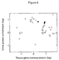


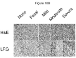

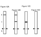
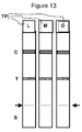
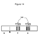


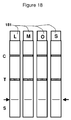

Classifications
-
- G—PHYSICS
- G01—MEASURING; TESTING
- G01N—INVESTIGATING OR ANALYSING MATERIALS BY DETERMINING THEIR CHEMICAL OR PHYSICAL PROPERTIES
- G01N33/00—Investigating or analysing materials by specific methods not covered by groups G01N1/00 - G01N31/00
- G01N33/48—Biological material, e.g. blood, urine; Haemocytometers
- G01N33/50—Chemical analysis of biological material, e.g. blood, urine; Testing involving biospecific ligand binding methods; Immunological testing
- G01N33/68—Chemical analysis of biological material, e.g. blood, urine; Testing involving biospecific ligand binding methods; Immunological testing involving proteins, peptides or amino acids
- G01N33/6893—Chemical analysis of biological material, e.g. blood, urine; Testing involving biospecific ligand binding methods; Immunological testing involving proteins, peptides or amino acids related to diseases not provided for elsewhere
-
- G—PHYSICS
- G01—MEASURING; TESTING
- G01N—INVESTIGATING OR ANALYSING MATERIALS BY DETERMINING THEIR CHEMICAL OR PHYSICAL PROPERTIES
- G01N2800/00—Detection or diagnosis of diseases
- G01N2800/06—Gastro-intestinal diseases
-
- G—PHYSICS
- G01—MEASURING; TESTING
- G01N—INVESTIGATING OR ANALYSING MATERIALS BY DETERMINING THEIR CHEMICAL OR PHYSICAL PROPERTIES
- G01N2800/00—Detection or diagnosis of diseases
- G01N2800/50—Determining the risk of developing a disease
Definitions
- Appendicitis is a condition characterized by inflammation of the appendix. All cases require removal of the inflamed appendix, either by laparotomy or laparoscopy. Untreated, mortality is high, mainly because of peritonitis and shock.
- Appendicitis is among many human diseases, for which the diagnosis is complicated by the heterogeneity of its clinical presentation. Patients with many other disorders can present with symptoms similar to those of appendicitis. Examples include the following: pelvic inflammatory disease (PID) or tubo-ovarian abscess, Endometriosis, ovarian cyst or torsion, ureterolithiasis and renal colic, degenerating uterine leiomyomata, diverticulitis, Crohn's disease, colonic carcinoma, rectus sheath hematoma, cholecystitis, bacterial enteritis, mesenteric adenitis, and omental torsion. It remains the most common surgical emergency of children, with initial diagnosis accuracy additionally challenged because of non-specific but similar symptoms of many other childhood conditions. Delays in accurate diagnosis lead to increased mortality, morbidity, and costs associated with the complications of appendicitis.
- PID pelvic inflammatory disease
- tubo-ovarian abscess Endometrios
- CT computed tomography
- the present invention generally relates to devices, kits and methods to determine acute appendicitis in a subject, such as a human subject.
- the inventors have discovered a set of appendicitis biomarkers which are present in a urine sample obtained from a subject with acute appendicitis.
- one aspect of the present invention provides devices, kits and methods to detect the presence of such appendicitis biomarkers in a urine sample from a subject, such as a human subject.
- the device is in the format of a dipstick test, in particular, a lateral flow immunoassay.
- an appendicitis biomarker is leucine ⁇ -2 glycoprotein (LRG). In some embodiments, an appendicitis biomarker is mannan-binding lectin serine protease 2 (MASP2). In some embodiments, an appendicitis biomarker is ⁇ -1-acid glycoprotein 1 (ORM).
- an appendicitis biomarker is selected from the groups selected from leucine-rich ⁇ -2-glycoprotein (LRG); S100-A8 (calgranulin); ⁇ -1-acid glycoprotein 1 (ORM); plasminogen (PLG); mannan-binding lectin serine protease 2 (MASP2); zinc- ⁇ -2-glycoprotein (AZGP1); apolipoprotein D (ApoD); and ⁇ -1-antichymotrypsin (SERPINA3).
- an appendicitis biomarker is selected from at least 1, or at least about 2, or at least about 3, or at least about 4, or at least about 5, or more than 5 of any and all combinations of appendicitis biomarkers disclosed in Table 1.
- One aspect of the present invention relates to a device for detecting at least one protein biomarker in a urine sample from a subject to identify if the subject is likely to have acute appendicitis, the device comprising: (a) at least one protein-binding agent which specifically binds to at least one biomarker protein selected from the group of: leucine ⁇ -2 glycoprotein (LRG), mannan-binding lectin serine protease 2 (MASP2), ⁇ -1-acid glycoprotein 1 (ORM); and (b) at least one solid support for the at least one protein binding-agent in (a), wherein the protein-binding agent is deposited on the solid support.
- LRG leucine ⁇ -2 glycoprotein
- MASP2 mannan-binding lectin serine protease 2
- ORM ⁇ -1-acid glycoprotein 1
- a protein-binding agent deposited on the solid support specifically binds the leucine ⁇ -2 glycoprotein (LRG) of SEQ ID NO: 1.
- a protein-binding agent deposited on the solid support specifically binds to the polypeptide of ⁇ -1-acid glycoprotein 1 (ORM) of SEQ ID NO: 3.
- a protein-binding agent deposited on the solid support specifically binds to the polypeptide of mannan-binding lectin serine protease 2 (MASP2) of SEQ ID NO: 5.
- the device is useful for detecting multiple appendicitis biomarkers, for example where the device further comprises at least one additional different protein-binding agent deposited on the solid support, wherein the additional protein-binding agent specifically binds to a biomarker protein selected from the group consisting of: leucine-rich ⁇ -2-glycoprotein (LRG); S100-A8 (calgranulin); ⁇ -1-acid glycoprotein 1 (ORM); plasminogen (PLG); mannan-binding lectin serine protease 2 (MASP2); zinc- ⁇ -2-glycoprotein (AZGP1); Apolipoprotein D (ApoD); and ⁇ -1-antichymotrypsin (SERPINA3).
- LRG leucine-rich ⁇ -2-glycoprotein
- S100-A8 calgranulin
- ⁇ -1-acid glycoprotein 1 ORM
- PLG plasminogen
- MASP2 mannan-binding lectin serine protease 2
- the device is useful for detecting multiple appendicitis biomarkers, for example where the device further comprises at least one additional different protein-binding agent deposited on the solid support, wherein the additional protein-binding agent specifically binds to a biomarker protein selected from the group consisting of: adipocyte specific adhesion molecule; AMBP; amyloid-like protein 2; angiotensin converting enzyme 2; BAZ1B; carbonic anhydrase 1; CD14; chromogranin A; FBLN7; FXR2; hemoglobin ⁇ ; hemoglobin ⁇ ; interleukin-1 receptor antagonist protein; inter- ⁇ -trypsin inhibitor; lipopolysaccharide binding protein; lymphatic vessel endothelial hyaluronan acid receptor 1; MLKL; nicastrin; novel protein (Accession No: IPI00550644); PDZK1 interacting protein 1; PRIC285; prostaglandin-H2 D-isomerase; Rcl
- the solid support of the device is in the format of a dipstick, a microfluidic chip or a cartridge.
- the dipstick is a lateral flaw immunoassay test strip.
- a single test strip tests for one appendicitis biomarker, such as LRG or ORM or S100-A8.
- a single test strip test for several appendicitis biomarkers for example, a single test strip test for all three appendicitis biomarkers: LRG, ORM and S100-A8; or a single test strip test for two appendicitis biomarkers: LRG and ORM; LRG and S100-A8; or ORM and S100-A8.
- a protein-binding agent is an antibody, antibody fragment, aptamer, small molecule or variant or fragment thereof.
- a subject is a mammalian subject such as a human subject.
- a protein-binding agent deposited on the device specifically binds to the specific appendicitis biomarker protein when the level of the appendicitis biomarker protein is at least 2-fold above a reference level for that appendicitis biomarker protein.
- a reference level for a particular appendicitis biomarker is an average level of the appendicitis biomarker protein in a plurality of urine samples from a population of healthy humans not having acute appendicitis.
- Another aspect of the present invention relates to the use of a device as disclosed herein to identify if a subject has acute appendicitis, wherein if at least one biomarker specifically binds to at least one protein-binding agent, the subject is likely to have acute appendicitis.
- kits comprising (a) a device as disclosed herein, and (b) a first agent, wherein the first agent produces a detectable signal in the presence of a protein-binding agent which deposited on the device is specifically bound to a biomarker protein.
- a kit optionally further comprises a second agent, wherein the second agent produces a different detectable signal in the presence of a second protein-binding agent deposited on the device which is specifically bound to a second biomarker protein.
- Another aspect of the present invention relates to a method to identify the likelihood of a subject to have acute appendicitis comprising: (a) measuring the level of at least one appendicitis biomarker protein selected from the group listed in Table 1 in a urine sample from the human subject; (b) comparing the level of the at least one biomarker protein measured in step (a) to a reference level for the measured appendicitis biomarker, where if the level of a measured appendicitis biomarker is at least 2-fold increased than the reference level for the particular appendicitis biomarker measured, it identifies that the subject is likely to have acute appendicitis.
- the method can be used to guide a clinician to direct an appropriate therapy to a subject which is identified to have acute appendicitis.
- the method further comprises determining the level of albumin in the urine sample from the human subject.
- the subject is a human subject and the human subject has exhibited at least one symptom of acute appendicitis.
- the method comprises measuring an appendicitis biomarker level by any method known by one of ordinary skill in the art, such as for example with the use of an immunoassay or an automated immunoassay, or a dipstick test, as disclosed herein.
- the method comprises measuring the level of the appendicitis biomarker leucine ⁇ -2 glycoprotein (LRG).
- the method comprises measuring the level of the appendicitis biomarker ⁇ -1-acid glycoprotein 1 (ORM).
- the method comprises measuring the level of the appendicitis biomarker mannan-binding lectin serine protease 2 (MASP2).
- the method comprises measuring the level of at least one the appendicitis biomarker selected from a group consisting of leucine ⁇ -2 glycoprotein (LRG), calgranulin A (S100-A8), ⁇ -1-acid glycoprotein 1 (ORM), plasminogen (PLG), mannan-binding lectin serine protease 2 (MASP2), zinc- ⁇ -2-glycoprotein (AZGP1), ⁇ -1-antichymotrypsin (SERPINA3) and apolipoprotein D (ApoD).
- LRG leucine ⁇ -2 glycoprotein
- S100-A8 calgranulin A
- ORM ⁇ -1-acid glycoprotein 1
- PSG plasminogen
- MASP2 mannan-binding lectin serine protease 2
- AZGP1 zinc- ⁇ -2-glycoprotein
- SERPINA3 ⁇ -1-antichymotrypsin
- ApoD apolipoprotein D
- the reference level in the method is a level of the particular appendicitis biomarker measured in a urine sample of a healthy human not having acute appendicitis. In some embodiments, the reference level is an average level of the appendicitis biomarker in a plurality of urine samples from a population of healthy humans not having acute appendicitis. In some embodiments, the reference level is a normalized level of the appendicitis biomarker in a urine sample of a healthy human not having acute appendicitis, wherein the normalization is performed against the level of albumin in the urine sample of a healthy human not having acute appendicitis.
- the method comprises measuring the level of at least one the appendicitis biomarker in a urine sample is collected in mid-stream. In some embodiments, the method comprises measuring the level of at least one the appendicitis biomarker by depositing the urine sample from the subject on a device, such as a test strip or dipstick device, as disclosed herein.
- Embodiments of the present invention are based on the discovery of eight biomarkers whose increase in urinary concentration correlate accurately with acute appendicitis. These eight biomarkers are leucine-rich ⁇ 2-glycoprotein (LRG), calgranulin A (S100-A8), ⁇ -1-acid glycoprotein 1 (orosomucoid) (ORM), plasminogen (PLG), mannan-binding lectin serine protease 2 (MASP2), zinc- ⁇ -2-glycoprotein (AZGP1), ⁇ -1-antichymotrypsin (SERPINA3) and apolipoprotein D (ApoD). These appendicitis biomarker proteins have been confirmed by Western immunoblotting (Example 2, Fig. 9 and 10 ) and further validated by target mass spectrometry (Example 2).
- LRG leucine-rich ⁇ 2-glycoprotein
- S100-A8 calgranulin A
- ORM ⁇ -1-acid glyco
- these biomarkers can be used as indicators of acute appendicitis.
- a physician can quickly make a diagnosis and administer appropriate medical treatment in a timely manner.
- the levels of these biomarkers in an individual is greater than the reference level or reference value of the respective biomarkers, at least one order of magnitude greater than that found in healthy individual not having acute appendicitis, it is indicative that the individual is indeed having acute appendicitis.
- a subject or individual is a mammalian subject, such as a human.
- Non-limiting symptoms of acute appendicitis include pain starting centrally (periumbilical) before localizing to the right iliac fossa (the lower right side of the abdomen); loss of appetite and fever; nausea or vomiting; the feeling of drowsiness; the feeling of general bad health; pain beginning and staying in the right iliac fossa, diarrhea and a more prolonged, smoldering course; increased frequency of urination; marked retching; tenesmus or "downward urge" (the feeling that a bowel movement will relieve discomfort); positive Rovsing's sign, Psoas sign, and/or Obturator sign.
- the invention provides a kit for predicting acute appendicitis in a human comprising an indicator or device that is responsive to a level of at least one biomarker in a sample of urine from a human upon contact with the sample of urine, wherein the appendicitis biomarker protein in a sample of urine is selected from a group consisting of leucine ⁇ -2 glycoprotein (LRG), calgranulin A (S100-A8), ⁇ -1-acid glycoprotein 1 (ORM), plasminogen (PLG), mannan-binding lectin serine protease 2 (MASP2), zinc- ⁇ -2-glycoprotein (AZGP1), ⁇ -1-antichymotrypsin (SERPINA3) and apolipoprotein D (ApoD), and wherein the indicator provides a positive test result when the appendicitis biomarker level exceeds a reference value.
- LRG leucine ⁇ -2 glycoprotein
- S100-A8 calgranulin A
- ORM
- the present invention provides a kit or device for predicting acute appendicitis in a subject, (e.g. a human subject) that are responsive to at least one marker selected from the list of appendicitis biomarkers listed in Table 1.
- the kit or device for predicting acute appendicitis in a subject is responsive to leucine ⁇ -2 glycoprotein (LRG).
- the kit or device for predicting acute appendicitis in a subject is responsive to leucine ⁇ -2 glycoprotein (LRG) and at least one marker selected from ⁇ -1-acid glycoprotein 1 (ORM), and/or mannan-binding lectin serine protease 2 (MASP2).
- the kit or device for predicting acute appendicitis in a subject is responsive to leucine ⁇ -2 glycoprotein (LRG) and at least one marker selected from the group consisting of calgranulin A (S100-A8), ⁇ -1-acid glycoprotein 1 (ORM), plasminogen (PLG), mannan-binding lectin serine protease 2 (MASP2), zinc- ⁇ -2-glycoprotein (AZGP1), ⁇ -1-antichymotrypsin (SERPINA3) and apolipoprotein D (ApoD).
- LRG leucine ⁇ -2 glycoprotein
- LRG leucine ⁇ -2 glycoprotein
- at least one marker selected from the group consisting of calgranulin A (S100-A8), ⁇ -1-acid glycoprotein 1 (ORM), plasminogen (PLG), mannan-binding lectin serine protease 2 (MASP2), zinc- ⁇ -2-glycoprotein (AZGP1), ⁇ -1-anti
- the kit or device for predicting acute appendicitis in a subject is responsive to leucine ⁇ -2 glycoprotein (LRG) and at least 1, or a least 2 or at least 3, or at least 4 or at least 5, or at least 6, or at least 7, or at least 8, or at least 9, or at least 10 other marker(s), in all and any combination, selected from the group consisting of the list of biomarkers listed in Table 1.
- LRG leucine ⁇ -2 glycoprotein
- the kit or device for predicting acute appendicitis in a subject is responsive to leucine ⁇ -2 glycoprotein (LRG) and at least one marker selected from the group consisting of adipocyte specific adhesion molecule; AMBP; amyloid-like protein 2; angiotensin converting enzyme 2; BAZ1B; carbonic anhydrase 1; CD14; chromogranin A; FBLN7; FXR2; hemoglobin ⁇ ; hemoglobin ⁇ ; interleukin-1 receptor antagonist protein; inter- ⁇ -trypsin inhibitor; lipopolysaccharide binding protein; lymphatic vessel endothelial hyaluronan acid receptor 1; MLKL; nicastrin; novel protein (Accession No: IPI00550644); PDZK1 interacting protein 1; PRIC285; prostaglandin-H2 D-isomerase; Rcl; S100-A9; serum amyloid A protein; SLC13A3; SLC2A
- LRG
- the indicator is in the form of a test strip such as a dipstick.
- the test strip is a lateral flow immunoassay (LFIA).
- the test strip is a double sandwich LFIA.
- test strip is a competitive LFIA.
- the reference value is an average level of the appendicitis biomarker in urine samples from a population of healthy humans not having acute appendicitis.
- healthy humans not having acute appendicitis do not exhibit any symptom associated with acute appendicitis as disclosed herein.
- the responsiveness of the indicator of the kit is by way of an immunoassay.
- the immunoassay is a lateral flow immunoassay test, also known as the immunochromatographic assay, or strip test.
- the invention provides a method of predicting acute appendicitis in a human comprising the steps of: (a) determining the level of at least one biomarker protein in a sample of urine from the human; and comparing the level of step (a) to a reference value to determine whether the human is suffering from acute appendicitis.
- the invention further comprises determining the level of albumin in the sample of urine from the human.
- the sample of urine is collected by the human.
- the human exhibits at least one symptom of acute appendicitis described herein.
- the human had an inconclusive CT to determine inflammation of the appendix.
- the human did not have a CT to determine inflammation of the appendix.
- the determination of the appendicitis biomarker level is completed with the use of an immunoassay.
- the immunoassay is a lateral flow immunoassay test, also known as the immunochromatographic assay, or strip test.
- the lateral flow immunoassay is a double antibody sandwich assay, a competitive assay, a quantitative assay or variations thereof.
- the appendicitis biomarker protein is leucine ⁇ -2 glycoprotein (LRG).
- the appendicitis biomarker protein is selected from a group consisting of leucine ⁇ -2 glycoprotein (LRG), calgranulin A (S100-A8), ⁇ -1-acid glycoprotein 1 (ORM), plasminogen (PLG), mannan-binding lectin serine protease 2 (MASP2), zinc- ⁇ -2-glycoprotein (AZGP1), ⁇ -1-antichymotrypsin (SERPINA3) and apolipoprotein D (ApoD).
- the appendicitis biomarker is selected from the group of biomarkers selected from any of those listed in Table 1.
- various combinations of appendicitis biomarkers can be selected.
- the method of predicting acute appendicitis in a human comprises the step of determining the level of leucine-rich ⁇ -2-glycoprotein (LRG) in a sample of urine from the human.
- LRG leucine-rich ⁇ -2-glycoprotein
- the method of predicting acute appendicitis in a human comprises the step of determining the levels of LRG and S100-A8 (calgranulin) in a sample of urine from the human.
- the method of predicting acute appendicitis in a human comprises the step of determining the levels of LRG and ⁇ -1-acid glycoprotein 1 (ORM) in a sample of urine from the human.
- ORM ⁇ -1-acid glycoprotein 1
- the method of predicting acute appendicitis in a human comprises the step of determining the levels of LRG and plasminogen (PLG) in a sample of urine from the human.
- LRG LRG and plasminogen
- the method of predicting acute appendicitis in a human comprises the step of determining the levels of LRG and mannan-binding lectin serine protease 2 (MASP2) in a sample of urine from the human.
- MASP2 mannan-binding lectin serine protease 2
- the method of predicting acute appendicitis in a human comprises the step of determining the levels of LRG and zinc- ⁇ -2-glycoprotein (AZGP1) in a sample of urine from the human.
- AZGP1 zinc- ⁇ -2-glycoprotein
- the method of predicting acute appendicitis in a human comprises the step of determining the levels of LRG and apolipoprotein D (ApoD) in a sample of urine from the human.
- ApoD apolipoprotein D
- the method of predicting acute appendicitis in a human comprises the step of determining the levels of ORM and S100-A8 in a sample of urine from the human.
- the method of predicting acute appendicitis in a human comprises the step of determining the levels of LRG, ORM and S100-A8 in a sample of urine from the human.
- the method of predicting acute appendicitis in a human comprises the step of determining the levels of LRG and ⁇ -1-antichymotrypsin (SERPINA3) in a sample of urine from the human.
- SERPINA3 ⁇ -1-antichymotrypsin
- Table 1 List of appendicitis biomarkers for use in the kits, devices and methods as disclosed herein for predicting acute appendicitis in the subject, for example a human subject.
- the SEQ ID NO refers to the amino acid sequence encoding the protein biomarker, and are incorporated herein by reference.
- the reference level or reference value is a level of a appendicitis biomarker in a urine sample of a healthy human not having acute appendicitis, or not having been diagnosed with acute appendicitis.
- a healthy human is any person who exhibits no symptom which commonly known to be associated with acute appendicitis as described herein.
- the reference value is an average level of the appendicitis biomarker in a plurality of urine samples from a population of healthy humans not having acute appendicitis or not having been diagnosed with acute appendicitis.
- a population of healthy subjects that have not been diagnosed with acute appendicitis is at least five healthy humans, at least 10 healthy humans, preferably 20 or more healthy humans.
- the average urine level of an appendicitis biomarker can be obtained by taking the sum of the level of an appendicitis biomarker from a number of humans divided by the number of humans.
- the reference level or reference value is a normalized level of the appendicitis biomarker in a urine sample of a healthy human not having acute appendicitis, wherein the normalization is performed against the level of albumin in the urine sample of a healthy human not having acute appendicitis, or not having been diagnosed with acute appendicitis.
- the normalized reference value for leucine ⁇ -2 glycoprotein (LRG), calgranulin A (S100-A8), ⁇ -1-acid glycoprotein 1 (ORM), plasminogen (PLG), mannan-binding lectin serine protease 2 (MASP2), Zinc- ⁇ -2-glycoprotein (AZGP1), ⁇ -1-antichymotrypsin (SERPINA3) and apolipoprotein D (ApoD) is 0.001.
- the urine sample is collected in mid-stream.
- the urine sample is obtained by depositing the urine on to a test strip.
- the test strip is a lateral flow immunoassay test, also known as the immunochromatographic assay.
- the lateral flow immunoassay is a double antibody sandwich assay, a competitive assay, a quantitative assay or variations thereof (See Figs. 11-19 ).
- kits or devices for predicting acute appendicitis in a subject for example, a human subject that is responsive to at least one appendicitis biomarker selected from the list of appendicitis biomarkers listed in Table 1.
- the kit or device for predicting acute appendicitis in a subject is responsive to leucine ⁇ -2 glycoprotein (LRG).
- the kit or device for predicting acute appendicitis in a subject is responsive to leucine ⁇ -2 glycoprotein (LRG) and at least one marker selected from ⁇ -1-acid glycoprotein 1 (ORM), and/or mannan-binding lectin serine protease 2 (MASP2).
- LRG leucine-rich alpha-2-glycoprotein 1
- LRG leucine-rich alpha-2-glycoprotein 1
- HMFT1766 leucine-rich repeat
- LRR leucine-rich repeat family of proteins, including LRG1
- LRG1 has been shown to be involved in protein-protein interaction, signal transduction, and cell adhesion and development. LRG1 is expressed during granulocyte differentiation.
- LRG can be detected in the methods, kits and devices using commercially available assay kits, e. g., from Immuno-Biological Laboratories, Inc., Human LRG Assay Kit, catalog number 27769. LRG can also be detected using the kits as disclosed in US patent application serial No 11/627,164 filed January 25, 2007 , and provisional patent application 60/761,808 filed January 25, 2006 , which are incorporated herein in their entirety by reference.
- SEQ ID NO: 1 is the polypeptide sequence for LRG (Leucine-rich alpha-2-glycoprotein) and has the amino acid sequence as follows:
- S100A8 is also known in the art as synonyms 60B8AG; CAGA; CFAG; CGLA; CP-10; L1Ag; MA387; MIF; Migration inhibitory factor-related protein 8 (MRP8); NIF; OTTHUMP00000015329; OTTHUMP00000015330; P8; S100 calcium-binding protein A8; S100 calcium-binding protein A8 (calgranulin A); S100A8; calgranulin A; cystic fibrosis antigen.
- S 100 calcium binding protein A8 (S 100 A8), also known as migration inhibitory factor-related protein (MRP-8) belongs to the S-100 family of calcium binding proteins associated with myeloid cell differentiation. They are highly expressed in resting neutrophils, keratinocytes (particularly in psoriasis), in infiltrating tissue macrophages and on epithelial cells in active inflammatory disease. The heterogeneity of macrophage subpopulations in chronic or acute inflammation is reflected by different expression of MRP8 and migration inhibitory factor-related proteins-14 (MRP14). Phagocytes expressing MRP8 and MRP14 belong to the early infiltrating cells, while MRP8 alone is found in chronic inflammatory tissues. The partially antagonistic functions of MRP8, MRP14 and of the Ca 2+ -dependent MRP8/14 heterocomplex makes them versatile mediators.
- MRP8 migration inhibitory factor-related protein
- MRP8 Human S100A8
- MRP8/14 The molecular weight of human MRP14
- MRP8/14 The molecular weight of human MRP14
- MRP8/14 The molecular weight of human MRP14
- MRP8/14 The molecular weight of human MRP14
- MRP8/114 The EF-hand motifs each on MRP8 and MRP14.
- MRP14 shows a higher affinity for calcium than MRP8, and the affinity of the C terminal EF2 is higher than that of the N-terminal EF1.
- the C-terminal domain also mainly determines the specificity of dimerization.
- the helix in EF2 undergoes a large conformational change upon calcium binding and may play a role as a trigger for Ca 2+ induced conformational change.
- S100A8 can be detected in the methods, kits and devices using commercial assays, such as, but without limitation, S100A8 assay kits from R & D Systems's Human MIF QUANTIKINE ® ELISA Kit ⁇ Catalog number: DMF00; and BMA Biomedicals, MRP8 Enzyme Immunoassay Product Code: S-1007.
- S100A8 can also be detected using the kits as disclosed in US Patent 7,501,256 and WO/2006/012588 which is incorporated herein in its entirety by reference.
- commercial polyclonal and monoclonal antibodies against S100A8 are also useful as protein-binding agents to S100A8 and are available from a variety of companies, e. g. , but not limited to commercial polyclonal and monoclonal antibodies against S100A8 are available from a variety of companies, e. g. Assay Designs, SIGMA-ALDRICH, R & D Systems, Novus Biologicals and Santa Cruz Biotechnology.
- SEQ ID NO: 2 is the polypeptide sequence for S100 A8 and has the amino acid sequence as follows:
- ORM alpha-1-acid-glycoprotein 1
- AGP1 alpha-1-acid-glycoprotein 1
- AGP-A alpha-1-acid-glycoprotein 1
- ORM1 alpha-1-acid-glycoprotein 1
- This gene encodes a key acute phase plasma protein. Because of its increase due to acute inflammation, this protein is classified as an acute-phase reactant. The specific function of this protein has not yet been determined; however, it may be involved in aspects of immunosuppression.
- ORM can be detected in the methods, kits and devices using commercial assays, such as, but without limitation, Human Orosomucoid ELISA Quantitation Kit from GenWay Biotech, Inc. catalog No. 40-288-22927F.
- commercial polyclonal and monoclonal antibodies against ORM are also useful as protein-binding agents to ORM and are available from a variety of companies, e. g., but not limited to Assay Designs, SIGMA-ALDRICH, Novus Biologicals, Lifespan Biosciences, R & D Systems, and Santa Cruz Biotechnology
- SEQ ID NO: 3 is the polypeptide sequence for ORM and has the amino acid sequence as follows:
- Plasminogen is also known in the art as PLG or DKFZp779M0222 and is a circulating zymogen that is converted to the active enzyme plasmin by cleavage of the peptide bond between arg560 and val561, which is mediated by urokinase (PLAU; MIM 191840) and tissue plasminogen activator (PLAT; MIM 173370).
- the main function of plasmin is to dissolve fibrin (see, e.g., FGA, MIM 134820) clots.
- Plasmin like trypsin, belongs to the family of serine proteinases.
- PLG can be detected in the methods, kits and devices using commercial assays, such as, but without limitation, commercial assay kits from Human Plasminogen ELISA Kit from Alpco Diagnostics 41-PLAHU-E01; Human Plasminogen ELISA Kit from AMERICAN DIAGNOSTICA, 640; Plasminogen Colorimetric Assay Kit from AMERICAN DIAGNOSTICA, 851; Human Plasminogen total antigen ELISA Assay Kit from innovative Research, IHPLGKT-TOT.
- PLG can also be detected using the kits as disclosed in International Patent Application WO/1991/005257 and European Patent Application EP1990914430 which is incorporated herein in its entirety by reference.
- commercial polyclonal and monoclonal antibodies against PLG are also useful as protein-binding agents to PLG and are available from a variety of companies, e. g. , but not limited to Rockland, Abcam, Assay Designs, EMD Biosciences, SIGMA-ALDRICH, Novus Biologicals, Lifespan Biosciences, R & D Systems, and Santa Cruz Biotechnology.
- SEQ ID NO: 4 is the polypeptide sequence for PLG and has the amino acid sequence as follows:
- MASP2 mannan-binding lectin serine peptidase 2
- MASP2 is also known in the art as aliases sMAP; MAP 19; MASP-2; MASP2 and is a Ra-reactive factor (RARF) which is a complement-dependent bactericidal factor that binds to the Ra and R2 polysaccharides expressed by certain enterobacteria.
- RARF Ra-reactive factor
- Alternate splicing of this gene results in two transcript variants encoding two RARF components that are involved in the mannan-binding lectin pathway of complement activation.
- the longer isoform is cleaved into two chains which form a heterodimer linked by a disulfide bond.
- the encoded proteins are members of the trypsin family of peptidases.
- MASP2 can be detected in the methods, kits and devices using commercial assays, such as, but without limitation, commercial assay kits such as Human MASP-2 ELISA Kit from Cell Sciences, HK326. MASP2 can also be detected using the kits as disclosed in International Patent Application WO/2007/028795 which is incorporated herein in its entirety by reference.
- commercial polyclonal and monoclonal antibodies against MASP2 are also useful as protein-binding agents to MASP2 and are available from a variety of companies, e. g. , but not limited to Cell Sciences, USBIO, and Santa Cruz Biotechnology.
- SEQ ID NO: 5 is the polypeptide sequence for MASP5 and has the amino acid sequence as follows:
- AZGP1 alpha-2-glycoprotein 1
- ZAG zinc-alpha-2-glycoprotein
- ZA2G zinc-alpha-2-glycoprotein
- AZGP1 Azgp1, ZNGP1
- LMF lipid-Mobilizing Factor
- AZGP1 is a soluble 41 kDa glycoprotein belonging to the immunoglobuline protein family and consisting of a single polypeptide chain. Human ZAG shares 59% sequence identity with the murine homolog.
- AZGP1 is closely related to antigens of the class1 major histocompatibility complex (MHC I) and shares 30-40 % sequence identity with the heavy chain of MHC I.
- MHC I major histocompatibility complex
- MHC-I members heterodimerize with beta-2-microglobuline (b2m) and bind peptides derived from intracellular proteins to present them to cytotoxic T cells.
- AZGP1 is a soluble protein rather than being anchored to plasma membranes that acts independently on b2m and binds the hydrophobic ligand which may relate to its function in lipid metabolism.
- AZGP1 is widespread in body fluids and is also found in various human tissues such as adipose tissue, prostate, breast, skin, salivary gland, trachea, broncheus, lung, gastrointestinal tract, pancreas, liver and kidney.
- AZGP1 acts as a lipid mobilizing factor to induce lipolysis in adipocytes and plays an important role in lipid utilization and loss of adipose tissue, especially during cachexia, which occurs in patient suffering from cancer, AIDS and other chronic illnesses.
- the role of AZGP1 in cancer cachexia is also connected with its ability to directly influence expression of uncoupling proteins (UCPs) which are implicated in the regulation of energy balance.
- UCPs uncoupling proteins
- AZGP1 expression is regulated particularly through TNF-alpha and the PPAR gamma nuclear receptor. AZGP1 expression is also upregulated by glucocorticoides and attenuated by eicosapentaenoic acid (EPA) and beta-3-adrenoreceptor antagonists.
- EPA eicosapentaenoic acid
- beta-3-adrenoreceptor antagonists eicosapentaenoic acid
- AZGP1 is overexpressed in certain human malignant tumors such as prostate, breast, lung or bladder cancer and can relate to tumor differentiation. Additionally, AZGP1 plays a role in obesity, diabetic kidney disorders, frontotemporal dementia and regulation of melanin production by melanocytes. AZGP1 is proposed to have a therapeutic use in obesity and cachexia. It can be used as a marker for clinical analysis of diabetic nephropathy and as a marker for certain tumors.
- AZGP1 can be detected in the methods, kits and devices using commercial assays, such as, but without limitation, Human Zinc-Alpha-2-Glycoprotein (ZA2G, ZAG) ELISA Kit, HRP Detection, from BioVendor Laboratory Medicine, Inc., RD191093100R, The assay is intended for the determination of human Zinc-alpha-2-glycoprotein in serum, plasma, cerebrospinal fluid, urine and cell lysate.; Human / Mouse / Rat ZAG EIA Kit from Raybiotech, Inc or Biovendor lab medicine Inc., EIA-ZAG-1.
- ZA2G, ZAG Human Zinc-Alpha-2-Glycoprotein
- HRP Detection from BioVendor Laboratory Medicine, Inc.
- the assay is intended for the determination of human Zinc-alpha-2-glycoprotein in serum, plasma, cerebrospinal fluid, urine and cell lysate.
- Human / Mouse / Rat ZAG EIA Kit
- commercial polyclonal and monoclonal antibodies against AZGP1 are also useful as protein-binding agents to AZGP1 and are available from a variety of companies, e. g. , but not limited to Abcam (Zinc Alpha 2 Glycoprotein antibody, catalog # ab47116) and Novus Biologicals (AZGP1 Antibody, catalog # H00000563-B01). Cell Sciences, USBIO, and Santa Cruz Biotechnology.
- SEQ ID NO: 6 is the polypeptide sequence for AZGP1 and has the amino acid sequence as follows:
- Apolipoprotein D (ApoD or APOD) is a polypeptide which is a high density lipoprotein that has no marked similarity to other apolipoprotein sequences. It has a high degree of homology to plasma retinol-binding protein and other members of the alpha 2 microglobulin protein superfamily of carrier proteins, also known as lipocalins. This glycoprotein is closely associated with the enzyme lecithin:cholesterol acyltransferase - an enzyme involved in lipoprotein metabolism.
- ApoD can be detected in the methods, kits and devices using as disclosed in International Patent Application WO/1996/019500 or U.S. Patents 5,804,368 or 5,804,368 or European Patent EP0301667 which are incorporated herein in their entirety by reference.
- SEQ ID NO: 7 is the polypeptide sequence for ApoD and has the amino acid sequence as follows:
- SERPINA3 ⁇ -1-antichymotrypsin (SERPINA3) is also known in the art as aliases serpin peptidase inhibitor, clade A (alpha-1 antiproteinase, antitrypsin), member 3, ACT; AACT; GIG24; GIG25 and MGC88254.
- the SERPINA3 polypeptide is a plasma protease inhibitor and member of the serine protease inhibitor class. Polymorphisms in this protein appear to be tissue specific and influence protease targeting. Variations in this protein's sequence have been implicated in Alzheimer's disease, and deficiency of this protein has been associated with liver disease. Mutations have been identified in patients with Parkinson disease and chronic obstructive pulmonary disease.
- SERPINA3 can be detected in the methods, kits and devices using as disclosed in International Patent Application WO/2005/039588 which is incorporated herein in its entirety by reference.
- commercial polyclonal and monoclonal antibodies against SERPINA3 are also useful as protein-binding agents to SERPINA3 and are available from a variety of companies, e. g. , but not limited to Proteintech Group, Lifespan Biosciences, and Santa Cruz Biotechnology.
- SEQ ID NO: 8 is the polypeptide sequence for SERPINA3 and has the amino acid sequence as follows:
- the level of appendicitis biomarker proteins such as those disclosed in Table 1, and in particular, the following appendicitis biomarker: leucine ⁇ -2 glycoprotein (LRG), calgranulin A (S100-A8), ⁇ -1-acid glycoprotein 1 (ORM), plasminogen (PLG), mannan-binding lectin serine protease 2 (MASP2), zinc- ⁇ -2-glycoprotein (AZGP1), ⁇ -1-antichymotrypsin (SERPINA3) or apolipoprotein D (ApoD), is measured to obtain a determination of whether a human patient has acute appendicitis.
- LRG leucine ⁇ -2 glycoprotein
- S100-A8 calgranulin A
- ORM ⁇ -1-acid glycoprotein 1
- PLG plasminogen
- MASP2 mannan-binding lectin serine protease 2
- AZGP1 zinc- ⁇ -2-glycoprotein
- SERPINA3
- a urinary biomarker protein level can be measured using any assay known to those of ordinary skilled in the art, including, but not limited to, Enzyme-Linked Immunosorbent Assay (ELISA), immunoprecipitation assays, radioimmunoassay, mass spectrometry, Western Blotting, and via dipsticks using conventional technology.
- ELISA Enzyme-Linked Immunosorbent Assay
- immunoprecipitation assays include immunoprecipitation assays, radioimmunoassay, mass spectrometry, Western Blotting, and via dipsticks using conventional technology.
- the levels of an appendicitis biomarker protein in a urine sample from the patient should be measured in the same manner as the reference value is measured.
- the levels of appendicitis biomarker proteins can be represented in arbitrary units dependent upon the assay used to measure the levels of appendicitis biomarker proteins, e.g., the intensity of the signal from the detectable label can correspond to the amount of appendicitis biomarker proteins present (e.g. as determined by eye, densitometry, an ELISA plate reader, a luminometer, or a scintillation counter).
- a protein-binding agent is a ligand that specifically binds to an appendicitis biomarker protein, and can be for example, a synthetic peptide, chemical, small molecule, or antibody or antibody fragment or variants thereof.
- a protein-binding agent is a ligand or antibody or antibody fragment, and in some embodiments, a protein-binding agent is preferably detectably labeled.
- immunoassays using antibodies are used to measure the levels of biomarker proteins in urine.
- the term "antibody” is intended to include immunoglobulin molecules and immunologically active determinants of immunoglobulin molecules, e.g., molecules that contain an antigen binding site which specifically binds (immunoreacts with) to the appendicitis biomarker to be measured.
- the term "antibody” is intended to include whole antibodies, e.g., of any isotype (IgG, IgA, IgM, IgE, etc), and includes fragments thereof which are also specifically reactive with the appendicitis biomarker proteins to be measured, e.g.
- Antibodies can be fragmented using conventional techniques.
- the term "antibody” includes segments of proteolytically-cleaved or recombinantly-prepared portions of an antibody molecule that are capable of selectively reacting with a certain protein.
- Non limiting examples of such proteolytic and/or recombinant fragments include Fab, F(ab')2, Fab' , Fv, dAbs and single chain antibodies (scFv) containing a VL and VH domain joined by a peptide linker.
- the scFv's can be covalently or non-covalently linked to form antibodies having two or more binding sites.
- antibody includes polyclonal, monoclonal, or other purified preparations of antibodies and recombinant antibodies.
- the term “antibody” is further intended to include humanized antibodies, bispecific antibodies, and chimeric molecules having at least one antigen binding determinant derived from an antibody molecule. In one embodiment, the antibody is detectably labeled.
- Antibodies to the appendicitis biomarker proteins can be generated using methods known to those skilled in the art. Alternatively, commercially available antibodies can be used. Antibodies to LRG, S100- A8, ORM1, PLG, MASP2, AZGP1, ApoD and SERPINA3 are commercially available.
- detectably labeled includes antibodies that are labeled by a measurable means and include, but are not limited to, antibodies that are enzymatically, radioactively, fluorescently, and chemiluminescently labeled. Antibodies can also be labeled with a detectable tag, such as c-Myc, HA, VSV-G, HSV, FLAG, V5, HIS, or biotin.
- a detectable tag such as c-Myc, HA, VSV-G, HSV, FLAG, V5, HIS, or biotin.
- the level of biomarker proteins present in the urine samples correlates to the intensity of the signal emitted from the detectably labeled antibody.
- the antibody is detectably labeled by linking the antibody to an enzyme.
- the enzyme when exposed to it's substrate, will react with the substrate in such a manner as to produce a chemical moiety which can be detected, for example, by spectrophotometric, fluorometric, or by visual means.
- Enzymes which can be used to detectably label the antibodies of the present invention include, but are not limited to, malate dehydrogenase, staphylococcal nuclease, delta-V-steroid isomerase, yeast alcohol dehydrogenase, alpha-glycerophosphate dehydrogenase, triose phosphate isomerase, horseradish peroxidase, alkaline phosphatase, asparaginase, glucose oxidase, beta-galactosidase, ribonuclease, urease, catalase, glucose-VI-phosphate dehydrogenase, glucoamylase and acetylcholinesterase. Chemiluminescence is another method that can be used to detect an antibody.
- Detection can also be accomplished using any of a variety of other immunoassays.
- radioactively labeling an antibody it is possible to detect the antibody through the use of radio immune assays.
- the radioactive isotope can be detected by such means as the use of a gamma counter or a scintillation counter or by audoradiography.
- Isotopes which are particularly useful for the purpose of the present invention are 3 H, 131 I, 35 S, 14 C, and preferably 125 I.
- fluorescent labeling compounds include CYE dyes, fluorescein isothiocyanate, rhodamine, phycoerytherin, phycocyanin, allophycocyanin, o-phthaldehyde and fluorescamine.
- An antibody can also be detectably labeled using fluorescence emitting metals such as 152 Eu, or others of the lanthanide series. These metals can be attached to the antibody using such metal chelating groups as diethylenetriaminepentaacetic acid (DTPA) or ethylenediaminetetraacetic acid (EDTA).
- DTPA diethylenetriaminepentaacetic acid
- EDTA ethylenediaminetetraacetic acid
- An antibody also can be detectably labeled by coupling it to a chemiluminescent compound. The presence of the chemiluminescent-antibody is then determined by detecting the presence of luminescence that arises during the course of a chemical reaction.
- chemiluminescent labeling compounds are luminol, luciferin, isoluminol, theromatic acridinium ester, imidazole, acridinium salt and oxalate ester.
- the levels of biomarker proteins in urine are detected by an immunoassay.
- Immunoassays include but are not limited to enzyme immunoassay (EIA), also called enzyme-linked immunosorbant assay (ELISA), radioimmunoassay (RIA), diffusion immunoassay (DIA), fluoroimmunoassay (FIA), chemiluminescent immunoassay (CLIA), counting immunoassay (CIA), lateral flow tests or immunoassay (LFIA), also known as lateral flow immunochromatographic assays, and magnetic immunoassay (MIA).
- EIA enzyme immunoassay
- ELISA enzyme-linked immunosorbant assay
- RIA radioimmunoassay
- DIA diffusion immunoassay
- FFIA fluoroimmunoassay
- CLIA chemiluminescent immunoassay
- CIA counting immunoassay
- LFIA also known as lateral flow immunochromatographic assays
- MIA magnetic
- An immunoassay is a biochemical test that measures the concentration of a substance in a biological sample, typically serum or urine, using the reaction of an antibody or antibodies to its antigen.
- the assay takes advantage of the specific binding of an antibody to its antigen.
- Monoclonal antibodies are often used as they only usually bind to one site of a particular molecule, and therefore provide a more specific and accurate test, which is less easily confused by the presence of other molecules.
- the antibodies picked must have a high affinity for the antigen (if there is antigen available, a very high proportion of it must bind to the antibody).
- the response of the biological sample being measured must be compared to standards of a known concentration. This is usually done through the plotting of a standard curve on a graph, the position of the curve at response of the unknown is then examined, and so the quantity of the unknown found.
- a defined amount of antibody is used in the assay where the defined amount of antibody binds completely to a fixed amount of antigen. This fixed amount of antigen is the reference level of biomarker in the urine. Thus, this defined amount of antibody is used to indicate whether the amount of antigen in the biological sample is at least at, below or above the reference level of biomarker (See Figs. 11-12 ).
- Detecting the quantity of antigen in the biological sample can be achieved by a variety of methods.
- One of the most common is to label either the antigen or the antibody.
- the label can consist of an enzyme (see enzyme immunoassay (EIA)), colloidal gold (lateral flow assays), radioisotopes such as I- 125 Radioimmunoassay (RIA), magnetic labels (magnetic immunoassay - MIA) or fluorescence.
- EIA enzyme immunoassay
- colloidal gold lateral flow assays
- radioisotopes such as I- 125 Radioimmunoassay (RIA)
- RIA Radioimmunoassay
- magnetic labels magnetic immunoassay - MIA
- fluorescence Other techniques include agglutination, nephelometry, turbidimetry and Western Blot.
- the immunoassay is a competitive immunoassay. In another embodiment, the immunoassay is a noncompetitive immunoassay.
- Immunoassays can be divided into those that involve labeled reagents and those which involve non-labeled reagents. Those which involve labeled reagents are divided into homogenous and heterogeneous (which require an extra step to remove unbound antibody or antigen from the site, usually using a solid phase reagent) immunoassays. Heterogeneous immunoassays can be competitive or non-competitive.
- the antigen in the unknown sample competes with labeled antigen to bind with antibodies.
- the amount of labeled antigen bound to the antibody site is then measured.
- the response will be inversely proportional to the concentration of antigen in the unknown. This is because the greater the response, the less antigen in the unknown was available to compete with the labeled antigen.
- noncompetitive immunoassays also referred to as the "sandwich assay"
- antigen in the unknown e. g. urine sample
- second antibody that is labeled is bound to the antigen, forming a sandwich.
- the amount of labeled antibody on the site is then measured.
- the results of the noncompetitive method will be directly proportional to the concentration of the antigen. This is because labeled antibody will not bind if the antigen is not present in the unknown sample, e. g urine sample.
- the levels of biomarker proteins in urine are detected by ELISA assay.
- ELISA assay There are different forms of ELISA which are well known to those skilled in the art, e.g. standard ELISA, competitive ELISA, and sandwich ELISA. The standard techniques for ELISA are described in " Methods in Immunodiagnosis", 2nd Edition, Rose and Bigazzi, eds. John Wiley & Sons, 1980 ; Campbell et al., "Methods and Immunology", W. A. Benjamin, Inc., 1964 ; and Oellerich, M. 1984, J. Clin. Chem. Clin. Biochem., 22:895-904 .
- Enzyme-linked immunosorbent assay also called ELISA, enzyme immunoAssay or EIA
- ELISA enzyme immunoAssay
- EIA enzyme immunoAssay
- the ELISA has been used as a diagnostic tool in medicine and plant pathology, as well as a quality control check in various industries.
- a known amount of anti-biomarker antibody is affixed to a solid surface, and then the sample, e. g. urine, containing the biomarker of interest is washed over the surface so that the antigen biomarker can bind to the immobilized antibodies (a first antibody).
- a detection antibody (a second antibody) is applied to the surface.
- the detection antibody is specific to antibodies from the subject. For example, if the subject is a human, the detection antibody should be an anti-human IgG antibody. If the subject is a dog, the detection antibody then should an anti-dog IgG antibody.
- This detection antibody can be linked to an enzyme, and in the final step a substance is added that the enzyme can convert to some detectable signal. For example, in the case of fluorescence ELISA, when light is shone upon the sample, any antigen/antibody complexes will fluoresce so that the amount of antibodies in the sample can be measured.
- test wells are coated with anti-biomarker antibody by incubation with 100 ⁇ l of purified anti-LRG antibody (3 ⁇ g/ml in PBS) per well overnight at room temperature, with PBS substituted for the antibody in control wells. After the plates have been washed three times with PBS-Tween, 250 ⁇ l of 2% BSA in PBS is added to each well, and the plates are incubated for 1 h at room temperature.
- the plates are washed three times with PBS-Tween and incubated for 1h at room temperature with test urine sample and control urine sample from healthy individuals diluted 1:100 in PBS-Tween-BSA; each urine sample is tested in triplicate in anti-LRG antibody-coated wells as well as in PBS control wells.
- the plate is then assayed (with appropriate controls) for the presence and/or the level of LRG by incubation for 1h at room temperature with 100 ⁇ l of goat anti-LRG IgG conjugated with horseradish peroxidase (Bio-Rad, Richmond, Calif.) per well diluted 1:2,000 in PBS-Tween-BSA.
- the substrate solution (o-phenylenediamine dihydrochloride; Sigma) is added to each well.
- the plates are then incubated for 30 min at room temperature in darkness, and the reaction is terminated by the addition of 2N sulfuric acid.
- the optical density values at 490 nm (OD490) are measured in a micro plate ELISA reader. For each urine sample, mean OD490 readings are calculated for the test wells and for the antigen control wells, the latter being subtracted from the former to obtain the net ELISA value.
- Performing an ELISA involves at least one antibody with specificity for a particular biomarker.
- a known amount of anti-biomarker antibody is immobilized on a solid support (usually a polystyrene micro titer plate) either non-specifically (via adsorption to the surface) or specifically (via capture by another antibody specific to the anti-biomarker antibody, in a "sandwich" ELISA).
- the detection antibody is added, forming a complex with the antigen.
- the detection antibody can be covalently linked to an enzyme, or can itself be detected by a secondary antibody which is linked to an enzyme through bio-conjugation.
- the plate is typically washed with a mild detergent solution to remove any proteins or antibodies that are not specifically bound.
- the plate is developed by adding an enzymatic substrate to produce a visible signal, which indicates the quantity of antigen in the sample.
- Older ELISAs utilize chromogenic substrates, though newer assays employ fluorogenic substrates with much higher sensitivity.
- a competitive ELISA is used.
- Purified anti-biomarker antibody is coated on the solid phase of multi-wells.
- Urine sample a defined amount of purified biomarker and horseradish peroxidase labeled with anti-biomarker antibody (secondary detection conjugated antibody) are added to coated wells to form competitive combination.
- secondary detection conjugated antibody horseradish peroxidase labeled with anti-biomarker antibody (secondary detection conjugated antibody) are added to coated wells to form competitive combination.
- HRP horseradish peroxidase labeled with HRP
- TMB horseradish peroxidase-conjugated antibodies
- the levels of appendicitis biomarker proteins are determined by contacting a urine sample with a first antibody that specifically binds to a biomarker protein to be measured under conditions permitting formation of a complex between the antibody and the appendicitis biomarker proteins (e.g. LRG, S100- A8, ORM1, PLG, MASP2, AZGP1, ApoD and SERPINA3).
- the amount of complex formed is then measured as a measure of the level of the appendicitis biomarker protein, and the amount of complex formed is compared to the amount of complex formed between the first antibody and a predetermined reference amount of the appendicitis biomarker protein.
- This predetermined reference level amount of the appendicitis biomarker protein is the amount found in the urine of healthy humans. A level above the reference level amount of an appendicitis biomarker protein indicates that the human has acute appendicitis.
- the first antibody is detectably labeled. Detectably labeling the first antibody is appropriate for use, for example, in standard ELISA assays where biomarker protein is absorbed to an ELISA plate, or in Western Blot analysis, or certain LFIA dipstick analyses.
- the first antibody is immobilized on a solid support, for example, when using a "Sandwich ELISA" or a dipstick analysis, then the amount of complex formed can measured by detecting binding of a second antibody that specifically binds to the appendicitis biomarker protein (e.g. LRG, S100- A8, ORM1, PLG, MASP2, AZGP1, ApoD and SERPINA3) under conditions permitting formation of a complex between the second antibody and the appendicitis biomarker protein, wherein the second antibody does not substantially cross-react with the first antibody, and wherein the second antibody is detectably labeled.
- a second antibody that specifically binds to the appendicitis biomarker protein
- the appendicitis biomarker protein e.g. LRG, S100- A8, ORM1, PLG, MASP2, AZGP1, ApoD and SERPINA3
- Any solid support can be used, including but not limited to, nitrocellulose, solid organic polymers, such as polystyrene, or laminated dipsticks such as described in U.S. patent 5,550,375 and 5,656,448 , which is specifically incorporated herein by reference in their entirety.
- the levels of two appendicitis biomarker proteins defining a first and a second appendicitis biomarker protein are measured using at least two antibodies specific to each appendicitis biomarker protein to be measured. Each antibody specifically reacts either the first appendicitis biomarker protein or the second appendicitis biomarker protein to be measured while not substantially cross-reacting with the other appendicitis biomarker proteins to be measured.
- the levels of three biomarker proteins defining a first biomarker protein, a second biomarker protein, and a third biomarker protein are measured using at least three antibodies specific to each biomarker protein to be measured, wherein each antibody specifically reacts either the first biomarker protein, the second biomarker protein, or the third biomarker protein to be measured while not substantially cross-reacting with the other biomarker proteins to be measured.
- the levels of four biomarker proteins defining a first, a second, a third and a fourth biomarker protein are measured using at least four antibodies specific to each biomarker protein to be measured, wherein each antibody specifically reacts either the first biomarker protein, the second biomarker protein, the third biomarker protein, or the fourth biomarker protein to be measured while not substantially cross-reacting with the other biomarker proteins to be measured.
- the appendicitis biomarker proteins are selected from the group consisting of LRG, S100- A8, ORM1, PLG, MASP2, AZGP1, ApoD and SERPINA3.
- the levels of biomarker proteins in urine are detected by a lateral flow immunoassay test (LFIA), also known as the immunochromatographic assay, or strip test.
- LFIAs are a simple device intended to detect the presence (or absence) of a target antigen in a fluid sample.
- LFIA tests are used for medical diagnostics either for home testing, point of care testing, or laboratory use.
- LFIA tests are a form of immunoassay in which the test sample flows along a solid substrate via capillary action. After the sample is applied to the test it encounters a coloured reagent which mixes with the sample and transits the substrate encountering lines or zones which have been pretreated with an antibody or antigen.
- LFIAs are essentially immunoassays adapted to operate along a single axis to suit the test strip format or a dipstick format. Strip tests are extremely versatile and can be easily modified by one skilled in the art for detecting an enormous range of antigens from fluid samples such as urine, blood, water samples etc. Strip tests are also known as dip stick test, the name bearing from the literal action of "dipping" the test strip into a fluid sample to be tested. LFIA strip test are easy to use, require minimum training and can easily be included as components of point-of-care test (POCT) diagnostics to be use on site in the field.
- POCT point-of-care test
- LFIA tests can be operated as either competitive or sandwich assays.
- Sandwich LFIAs are similar to sandwich ELISA.
- the sample first encounters coloured particles which are labeled with antibodies raised to the target antigen.
- the test line will also contain antibodies to the same target, although it may bind to a different epitope on the antigen.
- the test line will show as a coloured band in positive samples.
- Example 5 illustrates a sandwich LFIA in the test strip format.
- Competitive LFIAs are similar to competitive ELISA.
- the sample first encounters coloured particles which are labeled with the target antigen or an analogue.
- the test line contains antibodies to the target/its analogue. Unlabelled antigen in the sample will block the binding sites on the antibodies preventing uptake of the coloured particles.
- the test line will show as a coloured band in negative samples.
- a typical test strip consists of the following components: (1) sample application area comprising an absorbent pad (i. e. the matrix or material) onto which the test sample is applied; (2) conjugate or reagent pad- this contains antibodies specific to the target antigen conjugated to coloured particles (usually colloidal gold particles, or latex microspheres); test results area comprising a reaction membrane - typically a hydrophobic nitrocellulose or cellulose acetate membrane onto which anti-antigen antibodies are immobilized in a line across the membrane as a capture zone or test line (a control zone may also be present, containing antibodies specific for the conjugate antibodies); and (4) optional wick or waste reservoir - a further absorbent pad designed to draw the sample across the reaction membrane by capillary action and collect it.
- an absorbent pad i. e. the matrix or material
- conjugate or reagent pad- this contains antibodies specific to the target antigen conjugated to coloured particles (usually colloidal gold particles, or latex microspheres)
- test results area comprising a reaction membrane - typically
- the components of the strip are usually fixed to an inert backing material and may be presented in a simple dipstick format or within a plastic casing with a sample port and reaction window showing the capture and control zones. While not strictly necessary, most tests will incorporate a second line which contains an antibody that picks up free latex/gold in order to confirm the test has operated correctly.
- Figs. 11 -19 show the various components and embodiments of several test strips.
- the lateral flow immunoassay is a double antibody sandwich assay, a competitive assay, a quantitative assay or variations thereof.
- Figs. 15 , 16 , 17 , Example 5 and Example 6 exemplify double antibody sandwich LFIA in a test strip format.
- a diagnostic kit can comprise multiple LFIA test strips, one strip for a different biomarker protein.
- a diagnostic kit can comprise a single composite LFIA test strip for determining the levels of several biomarker proteins. Such diagnostic kits and LFIA test strips can be used as POCT in the field.
- a urine dipstick is a colorimetric chemical assay that can be used to determine the pH, specific gravity, protein, glucose, ketone, bilirubin, urobilinogen, blood, leukocyte, and nitrite levels of an individual's urine. It consists of a reagent stick-pad, which is immersed in a fresh urine specimen and then withdrawn. After predetermined times the colors of the reagent pad are compared to standardized reference charts.
- the urine dipstick offers an inexpensive and fast method to perform screening urinalyses, which help in identifying the presence of various diseases or health problems.
- a urine dipstick provides a simple and clear diagnostic guideline and can be used in the methods and kits as described herein. Accordingly, one aspect of the presents invention relates to a method for detecting acute appendicitis using a device, such as a dipstick, to test for the presence of appendicitis biomarkers as described herein.
- Dipsticks useful in the present invention can be used to test for at least one appendicitis biomarker, for example LRG or multiple biomarkers, such as any combination selected from the group of leucine-rich ⁇ -2-glycoprotein (LRG); S100-A8 (calgranulin); ⁇ -1-acid glycoprotein 1 (ORM); plasminogen (PLG); mannan-binding lectin serine protease 2 (MASP2); zinc- ⁇ -2-glycoprotein (AZGP1); apolipoprotein D (ApoD); ⁇ -1-antichymotrypsin (SERPINA3), or alternatively, multiple biomarkers selected from any combination listed in Table 1.
- LRG leucine-rich ⁇ -2-glycoprotein
- S100-A8 calgranulin
- ⁇ -1-acid glycoprotein 1 ORM
- PLG plasminogen
- MASP2 mannan-binding lectin serine protease 2
- Combination dipsticks can be used to test for at least two appendicitis biomarkers selected from the group of leucine-rich ⁇ -2-glycoprotein (LRG); S100-A8 (calgranulin); ⁇ -1-acid glycoprotein 1 (ORM); plasminogen (PLG); mannan-binding lectin serine protease 2 (MASP2); zinc- ⁇ -2-glycoprotein (AZGP1); apolipoprotein D (ApoD); ⁇ -1-antichymotrypsin (SERPINA3), or alternatively, multiple biomarkers selected from any combination listed in Table 1.
- LRG leucine-rich ⁇ -2-glycoprotein
- ORM plasminogen
- MASP2 mannan-binding lectin serine protease 2
- AZGP1 zinc- ⁇ -2-glycoprotein
- ApoD apolipoprotein D
- SERPINA3 ⁇ -1-antichymotrypsin
- Examples of combinations of two appendicitis biomarkers are LRG and ORM; LRG and S100-A8; LRG and PLG; LRG and MASP2; LRG and AZGP1; LRG and ApoD; LRG and SERPINA3; ORM and S100-A8; ORM and PLG; ORM and MASP2; ORM and ApoD; ORM and SERPINA3; S100-A8 and PLG; S100-A8 and MASP2; S100-A8 and ApoD; S100-A8 and SERPINA3; PLG and MASP2; PLG and ApoD; PLG and SEPRINA3; MASP2 and ApoD; MASP2 and SERPINA3; and Apo and SERPINA3.
- Combination dipsticks can be used to test for at least three appendicitis biomarkers, at least four appendicitis biomarkers, at least five appendicitis biomarkers, or at least six appendicitis biomarkers selected from the group of leucine-rich ⁇ -2-glycoprotein (LRG); S100-A8 (calgranulin); ⁇ -1-acid glycoprotein 1 (ORM); plasminogen (PLG); mannan-binding lectin serine protease 2 (MASP2); zinc- ⁇ -2-glycoprotein (AZGP1); apolipoprotein D (ApoD); ⁇ -1-antichymotrypsin (SERPINA3), or alternatively, multiple biomarkers selected from any combination listed in Table 1.
- LRG leucine-rich ⁇ -2-glycoprotein
- ORM plasminogen
- MASP2 mannan-binding lectin serine protease 2
- AZGP1 zinc- ⁇
- Combination dipsticks can be used to test for at least seven appendicitis biomarkers selected from the group of leucine-rich ⁇ -2-glycoprotein (LRG); S100-A8 (calgranulin); ⁇ -1-acid glycoprotein 1 (ORM); plasminogen (PLG); mannan-binding lectin serine protease 2 (MASP2); zinc- ⁇ -2-glycoprotein (AZGP1); apolipoprotein D (ApoD); ⁇ -1-antichymotrypsin (SERPINA3), or alternatively, multiple biomarkers selected from any combination listed in Table 1.
- LRG leucine-rich ⁇ -2-glycoprotein
- ORM plasminogen
- MASP2 mannan-binding lectin serine protease 2
- AZGP1 zinc- ⁇ -2-glycoprotein
- ApoD apolipoprotein D
- SERPINA3 ⁇ -1-antichymotrypsin
- An example of a combination of seven appendicitis biomarkers is LRG, ORM, S100-A8, PLG, MASP2, ApoD, and SERPINA3.
- Uses of dipsticks are commonly known in the art, and are described in U.S. Pat. No. 5,972,594 to Heine , which is incorporated herein in its entirety by reference which is used to detect the presence of neutrophil defensins to diagnose reproductive tract inflammation and preeclampsia.
- dipsticks and related components are well known in the art, for example dipsticks to detect leukocytes and leukocyte enzymes in body fluids have been patented.
- U.S. Patent No: 5,656,448 to Kang et al which is incorporated herein in its entirety discloses a dipstick encompassed for use in the present invention.
- U.S. Pat. No.4,758,508 to Schnabel, et al. describes an agent and a method for detecting esterolytic and/or proteolytic enzymes in body fluids.
- the present invention provides a LFIA device such as a dipstick to identify appendicitis biomarkers in a urine test sample.
- a LFIA device such as a dipstick
- a method for detecting acute appendicitis using a LFIA device, such as a dipstick having diagnostic test reagents to detect acute appendicitis.
- the diagnostic test reagents react with the test sample, such as urine test sample to produce a change upon contact with the test sample, such as urine.
- Another embodiment of the invention is a device, such as a dipstick, that has (1) a positive indication for the presence of acute appendicitis and (2) a negative indication for the absence of acute appendicitis. The difference between the positive indication and the negative indication is pre-determined.
- the present invention also provides a method for determining if a subject has a likelihood of acute appendicitis.
- the method begins with obtaining a urine sample from a subject, such as a symptomatic patient for appendicitis. Symptomatic patients for appendicitis are described herein.
- a device having diagnostic test reagents that detect the presence of at least one appendicitis biomarker such as leucine-rich ⁇ -2-glycoprotein (LRG); S100-A8 (calgranulin); ⁇ -1-acid glycoprotein 1 (ORM); plasminogen (PLG); mannan-binding lectin serine protease 2 (MASP2); zinc- ⁇ -2-glycoprotein (AZGP1); apolipoprotein D (ApoD); ⁇ -1-antichymotrypsin (SERPINA3) or any listed from Table 1 is contacted with the urine sample.
- LRG leucine-rich ⁇ -2-glycoprotein
- S100-A8 calgranulin
- ORM ⁇ -1-acid glycoprotein 1
- PLG plasminogen
- MASP2 mannan-binding lectin serine protease 2
- AZGP1 zinc- ⁇ -2-glycoprotein
- ApoD apoli
- the method to diagnose acute appendicitis in a subject uses a quantitative device (such as, for example, the MULTISTIX-2, MULTISTIX-10, URISTIX-4, or any appendicitis biomarker-detecting device as disclosed herein) or the subject inventive device that has two indications, one for a positive result and one for a negative result.
- a quantitative device such as, for example, the MULTISTIX-2, MULTISTIX-10, URISTIX-4, or any appendicitis biomarker-detecting device as disclosed herein
- the subject inventive device that has two indications, one for a positive result and one for a negative result.
- the MULTISTIX-2 produces quantitative results of 0, trace, +1, +2 and +3.
- Quantitative results also include "Between +1 and +2" and "Between +2 and +3.”
- a test result of 0, trace, and +1 corresponds to the absence of acute appendicitis).
- a test result of "Between +1 and +2", “Moderate (+2)”, “Between +2 and +3”, and “Large (+3)” corresponds to the presence of acute appendicitis).
- the pre-determination is done using a study where the range of the urine marker presence is determined based on the range in urine from confirmed appendicitis subjects as compared to the range of urine maker in the urine from healthy (i.e. confirmed non-appendicitis) subjects.
- a device such as a dipstick immunological device as disclosed herein can includes (1) a matrix (preferably filter paper) with diagnostic test reagents and (2) a mounting substrate (preferably polystyrene film), which typically does not absorb the test (e.g. urine) sample, such that the user can hold onto the substrate without contacting the sample.
- the device produces a visual change in the matrix upon contact with the urine sample.
- the matrix has two indicators-a first that indicates the presence of acute appendicitis and a second that indicates the absence of appendicitis. The first indicator produces a positive test result and the second indicator produces a negative result.
- test result is positive when the test result is pre-determined to correspond with a level of the appendicitis biomarker which is indicative of acute appendicitis. Conversely, a test result is negative when the test result is pre-determined to be below the level of an appendicitis biomarker which indicates the absence of acute appendicitis.
- the device such as a dipstick device determines the presence of acute appendicitis with the positive test result, and the absence of acute appendicitis with the negative test result.
- the diagnostic test reagents may be associated with the matrix by any physical or chemical means, including, for example impregnation, coating, linking, and covalent attachment.
- the matrix may take any convenient physical form, such as a card, pad, strip, or dipstick.
- Such diagnostic test reagents include the compositions of the above-referenced patents, including an ester (preferably a chromogenic ester) and a diazonium salt such as those described in U.S. Pat. No. 4,637,979 .
- Another preferred reagent is a derivatized pyrrole amino acid ester, a diazonium salt, a buffer, and non-reactive ingredients as described in U.S. Pat. Nos.
- the preferred amounts of these ingredients is based on dry weight at the time of impregnation and is as follows: about 0.4% w/w derivatized pyrrole amino acid ester, about 0.2% w/w diazonium salt, about 40.9% w/w buffer, and about 58.5% w/w non-reactive ingredients.
- the test reagent e. g. the anti-antigen antibody of the immunoassay is detectably labeled.
- the detectable label is selected from a group consisting of enzyme, fluorescent, biotin, gold, latex, hapten and radioisotope labeling.
- a detectable-hapten includes but is not limited to biotin, fluorescein, digoxigenin, dinitrophenyl (DNP).
- Other labels include but are not limited to colloidal gold and latex beads. The latex beads can also be colored.
- the detectable label is a dye.
- a “dye” refers to a substance, compound or particle that can be detected, particularly by visual, fluorescent or instrumental means.
- a dye can be, for example, but not limited to, a pigment produced as a coloring agent or ink, such as Brilliant Blue, 3132 Fast Red 2R and 4230 Malachite Blue Lake, all available from Hangzhou Hongyan Pigment Chemical Company, China.
- the “dye” can also be a particulate label, such as, but not limited to, blue latex beads or gold particles.
- the particulate labels may or may not be bound to a protein, depending upon if it is desired for the particles to move in the test strip or not. If the particles are to be immobilized in the test strip, the particles may be conjugated to a protein, e. g. the anti-antigen antibody, which in turn is bound to the test strip by either physical or chemical means.
- a colloidal gold conjugate consists of a suspension of gold particles coated with a selected protein or macromolecule (such as an antibody or antibody-based moiety).
- the gold particles may be manufactured to any chosen size from 1-250 nm. This gold probe detection system, when incubated with a specific target, such as in a tissue section, will reveal the target through the visibility of the gold particles themselves.
- gold particles will also reveal immobilized antigen on a solid phase such as a blotting membrane through the accumulated red color of the gold sol. Silver enhancement of this gold precipitate also gives further sensitivity of detection.
- Suppliers of colloidal gold reagents for labeling are available from SPI-MARKTM. Polystyrene latex Bead size 200 nm colored latex bead coated with antibody SIGMA ALDRICH®, Molecular Probes, Bangs Laboratory Inc., and AGILENT® Technologies.
- biotin-streptavidin a biotin-streptavidin system.
- the antibodies immunoreactive (i. e. specific for) with the biomarker of interest is biotinylated.
- Quantity of biotinylated antibody bound to the biomarker is determined using a streptavidin-peroxidase conjugate and a chromagenic substrate.
- streptavidin peroxidase detection kits are commercially available, e. g. from DAKO; Carpinteria, CA.
- Protein binding agents described herein such as antibodies and antibody-based moiety can alternatively be labeled with any of a number of fluorescent compounds such as fluorescein isothiocyanate, europium, lucifer yellow, rhodamine B isothiocyanate ( Wood, P. In: Principles and Practice of Immunoasay, Stockton Press, New York, pages 365-392 (1991 )) for use in immunoassays.
- fluorescent compounds such as fluorescein isothiocyanate, europium, lucifer yellow, rhodamine B isothiocyanate ( Wood, P. In: Principles and Practice of Immunoasay, Stockton Press, New York, pages 365-392 (1991 )
- these fluorophores can be used to quantify the biomarker of interest.
- Radioimmunoassay is another technique in which antibody can be used after labeling with a radioactive isotope such as 125 I.
- immunoassays can be easily automated by the use of appropriate instruments such as the IMXTM (Abbott, Irving, Tex.) for a fluorescent immunoassay and Ciba Coming ACS 180TM (Ciba Corning, Medfield, Mass.) for a chemiluminescent immunoassay.
- IMXTM Abbott, Irving, Tex.
- Ciba Coming ACS 180TM Ciba Corning, Medfield, Mass.
- a “LFIA test strip” or “dip stick” can include one or more bibulous or non-bibulous materials or matrices.
- the terms “material” and “matrix” are used interchangeably.
- a test strip comprises more than one material, the one or more materials are preferably in fluid communication.
- One material of a test strip may be overlaid on another material of the test strip, such as for example, filter paper overlaid on nitrocellulose membrane.
- a test strip can include a region comprising one or more materials followed by a region comprising one or more different materials. In this case, the regions are in fluid communication and may or may not partially overlap one another.
- Suitable materials for test strips include, but are not limited to, materials derived from cellulose, such as filter paper, chromatographic paper, nitrocellulose, and cellulose acetate, as well as materials made of glass fibers, nylon, dacron, PVC, polyacrylamide, crosslinked dextran, agarose, polyacrylate, ceramic materials, and the like.
- the material or materials of the test strip may optionally be treated to modify their capillary flow characteristics or the characteristics of the applied sample.
- the sample application region of the test strip may be treated with buffers to correct the pH or specific gravity of an applied urine sample, to ensure optimal test conditions.
- the material or materials can be a single structure such as a sheet cut into strips or it can be several strips or particulate material bound to a support or solid surface such as found, for example, in thin-layer chromatography and may have an absorbent pad either as an integral part or in liquid contact.
- the material can also be a sheet having lanes thereon, capable of spotting to induce lane formation, wherein a separate assay can be conducted in each lane.
- the material can have a rectangular, circular, oval, triagonal or other shape provided that there is at least one direction of traversal of a test solution by capillary migration. Other directions of traversal may occur such as in an oval or circular piece contacted in the center with the test solution. However, the main consideration is that there be at least one direction of flow to a predetermined site.
- the support for the test strip where a support is desired or necessary, will normally be water insoluble, frequently non-porous and rigid but may be elastic, usually hydrophobic, and porous and usually will be of the same length and width as the strip but may be larger or smaller.
- the support material can be transparent, and, when a test device is assembled, a transparent support material can be on the side of the test strip that can be viewed by the user, such that the transparent support material forms a protective layer over the test strip where it may be exposed to the external environment, such as by an aperture in the front of a test device.
- a wide variety of materials may be employed provided only that the support does not interfere with the capillary action of the material or materials, or non-specifically bind assay components, or interfere with the signal producing system.
- Illustrative polymers include polyethylene, polypropylene, poly(4-methylbutene), polystyrene, polymethacrylate, poly(ethylene terephthalate), nylon, poly(vinyl butyrate), glass, ceramics, metals, and the like.
- Elastic supports may be made of polyurethane, neoprene, latex, silicone rubber and the like.
- a dipstick device has one indication of the presence of acute appendicitis and a second indication for the absence of acute appendicitis.
- the two indications preferably are a negative (-) symbol and a positive (+) symbol, but could be any two indications.
- the device has the negative indication (e.g., the "-" portion of a possible "+” symbol) containing reagents that reacts with all samples. That is, the diagnostic test reagents react to some constituent analyte, such as urea which is present in all urine samples. Alternatively, the diagnostic test reagents test an aspect of the sample, such as pH, that every sample has.
- the positive indication (e.g., the "
- Another embodiment has the negative indicator (e.g., the "-" portion of a possible "+” symbol) which contains reagents that reacts with the sample which either has the absence of the test appendicitis biomarker (i.e.
- the positive indication e.g., the "
- the positive indication has a lower sensitivity to the presence of a test appendicitis biomarker (i.e. LRG biomarker) and thus such the reagents react only with urine samples containing level of the urine marker (i.e. LRG biomarker) above a pre-defined level.
- a test device such as a dipstick device has text on the device in two places. In one place the text indicates a positive result (i.e. the likelihood the subject has acute appendicitis). In another, it indicates a negative result (i.e. the likelihood the subject does not have acute appendicitis).
- matrices having the appropriate diagnostic test reagents For example, next to the negative indication is a matrix having diagnostic test reagents that react with all urine samples, regardless of the content of appendicitis biomarkers as disclosed herein.
- Next to the positive indication is a matrix having diagnostic test reagents that react only with samples that have the presence of the test appendicitis biomarker, (e.g.
- such a device such as one discussed in Figure 12 , does not require a chart, such as a coloration chart, to interpret the results.
- this enables the detection device, such as a dipstick device (and the corresponding method) to be used easily by one without special training and provides a more rapid diagnostic (and method) for determining if a subject is likely to have acute appendicitis.
- such a device is ideal for point-of-care testing application.
- dipsticks are commercially available from Bayer Corporation of Elkhart, Ind., as well as other commercial sources.
- the dipstick is dipped into a well mixed urine sample, and after a time period, for example between about thirty seconds (30s) to about two minutes (2 mins) or more, the various reagent bands are visually or optically examined for color changes.
- the bands can be visually compared to a preprinted color chart in order to determine the amount of each of the constituents or parameters being measured. It is also possible to optically scan using a machine or optical scanner the dipstick and thereby obtain instrument readings of color intensity or wave length through the use of a particular instrument adapted for reading the reagents and color of the dipstick.
- the instrument is capable of quantify a number of reagent bands as well as quantify the overall color intensity sensed on the band.
- the immunoassays operate on a purely qualitative basis. However it is possible to measure the intensity of the test line to determine the quantity of antigen in the sample when using an immunoassay such a LFIA.
- an immunoassay such as a LFIA.
- MIA magnetic immunoassay
- FIG. 8 of US Patent 6,004,821 shows a schematic depiction of such an automated calorimetric microscopical instrument assembly (which is denoted generally by the numeral 54), and which can be used to scan a urine sample, and can, without significant human intervention, colorometrically analyze the wavelengths of the colors imparted to the dipstick by the urine in the chamber 14, either colorometrically and/or morphometrically. Accordingly, such an instrument, which is specifically adapted to scan the reaction of the dipstick after contact with a urine sample for the presence of the appendicitis biomarkers (such as at least one selected from Table 1) is encompassed for use in the present invention.
- the appendicitis biomarkers such as at least one selected from Table 1
- the dipstick uses reagents such as copper-creatinine and iron-creatinine complexes have peroxidase activity.
- Other dipstick reagents can use reagents such as 3, 3', 5, 5'-tetramethylbenzidine (TMB), and diisopropyl benzene dihydroperoxide (DBDH) which are used with peroxidase.
- TMB 3, 3', 5, 5'-tetramethylbenzidine
- DBDH diisopropyl benzene dihydroperoxide
- a dipstick for use to detect the presence of appendicitis biomarkers is based upon the first-generation devices which relied on the same colorimetric reaction used for assessing the presence of glucose test strips for urine.
- a test kit for use herein can contain a benzidine derivative, which is oxidized to a blue polymer by the hydrogen peroxide formed in the oxidation reaction. Care must be taken if such a dipstick is generated to ensure the test strip is developed after a precise interval after contact with the urine test sample as well as frequent calibration of the meter to read the test result.
- the same principle is used in test strips that have been commercialized for the detection Diabetic ketoacidosis (DKA). These test strips use a beta-hydroxybutyrate-dehydrogenase enzyme instead of a glucose oxidizing enzyme and have been used to detect and help treat some of the complications that can result from prolonged hyperglycaemia. Blood alcohol sensors using the same approach but with alcohol dehydrogenase enzymes have been developed.
- the device such as a dipstick device uses an electrochemical method.
- Test strips contain a capillary that sucks up a reproducible amount of urine.
- the presence of an appendicitis biomarker such as any or a combination of those listed in Table 1 in the urine reacts with an enzyme electrode containing protein-binding agents with the test appendicitis biomarker.
- the coulometric method is a technique where the total amount of charge generated by the specific binding of the appendicitis biomarker to the specific protein-binding agent reaction is measured over a period of time. This is analogous to throwing a ball and measuring the distance it has covered so as to determine how hard it was thrown.
- the amperometric method is used by some meters and measures the electrical current generated at a specific point in time. This is analogous to throwing a ball and using the speed at which it is travelling at a point in time to estimate how hard it was thrown.
- the coulometric method can allow for variable test times, whereas the test time on a meter using the amperometric method is always fixed. Both methods give an estimation of the concentration of the appendicitis biomarker in the urine sample.
- the levels of appendicitis biomarker proteins in urine are detected by a magnetic immunoassay (MIA).
- MIA is a type of diagnostic immunoassay using magnetic beads as labels in lieu of conventional enzymes (ELISA), radioisotopes (RIA) or fluorescent moieties (fluorescent immunoassays).
- ELISA electroactive immunoassay
- RIA radioisotopes
- fluorescent immunoassays fluorescent immunoassays.
- This assay involves the specific binding of a protein binding agent to an appendicitis biomarker protein, such as an antibody binding to its antigen, where a magnetic label is conjugated to one element of the pair.
- the presence of magnetic beads is then detected by a magnetic reader (magnetometer) which measures the magnetic field change induced by the beads.
- the signal measured by the magnetometer is proportional to the antigen or biomarker quantity in the initial sample.
- Magnetic beads are made of nanometric-sized iron oxide particles encapsulated or glued together with polymers. These magnetic beads can range from 35nm up to 4.5 ⁇ m. The component magnetic nanoparticles range from 5 to 50nm and exhibit a unique quality referred to as superparamagnetism in the presence of an externally applied magnetic field. Magnetic labels exhibit several features very well adapted for such applications: they are not affected by reagent chemistry or photo-bleaching and are therefore stable over time; the magnetic background in a biomolecular sample is usually insignificant; sample turbidity or staining have no impact on magnetic properties; and magnetic beads can be manipulated remotely by magnetism.
- MIA Magnetic Activated Assay
- Dittmer WU and colleagues J Immunol Methods. 2008, 338:40-6
- the assay involves a 1-step sandwich immunoassay with no fluid replacement steps.
- the detection limit is the 10 pM range and the assay took only 15 minutes;
- Kuma H and colleagues (Rinsho Byori. 2007, 55:351-7 ) developed a sensitive immunoassay system using magnetic nanoparticles made from Fe 3 O 4 ;
- Kuramitz H reviews the current state of concerning electrochemical immunoassays using magnetic microbeads as a solid phase in Anal Bioanal Chem.
- Magnetometers are instruments that can detect the presence and measure the total magnetic signal of a sample.
- An effective MIA is one that is capable of separating naturally occurring magnetic background (noise) from the weak magnetically labeled target (signal).
- Various approaches and devices have been employed to achieve a meaningful signal-to-noise ratio (SNR) for bio-sensing applications: giant magneto-resistive sensors and spin valves, piezoresistive cantilevers, inductive sensors, superconducting quantum interference devices, anisotropic magneto-resistive rings, and miniature Hall sensors.
- SNR signal-to-noise ratio
- MIA that exploits the non-linear magnetic properties of magnetic labels can effectively use the intrinsic ability of a magnetic field to pass through plastic, water, nitrocellulose, and other materials, thus allowing for true volumetric measurements in various immunoassay formats.
- a MIA based on non-linear magnetization eliminates the impact of linear dia- or paramagnetic materials such as sample matrix, consumable plastics and/or nitrocellulose.
- the intrinsic magnetism of these materials is very weak, with typical susceptibility values of -10-5 (dia) or +10-3 (para), when one is investigating very small quantities of superparamagnetic materials, such as nanograms per test, the background signal generated by ancillary materials cannot be ignored.
- the levels of biomarker proteins in urine are detected by a diffusion immunoassay (DIA).
- DIA diffusion immunoassay
- the transport of molecules perpendicular to flow in a microchannel e. g. in a microfluidic chip, is affected by binding between antigens and antibodies.
- concentration of very dilute analytes, in this invention, the urine biomarkers can be measured in a few microliters of sample in seconds.
- Microfluidics is the manipulation of microliter volumes in channels with sub-millimeter dimensions.
- Microfluidic diffusion immunoassays for the detection of analytes or biomarkers in fluid samples have been described in the art, for example, in U. S. Patent No.
- U. S. Patent No. 6,541,213 describes the use of a credit-card sized microfluidic device to perform competitive immunoassays.
- the ability to perform assays in this microscale dimension affords an extremely rapid, homogenous, and cost effective alternative to current methods used commercially today.
- the credit-card sized microfluidic device can be integrated into the development of point-of-use systems that allow real-time answers to health questions while at the physician's office, home, workplace, school, shopping mall and other public places.
- lab cards integrated laboratory-tests-on-a-card
- lab cards stand alone, single use lab cards being developed to provide rapid on-site results in infectious diseases testing, nucleic acid testing, blood type analysis, cancer testing, and respiratory disease testing.
- the levels of biomarker proteins in urine are detected by an on-the-spot assay also known as point-of-care assay.
- Point-of-care testing is defined as diagnostic testing at or near the site of patient care.
- POCT brings the test conveniently and immediately to the patient. This increases the likelihood that the patient will receive the results in a timely manner.
- POCT is accomplished through the use of transportable, portable, and handheld instruments (e.g., blood glucose meter, nerve conduction study device) and test kits (e.g., CRP, HBA1C, Homocystein, HIV salivary assay, etc.).
- POCTs are well known in the art, especially immunoassays.
- the LFIA test strip or dip sticks can easily be integrated into a POCT diagnostic kit.
- One skilled in the art would be able to modify immunoassays for POCT using different format, e. g. ELISA in a microfluidic device format or a test strip format. For example, U. S.
- Patent Application No.2009/0181411 describes a microfluidic device-based point-of-care immunoassay for biomarker molecules associated with pathology in a vertebrate host, man or animal.
- the microfluidic devices such as chips are formatted to either hand-held cartridges (also termed "cards"), or cartridges for automated or semi-automated, machine-aided testing.
- Microfluidic device-based assays enable small-volume sampling, with point-of-care results from a broad variety of biological fluids and samples in real time.
- the assay cartridges can be single use reagent packs, or be fully self-contained and operable entirely by hand. This reference is incorporated herein by reference in its entirety.
- Embodiments of the invention further provide for diagnostic kits and products of manufacture comprising the diagnostic kits.
- the kits can comprise a means for predicting acute appendicitis in a human.
- the kit comprises an indicator responsive to the level of biomarker protein in a sample of urine, wherein the appendicitis biomarker protein is selected from the group consisting of LRG, S100- A8, ORM1, PLG, MASP2, AZGP1, ApoD and SERPINA3.
- the indicator is in the form of a LFIA test strip or a microfluidic device.
- a diagnostic kit can comprise multiple LFIA test strips, one strip for a different biomarker protein.
- a diagnostic kit can comprise a single composite LFIA test strip for determining the levels of several biomarker proteins.
- a diagnostic kit can comprise a single multichannel microfluidic device for determining the levels of several biomarker proteins. In another embodiment, a diagnostic kit can comprise several microfluidic devices for determining the levels of several biomarker proteins, one microfluidic device for a different biomarker protein.
- kits can further comprise cups or tubes, or any other collection device for sample collection of urine.
- the kit can optionally further comprise at least one diagram and/or instructions describing the interpretation of test results.
- Protein-binding agents proteins, antibodies or antisera against biomarker proteins
- the methods disclosed herein uses antibodies or anti-sera for detecting, quantifying, and/or labeling LRG, S100- A8, ORM1, PLG, MASP2, AZGP1, ApoD and SERPINA3 described herein.
- the antibodies can be obtained from a commercial source. These commercial antibodies can also be conjugated with labels, e.g. Cy 3 or FITC.
- Antibodies for use in the methods described herein can also be produced using standard methods to produce antibodies, for example, by monoclonal antibody production ( Campbell, A.M., Monoclonal Antibodies Technology: Laboratory Techniques in Biochemistry and Molecular Biology, Elsevier Science Publishers, Amsterdam, the Netherlands (1984 ); St. Groth et al., J. Immunology, (1990) 35: 1-21 ; and Kozbor et al., Immunology Today (1983) 4:72 ).
- Antibodies can also be readily obtained by using antigenic portions of the protein to screen an antibody library, such as a phage display library by methods well known in the art.
- U.S. patent 5,702,892 U.S.A. Health & Human Services
- WO 01/18058 Novopharm Biotech Inc.
- NM_052972.2, NM_002964.3, NM_000607.2, N_000301.2, NM_006610.2, N_001185.2, NM_001647.3, and NM_001085.4 respectively) can be cloned into the pQE30 vector containing an N-terminal hexa-histidine tag (QIAGEN, GmbH, Hilden, Germany), and then transformed into E. coli strain JM109 cells. Recombinant proteins is expressed and purified by affinity chromatography using Ni-nitriloacetic acid agarose (QIAGEN) according to the manufacturer's instructions. The final preparation yielded a single calculated molecular weight of 89707 kDa band on SDS-PAGE and is used for the immunization of rabbits.
- QIAGEN N-terminal hexa-histidine tag
- Detection of anti-antibodies to the appendicitis biomarkers can be achieved by direct labeling of the antibodies themselves, with labels including a radioactive label such as 3 H, 14 C, 35 S, 125 I, or 131 I, a fluorescent label (e. g. Cy3, Cy5, FITC), a hapten label such as biotin, heavy metal such as gold, or an enzyme such as horse radish peroxidase or alkaline phosphatase.
- a radioactive label such as 3 H, 14 C, 35 S, 125 I, or 131 I
- a fluorescent label e. g. Cy3, Cy5, FITC
- a hapten label such as biotin
- heavy metal such as gold
- an enzyme such as horse radish peroxidase or alkaline phosphatase.
- unlabeled primary antibody is used in conjunction with labeled secondary antibody, comprising antisera, polyclonal antisera or a monoclonal antibody specific for the primary
- the levels of the appendicitis biomarker proteins described herein in a sample can be determined by mass spectrometry such as MALDI/TOF (time-of-flight), SELDI/TOF, liquid chromatography-mass spectrometry (LC-MS), gas chromatography-mass spectrometry (GC-MS), high performance liquid chromatography-mass spectrometry (HPLC-MS), capillary electrophoresis-mass spectrometry, nuclear magnetic resonance spectrometry, or tandem mass spectrometry (e.g., MS/MS, MS/MS/MS, ESI-MS/MS, etc.).
- mass spectrometry such as MALDI/TOF (time-of-flight), SELDI/TOF, liquid chromatography-mass spectrometry (LC-MS), gas chromatography-mass spectrometry (GC-MS), high performance liquid chromatography-mass spectrometry (HPLC-MS), capillary electrophoresis-mass
- Mass spectrometry methods are well known in the art and have been used to quantify and/or identify biomolecules, such as proteins (see, e.g., Li et al. (2000) Tibtech 18:151-160 ; Rowley et al. (2000) Methods 20: 383-397 ; and Kuster and Mann (1998) Curr. Opin. Structural Biol. 8: 393-400 ). Further, mass spectrometric techniques have been developed that permit at least partial de novo sequencing of isolated proteins. Chait et al., Science 262:89-92 (1993 ); Keough et al., Proc. Natl. Acad. Sci. USA. 96:7131-6 (1999 ); reviewed in Bergman, EXS 88:133-44 (2000 ).
- a gas phase ion spectrophotometer is used.
- laser-desorption/ionization mass spectrometry is used to analyze the sample.
- Modern laser desorption/ionization mass spectrometry (“LDI-MS”) can be practiced in two main variations: matrix assisted laser desorption/ionization (“MALDI”) mass spectrometry and surface-enhanced laser desorption/ionization (“SELDI”).
- MALDI matrix assisted laser desorption/ionization
- SELDI surface-enhanced laser desorption/ionization
- MALDI matrix assisted laser desorption/ionization
- the analyte is mixed with a solution containing a matrix, and a drop of the liquid is placed on the surface of a substrate.
- the matrix solution then co-crystallizes with the biological molecules.
- the substrate is inserted into the mass spectrometer.
- Laser energy is directed to the substrate surface where it desorbs and ionizes the biological molecules without significantly fragmenting them. See, e.g., U.S. Pat. No. 5,118,937 (Hillenkamp et al. ), and U.S. Pat. No. 5,045,694 (Beavis & Chait ).
- the substrate surface is modified so that it is an active participant in the desorption process.
- the surface is derivatized with adsorbent and/or capture reagents that selectively bind the protein of interest.
- the surface is derivatized with energy absorbing molecules that are not desorbed when struck with the laser.
- the surface is derivatized with molecules that bind the protein of interest and that contain a photolytic bond that is broken upon application of the laser.
- the derivatizing agent generally is localized to a specific location on the substrate surface where the sample is applied. See, e.g., U.S. Pat. No. 5,719,060 and WO 98/59361 .
- the two methods can be combined by, for example, using a SELDI affinity surface to capture an analyte and adding matrix-containing liquid to the captured analyte to provide the energy absorbing material.
- Detection and quantification of the appendicitis biomarker proteins will typically depend on the detection of signal intensity. This, in turn, can reflect the quantity and character of a polypeptide bound to the substrate. For example, in certain embodiments, the signal strength of peak values from spectra of a first sample and a second sample can be compared (e.g., visually, by computer analysis etc.), to determine the relative amounts of particular biomolecules.
- Software programs such as the appendicitis biomarker WIZARD program (Ciphergen Biosystems, Inc., Fremont, Calif.) can be used to aid in analyzing mass spectra. The mass spectrometers and their techniques are well known to those of skill in the art.
- described herein is a method of diagnosing likelihood of acute appendicitis in a subject by in situ histochemical imaging of an appendix using at least a protein binding agent that bind specifically to a biomarker selected from the group consisting of leucine-rich ⁇ -2-glycoprotein (LRG); S100-A8 (calgranulin); ⁇ -1-acid glycoprotein 1 (ORM); plasminogen (PLG); mannan-binding lectin serine protease 2 (MASP2); zinc- ⁇ -2-glycoprotein (AZGP1); apolipoprotein D (ApoD); and ⁇ -1-antichymotrypsin (SERPINA3).
- LRG leucine-rich ⁇ -2-glycoprotein
- S100-A8 calgranulin
- ⁇ -1-acid glycoprotein 1 ORM
- PLG plasminogen
- MASP2 mannan-binding lectin serine protease 2
- AZGP1 zinc- ⁇
- the method further comprises at least one additional different protein-binding agent that bind specifically to a biomarker selected from the group consisting AMBP; amyloid-like protein 2; angiotensin converting enzyme 2; BAZ1B; carbonic anhydrase 1; CD 14; chromogranin A; FBLN7; FXR2; hemoglobin ⁇ ; hemoglobin ⁇ ; interleukin-1 receptor antagonist protein; inter- ⁇ -trypsin inhibitor; lipopolysaccharide binding protein; lymphatic vessel endothelial hyaluronan acid receptor 1; MLKL; nicastrin; novel protein (Accession No: IPI00550644); PDZK1 interacting protein 1; PRIC285; prostaglandin-H2 D-isomerase; Rcl; S100-A9; serum amyloid A protein; SLC13A3; SLC2A1; SLC2A2; SLC4A1; SLC9A3; SORBS1;
- the method for diagnosing likelihood of acute appendicitis in a subject comprise (a) introducing a protein-binding agent into the subject via a physiologically compatible vehicle in an amount effective for detection, wherein the protein binding agent in detectably labeled; (b) detecting the location of the protein-binding agent at the appendix with an extracorporeal detection means capable of detecting the labeling means; and (c) quantifying the protein-binding agent concentration in order to determine the presence and extent of inflammation in the appendix.
- the intensity of the label is directly proportional to the concentration of the protein-binding agent that binds specifically to an appendicitis biomarker protein.
- the protein-binding agent concentration measured by extracorporeal detection means in a patient is compared to the protein-binding agent concentration in a healthy individual, wherein in the detectable label and the imaging method are the same for both the patient and the healthy individuals.
- the patient has at least one symptom associated with acute appendicitis as disclosed herein or as known to one skilled in the art such as a physician.
- the protein-binding agent concentration at the appendix of a patient is compared to the protein-binding agent concentration that is the average obtained for a population, i. e. more than two individuals, preferably ten or more, of healthy individuals, wherein in the detectable label and the imaging method are the same for both the patient and the healthy individual.
- the protein-binding agent is introduced into the vascular system of the subject, for example, intravenously. In one embodiment, the protein-binding agent is introduced into the abdomen cavity of the subject, preferably within the vicinity of the appendix at the lower right abdomen. In one embodiment, the protein-binding agent is introduced into the peritoneal cavity, preferably within the vicinity of the appendix at the lower right abdomen.
- a fixed amount of time is allowed to lapse before imaging is performed.
- the protein-binding agent is an antibody or fragment thereof. In one embodiment, the protein-binding agent is a monoclonal antibody or active fragment thereof. In one embodiment, the protein-binding agent is a polyclonal antibody or active fragment thereof. For example, the protein-binding agent is an anti-LRG antibody or fragments thereof. In some embodiments, the protein-binding agent is an antibody that is specifically immunoreactive (i. e.
- a biomarker protein selected from the group consisting of leucine-rich ⁇ -2-glycoprotein (LRG); S100-A8 (calgranulin); ⁇ -1-acid glycoprotein 1 (ORM); plasminogen (PLG); mannan-binding lectin serine protease 2 (MASP2); zinc- ⁇ -2-glycoprotein (AZGP1); apolipoprotein D (ApoD); ⁇ -1-antichymotrypsin (SERPINA3); AMBP; amyloid-like protein 2; angiotensin converting enzyme 2; BAZ1B; carbonic anhydrase 1; CD14; chromogranin A; FBLN7; FXR2; hemoglobin ⁇ ; hemoglobin ⁇ ; interleukin-1 receptor antagonist protein; inter- ⁇ -trypsin inhibitor; lipopolysaccharide binding protein; lymphatic vessel endothelial hyaluronan acid receptor 1; MLKL;
- the protein-binding agent is conjugated to a label for extracorporeal detection of the protein binding agent located in the body of the subject.
- the detectable label on the protein-binding agent is selected from the group comprising of radioisotopes, paramagnetic labels, echogenic liposomes, biotin, and fluorescence.
- the extracorporeal detection method is selected from the group comprising magnetic resonance imaging (MRI), computer axial tomography (CAT) scan, positron emission tomography (PET) scan, electron beam, computed tomography (CT) scan, single photon emission computed tomography (SPECT) imaging, gamma imaging, angiography, abdominal ultrasound, and abdominal radioactive and fluorescent detection.
- MRI magnetic resonance imaging
- CAT computer axial tomography
- PET positron emission tomography
- CT computed tomography
- SPECT single photon emission computed tomography
- radionuclide is used as the labeling means and the step of detecting the location of the protein binding agent within the subject further includes detecting radiation therefrom with a radiation detector. In one embodiment, a radionuclide is the detectable label conjugated to the protein binding agent.
- step of detecting radiation further includes employing a gamma camera to detect and make an image of gamma radiation emitted by the labeling means of the protein binding reagent.
- Suitable radionuclides include Co-57, Cu-67, Ga-67, Ga-68, Ru-97, Tc-99m, In-111, In-113m, I-123, I-125, I-131, Hg-197, Au-198, and Pb-203.
- the radionuclides can be linked by direct labeling (e.g., by acidic buffered reactions or oxidative procedures) or by ligand exchange or chelation.
- the radionuclides are preferably imaged with a radiation detection means capable of detecting gamma radiation, such as a gamma camera or the like. Methods of radio labeling of proteins for imaging are well known to one skilled in the art, for examples, D.
- radiation imaging cameras employ a conversion medium (wherein the high energy gamma ray is absorbed, displacing an electron which emits a photon upon its return to the orbital state), photoelectric detectors arranged in a spatial detection chamber (to determine the position of the emitted photons), and circuitry to analyze the photons detected in the chamber and produce an image.
- a conversion medium wherein the high energy gamma ray is absorbed, displacing an electron which emits a photon upon its return to the orbital state
- photoelectric detectors arranged in a spatial detection chamber (to determine the position of the emitted photons)
- circuitry to analyze the photons detected in the chamber and produce an image.
- the invention can also be practiced with non-radioactive labeling means, such as magnetic contrast agents capable of detection in magnetic resonance imaging (MRI) systems.
- MRI magnetic resonance imaging
- a strong magnetic field is used to align the nuclear spin vectors of the atoms in a patient's body. The field is then disturbed and an image of the patient is read as the nuclei return to their equilibrium alignments.
- the protein binding agent can be linked to diamagnetic contrast agents, such as gadolinium, cobalt, nickel, manganese or copper complexes, to form conjugate diagnostic reagents that are imaged extracorporeally with an MRI system.
- diamagnetic contrast agents such as gadolinium, cobalt, nickel, manganese or copper complexes
- Other imaging techniques include plethysmography, thermography and ultrasonic scanning
- the protein binding agent such as an antibody can be genetically or chemically engineered to contain 99m Tc binding sites for nuclear scintigraphy imaging.
- In vivo localized quantitative imaging is performed (SPECT imaging) can be carried out on the subject.
- the protein binding agent can be labeled with gadolinium or echogenic liposomes for magnetic resonance and abdomen ultrasound imaging, respectively.
- protein binding agent e.g. antibody to echogenic liposomes for ultrasound imaging
- Antibody-conjugated echogenic liposomes have been developed for site-specific intravascular (30 MHz) and transvascular (15 MHz) image enhancement.
- anti-fibrinogen and anti-intercellular adhesion molecule-1 (anti-ICAM-1) antibodies have been conjugated to acoustically reflective liposomes and images obtained in animal models of thrombi and atherosclerotic lesions.
- These acoustic liposomes consist of a 60:8:2:30 molar mixture of phosphatidylcholine:phosphatidyl-ethanolamine:phosphatidylglycerol: cholesterol and are prepared by a dehydration/rehydration mixture.
- Liposomes are multilamellar with well separated lipid bilayers and internal vesicles which confers echogenicity. Their mean size is -800 nm as measured by quasielastic light scattering. These liposomes are stable in circulation, do not trap gas, pass through pulmonary capillaries and retain their properties at 37°C., even after conjugation with antibodies. Antibodies are modified by the addition of cysteines to the Cor N-terminus of the protein and conjugated to liposomes.
- a 12 MHz imaging catheter (Acuson) is used for imaging (resolution ⁇ 1 mm).
- the antibodies are thiolated with N-succinimidyl-3-(2-pyridyldithio) propionate, reduced, and conjugated with the liposomes by creating a thioether linkage between the antibody and phospholipid.
- the conjugated antibodies are stable and have a long shelf half-life. Imaging is by ultrasound.
- MRI magnetic resonance imaging
- Gd3 -labeling protein binding agent as a contrast agent.
- MRI has the advantages of rapid acquisition, increased resolution, and absence of radioactivity
- free Gd3 as a contrast agent is toxic, it is used in clinical MRI imaging bound to diethylenetriaminepentaacetic acid (DTPA).
- DTPA diethylenetriaminepentaacetic acid
- c-DTPA cyclic-diaminetriaminepentaacetic acid anhydride
- Polylysine-DTPA-Gd3 -coupled antibodies have been used for tumour imaging with up to 30 Gd3 ions conjugated without significantly affecting antigen affinity.
- 99m Tc-labeling of oxidation specific antibodies has been previously described ( Tsimikas et al., 1999, J Nucl Cardiol. 1999;6:41-53 ).
- 99m Tc-protein binding agent specific for the biomarkers described herein can be intravenously injected into the patient and is analyzed for the pharmacokinetics, organ distribution and appendix uptake.
- 1-5 mCi are intravenously injected in the patient and imaging can be performed with a dual detector ADAC vertex model gamma camera set to a 20% window for 99m Tc (VXUR collimator) equipped with ADAC PegasysTM computer software.
- planar (anterior, posterior and 45° oblique positions) and SPECT can be acquired on a 256 ⁇ 256 ⁇ 12 matrix for a minimum of 1 ⁇ 10 6 counts at 10 minutes post injection.
- Repeat imaging can be performed for 3-500,000 counts at various time points based on the optimal target to background ratio derived from in vivo uptake data.
- Previous imaging studies using whole monoclonal antibody have shown that whole monoclonal antibody often give a low signal to noise ratio due to the prolonged half-life of the 99m Tc-MAb in the circulation.
- the use of Fab, scFv, or smaller fragments can abrogate this problem under certain imaging conditions as the Fabs and scFvs have a very short half lives ( ⁇ 30 minutes).
- injections of MDA-LDL, Cu-OxLDL, or other appropriate antigen can be injected to clear the background signal.
- Computer systems and computer readable media to assay appendicitis biomarkers in urine samples are described.
- One aspect of the present invention relates to a system for analyzing a urine biological sample from a subject, where the system comprises: (a) a determination module configured to receive a urine biological sample and to determine an appendicitis biomarker level information, wherein the appendicitis biomarker level information comprises determination of at least one appendicitis biomarker level, i.e.
- an appendicitis biomarker such as LRG, or any or a combination of appendicitis biomarkers listed in Table 1;
- the computer comprises a storage device, a comparison module and a display module;
- the storage device configured to store appendicitis biomarker level information from the determination module;
- the comparison module adapted to compare the appendicitis biomarker level information stored on the storage device with reference data, and to provide a comparison result, wherein the comparison result comprises; (i) a comparison of the appendicitis biomarker level in the urine biological sample with the reference appendicitis biomarker level, and (ii) a determination of the appendicitis biomarker level in the biological sample above or below a threshold level relative to the reference appendicitis biomarker level, wherein a appendicitis biomarker level above the threshold level for
- the display module for displaying a content based in part on the comparison result for the user, wherein the content is a signal indicative of the likelihood of a subject having acute appendicitis (i.e. a positive test result) or unlikely to have acute appendicitis (i.e. a negative test result).
- Another aspect of the present invention relates to a computer readable medium having computer readable instructions recorded thereon to define software modules including a comparison module and a display module for implementing a method on a computer, the method comprising: (a) comparing with the comparison module the data stored on a storage device with reference data to provide a comparison result, wherein the comparison result is the appendicitis biomarker level information in the urine biological above a threshold level relative to a reference appendicitis biomarker level for that biomarker tested which is indicative of acute appendicitis; and (b) displaying a content based in part on the comparison result for the user, wherein the content is a signal indicative of acute appendicitis.
- the appendicitis biomarker threshold level which is used in the system, computer-readable medium and methods as disclosed herein that is indicative of acute appendicitis is at a level of at least about two-fold (2x) above the control or reference appendicitis biomarker level for that biomarker.
- the appendicitis biomarker is LRG
- the level of LRG in the test urine sample from the subject is at least about 2-fold above the reference LRG biomarker level, it is indicative of a subject likely to have or be at risk of acute appendicitis.
- a threshold level is at least about 3-fold, or at least about 4-fold, or at least about 5-fold, or at least about 6-fold, or at least about 7-fold, or at least about 8-fold, or at least about 9-fold, or at least about 10-fold or more than 10-fold above the reference level for that biomarker, and thus a the level of the appendicitis biomarker in the test urine sample above the threshold level it is indicative of a subject likely to have or be at risk of acute appendicitis.
- the system, computer-readable media and methods as disclosed herein is used to measure an appendicitis biomarker level in a biological sample, where the appendicitis biomarker level is the level of a polypeptide biomarker, for example any biomarker of Table 1 or of any SEQ ID NOs 1-49.
- the level of at least one biomarker protein is measured by immuno assay, for example western blot analysis or ELISA, or a highthrough-put protein detection method, for example but are not limited to automated immunohistochemistry apparatus, for example, robotically automated immunohistochemistry apparatus which in an automated system section the tissue or biological sample specimen, prepare slides, perform immunohistochemistry procedure and detect intensity of immunostaining, such as intensity of an antibody binding to a biomarker protein in the urine sample and produce output data.
- automated immunohistochemistry apparatus are commercially available, for example such Autostainers 360, 480, 720 and Labvision PT module machines from LabVision Corporation, which are disclosed in U.S.
- Patents 7,435,383 ; 6,998,270 ; 6,746,851 , 6,735,531 ; 6,349,264 ; and 5,839; 091 which are incorporated herein in their entirety by reference.
- Other commercially available automated immunohistochemistry instruments are also encompassed for use in the present invention, for example, but not are limited BONDTM Automated Immunohistochemistry & In Situ Hybridization System, Automate slide loader from GTI vision.
- Automated analysis of immunohistochemistry can be performed by commercially available systems such as, for example, IHC Scorer and Path EX, which can be combined with the Applied spectral Images (ASI) CytoLab view, also available from GTI vision or Applied Spectral Imaging (ASI) which can all be integrated into data sharing systems such as, for example, Laboratory Information System (LIS), which incorporates Picture Archive Communication System (PACS), also available from Applied Spectral Imaging (ASI) (see world-wide-web: spectral-imaging.com).
- LIS Laboratory Information System
- PES Picture Archive Communication System
- ASI Applied Spectral Imaging
- a determination module can be an automated immunohistochemistry systems such as NexES® automated immunohistochemistry (IHC) slide staining system or BenchMark® LT automated IHC instrument from Ventana Discovery SA, which can be combined with VIASTM image analysis system also available Ventana Discovery.
- BioGenex Super Sensitive MultiLink® Detection Systems in either manual or automated protocols can also be used as the detection module, preferably using the BioGenex Automated Staining Systems.
- Such systems can be combined with a BioGenex automated staining systems, the i6000TM (and its predecessor, the OptiMax® Plus), which is geared for the Clinical Diagnostics lab, and the GenoMx 6000TM, for Drug Discovery labs. Both systems BioGenex systems perform "All-in-One, All-at-Once" functions for cell and tissue testing, such as Immunohistochemistry (IHC) and In Situ Hybridization (ISH).
- IHC Immunohistochemistry
- ISH In Situ Hybridization
- a determination module used in the system, computer-readable media and methods as disclosed herein for determining appendicitis biomarker level measures the level of at least one appendicitis biomarker polypeptide, for instance the determination module is configured to detect the total level (i.e. amount) of at least one appendicitis biomarker polypeptide of Table 1 using any known systems for automated protein expression analysis, including for example, but not limited Mass Spectrometry systems including MALDI-TOF, or Matrix Assisted Laser Desorption Ionization - Time of Flight systems; SELDI-TOF-MS ProteinChip array profiling systems, e.g. Machines with Ciphergen Protein Biology System IITM software; systems for analyzing gene expression data (see for example U.S.
- Mass Spectrometry systems including MALDI-TOF, or Matrix Assisted Laser Desorption Ionization - Time of Flight systems
- SELDI-TOF-MS ProteinChip array profiling systems e.g. Machines with Ciphergen Protein Biology
- HT array systems and cartridge array systems available from Affymetrix (Santa Clara, CA 95051) AutoLoader, Complete GeneChip® Instrument System, Fluidics Station 450, Hybridization Oven 645, QC Toolbox Software Kit, Scanner 3000 7G, Scanner 3000 7G plus Targeted Genotyping System, Scanner 3000 7G Whole-Genome Association System, GeneTitanTM Instrument, GeneChip® Array Station, HT Array; an automated ELISA system (e.g.
- DSX® or DS2® form Dynax, Chantilly, VA or the ENEASYSTEM III®, Triturus®, The Mago® Plus); Densitometers (e.g. X-Rite-508-Spectro Densitometer®, The HYRYSTM 2 densitometer); automated Fluorescence in situ hybridization systems (see for example, United States Patent 6,136,540 ); 2D gel imaging systems coupled with 2-D imaging software; microplate readers; Fluorescence activated cell sorters (FACS) (e.g. Flow Cytometer FACSVantage SE, Becton Dickinson); and radio isotope analyzers (e.g. scintillation counters).
- FACS Fluorescence activated cell sorters
- the appendicitis biomarker level is the appendicitis biomarker polypeptide level of any biomarker listed in Table 1.
- the appendicitis biomarker level is LRG polypeptide (SEQ ID NO: 1).
- the appendicitis biomarker level is ORM (SEQ ID NO:3) or MASP2 (SEQ ID NO:5).
- the system, computer-readable media and methods as disclosed herein is used to measure at least one appendicitis biomarker level in the biological sample such as a urine sample.
- the system, computer-readable media and methods as disclosed herein is used to measure at least one appendicitis biomarker level in urine biological sample which is obtained from a mammalian subject, for example a human subject.
- the subject has at least one symptom of appendicitis as discussed herein.
- the system, computer-readable media and methods as disclosed herein is used to measure at least one appendicitis biomarker level in biological sample obtained from a subject who has experienced one or more symptoms of acute appendicitis include pain starting centrally (periumbilical) before localizing to the right iliac fossa (the lower right side of the abdomen); loss of appetite and fever; nausea or vomiting; the feeling of drowsiness; the feeling of general bad health; pain beginning and staying in the right iliac fossa, diarrhea and a more prolonged, smoldering course; increased frequency of urination; marked retching; tenesmus or "downward urge" (the feeling that a bowel movement will relieve discomfort); positive Rovsing's sign, Psoas sign, and/or Obturator sign.
- the system, computer-readable media and methods as disclosed herein comprises a determination module which has been configured to determine the level of an additional agent in the biological sample, for example, albumin.
- the system, computer-readable media and methods as disclosed herein is used to measure at least one appendicitis biomarker level in a urine biological sample to indicate if a subject has, or is at risk of acute appendicitis. Accordingly, in some embodiments, the system, computer-readable media and methods as disclosed herein is used to identify if a subject is has acute appendicitis.
- the system, computer-readable media and methods as disclosed herein is used to measure at least one appendicitis biomarker level in a urine biological sample obtained from a subject.
- Another aspect of the present invention relates to a method of treating a subject identified to have acute appendicitis comprising; (a) determining if the subject has, or is likely to have or is at risk of having acute appendicitis by measuring at least one appendicitis biomarker level in a urine sample obtained from the subject, and if high levels (e.g.
- the appendicitis biomarker protein exists in the urine biological sample from the subject, it indicates that the subject is likely to have acute appendicitis, and (b) administering an appropriate treatment to a subject determined to likely have acute appendicitis, where an appropriate treatment can be determined by an ordinary physician, for example by surgical resection of the appendix (i.e. appendectomy) if the appendicitis is severe, or antibiotics if the appendicitis is not severe.
- an appropriate treatment can be determined by an ordinary physician, for example by surgical resection of the appendix (i.e. appendectomy) if the appendicitis is severe, or antibiotics if the appendicitis is not severe.
- the method is performed on a subject who has experienced or exhibited symptoms of acute appendicitis or one or more of the following symptoms or risk factors: pain starting centrally (periumbilical) before localizing to the right iliac fossa (the lower right side of the abdomen); loss of appetite and fever; nausea or vomiting; the feeling of drowsiness; the feeling of general bad health; pain beginning and staying in the right iliac fossa, diarrhea and a more prolonged, smoldering course; increased frequency of urination; marked retching; tenesmus or "downward urge" (the feeling that a bowel movement will relieve discomfort); positive Rovsing's sign, Psoas sign, and/or Obturator sign.
- the diagnostic tool or device is used to test a urine sample from a subject who has experienced or exhibited symptoms of acute appendicitis or one or more of the following symptoms or risk factors: pain starting centrally (periumbilical) before localizing to the right iliac fossa (the lower right side of the abdomen); loss of appetite and fever; nausea or vomiting; the feeling of drowsiness; the feeling of general bad health; pain beginning and staying in the right iliac fossa, diarrhea and a more prolonged, smoldering course; increased frequency of urination; marked retching; tenesmus or "downward urge" (the feeling that a bowel movement will relieve discomfort); positive Rovsing's sign, Psoas sign, and/or Obturator sign.
- the device or methods as disclosed herein can be used to assess the urine sample from a subject at one or more indicated times following specific experienced symptoms of the subject, such as initial symptoms (e.g., at about 1 hour, 2-5 hours, 10 hours, 12 hours, 24 hours, 36 hours, 48 hours, and/ or 72 hours.
- initial symptoms e.g., at about 1 hour, 2-5 hours, 10 hours, 12 hours, 24 hours, 36 hours, 48 hours, and/ or 72 hours.
- biomarker is a biological characteristic that is measured and evaluated objectively as an indicator of normal biological or pathogenic processes (a diagnostic biomarker), or a pharmacological response to therapeutic intervention (a therapeutic biomarker).
- a “biomarker” can be any patient parameter that can be measured, for example, mRNA expression profiles, proteomic signatures, protein, hormone or lipid levels, imaging methods or electrical signals.
- biomarker refers to a protein, polypeptide or peptide in the sample.
- protein binding agent is used interchangeably herein with “protein binding molecule” or protein binding moiety” and refers to any entity which has specific affinity for a protein.
- protein-binding molecule also includes antibody-based binding moieties and antibodies and includes immunoglobulin molecules and immunologically active determinants of immunoglobulin molecules, e.g., molecules that contain an antigen binding site which specifically binds (immunoreacts with) to the Psap proteins.
- antibody-based binding moiety is intended to include whole antibodies, e.g., of any isotype (IgG, IgA, IgM, IgE, etc), and includes fragments thereof which are also specifically reactive with the Psap proteins.
- Antibodies can be fragmented using conventional techniques.
- the term includes segments of proteolytically-cleaved or recombinantly-prepared portions of an antibody molecule that are capable of selectively reacting with a certain protein.
- proteolytic and/or recombinant fragments include Fab, F(ab')2, Fab' , Fv, dAbs and single chain antibodies (scFv) containing a VL and VH domain joined by a peptide linker.
- the scFv's can be covalently or non-covalently linked to form antibodies having two or more binding sites.
- antibody-base binding moiety includes polyclonal, monoclonal, or other purified preparations of antibodies and recombinant antibodies.
- the term “antibody-base binding moiety” is further intended to include humanized antibodies, bispecific antibodies, and chimeric molecules having at least one antigen binding determinant derived from an antibody molecule.
- the antibody-based binding moiety detectably labeled.
- a "protein-binding agent” is a co-factor or binding protein that interacts with the appendicitis biomarker protein to be measured, for example a co-factor or binding protein or ligand to the appendicitis biomarker protein.
- labeled antibody includes antibodies that are labeled by a detectable means and include, but are not limited to, antibodies that are enzymatically, radioactively, fluorescently, and chemiluminescently labeled. Antibodies can also be labeled with a detectable tag, such as c-Myc, HA, VSV-G, HSV, FLAG, V5, or HIS.
- detectable tag such as c-Myc, HA, VSV-G, HSV, FLAG, V5, or HIS.
- telomere binding affinity refers to an entity such as a protein-binding molecule or antibody that recognizes and binds a desired polypeptide (e.g. a specific appendicitis biomarker protein) but that does not substantially recognize and bind other molecules in the sample, i.e. a urine sample.
- a desired polypeptide e.g. a specific appendicitis biomarker protein
- the term “specifically binds” refers to binding with a K d of 10 micromolar or less, preferably 1 micromolar or less, more preferably 100 nM or less, 10 nM or less, or 1 nM or less.
- antibody is meant to be an immunoglobulin protein that is capable of binding an antigen.
- Antibody as used herein is meant to include antibody fragments, e.g. F(ab') 2 , Fab', Fab, capable of binding the antigen or antigenic fragment of interest.
- humanized antibody is used herein to describe complete antibody molecules, i.e. composed of two complete light chains and two complete heavy chains, as well as antibodies consisting only of antibody fragments, e.g. Fab, Fab', F(ab') 2 , and Fv, wherein the CDRs are derived from a non-human source and the remaining portion of the Ig molecule or fragment thereof is derived from a human antibody, preferably produced from a nucleic acid sequence encoding a human antibody.
- human antibody and “humanized antibody” are used herein to describe an antibody of which all portions or majority (at least 80%) of the antibody molecule are derived from a nucleic acid sequence encoding a human antibody. Such human antibodies are most desirable for use in antibody therapies; as such antibodies would elicit little or no immune response in the human subject.
- chimeric antibody is used herein to describe an antibody molecule as well as antibody fragments, as described above in the definition of the term “humanized antibody.”
- the term “chimeric antibody” encompasses humanized antibodies.
- Chimeric antibodies have at least one portion of a heavy or light chain amino acid sequence derived from a first mammalian species and another portion of the heavy or light chain amino acid sequence derived from a second, different mammalian species.
- a variable region is derived from a non-human mammalian species and the constant region is derived from a human species.
- the chimeric antibody is preferably produced from a nucleotide sequence from a non-human mammal encoding a variable region and a nucleotide sequence from a human encoding a constant region of an antibody.
- probe refers to a molecule which can detectably distinguish between target molecules differing in structure. Detection can be accomplished in a variety of different ways depending on the type of probe used and the type of target molecule, thus, for example, detection may be based on discrimination of activity levels of the target molecule, but preferably is based on detection of specific binding. Examples of such specific binding include antibody binding and nucleic acid probe hybridization. Thus, for example, probes can include enzyme substrates, antibodies and antibody fragments, and preferably nucleic acid hybridization probes.
- label refers to a composition capable of producing a detectable signal indicative of the presence of the target polynucleotide in an assay sample. Suitable labels include radioisotopes, nucleotide chromophores, enzymes, substrates, fluorescent molecules, chemiluminescent moieties, magnetic particles, bioluminescent moieties, and the like. As such, a label is any composition detectable by spectroscopic, photochemical, biochemical, immunochemical, electrical, optical or chemical means.
- agent refers to a chemical entity or biological product, or combination of chemical entities or biological products.
- the chemical entity or biological product is preferably, but not necessarily a low molecular weight compound, but may also be a larger compound, for example, an oligomer of nucleic acids, amino acids, or carbohydrates including without limitation proteins, oligonucleotides, ribozymes, DNAzymes, glycoproteins, siRNAs, lipoproteins, aptamers, and modifications and combinations thereof.
- agent refers to any entity selected from a group comprising; chemicals; small molecules; nucleic acid sequences; nucleic acid analogues; proteins; peptides; aptamers; antibodies; or fragments thereof.
- a nucleic acid sequence may be RNA or DNA, and may be single or double stranded, and can be selected from a group comprising; nucleic acid encoding a protein of interest, oligonucleotides, nucleic acid analogues, for example peptide-nucleic acid (PNA), pseudo-complementary PNA (pc-PNA), locked nucleic acid (LNA), etc.
- PNA peptide-nucleic acid
- pc-PNA pseudo-complementary PNA
- LNA locked nucleic acid
- nucleic acid sequences include, for example, but not limited to, nucleic acid sequence encoding proteins, for example that act as transcriptional repressors, antisense molecules, ribozymes, small inhibitory nucleic acid sequences, for example but not limited to RNAi, shRNAi, siRNA, micro RNAi (mRNAi), antisense oligonucleotides etc.
- a protein and/or peptide agent can be any protein of interest, for example, but not limited to; mutated proteins; therapeutic proteins; truncated proteins, wherein the protein is normally absent or expressed at lower levels in the cell.
- Proteins can also be selected from a group comprising; mutated proteins, genetically engineered proteins, peptides, synthetic peptides, recombinant proteins, chimeric proteins, antibodies, midibodies, tribodies, humanized proteins, humanized antibodies, chimeric antibodies, modified proteins and fragments thereof.
- the agent is any chemical, entity or moiety, including without limitation synthetic and naturally-occurring non-proteinaceous entities.
- the agent is a small molecule having a chemical moiety.
- chemical moieties included unsubstituted or substituted alkyl, aromatic, or heterocyclyl moieties including macrolides, leptomycins and related natural products or analogues thereof.
- Agents can be known to have a desired activity and/or property, or can be selected from a library of diverse compounds.
- support refers to conventional supports such as beads, particles, dipsticks, fibers, filters, membranes and silane or silicate supports such as glass slides.
- reduced or “reduce” or “decrease” as used herein generally means a decrease by a statistically significant amount relative to a reference.
- reduced means statistically significant decrease of at least 10% as compared to a reference level, for example a decrease by at least 20%, at least 30%, at least 40%, at least t 50%, or least 60%, or least 70%, or least 80%, at least 90% or more, up to and including a 100% decrease (i.e. absent level as compared to a reference sample), or any decrease between 10-100% as compared to a reference level, as that term is defined herein.
- low as used herein generally means lower by a statically significant amount; for the avoidance of doubt, “low” means a statistically significant value at least 10% lower than a reference level, for example a value at least 20% lower than a reference level, at least 30% lower than a reference level, at least 40% lower than a reference level, at least 50% lower than a reference level, at least 60% lower than a reference level, at least 70% lower than a reference level, at least 80% lower than a reference level, at least 90% lower than a reference level, up to and including 100% lower than a reference level (i.e. absent level as compared to a reference sample).
- a reference level for example a value at least 20% lower than a reference level, at least 30% lower than a reference level, at least 40% lower than a reference level, at least 50% lower than a reference level, at least 60% lower than a reference level, at least 70% lower than a reference level, at least 80% lower than a reference level, at least 90% lower than a reference level, up to and including 100% lower
- “increased” or “increase” as used herein generally mean an increase by a statically significant amount; for the avoidance of doubt, “increased” means a statistically significant increase of at least 10% as compared to a reference level, including an increase of at least 20%, at least 30%, at least 40%, at least 50%, at least 60%, at least 70%, at least 80%, at least 90%, at least 100% or more, including, for example at least 2-fold, at least 3-fold, at least 4-fold, at least 5-fold, at least 10-fold increase or greater as compared to a reference level, as that term is defined herein.
- high generally means a higher by a statically significant amount relative to a reference; for the avoidance of doubt, “high” means a statistically significant value at least 10% higher than a reference level, for example at least 20% higher, at least 30% higher, at least 40% higher, at least 50% higher, at least 60% higher, at least 70% higher, at least 80% higher, at least 90% higher, at least 100% higher, at least 2-fold higher, at least 3-fold higher, at least 4-fold higher, at least 5-fold higher, at least 10-fold higher or more, as compared to a reference level.
- the terms “treat,” “treating,” and “treatment” refer to the alleviation or measurable lessening of one or more symptoms or measurable markers of a disease or disorder; while not intending to be limited to such, disease or disorders of particular interest include ischemic or ischemia/reperfusion injury and diabetes. Measurable lessening includes any statistically significant decline in a measurable marker or symptom.
- prevent refers to the avoidance or delay in manifestation of one or more symptoms or measurable markers of a disease or disorder.
- a delay in the manifestation of a symptom or marker is a delay relative to the time at which such symptom or marker manifests in a control or untreated subject with a similar likelihood or susceptibility of developing the disease or disorder.
- prevent include not only the complete avoidance or prevention of symptoms or markers, but also a reduced severity or degree of any one of those symptoms or markers, relative to those symptoms or markers arising in a control or non-treated individual with a similar likelihood or susceptibility of developing the disease or disorder, or relative to symptoms or markers likely to arise based on historical or statistical measures of populations affected by the disease or disorder.
- reduced severity is meant at least a 10% reduction in the severity or degree of a symptom or measurable disease marker, relative to a control or reference, e.g., at least 15%, 20%, 30%, 40%, 50%, 60%, 70%, 80%, 90%, 95%, 99% or even 100% (i.e., no symptoms or measurable markers).
- the term "reference level” is used interchangeably herein with “reference value” and refers to a level in a particular appendicitis biomarker which provides a baseline against which to compare the measured appendicitis biomarker protein level from the test urine biological sample.
- the reference level for a particular appendicitis biomarker protein can be calculated as the average level of that appendicitis biomarker protein level from a plurality of urine biological samples obtained from a plurality of subjects with similar demographics (i.e. age, gender, weight, ethnicity and the like) which do not have appendicitis.
- a reference level for a particular appendicitis biomarker protein can be from a plurality of subjects that do not have appendicitis.
- a reference level for a particular appendicitis biomarker protein can be from the same subject taken at an earlier timepoint.
- a reference level is normalized to "0" value, and an increase, for example at least about a 2-fold increase in the particular appendicitis biomarker protein measured by the determination module or in the system and methods as disclosed herein relative to the reference level would indicate a subject would likely have appendicitis (i.e. a positive appendicitis test result).
- a reference appendicitis biomarker level can be from an individual not affected by a given pathology (i.e. not affected with appendicitis or having a symptom of appendicitis), or, alternatively, from the same individual being tested, where the urine for the reference appendicitis biomarker level was taken at an at least one earlier time point (i.e. t 0 , t 1 , t 2 etc) when the subject did not exhibit a symptom of appendicitis.
- a reference appendicitis biomarker level can also be a pooled sample, taken from a plurality of individuals not affected by appendicitis.
- a reference appendicitis biomarker level can also be a fixed reference level of an appendicitis biomarker level, where a test appendicitis biomarker level above the fixed reference level (i.e. at least about 2-fold above the fixed reference level) identifies a subject likely to have appendicitis. It is preferred that a reference sample be from an individual or group of individuals of similar characteristics to the tested individual, e.g., that the reference be taken from individuals of similar age, gender, rave or ethnic background, etc. In some embodiments, other reference levels can also be used, for example a positive reference appendicitis biomarker level can be used as a positive control for a subject having a risk of acute appendicitis.
- the appendicitis biomarker level in the test urine biological sample is substantially the same or close in the value of the positive reference appendicitis biomarker level, it would indicate a positive test result for acute appendicitis.
- the term "computer” can refer to any non-human apparatus that is capable of accepting a structured input, processing the structured input according to prescribed rules, and producing results of the processing as output.
- Examples of a computer include: a computer; a general purpose computer; a supercomputer; a mainframe; a super mini-computer; a mini-computer; a workstation; a micro-computer; a server; an interactive television; a hybrid combination of a computer and an interactive television; and application-specific hardware to emulate a computer and/or software.
- a computer can have a single processor or multiple processors, which can operate in parallel and/or not in parallel.
- a computer also refers to two or more computers connected together via a network for transmitting or receiving information between the computers.
- An example of such a computer includes a distributed computer system for processing information via computers linked by a network.
- computer-readable medium may refer to any storage device used for storing data accessible by a computer, as well as any other means for providing access to data by a computer.
- Examples of a storage-device-type computer-readable medium include: a magnetic hard disk; a floppy disk; an optical disk, such as a CD-ROM and a DVD; a magnetic tape; a memory chip.
- software can refer to prescribed rules to operate a computer. Examples of software include: software; code segments; instructions; computer programs; and programmed logic.
- a "computer system” may refer to a system having a computer, where the computer comprises a computer-readable medium embodying software to operate the computer.
- proteomics may refer to the study of the expression, structure, and function of proteins within cells, including the way they work and interact with each other, providing different information than genomic analysis of gene expression.
- the term "consisting essentially of” refers to those elements required for a given embodiment. The term permits the presence of elements that do not materially affect the basic and novel or functional characteristic(s) of that embodiment of the invention.
- the term "consisting essentially of” refers to those elements required for a given embodiment. The term permits the presence of elements that do not materially affect the basic and novel or functional characteristic(s) of that embodiment of the invention.
- kits and methods thereof as described herein, which are exclusive of any element not recited in that description of the embodiment.
- the present invention can be defined by any of the following alphabetized paragraphs:
- urine has the advantage of being frequently and non-invasively available, abundant, and as a result of being a filtrate of serum, relatively simple in its composition. Consequently, detection of urinary proteins has been used to identify markers of disease affecting the kidney and the urogenital tract [5, 6], as well as distal organs such as the brain and the intestine [7, 8].
- the current understanding of the human urinary proteome is incomplete, specifically with respect to its overall composition and dynamics, not to mention the identity of variable components that may dependent on physiologic state and disease.
- the inventors herein extend the current characterization of the human urinary proteome by extensively fractionating urine using ultracentrifugation, gel electrophoresis, ion exchange and reverse phase chromatography, effectively reducing mixture complexity while minimizing loss of material.
- the inventors identify over 2,000 unique proteins in routinely collected individual urine specimens.
- the inventors provide assessments of the physical and tissue origins of the urinary proteome, as well as dependence of its detection on instrumental and individual variables.
- the inventors annotate the urinary proteome with respect to 27 common and more than 500 rare human diseases, thereby establishing a widely useful resource for the study of human pathophysiology and biomarker discovery.
- Urine was collected as clean catch, mid stream specimens as part of routine evaluation of 12 children and young adults (ages 1-18 years, median 11) presenting with acute abdominal pain in the Children's Hospital Boston's Emergency Department.
- urine was frozen at -80°C in 12 ml aliquots in polyethylene tubes. All samples were frozen within 6 hours of collection.
- Urine sedimentation Aliquots were thawed and centrifuged at 17,000 g for 15 minutes at 10°C to sediment cellular debris. Absence of intact cells in the sediment was confirmed by light microscopy (data not shown). Subsequently, supernatant was centrifuged at 210,000 g for 60 minutes at 4°C to sediment vesicles and high molecular weight complexes. Resultant pellets were resuspended in 0.5 ml of 0. 1 ⁇ Laemmli buffer, concentrated 10-fold to 0.05 ml by vacuum centrifugation and stored at -80°C.
- Eluted peptides were purified by reverse phase chromatography by using PepClean C-18 spin columns, according to manufacturer's instructions (Pierce). Residual purification solvents were removed by vacuum centrifugation and small proteins and peptides were resuspended in aqueous 50 mM ammonium bicarbonate buffer (pH 8.5).
- Protein precipitation Proteins remaining in solution after cation exchange were precipitated by adding trichloroacetic acid to 20 % (w/v), with deoxycholate to 0.02 % (w/v) and Triton X-100 to 2.5 % (v/v) as carriers, and incubating the samples for 16 hours at 4 °C. Precipitates were sedimented at 10,000 g for 15 minutes at 4 °C and pellets were washed twice with neat acetone at 4°C with residual acetone removed by air drying. Dried pellets were resuspended in 0.1 ml of 1 ⁇ Laemmli buffer.
- Proteins reduction, akylation and trypsinization Protein containing gel particles and cation exchange purified proteins were reduced with 10 mM dithiotreitol in 50 mM ammonium bicarbonate (pH 8.5) at 56°C for 45 minutes. They were subsequently alkylated with 55 mM iodoacetamide in 50 mM ammonium bicarbonate (pH 8.5) at room temperature in darkness for 30 minutes. Gel particles were washed 3 times with 50 mM ammonium bicarbonate (pH 8.5) prior to digestion. Alkylated peptides were purified by using PepClean C-18 spin columns as described above to remove residual iodoacetamide from the cation exchange fraction.
- Peptide mixtures were injected onto the column at a flow rate of 1000 nl/min and resolved at 400 nl/min using 45 min linear acetonitrile gradients from 5 to 40 % (v/v) aqueous acetonitrile in 0.1 % (v/v) formic acid.
- Mass spectrometer was operated in data dependent acquisition mode, recording high accuracy and high resolution survey Orbitrap spectra using the lock mass for internal mass calibration, with the resolution of 60,000 and m/z range of 350-2000.
- Six most intense multiply charged ions were sequentially fragmented by using collision induced dissociation, and spectra of their fragments were recorded in the linear ion trap, with the dynamic exclusion of precursor ions already selected for MS/MS of 60 sec.
- Spectral processing and peptide identification Custom written software was used to extract the 200 most intense peaks from each MS/MS spectrum and to generate mascot generic format files. Peak lists were searched against the human International Protein Index database (version 3.36, at the World Wide Website of "ebi.ac.uk/IPI") by using Mascot (version 2.1.04; Matrix Science), allowing for variable formation of N-pyroglutamate, Asn and Gln deamidation, N-acetylation, and methionine oxidation, requiring full trypsin cleavage of identified peptides with 2 possible miscleavages, and mass tolerances of 5 ppm and 0.8 Da for the precursor and fragment ions, respectively. Searches allowing semi-tryptic peptides did not affect overall search yields (data not shown). Spectral counts were calculated by summing the number of fragment ion spectra assigned to each unique precursor peptide.
- Synonym disease names were obtained from the content of Term or TermList elements for the main concept.
- OMIM documents matching an OMIM entry were obtained by searching Medline with a query of the form (Term1 OR Term2 ... Termk), where Termk include the 100 lowest frequency terms in a given OMIM entry.
- OMIM disease queries were executed by using Twease with the BM25EC scorer against abstracts in Medline [39], accessed July 7, 2008. Documents that matched the query with a BM25EC score above a Z-score of 10 were considered matching the OMIM disease [40].
- Each MeSH disease name and synonyms were expressed as a query of the form ("disease name"
- the inventors queried BioMart by using IPI identifiers for proteins in the urine proteome to obtain corresponding protein descriptions and gene names. Queries of the form (IPI-id
- the inventors estimated the odds that the number of overlapping documents found between a given disease and protein could occur by chance, considering the number of documents matching either the disease or the proteins in Medline. Only protein name/disease name pairs with odds ratio greater than 2,000 were reported. Lists of overlapping documents were formatted in HTML files organized in hierarchies of diseases or proteins.
- the inventors obtained urine as routinely collected clean catch, mid stream urine specimens, collected at the time of clinical evaluation.
- the inventors examined urine samples from 12 children and young adults evaluated for abdominal pain in our Emergency Department, all of whom were previously healthy. Also examined were the urine samples from asymptomatic patients evaluated 6-8 weeks after they underwent appendectomies. All urines exhibited normal profiles without evidence of renal disease or infection, as assessed by using clinical urinalysis (data not shown). All urine specimens were frozen within 6 hours of collection, consistent with earlier temporal analysis of whole urine specimens which indicated that no detectable degradation occurred for as long as 24 hours of 4 °C refrigerated storage with subsequent freezing at -80 °C [14-16]. This is expected given the fact that urine is stored in situ for many hours in the bladder, reaching a physiologic equilibrium prior to collection.
- Urine is a complex mixture with abundant proteins such as albumin and uromodulin obscuring the identification of less concentrated, biologically more informative proteins such as secreted cytokines and hormones for example.
- the inventors adopted a fractionation method that reduced mixture complexity while minimizing loss of material by first ultracentrifugating to fractionate urinary exosomes and other high molecular weight complexes from soluble peptides and proteins, subsequently capturing the latter by using size excluded cation exchange chromatography and trichloroacetic acid precipitation, respectively, which has been shown to capture more than 95% of proteins under similar conditions [17, 18].
- the inventors identified with high degree of accuracy [12], 126 unique peptides, corresponding to 2,362 proteins. These proteins include 891 proteins identified in an earlier high accuracy study of the human urine proteome [13], and more than 1,000 additional proteins identified for the first time ( Fig. 4 ). These data are provided as Additional Files, and can be accessed publicly from the inventors' server (at the World Wide Website of "childrenshospital.org/research/steenlab”).
- composition of the identified proteomes was characterized with respect to Gene Ontology (GO) annotated biological function, apparent physical origin, and predicted tissue expression.
- GO Gene Ontology
- analysis of GO annotated biological function revealed saturation of cellular components such as the cytoplasm, endoplasmic reticulum, golgi, lysosome, and the plasma membrane.
- Proteins from the nucleus were relatively under-represented, consistent with the general absence of intact cells in human urine. Similar to [13], the inventors observed a relative enrichment of hydrolases, peptidases, carbohydrate and lipid binding proteins, and a relative under-representation of nucleic acid binding proteins.
- IGFBPs insulin-like growth factor binding proteins
- the urinary proteome includes Nogo/reticulon which is involved in the regulation of neurite growth and is expressed in the nervous system but not the urogenital tract [26].
- the urinary proteome includes angiopoietin-2, involved in angiogenesis and vascular homeostasis, and is expressed by the vascular endothelium [27].
- the inventors assessed the extent of similarities and differences among them. For the 12 specimens studied in this example, the inventors detected 1,124 ⁇ 292 (mean ⁇ standard deviation) proteins per individual proteome, with the average concordance of 68 %, as calculated over all binary comparisons.
- Highly abundant proteins common to all individual proteomes include molecules involved in renotubular trafficking (uromodulin, cubilin, and megalin (LRP2)), serum filtered enzymes and carriers (bikunin (AMBP), aminopeptidase N, ceruloplasmin, apolipoproteins, and immunoglobulins), extracellular structural components (perlecan, glial fibrillary acidic proteins), as well as a variety of other secreted molecules such as CD44, tetraspanin, and lysosomal associated membrane proteins (LAMPs). Many of these have been detected in human urine previously, and many were identified for the first time.
- Examples of the latter include claudin, a regulator of tight junctions involved in the maintenance of glomerular and tubular integrity [28], collectrin, a novel homolog of the angiotensin converting enzyme related carboxypeptidease implicated in renal failure and the pathogenesis of polycystic kidney disease [29], SLC5A2, a tubular sodium-glucose transporter which causes autosomal recessive renal glucosuria when defective [30], and numerous other proteins with poorly understood functions such as peflin and trefoil factor 2.
- Proteins with high degree of apparent variability included complement factors, ⁇ 1-anti-trypsin, protein C inhibitor, galectin (LGALS3BP), CD59, CD14, ⁇ -enolase, ⁇ 2-macroglobulin, gelsolin, haptoglobin, hemopexin, intelectin, fibrinogen, arylsulfatase, serum amyloid A2, cystatin C, angiotensin, and resistin, among others.
- Many of these proteins are components of the acute phase response [32], consistent with the collection of some of the studied specimens from patients with acute abdominal pain.
- Other differences among proteomes included components of seminal fluid and other sex specific proteins such as semenogelin.
- the inventors annotated the identified urinary proteins with respect to possible associations with human disease by using machine learning and text mining of Medline abstracts.
- Annotations identified for the 26 common and more than 200 rare examined diseases are available in hypertext documents (Additional Files, http://www.childrenshospital.org/research/steenlab), with links to information about the identified proteins and original studies about their role in disease. They include common kidney diseases such as nephrotic syndrome (72 proteins) and nephritis (139), systemic illnesses such as sepsis (42), diseases of distal organs such as pneumonia (34), meningitis (22), and colitis (45).
- proteome was annotated with respect to more than 500 rare diseases, including storage diseases such as Niemann-Pick disease, immune system disorders such as Wiskott-Aldrich syndrome, and diseases of the nervous system such as spinocerebellar ataxia. These associations may be used to develop diagnostic tests or new approaches for the study and monitoring of disease progression.
- Appendicitis is among many human diseases, for which the diagnosis is complicated by the heterogeneity of its clinical presentation and shortage of diagnostic markers. As such, it remains the most common surgical emergency of children, with initial diagnosis accuracy additionally challenged because of non-specific but similar symptoms of many other childhood conditions (1) . Delays in accurate diagnosis lead to increased mortality, morbidity, and costs associated with the complications of appendicitis.
- CT computed tomography
- the inventors using an unbiased approach have profiled the molecular alterations on a proteomic scale, including molecules that are being secreted locally by the diseased tissues themselves or produced systemically in response to local disease.
- the inventors have identified various urinary markers for appendicitis. Because urine is abundant, obtained frequently and non-invasively, and as a result of being a serum filtrate, is relatively simple in its composition, the inventors have discovered urinary markers for the use in an simple and rapid method to identify a subject with appendicitis.
- MS mass spectrometry
- the inventors demonstrate the use of urine proteome profiling and have discovered urinary markers of acute appendicitis.
- the inventors identify more than 2,000 unique proteins in urine specimens routinely collected from children and young adults evaluated for acute abdominal pain in the emergency department (ED).
- ED emergency department
- Statistical comparisons of individual urine proteomes, pattern recognition class prediction, and gene expression profiling of diseased appendices were used to discover diagnostic markers. By carrying out a blinded, prospective study of these markers, the inventors assessed their diagnostic performance.
- Study population The inventors studied 67 children and young adults who presented to the ED suspected of having acute appendicitis. Patients were excluded if they had pre-existing autoimmune, neoplastic, renal or urologic disease or were pregnant. Urine was collected as clean catch, mid stream samples as part of routine ED evaluation of abdominal pain. Additional intra-individual control specimens were collected from selected patients with appendicitis after undergoing appendectomies. Informed consent was obtained prior to knowledge of final diagnosis and the urine remaining in the laboratory was retrieved and stored at -80°C within 6 hours of collection.
- the expected number of patients was estimated by using the Pearson 2 test to detect a difference at a two-sided statistical significance level of 5% and power of 90% that requires 6 patients in each group, assuming that 80% of the positive samples (5 patients) will contain at least one protein unique to the appendicitis as compared to the non-appendicitis group (21) . This study was approved by the Children's Hospital Boston Committee on Clinical Investigation, began in November of 2006, and ended in May of 2008.
- the LTQ-Orbitrap enables an unprecedented combination of high detection sensitivity in the attomolar (10-18 M) range, and high mass accuracy of less than 2 parts per million (0.00 1 Da for a typical 500 Da peptide), as described in detail in the accompanying manuscript (22) .Validation of markers was performed using 1 ml aliquots of coded specimens that were blinded to the final outcome. The entire experimental procedure is schematized in Figure 7 .
- RER relative enrichment ratios
- Urinary markers were additionally ranked by assessing the prevalence of their detection among different specimens by using a uniformity parameter ( U ), calculated by dividing the number of appendicitis cases in which they were detected by the total number of appendicitis cases. Urinary markers were filtered to have U > 0.7 and RER > 5 to identify those that were variably detected or insufficiently enriched, respectively. Support vector machine analysis and comparison of urine protein markers with tissue gene expression profiles of diseased appendices were carried as described herein. The latter was based on a previous study (24) . Receiver operating characteristics were calculated using standard methods.
- Table 3 Twenty four percent of patients were found to have no specific cause of their abdominal pain, with the remaining patients found to have a variety of common and rare mimicking conditions (Table 3). Table 2: Presenting signs, symptoms and diagnostic studies of 67 patients with acute abdominal pain. Values are reported as mean standard deviation, where appropriate. RLQ (right lower quadrant), US (ultrasound), CT (computer tomography).
- the markers also include a number of cell adhesion proteins such as adipocyte specific adhesion molecule, a component of the epithelial and endothelial tight junctions, leucine-rich ⁇ -2-glycoprotein (LRG), a marker of neutrophil differentiation involved in cell trafficking, vascular adhesion molecule 1, which mediates lymphocyte-endothelial adhesion, and lymphatic vessel endothelial hyaluronan acid receptor 1 involved in cell migration, consistent with earlier findings of leukocyte trafficking and infiltration into mucosal tissue that accompanies acute appendicitis.
- LRG leucine-rich ⁇ -2-glycoprotein
- Remaining top ranking markers do not appear to share any known functional or structural similarities, though some of them such as ⁇ - 1, 3- galactosyltransferase and VA0D1 have been shown to function specifically in the colonic epithelium, and therefore, may include components of the local and systemic appendicitis response. Additional markers were identified by using support vector machine (SVM) learning, as well as comparisons with tissue gene expression profiles of diseased appendices (Tables 6 and 7). In total, 49 markers were identified. Table 6.
- SVM support vector machine
- Urine marker proteins identified by comparisons with corresponding tissue gene overexpression * From Murphy CG, et al. Mucosal Immunol. 2008; 1:297-308. Protein Accession Number Affymetrix gene ID* Fold gene overexpression* S100-A8 IPI00007047 214370 at 67 S100-A9 IPI00027462 203535 at 45 Amyloid-like protein 2 IPI00031030 214456_x_at 38 Versican IPI00009802 211571_s_t 11 SPRX2 IPI00004446 205499_at 8.1 ⁇ -1-acid glycoprotein 1 IPI00022429 205041_s_at 7.8 Interleukin-1 receptor antagonist protein IPI00000045 212657_s_at 4.3 Lymphatic vessel endothelial hyaluronan acid receptor 1 IPI00290856 220037_s_at 2.0
- ROC receiver operating characteristic
- LRG and S 100-A8 having area under the curve (AUC) values of 0.97 and 0.84, respectively
- AUC area under the curve
- Other prospectively validated markers with apparently good performance included orosomucoid and ⁇ -1- antichymotrypsin (serpin A3); plasminogen, mannan-binding lectin serine protease 2 (MASP2), zinc- ⁇ -2-glycoprotein (AZGP) exhibited intermediate performance, and apolipoprotein D exhibited poor performance.
- LRG appears to be a marker of focal appendicitis
- S100-A8 appears to be a marker of progressive disease, reaching a peak level with moderate appendicitis.
- LRG was detected strongly in diseased as compared to normal appendices by using tissue immunohistochemistry ( Figure 10 ), consistent with its biological function and proposed role in appendicitis. Its enrichment in urine of patients with appendicitis relative to those with other conditions was confirmed by using Western immunoblotting ( Figure 9B ), demonstrating that clinical diagnostic immunoassays can be used as a method to identify the urinary markers disclosed herein.
- the inventors used urine proteome profiling to discover urinary markers of acute appendicitis. Usage of exhaustive protein capture and fractionation coupled with high accuracy mass spectrometry allowed the inventors to detect more than 2,000 unique proteins in routinely collected urine specimens, constituting the largest and most comprehensive characterization of protein composition of human urine to date (22) .
- the discovered urinary diagnostic markers (Tables 4, 6, and 7) were subsequently validated in a prospective, blinded study of children suspected of having acute appendicitis, identifying several with statistically significant enrichment in the urine of children with histologically proven appendicitis as compared to those without (Table 5).
- CT and US have led to substantial improvements in the diagnosis of acute appendicitis, with respect to both the rates of complications and unnecessary appendectomies (2-4) .
- significant diagnostic challenges remain, largely because of the non-specific nature of signs and symptoms of many conditions that can mimic acute appendicitis.
- CT and US findings can often be indeterminate or equivocal (26) .
- limited availability and experience with dedicated CT protocols for appendicitis, as well as future risk of cancer can often limit its usefulness (5) .
- urine proteomic profiling like gene expression profiling, may be susceptible to noise and selection bias.
- discovery urine proteomes were compared not only between patients with histologically proven appendicitis and those without, but also with the same patients after they recovered from appendectomies, thereby minimizing individual differences due to age, gender, physiologic state or genetic variation.
- High stringency identification criteria were used, essentially eliminating false identifications (22) .
- the discriminatory power of diagnostic markers was assessed by examining the level and uniformity of their enrichment in patients with appendicitis (Table 4), by using pattern recognition class prediction learning algorithms (Table 6), and by comparing discovered urine protein markers with tissue gene expression profiles of diseased appendices (Table 7) (24) .
- the 49 discovered urinary markers constitute an extensive characterization of the molecular response that accompanies acute appendicitis, including both systemically and locally produced molecules.
- the former are known components of the acute phase response, such as orosomucoid, plasminogen, angiotensin converting enzyme, carbonic anhydrase, TGF ⁇ , lipopolysaccharide binding protein, serum amyloid A, ⁇ -1-antichymotrypsin, AMBP (bikunin), and mannan-binding lectin serine protease (2) .
- vascular adhesion molecule 1 Numerous cell adhesion molecules that may participate in the local generation of the systemic inflammatory response or its localization to the appendiceal tissue were identified, including the vascular adhesion molecule 1, lymphatic vessel endothelial hyaluronan acid receptor 1, adipocyte specific adhesion molecule, supervillin, CD14, and leucine-rich ⁇ -2-glycoprotein.
- vascular adhesion molecule 1 lymphatic vessel endothelial hyaluronan acid receptor 1
- adipocyte specific adhesion molecule supervillin
- leucine-rich ⁇ -2-glycoprotein Likewise, several potential local inflammatory mediators and cytokines were identified such as chromogranin A, ⁇ -1,3- galactosyltransferase, interleukin-1 receptor antagonist protein, and S 100-A8.
- the discovered urinary diagnostic markers were validated in their ability to accurately diagnose acute appendicitis by measuring their urinary concentrations in a prospective and blinded study of 67 patients who were suspected to have acute appendicitis, with the final diagnosis verified by blinded histologic examination of removed appendices. Seven markers were successfully validated, including LRG, S100-A8, and ORM which exhibited excellent diagnostic performance ( Figure 9 , Table 5). The enrichment of LRG in urine of patients with appendicitis was confirmed by using Western immunoblotting ( Figure 9B ), and its enrichment in diseased as compared to normal appendices was demonstrated by using tissue immunohistochemistry ( Figure 10 ).
- LRG is expressed by differentiating neutrophils, liver, and high endothelial venules of the mesentery, including the meso-appendix, functioning in leukocyte activation and chemotaxis, respectively (29 , 30). Its enrichment in the urine of patients with acute appendicitis demonstrates that it may be shed by locally activated neutrophils and/or local inflammatory sites such as the meso-appendix through which they likely traffic ( Figure 10 ). As such, it is likely a specific marker of local inflammatory processes such as those that specifically characterize acute appendicitis, as opposed to general markers of systemic response such as the acute phase reactants, and macroscopic markers of local inflammation such as those observed using US and CT imaging.
- LRG appears to be enriched in the urine of patients with appendicitis in the absence of macroscopic inflammatory changes, as evidenced by its accurate diagnosis of appendicitis of 2 patients who exhibited normal imaging findings but had evidence of acute appendicitis on histologic examination, as well as its accurate diagnosis of the absence of appendicitis in a patient without histologic evidence of appendicitis, but who underwent appendectomy as a result of findings of appendiceal enlargement on CT.
- LRG appears to be enriched in the urine of patients with pyelonephritis, consistent with its proposed role in local inflammatory processes.
- LRG will be useful to diagnose acute appendicitis following ruling out other local tissue infections, such as pyelonephritis, abscesses, and pelvic inflammatory disease (31).
- LRG appears to be strongly expressed in diseased appendices, demonstrating that it may underlie a principal pathway of appendiceal inflammation by localizing or sustaining the local neutrophilic infiltration that specifically characterizes acute appendicitis (12, 13, 24).
- the inventors have not tested urine protein markers of acute appendicitis in patients evaluated in settings other than the emergency department, as well as in older adult patients, who may include other causes of abdominal pain from those observed in the study cohort.
- the inventors' demonstration of urinary markers for appendicitis establishes a useful paradigm for the identification of other clinically useful urinary markers of human disease, including infectious, endocrine, autoimmune and neoplastic diseases.
- the inventors assembled a discovery urine proteome dataset, derived from the analysis of 12 specimens, without any clinical urinalysis abnormalities, collected at the onset of the study, and distributed equally between patients with and without appendicitis. Six of these specimens were collected from patients who were found to have histologic evidence of appendicitis (2 mild, 3 moderate, 1 severe). Three specimens were collected from patients without appendicitis (1 with non-specific abdominal pain, 1 with constipation, 1 with mesenteric adenitis).
- the inventors were able to identify 2,362 proteins in routinely collected urine specimens with the apparent rate of false identifications of less than 1 %, 1 as ascertained from decoy database searching. 2 More than 1,200 identified proteins have not been detected in previous proteomic studies of urine, and more than 300 proteins appear to be filtered from serum and expressed in distal tissues, including the intestine. For the discovery of candidate appendicitis markers, the inventors further increased the stringency of peptide identifications to less than 0.1 % false identifications, yielding essentially no false protein identifications for proteins identified on the basis of multiple peptides. For example, proteins identified on the basis of 10 unique peptides (median for the entire dataset), have an approximate identification error frequency of 10-19.
- the inventors subjected the discovery urine proteome to support vector machine (SVM) learning in order to identify candidate urine markers that may be enriched as a group but not necessarily individually, as required by the relative expression ration (RER) analysis above.
- SVM vector machine
- This approach is implemented in a biomarker discovery program BDVAL that uses cross-validation to identify predictive biomarkers (Fabien Campagne, unpublished results, at the World Wide Website of "icb.med.cornell.edu/wiki/index.php/BDVAL”), similar to established methods for microarray class discovery. 8 Because of the low number of samples, the inventors performed cross-validation with four folds, repeated 5 times with random fold assignments (12 samples total, 6 cases, 6 controls).
- Table 6 lists 17 proteins identified by SVM analysis, which include several proteins that were identified by RER analysis, as well as many that were not, including additional components of the acute phase response, such as serum amyloid A, ⁇ -1-antichymotrypsin, and bikunin (AMBP).
- SVM analysis includes several proteins that were identified by RER analysis, as well as many that were not, including additional components of the acute phase response, such as serum amyloid A, ⁇ -1-antichymotrypsin, and bikunin (AMBP).
- exclusion of control specimens collected from asymptomatic patients after they underwent appendectomies increased the number of candidate markers to 273 by additionally including a variety of proteins unlikely to be related to the appendicitis response, such as the universal tyrosine kinase Src for example, suggesting that individually variant factors such as those that influence protein filtration and urine production may significantly affect biomarker discovery studies.
- Target nanoLC-MS/MS was accomplished by using the LTQ-Orbitrap mass spectrometer, using the parameters described, 1 but operated in an inclusion list dependent acquisition mode, searching detected precursor ions against m/z values of candidate marker peptides with a tolerance of 0.05 Da, using an inclusion list of masses and charges of candidate marker peptides, derived from the analysis of the discovery proteomes.
- Six most intense matched ions were sequentially fragmented by using collision induced dissociation, and spectra of their fragments were recorded in the linear ion trap, with the dynamic exclusion of precursor ions already selected for MS/MS of 60 sec.
- Such an approach is superior to conventional data dependent acquisition methods by minimizing the detection of non-target peptides.
- 9 Differences in apparent protein abundance were normalized relative to exogenously added SSB reference standard to account for instrumental variability. Absence of SSB from urine specimens without its addition was confirmed by searching the data against database of E. coli proteins (data not shown).
- Urine markers of appendiceal inflammatory response Urine markers of appendiceal inflammatory response
- S100-A8 is a cytokine expressed by diverse tissues, including a variety of endothelial and epithelial cells. 13, 14 It is upregulated specifically in inflammatory states, including the processes of neutrophil activation and migration. Findings of its overexpression in appendiceal tissue during acute appendicitis, 12 and enrichment in the urine of appendicitis patients demonstrate that like LRG, it is also a marker of local inflammation, though its expression in a wide variety of tissues may affect its diagnostic specificity, consistent with its slightly reduced dynamic range and performance as compared to those of LRG (Table 5, Figure 9 ). Accordingly, it has been found to be upregulated in a wide variety of conditions, including inflammatory bowel disease, 15 arthritis, 16 Kawasaki vasculitis ,17 cancer, 18 and sepsis. 19
- the levels of biomarker proteins described herein can be determined using lateral flow immunoassay (LFIA) test strips as illustrated in Fig. 11-12 .
- This test strip can be used in point-of-care testing (POCT).
- POCT point-of-care testing
- the test strip has a sample (S) position at one end of the test strip and a control (C) position found at the opposite end the test strip ( Fig. 11A ).
- C point-of-care testing
- T test position located at the middle of the test strip, between S and T.
- the solid support 101 can be made of plastic or other non porous material, supporting the. matrix 103.
- Located at S is a defined quantity of dehydrated anti-biomarker protein antibody.
- the defined quantity of dehydrated anti-biomarker protein antibody when rehydrated, will bind at saturation a fixed amount of biomarker antigen, meaning that this fixed amount of biomarker protein will completely occupy all of the Fv binding sites of that defined quantity of antibody. If there is additional biomarker protein in excess of the fixed amount of biomarker that is required to bind all of the amount of antibody from position S, the excess biomarker proteins will be free and are not bound to any antibody in the form of an antibody-biomarker complex.
- the fixed amount of biomarker protein is the predetermined reference level of biomarker protein which is the level found in healthy individuals who do not have acute appendicitis.
- the antibody at position S can be conjugated to colloidal gold beads or colored latex beads for visualization purposes.
- biomarker protein immobilized on the test strip. This is the same biomarker protein that binds the antibody deposited at position S.
- position C there is another immobilized protein, an antibody immunoreactive to the anti-biomarker protein antibody located at the S position ( Fig. 11 ).
- a sample of urine is applied at S.
- the water in the urine rehydrates the dehydrated anti-biomarker protein antibody that has been deposited at S.
- the dehydrated anti-biomarker protein antibody can be labeled with colloidal gold beads or colored latex beads.
- the biomarker protein in the urine binds to this rehydrated anti-biomarker protein antibody to form an antibody-biomarker complex. Any biomarker protein in the urine that is in excess of the rehydrated anti-biomarker protein antibody deposited at S will be free and is not bound to any antibody.
- a mixture of antibody-biomarker complex and free antibody or free biomarker will move by capillary action away from position S and will move toward the T position and subsequently to the C position.
- the mixture of antibody and biomarker protein will contain free anti-biomarker protein antibody and antibody-biomarker protein complexes.
- any free anti-biomarker protein antibody will bind to the immobilized biomarker protein at T.
- the localized concentration of free anti-biomarker protein antibody that is colloidal gold or latex bead labeled will become visible as a colored line at the T position ( Fig. 12B ).
- the biomarker protein in the urine is below the threshold reference value found in healthy humans, which is the predetermined reference level of biomarker protein.
- the mixture of antibody and biomarker protein will contain all antibody-biomarker protein complexes and no free anti-biomarker protein antibody.
- the T position there will be no anti-biomarker protein antibody captured by the immobilized biomarker protein.
- the antibody-biomarker complex formed initially at S will be bound and captured by the immobilized antibody immunoreactive against the anti-protein antibody coming from the S position.
- Fig. 12A-12D show the possible outcomes and interpretations of the results for such a test strip.
- Fig. 12A shows no band at position T but a distinct band at position C, indicating that the biomarker protein level is above predetermined reference level. Acute appendicitis is indicated.
- Fig. 12B shows a band at position T and a distinct band at position C, indicating that the biomarker protein level is below predetermined reference level. Acute appendicitis is not indicated.
- Fig. 12C shows a band at position T but no band at position C, indicating that the data at T may be a false positive.
- Fig. 12D shows no band at either positions T and C, indicating the data at T may be a false negative. Both Figs. 12C and 12D indicate invalid data and the lateral flow immunoassay should be repeated with a new test strip.
- the defined quantity of dehydrated anti-protein antibody at S position is such that there is just enough antibody to bind the biomarker protein from the sample (e. g. urine) when the biomarker protein is at the reference/control level.
- the reference/control level can be the level of the biomarker found in the samples of healthy individuals. Therefore, when the biomarker protein is at or above the reference level, all of the anti-biomarker antibody at the S position will be bound to the biomarker protein in the form of biomarker protein-antibody complex; there will be no free anti-biomarker protein antibody present.
- the choice of the anti-biomarker protein antibody placed at the S position can be any antibody that is specifically immunoreactive to any of the proteins of interest, e. g. biomarker described herein.
- the antibody can be monoclonal, polyclonal, or a mixture of both monoclonal and polyclonal antibodies.
- Antibody-based moiety can also be used.
- a kit comprising test strips for use as POCT can have several single biomarker protein test strips.
- the kit can test for only one biomarker or more then one biomarker proteins.
- the test strip can be labeled 131 on one end to identify the biomarker protein the test strip is used for, e.g. the label "L” represents leucine ⁇ -2 glycoprotein (LRG); "M” represents mannan-binding lectin serine protease 2 (MASP2); and "O” represents ⁇ -1-acid glycoprotein 1 (ORM) (see Fig. 13 ).
- the S position can have three different types of anti-biomarker protein antibodies, each type specifically immunoreactive to one biomarker protein and does not exhibit cross-reactivity with the other two non-ligand proteins ( Fig. 14 ).
- Arrowheads indicate the boundary limit that sample should not cross on the membrane.
- T or C up to three bands can be visible, each band corresponding to each of the biomarker protein that is being tested.
- Figure 14 shows an alternative design where three proteins can be studied simultaneously on the same test strip. The positions of the expected results in the T and C positions for each biomarker are indicated 141.
- the test strip can be designed in a form of a dipstick test strip ( Fig. 11B ).
- a dipstick test strip the strip is dipped into a sample (e. g. urine) at the S position end with sample level not to exceed the boundary limit.
- the strip is then laid horizontally with the membrane surface facing up on a flat surface.
- a fixed amount of time is given for the antibody rehydration, capillary action, and antibody biomarker protein binding reaction to take place.
- Fig.13 shows a method of using three separate dipstick test strips to test for the three biomarkers of interest.
- Each dipstick test strip is labeled 131 to indicate which biomarker protein is being tested.
- a diagnostic kit can comprise multiple types of single biomarker test strips, a type for each biomarker of interest.
- Fig. 15A-D An alternative embodiment of the lateral flow immunoassay (LFIA) test strips for determining the level of biomarker protein level is illustrated in Fig. 15A-D .
- This test strip can be used in point-of-care testing.
- the test strip contains two different anti-biomarker protein antibodies specific for the same biomarker, each antibody binds the biomarker at a different epitope.
- This is a double sandwich LFIA test strip.
- the first antibody is labeled (e. g. colored latex beads), deposited on the solid support matrix but is not immobilized on it, (i. e. the antibody is mobile), and is deposited in excess at the S position.
- the second anti-biomarker protein antibody is not labeled but is immobilized and is in excess at position T.
- This second anti-biomarker protein antibody binds an epitope on the biomarker that is not affected by the binding of the first antibody.
- position C there is an excess of non-labeled antibody against the anti-biomarker antibody deposited at the S position.
- the antibody at C serves to capture any free labeled anti-biomarker antibody migrating from S.
- sufficient free labeled anti-biomarker antibody is accumulated at C, a visible band appears.
- the band is a control to confirm that the band(s) observed on the test strip at T are due to the mobile antibody at the S position.
- a fluid sample e. g. urine
- the dehydrated anti-biomarker protein antibody can be labeled with colloidal gold beads or colored latex beads.
- the biomarker protein in the urine binds to this rehydrated anti-biomarker protein antibody to form an antibody-biomarker complex.
- a mixture of free anti-biomarker antibody and biomarker portein:antibody complexes is formed. The mixture migrates by capillary action towards the T and the C positions.
- the second anti-biomarker antibody immobilized at T will capture all the biomarker protein: antibody complexes but not the free anti-biomarker protein antibody.
- the localized concentration of anti-biomarker protein:antibody complexes that is colloidal gold or latex bead labeled will become visible as a colored line at the T position ( Fig. 15C ). Only when the biomarker protein is at or above the reference level will sufficient labeled antibody be captured at T to produce a visible band ( Fig. 15C ). When the biomarker is below the reference level, no visible band should appear at the T position ( Fig. 15D ).
- Fig. 16 An alternative embodiment of the lateral flow immunoassay (LFIA) test strips for determining the level of biomarker protein level is illustrated in Fig. 16 .
- This test strip can be used in point-of-care testing.
- the test strip is as described in Fig. 11 having a sample (S), a test (T), and a control (C) positions, all three spatially arranged as shown in Fig. 11 and Fig. 15 .
- the solid support 161 can be made of plastic or other non porous material, supporting the matrix 163.
- the S position contain an excess amount of dehydrate anti-biomarker protein antibody (first antibody) that can be labeled (e. g. colloidal gold or color latex bead).
- first antibody dehydrate anti-biomarker protein antibody
- the anti-biomarker protein antibody at S is mobile; once the antibody is re-hydrated, the antibody moves by capillary action towards the T and C positions.
- the T position contains a second anti-biomarker protein antibody that is also immunoreactive to the biomarker protein of interest, but to a different epitope on the biomarker ( Fig. 16 ).
- This second antibody is in excess and is immobilized on the matrix.
- This second anti-biomarker protein antibody binds a part of the biomarker protein that is different from the part of the protein that is bound by the first anti-biomarker protein antibody found at the S position.
- the second antibody at the T position will bind and capture both free unbound biomarker protein and biomarker protein-antibody complexes, and concentrate them at the T position.
- the C position contains a defined quantity of biomarker protein immobilized on the membrane ( Fig. 16B ).
- the defined quantity is the predetermined reference value of the biomarker protein being analyzed on the test strip.
- the reference/control level can be the level of the biomarker found in the samples of healthy men.
- a fluid sample e. g. urine
- a fluid mixture of free biomarker protein antibody and biomarker protein-antibody complex is formed and will move along the membrane by capillary action towards the T position and then subsequently to the C position.
- T position all of the biomarker protein-antibody complex will be captured and immobilized by the second anti-biomarker protein antibody.
- biomarker protein-antibody complexes wherein the anti-biomarker protein antibody that is colloidal gold or latex bead labeled, will become visible as a colored line at the T position ( Fig. 17A , B, D).
- the colored line expands and develops into a band. The greater the level of biomarker in the sample, the wider the colored band at the T position ( Fig. 17A and B ).
- the color band at the T position will be smaller or even absent than the color band at the C position ( Fig. 17C and D ). Acute appendicitis is not indicated.
- the C position band also serves as a test control to confirm that there is functional anti-protein antibody at the S position and that the functional anti-biomarker protein antibody is derived from the S position ( Fig. 17E and F).
- Fig. 17E shows a band at position T but no band at position C, indicating that the data at T may be a false positive.
- Fig. 17F shows no band at either positions T and C, indicating the data at T may be a false negative. Both Figs. 17E and 17F indicate invalid data and that the lateral flow immunoassay should be repeated with a new test strip.
- kits comprising test strips for use as POCT can have several single biomarker protein test strips. The kit can test for only one biomarker or more then one biomarker proteins.
- the test strip can be labeled 181 on one end to identify the biomarker protein the test strip is used for, e.g.
- L leucine ⁇ -2 glycoprotein
- M leucine ⁇ -2 glycoprotein
- MASP2 mannan-binding lectin serine protease 2
- ORM ⁇ -1-acid glycoprotein 1
- S ⁇ -1-antichymotrypsin
- Figure 19 shows an alternative embodiment of a test strip where three biomarker proteins can be studied simultaneously on the same test strip. The positions for each biomarker on the single strip are indicated 191.
- the test strip can be designed in a form of a dipstick test strip ( Fig. 16B ).
- a dipstick test strip the strip is dipped into a sample (e. g. urine) at the S position end with sample level not to exceed the boundary limit.
- the strip is then laid horizontally with the membrane surface facing up on a flat surface.
- a fixed amount of time is given for the antibody rehydration, capillary action, and antibody biomarker protein binding reaction to take place.
- there should be visible bands at the C position and depending on the levels of the biomarker protein(s) of interest there may or may not be a visible band at the T position ( Fig. 18 and 19 ) and the bands can be ay different thickness.
- Fig.18 shows a method of using four separate dipstick test strips to test for the four biomarkers of interest.
- Such test strip can be the component of a diagnostic kit.
- Each dipstick test strip is labeled 181 to indicate which biomarker protein is being tested.
- a diagnostic kit can comprise multiple types of single biomarker test strips, a type for each biomarker of interest.
Landscapes
- Life Sciences & Earth Sciences (AREA)
- Health & Medical Sciences (AREA)
- Engineering & Computer Science (AREA)
- Molecular Biology (AREA)
- Chemical & Material Sciences (AREA)
- Biomedical Technology (AREA)
- Urology & Nephrology (AREA)
- Hematology (AREA)
- Immunology (AREA)
- Biotechnology (AREA)
- Microbiology (AREA)
- Cell Biology (AREA)
- Proteomics, Peptides & Aminoacids (AREA)
- Food Science & Technology (AREA)
- Medicinal Chemistry (AREA)
- Physics & Mathematics (AREA)
- Analytical Chemistry (AREA)
- Biochemistry (AREA)
- General Health & Medical Sciences (AREA)
- General Physics & Mathematics (AREA)
- Pathology (AREA)
- Investigating Or Analysing Biological Materials (AREA)
Priority Applications (1)
| Application Number | Priority Date | Filing Date | Title |
|---|---|---|---|
| EP21163507.3A EP3913367A1 (de) | 2008-12-30 | 2009-12-30 | Verfahren zur vorhersage akuter appendizitis |
Applications Claiming Priority (4)
| Application Number | Priority Date | Filing Date | Title |
|---|---|---|---|
| US14128308P | 2008-12-30 | 2008-12-30 | |
| US18567609P | 2009-06-10 | 2009-06-10 | |
| PCT/US2009/069800 WO2010078411A1 (en) | 2008-12-30 | 2009-12-30 | Method of predicting acute appendicitis |
| EP09795888.8A EP2382466B1 (de) | 2008-12-30 | 2009-12-30 | Verfahren zur vorhersage einer akuten blinddarmentzündung |
Related Parent Applications (2)
| Application Number | Title | Priority Date | Filing Date |
|---|---|---|---|
| EP09795888.8A Division EP2382466B1 (de) | 2008-12-30 | 2009-12-30 | Verfahren zur vorhersage einer akuten blinddarmentzündung |
| EP09795888.8A Division-Into EP2382466B1 (de) | 2008-12-30 | 2009-12-30 | Verfahren zur vorhersage einer akuten blinddarmentzündung |
Related Child Applications (2)
| Application Number | Title | Priority Date | Filing Date |
|---|---|---|---|
| EP21163507.3A Division EP3913367A1 (de) | 2008-12-30 | 2009-12-30 | Verfahren zur vorhersage akuter appendizitis |
| EP21163507.3A Division-Into EP3913367A1 (de) | 2008-12-30 | 2009-12-30 | Verfahren zur vorhersage akuter appendizitis |
Publications (2)
| Publication Number | Publication Date |
|---|---|
| EP3032258A1 true EP3032258A1 (de) | 2016-06-15 |
| EP3032258B1 EP3032258B1 (de) | 2021-05-12 |
Family
ID=42026044
Family Applications (3)
| Application Number | Title | Priority Date | Filing Date |
|---|---|---|---|
| EP16150260.4A Active EP3032258B1 (de) | 2008-12-30 | 2009-12-30 | Verfahren zur vorhersage akuter appendizitis |
| EP21163507.3A Withdrawn EP3913367A1 (de) | 2008-12-30 | 2009-12-30 | Verfahren zur vorhersage akuter appendizitis |
| EP09795888.8A Active EP2382466B1 (de) | 2008-12-30 | 2009-12-30 | Verfahren zur vorhersage einer akuten blinddarmentzündung |
Family Applications After (2)
| Application Number | Title | Priority Date | Filing Date |
|---|---|---|---|
| EP21163507.3A Withdrawn EP3913367A1 (de) | 2008-12-30 | 2009-12-30 | Verfahren zur vorhersage akuter appendizitis |
| EP09795888.8A Active EP2382466B1 (de) | 2008-12-30 | 2009-12-30 | Verfahren zur vorhersage einer akuten blinddarmentzündung |
Country Status (7)
| Country | Link |
|---|---|
| US (3) | US8535891B2 (de) |
| EP (3) | EP3032258B1 (de) |
| AU (1) | AU2009335000B2 (de) |
| CA (2) | CA3013992A1 (de) |
| ES (1) | ES2573954T3 (de) |
| HK (1) | HK1163253A1 (de) |
| WO (1) | WO2010078411A1 (de) |
Cited By (1)
| Publication number | Priority date | Publication date | Assignee | Title |
|---|---|---|---|---|
| CN114858755A (zh) * | 2022-07-05 | 2022-08-05 | 中国航发四川燃气涡轮研究院 | 一种航空发动机涂层变频原位激光检测系统 |
Families Citing this family (19)
| Publication number | Priority date | Publication date | Assignee | Title |
|---|---|---|---|---|
| CN104956223B (zh) * | 2012-12-04 | 2017-04-19 | 雀巢产品技术援助有限公司 | 三甲基胺‑n‑氧化物作为增重和肥胖倾向的生物标志物 |
| EP3693738A1 (de) * | 2013-09-20 | 2020-08-12 | Astute Medical, Inc. | Verfahren und zusammensetzungen zur diagnose und prognose von appendizitis und differenzierung von ursachen für bauchschmerzen |
| JP5735670B1 (ja) * | 2014-01-14 | 2015-06-17 | 田中貴金属工業株式会社 | 免疫クロマト分析方法、免疫クロマト分析装置および免疫クロマト分析キット |
| BR112017005730B1 (pt) | 2014-09-26 | 2023-12-12 | Somalogic Operating Co., Inc | Método para triagem de um indivíduo quanto ao risco de um evento cardiovascular ou para predizer a probabilidade que um indivíduo tenha tal evento |
| WO2016065204A1 (en) * | 2014-10-22 | 2016-04-28 | The George Washington University, A Congressionally Chartered Not-For-Profit Corporation | Blood biomarkers for appendicitis and diagnostics methods using biomarkers |
| WO2016115089A1 (en) | 2015-01-12 | 2016-07-21 | The George Washington University a Congressionally Chartered Not-for-Profit Corporation | Blood biomarkers for respiratory infections |
| CN104826677B (zh) * | 2015-05-05 | 2016-06-15 | 南京闻智生物科技有限公司 | 一种用于急症腹痛疾病排查的免疫检测微流控芯片及其制备方法和检测方法 |
| EP3311164B1 (de) * | 2015-06-17 | 2022-04-20 | Astute Medical, Inc. | Verfahren und zusammensetzungen zur diagnose und prognose von appendizitis und differenzierung von ursachen für bauchschmerzen |
| US20170076054A1 (en) * | 2015-09-10 | 2017-03-16 | Iris International, Inc. | Particle analysis systems and methods |
| WO2017075649A1 (en) * | 2015-11-04 | 2017-05-11 | Joanne Macdonald | Multiplex lateral flow devices and assays |
| US11406274B2 (en) | 2016-09-12 | 2022-08-09 | Alio, Inc. | Wearable device with multimodal diagnostics |
| WO2018183558A1 (en) | 2017-03-29 | 2018-10-04 | Graftworx, Inc. | Wearable device with multimodal diagnostics |
| WO2019046814A1 (en) | 2017-09-01 | 2019-03-07 | Venn Biosciences Corporation | IDENTIFICATION AND USE OF GLYCOPEPTIDES AS BIOMARKERS FOR THE DIAGNOSIS AND MONITORING OF TREATMENT |
| WO2019055520A1 (en) * | 2017-09-13 | 2019-03-21 | Graftworx, Inc. | SYSTEMS AND METHODS FOR DETECTION OF PERIPROTETIC INFECTION |
| CN109212228A (zh) * | 2018-09-10 | 2019-01-15 | 南京昂科利医药科技创新研究院有限公司 | Mif作为早期帕金森病诊断的生物标志物在制备帕金森病诊断试剂中的用途 |
| CN112578123B (zh) * | 2019-09-27 | 2022-10-25 | 成都中医药大学 | 检测粪钙卫蛋白含量的试剂在制备子宫病变筛查试剂盒中的用途 |
| DE102020101794A1 (de) | 2020-01-27 | 2021-07-29 | Sick Ag | Absichern einer Maschine |
| WO2022070113A1 (en) * | 2020-09-29 | 2022-04-07 | Olive Diagnostics Ltd. | Home toilet system for monitoring urine components in real time while urination |
| CN115856290A (zh) * | 2022-12-15 | 2023-03-28 | 无锡市第二人民医院 | 一种用于检测钙结合蛋白a9的荧光免疫定量试纸条及其检测方法与应用 |
Citations (75)
| Publication number | Priority date | Publication date | Assignee | Title |
|---|---|---|---|---|
| US3899675A (en) | 1974-03-04 | 1975-08-12 | Cleon Corp | Whole body imaging |
| US4135884A (en) | 1977-08-29 | 1979-01-23 | Shen James T | Gamma stick |
| US4305924A (en) | 1979-08-08 | 1981-12-15 | Ventrex Laboratories, Inc. | Method and apparatus for performing in vitro clinical diagnostic tests using a solid phase assay system |
| US4444880A (en) | 1982-07-27 | 1984-04-24 | Syva Company | Periodate removal of ascorbate interference in dipsticks for immunoassays |
| US4454106A (en) | 1982-06-07 | 1984-06-12 | Gansow Otto A | Use of metal chelate conjugated monoclonal antibodies |
| US4472509A (en) | 1982-06-07 | 1984-09-18 | Gansow Otto A | Metal chelate conjugated monoclonal antibodies |
| US4634586A (en) | 1984-05-21 | 1987-01-06 | The Board Of Trustees Of The Leland Stanford Junior University | Reagent and method for radioimaging leukocytes |
| US4637979A (en) | 1984-04-06 | 1987-01-20 | Miles Laboratories, Inc. | Composition and test device for determining the presence of leukocytes containing a zwitterion coupling agent |
| US4645842A (en) | 1984-04-06 | 1987-02-24 | Miles Laboratories, Inc. | Pyrrole compounds for detecting the presence of hydrolytic analytes |
| US4647445A (en) | 1984-03-28 | 1987-03-03 | Massachusetts Institute Of Technology | Radiolabelled lipoproteins and method for making same |
| US4657855A (en) | 1984-04-06 | 1987-04-14 | Miles Laboratories, Inc. | Composition and test device for determining the presence of leukocytes, esterase and protease in a test sample |
| US4660563A (en) | 1984-12-31 | 1987-04-28 | Massachusetts Institute Of Technology | Method and means for detection of arterial lesions |
| US4704460A (en) | 1984-04-06 | 1987-11-03 | Miles Laboratories, Inc. | Novel compounds for detecting the presence of hydrolytic analytes in a test sample |
| US4716236A (en) | 1984-04-06 | 1987-12-29 | Miles Laboratories, Inc. | Method for synthesizing esters |
| US4758508A (en) | 1984-04-06 | 1988-07-19 | Miles Inc. | Analytical process and agents for the detection of esterolytic and/or proteolytic enzymes |
| US4774340A (en) | 1984-04-06 | 1988-09-27 | Miles Inc. | Method for preparing 3-hydroxy pyrroles and esters thereof |
| EP0301667A1 (de) | 1987-07-28 | 1989-02-01 | Istituto Sieroterapico Milanese "S. Belfanti" | Immunoassayverfahren von Apolipoproteinen |
| US4877599A (en) | 1986-11-10 | 1989-10-31 | New England Deaconess Hospital Corporation | Detection of vascular disease with labelled antibodies |
| US4943522A (en) | 1987-06-01 | 1990-07-24 | Quidel | Lateral flow, non-bibulous membrane assay protocols |
| US4994560A (en) | 1987-06-24 | 1991-02-19 | The Dow Chemical Company | Functionalized polyamine chelants and radioactive rhodium complexes thereof for conjugation to antibodies |
| WO1991005257A1 (fr) | 1989-10-02 | 1991-04-18 | Teijin Limited | Kit servant a l'immunoanalyse de complexes d'activateur de plasminogene tissulaire humain/inhibiteur d'activateur de plasminogene humain et procede d'immunoanalyse |
| US5045694A (en) | 1989-09-27 | 1991-09-03 | The Rockefeller University | Instrument and method for the laser desorption of ions in mass spectrometry |
| US5118937A (en) | 1989-08-22 | 1992-06-02 | Finnigan Mat Gmbh | Process and device for the laser desorption of an analyte molecular ions, especially of biomolecules |
| US5236824A (en) | 1988-04-26 | 1993-08-17 | Nippon Telegraph And Telephone Corporation | Laser magnetic immunoassay method and method by a magnetophoresis apparatus therefor |
| US5238811A (en) | 1988-04-26 | 1993-08-24 | Nippon Telegraph And Telephone Corporation | Laser magnetic immunoassay method and apparatus therefor and superparamagnetic material-labeled body and method for the manufacture of same |
| US5252493A (en) | 1986-09-22 | 1993-10-12 | Nippon Telegraph And Telephone Corporation | Laser magnetic immunoassay method and apparatus therefor |
| US5286850A (en) | 1990-03-26 | 1994-02-15 | The United States Of America As Represented By The Department Of Health And Human Services | Antibody DTPA-type ligand conjugates |
| WO1996019500A1 (en) | 1994-12-20 | 1996-06-27 | University Of Washington | METHODS AND MATERIALS FOR THE IMMUNOASSAY OF APOLIPOPROTEIN(a) AND LIPOPROTEIN(a) |
| US5550375A (en) | 1994-09-29 | 1996-08-27 | Microparts | Infrared-spectrometric sensor for gases |
| US5605831A (en) | 1992-01-31 | 1997-02-25 | University Of British Columbia | Human melanoma cell specific antigens and antibodies |
| US5622871A (en) | 1987-04-27 | 1997-04-22 | Unilever Patent Holdings B.V. | Capillary immunoassay and device therefor comprising mobilizable particulate labelled reagents |
| US5656448A (en) | 1990-01-10 | 1997-08-12 | Princeton Biomeditech Corporation | Dipstick immunoassay device |
| US5663044A (en) | 1994-11-15 | 1997-09-02 | Bayer Corporation | Methodology for colorimetrically determining the concentration of white blood cells in a biological fluid |
| WO1997040072A2 (en) | 1996-04-19 | 1997-10-30 | The University Of Sheffield | Adam proteins and uses thereof |
| US5702892A (en) | 1995-05-09 | 1997-12-30 | The United States Of America As Represented By The Department Of Health And Human Services | Phage-display of immunoglobulin heavy chain libraries |
| US5719060A (en) | 1993-05-28 | 1998-02-17 | Baylor College Of Medicine | Method and apparatus for desorption and ionization of analytes |
| US5770460A (en) | 1991-01-11 | 1998-06-23 | Quidel Corporation | One-step lateral flow nonbibulous assay |
| US5804368A (en) | 1995-09-15 | 1998-09-08 | Signet Laboratories | Method for screening for prostate cancer by measuring apolipoprotein D levels in body fluid |
| US5839091A (en) | 1996-10-07 | 1998-11-17 | Lab Vision Corporation | Method and apparatus for automatic tissue staining |
| WO1998059361A1 (en) | 1997-06-20 | 1998-12-30 | Ciphergen Biosystems, Inc. | Retentate chromatography and protein chip arrays with applications in biology and medicine |
| US5972594A (en) | 1997-02-05 | 1999-10-26 | University Of Pittsburgh | Method for screening for reproductive tract inflammation and preeclampsia using neutrophil defensins |
| US6004821A (en) | 1998-03-07 | 1999-12-21 | Levine; Robert A. | Method and apparatus for performing chemical, qualitative, quantitative, and semi-quantitative analyses of a urine sample |
| US6136540A (en) | 1994-10-03 | 2000-10-24 | Ikonisys Inc. | Automated fluorescence in situ hybridization detection of genetic abnormalities |
| US6187598B1 (en) | 1987-04-27 | 2001-02-13 | Conopco Inc. | Capillary immunoassay and device therefor comprising mobilizable particulate labelled reagents |
| WO2001018058A2 (en) | 1999-09-07 | 2001-03-15 | Viventia Biotech Inc. | Enhanced phage display library of human vh fragments and methods for producing same |
| US6485982B1 (en) | 1988-06-27 | 2002-11-26 | Armkel, Llc | Test device and method for colored particle immunoassay |
| US20020182702A1 (en) | 1999-05-27 | 2002-12-05 | Ruben Steven M. | ADAM polynucleotides, polypeptides, and antibodies |
| US6541213B1 (en) | 1996-03-29 | 2003-04-01 | University Of Washington | Microscale diffusion immunoassay |
| US20030077616A1 (en) | 2001-04-19 | 2003-04-24 | Ciphergen Biosystems, Inc. | Biomolecule characterization using mass spectrometry and affinity tags |
| US6565808B2 (en) | 2001-05-18 | 2003-05-20 | Acon Laboratories | Line test device and methods of use |
| US20030134304A1 (en) | 2001-08-13 | 2003-07-17 | Jan Van Der Greef | Method and system for profiling biological systems |
| US20030194711A1 (en) | 2002-04-10 | 2003-10-16 | Matthew Zapala | System and method for analyzing gene expression data |
| US20030199001A1 (en) | 2002-04-23 | 2003-10-23 | Pitt Aldo M. | Sample preparation of biological fluids for proteomic applications |
| WO2004023973A2 (en) * | 2002-09-12 | 2004-03-25 | Incyte Corporation | Molecules for diagnostics and therapeutics |
| US6716410B1 (en) | 1999-10-26 | 2004-04-06 | The Regents Of The University Of California | Reagents and methods for diagnosing, imaging and treating atherosclerotic disease |
| WO2004032711A2 (en) * | 2002-10-09 | 2004-04-22 | Dmi Biosciences, Inc. | Diagnosis and monitoring of inflammation, ischemia and appendicitis |
| US6735531B2 (en) | 1996-10-07 | 2004-05-11 | Lab Vision Corporation | Method and apparatus for automatic tissue staining |
| US6746851B1 (en) | 2000-01-14 | 2004-06-08 | Lab Vision Corporation | Method for automated staining of specimen slides |
| WO2005039588A2 (en) | 2003-10-22 | 2005-05-06 | Novartis Ag | Methods for determining the risk of developing liver and lung toxicity |
| US6949377B2 (en) | 2001-03-05 | 2005-09-27 | Ho Winston Z | Chemiluminescence-based microfluidic biochip |
| WO2006012588A2 (en) | 2004-07-23 | 2006-02-02 | Aspenbio, Pharma, Inc. | Methods and devices for diagnosis of appendicitis |
| US6998270B2 (en) | 2001-11-26 | 2006-02-14 | Lab Vision Corporation | Automated tissue staining system and reagent container |
| WO2006125973A2 (en) * | 2005-05-23 | 2006-11-30 | St George's Enterprises Limited | Diagnosis of tuberculosis using gene expression marker analysis |
| WO2007028795A1 (en) | 2005-09-09 | 2007-03-15 | Innogenetics N.V. | Nucleic acid variants in the masp-2 gene associated with altered innate immunity |
| US20070059775A1 (en) | 2005-03-29 | 2007-03-15 | The Trustees Of Columbia University In The City Of New York | Synthesis and conjugation of iron oxide nanoparticles to antibodies for targeting specific cells using fluorescence and MR imaging techniques |
| WO2008092094A2 (en) * | 2007-01-26 | 2008-07-31 | University Of South Florida | Method and materials for detection, diagnosis and management of ovarian cancer |
| US20080241967A1 (en) | 2004-10-06 | 2008-10-02 | Feng Chen | Radioimmunoassay using nanoparticle-antibody conjugates |
| US20090016965A1 (en) | 2001-07-31 | 2009-01-15 | Raghaven Ragajopalan | Internal image antibodies for optical imaging and therapy |
| US20090181359A1 (en) | 2007-10-25 | 2009-07-16 | Lou Sheng C | Method of performing ultra-sensitive immunoassays |
| US20090181411A1 (en) | 2006-06-23 | 2009-07-16 | Micronics, Inc. | Methods and devices for microfluidic point-of-care immunoassays |
| US20090194707A1 (en) | 2006-03-07 | 2009-08-06 | Nanyang Technological University | Microfluidic immunoassay device |
| US20090216082A1 (en) | 2005-04-01 | 2009-08-27 | Elisha Rabinovitz | Device, System and Method for In Vivo Magnetic Immunoassay Analysis |
| US7604956B2 (en) | 2004-03-01 | 2009-10-20 | Biotraces, Inc. | Supersensitive immunoassays |
| US20090263834A1 (en) | 2006-04-18 | 2009-10-22 | Advanced Liquid Logic, Inc. | Droplet Actuator Devices and Methods for Immunoassays and Washing |
| US20090297620A1 (en) | 2008-05-29 | 2009-12-03 | Toto Ltd. | Anti-Tumor Agent |
Family Cites Families (12)
| Publication number | Priority date | Publication date | Assignee | Title |
|---|---|---|---|---|
| IL121084A0 (en) | 1997-06-15 | 1997-11-20 | Yeda Res & Dev | Compositions and methods for diagnosis of tumors by X-ray imaging |
| CN101175862A (zh) * | 2005-02-10 | 2008-05-07 | 肿瘤疗法科学股份有限公司 | 诊断膀胱癌的方法 |
| US7416850B2 (en) | 2006-01-25 | 2008-08-26 | Jemmerson Ronald R | Cytochrome c and leucine-rich alpha-2-glycoprotein-1 assays, methods and antibodies |
| KR100679173B1 (ko) * | 2006-02-28 | 2007-02-06 | 주식회사 바이오인프라 | 위암 진단용 단백질 마커 및 이를 이용한 진단키트 |
| US7623910B2 (en) * | 2006-03-10 | 2009-11-24 | University Of Rochester | ECG-based differentiation of LQT1 and LQT2 mutation |
| US8015538B2 (en) | 2006-10-11 | 2011-09-06 | International Business Machines Corporation | Design structure with a deep sub-collector, a reach-through structure and trench isolation |
| US20090104605A1 (en) * | 2006-12-14 | 2009-04-23 | Gary Siuzdak | Diagnosis of sepsis |
| ES2304306B1 (es) * | 2007-03-20 | 2009-07-07 | Indas Biotech, S.L.U. | Metodo de diagnostico y/o pronostico de cancer vesical. |
| WO2008134526A2 (en) * | 2007-04-27 | 2008-11-06 | University Of Florida Research Foundation Inc. | Glycoprotein profiling of bladder cancer |
| US20110117111A1 (en) * | 2008-03-26 | 2011-05-19 | Johns Hopkins University | Microrna-based diagnostic testing and therapies for inflammatory bowel disease and related diseases |
| US8497078B2 (en) * | 2008-05-23 | 2013-07-30 | The Johns Hopkins University | Biomarkers for myocardial ischemia |
| EP3151973B1 (de) | 2014-06-09 | 2018-08-15 | The Procter and Gamble Company | Spülspender zur bereitstellung einer konsistenten benutzererfahrung |
-
2009
- 2009-12-30 WO PCT/US2009/069800 patent/WO2010078411A1/en active Application Filing
- 2009-12-30 EP EP16150260.4A patent/EP3032258B1/de active Active
- 2009-12-30 CA CA3013992A patent/CA3013992A1/en not_active Abandoned
- 2009-12-30 ES ES09795888.8T patent/ES2573954T3/es active Active
- 2009-12-30 EP EP21163507.3A patent/EP3913367A1/de not_active Withdrawn
- 2009-12-30 EP EP09795888.8A patent/EP2382466B1/de active Active
- 2009-12-30 AU AU2009335000A patent/AU2009335000B2/en not_active Ceased
- 2009-12-30 US US13/142,598 patent/US8535891B2/en active Active
- 2009-12-30 CA CA2751161A patent/CA2751161C/en active Active
-
2012
- 2012-04-18 HK HK12103831.3A patent/HK1163253A1/zh unknown
-
2013
- 2013-08-09 US US13/963,017 patent/US8871453B2/en active Active
-
2014
- 2014-10-03 US US14/506,461 patent/US9933439B2/en active Active
Patent Citations (79)
| Publication number | Priority date | Publication date | Assignee | Title |
|---|---|---|---|---|
| US3899675A (en) | 1974-03-04 | 1975-08-12 | Cleon Corp | Whole body imaging |
| US4135884A (en) | 1977-08-29 | 1979-01-23 | Shen James T | Gamma stick |
| US4305924A (en) | 1979-08-08 | 1981-12-15 | Ventrex Laboratories, Inc. | Method and apparatus for performing in vitro clinical diagnostic tests using a solid phase assay system |
| US4454106A (en) | 1982-06-07 | 1984-06-12 | Gansow Otto A | Use of metal chelate conjugated monoclonal antibodies |
| US4472509A (en) | 1982-06-07 | 1984-09-18 | Gansow Otto A | Metal chelate conjugated monoclonal antibodies |
| US4444880A (en) | 1982-07-27 | 1984-04-24 | Syva Company | Periodate removal of ascorbate interference in dipsticks for immunoassays |
| US4647445A (en) | 1984-03-28 | 1987-03-03 | Massachusetts Institute Of Technology | Radiolabelled lipoproteins and method for making same |
| US4645842A (en) | 1984-04-06 | 1987-02-24 | Miles Laboratories, Inc. | Pyrrole compounds for detecting the presence of hydrolytic analytes |
| US4637979A (en) | 1984-04-06 | 1987-01-20 | Miles Laboratories, Inc. | Composition and test device for determining the presence of leukocytes containing a zwitterion coupling agent |
| US4657855A (en) | 1984-04-06 | 1987-04-14 | Miles Laboratories, Inc. | Composition and test device for determining the presence of leukocytes, esterase and protease in a test sample |
| US4704460A (en) | 1984-04-06 | 1987-11-03 | Miles Laboratories, Inc. | Novel compounds for detecting the presence of hydrolytic analytes in a test sample |
| US4716236A (en) | 1984-04-06 | 1987-12-29 | Miles Laboratories, Inc. | Method for synthesizing esters |
| US4758508A (en) | 1984-04-06 | 1988-07-19 | Miles Inc. | Analytical process and agents for the detection of esterolytic and/or proteolytic enzymes |
| US4774340A (en) | 1984-04-06 | 1988-09-27 | Miles Inc. | Method for preparing 3-hydroxy pyrroles and esters thereof |
| US4634586A (en) | 1984-05-21 | 1987-01-06 | The Board Of Trustees Of The Leland Stanford Junior University | Reagent and method for radioimaging leukocytes |
| US4660563A (en) | 1984-12-31 | 1987-04-28 | Massachusetts Institute Of Technology | Method and means for detection of arterial lesions |
| US5252493A (en) | 1986-09-22 | 1993-10-12 | Nippon Telegraph And Telephone Corporation | Laser magnetic immunoassay method and apparatus therefor |
| US4877599A (en) | 1986-11-10 | 1989-10-31 | New England Deaconess Hospital Corporation | Detection of vascular disease with labelled antibodies |
| US5622871A (en) | 1987-04-27 | 1997-04-22 | Unilever Patent Holdings B.V. | Capillary immunoassay and device therefor comprising mobilizable particulate labelled reagents |
| US6187598B1 (en) | 1987-04-27 | 2001-02-13 | Conopco Inc. | Capillary immunoassay and device therefor comprising mobilizable particulate labelled reagents |
| US4943522A (en) | 1987-06-01 | 1990-07-24 | Quidel | Lateral flow, non-bibulous membrane assay protocols |
| US4994560A (en) | 1987-06-24 | 1991-02-19 | The Dow Chemical Company | Functionalized polyamine chelants and radioactive rhodium complexes thereof for conjugation to antibodies |
| EP0301667A1 (de) | 1987-07-28 | 1989-02-01 | Istituto Sieroterapico Milanese "S. Belfanti" | Immunoassayverfahren von Apolipoproteinen |
| US5236824A (en) | 1988-04-26 | 1993-08-17 | Nippon Telegraph And Telephone Corporation | Laser magnetic immunoassay method and method by a magnetophoresis apparatus therefor |
| US5238811A (en) | 1988-04-26 | 1993-08-24 | Nippon Telegraph And Telephone Corporation | Laser magnetic immunoassay method and apparatus therefor and superparamagnetic material-labeled body and method for the manufacture of same |
| US6485982B1 (en) | 1988-06-27 | 2002-11-26 | Armkel, Llc | Test device and method for colored particle immunoassay |
| US5118937A (en) | 1989-08-22 | 1992-06-02 | Finnigan Mat Gmbh | Process and device for the laser desorption of an analyte molecular ions, especially of biomolecules |
| US5045694A (en) | 1989-09-27 | 1991-09-03 | The Rockefeller University | Instrument and method for the laser desorption of ions in mass spectrometry |
| WO1991005257A1 (fr) | 1989-10-02 | 1991-04-18 | Teijin Limited | Kit servant a l'immunoanalyse de complexes d'activateur de plasminogene tissulaire humain/inhibiteur d'activateur de plasminogene humain et procede d'immunoanalyse |
| US5656448A (en) | 1990-01-10 | 1997-08-12 | Princeton Biomeditech Corporation | Dipstick immunoassay device |
| US5286850A (en) | 1990-03-26 | 1994-02-15 | The United States Of America As Represented By The Department Of Health And Human Services | Antibody DTPA-type ligand conjugates |
| US5770460A (en) | 1991-01-11 | 1998-06-23 | Quidel Corporation | One-step lateral flow nonbibulous assay |
| US5605831A (en) | 1992-01-31 | 1997-02-25 | University Of British Columbia | Human melanoma cell specific antigens and antibodies |
| US5719060A (en) | 1993-05-28 | 1998-02-17 | Baylor College Of Medicine | Method and apparatus for desorption and ionization of analytes |
| US5550375A (en) | 1994-09-29 | 1996-08-27 | Microparts | Infrared-spectrometric sensor for gases |
| US6136540A (en) | 1994-10-03 | 2000-10-24 | Ikonisys Inc. | Automated fluorescence in situ hybridization detection of genetic abnormalities |
| US5663044A (en) | 1994-11-15 | 1997-09-02 | Bayer Corporation | Methodology for colorimetrically determining the concentration of white blood cells in a biological fluid |
| WO1996019500A1 (en) | 1994-12-20 | 1996-06-27 | University Of Washington | METHODS AND MATERIALS FOR THE IMMUNOASSAY OF APOLIPOPROTEIN(a) AND LIPOPROTEIN(a) |
| US5702892A (en) | 1995-05-09 | 1997-12-30 | The United States Of America As Represented By The Department Of Health And Human Services | Phage-display of immunoglobulin heavy chain libraries |
| US5804368A (en) | 1995-09-15 | 1998-09-08 | Signet Laboratories | Method for screening for prostate cancer by measuring apolipoprotein D levels in body fluid |
| US7271007B2 (en) | 1996-03-29 | 2007-09-18 | University Of Washington | Microscale diffusion immunoassay |
| US6541213B1 (en) | 1996-03-29 | 2003-04-01 | University Of Washington | Microscale diffusion immunoassay |
| WO1997040072A2 (en) | 1996-04-19 | 1997-10-30 | The University Of Sheffield | Adam proteins and uses thereof |
| US5839091A (en) | 1996-10-07 | 1998-11-17 | Lab Vision Corporation | Method and apparatus for automatic tissue staining |
| US6349264B1 (en) | 1996-10-07 | 2002-02-19 | Lab Vision Corporation | Method and apparatus for automatic tissue staining |
| US6735531B2 (en) | 1996-10-07 | 2004-05-11 | Lab Vision Corporation | Method and apparatus for automatic tissue staining |
| US5972594A (en) | 1997-02-05 | 1999-10-26 | University Of Pittsburgh | Method for screening for reproductive tract inflammation and preeclampsia using neutrophil defensins |
| WO1998059361A1 (en) | 1997-06-20 | 1998-12-30 | Ciphergen Biosystems, Inc. | Retentate chromatography and protein chip arrays with applications in biology and medicine |
| US6004821A (en) | 1998-03-07 | 1999-12-21 | Levine; Robert A. | Method and apparatus for performing chemical, qualitative, quantitative, and semi-quantitative analyses of a urine sample |
| US20020182702A1 (en) | 1999-05-27 | 2002-12-05 | Ruben Steven M. | ADAM polynucleotides, polypeptides, and antibodies |
| WO2001018058A2 (en) | 1999-09-07 | 2001-03-15 | Viventia Biotech Inc. | Enhanced phage display library of human vh fragments and methods for producing same |
| US6716410B1 (en) | 1999-10-26 | 2004-04-06 | The Regents Of The University Of California | Reagents and methods for diagnosing, imaging and treating atherosclerotic disease |
| US6746851B1 (en) | 2000-01-14 | 2004-06-08 | Lab Vision Corporation | Method for automated staining of specimen slides |
| US6949377B2 (en) | 2001-03-05 | 2005-09-27 | Ho Winston Z | Chemiluminescence-based microfluidic biochip |
| US20030077616A1 (en) | 2001-04-19 | 2003-04-24 | Ciphergen Biosystems, Inc. | Biomolecule characterization using mass spectrometry and affinity tags |
| US6565808B2 (en) | 2001-05-18 | 2003-05-20 | Acon Laboratories | Line test device and methods of use |
| US20090016965A1 (en) | 2001-07-31 | 2009-01-15 | Raghaven Ragajopalan | Internal image antibodies for optical imaging and therapy |
| US20030134304A1 (en) | 2001-08-13 | 2003-07-17 | Jan Van Der Greef | Method and system for profiling biological systems |
| US7435383B2 (en) | 2001-11-26 | 2008-10-14 | Lab Vision Corporation | Automated tissue staining system and reagent container |
| US6998270B2 (en) | 2001-11-26 | 2006-02-14 | Lab Vision Corporation | Automated tissue staining system and reagent container |
| US20030194711A1 (en) | 2002-04-10 | 2003-10-16 | Matthew Zapala | System and method for analyzing gene expression data |
| US20030199001A1 (en) | 2002-04-23 | 2003-10-23 | Pitt Aldo M. | Sample preparation of biological fluids for proteomic applications |
| WO2004023973A2 (en) * | 2002-09-12 | 2004-03-25 | Incyte Corporation | Molecules for diagnostics and therapeutics |
| WO2004032711A2 (en) * | 2002-10-09 | 2004-04-22 | Dmi Biosciences, Inc. | Diagnosis and monitoring of inflammation, ischemia and appendicitis |
| WO2005039588A2 (en) | 2003-10-22 | 2005-05-06 | Novartis Ag | Methods for determining the risk of developing liver and lung toxicity |
| US7604956B2 (en) | 2004-03-01 | 2009-10-20 | Biotraces, Inc. | Supersensitive immunoassays |
| US7501256B2 (en) | 2004-07-23 | 2009-03-10 | Aspenbio Pharma, Inc. | Methods and devices for diagnosis of appendicitis |
| WO2006012588A2 (en) | 2004-07-23 | 2006-02-02 | Aspenbio, Pharma, Inc. | Methods and devices for diagnosis of appendicitis |
| US20080241967A1 (en) | 2004-10-06 | 2008-10-02 | Feng Chen | Radioimmunoassay using nanoparticle-antibody conjugates |
| US20070059775A1 (en) | 2005-03-29 | 2007-03-15 | The Trustees Of Columbia University In The City Of New York | Synthesis and conjugation of iron oxide nanoparticles to antibodies for targeting specific cells using fluorescence and MR imaging techniques |
| US20090216082A1 (en) | 2005-04-01 | 2009-08-27 | Elisha Rabinovitz | Device, System and Method for In Vivo Magnetic Immunoassay Analysis |
| WO2006125973A2 (en) * | 2005-05-23 | 2006-11-30 | St George's Enterprises Limited | Diagnosis of tuberculosis using gene expression marker analysis |
| WO2007028795A1 (en) | 2005-09-09 | 2007-03-15 | Innogenetics N.V. | Nucleic acid variants in the masp-2 gene associated with altered innate immunity |
| US20090194707A1 (en) | 2006-03-07 | 2009-08-06 | Nanyang Technological University | Microfluidic immunoassay device |
| US20090263834A1 (en) | 2006-04-18 | 2009-10-22 | Advanced Liquid Logic, Inc. | Droplet Actuator Devices and Methods for Immunoassays and Washing |
| US20090181411A1 (en) | 2006-06-23 | 2009-07-16 | Micronics, Inc. | Methods and devices for microfluidic point-of-care immunoassays |
| WO2008092094A2 (en) * | 2007-01-26 | 2008-07-31 | University Of South Florida | Method and materials for detection, diagnosis and management of ovarian cancer |
| US20090181359A1 (en) | 2007-10-25 | 2009-07-16 | Lou Sheng C | Method of performing ultra-sensitive immunoassays |
| US20090297620A1 (en) | 2008-05-29 | 2009-12-03 | Toto Ltd. | Anti-Tumor Agent |
Non-Patent Citations (149)
| Title |
|---|
| "Kirk-Othmer Encyclopedia of Chemical Technology, 4.sup.th ed.", vol. 15, 1995, JOHN WILEY & SONS, pages: 1071 - 1094 |
| "Plasma Proteome Project (HUPO PPP) reference specimens using surface enhanced laser desorption/ionization-time of flight (SELDI-TOF) mass spectrometry: multi-institution correlation of spectra and identification of biomarkers", PROTEOMICS, vol. 5, 2005, pages 3467 - 74 |
| "The Merck Manual of Diagnosis and Therapy, 18th Edition,", 2006, MERCK RESEARCH LABORATORIES, ISBN: 0-911910-18-2 |
| .: "Current Protocols in Immunology", JOHN WILEY AND SONS, INC. |
| A KENTSIS; YY LIN; K KUREK; M COLICCHIO; YY WANG; F MONIGATTI; F CAMPAGNE; BH HORWITZ; H STEEN; RG BACHUR, DISCOVERY AND VALIDATION OF URINE MARKERS OF ACUTE APPENDICITIS USING HIGH ACCURACY MASS SPECTROMETRY, 2008 |
| A PROPST; T PROPST; M HEROLD; W VOGEL; G JUDMAIER: "Interleukin-1 receptor antagonist in differential diagnosis of inflammatory bowel diseases", EUR J GASTROENTEROLHEPATOL, vol. 7, 1995, pages 1031 - 6 |
| A. BHATTACHARYYA; C. M. KLAPPERICH, BIOMEDICAL MICRODEVICES, vol. 9, 2007, pages 245 - 251 |
| A. R. KIMMERL: "BASIC METHODS IN MOLECULAR BIOLOGY", 1987, ACADEMIC PRESS INC. |
| ADACHI J; KUMAR C; ZHANG Y; OLSEN JV; MANN M.: "The human urinary proteome contains more than 1500 proteins, including a large proportion of membrane proteins", GENOME BIOL, vol. 7, 2006, pages R80 |
| ADDISS DG; SHAFFER N; FOWLER BS; TAUXE RV.: "The epidemiology of appendicitis and appendectomy in the United States", AM J EPIDEMIOL, vol. 132, 1990, pages 910 - 25 |
| AM SKINNER; PE CLAYTON; DA PRICE; GM ADDISON; CY MUI: "Variability in the urinary excretion of growth hormone in children: a comparison with other urinary proteins", JENDOCRINOL, vol. 138, 1993, pages 337 - 43 |
| ANAL BIOANAL CHEM, vol. 394, 2009, pages 61 - 9 |
| APAK S; KAZEZ A; OZEL SK; USTUNDAG B; AKPOLAT N; KIZIRGIL A.: "Spot urine 5-hydroxyindoleacetic acid levels in the early diagnosis of acute appendicitis", J PEDIATR SURG, vol. 40, 2005, pages 1436 - 9 |
| AZ TRAUM; MP WELLS; M AIVADO; TA LIBERMANN; MF RAMONI; AD SCHACHTER: "SELDI-TOF MS of quadruplicate urine and serum samples to evaluate changes related to storage conditions", PROTEOMICS, vol. 6, 2006, pages 1676 - 80 |
| BAKER JR: "Principles of Biological Microtechnique", 1970, METHUEN |
| BALTIMORE:: "A Catalog of Human Genes and Genetic Disorders, 12 edn.", 1998, JOHNS HOPKINS UNIVERSITY PRESS, article VA MCKUSICK: "Mendelian Inheritance in Man" |
| BERGER RP; KOCHANEK PM.: "Urinary S100B concentrations are increased after brain injury in children: A preliminary study", PEDIATR CRIT CARE MED, vol. 7, 2006, pages 557 - 61 |
| BERGMAN, EXS, vol. 88, 2000, pages 133 - 44 |
| BERRYMAN MA ET AL., J. HISTOCHEM CYTOCHEM, vol. 40, no. 6, 1992, pages 845 - 857 |
| BINI L; MAGI B; MARZOCCHI B ET AL.: "Two-dimensional electrophoretic patterns of acute-phase human serum proteins in the course of bacterial and viral diseases", ELECTROPHORESIS, vol. 17, 1996, pages 612 - 6 |
| BOLANDPARVAZ S; VASEI M; OWJI AA ET AL.: "Urinary 5-hydroxy indole acetic acid as a test for early diagnosis of acute appendicitis", CLIN BIOCHEM, vol. 37, 2004, pages 985 - 9 |
| BRENNER DJ; HALL EJ.: "Computed tomography--an increasing source of radiation exposure", N ENGL J MED, vol. 357, 2007, pages 2277 - 84 |
| BROWN ET AL.: "BIOTECHNOLOGY AND PHARMACY", 1993, CHAPMAN & HALL, article "Clinical Use of Monoclonal Antibodies", pages: 227 - 49 |
| BULLOCK GR AND PETRUSZ P: "Techniques in Immunocytochemistry", vol. 1, 2, 3,, 1982, ACADEMIC PRESS |
| BUNDY DG; BYERLEY JS; LILES EA; PERRIN EM; KATZNELSON J; RICE HE.: "Does this child have appendicitis?", JAMA, vol. 298, 2007, pages 438 - 5 1 |
| C RENARD; O CHAPPEY; MP WAUTIER; M NAGASHIMA; E LUNDH; J MORSER; L ZHAO; AM SCHMIDT; JM SCHERRMANN; JL WAUTIER: "Recombinant advanced glycation end product receptor pharmacokinetics in normal and diabetic rats", MOL PHARMACOL, vol. 52, 1997, pages 54 - 62 |
| CAMPBELL ET AL.: "Methods and Immunology", 1964, W. A. BENJAMIN, INC. |
| CAMPBELL MJ.: "Estimating sample sizes for binary, ordered categorical, and continuous outcomes in two group comparisons", BRITISH MEDICAL JOURNAL, vol. 3, no. 11, 1995, pages 1145 - 48 |
| CAMPBELL, A.M.: "Monoclonal Antibodies Technology: Laboratory Techniques in Biochemistry and Molecular Biology", 1984, ELSEVIER SCIENCE PUBLISHERS |
| CARVALHO PC; HEWEL J; BARBOSA VC; YATES JR, 3RD.: "Identifying differences in protein expression levels by spectral counting and feature selection", GENET MOL RES, vol. 7, 2008, pages 342 - 56 |
| CB LAURELL: "Composition and variation of the gel electrophoretic fractions of plasma, cerebrosinal fluid and urine", SCAND J CLIN LAB INVEST, vol. 124, 1972, pages 7 1 - 82 |
| CHAIT ET AL., SCIENCE, vol. 262, 1993, pages 89 - 92 |
| CINDIK N; BASKIN E; AGRAS PI; KINIK ST; TURAN M; SAATCI U.: "Effect of obesity on inflammatory markers and renal functions.", ACTA PAEDIATR, vol. 94, 2005, pages 1732 - 7 |
| CROWTHER J. R.: "The ELISA guidebook", 2000 |
| D VARGA-SZABO; I PLEINES; B NIESWANDT: "Cell adhesion mechanisms in platelets", ARTERIOSCLER THROMB VASC BIOL, vol. 28, 2008, pages 403 - 12 |
| D. HNATOWICH, SCIENCE, vol. 220, 1983, pages 613 - 615 |
| DA SCHEINBERG ET AL., SCIENCE, vol. 215, 1982, pages 1511 - 1513 |
| DAVIS ET AL.: "Basic Methods in Molecular Biology", 1986, ELSEVIER SCIENCE PUBLISHING, INC. |
| DE PALO EF; GATTI R; LANCERIN F; CAPPELLIN E; SARTORIO A; SPINELLA P.: "The measurement of insulin-like growth factor-I (IGF-I) concentration in random urine samples", CLIN CHEM LAB MED, vol. 40, 2002, pages 574 - 8 |
| DE SENY D; FILLET M; RIBBENS C ET AL.: "Monomeric calgranulins measured by SELDI-TOF mass spectrometry and calprotectin measured by ELISA as biomarkers in arthritis", CLIN CHEM, vol. 54, 2008, pages 1066 - 75 |
| DITTMER WU, J IMMUNOL METHODS, vol. 338, 2008, pages 40 - 6 |
| DL PFLUGH; SE MAHER; AL BOTHWELL: "Ly-6 superfamily members Ly-6A/E, Ly6C, and Ly-6I recognize two potential ligands expressed by B lymphocytes", J IMMUNOL, vol. 169, 2002, pages 5130 - 6 |
| E NEMETH; MS TUTTLE; J POWELSON; MB VAUGHN; A DONOVAN; DM WARD; T GANZ; J KAPLAN: "Hepcidin regulates cellular iron efflux by binding to ferroportin and inducing its internalization", SCIENCE, vol. 306, 2004, pages 2090 - 3 |
| ED HARLOW AND DAVID LANE: "Antibodies: A Laboratory Manual", 1988, COLD SPRING HARBOR LABORATORY PRESS |
| EF DE PALO; R GATTI; F LANCERIN; E CAPPELLIN; A SARTORIO; P SPINELLA: "The measurement of insulin-like growth factor-I (IGF-I) concentration in random urine samples", CLIN CHEM LAB MED, vol. 40, 2002, pages 574 - 8 |
| EH SCHUCHMAN: "The pathogenesis and treatment of acid sphingomyelinase~,deficient Niemann-Pick disease", J INHERIT METAB DIS, vol. 30, 2007, pages 654 - 63 |
| ELIAS JE; GYGI SP.: "Target-decoy search strategy for increased confidence in large-scale protein identifications by mass spectrometry", NAT METHODS, vol. 4, 2007, pages 207 - 14 |
| EM WRIGHT: "Renal Na(+)-glucose cotransporters.", AM J PHYSIOL RENAL PHYSIOL, vol. 280, 2001, pages F10 - 8 |
| F CAMPAGNE: "Objective and automated protocols for the evaluation of biomedical search engines using No Title Evaluation protocols", BMC BIOINFORMATICS, vol. 9, 2008, pages 132 |
| FAGERBERG UL; LOOF L; LINDHOLM J; HANSSON LO; FINKEL Y.: "Fecal calprotectin: a quantitative marker of colonic inflammation in children with inflammatory bowel disease", J PEDIATR GASTROENTEROL NUTR, vol. 45, 2007, pages 414 - 20 |
| FOELL D; WITTKOWSKI H; VOGL T; ROTH J.: "S100 proteins expressed in phagocytes: a novel group of damage-associated molecular pattern molecules", J LEUKOC BIOL, vol. 81, 2007, pages 28 - 37 |
| FRANK Y. H. LIN ET AL., CLINICAL AND DIAGNOSTIC LABORATORY IMMUNOLOGY, vol. 12, 2005, pages 418 - 425 |
| GROSELJ-GRENC M ET AL: "Interleukin-6 and lipopolysaccharide-binding protein in acute appendicitis in children", SCANDINAVIAN JOURNAL OF CLINICAL AND LABORATORY INVESTIGATION, vol. 67, no. 2, 2007, pages 197 - 206, XP009131434, ISSN: 0036-5513 * |
| H KESHISHIAN; T ADDONA; M BURGESS; E KUHN; SA CARR: "Quantitative, multiplexed assays for low abundance proteins in plasma by targeted mass spectrometry and stable isotope dilution", MOL CELL PROTEOMICS, vol. 6, 2007, pages 2212 - 29 |
| H ZHOU; PS YUEN; T PISITKUN; PA GONZALES; H YASUDA; JW DEAR; P GROSS; MA KNEPPER; RA STAR: "Collection, storage, preservation, and normalization of human urinary exosomes for biomarker discovery", KIDNEY INT, vol. 69, 2006, pages 1471 - 6 |
| HATCH ET AL., NATURE BIOTECHNOLOGY, vol. 19, no. 5, 2001, pages 461 - 465 |
| HAYAT MA: "Colloidal Gold. Principles. Methods and Applications", vol. 1-3, 1989, ACADEMIC PRESS |
| HIRATSUKA S; WATANABE A; ABURATANI H; MARU Y.: "Tumour-mediated upregulation of chemoattractants and recruitment of myeloid cells predetermines lung metastasis", NAT CELL BIOL, vol. 8, 2006, pages 1369 - 75 |
| HIRONO K; FOELL D; XING Y ET AL.: "Expression of myeloid-related protein-8 and -14 in patients with acute Kawasaki disease", J AM COLL CARDIOL, vol. 48, 2006, pages 1257 - 64 |
| HSIEH ET AL.: "Technical Proceedings of the 2007 NSTI Nanotechnology Conference and Trade Show, Chapter 4: Micro and Nano Fluidics", NANOTECH, vol. 3, 2007, pages 292 - 295 |
| J ADACHI; C KUMAR; Y ZHANG; JV OLSEN; M MANN: "The human urinary proteome contains more than 1500 proteins, including a large proportion of membrane proteins", GENOME BIOL, vol. 7, 2006, pages R80 |
| J IMMUNOL, vol. 168, 2002, pages 1050 - 9 |
| J STAHL-ZENG; V LANGE; R OSSOLA; K ECKHARDT; W KREK; R AEBERSOLD; B DOMON: "High sensitivity detection of plasma proteins by multiple reaction monitoring of N-glycosites", MOL CELL PROTEOMICS, vol. 6, 2007, pages 1809 - 17 |
| JAFFE JD; KESHISHIAN H; CHANG B; ADDONA TA; GILLETTE MA; CARR SA.: "Accurate inclusion mass screening: a bridge from unbiased discovery to targeted assay development for biomarker verification", MOL CELL PROTEOMICS, 2008 |
| JE ELIAS; SP GYGI: "Target-decoy search strategy for increased confidence in large-scale protein identifications by mass spectrometry", NAT METHODS, vol. 4, 2007, pages 207 - 14 |
| JEFFREY TRAVIS: "Fundamentals of RIA and Other Ligand Assays", 1979, SCIENTIFIC NEWSLETTERS |
| JH TSENG, ABDOMINAL IMAGING, vol. 26, 2001, pages 171 - 177 |
| JN ADKINS; SM VARNUM; KJ AUBERRY; RJ MOORE; NH ANGELL; RD SMITH; DL SPRINGER; JG POUNDS: "Toward a human blood serum proteome: analysis by multidimensional separation coupled with mass spectrometry", MOL CELL PROTEOMICS, vol. 1, 2002, pages 947 - 55 |
| JS SANDERS; H VAN GOOR; R HANEMAAIJER; CG KALLENBERG; CA STEGEMAN: "Renal expression of matrix metalloproteinases in human ANCA-associated glomerulonephritis", NEPHROL DIAL TRANSPLANT, vol. 19, 2004, pages 1412 - 9 |
| K. SCOTT PHILLIPS; QUAN CHENG, ANAL. CHEM., vol. 77, 2005, pages 327 - 334 |
| KAFETZIS DA; VELISSARIOU IM; NIKOLAIDES P ET AL.: "Procalcitonin as a predictor of severe appendicitis in children", EUR J CLIN MICROBIOL INFECT DIS, vol. 24, 2005, pages 484 - 7 |
| KASHYAP, M. L. ET AL., J. CLIN. INVEST, vol. 60, 1977, pages 171 - 180 |
| KB HELLE; A CORTI; MH METZ-BOUTIGUE; B TOTA: "The endocrine role for chromogranin A: a prohormone for peptides with regulatory properties", CELL MOL LIFE SCI, vol. 64, 2007, pages 2863 - 86 |
| KC DORFF; MJ WOOD; F CAMPAGNE: "Breaking and fixing BM25 scoring with query expansion, a biologically inspired double mutant recovery experiment", TREC, 2006, pages 836 - 850 |
| KENTSIS A; MONIGATTI F; DORFF K; CAMPAGNE F; BACHUR RG; STEEN H., URINE PROTEOMICS FOR PROFILING OF HUMAN DISEASE USING HIGH ACCURACY MASS SPECTROMETRY, 2008 |
| KENTSIS A; TOPISIROVIC I; CULJKOVIC B; SHAO L; BORDEN KL.: "Ribavirin suppresses eIF4E-mediated oncogenic transformation by physical mimicry of the 7-methyl guanosine mRNA cap", PROC NATL ACAD SCI USA, vol. 101, 2004, pages 18105 - 10 |
| KEOUGH ET AL., PROC. NATL. ACAD. SCI. USA., vol. 96, 1999, pages 7131 - 6 |
| KHARBANDA AB; TAYLOR GA; BACHUR RG.: "Suspected appendicitis in children: rectal and intravenous contrast-enhanced versus intravenous contrast-enhanced CT", RADIOLOGY, vol. 243, 2007, pages 520 - 6 |
| KOZBOR ET AL., IMMUNOLOGY TODAY, vol. 4, 1983, pages 72 |
| KP GUDEHITHLU; AA PEGORARO; G DUNEA; JA ARRUDA; AK SINGH: "Degradation of albumin by the renal proximal tubule cells and the subsequent fate of its fragments", KIDNEY INT, vol. 65, 2004, pages 2113 - 22 |
| KRODEL, E. ET AL.: "Bio luminescence and Chemiluminescence: Current Status", 1991, JOHN WILEY AND SONS INC., pages: 107 - 110 |
| KUMA H, RINSHO BYORI, vol. 55, 2007, pages 351 - 7 |
| KUMAR RAKESH K ET AL: "Dimeric S100A8 in human neutrophils is diminished after phagocytosis", JOURNAL OF LEUKOCYTE BIOLOGY, FEDERATION OF AMERICAN SOCIETIES FOR EXPERIMENTAL BIOLOGY, US, vol. 70, no. 1, 1 July 2001 (2001-07-01), pages 59 - 64, XP002464458, ISSN: 0741-5400 * |
| KUSTER; MANN, CURR. OPIN. STRUCTURAL BIOL, vol. 8, 1998, pages 393 - 400 |
| LI ET AL., TIBTECH, vol. 18, 2000, pages 151 - 160 |
| LILLIE RD: "Histopathologic Technique and practical Histochemistry, 3rd ed,", 1965, MCGRAW HILL |
| LISTER-JAMES ET AL., J NUCL MED., vol. 40, 1996, pages 221 - 233 |
| LIU, QING-YU, ABDOMINAL IMAGING, 2009 |
| LYCOPOULOU LILIA ET AL: "Serum amyloid A protein levels as a possible aid in the diagnosis of acute appendicitis in children", CLINICAL CHEMISTRY AND LABORATORY MEDICINE, vol. 43, no. 1, 2005, pages 49 - 53, XP009131433, ISSN: 1434-6621 * |
| M. R. MCDEVITT ET AL., CANCER RES., vol. 60, 2000, pages 6095 - 6100 |
| MANIATIS ET AL.: "Molecular Cloning: A Laboratory Manual", 1982, COLD SPRING HARBOR LABORATORY PRESS |
| MAZZUCCHELLI L; HAUSER C; ZGRAGGEN K ET AL.: "Expression of interleukin-8 gene in inflammatory bowel disease is related to the histological grade of active inflammation", AM J PATHOL, vol. 144, 1994, pages 997 - 1007 |
| MAZZUCCHELLI L; HAUSER C; ZGRAGGEN K ET AL.: "Expression of interleukin-8 gene in inflammatory bowel disease is related to the histological grade of active inflammation.", AM J PATHOL, vol. 144, 1994, pages 997 - 1007 |
| MURPHY CG ET AL., MUCOSAL IMMUNOL., vol. 1, 2008, pages 297 - 308 |
| MURPHY CG; GLICKMAN JN; TOMCZAK K ET AL.: "Acute Appendicitis is Characterized by a Uniform and Highly Selective Pattern of Inflammatory Gene Expression", MUCOSAL IMMUNOL, vol. 1, 2008, pages 297 - 308 |
| N CINDIK; E BASKIN; PI AGRAS; ST KINIK; M TURAN; U SAATCI: "Effect of obesity on inflammatory markers and renal functions.", ACTA PAEDIATR, vol. 94, 2005, pages 1732 - 7 |
| NL ANDERSON; NG ANDERSON: "The human plasma proteome: history, character, and diagnostic prospects", MOL CELL PROTEOMICS, vol. 1, 2002, pages 845 - 67 |
| O'DONNELL LC; DRUHAN LJ; AVALOS BR.: "Molecular characterization and expression analysis of leucine-rich alpha 2-glycoprotein, a novel marker of granulocytic differentiation", J LEUKOC BIOL, vol. 72, 2002, pages 478 - 85 |
| OELLERICH, M., J. CLIN. CHEM. CLIN. BIOCHEM., vol. 22, 1984, pages 895 - 904 |
| OETTING WS; ROGERS TB; KRICK TP; MATAS AJ; IBRAHIM HN.: "Urinary beta2- microglobulin is associated with acute renal allograft rejection", AM J KIDNEY DIS, vol. 47, 2006, pages 898 - 904 |
| OKAMOTO T; SANO K; OGASAHARA K.: "Receiver-operating characteristic analysis of leukocyte counts and serum C-reactive protein levels in children with advanced appendicitis.", SURG TODAY, vol. 36, 2006, pages 515 - 8 |
| OLD WM; MEYER-ARENDT K; AVELINE-WOLF L ET AL.: "Comparison of label-free methods for quantifying human proteins by shotgun proteomics", MOL CELL PROTEOMICS, vol. 4, 2005, pages 1487 - 502 |
| P VADAS; M GOLD; B PERELMAN; GM LISS; G LACK; T BLYTH; FE SIMONS; KJ SIMONS; D CASS; J YEUNG: "Platelet-activating factor, PAF acetylhydrolase, and severe anaphylaxis", NENGL J MED, vol. 358, 2008, pages 28 - 35 |
| PAAJANEN H; MANSIKKA A; LAATO M; RISTAMAKI R; PULKKI K; KOSTIAINEN S.: "Novel serum inflammatory markers in acute appendicitis", SCAND J CLIN LAB INVEST, vol. 62, 2002, pages 579 - 84 |
| PARTRICK DA; JANIK JE; JANIK JS; BENSARD DD; KARRER FM.: "Increased CT scan utilization does not improve the diagnostic accuracy of appendicitis in children", J PEDIATR SURG, vol. 38, 2003, pages 659 - 62 |
| PASSEY RJ; XU K; HUME DA; GECZY CL.: "S100A8: emerging functions and regulation", J LEUKOC BIOL, vol. 66, 1999, pages 549 - 56 |
| PAYEN D; LUKASZEWICZ AC; BELIKOVA I ET AL.: "Gene profiling in human blood leucocytes during recovery from septic shock", INTENSIVE CARE MED, vol. 34, 2008, pages 1371 - 6 |
| PC CARVALHO; J HEWEL; VC BARBOSA; JR YATES, 3RD: "Identifying differences in protein expression levels by spectral counting and feature selection", GENET MOL RES, vol. 7, 2008, pages 342 - 56 |
| PECK J; PECK A; PECK C.: "The clinical role of noncontrast helical computed tomography in the diagnosis of acute appendicitis", AM J SURG, vol. 180, 2000, pages 133 - 6 |
| PISITKUN T; JOHNSTONE R; KNEPPER MA.: "Discovery of appendicitis biomarker s", MOL CELL PROTEOMICS, 2006 |
| PROPST A; PROPST T; HEROLD M; VOGEL W; JUDMAIER G.: "Interleukin-1 receptor antagonist in differential diagnosis of inflammatory bowel diseases", EUR J GASTROENTEROL HEPATOL, vol. 7, 1995, pages 1031 - 6 |
| R YAN; Q SHI; X HU; X ZHOU: "Reticulon proteins: emerging players in neurodegenerative diseases", CELL MOL LIFE SCI, vol. 63, 2006, pages 877 - 89 |
| R. MACKLIS, B. ET AL., SCIENCE, vol. 240, 1988, pages 1024 - 1026 |
| RADMACHER MD; MCSHANE LM; SIMON R.: "A paradigm for class prediction using gene expression profiles", J COMPUT BIOL, vol. 9, 2002, pages 505 - 11 |
| RAI AJ; STEMMER PM; ZHANG Z ET AL., ANALYSIS OF HUMAN PROTEOME ORGANIZATION |
| RAO PM; RHEA JT; NOVELLINE RA; MOSTAFAVI AA; MCCABE CJ.: "Effect of computed tomography of the appendix on treatment of patients and use of hospital resources", N ENGL J MED, vol. 338, 1998, pages 141 - 6 |
| RF RITCHIE; GE PALOMAKI; LM NEVEUX; O NAVOLOTSKAIA; TB LEDUE; WY CRAIG: "Reference distributions for the positive acute phase serum proteins, alpha 1-acid glycoprotein (orosomucoid), alphal-antitrypsin, and haptoglobin: a practical, simple, and clinically relevant approach in a large cohort", J CLIN LAB ANAL, vol. 14, 2000, pages 284 - 92 |
| RIVERA-CHAVEZ FA; PETERS-HYBKI DL; BARBER RC ET AL.: "Innate immunity genes influence the severity of acute appendicitis", ANN SURG, vol. 240, 2004, pages 269 - 77 |
| ROBERT A. MEYERS: "Molecular Biology and Biotechnology: a Comprehensive Desk Reference", 1995, VCH PUBLISHERS, INC., ISBN: 1-56081-569-8 |
| ROBERT S. PORTER ET AL.: "The Encyclopedia of Molecular Biology", 1994, BLACKWELL SCIENCE LTD., ISBN: 0-632-02182-9 |
| ROSE AND BIGAZZI,: "Methods in Immunodiagnosis, 2nd Edition,", 1980, JOHN WILEY & SONS |
| ROWLEY ET AL., METHODS, vol. 20, 2000, pages 383 - 397 |
| RP BERGER; PM KOCHANEK: "Urinary S100B concentrations are increased after brain injury in children: A preliminary study", PEDIATR CRIT CARE MED, vol. 7, 2006, pages 557 - 61 |
| RP WORONIECKI; TN ORLOVA; N MENDELEV; IF SHATAT; SM HAILPERN; FJ KASKEL; MS GOLIGORSKY; E O'RIORDAN: "Urinary proteome of steroid-sensitive and steroid-resistant idiopathic nephrotic syndrome of childhood", AM J NEPHROL, vol. 26, 2006, pages 258 - 67 |
| RS LEE; F MONIGATTI; AC BRISCOE; Z WALDON; MR FREEMAN; H STEEN: "Optimizing Sample Handling for Urinary Proteomics", J PROTEOME RES, vol. 7, 2008, pages 4022 - 4030 |
| S ANGELOW; AS YU: "Claudins and paracellular transport: an update", CURR OPIN NEPHROL HYPERTENS, vol. 16, 2007, pages 459 - 64 |
| SACK ULRICH ET AL: "Diagnostic value of blood inflammatory markers for detection of acute appendicitis in children", BMC SURGERY, BIOMED CENTRAL, LONDON, GB, vol. 6, no. 1, 28 November 2006 (2006-11-28), pages 15 - 22, XP021023548, ISSN: 1471-2482 * |
| SAITO K; TANAKA T; KANDA H ET AL., GENE EXPRESSION PROFILING OF MUCOSAL |
| SAMBROOK ET AL.: "Molecular Cloning: A Laboratory Manual (2 ed.),", 1989, COLD SPRING HARBOR LABORATORY PRESS |
| SKINNER AM; CLAYTON PE; PRICE DA; ADDISON GM; MUI CY.: "Variability in the urinary excretion of growth hormone in children: a comparison with other urinary proteins", J ENDOCRINOL, vol. 138, 1993, pages 337 - 43 |
| SKOOG: "Principles of Instrumental Analysis, 3rd edition.,", 1985, SAUNDERS COLLEGE PUBLISHING |
| ST. GROTH ET AL., J. IMMUNOLOGY, vol. 35, 1990, pages 1 - 21 |
| T HATO; M TABATA; Y OIKE: "The role of angiopoietin-like proteins in angiogenesis and metabolism", TRENDS CARDIOVASC MED, vol. 18, 2008, pages 6 - 14 |
| T PISITKUN; R JOHNSTONE; MA KNEPPER: "Discovery of appendicitis biomarker s", MOL CELL PROTEOMICS, 2006 |
| T PISITKUN; RF SHEN; MA KNEPPER: "Identification and proteomic profiling of exosomes in human urine", PROC NATL ACAD SCI US A, vol. 101, 2004, pages 13368 - 73 |
| TAHA AS; GRANT V; KELLY RW.: "Urinalysis for interleukin-8 in the non-invasive diagnosis of acute and chronic inflammatory diseases", POSTGRAD MED J, vol. 79, 2003, pages 159 - 63 |
| TSIMIKAS ET AL., J NUCL CARDIOL., vol. 6, 1999, pages 41 - 53 |
| TSUJI M; PURI P; REEN DJ.: "Characterisation of the local inflammatory response in appendicitis", J PEDIATR GASTROENTEROL NUTR, vol. 16, 1993, pages 43 - 8 |
| W SUN; F LI; S WU; X WANG; D ZHENG; J WANG; Y GAO: "Human urine proteome analysis by three separation approaches", PROTEOMICS, vol. 5, 2005, pages 4994 - 5001 |
| W. J. MCBRIDE ET AL., J. NUCL. MED, vol. 50, 2009, pages 991 - 998 |
| W. J. MCBRIDE ET AL., J. NUCL. MED., vol. 50, 2009, pages 991 - 998 |
| WEEKS, I. ET AL., CLIN. CHEM, vol. 29, 1983, pages 1480 - 1483 |
| WERNER LUTTMANN: "Immunology", 2006, ELSEVIER |
| WOOD, P.: "Principles and Practice of Immunoasay", 1991, STOCKTON PRESS, pages: 365 - 392 |
| WORONIECKI RP; ORLOVA TN; MENDELEVN ET AL.: "Urinary proteome of steroid-sensitive and steroid-resistant idiopathic nephrotic syndrome of childhood", AM J NEPHROL, vol. 26, 2006, pages 258 - 67 |
| WS OETTING; TB ROGERS; TP KRICK; AJ MATAS; HN IBRAHIM: "Urinary beta2- microglobulin is associated with acute renal allograft rejection", AM J KIDNEY DIS, vol. 47, 2006, pages 898 - 904 |
| WU ET AL., ARTERIOSCLER THROMB VASC BIOL., vol. 15, 1995, pages 529 - 533 |
| Y ZHANG; J WADA: "Collectrin, a homologue of ACE2, its transcriptional control and functional perspectives", BIOCHEM BIOPHYS RES COMMUN, vol. 363, 2007, pages 1 - 5 |
| Y. R. CHEMLA ET AL., PROC NATL ACAD SCI USA., vol. 97, 2000, pages 14268 - 14272 |
Cited By (2)
| Publication number | Priority date | Publication date | Assignee | Title |
|---|---|---|---|---|
| CN114858755A (zh) * | 2022-07-05 | 2022-08-05 | 中国航发四川燃气涡轮研究院 | 一种航空发动机涂层变频原位激光检测系统 |
| CN114858755B (zh) * | 2022-07-05 | 2022-10-21 | 中国航发四川燃气涡轮研究院 | 一种航空发动机涂层变频原位激光检测系统 |
Also Published As
| Publication number | Publication date |
|---|---|
| ES2573954T3 (es) | 2016-06-13 |
| CA2751161A1 (en) | 2010-07-08 |
| CA2751161C (en) | 2018-10-02 |
| EP3913367A1 (de) | 2021-11-24 |
| HK1163253A1 (zh) | 2012-09-07 |
| EP2382466B1 (de) | 2016-03-09 |
| EP3032258B1 (de) | 2021-05-12 |
| EP2382466A1 (de) | 2011-11-02 |
| US8871453B2 (en) | 2014-10-28 |
| CA3013992A1 (en) | 2010-07-08 |
| WO2010078411A1 (en) | 2010-07-08 |
| AU2009335000B2 (en) | 2014-11-13 |
| US8535891B2 (en) | 2013-09-17 |
| US20150031049A1 (en) | 2015-01-29 |
| US9933439B2 (en) | 2018-04-03 |
| AU2009335000A1 (en) | 2011-08-18 |
| US20120028268A1 (en) | 2012-02-02 |
| US20130338243A1 (en) | 2013-12-19 |
Similar Documents
| Publication | Publication Date | Title |
|---|---|---|
| US9933439B2 (en) | Method of predicting acute appendicitis | |
| RU2687578C2 (ru) | Способ применения информации о сложной группе биомаркеров для диагностики злокачественной опухоли легкого у субъекта, диагностический набор и вычислительная система с её использованием | |
| Kentsis et al. | Discovery and validation of urine markers of acute pediatric appendicitis using high-accuracy mass spectrometry | |
| US20120015368A1 (en) | Biomarkers for early diagnosis of systemic tissue fibrosis | |
| US20180313826A1 (en) | Biomarker for coronary artery disease | |
| CN104204807A (zh) | 用于胃癌的生物标志物及其应用 | |
| WO2015164616A1 (en) | Biomarkers for detection of tuberculosis | |
| Christians et al. | The role of proteomics in the study of kidney diseases and in the development of diagnostic tools | |
| Chen et al. | Targeted protein quantitation in human body fluids by mass spectrometry | |
| US20150285804A1 (en) | Diagnostic method for colorectal cancer | |
| KR102018205B1 (ko) | 대장암 진단용 조성물 및 상기 조성물을 이용한 대장암 진단 방법 | |
| US10725050B2 (en) | Method for separating the fraction bound to glycosaminoglycans and applications thereof | |
| US20130023428A1 (en) | Biomarkers for neurological conditions | |
| JP6438474B2 (ja) | 非グリコシル化suPARバイオマーカー及びその使用 | |
| AU2015200306B2 (en) | Method of predicting acute appendicitis | |
| KR102018209B1 (ko) | 위암 진단용 조성물 및 상기 조성물을 이용한 위암 진단 방법 | |
| US20230305010A1 (en) | Timp1 as a marker for cholangiocarcinoma | |
| WO2023039417A1 (en) | Biomarkers for detection and treatment of cirrhosis and hepatocellular carcinoma | |
| Douglas et al. | Amyloid Beta and Tau as Alzheimer’s Disease Blood Biomarkers: Promise From New Technologies | |
| KR20180026654A (ko) | 피험체의 폐암 진단을 위한 복합 바이오마커군, 이를 이용하는 폐암 진단용 키트, 복합 바이오마커군의 정보를 이용하는 방법 및 이를 수행하는 컴퓨팅 시스템 |
Legal Events
| Date | Code | Title | Description |
|---|---|---|---|
| PUAI | Public reference made under article 153(3) epc to a published international application that has entered the european phase |
Free format text: ORIGINAL CODE: 0009012 |
|
| AC | Divisional application: reference to earlier application |
Ref document number: 2382466 Country of ref document: EP Kind code of ref document: P |
|
| AK | Designated contracting states |
Kind code of ref document: A1 Designated state(s): AT BE BG CH CY CZ DE DK EE ES FI FR GB GR HR HU IE IS IT LI LT LU LV MC MK MT NL NO PL PT RO SE SI SK SM TR |
|
| STAA | Information on the status of an ep patent application or granted ep patent |
Free format text: STATUS: REQUEST FOR EXAMINATION WAS MADE |
|
| 17P | Request for examination filed |
Effective date: 20161215 |
|
| RBV | Designated contracting states (corrected) |
Designated state(s): AT BE BG CH CY CZ DE DK EE ES FI FR GB GR HR HU IE IS IT LI LT LU LV MC MK MT NL NO PL PT RO SE SI SK SM TR |
|
| STAA | Information on the status of an ep patent application or granted ep patent |
Free format text: STATUS: EXAMINATION IS IN PROGRESS |
|
| 17Q | First examination report despatched |
Effective date: 20190318 |
|
| GRAP | Despatch of communication of intention to grant a patent |
Free format text: ORIGINAL CODE: EPIDOSNIGR1 |
|
| STAA | Information on the status of an ep patent application or granted ep patent |
Free format text: STATUS: GRANT OF PATENT IS INTENDED |
|
| INTG | Intention to grant announced |
Effective date: 20201208 |
|
| GRAS | Grant fee paid |
Free format text: ORIGINAL CODE: EPIDOSNIGR3 |
|
| GRAA | (expected) grant |
Free format text: ORIGINAL CODE: 0009210 |
|
| STAA | Information on the status of an ep patent application or granted ep patent |
Free format text: STATUS: THE PATENT HAS BEEN GRANTED |
|
| AC | Divisional application: reference to earlier application |
Ref document number: 2382466 Country of ref document: EP Kind code of ref document: P |
|
| AK | Designated contracting states |
Kind code of ref document: B1 Designated state(s): AT BE BG CH CY CZ DE DK EE ES FI FR GB GR HR HU IE IS IT LI LT LU LV MC MK MT NL NO PL PT RO SE SI SK SM TR |
|
| REG | Reference to a national code |
Ref country code: GB Ref legal event code: FG4D |
|
| REG | Reference to a national code |
Ref country code: CH Ref legal event code: EP |
|
| REG | Reference to a national code |
Ref country code: DE Ref legal event code: R096 Ref document number: 602009063694 Country of ref document: DE |
|
| REG | Reference to a national code |
Ref country code: IE Ref legal event code: FG4D |
|
| REG | Reference to a national code |
Ref country code: AT Ref legal event code: REF Ref document number: 1392510 Country of ref document: AT Kind code of ref document: T Effective date: 20210615 |
|
| REG | Reference to a national code |
Ref country code: LT Ref legal event code: MG9D |
|
| REG | Reference to a national code |
Ref country code: AT Ref legal event code: MK05 Ref document number: 1392510 Country of ref document: AT Kind code of ref document: T Effective date: 20210512 |
|
| REG | Reference to a national code |
Ref country code: NL Ref legal event code: MP Effective date: 20210512 |
|
| PG25 | Lapsed in a contracting state [announced via postgrant information from national office to epo] |
Ref country code: HR Free format text: LAPSE BECAUSE OF FAILURE TO SUBMIT A TRANSLATION OF THE DESCRIPTION OR TO PAY THE FEE WITHIN THE PRESCRIBED TIME-LIMIT Effective date: 20210512 Ref country code: BG Free format text: LAPSE BECAUSE OF FAILURE TO SUBMIT A TRANSLATION OF THE DESCRIPTION OR TO PAY THE FEE WITHIN THE PRESCRIBED TIME-LIMIT Effective date: 20210812 Ref country code: AT Free format text: LAPSE BECAUSE OF FAILURE TO SUBMIT A TRANSLATION OF THE DESCRIPTION OR TO PAY THE FEE WITHIN THE PRESCRIBED TIME-LIMIT Effective date: 20210512 Ref country code: LT Free format text: LAPSE BECAUSE OF FAILURE TO SUBMIT A TRANSLATION OF THE DESCRIPTION OR TO PAY THE FEE WITHIN THE PRESCRIBED TIME-LIMIT Effective date: 20210512 Ref country code: FI Free format text: LAPSE BECAUSE OF FAILURE TO SUBMIT A TRANSLATION OF THE DESCRIPTION OR TO PAY THE FEE WITHIN THE PRESCRIBED TIME-LIMIT Effective date: 20210512 |
|
| PG25 | Lapsed in a contracting state [announced via postgrant information from national office to epo] |
Ref country code: IS Free format text: LAPSE BECAUSE OF FAILURE TO SUBMIT A TRANSLATION OF THE DESCRIPTION OR TO PAY THE FEE WITHIN THE PRESCRIBED TIME-LIMIT Effective date: 20210912 Ref country code: LV Free format text: LAPSE BECAUSE OF FAILURE TO SUBMIT A TRANSLATION OF THE DESCRIPTION OR TO PAY THE FEE WITHIN THE PRESCRIBED TIME-LIMIT Effective date: 20210512 Ref country code: GR Free format text: LAPSE BECAUSE OF FAILURE TO SUBMIT A TRANSLATION OF THE DESCRIPTION OR TO PAY THE FEE WITHIN THE PRESCRIBED TIME-LIMIT Effective date: 20210813 Ref country code: SE Free format text: LAPSE BECAUSE OF FAILURE TO SUBMIT A TRANSLATION OF THE DESCRIPTION OR TO PAY THE FEE WITHIN THE PRESCRIBED TIME-LIMIT Effective date: 20210512 Ref country code: ES Free format text: LAPSE BECAUSE OF FAILURE TO SUBMIT A TRANSLATION OF THE DESCRIPTION OR TO PAY THE FEE WITHIN THE PRESCRIBED TIME-LIMIT Effective date: 20210512 Ref country code: PL Free format text: LAPSE BECAUSE OF FAILURE TO SUBMIT A TRANSLATION OF THE DESCRIPTION OR TO PAY THE FEE WITHIN THE PRESCRIBED TIME-LIMIT Effective date: 20210512 Ref country code: PT Free format text: LAPSE BECAUSE OF FAILURE TO SUBMIT A TRANSLATION OF THE DESCRIPTION OR TO PAY THE FEE WITHIN THE PRESCRIBED TIME-LIMIT Effective date: 20210913 Ref country code: NO Free format text: LAPSE BECAUSE OF FAILURE TO SUBMIT A TRANSLATION OF THE DESCRIPTION OR TO PAY THE FEE WITHIN THE PRESCRIBED TIME-LIMIT Effective date: 20210812 |
|
| PG25 | Lapsed in a contracting state [announced via postgrant information from national office to epo] |
Ref country code: NL Free format text: LAPSE BECAUSE OF FAILURE TO SUBMIT A TRANSLATION OF THE DESCRIPTION OR TO PAY THE FEE WITHIN THE PRESCRIBED TIME-LIMIT Effective date: 20210512 |
|
| PG25 | Lapsed in a contracting state [announced via postgrant information from national office to epo] |
Ref country code: RO Free format text: LAPSE BECAUSE OF FAILURE TO SUBMIT A TRANSLATION OF THE DESCRIPTION OR TO PAY THE FEE WITHIN THE PRESCRIBED TIME-LIMIT Effective date: 20210512 Ref country code: SK Free format text: LAPSE BECAUSE OF FAILURE TO SUBMIT A TRANSLATION OF THE DESCRIPTION OR TO PAY THE FEE WITHIN THE PRESCRIBED TIME-LIMIT Effective date: 20210512 Ref country code: SM Free format text: LAPSE BECAUSE OF FAILURE TO SUBMIT A TRANSLATION OF THE DESCRIPTION OR TO PAY THE FEE WITHIN THE PRESCRIBED TIME-LIMIT Effective date: 20210512 Ref country code: EE Free format text: LAPSE BECAUSE OF FAILURE TO SUBMIT A TRANSLATION OF THE DESCRIPTION OR TO PAY THE FEE WITHIN THE PRESCRIBED TIME-LIMIT Effective date: 20210512 Ref country code: DK Free format text: LAPSE BECAUSE OF FAILURE TO SUBMIT A TRANSLATION OF THE DESCRIPTION OR TO PAY THE FEE WITHIN THE PRESCRIBED TIME-LIMIT Effective date: 20210512 Ref country code: CZ Free format text: LAPSE BECAUSE OF FAILURE TO SUBMIT A TRANSLATION OF THE DESCRIPTION OR TO PAY THE FEE WITHIN THE PRESCRIBED TIME-LIMIT Effective date: 20210512 |
|
| PGFP | Annual fee paid to national office [announced via postgrant information from national office to epo] |
Ref country code: FR Payment date: 20211227 Year of fee payment: 13 Ref country code: GB Payment date: 20211227 Year of fee payment: 13 Ref country code: DE Payment date: 20211227 Year of fee payment: 13 |
|
| REG | Reference to a national code |
Ref country code: DE Ref legal event code: R097 Ref document number: 602009063694 Country of ref document: DE |
|
| PLBE | No opposition filed within time limit |
Free format text: ORIGINAL CODE: 0009261 |
|
| STAA | Information on the status of an ep patent application or granted ep patent |
Free format text: STATUS: NO OPPOSITION FILED WITHIN TIME LIMIT |
|
| 26N | No opposition filed |
Effective date: 20220215 |
|
| PG25 | Lapsed in a contracting state [announced via postgrant information from national office to epo] |
Ref country code: IS Free format text: LAPSE BECAUSE OF FAILURE TO SUBMIT A TRANSLATION OF THE DESCRIPTION OR TO PAY THE FEE WITHIN THE PRESCRIBED TIME-LIMIT Effective date: 20210912 |
|
| PG25 | Lapsed in a contracting state [announced via postgrant information from national office to epo] |
Ref country code: MC Free format text: LAPSE BECAUSE OF FAILURE TO SUBMIT A TRANSLATION OF THE DESCRIPTION OR TO PAY THE FEE WITHIN THE PRESCRIBED TIME-LIMIT Effective date: 20210512 Ref country code: IT Free format text: LAPSE BECAUSE OF FAILURE TO SUBMIT A TRANSLATION OF THE DESCRIPTION OR TO PAY THE FEE WITHIN THE PRESCRIBED TIME-LIMIT Effective date: 20210512 |
|
| REG | Reference to a national code |
Ref country code: CH Ref legal event code: PL |
|
| REG | Reference to a national code |
Ref country code: BE Ref legal event code: MM Effective date: 20211231 |
|
| PG25 | Lapsed in a contracting state [announced via postgrant information from national office to epo] |
Ref country code: LU Free format text: LAPSE BECAUSE OF NON-PAYMENT OF DUE FEES Effective date: 20211230 Ref country code: IE Free format text: LAPSE BECAUSE OF NON-PAYMENT OF DUE FEES Effective date: 20211230 |
|
| PG25 | Lapsed in a contracting state [announced via postgrant information from national office to epo] |
Ref country code: BE Free format text: LAPSE BECAUSE OF NON-PAYMENT OF DUE FEES Effective date: 20211231 |
|
| PG25 | Lapsed in a contracting state [announced via postgrant information from national office to epo] |
Ref country code: LI Free format text: LAPSE BECAUSE OF NON-PAYMENT OF DUE FEES Effective date: 20211231 Ref country code: CH Free format text: LAPSE BECAUSE OF NON-PAYMENT OF DUE FEES Effective date: 20211231 |
|
| PG25 | Lapsed in a contracting state [announced via postgrant information from national office to epo] |
Ref country code: HU Free format text: LAPSE BECAUSE OF FAILURE TO SUBMIT A TRANSLATION OF THE DESCRIPTION OR TO PAY THE FEE WITHIN THE PRESCRIBED TIME-LIMIT; INVALID AB INITIO Effective date: 20091230 Ref country code: CY Free format text: LAPSE BECAUSE OF FAILURE TO SUBMIT A TRANSLATION OF THE DESCRIPTION OR TO PAY THE FEE WITHIN THE PRESCRIBED TIME-LIMIT Effective date: 20210512 |
|
| P01 | Opt-out of the competence of the unified patent court (upc) registered |
Effective date: 20230524 |
|
| REG | Reference to a national code |
Ref country code: DE Ref legal event code: R119 Ref document number: 602009063694 Country of ref document: DE |
|
| GBPC | Gb: european patent ceased through non-payment of renewal fee |
Effective date: 20221230 |
|
| PG25 | Lapsed in a contracting state [announced via postgrant information from national office to epo] |
Ref country code: GB Free format text: LAPSE BECAUSE OF NON-PAYMENT OF DUE FEES Effective date: 20221230 Ref country code: DE Free format text: LAPSE BECAUSE OF NON-PAYMENT OF DUE FEES Effective date: 20230701 |
|
| PG25 | Lapsed in a contracting state [announced via postgrant information from national office to epo] |
Ref country code: FR Free format text: LAPSE BECAUSE OF NON-PAYMENT OF DUE FEES Effective date: 20221231 |
|
| PG25 | Lapsed in a contracting state [announced via postgrant information from national office to epo] |
Ref country code: MK Free format text: LAPSE BECAUSE OF FAILURE TO SUBMIT A TRANSLATION OF THE DESCRIPTION OR TO PAY THE FEE WITHIN THE PRESCRIBED TIME-LIMIT Effective date: 20210512 |