EP3016512B1 - Lösliches cd33 zur behandlung myelodysplastischer syndrome (mds) - Google Patents
Lösliches cd33 zur behandlung myelodysplastischer syndrome (mds) Download PDFInfo
- Publication number
- EP3016512B1 EP3016512B1 EP14819813.8A EP14819813A EP3016512B1 EP 3016512 B1 EP3016512 B1 EP 3016512B1 EP 14819813 A EP14819813 A EP 14819813A EP 3016512 B1 EP3016512 B1 EP 3016512B1
- Authority
- EP
- European Patent Office
- Prior art keywords
- mds
- cells
- mdsc
- fusion protein
- amino acid
- Prior art date
- Legal status (The legal status is an assumption and is not a legal conclusion. Google has not performed a legal analysis and makes no representation as to the accuracy of the status listed.)
- Active
Links
Images


Classifications
-
- C—CHEMISTRY; METALLURGY
- C07—ORGANIC CHEMISTRY
- C07K—PEPTIDES
- C07K14/00—Peptides having more than 20 amino acids; Gastrins; Somatostatins; Melanotropins; Derivatives thereof
- C07K14/435—Peptides having more than 20 amino acids; Gastrins; Somatostatins; Melanotropins; Derivatives thereof from animals; from humans
- C07K14/705—Receptors; Cell surface antigens; Cell surface determinants
- C07K14/70503—Immunoglobulin superfamily
-
- C—CHEMISTRY; METALLURGY
- C07—ORGANIC CHEMISTRY
- C07K—PEPTIDES
- C07K14/00—Peptides having more than 20 amino acids; Gastrins; Somatostatins; Melanotropins; Derivatives thereof
- C07K14/435—Peptides having more than 20 amino acids; Gastrins; Somatostatins; Melanotropins; Derivatives thereof from animals; from humans
- C07K14/705—Receptors; Cell surface antigens; Cell surface determinants
-
- A—HUMAN NECESSITIES
- A61—MEDICAL OR VETERINARY SCIENCE; HYGIENE
- A61K—PREPARATIONS FOR MEDICAL, DENTAL OR TOILETRY PURPOSES
- A61K38/00—Medicinal preparations containing peptides
- A61K38/16—Peptides having more than 20 amino acids; Gastrins; Somatostatins; Melanotropins; Derivatives thereof
- A61K38/17—Peptides having more than 20 amino acids; Gastrins; Somatostatins; Melanotropins; Derivatives thereof from animals; from humans
- A61K38/177—Receptors; Cell surface antigens; Cell surface determinants
- A61K38/1774—Immunoglobulin superfamily (e.g. CD2, CD4, CD8, ICAM molecules, B7 molecules, Fc-receptors, MHC-molecules)
-
- A—HUMAN NECESSITIES
- A61—MEDICAL OR VETERINARY SCIENCE; HYGIENE
- A61K—PREPARATIONS FOR MEDICAL, DENTAL OR TOILETRY PURPOSES
- A61K47/00—Medicinal preparations characterised by the non-active ingredients used, e.g. carriers or inert additives; Targeting or modifying agents chemically bound to the active ingredient
- A61K47/50—Medicinal preparations characterised by the non-active ingredients used, e.g. carriers or inert additives; Targeting or modifying agents chemically bound to the active ingredient the non-active ingredient being chemically bound to the active ingredient, e.g. polymer-drug conjugates
- A61K47/69—Medicinal preparations characterised by the non-active ingredients used, e.g. carriers or inert additives; Targeting or modifying agents chemically bound to the active ingredient the non-active ingredient being chemically bound to the active ingredient, e.g. polymer-drug conjugates the conjugate being characterised by physical or galenical forms, e.g. emulsion, particle, inclusion complex, stent or kit
- A61K47/6905—Medicinal preparations characterised by the non-active ingredients used, e.g. carriers or inert additives; Targeting or modifying agents chemically bound to the active ingredient the non-active ingredient being chemically bound to the active ingredient, e.g. polymer-drug conjugates the conjugate being characterised by physical or galenical forms, e.g. emulsion, particle, inclusion complex, stent or kit the form being a colloid or an emulsion
- A61K47/6911—Medicinal preparations characterised by the non-active ingredients used, e.g. carriers or inert additives; Targeting or modifying agents chemically bound to the active ingredient the non-active ingredient being chemically bound to the active ingredient, e.g. polymer-drug conjugates the conjugate being characterised by physical or galenical forms, e.g. emulsion, particle, inclusion complex, stent or kit the form being a colloid or an emulsion the form being a liposome
- A61K47/6913—Medicinal preparations characterised by the non-active ingredients used, e.g. carriers or inert additives; Targeting or modifying agents chemically bound to the active ingredient the non-active ingredient being chemically bound to the active ingredient, e.g. polymer-drug conjugates the conjugate being characterised by physical or galenical forms, e.g. emulsion, particle, inclusion complex, stent or kit the form being a colloid or an emulsion the form being a liposome the liposome being modified on its surface by an antibody
-
- A—HUMAN NECESSITIES
- A61—MEDICAL OR VETERINARY SCIENCE; HYGIENE
- A61P—SPECIFIC THERAPEUTIC ACTIVITY OF CHEMICAL COMPOUNDS OR MEDICINAL PREPARATIONS
- A61P19/00—Drugs for skeletal disorders
- A61P19/02—Drugs for skeletal disorders for joint disorders, e.g. arthritis, arthrosis
-
- A—HUMAN NECESSITIES
- A61—MEDICAL OR VETERINARY SCIENCE; HYGIENE
- A61P—SPECIFIC THERAPEUTIC ACTIVITY OF CHEMICAL COMPOUNDS OR MEDICINAL PREPARATIONS
- A61P31/00—Antiinfectives, i.e. antibiotics, antiseptics, chemotherapeutics
- A61P31/04—Antibacterial agents
-
- A—HUMAN NECESSITIES
- A61—MEDICAL OR VETERINARY SCIENCE; HYGIENE
- A61P—SPECIFIC THERAPEUTIC ACTIVITY OF CHEMICAL COMPOUNDS OR MEDICINAL PREPARATIONS
- A61P35/00—Antineoplastic agents
-
- A—HUMAN NECESSITIES
- A61—MEDICAL OR VETERINARY SCIENCE; HYGIENE
- A61P—SPECIFIC THERAPEUTIC ACTIVITY OF CHEMICAL COMPOUNDS OR MEDICINAL PREPARATIONS
- A61P35/00—Antineoplastic agents
- A61P35/02—Antineoplastic agents specific for leukemia
-
- A—HUMAN NECESSITIES
- A61—MEDICAL OR VETERINARY SCIENCE; HYGIENE
- A61P—SPECIFIC THERAPEUTIC ACTIVITY OF CHEMICAL COMPOUNDS OR MEDICINAL PREPARATIONS
- A61P37/00—Drugs for immunological or allergic disorders
- A61P37/02—Immunomodulators
-
- A—HUMAN NECESSITIES
- A61—MEDICAL OR VETERINARY SCIENCE; HYGIENE
- A61P—SPECIFIC THERAPEUTIC ACTIVITY OF CHEMICAL COMPOUNDS OR MEDICINAL PREPARATIONS
- A61P37/00—Drugs for immunological or allergic disorders
- A61P37/02—Immunomodulators
- A61P37/06—Immunosuppressants, e.g. drugs for graft rejection
-
- A—HUMAN NECESSITIES
- A61—MEDICAL OR VETERINARY SCIENCE; HYGIENE
- A61P—SPECIFIC THERAPEUTIC ACTIVITY OF CHEMICAL COMPOUNDS OR MEDICINAL PREPARATIONS
- A61P7/00—Drugs for disorders of the blood or the extracellular fluid
-
- C—CHEMISTRY; METALLURGY
- C07—ORGANIC CHEMISTRY
- C07K—PEPTIDES
- C07K14/00—Peptides having more than 20 amino acids; Gastrins; Somatostatins; Melanotropins; Derivatives thereof
- C07K14/435—Peptides having more than 20 amino acids; Gastrins; Somatostatins; Melanotropins; Derivatives thereof from animals; from humans
- C07K14/705—Receptors; Cell surface antigens; Cell surface determinants
- C07K14/70596—Molecules with a "CD"-designation not provided for elsewhere
-
- C—CHEMISTRY; METALLURGY
- C07—ORGANIC CHEMISTRY
- C07K—PEPTIDES
- C07K19/00—Hybrid peptides, i.e. peptides covalently bound to nucleic acids, or non-covalently bound protein-protein complexes
-
- G—PHYSICS
- G01—MEASURING; TESTING
- G01N—INVESTIGATING OR ANALYSING MATERIALS BY DETERMINING THEIR CHEMICAL OR PHYSICAL PROPERTIES
- G01N33/00—Investigating or analysing materials by specific methods not covered by groups G01N1/00 - G01N31/00
- G01N33/48—Biological material, e.g. blood, urine; Haemocytometers
- G01N33/50—Chemical analysis of biological material, e.g. blood, urine; Testing involving biospecific ligand binding methods; Immunological testing
- G01N33/68—Chemical analysis of biological material, e.g. blood, urine; Testing involving biospecific ligand binding methods; Immunological testing involving proteins, peptides or amino acids
- G01N33/6872—Intracellular protein regulatory factors and their receptors, e.g. including ion channels
-
- A—HUMAN NECESSITIES
- A61—MEDICAL OR VETERINARY SCIENCE; HYGIENE
- A61K—PREPARATIONS FOR MEDICAL, DENTAL OR TOILETRY PURPOSES
- A61K38/00—Medicinal preparations containing peptides
-
- A—HUMAN NECESSITIES
- A61—MEDICAL OR VETERINARY SCIENCE; HYGIENE
- A61K—PREPARATIONS FOR MEDICAL, DENTAL OR TOILETRY PURPOSES
- A61K47/00—Medicinal preparations characterised by the non-active ingredients used, e.g. carriers or inert additives; Targeting or modifying agents chemically bound to the active ingredient
- A61K47/50—Medicinal preparations characterised by the non-active ingredients used, e.g. carriers or inert additives; Targeting or modifying agents chemically bound to the active ingredient the non-active ingredient being chemically bound to the active ingredient, e.g. polymer-drug conjugates
- A61K47/69—Medicinal preparations characterised by the non-active ingredients used, e.g. carriers or inert additives; Targeting or modifying agents chemically bound to the active ingredient the non-active ingredient being chemically bound to the active ingredient, e.g. polymer-drug conjugates the conjugate being characterised by physical or galenical forms, e.g. emulsion, particle, inclusion complex, stent or kit
- A61K47/6905—Medicinal preparations characterised by the non-active ingredients used, e.g. carriers or inert additives; Targeting or modifying agents chemically bound to the active ingredient the non-active ingredient being chemically bound to the active ingredient, e.g. polymer-drug conjugates the conjugate being characterised by physical or galenical forms, e.g. emulsion, particle, inclusion complex, stent or kit the form being a colloid or an emulsion
- A61K47/6911—Medicinal preparations characterised by the non-active ingredients used, e.g. carriers or inert additives; Targeting or modifying agents chemically bound to the active ingredient the non-active ingredient being chemically bound to the active ingredient, e.g. polymer-drug conjugates the conjugate being characterised by physical or galenical forms, e.g. emulsion, particle, inclusion complex, stent or kit the form being a colloid or an emulsion the form being a liposome
-
- C—CHEMISTRY; METALLURGY
- C07—ORGANIC CHEMISTRY
- C07K—PEPTIDES
- C07K14/00—Peptides having more than 20 amino acids; Gastrins; Somatostatins; Melanotropins; Derivatives thereof
- C07K14/435—Peptides having more than 20 amino acids; Gastrins; Somatostatins; Melanotropins; Derivatives thereof from animals; from humans
- C07K14/705—Receptors; Cell surface antigens; Cell surface determinants
- C07K14/70503—Immunoglobulin superfamily
- C07K14/70535—Fc-receptors, e.g. CD16, CD32, CD64 (CD2314/705F)
-
- C—CHEMISTRY; METALLURGY
- C07—ORGANIC CHEMISTRY
- C07K—PEPTIDES
- C07K2319/00—Fusion polypeptide
-
- C—CHEMISTRY; METALLURGY
- C07—ORGANIC CHEMISTRY
- C07K—PEPTIDES
- C07K2319/00—Fusion polypeptide
- C07K2319/30—Non-immunoglobulin-derived peptide or protein having an immunoglobulin constant or Fc region, or a fragment thereof, attached thereto
-
- C—CHEMISTRY; METALLURGY
- C07—ORGANIC CHEMISTRY
- C07K—PEPTIDES
- C07K2319/00—Fusion polypeptide
- C07K2319/32—Fusion polypeptide fusions with soluble part of a cell surface receptor, "decoy receptors"
-
- C—CHEMISTRY; METALLURGY
- C07—ORGANIC CHEMISTRY
- C07K—PEPTIDES
- C07K2319/00—Fusion polypeptide
- C07K2319/70—Fusion polypeptide containing domain for protein-protein interaction
-
- G—PHYSICS
- G01—MEASURING; TESTING
- G01N—INVESTIGATING OR ANALYSING MATERIALS BY DETERMINING THEIR CHEMICAL OR PHYSICAL PROPERTIES
- G01N2333/00—Assays involving biological materials from specific organisms or of a specific nature
- G01N2333/435—Assays involving biological materials from specific organisms or of a specific nature from animals; from humans
- G01N2333/46—Assays involving biological materials from specific organisms or of a specific nature from animals; from humans from vertebrates
- G01N2333/47—Assays involving proteins of known structure or function as defined in the subgroups
- G01N2333/4701—Details
- G01N2333/4727—Calcium binding proteins, e.g. calmodulin
-
- G—PHYSICS
- G01—MEASURING; TESTING
- G01N—INVESTIGATING OR ANALYSING MATERIALS BY DETERMINING THEIR CHEMICAL OR PHYSICAL PROPERTIES
- G01N2333/00—Assays involving biological materials from specific organisms or of a specific nature
- G01N2333/435—Assays involving biological materials from specific organisms or of a specific nature from animals; from humans
- G01N2333/705—Assays involving receptors, cell surface antigens or cell surface determinants
- G01N2333/70503—Immunoglobulin superfamily, e.g. VCAMs, PECAM, LFA-3
-
- G—PHYSICS
- G01—MEASURING; TESTING
- G01N—INVESTIGATING OR ANALYSING MATERIALS BY DETERMINING THEIR CHEMICAL OR PHYSICAL PROPERTIES
- G01N2500/00—Screening for compounds of potential therapeutic value
- G01N2500/02—Screening involving studying the effect of compounds C on the interaction between interacting molecules A and B (e.g. A = enzyme and B = substrate for A, or A = receptor and B = ligand for the receptor)
Definitions
- MDS Myelodysplastic syndromes
- AML acute myeloid leukemia
- lenalidomide represents the only targeted therapeutic.
- Treatment with LEN yields sustained red blood cell transfusion independence accompanied by partial or complete resolution of cytogenetic abnormalities in the majority of patients with a chromosome 5q deletion (del5q), whereas only a minority of patients with non-del5q MDS achieve a meaningful response, infrequently accompanied by cytogenetic improvement.
- responses in patients with del5q MDS are relatively durable, lasting a median of 2.5 years, resistance emerges over time with resumption of transfusion dependence.
- Brinkman-Van der Linden et al. (Molecular and Cellular Biology Jun 2003, 23 (12) 4199-4206 ) examines the binding specificity and expression pattern of mouse CD33 and explores its functions by generating mice deficient in mouse CD33.
- Brinkman-Van der Linden et al. also discloses a recombinant fusion protein consisting of an extracellular domain of human CD33 and an immunoglobulin Fc region.
- TLR2 is a functional receptor for acute-phase serum amyloid A.
- Cheng et al. also discloses a recombinant fusion protein consisting of an extracellular domain of toll like receptor 4 (TLR4) and an immunoglobulin Fc region.
- TLR4 toll like receptor 4
- US Patent Application Publication No. US 2010/0166775 discloses a method of identifying a compound for use in therapy which modulates the interaction of S100A9 with a ligand.
- US Patent Application Publication No. US 2010/0166775 also discloses a recombinant fusion protein consisting of an extracellular domain of Receptor for Advanced Glycation End Products (RAGE) and an immunoglobulin Fc region.
- RAGE Receptor for Advanced Glycation End Products
- Björk et al. discloses the identification of human S100A9 as a target for treatment of autoimmune diseases via binding to quinolone-3-carboxamides. Björk et al. also discloses that RAGE and TLR4 are blocked in their binding to S100A9 by the same antibody Fab fragment.
- the present invention provides a chimeric recombinant fusion protein, comprising:
- the present invention provides a multimeric complex comprising two or more fusion proteins of the invention conjugated to a core molecule or particle.
- the present invention provides the fusion protein of the invention or the multimeric complex of the invention for use in treating a disease or condition caused or exacerbated by S100A9 activity, comprising administering a composition comprising said fusion protein or multimeric complex to a subject in need thereof.
- CD33 + myeloid-derived suppressor cells specifically accumulate in the BM of MDS patients and impair hematopoiesis through a mechanism that involves S100A9 as an endogenous ligand for CD33 initiated signaling. Therefore, disclosed are compositions and methods for treating MDS that generally involve administering an effective amount of a CD33/S100A9 antagonist to inhibit activation of MDSCs. For example, the method can involve administering to the subject a therapeutically effective amount of a composition comprising an agent that binds and sequesters S100A9.
- the CD33/S100A9 antagonist binds and sequesters endogenous S100A9 and inhibits its binding to CD33 receptor on MDSCs.
- the CD33/S100A9 antagonist of the invention is a molecule containing two or more S100A9-binding domains.
- the CD33/S100A9 antagonist is a chimeric fusion protein comprising a combination of the ectodomain of CD33, Toll like Receptor 4 (TLR4), or Receptor for Advanced Glycation End Products (RAGE).
- TLR4 Toll like Receptor 4
- RAGE Receptor for Advanced Glycation End Products
- This ectodomain of CD33, TLR4, and/or RAGE can be any fragment of CD33, TLR4, and/or RAGE capable of binding S100A9.
- the CD33 ectodomain contains only the variable region of CD33.
- a recombinant fusion protein comprising an immunoglobulin Fc region; and one, two, three, or more of an extracellular domain of human CD33, extracellular domain of TLR4, extracellular domain of RAGE, or a combination thereof that binds S100A9 protein and is linked by a peptide bond or a peptide linker sequence to the carboxy-terminus of the immunoglobulin Fc region.
- the fusion protein can further contain a biotin acceptor peptide that can be biotinylated with biotin ligase (BirA) in the presence of biotin and ATP.
- the fusion protein comprises a formula selected from the group consisting of:
- the fusion protein comprises a formula selected from the group consisting of:
- the S100A9 protein can be present as a monomer or dimer.
- the S100A9 protein can be in association with S100A8 protein as heterodimer.
- the CD33/S100A9 antagonist binds and sequesters the S100A8/A9 heterodimer complex.
- the CD33/S100A9 antagonist contains at least 2, 3, or more S100A9-binding domains.
- the CD33/S100A9 antagonist is a chimeric fusion protein comprising a combination of one or more CD33 ectodomains, one or more TLR4 ectodomains, and/or one or more RAGE ectodomains.
- the fusion protein further comprises an immunoglobulin heavy chain constant region (Fc), e.g., from IgG4, IgG2 or IgG1.
- fusion protein can also be combined to form a multivalent complex. Therefore, in some embodiments, two, three, four, five, or more fusion proteins can be linked to a core molecule or particle.
- the Fc portion can be biotinylated.
- the biotinylated fusion protein can then be conjugated to a multimeric (e.g., tetrameric) streptavidin.
- two or more fusion proteins are conjugated to the surface of a liposome or other microparticle.
- the multivalent complex can be homogeneous, i.e., containing multiple copies of one type of one fusion protein, or it can be heterogeneous.
- the multivalent complex can contain combinations of CD33, TLR4, and RAGE fusion proteins of the invention.
- the multivalent complex contains multivalent fusion proteins, i.e., at least two fusion protein containing two or more S100A9-binding domains.
- the CD33/S100A9 inhibitor binds and inhibits endogenous CD33 receptor on MDSCs. Therefore, in some examples, the CD33/S100A9 antagonist is a molecule containing a CD33 binding domain.
- the CD33/S100A9 inhibitor can be a recombinant protein that binds CD33 without activating MDSCs and competes for binding of endogenous S1 00A9.
- the CD33/S100A9 inhibitor can be a mutant or truncated variant of S100A9, i.e., dominant negative S100A9.
- the CD33/S100A9 inhibitor is an antibody or aptamer that specifically binds CD33 or S100A9 thereby inhibiting endogenous CD33/S100A9 activation.
- kits for identifying an agent for treating MDS involve screening candidate agents for the ability to prevent S100A9 binding to CD33. In other examples, the methods involve screening candidate agents for the ability to prevent activation of CD33 by S100A9.
- Immature myeloid-derived suppressor cells known to accumulate in tumor bearing mice and cancer patients, are site-specific inflammatory and T cell immunosuppressive effector cells that contribute to cancer progression ( Gabrilovich, D.I., et al. 2009. Nat Rev Immunol 9:162-174 ; Kusmartsev, S., et al. 2006. Cancer Immunol Immunother 55:237-245 ).
- Human MDSCs lack most markers of mature immune cells (LIN - , HLA-DR - ) but possess CD33, the prototypical member of Sialic acid-binding immunoglobulin-like (Ig) super-family of lectins (Siglec) ( Gabrilovich, D.I., et al. 2009. Nat Rev Immunol 9:162-174 ; Talmadge, J.E. 2007. Clin Cancer Res 13:5243-5248 ; Talmadge, J.E., et al. 2007. Cancer Metastasis Rev 26:373-400 ; Crocker, P.R., et al. 2007. Nat Rev Immunol 7:255-266 ).
- CD33 possesses an immunoreceptor tyrosine-based inhibitory motif (ITIM) that is associated with immune suppression ( Crocker, P.R., et al. 2007. Nat Rev Immunol 7:255-266 ).
- ITIM immunoreceptor tyrosine-based inhibitory motif
- LIN - HLA-DR - CD33 + MDSCs specifically accumulate in the BM of MDS patients (herein referred to as MDS-MDSC) and impair hematopoiesis through a mechanism that involves S100A9 as an endogenous ligand for CD33 initiated signaling.
- MDS-MDSC MDS-MDSC
- S100A9Tg S100A9 transgenic mice
- S100A9Tg mice may therefore serve as a useful model for the study of MDS pathogenesis, treatment and the overall role of MDSC in cancer.
- compositions and methods for treating MDS that involve the use of a CD33/S100A9 inhibitor to inhibit activation of myeloid-derived suppressor cells (MDSCs).
- MDSCs myeloid-derived suppressor cells
- the CD33/S100A9 inhibitor binds and sequesters endogenous S100A9 and inhibits its binding to CD33 receptor on MDSCs.
- the CD33/S1 00A9 antagonist of the invention is a molecule containing at least two S100A9 binding domains.
- the CD33/S100A9 inhibitor binds and inhibits endogenous CD33 receptor on MDSCs. Therefore, in some examples, the CD33/S100A9 antagonist is a molecule containing a CD33 binding domain.
- the CD33/S100A9 inhibitor can comprise a soluble CD33 receptor.
- a "soluble receptor” is a receptor polypeptide that is not bound to a cell membrane. Soluble receptors are most commonly ligand-binding receptor polypeptides that lack transmembrane and cytoplasmic domains. Soluble receptors can comprise additional amino acid residues, such as affinity tags that provide for purification of the polypeptide or provide sites for attachment of the polypeptide to a substrate, or immunoglobulin constant region sequences. Many cell-surface receptors have naturally occurring, soluble counterparts that are produced by proteolysis. Soluble receptor polypeptides are said to be substantially free of transmembrane and intracellular polypeptide segments when they lack sufficient portions of these segments to provide membrane anchoring or signal transduction, respectively.
- the CD33/S100A9 antagonist of the invention is a chimeric fusion protein comprising a combination of the ectodomain of CD33, Toll like Receptor 4 (TLR4) or Receptor for Advanced Glycation End Products (RAGE).
- TLR4 Toll like Receptor 4
- RAGE Receptor for Advanced Glycation End Products
- Fusion proteins also known as chimeric proteins, are proteins created through the joining of two or more genes which originally coded for separate proteins. Translation of this fusion gene results in a single polypeptide with function properties derived from each of the original proteins. Recombinant fusion proteins can be created artificially by recombinant DNA technology for use in biological research or therapeutics. Chimeric mutant proteins occur naturally when a large-scale mutation, typically a chromosomal translocation, creates a novel coding sequence containing parts of the coding sequences from two different genes.
- fusion proteins are made possible by the fact that many protein functional domains are modular.
- the linear portion of a polypeptide which corresponds to a given domain, such as a tyrosine kinase domain may be removed from the rest of the protein without destroying its intrinsic enzymatic capability.
- any of the herein disclosed functional domains can be used to design a fusion protein.
- a recombinant fusion protein is a protein created through genetic engineering of a fusion gene. This typically involves removing the stop codon from a cDNA sequence coding for the first protein, then appending the cDNA sequence of the second protein in frame through ligation or overlap extension PCR. That DNA sequence will then be expressed by a cell as a single protein.
- the protein can be engineered to include the full sequence of both original proteins, or only a portion of either.
- linker or "spacer" peptides are also added which make it more likely that the proteins fold independently and behave as expected.
- linkers in protein or peptide fusions are sometimes engineered with cleavage sites for proteases or chemical agents which enable the liberation of the two separate proteins.
- This technique is often used for identification and purification of proteins, by fusing a GST protein, FLAG peptide, or a hexa-his peptide (aka: a 6xhis-tag) which can be isolated using nickel or cobalt resins (affinity chromatography).
- CD33, TLR4, and RAGE ectodomains are known and adaptable for use in the disclosed compositions and methods.
- the ectodomain of Homo sapiens Myeloid cell surface antigen CD33 (Uniprot P20138 [aa.
- 18-259 can have the following amino acid sequence: 18-DPN FWLQVQESVT VQEGLCVLVP CTFFHPIPYY DKNSPVHGYW FREGAIISRD SPVATNKLDQ EVQEETQGRF RLLGDPSRNN CSLSIVDARR RDNGSYFFRM ERGSTKYSYK SPQLSVHVTD LTHRPKILIP GTLEPGHSKN LTCSVSWACE QGTPPIFSWL SAAPTSLGPR TTHSSVLIIT PRPQDHGTNL TCQVKFAGAG VTTERTIQLN VTYVPQNPTT GIFPGDGSGK QETRAGVVH-259 (SEQ ID NO:1), or a fragment or variant thereof, e.g., having an amino acid sequence having at least 65%, 70%, 71%, 72%, 73%, 74%, 75%, 76%, 77%, 78%, 79%, 80%, 81%, 82%, 83%, 84%, 85%, 86%, 87%
- SNPs single nucleotide polymorphisms
- the extracellular domain of Homo sapiens CD33 can also have the following amino acid sequence: 18-DPN FX 1 LQVQESVT VQEGLCVLVP CTFFHPIPYY DKNSPVHGYW FREGAIISX 2 D SPVATNKLDQ EVQEETQGRF RLLGDPSRNN CSLSIVDARR RDNGSYFFRM ERGSTKYX 3 YK SPQLSVHVTD LTHRPKILIP GTLEPGHSKN LTCSVSWACE QGTPPIFSWL SAAPTSLGPR TTHSSVLIIT PRPQDHGTNL TCQVKFAGAG VTTERTIQLN VTYVPQNPTT GIFPGDGSGK QETRAGVVH-259, where X 1 is W or R; wherein X 2 is R or G; and wherein X 3 is S or N (SEQ ID NO:2).
- the CD33 ectodomain contains only the variable region of CD33 ("vCD33").
- the vCD33 portion has the following amino acid sequence: 19-PN FWLQVQESVT VQEGLCVLVP CTFFHPIPYY DKNSPVHGYW FREGAIISRD SPVATNKLDQ EVQEETQGRF RLLGDPSRNN CSLSIVDARR RDNGSYFFRM ERGSTKYSYK SPQLSVHVTD -135 (SEQ ID NO:3), or a fragment or variant thereof, e.g., having an amino acid sequence having at least 65%, 70%, 71%, 72%, 73%, 74%, 75%, 76%, 77%, 78%, 79%, 80%, 81%, 82%, 83%, 84%, 85%, 86%, 87%, 88%, 89%, 90%, 91%, 92%, 93%, 94%, 95%, 96%, 97%, 98%, 99% sequence identity
- the vCD33 can also have any one or more of the disclosed SNPs (i.e., W22R, R69G, S128N). Therefore, in some embodiments, the vCD33 portion has the following amino acid sequence: 19-PN FX 1 LQVQESVT VQEGLCVLVP CTFFHPIPYY DKNSPVHGYW FREGAIISX 2 D SPVATNKLDQ EVQEETQGRF RLLGDPSRNN CSLSIVDARR RDNGSYFFRM ERGSTKYX 3 YK SPQLSVHVTD-135, where X 1 is W or R; wherein X 2 is R or G; and wherein X 3 is S or N (SEQ ID NO:4).
- variable domain of CD33 can be further mutated to create more glycosylation sites in order to enhance binding to S100A9. These mutations can be screened in silico and/or tested in vitro to evaluate S100A9 binding.
- N-linked carbohydrates are linked through N-Acetylglucosamine and asparagines.
- the N-linked consensus sequence is Asn-X 1 - X 2 , wherein X 1 is any amino acid other than Pro, and X 2 is Ser or Thr.
- Most O-linked carbohydrate covalent attachments involve a linkage between the monosaccharide N- Acetylgalactosamine and the amino acids serine or threonine.
- Non-limiting examples of mutant sites will be but not limited at CD33 residues that can be mutated to create more glycosylation sites include Q26N, P40N, L78T, and E84N. Other residues can be identified and tested using routine methods.
- protein-protein reactions can be assayed using a binding affinity assay, such as an assay that uses surface plasmon resonance (SPR) to detect unlabeled interactants in real time, e.g., using a BiacoreTM Sensor Chip (GE Healtchare).
- SPR surface plasmon resonance
- TLR4 Homo sapiens Toll-like receptor 4
- TLR4 can have the following amino acid sequence: 24-ESWEPCV EVVPNITYQC MELNFYKIPD NLPFSTKNLD LSFNPLRHLG SYSFFSFPEL QVLDLSRCEI QTIEDGAYQS LSHLSTLILT GNPIQSLALG AFSGLSSLQK LVAVETNLAS LENFPIGHLK TLKELNVAHN LIQSFKLPEY FSNLTNLEHL DLSSNKIQSI YCTDLRVLHQ MPLLNLSLDL SLNPMNFIQP GAFKEIRLHK LTLRNNFDSL NVMKTCIQGL AGLEVHRLVL GEFRNEGNLE KFDKSALEGL CNLTIEEFRL AYLDYYLDDI IDLFNCLTNV SSFSLVSVTI ERVKDFSYNF GWQHLELVNC KFGQFPTLKL KSLKRL
- the ectodomain of TLR4 has previously been used in a fusion protein with myeloid differentiation factor 2 (MD-2) to function as a lipopolysaccharide (LPS) trap.
- MD-2 is necessary for TLR4 to bind LPS.
- the fusion protein containing TLR4 does not also contain MD-2.
- the fusion protein contains both TLR4 and CD33 ectodomains.
- the extracellular domain of Homo sapiens Receptor for Advanced Glycation End Products (Accession No. NP_001127 [aa. 23-342]) can have the following amino acid sequence: 23- AQNITARI GEPLVLKCKG APKKPPQRLE WKLNTGRTEA WKVLSPQGGG PWDSVARVLP NGSLFLPAVG IQDEGIFRCQ AMNRNGKETK SNYRVRVYQI PGKPEIVDSA SELTAGVPNK VGTCVSEGSY PAGTLSWHLD GKPLVPNEKG VSVKEQTRRH PETGLFTLQS ELMVTPARGG DPRPTFSCSF SPGLPRHRAL RTAPIQPRVW EPVPLEEVQL VVEPEGGAVA PGGTVTLTCE VPAQPSPQIH WMKDGVPLPL PPSPVLILPE IGPQDQGTYS CVATHSSHGP QESRAVSISI IEPGEEGPTA GS
- the extracellular domains of CD33, TLR4, and/or RAGE are expressed as a fusion with immunoglobulin heavy chain constant regions, i.e. an Fc region, which contains two constant region domains and lacks the variable region.
- Fc region immunoglobulin heavy chain constant regions
- Such fusions can be secreted as multimeric molecules wherein the Fc portions are disulfide bonded to each other and two receptor polypeptides are arrayed in close proximity to each other.
- This chimeric molecule can be produced as fusion protein, which can be formed by the chemical coupling of the constituent polypeptides or it can be expressed as a single polypeptide from nucleic acid sequence encoding the single contiguous fusion protein.
- a single chain fusion protein is a fusion protein having a single contiguous polypeptide backbone.
- Fusion proteins can be prepared using conventional techniques in molecular biology to join the two genes in frame into a single nucleic acid, and then expressing the nucleic acid in an appropriate host cell under conditions in which the fusion protein is produced.
- the CD33/S1 00A9 inhibitor can be a chimeric fusion protein comprising the ectodomain of CD33, TLR4, and/or RAGE and an immunoglobulin heavy chain constant region (Fc), e.g., from IgG1.
- the CD33/S100A9 antagonist contains at least 2, 3, or more S100A9-binding domains.
- the antagonist contains at least one CD33 ectodomain and a S100A9-binding domain selected from the group consisting of a TLR4 ectodomain and a RAGE ectodomain.
- fusion proteins can be linked to a core molecule or particle.
- a multivalent complex comprises a multimer of two or three or four or more of the disclosed fusion proteins associated (e.g. covalently or otherwise linked) with one another preferably via a linker molecule.
- Suitable linker molecules include multivalent attachment molecules such as avidin, streptavidin and extravidin, each of which can have four binding sites for biotin.
- the fusion protein can be biotinylated. Once biotinylated, the biotinylated fusion protein can then be conjugated to a tetrameric streptavidin. Proteins can be biotinylated chemically or enzymatically. Chemical biotinylation utilises various conjugation chemistries to yield nonspecific biotinylation of amines, carboxylates, sulfhydryls and carbohydrates. Enzymatic biotinylation results in biotinylation of a specific lysine within a certain sequence by a bacterial biotin ligase.
- Most chemical biotinylation reagents consist of a reactive group attached via a linker to the valeric acid side chain of biotin.
- This linker can also mediate the solubility of biotinylation reagents; linkers that incorporate poly(ethylene) glycol (PEG) can make water-insoluble reagents soluble or increase the solubility of biotinylation reagents that are already soluble to some extent.
- linkers that incorporate poly(ethylene) glycol (PEG) can make water-insoluble reagents soluble or increase the solubility of biotinylation reagents that are already soluble to some extent.
- Enzymatic biotinylation is most often carried out by genetically linking the protein of interest at its N-terminus, C-terminus or at an internal loop to a 15 amino acid biotin acceptor peptide, also termed AviTagTM or Acceptor Peptide (AP).
- the tagged protein is then incubated with biotin ligase (BirA) in the presence of biotin and ATP.
- BirA biotin ligase
- Enzymatic biotinylation can be carried out in vitro but BirA also reacts specifically with its target peptide inside mammalian and bacterial cells and at the cell surface, while other cellular proteins are not modified.
- the fusion protein can further contain a biotin acceptor peptide that can be biotinylated with BirA in the presence of biotin and ATP.
- the Fc portion can be enzymatically biotinylated using the AviTagTM system (Avidity, LLC, Aurora, Colorado), which involves incorporating a 15 amino acid peptide sequence into the chimeric fusion protein that can be biotinylated by the BirA enzyme of E. coli.
- this biotin acceptor peptide has the amino acid sequence GLNDIFEAQKIEWHE (SEQ ID NO:7).
- Oligonucleotides are readily biotinylated in the course of oligonucleotide synthesis by the phosphoramidite method using biotin phosphoramidite. Upon the standard deprotection, the conjugates obtained can be purified using reverse-phase or anion-exchange HPLC.
- two or more fusion proteins are conjugated to the surface of a liposome or other microparticle to form the multivalent complex.
- a number of reports describe the attachment of antibodies to liposomes.
- US 5,620,689 discloses so-called "immunoliposomes" in which antibody or antibody fragments effective to bind to a chosen antigen on a B lymphocyte or a T lymphocyte, are attached to the distal ends of the membrane lipids in liposomes having a surface coating of polyethylene glycol chains.
- the disclosed multivalent complexes comprising immunoliposomes containing antibodies that specifically bind the disclosed fusion proteins.
- the antibodies can bind the Fc portion of the fusion protein, or tag on the protein, such as the biotin acceptor peptide.
- the multivalent complex can be homogeneous, i.e., containing multiple copies of one type of one fusion protein, or it can be heterogeneous.
- the multivalent complex can contain combinations of CD33 and TLR4 fusion proteins of the invention.
- the multivalent complex contains multivalent fusion proteins, i.e., at least two fusion protein containing two or more S 1 00A9-binding domains.
- the disclosed chimeric fusion proteins can also contain a peptide linker sequences connecting the one or more CD33 and/or TLR4 ectodomains to each other, to the Fc portion, or any combination thereof.
- the peptide linker can have the amino acid sequence DIEGRMD (SEQ ID NO:8).
- the CD33/S100A9 inhibitor binds and inhibits endogenous CD33 receptor on MDSCs.
- the CD33/S100A9 inhibitor can be a recombinant protein that binds CD33 without activating MDSCs and competes for binding of endogenous S100A9 (e.g., a mutant or truncated variant of S100A9).
- Antibodies that can be used in the disclosed compositions and methods include whole immunoglobulin (i.e., an intact antibody) of any class, fragments thereof, and synthetic proteins containing at least the antigen binding variable domain of an antibody.
- the variable domains differ in sequence among antibodies and are used in the binding and specificity of each particular antibody for its particular antigen. However, the variability is not usually evenly distributed through the variable domains of antibodies. It is typically concentrated in three segments called complementarity determining regions (CDRs) or hypervariable regions both in the light chain and the heavy chain variable domains. The more highly conserved portions of the variable domains are called the framework (FR).
- CDRs complementarity determining regions
- FR framework
- variable domains of native heavy and light chains each comprise four FR regions, largely adopting a beta-sheet configuration, connected by three CDRs, which form loops connecting, and in some cases forming part of, the beta-sheet structure.
- the CDRs in each chain are held together in close proximity by the FR regions and, with the CDRs from the other chain, contribute to the formation of the antigen binding site of antibodies.
- fragments of antibodies which have bioactivity.
- the fragments whether attached to other sequences or not, include insertions, deletions, substitutions, or other selected modifications of particular regions or specific amino acids residues, provided the activity of the fragment is not significantly altered or impaired compared to the nonmodified antibody or antibody fragment.
- a single chain antibody can be created by fusing together the variable domains of the heavy and light chains using a short peptide linker, thereby reconstituting an antigen binding site on a single molecule.
- Single-chain antibody variable fragments scFvs
- the linker is chosen to permit the heavy chain and light chain to bind together in their proper conformational orientation.
- Divalent single-chain variable fragments can be engineered by linking two scFvs. This can be done by producing a single peptide chain with two VH and two VL regions, yielding tandem scFvs. ScFvs can also be designed with linker peptides that are too short for the two variable regions to fold together (about five amino acids), forcing scFvs to dimerize. This type is known as diabodies. Diabodies have been shown to have dissociation constants up to 40-fold lower than corresponding scFvs, meaning that they have a much higher affinity to their target. Still shorter linkers (one or two amino acids) lead to the formation of trimers (triabodies or tribodies). Tetrabodies have also been produced. They exhibit an even higher affinity to their targets than diabodies.
- aptamer refers to oligonucleic acid or peptide molecules that bind to a specific target molecule. These molecules are generally selected from a random sequence pool. The selected aptamers are capable of adapting unique tertiary structures and recognizing target molecules with high affinity and specificity.
- a "nucleic acid aptamer” is a DNA or RNA oligonucleic acid that binds to a target molecule via its conformation, and thereby inhibits or suppresses functions of such molecule.
- a nucleic acid aptamer may be constituted by DNA, RNA, or a combination thereof.
- a "peptide aptamer” is a combinatorial protein molecule with a variable peptide sequence inserted within a constant scaffold protein. Identification of peptide aptamers is typically performed under stringent yeast dihybrid conditions, which enhances the probability for the selected peptide aptamers to be stably expressed and correctly folded in an intracellular context.
- Nucleic acid aptamers are typically oligonucleotides ranging from 15-50 bases in length that fold into defined secondary and tertiary structures, such as stem-loops or G-quartets. Nucleic acid aptamers preferably bind the target molecule with a K d less than 10 -6 , 10 -8 , 10 -10 , or 10 -12 . Nucleic acid aptamers can also bind the target molecule with a very high degree of specificity. It is preferred that the nucleic acid aptamers have a K d with the target molecule at least 10, 100, 1000, 10,000, or 100,000 fold lower than the K d of other non-targeted molecules.
- Nucleic acid aptamers are typically isolated from complex libraries of synthetic oligonucleotides by an iterative process of adsorption, recovery and reamplification.
- nucleic acid aptamers may be prepared using the SELEX (Systematic Evolution of Ligands by Exponential Enrichment) method.
- the SELEX method involves selecting an RNA molecule bound to a target molecule from an RNA pool composed of RNA molecules each having random sequence regions and primer-binding regions at both ends thereof, amplifying the recovered RNA molecule via RT-PCR, performing transcription using the obtained cDNA molecule as a template, and using the resultant as an RNA pool for the subsequent procedure.
- the base sequence lengths of the random sequence region and the primer binding region are not particularly limited. In general, the random sequence region contains about 20 to 80 bases and the primer binding region contains about 15 to 40 bases. Specificity to a target molecule may be enhanced by prospectively mixing molecules similar to the target molecule with RNA pools and using a pool containing RNA molecules that did not bind to the molecule of interest. An RNA molecule that was obtained as a final product by such technique is used as an RNA aptamer. An aptamer database containing comprehensive sequence information on aptamers and unnatural ribozymes that have been generated by in vitro selection methods is available at aptamer.icmb.utexas.edu.
- a nucleic acid aptamer generally has higher specificity and affinity to a target molecule than an antibody. Accordingly, a nucleic acid aptamer can specifically, directly, and firmly bind to a target molecule. Since the number of target amino acid residues necessary for binding may be smaller than that of an antibody, for example, a nucleic acid aptamer is superior to an antibody, when selective suppression of functions of a given protein among highly homologous proteins is intended.
- Non-modified nucleic acid aptamers are cleared rapidly from the bloodstream, with a half-life of minutes to hours, mainly due to nuclease degradation and clearance from the body by the kidneys, a result of the aptamer's inherently low molecular weight. This rapid clearance can be an advantage in applications such as in vivo diagnostic imaging.
- modifications such as 2'-fluorine-substituted pyrimidines, polyethylene glycol (PEG) linkage, etc. are available to increase the serum half-life of aptamers to the day or even week time scale.
- RNA ribonucleic acid
- Spiegelmers are ribonucleic acid (RNA)-like molecules built from the unnatural L-ribonucleotides. Spiegelmers are therefore the stereochemical mirror images (enantiomers) of natural oligonucleotides. Like other aptamers, Spiegelmers are able to bind target molecules such as proteins. The affinity of Spiegelmers to their target molecules often lies in the pico-to nanomolar range and is thus comparable to antibodies. In contrast to other aptamers, Spiegelmers have high stability in blood serum since they are less susceptible to be cleaved hydrolytically by enzymes. Nonetheless, they are excreted by the kidneys in a short time due to their low molar mass.
- Spiegelmers may not be directly produced by the SELEX method. This is because L-nucleic acids are not amenable to enzymatic methods, such as polymerase chain reaction. Instead, the sequence of a natural aptamer identified by the SELEX method is determined and then used in the artificial synthesis of the mirror image of the natural aptamer.
- Peptide aptamers are proteins that are designed to interfere with other protein interactions inside cells. They consist of a variable peptide loop attached at both ends to a scaffold. This double structural constraint greatly increases the binding affinity of the peptide aptamer to levels comparable to an antibody.
- variable loop length is typically composed of about ten to twenty amino acids, and the scaffold may be any protein which has good solubility.
- the bacterial protein Thioredoxin-A is the most used scaffold protein, the variable loop being inserted within the reducing active site, the two Cysteines lateral chains being able to form a disulfide bridge.
- Peptide aptamer selection can be made using different systems, but the most used is currently the yeast two-hybrid system.
- Peptide aptamer can also be selected from combinatorial peptide libraries constructed by phage display and other surface display technologies such as mRNA display, ribosome display, bacterial display and yeast display. These experimental procedures are also known as biopannings. Among peptides obtained from biopannings, mimotopes can be considered as a kind of peptide aptamers. All the peptides panned from combinatorial peptide libraries have been stored in a special database with the name MimoDB.
- compositions can be used therapeutically in combination with a pharmaceutically acceptable carrier.
- pharmaceutically acceptable is meant a material that is not biologically or otherwise undesirable, i.e., the material may be administered to a subject, along with the nucleic acid or vector, without causing any undesirable biological effects or interacting in a deleterious manner with any of the other components of the pharmaceutical composition in which it is contained.
- the carrier would naturally be selected to minimize any degradation of the active ingredient and to minimize any adverse side effects in the subject, as would be well known to one of skill in the art.
- Suitable carriers and their formulations are described in Remington: The Science and Practice of Pharmacy (19th ed.) ed. A.R. Gennaro, Mack Publishing Company, Easton, PA 1995 .
- an appropriate amount of a pharmaceutically-acceptable salt is used in the formulation to render the formulation isotonic.
- the pharmaceutically-acceptable carrier include, but are not limited to, saline, Ringer's solution and dextrose solution.
- the pH of the solution is preferably from about 5 to about 8, and more preferably from about 7 to about 7.5.
- Further carriers include sustained release preparations such as semipermeable matrices of solid hydrophobic polymers containing the antibody, which matrices are in the form of shaped articles, e.g., films, liposomes or microparticles. It will be apparent to those persons skilled in the art that certain carriers may be more preferable depending upon, for instance, the route of administration and concentration of composition being administered.
- compositions can be administered intramuscularly or subcutaneously. Other compounds will be administered according to standard procedures used by those skilled in the art.
- compositions may include carriers, thickeners, diluents, buffers, preservatives, surface active agents and the like in addition to the molecule of choice.
- Pharmaceutical compositions may also include one or more active ingredients such as antimicrobial agents, antiinflammatory agents, anesthetics, and the like.
- Preparations for parenteral administration include sterile aqueous or non-aqueous solutions, suspensions, and emulsions.
- non-aqueous solvents are propylene glycol, polyethylene glycol, vegetable oils such as olive oil, and injectable organic esters such as ethyl oleate.
- Aqueous carriers include water, alcoholic/aqueous solutions, emulsions or suspensions, including saline and buffered media.
- Parenteral vehicles include sodium chloride solution, Ringer's dextrose, dextrose and sodium chloride, lactated Ringer's, or fixed oils.
- Intravenous vehicles include fluid and nutrient replenishers, electrolyte replenishers (such as those based on Ringer's dextrose), and the like. Preservatives and other additives may also be present such as, for example, antimicrobials, anti-oxidants, chelating agents, and inert gases and the like.
- compositions for oral administration include powders or granules, suspensions or solutions in water or non-aqueous media, capsules, sachets, or tablets. Thickeners, flavorings, diluents, emulsifiers, dispersing aids or binders may be desirable.
- compositions may potentially be administered as a pharmaceutically acceptable acid- or base- addition salt, formed by reaction with inorganic acids such as hydrochloric acid, hydrobromic acid, perchloric acid, nitric acid, thiocyanic acid, sulfuric acid, and phosphoric acid, and organic acids such as formic acid, acetic acid, propionic acid, glycolic acid, lactic acid, pyruvic acid, oxalic acid, malonic acid, succinic acid, maleic acid, and fumaric acid, or by reaction with an inorganic base such as sodium hydroxide, ammonium hydroxide, potassium hydroxide, and organic bases such as mono-, di-, trialkyl and aryl amines and substituted ethanolamines.
- inorganic acids such as hydrochloric acid, hydrobromic acid, perchloric acid, nitric acid, thiocyanic acid, sulfuric acid, and phosphoric acid
- organic acids such as formic acid, acetic acid, propionic acid, glyco
- phagocytes are activated by PAMPs that are recognized by PRRs. Phagocytes are also activated by endogenous danger signals called alarmins or DAMPs via partly specific, partly common PRRs.
- PRRs Phagocytes
- DAMPs endogenous danger signals
- S100A8 and S100A9 Two members of the S100 protein family, S100A8 and S100A9, have been identified recently as important endogenous DAMPs.
- S100A8 and S100A9 also called calprotectin
- TLR4 endogenous activators of TLR4 and have been shown to promote lethal, endotoxin-induced shock.
- S100A8/S 100A9 is not only involved in promoting the inflammatory response in infections but was also identified as a potent amplifier of inflammation in autoimmunity as well as in cancer development and tumor spread.
- This proinflammatory action of S100A8/S 100A9 involves autocrine and paracrine mechanisms in phagocytes, endothelium, and other cells. As a net result, extravasation of leukocytes into inflamed tissues and their subsequent activation are increased.
- S100A8/S100A9 plays a pivotal role during amplification of inflammation.
- Diseases that are associated with S100A8/S100A9 activity include, but are not limited to, rheumatoid arthritis, juvenile idiopathic arthritis, psoriatic arthritis, sepsis, atherosclerosis, acute coronary syndrome/myocardial infarction, diabetes, psoriasis/inflammatory skin disease, inflammatory bowel disease, vasculitis, transplant rejection, SLE/glomerulonephritis, pancreatitis, cancer, dermatomyositis/polymyositis, and hyperzincemia/systemic inflammation.
- disease characterized by recurrent infections, hepato-splenomegaly, anemia, vasculopathie, concomitant cutaneous ulcers, and systemic inflammation are defined by extraordinarily high levels of S100A8/S100A9 in extracellular fluids.
- the use of the invention involves treating infection and/or preventing sepsis in a patient in need thereof.
- Sepsis is caused by the immune system's response to a serious infection, most commonly bacteria, but also fungi, viruses, and parasites in the blood, urinary tract, lungs, skin, or other tissues.
- An invading pathogen is recognised by its pathogen-associated molecular pattern (PAMP).
- PAMPs lipopolysaccharides (LPS) in Gram-negative bacteria, flagellin in Gram-negative bacteria, muramyl dipeptide in the peptidoglycan cell wall of a Gram-positive bacteria and CpG bacterial DNA.
- PRR pattern recognition receptors
- S100A8/S100A9 acts as an endogenous TLR4 ligand involved in amplification of LPS effects on phagocytes upstream of TNF- ⁇ .
- TNF- ⁇ is critical for LPS toxicity, blockade of TNF- ⁇ had a harmful rather than protective effect in human sepsis.
- S100A8/S100A9 levels were demonstrated to decrease in surviving patients during recovery from sepsis, and nonsurvivors were characterized by high S100A8/S100A9 serum levels.
- Sepsis is usually treated with intravenous fluids and antibiotics.
- the disclosed CD33/S100A9 antagonist can be used instead of, or in addition to, intravenous antibiotics.
- the use of the invention involves treating an inflammatory and/or autoimmune disease in a patient in need thereof.
- S100A8/S100A9 contributes to the pathogenesis of different types of arthritis. Macrophage-derived S100A8/S100A9 amplifies the inflammatory response in antigen-induced arthritis and also acts as endogenous DAMP in the absence of infection or pathogenic agents.
- Autoimmunity is the failure of an organism in recognizing its own constituent parts as self, thus leading to an immune response against its own cells and tissues. Any disease that results from such an aberrant immune response is termed an autoimmune or autoinflammatory disease.
- Autoimmune and autoinflammatory diseases share common characteristics in that both groups of disorders result from the immune system attacking the body's own tissues, and also result in increased inflammation.
- Prominent examples include Celiac disease, diabetes mellitus type 1 (IDDM), Sarcoidosis, systemic lupus erythematosus (SLE), Sjögren's syndrome, Churg-Strauss Syndrome, Hashimoto's thyroiditis, Graves' disease, idiopathic thrombocytopenic purpura, Addison's Disease, rheumatoid arthritis (RA), gouty arthritis, Polymyositis (PM), Dermatomyositis (DM), graft-versus-host disease, pernicious anemia, Addison disease, scleroderma, Goodpasture's syndrome, inflammatory bowel diseases such as Crohn's disease, colitis, atypical colitis, chemical colitis; collagenous colitis, distal colitis, diversion colitis: fulminant colitis, indeterminate colitis, infectious colitis, ischemic colitis, lymphocytic colitis, microscopic colitis, gastroenteritis, Hirschsprung'
- Non-immunological therapies such as hormone replacement in Hashimoto's thyroiditis or Type 1 diabetes mellitus treat outcomes of the autoaggressive response, thus these are palliative treatments. Dietary manipulation limits the severity of celiac disease. Steroidal or NSAID treatment limits inflammatory symptoms of many diseases. IVIG is used for CIDP and GBS.
- Specific immunomodulatory therapies such as the TNF ⁇ antagonists (e.g. etanercept), the B cell depleting agent rituximab, the anti-IL-6 receptor tocilizumab and the costimulation blocker abatacept have been shown to be useful in treating RA. Some of these immunotherapies may be associated with increased risk of adverse effects, such as susceptibility to infection.
- the disclosed CD33/S100A9 antagonist can be used instead of, or in addition to, these existing treatments.
- neurodegenerative disease includes neurodegenerative disease associated with protein aggregation, also referred to as “protein aggregation disorders”, “protein conformation disorders”, or “proteinopathies”.
- Neurodegenerative disease associated with protein aggregation include diseases or disorders characterized by the formation of detrimental intracellular protein aggregates (e.g., inclusions in the cytosol or nucleus) or extracellular protein aggregates (e.g., plaques).
- “Detrimental protein aggregation” is the undesirable and harmful accumulation, oligomerization, fibrillization or aggregation, of two or more, hetero- or homomeric, proteins or peptides.
- a detrimental protein aggregate may be deposited in bodies, inclusions or plaques, the characteristics of which are often indicative of disease and contain disease- specific proteins.
- superoxide dismutase-1 aggregates are associated with ALS
- poly-Q aggregates are associated with Huntington's disease
- ⁇ -synuclein-containing Lewy bodies are associated with Parkinson's disease.
- Neurological diseases are also associated with immune failure related to increasing levels of disease-causing factors that exceed the ability of the immune system to contain, or a situation in which immune function deteriorates or is suppressed concomitantly with disease progression, due to factors indirectly or directly related to the disease-causing entity.
- MDSCs can cause T-cell deficiency by suppressing effector T cell activity, thus promoting neurodegenerative disease associated with immune failure.
- Protein Aggregation Disorders or Proteopathies include Protein Conformational Disorders, Alpha-Synucleinopathies, Polyglutamine Diseases, Serpinopathies, Tauopathies or other related disorders.
- neuropathic pain examples include diabetic polyneuropathy, entrapment neuropathy, phantom pain, thalamic pain after stroke, post-herpetic neuralgia, atypical facial neuralgia pain after tooth extraction and the like, spinal cord injury, trigeminal neuralgia and cancer pain resistant to narcotic analgesics such as morphine.
- the neuropathic pain includes the pain caused by either central or peripheral nerve damage. And it includes the pain caused by either mononeuropathy or polyneuropathy.
- the method involves enhancing tumor immune response in a patient in need thereof.
- S100A8/S100A9 expression is increased in patients with various tumors. Soluble factors secreted from tumor cells are believed to induce overexpression of S100A8/S100A9, resulting in increased generation of MDSCs. These MDSCs can inhibit anti-tumor responses by CD8+ T cells and thus promote tumor growth.
- the cancer of the disclosed methods can be any cell in a subject undergoing unregulated growth, invasion, or metastasis.
- the cancer can be any neoplasm or tumor for which radiotherapy is currently used.
- the cancer can be any tumor that is resistant to standard of care therapy.
- the cancer can be a neoplasm or tumor that is not sufficiently sensitive to radiotherapy using standard methods.
- the cancer can be a sarcoma, lymphoma, leukemia, carcinoma, blastoma, or germ cell tumor.
- a representative but non-limiting list of cancers that the disclosed compositions can be used to treat include lymphoma, B cell lymphoma, T cell lymphoma, mycosis fungoides, Hodgkin's Disease, myeloid leukemia, bladder cancer, brain cancer, nervous system cancer, head and neck cancer, squamous cell carcinoma of head and neck, kidney cancer, lung cancers such as small cell lung cancer and non-small cell lung cancer, neuroblastoma/glioblastoma, ovarian cancer, pancreatic cancer, prostate cancer, skin cancer, liver cancer, melanoma, squamous cell carcinomas of the mouth, throat, larynx, and lung, colon cancer, cervical cancer, cervical carcinoma, breast cancer, epithelial cancer, renal cancer, genitourinary cancer, pulmonary cancer, esophageal carcinoma, head and neck carcinoma, large bowel cancer, hematopoietic cancers; testicular cancer; colon and rectal cancers, prostatic cancer, and pancreatic cancer.
- LIN - HLA-DR - CD33 + MDSCs specifically accumulate in the BM of myelodysplastic syndromes (MDS) patients and impair hematopoiesis through a mechanism that involves S 100A9 as an endogenous ligand for CD33 initiated signaling. Therefore, the disclosed method can involve treating MDS in a patient in need thereof.
- the myelodysplastic syndromes (MDS) are hematological (blood-related) medical conditions with ineffective production (or dysplasia) of the myeloid class of blood cells.
- the MDS patient has a chromosome 5q deletion (del(5q)). However, in other cases, the patient has non-del5q MDS.
- lenalidomide (LEN) represents the only targeted therapeutic. Therefore, the disclosed CD33/S100A9 antagonist can be used instead of, or in addition to, lenalidomide.
- the use of the invention involves treating anemia of chronic disease (including cancer-related anemia) in a patient, comprising administering to the subject an effective amount of a composition as disclosed herein.
- the disclosed CD33/S100A9 inhibitors may be administered in a therapeutically effective amount to a subject to treat a disease caused or exacerbated by S100A9 activity.
- the disclosed compositions including pharmaceutical composition, may be administered in a number of ways depending on whether local or systemic treatment is desired, and on the area to be treated.
- the disclosed compositions can be administered intravenously, intraperitoneally, intramuscularly, subcutaneously, intracavity, or transdermally.
- compositions may be administered orally, parenterally (e.g., intravenously), by intramuscular injection, by intraperitoneal injection, transdermally, extracorporeally, ophthalmically, vaginally, rectally, intranasally, topically or the like, including topical intranasal administration or administration by inhalant.
- Parenteral administration of the composition is generally characterized by injection.
- Injectables can be prepared in conventional forms, either as liquid solutions or suspensions, solid forms suitable for solution of suspension in liquid prior to injection, or as emulsions.
- a revised approach for parenteral administration involves use of a slow release or sustained release system such that a constant dosage is maintained. See, e.g., U.S. Patent No. 3,610,795 .
- compositions disclosed herein may be administered prophylactically to patients or subjects who are at risk for MDS.
- the disclosed method can further comprise identifying a subject at risk for MDS prior to administration of the herein disclosed compositions.
- compositions required will vary from subject to subject, depending on the species, age, weight and general condition of the subject, the severity of the allergic disorder being treated, the particular nucleic acid or vector used, its mode of administration and the like. Thus, it is not possible to specify an exact amount for every composition. However, an appropriate amount can be determined by one of ordinary skill in the art using only routine experimentation given the teachings herein. For example, effective dosages and schedules for administering the compositions may be determined empirically, and making such determinations is within the skill in the art. The dosage ranges for the administration of the compositions are those large enough to produce the desired effect in which the symptoms disorder are effected.
- the dosage should not be so large as to cause adverse side effects, such as unwanted cross-reactions, anaphylactic reactions, and the like.
- the dosage will vary with the age, condition, sex and extent of the disease in the patient, route of administration, or whether other drugs are included in the regimen, and can be determined by one of skill in the art.
- the dosage can be adjusted by the individual physician in the event of any counterindications. Dosage can vary, and can be administered in one or more dose administrations daily, for one or several days. Guidance can be found in the literature for appropriate dosages for given classes of pharmaceutical products.
- a typical daily dosage of the antibody used alone might range from about 1 ⁇ g/kg to up to 100 mg/kg of body weight or more per day, depending on the factors mentioned above.
- Also disclosed herein is a method of identifying an agent that can be used to treat MDS in a subject.
- the method can comprise providing a sample comprising CD33 and S100A9 under conditions that allow CD33 and S100A9 to bind, contacting the sample with a candidate agent, detecting the level of CD33/S100A9 binding, and comparing the binding level to a control, wherein a decrease in CD33/S100A9 binding compared to the control identifies an agent that can be used to treat an inflammatory disease.
- the binding of S100A9 to CD33 can be detected using routine methods, such as immunodetection methods, that do not disturb protein binding.
- the methods can be cell-based or cell-free assays.
- the steps of various useful immunodetection methods have been described in the scientific literature, such as, e.g., Maggio et al., Enzyme-Immunoassay, (1987 ) and Nakamura, et al., Enzyme Immunoassays: Heterogeneous and Homogeneous Systems, Handbook of Experimental Immunology, Vol. 1: Immunochemistry, 27.1-27.20 (1986 ).
- Immunoassays in their most simple and direct sense, are binding assays involving binding between antibodies and antigen.
- immunoassays are enzyme linked immunosorbent assays (ELISAs), radioimmunoassays (RIA), radioimmune precipitation assays (RIPA), immunobead capture assays, Western blotting, dot blotting, gel-shift assays, Flow cytometry, protein arrays, multiplexed bead arrays, magnetic capture, in vivo imaging, fluorescence resonance energy transfer (FRET), and fluorescence recovery/localization after photobleaching (FRAP/ FLAP).
- ELISAs enzyme linked immunosorbent assays
- RIA radioimmunoassays
- RIPA radioimmune precipitation assays
- immunobead capture assays Western blotting
- dot blotting dot blotting
- gel-shift assays Flow cytometry
- protein arrays multiplexed bead arrays
- magnetic capture in vivo imaging
- FRET fluorescence resonance energy transfer
- FRAP/ FLAP fluorescence recovery/
- candidate agents can be identified from large libraries of natural products or synthetic (or semi-synthetic) extracts or chemical libraries according to methods known in the art. Those skilled in the field of drug discovery and development will understand that the precise source of test extracts or compounds is not critical to the screening procedure(s) used.
- any number of chemical extracts or compounds can be screened using the exemplary methods described herein.
- extracts or compounds include, but are not limited to, plant-, fungal-, prokaryotic- or animal-based extracts, fermentation broths, and synthetic compounds, as well as modification of existing compounds.
- Numerous methods are also available for generating random or directed synthesis (e.g., semi-synthesis or total synthesis) of any number of chemical compounds, including, but not limited to, saccharide-, lipid-, peptide-, and nucleic acid-based compounds.
- Synthetic compound libraries are commercially available, e.g., from purveyors of chemical libraries including but not limited to ChemBridge Corporation (16981 Via Tazon, Suite G, San Diego, CA, 92127, USA, www.chembridge.com); ChemDiv (6605 Nancy Ridge Drive, San Diego, CA 92121, USA); Life Chemicals (1103 Orange Center Road, Orange, CT 06477); Maybridge (Trevillett, Tintagel, Cornwall PL34 0HW, UK)
- libraries of natural compounds in the form of bacterial, fungal, plant, and animal extracts are commercially available from a number of sources, including O2H, (Cambridge, UK), MerLion Pharmaceuticals Pte Ltd (Singapore Science Park II, Singapore 117528) and Galapagos NV (Generaal De Wittelaan L11 A3, B-2800 Mechelen, Belgium).
- natural and synthetically produced libraries are produced, if desired, according to methods known in the art, e.g., by standard extraction and fractionation methods or by standard synthetic methods in combination with solid phase organic synthesis, micro-wave synthesis and other rapid throughput methods known in the art to be amenable to making large numbers of compounds for screening purposes.
- any library or compound, including sample format and dissolution is readily modified and adjusted using standard chemical, physical, or biochemical methods.
- dereplication e.g., taxonomic dereplication, biological dereplication, and chemical dereplication, or any combination thereof
- the elimination of replicates or repeats of materials already known for their effect on MDS should be employed whenever possible.
- Candidate agents encompass numerous chemical classes, but are most often organic molecules, e.g., small organic compounds having a molecular weight of more than 100 and less than about 2,500 Daltons.
- Candidate agents can include functional groups necessary for structural interaction with proteins, particularly hydrogen bonding, and typically include at least an amine, carbonyl, hydroxyl or carboxyl group, for example, at least two of the functional chemical groups.
- the candidate agents often contain cyclical carbon or heterocyclic structures and/or aromatic or polyaromatic structures substituted with one or more of the above functional groups.
- the candidate agents are proteins.
- the candidate agents are naturally occurring proteins or fragments of naturally occurring proteins.
- cellular extracts containing proteins, or random or directed digests of proteinaceous cellular extracts can be used.
- libraries of procaryotic and eucaryotic proteins can be made for screening using the methods herein.
- the libraries can be bacterial, fungal, viral, and vertebrate proteins, and human proteins.
- Example 1 Microenvironment Induced Myelodysplasia mediated by Myeloid-Derived Suppressor Cells.
- MDSpatients The majority of patients with MDS were low risk unless otherwise specified. All patients were confirmed by central review and classified in accordance with either the World Health Organization criteria or International Prognostic Scoring System (IPSS). Patients were recruited from the Malignant Hematology clinic at the H. Lee Moffitt Cancer Center & Research Institute and the Radboud University Nijmegen Medical Centre, Department of Hematology in the Netherlands. Bone marrow mononuclear cells (BM-MNC) were isolated from heparinized BM aspirates by Ficoll-Hypaque gradient centrifugation, as previously described ( Wei, S., et al. 2009. Proc Natl Acad Sci U S A 106:12974-12979 ). MDSCs were defined and purified by fluorescence activated cell sorting (FACS) of CD33 + cells lacking expression of lineage (Lin - ) markers (CD3, CD14, CD16, CD19, CD20, CD56) and HLA-DR.
- FACS fluorescence activated
- Wild type (WT) FVB/NJ mice were purchased from Jackson Laboratories, and S100A9 knockout mice (KO) and S100A9 transgenic mice (Tg) were generated and used as previously described ( Cheng, P., et al. 2008. J Exp Med 205:2235-2249 ; Manitz, M.P., et al. 2003. Mol Cell Biol 23:1034-1043 ). S100A9Tg mice were generated from FVB/NJ homozygous mice and bred for more than 15 generations.
- Peripheral blood for CBC was collected from the antero-orbital vein by Vivarium staff at the center weekly.
- WT recipients were engrafted, WBC > 3x10 3 cells/ml of blood
- mice were euthanized by CO 2 aspiration at which point peripheral blood was collected by heart puncture followed by dissection of tibias and femurs (as before) and spleen for assessment.
- FISH Fluorescence in situ hybridization
- BM-MNCs were purified from MDS patients, diluted to a concentration of 3x10 5 cells/ml, cytospinned onto microscope slides and fixed with methanol/acetone (3:1 ratio at -20 °C for 30 min). Washes were done with triton X-100 buffer for 5 min and 50 mM Tris Buffer (pH 7.4) for 10 min prior to blocking for non-specific binding with serum.
- Rat anti-human glycophorin A was pre-conjugated to Alexa-647 using a kit from Molecular Probes) before addition to the sample (1:50 dilution, AbD Serotec) followed by mounting the slides with aqueous medium (Molecular Probes, USA). Immunofluorescence was detected using a Zeiss automated upright fluorescence microscope and images captured by a Nikon camera with the Capture Program AxioVision. Detailed methods for immuno-staining on S100A9 transfected SJCRH30 cells have been published previously (Chen, X., et al. 2008. Blood 113(14):3226-34).
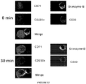
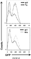
- SJCRH30 cells which lack detectable expression levels of both CD33 and S100A9, were transfected with either S100A9 or S100A8 (negative control) for 48-72 hours. After incubation with CD33-fusion for 30 min (2 ⁇ g/ml), 1 x 10 4 cells were cytospinned onto slides and then stained with a secondary anti-human IgG1-APC before analysis by immunofluorescence microscopy. Similarly, SJCRH30 cells, stable-transfected with CD33, were incubated with rhS100A9 tagged with DDK for various time points and stained by the same methodology before analysis.
- CD45 + CD33 + CD11b + Lin - cells were sorted from full bone marrow of MDS patients by FACS.
- the following antibodies were used: CD45-PECy7, CD33-PECy5, CD11b-FITC, CD3-PE, CD14-PE, CD20-PE (all Beckman Coulter, Fullerton, CA, USA), CD16-PE, CD19-PE (DAKO, Glostrup, Denmark) and CD56-PE (BD Biosciences, San Diego, CA, USA).
- T cells were isolated by Magnetic Activated Cell Sorting (MACS) using CD3 microbeads (Miltenyi Biotec, Aubern, CA, USA) from autologous peripheral blood. 20,000 T cells were seeded in a 96 wells round bottom plate in triplicate in Iscove's modified Dulbecco medium (IMDM; Invitrogen, Carlsbad, CA) supplemented with 10% human serum PAA (PAA Laboratories, Pasching, Austria). Cultures were stimulated with 30 U/ml IL-2 (Chiron, Emeryville, CA, USA), and anti-CD3/anti-CD28 coated beads (Invitrogen, Carlsbad, CA, USA) at a 1:2 ratio of T cells to beads.
- IMDM Iscove's modified Dulbecco medium
- PAA PAA Laboratories, Pasching, Austria
- MDSCs were admixed with T cell cultures at ratios of 1:2 and 1:4 and supplemented with 10 ng/ml GM-CSF to support MDSC viability. After 3 days of co-culture, culture supernatants were harvested to measure IFN- ⁇ concentration by ELISA (Pierce Endogen, Rockford, IL, USA). Subsequently, 0.5 ⁇ Ci 3 H-thymidine (Perkin Elmer, Groningen, the Netherlands) was added to each well and, after overnight incubation, 3 H -thymidine incorporation was measured using a 1205 Wallac Betaplate counter (PerkinElmer). To determine whether differences in proliferation and IFN- ⁇ production were statistically significant, one-way Anova with Bonferroni post-hoc test was used. Statistical significance was accepted for p values ⁇ 0.05.
- Colony-forming Assay Cells isolated from either human BM, or from S100A9Tg, S100A9KO or WT tibias and femurs were subjected to ACK for 5 min at room temperature (Sigma) to lyse the red blood cells. Remaining BM cells were then seeded into complete methylcellulose media (MethoCult complete medium with necessary cytokines and growth factors (StemCell Technologies) and the mixture was placed in duplicate gridded 35-mm culture dishes (2 ⁇ 10 5 cells/dish) and incubated at 37°C in 5% CO 2 for 7-14 days. After incubation, colonies of BFU-E and CFU-GM were identified manually and counted using an inverted light microscope. For the colony formation assays performed using ATRA treated mice, we administered ATRA at 250 ⁇ g (200 ⁇ l) or vehicle (Olive oil) orally for five consecutive days before resting two days.
- RT-PCR and quantitative RT-PCR (qRT-PCR) reactions were performed by means of iQ SYBR Green Supermix (Bio-Rad).
- the reaction mixture (25 ⁇ l total) contained 12.5 ⁇ l iQ SYBR green supermix, 0.25 ⁇ l forward primer (s GAPDH) (20 ⁇ M), 11 ⁇ l RNase-free water, and 1.0 ⁇ l cDNA.
- the following cycles were performed 1 x 3 min at 95°C, 40 amplification cycles (15 s 95 °C, 60 s 56 °C), 1x 1 min 95 °C, 1 x 1 negative control without cDNA template was run with every assay.
- CD33 / Siglec 3 chimeric fusion protein Preparation of the CD33 / Siglec 3 chimeric fusion protein.
- Recombinant soluble fusion of CD33/Siglec 3 ectodomain were constructed as described previously ( Cannon, J.P., et al. 2012. Immunogenetics 64:39-47 ; Cannon, J.P., et al. 2011. Methods Mol Biol 748:51-67 ; Cannon, J.P., et al. 2008. Immunity 29:228-237 ).
- cDNA fragments encoding CD33/Siglec 3 ectodomain were amplified by PCR and inserted into a vector that encodes the human Fc ⁇ followed by a c-terminal recognition site for E. coli biotin ligase.
- This vector has been engineered to facilitate the fusion of gene segments encoding extracellular Ig-type domains to the Fc region of human IgG1.
- the recombinant proteins were expressed in 293T cells post-transfection, using Lipofectamine (Invitrogen), with three successive harvests of 25 ml OPTI-MEM I serum-free medium. The harvests were pooled, centrifuged at 500g for 10 min to remove debris and stored at 4 °C in 0.02% sodium azide. Concentrations of CD33-fusions in culture supernatants were determined by Bradford assay (Biorad, Carlsbad, CA).
- Mass spectrometry Following in-gel tryptic digestion, peptides were extracted and concentrated under vacuum centrifugation. A nanoflow liquid chromatograph (Easy-nLC, Proxeon, Odense, Denmark) coupled to an electrospray ion trap mass spectrometer (LTQ, Thermo, San Jose, CA) was used for tandem mass spectrometry peptide sequencing experiments. The sample was first loaded onto a trap column (BioSphere C18 reversed-phase resin, 5 um, 120 ⁇ , 100 ⁇ m ID, NanoSeparations, Nieuwkoop, Netherlands) and washed for 3 minutes at 8 ml/minute.
- a trap column BioSphere C18 reversed-phase resin, 5 um, 120 ⁇ , 100 ⁇ m ID, NanoSeparations, Nieuwkoop, Netherlands
- the trapped peptides were eluted onto the analytical column, (BioSphere C18 reversed-phase resin, 150 mm, 5 ⁇ m, 120 ⁇ , 100 ⁇ m ID, NanoSeparations, Nieuwkoop, Netherlands). Peptides were eluted in a 60 minute gradient from 5% B to 45% B (solvent A: 2% acetonitrile + 0.1% formic acid; solvent B: 90% acetonitrile + 0.1% formic acid) with a flow rate of 300 nl/min. Five tandem mass spectra were collected in a data-dependent manner following each survey scan. Sequences were assigned using Mascot (www.matrixscience.com) searches against human IPI entries.
- CD33 plasmid (GeneCopeia) was subcloned into a pShuttle-IRES-hrGFP-1 vector (containing the CMV promoter and hrGFP). The PmeI-digested shuttle vectors were then co-transformed into electro-competent BJ5183 bacteria with pAdEasy-1 (containing the viral backbone) and selected on Kanamycin LB plates. The plasmid in the bacteria was amplified and purified using a plasmid maxiprep system (Qiagen). The complete adenoviral vector was linearized by PacI digestion and then transfected into AD293 cells using Lipofectamine (Invitrogen).
- adenoviruses were amplified in AD293 cells.
- Viral stocks were obtained by amplification of the AD293 cells followed by standard two-step CsCl gradient ultracentrifugation, dialysis, and storage in a glycerol stock (10% volume/volume) at -80°C.
- the titer of each viral stock was routinely tested to be 10 11 -10 12 pfu by plaque forming assay using AD293 cells.
- Plasmids, pcDNA3 wild-type DAP12 and P23-DAP12 were cut with HindIII and XhoI (Promega), and DNA Polymerase I Large (KlenowI) (New England Biolabs Inc.) was used to fill in recessed 3' ends of DNA fragments.
- pWPI which contains a GFP expression cassette (Addgene Organization) was digested using PMEI (New England Biolabs Inc.). After cutting DNA and vector, pWPI were purified by Strata Prep PCR purification kit (Qiagen), DNAs were ligated (Takara DNA ligation kit, Fisher) into vector pWPI and STBL2 competent cells were transformed (Invitrogen).
- 293T cells were transfected with pWPI lentivirus vector, the packaging plasmid, psPAX2, and the envelope plasmid, pMD2.G (Addgene Organization) using the Lipofectemine-2000 (Invitrogen) at a ratio of 4:3:1 according to standard protocols. Following 6 hours of incubation, the transfection reagent was removed and replaced with fresh DMEM supplemented with 10% fetal bovine serum. Virus containing medium was collected 24-48hr later. Cell infection was performed as described above. Four days after the first infection, transduced cells were isolated by FACS sorting GFP + cells with >99% purity.
- MDSCs isolated from MDS patients were infected three times using virus-containing infection medium at 24 hr intervals in the presence of 8 ⁇ g/ml polyberene. For each infection, cells were plated in 12-well dishes at 1 x 10 6 cells/well. Four days after the first infection, cells were harvested and used for real time PCR, Western blot analysis, flow cytometry, or colony formation assays.
- BM-MNCs were stained with appropriate specific conjugated antibodies in PBS with 2% BSA buffer.
- FITC anti-CD3 and FITC anti-HLA-DR were used as positive controls and isotype IgG were used for negative control and to detect non-specific staining.
- Cells were gently mixed and incubated for 30 min at 4 °C in the dark. Samples were washed with PBS and centrifuged at 500 g for 5 min. For cells used in phenotypic analysis only, 0.5 ml of 1% paraformaldehyde in PBS was added prior to analysis.
- Cells were washed in PBS and then stained with PE-conjugated mAbs specific to CD40, CD80, CD83, CD86, CCR7, CD11c, CD14, HLA-DR, TLR2, or TLR4 and relevant isotype controls (eBioscience) for 30 min in the dark, on ice. The cells were then washed with PBS containing 0.5% BSA. Live cells were gated based on negative staining for 7-AAD. Samples were acquired on a FACSCalibur flow cytometer and the analysis was performed using Flowjo 6.3.4 software.
- Arginase activity was measured in cell lysates as previously described ( Youn, J.I., et al. 2008. J Immunol 181:5791-5802 ). In brief, cells were lysed for 30 min with 100 ⁇ l of 0.1% Triton X-100. Subsequently, 100 ⁇ l of 25 mM Tris-HCl and 10 ⁇ l of 10 mM MnCl 2 were added, and the enzyme was activated by heating for 10 min at 56°C. Arginine hydrolysis was conducted by incubating the lysate with 100 ⁇ l of 0.5 M L-arginine (pH 9.7) at 37 °C for 120 min.
- Cell lysates were prepared by resuspending cell pellet in 1% NP-40, 10 mM Tris, 140 mM NaCl, 0.1 mM PMSF, 10 mM iodoacetamide, 50 mM NaF, 1 mM EDTA, 0.4 mM sodium orthovanadate, 10 ⁇ g/ml leupeptin, 10 ⁇ g/ml pepsatin, and 10 ⁇ g/ml aprotinin and lysing on ice for 30 min. Cell lysates were centrifuged at 12,000 g for 15 min to remove nuclei and cell debris. The protein concentration of the soluble extracts was determined by using the Bio-Rad (Bradford) protein assay.
- Lysate from S100A9 un-transfected, empty vector and S100A9 transfect SJCRH30 or AD293 cells was serially-diluted (as indicated in the figure) with the first lysate, starting at 50 ⁇ g, followed by loading onto a nitrocellulose membrane and blocked overnight at 4°C in 5% milk. Following incubation with CD33-fusion for 2 hours at room temperature, and staining with an anti-human IgG HRP-conjugated secondary, the membrane was washed and used for radiograph analysis to demonstrated specific S100A9 binding to CD33. The membrane was afterwards used for Coomassie blue staining to show the relative equal loading of proteins on each dot in the nitrocellulose membrane.
- CBC Complete peripheral blood cell counts
- Bone marrow aspirates and touch-imprints were stained using Wright-Giemsa stain. Sections of bone marrow and spleen were fixed in 10% phosphate-buffered neutral formalin, decalcified (only applied to bone marrow) and embedded in paraffin by routine procedures. Sections were cut at 4 ⁇ m and stained with hematoxylin and eosin (H&E) and periodic acid Schiff (PAS). The presence of myelodysplastic features characteristic of MDS was evaluated by experienced hematopathologist. BM core biopsy shows 50% cellularity with maturing trilineage hematopoiesis (H&E, 200x).
- Figure 5E shows a high power view of the BM biopsy that demonstrates normal appearing megakaryocytes with normal lobation.
- Mixed myeloid and erythroid precursors are normally distributed with estimated M:E ratio of 2:1 (H&E, 600x).
- Wright-Giemsa stained BM aspirate exhibits full maturation in all three lineages without dysplastic features (Wright-Giemsa, 1000x).
- Inlet shows a normal lobated megakaryocyte ( Figure 5F ).
- Touch imprint of mouse spleen displays predominance of small and mature appearing lymphocytes intermingled with occasional erythropoietic precursors (Wright-Giemsa, 1000 x) ( Figure 5G ).
- BM core biopsy reveals hypercellularity, approximately 95% with increased megakaryocytes, especially in small forms (H&E, 200x) ( Figure 5H ).
- High power magnification highlights dysplastic megakaryocytes with single or hypolobated, or disjointed nuclei and markedly increased in number (H&E, 600x) (insert) ( Figure 5I ).
- Inlet includes two markedly dysplastic micromegakaryocytes with hypolobation.
- BM aspirates exhibits mildly increased blasts admixed with myeloid and erythroid precursors. The latter demonstrates slightly irregular nuclear contour and minimal megaloblastoid changes (Wright-Giemsa, 1000x) ( Figure 5J ).
- Inlet contains two blasts showing delicate or fine chromatin, prominent nucleoli, high N:C ratio, and scant basophilic cytoplasm.
- Touch preparation of transgenic mouse spleen show increased erythroid precursors; some of them displaying enlarged size with abnormal nuclearity and occasional nuclear bridge (Wright-Giemsa, 1000x) ( Figure 5K ).
- Lin - HLA-DR - CD33 + MDSC are expanded in MDSprimary BM specimens and direct suppression of autologous erythroid precursors.
- MDS patients exhibited markedly higher numbers of MDSCs (median 35.5%, P ⁇ 0.0001) compared to healthy donors or non-MDS cancer patients (less than 5%, Figure 1A ).
- LIN - HLA-DR - CD33 + MDSCs were sorted from MDS specimens with chromosome 5q [del(5q)] or 7q [del(7q)] deletion and analyzed by fluorescence in situ hybridization (FISH) with specific probes.
- the raw CT for a given mutation assay in a test sample is compared with a predefined CT cutoff. Based on the difference, the mutation can be considered as "Present” (+), "Borderline” (-/+), or "Absent” (-).
- MDSCs were depleted from MDS BM specimens prior to anti-CD3/anti-CD28 stimulation and then the MDSC were added back to the control group.
- MDSC-depletion significantly improved T cell responses compared to the MDSC-supplemented group ( Figure IE), thereby linking the observed impaired T cell responsiveness to the actions of BM-derived MDSCs.
- suppressive MDS-MDSC overproduced suppressive cytokines such as IL-10 and TGF- ⁇ ( Figure 1F & G ), as well as nitric oxide (NO) and arginase compared to MDSCs isolated from healthy donors ( Figure 1H & I ).
- MDSC-mediated suppressive and cytotoxic effector functions require direct contact with target cells.
- One mechanism utilized by cytotoxic effectors is the mobilization of pore-forming granules and the release of caspase-activating effector proteases, such as granzyme B, to the site of effector:target contact thereby inducing apoptosis of the target cell ( Chen, X., et al. 2008. Blood 113(14):3226-34 ).
- HPCs hematopoietic progenitor cells
- MDS-MDSC granule mobilization and release of granzyme B were examined using four-color immunofluorescence staining.
- MDS-MDSCs exhibited strong granzyme B polarization at the site of cell contact with CD235a + (glycophorin A)/CD71 + autologous erythroid precursors ( Figure 1J ). After 30 minutes incubation, the frequency of such effector-target conjugates in MDS patient specimens was significantly higher (34%) than in samples from healthy donors (5% P ⁇ 0.001, Figure 1K & L ).
- BFU-E Burst-forming unit-erythroid
- CFU-GM colony forming unit-granulocyte/macrophage
- CD33 membrane expression density on MDS-MDSC was investigated. These cells were found to robustly express CD33 at levels higher than LIN - HLA-DWCD33 + cells isolated from non-MDS-associated cancer patients and healthy donor BM cells ( Figure 2A ).
- CD33 was cross-linked in U937 cells (a human monocytic cell line with high CD33 expression), which triggered IL-10, TGF- ⁇ and VEGF secretion ( Figure 2B ).
- CD33 was overexpressed with an adenovirus vector in BM-MNC from healthy donors, which significantly suppressed myeloid cell development as evidenced by reduced expression of the maturation markers CD11c, CD80, and CCR7 ( Figure 2C ).
- CD33 was knocked down in MDS-MDSC using a lentiviral vector (LV) containing CD33-specific-shRNA.
- LV lentiviral vector
- S100A9 is a native ligand for CD33. Although these investigations showed that CD33/Siglec 3 is a key receptor involved in functional activation of MDSC, its native ligand was unknown. Therefore, to identify potential ligand(s) for this receptor, a chimeric fusion protein with the ectodomain of CD33 and the Fc portion of human IgG (here on referred to as CD33-fusion) was produced. BM cell lysates from MDS patients were immunoprecipitated with the CD33-fusion followed by high throughput mass-spectrometric analysis on associated peptides. S100A9, an inflammatory signaling molecule known to activate MDSC, was among the most prominent protein bands identified ( Figure 3A ).
- both S100A8 and S100A9 transfectants were prepared in the rhabdomyosarcoma cell line SJCRH30, which lacks detectable expression of endogenous CD33, S100A8 or S100A9.
- the CD33-fusion (APC) stained only S100A9 transfected cells, but not S100A8 transfected cells, confirming binding specificity ( Figure 3B .
- Transfectants were stained with FITC and specific binding by the CD33-fusion was stained with APC, (nuclei were stained with DAPI).
- FIG. 4D demonstrates the transfection efficiency of the CD33 adenovirus at both the gene and protein expression level measured by QPCR and GFP by flow cytometry, respectively.
- S100A9 transgenic mice recapitulate features of human MDS. Given the findings that S100A8/S100A9 triggers CD33/Siglec 3 signaling and is involved in MDSC activation ( Ehrchen, J.M., et al. 2009. J Leukoc Biol 86:557-566 ; Vogl, T., et al. 2007. Nat Med 13:1042-1049 ; Cheng, P., et al. 2008. J Exp Med 205:2235-2249 ), S100A9 transgenic mice (S100A9Tg) were generated to study MDSC-associated inflammation ( Cheng, P., et al. 2008. J Exp Med 205:2235-2249 ).
- CBC serial complete blood counts
- S100A9Tg mice developed severe pan-cytopenia (detailed descriptions summarized in the figure legend). Although this model closely replicates the inflammatory milieu observed in human MDS, HSPC from S100A9Tg mice also express the S100A9 protein. Given that the provenance of this protein in human MDS is not known, cellular expression of S100A9 in MDS BM-MNC was investigated by flow cytometry. S100A9 intracellular staining was detected in CD33 + cells, whereas CD34 + HSCs had no demonstrable S100A9 expression ( Figure 5O and 5P ).
- S100A9 expression was not detected in other immune cells such as CD3 + lymphocytes, CD19 + B cells or CD56 + NK cells.
- Table 2 Complete peripheral blood count of S100A9Tg and wt-mice Test Units 6 weeks 18 weeks 24 weeks WT S100A9-Tg WT S100A9-Tg WT S100A9-Tg WBC 10 3 ⁇ l 5.4 ⁇ 0.5 4.2 ⁇ 1.5 6.0 ⁇ 1.4 2.9 ⁇ 0.2* 6.3 ⁇ 0.8 3.0 ⁇ 0.4* LYM 10 3 / ⁇ l 3.9 ⁇ 0.5 2.6 ⁇ 1.2 4.7 ⁇ 1.1 2.4 ⁇ 0.1* 4.8 ⁇ 0.7 2.5 ⁇ 0.3* MONO 10 3 / ⁇ l 0.4 ⁇ 0.1 0.3 ⁇ 0.2 0.4 ⁇ 0.1 0.2 ⁇ 0.1* 0.4 ⁇ 0.1 0.2 ⁇ 0.1* GRAN 10 3 / ⁇ l 1.1 ⁇ 0.6 1.2 ⁇ 1.2 0.9 ⁇ 0.3 0.3 ⁇ 0.2* 1.1 ⁇ 0.4 0.3 ⁇ 0.1** HCT % 48.1 ⁇ 3.2 42.4 ⁇ 2.1 45.7 ⁇ 0.7 35.5 ⁇ 3.0** 45.4 ⁇ 3.5 3
- Peripheral blood samples were prepared from both S100A9Tg and control (WT) mice in ages of 6, 18 and 24 weeks and analyzed on a Hema True Hematology Analyzer (Heska). * p ⁇ 0.05; ** p ⁇ 0.01;*** p ⁇ 0.001 vs wt-mice
- BM CD34+ (MDSC depleted, MDSC-) from MDS patient specimens were treated with rhS100A9 for 24 and 48 hours and assessed apoptosis by flow cytometry.
- ATRA -trans-retinoic acid
- S100A9Tg mice were treated with all -trans-retinoic acid (ATRA).
- ATRA induces MDSC differentiation into mature myeloid cells and neutralizes ROS production, thereby extinguishing MDSC through forced terminal differentiation ( Nefedova, Y., et al. 2007. Cancer Res 67:11021-11028 ; Mirza, N., et al. 2006. Cancer Res 66:9299-9307 ).
- ATRA was examined whether ATRA would induce MDSC differentiation in S100A9Tg mice and improve hematopoiesis.
- S100A9Tg and WT mice were treated with ATRA (250 ⁇ g/200 ⁇ l) or vehicle control orally for five consecutive days. Two days after completion of the treatment, ATRA reduced the total number of MDSC while numbers in WT mice remained at basal levels ( Figure 7A ). Reductions in MDSC number in S100A9Tg mice were coupled to an increase in mature cells following ATRA treatment ( Figure 7B ). Treatment of primary MDS BM specimens with ATRA also reduced in vitro MDSC accumulation. Importantly, BM progenitor cultures from ATRA-treated S100A9Tg mice showed significantly improved BFU-E recovery compared to vehicle-treated controls ( Figure 7C ). Analysis of changes in peripheral blood counts showed that RBC, WBC and platelet counts significantly increased compared to vehicle treated controls ( Figure 7D ), indicating that terminal differentiation of MDSCs can restore effective hematopoiesis.
- ITIM signals can be over-ridden by stimulatory immunoreceptor tyrosine-based activation motif (ITAM)-mediated signals ( Lanier, L.L. 2005. Annu Rev Immunol 23:225-274 ; Ravetch, J.V., et al. 2000. Science 290:84-89 ).
- ITAM stimulatory immunoreceptor tyrosine-based activation motif
- Some CD33-related receptors, such as certain Siglecs lack ITIMs and instead function as activating receptors.
- Mouse Siglec-H and human Siglec-14 have been shown to interact with DAP12 ( Blasius, A.L., et al. 2006.
- DAP12 mRNA was significantly lower in MDS-MDSC in all specimens tested ( Figure 7E ). It was reasoned that overriding CD33-ITIM signaling via DAP12 would induce the differentiation of these immature myeloid cells and improve hematopoiesis.
- a constitutively active form of DAP12 named P23
- P23 promotes primary human DC maturation as demonstrated by up-regulation of CD80, CD83, and CCR7 antigens ( Figure 10 ) ( Blasius, A.L., et al. 2006. Blood 107:2474-2476 ; Blasius, A.L., et al. 2006. Trends Immunol 27:255-260 ). Based on these findings, it is possible that P23 could induce the maturation of MDS-MDSC and thereby prevent or disrupt hematopoietic suppression. To test this, expression of the human monocytic and granulocytic surface markers CD14 and CD15 were analyzed after transfection ( Araki, H., et al. 2004.
- Inflammatory stimuli within the BM microenvironment are recognized as important biological signals stimulating progenitor cell proliferation and apoptosis in MDS.
- a recent population-based study extended this further by demonstrating a strong linkage between chronic immune stimulation and MDS predisposition ( Kristinsson, S.Y., et al. 2011. J Clin Oncol 29:2897-2903 ).
- Definitive evidence that niche intrinsic abnormalities per se can alone account for development of MDS in a cell non-autonomous fashion are limited.
- Raaijmakers and colleagues showed that selective osteo-progenitor dysfunction caused by deletion of Dicer1 in the mesenchymal component of the BM microenvironment was sufficient to perturb hematopoiesis and lead to development of myeloid dysplasia, followed by secondary emergence of myeloid-restricted genetic abnormalities ( Raaijmakers, M.H., et al. 2010. Nature 464:852-857 ).
- Lin - HLA-DR - CD33 + MDSC accumulate in the BM of MDS patients, derive from a population that is distinct from the neoplastic clone, and serve as cellular effectors that suppress hematopoiesis, promote T cell tolerance and serve as a key source of myelosuppressive and inflammatory molecules such as IL-10, TGF- ⁇ , NO, and arginase.
- S100A9 also known as migration inhibitory factor-related protein 14 (MRP-14) or calgranulin-B, can serve as an endogenous native ligand for CD33/Siglec 3.
- the time dependent accumulation of MDSC in the transgenic mouse model parallels recent human findings that MDSCs expand with age accompanied by rising serum levels of proinflammatory cytokines (e.g., TNF- ⁇ , IL-6, and IL-1 ⁇ ), providing evidence that such senescence dependent changes driving MDSC expansion may play an important role in the age dependent pathogenesis of MDS ( Verschoor CP, et al. 2013. J Leukoc Biol 93(4):633-7 ).
- the LIN - HLA-DR - CD33 + phenotype did not alone confer suppressor cell function, as evidenced by lack of LIN - HLA-DR - CD33 + MDSC suppression from age-matched healthy donors or non-MDS cancer patients.
- the disclosed studies demonstrate the necessity for activation of innate immune signaling and generation of pro-inflammatory molecules, such as S100A9, for the induction of MDS-MDSC-mediated suppressor function. Furthermore, the disclosed finding that primary human MDS-MDSC lack molecular genetic abnormalities intrinsic to the malignant clone indicate that MDSC derive from non-neoplastic HSC, and that MDSC activation and expansion likely precedes emergence of genetically distinct MDS clones.
- CD33 a Siglec receptor expressed by many immune cells including MDSC, is shown herein to be markedly over-expressed by MDS-MDSC, and this receptor is shown to control the suppressive functions of MDS-MDSC through disruption of ITIM-mediated signaling.
- rhS100A9 directly triggers apoptosis in human HPCs, indicates that this ligand exerts dual roles in the promotion of ineffective hematopoiesis that involve both cellular (MDSC) and humoral mechanisms (CD33, TLR4).
- cellular response to CD33 ligand engagement appears cell type specific, i.e., apoptosis in HPC versus activation and proliferation in MDSC.
- Compensatory regeneration within the myeloid compartment could account for the increased proliferative index observed in MDS ( Raza et. al. 1995. Blood 86(1):268-276 ) and preferential myeloid skewing that occurs with age.
- Over-expression of CD33 may therefore impair maturation signals from ITAM associated receptors that is critical to expansion of immature MDSC ( Figure 7G and H ). Consistent with this, shRNA silencing of CD33 reduced myelosuppressive cytokine elaboration, and importantly, restored hematopoietic progenitor colony forming capacity.
- DAP12 signaling was sufficient to override CD33-ITIM inhibition and induce MDSC differentiation, which restored erythropoiesis upon active-DAP12 transfection into MDS-MDSC. More importantly, DAP12 activation can inhibit TLR-mediated signaling pathways, which may also play a role in the inflammation-mediated BM suppression ( Turnbull, I.R., et al. 2007. Nat Rev Immunol 7:155-161 ; Hamerman, J.A., et al. 2006. J Immunol 177:2051-2055 ).
- Prolonged activation of innate immune signaling with age may disrupt the BM endosteal niche supporting maintenance of hematopoietic stem cells, and favor translocation of myeloid progenitors to an angiogenic niche characteristic of MDS ( Bellamy, W.T., et al. 2001. Blood 97:1427-1434 ).
- This is supported by the disclosed findings of age dependent development of cytopenias with emergence of dysplastic cytological features in the S100A9Tg mice.
- the competitive transplant experiments showed that admixture of S100A9Tg with WT donor HSCs delays, albeit with time still impairs hematopoiesis.
- the disclosed findings support a model for MDS pathogenesis in which sustained activation of innate immune signaling in the BM microenvironment creates a permissive inflammatory milieu that is sufficient for development of myelodysplasia.
- Cell autonomous neoplastic hematopoietic progenitors may emerge following acquisition of secondary genetic abnormalities in the myeloid compartment.
- S100A9Tg mice simulate human MDS and can serve as an in vivo model to study MDS pathogenesis and development of novel therapeutics. Nevertheless, therapeutic interventions that promote MDSC maturation may have remitting potential when applied early in the disease course.
- Active caspase-1 (Table 3) and IL-1 ⁇ generation (Table 4) were assessed in four populations: stem cells (CD34+CD38-), progenitors (CD34+CD38+), erythroids (CD71+), and myeloid cells (CD33+).
- Mean fluorescent intensity (MFI) values are shown in Table 3. Table 3. Treatment with CD33-IgG decreases active caspase-1 generation in an MDS specimen.
- Stem Cells Progenitors Erythroids Myeloids Ancestry Subset Value Type For LIVE/CD34+/STEM Geom.
- Active caspase-1 and IL-1 ⁇ generation were assessed in four populations: stem cells (CD34+CD38-), progenitors (CD34+CD38+), erythroids (CD71+), and myeloid cells (CD33+).
- Mean fluorescent intensity (MFI) values are found in the Tables 3 and 4.
- FIG. 14 shows erythroid burst-forming units (BFU-E) ( Fig. 14B ), multipotential colony forming units (CFU-GEMM) ( Fig. 14A ), and granulocyte/macrophage colony forming units (CFU-GM) ( Fig. 14C ) in MDS patient specimens treated with IgG, plasma, or 0.1, 0.5, or 1.0 ⁇ g of CD33-IgG chimeric trap.
- BFU-E erythroid burst-forming units
- CFU-GEMM multipotential colony forming units
- CFU-GM granulocyte/macrophage colony forming units
Landscapes
- Health & Medical Sciences (AREA)
- Life Sciences & Earth Sciences (AREA)
- Chemical & Material Sciences (AREA)
- Organic Chemistry (AREA)
- Immunology (AREA)
- Medicinal Chemistry (AREA)
- General Health & Medical Sciences (AREA)
- Engineering & Computer Science (AREA)
- Cell Biology (AREA)
- Proteomics, Peptides & Aminoacids (AREA)
- Veterinary Medicine (AREA)
- Pharmacology & Pharmacy (AREA)
- Animal Behavior & Ethology (AREA)
- Public Health (AREA)
- Molecular Biology (AREA)
- Gastroenterology & Hepatology (AREA)
- Zoology (AREA)
- Bioinformatics & Cheminformatics (AREA)
- Biochemistry (AREA)
- Genetics & Genomics (AREA)
- Biophysics (AREA)
- Toxicology (AREA)
- General Chemical & Material Sciences (AREA)
- Nuclear Medicine, Radiotherapy & Molecular Imaging (AREA)
- Chemical Kinetics & Catalysis (AREA)
- Epidemiology (AREA)
- Hematology (AREA)
- Biomedical Technology (AREA)
- Urology & Nephrology (AREA)
- Dispersion Chemistry (AREA)
- Oncology (AREA)
- Biotechnology (AREA)
- Microbiology (AREA)
- Food Science & Technology (AREA)
- Physics & Mathematics (AREA)
- Analytical Chemistry (AREA)
- General Physics & Mathematics (AREA)
- Pathology (AREA)
- Rheumatology (AREA)
- Diabetes (AREA)
Claims (15)
- Chimäres rekombinantes Fusionsprotein, umfassend:(a) eine Kombination von mindestens zwei S100A9-Bindungsgruppierungen, ausgewählt aus der Gruppe bestehend aus einer extrazellulären Domäne von humanem CD33, einer extrazellulären Domäne von Toll-like Rezeptor 4 (TLR4) und einer extrazellulären Domäne von Rezeptor für Advanced-Glycation-Endprodukte (RAGE); und(b) eine Immunglobulin-Fc-Region.
- Fusionsprotein nach Anspruch 1, umfassend eine Formel ausgewählt aus der Gruppe bestehend aus:eCD33 - eTLR4 - Fc,eCD33 - eRAGE - Fc,eTLR4 - eRAGE - Fc,eRAGE - eTLR4 - Fc,eCD33 - eTLR4 - eRAGE - Fc,eCD33 - eRAGE - eTLR4 - Fc,eTLR4 - eCD33 - eRAGE - Fc,eRAGE - eCD33 - eTLR4 - Fc,eTLR4 - eRAGE - eCD33 - Fc undeRAGE - eTLR4 - eCD33 - Fc,wobei "eCD33" die extrazelluläre Domäne von menschlichem CD33 umfasst,
wobei "eTLR4" die extrazelluläre Domäne von TLR4 umfasst,
wobei "eRAGE" die extrazelluläre Domäne von RAGE umfasst,
wobei "Fc" die Immunglobulin-Fc-Region umfasst und
wobei "-" aus einem Peptidlinker oder einer Peptidbindung besteht. - Fusionsprotein nach Anspruch 1 oder 2, ferner umfassend ein Biotin-Akzeptor-Peptid, das mit Biotin-Ligase (BirA) in Gegenwart von Biotin und ATP biotinyliert werden kann.
- Fusionsprotein nach Anspruch 3, wobei das Biotin-Akzeptor-Peptid die Aminosäuresequenz SEQ ID NO:7 oder eine Aminosäuresequenz mit mindestens 90 % Identität mit SEQ ID NO:7. umfasst.
- Fusionsprotein nach einem der Ansprüche 1 bis 4, wobei die extrazelluläre Domäne des humanen CD33 nur die variable Region umfasst.
- Fusionsprotein nach einem der Ansprüche 1 bis 5, wobei die extrazelluläre Domäne des humanen CD33 die Aminosäuresequenz SEQ ID NO:1 oder eine Aminosäuresequenz mit mindestens 90 % Identität mit SEQ ID NO:1 umfasst, wobei die extrazelluläre Domäne von humanem CD33 vorzugsweise die Aminosäuresequenz SEQ ID NO:2 umfasst.
- Fusionsprotein nach Anspruch 5, wobei die variable Region des humanen CD33 die Aminosäuresequenz SEQ ID NO:3 oder eine Aminosäuresequenz mit mindestens 90 % Identität mit SEQ ID NO:3 umfasst, wobei die variable Region des humanen CD33 vorzugsweise die Aminosäuresequenz SEQ ID NO:4 umfasst.
- Fusionsprotein nach einem der Ansprüche 1 bis 7, wobei die extrazelluläre Domäne des humanen TLR4 die Aminosäuresequenz SEQ ID NO:5 oder eine Aminosäuresequenz mit mindestens 90 % Identität mit SEQ ID NO: 5 umfasst.
- Fusionsprotein nach einem der Ansprüche 1 bis 8, wobei die extrazelluläre Domäne von RAGE die Aminosäuresequenz SEQ ID NO:6 oder eine Aminosäuresequenz mit mindestens 90 % Identität mit SEQ ID NO: 6 umfasst.
- Multimerkomplex, umfassend zwei oder mehr Fusionsproteine nach einem der Ansprüche 1 bis 9, die an ein Kernmolekül oder Partikel konjugiert sind.
- Multimerkomplex nach Anspruch 10, wobei das Kernmolekül Streptavidin ist, wobei die zwei oder mehr Fusionsproteine biotinyliert sind.
- Multimerkomplex nach Anspruch 10, wobei das Kernmolekül ein Liposom ist, das Antikörper umfasst, die spezifisch an die zwei oder mehrere Fusionsproteine binden, wobei die Antikörper vorzugsweise spezifisch das Biotin-Akzeptor-Peptid binden.
- Multimerkomplex nach einem der Ansprüche 10 bis 12, umfassend von 2 bis 5 Fusionsproteine, die an das Kernmolekül oder Partikel konjugiert sind.
- Fusionsprotein nach einem der Ansprüche 1 bis 9 oder der Multimerkomplex nach einem der Ansprüche 10 bis 13 zur Verwendung beim Behandeln einer Krankheit oder eines Zustands, die/der durch S100A9-Aktivität hervorgerufen oder verschlimmert wird, umfassend ein Verabreichen einer Zusammensetzung, die das Fusionsprotein oder den Multimerkomplex umfasst, an ein Subjekt, das dessen bedarf.
- Fusionsprotein oder Multimerkomplex zur Verwendung nach Anspruch 14, wobei die Verwendung ein Behandeln einer Infektion, Sepsis oder einer Kombination davon, einer Autoimmunerkrankung, rheumatoider Arthritis, einer Krebserkrankung oder einem myelodysplastischen Syndrom (MDS) bei dem Subjekt umfasst.
Priority Applications (6)
| Application Number | Priority Date | Filing Date | Title |
|---|---|---|---|
| SM20200118T SMT202000118T1 (it) | 2013-07-05 | 2014-07-03 | Cd33 solubile per il trattamento delle sindromi mielodisplastiche (mds) |
| PL14819813T PL3016512T3 (pl) | 2013-07-05 | 2014-07-03 | Rozpuszczalny CD33 do leczenia zespołów mielodysplastycznych (MDS) |
| EP20150607.8A EP3711484A1 (de) | 2013-07-05 | 2014-07-03 | Lösliches cd33 zur behandlung myelodysplastischer syndrome (mds) |
| SI201431468T SI3016512T1 (sl) | 2013-07-05 | 2014-07-03 | Topni CD33 za zdravljenje mielodisplastičnih sindromov (MDS) |
| RS20200171A RS59925B1 (sr) | 2013-07-05 | 2014-07-03 | Rastvorljivi cd33 za lečenje mijelodisplastičnih sindroma (mds) |
| HRP20200085TT HRP20200085T1 (hr) | 2013-07-05 | 2014-07-03 | Topljivi cd33 za liječenje mijelodisplastičnih sindroma (mds) |
Applications Claiming Priority (5)
| Application Number | Priority Date | Filing Date | Title |
|---|---|---|---|
| US201361843274P | 2013-07-05 | 2013-07-05 | |
| US201461930798P | 2014-01-23 | 2014-01-23 | |
| US201461931366P | 2014-01-24 | 2014-01-24 | |
| US201461978009P | 2014-04-10 | 2014-04-10 | |
| PCT/US2014/045444 WO2015003149A2 (en) | 2013-07-05 | 2014-07-03 | Soluble cd33 for treating myelodysplastic syndromes (mds) |
Related Child Applications (1)
| Application Number | Title | Priority Date | Filing Date |
|---|---|---|---|
| EP20150607.8A Division EP3711484A1 (de) | 2013-07-05 | 2014-07-03 | Lösliches cd33 zur behandlung myelodysplastischer syndrome (mds) |
Publications (3)
| Publication Number | Publication Date |
|---|---|
| EP3016512A2 EP3016512A2 (de) | 2016-05-11 |
| EP3016512A4 EP3016512A4 (de) | 2017-01-11 |
| EP3016512B1 true EP3016512B1 (de) | 2020-01-08 |
Family
ID=52144293
Family Applications (2)
| Application Number | Title | Priority Date | Filing Date |
|---|---|---|---|
| EP14819813.8A Active EP3016512B1 (de) | 2013-07-05 | 2014-07-03 | Lösliches cd33 zur behandlung myelodysplastischer syndrome (mds) |
| EP20150607.8A Withdrawn EP3711484A1 (de) | 2013-07-05 | 2014-07-03 | Lösliches cd33 zur behandlung myelodysplastischer syndrome (mds) |
Family Applications After (1)
| Application Number | Title | Priority Date | Filing Date |
|---|---|---|---|
| EP20150607.8A Withdrawn EP3711484A1 (de) | 2013-07-05 | 2014-07-03 | Lösliches cd33 zur behandlung myelodysplastischer syndrome (mds) |
Country Status (19)
| Country | Link |
|---|---|
| US (3) | US10344072B2 (de) |
| EP (2) | EP3016512B1 (de) |
| JP (2) | JP6673200B2 (de) |
| AU (3) | AU2014285079A1 (de) |
| BR (1) | BR112015032763A2 (de) |
| CA (1) | CA2917485A1 (de) |
| CY (1) | CY1122813T1 (de) |
| DK (1) | DK3016512T3 (de) |
| ES (1) | ES2777227T3 (de) |
| HR (1) | HRP20200085T1 (de) |
| HU (1) | HUE048842T2 (de) |
| LT (1) | LT3016512T (de) |
| MX (1) | MX377002B (de) |
| PL (1) | PL3016512T3 (de) |
| PT (1) | PT3016512T (de) |
| RS (1) | RS59925B1 (de) |
| SI (1) | SI3016512T1 (de) |
| SM (1) | SMT202000118T1 (de) |
| WO (1) | WO2015003149A2 (de) |
Families Citing this family (12)
| Publication number | Priority date | Publication date | Assignee | Title |
|---|---|---|---|---|
| EP3016512B1 (de) * | 2013-07-05 | 2020-01-08 | H. Lee Moffitt Cancer Center And Research Institute, Inc. | Lösliches cd33 zur behandlung myelodysplastischer syndrome (mds) |
| US10688077B2 (en) | 2015-02-26 | 2020-06-23 | H. Lee Moffitt Cancer Center And Research Institute, Inc. | Inflammasome activation in myelodysplastic syndromes |
| WO2016179035A1 (en) * | 2015-05-01 | 2016-11-10 | H. Lee Moffitt Cancer Center And Research Institute, Inc. | Inflammasome activation in myelodysplastic syndromes |
| WO2016179036A1 (en) * | 2015-05-01 | 2016-11-10 | H. Lee Moffitt Cancer Center And Research Institute, Inc. | S100a9 levels for predicting lenalidomide and erythropoietin responsiveness |
| EP3379248B1 (de) * | 2015-11-18 | 2020-09-23 | Tokyo Ohka Kogyo Co., Ltd. | Verfahren zur messung der arginase-aktivität, kit für den nachweis der arginase-aktivität, kit für den nachweis von arginase-assoziierten erkrankungen und screening-verfahren für arginase-inhibitor oder wirkstoff |
| EP3802582A4 (de) * | 2018-06-07 | 2022-06-15 | Palleon Pharmaceuticals Inc. | Multimere proteine zum nachweis eines kohlenhydrats und/oder zur behandlung einer siglec-vermittelten störung |
| CA3103367A1 (en) * | 2018-06-13 | 2019-12-19 | The Council Of The Queensland Institute Of Medical Research | Viral detection assay |
| US12398402B2 (en) | 2018-09-12 | 2025-08-26 | Fred Hutchinson Cancer Center | Reducing CD33 expression to selectively protect therapeutic cells |
| CN111840561B (zh) * | 2020-08-11 | 2022-03-04 | 大连医科大学附属第一医院 | S100a9抑制剂在制备治疗胰腺炎的药物中的应用 |
| WO2022152902A1 (en) * | 2021-01-18 | 2022-07-21 | Active Biotech Ab | Tasquinimod or a pharmaceutically acceptable salt thereof for use in the treatment of myelodysplastic syndrome |
| WO2022216157A1 (en) * | 2021-04-09 | 2022-10-13 | Stichting Radboud Universiteit | Off the shelf proximity biotinylation enzyme |
| CN116377048B (zh) * | 2022-04-22 | 2024-05-07 | 广州市妇女儿童医疗中心 | S100a8/a9复合体在诊治先天性巨结肠中的应用 |
Family Cites Families (19)
| Publication number | Priority date | Publication date | Assignee | Title |
|---|---|---|---|---|
| FR1601438A (de) | 1968-10-17 | 1970-08-24 | ||
| US5620689A (en) | 1989-10-20 | 1997-04-15 | Sequus Pharmaceuuticals, Inc. | Liposomes for treatment of B-cell and T-cell disorders |
| ATE486937T1 (de) | 1996-07-12 | 2010-11-15 | Genentech Inc | Chimärische heteromultimerische adhesine |
| US20040033603A1 (en) * | 2002-08-19 | 2004-02-19 | Lin Zhang | Biotinylation of proteins |
| CA2504818C (en) | 2002-11-07 | 2013-04-23 | Immunogen, Inc. | Anti-cd33 antibodies and method for treatment of acute myeloid leukemia using the same |
| ES2897506T3 (es) | 2003-01-09 | 2022-03-01 | Macrogenics Inc | Identificación y modificación de anticuerpos con regiones Fc variantes y métodos de utilización de los mismos |
| WO2007001332A2 (en) * | 2004-08-04 | 2007-01-04 | University Of Massachusetts | Anti-pathogen immunoadhesins |
| US20060241040A1 (en) * | 2005-04-06 | 2006-10-26 | Alberto Visintin | Methods of treating disorders associated with toll-like receptor 4 (TLR4) signalling |
| EP1736482A1 (de) | 2005-06-20 | 2006-12-27 | INSERM (Institut National de la Santé et de la Recherche Medicale) | Rekombinanter trimerer 4-1BB Ligand |
| TW200726776A (en) * | 2005-07-29 | 2007-07-16 | Friedrich Alexander University Of Erlangen Nuremberg | CD33-specific single-chain immunotoxin and methods of use |
| AU2006330807A1 (en) | 2005-11-28 | 2007-07-05 | Medimmune, Llc | Antagonists of HMBG1 and/or rage and methods of use thereof |
| EP2044115A2 (de) * | 2006-06-12 | 2009-04-08 | Receptor Biologix, Inc. | Für die oberflächenrezeptoren von pan-zellen spezifische therapeutika |
| WO2008131908A1 (en) * | 2007-04-26 | 2008-11-06 | Active Biotech Ab | S100a9 interaction screening method |
| NZ581550A (en) | 2007-06-14 | 2012-01-12 | Galactica Pharmaceuticals Inc | Rage fusion proteins |
| CN101838329A (zh) * | 2009-03-18 | 2010-09-22 | 嘉和生物药业有限公司 | 抗血管新生融合蛋白 |
| DE102009045006A1 (de) | 2009-09-25 | 2011-04-14 | Technische Universität Dresden | Anti-CD33 Antikörper und ihre Anwendung zum Immunotargeting bei der Behandlung von CD33-assoziierten Erkrankungen |
| UA112062C2 (uk) | 2010-10-04 | 2016-07-25 | Бьорінгер Інгельхайм Інтернаціональ Гмбх | Cd33-зв'язувальний агент |
| EP2654790B1 (de) | 2010-12-22 | 2019-02-06 | Teva Pharmaceuticals Australia Pty Ltd | Modifizierter antikörper mit verbesserter halbwertzeit |
| EP3016512B1 (de) * | 2013-07-05 | 2020-01-08 | H. Lee Moffitt Cancer Center And Research Institute, Inc. | Lösliches cd33 zur behandlung myelodysplastischer syndrome (mds) |
-
2014
- 2014-07-03 EP EP14819813.8A patent/EP3016512B1/de active Active
- 2014-07-03 LT LTEP14819813.8T patent/LT3016512T/lt unknown
- 2014-07-03 EP EP20150607.8A patent/EP3711484A1/de not_active Withdrawn
- 2014-07-03 US US14/902,719 patent/US10344072B2/en active Active
- 2014-07-03 BR BR112015032763A patent/BR112015032763A2/pt not_active Application Discontinuation
- 2014-07-03 HR HRP20200085TT patent/HRP20200085T1/hr unknown
- 2014-07-03 SI SI201431468T patent/SI3016512T1/sl unknown
- 2014-07-03 AU AU2014285079A patent/AU2014285079A1/en not_active Abandoned
- 2014-07-03 WO PCT/US2014/045444 patent/WO2015003149A2/en not_active Ceased
- 2014-07-03 MX MX2016000211A patent/MX377002B/es active IP Right Grant
- 2014-07-03 CA CA2917485A patent/CA2917485A1/en not_active Abandoned
- 2014-07-03 HU HUE14819813A patent/HUE048842T2/hu unknown
- 2014-07-03 PT PT148198138T patent/PT3016512T/pt unknown
- 2014-07-03 JP JP2016524377A patent/JP6673200B2/ja not_active Expired - Fee Related
- 2014-07-03 RS RS20200171A patent/RS59925B1/sr unknown
- 2014-07-03 SM SM20200118T patent/SMT202000118T1/it unknown
- 2014-07-03 PL PL14819813T patent/PL3016512T3/pl unknown
- 2014-07-03 DK DK14819813.8T patent/DK3016512T3/da active
- 2014-07-03 ES ES14819813T patent/ES2777227T3/es active Active
-
2018
- 2018-07-11 AU AU2018205105A patent/AU2018205105B2/en not_active Ceased
-
2019
- 2019-06-25 US US16/451,525 patent/US11584787B2/en active Active
- 2019-09-20 JP JP2019171342A patent/JP2020011979A/ja active Pending
-
2020
- 2020-02-14 CY CY20201100135T patent/CY1122813T1/el unknown
- 2020-09-04 AU AU2020227092A patent/AU2020227092A1/en not_active Abandoned
-
2023
- 2023-02-20 US US18/171,600 patent/US20230348555A1/en active Pending
Non-Patent Citations (2)
| Title |
|---|
| A. A. BASIORKA ET AL: "The NLRP3 inflammasome functions as a driver of the myelodysplastic syndrome phenotype", BLOOD, vol. 128, no. 25, 22 December 2016 (2016-12-22), US, pages 2960 - 2975, XP055544699, ISSN: 0006-4971, DOI: 10.1182/blood-2016-07-730556 * |
| PER BJÖRK ET AL: "Identification of Human S100A9 as a Novel Target for Treatment of Autoimmune Disease via Binding to Quinoline-3-Carboxamides", PLOS BIOLOGY, vol. 7, no. 4, 28 April 2009 (2009-04-28), pages e1000097, XP055544705, DOI: 10.1371/journal.pbio.1000097 * |
Also Published As
| Publication number | Publication date |
|---|---|
| JP2020011979A (ja) | 2020-01-23 |
| BR112015032763A2 (pt) | 2017-11-07 |
| US20160215038A1 (en) | 2016-07-28 |
| US11584787B2 (en) | 2023-02-21 |
| PT3016512T (pt) | 2020-02-24 |
| MX377002B (es) | 2025-03-07 |
| CY1122813T1 (el) | 2021-05-05 |
| HUE048842T2 (hu) | 2020-08-28 |
| SMT202000118T1 (it) | 2020-03-13 |
| JP6673200B2 (ja) | 2020-03-25 |
| AU2018205105A1 (en) | 2018-07-26 |
| AU2018205105B2 (en) | 2020-06-04 |
| AU2020227092A1 (en) | 2020-09-24 |
| JP2016523930A (ja) | 2016-08-12 |
| SI3016512T1 (sl) | 2020-07-31 |
| CA2917485A1 (en) | 2015-01-08 |
| US20200055916A1 (en) | 2020-02-20 |
| AU2014285079A1 (en) | 2016-01-21 |
| MX2016000211A (es) | 2016-07-08 |
| HRP20200085T1 (hr) | 2020-06-26 |
| EP3016512A2 (de) | 2016-05-11 |
| PL3016512T3 (pl) | 2020-09-07 |
| DK3016512T3 (da) | 2020-02-03 |
| WO2015003149A2 (en) | 2015-01-08 |
| EP3016512A4 (de) | 2017-01-11 |
| ES2777227T3 (es) | 2020-08-04 |
| EP3711484A1 (de) | 2020-09-23 |
| RS59925B1 (sr) | 2020-03-31 |
| US20230348555A1 (en) | 2023-11-02 |
| US10344072B2 (en) | 2019-07-09 |
| LT3016512T (lt) | 2020-03-10 |
| WO2015003149A3 (en) | 2015-03-12 |
Similar Documents
| Publication | Publication Date | Title |
|---|---|---|
| US20230348555A1 (en) | Soluble cd33 for treating myelodysplastic syndromes (mds) | |
| US20250011461A1 (en) | Human mesothelin chimeric antigen receptors and uses thereof | |
| EP3616720B1 (de) | Pharmazeutische zusammensetzung zur krebsbehandlung | |
| KR102644950B1 (ko) | 입양 세포 치료법을 이용한 b 세포 악성종양의 치료 방법 | |
| JP2020534871A (ja) | キメラエンガルフメント受容体分子および使用方法 | |
| WO2022216837A9 (en) | Treatment of cancer with nk cells and an egfr targeted antibody | |
| AU2022254689A1 (en) | Treatment of cancer with nk cells and a her2 targeted antibody | |
| US20240262909A1 (en) | Tim-3 modulates anti-tumor immunity by regulating inflammasome activation | |
| Li et al. | CD200R1-CD200 checkpoint inhibits phagocytosis differently from SIRPα-CD47 to suppress tumor growth | |
| JP2022513082A (ja) | 免疫応答を調節するためのIRE1α-XBP1シグナル伝達経路バイオマーカーの使用 | |
| US20250295693A1 (en) | Hybrid tumor/cancer therapy based on targeting the resolution of or inducing transcription-replication conflicts (trcs) | |
| HK40077678A (en) | Human mesothelin chimeric antigen receptors and uses thereof | |
| HK1224332B (en) | Human mesothelin chimeric antigen receptors and uses thereof |
Legal Events
| Date | Code | Title | Description |
|---|---|---|---|
| PUAI | Public reference made under article 153(3) epc to a published international application that has entered the european phase |
Free format text: ORIGINAL CODE: 0009012 |
|
| 17P | Request for examination filed |
Effective date: 20160201 |
|
| AK | Designated contracting states |
Kind code of ref document: A2 Designated state(s): AL AT BE BG CH CY CZ DE DK EE ES FI FR GB GR HR HU IE IS IT LI LT LU LV MC MK MT NL NO PL PT RO RS SE SI SK SM TR |
|
| AX | Request for extension of the european patent |
Extension state: BA ME |
|
| DAX | Request for extension of the european patent (deleted) | ||
| A4 | Supplementary search report drawn up and despatched |
Effective date: 20161209 |
|
| RIC1 | Information provided on ipc code assigned before grant |
Ipc: A01N 25/00 20060101AFI20161202BHEP Ipc: C12P 21/04 20060101ALI20161202BHEP Ipc: A61K 39/00 20060101ALI20161202BHEP |
|
| STAA | Information on the status of an ep patent application or granted ep patent |
Free format text: STATUS: REQUEST FOR EXAMINATION WAS MADE |
|
| STAA | Information on the status of an ep patent application or granted ep patent |
Free format text: STATUS: EXAMINATION IS IN PROGRESS |
|
| 17Q | First examination report despatched |
Effective date: 20180529 |
|
| GRAP | Despatch of communication of intention to grant a patent |
Free format text: ORIGINAL CODE: EPIDOSNIGR1 |
|
| STAA | Information on the status of an ep patent application or granted ep patent |
Free format text: STATUS: GRANT OF PATENT IS INTENDED |
|
| INTG | Intention to grant announced |
Effective date: 20190726 |
|
| GRAS | Grant fee paid |
Free format text: ORIGINAL CODE: EPIDOSNIGR3 |
|
| GRAA | (expected) grant |
Free format text: ORIGINAL CODE: 0009210 |
|
| STAA | Information on the status of an ep patent application or granted ep patent |
Free format text: STATUS: THE PATENT HAS BEEN GRANTED |
|
| AK | Designated contracting states |
Kind code of ref document: B1 Designated state(s): AL AT BE BG CH CY CZ DE DK EE ES FI FR GB GR HR HU IE IS IT LI LT LU LV MC MK MT NL NO PL PT RO RS SE SI SK SM TR |
|
| REG | Reference to a national code |
Ref country code: GB Ref legal event code: FG4D |
|
| REG | Reference to a national code |
Ref country code: CH Ref legal event code: EP |
|
| REG | Reference to a national code |
Ref country code: HR Ref legal event code: TUEP Ref document number: P20200085T Country of ref document: HR |
|
| REG | Reference to a national code |
Ref country code: DE Ref legal event code: R096 Ref document number: 602014059801 Country of ref document: DE |
|
| REG | Reference to a national code |
Ref country code: DK Ref legal event code: T3 Effective date: 20200129 |
|
| REG | Reference to a national code |
Ref country code: IE Ref legal event code: FG4D |
|
| REG | Reference to a national code |
Ref country code: FI Ref legal event code: FGE |
|
| REG | Reference to a national code |
Ref country code: AT Ref legal event code: REF Ref document number: 1221533 Country of ref document: AT Kind code of ref document: T Effective date: 20200215 |
|
| REG | Reference to a national code |
Ref country code: SE Ref legal event code: TRGR |
|
| REG | Reference to a national code |
Ref country code: PT Ref legal event code: SC4A Ref document number: 3016512 Country of ref document: PT Date of ref document: 20200224 Kind code of ref document: T Free format text: AVAILABILITY OF NATIONAL TRANSLATION Effective date: 20200214 |
|
| REG | Reference to a national code |
Ref country code: NL Ref legal event code: FP |
|
| REG | Reference to a national code |
Ref country code: RO Ref legal event code: EPE |
|
| REG | Reference to a national code |
Ref country code: NO Ref legal event code: T2 Effective date: 20200108 |
|
| REG | Reference to a national code |
Ref country code: EE Ref legal event code: FG4A Ref document number: E018670 Country of ref document: EE Effective date: 20200220 |
|
| REG | Reference to a national code |
Ref country code: GR Ref legal event code: EP Ref document number: 20200400434 Country of ref document: GR Effective date: 20200511 |
|
| REG | Reference to a national code |
Ref country code: SK Ref legal event code: T3 Ref document number: E 33774 Country of ref document: SK |
|
| REG | Reference to a national code |
Ref country code: HR Ref legal event code: T1PR Ref document number: P20200085 Country of ref document: HR |
|
| REG | Reference to a national code |
Ref country code: HR Ref legal event code: ODRP Ref document number: P20200085 Country of ref document: HR Payment date: 20200624 Year of fee payment: 7 |
|
| PGFP | Annual fee paid to national office [announced via postgrant information from national office to epo] |
Ref country code: RO Payment date: 20200626 Year of fee payment: 7 Ref country code: LT Payment date: 20200625 Year of fee payment: 7 Ref country code: EE Payment date: 20200629 Year of fee payment: 7 Ref country code: MC Payment date: 20200629 Year of fee payment: 7 Ref country code: LU Payment date: 20200624 Year of fee payment: 7 Ref country code: FR Payment date: 20200611 Year of fee payment: 7 |
|
| REG | Reference to a national code |
Ref country code: ES Ref legal event code: FG2A Ref document number: 2777227 Country of ref document: ES Kind code of ref document: T3 Effective date: 20200804 |
|
| REG | Reference to a national code |
Ref country code: HU Ref legal event code: AG4A Ref document number: E048842 Country of ref document: HU |
|
| PGFP | Annual fee paid to national office [announced via postgrant information from national office to epo] |
Ref country code: IS Payment date: 20200612 Year of fee payment: 7 Ref country code: LV Payment date: 20200629 Year of fee payment: 7 Ref country code: NL Payment date: 20200615 Year of fee payment: 7 Ref country code: HR Payment date: 20200624 Year of fee payment: 7 Ref country code: BG Payment date: 20200612 Year of fee payment: 7 Ref country code: GB Payment date: 20200624 Year of fee payment: 7 Ref country code: SK Payment date: 20200617 Year of fee payment: 7 Ref country code: RS Payment date: 20200608 Year of fee payment: 7 Ref country code: BE Payment date: 20200617 Year of fee payment: 7 Ref country code: SM Payment date: 20200629 Year of fee payment: 7 |
|
| PGFP | Annual fee paid to national office [announced via postgrant information from national office to epo] |
Ref country code: MK Payment date: 20200610 Year of fee payment: 7 |
|
| REG | Reference to a national code |
Ref country code: DE Ref legal event code: R097 Ref document number: 602014059801 Country of ref document: DE |
|
| PGFP | Annual fee paid to national office [announced via postgrant information from national office to epo] |
Ref country code: PT Payment date: 20200717 Year of fee payment: 7 Ref country code: NO Payment date: 20200710 Year of fee payment: 7 Ref country code: CZ Payment date: 20200630 Year of fee payment: 7 Ref country code: CY Payment date: 20200617 Year of fee payment: 7 Ref country code: IE Payment date: 20200709 Year of fee payment: 7 Ref country code: TR Payment date: 20200702 Year of fee payment: 7 Ref country code: FI Payment date: 20200709 Year of fee payment: 7 Ref country code: DE Payment date: 20200624 Year of fee payment: 7 Ref country code: GR Payment date: 20200728 Year of fee payment: 7 Ref country code: ES Payment date: 20200803 Year of fee payment: 7 Ref country code: DK Payment date: 20200710 Year of fee payment: 7 |
|
| PLBE | No opposition filed within time limit |
Free format text: ORIGINAL CODE: 0009261 |
|
| STAA | Information on the status of an ep patent application or granted ep patent |
Free format text: STATUS: NO OPPOSITION FILED WITHIN TIME LIMIT |
|
| PGFP | Annual fee paid to national office [announced via postgrant information from national office to epo] |
Ref country code: SI Payment date: 20200618 Year of fee payment: 7 Ref country code: SE Payment date: 20200710 Year of fee payment: 7 Ref country code: CH Payment date: 20200716 Year of fee payment: 7 Ref country code: HU Payment date: 20200614 Year of fee payment: 7 Ref country code: IT Payment date: 20200610 Year of fee payment: 7 Ref country code: PL Payment date: 20200619 Year of fee payment: 7 Ref country code: AT Payment date: 20200625 Year of fee payment: 7 |
|
| 26N | No opposition filed |
Effective date: 20201009 |
|
| REG | Reference to a national code |
Ref country code: HU Ref legal event code: HC9C Owner name: H. LEE MOFFITT CANCER CENTER AND RESEARCH INST, US |
|
| PGFP | Annual fee paid to national office [announced via postgrant information from national office to epo] |
Ref country code: MT Payment date: 20200717 Year of fee payment: 7 |
|
| REG | Reference to a national code |
Ref country code: LT Ref legal event code: MM4D Effective date: 20210703 |
|
| REG | Reference to a national code |
Ref country code: HR Ref legal event code: PBON Ref document number: P20200085 Country of ref document: HR Effective date: 20210703 |
|
| PG25 | Lapsed in a contracting state [announced via postgrant information from national office to epo] |
Ref country code: CZ Free format text: LAPSE BECAUSE OF NON-PAYMENT OF DUE FEES Effective date: 20210703 |
|
| REG | Reference to a national code |
Ref country code: DE Ref legal event code: R119 Ref document number: 602014059801 Country of ref document: DE |
|
| REG | Reference to a national code |
Ref country code: EE Ref legal event code: MM4A Ref document number: E018670 Country of ref document: EE Effective date: 20210731 |
|
| REG | Reference to a national code |
Ref country code: DK Ref legal event code: EBP Effective date: 20210731 |
|
| REG | Reference to a national code |
Ref country code: FI Ref legal event code: MAE Ref country code: CH Ref legal event code: PL Ref country code: NO Ref legal event code: MMEP |
|
| REG | Reference to a national code |
Ref country code: NL Ref legal event code: MM Effective date: 20210801 |
|
| REG | Reference to a national code |
Ref country code: AT Ref legal event code: UEP Ref document number: 1221533 Country of ref document: AT Kind code of ref document: T Effective date: 20200108 Ref country code: AT Ref legal event code: MM01 Ref document number: 1221533 Country of ref document: AT Kind code of ref document: T Effective date: 20210703 |
|
| GBPC | Gb: european patent ceased through non-payment of renewal fee |
Effective date: 20210703 |
|
| PG25 | Lapsed in a contracting state [announced via postgrant information from national office to epo] |
Ref country code: MC Free format text: LAPSE BECAUSE OF NON-PAYMENT OF DUE FEES Effective date: 20210802 |
|
| REG | Reference to a national code |
Ref country code: BE Ref legal event code: MM Effective date: 20210731 |
|
| REG | Reference to a national code |
Ref country code: SK Ref legal event code: MM4A Ref document number: E 33774 Country of ref document: SK Effective date: 20210703 |
|
| PG25 | Lapsed in a contracting state [announced via postgrant information from national office to epo] |
Ref country code: RS Free format text: LAPSE BECAUSE OF NON-PAYMENT OF DUE FEES Effective date: 20210703 Ref country code: LT Free format text: LAPSE BECAUSE OF NON-PAYMENT OF DUE FEES Effective date: 20210703 Ref country code: LI Free format text: LAPSE BECAUSE OF NON-PAYMENT OF DUE FEES Effective date: 20210731 Ref country code: HU Free format text: LAPSE BECAUSE OF NON-PAYMENT OF DUE FEES Effective date: 20210704 Ref country code: GB Free format text: LAPSE BECAUSE OF NON-PAYMENT OF DUE FEES Effective date: 20210703 Ref country code: FI Free format text: LAPSE BECAUSE OF NON-PAYMENT OF DUE FEES Effective date: 20210703 Ref country code: DE Free format text: LAPSE BECAUSE OF NON-PAYMENT OF DUE FEES Effective date: 20220201 Ref country code: CH Free format text: LAPSE BECAUSE OF NON-PAYMENT OF DUE FEES Effective date: 20210731 Ref country code: BG Free format text: LAPSE BECAUSE OF NON-PAYMENT OF DUE FEES Effective date: 20220131 Ref country code: AT Free format text: LAPSE BECAUSE OF NON-PAYMENT OF DUE FEES Effective date: 20210703 |
|
| PG25 | Lapsed in a contracting state [announced via postgrant information from national office to epo] |
Ref country code: SM Free format text: LAPSE BECAUSE OF NON-PAYMENT OF DUE FEES Effective date: 20220105 Ref country code: SK Free format text: LAPSE BECAUSE OF NON-PAYMENT OF DUE FEES Effective date: 20210703 Ref country code: SE Free format text: LAPSE BECAUSE OF NON-PAYMENT OF DUE FEES Effective date: 20210704 Ref country code: RO Free format text: LAPSE BECAUSE OF NON-PAYMENT OF DUE FEES Effective date: 20210703 Ref country code: PT Free format text: LAPSE BECAUSE OF NON-PAYMENT OF DUE FEES Effective date: 20220103 Ref country code: NO Free format text: LAPSE BECAUSE OF NON-PAYMENT OF DUE FEES Effective date: 20210731 Ref country code: NL Free format text: LAPSE BECAUSE OF NON-PAYMENT OF DUE FEES Effective date: 20210801 Ref country code: LV Free format text: LAPSE BECAUSE OF NON-PAYMENT OF DUE FEES Effective date: 20210703 Ref country code: LU Free format text: LAPSE BECAUSE OF NON-PAYMENT OF DUE FEES Effective date: 20210703 Ref country code: HR Free format text: LAPSE BECAUSE OF NON-PAYMENT OF DUE FEES Effective date: 20210703 Ref country code: GR Free format text: LAPSE BECAUSE OF NON-PAYMENT OF DUE FEES Effective date: 20220207 Ref country code: FR Free format text: LAPSE BECAUSE OF NON-PAYMENT OF DUE FEES Effective date: 20210731 Ref country code: EE Free format text: LAPSE BECAUSE OF NON-PAYMENT OF DUE FEES Effective date: 20210731 Ref country code: CY Free format text: LAPSE BECAUSE OF NON-PAYMENT OF DUE FEES Effective date: 20210703 |
|
| PG25 | Lapsed in a contracting state [announced via postgrant information from national office to epo] |
Ref country code: IT Free format text: LAPSE BECAUSE OF NON-PAYMENT OF DUE FEES Effective date: 20210703 Ref country code: IE Free format text: LAPSE BECAUSE OF NON-PAYMENT OF DUE FEES Effective date: 20210703 Ref country code: DK Free format text: LAPSE BECAUSE OF NON-PAYMENT OF DUE FEES Effective date: 20210731 Ref country code: BE Free format text: LAPSE BECAUSE OF NON-PAYMENT OF DUE FEES Effective date: 20210731 |
|
| REG | Reference to a national code |
Ref country code: ES Ref legal event code: FD2A Effective date: 20220906 |
|
| PG25 | Lapsed in a contracting state [announced via postgrant information from national office to epo] |
Ref country code: ES Free format text: LAPSE BECAUSE OF NON-PAYMENT OF DUE FEES Effective date: 20210704 |
|
| PG25 | Lapsed in a contracting state [announced via postgrant information from national office to epo] |
Ref country code: PL Free format text: LAPSE BECAUSE OF NON-PAYMENT OF DUE FEES Effective date: 20210703 |
|
| PG25 | Lapsed in a contracting state [announced via postgrant information from national office to epo] |
Ref country code: AL Free format text: LAPSE BECAUSE OF NON-PAYMENT OF DUE FEES Effective date: 20210703 |
|
| PG25 | Lapsed in a contracting state [announced via postgrant information from national office to epo] |
Ref country code: MT Free format text: LAPSE BECAUSE OF NON-PAYMENT OF DUE FEES Effective date: 20210307 |
|
| PG25 | Lapsed in a contracting state [announced via postgrant information from national office to epo] |
Ref country code: IS Free format text: LAPSE BECAUSE OF NON-PAYMENT OF DUE FEES Effective date: 20220204 |