EP3010391B1 - Methods and kits for assessing neurological and ophthalmic function and localizing neurological lesions - Google Patents
Methods and kits for assessing neurological and ophthalmic function and localizing neurological lesions Download PDFInfo
- Publication number
- EP3010391B1 EP3010391B1 EP14814294.6A EP14814294A EP3010391B1 EP 3010391 B1 EP3010391 B1 EP 3010391B1 EP 14814294 A EP14814294 A EP 14814294A EP 3010391 B1 EP3010391 B1 EP 3010391B1
- Authority
- EP
- European Patent Office
- Prior art keywords
- eye
- eye movement
- subject
- movement
- tracking
- Prior art date
- Legal status (The legal status is an assumption and is not a legal conclusion. Google has not performed a legal analysis and makes no representation as to the accuracy of the status listed.)
- Not-in-force
Links
Images


Classifications
-
- A—HUMAN NECESSITIES
- A61—MEDICAL OR VETERINARY SCIENCE; HYGIENE
- A61B—DIAGNOSIS; SURGERY; IDENTIFICATION
- A61B5/00—Measuring for diagnostic purposes; Identification of persons
- A61B5/72—Signal processing specially adapted for physiological signals or for diagnostic purposes
- A61B5/7271—Specific aspects of physiological measurement analysis
- A61B5/7278—Artificial waveform generation or derivation, e.g. synthesizing signals from measured signals
-
- A—HUMAN NECESSITIES
- A61—MEDICAL OR VETERINARY SCIENCE; HYGIENE
- A61B—DIAGNOSIS; SURGERY; IDENTIFICATION
- A61B3/00—Apparatus for testing the eyes; Instruments for examining the eyes
- A61B3/0016—Operational features thereof
- A61B3/0025—Operational features thereof characterised by electronic signal processing, e.g. eye models
-
- A—HUMAN NECESSITIES
- A61—MEDICAL OR VETERINARY SCIENCE; HYGIENE
- A61B—DIAGNOSIS; SURGERY; IDENTIFICATION
- A61B3/00—Apparatus for testing the eyes; Instruments for examining the eyes
- A61B3/0016—Operational features thereof
- A61B3/0041—Operational features thereof characterised by display arrangements
-
- A—HUMAN NECESSITIES
- A61—MEDICAL OR VETERINARY SCIENCE; HYGIENE
- A61B—DIAGNOSIS; SURGERY; IDENTIFICATION
- A61B3/00—Apparatus for testing the eyes; Instruments for examining the eyes
- A61B3/02—Subjective types, i.e. testing apparatus requiring the active assistance of the patient
- A61B3/08—Subjective types, i.e. testing apparatus requiring the active assistance of the patient for testing binocular or stereoscopic vision, e.g. strabismus
- A61B3/085—Subjective types, i.e. testing apparatus requiring the active assistance of the patient for testing binocular or stereoscopic vision, e.g. strabismus for testing strabismus
-
- A—HUMAN NECESSITIES
- A61—MEDICAL OR VETERINARY SCIENCE; HYGIENE
- A61B—DIAGNOSIS; SURGERY; IDENTIFICATION
- A61B3/00—Apparatus for testing the eyes; Instruments for examining the eyes
- A61B3/10—Objective types, i.e. instruments for examining the eyes independent of the patients' perceptions or reactions
- A61B3/113—Objective types, i.e. instruments for examining the eyes independent of the patients' perceptions or reactions for determining or recording eye movement
-
- A—HUMAN NECESSITIES
- A61—MEDICAL OR VETERINARY SCIENCE; HYGIENE
- A61B—DIAGNOSIS; SURGERY; IDENTIFICATION
- A61B5/00—Measuring for diagnostic purposes; Identification of persons
- A61B5/03—Measuring fluid pressure within the body other than blood pressure, e.g. cerebral pressure ; Measuring pressure in body tissues or organs
- A61B5/031—Intracranial pressure
-
- A—HUMAN NECESSITIES
- A61—MEDICAL OR VETERINARY SCIENCE; HYGIENE
- A61B—DIAGNOSIS; SURGERY; IDENTIFICATION
- A61B5/00—Measuring for diagnostic purposes; Identification of persons
- A61B5/40—Detecting, measuring or recording for evaluating the nervous system
- A61B5/4058—Detecting, measuring or recording for evaluating the nervous system for evaluating the central nervous system
- A61B5/4064—Evaluating the brain
Definitions
- the present invention relates to methods and kits for testing for physiologic function of the cranial nerves.
- Automated eye movement tracking has been used for marketing and advertising research, the development of assistive devices for immobile individuals, and for video games. Spatial calibration of the device requires the subject to have relatively intact ocular motility that implies function of cranial nerves II (optic), III (oculomotor), IV (trochlear) and VI (abducens) and their associated nuclei as well as sufficient cerebral function to enable cognition and volition for calibration.
- Calibrated eye movement tracking has been utilized to detect cognitive impairment secondary to axonal shearing after mild traumatic brain injury ( Lee et al., Brain research. 2011; 1399:59-65 ; Contreras et al., Brain Research 2011; 1398:55-63 and Maruta et al., The Journal of Head Trauma Rehabilitation 2010; 25(4):293-305 ).
- ICP intracranial pressure
- Visual fields may be impaired in treated hydrocephalus ( Zeiner et al., Childs Nerv Syst. 1985; 1(2):115-122 ), and there is increased latency in light-flash evoked responses in acutely hydrocephalic children relative to their post treatment state ( Sjostrom et al., Childs Nerv Syst. 1995; 11(7):3 81-3 87 ).
- Clinically apparent disruption of ocular motility may precede computed tomography (CT) findings in some acute hydrocephalics ( Tzekov et al., Pediatric Neurosurgery 1991; 17(6):317-320 and Chou et al., Neurosurgery Clinics of North America 1999; 10(4):587-608 ).
- the optic nerve (II) is most frequently analyzed because it can be visualized directly with ophthalmoscopy, and indirectly with ultrasound. Edema of the optic nerve appears earlier than ocular fundus changes, and resolves after treatment of elevated ICP ( Gangemi et al., Neurochirurgia 1987; 30(2):53-55 ). Fluctuating elevated neural pressure leads to impaired axonal transport along the optic nerve after as little as 30 minutes in a rabbit model ( Balaratnasingam et al., Brain Research 2011; 1417:67-76 ). Axoplasmic flow stasis and intraneuronal ischemia may occur in the optic nerve exposed to chronically elevated ICP ( Lee et al., Current Neurology and Neuroscience Reports. Feb 23 2012 ).
- the diagnosis of elevated intracranial pressure relies on history, physical exam, radiographic imaging, and possibly direct invasive assessment of the subarachnoid space or structures contiguous with it via cannulated needle tap of a shunt or monitoring device placement.
- Chemical dilatation of the pupil to assess for papilledema may be unpleasant for the examinee, relies on the experience of the examiner and obfuscates further examination of the pupillary reflex.
- Papilledema is not always a sensitive marker for hydrocephalus, and in one study was present in as few as 14% of patients with a shunt malfunction ( Nazir et al., J Aapos 2009; 13(1):63-66 ) consistent with the relatively short intracranial course of II relative to cranial nerves III and IV. Compartmentalization of subarachnoid spaces is hypothesized to explain why papilledema may be present in a patient without elevated ICP, and not occur in patients with elevated ICP ( Killer et al., Clinical & Experimental Ophthalmology 2009; 37(5):444-447 ).
- the process of spatial calibration may mask deficits in ocular motility. If there is a persistent and replicable weakness in movement of an eye, the camera will interpret the eye's ability to move in the direction of that weakness as the full potential range of motion in that direction due to the calibration process. In other words if the subject is directed to look at a position but consistently only moves halfway there, the calibration process will account for that when tracking subsequent eye movements and interpret movements to the halfway point as occurring at the full range of normal motion. If during calibration one eye only makes it halfway to the target, but the other eye is fully there, the camera will interpret both eyes as being together when one performs half the eye movement as the other. Thus binocular spatial calibration may preclude detection of disconjugate gaze unless each eye is calibrated separately using a dichoptic apparatus ( Schotter, et al., PLoS One, 2012; 7: e35608 ).
- Conjugate gaze is the motion of both eyes in the same direction at the same time.
- the conjugate gaze is believed to be controlled by the following four different mechanisms: the saccadic system that allows for voluntary direction of the gaze, the pursuit system that allows the subject to follow a moving object, the optokinetic system that restores gaze despite movements of the outside world, and the vestibulo-ocular reflex system (VOR system) that corrects for the movements of the head to preserve the stable visual image of the world.
- the saccadic system that allows for voluntary direction of the gaze
- the pursuit system that allows the subject to follow a moving object
- the optokinetic system that restores gaze despite movements of the outside world
- VOR system vestibulo-ocular reflex system
- Disconjugate gaze or strabismus is a failure of the eyes to turn together in the same direction.
- Normal coordinated movements of the eyes produces conjugate gaze, in which the eyes are aligned for binocular 3-dimensional vision. Misalignment results in loss of this vision.
- diplopia or double vision usually results and may be perceived as a blurred image if the two images are very closely aligned.
- Pathology usually resides either in the oculomotor muscles or their neuronal pathways including the medial longitudinal fasiculus, the paramedian pontine reticular formation, the medullary reticular formation, the superior colliculus, or the cranial nerves III, IV, or VI or their nuclei.
- Assessment of eye movement conjugacy is commonly performed by primary care physicians, neurologists, ophthalmologists, neurosurgeons, emergency medicine doctors, and trauma surgeons to rapidly assess global neurologic functioning. In stable patients, ophthalmologists and neurologists perform more detailed examination to assess the alignment of the eyes such as the cover test and Hirschberg corneal reflex test. Other tests used to assess binocular conjugacy include the Titmus House Fly test, Lang's stereo test, the Hess screen, red-filter test, Maddox rod evaluation and Lancaster red-green test.
- binocular gaze conjugacy may only be assessable with simpler algorithms, such as following an object moving in a set trajectory ( Cavezian, et al., Res Dev Disabil., 2010; 31: 1102-1108 ).
- Cavezian, et al., Res Dev Disabil., 2010; 31: 1102-1108 When such tests are performed in conjunction with the remainder of the neurophthalmic and physical evaluation, one can localize neurologic lesions and quantitate ocular motility deficits with great accuracy. Despite this capability, these tests are not used routinely in the emergency setting due to the need for a trained practitioner to administer them, the requirement for sophisticated equipment, and the urgent nature of many neurologic disorders.
- etiology of injury affects the anatomic sequelae and ranges from global mechanisms such as acceleration/deceleration and blast, to potentially more focal mechanisms such as blunt impact and penetrating trauma.
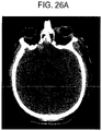
- Some injury mechanisms result in structural changes to the brain that can be visualized using conventional imaging such as MRI and CT scan, while other injuries appear radiographically normal.
- Acceleration/deceleration injury may result in structurally visible coup/contrecoup injuries and less visible diffuse axonal injury (DAI) ( Cecil, et al., Journal of Neurosurgery, 1998; 88: 795-801 ) Acceleration/deceleration is also thought to be one of the potential mechanisms for concussion which is the most common form of civilian radiographically normal brain injury ( Bayly, et al., Journal of Neurotrauma, 2005; 22: 845-856 ; Daneshvar, et al., Physical Medicine and Rehabilitation Clinics of North America, 2011; 22: 683-700 ).
- DAI diffuse axonal injury
- Concussion is brain injury, most often resulting from blunt impact, in the absence of structural abnormality by conventional radiographic imaging such as computed tomography (CT) scan ( McCrory, et al., The Physician and Sports Medicine, 2009; 37: 141-159 ). Concussion may include transient loss or disruption of neurologic function.
- CT computed tomography
- subconcussion may be used to describe the sequelae of brain injury in the absence of transient loss or disruption of neurologic function. Both concussion and subconcussion as well as blast injury may be termed "non-structural" brain injury.
- Blast injury resembles blunt impact brain injury in that both may be associated with radiographically apparent cerebral edema and intracranial hemorrhage, however with blast injury the edema onset may be more rapid and severe, and there is greater likelihood of clinical vasospasm ( Armonda, et al., Neurosurgery, 2006; 59: 1215-1225 ). Blast injury is very frequently radiographically normal, yet mild or moderate blast injury is strongly associated with post-traumatic stress disorder and other cognitive dysfunctions ( Cernak, et al., The Journal of Trauma, 2001; 50: 695-706 ).
- Blast injury affects the brain through several mechanisms: primary brain injury caused by blast-wave induced changes in atmospheric pressure directly impacting the brain; secondary injury resulting from objects put in motion by the blast that impact the head, and tertiary injury resulting from the victim striking the head upon falling or being propelled into a solid object ( Warden, The Journal of Head Trauma Rehabilitation, 2006; 21: 398-402 ).
- Brain injury may be associated with short term sequelae including headaches and memory problems, and longer term problems including dementia, Parkinsonism and motor-neuron disease ( Daneshvar, et al., Physical Medicine and Rehabilitation Clinics of North America, 2011; 22: 683-700 ). Both concussion and mild blast injury may be associated with post-traumatic stress disorder and cognitive impairment ( Taber, et al., The Journal of Neuropsychiatry and Clinical Neurosciences, 2006; 18: 141-145 ). Clinical tests for concussion show poor test reliability ( Broglio, et al., Journal of Athletic Training, 2007; 42: 509-514 ) and thus concussion remains a diagnosis that is difficult to treat because it is difficult to detect. Traumatic brain injury can impact eye movement through a multitude of mechanisms including direct compression of cranial nerves, trauma to cranial nerves, injury to cranial nerve nuclei and supranuclear impacts.
- ICP intracranial pressure
- the IIIrd nerve (oculomotor) may be directly compressed by the medial aspect of the temporal lobe with frontal or temporal mass lesions, or diffuse supratentorial mass effect.
- the VIth nerve (abducens) is anatomically vulnerable to infratentorial mass effect at the prepontine cistern and to hydrocephalus from stretch as it traverses the tentorial edge.
- Elevated intracranial pressure slows axoplasmic transport along cranial nerves ( Balarratnasingam, et al., Brain Research, 2011; 1417: 67-76 ).
- the optic nerve (II) is most frequently analyzed because it can be visualized directly with ophthalmoscopy, and indirectly with ultrasound. Edema of the optic nerve appears earlier than ocular fundus changes, and resolves after treatment of elevated ICP Gangemi, et al., Neurochirurgia, 1987; 30: 53-55 ).
- Fluctuating elevated neural pressure leads to impaired axonal transport along the optic nerve after as little as 30 minutes in a rabbit model ( Balarratnasingam, et al., Brain Research, 2011; 1417: 67-76 ).
- Axoplasmic flow stasis and intraneuronal ischemia may occur in the optic nerve exposed to chronically elevated ICP ( Lee, et al., Current Neurology and Neuroscience Reports, 2012 ).
- the trochlear nerve (IV), followed by oculomotor (III) and then abducens (VI), has the greatest length of exposure to the subarachnoid space with the narrowest diameter, and thus may be most vulnerable to a pressure induced palsy ( Hanson, et al., Neurology, 2004; 62: 33-36 ; Adler, et al., Journal of Neurosurgery, 2002; 96: 1103-1113 ) .
- the optic nerve (II) has approximately the same length of exposure as the abducens ( Murali, et al., in Head Injury (ed.
- Papilledema is not always a sensitive marker for hydrocephalus leading to elevated ICP, and in one study was present in as few as 14% of patients with a shunt malfunction ( Nazir, et al., J Aapos, 2009; 13: 62-66 ) consistent with the relatively short intracranial course of II compared to cranial nerves III and IV. Compartmentalization of subarachnoid spaces is hypothesized to explain why papilledema may be present in a patient without elevated ICP, and not occur in patients with elevated ICP ( Killer, et al., Clinical & Experimental Ophthalmology, 2009; 37: 444-447 ).
- the invention provides a method as defined in claim 1.
- the invention provides a kit as defined in claim 5.
- eye movement of both eyes of the subject are tracked and analyzed.
- both x and y coordinates of eye position for one or both eyes of a subject are collected for at least about 100, 500, 1,000, 5,000, 10,000, 50,000, 100,000, 200,000 or more eye positions.
- the eye position is effectively the pupil position.
- the eye movement is tracked for about 30, 60, 90, 100, 120, 150, 180, 200, 220, 240, 270, 300, 360 or more seconds.
- the comparing eye movement of at least one eye of the subject to a normal or mean eye movement may feature comparing eye movement of at least one eye of the subject to the eye movement of the other eye of the subject or may feature comparing eye movement of at least one eye of the subject to the eye movement of an eye of one or more other subjects or controls.
- the comparing eye movement of at least one eye of the subject to a normal or mean eye movement may feature comparing the eye movement of both eyes of the subject to the eye movement of one or both eyes of one or more other subjects or controls.
- the method may feature collecting raw x and y cartesian coordinates of pupil position, normalizing the raw x and y Cartesian coordinates, and sorting the data by eye.
- the method may also feature calculating individual metrics, such as, for instance, segment mean, segment median, and segment variance.
- the method may also feature calculating specific metrics such as, for example, or segment standard deviation and segment skew such as, for instance, or segment normalized skew, such as, for instance,
- the method may also feature calculating box height, box width, box area, or box aspect ratio. box height box width box aspect ratio box area
- the method may also feature calculating conjugacy of eye movement or variance from perfect conjugacy of eye movement, such as, for example, or variance x ratio top/bottom (conjugacy), variance y ratio top/bottom (conjugacy), variance x ratio left/right (conjugacy), or variance y ratio left/right (conjugacy).
- conjugacy of eye movement or variance from perfect conjugacy of eye movement such as, for example, or variance x ratio top/bottom (conjugacy), variance y ratio top/bottom (conjugacy), variance x ratio left/right (conjugacy), or variance y ratio left/right (conjugacy).
- one or more of the L height, L width, L area, L varXrit, L varXlef, L varTotal, R height, R width, R area, R varYtop, R varXrit, R varXlef, R varTotal, Conj varX, Conj varXrit, Conj varXbot, Conj varXlef and Conj varYlef may be especially useful for demonstrating or detecting or assessing structural or non-structural traumatic brain injury such as, for instance, a concussion, subconcussion or blast injury. In some instances, two, three, four, five, six, seven, eight, nine, ten or more metrics may be observed or determined.
- a standard deviation or p value of 0.25, 0.3, 0.4, 0.5, 0.75. 0.8, 0.9, 1.0, 1.5, 2.0, 2.5 or more may reflect that a subject has structural or non-structural traumatic brain injury such as, for instance, a concussion, subconcussion or blast injury.
- eye movement of both eyes of the subject are tracked and analyzed.
- both x and y coordinates of eye position for one or both eyes of a subject are collected for at least about 100, 500, 1,000, 5,000, 10,000, 50,000, 100,000, 200,000 or more eye positions.
- the eye position is effectively the pupil position.
- the eye movement is tracked for about 30, 60, 90, 100, 120, 150, 180, 200, 220, 240, 270, 300, 360 or more seconds.
- the comparing eye movement of at least one eye of the subject to a normal or mean eye movement may feature comparing eye movement of at least one eye of the subject to the eye movement of the other eye of the subject or may feature comparing eye movement of at least one eye of the subject to the eye movement of an eye of one or more other subjects or controls.
- the comparing eye movement of at least one eye of the subject to a normal or mean eye movement may feature comparing the eye movement of both eyes of the subject to the eye movement of one or both eyes of one or more other subjects or controls.
- the method may feature collecting raw x and y cartesian coordinates of pupil position, normalizing the raw x and y Cartesian coordinates, and sorting the data by eye.
- the method may also feature calculating individual metrics, such as, for instance, segment mean, segment median, and segment variance.
- the method may also feature calculating specific metrics such as, for example, or segment standard deviation and segment skew such as, for instance, or segment normalized skew, such as, for instance,
- the method may also feature calculating conjugacy of eye movement or variance from perfect conjugacy of eye movement, such as, for example, or variance x ratio top/bottom (conjugacy), variance y ratio top/bottom (conjugacy), variance x ratio left/right (conjugacy), or variance y ratio left/right (conjugacy).
- conjugacy of eye movement or variance from perfect conjugacy of eye movement such as, for example, or variance x ratio top/bottom (conjugacy), variance y ratio top/bottom (conjugacy), variance x ratio left/right (conjugacy), or variance y ratio left/right (conjugacy).
- one or more of the L height, L width, L area, L varXrit, L varXlef, L varTotal, R height, R width, R area, R varYtop, R varXrit, R varXlef, R varTotal, Conj varX, Conj varXrit, Conj varXbot, Conj varXlef and Conj varYlef may be especially useful for demonstrating or detecting or assessing structural or non-structural traumatic brain injury such as, for instance, a concussion, subconcussion or blast injury. In some instances, two, three, four, five, six, seven, eight, nine, ten or more metrics may be observed or determined.
- a standard deviation or p value of 0.25, 0.3, 0.4, 0.5, 0.75. 0.8, 0.9, 1.0, 1.5, 2.0, 2.5 or more may reflect that a subject has structural or non-structural traumatic brain injury such as, for instance, a concussion, subconcussion or blast injury.
- the eye movement is tracked for about 30, 60, 90, 100, 120, 150, 180, 200, 220, 240, 270, 300, 360 or more seconds.
- eye movement of both eyes of the subject are tracked and analyzed.
- both x and y coordinates of eye position for one or both eyes of a subject are collected for at least about 100, 500, 1,000, 5,000, 10,000, 50,000, 100,000, 200,000 or more eye positions.
- the method may additionally feature sorting the data by eye.
- the one or more individual metric may be any one of or segment standard deviation and segment skew such as, for instance, or segment normalized skew, such as, for instance,
- the method may also feature calculating box height, box width, box area, or box aspect ratio. box height box width box aspect ratio box area
- the method may also feature calculating conjugacy of eye movement or variance from perfect conjugacy of eye movement, such as, for example, or variance x ratio top/bottom (conjugacy), variance y ratio top/bottom (conjugacy), variance x ratio left/right (conjugacy), or variance y ratio left/right (conjugacy).
- conjugacy of eye movement or variance from perfect conjugacy of eye movement such as, for example, or variance x ratio top/bottom (conjugacy), variance y ratio top/bottom (conjugacy), variance x ratio left/right (conjugacy), or variance y ratio left/right (conjugacy).
- one or more of the L height, L width, L area, L varXrit, L varXlef, L varTotal, R height, R width, R area, R varYtop, R varXrit, R varXlef, R varTotal, Conj varX, Conj varXrit, Conj varXbot, Conj varXlef and Conj varYlef may be especially useful for demonstrating or detecting or assessing structural or non-structural traumatic brain injury such as, for instance, a concussion, subconcussion or blast injury. In some instances, two, three, four, five, six, seven, eight, nine, ten or more metrics may be observed or determined.
- a standard deviation or p value of 0.25, 0.3, 0.4, 0.5, 0.75. 0.8, 0.9, 1.0, 1.5, 2.0, 2.5 or more may reflect that a subject has structural or non-structural traumatic brain injury such as, for instance, a concussion, subconcussion or blast injury.
- kits useful for detecting, screening for or quantitating structural and non-structural traumatic brain injury in a subject containing a device for tracking eye movement, one or more means for analyzing eye movement tracking data such as, for instance, an algorithm or computer program, and instructions. Processing eye movement observations, making measurements of eye movement observations, determining distributions of values measured and performing statistical tests may all be accomplished using suitable computer software that may be included in such a kit.
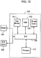
- the computer system or computing device 1000 can be used to implement a device that includes the processor 106 and the display 108, the eye movement/gaze tracker component 104, etc.
- the computing system 1000 includes a bus 1005 or other communication component for communicating information and a processor 1010 or processing circuit coupled to the bus 1005 for processing information.
- the computing system 1000 can also include one or more processors 1010 or processing circuits coupled to the bus for processing information.
- the computing system 1000 also includes main memory 1015, such as a random access memory (RAM) or other dynamic storage device, coupled to the bus 1005 for storing information, and instructions to be executed by the processor 1010.
- Main memory 1015 can also be used for storing position information, temporary variables, or other intermediate information during execution of instructions by the processor 1010.
- the computing system 1000 may further include a read only memory (ROM) 1010 or other static storage device coupled to the bus 1005 for storing static information and instructions for the processor 1010.
- ROM read only memory
- a storage device 1025 such as a solid state device, magnetic disk or optical disk, is coupled to the bus 1005 for persistently storing information and instructions.
- the computing system 1000 may be coupled via the bus 1005 to a display 1035, such as a liquid crystal display, or active matrix display, for displaying information to a user.
- a display 1035 such as a liquid crystal display, or active matrix display
- An input device 1030 such as a keyboard including alphanumeric and other keys, may be coupled to the bus 1005 for communicating information and command selections to the processor 1010.
- the input device 1030 has a touch screen display 1035.
- the input device 1030 can include a cursor control, such as a mouse, a trackball, or cursor direction keys, for communicating direction information and command selections to the processor 1010 and for controlling cursor movement on the display 1035.
- the processes described herein can be implemented by the computing system 1000 in response to the processor 1010 executing an arrangement of instructions contained in main memory 1015. Such instructions can be read into main memory 1015 from another computer-readable medium, such as the storage device 1025. Execution of the arrangement of instructions contained in main memory 1015 causes the computing system 1000 to perform the illustrative processes described herein. One or more processors in a multi-processing arrangement may also be employed to execute the instructions contained in main memory 1015. In alternative implementations, hard-wired circuitry may be used in place of or in combination with software instructions to effect illustrative implementations. Thus, implementations are not limited to any specific combination of hardware circuitry and software.
- tracking eye movement may be performed using any suitable device such as, for example, an Eyelink ® 1000 binocular eye tracker (500 Hz sampling, SR Research).
- the eye tracking movement samples may be obtained at any suitable frequency, such as for instance, 10 Hz to 10,000 Hz or more.
- the subject may be positioned an appropriate distance from the device, such as, for example, 10, 20, 30, 40, 50, 55, 60, 70, 80, 90 cm or more, or even a meter or more from the device screen.
- the subject's head may be stabilized, such as, for instance by using a chinrest or similar stabilizing mechanism.
- the subject may be seated or reclining.
- the presentation monitor of the device is adjusted so as to substantially match the subject's gaze direction.
- the tracking eye movement may be performed for a total of, for example, 30, 60, 90, 120, 150, 180, 200, 220, 240, 270, 300, 330, 360, 400, 450, 500 seconds or more, or for 5, 10, 15, 20, 25, 30, 45, 60, or 90 minutes or more.
- 1,000, 5, 000, 10,000, 20,000, 25, 000, 50,000, 75,000, 100,000, 150,000, 200,000, 250,000, 300,000 or more samples of eye position may be obtained.
- the tracking eye movement may be performed using a video oculography device, such as, for instance, goggles, or using a web-cam based tracking system.
- analyzing eye movement may be performed by any suitable means.
- a stimulus and an analysis stream are provided that allows interpreting raw eye position data.
- an algorithm may be provided for looking at pupil position directly thereby yielding information about ocular motility.
- a device is adapted into a novel mobile system that may analyze eye movement close in time or substantially concurrent to the eye movement itself.
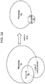
- the visual stimulus may be, for instance, a video such as a music video that may move, for instance clockwise, along the outer edge, of a computer monitor.
- a video may be provided starting at the upper or lower, left or right hand corners, of a screen.
- the visual stimulus such as a video, e.g. a music video, may be provided in a substantially square aperture with an area of approximately 10, 12, 14, 16, 18, 20, 25, or degrees, for example, approximately 1/10, 1/8, 1/6, 1/5, 1 ⁇ 4, 1/3, 1 ⁇ 2 of the size of the screen or so.
- the visual stimulus such as, for example a music video
- a visual stimulus for instance, a music video may cover each edge of a monitor in about 2, 5, 10, 15, 20, 30, 45 or 60 seconds or so. Therefore, in some instances, a full cycle may take, for instance, 10, 20, 30, 40, 50, 60, 75, 100, 120, 150, 180 seconds or so.
- Multiple cycles of such a visual stimulus for instance a music video may be played, for instance, one, two, three, four, five, six, seven, eight, nine, ten, twelve, fifteen, twenty or more full cycles.
- the visual stimulus may be provided, the eye movement may be tracked, in effect, in some instances the video may be played for a total of, for example, 30, 60, 90, 120, 150, 180, 200, 220, 240, 270, 300, 330, 360, 400, 450, 500 seconds or more.
- a countdown video may be played in the starting position for, for instance, 5, 10, 15, 20, 25, or 30 seconds or more before beginning the visual stimulus, e.g. video, to provide subjects sufficient time to orient to the visual stimulus.
- the visual stimulus for instance a video, may be continued for an addition 2, 5, 10, 15, 20, 30, 45 or 60 seconds or so after the eye movement tracking is performed to reduce or substantially avoid boundary effects.
- the same result could be obtained by having the visual stimulus moving over any distance x relative to any amount of time t.
- the ideal stimulus would move however in the both the x and y Cartesian planes to optimize the assessment capability of the method.
- comparing eye movement of a first eye of the subject to eye movement of a second eye of the subject may be performed by analyzing data.
- Data from the tracking eye movement may provide an indication of whether an individual subject's gaze is conjugate (eyes are moving together) versus disconjugate.
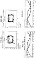
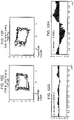
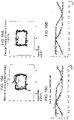
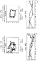
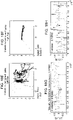
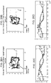
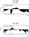
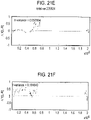
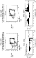
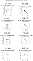
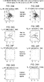
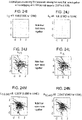
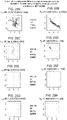
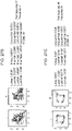
- Comparing eye movement of a first eye of the subject to eye movement of a second eye of the subject may feature generating scatterplots. Comparing eye movement of a first eye of the subject to eye movement of a second eye of the subject, features plotting the horizontal eye position along one axis and vertical eye position along an orthogonal axis.
- Such comparing eye movement of the subject to a control, or comparing eye movement of a first eye of the subject to eye movement of a second eye of the subject may feature generating, plotting pairs of (x,y) values, for instance, 50,000, 100,000 or more pairs of values (x,y). Such pairs of values (x,y) are plotted representing, for instance, the two components of the instantaneous angle of pupil reflection (horizontal, vertical) over a period of time, for instance, 100 or 200 seconds or more.
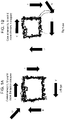
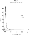
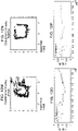
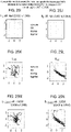
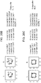
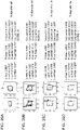
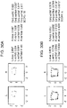
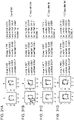
- figures substantially resembling boxes that reflect the trajectory traveled by the visual stimulation, such as when it moves across a screen.
- these figures substantially resembling boxes may look like, for instance, substantially equilateral rectangles or squares, reflecting the trajectory traveled by the visual stimulus across a screen.
- such figures may not substantially resemble a box, a rectangle or a square.
- the cranial nerve having reduced or impaired function or conduction may be identified.
- the figures generated that reflect the trajectory traveled by the visual stimulation may demonstrate abnormal distribution of or absence of normal plotting pairs in particular areas.
- Increased variability along the y-axis may for example reflect cranial nerve II dysfunction. Decreased variability along the y-axis, or decreased height to width ratio may reflect CN III dysfunction. Increased height to width ratio may reflect CN IV or VI dysfunction.
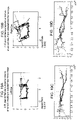
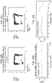
- the height of the box may be mathematically determined by assessing the position of the pupil as the video traverses the top and bottom of the presented visual stimulus. This "actual" height may be different from the perceived height mathematically, since the perceived height can represent aberrant pupillary motion due to the patient's ocular motility dysfunction. The integrity of the box walls may also be indicative of other types of dysfunction. Both cranial nerve palsies and mass effect may cause defects in box trajectory.
- Supratentorial mass lesions and CN III defects may impact the top and/or bottom of the box.
- Infratentorial mass lesions or CN VI palsies may impact the sides of the box.
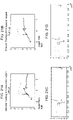
- the upper left quadrant of the figure may reflect activity, function or conduction of cranial nerves III and VI
- the lower left quadrant of the figure may reflect activity, function or conduction of cranial nerves III and IV
- the upper right quadrant and the lower right quadrants may reflect activity, function or conduction of cranial nerve III.
- the upper and lower left quadrants of the figure may reflect activity, function or conduction of cranial nerve III
- the lower right quadrant of the figure may reflect activity, function or conduction of cranial nerve III
- the upper right quadrant and the lower right quadrant may reflect activity, function or conduction of cranial nerves IV and VI.
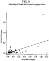
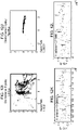
- Comparing eye movement of a first eye of the subject to eye movement of a second eye of the subject may feature determining the distribution of certain measurements in the control population and comparing the subject with these control distributions.
- visual stimulus trajectory may be divided into four time components, for instance, two, three, four, five, six or more repetitions of the first few, for instance, 2, 5, 10, 15, 20 or so seconds of each rotation cycle.
- comparing eye movement of the subject to a control may feature evaluating such variables as the relative variance in each arm, and the relative integrity of each arm.
- Comparing eye movement of the subject to a control, or comparing eye movement of a first eye of the subject to eye movement of a second eye of the subject may also feature measuring the integrity of each subject's values.
- the sides or arms of the figures e.g. the top of the box and the bottom of the box
- the resulting score may indicate how different the subject's values are compared with the control values, such as, for instance, in units of standard deviations.
- identifying the subject as having eye movement significantly different from the control, or identifying the subject as having eye movement of a first eye that is significantly different from eye movement of a second eye may be performed using a z-score. Because 95% of all values in a normal distribution lie within two standard deviations of the mean, a z-score of 2 may be used as a significance threshold. Subjects with z-scores above, for instance, 2 in either or both, or 1, 2, 3, or 4 sides or arms of the figures may be judged to have significant disturbances of ocular motility.
- identifying the subject as having eye movement significantly different from the control, or identifying the subject as having eye movement of a first eye that is significantly different from eye movement of a second eye may be performed by assessing whether it has or there is a difference that exceeds a predetermined threshold.
- Identifying the subject as having eye movement significantly different from the control, or identifying the subject as having eye movement of a first eye that is significantly different from eye movement of a second eye may feature determining relative variance.
- multiple such as 1,000, 2,000, 3,000, 5,000, 10,000, 20,000 or more point distributions may be generated by, for instance, taking multiple samples from a multiple number of values randomly chosen with replacement from the multiple control values.
- the relative variance in either or both, or 1, 2, 3, or 4 sides or arms of the figures may be compared respectively with the corresponding control distribution, and the percent of the control distribution with variance below that of the test value may be determined.
- a p-value of .05 a widely accepted measure of statistical significance corresponds to 95% of control values falling below the test value.
- subjects with variance higher than 95% of the values in the control distributions may be determined to have significant disturbances of ocular motility.
- the video may also move in other trajectories not resembling a rectangle, such as a triangle, circle or linear or nonlinear trajectories.
- the trajectories can be resolved into vectors along Cartesian coordinates (horizontal vertical or x,y) the same principles will apply.
- any trajectory e.g. any shape, or line, or curve, etc.
- studied over time may provide information about Central Nervous System function or dysfunction.
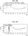
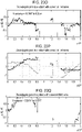
- Comparing the movement of one eye of a subject to the other eye of a subject may be performed by comparing the x,y Cartesian coordinates at any time point t, for example, by subtracting the x coordinate of the left eye from the x coordinate of the right eye or vice versa, or by subtracting the y coordinate of the left eye from the y coordinate of the right eye or vice versa.
- the sums of the differences between all of the x coordinates over the time tested informs regarding horizontal movement of the pupil.
- the sums of the differences in y coordinates over time informs regarding vertical movement of the pupil.
- the total sum of the differences between both x and y coordinates over the time tested may be totaled to obtain a measure of total disconjugacy of gaze, which is a surrogate marker for central nervous system integrity.
- a measure of total disconjugacy of gaze which is a surrogate marker for central nervous system integrity.
- CNS central nervous system
- Eye movement may also be tracked without using a moving stimulus (not according to the invention). It is possible to assess conjugacy without having the stimulus move at all, but by assessing the x, y coordinates over times during naturalistic viewing. For example, eye movement may be tracked during television watching or live viewing of an environment without a specific viewing apparatus such as a monitor or screen.
- comparing the x or y Cartesian coordinates at any time point for the eye movement of a first eye of the subject to the respective x or y Cartesian coordinates at any time point for the eye movement of a second eye of the subject may be performed by analyzing data.
- Data from the tracking eye movement may provide an indication of whether an individual subject's gaze is conjugate (eyes are moving together) versus disconjugate.
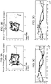
- Comparing the x or y Cartesian coordinates at any time point for the eye movement of a first eye of the subject to the respective x or y Cartesian coordinates at any time point for the eye movement of a second eye of the subject may feature generating scatterplots.
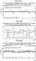
- Comparing the x or y Cartesian coordinates at any time point for the eye movement of a first eye of the subject to the respective x or y Cartesian coordinates at any time point for the eye movement of a second eye of the subject may feature plotting the difference between the horizontal eye positions along one axis and time along an orthogonal axis, as well as the difference between the vertical eye positions along one axis and time along an orthogonal axis.
- Such comparing the x or y Cartesian coordinates at any time point for the eye movement of a first eye of the subject to the respective x or y Cartesian coordinates at any time point for the eye movement of a second eye of the subject may feature generating, plotting pairs of (x, y) values, for instance, 25,000, 50,000, 75,000, 100,000, 150,000 or more pairs of values (x, y).
- pairs of values (x, y) may be plotted representing, for instance, the two components of the instantaneous angle of pupil reflection (horizontal, vertical) over a period of time, for instance, 100 or 200 or 250 or 300 seconds or more.
- comparing the x or y Cartesian coordinates at any time point for the eye movement of a first eye of the subject to the respective x or y Cartesian coordinates at the time point for the eye movement of a second eye of the subject may allow generating plots assessing conjugacy of eye movements over time.
- Comparing the x or y Cartesian coordinates at any time point for the eye movement of a first eye of the subject to the respective x or y Cartesian coordinates at the time point for the eye movement of a second eye of the subject may feature determining the distribution of certain measurements in the control population and comparing the subject with these control distributions.
- visual stimulus trajectory may be divided into four time components, for instance, two, three, four, five, six or more repetitions of the first few, for instance, 2, 5, 10, 15, 20 or so seconds of each rotation cycle.
- comparing the x or y Cartesian coordinates at any time point for the eye movement of a first eye of the subject to the respective x or y Cartesian coordinates at any time point for the eye movement of a second eye of the subject may feature evaluating such variables as the relative variance in each arm, and the relative integrity of each arm.
- Comparing the x or y Cartesian coordinates at any time point for the eye movement of a first eye of the subject to the respective x or y Cartesian coordinates at the time point for the eye movement of a second eye of the subject may be performed by comparing the x, y Cartesian coordinates at any time point t, for example, by subtracting the x coordinate of the left eye from the x coordinate of the right eye or vice versa, or by subtracting the y coordinate of the left eye from the y coordinate of the right eye or vice versa.
- the sums of the differences between all of the x coordinates over the time tested informs regarding horizontal movement of the pupil.
- the sums of the differences in y coordinates over time informs regarding vertical movement of the pupil.
- the total sum of the differences between both x and y coordinates over the time tested may be totaled to obtain a measure of total disconjugacy of gaze, which may be a surrogate marker for central nervous system integrity.
- a measure of total disconjugacy of gaze which may be a surrogate marker for central nervous system integrity.
- CNS central nervous system
- Providing a sum of the differences between all of the x coordinates of the first eye compared to the second eye over the time tested or providing a sum of the differences in y coordinates of the first eye compared to the second eye over the time tested or both may be performed subsequent to comparing the x, y Cartesian coordinates at the time point t. For example, by subtracting the x coordinate of the left eye from the x coordinate of the right eye or vice versa. Also, by subtracting the y coordinate of the left eye from the y coordinate of the right eye or vice versa. The sums of the differences between all of the x coordinates over the time tested informs regarding horizontal movement of the pupil. The sums of the differences in y coordinates over time informs regarding vertical movement of the pupil.
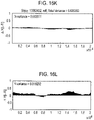
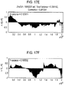
- the total sum of the differences between both x and y coordinates over the time tested can be summed to obtain a measure of total disconjugacy of gaze, or as an average of five eyebox trajectory cycles formulaically represented as follows: where X ijk refers to the x-coordinate of the pupil, and k refers to the left or right eye of a subject. In cases where a subject's data was missing at any given time point in the five cycles, the denominator of the equation was the number of cycles where the data was present. The difference in the x and y position, for the left and right eye, may then be computed. This vector of difference may then be plotted graphically for purposes of assessment and interpretation.
- a variance of the data may be computed with respect to an expected mean of zero. This is significant because the code assumes that a healthy subject has zero vertical or horizontal pupil position difference between each eye.
- the variance for either horizontal (x) or vertical (substitute y for x) movement may be computed as follows:
- Providing a total sum of the differences between both x and y coordinates of the first eye compared to the second eye over the time tested may be performed by calculating the total variance in both the horizontal and vertical planes between the first and the second eyes.
- the total variance may be computed as follows:
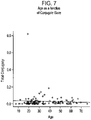
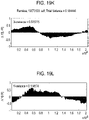
- the Var x or the Var y or both, calculated as described herein may be 0.05, 0.07, 0.1, 0.15, 0.20, 0.25, 0.30, 0.40, 0.50, 0.60, 0.75, 0.90, 1.0, 1.10, 1.25, 1.50, 1.75, or 2.0 or more.
- the Var Tot calculated as described herein may be 0.1, 0.15, 0.20, 0.25, 0.30, 0.40, 0.50, 0.60, 0.75, 0.90, 1.0, 1.10, 1.25, 1.50, 1.75, 2.0, 2.50, 3.0 or 4.0 or more, in subjects having a neurological disease or condition characterized by or featuring disconjugacy of gaze or strabismus.
- a non-transitory computer-readable medium as defined in claim 9 is provided.
- Subject or “patient” refers to a mammal, preferably a human, in need of or undergoing treatment or screening for a condition, disorder or disease such as, for instance, increased intracranial pressure.
- assessing central nervous system integrity is meant identifying one or more symptoms that may indicate a pathology of or affecting the central nervous system, or identifying, assessing, quantifying or diagnosing a pathology of the central nervous system.
- the pathology may be, for instance, one or more of increased intracranial pressure, hydrocephalus, concussion, dementia, schizophrenia, amyotrophic lateral sclerosis, muscular sclerosis, autism and Fragile X disease.
- localizing a central nervous system lesion is meant in some instances determining information that may predict a likely position of a lesion, for instance, determining the side of the body, for instance, left or right, where a lesion may likely be located within the central nervous system. In other instances, “localizing a central nervous system lesion” may mean determining a particular fossa or compartment, such as, for instance, a fascia compartment or brain ventricle in which a lesion is likely located within the central nervous system.
- having eye movement of a first eye that is significantly different from eye movement of a second eye is meant displaying eye movement in a first eye over 5, 10, 25, 50, 100, 1,000, 5,000, 10,000 or more observations, tracked with at least x, y coordinate positions, that is at least 5%, 10%, 15%, 20%, 25%, 30%, 40%, 50%, 75%, or 100% or more variant compared to the corresponding eye movement observations tracked from the second eye.
- the 5%, 10%, 15%, 20%, 25%, 30%, 40%, 50%, 75%, or 100% or more variant may be calculated or observed either numerically or graphically.
- "having eye movement of a first eye that is significantly different from eye movement of a second eye” is meant displaying eye movement in a first eye over 5, 10, 25, 50, 100, 1,000, 5,000, 10,000 or more observations, tracked with at least x, y coordinate positions, that, when graphically displayed in a scatterplot as described herein, is at least 5°, 10°, 15°, 20°, 25°, 30°, 40°, 50°, 60°, 75° or 90° or more variant compared to the corresponding eye movement observations tracked and graphically displayed on a scatterplot as described herein from the second eye.
- vergence or “vergence disorders” refers generally to convergence, when the eyes rotate inward as an object moves closer, and to divergence, when the eyes rotate outward as an object moves farther away. Both convergence and divergence are tested to some extent as an object moves around, effectively assessing sustained vergence. Most vergence disorders are due to the pathologies and causes described herein, for instance, trauma. Some vergence disorders may be congenital. The methods and algorithms described herein facilitate screening for such vergence and vergence disorders.
- the methods described herein are distinct from conventional methods. As applied to determining intracranial pressure, a conventional ICP monitor determines the brain's pressure number in one spot, an O 2 monitor determines an oxygenation number in one spot, imaging reveals what the brain looks like, but the methods described herein provide methods for testing for physiologic function of the cranial nerves that may reflect factors that may delay axoplasmic transport such as elevated intracranial pressure.
- the methods described herein may be used to detect elevated intracranial pressure and assess or determine the severity of the same. Similarly, the methods described herein may be used to localize the intracranial cause of such intracranial pressure and to monitor progression of lesions or diffuse processes within the cranium. Likewise, the methods described herein may be used to detect concussion and assess or determine the severity of the same.
- the methods described herein provide high sensitivity. No patient yet evaluated with an abnormal physical exam or films consistent with elevated ICP has had normal eye movement tracking.
- the methods described herein may be used to reduce the need for CT scans among potential shunt malfunction patients, patients with lesions causing elevated intracranial pressure, and may be used to screen patient populations such as emergency room ER populations, sports participants, soldiers or other combatants, nursing home residents or other populations at risk for falling for elevated intracranial pressure or concussion.
- High resolution automated eye movement tracking occurring over, for instance, about 220 seconds, is a powerful tool for detecting subclinically apparent ocular motility dysfunction, and thus aid in the rapid diagnosis of elevated intracranial pressure or concussion.
- the IVth nerve would be most vulnerable (median length 33 mm ( Hanson et al., Neurology 2004; 62(1):33-36 )), the IIIrd nerve would be second most vulnerable (26 mm ( Adler et al., Journal of Neurosurgery 2002; 96(6): 1103-1112 )) and IInd and VIth would be approximately equally least vulnerable (5 to 16 mm for II ( Murali, R. Injuries of the Cranial Nerves. In: Golfinos PCaJ, ed. Head Injury. 4th ed. New York: McGraw Hill; 2000 ), and 11 mm median length for VI ( Hanson et al., Neurology 2004; 62(1):33-36 )).
- the abducens nerve (VI) exits the brainstem from its tethering at the medullopontine junction and courses intracranially before entering Dorello's canal, where it is again tethered by fibrous and osseous structures. Elevation of supratentorial ICP forces the parahippocampal gyri down past the free edge of the tentorium while the brainstem with the tethered VIth nerve moves caudally toward the foramen magnum, stretching the nerve where it enters Dorello's canal ( Hanson et al., Neurology 2004; 62(1):33-36 ).
- the data presented herein does not feature a calibration step in eye movement tracking. Thus patients need not reliably follow instructions, and the data does not filter out the possible effects of cranial neuropathy. Unlike other studies ( Contreras et al., Brain research 2011; 1398:55-63 ; Maruta et al., The Journal of Head Trauma Rehabilitation 2010; 25(4):293-305 ; Contreras et al., Journal of Biological Physics 2008; 34(3-4):381-392 and Trojano et al., J Neurol 2012 ;(published online; ahead of print)) the data presented herein does not use saccade count or spatial accuracy as the measure. In addition to results based on the moving aperture's periodic envelope presented in this paper, the methodology also affords a very fine-scale data showing eye movements in response to the successive frames of the movie itself.
- the methods described herein build on pre-existing methods that rely on intact ocular motility to address clinical questions.
- the methods described herein differ in several ways.
- the present methods feature diagnosing specific clinical conditions related to vision and ocular motility reflecting the function of cranial nerves II, III, IV, VI and associated nuclei rather than measuring cognitive impairment due to primarily cortical mild to moderate traumatic brain injury.
- the present methods use more fine-scale information, using, for instance, about 100,000 measurements to pull out subtle differences that can be lost through the somewhat arbitrary thresholding of velocity measures into saccades.
- the present methods do not use measurements of spatial accuracy, which requires transforming the raw data by a series of scaling and rotating processes whose effectiveness depends on the ability of their subjects to follow precise commands reliably. In such methods previously used, it is necessary to exclude the vast majority of neurologically compromised patients. Further, such methods previously used lose any information related to the function of cranial nerves II, III, IV and VI, because the spatial distortions expected to result from damage to these nerves is reversed in the process of spatial calibration.
- Trojano et al. J Neurol 2012 ;(published online; ahead of print) recently described uncalibrated eye movement measurements in a population of minimally conscious and persistently vegetative patients.
- the methods described herein differ in several ways.
- First, Trojano et al. report data from 11 rather than 25 healthy control subjects.
- Second, Trojano et al. evaluate chronic disorders of consciousness rather than acute changes in intracranial pressure.
- Third, Trojano et al. sample eye movements at 60 Hz rather than 500 Hz, effectively reducing the power of the data 100-fold.
- Trojano et al. report differences in on-target and off-target fixations between the groups, despite not having spatially calibrated the data, making these values noisy.
- Finally, Trojano et al. use static stimuli moving in a quasi-periodic way.
- the methods described herein use moving images shown within an aperture that moves periodically and allows assessing both coarse and fine eye movement characteristics in both controls and patients.
- the data presented herein are consistent with compartmentalization of subarachnoid spaces, as several of the patients demonstrate elevated ICP on one side of the brain, but not the other.
- the methods for ICP assessment described herein represent a significant advantage over conventional radiographic studies because while the latter depict how the brain appears, our technique captures how well it functions.
- CT scanning may require brief sedation in a pediatric population and risks radiation exposure, while MR may require prolonged sedation.
- Brain imaging may not be diagnostic of elevated ICP in patients with chronically enlarged ventricles without classic findings such as transependymal flow on T2 weighted MR imaging ( Mizrachi et al., J Neuroophthalmol. 2006; 26(4):260-263 ).
- Patients with non-compliant and slit ventricles may also have elevated ICP in the absence of radiographic abnormality ( Engel et al., Neurosurgery 1979; 5(5):549-552 ).
- Shunt tapping risks infection and malfunction, particularly in patients with slit ventricles.
- Invasive monitoring risks intracranial hemorrhage.
- additional low-risk, rapid techniques for assessment of hydrocephalus or elevated ICP may be useful to those assessing populations at risk for these pathologies.
- the methods described herein provide a useful adjunct for diagnosis of elevated ICP and the prospective monitoring of such patients at risk for its development.
- No patients with elevated ICP by history, physical examination and radiology have demonstrated normal ocular motility, demonstrating that the methods described herein are sensitive.
- the data presented herein demonstrate that patients with grossly intact extraocular movements on physical exam, and relatively minimal changes in pathology, may have profound disruption on high resolution tracking.
- the methods described herein provide a useful adjunct for diagnosis of concussion and prospective monitoring of such patients at risk for developing the same.
- the data presented herein demonstrate that patients with grossly intact extraocular movements on physical exam, and relatively minimal changes in pathology, may have profound disruption on high resolution tracking.
- tracking results may need to be compared to each patient's own baseline data.
- subjects with a history of traumatic brain injury may have tracking results that may need to be compared to each patient's own baseline data.
- the data presented herein demonstrates in part that it is possible to diagnose elevated intracranial pressure and concussion by analysis of eye movements during watching of a video.
- the methods described herein are significantly different from other technologies since imaging studies enable one to see the brain and invasive techniques enable determination of an arbitrary pressure or oxygenation number.
- the methods described herein actually assess physiologic functioning.
- the methods described herein have many clinical applications including, for instance, i) assessing function of cranial nerves II, III, IV and VI, and perhaps even VII, VIII, and/or X; ii) detecting and quantitatively monitoring any process impeding or improving the function of the above (e.g. demonstrating elevated ICP or increased brain mass effect, that may be applied to such things as aneurysms, multiple sclerosis, sarcoidosis, tumors, aging, alcohol abuse, intoxicants/narcotics, etc.), iii) localizing pathology and identifying the nature of that pathology within the brain (e.g.
- the methods described herein may provide means for in-person screening such as to, for example, assess vision, assess ocular motility, and assess cognitive dysfunction all relatively simultaneously (e.g.
- the methods described herein may be used to assess variance, which appears to increase with cognitive decay. This could be used, for instance, to target advertising by stratification of intelligence. Further, the methods described herein may be used to assess disconjugate gaze that apparently increases with cognitive decay. Still further, the methods described herein may be used for intelligence or neurologic function testing.
- the present invention features a novel eye movement tracking method that is useful for quantitating gaze conjugacy, and thus disconjugacy, during naturalistic viewing.
- the method assesses vergence, or the ability of the eyes to focus on a single point. When the point moves closer the eyes converge and if it moves further away they diverge. Watching a moving stimulus on a monitor thus requires sustained vergence. It may be performed while a subject watches television or a video moving inside an aperture with a set trajectory for about 220 seconds at a fixed distance from a viewing monitor. It may also be performed as the subject views natural stimuli over time.
- the position of each pupil may be recorded over time elapsed as the video travels on its time course, enabling detection of impaired ability to move the pupils relative to time and therefore relative to each other.
- This method has high test-retest reliability in control subjects without significant neurologic or ophthalmic impairments using both a stationary and portable eye tracking device.
- Eye movement tracking for neuropsychiatric and brain injury research has been performed for nearly 30 years and can evaluate smooth pursuit, saccades, fixation, pupil size and other aspects of gaze.
- Spatial calibration of the eye tracker is generally performed for each individual being tracked. With calibration, the eye-tracker measures the relative position of pupil and corneal reflection for a period of about 400-800 ms while the subject looks at a target or targets of known position to generate meaningful spatial coordinates during subsequent pupil movement.
- the process of spatial calibration implies relatively preserved neurologic function because it requires that the subject is able to follow commands and look at specific points.
- the process of spatial calibration may mask deficits in ocular motility. If there is a persistent and replicable weakness in movement of an eye, the camera may interpret the eye's ability to move in the direction of that weakness as the full potential range of motion in that direction due to the calibration process. In other words if the subject is directed to look at a position but consistently only moves halfway there, the calibration process may account for that when tracking subsequent eye movements and interpret movements to the halfway point as occurring at the full range of normal motion. If during calibration one eye only makes it halfway to the target, but the other eye is fully there, the camera may interpret both eyes as being together when one performs half the eye movement as the other. Thus binocular spatial calibration may preclude detection of disconjugate gaze unless each eye is calibrated separately using a dichoptic apparatus ( Schotter, et al., PLoS One, 2012; 7: e35608 ).
- the present invention provides a novel technique for non-spatially calibrated tracking performed while subjects watch a music video moving inside an aperture on a computer monitor.
- the aperture moves around the monitor periphery at a known rate so that the position of the pupil can be predicted at any given time based on the time elapsed since the start of the video.
- the method detects impaired ability to move one pupil relative to the other.
- Uncalibrated tracking not only does not compensate for impaired motility, but also can be used in patients who do not follow commands such as aphasics, foreign-language speakers, persistently vegetative individuals and small children. It can also be used on animals.
- the method and associated algorithm elicits pupil movement in a maximum range of about 15° in any direction from midposition, or approximately 30° total from top to bottom or side to side.
- the method and associated algorithm may not require or assess the full range of ocular motility, nor the entire visual field. Use of a larger monitor, or one positioned closer to the subject would enable assessment of these.
- the observed and measured conjugacy was significantly higher in the horizontal plane than vertical. This may reflect any of multiple factors: (1) the shape of the monitor was not a perfect square but rather a 17" diameter rectangle. Each side was traversed in 10 seconds so the eyes had a greater distance to travel horizontally than vertically. Because the eyes were moving faster horizontally they may possibly be more conjugate. (2) Humans have stronger event related desynchronization on electroencephalogram with horizontal versus vertical eye movements ( Kaiser, et al., Clin Neurophysiol., 2009; 120: 1988-1993 ). Humans may have evolved to have higher conjugacy in the horizontal plane than in the vertical because more prey and predators are likely to be at near the same altitude rather than above or below.
- the technique described herein differs from uncalibrated tracking using static stimuli for on-target and off-target fixations in a population of minimally conscious and persistently vegetative patients that have open eyes ( Trojano, et al., J Neurol., 2012 (published online; ahead of print)).
- the moving images shown within an aperture that moves periodically allow assessing both coarse and fine eye movement characteristics in both controls and neurologically impaired subjects.
- the present methods do not use saccade count or spatial accuracy which requires transformation of raw data by a series of scaling and rotating processes whose effectiveness depends on the ability of their subjects to follow precise commands reliably.
- the present methods also differ from gaze estimation, which requires either a fixed head position or multiple light sources and cameras to localize the pupil ( Guestrin, et al., IEEE Trans Biomed Eng., 2006; 53: 1124-1133 ).
- Video oculography is a relatively newer technique that uses infrared cameras mounted in goggles to track the center of the pupil's position as the eye moves. It has been demonstrated to be useful in screening for neurovestibular and labyrinthine dysfunction and most recently in distinguishing these from vertebrobasilar stroke ( Newman-Toker, et al., Stroke, 2013; 44: 1158-1161 ). Video oculography generally relies on spatial calibration ( Hong, et al., Behav Res Methods, 2005; 37: 133-138 ; Schreiber, et al., IEEE Trans Biomed Eng., 2004; 51: 676-679 ). The use of our non-calibrated stimulus algorithm with video oculography rather than a sole eye tracking camera might be an interesting subject for future study.
- the methods described herein provide both sensitivity and specificity. Because so many different cortical functions are required for watching a video, any process impeding global cranial function or specific cranial nerve function will likely be revealed by the present methods. Tracking may be confounded in patients with a history of prior brain insult, who are intoxicated, or are under the influence of pharmacologic agents. Patients' cognitive abilities, attention span and distractibility will impact the quality of ocular motility data.
- A-pattern strabismus In a population of 14,006 consecutive patients examined at a pediatric eye clinic in Rome, 2.72% demonstrated either A or V-pattern strabismus ( Dickmann, et al., Ophthalmic Epidemiol., 2012; 19: 302-305 ).
- A-pattern was associated with a greater prevalence of neurological impairment, hydrocephalus and meningomyelocele, while those with V-pattern exhibited a greater prevalence of craniosynostosis and malformative syndromes ( Dickmann, et al., Ophthalmic Epidemiol., 2012; 19: 302-305 ).
- Delays in treatment of strabismus onset following binocular vision maturation may be associated with permanent disruption of stereopsis and sensory fusion ( Fawcett, Curr Opin Ophthalmol., 2005; 16: 298-302 ).
- disconjugacy and vergence disorders may be due to neurologic causes including trauma, hydrocephalus, demyelination, inflammation, infection, degenerative disease, neoplasm/paraneoplastic syndrome, metabolic disease including diabetes, or vascular disruption such as stroke, hemorrhage or aneurysm formation.
- Disconjugacy may also be due to ophthalmologic causes such as conjunctivitis, ophthalmoplegia, ocular injury or other diseases.
- the methods described herein are useful for screening for strabismus or congenital disconjugate gaze, screening for acquired disconjugate gaze due to neurologic causes including trauma, hydrocephalus, demyelination, inflammation, infection, degenerative disease, neoplasm/paraneoplastic syndrome, metabolic disease including diabetes, or vascular disruption such as stroke, hemorrhage or aneurysm formation.
- Disconjugacy may also be due to ophthalmologic causes such as conjunctivitis, ophthalmoplegia, ocular injury or other diseases, and assessing reading/learning disorders.
- Binocular tracking may be used to compare the non-spatially calibrated trajectory of one eye to the other. Subtle differences between the trajectories of the two eyes may be detected. These differences provide valuable information regarding the physiologic function or dysfunction of the movement of one eye relative to the other. In the absence of known structural ocular injury, such differences reflect physiologic differences in the function of the two sides of the brain. Since brain lesions due to stroke, trauma or concussion, tumors, demyelinating disease, hydrocephalus, degenerative disease, etc.
- comparing the eye movement of one eye to the eye movement of the other eye may be used to either confirm the presence of a lesion, to differentiate the existence of a lesion from other more global factors that may affect a person's ability to participate in an eye tracking task, such as fatigue, intoxication, medications, drug abuse, malingering, or lack of willingness to participate in an eye tracking task.
- binocular tracking and directly comparing the trajectories obtained over time may be used to diagnose pathology and to distinguish between these diagnoses and global factors that may impact eye tracking.
- a video oculography device such as goggles may be used to evaluate eye movements over time rather than with spatial calibration.
- the eye tracking device may also be located remotely and function via the internet or other visualization mechanism.
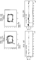
- a computing system is described in Figures 13-14 . Implementations of the observer matter and the functional operations described herein can be implemented in other types of digital electronic circuitry, or in computer software, firmware, or hardware, including the structures disclosed in this specification and their structural equivalents, or in combinations of one or more of them.
- the computer system or computing device 1000 can be used to implement a device that includes the processor 106 and the display 108, the eye movement/gaze tracker component 104, etc.
- the computing system 1000 includes a bus 1005 or other communication component for communicating information and a processor 1010 or processing circuit coupled to the bus 1005 for processing information.
- the computing system 1000 can also include one or more processors 1010 or processing circuits coupled to the bus for processing information.
- the computing system 1000 also includes main memory 1015, such as a random access memory (RAM) or other dynamic storage device, coupled to the bus 1005 for storing information, and instructions to be executed by the processor 1010.
- Main memory 1015 can also be used for storing position information, temporary variables, or other intermediate information during execution of instructions by the processor 1010.
- the computing system 1000 may further include a read only memory (ROM) 1010 or other static storage device coupled to the bus 1005 for storing static information and instructions for the processor 1010.
- ROM read only memory
- a storage device 1025 such as a solid state device, magnetic disk or optical disk, is coupled to the bus 1005 for persistently storing information and instructions.
- the computing system 1000 may be coupled via the bus 1005 to a display 1035, such as a liquid crystal display, or active matrix display, for displaying information to a user.
- a display 1035 such as a liquid crystal display, or active matrix display
- An input device 1030 such as a keyboard including alphanumeric and other keys, may be coupled to the bus 1005 for communicating information and command selections to the processor 1010.
- the input device 1030 has a touch screen display 1035.
- the input device 1030 can include a cursor control, such as a mouse, a trackball, or cursor direction keys, for communicating direction information and command selections to the processor 1010 and for controlling cursor movement on the display 1035.
- the processes described herein can be implemented by the computing system 1000 in response to the processor 1010 executing an arrangement of instructions contained in main memory 1015. Such instructions can be read into main memory 1015 from another computer-readable medium, such as the storage device 1025. Execution of the arrangement of instructions contained in main memory 1015 causes the computing system 1000 to perform the illustrative processes described herein. One or more processors in a multi-processing arrangement may also be employed to execute the instructions contained in main memory 1015. In alternative implementations, hard-wired circuitry may be used in place of or in combination with software instructions to effect illustrative implementations. Thus, implementations are not limited to any specific combination of hardware circuitry and software.
- Implementations of the observer matter and the operations described herein can be implemented in digital electronic circuitry, or in computer software, firmware, or hardware, including the structures disclosed in this specification and their structural equivalents, or in combinations of one or more of them.
- the observer matter described herein can be implemented as one or more computer programs, i.e., one or more modules of computer program instructions, encoded on one or more computer storage media for execution by, or to control the operation of, data processing apparatus.
- the program instructions can be encoded on an artificially-generated propagated signal, e.g., a machine-generated electrical, optical, or electromagnetic signal that is generated to encode information for transmission to suitable receiver apparatus for execution by a data processing apparatus.
- a computer storage medium can be, or be included in, a computer-readable storage device, a computer-readable storage substrate, a random or serial access memory array or device, or a combination of one or more of them.
- a computer storage medium is not a propagated signal, a computer storage medium can be a source or destination of computer program instructions encoded in an artificially-generated propagated signal.
- the computer storage medium can also be, or be included in, one or more separate components or media (e.g., multiple CDs, disks, or other storage devices). Accordingly, the computer storage medium is both tangible and non-transitory.
- the operations described herein can be performed by a data processing apparatus on data stored on one or more computer-readable storage devices or received from other sources.
- the term "data processing apparatus” or “computing device” encompasses all kinds of apparatus, devices, and machines for processing data, including by way of example a programmable processor, a computer, a system on a chip, or multiple ones, or combinations of the foregoing.
- the apparatus can include special purpose logic circuitry, e.g., an FPGA (field programmable gate array) or an ASIC (application-specific integrated circuit).
- the apparatus can also include, in addition to hardware, code that creates an execution environment for the computer program in question, e.g., code that constitutes processor firmware, a protocol stack, a database management system, an operating system, a cross-platform runtime environment, a virtual machine, or a combination of one or more of them.
- the apparatus and execution environment can realize various different computing model infrastructures, such as web services, distributed computing and grid computing infrastructures.
- a computer program (also known as a program, software, software application, script, or code) can be written in any form of programming language, including compiled or interpreted languages, declarative or procedural languages, and it can be deployed in any form, including as a stand-alone program or as a module, component, subroutine, object, or other unit suitable for use in a computing environment.
- a computer program may, but need not, correspond to a file in a file system.
- a program can be stored in a portion of a file that holds other programs or data (e.g., one or more scripts stored in a markup language document), in a single file dedicated to the program in question, or in multiple coordinated files (e.g., files that store one or more modules, sub-programs, or portions of code).
- a computer program can be deployed to be executed on one computer or on multiple computers that are located at one site or distributed across multiple sites and interconnected by a communication network.
- processors suitable for the execution of a computer program include, by way of example, both general and special purpose microprocessors, and any one or more processors of any kind of digital computer.
- a processor will receive instructions and data from a read-only memory or a random access memory or both.
- the essential elements of a computer are a processor for performing actions in accordance with instructions and one or more memory devices for storing instructions and data.
- a computer will also include, or be operatively coupled to receive data from or transfer data to, or both, one or more mass storage devices for storing data, e.g., magnetic, magneto-optical disks, or optical disks.
- mass storage devices for storing data, e.g., magnetic, magneto-optical disks, or optical disks.
- a computer need not have such devices.
- a computer can be embedded in another device, e.g., a mobile telephone, a personal digital assistant (PDA), a mobile audio or video player, a game console, a Global Positioning System (GPS) receiver, or a portable storage device (e.g., a universal serial bus (USB) flash drive), to name just a few.
- Devices suitable for storing computer program instructions and data include all forms of non-volatile memory, media and memory devices, including by way of example semiconductor memory devices, e.g., EPROM, EEPROM, and flash memory devices; magnetic disks, e.g., internal hard disks or removable disks; magneto-optical disks; and CD-ROM and DVD-ROM disks.
- the processor and the memory can be supplemented by, or incorporated in, special purpose logic circuitry.
- implementations of the observer matter described in this specification can be implemented on a computer having a display device, e.g., a CRT (cathode ray tube) or LCD (liquid crystal display) monitor, for displaying information to the user and a keyboard and a pointing device, e.g., a mouse or a trackball, by which the user can provide input to the computer.
- a display device e.g., a CRT (cathode ray tube) or LCD (liquid crystal display) monitor
- keyboard and a pointing device e.g., a mouse or a trackball
- Other kinds of devices can be used to provide for interaction with a user as well; for example, feedback provided to the user can be any form of sensory feedback, e.g., visual feedback, auditory feedback, or tactile feedback; and input from the user can be received in any form, including acoustic, speech, or tactile input.
- the raw data is preprocessed as follows: for the x (horizontal) and y (vertical) vectors independently, the mean is subtracted and divided by the standard deviation (which is the square root of the variance). This puts all the data in the same relative frame (zero-mean, max and min about 1 and -1). This is the reason the boxes look square (even if the stimulus presentation monitor is not square).
- the methods described herein provide means for assessing or quantifying disconjugate gaze or disconjugate eye movement. These means feature receiving an array of pupil x and y coordinates that may be generated or obtained according to the methods described herein. These coordinates may be be averaged across, for instance, five eyebox trajectory cycles. Formulaically this can be represented as as follows: where X ijk refers to the x-coordinate of the pupil, and k refers to the left or right eye of a subject. The difference in the x and y position, for the left and right eye, may then be computed. This vector of difference may then be plotted graphically for purposes of assessment and interpretation.
- a variance of the data may be computed with respect to an expected mean of zero. This is significant because the code assumes that a healthy subject has zero lateral or longitudinal pupil position difference between each eye.
- the variance may be computed as follows:
- the total variance may be computed as follows:
- the variance in X, Y, and the total variance may be plotted in order to assess the amount of disconjugation (i.e. disconjugate gaze) present in a subject.
- the methods described herein may identify strabismus.
- A-pattern was associated with a greater prevalence of neurological impairment, hydrocephalus and meningomyelocele, while those with V-pattern exhibited a greater prevalence of craniosynostosis and malformative syndromes ( Dickmann, et al., Ophthalmic Epidemiol., 2012; 19: 302-305 ).
- Delays in treatment of strabismus onset following binocular vision maturation may be associated with permanent disruption of stereopsis and sensory fusion ( Fawcett, Curr Opin Ophthalmol., 2005; 16: 298-302 ).
- disconjugacy may be due to neurologic causes including trauma, hydrocephalus, demyelination, inflammation, infection, degenerative disease, neoplasm/paraneoplastic syndrome, metabolic disease including diabetes, or vascular disruption such as stroke, hemorrhage or aneurysm formation.
- Disconjugacy may also be due to ophthalmologic causes such as conjunctivitis, ophthalmoplegia, ocular injury or other diseases.
- the methods described herein may feature assessing conjugacy or disconjugacy of eye movement in correlation with structural and non-structural traumatic brain injury, including concussion or blast injury.
- a purpose of the prospective observational study described herein was to quantitate differences in eye tracking of structurally and non-structurally brain injured subjects relative to non-brain but bodily injured and healthy non-injured controls to identify the eye tracking parameters associated with structural and non-structural injury. Another purpose was to identify a correlation between impaired eye tracking and clinical neurologic functioning. Eye tracking and clinical concussion assessments were performed on 44 injured subjects, and eye tracking was performed only on 31 healthy normal controls. 51 eye tracking parameters were assessed in each patient. 10 parameters showed statistically significant differences between negative controls (healthy normal people and corporally injured trauma patients) and both positive controls (patients with structural brain injury) and patients with non-structural brain injury.
- a novel technique for automated eye movement tracking was developed using temporal rather than spatial calibration.
- the position of the pupil is predicted based on time elapsed since the start of the video rather than spatial calibration, enabling detection of impaired ability to move the pupil relative to normal controls or the opposite eye.
- Temporal calibration offers the additional advantage of utility to populations that may not be willing or able to cooperate with calibration instructions such as young children, foreign-language speakers, minimally conscious persons, or aphasics.
- the data presented herein quantitates differences in eye tracking of structurally and non-structurally brain injured subjects relative to non-brain but bodily injured and healthy non-injured controls to identify the parameters associated with structural and non-structural injury.
- the data presented herein further establish a correlation between impaired eye tracking and clinical neurologic functioning.
- Raw x and y cartesian coordinates of pupil position are collected and stored in a one-dimensional vector:
- Index i corresponds to an individual data point.
- the size of i depends on the eye tracking hardware capture frequency and the time of tracking.
- x represents the left normalized raw x pupil position minus the right normalized raw x pupil position.
- / corresponds to the top, right, bottom and left segments of the box.
- the variance here does not follow the traditional form of statistical variance.
- the average of the data points is subtracted from the sum of individual data points. In this case, the average is forced to zero, thus inferring that the hypothetical control patient has perfect conjugacy (left and right eye move precisely together).
- Eye movements contain clinically important information about neurological integrity.
- Clinical devices may take advantage of the relative ease of automated eye-movement tracking, for applications such as assessing recovery following clinical intervention.
- a technique was designed that can reliably measure eye movements with precision, without initial spatial calibration.
- Temporal features of the data were measured, rather than traditional spatial measures such as accuracy or speed.
- the methods reliably discriminated between the presence and absence of neurological impairment using these uncalibrated measurements.
- the results indicate that this technique may be extended to assess neurologic integrity and quantify deficits, simply by having patients watch TV.
- Eye movements have long been known to contain clinically relevant information about neurological integrity.
- Assessment of ocular motility is a standard part of any neurological exam, because it is easy and informative.
- problems with the standard clinical exam including that it is normally administered by an expert, and generally is only qualitative, not quantitative.
- the calibration process itself may reduce the sensitivity of the eye tracking test. For example, consider a patient with impaired vertical ocular motility. Because the calibration process assumes that the eyes cover the full range of locations mapped out by the calibration points, it assigns the maximum pupil angle up and down incorrectly to the 'top' and 'bottom' of the monitor, respectively. In such instances, all future measurements for that observer are adjusted to conform to that incorrect assignment. Thus, impaired ocular motility may be undetected in tests that begin with a spatial calibration of the eye tracker.
- Eye movement measurements may reflect severity of damage to the brain, as well as recovery following clinical intervention.
- the methods described herein were used to test patients from neurosurgery, emergency department and ophthalmology clinics as well as a control set of healthy volunteers.
- the success of the method involves two features. First, the methods described herein do not use spatial measures of accuracy as a variable of interest. By looking at eye movement trajectories in the time domain rather than the spatial domain, it is possible to quantify measures that do not rely on spatial calibration. Second, the measures are easily visualized and evaluated, making them immediately useful to the clinician or researcher.
- Eye Movement Tracking Observers' eye movements were recorded using an Eyelink 1000 binocular eye tracker (500 Hz sampling, SR Research). All observers were seated approximately 55 cm from the screen. Some test patients were tracked on multiple visits at different stages of diagnosis, surgery, and recovery.
- the visual stimulus provided as a music video that played continuously while it moved clockwise along the outer edges of a computer monitor. Observers were instructed to watch the video. The stimulus was expected to evoke smooth pursuit eye movements as well as possible saccades and microsaccades as the observers scanned the video.
- the video was presented in a square aperture with an area approximately 1/8 of the size of the screen (about 16° of visual angle). This square aperture started at the upper left hand corner of the screen and moved at a constant speed, taking 10 seconds to traverse each edge of the monitor. A full cycle took 40 seconds, and five full cycles were played, for a total of 200 seconds. A countdown video played in the starting position for 10 seconds before the music video began, to give observers time to orient to the stimulus. Only the 200 seconds of the music video were used for analyses.
- the eye tracker sampled eye position at 500 Hz, yielding 100,000 samples of eye position over 200 seconds.
- Phase was calculated as phase of the sine function that best fit the data.
- the 8 following complementary procedures were used to assess the statistical significance of any differences in these two measures (phase difference and model fit) as compared between the neurologically intact control observers and the test patient observers.
- Uncalibrated tracking may provide a quantitative measure of the ability to fixate, attend, and follow a stimulus. These date demonstrate that it is possible to collect reliable highfrequency eye movement data without first completing a spatial calibration for each observer. Many patients are not capable of calibrated eye tracking. The ability to track eye movements in these populations provides new insights about a variety of disorders that disturb the ocular-motor system, including but not limited to brain injury, stroke, and psychiatric disorders. Possible applications include clinical screening, diagnosis, monitoring the efficacy of treatment, and tracking progression of impairment and recovery.
- Eye Movement Tracking The subjects' eye movements were recorded using an Eyelink 1000 binocular eye tracker (500 Hz sampling, SR Research). Healthy volunteers were seated 55 cm from the screen with their head stabilized using a chinrest. Stimulus was presented on average 55 cm from patient eyes, with the presentation monitor adjusted to match gaze direction. Subjects used a chinrest.
- Integrity For the integrity measure, each patient's pair of values from arms 1 (the top of the box) and 3 (the bottom of the box) was z-scored using the mean and standard deviation calculated from the control population. The resulting score indicated how different the patient values were compared with the control values, in units of standard deviations. Because 95% of all values in a normal distribution lie within two standard deviations of the mean, a z-score of 2 was used as a significance threshold. Patients with z-scores above 2 in either or both arms were thus judged to have significant disturbances of ocular motility.
- relative variance Because relative variance is a ratio, it cannot be analyzed using z-scores, since the assumption of a normal distribution does not hold for ratios. Instead, 5,000 point distributions were generated using a bootstrapping method that took 5,000 samples from 25 values randomly chosen with replacement from the 45 control values. For each subject, the relative variance in arms 1 and 3 were compared respectively with the corresponding control distribution, and the percent of the control distribution with variance below that of the test value was determined. A p-value of .05 (a widely accepted measure of statistical significance) corresponds to 95% of control values falling below the test value. Thus, subjects with variance higher than 95% of the values in the control distributions were determined to have significant disturbances of ocular motility.
- the units of relative variance are related to size in degree of visual angle, but are not exactly identical to degrees of visual angle, because there was no spatial calibration. These may be referred to as time-degrees units.
- Control distributions As expected, the control distributions for the integrity measurements were normally distributed with a mean of 0.2 and an average standard deviation of 0.05 (5% deviation). The control distributions of relative variance peaked at 0.25 (reflecting equal variance across the four arms).
- Control subjects were employees, volunteers, visitors and patients at the Bellevue Hospital Center recruited in accordance with Institutional Review Board policy. Inclusion criteria for normal control subjects were: age 7 to 100 years, vision correctable to within 20/500 bilaterally, intact ocular motility, and ability to provide a complete ophthalmologic, medical and neurologic history as well as medications/drugs/alcohol consumed within the 24 hours prior to tracking. Parents were asked to corroborate details of the above for children aged 7-17.
- Exclusion criteria were history of: strabismus, diplopia, palsy of cranial nerves III, IV or VI, papilledema, optic neuritis or other known disorder affecting cranial nerve II, macular edema, retinal degeneration, dementia or cognitive impairment, hydrocephalus, sarcoidosis, myasthenia gravis, multiple sclerosis or other demyelinating disease, and active or acute epilepsy, stroke/hemorrhage or brain injury sufficiently significant to result in hospitalization. Subjects reporting any minor brain injury regardless of loss of consciousness within the previous week were also excluded.
- Each subjects' eye movements were recorded with an Eyelink 1000 eye tracker at a fixed distance of 55 cm from a computer monitor over a time period of 220 seconds.
- the stationary tracker the subject was seated in an adjustable height chair, using an adjustable height chinrest.
- Portable tracker subjects were seated in either a height adjustable or height-fixed chair, with the monitor height adjusted to the subject.
- the portable tracker chinrest was attached to the monitor, while the stationary tracker chinrest was attached to the same table as the computer monitor.
- the visual stimuli were the music videos Shakira Waka-Waka, K'naan Wavin' Flag, or the Under the Sea song from the Little Mermaid.
- the video was played continuously in a square aperture with an area approximately 1/8 the screen size while moving clockwise along the outer edges of the monitor for five complete cycles of 40 seconds each.
- the first and last 10 seconds of each data set were discarded to yield 200 seconds of data.
- the afferent stimulus was presented binocularly and eye tracking was performed binocularly. Subjects were not spatially calibrated to the tracker to enable independent analysis of each pupil position over time.
- subjects were assessed for gaze conjugacy using a naturalistic viewing stimulus. This consisted of watching television as eye movements were tracked over time. Subjects were not seated at a fixed distance from the monitor but were able to move their heads during viewing.
- the eye tracker sampled pupil position at 500 Hz, yielding 100,000 samples over 200 seconds. Scatterplots of the entire time series were created by plotting the 100,000 (x, y) pairs representing the two orthogonal components of the instantaneous angle of pupil reflection over time to create 'box trajectories' that reflected the temporal nature of the pupillary movement. These figures look like boxes, reflecting the timing of the aperture as it moved around the screen.
- the total sum of the differences between both x and y coordinates over the time tested can be summed to obtain a measure of total disconjugacy of gaze, or as an average of five eyebox trajectory cycles formulaically represented as follows: where X ijk refers to the x-coordinate of the pupil, and k refers to the left or right eye of a subject. In cases where a subject's data was missing at any given time point in the five cycles, the denominator of the equation was the number of cycles where the data was present. The difference in the x and y position, for the left and right eye, may then be computed. This vector_of difference may then be plotted graphically for purposes of assessment and interpretation.
- a variance of the data may be computed with respect to an expected mean of zero. This is significant because the code assumes that a healthy subject has zero vertical or horizontal pupil position difference between each eye.
- the variance for either horizontal (x) or vertical (substitute y for x) movement may be computed as follows:
- the total variance in both the horizontal and vertical planes may be computed as follows:
- the variance in X, Y, and the total variance may be plotted in order to assess the amount of disconjugation present in a subject.
- Test-retest on the stationary tracker and from the stationary to the portable tracker A paired t-test was used to determine if the mean of the subject-paired differences between the total variances for two separate eyetracking sessions was statistically significantly different from 0.
- the single greatest outlier (conjugacy of .8214) in the control population was a 23 year old male student who wears corrective contact lenses and takes adderal for attention deficit and hyperactivity disorder. This subject underwent repeat tracking which remained disconjugate, (.2600) however less than previously.
- the second greatest outlier (conjugacy .486) was a 39 year old male hospital employee who denied any ophthalmic or medical history, as well as the use of alcohol or drugs in the prior 24 hours. In both of these subjects the X-conjugacy was not a statistical outlier and only the y coordinates were disconjugate.
- Subjects (n 27) demonstrated high test-retest reliability between two separate eyetracking sessions on the stationary tracker ( Figure 10 ).
- a paired t-test was used to determine if the mean of the subject-paired differences between the total variances for two separate eyetracking sessions was statistically significantly different from 0. With 26 degrees of freedom, the test resulted in a t-statistic of 1.2778 and a p-value of 0.2126 showing that the mean of the subject-paired differences was not statistically significantly different from 0.
- Subjects (n 24) demonstrated high test-retest reliability between separate eyetracking sessions on the stationary tracker and the portable tracker ( Figure 11 ).
- a paired t-test with 23 degrees of freedom (n 24), resulted in a t-statistic of 1.3661 and a p-value of 0.1851 showing that the mean of the subject-paired differences was not statistically significantly different from 0.
- Figure 1 represents the eye tracking trajectories of subjects with normal compared to known (well-defined) neurophthalmic abnormality.
- Subjects that received minor trauma insufficiently traumatizing to result in sufficient sequelae were excluded.
- Subjects suffering burns, anoxic injury or multiple/extensive injuries resulting in any medical, surgical or hemodynamic instability were also excluded.
- Particularly for the purposes of eye tracking all subjects that were blind (no light perception), missing eyes, and not opening eyes were excluded from the research. It is pertinent that subjects are able to detect light and have both eyes in order for the eye tracking data to be effective and significant. Any physical or mental injury or baseline disability rendering task completion difficult was excluded, also inability to participate in longtitudinal care, or obvious intoxication or blood alcohol level greater than 0.2. Pregnant individuals and prisoners were also excluded from the study.
- a portable binocular eye movement tracker was constructed by attaching an adjustable arm to a rolling cart.
- a computer monitor was attached to the proximal portion of the arm, and a chinrest was attached to the distal aspect of the arm such that the chinrest centered the subject's eyes 55 cm away from the monitor.
- Subjects' eye movements were recorded with an Eyelink 1000 eye tracker over a time period of 220 seconds.
- Portable tracker subjects were seated in either a height adjustable or height-fixed chair or bed, with the monitor height adjusted to the subject.
- the visual stimuli were the music videos Shakira Waka-Waka, K'naan Wavin' Flag, Mission Museum Bhumbroo or Michael Jackson Man in the Mirror.
- the video was played continuously in a square aperture with an area approximately 1/9 the screen area while moving clockwise along the outer edges of the monitor for five complete cycles of 40 seconds each.
- the first and last 10 seconds of each data set were discarded to yield 200 seconds of data.
- the afferent stimulus was presented binocularly, and eye tracking was performed binocularly. Subjects were not spatially calibrated to the tracker to enable independent analysis of each pupil position over time.
- the eye tracker sampled pupil position at 500 Hz, yielding 100,000 samples over 200 seconds. Scatterplots of the entire time series were created by plotting the 100,000 (x,y) pairs representing the two orthogonal components of the instantaneous angle of pupil reflection over time to create 'box trajectories' that reflected the temporal nature of the pupillary movement. These figures look like boxes, reflecting the timing of the aperture as it moved around the screen.
- the sports concussion assessment tool (SCAT) was administered, and standardized assessment of concussion (SAC) scores were obtained on thirty-seven subjects. Stepwise multiple regression analysis was performed to evaluate the impact of each eye-tracking parameter on the SCAT and SAC scores. Parameters with p-values > 0.1 were removed from the model.
- Table 4 provides group means for each of the 51 measured parameters.
- Table 5 provides p-values. Ten of the 51 measured parameters demonstrated statistically significant differences between negative controls (either normal healthy people, or corporally injured but not brain injured controls) and both positive controls (structurally brain injured) and non-structurally brain injured people. 8 additional parameters showed statistically significant differences between negative controls (healthy normal people and corporally injured trauma patients) and patients with either structural or non-structural brain injury. 10 of the eye tracking measures showed statistically significant correlation between SCAT or SAC scores, suggesting that these eye tracking parameters correlated with a validated clinical outcome measure.
Landscapes
- Health & Medical Sciences (AREA)
- Life Sciences & Earth Sciences (AREA)
- Engineering & Computer Science (AREA)
- Veterinary Medicine (AREA)
- Biophysics (AREA)
- Biomedical Technology (AREA)
- Heart & Thoracic Surgery (AREA)
- Medical Informatics (AREA)
- Molecular Biology (AREA)
- Surgery (AREA)
- Animal Behavior & Ethology (AREA)
- General Health & Medical Sciences (AREA)
- Public Health (AREA)
- Physics & Mathematics (AREA)
- Ophthalmology & Optometry (AREA)
- Neurology (AREA)
- Pathology (AREA)
- Neurosurgery (AREA)
- Physiology (AREA)
- Signal Processing (AREA)
- Psychology (AREA)
- Hematology (AREA)
- Human Computer Interaction (AREA)
- Artificial Intelligence (AREA)
- Computer Vision & Pattern Recognition (AREA)
- Psychiatry (AREA)
- Eye Examination Apparatus (AREA)
- Other Investigation Or Analysis Of Materials By Electrical Means (AREA)
Applications Claiming Priority (4)
| Application Number | Priority Date | Filing Date | Title |
|---|---|---|---|
| US201361835927P | 2013-06-17 | 2013-06-17 | |
| US201361881014P | 2013-09-23 | 2013-09-23 | |
| US201461929238P | 2014-01-20 | 2014-01-20 | |
| PCT/US2014/042645 WO2014204904A1 (en) | 2013-06-17 | 2014-06-17 | Methods and kits for assessing neurological and ophthalmic function and localizing neurological lesions |
Publications (3)
| Publication Number | Publication Date |
|---|---|
| EP3010391A1 EP3010391A1 (en) | 2016-04-27 |
| EP3010391A4 EP3010391A4 (en) | 2016-12-07 |
| EP3010391B1 true EP3010391B1 (en) | 2022-06-15 |
Family
ID=52105176
Family Applications (1)
| Application Number | Title | Priority Date | Filing Date |
|---|---|---|---|
| EP14814294.6A Not-in-force EP3010391B1 (en) | 2013-06-17 | 2014-06-17 | Methods and kits for assessing neurological and ophthalmic function and localizing neurological lesions |
Country Status (6)
| Country | Link |
|---|---|
| US (4) | US10610165B2 (enExample) |
| EP (1) | EP3010391B1 (enExample) |
| JP (1) | JP6676522B2 (enExample) |
| CN (1) | CN105764407A (enExample) |
| AU (1) | AU2014281725B2 (enExample) |
| WO (1) | WO2014204904A1 (enExample) |
Families Citing this family (32)
| Publication number | Priority date | Publication date | Assignee | Title |
|---|---|---|---|---|
| US20170157038A1 (en) * | 2008-11-13 | 2017-06-08 | Gholam A. Peyman | Ophthalmic drug delivery method |
| BR112014023637B1 (pt) | 2012-03-26 | 2021-12-21 | New York University | Métodos para avaliação da integridade do sistema nervoso central e para detecção, diagnóstico ou avaliação de pressão intracraniana aumentada |
| AU2014281725B2 (en) | 2013-06-17 | 2019-10-10 | New York University | Methods and kits for assessing neurological and ophthalmic function and localizing neurological lesions |
| JP2017529891A (ja) | 2014-08-04 | 2017-10-12 | ニューヨーク ユニバーシティ | 薬物使用、薬物乱用および昏睡、核間性眼筋麻痺、注意欠陥多動性障害(adhd)、慢性外傷性脳症、統合失調症スペクトラム障害、およびアルコール摂取を診断、評価、または定量化するための方法およびキット |
| US20160377639A1 (en) * | 2015-06-27 | 2016-12-29 | William Beaumont Hospital | Methods for detecting, diagnosing and treating traumatic brain injury |
| US20170042462A1 (en) * | 2015-08-10 | 2017-02-16 | Neuro Kinetics, Inc. | Automated Data Acquisition, Appraisal and Analysis in Noninvasive Rapid Screening of Neuro-Otologic Conditions Using Combination of Subject's Objective Oculomotor Vestibular and Reaction Time Analytic Variables |
| US11064881B2 (en) * | 2015-11-13 | 2021-07-20 | Hennepin Healthcare System, Inc | Method for predicting convergence disorders caused by concussion or other neuropathology |
| JP6693105B2 (ja) | 2015-12-01 | 2020-05-13 | 株式会社Jvcケンウッド | 視線検出装置及び視線検出方法 |
| NL2016085B1 (en) | 2016-01-13 | 2017-07-24 | Stichting Vu-Vumc | Method, system and computer readable medium to determine a strabismus angle between the eyes of an individual. |
| EP3493728B1 (en) * | 2016-08-02 | 2025-11-05 | New York University | Methods and kits for assessing neurological function and localizing neurological lesions |
| JP2020509790A (ja) * | 2016-09-23 | 2020-04-02 | ノバサイト リミテッド | スクリーニング装置及び方法 |
| US10863902B2 (en) | 2016-10-03 | 2020-12-15 | Oculogica Inc. | Method for detecting glaucoma |
| US10201274B2 (en) | 2016-10-20 | 2019-02-12 | Oculogica Inc | Eye tracking system with biometric identification |
| WO2018105459A1 (ja) * | 2016-12-09 | 2018-06-14 | パイオニア株式会社 | 眠気推定装置 |
| US11141095B2 (en) * | 2017-02-17 | 2021-10-12 | Oculogica Inc. | Method and system for detecting concussion |
| ES2909057T3 (es) * | 2017-09-08 | 2022-05-05 | Tobii Ab | Seguimiento ocular que utiliza la posición del centro del globo ocular |
| CN114403802A (zh) | 2017-09-13 | 2022-04-29 | 奥库洛吉卡公司 | 眼睛跟踪系统 |
| JP7024304B2 (ja) * | 2017-10-10 | 2022-02-24 | 株式会社ニデック | 眼科装置 |
| WO2019148003A1 (en) * | 2018-01-26 | 2019-08-01 | Hennepin Healthcare System, Inc. | Methods and kits for detection of blast brain injury, its severity, distance from blast epicenter, and classification relative to the larger spectrum of concussion |
| US12290373B2 (en) | 2018-04-27 | 2025-05-06 | C. Light Technologies, Inc. | Method of detection, prognostication, and monitoring of neurological disorders |
| US11544844B2 (en) | 2019-07-25 | 2023-01-03 | Canon Medical Systems Corporation | Medical image processing method and apparatus |
| US12293526B2 (en) | 2020-02-10 | 2025-05-06 | Nec Corporation | Gaze estimation system, gaze estimation method, and computer program |
| CA3201453A1 (en) * | 2020-12-23 | 2022-06-30 | Viewmind, Inc. | System and method for detecting neurological disorders and for measuring general cognitive performance |
| US12390172B2 (en) | 2021-02-19 | 2025-08-19 | Canon Medical Systems Corporation | Method and apparatus for displaying medical images |
| US20240237897A1 (en) * | 2021-04-09 | 2024-07-18 | The Regents Of The University Of California | Head-mounted device including display for fully-automated ophthalmic imaging |
| CN114049308A (zh) * | 2021-10-21 | 2022-02-15 | 广州爱尔眼科医院有限公司 | 一种视觉功能检测系统 |
| US11941115B2 (en) | 2021-11-29 | 2024-03-26 | Bank Of America Corporation | Automatic vulnerability detection based on clustering of applications with similar structures and data flows |
| US11928221B2 (en) | 2021-11-29 | 2024-03-12 | Bank Of America Corporation | Source code clustering for automatically identifying false positives generated through static application security testing |
| US11903711B2 (en) | 2022-06-09 | 2024-02-20 | EarliTec Diagnostics, Inc. | Assessing developmental disorders via eye tracking |
| AU2024285555A1 (en) | 2023-06-05 | 2025-09-25 | EarliTec Diagnostics, Inc. | Systems and methods for using portable computer devices having eye-tracking capability |
| WO2025107077A1 (en) * | 2023-11-23 | 2025-05-30 | Pragmaclin Research Inc. | System and method for performing a motor assessment |
| CN118766478B (zh) * | 2024-08-20 | 2025-05-13 | 深圳高视科技有限公司 | 基于眼神经实现脑神经的智能检测方法及系统 |
Citations (2)
| Publication number | Priority date | Publication date | Assignee | Title |
|---|---|---|---|---|
| US5042937A (en) * | 1989-12-11 | 1991-08-27 | Pulse Medical Instruments | Optical system for an ophthamological instrument for examination of pupillary responses |
| US20130044291A1 (en) * | 2011-05-20 | 2013-02-21 | Panasonic Corporation | Visual fatigue level measuring device, visual fatigue level measuring method, visual fatigue level measuring system, and three-dimensional glasses |
Family Cites Families (58)
| Publication number | Priority date | Publication date | Assignee | Title |
|---|---|---|---|---|
| US4576184A (en) | 1981-08-31 | 1986-03-18 | Pharmometrics Corporation | Drug abuse detection |
| US4838681A (en) * | 1986-01-28 | 1989-06-13 | George Pavlidis | Method and means for detecting dyslexia |
| US4889422A (en) * | 1986-01-28 | 1989-12-26 | George Pavlidis | Method and means for detecting dyslexia |
| US6622036B1 (en) | 2000-02-09 | 2003-09-16 | Cns Response | Method for classifying and treating physiologic brain imbalances using quantitative EEG |
| US20020024633A1 (en) | 1999-04-09 | 2002-02-28 | Daehoon Kim | Pupil evaluation system |
| US6346887B1 (en) | 1999-09-14 | 2002-02-12 | The United States Of America As Represented By The Secretary Of The Navy | Eye activity monitor |
| EP1246667B1 (en) | 1999-12-30 | 2005-03-23 | Medtronic, Inc. | User authentication in medical device systems |
| BR0108476A (pt) | 2000-02-11 | 2003-04-22 | Marcio Marc Abreu | Sistema e método para comunicar informação de revocação de produto, advertências relacionadas ao produto ou outra informação relacionada aos usuários de produtos |
| JP4584408B2 (ja) * | 2000-05-17 | 2010-11-24 | 株式会社ニューオプト | 眼球画像解析システム |
| US6652458B2 (en) | 2000-06-20 | 2003-11-25 | The Mclean Hospital Corporation | ADHD detection by eye saccades |
| EP1219243A1 (en) | 2000-12-28 | 2002-07-03 | Matsushita Electric Works, Ltd. | Non-invasive brain function examination |
| DE10132378A1 (de) | 2001-07-06 | 2003-04-24 | Zeiss Carl Meditec Ag | Verfahren und Vorrichtung zur Verfolgung von Augenbewegungen |
| JP4543594B2 (ja) * | 2001-07-31 | 2010-09-15 | パナソニック電工株式会社 | 脳機能検査装置および脳機能検査システム |
| DE60323410D1 (de) | 2003-11-30 | 2008-10-16 | Volvo Technology Corp | Verfahren und system zum erkennen einer fahrerbeeinträchtigung |
| US7384399B2 (en) | 2004-02-11 | 2008-06-10 | Jamshid Ghajar | Cognition and motor timing diagnosis and training system and method |
| US7819818B2 (en) | 2004-02-11 | 2010-10-26 | Jamshid Ghajar | Cognition and motor timing diagnosis using smooth eye pursuit analysis |
| CN102670163B (zh) | 2004-04-01 | 2016-04-13 | 威廉·C·托奇 | 控制计算装置的系统及方法 |
| WO2007075460A2 (en) | 2005-12-16 | 2007-07-05 | Novavision, Inc. | Adjustable device for vision testing and therapy |
| US7535991B2 (en) | 2006-10-16 | 2009-05-19 | Oraya Therapeutics, Inc. | Portable orthovoltage radiotherapy |
| WO2009052833A1 (en) | 2007-10-23 | 2009-04-30 | Mindmetic Ltd. | Method, system and computer program for automated interpretation of measurements in response to stimuli |
| CA2704777C (en) | 2007-11-01 | 2017-09-12 | Catholic Healthcare West | Method of detecting neurological disease |
| WO2009085204A2 (en) | 2007-12-23 | 2009-07-09 | Oraya Therapeutics, Inc. | Methods and devices for detecting, controlling, and predicting radiation delivery |
| WO2010042557A2 (en) | 2008-10-06 | 2010-04-15 | Neuro Kinetics, Inc. | Method and apparatus for corrective secondary saccades analysis with video oculography system |
| WO2010062400A1 (en) * | 2008-11-28 | 2010-06-03 | Neuroptics, Inc. | Methods, systems, and devices for monitoring anisocoria and asymmetry of pupillary reaction to stimulus |
| US9229227B2 (en) | 2010-02-28 | 2016-01-05 | Microsoft Technology Licensing, Llc | See-through near-eye display glasses with a light transmissive wedge shaped illumination system |
| US8732795B2 (en) | 2010-05-21 | 2014-05-20 | Epic Systems Corporation | System and method for user authentication |
| CA2827498C (en) | 2011-01-28 | 2020-09-22 | Dignity Health | Method of detecting neurological disease |
| CA2754835C (en) * | 2011-10-07 | 2021-06-15 | Baycrest Centre For Geriatric Care | Methods and systems for assessing cognitive function |
| US9078612B2 (en) | 2011-12-02 | 2015-07-14 | Third Eye Diagnostics, Inc. | Devices and methods for noninvasive measurement of intracranial pressure |
| US20130208952A1 (en) | 2012-02-13 | 2013-08-15 | Geoffrey Auchinleck | Method and Apparatus for Improving Accuracy of Biometric Identification in Specimen Collection Applications |
| BR112014023637B1 (pt) | 2012-03-26 | 2021-12-21 | New York University | Métodos para avaliação da integridade do sistema nervoso central e para detecção, diagnóstico ou avaliação de pressão intracraniana aumentada |
| US9101312B2 (en) | 2012-04-18 | 2015-08-11 | TBI Diagnostics LLC | System for the physiological evaluation of brain function |
| US8668337B2 (en) | 2012-04-18 | 2014-03-11 | TBI Diagnostics LLC | System for the physiological evaluation of brain function |
| US9004687B2 (en) | 2012-05-18 | 2015-04-14 | Sync-Think, Inc. | Eye tracking headset and system for neuropsychological testing including the detection of brain damage |
| US8808179B1 (en) * | 2012-08-06 | 2014-08-19 | James Z. Cinberg | Method and associated apparatus for detecting minor traumatic brain injury |
| US8585589B1 (en) * | 2012-08-06 | 2013-11-19 | James Z. Cinberg | Method and associated apparatus for detecting minor traumatic brain injury |
| US8951046B2 (en) | 2012-08-10 | 2015-02-10 | Sync-Think, Inc. | Desktop-based opto-cognitive device and system for cognitive assessment |
| US10085688B2 (en) | 2012-11-20 | 2018-10-02 | El-Mar Inc. | Method of identifying an individual with a disorder or efficacy of a treatment of a disorder |
| US9265458B2 (en) | 2012-12-04 | 2016-02-23 | Sync-Think, Inc. | Application of smooth pursuit cognitive testing paradigms to clinical drug development |
| US9380976B2 (en) | 2013-03-11 | 2016-07-05 | Sync-Think, Inc. | Optical neuroinformatics |
| BR112015022523A2 (pt) | 2013-03-11 | 2017-07-18 | Childrens Healthcare Atlanta Inc | sistemas e métodos para detecção de condições de desenvolvimento e cognitivas |
| AU2014281725B2 (en) | 2013-06-17 | 2019-10-10 | New York University | Methods and kits for assessing neurological and ophthalmic function and localizing neurological lesions |
| US9895100B2 (en) | 2013-10-04 | 2018-02-20 | Indiana University Research And Technology Corporation | Eye movement monitoring of brain function |
| ES2848833T3 (es) | 2013-10-17 | 2021-08-12 | Childrens Healthcare Atlanta Inc | Métodos para valorar el desarrollo de bebés y niños por medio de seguimiento de ojos |
| US9958939B2 (en) | 2013-10-31 | 2018-05-01 | Sync-Think, Inc. | System and method for dynamic content delivery based on gaze analytics |
| US9721476B2 (en) | 2013-11-06 | 2017-08-01 | Sync-Think, Inc. | System and method for dynamic cognitive training |
| US9459451B2 (en) | 2013-12-26 | 2016-10-04 | Microsoft Technology Licensing, Llc | Eye tracking apparatus, method and system |
| WO2015164807A1 (en) | 2014-04-25 | 2015-10-29 | Texas State University | Detection of brain injury and subject state with eye movement biometrics |
| JP6550460B2 (ja) | 2014-05-09 | 2019-07-24 | グーグル エルエルシー | 眼信号を識別するためのシステムおよび方法、ならびに連続バイオメトリック認証 |
| WO2016009334A1 (en) | 2014-07-17 | 2016-01-21 | Bitoun Pierre | Measurement of ocular parameters using vibrations induced in the eye |
| JP2017529891A (ja) | 2014-08-04 | 2017-10-12 | ニューヨーク ユニバーシティ | 薬物使用、薬物乱用および昏睡、核間性眼筋麻痺、注意欠陥多動性障害(adhd)、慢性外傷性脳症、統合失調症スペクトラム障害、およびアルコール摂取を診断、評価、または定量化するための方法およびキット |
| WO2016090376A1 (en) | 2014-12-05 | 2016-06-09 | Texas State University | Eye tracking via patterned contact lenses |
| US20160213248A1 (en) | 2015-01-22 | 2016-07-28 | Ovard, Llc | Gaze stabilization system and method |
| US20170091392A1 (en) | 2015-05-01 | 2017-03-30 | Steven C. White | Biometric identification telemedicine software |
| US11064881B2 (en) | 2015-11-13 | 2021-07-20 | Hennepin Healthcare System, Inc | Method for predicting convergence disorders caused by concussion or other neuropathology |
| US10863902B2 (en) | 2016-10-03 | 2020-12-15 | Oculogica Inc. | Method for detecting glaucoma |
| US10201274B2 (en) | 2016-10-20 | 2019-02-12 | Oculogica Inc | Eye tracking system with biometric identification |
| US11141095B2 (en) | 2017-02-17 | 2021-10-12 | Oculogica Inc. | Method and system for detecting concussion |
-
2014
- 2014-06-17 AU AU2014281725A patent/AU2014281725B2/en active Active
- 2014-06-17 US US14/392,310 patent/US10610165B2/en active Active
- 2014-06-17 CN CN201480045570.0A patent/CN105764407A/zh active Pending
- 2014-06-17 EP EP14814294.6A patent/EP3010391B1/en not_active Not-in-force
- 2014-06-17 JP JP2016519715A patent/JP6676522B2/ja active Active
- 2014-06-17 WO PCT/US2014/042645 patent/WO2014204904A1/en not_active Ceased
-
2018
- 2018-06-11 US US16/005,659 patent/US10966669B2/en active Active
-
2021
- 2021-02-10 US US17/173,173 patent/US20210321955A1/en active Pending
-
2024
- 2024-11-05 US US18/937,417 patent/US20250057486A1/en active Pending
Patent Citations (2)
| Publication number | Priority date | Publication date | Assignee | Title |
|---|---|---|---|---|
| US5042937A (en) * | 1989-12-11 | 1991-08-27 | Pulse Medical Instruments | Optical system for an ophthamological instrument for examination of pupillary responses |
| US20130044291A1 (en) * | 2011-05-20 | 2013-02-21 | Panasonic Corporation | Visual fatigue level measuring device, visual fatigue level measuring method, visual fatigue level measuring system, and three-dimensional glasses |
Non-Patent Citations (2)
| Title |
|---|
| G WIEST ET AL: "Monocular elevation paresis and contralateral downgaze paresis from unilateral mesodiencephalic infarction.", JOURNAL OF NEUROLOGY NEUROSURGERY & PSYCHIATRY., vol. 60, no. 5, 1 May 1996 (1996-05-01), GB, pages 579 - 581, XP055694693, ISSN: 0022-3050, DOI: 10.1136/jnnp.60.5.579 * |
| HACISALIHZADE S S ET AL: "AUTOMATIC ANALYSIS OF EYE MOVEMENTS WITH A PC", IMAGES OF THE TWENTY FIRST CENTURY. SEATTLE, NOV. 9 - 12, 1989; [PROCEEDINGS OF THE ANNUAL INTERNATIONAL CONFERENCE OF THE ENGINEERING IN MEDICINE AND BIOLOGY SOCIETY], NEW YORK, IEEE, US, vol. 11 PART 02/06, 9 November 1989 (1989-11-09), pages 652/653, XP000091884 * |
Also Published As
| Publication number | Publication date |
|---|---|
| JP6676522B2 (ja) | 2020-04-08 |
| AU2014281725A1 (en) | 2016-01-21 |
| CN105764407A (zh) | 2016-07-13 |
| JP2016523632A (ja) | 2016-08-12 |
| US20210321955A1 (en) | 2021-10-21 |
| US10966669B2 (en) | 2021-04-06 |
| US20160278716A1 (en) | 2016-09-29 |
| EP3010391A4 (en) | 2016-12-07 |
| US20190046124A1 (en) | 2019-02-14 |
| EP3010391A1 (en) | 2016-04-27 |
| WO2014204904A1 (en) | 2014-12-24 |
| US10610165B2 (en) | 2020-04-07 |
| AU2014281725B2 (en) | 2019-10-10 |
| US20250057486A1 (en) | 2025-02-20 |
Similar Documents
| Publication | Publication Date | Title |
|---|---|---|
| US20250057486A1 (en) | Methods And Kits For Assessing Neurological And Ophthalmic Function And Localizing Neurological Lesions | |
| US20250098995A1 (en) | Methods and kits for diagnosing, assessing or quantitating drug use, drug abuse and narcosis, internuclear ophthalmoplegia, attention deficit hyperactivity disorder (adhd), chronic traumaticenceph alopathy,schizophrenia spectrum disorders and alcohol consumption | |
| US11944449B2 (en) | Methods and kits for assessing neurological function and localizing neurological lesions | |
| US20250107709A1 (en) | Methods And Kits For Assessing Central Nervous System Integrity |
Legal Events
| Date | Code | Title | Description |
|---|---|---|---|
| PUAI | Public reference made under article 153(3) epc to a published international application that has entered the european phase |
Free format text: ORIGINAL CODE: 0009012 |
|
| 17P | Request for examination filed |
Effective date: 20160115 |
|
| AK | Designated contracting states |
Kind code of ref document: A1 Designated state(s): AL AT BE BG CH CY CZ DE DK EE ES FI FR GB GR HR HU IE IS IT LI LT LU LV MC MK MT NL NO PL PT RO RS SE SI SK SM TR |
|
| AX | Request for extension of the european patent |
Extension state: BA ME |
|
| DAX | Request for extension of the european patent (deleted) | ||
| A4 | Supplementary search report drawn up and despatched |
Effective date: 20161107 |
|
| RIC1 | Information provided on ipc code assigned before grant |
Ipc: A61B 3/113 20060101AFI20161031BHEP |
|
| STAA | Information on the status of an ep patent application or granted ep patent |
Free format text: STATUS: EXAMINATION IS IN PROGRESS |
|
| 17Q | First examination report despatched |
Effective date: 20200520 |
|
| GRAP | Despatch of communication of intention to grant a patent |
Free format text: ORIGINAL CODE: EPIDOSNIGR1 |
|
| STAA | Information on the status of an ep patent application or granted ep patent |
Free format text: STATUS: GRANT OF PATENT IS INTENDED |
|
| INTG | Intention to grant announced |
Effective date: 20220204 |
|
| GRAS | Grant fee paid |
Free format text: ORIGINAL CODE: EPIDOSNIGR3 |
|
| GRAA | (expected) grant |
Free format text: ORIGINAL CODE: 0009210 |
|
| STAA | Information on the status of an ep patent application or granted ep patent |
Free format text: STATUS: THE PATENT HAS BEEN GRANTED |
|
| AK | Designated contracting states |
Kind code of ref document: B1 Designated state(s): AL AT BE BG CH CY CZ DE DK EE ES FI FR GB GR HR HU IE IS IT LI LT LU LV MC MK MT NL NO PL PT RO RS SE SI SK SM TR |
|
| REG | Reference to a national code |
Ref country code: CH Ref legal event code: EP Ref country code: GB Ref legal event code: FG4D |
|
| REG | Reference to a national code |
Ref country code: IE Ref legal event code: FG4D |
|
| REG | Reference to a national code |
Ref country code: DE Ref legal event code: R096 Ref document number: 602014084043 Country of ref document: DE |
|
| REG | Reference to a national code |
Ref country code: AT Ref legal event code: REF Ref document number: 1497880 Country of ref document: AT Kind code of ref document: T Effective date: 20220715 |
|
| REG | Reference to a national code |
Ref country code: LT Ref legal event code: MG9D |
|
| REG | Reference to a national code |
Ref country code: NL Ref legal event code: MP Effective date: 20220615 |
|
| PG25 | Lapsed in a contracting state [announced via postgrant information from national office to epo] |
Ref country code: SE Free format text: LAPSE BECAUSE OF FAILURE TO SUBMIT A TRANSLATION OF THE DESCRIPTION OR TO PAY THE FEE WITHIN THE PRESCRIBED TIME-LIMIT Effective date: 20220615 Ref country code: NO Free format text: LAPSE BECAUSE OF FAILURE TO SUBMIT A TRANSLATION OF THE DESCRIPTION OR TO PAY THE FEE WITHIN THE PRESCRIBED TIME-LIMIT Effective date: 20220915 Ref country code: LT Free format text: LAPSE BECAUSE OF FAILURE TO SUBMIT A TRANSLATION OF THE DESCRIPTION OR TO PAY THE FEE WITHIN THE PRESCRIBED TIME-LIMIT Effective date: 20220615 Ref country code: HR Free format text: LAPSE BECAUSE OF FAILURE TO SUBMIT A TRANSLATION OF THE DESCRIPTION OR TO PAY THE FEE WITHIN THE PRESCRIBED TIME-LIMIT Effective date: 20220615 Ref country code: GR Free format text: LAPSE BECAUSE OF FAILURE TO SUBMIT A TRANSLATION OF THE DESCRIPTION OR TO PAY THE FEE WITHIN THE PRESCRIBED TIME-LIMIT Effective date: 20220916 Ref country code: FI Free format text: LAPSE BECAUSE OF FAILURE TO SUBMIT A TRANSLATION OF THE DESCRIPTION OR TO PAY THE FEE WITHIN THE PRESCRIBED TIME-LIMIT Effective date: 20220615 Ref country code: BG Free format text: LAPSE BECAUSE OF FAILURE TO SUBMIT A TRANSLATION OF THE DESCRIPTION OR TO PAY THE FEE WITHIN THE PRESCRIBED TIME-LIMIT Effective date: 20220915 |
|
| PGFP | Annual fee paid to national office [announced via postgrant information from national office to epo] |
Ref country code: GB Payment date: 20220805 Year of fee payment: 9 Ref country code: DE Payment date: 20220816 Year of fee payment: 9 |
|
| REG | Reference to a national code |
Ref country code: AT Ref legal event code: MK05 Ref document number: 1497880 Country of ref document: AT Kind code of ref document: T Effective date: 20220615 |
|
| PG25 | Lapsed in a contracting state [announced via postgrant information from national office to epo] |
Ref country code: RS Free format text: LAPSE BECAUSE OF FAILURE TO SUBMIT A TRANSLATION OF THE DESCRIPTION OR TO PAY THE FEE WITHIN THE PRESCRIBED TIME-LIMIT Effective date: 20220615 Ref country code: LV Free format text: LAPSE BECAUSE OF FAILURE TO SUBMIT A TRANSLATION OF THE DESCRIPTION OR TO PAY THE FEE WITHIN THE PRESCRIBED TIME-LIMIT Effective date: 20220615 |
|
| PGFP | Annual fee paid to national office [announced via postgrant information from national office to epo] |
Ref country code: FR Payment date: 20220808 Year of fee payment: 9 |
|
| PG25 | Lapsed in a contracting state [announced via postgrant information from national office to epo] |
Ref country code: NL Free format text: LAPSE BECAUSE OF FAILURE TO SUBMIT A TRANSLATION OF THE DESCRIPTION OR TO PAY THE FEE WITHIN THE PRESCRIBED TIME-LIMIT Effective date: 20220615 |
|
| PG25 | Lapsed in a contracting state [announced via postgrant information from national office to epo] |
Ref country code: SM Free format text: LAPSE BECAUSE OF FAILURE TO SUBMIT A TRANSLATION OF THE DESCRIPTION OR TO PAY THE FEE WITHIN THE PRESCRIBED TIME-LIMIT Effective date: 20220615 Ref country code: SK Free format text: LAPSE BECAUSE OF FAILURE TO SUBMIT A TRANSLATION OF THE DESCRIPTION OR TO PAY THE FEE WITHIN THE PRESCRIBED TIME-LIMIT Effective date: 20220615 Ref country code: RO Free format text: LAPSE BECAUSE OF FAILURE TO SUBMIT A TRANSLATION OF THE DESCRIPTION OR TO PAY THE FEE WITHIN THE PRESCRIBED TIME-LIMIT Effective date: 20220615 Ref country code: PT Free format text: LAPSE BECAUSE OF FAILURE TO SUBMIT A TRANSLATION OF THE DESCRIPTION OR TO PAY THE FEE WITHIN THE PRESCRIBED TIME-LIMIT Effective date: 20221017 Ref country code: ES Free format text: LAPSE BECAUSE OF FAILURE TO SUBMIT A TRANSLATION OF THE DESCRIPTION OR TO PAY THE FEE WITHIN THE PRESCRIBED TIME-LIMIT Effective date: 20220615 Ref country code: EE Free format text: LAPSE BECAUSE OF FAILURE TO SUBMIT A TRANSLATION OF THE DESCRIPTION OR TO PAY THE FEE WITHIN THE PRESCRIBED TIME-LIMIT Effective date: 20220615 Ref country code: CZ Free format text: LAPSE BECAUSE OF FAILURE TO SUBMIT A TRANSLATION OF THE DESCRIPTION OR TO PAY THE FEE WITHIN THE PRESCRIBED TIME-LIMIT Effective date: 20220615 Ref country code: AT Free format text: LAPSE BECAUSE OF FAILURE TO SUBMIT A TRANSLATION OF THE DESCRIPTION OR TO PAY THE FEE WITHIN THE PRESCRIBED TIME-LIMIT Effective date: 20220615 |
|
| REG | Reference to a national code |
Ref country code: CH Ref legal event code: PL |
|
| REG | Reference to a national code |
Ref country code: BE Ref legal event code: MM Effective date: 20220630 |
|
| PG25 | Lapsed in a contracting state [announced via postgrant information from national office to epo] |
Ref country code: PL Free format text: LAPSE BECAUSE OF FAILURE TO SUBMIT A TRANSLATION OF THE DESCRIPTION OR TO PAY THE FEE WITHIN THE PRESCRIBED TIME-LIMIT Effective date: 20220615 Ref country code: IS Free format text: LAPSE BECAUSE OF FAILURE TO SUBMIT A TRANSLATION OF THE DESCRIPTION OR TO PAY THE FEE WITHIN THE PRESCRIBED TIME-LIMIT Effective date: 20221015 |
|
| REG | Reference to a national code |
Ref country code: DE Ref legal event code: R097 Ref document number: 602014084043 Country of ref document: DE |
|
| PG25 | Lapsed in a contracting state [announced via postgrant information from national office to epo] |
Ref country code: MC Free format text: LAPSE BECAUSE OF FAILURE TO SUBMIT A TRANSLATION OF THE DESCRIPTION OR TO PAY THE FEE WITHIN THE PRESCRIBED TIME-LIMIT Effective date: 20220615 Ref country code: AL Free format text: LAPSE BECAUSE OF FAILURE TO SUBMIT A TRANSLATION OF THE DESCRIPTION OR TO PAY THE FEE WITHIN THE PRESCRIBED TIME-LIMIT Effective date: 20220615 |
|
| PLBE | No opposition filed within time limit |
Free format text: ORIGINAL CODE: 0009261 |
|
| STAA | Information on the status of an ep patent application or granted ep patent |
Free format text: STATUS: NO OPPOSITION FILED WITHIN TIME LIMIT |
|
| PG25 | Lapsed in a contracting state [announced via postgrant information from national office to epo] |
Ref country code: LU Free format text: LAPSE BECAUSE OF NON-PAYMENT OF DUE FEES Effective date: 20220617 Ref country code: LI Free format text: LAPSE BECAUSE OF NON-PAYMENT OF DUE FEES Effective date: 20220630 Ref country code: IE Free format text: LAPSE BECAUSE OF NON-PAYMENT OF DUE FEES Effective date: 20220617 Ref country code: DK Free format text: LAPSE BECAUSE OF FAILURE TO SUBMIT A TRANSLATION OF THE DESCRIPTION OR TO PAY THE FEE WITHIN THE PRESCRIBED TIME-LIMIT Effective date: 20220615 Ref country code: CH Free format text: LAPSE BECAUSE OF NON-PAYMENT OF DUE FEES Effective date: 20220630 |
|
| 26N | No opposition filed |
Effective date: 20230316 |
|
| PG25 | Lapsed in a contracting state [announced via postgrant information from national office to epo] |
Ref country code: SI Free format text: LAPSE BECAUSE OF FAILURE TO SUBMIT A TRANSLATION OF THE DESCRIPTION OR TO PAY THE FEE WITHIN THE PRESCRIBED TIME-LIMIT Effective date: 20220615 Ref country code: BE Free format text: LAPSE BECAUSE OF NON-PAYMENT OF DUE FEES Effective date: 20220630 |
|
| P01 | Opt-out of the competence of the unified patent court (upc) registered |
Effective date: 20230526 |
|
| REG | Reference to a national code |
Ref country code: DE Ref legal event code: R119 Ref document number: 602014084043 Country of ref document: DE |
|
| PG25 | Lapsed in a contracting state [announced via postgrant information from national office to epo] |
Ref country code: IT Free format text: LAPSE BECAUSE OF FAILURE TO SUBMIT A TRANSLATION OF THE DESCRIPTION OR TO PAY THE FEE WITHIN THE PRESCRIBED TIME-LIMIT Effective date: 20220615 |
|
| GBPC | Gb: european patent ceased through non-payment of renewal fee |
Effective date: 20230617 |
|
| PG25 | Lapsed in a contracting state [announced via postgrant information from national office to epo] |
Ref country code: HU Free format text: LAPSE BECAUSE OF FAILURE TO SUBMIT A TRANSLATION OF THE DESCRIPTION OR TO PAY THE FEE WITHIN THE PRESCRIBED TIME-LIMIT; INVALID AB INITIO Effective date: 20140617 |
|
| PG25 | Lapsed in a contracting state [announced via postgrant information from national office to epo] |
Ref country code: MK Free format text: LAPSE BECAUSE OF FAILURE TO SUBMIT A TRANSLATION OF THE DESCRIPTION OR TO PAY THE FEE WITHIN THE PRESCRIBED TIME-LIMIT Effective date: 20220615 Ref country code: DE Free format text: LAPSE BECAUSE OF NON-PAYMENT OF DUE FEES Effective date: 20240103 Ref country code: CY Free format text: LAPSE BECAUSE OF FAILURE TO SUBMIT A TRANSLATION OF THE DESCRIPTION OR TO PAY THE FEE WITHIN THE PRESCRIBED TIME-LIMIT Effective date: 20220615 Ref country code: GB Free format text: LAPSE BECAUSE OF NON-PAYMENT OF DUE FEES Effective date: 20230617 |
|
| PG25 | Lapsed in a contracting state [announced via postgrant information from national office to epo] |
Ref country code: FR Free format text: LAPSE BECAUSE OF NON-PAYMENT OF DUE FEES Effective date: 20230630 |
|
| PG25 | Lapsed in a contracting state [announced via postgrant information from national office to epo] |
Ref country code: MT Free format text: LAPSE BECAUSE OF FAILURE TO SUBMIT A TRANSLATION OF THE DESCRIPTION OR TO PAY THE FEE WITHIN THE PRESCRIBED TIME-LIMIT Effective date: 20220615 |
|
| PG25 | Lapsed in a contracting state [announced via postgrant information from national office to epo] |
Ref country code: BG Free format text: LAPSE BECAUSE OF FAILURE TO SUBMIT A TRANSLATION OF THE DESCRIPTION OR TO PAY THE FEE WITHIN THE PRESCRIBED TIME-LIMIT Effective date: 20220615 |
|
| PG25 | Lapsed in a contracting state [announced via postgrant information from national office to epo] |
Ref country code: BG Free format text: LAPSE BECAUSE OF FAILURE TO SUBMIT A TRANSLATION OF THE DESCRIPTION OR TO PAY THE FEE WITHIN THE PRESCRIBED TIME-LIMIT Effective date: 20220615 |
|
| PG25 | Lapsed in a contracting state [announced via postgrant information from national office to epo] |
Ref country code: TR Free format text: LAPSE BECAUSE OF FAILURE TO SUBMIT A TRANSLATION OF THE DESCRIPTION OR TO PAY THE FEE WITHIN THE PRESCRIBED TIME-LIMIT Effective date: 20220615 |