EP3003298B1 - Anti-fibrogenic compounds, methods and uses thereof - Google Patents
Anti-fibrogenic compounds, methods and uses thereof Download PDFInfo
- Publication number
- EP3003298B1 EP3003298B1 EP14806967.7A EP14806967A EP3003298B1 EP 3003298 B1 EP3003298 B1 EP 3003298B1 EP 14806967 A EP14806967 A EP 14806967A EP 3003298 B1 EP3003298 B1 EP 3003298B1
- Authority
- EP
- European Patent Office
- Prior art keywords
- fibrosis
- kynurenine
- expression
- fibrotic disease
- mmp
- Prior art date
- Legal status (The legal status is an assumption and is not a legal conclusion. Google has not performed a legal analysis and makes no representation as to the accuracy of the status listed.)
- Active
Links
Images


Classifications
-
- A—HUMAN NECESSITIES
- A61—MEDICAL OR VETERINARY SCIENCE; HYGIENE
- A61K—PREPARATIONS FOR MEDICAL, DENTAL OR TOILETRY PURPOSES
- A61K31/00—Medicinal preparations containing organic active ingredients
- A61K31/33—Heterocyclic compounds
- A61K31/395—Heterocyclic compounds having nitrogen as a ring hetero atom, e.g. guanethidine or rifamycins
- A61K31/435—Heterocyclic compounds having nitrogen as a ring hetero atom, e.g. guanethidine or rifamycins having six-membered rings with one nitrogen as the only ring hetero atom
- A61K31/47—Quinolines; Isoquinolines
-
- A—HUMAN NECESSITIES
- A61—MEDICAL OR VETERINARY SCIENCE; HYGIENE
- A61K—PREPARATIONS FOR MEDICAL, DENTAL OR TOILETRY PURPOSES
- A61K31/00—Medicinal preparations containing organic active ingredients
- A61K31/185—Acids; Anhydrides, halides or salts thereof, e.g. sulfur acids, imidic, hydrazonic or hydroximic acids
- A61K31/19—Carboxylic acids, e.g. valproic acid
- A61K31/195—Carboxylic acids, e.g. valproic acid having an amino group
- A61K31/197—Carboxylic acids, e.g. valproic acid having an amino group the amino and the carboxyl groups being attached to the same acyclic carbon chain, e.g. gamma-aminobutyric acid [GABA], beta-alanine, epsilon-aminocaproic acid or pantothenic acid
- A61K31/198—Alpha-amino acids, e.g. alanine or edetic acid [EDTA]
-
- A—HUMAN NECESSITIES
- A61—MEDICAL OR VETERINARY SCIENCE; HYGIENE
- A61P—SPECIFIC THERAPEUTIC ACTIVITY OF CHEMICAL COMPOUNDS OR MEDICINAL PREPARATIONS
- A61P1/00—Drugs for disorders of the alimentary tract or the digestive system
- A61P1/04—Drugs for disorders of the alimentary tract or the digestive system for ulcers, gastritis or reflux esophagitis, e.g. antacids, inhibitors of acid secretion, mucosal protectants
-
- A—HUMAN NECESSITIES
- A61—MEDICAL OR VETERINARY SCIENCE; HYGIENE
- A61P—SPECIFIC THERAPEUTIC ACTIVITY OF CHEMICAL COMPOUNDS OR MEDICINAL PREPARATIONS
- A61P1/00—Drugs for disorders of the alimentary tract or the digestive system
- A61P1/16—Drugs for disorders of the alimentary tract or the digestive system for liver or gallbladder disorders, e.g. hepatoprotective agents, cholagogues, litholytics
-
- A—HUMAN NECESSITIES
- A61—MEDICAL OR VETERINARY SCIENCE; HYGIENE
- A61P—SPECIFIC THERAPEUTIC ACTIVITY OF CHEMICAL COMPOUNDS OR MEDICINAL PREPARATIONS
- A61P11/00—Drugs for disorders of the respiratory system
-
- A—HUMAN NECESSITIES
- A61—MEDICAL OR VETERINARY SCIENCE; HYGIENE
- A61P—SPECIFIC THERAPEUTIC ACTIVITY OF CHEMICAL COMPOUNDS OR MEDICINAL PREPARATIONS
- A61P13/00—Drugs for disorders of the urinary system
- A61P13/12—Drugs for disorders of the urinary system of the kidneys
-
- A—HUMAN NECESSITIES
- A61—MEDICAL OR VETERINARY SCIENCE; HYGIENE
- A61P—SPECIFIC THERAPEUTIC ACTIVITY OF CHEMICAL COMPOUNDS OR MEDICINAL PREPARATIONS
- A61P15/00—Drugs for genital or sexual disorders; Contraceptives
-
- A—HUMAN NECESSITIES
- A61—MEDICAL OR VETERINARY SCIENCE; HYGIENE
- A61P—SPECIFIC THERAPEUTIC ACTIVITY OF CHEMICAL COMPOUNDS OR MEDICINAL PREPARATIONS
- A61P17/00—Drugs for dermatological disorders
- A61P17/02—Drugs for dermatological disorders for treating wounds, ulcers, burns, scars, keloids, or the like
-
- A—HUMAN NECESSITIES
- A61—MEDICAL OR VETERINARY SCIENCE; HYGIENE
- A61P—SPECIFIC THERAPEUTIC ACTIVITY OF CHEMICAL COMPOUNDS OR MEDICINAL PREPARATIONS
- A61P19/00—Drugs for skeletal disorders
- A61P19/04—Drugs for skeletal disorders for non-specific disorders of the connective tissue
-
- A—HUMAN NECESSITIES
- A61—MEDICAL OR VETERINARY SCIENCE; HYGIENE
- A61P—SPECIFIC THERAPEUTIC ACTIVITY OF CHEMICAL COMPOUNDS OR MEDICINAL PREPARATIONS
- A61P19/00—Drugs for skeletal disorders
- A61P19/08—Drugs for skeletal disorders for bone diseases, e.g. rachitism, Paget's disease
-
- A—HUMAN NECESSITIES
- A61—MEDICAL OR VETERINARY SCIENCE; HYGIENE
- A61P—SPECIFIC THERAPEUTIC ACTIVITY OF CHEMICAL COMPOUNDS OR MEDICINAL PREPARATIONS
- A61P37/00—Drugs for immunological or allergic disorders
-
- A—HUMAN NECESSITIES
- A61—MEDICAL OR VETERINARY SCIENCE; HYGIENE
- A61P—SPECIFIC THERAPEUTIC ACTIVITY OF CHEMICAL COMPOUNDS OR MEDICINAL PREPARATIONS
- A61P37/00—Drugs for immunological or allergic disorders
- A61P37/02—Immunomodulators
-
- A—HUMAN NECESSITIES
- A61—MEDICAL OR VETERINARY SCIENCE; HYGIENE
- A61P—SPECIFIC THERAPEUTIC ACTIVITY OF CHEMICAL COMPOUNDS OR MEDICINAL PREPARATIONS
- A61P43/00—Drugs for specific purposes, not provided for in groups A61P1/00-A61P41/00
-
- A—HUMAN NECESSITIES
- A61—MEDICAL OR VETERINARY SCIENCE; HYGIENE
- A61P—SPECIFIC THERAPEUTIC ACTIVITY OF CHEMICAL COMPOUNDS OR MEDICINAL PREPARATIONS
- A61P9/00—Drugs for disorders of the cardiovascular system
-
- A—HUMAN NECESSITIES
- A61—MEDICAL OR VETERINARY SCIENCE; HYGIENE
- A61P—SPECIFIC THERAPEUTIC ACTIVITY OF CHEMICAL COMPOUNDS OR MEDICINAL PREPARATIONS
- A61P9/00—Drugs for disorders of the cardiovascular system
- A61P9/10—Drugs for disorders of the cardiovascular system for treating ischaemic or atherosclerotic diseases, e.g. antianginal drugs, coronary vasodilators, drugs for myocardial infarction, retinopathy, cerebrovascula insufficiency, renal arteriosclerosis
Definitions
- the present invention relates to novel methods for the treatment of fibrosis. More specifically, the description provided herein relates to the use of kynurenine, kynurenic acid, xanthurenic acid, and/or related compounds for the treatment of fibrotic disease, in particular diseases or conditions of the skin such as keloids and hypertrophic scarring.
- Fibrosis a disorder belonging to a group of fibroproliferative conditions, is seen in different organs such skin, liver, lung, kidney and arteries. It is estimated that approximately 40% of all deaths in the United States are caused, in part, by fibroproliferative disorders. Excessive accumulation of extracellular matrix due to either over production of matrix such as fibronectin, type I and III collagens, low levels of matrix degrading enzymes such as matrix metalloproteinases (MMPs) or both are the common features of all of these fibrotic conditions.
- MMPs matrix metalloproteinases
- wound healing in the skin is a dynamic process involving tissue response to different types of insults.
- This process involves a continuous sequence of signals and responses in which platelets, fibroblasts, epithelial, endothelial and immune cells come together outside of their usual domain in order to orchestrate the very complex process of tissue repair.
- These signals which are mainly growth factors (GFs) and cytokines, orchestrate the initiation, continuation and termination of wound healing (Scott et al. 1994).
- GFs growth factors
- cytokines orchestrate the initiation, continuation and termination of wound healing
- fibroproliferative disorders complication following surgical incision, traumatic wounds, and severe thermal injury.
- an important component of wound healing is its timely cessation and without such a timely cessation there may be a buildup of excess matrix, a deleterious fibrotic condition seen in millions of patients worldwide.
- Matrix metalloproteinases represent a group of diverse proteolytic enzymes involved in ECM turnover and connective tissue remodeling during physiological conditions such as embryonic growth and development, uterine involution, bone growth, bone resorption and wound healing.
- the level of MMP expression in normal cells is low and that allows healthy connective tissue remodeling.
- an imbalance in expression of MMPs has been implicated in a number of pathological conditions such as dermal fibrosis, rheumatoid arthritis, atherosclerosis, and tumor invasion and metastasis.
- Iannitti et al. (Am J Respir Crit Care Med vol. 187, no. 6, pp. 609-620, 15 March 2013 ) discloses the use of L-kynurenine and 3-hydroxy-kynurenine in the treatment of lung cystic fibrosis by restoring protective immunity to infection and improving lung inflammation.
- EP 1,369,114 discloses pharmaceutical compounds, e.g. kynurenine, for use as a medicament for immunosuppressive treatment of autoimmune diseases, rheumatic diseases, recurrent abortion, or rejection in case of cell, tissue, or organ transplantations.
- WO 2008/087461 discloses kynurenic acid derivatives for the treatment of conditions characterized by hypermotility and inflammation of the gastrointestinal tract or gout.
- WO 2004/007461 discloses 8-hydroxy quinoline derivatives for the treatment of neurological conditions, more specifically neurodegenerative conditions such as Alzheimer's disease.
- a fibrotic disease selected from one or more of the following: keloid; hypertrophic scarring; pulmonary fibrosis; kidney fibrosis; liver cirrhosis; endomyocardial fibrosis; mediastinal fibrosis; myelofibrosis; retroperitoneal fibrosis; progressive massive fibrosis; nephrogenic systemic fibrosis; old myocardial infarction; scleroderma; systemic sclerosis; and uterine fibroids.
- Chavez-Munoz et al. Journal of Investigative Dermatology vol. 132, pp. 1501-1505, 1 May 2012 ); as well as Forouzandeh et al. (Wound Repair and Regeneration, vol. 18, no. 6, pp. 614-623, 18 October 2010 ) disclose the use of an indoleamine 2,3-dioxygenase (IDO) expressing skin substitute to improve scar formation in a fibrotic animal model.
- IDO indoleamine 2,3-dioxygenase
- references to methods of treatment in the subsequent paragraphs of this description are to be interpreted as references to the compounds, pharmaceutical compositions and medicaments of the present invention for use in a method for treatment of the human (or animal) body by therapy (or for diagnosis).
- the present invention is based, in part, on the surprising discovery that certain compounds kynurenine and its analogues/isoforms, kynurenic acid, and xanthurenic acid - are capable of stimulating MMP1 and MMP3 expression, while inhibiting collagen and fibronectin expression. Furthermore, as described herein these compounds, when applied in vivo, are capable of inhibiting, preventing or reducing the formation of keloid scar.
- a compound having the structure of kynurenic acid or a pharmaceutically acceptable salt thereof for use in the treatment of fibrotic disease wherein the fibrotic disease is selected from one or more of the following: keloid; hypertrophic scarring; pulmonary fibrosis; kidney fibrosis; liver cirrhosis; endomyocardial fibrosis; mediastinal fibrosis; myelofibrosis; retroperitoneal fibrosis; progressive massive fibrosis; nephrogenic systemic fibrosis; old myocardial infarction; scleroderma; systemic sclerosis; and uterine fibroids.
- a compound having the structure of xanthurenic acid or a pharmaceutically acceptable salt thereof for use in the treatment of fibrotic disease wherein the fibrotic disease is selected from one or more of the following: keloid; hypertrophic scarring; pulmonary fibrosis; kidney fibrosis; liver cirrhosis; endomyocardial fibrosis; mediastinal fibrosis; myelofibrosis; retroperitoneal fibrosis; progressive massive fibrosis; nephrogenic systemic fibrosis; old myocardial infarction; scleroderma; systemic sclerosis; and uterine fibroids.
- a compound having the structure of kynurenine or a pharmaceutically acceptable salt thereof for use in the treatment of fibrotic disease wherein the fibrotic disease is selected from one or more of the following: keloid; hypertrophic scarring; pulmonary fibrosis; kidney fibrosis; liver cirrhosis; endomyocardial fibrosis; mediastinal fibrosis; myelofibrosis; retroperitoneal fibrosis; progressive massive fibrosis; nephrogenic systemic fibrosis; old myocardial infarction; scleroderma; systemic sclerosis; and uterine fibroids.
- a pharmaceutical composition for use in the treatment of fibrotic disease wherein the pharmaceutical composition comprises a compound having the structure of kynurenic acid, xanthurenic acid or kynurenine, or pharmaceutically acceptable salt thereof, and a pharmaceutically acceptable excipient, for use in the treatment of fibrotic disease wherein the fibrotic disease is selected from one or more of the following: keloid; hypertrophic scarring; pulmonary fibrosis; kidney fibrosis; liver cirrhosis; endomyocardial fibrosis; mediastinal fibrosis; myelofibrosis; retroperitoneal fibrosis; progressive massive fibrosis; nephrogenic systemic fibrosis; old myocardial infarction; scleroderma; systemic sclerosis; and uterine fibroids.
- the fibrotic disease is keloid or hypertrophic scarring.
- the fibrotic disease is pulmonary fibrosis.
- the fibrotic disease is liver cirrhosis.
- the fibrotic disease is kidney fibrosis.
- the fibrotic disease is endomyocardial fibrosis or scleroderma.
- a ⁇ subject' refers to an animal, such as a bird or a mammal. Specific animals include rat, mouse, dog, cat, cow, sheep, horse, pig or primate.
- a subject may further be a human, alternatively referred to as a patient.
- a subject may further be a transgenic animal.
- a subject may further be a rodent, such as a beaver, mouse or a rat.
- an 'inhibitor' refers to a drug, compound or an agent that restrains or retards a physiological, chemical or enzymatic action or function.
- An inhibitor may cause at least 5% decrease in enzyme activity.
- An inhibitor may also refer to a drug, compound or agent that prevents or reduces the expression, transcription or translation of a gene or protein.
- 'Indoleamine 2, 3-Dioxygenase', or 'IDO' is a heme-containing rate limiting enzyme that catalyzes tryptophan to N-formylkynurenine and then to kynurenine (Kyn), and is found in non-hepatic cells mainly in macrophages and trophoblasts.
- kynurenine as well as its breakdown products kynurenic acid and xanthurenic acid, induce MMP-1 and MMP-3, as well as showing a reduction of fibrosis in vitro and in vivo.
- the 'matrix metalloprotease', or 'MMP' family consist of 25 zinc- and calcium-dependent proteinases in the mammalian system. According to their substrate specificity, primary structure and cellular localization, 5 different subfamilies of closely related members known as collagenases, gelatinases, stromelysins, matrilysins, and membrane-type MMPs have been identified (Murphy et al. 2002).
- MMP1 is the major enzyme involved in the collagenolytic process, breaking down the interstitial collagens such as types I, II, and III, while MMP-3 (stromelysin-1) is a protease known to degrade mainly the noncollagenous portion of the ECM such as fibronectin, proteoglycans, and laminin (Kahari and Saarialho-Kere 1997).
- MMPs produced by keratinocytes facilitate epithelial migration, while MMPs expressed by fibroblasts promote tissue remodeling (Salo et al. 1991).
- 'Fibrosis' is a general terms that involves the formation or development of excess fibrous connective tissue in an organ or tissue as a reparative or reactive process, as opposed to a formation of fibrous tissue as a normal constituent of an organ or tissue.
- Scarring is confluent fibrosis that obliterates the architecture of the underlying organ or tissue.
- fibrosis there are many diseases and/or conditions that are characterized by or associated with fibrosis, including, but not limited to: keloid, hypertrophic scar, pulmonary fibrosis, kidney fibrosis, liver cirrhosis, chronic inflammation of tunica albugenia (CITA), endomyocardial fibrosis, mediastinal fibrosis, myelofibrosis, retroperitoneal fibrosis, progressive massive fibrosis, nephrogenic systemic fibrosis, Crohn's disease, old myocardial infarction, scleroderma, and systemic sclerosis.
- CITA tunica albugenia
- the term 'treatment' may refer to treatment of existing fibrosis or fibrotic disease, or alternately may refer to treatment which occurs before or during the fibrotic process in order to prevent the development or progression of fibrosis.
- the compounds described herein may be in isolation, or may be linked to or in combination with tracer compounds, liposomes, carbohydrate carriers, polymeric carriers or other agents or excipients as will be apparent to one of skill in the art.
- such compounds may comprise a medicament, wherein such compounds may be present in a pharmacologically effective amount.
- the compounds may be suitable for administration to a subject in need thereof, by virtue of the fact that the subject may benefit from prophylaxis or treatment of fibrosis or fibrotic disease.
- the compounds may also include tautomers or stereoisomers.
- FS refers to FibroStops (for example, FS1 is used as an abbreviation for kynurenine (or DL-kynurenine or DL-Kyn) and FS2 or KA may be used as an abbreviation for kynurenic acid).
- L-kynurenine may be represented herein as L-Kyn
- D-kynurenine may be represented herein as D-Kyn.
- xanthurenic acid may be represented herein as XA.
- the term 'medicament' as used herein refers to a composition that may be administered to a patient or test subject and is capable of producing an effect in the patient or test subject.
- the effect may be chemical, biological or physical, and the patient or test subject may be human, or a non-human animal, such as a rodent or transgenic mouse, or a dog, cat, cow, sheep, horse, hamster, guinea pig, rabbit or pig.
- the medicament may be comprised of the effective chemical entity alone or in combination with a pharmaceutically acceptable excipient.
- excipient' may include any and all solvents, dispersion media, coatings, antibacterial, antimicrobial or antifungal agents, isotonic and absorption delaying agents, and the like that are physiologically compatible.
- An excipient may be suitable for intravenous, intraperitoneal, intramuscular, subcutaneous, intrathecal, topical or oral administration.
- An excipient may include sterile aqueous solutions or dispersions for extemporaneous preparation of sterile injectable solutions or dispersion. Use of such media for preparation of medicaments is known in the art.
- compositions or compounds according to some embodiments may be administered in any of a variety of known routes.
- methods that may be suitable for the administration of a compound include orally, intravenous, inhalation, intramuscular, subcutaneous, topical, intraperitoneal, intra-rectal or intra-vaginal suppository, sublingual, and the like.
- the compounds described herein may be administered as a sterile aqueous solution, or may be administered in a fat-soluble excipient, or in another solution, suspension, patch, tablet or paste format as is appropriate.
- a composition comprising the compounds described herein may be formulated for administration by inhalation. For instance, a compound may be combined with an excipient to allow dispersion in an aerosol.
- inhalation formulations will be known to those skilled in the art.
- Other agents may be included in combination with the compounds described herein to aid uptake or metabolism, or delay dispersion within the host, such as in a controlled-release formulation.
- controlled release formulations will be known to those of skill in the art, and may include microencapsulation, embolism within a carbohydrate or polymer matrix, and the like.
- Other methods known in the art for making formulations are found in, for example, " Remington's Pharmaceutical Sciences", (19th edition), ed. A. Gennaro, 1995, Mack Publishing Company, Easton, Pa .
- compositions or compounds of some embodiments described herein may vary depending on the route of administration (oral, intravenous, inhalation, or the like) and the form in which the composition or compound is administered (solution, controlled release or the like). Determination of appropriate dosages is within the ability of one of skill in the art.
- an 'effective amount', a 'therapeutically effective amount', or a 'pharmacologically effective amount' of a medicament refers to an amount of a medicament present in such a concentration to result in a therapeutic level of drug delivered over the term that the drug is used. This may be dependent on mode of delivery, time period of the dosage, age, weight, general health, sex and diet of the subject receiving the medicament. Methods of determining effective amounts are known in the art.
- a method for treatment of a subject having or suspected of having a fibrotic disease comprising administering to the subject a therapeutically effective amount of a compound having a structure corresponding to Formula I, II, or III.
- the fibrotic disease may be one of the following: keloid, hypertrophic scar, pulmonary fibrosis, kidney fibrosis, liver cirrhosis, endomyocardial fibrosis, mediastinal fibrosis, myelofibrosis, retroperitoneal fibrosis, progressive massive fibrosis, nephrogenic systemic fibrosis, old myocardial infarction, scleroderma, systemic sclerosis, uterine fibroids.
- DMEM Dulbecco's Modified Eagle Medium
- antibiotic-antimycotic preparation 100 u/ml penicillin, 100 ⁇ g /ml streptomycin, 0.25 ⁇ g/ml amphotericin B
- Specimens were dissected free of fat and minced into small pieces less than 2.0 mm in diameter, washed six times with DMEM, distributed into 60 ⁇ 15-mm Petri dishes and incubated at 37 °C in a water-jacked humidified incubator in an atmosphere of 5% CO2. The medium was replaced twice weekly. Upon reaching confluence, the cells were released by trypsinization (0.1% trypsin, Invitrogen Life Technologies TM ) and (0.02% EDTA, Sigma TM , St. Louis, MO), split for subculture at a ratio of 1:6, and reseeded onto 75-cm 2 flasks. Fibroblasts from passages 3-7 were used for this study.
- Human foreskin keratinocytes were established as previously described (Ghahary et al., 1998). Cells were cultured in serum-free keratinocyte medium (KSFM; Invitrogen Life Technologies TM ) supplemented with bovine pituitary extract (50 ⁇ g/ml) and EGF (0.2 ng/ml). These cells were used at passages 2-5.
- KSFM serum-free keratinocyte medium
- bovine pituitary extract 50 ⁇ g/ml
- EGF 0.2 ng/ml
- Synoviocytes were obtained by enzymatic digestion of synovial membrane from patients with rheumatoid arthritis during joint replacement with 1 mg/ml collagenase (Sigma TM ) in
- RPMI1640 Invitrogen Life Technologies TM ) for 4 hours at 37 °C.
- Dissociated cells were plated in synoviocyte growth medium (Cell Applications Inc. TM , San Diego, CA) supplemented with penicillin G sodium (100 U/mL), streptomycin sulfate (100 ⁇ g/mL), and amphotericin B (0.25 ⁇ g/mL).
- synoviocyte growth medium Cell Applications Inc. TM , San Diego, CA
- penicillin G sodium 100 U/mL
- streptomycin sulfate 100 ⁇ g/mL
- amphotericin B 0.25 ⁇ g/mL
- the squamous cell carcinoma (UMSCC) cell line derived from patients with head and neck cancer (ATCC TM , Manassas, VA) were maintained in RPMI-1640 medium with 10% FBS.
- the Human keratinocyte cell line HACAT (ATCC) and the carcinomic human alveolar basal epithelial cell line A549 (ATCC TM ) were cultured in DMEM with 10% FBS.
- the diploid lung fibroblasts IMR-90 (ATCC TM ) were maintained in Minimum Essential Medium (MEM, Invitrogen TM ) with 10% FBS.
- IDO Indoleamine 2, 3-Dioxygenase
- adenoviral vector The construction of Indoleamine 2, 3-Dioxygenase (IDO) expressing adenoviral vector has been previously described (Li et al., 2004). Recombinant adenoviruses were used to infect human skin fibroblasts at the multiplicity of infection (MOI) of 100. Free viral particles were removed from culture medium 30 hours after infection. The success of infection was determined by fluorescent microscopy using a Motic TM inverted microscope equipped with a fluorescein isothiocyanate (FITC) filter (Motic Instruments TM , Richmond, BC, Canada) to view the reporter gene GFP. The expression of IDO was assessed by western blot using anti-human IDO antibody as described previously (Li et al., 2004). The biologic activity of IDO was evaluated by measuring the levels of tryptophan degrading product, kynurenine, present in conditioned medium.
- MOI multiplicity of infection
- kynurenine The levels of kynurenine were measured by a method previously described (Tokikawa et al., 1988). In brief, about 2 ml of conditioned media was collected from the same cell number initiated culture 3 days post transfection. Proteins from conditioned media were precipitated by trichloroacetic acid. After centrifugation to remove precipitated proteins, about 0.5 ml of supernatant was transferred into a new 1.5 ml tube and incubated with equal volume Ehrich's reagent (Sigma TM ) for 10 minutes at room temperature. The absorption of resultant solution was measured at 490 nm by spectrophotometer within 2 hours. The values of kynurenine in conditioned media were calculated by a standard curve with defined kynurenine concentration (0-20 ⁇ g/ml).
- fibroblasts were transduced by either none or control mock vector or IDO adenovirus for 30 hours. Viruses were removed by washing with PBS. Fresh DMEM containing 10% FBS and antibiotics were added and cells were continued to be cultured for another 48 hours. Conditioned media from either untreated, mock vector, or IDO adenovirus transduced fibroblasts were then collected. Fibroblasts at 80% confluence were treated with media containing 90% of conditioned media plus 10% fresh media in the presence of 10% FBS. Cells were then harvested after 48 hours and western blot analysis was performed.
- fibroblasts at 80% confluence were treated with either kynurenine or tryptophan at the indicated concentrations as mentioned in the result section in DMEM containing 2% FBS and antibiotics for 48 hours. Cells were then harvested by trypsinization and western blot analysis was performed.
- synoviocytes IMR-90, keratinocytes, UMSCC and A549 were treated with kynurenine at concentrations of 12.5 to150 ⁇ g/ml in appropriate media for each cell type as described above for 48 hours. Cells were then harvested for western blot analysis.
- Cells were harvested by Trypsin/EDTA and lysed with cell lysis buffer containing 50 mM Tris-HCl (pH7.40), 150 mM NaCl, 10 mM EDTA, 5 mM EGTA, 1% TritonX-100 TM , 0.5% Igepal CA-630, 0.025% NaN3 and protease inhibitor cocktail (Sigma TM ). Cell debris was removed by centrifugation at 20,000 ⁇ g for 10 minutes. The protein concentration in supernatant was determined using the MicroBCA TM method (Pierce TM , Rockford, IL).
- Proteins in supernatant were mixed with protein sample loading buffer (final concentration: 60 mM Tris-HCl (pH 6.80), 2% SDS, 10% glycerol, 1.5% ⁇ -mercaptoethanol, 0.002% bromophenol blue) and size fractioned by 10% of SDS-polyacrylamide gel.
- protein sample loading buffer final concentration: 60 mM Tris-HCl (pH 6.80), 2% SDS, 10% glycerol, 1.5% ⁇ -mercaptoethanol, 0.002% bromophenol blue
- size fractioned by 10% of SDS-polyacrylamide gel After proteins were transferred onto nitrocellulose membrane by iBlot TM (Invitrogen Life Technologies TM ), non-specific binding were blocked with phosphate buffer saline twenty20 (PBS-T) containing 5% skim milk for 1 hour. The membrane was then incubated with primary antibody overnight.
- PBS-T phosphate buffer saline twenty20
- ECL TM enhanced chemiluminescence
- the primary antibodies used in this study were: mouse monoclonal anti- human MMP-1 (R&D Systems TM , Minneapolis, MN), mouse monoclonal anti- human MMP-3 (R&D System TM ), rabbit monoclonal anti-human MMP-2 (Epitomics TM , Burlingame, CA), rabbit polyclonal anti-phospho-MEK1/2 (Ser217/221 TM ) (Cell Signaling Technology TM , Danvers, MA), rabbit polyclonal anti-phospho-p44/42 MAPK (Thr202/Tyr204) (Cell Signaling Technology TM ), monoclonal anti- ⁇ -actin (Sigma TM ), and mouse anti-type-i procollagen (Developmental Studies Hybridoma Bank TM , Iowa City, IA).
- the secondary antibodies were either goat anti-mouse IgG (H+L) HPR conjugate or goat anti-rabbit IgG (H+L) HPR conjugate (Bio-rad Laboratory TM (Mississauga, ON, Canada). Secondary antibodies were used at a concentration of 1:3000.
- MMPs The activity of MMPs was assessed using a F-FAM/QXL TM 520 fluorescence resonance energy transfer (FRET) peptide as the MMP substrate (SensoLyte 520 TM generic MMP assay kit, AnaSpec, Inc. TM , Fremont, CA) according to the manufacturer's protocol.
- FRET fluorescence resonance energy transfer
- cells were treated with or without 50 ⁇ g/ml of kynurenine for 48 hours.
- Conditioned media were collected and incubated with 1mM of APMA (4-aminophenyl-mercuric acetate, in component C, AnaSpect TM ) at 37 °C for 3 hrs.
- Human fibroblasts at 90% confluence were starved in DMEM without FBS overnight followed by the treatment with or without 100 ⁇ g/ml of kynurenine for 2 hours.
- Protein phosphorylation was evaluated using the Human Phospho-Kinase Array TM (R&D System TM ) according to the manufacturer's instructions. Briefly, capture and control antibodies were spotted in duplicate on nitrocellulose membranes (total 46 kinase phosphorylation sites). Cell lysates (300 ⁇ g of total protein per array) were incubated with array overnight. The array was washed to remove unbound proteins, followed by incubation with the cocktail of biotinylated detection antibodies.
- mice Female rabbits (New Zealand white) weighing 4.5-5 kg were used for this study. The protocol was reviewed and approved by the University of British Columbia animal care committees. The rabbit ear model of hypertrophic scar was created as described previously (Rahmani-Neishaboor, et al., 2010). Briefly, 2 rabbits were anesthetized by intramuscular injection of ketamine (22.5 mg/kg) and xylazine (2.5 mg/kg) followed by isoflurane gas through tracheal intubation. Four wounds were created down to bare cartilage on the ventral side of each ear using an 8-mm dermal biopsy punch to remove full-thickness sections of skin. Antibiotics were applied on wounds daily until kynurenine treatment was started.
- Kynureine in CMC gel (Rahmani-Neishaboor et al., 2010) with a concentration of 500 ⁇ g/ml was applied topically to the wounds of the experimental group (0.1 ml per wound) daily for 3 weeks starting at 1 week post wounding.
- the wounds of the control group were received the treatment with an equal amount of cream alone daily.
- Scar elevation was quantified by measuring Scar Elevation Index (SEI) from the H & E stained tissue section.
- SEI Scar Elevation Index
- the SEI is a ratio of total height in the wound tissue to the normal tissue below the hypertrophic scar.
- a SEI of 1 indicates that the scar height is equal to the surrounding unwounded dermis; an SEI > 1 indicates a raised hypertrophic scar.
- MTT [3-(4, 5-Dimethylthiazol-2-yl)-2, 5-diphenyltetrazolium bromide] assay.
- 10,000 cells were seeded on a 24 well-plate and incubated with different concentrations of kynurenine for 48 hours. Media were removed and 0.2 ml of MTT (5 mg/ml in DMEM containing 2% FBS) was added. Cells were incubated with MTT for 4 hours. After washing 3 times with PBS, 0.2 ml of DMSO was added to dissolve the crystals. Absorbance was measured at 570 nm.
- cDNA was synthesized by cDNA synthesis kit from Roche according to manufacture's introduction using1 ⁇ g of total RNA in each sample. Quantitative real-time PCR for rabbit type-1 ⁇ 1 collagen, MMP-1 and housekeeper gene ⁇ -actin were performed in ViiA7 (Invitrogen TM ). cDNA samples were added to a PCR reaction master mix containing STBR Green Master Mix TM (Rox) (Roche TM , Indianapolis, IN). All reactions were performed in duplicate using the following cycle conditions: 1 cycle of 95°C for 10 minutes, 40 cycles of 95°C for 15 seconds and 60 °C for 1 minute. The expression level of type-1 ⁇ 1 collagen and MMP-1 in each sample was normalised to ⁇ -actin.
- RT-PCR primers rabbit type-1 ⁇ 1 collagen: 5'-ACAAGGGTGAGACAGGCGAAC-3' (Forward), 5'-GCCGTTGAGTCCATCTTTCCC-3' (Reverse); MMP-1, 5'-TCTGGCCACATCTGCCAATGG-3' (Forward), 5'-AGGGAAGCCAAAGGAGCTGTG-3' (Reverse); ⁇ -actin, 5'-AACGAGCGCTTCCGTTGGCCC-3' (Forward), 5'-CTTCTGCATGCGGTCCGCGA-3'(Reverse).
- Example 1 Indoleamine 2, 3-Dioxygenase (IDO) expression up-regulates MMP-1 expression in human dermal fibroblasts
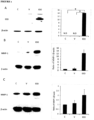
- a human IDO recombinant adenoviral vector was used for gene transduction in human dermal fibroblasts by a procedure previously reported (Li et al., 2004). Transfection efficiency was evaluated by detecting IDO protein expression and its activity through Western blot analysis and the kynurenine measurement in conditioned media, respectively. As shown in Figure 1A left panel, the IDO protein was expressed in IDO adenovirus-transduced fibroblasts, but undetectable in control and mock adenovirus-transduced fibroblasts.
- the level of kynurenine, an index for IDO activity, was significantly higher in IDO adenovirus-transduced fibroblasts (14.3 ⁇ 0.46 ⁇ g/ml, n 3) compared to those in untransduced or mock- transduced controls ( Figure 1A , right panel).
- IDO is an intracellular enzyme that converts tryptophan into kynurenine. Therefore, it must be clarified whether the effect of MMP-1 stimulation in IDO-expressing fibroblasts is due to the IDO protein itself or to tryptophan metabolites.
- conditioned media from both IDO-expressing fibroblasts and controls were collected after 48 hours. A combination of 90% collected conditioned media and 10% fresh media was then used to treat dermal fibroblasts. Cells were harvested 48 hours after treatment.
- IDO is an enzyme converting tryptophan into kynurenine.
- a factor either depletion of tryptophan or increase of kynurenine
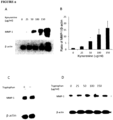
- IDO up-regulation of MMP-1 expression To examine what factor is responsible for IDO up-regulation of MMP-1 expression, fibroblasts were grown in either tryptophan-depleted cultured media or regular media with various concentrations of kynurenine. Cells were then evaluated for MMP-1 expression by western blotting. As shown in Figure 2C , there was no significant difference in the expression of MMP-1 between fibroblasts grown in the presence of 25 ⁇ g/ml tryptophan or in the tryptophan-depleted cultured media.
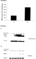
- conditioned media from fibroblasts in the presence or absence of 50 ⁇ g/ml of kynurenine were collected 48 hours after treatment.
- the MMP activity in the conditioned media was detected by a SensoLyte 520 TM generic MMP assay kit using a 5-FAM/QXL TM 520 fluorescence resonance energy transfer (FRET) peptide as a MMP substrate.
- FRET fluorescence resonance energy transfer
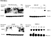
- Example 4 Mesenchymal and epithelial cells respond differently to kynurenine treatment
- mesenchymal cells such as an immobilized lung fibroblast cell line IMR-90 and fibroblast-like synoviocytes
- epithelial cells such as lung epithelial carcinoma cell line A549, primary dermal keratinocytes, human immobilized keratinocyte cell line HACAT, and head and neck squamous cell carcinoma cell line UMSCC
- MMP-1 expression in synoviocytes and IMR-90 were up-regulated by kynurenine treatments at concentrations of 12.5 ⁇ g/ml to 150 ⁇ g/ml, as shown in Figure 5 .
- MMP-1 mesenchymal and epithelial cells in response to kynurenine-stimulating MMP-1 expression.
- Example 5 Identification of the phosphorylated signal molecules by phospho-kinase array in cells treated with kynurenine
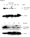
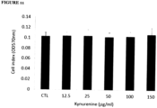
- kynurenine was tested for its effect on collagen expression and cell proliferation. As shown in Figure 10 (top), the addition of kynurenine 25-150 ⁇ g/ml remarkably decreases the expression of type 1 procollagen. However, it had no significant effect on fibroblast proliferation, even when the cells were cultured at concentrations up to 150 ⁇ g/ml of kynurenine ( Figure 11 ). Also, testing of the kynurenine analogues/metabolites, kynurenic acid and xanthurenic acid, demonstrate that these compounds are also effective at inhibiting expression of type 1 procollagen ( Figure 10 (bottom)).
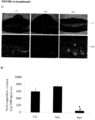
- Example 8 Topical application of kynurenine on rabbit ear wounds reduces scarring
- kynurenine can be used as an anti-fibrotic agent for the treatment or prevention of hypertrophic scarring.
- a rabbit ear hypertrophic scar model was used. Wounds were treated daily with o.1 ml of carboxymethyl cellulose (CMC) gel containing 50 ⁇ g of kynurenine for three weeks starting at day 8 post-wounding.
- CMC carboxymethyl cellulose
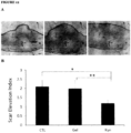
- Massons' trichrome staining for collagen revealed a significant reduction in collagen content in wounds treated with kynurenine, compared to those wounds receiving either no treatment or gel alone ( Figure 12C ). Consistent with this finding, the hydroxyproline content (used as an index for tissue collagen content) was significant lower in wounds treated with kynurenine compared to those wounds receiving either no treatment or gel alone ( Figure 12D ).
- kynurenine Different isoforms of kynurenine were tested for their ability to affect MMP-1 expression. Isoforms tested were DL-kynurenine (DL-Kyn) or D-kynurenine (D-Kyn) and L-kynurenine (L-Kyn). The result showed that all isoforms increase the MMP-1 expression in dermal fibroblasts, however, L-kynurenine seems to have more activity compared to other two isoforms - see Figure 14 .
- DL-Kyn DL-kynurenine
- D-Kyn D-kynurenine
- L-Kyn L-kynurenine
- Dermal fibroblasts were treated with either FS-1 (DL-kynurenine) or D-kynurenine or L-kynurenine or FS-2 (kynurenic acid) as shown in Figure 15 .
- Type-1, ⁇ 1-collagen expression was detected by real-time PCR. Results indicate that these isoforms/analogues have similar efficacy in reducing collagen expression.
- Example 11 Kynurenine and its metabolites down-regulate fibronectin expression in cultured fibroblasts
- Dermal fibroblasts were treated with various concentration of either DL-kynurenine (FS1), L-kynurenine, D-kynurenine or kynurenic acid (FS2) as shown in Figure 16 .
- FS1 DL-kynurenine
- L-kynurenine L-kynurenine
- D-kynurenine D-kynurenine
- FS2 kynurenic acid
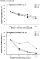
- the findings in Figure 17 showed that, there was almost 5-fold reduction in conA-induced splenocyte proliferation following treatment with 100 and 150 ⁇ g/ml D-Kynurenine, L-Kynurenine or DL-Kynurenine after 96 hours (P ⁇ 0.05), although splenocyte proliferation significantly reduced about 2-fold by D-Kynurenine, L-Kynurenine and DL-Kynurenine at 100 and 150 ⁇ g/ml after 48hours.
- FS2 has less effect on proliferation than other metabolites.
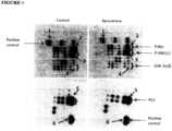
- the findings in Figure 18 showed that FS1 has immune suppressive effect on some of the proinflammatory cytokine and chemokine production, like IL-1, IL-2, CXCL9, and CXCL10. Besides it can significantly decrease IL-17 production which is thought to have an important role in inflammation.
Landscapes
- Health & Medical Sciences (AREA)
- Animal Behavior & Ethology (AREA)
- General Health & Medical Sciences (AREA)
- Chemical & Material Sciences (AREA)
- Veterinary Medicine (AREA)
- Public Health (AREA)
- Life Sciences & Earth Sciences (AREA)
- Medicinal Chemistry (AREA)
- Pharmacology & Pharmacy (AREA)
- Engineering & Computer Science (AREA)
- Bioinformatics & Cheminformatics (AREA)
- Chemical Kinetics & Catalysis (AREA)
- Nuclear Medicine, Radiotherapy & Molecular Imaging (AREA)
- General Chemical & Material Sciences (AREA)
- Organic Chemistry (AREA)
- Epidemiology (AREA)
- Physical Education & Sports Medicine (AREA)
- Immunology (AREA)
- Biomedical Technology (AREA)
- Cardiology (AREA)
- Heart & Thoracic Surgery (AREA)
- Urology & Nephrology (AREA)
- Reproductive Health (AREA)
- Dermatology (AREA)
- Vascular Medicine (AREA)
- Pulmonology (AREA)
- Orthopedic Medicine & Surgery (AREA)
- Rheumatology (AREA)
- Gastroenterology & Hepatology (AREA)
- Endocrinology (AREA)
- Pharmaceuticals Containing Other Organic And Inorganic Compounds (AREA)
- Acyclic And Carbocyclic Compounds In Medicinal Compositions (AREA)
- Medicines That Contain Protein Lipid Enzymes And Other Medicines (AREA)
Description
- The present invention relates to novel methods for the treatment of fibrosis. More specifically, the description provided herein relates to the use of kynurenine, kynurenic acid, xanthurenic acid, and/or related compounds for the treatment of fibrotic disease, in particular diseases or conditions of the skin such as keloids and hypertrophic scarring.
- Fibrosis, a disorder belonging to a group of fibroproliferative conditions, is seen in different organs such skin, liver, lung, kidney and arteries. It is estimated that approximately 40% of all deaths in the United States are caused, in part, by fibroproliferative disorders. Excessive accumulation of extracellular matrix due to either over production of matrix such as fibronectin, type I and III collagens, low levels of matrix degrading enzymes such as matrix metalloproteinases (MMPs) or both are the common features of all of these fibrotic conditions.
- As in all other organs, wound healing in the skin is a dynamic process involving tissue response to different types of insults. This process involves a continuous sequence of signals and responses in which platelets, fibroblasts, epithelial, endothelial and immune cells come together outside of their usual domain in order to orchestrate the very complex process of tissue repair. These signals, which are mainly growth factors (GFs) and cytokines, orchestrate the initiation, continuation and termination of wound healing (Scott et al. 1994). An imbalance in the synthesis and release of cytokines and GFs at the wound site may result in either retarded wound healing (e.g. in diabetic and elderly populations) or over-healing (e.g. fibroproliferative disorders, complication following surgical incision, traumatic wounds, and severe thermal injury). Thus, an important component of wound healing is its timely cessation and without such a timely cessation there may be a buildup of excess matrix, a deleterious fibrotic condition seen in millions of patients worldwide.
- Matrix metalloproteinases (MMPs) represent a group of diverse proteolytic enzymes involved in ECM turnover and connective tissue remodeling during physiological conditions such as embryonic growth and development, uterine involution, bone growth, bone resorption and wound healing. The level of MMP expression in normal cells is low and that allows healthy connective tissue remodeling. However, an imbalance in expression of MMPs has been implicated in a number of pathological conditions such as dermal fibrosis, rheumatoid arthritis, atherosclerosis, and tumor invasion and metastasis.
- Iannitti et al. (Am J Respir Crit Care Med vol. 187, no. 6, pp. 609-620, 15 March 2013) discloses the use of L-kynurenine and 3-hydroxy-kynurenine in the treatment of lung cystic fibrosis by restoring protective immunity to infection and improving lung inflammation.
-
EP 1,369,114 discloses pharmaceutical compounds, e.g. kynurenine, for use as a medicament for immunosuppressive treatment of autoimmune diseases, rheumatic diseases, recurrent abortion, or rejection in case of cell, tissue, or organ transplantations. -
WO 2008/087461 discloses kynurenic acid derivatives for the treatment of conditions characterized by hypermotility and inflammation of the gastrointestinal tract or gout.WO 2004/007461 discloses 8-hydroxy quinoline derivatives for the treatment of neurological conditions, more specifically neurodegenerative conditions such as Alzheimer's disease. - However, there is no disclosure in any one of these documents (Iannitti et al.;
EP 1,369,114 ;WO 2008/087461 ; orWO 2004/007461 ) of the treatment of a fibrotic disease selected from one or more of the following: keloid; hypertrophic scarring; pulmonary fibrosis; kidney fibrosis; liver cirrhosis; endomyocardial fibrosis; mediastinal fibrosis; myelofibrosis; retroperitoneal fibrosis; progressive massive fibrosis; nephrogenic systemic fibrosis; old myocardial infarction; scleroderma; systemic sclerosis; and uterine fibroids. - Chavez-Munoz et al. (Journal of Investigative Dermatology vol. 132, pp. 1501-1505, 1 May 2012); as well as Forouzandeh et al. (Wound Repair and Regeneration, vol. 18, no. 6, pp. 614-623, 18 October 2010) disclose the use of an
indoleamine 2,3-dioxygenase (IDO) expressing skin substitute to improve scar formation in a fibrotic animal model. - However, neither Chavez-Munoz et al., nor Forouzandeh et al., disclose the use of kynurenine, kynurenic acid, or xanthurenic acid. None of the documents provide sufficient evidence supporting the hypothesis that the reported anti-scarring properties of the IDO-expressing skin substitute are due to depletion of tryptophan and/or increase in IDO metabolites, let alone teach which particular IDO metabolites (e.g. kynurenine derivatives, NAD, quinolinic acid, or picolinic acid) might be responsible for the relevant activity.
- Poormasjedi-Meibod et al. (PLoS ONE, vol. 9, no. 3, pp. 1-13, 1 January 2014) discloses that KynA (kynurenic acid) is a promising candidate antifibrogenic agent to improve healing outcome in patients at risk of hypertrophic scarring. Yunyuan Li et al. (Journal of Investigative Dermatology, vol. 134, no. 3, pp. 643-650, 1 March 2014) discloses findings which suggest that kynurenine can potentially be used as an antifibrogenic agent for treating hypertrophic scarring. The documents by Poormasjedi-Meibod et al. and Yunyuan Li et al. are published after the priority date of the present patent application.
- Current treatment modalities for any fibrotic condition including dermal fibro-proliferating disorders such as hypertrophic scarring (HSc) and keloid remain unsatisfactory. Accordingly, it would be desirable to have therapeutic strategies for the treatment of various fibrotic diseases and conditions.
- The present invention is defined by the independent claims. The dependent claims depict other embodiments of the invention.
- Any embodiment not falling under the scope of the appended claims does not form part of the invention.
- The references to methods of treatment in the subsequent paragraphs of this description are to be interpreted as references to the compounds, pharmaceutical compositions and medicaments of the present invention for use in a method for treatment of the human (or animal) body by therapy (or for diagnosis).
- The present invention is based, in part, on the surprising discovery that certain compounds kynurenine and its analogues/isoforms, kynurenic acid, and xanthurenic acid - are capable of stimulating MMP1 and MMP3 expression, while inhibiting collagen and fibronectin expression. Furthermore, as described herein these compounds, when applied in vivo, are capable of inhibiting, preventing or reducing the formation of keloid scar.
- In one embodiment, there is provided a compound having the structure of kynurenic acid

- In another embodiment, there is provided a compound having the structure of xanthurenic acid

- In another embodiment, there is provided a compound having the structure of kynurenine

- In another embodiment, there is provided a pharmaceutical composition for use in the treatment of fibrotic disease, wherein the pharmaceutical composition comprises a compound having the structure of kynurenic acid, xanthurenic acid or kynurenine, or pharmaceutically acceptable salt thereof, and a pharmaceutically acceptable excipient, for use in the treatment of fibrotic disease wherein the fibrotic disease is selected from one or more of the following: keloid; hypertrophic scarring; pulmonary fibrosis; kidney fibrosis; liver cirrhosis; endomyocardial fibrosis; mediastinal fibrosis; myelofibrosis; retroperitoneal fibrosis; progressive massive fibrosis; nephrogenic systemic fibrosis; old myocardial infarction; scleroderma; systemic sclerosis; and uterine fibroids.
- In a preferred embodiment of the invention, the fibrotic disease is keloid or hypertrophic scarring. In another preferred embodiment of the invention, the fibrotic disease is pulmonary fibrosis. In yet another preferred embodiment of the invention, the fibrotic disease is liver cirrhosis. In yet another preferred embodiment of the invention, the fibrotic disease is kidney fibrosis. In yet another preferred embodiment of the invention, the fibrotic disease is endomyocardial fibrosis or scleroderma.
-
-
Figure 1 : Indoleamine 2, 3-Dioxygenase (IDO) up-regulation of MMP-1 expression in human dermal fibroblasts. Panel A shows fibroblasts that were transduced with either nothing (C), adenoviral vector (V) or a vector bearing the IDO recombinant gene (IDO) for 48 hrs, where IDO and its activity was detected by Western blotting (left Panel) and measurement of the kynurenine levels (right panel), respectively (N.D indicates the level of kynurenine was not detectable). Panel B shows both untreated, adenoviral vector, and IDO-transduced fibroblasts, that were lysed after being cultured for 48 hours, and the expression of MMP-1 was detected by Western blotting. Panel C shows fibroblasts that were incubated with the conditioned media taken from either control, empty vector or IDO adenoviral vector-transduced fibroblasts for 48 hours, where the expression of MMP-1 was analyzed by Western blotting. β-actin was used as a loading control in panels A, Band C. * indicates p <0.001. -
Figure 2 : Effects of Kynurenine and tryptophan on MMP-1 expression in human dermal fibroblasts. Panels A and B show dermal fibroblasts that were cultured in the presence of various concentrations of kynurenine for 48 hours, when the cells were harvested and lysed, before Western blotting was performed, showing the ratio of MMP-1 to β-actin is presented in panel B. Panel C shows dermal fibroblasts that were cultured in the presence or absence of tryptophan (25 mg/ml) for 48 hrs, when cells were harvested and lysed, and MMP-1 expression was evaluated by Western blotting. Panel D shows fibroblasts that were cultured in the presence of different concentrations of tryptophan for 48 hours, when the expression of MMP-1 was evaluated by Western blotting. β-actin was used for a loading control in all panels. -
Figure 3 : Effects of kynurenine on MMP-2 and -3 expression in human dermal fibroblasts - shows dermal fibroblasts that were cultured in the presence of various concentrations of kynurenine for 48 hours, before cells were harvested and lysed, and Western blotting was performed using either a rabbit monoclonal anti-human MMP-2 antibody (Panel A) or a mouse monoclonal anti-MMP-3 antibody (Panel B). Panel C shows the ratio of MMP-3 expression to b-actin for three independent experiments, β-actin was used for a loading control in all experiments. -
Figure 4 : Detect the activity of MMPs in conditioned media of human dermal fibroblasts using SensoLyte 520 generic MMP assay fluorimetric kit - shows fibroblasts that were cultured in the presence of (Kyn) or absence (CTL) of 50 µg/ml of kynurenine for 48 hours, when cell conditioned media was collected, then after centrifugation at 1000 × g for 10 minutes, supernatant was used to detect the activity of MMPs according to manufacture's instructions (media were incubated with 1mM APMA at 37°C for 3 hrs. 50 µl/well of MMP containing sample was then mixed with 50 µl of MMP substrate solution and medium from before cell culture was mixed with 50 µl of MMP substrate solution and used as a substrate control and afterincubation 1 hour, the fluorescence intensity at EX/EM=490 nm/520 nm were measured). The activity of MMPs are represented as relative fluorescence unit (RFU). Data was expressed as mean ± SD (n=3). * indicates P< 0.05. -
Figure 5 : Effects of kynurenine on MMP-1 expression in different types of mesenchymal cells -shows cells that were cultured and treated with kynurenine at concentrations of 12.5 to 150 µg/ml for 48 hours and MMP-1 expression was analysed by Western blotting and β-actin was used as a loading control in all experiments. Panel A shows MMP-1 expression in synoviocytes. Panel B shows MMP-1 expression in lung fibroblast cell line IMR-90. -
Figure 6 : Effects of kynurenine on MMP-1 expression in different types of epithelial cells -shows cells were cultured and treated with kynurenine at concentrations of 12.5 to 150 µg/ml for 48 hours and MMP-1 expression was analysed by western blotting and β-actin was used as a loading control in all experiments, where the top panels and bottom left panel, show fibroblast lysates from either untreated or kynurenine treated that were used as negative and positive controls, respectively. -
Figure 7 : Kynurenine stimulates ERK1/2 phosphorylation in human dermal fibroblasts - shows dermal fibroblasts that were cultured in the absence or presence of 100 µg/ml of kynurenine for 60 minutes, then cells were harvested and lysed with cell lysis buffer, before an antibody array was performed using a human phospho-kinase array kit (R & D System™), withspot 1, positive control;spot 2, phospho-P38α;spot 3, phosphor-ERK1/2;spot 4, phosphor-GSK-3α/β;spot 5, phosphor-P53;spot 6, positive control. -
Figure 8 : Kynurenine stimulation of MEK and ERK1/2 phosphorylation in human dermal fibroblasts - shows dermal fibroblasts that were cultured in the presence of 100 µg/ml of kynurenine at indicated time points, when cells were harvested and lysed, before Western blotting was performed by using either phosphorylated-MEK or phosphorylated-ERK1/2 antibody (β-actin was used as a loading control). -
Figure 9 - Addition of MEK or ERK1/2 phosphorylation inhibitors negates the effect of kynurenine-stimulating MMP-1 expression in dermal fibroblasts. Panel A: shows dermal fibroblasts were cultured in the absence or presence of 100 µg/ml kynurenine with or without various concentration of PD98059. Panel B: shows dermal fibroblasts were cultured in the absence or presence of 100 µg/ml kynurenine with or without 30 µM of PD98059 (ERK1/2 inhibitor), 30 µM of U0126 (MEK inhibitor) or 10 µM of U0126. MMP-1 expression was detected by Western blot (β-actin was used as a loading control for all experiments). -
Figure 10 - Effect of kynurenine, kynurenic acid, and xanthurenic acid onprocollagen type 1 expression in dermal fibroblasts - shows human dermal fibroblasts were treated with indicated concentrations of kynurenine for 48 hours (top), where cells were harvested and lysed with cell lysis buffer and a total 50 µg of protein was fractionated by 8% SDS-PAGE, before Western blotting was performed by using antibody against pro-collagen. β-actin was used a loading control, with kynurenic acid (KA) and xanthurenic acid (XA) also tested (bottom). -
Figure 11 - Effect of kynurenine on fibroblast proliferation - shows human dermal fibroblasts were cultured in the presence of indicated concentrations of kynurenine for 48 hours. MTT cell proliferation assay was performed as described herein, with cell proliferation indicated as cell index (OD570nm) in the MTT assay. -
Figure 12 - Clinical appearance and histology of wound and scars - shows rabbit ear wounds that were treated daily with either nothing (CTL), CMC gel alone (Gel), or 50 µg of kynurenine (Kyn) in 0.1 ml of CMC gel started fromday 8 for a total of 3 weeks. Panel A: shows the microscopic histology of wounds receiving either nothing (CTL), CMC gel (Gel) or kynurenine in CMC gel (Kyn) on Day 28 at magnification ×25. Panel B: shows the scar elevation index (SEI) as measured (Mean ± SD of SEI for untreated, CMC gel, and kynurenine in CMC gel-treated wounds) * shows a significant difference between kynurenine-treated and untreated controls (P<0.001); ** shows a significant difference between kynurenine and CMC gel control groups (P<0.01). Panel C: shows Massons' trichrome stained full-thickness skin sections from either untreated skin wound (left panels), cream treated skin wound (middle panels), or kynurenine treated wound (right panels) at both at magnification ×25 and ×100. Panel D: shows the total hydroxyproline content of skins from either untreated wounds (total 4 wounds), cream treated wounds (total 4 wounds), or kynurenine treated wounds (total 8 wounds) - * indicates p<0.01. -
Figure 13 - Topical application of kynurenine decreases type-1 on collagen and increases MMP-1 expression in rabbit ear skin - shows wounds in rabbit ear that were treated with either nothing (CTL) or gel alone (Gel) or kynurenine plus gel (Kyn) as described above, where skin wounds were used to extract total RNA by Trizol™ and 1 µg of RNA was used to synthesize cDNA for quantitative RT-PCR for type-1 α1 collagen, MMP-1 and β-actin. Panel A: shows the relative expression level of type-1 α1 collagen in rabbit ear skin tissue. Panel B: shows the relative expression level of MMP-1 in rabbit ear skin tissue - * indicates p< 0.05. -
Figure 14 - Effect of kynurenine isoform on MMP-1 expression in human dermal fibroblasts - shows dermal fibroblasts that were cultured in the absence (CTL) or presence of 50 µg/ml either DL-kynurenine (DL-Kyn) or D-kynurenine (D-Kyn) or L-kynurenine (L-Kyn) for 48 hours, at which time cells were harvested and lysed in protein lysis buffer (50 µg total protein was loaded on 10% SDS acrylamide gel) before Western blotting was performed with anti-human MMP-1 antibody, with β-actin as a loading control, which shows that all kynurenine isoforms tested increase MMP-1 expression in dermal fibroblasts, however, L-kynureine seems have more activity compared to other two isoforms. -
Figure 15 - Effects of kynurenine (FS1) analogues on collagen expression in human dermal fibroblasts - shows dermal fibroblasts that were treated with various concentration of either DL-kynurenine (FS1), L-kynurenine, D-kynurenine or kynurenic acid (FS2) and the corresponding collagen expression in mRNA levels as detected by real-time PCR, with β-actin as a loading control. -
Figure 16 - Effects of kynurenine (FS1) analogues on fibronectin expression in human dermal fibroblasts - shows dermal fibroblasts were treated with various concentration of either DL-kynurenine (FS1), L-kynurenine, D-kynurenine or kynurenic acid (FS2) and the corresponding fibronectin expression in mRNA levels as detected by real-time PCR, with β-actin as a loading control. -
Figure 17 - Comparing the suppressive effect of 50, 100, 150 µg/mL Tryptophan metabolites (FS1, LK, FS2, DK) on ConA-simulating splenocyte proliferation - shows that there was almost a 5-fold reduction in splenocyte proliferation following treatment with 100 and 150µg/ml of D-Kyn, L-Kyn, DL-Kyn (FS-1) and Kynurenic acid (FS2) after 96 hours (P<0.05), although splenocytes proliferation significantly reduced about 2-fold by D-Kyn, L-Kyn and DL-Kyn at 100 and 150µg/ml after 48hours, but FS2 showed less of an effect on proliferation. -
Figure 18 - Immune factor protein microarray in FS1 (DL-kynurenine) treated and untreated mouse splenocytes - shows that FS1 has immune suppressive effect on some proinflammatory cytokine and chemokine production, like IL-1, IL-2, CXCL9, and CXCL10 and FS1 shows a significant decrease in IL-17 production, which is thought to have an important role in inflammation. Panel A: shows activated splenocytes that were left untreated (ConA) or treated with 100µg/mL of Kyn (ConA+Kyn) for 48 hrs, at which time the conditioned media (CM) was collected from untreated and treated cells and was then exposed to a Proteome Profiler Antibody Array™ membrane with density value percentages are shown for both the untreated and treated cells for each reference spot as shown in Pane B. Panel B: shows signals identified by Proteome Profiler Antibody Array membrane. Panel C: shows spot number shown in panel B represents reference protein. -
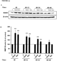
Figure 19 - Lasting effect of FS1 and FS2 on MMP1 expression in human dermal fibroblasts. Panel A: shows the lasting effect of kynurenine (FS1) and kynurenic acid (FS2) on MMP1 expression, where fibroblasts were treated with FS1 or FS2 (100 µg/ml) for 48 hours and the medium was replaced and cells were harvested immediately, and at 12, 24, and 48 hours after treatment removal, followed evaluation of MMP1 expression in dermal fibroblasts using Western blotting. Panel B: shows the MMP1/β-actin expression ratio as calculated in treated fibroblasts. Data is mean ± SEM of 4 independent experiments (*P-value<0.05 and **P-value<0.01, n=4). - Any terms not directly defined herein shall be understood to have the meanings commonly associated with them as understood within the art of the invention. As employed throughout the specification, the following terms, unless otherwise indicated, shall be understood to have the following meanings.
- As used herein a `subject' refers to an animal, such as a bird or a mammal. Specific animals include rat, mouse, dog, cat, cow, sheep, horse, pig or primate. A subject may further be a human, alternatively referred to as a patient. A subject may further be a transgenic animal. A subject may further be a rodent, such as a beaver, mouse or a rat.
- As used herein, an 'inhibitor' refers to a drug, compound or an agent that restrains or retards a physiological, chemical or enzymatic action or function. An inhibitor may cause at least 5% decrease in enzyme activity. An inhibitor may also refer to a drug, compound or agent that prevents or reduces the expression, transcription or translation of a gene or protein.
- '
Indoleamine 2, 3-Dioxygenase', or 'IDO', is a heme-containing rate limiting enzyme that catalyzes tryptophan to N-formylkynurenine and then to kynurenine (Kyn), and is found in non-hepatic cells mainly in macrophages and trophoblasts. Recent findings have implicated catabolism of tryptophan, an essential amino acid, by IDO as being involved in immune tolerance (Kahari and Saarialho-Kere 1997). As demonstrated herein, kynurenine, as well as its breakdown products kynurenic acid and xanthurenic acid, induce MMP-1 and MMP-3, as well as showing a reduction of fibrosis in vitro and in vivo. - The 'matrix metalloprotease', or 'MMP' family consist of 25 zinc- and calcium-dependent proteinases in the mammalian system. According to their substrate specificity, primary structure and cellular localization, 5 different subfamilies of closely related members known as collagenases, gelatinases, stromelysins, matrilysins, and membrane-type MMPs have been identified (Murphy et al. 2002). From all of these MMPs, MMP1 is the major enzyme involved in the collagenolytic process, breaking down the interstitial collagens such as types I, II, and III, while MMP-3 (stromelysin-1) is a protease known to degrade mainly the noncollagenous portion of the ECM such as fibronectin, proteoglycans, and laminin (Kahari and Saarialho-Kere 1997). Increases in both MMP1 and MMP-3 expressions and released by fibroblasts can initiate degradation of almost all major components of the ECM (Saus et al. 1988). It is now accepted that MMPs produced by keratinocytes facilitate epithelial migration, while MMPs expressed by fibroblasts promote tissue remodeling (Salo et al. 1991).
- 'Fibrosis' is a general terms that involves the formation or development of excess fibrous connective tissue in an organ or tissue as a reparative or reactive process, as opposed to a formation of fibrous tissue as a normal constituent of an organ or tissue. Scarring is confluent fibrosis that obliterates the architecture of the underlying organ or tissue. There are many diseases and/or conditions that are characterized by or associated with fibrosis, including, but not limited to: keloid, hypertrophic scar, pulmonary fibrosis, kidney fibrosis, liver cirrhosis, chronic inflammation of tunica albugenia (CITA), endomyocardial fibrosis, mediastinal fibrosis, myelofibrosis, retroperitoneal fibrosis, progressive massive fibrosis, nephrogenic systemic fibrosis, Crohn's disease, old myocardial infarction, scleroderma, and systemic sclerosis.
- There are provided herein a number of compounds for use in the treatment of diseases or conditions characterized by or related to fibrosis. In the context of the current description, the term 'treatment' may refer to treatment of existing fibrosis or fibrotic disease, or alternately may refer to treatment which occurs before or during the fibrotic process in order to prevent the development or progression of fibrosis. The compounds described herein may be in isolation, or may be linked to or in combination with tracer compounds, liposomes, carbohydrate carriers, polymeric carriers or other agents or excipients as will be apparent to one of skill in the art. In an alternate embodiment, such compounds may comprise a medicament, wherein such compounds may be present in a pharmacologically effective amount. The compounds may be suitable for administration to a subject in need thereof, by virtue of the fact that the subject may benefit from prophylaxis or treatment of fibrosis or fibrotic disease. The compounds may also include tautomers or stereoisomers.
- As used herein "FS" refers to FibroStops (for example, FS1 is used as an abbreviation for kynurenine (or DL-kynurenine or DL-Kyn) and FS2 or KA may be used as an abbreviation for kynurenic acid). L-kynurenine may be represented herein as L-Kyn and D-kynurenine may be represented herein as D-Kyn. Similarly, xanthurenic acid may be represented herein as XA.
- The term 'medicament' as used herein refers to a composition that may be administered to a patient or test subject and is capable of producing an effect in the patient or test subject. The effect may be chemical, biological or physical, and the patient or test subject may be human, or a non-human animal, such as a rodent or transgenic mouse, or a dog, cat, cow, sheep, horse, hamster, guinea pig, rabbit or pig. The medicament may be comprised of the effective chemical entity alone or in combination with a pharmaceutically acceptable excipient.
- The term 'pharmaceutically acceptable excipient' may include any and all solvents, dispersion media, coatings, antibacterial, antimicrobial or antifungal agents, isotonic and absorption delaying agents, and the like that are physiologically compatible. An excipient may be suitable for intravenous, intraperitoneal, intramuscular, subcutaneous, intrathecal, topical or oral administration. An excipient may include sterile aqueous solutions or dispersions for extemporaneous preparation of sterile injectable solutions or dispersion. Use of such media for preparation of medicaments is known in the art.
- Compositions or compounds according to some embodiments may be administered in any of a variety of known routes. Examples of methods that may be suitable for the administration of a compound include orally, intravenous, inhalation, intramuscular, subcutaneous, topical, intraperitoneal, intra-rectal or intra-vaginal suppository, sublingual, and the like. The compounds described herein may be administered as a sterile aqueous solution, or may be administered in a fat-soluble excipient, or in another solution, suspension, patch, tablet or paste format as is appropriate. A composition comprising the compounds described herein may be formulated for administration by inhalation. For instance, a compound may be combined with an excipient to allow dispersion in an aerosol. Examples of inhalation formulations will be known to those skilled in the art. Other agents may be included in combination with the compounds described herein to aid uptake or metabolism, or delay dispersion within the host, such as in a controlled-release formulation. Examples of controlled release formulations will be known to those of skill in the art, and may include microencapsulation, embolism within a carbohydrate or polymer matrix, and the like. Other methods known in the art for making formulations are found in, for example, "Remington's Pharmaceutical Sciences", (19th edition), ed. A. Gennaro, 1995, Mack Publishing Company, Easton, Pa.
- The dosage of the compositions or compounds of some embodiments described herein may vary depending on the route of administration (oral, intravenous, inhalation, or the like) and the form in which the composition or compound is administered (solution, controlled release or the like). Determination of appropriate dosages is within the ability of one of skill in the art. As used herein, an 'effective amount', a 'therapeutically effective amount', or a 'pharmacologically effective amount' of a medicament refers to an amount of a medicament present in such a concentration to result in a therapeutic level of drug delivered over the term that the drug is used. This may be dependent on mode of delivery, time period of the dosage, age, weight, general health, sex and diet of the subject receiving the medicament. Methods of determining effective amounts are known in the art.
- In one embodiment, there is provided a method for treatment of a subject having or suspected of having a fibrotic disease, the method comprising administering to the subject a therapeutically effective amount of a compound having a structure corresponding to Formula I, II, or III. The fibrotic disease may be one of the following: keloid, hypertrophic scar, pulmonary fibrosis, kidney fibrosis, liver cirrhosis, endomyocardial fibrosis, mediastinal fibrosis, myelofibrosis, retroperitoneal fibrosis, progressive massive fibrosis, nephrogenic systemic fibrosis, old myocardial infarction, scleroderma, systemic sclerosis, uterine fibroids.
- Neonatal foreskin and joints used as the sources of fibroblasts, keratinocytes and synoviocytes. The procedures were done based on the approval of Human Ethics Committee of the University of British Columbia. Cultures of human foreskin fibroblasts were established as described previously (Li et al., 2006). Briefly, foreskin was collected and washed three times with Dulbecco's Modified Eagle Medium (DMEM; GIBCO™, Grand Island, NY) supplemented with antibiotic-antimycotic preparation (100 u/ml penicillin, 100 µg /ml streptomycin, 0.25 µg/ml amphotericin B) (Invitrogen Life Technologies™, Gaithersburg, MD). Specimens were dissected free of fat and minced into small pieces less than 2.0 mm in diameter, washed six times with DMEM, distributed into 60 × 15-mm Petri dishes and incubated at 37 °C in a water-jacked humidified incubator in an atmosphere of 5% CO2. The medium was replaced twice weekly. Upon reaching confluence, the cells were released by trypsinization (0.1% trypsin, Invitrogen Life Technologies™) and (0.02% EDTA, Sigma™, St. Louis, MO), split for subculture at a ratio of 1:6, and reseeded onto 75-cm2 flasks. Fibroblasts from passages 3-7 were used for this study.
- Human foreskin keratinocytes were established as previously described (Ghahary et al., 1998). Cells were cultured in serum-free keratinocyte medium (KSFM; Invitrogen Life Technologies™) supplemented with bovine pituitary extract (50 µg/ml) and EGF (0.2 ng/ml). These cells were used at passages 2-5.
- Synoviocytes were obtained by enzymatic digestion of synovial membrane from patients with rheumatoid arthritis during joint replacement with 1 mg/ml collagenase (Sigma™) in
- RPMI1640 (Invitrogen Life Technologies™) for 4 hours at 37 °C. Dissociated cells were plated in synoviocyte growth medium (Cell Applications Inc.™, San Diego, CA) supplemented with penicillin G sodium (100 U/mL), streptomycin sulfate (100 µg/mL), and amphotericin B (0.25 µg/mL). Synoviocytes were found to be morphologically homogenous fibroblast-like cells and were used at passages 2-5.
- The squamous cell carcinoma (UMSCC) cell line derived from patients with head and neck cancer (ATCC™, Manassas, VA) were maintained in RPMI-1640 medium with 10% FBS. The Human keratinocyte cell line HACAT (ATCC) and the carcinomic human alveolar basal epithelial cell line A549 (ATCC™) were cultured in DMEM with 10% FBS. The diploid lung fibroblasts IMR-90 (ATCC™) were maintained in Minimum Essential Medium (MEM, Invitrogen™) with 10% FBS.
- The construction of
Indoleamine 2, 3-Dioxygenase (IDO) expressing adenoviral vector has been previously described (Li et al., 2004). Recombinant adenoviruses were used to infect human skin fibroblasts at the multiplicity of infection (MOI) of 100. Free viral particles were removed fromculture medium 30 hours after infection. The success of infection was determined by fluorescent microscopy using a Motic™ inverted microscope equipped with a fluorescein isothiocyanate (FITC) filter (Motic Instruments™, Richmond, BC, Canada) to view the reporter gene GFP. The expression of IDO was assessed by western blot using anti-human IDO antibody as described previously (Li et al., 2004). The biologic activity of IDO was evaluated by measuring the levels of tryptophan degrading product, kynurenine, present in conditioned medium. - The levels of kynurenine were measured by a method previously described (Tokikawa et al., 1988). In brief, about 2 ml of conditioned media was collected from the same cell number initiated
culture 3 days post transfection. Proteins from conditioned media were precipitated by trichloroacetic acid. After centrifugation to remove precipitated proteins, about 0.5 ml of supernatant was transferred into a new 1.5 ml tube and incubated with equal volume Ehrich's reagent (Sigma™) for 10 minutes at room temperature. The absorption of resultant solution was measured at 490 nm by spectrophotometer within 2 hours. The values of kynurenine in conditioned media were calculated by a standard curve with defined kynurenine concentration (0-20 µg/ml). - For collection of conditioned media, fibroblasts were transduced by either none or control mock vector or IDO adenovirus for 30 hours. Viruses were removed by washing with PBS. Fresh DMEM containing 10% FBS and antibiotics were added and cells were continued to be cultured for another 48 hours. Conditioned media from either untreated, mock vector, or IDO adenovirus transduced fibroblasts were then collected. Fibroblasts at 80% confluence were treated with media containing 90% of conditioned media plus 10% fresh media in the presence of 10% FBS. Cells were then harvested after 48 hours and western blot analysis was performed.
- In another set of experiments, fibroblasts at 80% confluence were treated with either kynurenine or tryptophan at the indicated concentrations as mentioned in the result section in DMEM containing 2% FBS and antibiotics for 48 hours. Cells were then harvested by trypsinization and western blot analysis was performed.
- Similarly, other cells such as synoviocytes, IMR-90, keratinocytes, UMSCC and A549 were treated with kynurenine at concentrations of 12.5 to150 µg/ml in appropriate media for each cell type as described above for 48 hours. Cells were then harvested for western blot analysis.
- Cells were harvested by Trypsin/EDTA and lysed with cell lysis buffer containing 50 mM Tris-HCl (pH7.40), 150 mM NaCl, 10 mM EDTA, 5 mM EGTA, 1% TritonX-100™, 0.5% Igepal CA-630, 0.025% NaN3 and protease inhibitor cocktail (Sigma™). Cell debris was removed by centrifugation at 20,000 × g for 10 minutes. The protein concentration in supernatant was determined using the MicroBCA™ method (Pierce™, Rockford, IL). Proteins in supernatant were mixed with protein sample loading buffer (final concentration: 60 mM Tris-HCl (pH 6.80), 2% SDS, 10% glycerol, 1.5% β-mercaptoethanol, 0.002% bromophenol blue) and size fractioned by 10% of SDS-polyacrylamide gel. After proteins were transferred onto nitrocellulose membrane by iBlot™ (Invitrogen Life Technologies™), non-specific binding were blocked with phosphate buffer saline twenty20 (PBS-T) containing 5% skim milk for 1 hour. The membrane was then incubated with primary antibody overnight. After incubation with a secondary antibody for 1 hour, protein bands were visualized by an enhanced chemiluminescence (ECL™) detection system (Santa Cruz Biotechnology™, Santa Cruz, CA). The primary antibodies used in this study were: mouse monoclonal anti- human MMP-1 (R&D Systems™, Minneapolis, MN), mouse monoclonal anti- human MMP-3 (R&D System™), rabbit monoclonal anti-human MMP-2 (Epitomics™, Burlingame, CA), rabbit polyclonal anti-phospho-MEK1/2 (Ser217/221™) (Cell Signaling Technology™, Danvers, MA), rabbit polyclonal anti-phospho-p44/42 MAPK (Thr202/Tyr204) (Cell Signaling Technology™), monoclonal anti-β-actin (Sigma™), and mouse anti-type-i procollagen (Developmental Studies Hybridoma Bank™, Iowa City, IA). The secondary antibodies were either goat anti-mouse IgG (H+L) HPR conjugate or goat anti-rabbit IgG (H+L) HPR conjugate (Bio-rad Laboratory™ (Mississauga, ON, Canada). Secondary antibodies were used at a concentration of 1:3000.
- The activity of MMPs was assessed using a F-FAM/QXL™ 520 fluorescence resonance energy transfer (FRET) peptide as the MMP substrate (SensoLyte 520™ generic MMP assay kit, AnaSpec, Inc.™, Fremont, CA) according to the manufacturer's protocol. In brief, cells were treated with or without 50 µg/ml of kynurenine for 48 hours. Conditioned media were collected and incubated with 1mM of APMA (4-aminophenyl-mercuric acetate, in component C, AnaSpect™) at 37 °C for 3 hrs. After activation MMPs with APMA, 50 µl/well in 96-well plate of conditioned media was mixed with 50 µl of MMP substrate solution. After incubated at room temperature for 60 minutes, the fluorescence intensity at EX/EM=490 nm/520 nm in each sample including the substrate control were measured using Infinite F500™ fluorescence microplate reader (Tecan Group Ltd™, Morrisville, NC).
- Human fibroblasts at 90% confluence were starved in DMEM without FBS overnight followed by the treatment with or without 100 µg/ml of kynurenine for 2 hours. Protein phosphorylation was evaluated using the Human Phospho-Kinase Array™ (R&D System™) according to the manufacturer's instructions. Briefly, capture and control antibodies were spotted in duplicate on nitrocellulose membranes (total 46 kinase phosphorylation sites). Cell lysates (300 µg of total protein per array) were incubated with array overnight. The array was washed to remove unbound proteins, followed by incubation with the cocktail of biotinylated detection antibodies. After incubation with streptavidin-HPR for 30 minutes, signals were visualized by ECL detection system (Santa Cruz™). Blots were analyzed by densitometry, and protein phosphorylation was normalized to a positive control which was represented in each membrane.
- Female rabbits (New Zealand white) weighing 4.5-5 kg were used for this study. The protocol was reviewed and approved by the University of British Columbia animal care committees. The rabbit ear model of hypertrophic scar was created as described previously (Rahmani-Neishaboor, et al., 2010). Briefly, 2 rabbits were anesthetized by intramuscular injection of ketamine (22.5 mg/kg) and xylazine (2.5 mg/kg) followed by isoflurane gas through tracheal intubation. Four wounds were created down to bare cartilage on the ventral side of each ear using an 8-mm dermal biopsy punch to remove full-thickness sections of skin. Antibiotics were applied on wounds daily until kynurenine treatment was started.
- Kynureine in CMC gel (Rahmani-Neishaboor et al., 2010) with a concentration of 500 µg/ml was applied topically to the wounds of the experimental group (0.1 ml per wound) daily for 3 weeks starting at 1 week post wounding. The wounds of the control group were received the treatment with an equal amount of cream alone daily.
- Animals were sacrificed on
weeks 3 after treatments. Scars (10 mm punch biopsies) were harvested. Each scar was sectioned in two along its longitudinal axis and half of which was processed for routine histological analysis and another half was kept at -80°C for future use. - Scar elevation was quantified by measuring Scar Elevation Index (SEI) from the H & E stained tissue section. The SEI is a ratio of total height in the wound tissue to the normal tissue below the hypertrophic scar. A SEI of 1 indicates that the scar height is equal to the surrounding unwounded dermis; an SEI > 1 indicates a raised hypertrophic scar.
- The effect of kynurenine on human dermal fibroblast proliferation was detected by MTT [3-(4, 5-Dimethylthiazol-2-yl)-2, 5-diphenyltetrazolium bromide] assay. In brief, 10,000 cells were seeded on a 24 well-plate and incubated with different concentrations of kynurenine for 48 hours. Media were removed and 0.2 ml of MTT (5 mg/ml in DMEM containing 2% FBS) was added. Cells were incubated with MTT for 4 hours. After washing 3 times with PBS, 0.2 ml of DMSO was added to dissolve the crystals. Absorbance was measured at 570 nm.
- Measurement of hydroxyproline content from skin sample: According to a method previously reported (Gawronskao-Kozak B. et al. 2006), half of 8 mm diameter skin punches were weighed and frozen in -80°C. Skins were homogenized in 2 ml of PBS and stored at 4 °C overnight. The next day, 1 ml of 6N HCl was added and the mixture was heated at 120 °C for 5 hours. 20 µl of cooled samples and 50 µl of chloramine T solution were added to the 96-well plate and incubated at room temperature for 20 minutes. 50 µl of Erlich solution was then added and the mixture was incubated at 65 °C for 15 minutes. Absorbance was read at 570 nm. Hydroxyproline concentration was calculated by a standard curve.
- RNA was extracted by Trizol™ (Invitrogen Life Technologies™). Briefly, 1 ml of Trizol™ was added to the homogenized skin tissue. 250 µl of chloroform was added after the mixture was standed at room temperature for 5 minutes. Top aqueous phase was transferred into a new eppendorf tube after centrifugation for 10 minute at 20,000 × g. Equal volume isopropanol was added to the aqueous phase and mixed gently. The pellet was washed with 1 ml of 75% ethanol after centrifugation for 20 minutes. RNA was dissolved in DEPC treated H2O and its concentration was measured by Nanodrop2000™. cDNA was synthesized by cDNA synthesis kit from Roche according to manufacture's introduction using1 µg of total RNA in each sample. Quantitative real-time PCR for rabbit type-1 α1 collagen, MMP-1 and housekeeper gene β-actin were performed in ViiA7 (Invitrogen™). cDNA samples were added to a PCR reaction master mix containing STBR Green Master Mix™ (Rox) (Roche™, Indianapolis, IN). All reactions were performed in duplicate using the following cycle conditions: 1 cycle of 95°C for 10 minutes, 40 cycles of 95°C for 15 seconds and 60 °C for 1 minute. The expression level of type-1 α1 collagen and MMP-1 in each sample was normalised to β-actin. RT-PCR primers: rabbit type-1 α1 collagen: 5'-ACAAGGGTGAGACAGGCGAAC-3' (Forward), 5'-GCCGTTGAGTCCATCTTTCCC-3' (Reverse); MMP-1, 5'-TCTGGCCACATCTGCCAATGG-3' (Forward), 5'-AGGGAAGCCAAAGGAGCTGTG-3' (Reverse); β-actin, 5'-AACGAGCGCTTCCGTTGGCCC-3' (Forward), 5'-CTTCTGCATGCGGTCCGCGA-3'(Reverse).
- To assess the effect of IDO on MMP-1 expression, a human IDO recombinant adenoviral vector was used for gene transduction in human dermal fibroblasts by a procedure previously reported (Li et al., 2004). Transfection efficiency was evaluated by detecting IDO protein expression and its activity through Western blot analysis and the kynurenine measurement in conditioned media, respectively. As shown in
Figure 1A left panel, the IDO protein was expressed in IDO adenovirus-transduced fibroblasts, but undetectable in control and mock adenovirus-transduced fibroblasts. The level of kynurenine, an index for IDO activity, was significantly higher in IDO adenovirus-transduced fibroblasts (14.3 ± 0.46 µg/ml, n=3) compared to those in untransduced or mock- transduced controls (Figure 1A , right panel). - The expression of MMP-1 in control, mock- transduced and IDO-expressing fibroblasts was examined by using Western blot analysis. As shown in
Figure 1B , there was a more than nine fold increase in MMP-1 expression in IDO-expressing fibroblasts (12.56 ± 2.37, n=3) as compared to those in mock-transduced (1.37 ± 0.59, n=3) and untreated control fibroblasts (1 ± o, n=3). This finding suggests that up-regulation of MMP-1 expression in IDO-expressing fibroblasts is not due to adenovirus infection, since the mock-transduced fibroblasts showed no significant difference in MMP-1 expression from the untreated fibroblasts. - IDO is an intracellular enzyme that converts tryptophan into kynurenine. Therefore, it must be clarified whether the effect of MMP-1 stimulation in IDO-expressing fibroblasts is due to the IDO protein itself or to tryptophan metabolites. To address this, conditioned media from both IDO-expressing fibroblasts and controls were collected after 48 hours. A combination of 90% collected conditioned media and 10% fresh media was then used to treat dermal fibroblasts. Cells were harvested 48 hours after treatment. As shown in
Figure 1C , a significant increase in MMP-1 expression was observed in cells treated with conditioned media from IDO-transduced fibroblasts (2.06 ± 0.62, n=3) as compared to those in either mock-transduced (1.16 ± 0.31, n=3) or untreated control fibroblasts (1 ± 0, n=3). This result suggests that a factor or factors in conditioned media from IDO adenovirus infected fibroblast rather than intracellular IDO protein is responsible for an increased level of MMP-1 expression in fibroblasts. - IDO is an enzyme converting tryptophan into kynurenine. To examine what factor (either depletion of tryptophan or increase of kynurenine) is responsible for IDO up-regulation of MMP-1 expression. To examine what factor is responsible for IDO up-regulation of MMP-1 expression, fibroblasts were grown in either tryptophan-depleted cultured media or regular media with various concentrations of kynurenine. Cells were then evaluated for MMP-1 expression by western blotting. As shown in
Figure 2C , there was no significant difference in the expression of MMP-1 between fibroblasts grown in the presence of 25 µg/ml tryptophan or in the tryptophan-depleted cultured media. However, the MMP-1 expression was significantly increased in response to different doses (25-150 µg/ml) of kynurenine (Figure 2A and Figure 2B ). These findings suggest that the presence of kynurenine, but not tryptophan depletion, contributes to the up-regulation of MMP-1 in IDO-expressing cells. Furthermore, we found that as little as 12.5 µg/ml of kynurenine could stimulate MMP-1 expression in dermal fibroblasts (data not shown). This concentration of kynurenine is similar to that detected in conditioned media from IDO expressing fibroblasts (Figure 1A right panel). The stimulation of MMP-1 in fibroblasts is thus clearly specific to kynurenine as the addition of various concentration of tryptophan with a similar structure failed to increase the expression of MMP-1 in dermal fibroblasts (Figure 2D ). - To investigate whether kynurenine also affects the expression of other MMPs, we treated dermal fibroblasts with kynurenine at similar concentrations to those used in
Figure 2 . Western blotting was used to detect MMP-2 and -3 expression using untreated cells as controls. As shown inFigure 3A , there was no significant difference in MMP-2 expression between kynurenine-treated and untreated fibroblasts. However, under similar conditions, kynurenine treatment significantly increased MMP-3 expression in dermal fibroblasts in a dose-dependent manner (Figure 3B/3C ). Furthermore, to test whether the increased levels of MMPs in kynurenine-treated fibroblasts were followed by increased MMP activity, conditioned media from fibroblasts in the presence or absence of 50 µg/ml of kynurenine were collected 48 hours after treatment. The MMP activity in the conditioned media was detected by a SensoLyte 520™ generic MMP assay kit using a 5-FAM/QXL™520 fluorescence resonance energy transfer (FRET) peptide as a MMP substrate. As shown inFigure 4 , the mean activity of MMPs in conditioned media from the kynurenine treated fibroblast was significantly higher than in the control media. This indicates that the increased MMPS in fibroblasts treated by kynurenine have enzymatic activity. - To determine what types of cells are sensitive to kynurenine-induced MMP-1 expression, both mesenchymal cells (such as an immobilized lung fibroblast cell line IMR-90 and fibroblast-like synoviocytes) and epithelial cells (such as lung epithelial carcinoma cell line A549, primary dermal keratinocytes, human immobilized keratinocyte cell line HACAT, and head and neck squamous cell carcinoma cell line UMSCC) were used. As with the dermal fibroblasts, MMP-1 expression in synoviocytes and IMR-90 were up-regulated by kynurenine treatments at concentrations of 12.5 µg/ml to 150 µg/ml, as shown in
Figure 5 . However, the expression of MMP-1 in all epithelial cells tested, including dermal keratinocytes, HACAT, A549 and UMSCC, did not significantly differ from the untreated controls in response to the various concentration of kynurenine (Figure 6 ). These results suggest that there is a difference between mesenchymal and epithelial cells in response to kynurenine-stimulating MMP-1 expression. - To determine the possible mechanism of kynurenine up-regulated MMP-1 expression in dermal fibroblasts, we analyzed the activation of multiple serine, threonine or tyrosine kinases, using a phosphor-kinase array. This array gives the possibility of simultaneously detecting the activation status of 46 different protein kinases and their downstream transcript factors. As shown in
Figure 7 , after 1 hour of treatment in dermal fibroblasts with kynurenine, extracellular signal-regulatedkinases 1/2 (ERK1/2) was activated. - To confirm these results from the phospho-kinase array, dermal fibroblasts were treated with 100 µg/ml of kynurenine at different times. Immunoblotting analysis, using a different antibody from those placed on the array, was then used to detect the phosphorylation of ERK1/2 and its upstream molecule mitogen-activated protein/extracellular signal-regulated kinase kinase (MEK). As shown in
Figure 8 , ERK1/2 was phosphorylated in cells treated with kynurenine. The result was further confirmed by detection of the ERK1/2 upstream signal molecule MEK phosphorylation in cells treated with kynurenine (Figure 8 ). Both ERK1/2 and MEK showed similar patterns of activation, with a peak at 8 hours following kynurenine treatments (Figure 8 ). - In another set of experiments, we tested whether the activation of the MEK-ERK1/2 MAPK pathway by kynurenine is associated with kynurenine-stimulating MMP-1 expression in dermal fibroblasts. To do this, we examined the effects of inhibitors of either MEK or ERK1/2 phosphorylation on kynurenine-stimulating MMP-1 expression. As shown in
Figure 9A , the addition of PD98059, a specific inhibitor for ERK1/2 activation effectively prevented the stimulatory effect of kynurenine on MMP-1 expression, in a dose-dependent manner. Similarly, treatment of cells with 10 µM and 30 µM of U0126, a specific inhibitor for MEK activation, also significantly reduced the up-regulation of MMP-1 expression by kynurenine (Figure 9B ). These results demonstrate that the activation of the MEK-ERK1/2 signaling pathway contributes to the up-regulation of MMP-1 expression induced by kynurenine in dermal fibroblasts. - Before studying its anti-fibrotic role in vivo, kynurenine was tested for its effect on collagen expression and cell proliferation. As shown in
Figure 10 (top), the addition of kynurenine 25-150 µg/ml remarkably decreases the expression oftype 1 procollagen. However, it had no significant effect on fibroblast proliferation, even when the cells were cultured at concentrations up to 150 µg/ml of kynurenine (Figure 11 ). Also, testing of the kynurenine analogues/metabolites, kynurenic acid and xanthurenic acid, demonstrate that these compounds are also effective at inhibiting expression oftype 1 procollagen (Figure 10 (bottom)). - Since treatment of dermal fibroblasts with kynurenine showed an increase in both the MMP-1 and -3 expression as well as a decrease in type-1 procollagen expression, we were interested to know whether kynurenine can be used as an anti-fibrotic agent for the treatment or prevention of hypertrophic scarring. To achieve this, as described previously (Rahmani-Neishaboor et al., 2010; Kloeters et al., 2007; Xie et al., 2008), a rabbit ear hypertrophic scar model was used. Wounds were treated daily with o.1 ml of carboxymethyl cellulose (CMC) gel containing 50 µg of kynurenine for three weeks starting at
day 8 post-wounding. The dose of 50 mg kynurenine per wound was matched with that used in an in vitro system with an optimum outcome. The result showed no significant difference to wound closure in kynurenine-treated wounds as compared to that of either untreated or CMC gel treated controls (data not shown). However, as shown inFigure 12A , significantly less scarring was seen in wounds treated with kynurenine than either non-treated wounds or the vehicle-only control wounds after three weeks. The average scar elevation index (SEI) was significantly reduced in the kynurenine-treated group (1.172 ± 0.156, n=8) as compared to the vehicle-only control group (1.978 ± 0.442, n=4, p<0.01) and the untreated group (2.098 ± 0.324, n=4, p<0.001) (Figure 12B ). Massons' trichrome staining for collagen revealed a significant reduction in collagen content in wounds treated with kynurenine, compared to those wounds receiving either no treatment or gel alone (Figure 12C ). Consistent with this finding, the hydroxyproline content (used as an index for tissue collagen content) was significant lower in wounds treated with kynurenine compared to those wounds receiving either no treatment or gel alone (Figure 12D ). - Finally, we demonstrated that topical application of kynurenine in a rabbit ear fibrotic model decreased the expression of type-1 α1 collagen and increased the expression of MMP-1, as compared to those in wounds received either no treatment or gel alone (
Figure 13 ). These results further support the supposition that kynurenine could potentially be used as an anti-fibrotic factor for treating hypertrophic scarring and even keloid, as frequently seen in patients with burn injuries or surgical incisions. - Different isoforms of kynurenine were tested for their ability to affect MMP-1 expression. Isoforms tested were DL-kynurenine (DL-Kyn) or D-kynurenine (D-Kyn) and L-kynurenine (L-Kyn). The result showed that all isoforms increase the MMP-1 expression in dermal fibroblasts, however, L-kynurenine seems to have more activity compared to other two isoforms - see
Figure 14 . - Dermal fibroblasts were treated with either FS-1 (DL-kynurenine) or D-kynurenine or L-kynurenine or FS-2 (kynurenic acid) as shown in
Figure 15 . Type-1, α1-collagen expression was detected by real-time PCR. Results indicate that these isoforms/analogues have similar efficacy in reducing collagen expression. - Dermal fibroblasts were treated with various concentration of either DL-kynurenine (FS1), L-kynurenine, D-kynurenine or kynurenic acid (FS2) as shown in
Figure 16 . The expression of fibronectin was detected by real-time PCR. Results demonstrate that kynurenine, DL-kynurenine, and L-kynurenine are all capable of down-regulating fibronectin expression, indicating that kynurenine metabolites may be also suitable for prevention or treatment of fibroproliferative disorders. - The findings in
Figure 17 showed that, there was almost 5-fold reduction in conA-induced splenocyte proliferation following treatment with 100 and 150µg/ml D-Kynurenine, L-Kynurenine or DL-Kynurenine after 96 hours (P<0.05), although splenocyte proliferation significantly reduced about 2-fold by D-Kynurenine, L-Kynurenine and DL-Kynurenine at 100 and 150µg/ml after 48hours. FS2 has less effect on proliferation than other metabolites. The findings inFigure 18 showed that FS1 has immune suppressive effect on some of the proinflammatory cytokine and chemokine production, like IL-1, IL-2, CXCL9, and CXCL10. Besides it can significantly decrease IL-17 production which is thought to have an important role in inflammation. - To determine the lasting effect of kynurenic acid (KynA) and kynurenine (Kyn) on MMP1 expression in fibroblasts, cells were treated with 100 µg/ml of the drug. Following 48 hours of treatment, the medium was changed with fresh medium and cells were then harvested at 0, 12, 24 or 48 hours post treatment removal. There was a marked increase in MMP1 expression in fibroblasts in response to either KynA or Kyn treatment at 48 hours after treatment. Following the removal of Kyn and KynA, the MMP1 expression remained significantly higher than the untreated cells for another 24 hours (
Figure 19A ). Interestingly, while the MMP1 protein expression gradually reduced to normal levels within 48 hours after Kyn removal, the MMP1 expression in response to KynA remained higher than controls (Figure 19A). Figure 19B represents the quantitative analysis of data inFigure 19A (* P-value<0.05, ** P-value<0.01, n=4). From these results it appears that KynA has a longer lasting effect on expression of MMP-1 relative to Kyn in treated fibroblasts. - Although various embodiments are disclosed herein, many adaptations and modifications may be made within the scope of the invention in accordance with the common general knowledge of those skilled in this art. Such modifications include the substitution of known equivalents for any aspect of the invention in order to achieve the same result in substantially the same way. Numeric ranges are inclusive of the numbers defining the range. The word "comprising" is used herein as an open ended term, substantially equivalent to the phrase "including, but not limited to", and the word "comprises" has a corresponding meaning. As used herein, the singular forms "a", "an" and "the" include plural referents unless the context clearly dictates otherwise. Thus, for example, reference to "a thing" includes more than one such thing. Citation of references herein is not an admission that such references are prior art to an embodiment of the present invention. The invention includes all embodiments and variations substantially as hereinbefore described and with reference to the examples and drawings.
-
- Gawronskao-Kozak B. et al. Wound Repair Regeneration (2006) Scarless skin repair in immunodeficient mice 14:265-276.
- Ghahary A, Tredget EE, Chang LJ, et al. (1998) Genetically modified dermal keratinocytes express high levels of transforming growth factor-β1. J Invest Dermatol 110:800-805.
- Kahari, V. M. and U. Saarialho-Kere (1997). "Matrix metalloproteinases in skin." Exp Dermatol 6(5): 199-213.
- Kloeters O, Tandara A, Mustoe T (2007) Hypertrophic scar model in the rabbit ear: a reproducible model for studying scar tissue behavior with new observation on silicone gel sheeting for scar reduction. Wound Repair Regen 15:S40-S45.
- Li Y, Tredget EE, Ghaffari A, et al. (2006) Local expression of .
- Li Y, Tredget EE, Kilani RT, et al. (2004) Expression of .
- Rahmani-Neishaboor E, Yau FM, Jalili R, et al. (2010) Improvement of hypertrophic scarring by using topical anti-fibrogenic/anti-inflammatory factors in a rabbit ear model. Wound Repair Regen 18:401-408.
- Salo, T., J. G. Lyons, et al. (1991). "Transforming growth factor-.
- Saus, J., S. Quinones, et al. (1988). "The complete primary structure of human matrix metalloproteinase-3. Identity with stromelysin." J Biol Chem 263(14): 6742-5.
- Scott, P. G., A. Ghahary, et al. (1994). In: Advances in Structural Biology 3: 157-201.
- Tokikawa O, Kuroiwa T, Yamazaki F, et al. (1988) Mechanism of interferon-γ action: characterization of .
- Xie JL, Bian HN, Qi SH, et al. (2008) Basic fibroblast growth factor (bFGF) alleviates the scar of the rabbit ear model in wound healing. Wound Repair Regen 16:576-581.
Claims (14)
- A compound having the structure of kynurenic acid

- A compound having the structure of xanthurenic acid

- A compound having the structure of kynurenine

- The compound for use according to any one of claims 1 to 3, wherein the fibrotic disease is keloid or hypertrophic scarring.
- The compound for use according to any one of claims 1 to 3, wherein the fibrotic disease is pulmonary fibrosis.
- The compound for use according to any one of claims 1 to 3, wherein the fibrotic disease is liver cirrhosis.
- The compound for use according to any one of claims 1 to 3, wherein the fibrotic disease is kidney fibrosis.
- The compound for use according to any one of claims 1 to 3, wherein the fibrotic disease is endomyocardial fibrosis or scleroderma.
- A pharmaceutical composition for use in the treatment of fibrotic disease, wherein the pharmaceutical composition comprises a compound for use according to any one of claims 1-3 or pharmaceutically acceptable salt thereof and a pharmaceutically acceptable excipient, wherein the fibrotic disease is selected from one or more of the following: keloid; hypertrophic scarring; pulmonary fibrosis; kidney fibrosis; liver cirrhosis; endomyocardial fibrosis; mediastinal fibrosis; myelofibrosis; retroperitoneal fibrosis; progressive massive fibrosis; nephrogenic systemic fibrosis; old myocardial infarction; scleroderma; systemic sclerosis; and uterine fibroids.
- The pharmaceutical composition for use according to claim 9, wherein the fibrotic disease is keloid or hypertrophic scarring.
- The pharmaceutical composition for use according to claim 9, wherein the fibrotic disease is pulmonary fibrosis.
- The pharmaceutical composition for use according to claim 9, wherein the fibrotic disease is liver cirrhosis.
- The pharmaceutical composition for use according to claim 9, wherein the fibrotic disease is kidney fibrosis.
- The pharmaceutical composition for use according to claim 9, wherein the fibrotic disease is endomyocardial fibrosis or scleroderma.
Applications Claiming Priority (2)
| Application Number | Priority Date | Filing Date | Title |
|---|---|---|---|
| US201361831404P | 2013-06-05 | 2013-06-05 | |
| PCT/CA2014/000484 WO2014194407A1 (en) | 2013-06-05 | 2014-06-04 | Anti-fibrogenic compounds, methods and uses thereof |
Publications (3)
| Publication Number | Publication Date |
|---|---|
| EP3003298A1 EP3003298A1 (en) | 2016-04-13 |
| EP3003298A4 EP3003298A4 (en) | 2017-03-22 |
| EP3003298B1 true EP3003298B1 (en) | 2024-03-20 |
Family
ID=52007349
Family Applications (1)
| Application Number | Title | Priority Date | Filing Date |
|---|---|---|---|
| EP14806967.7A Active EP3003298B1 (en) | 2013-06-05 | 2014-06-04 | Anti-fibrogenic compounds, methods and uses thereof |
Country Status (14)
| Country | Link |
|---|---|
| US (1) | US9737523B2 (en) |
| EP (1) | EP3003298B1 (en) |
| JP (1) | JP6473938B2 (en) |
| KR (1) | KR102294405B1 (en) |
| AU (1) | AU2014277568B2 (en) |
| CA (1) | CA2914261C (en) |
| DK (1) | DK3003298T3 (en) |
| ES (1) | ES2978920T3 (en) |
| PH (1) | PH12015502717B1 (en) |
| PL (1) | PL3003298T3 (en) |
| RU (1) | RU2726416C2 (en) |
| SG (1) | SG11201510003VA (en) |
| WO (1) | WO2014194407A1 (en) |
| ZA (1) | ZA201600015B (en) |
Families Citing this family (5)
| Publication number | Priority date | Publication date | Assignee | Title |
|---|---|---|---|---|
| WO2018144461A1 (en) | 2017-01-31 | 2018-08-09 | The Board Of Regents Of The University Of Oklahoma | Animal wound model and methods of use |
| CN112168822B (en) * | 2020-09-27 | 2022-11-08 | 集美大学 | The use of kynuric acid in improving dyslipidemia, obesity and intestinal flora disorder caused by high-fat diet |
| US12150461B2 (en) * | 2021-05-04 | 2024-11-26 | Sovida Solutions Ltd. | Nicotinamide adenine dinucleotide (NAD) compositions, methods of manufacturing thereof, and methods of use thereof |
| KR20240004954A (en) * | 2021-05-07 | 2024-01-11 | 바치바이오메드 인크. | METHODS OF USING A SMALL MOLECULE CHEMICAL COMPOUND TO REDUCE THE APPEARANCE OF POST-ACNE ATROPHIC SCARRING |
| CN119055650A (en) * | 2024-09-06 | 2024-12-03 | 上海市东方医院(同济大学附属东方医院) | Application of Kynurenic Acid in the Treatment of Chronic Liver Cirrhosis |
Family Cites Families (6)
| Publication number | Priority date | Publication date | Assignee | Title |
|---|---|---|---|---|
| TWI233805B (en) * | 1999-07-01 | 2005-06-11 | Fujisawa Pharmaceutical Co | Stabilized pharmaceutical composition in lyophilized form as antifungal agent |
| EP1369114A1 (en) * | 2002-06-07 | 2003-12-10 | Peter Priv. Doz. Dr. Terness | Use of tryptophan metabolites as pharmaceutical agents |
| AU2002950217A0 (en) * | 2002-07-16 | 2002-09-12 | Prana Biotechnology Limited | 8- Hydroxy Quinoline Derivatives |
| JP2008520587A (en) * | 2004-11-17 | 2008-06-19 | アンジオゲン ファーマシューティカルズ ピーティーワイ リミテッド | Methods for regulating B cell function |
| HUP0700051A2 (en) * | 2007-01-17 | 2008-09-29 | Univ Szegedi | Use of kynurenic acid and derivatives there of for treating of gastrointestinal disorders associated with enhanced motility and inflammation |
| BR112014030835B1 (en) | 2012-06-15 | 2022-01-11 | Conaris Research Institute Ag | COMPOSITION CONTAINING NICOTINIC ACID AND/OR NICOTINAMIDE AND/OR TRYPTOPHAN, AND USE THEREOF |
-
2014
- 2014-06-04 KR KR1020167000203A patent/KR102294405B1/en active Active
- 2014-06-04 CA CA2914261A patent/CA2914261C/en active Active
- 2014-06-04 AU AU2014277568A patent/AU2014277568B2/en active Active
- 2014-06-04 JP JP2016517101A patent/JP6473938B2/en active Active
- 2014-06-04 WO PCT/CA2014/000484 patent/WO2014194407A1/en not_active Ceased
- 2014-06-04 DK DK14806967.7T patent/DK3003298T3/en active
- 2014-06-04 EP EP14806967.7A patent/EP3003298B1/en active Active
- 2014-06-04 PL PL14806967.7T patent/PL3003298T3/en unknown
- 2014-06-04 US US14/896,269 patent/US9737523B2/en active Active
- 2014-06-04 PH PH1/2015/502717A patent/PH12015502717B1/en unknown
- 2014-06-04 SG SG11201510003VA patent/SG11201510003VA/en unknown
- 2014-06-04 ES ES14806967T patent/ES2978920T3/en active Active
- 2014-06-04 RU RU2015156196A patent/RU2726416C2/en active
-
2016
- 2016-01-04 ZA ZA2016/00015A patent/ZA201600015B/en unknown
Also Published As
| Publication number | Publication date |
|---|---|
| RU2015156196A (en) | 2017-07-14 |
| DK3003298T3 (en) | 2024-06-17 |
| PH12015502717B1 (en) | 2023-09-13 |
| EP3003298A4 (en) | 2017-03-22 |
| EP3003298A1 (en) | 2016-04-13 |
| US9737523B2 (en) | 2017-08-22 |
| AU2014277568A1 (en) | 2016-01-28 |
| US20160120857A1 (en) | 2016-05-05 |
| SG11201510003VA (en) | 2016-01-28 |
| RU2015156196A3 (en) | 2018-05-24 |
| WO2014194407A1 (en) | 2014-12-11 |
| KR20160018676A (en) | 2016-02-17 |
| AU2014277568B2 (en) | 2019-09-19 |
| BR112015030569A8 (en) | 2020-01-07 |
| CA2914261C (en) | 2020-02-11 |
| PH12015502717A1 (en) | 2016-03-14 |
| NZ715579A (en) | 2021-05-28 |
| JP6473938B2 (en) | 2019-02-27 |
| RU2726416C2 (en) | 2020-07-14 |
| ZA201600015B (en) | 2019-07-31 |
| JP2016521695A (en) | 2016-07-25 |
| BR112015030569A2 (en) | 2017-07-25 |
| CA2914261A1 (en) | 2014-12-11 |
| PL3003298T3 (en) | 2024-07-15 |
| ES2978920T3 (en) | 2024-09-23 |
| KR102294405B1 (en) | 2021-08-25 |
Similar Documents
| Publication | Publication Date | Title |
|---|---|---|
| Lu et al. | Nesfatin-1 functions as a switch for phenotype transformation and proliferation of VSMCs in hypertensive vascular remodeling | |
| Li et al. | Kynurenine increases matrix metalloproteinase-1 and-3 expression in cultured dermal fibroblasts and improves scarring in vivo | |
| Luo et al. | Suppression of SMOC2 reduces bleomycin (BLM)-induced pulmonary fibrosis by inhibition of TGF-β1/SMADs pathway | |
| EP3003298B1 (en) | Anti-fibrogenic compounds, methods and uses thereof | |
| Gu et al. | Akebia Saponin D suppresses inflammation in chondrocytes via the NRF2/HO-1/NF-κB axis and ameliorates osteoarthritis in mice | |
| US10702544B2 (en) | Composition for ameliorating loss of hair and graying of hair, and use thereof | |
| Zanotti et al. | Altered production of extra-cellular matrix components by muscle-derived Duchenne muscular dystrophy fibroblasts before and after TGF-β1 treatment | |
| Wang et al. | Apelin attenuates TGF-β1-induced epithelial to mesenchymal transition via activation of PKC-ε in human renal tubular epithelial cells | |
| WO2017100276A1 (en) | Compositions and methods for treatment, amelioration, and prevention of diabetes-related skin ulcers | |
| KR101548317B1 (en) | A cosmetic composition comprising neural stem cell culture medium or extract, and process for producing the same | |
| Huang et al. | Chemerin deficiency regulates adipogenesis is depot different through TIMP1 | |
| Yu et al. | Irisin mitigates rheumatoid arthritis by suppressing mitochondrial fission via inhibiting YAP-Drp1 signaling pathway | |
| US12006501B2 (en) | Composition of drug targets and method of using thereof | |
| Huang et al. | A novel caffeic acid derivative prevents renal remodeling after ischemia/reperfusion injury | |
| Huang et al. | Dichloroacetate Prevents Sepsis Associated Encephalopathy by Inhibiting Microglia Pyroptosis through PDK4/NLRP3: Huang, Zheng, Wang, Zhao, Liu, Lin, Zhu, Xie, Lv, Wang, and Mo | |
| Liu et al. | MPT0E028, a novel pan-HDAC inhibitor, prevents pulmonary fibrosis through inhibition of TGF-β-induced CTGF expression in human lung fibroblasts: Involvement of MKP-1 activation | |
| Riaz et al. | Anti-hypertrophic effect of Na+/H+ exchanger-1 inhibition is mediated by reduced cathepsin B | |
| He et al. | Pharmacological regulation of tissue fibrosis by targeting the mechanical contraction of myofibroblasts | |
| Zhou et al. | Blocking 5-LO pathway alleviates renal fibrosis by inhibiting the epithelial-mesenchymal transition | |
| KR20180010245A (en) | Methods for targeting APE1 / REF-1 to inhibit hypoxia signaling genes | |
| JP2009521928A (en) | Methods for identifying compounds useful in the treatment of neurodegenerative diseases | |
| NZ715579B2 (en) | Anti-fibrogenic compounds, methods and uses thereof | |
| Wang et al. | A CD36 synthetic peptide inhibits silica-induced lung fibrosis in the mice | |
| BR112015030569B1 (en) | USE OF ANTI-FIBROGENIC COMPOUNDS | |
| Li et al. | NEK2 promotes the progression of osteoarthritis by stabilizing ATF2 through phosphorylation at Ser-112 and inhibiting autophagy |
Legal Events
| Date | Code | Title | Description |
|---|---|---|---|
| PUAI | Public reference made under article 153(3) epc to a published international application that has entered the european phase |
Free format text: ORIGINAL CODE: 0009012 |
|
| 17P | Request for examination filed |
Effective date: 20151216 |
|
| AK | Designated contracting states |
Kind code of ref document: A1 Designated state(s): AL AT BE BG CH CY CZ DE DK EE ES FI FR GB GR HR HU IE IS IT LI LT LU LV MC MK MT NL NO PL PT RO RS SE SI SK SM TR |
|
| AX | Request for extension of the european patent |
Extension state: BA ME |
|
| DAX | Request for extension of the european patent (deleted) | ||
| A4 | Supplementary search report drawn up and despatched |
Effective date: 20170216 |
|
| RIC1 | Information provided on ipc code assigned before grant |
Ipc: A61K 31/198 20060101AFI20170210BHEP Ipc: A61P 19/04 20060101ALI20170210BHEP Ipc: A61K 31/47 20060101ALI20170210BHEP |
|
| STAA | Information on the status of an ep patent application or granted ep patent |
Free format text: STATUS: EXAMINATION IS IN PROGRESS |
|
| 17Q | First examination report despatched |
Effective date: 20180809 |
|
| GRAP | Despatch of communication of intention to grant a patent |
Free format text: ORIGINAL CODE: EPIDOSNIGR1 |
|
| STAA | Information on the status of an ep patent application or granted ep patent |
Free format text: STATUS: GRANT OF PATENT IS INTENDED |
|
| RIC1 | Information provided on ipc code assigned before grant |
Ipc: A61P 43/00 20060101ALI20230928BHEP Ipc: A61P 37/02 20060101ALI20230928BHEP Ipc: A61P 19/08 20060101ALI20230928BHEP Ipc: A61P 37/00 20060101ALI20230928BHEP Ipc: A61P 17/02 20060101ALI20230928BHEP Ipc: A61P 11/00 20060101ALI20230928BHEP Ipc: A61P 9/10 20060101ALI20230928BHEP Ipc: A61P 9/00 20060101ALI20230928BHEP Ipc: A61P 15/00 20060101ALI20230928BHEP Ipc: A61P 13/12 20060101ALI20230928BHEP Ipc: A61P 1/16 20060101ALI20230928BHEP Ipc: A61P 1/04 20060101ALI20230928BHEP Ipc: A61K 31/47 20060101ALI20230928BHEP Ipc: A61P 19/04 20060101ALI20230928BHEP Ipc: A61K 31/198 20060101AFI20230928BHEP |
|
| INTG | Intention to grant announced |
Effective date: 20231016 |
|
| GRAS | Grant fee paid |
Free format text: ORIGINAL CODE: EPIDOSNIGR3 |
|
| GRAA | (expected) grant |
Free format text: ORIGINAL CODE: 0009210 |
|
| STAA | Information on the status of an ep patent application or granted ep patent |
Free format text: STATUS: THE PATENT HAS BEEN GRANTED |
|
| AK | Designated contracting states |
Kind code of ref document: B1 Designated state(s): AL AT BE BG CH CY CZ DE DK EE ES FI FR GB GR HR HU IE IS IT LI LT LU LV MC MK MT NL NO PL PT RO RS SE SI SK SM TR |
|
| REG | Reference to a national code |
Ref country code: GB Ref legal event code: FG4D |
|
| REG | Reference to a national code |
Ref country code: CH Ref legal event code: EP |
|
| REG | Reference to a national code |
Ref country code: DE Ref legal event code: R096 Ref document number: 602014089735 Country of ref document: DE |
|
| REG | Reference to a national code |
Ref country code: IE Ref legal event code: FG4D |
|
| REG | Reference to a national code |
Ref country code: DK Ref legal event code: T3 Effective date: 20240610 |
|
| PG25 | Lapsed in a contracting state [announced via postgrant information from national office to epo] |
Ref country code: LT Free format text: LAPSE BECAUSE OF FAILURE TO SUBMIT A TRANSLATION OF THE DESCRIPTION OR TO PAY THE FEE WITHIN THE PRESCRIBED TIME-LIMIT Effective date: 20240320 |
|
| REG | Reference to a national code |
Ref country code: SE Ref legal event code: TRGR |
|
| REG | Reference to a national code |
Ref country code: NL Ref legal event code: FP Ref country code: LT Ref legal event code: MG9D |
|
| PG25 | Lapsed in a contracting state [announced via postgrant information from national office to epo] |
Ref country code: GR Free format text: LAPSE BECAUSE OF FAILURE TO SUBMIT A TRANSLATION OF THE DESCRIPTION OR TO PAY THE FEE WITHIN THE PRESCRIBED TIME-LIMIT Effective date: 20240621 |
|
| PG25 | Lapsed in a contracting state [announced via postgrant information from national office to epo] |
Ref country code: RS Free format text: LAPSE BECAUSE OF FAILURE TO SUBMIT A TRANSLATION OF THE DESCRIPTION OR TO PAY THE FEE WITHIN THE PRESCRIBED TIME-LIMIT Effective date: 20240620 Ref country code: HR Free format text: LAPSE BECAUSE OF FAILURE TO SUBMIT A TRANSLATION OF THE DESCRIPTION OR TO PAY THE FEE WITHIN THE PRESCRIBED TIME-LIMIT Effective date: 20240320 |
|
| PG25 | Lapsed in a contracting state [announced via postgrant information from national office to epo] |
Ref country code: RS Free format text: LAPSE BECAUSE OF FAILURE TO SUBMIT A TRANSLATION OF THE DESCRIPTION OR TO PAY THE FEE WITHIN THE PRESCRIBED TIME-LIMIT Effective date: 20240620 Ref country code: NO Free format text: LAPSE BECAUSE OF FAILURE TO SUBMIT A TRANSLATION OF THE DESCRIPTION OR TO PAY THE FEE WITHIN THE PRESCRIBED TIME-LIMIT Effective date: 20240620 Ref country code: LT Free format text: LAPSE BECAUSE OF FAILURE TO SUBMIT A TRANSLATION OF THE DESCRIPTION OR TO PAY THE FEE WITHIN THE PRESCRIBED TIME-LIMIT Effective date: 20240320 Ref country code: HR Free format text: LAPSE BECAUSE OF FAILURE TO SUBMIT A TRANSLATION OF THE DESCRIPTION OR TO PAY THE FEE WITHIN THE PRESCRIBED TIME-LIMIT Effective date: 20240320 Ref country code: GR Free format text: LAPSE BECAUSE OF FAILURE TO SUBMIT A TRANSLATION OF THE DESCRIPTION OR TO PAY THE FEE WITHIN THE PRESCRIBED TIME-LIMIT Effective date: 20240621 Ref country code: FI Free format text: LAPSE BECAUSE OF FAILURE TO SUBMIT A TRANSLATION OF THE DESCRIPTION OR TO PAY THE FEE WITHIN THE PRESCRIBED TIME-LIMIT Effective date: 20240320 Ref country code: BG Free format text: LAPSE BECAUSE OF FAILURE TO SUBMIT A TRANSLATION OF THE DESCRIPTION OR TO PAY THE FEE WITHIN THE PRESCRIBED TIME-LIMIT Effective date: 20240320 |
|
| PG25 | Lapsed in a contracting state [announced via postgrant information from national office to epo] |
Ref country code: LV Free format text: LAPSE BECAUSE OF FAILURE TO SUBMIT A TRANSLATION OF THE DESCRIPTION OR TO PAY THE FEE WITHIN THE PRESCRIBED TIME-LIMIT Effective date: 20240320 |
|
| REG | Reference to a national code |
Ref country code: ES Ref legal event code: FG2A Ref document number: 2978920 Country of ref document: ES Kind code of ref document: T3 Effective date: 20240923 |
|
| PG25 | Lapsed in a contracting state [announced via postgrant information from national office to epo] |
Ref country code: IS Free format text: LAPSE BECAUSE OF FAILURE TO SUBMIT A TRANSLATION OF THE DESCRIPTION OR TO PAY THE FEE WITHIN THE PRESCRIBED TIME-LIMIT Effective date: 20240720 |
|
| PG25 | Lapsed in a contracting state [announced via postgrant information from national office to epo] |
Ref country code: PT Free format text: LAPSE BECAUSE OF FAILURE TO SUBMIT A TRANSLATION OF THE DESCRIPTION OR TO PAY THE FEE WITHIN THE PRESCRIBED TIME-LIMIT Effective date: 20240722 Ref country code: SM Free format text: LAPSE BECAUSE OF FAILURE TO SUBMIT A TRANSLATION OF THE DESCRIPTION OR TO PAY THE FEE WITHIN THE PRESCRIBED TIME-LIMIT Effective date: 20240320 |
|
| PG25 | Lapsed in a contracting state [announced via postgrant information from national office to epo] |
Ref country code: EE Free format text: LAPSE BECAUSE OF FAILURE TO SUBMIT A TRANSLATION OF THE DESCRIPTION OR TO PAY THE FEE WITHIN THE PRESCRIBED TIME-LIMIT Effective date: 20240320 |
|
| PG25 | Lapsed in a contracting state [announced via postgrant information from national office to epo] |
Ref country code: SK Free format text: LAPSE BECAUSE OF FAILURE TO SUBMIT A TRANSLATION OF THE DESCRIPTION OR TO PAY THE FEE WITHIN THE PRESCRIBED TIME-LIMIT Effective date: 20240320 |
|
| PG25 | Lapsed in a contracting state [announced via postgrant information from national office to epo] |
Ref country code: SM Free format text: LAPSE BECAUSE OF FAILURE TO SUBMIT A TRANSLATION OF THE DESCRIPTION OR TO PAY THE FEE WITHIN THE PRESCRIBED TIME-LIMIT Effective date: 20240320 Ref country code: SK Free format text: LAPSE BECAUSE OF FAILURE TO SUBMIT A TRANSLATION OF THE DESCRIPTION OR TO PAY THE FEE WITHIN THE PRESCRIBED TIME-LIMIT Effective date: 20240320 Ref country code: RO Free format text: LAPSE BECAUSE OF FAILURE TO SUBMIT A TRANSLATION OF THE DESCRIPTION OR TO PAY THE FEE WITHIN THE PRESCRIBED TIME-LIMIT Effective date: 20240320 Ref country code: PT Free format text: LAPSE BECAUSE OF FAILURE TO SUBMIT A TRANSLATION OF THE DESCRIPTION OR TO PAY THE FEE WITHIN THE PRESCRIBED TIME-LIMIT Effective date: 20240722 Ref country code: IS Free format text: LAPSE BECAUSE OF FAILURE TO SUBMIT A TRANSLATION OF THE DESCRIPTION OR TO PAY THE FEE WITHIN THE PRESCRIBED TIME-LIMIT Effective date: 20240720 Ref country code: EE Free format text: LAPSE BECAUSE OF FAILURE TO SUBMIT A TRANSLATION OF THE DESCRIPTION OR TO PAY THE FEE WITHIN THE PRESCRIBED TIME-LIMIT Effective date: 20240320 |
|
| REG | Reference to a national code |
Ref country code: DE Ref legal event code: R097 Ref document number: 602014089735 Country of ref document: DE |
|
| PLBE | No opposition filed within time limit |
Free format text: ORIGINAL CODE: 0009261 |
|
| STAA | Information on the status of an ep patent application or granted ep patent |
Free format text: STATUS: NO OPPOSITION FILED WITHIN TIME LIMIT |
|
| PG25 | Lapsed in a contracting state [announced via postgrant information from national office to epo] |
Ref country code: MC Free format text: LAPSE BECAUSE OF FAILURE TO SUBMIT A TRANSLATION OF THE DESCRIPTION OR TO PAY THE FEE WITHIN THE PRESCRIBED TIME-LIMIT Effective date: 20240320 |
|
| PG25 | Lapsed in a contracting state [announced via postgrant information from national office to epo] |
Ref country code: LU Free format text: LAPSE BECAUSE OF NON-PAYMENT OF DUE FEES Effective date: 20240604 |
|
| 26N | No opposition filed |
Effective date: 20241223 |
|
| PG25 | Lapsed in a contracting state [announced via postgrant information from national office to epo] |
Ref country code: SI Free format text: LAPSE BECAUSE OF FAILURE TO SUBMIT A TRANSLATION OF THE DESCRIPTION OR TO PAY THE FEE WITHIN THE PRESCRIBED TIME-LIMIT Effective date: 20240320 |
|
| P01 | Opt-out of the competence of the unified patent court (upc) registered |
Free format text: CASE NUMBER: APP_22356/2025 Effective date: 20250512 |
|
| PGFP | Annual fee paid to national office [announced via postgrant information from national office to epo] |
Ref country code: PL Payment date: 20250526 Year of fee payment: 12 Ref country code: DE Payment date: 20250617 Year of fee payment: 12 |
|
| PGFP | Annual fee paid to national office [announced via postgrant information from national office to epo] |
Ref country code: GB Payment date: 20250625 Year of fee payment: 12 Ref country code: DK Payment date: 20250627 Year of fee payment: 12 |
|
| PGFP | Annual fee paid to national office [announced via postgrant information from national office to epo] |
Ref country code: NL Payment date: 20250630 Year of fee payment: 12 Ref country code: BE Payment date: 20250625 Year of fee payment: 12 |
|
| PGFP | Annual fee paid to national office [announced via postgrant information from national office to epo] |
Ref country code: FR Payment date: 20250630 Year of fee payment: 12 |
|
| PGFP | Annual fee paid to national office [announced via postgrant information from national office to epo] |
Ref country code: AT Payment date: 20250626 Year of fee payment: 12 |
|
| PGFP | Annual fee paid to national office [announced via postgrant information from national office to epo] |
Ref country code: TR Payment date: 20250527 Year of fee payment: 12 |
|
| PGFP | Annual fee paid to national office [announced via postgrant information from national office to epo] |
Ref country code: CZ Payment date: 20250526 Year of fee payment: 12 |
|
| PGFP | Annual fee paid to national office [announced via postgrant information from national office to epo] |
Ref country code: IE Payment date: 20250620 Year of fee payment: 12 |
|
| PGFP | Annual fee paid to national office [announced via postgrant information from national office to epo] |
Ref country code: SE Payment date: 20250619 Year of fee payment: 12 |
|
| PGFP | Annual fee paid to national office [announced via postgrant information from national office to epo] |
Ref country code: ES Payment date: 20250710 Year of fee payment: 12 |
|
| PGFP | Annual fee paid to national office [announced via postgrant information from national office to epo] |
Ref country code: IT Payment date: 20250606 Year of fee payment: 12 |
|
| PGFP | Annual fee paid to national office [announced via postgrant information from national office to epo] |
Ref country code: CH Payment date: 20250701 Year of fee payment: 12 |
|
| PG25 | Lapsed in a contracting state [announced via postgrant information from national office to epo] |
Ref country code: CY Free format text: LAPSE BECAUSE OF FAILURE TO SUBMIT A TRANSLATION OF THE DESCRIPTION OR TO PAY THE FEE WITHIN THE PRESCRIBED TIME-LIMIT; INVALID AB INITIO Effective date: 20140604 |
|
| PG25 | Lapsed in a contracting state [announced via postgrant information from national office to epo] |
Ref country code: HU Free format text: LAPSE BECAUSE OF FAILURE TO SUBMIT A TRANSLATION OF THE DESCRIPTION OR TO PAY THE FEE WITHIN THE PRESCRIBED TIME-LIMIT; INVALID AB INITIO Effective date: 20140604 |