EP2997134B1 - Human application of engineered chimeric antigen receptor (car) t-cells - Google Patents
Human application of engineered chimeric antigen receptor (car) t-cells Download PDFInfo
- Publication number
- EP2997134B1 EP2997134B1 EP14798046.0A EP14798046A EP2997134B1 EP 2997134 B1 EP2997134 B1 EP 2997134B1 EP 14798046 A EP14798046 A EP 14798046A EP 2997134 B1 EP2997134 B1 EP 2997134B1
- Authority
- EP
- European Patent Office
- Prior art keywords
- cells
- car
- cell
- antigen
- herv
- Prior art date
- Legal status (The legal status is an assumption and is not a legal conclusion. Google has not performed a legal analysis and makes no representation as to the accuracy of the status listed.)
- Revoked
Links
- 210000001744 T-lymphocyte Anatomy 0.000 title claims description 751
- 108010019670 Chimeric Antigen Receptors Proteins 0.000 title claims description 648
- 210000004027 cell Anatomy 0.000 claims description 544
- 239000000427 antigen Substances 0.000 claims description 174
- 102000036639 antigens Human genes 0.000 claims description 173
- 108091007433 antigens Proteins 0.000 claims description 172
- 241000192019 Human endogenous retrovirus K Species 0.000 claims description 161
- 206010028980 Neoplasm Diseases 0.000 claims description 150
- 102100024222 B-lymphocyte antigen CD19 Human genes 0.000 claims description 117
- 101000980825 Homo sapiens B-lymphocyte antigen CD19 Proteins 0.000 claims description 117
- 108020004414 DNA Proteins 0.000 claims description 107
- 102000003812 Interleukin-15 Human genes 0.000 claims description 60
- 108090000172 Interleukin-15 Proteins 0.000 claims description 60
- 108010020764 Transposases Proteins 0.000 claims description 59
- 102000008579 Transposases Human genes 0.000 claims description 59
- 239000012528 membrane Substances 0.000 claims description 43
- 201000011510 cancer Diseases 0.000 claims description 36
- 208000037265 diseases, disorders, signs and symptoms Diseases 0.000 claims description 34
- 108020004999 messenger RNA Proteins 0.000 claims description 33
- 108090000765 processed proteins & peptides Proteins 0.000 claims description 33
- 201000010099 disease Diseases 0.000 claims description 31
- 102000040430 polynucleotide Human genes 0.000 claims description 24
- 108091033319 polynucleotide Proteins 0.000 claims description 24
- 239000002157 polynucleotide Substances 0.000 claims description 24
- 102000004196 processed proteins & peptides Human genes 0.000 claims description 23
- 229920001184 polypeptide Polymers 0.000 claims description 21
- 101000581981 Homo sapiens Neural cell adhesion molecule 1 Proteins 0.000 claims description 16
- 102100027347 Neural cell adhesion molecule 1 Human genes 0.000 claims description 16
- 108020001507 fusion proteins Proteins 0.000 claims description 16
- 102000037865 fusion proteins Human genes 0.000 claims description 16
- 210000000822 natural killer cell Anatomy 0.000 claims description 16
- 239000013604 expression vector Substances 0.000 claims description 14
- 101001012157 Homo sapiens Receptor tyrosine-protein kinase erbB-2 Proteins 0.000 claims description 9
- 102100030086 Receptor tyrosine-protein kinase erbB-2 Human genes 0.000 claims description 9
- 241000713772 Human immunodeficiency virus 1 Species 0.000 claims description 8
- 101710121417 Envelope glycoprotein Proteins 0.000 claims description 7
- 230000005867 T cell response Effects 0.000 claims description 7
- 102100022005 B-lymphocyte antigen CD20 Human genes 0.000 claims description 5
- 108010022366 Carcinoembryonic Antigen Proteins 0.000 claims description 5
- 102100025475 Carcinoembryonic antigen-related cell adhesion molecule 5 Human genes 0.000 claims description 5
- 101000897405 Homo sapiens B-lymphocyte antigen CD20 Proteins 0.000 claims description 5
- 101000998120 Homo sapiens Interleukin-3 receptor subunit alpha Proteins 0.000 claims description 5
- 101000623901 Homo sapiens Mucin-16 Proteins 0.000 claims description 5
- 102100033493 Interleukin-3 receptor subunit alpha Human genes 0.000 claims description 5
- 108010008707 Mucin-1 Proteins 0.000 claims description 5
- 102100034256 Mucin-1 Human genes 0.000 claims description 5
- 102100023123 Mucin-16 Human genes 0.000 claims description 5
- 210000000173 T-lymphoid precursor cell Anatomy 0.000 claims description 5
- 108010026331 alpha-Fetoproteins Proteins 0.000 claims description 5
- 102000052116 epidermal growth factor receptor activity proteins Human genes 0.000 claims description 5
- 108700015053 epidermal growth factor receptor activity proteins Proteins 0.000 claims description 5
- YOHYSYJDKVYCJI-UHFFFAOYSA-N n-[3-[[6-[3-(trifluoromethyl)anilino]pyrimidin-4-yl]amino]phenyl]cyclopropanecarboxamide Chemical compound FC(F)(F)C1=CC=CC(NC=2N=CN=C(NC=3C=C(NC(=O)C4CC4)C=CC=3)C=2)=C1 YOHYSYJDKVYCJI-UHFFFAOYSA-N 0.000 claims description 5
- 102100025221 CD70 antigen Human genes 0.000 claims description 4
- 101000934356 Homo sapiens CD70 antigen Proteins 0.000 claims description 4
- 101001103039 Homo sapiens Inactive tyrosine-protein kinase transmembrane receptor ROR1 Proteins 0.000 claims description 4
- 101000878605 Homo sapiens Low affinity immunoglobulin epsilon Fc receptor Proteins 0.000 claims description 4
- 101001103036 Homo sapiens Nuclear receptor ROR-alpha Proteins 0.000 claims description 4
- 101000851376 Homo sapiens Tumor necrosis factor receptor superfamily member 8 Proteins 0.000 claims description 4
- 102100039615 Inactive tyrosine-protein kinase transmembrane receptor ROR1 Human genes 0.000 claims description 4
- 102100038007 Low affinity immunoglobulin epsilon Fc receptor Human genes 0.000 claims description 4
- 102000000440 Melanoma-associated antigen Human genes 0.000 claims description 4
- 108050008953 Melanoma-associated antigen Proteins 0.000 claims description 4
- 101001039269 Rattus norvegicus Glycine N-methyltransferase Proteins 0.000 claims description 4
- 101800001690 Transmembrane protein gp41 Proteins 0.000 claims description 4
- 102100036857 Tumor necrosis factor receptor superfamily member 8 Human genes 0.000 claims description 4
- 102100028757 Chondroitin sulfate proteoglycan 4 Human genes 0.000 claims description 3
- 101000916489 Homo sapiens Chondroitin sulfate proteoglycan 4 Proteins 0.000 claims description 3
- 101000801433 Homo sapiens Trophoblast glycoprotein Proteins 0.000 claims description 3
- 101000851007 Homo sapiens Vascular endothelial growth factor receptor 2 Proteins 0.000 claims description 3
- 102000003735 Mesothelin Human genes 0.000 claims description 3
- 108090000015 Mesothelin Proteins 0.000 claims description 3
- 102100033579 Trophoblast glycoprotein Human genes 0.000 claims description 3
- 102100033177 Vascular endothelial growth factor receptor 2 Human genes 0.000 claims description 3
- 108010087914 epidermal growth factor receptor VIII Proteins 0.000 claims description 3
- 102100038080 B-cell receptor CD22 Human genes 0.000 claims description 2
- 101000884305 Homo sapiens B-cell receptor CD22 Proteins 0.000 claims description 2
- 101000934338 Homo sapiens Myeloid cell surface antigen CD33 Proteins 0.000 claims description 2
- 101000874179 Homo sapiens Syndecan-1 Proteins 0.000 claims description 2
- 102100025243 Myeloid cell surface antigen CD33 Human genes 0.000 claims description 2
- 108010072866 Prostate-Specific Antigen Proteins 0.000 claims description 2
- 108091008611 Protein Kinase B Proteins 0.000 claims description 2
- 102100033810 RAC-alpha serine/threonine-protein kinase Human genes 0.000 claims description 2
- 102100029986 Receptor tyrosine-protein kinase erbB-3 Human genes 0.000 claims description 2
- 101710100969 Receptor tyrosine-protein kinase erbB-3 Proteins 0.000 claims description 2
- 102100035721 Syndecan-1 Human genes 0.000 claims description 2
- 125000003275 alpha amino acid group Chemical group 0.000 claims 4
- 102100027723 Endogenous retrovirus group K member 6 Rec protein Human genes 0.000 claims 2
- 102000007066 Prostate-Specific Antigen Human genes 0.000 claims 1
- 102000013529 alpha-Fetoproteins Human genes 0.000 claims 1
- 230000014509 gene expression Effects 0.000 description 163
- 108090000695 Cytokines Proteins 0.000 description 80
- 102000004127 Cytokines Human genes 0.000 description 79
- 238000000034 method Methods 0.000 description 74
- 108090000623 proteins and genes Proteins 0.000 description 68
- 240000007019 Oxalis corniculata Species 0.000 description 66
- 230000000638 stimulation Effects 0.000 description 60
- 108091008874 T cell receptors Proteins 0.000 description 58
- 102000016266 T-Cell Antigen Receptors Human genes 0.000 description 56
- 239000013612 plasmid Substances 0.000 description 55
- 238000004520 electroporation Methods 0.000 description 49
- 239000013598 vector Substances 0.000 description 43
- 210000004881 tumor cell Anatomy 0.000 description 42
- 239000000523 sample Substances 0.000 description 41
- 108010002350 Interleukin-2 Proteins 0.000 description 39
- 102000000588 Interleukin-2 Human genes 0.000 description 39
- 238000000684 flow cytometry Methods 0.000 description 39
- 238000004458 analytical method Methods 0.000 description 37
- 238000001727 in vivo Methods 0.000 description 36
- 102100030704 Interleukin-21 Human genes 0.000 description 35
- 241000699670 Mus sp. Species 0.000 description 35
- 108010074108 interleukin-21 Proteins 0.000 description 35
- 230000002688 persistence Effects 0.000 description 35
- 238000004519 manufacturing process Methods 0.000 description 34
- 201000001441 melanoma Diseases 0.000 description 31
- 238000012360 testing method Methods 0.000 description 31
- 238000012546 transfer Methods 0.000 description 31
- 102100034922 T-cell surface glycoprotein CD8 alpha chain Human genes 0.000 description 29
- 101000716102 Homo sapiens T-cell surface glycoprotein CD4 Proteins 0.000 description 27
- 102100036011 T-cell surface glycoprotein CD4 Human genes 0.000 description 27
- 210000000612 antigen-presenting cell Anatomy 0.000 description 27
- 210000003719 b-lymphocyte Anatomy 0.000 description 26
- 108700010039 chimeric receptor Proteins 0.000 description 26
- 239000000203 mixture Substances 0.000 description 26
- 230000002085 persistent effect Effects 0.000 description 26
- 238000010200 validation analysis Methods 0.000 description 26
- 230000004913 activation Effects 0.000 description 25
- 238000003556 assay Methods 0.000 description 25
- 230000007774 longterm Effects 0.000 description 25
- 230000003834 intracellular effect Effects 0.000 description 24
- 102000004169 proteins and genes Human genes 0.000 description 24
- 210000001519 tissue Anatomy 0.000 description 24
- 238000011282 treatment Methods 0.000 description 24
- 239000012636 effector Substances 0.000 description 23
- 150000007523 nucleic acids Chemical class 0.000 description 23
- 210000003819 peripheral blood mononuclear cell Anatomy 0.000 description 23
- 238000003752 polymerase chain reaction Methods 0.000 description 23
- 101000914514 Homo sapiens T-cell-specific surface glycoprotein CD28 Proteins 0.000 description 22
- 102100027213 T-cell-specific surface glycoprotein CD28 Human genes 0.000 description 22
- 150000001413 amino acids Chemical group 0.000 description 22
- 238000003501 co-culture Methods 0.000 description 22
- 230000012010 growth Effects 0.000 description 22
- 239000000047 product Substances 0.000 description 22
- 230000009261 transgenic effect Effects 0.000 description 22
- 210000004698 lymphocyte Anatomy 0.000 description 21
- 235000018102 proteins Nutrition 0.000 description 21
- 102100036301 C-C chemokine receptor type 7 Human genes 0.000 description 20
- 101000716065 Homo sapiens C-C chemokine receptor type 7 Proteins 0.000 description 20
- 241000700605 Viruses Species 0.000 description 20
- 210000005087 mononuclear cell Anatomy 0.000 description 20
- 230000011664 signaling Effects 0.000 description 20
- 108700018351 Major Histocompatibility Complex Proteins 0.000 description 19
- 239000011651 chromium Substances 0.000 description 19
- 239000012634 fragment Substances 0.000 description 19
- 230000006870 function Effects 0.000 description 19
- 230000020382 suppression by virus of host antigen processing and presentation of peptide antigen via MHC class I Effects 0.000 description 19
- 108091007741 Chimeric antigen receptor T cells Proteins 0.000 description 18
- 238000002474 experimental method Methods 0.000 description 18
- 238000001802 infusion Methods 0.000 description 18
- 230000035755 proliferation Effects 0.000 description 18
- 102000005962 receptors Human genes 0.000 description 18
- 108020003175 receptors Proteins 0.000 description 18
- 230000000694 effects Effects 0.000 description 17
- 238000000338 in vitro Methods 0.000 description 17
- 238000010186 staining Methods 0.000 description 17
- 102100025278 Coxsackievirus and adenovirus receptor Human genes 0.000 description 16
- 101000858031 Homo sapiens Coxsackievirus and adenovirus receptor Proteins 0.000 description 16
- 208000007660 Residual Neoplasm Diseases 0.000 description 16
- 230000009089 cytolysis Effects 0.000 description 16
- 208000032839 leukemia Diseases 0.000 description 16
- 102000039446 nucleic acids Human genes 0.000 description 16
- 108020004707 nucleic acids Proteins 0.000 description 16
- 230000000644 propagated effect Effects 0.000 description 16
- 238000002560 therapeutic procedure Methods 0.000 description 16
- VYZAMTAEIAYCRO-UHFFFAOYSA-N Chromium Chemical compound [Cr] VYZAMTAEIAYCRO-UHFFFAOYSA-N 0.000 description 15
- 241000701022 Cytomegalovirus Species 0.000 description 15
- 230000000259 anti-tumor effect Effects 0.000 description 15
- 229910052804 chromium Inorganic materials 0.000 description 15
- 230000004068 intracellular signaling Effects 0.000 description 15
- 230000001404 mediated effect Effects 0.000 description 15
- 208000023275 Autoimmune disease Diseases 0.000 description 14
- 210000001185 bone marrow Anatomy 0.000 description 14
- 230000010354 integration Effects 0.000 description 14
- 108091035539 telomere Proteins 0.000 description 14
- 210000003411 telomere Anatomy 0.000 description 14
- 102000055501 telomere Human genes 0.000 description 14
- 230000001225 therapeutic effect Effects 0.000 description 14
- 206010025323 Lymphomas Diseases 0.000 description 13
- 239000004698 Polyethylene Substances 0.000 description 13
- 238000000540 analysis of variance Methods 0.000 description 13
- 238000013459 approach Methods 0.000 description 13
- MHMNJMPURVTYEJ-UHFFFAOYSA-N fluorescein-5-isothiocyanate Chemical compound O1C(=O)C2=CC(N=C=S)=CC=C2C21C1=CC=C(O)C=C1OC1=CC(O)=CC=C21 MHMNJMPURVTYEJ-UHFFFAOYSA-N 0.000 description 13
- 238000003384 imaging method Methods 0.000 description 13
- 210000005259 peripheral blood Anatomy 0.000 description 13
- 230000004083 survival effect Effects 0.000 description 13
- 230000008685 targeting Effects 0.000 description 13
- 230000003612 virological effect Effects 0.000 description 13
- 241000699666 Mus <mouse, genus> Species 0.000 description 12
- 239000006146 Roswell Park Memorial Institute medium Substances 0.000 description 12
- 230000001472 cytotoxic effect Effects 0.000 description 12
- 108010048367 enhanced green fluorescent protein Proteins 0.000 description 12
- 230000001965 increasing effect Effects 0.000 description 12
- 239000011886 peripheral blood Substances 0.000 description 12
- 230000008569 process Effects 0.000 description 12
- 230000035899 viability Effects 0.000 description 12
- -1 CD86 Proteins 0.000 description 11
- 108091027967 Small hairpin RNA Proteins 0.000 description 11
- 108010004469 allophycocyanin Proteins 0.000 description 11
- 238000009169 immunotherapy Methods 0.000 description 11
- 230000002147 killing effect Effects 0.000 description 11
- 239000004055 small Interfering RNA Substances 0.000 description 11
- 101100383038 Homo sapiens CD19 gene Proteins 0.000 description 10
- 108060003951 Immunoglobulin Proteins 0.000 description 10
- 102100034349 Integrase Human genes 0.000 description 10
- 210000000662 T-lymphocyte subset Anatomy 0.000 description 10
- 238000007792 addition Methods 0.000 description 10
- 239000003153 chemical reaction reagent Substances 0.000 description 10
- 230000004069 differentiation Effects 0.000 description 10
- 239000003814 drug Substances 0.000 description 10
- 239000001963 growth medium Substances 0.000 description 10
- 102000018358 immunoglobulin Human genes 0.000 description 10
- 230000003211 malignant effect Effects 0.000 description 10
- 239000000243 solution Substances 0.000 description 10
- 238000001890 transfection Methods 0.000 description 10
- 208000024893 Acute lymphoblastic leukemia Diseases 0.000 description 9
- 229930193140 Neomycin Natural products 0.000 description 9
- 230000008901 benefit Effects 0.000 description 9
- 238000002659 cell therapy Methods 0.000 description 9
- 238000012258 culturing Methods 0.000 description 9
- 231100000433 cytotoxic Toxicity 0.000 description 9
- 238000011534 incubation Methods 0.000 description 9
- 208000015181 infectious disease Diseases 0.000 description 9
- 230000036210 malignancy Effects 0.000 description 9
- 206010061289 metastatic neoplasm Diseases 0.000 description 9
- 229960004927 neomycin Drugs 0.000 description 9
- 239000006228 supernatant Substances 0.000 description 9
- 108091032973 (ribonucleotides)n+m Proteins 0.000 description 8
- 208000014697 Acute lymphocytic leukaemia Diseases 0.000 description 8
- 102100027207 CD27 antigen Human genes 0.000 description 8
- 102100026122 High affinity immunoglobulin gamma Fc receptor I Human genes 0.000 description 8
- 101000914511 Homo sapiens CD27 antigen Proteins 0.000 description 8
- 101000913074 Homo sapiens High affinity immunoglobulin gamma Fc receptor I Proteins 0.000 description 8
- 108010074328 Interferon-gamma Proteins 0.000 description 8
- 102000015696 Interleukins Human genes 0.000 description 8
- 108010063738 Interleukins Proteins 0.000 description 8
- 241001529936 Murinae Species 0.000 description 8
- 241000204031 Mycoplasma Species 0.000 description 8
- 108091061960 Naked DNA Proteins 0.000 description 8
- 108091028043 Nucleic acid sequence Proteins 0.000 description 8
- 208000006664 Precursor Cell Lymphoblastic Leukemia-Lymphoma Diseases 0.000 description 8
- 102100032101 Tumor necrosis factor ligand superfamily member 9 Human genes 0.000 description 8
- 230000001419 dependent effect Effects 0.000 description 8
- 239000012091 fetal bovine serum Substances 0.000 description 8
- 238000001943 fluorescence-activated cell sorting Methods 0.000 description 8
- 230000004907 flux Effects 0.000 description 8
- 230000028993 immune response Effects 0.000 description 8
- 239000007924 injection Substances 0.000 description 8
- 238000002347 injection Methods 0.000 description 8
- 210000004185 liver Anatomy 0.000 description 8
- 239000003550 marker Substances 0.000 description 8
- 239000000463 material Substances 0.000 description 8
- 239000013641 positive control Substances 0.000 description 8
- 230000004044 response Effects 0.000 description 8
- 210000000952 spleen Anatomy 0.000 description 8
- 241001430294 unidentified retrovirus Species 0.000 description 8
- CURLTUGMZLYLDI-UHFFFAOYSA-N Carbon dioxide Chemical compound O=C=O CURLTUGMZLYLDI-UHFFFAOYSA-N 0.000 description 7
- 108091026890 Coding region Proteins 0.000 description 7
- 101000638251 Homo sapiens Tumor necrosis factor ligand superfamily member 9 Proteins 0.000 description 7
- 102100034353 Integrase Human genes 0.000 description 7
- 102100037850 Interferon gamma Human genes 0.000 description 7
- 206010027476 Metastases Diseases 0.000 description 7
- 108700019146 Transgenes Proteins 0.000 description 7
- GLNADSQYFUSGOU-GPTZEZBUSA-J Trypan blue Chemical compound [Na+].[Na+].[Na+].[Na+].C1=C(S([O-])(=O)=O)C=C2C=C(S([O-])(=O)=O)C(/N=N/C3=CC=C(C=C3C)C=3C=C(C(=CC=3)\N=N\C=3C(=CC4=CC(=CC(N)=C4C=3O)S([O-])(=O)=O)S([O-])(=O)=O)C)=C(O)C2=C1N GLNADSQYFUSGOU-GPTZEZBUSA-J 0.000 description 7
- 230000000735 allogeneic effect Effects 0.000 description 7
- 235000001014 amino acid Nutrition 0.000 description 7
- 238000002617 apheresis Methods 0.000 description 7
- 210000004369 blood Anatomy 0.000 description 7
- 239000008280 blood Substances 0.000 description 7
- 238000004113 cell culture Methods 0.000 description 7
- 238000006243 chemical reaction Methods 0.000 description 7
- 238000011018 current good manufacturing practice Methods 0.000 description 7
- 238000001514 detection method Methods 0.000 description 7
- 229940079593 drug Drugs 0.000 description 7
- 239000002158 endotoxin Substances 0.000 description 7
- 238000005516 engineering process Methods 0.000 description 7
- 108010078428 env Gene Products Proteins 0.000 description 7
- 210000004700 fetal blood Anatomy 0.000 description 7
- 210000000987 immune system Anatomy 0.000 description 7
- 230000001939 inductive effect Effects 0.000 description 7
- 230000036512 infertility Effects 0.000 description 7
- 238000013411 master cell bank Methods 0.000 description 7
- 230000009401 metastasis Effects 0.000 description 7
- 210000000056 organ Anatomy 0.000 description 7
- 230000001717 pathogenic effect Effects 0.000 description 7
- 239000013603 viral vector Substances 0.000 description 7
- IJGRMHOSHXDMSA-UHFFFAOYSA-N Atomic nitrogen Chemical compound N#N IJGRMHOSHXDMSA-UHFFFAOYSA-N 0.000 description 6
- 108091003079 Bovine Serum Albumin Proteins 0.000 description 6
- 108020004705 Codon Proteins 0.000 description 6
- IAZDPXIOMUYVGZ-UHFFFAOYSA-N Dimethylsulphoxide Chemical compound CS(C)=O IAZDPXIOMUYVGZ-UHFFFAOYSA-N 0.000 description 6
- 101000917826 Homo sapiens Low affinity immunoglobulin gamma Fc region receptor II-a Proteins 0.000 description 6
- 101000917824 Homo sapiens Low affinity immunoglobulin gamma Fc region receptor II-b Proteins 0.000 description 6
- 102100029204 Low affinity immunoglobulin gamma Fc region receptor II-a Human genes 0.000 description 6
- 208000031422 Lymphocytic Chronic B-Cell Leukemia Diseases 0.000 description 6
- 206010027480 Metastatic malignant melanoma Diseases 0.000 description 6
- 208000015914 Non-Hodgkin lymphomas Diseases 0.000 description 6
- 108010004729 Phycoerythrin Proteins 0.000 description 6
- 108010076504 Protein Sorting Signals Proteins 0.000 description 6
- 230000003213 activating effect Effects 0.000 description 6
- 239000011324 bead Substances 0.000 description 6
- 230000008859 change Effects 0.000 description 6
- 238000012512 characterization method Methods 0.000 description 6
- 239000002299 complementary DNA Substances 0.000 description 6
- 230000003013 cytotoxicity Effects 0.000 description 6
- 231100000135 cytotoxicity Toxicity 0.000 description 6
- 230000007423 decrease Effects 0.000 description 6
- 238000003306 harvesting Methods 0.000 description 6
- 238000011134 hematopoietic stem cell transplantation Methods 0.000 description 6
- 230000001976 improved effect Effects 0.000 description 6
- 238000007912 intraperitoneal administration Methods 0.000 description 6
- 210000000265 leukocyte Anatomy 0.000 description 6
- 210000001165 lymph node Anatomy 0.000 description 6
- 230000001394 metastastic effect Effects 0.000 description 6
- 208000021039 metastatic melanoma Diseases 0.000 description 6
- 244000052769 pathogen Species 0.000 description 6
- 239000008188 pellet Substances 0.000 description 6
- 239000008194 pharmaceutical composition Substances 0.000 description 6
- 238000002360 preparation method Methods 0.000 description 6
- 238000003753 real-time PCR Methods 0.000 description 6
- 230000017105 transposition Effects 0.000 description 6
- 208000031261 Acute myeloid leukaemia Diseases 0.000 description 5
- 108090000672 Annexin A5 Proteins 0.000 description 5
- 102000004121 Annexin A5 Human genes 0.000 description 5
- 108010083359 Antigen Receptors Proteins 0.000 description 5
- 208000010839 B-cell chronic lymphocytic leukemia Diseases 0.000 description 5
- 102100040840 C-type lectin domain family 7 member A Human genes 0.000 description 5
- 241000283707 Capra Species 0.000 description 5
- 102000003886 Glycoproteins Human genes 0.000 description 5
- 108090000288 Glycoproteins Proteins 0.000 description 5
- 102000001398 Granzyme Human genes 0.000 description 5
- 108060005986 Granzyme Proteins 0.000 description 5
- 102100029360 Hematopoietic cell signal transducer Human genes 0.000 description 5
- 101000990188 Homo sapiens Hematopoietic cell signal transducer Proteins 0.000 description 5
- 108091006905 Human Serum Albumin Proteins 0.000 description 5
- 102000008100 Human Serum Albumin Human genes 0.000 description 5
- 241000701044 Human gammaherpesvirus 4 Species 0.000 description 5
- 241000725303 Human immunodeficiency virus Species 0.000 description 5
- 108010017535 Interleukin-15 Receptors Proteins 0.000 description 5
- 102000004556 Interleukin-15 Receptors Human genes 0.000 description 5
- 241000713666 Lentivirus Species 0.000 description 5
- 238000000692 Student's t-test Methods 0.000 description 5
- 235000014633 carbohydrates Nutrition 0.000 description 5
- 150000001720 carbohydrates Chemical class 0.000 description 5
- 229910002092 carbon dioxide Inorganic materials 0.000 description 5
- 230000024245 cell differentiation Effects 0.000 description 5
- 230000010261 cell growth Effects 0.000 description 5
- 239000006285 cell suspension Substances 0.000 description 5
- 210000001151 cytotoxic T lymphocyte Anatomy 0.000 description 5
- 108010025838 dectin 1 Proteins 0.000 description 5
- LOKCTEFSRHRXRJ-UHFFFAOYSA-I dipotassium trisodium dihydrogen phosphate hydrogen phosphate dichloride Chemical compound P(=O)(O)(O)[O-].[K+].P(=O)(O)([O-])[O-].[Na+].[Na+].[Cl-].[K+].[Cl-].[Na+] LOKCTEFSRHRXRJ-UHFFFAOYSA-I 0.000 description 5
- 238000011038 discontinuous diafiltration by volume reduction Methods 0.000 description 5
- 230000007717 exclusion Effects 0.000 description 5
- 239000012894 fetal calf serum Substances 0.000 description 5
- 210000005260 human cell Anatomy 0.000 description 5
- 210000002865 immune cell Anatomy 0.000 description 5
- 230000000977 initiatory effect Effects 0.000 description 5
- 239000007788 liquid Substances 0.000 description 5
- 238000011068 loading method Methods 0.000 description 5
- 239000002609 medium Substances 0.000 description 5
- 201000006417 multiple sclerosis Diseases 0.000 description 5
- 239000002953 phosphate buffered saline Substances 0.000 description 5
- 230000002062 proliferating effect Effects 0.000 description 5
- 108010054624 red fluorescent protein Proteins 0.000 description 5
- 230000002829 reductive effect Effects 0.000 description 5
- 238000010361 transduction Methods 0.000 description 5
- 238000011870 unpaired t-test Methods 0.000 description 5
- 238000001262 western blot Methods 0.000 description 5
- 229920001817 Agar Polymers 0.000 description 4
- 102100023635 Alpha-fetoprotein Human genes 0.000 description 4
- 102000006306 Antigen Receptors Human genes 0.000 description 4
- 241000228212 Aspergillus Species 0.000 description 4
- 241000894006 Bacteria Species 0.000 description 4
- 101000914484 Homo sapiens T-lymphocyte activation antigen CD80 Proteins 0.000 description 4
- 101000851370 Homo sapiens Tumor necrosis factor receptor superfamily member 9 Proteins 0.000 description 4
- 108091054437 MHC class I family Proteins 0.000 description 4
- PXIPVTKHYLBLMZ-UHFFFAOYSA-N Sodium azide Chemical compound [Na+].[N-]=[N+]=[N-] PXIPVTKHYLBLMZ-UHFFFAOYSA-N 0.000 description 4
- 102100027222 T-lymphocyte activation antigen CD80 Human genes 0.000 description 4
- 108091023040 Transcription factor Proteins 0.000 description 4
- 102000040945 Transcription factor Human genes 0.000 description 4
- 101710165473 Tumor necrosis factor receptor superfamily member 4 Proteins 0.000 description 4
- 102100022153 Tumor necrosis factor receptor superfamily member 4 Human genes 0.000 description 4
- 102100036856 Tumor necrosis factor receptor superfamily member 9 Human genes 0.000 description 4
- 108700005077 Viral Genes Proteins 0.000 description 4
- 108010017070 Zinc Finger Nucleases Proteins 0.000 description 4
- 230000002159 abnormal effect Effects 0.000 description 4
- 239000008272 agar Substances 0.000 description 4
- 210000000601 blood cell Anatomy 0.000 description 4
- 239000000872 buffer Substances 0.000 description 4
- 210000000170 cell membrane Anatomy 0.000 description 4
- 238000011109 contamination Methods 0.000 description 4
- 230000000139 costimulatory effect Effects 0.000 description 4
- 238000004132 cross linking Methods 0.000 description 4
- 238000010790 dilution Methods 0.000 description 4
- 239000012895 dilution Substances 0.000 description 4
- 239000003623 enhancer Substances 0.000 description 4
- 230000004927 fusion Effects 0.000 description 4
- 230000001738 genotoxic effect Effects 0.000 description 4
- 210000002443 helper t lymphocyte Anatomy 0.000 description 4
- 230000005847 immunogenicity Effects 0.000 description 4
- 238000010212 intracellular staining Methods 0.000 description 4
- 229930027917 kanamycin Natural products 0.000 description 4
- 229960000318 kanamycin Drugs 0.000 description 4
- SBUJHOSQTJFQJX-NOAMYHISSA-N kanamycin Chemical compound O[C@@H]1[C@@H](O)[C@H](O)[C@@H](CN)O[C@@H]1O[C@H]1[C@H](O)[C@@H](O[C@@H]2[C@@H]([C@@H](N)[C@H](O)[C@@H](CO)O2)O)[C@H](N)C[C@@H]1N SBUJHOSQTJFQJX-NOAMYHISSA-N 0.000 description 4
- 229930182823 kanamycin A Natural products 0.000 description 4
- 230000002101 lytic effect Effects 0.000 description 4
- 230000007246 mechanism Effects 0.000 description 4
- 210000003071 memory t lymphocyte Anatomy 0.000 description 4
- 238000001543 one-way ANOVA Methods 0.000 description 4
- 238000012545 processing Methods 0.000 description 4
- 230000001105 regulatory effect Effects 0.000 description 4
- 230000010076 replication Effects 0.000 description 4
- 230000002441 reversible effect Effects 0.000 description 4
- 206010039073 rheumatoid arthritis Diseases 0.000 description 4
- 210000002966 serum Anatomy 0.000 description 4
- 230000002459 sustained effect Effects 0.000 description 4
- 230000026683 transduction Effects 0.000 description 4
- 230000004614 tumor growth Effects 0.000 description 4
- 239000011534 wash buffer Substances 0.000 description 4
- 230000003442 weekly effect Effects 0.000 description 4
- 206010000830 Acute leukaemia Diseases 0.000 description 3
- HJCMDXDYPOUFDY-WHFBIAKZSA-N Ala-Gln Chemical compound C[C@H](N)C(=O)N[C@H](C(O)=O)CCC(N)=O HJCMDXDYPOUFDY-WHFBIAKZSA-N 0.000 description 3
- 206010006187 Breast cancer Diseases 0.000 description 3
- 208000026310 Breast neoplasm Diseases 0.000 description 3
- 102000016289 Cell Adhesion Molecules Human genes 0.000 description 3
- 108010067225 Cell Adhesion Molecules Proteins 0.000 description 3
- 108010047041 Complementarity Determining Regions Proteins 0.000 description 3
- 201000011001 Ebola Hemorrhagic Fever Diseases 0.000 description 3
- 101710091045 Envelope protein Proteins 0.000 description 3
- 241000588724 Escherichia coli Species 0.000 description 3
- 229920001917 Ficoll Polymers 0.000 description 3
- 241000233866 Fungi Species 0.000 description 3
- 102100031181 Glyceraldehyde-3-phosphate dehydrogenase Human genes 0.000 description 3
- 239000004471 Glycine Substances 0.000 description 3
- 208000017604 Hodgkin disease Diseases 0.000 description 3
- 208000010747 Hodgkins lymphoma Diseases 0.000 description 3
- 241000282412 Homo Species 0.000 description 3
- 101001018097 Homo sapiens L-selectin Proteins 0.000 description 3
- 101000603882 Homo sapiens Nuclear receptor subfamily 1 group I member 3 Proteins 0.000 description 3
- 101000946860 Homo sapiens T-cell surface glycoprotein CD3 epsilon chain Proteins 0.000 description 3
- 241000713887 Human endogenous retrovirus Species 0.000 description 3
- 206010021245 Idiopathic thrombocytopenic purpura Diseases 0.000 description 3
- 108010073807 IgG Receptors Proteins 0.000 description 3
- 108010002586 Interleukin-7 Proteins 0.000 description 3
- 102000000704 Interleukin-7 Human genes 0.000 description 3
- 108091092195 Intron Proteins 0.000 description 3
- 102100033467 L-selectin Human genes 0.000 description 3
- 102100029185 Low affinity immunoglobulin gamma Fc region receptor III-B Human genes 0.000 description 3
- 102000043129 MHC class I family Human genes 0.000 description 3
- 101000687343 Mus musculus PR domain zinc finger protein 1 Proteins 0.000 description 3
- 208000033776 Myeloid Acute Leukemia Diseases 0.000 description 3
- 206010033128 Ovarian cancer Diseases 0.000 description 3
- 206010061535 Ovarian neoplasm Diseases 0.000 description 3
- 102000010292 Peptide Elongation Factor 1 Human genes 0.000 description 3
- 108010077524 Peptide Elongation Factor 1 Proteins 0.000 description 3
- 102100040678 Programmed cell death protein 1 Human genes 0.000 description 3
- 101710188315 Protein X Proteins 0.000 description 3
- 241000725643 Respiratory syncytial virus Species 0.000 description 3
- 102000001712 STAT5 Transcription Factor Human genes 0.000 description 3
- 108010029477 STAT5 Transcription Factor Proteins 0.000 description 3
- 230000006044 T cell activation Effects 0.000 description 3
- 102100035794 T-cell surface glycoprotein CD3 epsilon chain Human genes 0.000 description 3
- 208000031981 Thrombocytopenic Idiopathic Purpura Diseases 0.000 description 3
- 230000001594 aberrant effect Effects 0.000 description 3
- SAZUGELZHZOXHB-UHFFFAOYSA-N acecarbromal Chemical compound CCC(Br)(CC)C(=O)NC(=O)NC(C)=O SAZUGELZHZOXHB-UHFFFAOYSA-N 0.000 description 3
- 239000013543 active substance Substances 0.000 description 3
- 230000001154 acute effect Effects 0.000 description 3
- 230000002411 adverse Effects 0.000 description 3
- 230000003466 anti-cipated effect Effects 0.000 description 3
- 230000006023 anti-tumor response Effects 0.000 description 3
- 201000003710 autoimmune thrombocytopenic purpura Diseases 0.000 description 3
- 238000005415 bioluminescence Methods 0.000 description 3
- 230000029918 bioluminescence Effects 0.000 description 3
- 230000004663 cell proliferation Effects 0.000 description 3
- 230000003833 cell viability Effects 0.000 description 3
- 238000005119 centrifugation Methods 0.000 description 3
- 239000003795 chemical substances by application Substances 0.000 description 3
- 238000002512 chemotherapy Methods 0.000 description 3
- 230000001684 chronic effect Effects 0.000 description 3
- 208000032852 chronic lymphocytic leukemia Diseases 0.000 description 3
- 230000000295 complement effect Effects 0.000 description 3
- 108091008034 costimulatory receptors Proteins 0.000 description 3
- XUJNEKJLAYXESH-UHFFFAOYSA-N cysteine Natural products SCC(N)C(O)=O XUJNEKJLAYXESH-UHFFFAOYSA-N 0.000 description 3
- 235000018417 cysteine Nutrition 0.000 description 3
- 230000001086 cytosolic effect Effects 0.000 description 3
- 230000007812 deficiency Effects 0.000 description 3
- 208000035475 disorder Diseases 0.000 description 3
- 239000002552 dosage form Substances 0.000 description 3
- 238000011124 ex vivo culture Methods 0.000 description 3
- 239000012467 final product Substances 0.000 description 3
- 238000001415 gene therapy Methods 0.000 description 3
- 231100000025 genetic toxicology Toxicity 0.000 description 3
- 108020004445 glyceraldehyde-3-phosphate dehydrogenase Proteins 0.000 description 3
- 238000011194 good manufacturing practice Methods 0.000 description 3
- 239000005090 green fluorescent protein Substances 0.000 description 3
- 210000003958 hematopoietic stem cell Anatomy 0.000 description 3
- 238000003119 immunoblot Methods 0.000 description 3
- 238000013394 immunophenotyping Methods 0.000 description 3
- 238000013339 in-process testing Methods 0.000 description 3
- 230000006698 induction Effects 0.000 description 3
- 210000004072 lung Anatomy 0.000 description 3
- 210000002540 macrophage Anatomy 0.000 description 3
- 238000012423 maintenance Methods 0.000 description 3
- 230000031864 metaphase Effects 0.000 description 3
- 230000004048 modification Effects 0.000 description 3
- 238000012986 modification Methods 0.000 description 3
- 210000000581 natural killer T-cell Anatomy 0.000 description 3
- 239000013642 negative control Substances 0.000 description 3
- 229910052757 nitrogen Inorganic materials 0.000 description 3
- 230000001575 pathological effect Effects 0.000 description 3
- 238000003068 pathway analysis Methods 0.000 description 3
- 108010089193 pattern recognition receptors Proteins 0.000 description 3
- 102000007863 pattern recognition receptors Human genes 0.000 description 3
- 229910052698 phosphorus Inorganic materials 0.000 description 3
- 230000026731 phosphorylation Effects 0.000 description 3
- 238000006366 phosphorylation reaction Methods 0.000 description 3
- 238000007781 pre-processing Methods 0.000 description 3
- 239000003755 preservative agent Substances 0.000 description 3
- 230000001902 propagating effect Effects 0.000 description 3
- 238000012429 release testing Methods 0.000 description 3
- 230000000717 retained effect Effects 0.000 description 3
- 230000001177 retroviral effect Effects 0.000 description 3
- 238000010079 rubber tapping Methods 0.000 description 3
- 230000028327 secretion Effects 0.000 description 3
- 239000007787 solid Substances 0.000 description 3
- 230000002269 spontaneous effect Effects 0.000 description 3
- 210000000130 stem cell Anatomy 0.000 description 3
- 230000004936 stimulating effect Effects 0.000 description 3
- 230000009469 supplementation Effects 0.000 description 3
- 230000007755 survival signaling Effects 0.000 description 3
- 201000000596 systemic lupus erythematosus Diseases 0.000 description 3
- 208000001608 teratocarcinoma Diseases 0.000 description 3
- 238000010257 thawing Methods 0.000 description 3
- 230000009258 tissue cross reactivity Effects 0.000 description 3
- 230000002103 transcriptional effect Effects 0.000 description 3
- 238000011269 treatment regimen Methods 0.000 description 3
- 238000007492 two-way ANOVA Methods 0.000 description 3
- 230000003827 upregulation Effects 0.000 description 3
- XLYOFNOQVPJJNP-UHFFFAOYSA-N water Substances O XLYOFNOQVPJJNP-UHFFFAOYSA-N 0.000 description 3
- YBJHBAHKTGYVGT-ZKWXMUAHSA-N (+)-Biotin Chemical compound N1C(=O)N[C@@H]2[C@H](CCCCC(=O)O)SC[C@@H]21 YBJHBAHKTGYVGT-ZKWXMUAHSA-N 0.000 description 2
- BZTDTCNHAFUJOG-UHFFFAOYSA-N 6-carboxyfluorescein Chemical compound C12=CC=C(O)C=C2OC2=CC(O)=CC=C2C11OC(=O)C2=CC=C(C(=O)O)C=C21 BZTDTCNHAFUJOG-UHFFFAOYSA-N 0.000 description 2
- 102000007469 Actins Human genes 0.000 description 2
- 108010085238 Actins Proteins 0.000 description 2
- 239000012114 Alexa Fluor 647 Substances 0.000 description 2
- 208000032791 BCR-ABL1 positive chronic myelogenous leukemia Diseases 0.000 description 2
- 108700031361 Brachyury Proteins 0.000 description 2
- 101100043687 Caenorhabditis elegans stim-1 gene Proteins 0.000 description 2
- 241000222120 Candida <Saccharomycetales> Species 0.000 description 2
- 102000019034 Chemokines Human genes 0.000 description 2
- 108010012236 Chemokines Proteins 0.000 description 2
- 206010065163 Clonal evolution Diseases 0.000 description 2
- PHEDXBVPIONUQT-UHFFFAOYSA-N Cocarcinogen A1 Natural products CCCCCCCCCCCCCC(=O)OC1C(C)C2(O)C3C=C(C)C(=O)C3(O)CC(CO)=CC2C2C1(OC(C)=O)C2(C)C PHEDXBVPIONUQT-UHFFFAOYSA-N 0.000 description 2
- 108700010070 Codon Usage Proteins 0.000 description 2
- 241000702421 Dependoparvovirus Species 0.000 description 2
- KCXVZYZYPLLWCC-UHFFFAOYSA-N EDTA Chemical compound OC(=O)CN(CC(O)=O)CCN(CC(O)=O)CC(O)=O KCXVZYZYPLLWCC-UHFFFAOYSA-N 0.000 description 2
- 102000004190 Enzymes Human genes 0.000 description 2
- 108090000790 Enzymes Proteins 0.000 description 2
- 108090000331 Firefly luciferases Proteins 0.000 description 2
- 208000009329 Graft vs Host Disease Diseases 0.000 description 2
- WZUVPPKBWHMQCE-UHFFFAOYSA-N Haematoxylin Chemical compound C12=CC(O)=C(O)C=C2CC2(O)C1C1=CC=C(O)C(O)=C1OC2 WZUVPPKBWHMQCE-UHFFFAOYSA-N 0.000 description 2
- 208000030836 Hashimoto thyroiditis Diseases 0.000 description 2
- 208000002250 Hematologic Neoplasms Diseases 0.000 description 2
- 241000238631 Hexapoda Species 0.000 description 2
- 102100022132 High affinity immunoglobulin epsilon receptor subunit gamma Human genes 0.000 description 2
- 108091010847 High affinity immunoglobulin epsilon receptor subunit gamma Proteins 0.000 description 2
- 208000021519 Hodgkin lymphoma Diseases 0.000 description 2
- 101100166600 Homo sapiens CD28 gene Proteins 0.000 description 2
- 108010001336 Horseradish Peroxidase Proteins 0.000 description 2
- 241000829111 Human polyomavirus 1 Species 0.000 description 2
- MHAJPDPJQMAIIY-UHFFFAOYSA-N Hydrogen peroxide Chemical compound OO MHAJPDPJQMAIIY-UHFFFAOYSA-N 0.000 description 2
- 101150106931 IFNG gene Proteins 0.000 description 2
- 108010066719 Interleukin Receptor Common gamma Subunit Proteins 0.000 description 2
- 102000018682 Interleukin Receptor Common gamma Subunit Human genes 0.000 description 2
- 102000013462 Interleukin-12 Human genes 0.000 description 2
- 108010065805 Interleukin-12 Proteins 0.000 description 2
- 108010044467 Isoenzymes Proteins 0.000 description 2
- 241000701460 JC polyomavirus Species 0.000 description 2
- PWKSKIMOESPYIA-BYPYZUCNSA-N L-N-acetyl-Cysteine Chemical compound CC(=O)N[C@@H](CS)C(O)=O PWKSKIMOESPYIA-BYPYZUCNSA-N 0.000 description 2
- 108060001084 Luciferase Proteins 0.000 description 2
- 239000005089 Luciferase Substances 0.000 description 2
- 241000829100 Macaca mulatta polyomavirus 1 Species 0.000 description 2
- 102000018697 Membrane Proteins Human genes 0.000 description 2
- 108010052285 Membrane Proteins Proteins 0.000 description 2
- 206010061309 Neoplasm progression Diseases 0.000 description 2
- 239000000020 Nitrocellulose Substances 0.000 description 2
- 208000012868 Overgrowth Diseases 0.000 description 2
- 102000004503 Perforin Human genes 0.000 description 2
- 108010056995 Perforin Proteins 0.000 description 2
- 101710089372 Programmed cell death protein 1 Proteins 0.000 description 2
- 206010060862 Prostate cancer Diseases 0.000 description 2
- 208000000236 Prostatic Neoplasms Diseases 0.000 description 2
- 239000012980 RPMI-1640 medium Substances 0.000 description 2
- 108091081062 Repeated sequence (DNA) Proteins 0.000 description 2
- 208000021386 Sjogren Syndrome Diseases 0.000 description 2
- 230000006052 T cell proliferation Effects 0.000 description 2
- 241000251539 Vertebrata <Metazoa> Species 0.000 description 2
- 208000036142 Viral infection Diseases 0.000 description 2
- 238000002835 absorbance Methods 0.000 description 2
- 230000035508 accumulation Effects 0.000 description 2
- 238000009825 accumulation Methods 0.000 description 2
- 230000006786 activation induced cell death Effects 0.000 description 2
- 230000017488 activation-induced cell death of T cell Effects 0.000 description 2
- 239000000443 aerosol Substances 0.000 description 2
- 230000004075 alteration Effects 0.000 description 2
- 230000006907 apoptotic process Effects 0.000 description 2
- 230000003190 augmentative effect Effects 0.000 description 2
- 230000001580 bacterial effect Effects 0.000 description 2
- 230000033228 biological regulation Effects 0.000 description 2
- 238000013406 biomanufacturing process Methods 0.000 description 2
- 238000001574 biopsy Methods 0.000 description 2
- 210000000481 breast Anatomy 0.000 description 2
- 210000004899 c-terminal region Anatomy 0.000 description 2
- 230000030833 cell death Effects 0.000 description 2
- 230000006037 cell lysis Effects 0.000 description 2
- 210000002421 cell wall Anatomy 0.000 description 2
- 230000001413 cellular effect Effects 0.000 description 2
- 210000003169 central nervous system Anatomy 0.000 description 2
- 239000002975 chemoattractant Substances 0.000 description 2
- 208000024207 chronic leukemia Diseases 0.000 description 2
- 238000010367 cloning Methods 0.000 description 2
- 230000004186 co-expression Effects 0.000 description 2
- 230000001143 conditioned effect Effects 0.000 description 2
- 238000011443 conventional therapy Methods 0.000 description 2
- 230000000875 corresponding effect Effects 0.000 description 2
- 102000003675 cytokine receptors Human genes 0.000 description 2
- 108010057085 cytokine receptors Proteins 0.000 description 2
- 230000010250 cytokine signaling pathway Effects 0.000 description 2
- 230000001461 cytolytic effect Effects 0.000 description 2
- 238000012303 cytoplasmic staining Methods 0.000 description 2
- 210000005220 cytoplasmic tail Anatomy 0.000 description 2
- 238000012217 deletion Methods 0.000 description 2
- 230000037430 deletion Effects 0.000 description 2
- 210000004443 dendritic cell Anatomy 0.000 description 2
- 238000009795 derivation Methods 0.000 description 2
- 201000001981 dermatomyositis Diseases 0.000 description 2
- 238000013461 design Methods 0.000 description 2
- 238000011161 development Methods 0.000 description 2
- 230000018109 developmental process Effects 0.000 description 2
- 206010012601 diabetes mellitus Diseases 0.000 description 2
- 230000029087 digestion Effects 0.000 description 2
- 239000003085 diluting agent Substances 0.000 description 2
- 230000003828 downregulation Effects 0.000 description 2
- 230000008030 elimination Effects 0.000 description 2
- 238000003379 elimination reaction Methods 0.000 description 2
- 230000008029 eradication Effects 0.000 description 2
- 239000013613 expression plasmid Substances 0.000 description 2
- 238000009472 formulation Methods 0.000 description 2
- 239000012595 freezing medium Substances 0.000 description 2
- 230000002538 fungal effect Effects 0.000 description 2
- 238000012239 gene modification Methods 0.000 description 2
- 230000005017 genetic modification Effects 0.000 description 2
- 235000013617 genetically modified food Nutrition 0.000 description 2
- 239000011521 glass Substances 0.000 description 2
- 208000024908 graft versus host disease Diseases 0.000 description 2
- 239000003102 growth factor Substances 0.000 description 2
- 238000012787 harvest procedure Methods 0.000 description 2
- 239000000833 heterodimer Substances 0.000 description 2
- 238000004128 high performance liquid chromatography Methods 0.000 description 2
- 230000028996 humoral immune response Effects 0.000 description 2
- 235000003642 hunger Nutrition 0.000 description 2
- 238000009396 hybridization Methods 0.000 description 2
- 230000003308 immunostimulating effect Effects 0.000 description 2
- 239000007943 implant Substances 0.000 description 2
- 230000006872 improvement Effects 0.000 description 2
- 238000007901 in situ hybridization Methods 0.000 description 2
- 230000004073 interleukin-2 production Effects 0.000 description 2
- 238000001990 intravenous administration Methods 0.000 description 2
- 238000002955 isolation Methods 0.000 description 2
- 238000005304 joining Methods 0.000 description 2
- 210000003734 kidney Anatomy 0.000 description 2
- 239000003446 ligand Substances 0.000 description 2
- 230000000670 limiting effect Effects 0.000 description 2
- 230000035800 maturation Effects 0.000 description 2
- 238000005259 measurement Methods 0.000 description 2
- 238000002493 microarray Methods 0.000 description 2
- 238000012544 monitoring process Methods 0.000 description 2
- 230000000877 morphologic effect Effects 0.000 description 2
- 230000001400 myeloablative effect Effects 0.000 description 2
- 229920001220 nitrocellulos Polymers 0.000 description 2
- 238000010606 normalization Methods 0.000 description 2
- 231100000590 oncogenic Toxicity 0.000 description 2
- 230000002246 oncogenic effect Effects 0.000 description 2
- 238000007427 paired t-test Methods 0.000 description 2
- 230000005298 paramagnetic effect Effects 0.000 description 2
- 230000037361 pathway Effects 0.000 description 2
- 230000002093 peripheral effect Effects 0.000 description 2
- 229920001223 polyethylene glycol Polymers 0.000 description 2
- 238000002600 positron emission tomography Methods 0.000 description 2
- 238000012805 post-processing Methods 0.000 description 2
- 230000003389 potentiating effect Effects 0.000 description 2
- 238000007639 printing Methods 0.000 description 2
- 230000001566 pro-viral effect Effects 0.000 description 2
- 238000003908 quality control method Methods 0.000 description 2
- 238000011084 recovery Methods 0.000 description 2
- 230000009467 reduction Effects 0.000 description 2
- 230000003362 replicative effect Effects 0.000 description 2
- 230000004043 responsiveness Effects 0.000 description 2
- 238000012216 screening Methods 0.000 description 2
- 230000009758 senescence Effects 0.000 description 2
- 238000000926 separation method Methods 0.000 description 2
- 238000012163 sequencing technique Methods 0.000 description 2
- 210000003491 skin Anatomy 0.000 description 2
- 238000001228 spectrum Methods 0.000 description 2
- 230000010473 stable expression Effects 0.000 description 2
- 238000010561 standard procedure Methods 0.000 description 2
- 239000007858 starting material Substances 0.000 description 2
- 230000037351 starvation Effects 0.000 description 2
- 238000007619 statistical method Methods 0.000 description 2
- 238000007920 subcutaneous administration Methods 0.000 description 2
- 239000000126 substance Substances 0.000 description 2
- 239000000758 substrate Substances 0.000 description 2
- 230000008093 supporting effect Effects 0.000 description 2
- 210000001541 thymus gland Anatomy 0.000 description 2
- 238000002054 transplantation Methods 0.000 description 2
- 230000032258 transport Effects 0.000 description 2
- 238000012384 transportation and delivery Methods 0.000 description 2
- 230000005909 tumor killing Effects 0.000 description 2
- 230000005751 tumor progression Effects 0.000 description 2
- 238000012418 validation experiment Methods 0.000 description 2
- 239000012130 whole-cell lysate Substances 0.000 description 2
- RPROHCOBMVQVIV-UHFFFAOYSA-N 2,3,4,5-tetrahydro-1h-pyrido[4,3-b]indole Chemical compound N1C2=CC=CC=C2C2=C1CCNC2 RPROHCOBMVQVIV-UHFFFAOYSA-N 0.000 description 1
- 108010082808 4-1BB Ligand Proteins 0.000 description 1
- NALREUIWICQLPS-UHFFFAOYSA-N 7-imino-n,n-dimethylphenothiazin-3-amine;hydrochloride Chemical compound [Cl-].C1=C(N)C=C2SC3=CC(=[N+](C)C)C=CC3=NC2=C1 NALREUIWICQLPS-UHFFFAOYSA-N 0.000 description 1
- DKVRNHPCAOHRSI-KQYNXXCUSA-N 7-methyl-GTP Chemical compound C1=2N=C(N)NC(=O)C=2[N+](C)=CN1[C@@H]1O[C@H](COP(O)(=O)OP(O)(=O)OP(O)([O-])=O)[C@@H](O)[C@H]1O DKVRNHPCAOHRSI-KQYNXXCUSA-N 0.000 description 1
- 208000026872 Addison Disease Diseases 0.000 description 1
- 241000701242 Adenoviridae Species 0.000 description 1
- 206010002556 Ankylosing Spondylitis Diseases 0.000 description 1
- 108090001008 Avidin Proteins 0.000 description 1
- 208000037914 B-cell disorder Diseases 0.000 description 1
- 208000003950 B-cell lymphoma Diseases 0.000 description 1
- 241000193830 Bacillus <bacterium> Species 0.000 description 1
- 208000035143 Bacterial infection Diseases 0.000 description 1
- 102100021663 Baculoviral IAP repeat-containing protein 5 Human genes 0.000 description 1
- 208000023328 Basedow disease Diseases 0.000 description 1
- 208000008439 Biliary Liver Cirrhosis Diseases 0.000 description 1
- 208000033222 Biliary cirrhosis primary Diseases 0.000 description 1
- 241000588807 Bordetella Species 0.000 description 1
- 241000283690 Bos taurus Species 0.000 description 1
- 208000011691 Burkitt lymphomas Diseases 0.000 description 1
- 102100024217 CAMPATH-1 antigen Human genes 0.000 description 1
- 210000004366 CD4-positive T-lymphocyte Anatomy 0.000 description 1
- 101150013553 CD40 gene Proteins 0.000 description 1
- 108010065524 CD52 Antigen Proteins 0.000 description 1
- 108010084313 CD58 Antigens Proteins 0.000 description 1
- 108010032795 CD8 receptor Proteins 0.000 description 1
- 102100027221 CD81 antigen Human genes 0.000 description 1
- 108010021064 CTLA-4 Antigen Proteins 0.000 description 1
- 229940045513 CTLA4 antagonist Drugs 0.000 description 1
- 102000000905 Cadherin Human genes 0.000 description 1
- 108050007957 Cadherin Proteins 0.000 description 1
- OYPRJOBELJOOCE-UHFFFAOYSA-N Calcium Chemical compound [Ca] OYPRJOBELJOOCE-UHFFFAOYSA-N 0.000 description 1
- 241000589876 Campylobacter Species 0.000 description 1
- 206010057248 Cell death Diseases 0.000 description 1
- 102000009410 Chemokine receptor Human genes 0.000 description 1
- 108050000299 Chemokine receptor Proteins 0.000 description 1
- 201000006082 Chickenpox Diseases 0.000 description 1
- 241001227713 Chiron Species 0.000 description 1
- 241000606161 Chlamydia Species 0.000 description 1
- 206010008748 Chorea Diseases 0.000 description 1
- 208000031404 Chromosome Aberrations Diseases 0.000 description 1
- 206010008909 Chronic Hepatitis Diseases 0.000 description 1
- 208000010833 Chronic myeloid leukaemia Diseases 0.000 description 1
- 208000015943 Coeliac disease Diseases 0.000 description 1
- 206010009900 Colitis ulcerative Diseases 0.000 description 1
- 208000035473 Communicable disease Diseases 0.000 description 1
- 102100032768 Complement receptor type 2 Human genes 0.000 description 1
- 208000019736 Cranial nerve disease Diseases 0.000 description 1
- 241001337994 Cryptococcus <scale insect> Species 0.000 description 1
- 102100026234 Cytokine receptor common subunit gamma Human genes 0.000 description 1
- 102100039498 Cytotoxic T-lymphocyte protein 4 Human genes 0.000 description 1
- 102000053602 DNA Human genes 0.000 description 1
- 241000255581 Drosophila <fruit fly, genus> Species 0.000 description 1
- 239000006144 Dulbecco’s modified Eagle's medium Substances 0.000 description 1
- 238000002965 ELISA Methods 0.000 description 1
- 101150029707 ERBB2 gene Proteins 0.000 description 1
- 102100025137 Early activation antigen CD69 Human genes 0.000 description 1
- 206010015218 Erythema multiforme Diseases 0.000 description 1
- 206010015226 Erythema nodosum Diseases 0.000 description 1
- 108010075944 Erythropoietin Receptors Proteins 0.000 description 1
- 102100036509 Erythropoietin receptor Human genes 0.000 description 1
- 241000620209 Escherichia coli DH5[alpha] Species 0.000 description 1
- 108700039887 Essential Genes Proteins 0.000 description 1
- 108010087819 Fc receptors Proteins 0.000 description 1
- 102000009109 Fc receptors Human genes 0.000 description 1
- 241000710781 Flaviviridae Species 0.000 description 1
- 241000713889 Friend spleen focus-forming virus Species 0.000 description 1
- 206010017533 Fungal infection Diseases 0.000 description 1
- 101710177291 Gag polyprotein Proteins 0.000 description 1
- 102100021260 Galactosylgalactosylxylosylprotein 3-beta-glucuronosyltransferase 1 Human genes 0.000 description 1
- 208000007465 Giant cell arteritis Diseases 0.000 description 1
- 206010018372 Glomerulonephritis membranous Diseases 0.000 description 1
- 108010024636 Glutathione Proteins 0.000 description 1
- 102000006587 Glutathione peroxidase Human genes 0.000 description 1
- 208000024869 Goodpasture syndrome Diseases 0.000 description 1
- 102000004269 Granulocyte Colony-Stimulating Factor Human genes 0.000 description 1
- 108010017080 Granulocyte Colony-Stimulating Factor Proteins 0.000 description 1
- 206010072579 Granulomatosis with polyangiitis Diseases 0.000 description 1
- 102100038393 Granzyme H Human genes 0.000 description 1
- 101710113220 Granzyme H Proteins 0.000 description 1
- 208000015023 Graves' disease Diseases 0.000 description 1
- 208000031886 HIV Infections Diseases 0.000 description 1
- 208000037357 HIV infectious disease Diseases 0.000 description 1
- 102000006354 HLA-DR Antigens Human genes 0.000 description 1
- 108010058597 HLA-DR Antigens Proteins 0.000 description 1
- 239000012981 Hank's balanced salt solution Substances 0.000 description 1
- 241000589989 Helicobacter Species 0.000 description 1
- 201000004331 Henoch-Schoenlein purpura Diseases 0.000 description 1
- 206010019617 Henoch-Schonlein purpura Diseases 0.000 description 1
- 241000700739 Hepadnaviridae Species 0.000 description 1
- 206010019755 Hepatitis chronic active Diseases 0.000 description 1
- 241000700586 Herpesviridae Species 0.000 description 1
- 229920000209 Hexadimethrine bromide Polymers 0.000 description 1
- 102000008949 Histocompatibility Antigens Class I Human genes 0.000 description 1
- 102000018713 Histocompatibility Antigens Class II Human genes 0.000 description 1
- 241000228402 Histoplasma Species 0.000 description 1
- 101100005713 Homo sapiens CD4 gene Proteins 0.000 description 1
- 101000914479 Homo sapiens CD81 antigen Proteins 0.000 description 1
- 101000941929 Homo sapiens Complement receptor type 2 Proteins 0.000 description 1
- 101001055227 Homo sapiens Cytokine receptor common subunit gamma Proteins 0.000 description 1
- 101000934374 Homo sapiens Early activation antigen CD69 Proteins 0.000 description 1
- 101000894906 Homo sapiens Galactosylgalactosylxylosylprotein 3-beta-glucuronosyltransferase 1 Proteins 0.000 description 1
- 101001057504 Homo sapiens Interferon-stimulated gene 20 kDa protein Proteins 0.000 description 1
- 101001002657 Homo sapiens Interleukin-2 Proteins 0.000 description 1
- 101001055144 Homo sapiens Interleukin-2 receptor subunit alpha Proteins 0.000 description 1
- 101000852998 Homo sapiens Interleukin-27 subunit alpha Proteins 0.000 description 1
- 101000917858 Homo sapiens Low affinity immunoglobulin gamma Fc region receptor III-A Proteins 0.000 description 1
- 101000917839 Homo sapiens Low affinity immunoglobulin gamma Fc region receptor III-B Proteins 0.000 description 1
- 101000738771 Homo sapiens Receptor-type tyrosine-protein phosphatase C Proteins 0.000 description 1
- 101000713602 Homo sapiens T-box transcription factor TBX21 Proteins 0.000 description 1
- 101000809875 Homo sapiens TYRO protein tyrosine kinase-binding protein Proteins 0.000 description 1
- 101000596093 Homo sapiens Transcription initiation factor TFIID subunit 1 Proteins 0.000 description 1
- 206010020460 Human T-cell lymphotropic virus type I infection Diseases 0.000 description 1
- 241000714260 Human T-lymphotropic virus 1 Species 0.000 description 1
- 241000714259 Human T-lymphotropic virus 2 Species 0.000 description 1
- 241000701024 Human betaherpesvirus 5 Species 0.000 description 1
- 241000701041 Human betaherpesvirus 7 Species 0.000 description 1
- 241001502974 Human gammaherpesvirus 8 Species 0.000 description 1
- 241000701027 Human herpesvirus 6 Species 0.000 description 1
- 241000702617 Human parvovirus B19 Species 0.000 description 1
- 206010020751 Hypersensitivity Diseases 0.000 description 1
- 206010020850 Hyperthyroidism Diseases 0.000 description 1
- 206010062767 Hypophysitis Diseases 0.000 description 1
- 201000009794 Idiopathic Pulmonary Fibrosis Diseases 0.000 description 1
- 208000031814 IgA Vasculitis Diseases 0.000 description 1
- 208000010159 IgA glomerulonephritis Diseases 0.000 description 1
- 206010021263 IgA nephropathy Diseases 0.000 description 1
- 108010073816 IgE Receptors Proteins 0.000 description 1
- 102000009438 IgE Receptors Human genes 0.000 description 1
- 102000009490 IgG Receptors Human genes 0.000 description 1
- 206010061598 Immunodeficiency Diseases 0.000 description 1
- 206010061218 Inflammation Diseases 0.000 description 1
- 108010061833 Integrases Proteins 0.000 description 1
- 108010064593 Intercellular Adhesion Molecule-1 Proteins 0.000 description 1
- 102100037877 Intercellular adhesion molecule 1 Human genes 0.000 description 1
- 102000008070 Interferon-gamma Human genes 0.000 description 1
- 102100027268 Interferon-stimulated gene 20 kDa protein Human genes 0.000 description 1
- 108010032774 Interleukin-2 Receptor alpha Subunit Proteins 0.000 description 1
- 102000007351 Interleukin-2 Receptor alpha Subunit Human genes 0.000 description 1
- 102100036678 Interleukin-27 subunit alpha Human genes 0.000 description 1
- 108090001005 Interleukin-6 Proteins 0.000 description 1
- OUYCCCASQSFEME-QMMMGPOBSA-N L-tyrosine Chemical compound OC(=O)[C@@H](N)CC1=CC=C(O)C=C1 OUYCCCASQSFEME-QMMMGPOBSA-N 0.000 description 1
- 208000031671 Large B-Cell Diffuse Lymphoma Diseases 0.000 description 1
- 108091026898 Leader sequence (mRNA) Proteins 0.000 description 1
- 208000005777 Lupus Nephritis Diseases 0.000 description 1
- 102000008072 Lymphokines Human genes 0.000 description 1
- 108010074338 Lymphokines Proteins 0.000 description 1
- 108091054438 MHC class II family Proteins 0.000 description 1
- 101710125418 Major capsid protein Proteins 0.000 description 1
- 101710141452 Major surface glycoprotein G Proteins 0.000 description 1
- 241000124008 Mammalia Species 0.000 description 1
- 201000005505 Measles Diseases 0.000 description 1
- 102000012750 Membrane Glycoproteins Human genes 0.000 description 1
- 108010090054 Membrane Glycoproteins Proteins 0.000 description 1
- 241001465754 Metazoa Species 0.000 description 1
- 208000005647 Mumps Diseases 0.000 description 1
- 208000000112 Myalgia Diseases 0.000 description 1
- 208000031888 Mycoses Diseases 0.000 description 1
- 208000033761 Myelogenous Chronic BCR-ABL Positive Leukemia Diseases 0.000 description 1
- 108700026244 Open Reading Frames Proteins 0.000 description 1
- 241000712464 Orthomyxoviridae Species 0.000 description 1
- 101150084044 P gene Proteins 0.000 description 1
- 241000711504 Paramyxoviridae Species 0.000 description 1
- 208000030852 Parasitic disease Diseases 0.000 description 1
- 206010034277 Pemphigoid Diseases 0.000 description 1
- KHGNFPUMBJSZSM-UHFFFAOYSA-N Perforine Natural products COC1=C2CCC(O)C(CCC(C)(C)O)(OC)C2=NC2=C1C=CO2 KHGNFPUMBJSZSM-UHFFFAOYSA-N 0.000 description 1
- 208000031845 Pernicious anaemia Diseases 0.000 description 1
- 206010057249 Phagocytosis Diseases 0.000 description 1
- 108090000430 Phosphatidylinositol 3-kinases Proteins 0.000 description 1
- 102000003993 Phosphatidylinositol 3-kinases Human genes 0.000 description 1
- 241000709664 Picornaviridae Species 0.000 description 1
- 241000224016 Plasmodium Species 0.000 description 1
- 241000233870 Pneumocystis Species 0.000 description 1
- 206010065159 Polychondritis Diseases 0.000 description 1
- 241001505332 Polyomavirus sp. Species 0.000 description 1
- 208000012654 Primary biliary cholangitis Diseases 0.000 description 1
- ONIBWKKTOPOVIA-UHFFFAOYSA-N Proline Natural products OC(=O)C1CCCN1 ONIBWKKTOPOVIA-UHFFFAOYSA-N 0.000 description 1
- 102100038358 Prostate-specific antigen Human genes 0.000 description 1
- 229940124158 Protease/peptidase inhibitor Drugs 0.000 description 1
- 241000589516 Pseudomonas Species 0.000 description 1
- 201000004681 Psoriasis Diseases 0.000 description 1
- 238000001190 Q-PCR Methods 0.000 description 1
- 239000012083 RIPA buffer Substances 0.000 description 1
- 108010092799 RNA-directed DNA polymerase Proteins 0.000 description 1
- 102100037422 Receptor-type tyrosine-protein phosphatase C Human genes 0.000 description 1
- 108020004511 Recombinant DNA Proteins 0.000 description 1
- 108010008281 Recombinant Fusion Proteins Proteins 0.000 description 1
- 102000007056 Recombinant Fusion Proteins Human genes 0.000 description 1
- 206010070308 Refractory cancer Diseases 0.000 description 1
- 208000006265 Renal cell carcinoma Diseases 0.000 description 1
- 108010052090 Renilla Luciferases Proteins 0.000 description 1
- 108700008625 Reporter Genes Proteins 0.000 description 1
- 241000712907 Retroviridae Species 0.000 description 1
- 241000711931 Rhabdoviridae Species 0.000 description 1
- 102000004167 Ribonuclease P Human genes 0.000 description 1
- 108090000621 Ribonuclease P Proteins 0.000 description 1
- 241000606701 Rickettsia Species 0.000 description 1
- 240000004808 Saccharomyces cerevisiae Species 0.000 description 1
- 241000607142 Salmonella Species 0.000 description 1
- 206010039710 Scleroderma Diseases 0.000 description 1
- 102000003800 Selectins Human genes 0.000 description 1
- 108090000184 Selectins Proteins 0.000 description 1
- MTCFGRXMJLQNBG-UHFFFAOYSA-N Serine Natural products OCC(N)C(O)=O MTCFGRXMJLQNBG-UHFFFAOYSA-N 0.000 description 1
- 241000607768 Shigella Species 0.000 description 1
- FAPWRFPIFSIZLT-UHFFFAOYSA-M Sodium chloride Chemical compound [Na+].[Cl-] FAPWRFPIFSIZLT-UHFFFAOYSA-M 0.000 description 1
- 241000589970 Spirochaetales Species 0.000 description 1
- 241001279361 Stachybotrys Species 0.000 description 1
- 241000191940 Staphylococcus Species 0.000 description 1
- 241000194017 Streptococcus Species 0.000 description 1
- 238000003639 Student–Newman–Keuls (SNK) method Methods 0.000 description 1
- 108010002687 Survivin Proteins 0.000 description 1
- 208000027522 Sydenham chorea Diseases 0.000 description 1
- 230000024932 T cell mediated immunity Effects 0.000 description 1
- 108010092262 T-Cell Antigen Receptors Proteins 0.000 description 1
- 102100036840 T-box transcription factor TBX21 Human genes 0.000 description 1
- 208000000389 T-cell leukemia Diseases 0.000 description 1
- 208000028530 T-cell lymphoblastic leukemia/lymphoma Diseases 0.000 description 1
- 206010042971 T-cell lymphoma Diseases 0.000 description 1
- 208000027585 T-cell non-Hodgkin lymphoma Diseases 0.000 description 1
- 102100038717 TYRO protein tyrosine kinase-binding protein Human genes 0.000 description 1
- 208000001106 Takayasu Arteritis Diseases 0.000 description 1
- 241000710924 Togaviridae Species 0.000 description 1
- 102000002689 Toll-like receptor Human genes 0.000 description 1
- 108020000411 Toll-like receptor Proteins 0.000 description 1
- 102100035222 Transcription initiation factor TFIID subunit 1 Human genes 0.000 description 1
- 101710154918 Trigger factor Proteins 0.000 description 1
- 229920004890 Triton X-100 Polymers 0.000 description 1
- 239000013504 Triton X-100 Substances 0.000 description 1
- 102100040245 Tumor necrosis factor receptor superfamily member 5 Human genes 0.000 description 1
- 206010067584 Type 1 diabetes mellitus Diseases 0.000 description 1
- 108090000848 Ubiquitin Proteins 0.000 description 1
- 102000044159 Ubiquitin Human genes 0.000 description 1
- 201000006704 Ulcerative Colitis Diseases 0.000 description 1
- 206010046980 Varicella Diseases 0.000 description 1
- 241000700647 Variola virus Species 0.000 description 1
- 108010067390 Viral Proteins Proteins 0.000 description 1
- 230000004156 Wnt signaling pathway Effects 0.000 description 1
- UYRDHEJRPVSJFM-VSWVFQEASA-N [(1s,3r)-3-hydroxy-4-[(3e,5e,7e,9e,11z)-11-[4-[(e)-2-[(1r,3s,6s)-3-hydroxy-1,5,5-trimethyl-7-oxabicyclo[4.1.0]heptan-6-yl]ethenyl]-5-oxofuran-2-ylidene]-3,10-dimethylundeca-1,3,5,7,9-pentaenylidene]-3,5,5-trimethylcyclohexyl] acetate Chemical compound C[C@@]1(O)C[C@@H](OC(=O)C)CC(C)(C)C1=C=C\C(C)=C\C=C\C=C\C=C(/C)\C=C/1C=C(\C=C\[C@]23[C@@](O2)(C)C[C@@H](O)CC3(C)C)C(=O)O\1 UYRDHEJRPVSJFM-VSWVFQEASA-N 0.000 description 1
- 238000002679 ablation Methods 0.000 description 1
- 238000010521 absorption reaction Methods 0.000 description 1
- 229940022720 acetadote Drugs 0.000 description 1
- 229960004308 acetylcysteine Drugs 0.000 description 1
- 239000012190 activator Substances 0.000 description 1
- 210000005006 adaptive immune system Anatomy 0.000 description 1
- 239000000654 additive Substances 0.000 description 1
- 230000000996 additive effect Effects 0.000 description 1
- 108700010877 adenoviridae proteins Proteins 0.000 description 1
- 238000011467 adoptive cell therapy Methods 0.000 description 1
- 239000011543 agarose gel Substances 0.000 description 1
- 238000013019 agitation Methods 0.000 description 1
- 230000003321 amplification Effects 0.000 description 1
- 206010002026 amyotrophic lateral sclerosis Diseases 0.000 description 1
- 238000009175 antibody therapy Methods 0.000 description 1
- 230000030741 antigen processing and presentation Effects 0.000 description 1
- 230000007503 antigenic stimulation Effects 0.000 description 1
- 230000001640 apoptogenic effect Effects 0.000 description 1
- 230000036528 appetite Effects 0.000 description 1
- 235000019789 appetite Nutrition 0.000 description 1
- 239000012736 aqueous medium Substances 0.000 description 1
- 230000005784 autoimmunity Effects 0.000 description 1
- 208000022362 bacterial infectious disease Diseases 0.000 description 1
- 244000052616 bacterial pathogen Species 0.000 description 1
- 239000003855 balanced salt solution Substances 0.000 description 1
- 230000004888 barrier function Effects 0.000 description 1
- 230000002457 bidirectional effect Effects 0.000 description 1
- 230000009141 biological interaction Effects 0.000 description 1
- 229960000074 biopharmaceutical Drugs 0.000 description 1
- 229960002685 biotin Drugs 0.000 description 1
- 235000020958 biotin Nutrition 0.000 description 1
- 239000011616 biotin Substances 0.000 description 1
- 230000000903 blocking effect Effects 0.000 description 1
- 230000037396 body weight Effects 0.000 description 1
- 210000000988 bone and bone Anatomy 0.000 description 1
- 108010006025 bovine growth hormone Proteins 0.000 description 1
- 210000004556 brain Anatomy 0.000 description 1
- KQNZDYYTLMIZCT-KQPMLPITSA-N brefeldin A Chemical compound O[C@@H]1\C=C\C(=O)O[C@@H](C)CCC\C=C\[C@@H]2C[C@H](O)C[C@H]21 KQNZDYYTLMIZCT-KQPMLPITSA-N 0.000 description 1
- JUMGSHROWPPKFX-UHFFFAOYSA-N brefeldin-A Natural products CC1CCCC=CC2(C)CC(O)CC2(C)C(O)C=CC(=O)O1 JUMGSHROWPPKFX-UHFFFAOYSA-N 0.000 description 1
- 208000000594 bullous pemphigoid Diseases 0.000 description 1
- 239000011575 calcium Substances 0.000 description 1
- 229910052791 calcium Inorganic materials 0.000 description 1
- 238000004364 calculation method Methods 0.000 description 1
- 229940112129 campath Drugs 0.000 description 1
- 230000000711 cancerogenic effect Effects 0.000 description 1
- 239000002775 capsule Substances 0.000 description 1
- 101150058049 car gene Proteins 0.000 description 1
- 125000000837 carbohydrate group Chemical group 0.000 description 1
- 229910052799 carbon Inorganic materials 0.000 description 1
- 239000001569 carbon dioxide Substances 0.000 description 1
- 235000011089 carbon dioxide Nutrition 0.000 description 1
- 231100000504 carcinogenesis Toxicity 0.000 description 1
- 239000000969 carrier Substances 0.000 description 1
- 230000003915 cell function Effects 0.000 description 1
- 230000006727 cell loss Effects 0.000 description 1
- 239000002458 cell surface marker Substances 0.000 description 1
- 230000007541 cellular toxicity Effects 0.000 description 1
- 210000003679 cervix uteri Anatomy 0.000 description 1
- 230000000973 chemotherapeutic effect Effects 0.000 description 1
- 229930002875 chlorophyll Natural products 0.000 description 1
- 235000019804 chlorophyll Nutrition 0.000 description 1
- ATNHDLDRLWWWCB-AENOIHSZSA-M chlorophyll a Chemical compound C1([C@@H](C(=O)OC)C(=O)C2=C3C)=C2N2C3=CC(C(CC)=C3C)=[N+]4C3=CC3=C(C=C)C(C)=C5N3[Mg-2]42[N+]2=C1[C@@H](CCC(=O)OC\C=C(/C)CCC[C@H](C)CCC[C@H](C)CCCC(C)C)[C@H](C)C2=C5 ATNHDLDRLWWWCB-AENOIHSZSA-M 0.000 description 1
- 230000002759 chromosomal effect Effects 0.000 description 1
- 231100000005 chromosome aberration Toxicity 0.000 description 1
- 210000001726 chromosome structure Anatomy 0.000 description 1
- 208000025302 chronic primary adrenal insufficiency Diseases 0.000 description 1
- 239000007979 citrate buffer Substances 0.000 description 1
- 210000001072 colon Anatomy 0.000 description 1
- 239000002131 composite material Substances 0.000 description 1
- 239000000470 constituent Substances 0.000 description 1
- 238000010276 construction Methods 0.000 description 1
- 230000002596 correlated effect Effects 0.000 description 1
- 239000003246 corticosteroid Substances 0.000 description 1
- 229960001334 corticosteroids Drugs 0.000 description 1
- 230000005574 cross-species transmission Effects 0.000 description 1
- 238000005138 cryopreservation Methods 0.000 description 1
- 231100000409 cytocidal Toxicity 0.000 description 1
- 230000000445 cytocidal effect Effects 0.000 description 1
- 230000016396 cytokine production Effects 0.000 description 1
- 238000004163 cytometry Methods 0.000 description 1
- 210000000805 cytoplasm Anatomy 0.000 description 1
- 210000004395 cytoplasmic granule Anatomy 0.000 description 1
- 230000034994 death Effects 0.000 description 1
- 230000003247 decreasing effect Effects 0.000 description 1
- 230000002950 deficient Effects 0.000 description 1
- 238000000432 density-gradient centrifugation Methods 0.000 description 1
- UQLDLKMNUJERMK-UHFFFAOYSA-L di(octadecanoyloxy)lead Chemical compound [Pb+2].CCCCCCCCCCCCCCCCCC([O-])=O.CCCCCCCCCCCCCCCCCC([O-])=O UQLDLKMNUJERMK-UHFFFAOYSA-L 0.000 description 1
- 238000012631 diagnostic technique Methods 0.000 description 1
- 238000009792 diffusion process Methods 0.000 description 1
- 238000006471 dimerization reaction Methods 0.000 description 1
- 210000001840 diploid cell Anatomy 0.000 description 1
- BFMYDTVEBKDAKJ-UHFFFAOYSA-L disodium;(2',7'-dibromo-3',6'-dioxido-3-oxospiro[2-benzofuran-1,9'-xanthene]-4'-yl)mercury;hydrate Chemical compound O.[Na+].[Na+].O1C(=O)C2=CC=CC=C2C21C1=CC(Br)=C([O-])C([Hg])=C1OC1=C2C=C(Br)C([O-])=C1 BFMYDTVEBKDAKJ-UHFFFAOYSA-L 0.000 description 1
- 238000009826 distribution Methods 0.000 description 1
- 239000000975 dye Substances 0.000 description 1
- 210000001671 embryonic stem cell Anatomy 0.000 description 1
- 230000002255 enzymatic effect Effects 0.000 description 1
- 230000003628 erosive effect Effects 0.000 description 1
- 210000003238 esophagus Anatomy 0.000 description 1
- 238000011156 evaluation Methods 0.000 description 1
- 230000001747 exhibiting effect Effects 0.000 description 1
- 230000002349 favourable effect Effects 0.000 description 1
- 239000012997 ficoll-paque Substances 0.000 description 1
- 210000000285 follicular dendritic cell Anatomy 0.000 description 1
- 238000007710 freezing Methods 0.000 description 1
- 230000008014 freezing Effects 0.000 description 1
- 238000002825 functional assay Methods 0.000 description 1
- 244000053095 fungal pathogen Species 0.000 description 1
- 210000004475 gamma-delta t lymphocyte Anatomy 0.000 description 1
- 239000000499 gel Substances 0.000 description 1
- 238000003633 gene expression assay Methods 0.000 description 1
- 230000002068 genetic effect Effects 0.000 description 1
- 238000010353 genetic engineering Methods 0.000 description 1
- 231100000024 genotoxic Toxicity 0.000 description 1
- 231100000734 genotoxic potential Toxicity 0.000 description 1
- RWSXRVCMGQZWBV-WDSKDSINSA-N glutathione Chemical compound OC(=O)[C@@H](N)CCC(=O)N[C@@H](CS)C(=O)NCC(O)=O RWSXRVCMGQZWBV-WDSKDSINSA-N 0.000 description 1
- 239000011544 gradient gel Substances 0.000 description 1
- 230000005484 gravity Effects 0.000 description 1
- 208000035474 group of disease Diseases 0.000 description 1
- 230000036541 health Effects 0.000 description 1
- 230000011132 hemopoiesis Effects 0.000 description 1
- 230000003284 homeostatic effect Effects 0.000 description 1
- 230000009675 homeostatic proliferation Effects 0.000 description 1
- 238000002744 homologous recombination Methods 0.000 description 1
- 230000006801 homologous recombination Effects 0.000 description 1
- 239000005556 hormone Substances 0.000 description 1
- 229940088597 hormone Drugs 0.000 description 1
- 208000033519 human immunodeficiency virus infectious disease Diseases 0.000 description 1
- 230000028644 hyphal growth Effects 0.000 description 1
- 230000001900 immune effect Effects 0.000 description 1
- 239000012642 immune effector Substances 0.000 description 1
- 230000002163 immunogen Effects 0.000 description 1
- 208000015446 immunoglobulin a vasculitis Diseases 0.000 description 1
- 229940072221 immunoglobulins Drugs 0.000 description 1
- 238000003364 immunohistochemistry Methods 0.000 description 1
- 229940121354 immunomodulator Drugs 0.000 description 1
- 230000005917 in vivo anti-tumor Effects 0.000 description 1
- 238000011503 in vivo imaging Methods 0.000 description 1
- 230000002779 inactivation Effects 0.000 description 1
- 210000004263 induced pluripotent stem cell Anatomy 0.000 description 1
- 230000006882 induction of apoptosis Effects 0.000 description 1
- 230000002458 infectious effect Effects 0.000 description 1
- 208000027866 inflammatory disease Diseases 0.000 description 1
- 230000004054 inflammatory process Effects 0.000 description 1
- 206010022000 influenza Diseases 0.000 description 1
- 239000003112 inhibitor Substances 0.000 description 1
- 210000005007 innate immune system Anatomy 0.000 description 1
- 238000003780 insertion Methods 0.000 description 1
- 230000037431 insertion Effects 0.000 description 1
- 238000007689 inspection Methods 0.000 description 1
- 102000006495 integrins Human genes 0.000 description 1
- 108010044426 integrins Proteins 0.000 description 1
- 230000002452 interceptive effect Effects 0.000 description 1
- 229960003130 interferon gamma Drugs 0.000 description 1
- 229940047122 interleukins Drugs 0.000 description 1
- 108091008582 intracellular receptors Proteins 0.000 description 1
- 102000027411 intracellular receptors Human genes 0.000 description 1
- 238000007918 intramuscular administration Methods 0.000 description 1
- 230000009545 invasion Effects 0.000 description 1
- 238000011031 large-scale manufacturing process Methods 0.000 description 1
- 231100000518 lethal Toxicity 0.000 description 1
- 230000001665 lethal effect Effects 0.000 description 1
- 230000021633 leukocyte mediated immunity Effects 0.000 description 1
- 238000012454 limulus amebocyte lysate test Methods 0.000 description 1
- 238000012417 linear regression Methods 0.000 description 1
- 210000005228 liver tissue Anatomy 0.000 description 1
- 239000012160 loading buffer Substances 0.000 description 1
- 230000004807 localization Effects 0.000 description 1
- 238000004020 luminiscence type Methods 0.000 description 1
- 210000004324 lymphatic system Anatomy 0.000 description 1
- 230000000527 lymphocytic effect Effects 0.000 description 1
- 210000003563 lymphoid tissue Anatomy 0.000 description 1
- 239000006166 lysate Substances 0.000 description 1
- 239000012139 lysis buffer Substances 0.000 description 1
- 210000004962 mammalian cell Anatomy 0.000 description 1
- 238000013507 mapping Methods 0.000 description 1
- 239000011159 matrix material Substances 0.000 description 1
- 210000002752 melanocyte Anatomy 0.000 description 1
- 201000008350 membranous glomerulonephritis Diseases 0.000 description 1
- 231100000855 membranous nephropathy Toxicity 0.000 description 1
- 230000004060 metabolic process Effects 0.000 description 1
- 230000000813 microbial effect Effects 0.000 description 1
- 244000005700 microbiome Species 0.000 description 1
- 230000005012 migration Effects 0.000 description 1
- 238000013508 migration Methods 0.000 description 1
- 235000013336 milk Nutrition 0.000 description 1
- 239000008267 milk Substances 0.000 description 1
- 210000004080 milk Anatomy 0.000 description 1
- 230000003278 mimic effect Effects 0.000 description 1
- 239000002991 molded plastic Substances 0.000 description 1
- 238000010369 molecular cloning Methods 0.000 description 1
- 238000010172 mouse model Methods 0.000 description 1
- 208000010805 mumps infectious disease Diseases 0.000 description 1
- 206010028417 myasthenia gravis Diseases 0.000 description 1
- 210000000066 myeloid cell Anatomy 0.000 description 1
- 230000001338 necrotic effect Effects 0.000 description 1
- 230000010309 neoplastic transformation Effects 0.000 description 1
- 208000002040 neurosyphilis Diseases 0.000 description 1
- 230000037311 normal skin Effects 0.000 description 1
- 238000003199 nucleic acid amplification method Methods 0.000 description 1
- 230000003287 optical effect Effects 0.000 description 1
- 210000001672 ovary Anatomy 0.000 description 1
- 239000002245 particle Substances 0.000 description 1
- 230000007170 pathology Effects 0.000 description 1
- 239000013610 patient sample Substances 0.000 description 1
- 239000000137 peptide hydrolase inhibitor Substances 0.000 description 1
- 229930192851 perforin Natural products 0.000 description 1
- UTIQDNPUHSAVDN-UHFFFAOYSA-N peridinin Natural products CC(=O)OC1CC(C)(C)C(=C=CC(=CC=CC=CC=C2/OC(=O)C(=C2)C=CC34OC3(C)CC(O)CC4(C)C)C)C(C)(O)C1 UTIQDNPUHSAVDN-UHFFFAOYSA-N 0.000 description 1
- 230000008823 permeabilization Effects 0.000 description 1
- 230000008782 phagocytosis Effects 0.000 description 1
- 230000003285 pharmacodynamic effect Effects 0.000 description 1
- PHEDXBVPIONUQT-RGYGYFBISA-N phorbol 13-acetate 12-myristate Chemical compound C([C@]1(O)C(=O)C(C)=C[C@H]1[C@@]1(O)[C@H](C)[C@H]2OC(=O)CCCCCCCCCCCCC)C(CO)=C[C@H]1[C@H]1[C@]2(OC(C)=O)C1(C)C PHEDXBVPIONUQT-RGYGYFBISA-N 0.000 description 1
- 210000003635 pituitary gland Anatomy 0.000 description 1
- 210000004180 plasmocyte Anatomy 0.000 description 1
- 201000000317 pneumocystosis Diseases 0.000 description 1
- 230000008488 polyadenylation Effects 0.000 description 1
- 201000006292 polyarteritis nodosa Diseases 0.000 description 1
- 208000005987 polymyositis Diseases 0.000 description 1
- 238000010837 poor prognosis Methods 0.000 description 1
- 230000034190 positive regulation of NF-kappaB transcription factor activity Effects 0.000 description 1
- 230000004481 post-translational protein modification Effects 0.000 description 1
- 210000004986 primary T-cell Anatomy 0.000 description 1
- 229940002612 prodrug Drugs 0.000 description 1
- 239000000651 prodrug Substances 0.000 description 1
- XJMOSONTPMZWPB-UHFFFAOYSA-M propidium iodide Chemical compound [I-].[I-].C12=CC(N)=CC=C2C2=CC=C(N)C=C2[N+](CCC[N+](C)(CC)CC)=C1C1=CC=CC=C1 XJMOSONTPMZWPB-UHFFFAOYSA-M 0.000 description 1
- 210000002307 prostate Anatomy 0.000 description 1
- 239000003531 protein hydrolysate Substances 0.000 description 1
- 230000006337 proteolytic cleavage Effects 0.000 description 1
- 208000005069 pulmonary fibrosis Diseases 0.000 description 1
- 238000000746 purification Methods 0.000 description 1
- 238000011002 quantification Methods 0.000 description 1
- 230000005855 radiation Effects 0.000 description 1
- 230000002285 radioactive effect Effects 0.000 description 1
- 201000008158 rapidly progressive glomerulonephritis Diseases 0.000 description 1
- 239000003642 reactive oxygen metabolite Substances 0.000 description 1
- 230000006798 recombination Effects 0.000 description 1
- 238000005215 recombination Methods 0.000 description 1
- 230000007115 recruitment Effects 0.000 description 1
- 230000000306 recurrent effect Effects 0.000 description 1
- 238000004064 recycling Methods 0.000 description 1
- 208000016691 refractory malignant neoplasm Diseases 0.000 description 1
- 230000025915 regulation of apoptotic process Effects 0.000 description 1
- 210000003289 regulatory T cell Anatomy 0.000 description 1
- 238000011160 research Methods 0.000 description 1
- 238000003757 reverse transcription PCR Methods 0.000 description 1
- 201000003068 rheumatic fever Diseases 0.000 description 1
- 201000005404 rubella Diseases 0.000 description 1
- 108010038196 saccharide-binding proteins Proteins 0.000 description 1
- 201000000306 sarcoidosis Diseases 0.000 description 1
- 230000003248 secreting effect Effects 0.000 description 1
- 230000035945 sensitivity Effects 0.000 description 1
- 125000003607 serino group Chemical group [H]N([H])[C@]([H])(C(=O)[*])C(O[H])([H])[H] 0.000 description 1
- 108091006024 signal transducing proteins Proteins 0.000 description 1
- 102000034285 signal transducing proteins Human genes 0.000 description 1
- 238000004088 simulation Methods 0.000 description 1
- 238000002741 site-directed mutagenesis Methods 0.000 description 1
- 210000004927 skin cell Anatomy 0.000 description 1
- 238000002415 sodium dodecyl sulfate polyacrylamide gel electrophoresis Methods 0.000 description 1
- 241000894007 species Species 0.000 description 1
- 102000009076 src-Family Kinases Human genes 0.000 description 1
- 108010087686 src-Family Kinases Proteins 0.000 description 1
- 230000006641 stabilisation Effects 0.000 description 1
- 238000011105 stabilization Methods 0.000 description 1
- 238000000528 statistical test Methods 0.000 description 1
- 210000002784 stomach Anatomy 0.000 description 1
- 238000006467 substitution reaction Methods 0.000 description 1
- 229910052717 sulfur Inorganic materials 0.000 description 1
- 230000001502 supplementing effect Effects 0.000 description 1
- 230000003319 supportive effect Effects 0.000 description 1
- 230000001629 suppression Effects 0.000 description 1
- 238000001356 surgical procedure Methods 0.000 description 1
- 239000000725 suspension Substances 0.000 description 1
- 208000024891 symptom Diseases 0.000 description 1
- 208000011580 syndromic disease Diseases 0.000 description 1
- 230000002195 synergetic effect Effects 0.000 description 1
- 238000012385 systemic delivery Methods 0.000 description 1
- 208000002025 tabes dorsalis Diseases 0.000 description 1
- 206010043207 temporal arteritis Diseases 0.000 description 1
- 210000001550 testis Anatomy 0.000 description 1
- 230000004797 therapeutic response Effects 0.000 description 1
- ANRHNWWPFJCPAZ-UHFFFAOYSA-M thionine Chemical compound [Cl-].C1=CC(N)=CC2=[S+]C3=CC(N)=CC=C3N=C21 ANRHNWWPFJCPAZ-UHFFFAOYSA-M 0.000 description 1
- 208000005057 thyrotoxicosis Diseases 0.000 description 1
- 230000001988 toxicity Effects 0.000 description 1
- 231100000419 toxicity Toxicity 0.000 description 1
- 238000013518 transcription Methods 0.000 description 1
- 230000035897 transcription Effects 0.000 description 1
- 108091006106 transcriptional activators Proteins 0.000 description 1
- 108091008023 transcriptional regulators Proteins 0.000 description 1
- 230000002463 transducing effect Effects 0.000 description 1
- 238000003151 transfection method Methods 0.000 description 1
- 230000009466 transformation Effects 0.000 description 1
- 238000011426 transformation method Methods 0.000 description 1
- 230000001052 transient effect Effects 0.000 description 1
- 230000007704 transition Effects 0.000 description 1
- 238000004627 transmission electron microscopy Methods 0.000 description 1
- 108010055094 transporter associated with antigen processing (TAP) Proteins 0.000 description 1
- 230000010415 tropism Effects 0.000 description 1
- 229960001814 trypan blue Drugs 0.000 description 1
- 230000005740 tumor formation Effects 0.000 description 1
- 238000011332 tumor tissue staining Methods 0.000 description 1
- 210000003171 tumor-infiltrating lymphocyte Anatomy 0.000 description 1
- 230000007306 turnover Effects 0.000 description 1
- OUYCCCASQSFEME-UHFFFAOYSA-N tyrosine Natural products OC(=O)C(N)CC1=CC=C(O)C=C1 OUYCCCASQSFEME-UHFFFAOYSA-N 0.000 description 1
- 210000003954 umbilical cord Anatomy 0.000 description 1
- 238000012762 unpaired Student’s t-test Methods 0.000 description 1
- 210000003462 vein Anatomy 0.000 description 1
- 231100000925 very toxic Toxicity 0.000 description 1
- 244000000009 viral human pathogen Species 0.000 description 1
- 230000009385 viral infection Effects 0.000 description 1
- 244000052613 viral pathogen Species 0.000 description 1
- 230000006490 viral transcription Effects 0.000 description 1
- 238000003142 viral transduction method Methods 0.000 description 1
- 238000011179 visual inspection Methods 0.000 description 1
- 108010065816 zeta chain antigen T cell receptor Proteins 0.000 description 1
Images
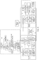
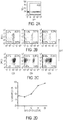
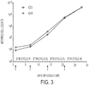


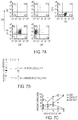

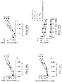
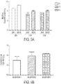
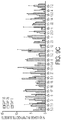
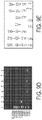
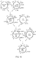
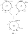


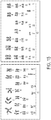
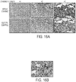

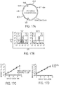
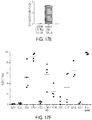
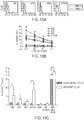
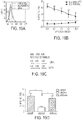
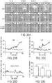
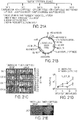
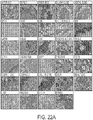
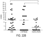
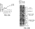
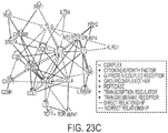


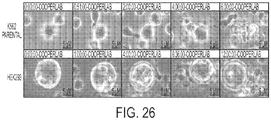
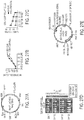


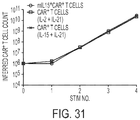
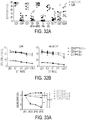
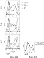
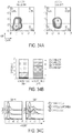


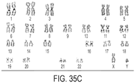
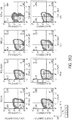


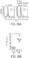
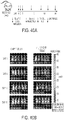


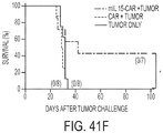
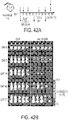


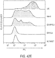
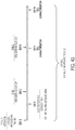
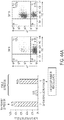
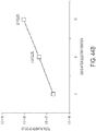
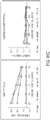


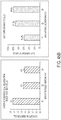
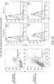
Classifications
-
- C—CHEMISTRY; METALLURGY
- C12—BIOCHEMISTRY; BEER; SPIRITS; WINE; VINEGAR; MICROBIOLOGY; ENZYMOLOGY; MUTATION OR GENETIC ENGINEERING
- C12N—MICROORGANISMS OR ENZYMES; COMPOSITIONS THEREOF; PROPAGATING, PRESERVING, OR MAINTAINING MICROORGANISMS; MUTATION OR GENETIC ENGINEERING; CULTURE MEDIA
- C12N15/00—Mutation or genetic engineering; DNA or RNA concerning genetic engineering, vectors, e.g. plasmids, or their isolation, preparation or purification; Use of hosts therefor
- C12N15/09—Recombinant DNA-technology
- C12N15/63—Introduction of foreign genetic material using vectors; Vectors; Use of hosts therefor; Regulation of expression
- C12N15/79—Vectors or expression systems specially adapted for eukaryotic hosts
- C12N15/85—Vectors or expression systems specially adapted for eukaryotic hosts for animal cells
-
- A—HUMAN NECESSITIES
- A61—MEDICAL OR VETERINARY SCIENCE; HYGIENE
- A61K—PREPARATIONS FOR MEDICAL, DENTAL OR TOILETRY PURPOSES
- A61K39/00—Medicinal preparations containing antigens or antibodies
- A61K39/0005—Vertebrate antigens
- A61K39/0011—Cancer antigens
-
- A—HUMAN NECESSITIES
- A61—MEDICAL OR VETERINARY SCIENCE; HYGIENE
- A61K—PREPARATIONS FOR MEDICAL, DENTAL OR TOILETRY PURPOSES
- A61K35/00—Medicinal preparations containing materials or reaction products thereof with undetermined constitution
- A61K35/12—Materials from mammals; Compositions comprising non-specified tissues or cells; Compositions comprising non-embryonic stem cells; Genetically modified cells
- A61K35/14—Blood; Artificial blood
- A61K35/17—Lymphocytes; B-cells; T-cells; Natural killer cells; Interferon-activated or cytokine-activated lymphocytes
-
- A—HUMAN NECESSITIES
- A61—MEDICAL OR VETERINARY SCIENCE; HYGIENE
- A61K—PREPARATIONS FOR MEDICAL, DENTAL OR TOILETRY PURPOSES
- A61K39/00—Medicinal preparations containing antigens or antibodies
- A61K39/0005—Vertebrate antigens
- A61K39/0011—Cancer antigens
- A61K39/001102—Receptors, cell surface antigens or cell surface determinants
- A61K39/001103—Receptors for growth factors
- A61K39/001104—Epidermal growth factor receptors [EGFR]
-
- A—HUMAN NECESSITIES
- A61—MEDICAL OR VETERINARY SCIENCE; HYGIENE
- A61K—PREPARATIONS FOR MEDICAL, DENTAL OR TOILETRY PURPOSES
- A61K39/00—Medicinal preparations containing antigens or antibodies
- A61K39/0005—Vertebrate antigens
- A61K39/0011—Cancer antigens
- A61K39/001102—Receptors, cell surface antigens or cell surface determinants
- A61K39/001103—Receptors for growth factors
- A61K39/001106—Her-2/neu/ErbB2, Her-3/ErbB3 or Her 4/ErbB4
-
- A—HUMAN NECESSITIES
- A61—MEDICAL OR VETERINARY SCIENCE; HYGIENE
- A61K—PREPARATIONS FOR MEDICAL, DENTAL OR TOILETRY PURPOSES
- A61K39/00—Medicinal preparations containing antigens or antibodies
- A61K39/0005—Vertebrate antigens
- A61K39/0011—Cancer antigens
- A61K39/001102—Receptors, cell surface antigens or cell surface determinants
- A61K39/001103—Receptors for growth factors
- A61K39/001109—Vascular endothelial growth factor receptors [VEGFR]
-
- A—HUMAN NECESSITIES
- A61—MEDICAL OR VETERINARY SCIENCE; HYGIENE
- A61K—PREPARATIONS FOR MEDICAL, DENTAL OR TOILETRY PURPOSES
- A61K39/00—Medicinal preparations containing antigens or antibodies
- A61K39/0005—Vertebrate antigens
- A61K39/0011—Cancer antigens
- A61K39/001102—Receptors, cell surface antigens or cell surface determinants
- A61K39/001103—Receptors for growth factors
- A61K39/00111—Hepatocyte growth factor receptor [HGFR or c-met]
-
- A—HUMAN NECESSITIES
- A61—MEDICAL OR VETERINARY SCIENCE; HYGIENE
- A61K—PREPARATIONS FOR MEDICAL, DENTAL OR TOILETRY PURPOSES
- A61K39/00—Medicinal preparations containing antigens or antibodies
- A61K39/0005—Vertebrate antigens
- A61K39/0011—Cancer antigens
- A61K39/001102—Receptors, cell surface antigens or cell surface determinants
- A61K39/001111—Immunoglobulin superfamily
- A61K39/001112—CD19 or B4
-
- A—HUMAN NECESSITIES
- A61—MEDICAL OR VETERINARY SCIENCE; HYGIENE
- A61K—PREPARATIONS FOR MEDICAL, DENTAL OR TOILETRY PURPOSES
- A61K39/00—Medicinal preparations containing antigens or antibodies
- A61K39/0005—Vertebrate antigens
- A61K39/0011—Cancer antigens
- A61K39/001102—Receptors, cell surface antigens or cell surface determinants
- A61K39/001111—Immunoglobulin superfamily
- A61K39/001113—CD22, BL-CAM, siglec-2 or sialic acid- binding Ig-related lectin 2
-
- A—HUMAN NECESSITIES
- A61—MEDICAL OR VETERINARY SCIENCE; HYGIENE
- A61K—PREPARATIONS FOR MEDICAL, DENTAL OR TOILETRY PURPOSES
- A61K39/00—Medicinal preparations containing antigens or antibodies
- A61K39/0005—Vertebrate antigens
- A61K39/0011—Cancer antigens
- A61K39/001102—Receptors, cell surface antigens or cell surface determinants
- A61K39/001116—Receptors for cytokines
- A61K39/001119—Receptors for interleukins [IL]
-
- A—HUMAN NECESSITIES
- A61—MEDICAL OR VETERINARY SCIENCE; HYGIENE
- A61K—PREPARATIONS FOR MEDICAL, DENTAL OR TOILETRY PURPOSES
- A61K39/00—Medicinal preparations containing antigens or antibodies
- A61K39/0005—Vertebrate antigens
- A61K39/0011—Cancer antigens
- A61K39/001102—Receptors, cell surface antigens or cell surface determinants
- A61K39/001124—CD20
-
- A—HUMAN NECESSITIES
- A61—MEDICAL OR VETERINARY SCIENCE; HYGIENE
- A61K—PREPARATIONS FOR MEDICAL, DENTAL OR TOILETRY PURPOSES
- A61K39/00—Medicinal preparations containing antigens or antibodies
- A61K39/0005—Vertebrate antigens
- A61K39/0011—Cancer antigens
- A61K39/001102—Receptors, cell surface antigens or cell surface determinants
- A61K39/001129—Molecules with a "CD" designation not provided for elsewhere
-
- A—HUMAN NECESSITIES
- A61—MEDICAL OR VETERINARY SCIENCE; HYGIENE
- A61K—PREPARATIONS FOR MEDICAL, DENTAL OR TOILETRY PURPOSES
- A61K39/00—Medicinal preparations containing antigens or antibodies
- A61K39/0005—Vertebrate antigens
- A61K39/0011—Cancer antigens
- A61K39/001148—Regulators of development
- A61K39/00115—Apoptosis related proteins, e.g. survivin or livin
- A61K39/001151—Apoptosis related proteins, e.g. survivin or livin p53
-
- A—HUMAN NECESSITIES
- A61—MEDICAL OR VETERINARY SCIENCE; HYGIENE
- A61K—PREPARATIONS FOR MEDICAL, DENTAL OR TOILETRY PURPOSES
- A61K39/00—Medicinal preparations containing antigens or antibodies
- A61K39/0005—Vertebrate antigens
- A61K39/0011—Cancer antigens
- A61K39/001154—Enzymes
- A61K39/001164—GTPases, e.g. Ras or Rho
-
- A—HUMAN NECESSITIES
- A61—MEDICAL OR VETERINARY SCIENCE; HYGIENE
- A61K—PREPARATIONS FOR MEDICAL, DENTAL OR TOILETRY PURPOSES
- A61K39/00—Medicinal preparations containing antigens or antibodies
- A61K39/0005—Vertebrate antigens
- A61K39/0011—Cancer antigens
- A61K39/001166—Adhesion molecules, e.g. NRCAM, EpCAM or cadherins
- A61K39/001168—Mesothelin [MSLN]
-
- A—HUMAN NECESSITIES
- A61—MEDICAL OR VETERINARY SCIENCE; HYGIENE
- A61K—PREPARATIONS FOR MEDICAL, DENTAL OR TOILETRY PURPOSES
- A61K39/00—Medicinal preparations containing antigens or antibodies
- A61K39/0005—Vertebrate antigens
- A61K39/0011—Cancer antigens
- A61K39/001169—Tumor associated carbohydrates
- A61K39/00117—Mucins, e.g. MUC-1
-
- A—HUMAN NECESSITIES
- A61—MEDICAL OR VETERINARY SCIENCE; HYGIENE
- A61K—PREPARATIONS FOR MEDICAL, DENTAL OR TOILETRY PURPOSES
- A61K39/00—Medicinal preparations containing antigens or antibodies
- A61K39/0005—Vertebrate antigens
- A61K39/0011—Cancer antigens
- A61K39/001169—Tumor associated carbohydrates
- A61K39/001171—Gangliosides, e.g. GM2, GD2 or GD3
-
- A—HUMAN NECESSITIES
- A61—MEDICAL OR VETERINARY SCIENCE; HYGIENE
- A61K—PREPARATIONS FOR MEDICAL, DENTAL OR TOILETRY PURPOSES
- A61K39/00—Medicinal preparations containing antigens or antibodies
- A61K39/0005—Vertebrate antigens
- A61K39/0011—Cancer antigens
- A61K39/001174—Proteoglycans, e.g. glypican, brevican or CSPG4
-
- A—HUMAN NECESSITIES
- A61—MEDICAL OR VETERINARY SCIENCE; HYGIENE
- A61K—PREPARATIONS FOR MEDICAL, DENTAL OR TOILETRY PURPOSES
- A61K39/00—Medicinal preparations containing antigens or antibodies
- A61K39/0005—Vertebrate antigens
- A61K39/0011—Cancer antigens
- A61K39/00118—Cancer antigens from embryonic or fetal origin
- A61K39/001181—Alpha-feto protein
-
- A—HUMAN NECESSITIES
- A61—MEDICAL OR VETERINARY SCIENCE; HYGIENE
- A61K—PREPARATIONS FOR MEDICAL, DENTAL OR TOILETRY PURPOSES
- A61K39/00—Medicinal preparations containing antigens or antibodies
- A61K39/0005—Vertebrate antigens
- A61K39/0011—Cancer antigens
- A61K39/00118—Cancer antigens from embryonic or fetal origin
- A61K39/001182—Carcinoembryonic antigen [CEA]
-
- A—HUMAN NECESSITIES
- A61—MEDICAL OR VETERINARY SCIENCE; HYGIENE
- A61K—PREPARATIONS FOR MEDICAL, DENTAL OR TOILETRY PURPOSES
- A61K39/00—Medicinal preparations containing antigens or antibodies
- A61K39/0005—Vertebrate antigens
- A61K39/0011—Cancer antigens
- A61K39/00119—Melanoma antigens
-
- A—HUMAN NECESSITIES
- A61—MEDICAL OR VETERINARY SCIENCE; HYGIENE
- A61K—PREPARATIONS FOR MEDICAL, DENTAL OR TOILETRY PURPOSES
- A61K39/00—Medicinal preparations containing antigens or antibodies
- A61K39/0005—Vertebrate antigens
- A61K39/0011—Cancer antigens
- A61K39/001193—Prostate associated antigens e.g. Prostate stem cell antigen [PSCA]; Prostate carcinoma tumor antigen [PCTA]; PAP or PSGR
- A61K39/001194—Prostate specific antigen [PSA]
-
- A—HUMAN NECESSITIES
- A61—MEDICAL OR VETERINARY SCIENCE; HYGIENE
- A61K—PREPARATIONS FOR MEDICAL, DENTAL OR TOILETRY PURPOSES
- A61K39/00—Medicinal preparations containing antigens or antibodies
- A61K39/46—Cellular immunotherapy
- A61K39/461—Cellular immunotherapy characterised by the cell type used
- A61K39/4611—T-cells, e.g. tumor infiltrating lymphocytes [TIL], lymphokine-activated killer cells [LAK] or regulatory T cells [Treg]
-
- A—HUMAN NECESSITIES
- A61—MEDICAL OR VETERINARY SCIENCE; HYGIENE
- A61K—PREPARATIONS FOR MEDICAL, DENTAL OR TOILETRY PURPOSES
- A61K39/00—Medicinal preparations containing antigens or antibodies
- A61K39/46—Cellular immunotherapy
- A61K39/463—Cellular immunotherapy characterised by recombinant expression
- A61K39/4631—Chimeric Antigen Receptors [CAR]
-
- A—HUMAN NECESSITIES
- A61—MEDICAL OR VETERINARY SCIENCE; HYGIENE
- A61K—PREPARATIONS FOR MEDICAL, DENTAL OR TOILETRY PURPOSES
- A61K39/00—Medicinal preparations containing antigens or antibodies
- A61K39/46—Cellular immunotherapy
- A61K39/464—Cellular immunotherapy characterised by the antigen targeted or presented
- A61K39/4643—Vertebrate antigens
- A61K39/4644—Cancer antigens
- A61K39/464402—Receptors, cell surface antigens or cell surface determinants
- A61K39/464403—Receptors for growth factors
- A61K39/464404—Epidermal growth factor receptors [EGFR]
-
- A—HUMAN NECESSITIES
- A61—MEDICAL OR VETERINARY SCIENCE; HYGIENE
- A61K—PREPARATIONS FOR MEDICAL, DENTAL OR TOILETRY PURPOSES
- A61K39/00—Medicinal preparations containing antigens or antibodies
- A61K39/46—Cellular immunotherapy
- A61K39/464—Cellular immunotherapy characterised by the antigen targeted or presented
- A61K39/4643—Vertebrate antigens
- A61K39/4644—Cancer antigens
- A61K39/464402—Receptors, cell surface antigens or cell surface determinants
- A61K39/464403—Receptors for growth factors
- A61K39/464406—Her-2/neu/ErbB2, Her-3/ErbB3 or Her 4/ ErbB4
-
- A—HUMAN NECESSITIES
- A61—MEDICAL OR VETERINARY SCIENCE; HYGIENE
- A61K—PREPARATIONS FOR MEDICAL, DENTAL OR TOILETRY PURPOSES
- A61K39/00—Medicinal preparations containing antigens or antibodies
- A61K39/46—Cellular immunotherapy
- A61K39/464—Cellular immunotherapy characterised by the antigen targeted or presented
- A61K39/4643—Vertebrate antigens
- A61K39/4644—Cancer antigens
- A61K39/464402—Receptors, cell surface antigens or cell surface determinants
- A61K39/464403—Receptors for growth factors
- A61K39/464409—Vascular endothelial growth factor receptors [VEGFR]
-
- A—HUMAN NECESSITIES
- A61—MEDICAL OR VETERINARY SCIENCE; HYGIENE
- A61K—PREPARATIONS FOR MEDICAL, DENTAL OR TOILETRY PURPOSES
- A61K39/00—Medicinal preparations containing antigens or antibodies
- A61K39/46—Cellular immunotherapy
- A61K39/464—Cellular immunotherapy characterised by the antigen targeted or presented
- A61K39/4643—Vertebrate antigens
- A61K39/4644—Cancer antigens
- A61K39/464402—Receptors, cell surface antigens or cell surface determinants
- A61K39/464403—Receptors for growth factors
- A61K39/46441—Hepatocyte growth factor receptor [HGFR or c-met]
-
- A—HUMAN NECESSITIES
- A61—MEDICAL OR VETERINARY SCIENCE; HYGIENE
- A61K—PREPARATIONS FOR MEDICAL, DENTAL OR TOILETRY PURPOSES
- A61K39/00—Medicinal preparations containing antigens or antibodies
- A61K39/46—Cellular immunotherapy
- A61K39/464—Cellular immunotherapy characterised by the antigen targeted or presented
- A61K39/4643—Vertebrate antigens
- A61K39/4644—Cancer antigens
- A61K39/464402—Receptors, cell surface antigens or cell surface determinants
- A61K39/464411—Immunoglobulin superfamily
- A61K39/464413—CD22, BL-CAM, siglec-2 or sialic acid binding Ig-related lectin 2
-
- A—HUMAN NECESSITIES
- A61—MEDICAL OR VETERINARY SCIENCE; HYGIENE
- A61K—PREPARATIONS FOR MEDICAL, DENTAL OR TOILETRY PURPOSES
- A61K39/00—Medicinal preparations containing antigens or antibodies
- A61K39/46—Cellular immunotherapy
- A61K39/464—Cellular immunotherapy characterised by the antigen targeted or presented
- A61K39/4643—Vertebrate antigens
- A61K39/4644—Cancer antigens
- A61K39/464402—Receptors, cell surface antigens or cell surface determinants
- A61K39/464416—Receptors for cytokines
- A61K39/464419—Receptors for interleukins [IL]
-
- A—HUMAN NECESSITIES
- A61—MEDICAL OR VETERINARY SCIENCE; HYGIENE
- A61K—PREPARATIONS FOR MEDICAL, DENTAL OR TOILETRY PURPOSES
- A61K39/00—Medicinal preparations containing antigens or antibodies
- A61K39/46—Cellular immunotherapy
- A61K39/464—Cellular immunotherapy characterised by the antigen targeted or presented
- A61K39/4643—Vertebrate antigens
- A61K39/4644—Cancer antigens
- A61K39/464402—Receptors, cell surface antigens or cell surface determinants
- A61K39/464416—Receptors for cytokines
- A61K39/46442—Receptors for interferons [IFN]
-
- A—HUMAN NECESSITIES
- A61—MEDICAL OR VETERINARY SCIENCE; HYGIENE
- A61K—PREPARATIONS FOR MEDICAL, DENTAL OR TOILETRY PURPOSES
- A61K39/00—Medicinal preparations containing antigens or antibodies
- A61K39/46—Cellular immunotherapy
- A61K39/464—Cellular immunotherapy characterised by the antigen targeted or presented
- A61K39/4643—Vertebrate antigens
- A61K39/4644—Cancer antigens
- A61K39/464402—Receptors, cell surface antigens or cell surface determinants
- A61K39/464424—CD20
-
- A—HUMAN NECESSITIES
- A61—MEDICAL OR VETERINARY SCIENCE; HYGIENE
- A61K—PREPARATIONS FOR MEDICAL, DENTAL OR TOILETRY PURPOSES
- A61K39/00—Medicinal preparations containing antigens or antibodies
- A61K39/46—Cellular immunotherapy
- A61K39/464—Cellular immunotherapy characterised by the antigen targeted or presented
- A61K39/4643—Vertebrate antigens
- A61K39/4644—Cancer antigens
- A61K39/464402—Receptors, cell surface antigens or cell surface determinants
- A61K39/464429—Molecules with a "CD" designation not provided for elsewhere
-
- A—HUMAN NECESSITIES
- A61—MEDICAL OR VETERINARY SCIENCE; HYGIENE
- A61K—PREPARATIONS FOR MEDICAL, DENTAL OR TOILETRY PURPOSES
- A61K39/00—Medicinal preparations containing antigens or antibodies
- A61K39/46—Cellular immunotherapy
- A61K39/464—Cellular immunotherapy characterised by the antigen targeted or presented
- A61K39/4643—Vertebrate antigens
- A61K39/4644—Cancer antigens
- A61K39/464448—Regulators of development
- A61K39/46445—Apoptosis related proteins, e.g. survivin or livin
- A61K39/464451—Apoptosis related proteins, e.g. survivin or livin p53
-
- A—HUMAN NECESSITIES
- A61—MEDICAL OR VETERINARY SCIENCE; HYGIENE
- A61K—PREPARATIONS FOR MEDICAL, DENTAL OR TOILETRY PURPOSES
- A61K39/00—Medicinal preparations containing antigens or antibodies
- A61K39/46—Cellular immunotherapy
- A61K39/464—Cellular immunotherapy characterised by the antigen targeted or presented
- A61K39/4643—Vertebrate antigens
- A61K39/4644—Cancer antigens
- A61K39/464454—Enzymes
- A61K39/464464—GTPases, e.g. Ras or Rho
-
- A—HUMAN NECESSITIES
- A61—MEDICAL OR VETERINARY SCIENCE; HYGIENE
- A61K—PREPARATIONS FOR MEDICAL, DENTAL OR TOILETRY PURPOSES
- A61K39/00—Medicinal preparations containing antigens or antibodies
- A61K39/46—Cellular immunotherapy
- A61K39/464—Cellular immunotherapy characterised by the antigen targeted or presented
- A61K39/4643—Vertebrate antigens
- A61K39/4644—Cancer antigens
- A61K39/464466—Adhesion molecules, e.g. NRCAM, EpCAM or cadherins
- A61K39/464468—Mesothelin [MSLN]
-
- A—HUMAN NECESSITIES
- A61—MEDICAL OR VETERINARY SCIENCE; HYGIENE
- A61K—PREPARATIONS FOR MEDICAL, DENTAL OR TOILETRY PURPOSES
- A61K39/00—Medicinal preparations containing antigens or antibodies
- A61K39/46—Cellular immunotherapy
- A61K39/464—Cellular immunotherapy characterised by the antigen targeted or presented
- A61K39/4643—Vertebrate antigens
- A61K39/4644—Cancer antigens
- A61K39/464469—Tumor associated carbohydrates
- A61K39/46447—Mucins, e.g. MUC-1
-
- A—HUMAN NECESSITIES
- A61—MEDICAL OR VETERINARY SCIENCE; HYGIENE
- A61K—PREPARATIONS FOR MEDICAL, DENTAL OR TOILETRY PURPOSES
- A61K39/00—Medicinal preparations containing antigens or antibodies
- A61K39/46—Cellular immunotherapy
- A61K39/464—Cellular immunotherapy characterised by the antigen targeted or presented
- A61K39/4643—Vertebrate antigens
- A61K39/4644—Cancer antigens
- A61K39/464469—Tumor associated carbohydrates
- A61K39/464471—Gangliosides, e.g. GM2, GD2 or GD3
-
- A—HUMAN NECESSITIES
- A61—MEDICAL OR VETERINARY SCIENCE; HYGIENE
- A61K—PREPARATIONS FOR MEDICAL, DENTAL OR TOILETRY PURPOSES
- A61K39/00—Medicinal preparations containing antigens or antibodies
- A61K39/46—Cellular immunotherapy
- A61K39/464—Cellular immunotherapy characterised by the antigen targeted or presented
- A61K39/4643—Vertebrate antigens
- A61K39/4644—Cancer antigens
- A61K39/464474—Proteoglycans, e.g. glypican, brevican or CSPG4
-
- A—HUMAN NECESSITIES
- A61—MEDICAL OR VETERINARY SCIENCE; HYGIENE
- A61K—PREPARATIONS FOR MEDICAL, DENTAL OR TOILETRY PURPOSES
- A61K39/00—Medicinal preparations containing antigens or antibodies
- A61K39/46—Cellular immunotherapy
- A61K39/464—Cellular immunotherapy characterised by the antigen targeted or presented
- A61K39/4643—Vertebrate antigens
- A61K39/4644—Cancer antigens
- A61K39/46448—Cancer antigens from embryonic or fetal origin
- A61K39/464482—Carcinoembryonic antigen [CEA]
-
- A—HUMAN NECESSITIES
- A61—MEDICAL OR VETERINARY SCIENCE; HYGIENE
- A61K—PREPARATIONS FOR MEDICAL, DENTAL OR TOILETRY PURPOSES
- A61K39/00—Medicinal preparations containing antigens or antibodies
- A61K39/46—Cellular immunotherapy
- A61K39/464—Cellular immunotherapy characterised by the antigen targeted or presented
- A61K39/4643—Vertebrate antigens
- A61K39/4644—Cancer antigens
- A61K39/46449—Melanoma antigens
-
- A—HUMAN NECESSITIES
- A61—MEDICAL OR VETERINARY SCIENCE; HYGIENE
- A61K—PREPARATIONS FOR MEDICAL, DENTAL OR TOILETRY PURPOSES
- A61K39/00—Medicinal preparations containing antigens or antibodies
- A61K39/46—Cellular immunotherapy
- A61K39/464—Cellular immunotherapy characterised by the antigen targeted or presented
- A61K39/4643—Vertebrate antigens
- A61K39/4644—Cancer antigens
- A61K39/464493—Prostate associated antigens e.g. Prostate stem cell antigen [PSCA]; Prostate carcinoma tumor antigen [PCTA]; Prostatic acid phosphatase [PAP]; Prostate-specific G-protein-coupled receptor [PSGR]
- A61K39/464494—Prostate specific antigen [PSA]
-
- A—HUMAN NECESSITIES
- A61—MEDICAL OR VETERINARY SCIENCE; HYGIENE
- A61K—PREPARATIONS FOR MEDICAL, DENTAL OR TOILETRY PURPOSES
- A61K39/00—Medicinal preparations containing antigens or antibodies
- A61K39/46—Cellular immunotherapy
- A61K39/464—Cellular immunotherapy characterised by the antigen targeted or presented
- A61K39/464838—Viral antigens
-
- A—HUMAN NECESSITIES
- A61—MEDICAL OR VETERINARY SCIENCE; HYGIENE
- A61P—SPECIFIC THERAPEUTIC ACTIVITY OF CHEMICAL COMPOUNDS OR MEDICINAL PREPARATIONS
- A61P37/00—Drugs for immunological or allergic disorders
-
- C—CHEMISTRY; METALLURGY
- C07—ORGANIC CHEMISTRY
- C07K—PEPTIDES
- C07K14/00—Peptides having more than 20 amino acids; Gastrins; Somatostatins; Melanotropins; Derivatives thereof
- C07K14/435—Peptides having more than 20 amino acids; Gastrins; Somatostatins; Melanotropins; Derivatives thereof from animals; from humans
- C07K14/52—Cytokines; Lymphokines; Interferons
- C07K14/54—Interleukins [IL]
- C07K14/5443—IL-15
-
- C—CHEMISTRY; METALLURGY
- C07—ORGANIC CHEMISTRY
- C07K—PEPTIDES
- C07K14/00—Peptides having more than 20 amino acids; Gastrins; Somatostatins; Melanotropins; Derivatives thereof
- C07K14/435—Peptides having more than 20 amino acids; Gastrins; Somatostatins; Melanotropins; Derivatives thereof from animals; from humans
- C07K14/705—Receptors; Cell surface antigens; Cell surface determinants
- C07K14/70503—Immunoglobulin superfamily
- C07K14/7051—T-cell receptor (TcR)-CD3 complex
-
- C—CHEMISTRY; METALLURGY
- C07—ORGANIC CHEMISTRY
- C07K—PEPTIDES
- C07K14/00—Peptides having more than 20 amino acids; Gastrins; Somatostatins; Melanotropins; Derivatives thereof
- C07K14/435—Peptides having more than 20 amino acids; Gastrins; Somatostatins; Melanotropins; Derivatives thereof from animals; from humans
- C07K14/705—Receptors; Cell surface antigens; Cell surface determinants
- C07K14/70503—Immunoglobulin superfamily
- C07K14/70521—CD28, CD152
-
- C—CHEMISTRY; METALLURGY
- C07—ORGANIC CHEMISTRY
- C07K—PEPTIDES
- C07K16/00—Immunoglobulins [IGs], e.g. monoclonal or polyclonal antibodies
- C07K16/18—Immunoglobulins [IGs], e.g. monoclonal or polyclonal antibodies against material from animals or humans
- C07K16/28—Immunoglobulins [IGs], e.g. monoclonal or polyclonal antibodies against material from animals or humans against receptors, cell surface antigens or cell surface determinants
- C07K16/2896—Immunoglobulins [IGs], e.g. monoclonal or polyclonal antibodies against material from animals or humans against receptors, cell surface antigens or cell surface determinants against molecules with a "CD"-designation, not provided for elsewhere
-
- C—CHEMISTRY; METALLURGY
- C07—ORGANIC CHEMISTRY
- C07K—PEPTIDES
- C07K16/00—Immunoglobulins [IGs], e.g. monoclonal or polyclonal antibodies
- C07K16/18—Immunoglobulins [IGs], e.g. monoclonal or polyclonal antibodies against material from animals or humans
- C07K16/28—Immunoglobulins [IGs], e.g. monoclonal or polyclonal antibodies against material from animals or humans against receptors, cell surface antigens or cell surface determinants
- C07K16/30—Immunoglobulins [IGs], e.g. monoclonal or polyclonal antibodies against material from animals or humans against receptors, cell surface antigens or cell surface determinants from tumour cells
-
- C—CHEMISTRY; METALLURGY
- C12—BIOCHEMISTRY; BEER; SPIRITS; WINE; VINEGAR; MICROBIOLOGY; ENZYMOLOGY; MUTATION OR GENETIC ENGINEERING
- C12N—MICROORGANISMS OR ENZYMES; COMPOSITIONS THEREOF; PROPAGATING, PRESERVING, OR MAINTAINING MICROORGANISMS; MUTATION OR GENETIC ENGINEERING; CULTURE MEDIA
- C12N15/00—Mutation or genetic engineering; DNA or RNA concerning genetic engineering, vectors, e.g. plasmids, or their isolation, preparation or purification; Use of hosts therefor
- C12N15/09—Recombinant DNA-technology
- C12N15/87—Introduction of foreign genetic material using processes not otherwise provided for, e.g. co-transformation
-
- C—CHEMISTRY; METALLURGY
- C12—BIOCHEMISTRY; BEER; SPIRITS; WINE; VINEGAR; MICROBIOLOGY; ENZYMOLOGY; MUTATION OR GENETIC ENGINEERING
- C12N—MICROORGANISMS OR ENZYMES; COMPOSITIONS THEREOF; PROPAGATING, PRESERVING, OR MAINTAINING MICROORGANISMS; MUTATION OR GENETIC ENGINEERING; CULTURE MEDIA
- C12N5/00—Undifferentiated human, animal or plant cells, e.g. cell lines; Tissues; Cultivation or maintenance thereof; Culture media therefor
- C12N5/06—Animal cells or tissues; Human cells or tissues
- C12N5/0602—Vertebrate cells
- C12N5/0634—Cells from the blood or the immune system
- C12N5/0636—T lymphocytes
-
- A—HUMAN NECESSITIES
- A61—MEDICAL OR VETERINARY SCIENCE; HYGIENE
- A61K—PREPARATIONS FOR MEDICAL, DENTAL OR TOILETRY PURPOSES
- A61K39/00—Medicinal preparations containing antigens or antibodies
- A61K2039/505—Medicinal preparations containing antigens or antibodies comprising antibodies
-
- A—HUMAN NECESSITIES
- A61—MEDICAL OR VETERINARY SCIENCE; HYGIENE
- A61K—PREPARATIONS FOR MEDICAL, DENTAL OR TOILETRY PURPOSES
- A61K39/00—Medicinal preparations containing antigens or antibodies
- A61K2039/51—Medicinal preparations containing antigens or antibodies comprising whole cells, viruses or DNA/RNA
- A61K2039/515—Animal cells
- A61K2039/5158—Antigen-pulsed cells, e.g. T-cells
-
- A—HUMAN NECESSITIES
- A61—MEDICAL OR VETERINARY SCIENCE; HYGIENE
- A61K—PREPARATIONS FOR MEDICAL, DENTAL OR TOILETRY PURPOSES
- A61K2239/00—Indexing codes associated with cellular immunotherapy of group A61K39/46
- A61K2239/31—Indexing codes associated with cellular immunotherapy of group A61K39/46 characterized by the route of administration
-
- A—HUMAN NECESSITIES
- A61—MEDICAL OR VETERINARY SCIENCE; HYGIENE
- A61K—PREPARATIONS FOR MEDICAL, DENTAL OR TOILETRY PURPOSES
- A61K2239/00—Indexing codes associated with cellular immunotherapy of group A61K39/46
- A61K2239/38—Indexing codes associated with cellular immunotherapy of group A61K39/46 characterised by the dose, timing or administration schedule
-
- A—HUMAN NECESSITIES
- A61—MEDICAL OR VETERINARY SCIENCE; HYGIENE
- A61K—PREPARATIONS FOR MEDICAL, DENTAL OR TOILETRY PURPOSES
- A61K2239/00—Indexing codes associated with cellular immunotherapy of group A61K39/46
- A61K2239/46—Indexing codes associated with cellular immunotherapy of group A61K39/46 characterised by the cancer treated
- A61K2239/57—Skin; melanoma
-
- C—CHEMISTRY; METALLURGY
- C07—ORGANIC CHEMISTRY
- C07K—PEPTIDES
- C07K2317/00—Immunoglobulins specific features
- C07K2317/60—Immunoglobulins specific features characterized by non-natural combinations of immunoglobulin fragments
- C07K2317/62—Immunoglobulins specific features characterized by non-natural combinations of immunoglobulin fragments comprising only variable region components
- C07K2317/622—Single chain antibody (scFv)
-
- C—CHEMISTRY; METALLURGY
- C07—ORGANIC CHEMISTRY
- C07K—PEPTIDES
- C07K2319/00—Fusion polypeptide
- C07K2319/01—Fusion polypeptide containing a localisation/targetting motif
- C07K2319/02—Fusion polypeptide containing a localisation/targetting motif containing a signal sequence
-
- C—CHEMISTRY; METALLURGY
- C07—ORGANIC CHEMISTRY
- C07K—PEPTIDES
- C07K2319/00—Fusion polypeptide
- C07K2319/01—Fusion polypeptide containing a localisation/targetting motif
- C07K2319/03—Fusion polypeptide containing a localisation/targetting motif containing a transmembrane segment
-
- C—CHEMISTRY; METALLURGY
- C12—BIOCHEMISTRY; BEER; SPIRITS; WINE; VINEGAR; MICROBIOLOGY; ENZYMOLOGY; MUTATION OR GENETIC ENGINEERING
- C12N—MICROORGANISMS OR ENZYMES; COMPOSITIONS THEREOF; PROPAGATING, PRESERVING, OR MAINTAINING MICROORGANISMS; MUTATION OR GENETIC ENGINEERING; CULTURE MEDIA
- C12N2501/00—Active agents used in cell culture processes, e.g. differentation
- C12N2501/20—Cytokines; Chemokines
- C12N2501/23—Interleukins [IL]
- C12N2501/2301—Interleukin-1 (IL-1)
-
- C—CHEMISTRY; METALLURGY
- C12—BIOCHEMISTRY; BEER; SPIRITS; WINE; VINEGAR; MICROBIOLOGY; ENZYMOLOGY; MUTATION OR GENETIC ENGINEERING
- C12N—MICROORGANISMS OR ENZYMES; COMPOSITIONS THEREOF; PROPAGATING, PRESERVING, OR MAINTAINING MICROORGANISMS; MUTATION OR GENETIC ENGINEERING; CULTURE MEDIA
- C12N2501/00—Active agents used in cell culture processes, e.g. differentation
- C12N2501/20—Cytokines; Chemokines
- C12N2501/23—Interleukins [IL]
- C12N2501/2321—Interleukin-21 (IL-21)
-
- C—CHEMISTRY; METALLURGY
- C12—BIOCHEMISTRY; BEER; SPIRITS; WINE; VINEGAR; MICROBIOLOGY; ENZYMOLOGY; MUTATION OR GENETIC ENGINEERING
- C12N—MICROORGANISMS OR ENZYMES; COMPOSITIONS THEREOF; PROPAGATING, PRESERVING, OR MAINTAINING MICROORGANISMS; MUTATION OR GENETIC ENGINEERING; CULTURE MEDIA
- C12N2502/00—Coculture with; Conditioned medium produced by
- C12N2502/11—Coculture with; Conditioned medium produced by blood or immune system cells
-
- Y—GENERAL TAGGING OF NEW TECHNOLOGICAL DEVELOPMENTS; GENERAL TAGGING OF CROSS-SECTIONAL TECHNOLOGIES SPANNING OVER SEVERAL SECTIONS OF THE IPC; TECHNICAL SUBJECTS COVERED BY FORMER USPC CROSS-REFERENCE ART COLLECTIONS [XRACs] AND DIGESTS
- Y02—TECHNOLOGIES OR APPLICATIONS FOR MITIGATION OR ADAPTATION AGAINST CLIMATE CHANGE
- Y02A—TECHNOLOGIES FOR ADAPTATION TO CLIMATE CHANGE
- Y02A50/00—TECHNOLOGIES FOR ADAPTATION TO CLIMATE CHANGE in human health protection, e.g. against extreme weather
- Y02A50/30—Against vector-borne diseases, e.g. mosquito-borne, fly-borne, tick-borne or waterborne diseases whose impact is exacerbated by climate change
Definitions
- the present invention relates generally to the fields of medicine, immunology, cell biology, and molecular biology.
- the field of the invention concerns immunotherapy. More particularly, it concerns the manufacture of clinical-grade chimeric antigen receptor (CAR) T cells and therapeutic methods using such cells.
- CAR clinical-grade chimeric antigen receptor
- TAAs tumor-associated antigens
- Such a combination of gene therapy with immunotherapy can redirect the specificity of T cells for B-lineage antigens and patients with advanced B-cell malignancies benefit from infusion of such tumor-specific T cells (Jena et al ., 2010; Till et al ., 2008; Porter et al ., 2011; Brentjens et al ., 2011; Cooper and Bollard, 2012; Kalos et al ., 2011; Kochenderfer et al ., 2010; Kochenderfer et al ., 2012; Brentjens et al ., 2013).
- CAR chimeric antigen receptor
- the present invention relates to:
- a method of providing a T-cell response in a human subject having a disease comprising obtaining a sample of cells from the subject, (comprising T-cells or T-cell progenitors); transfecting the cells with a nucleic acid encoding chimeric T-cell receptor (CAR), capable of integration into the genome of the cells, and administering an effective amount of the transgenic cells to the subject to provide a T-cell response.
- a sample of cells from the subject comprising obtaining a sample of cells from the subject, (comprising T-cells or T-cell progenitors); transfecting the cells with a nucleic acid encoding chimeric T-cell receptor (CAR), capable of integration into the genome of the cells, and administering an effective amount of the transgenic cells to the subject to provide a T-cell response.
- CAR chimeric T-cell receptor
- a method of the present disclosure comprises: (a) obtaining a sample of cells from the subject, the sample comprising T-cells or T-cell progenitors; (b) transfecting the cells with a DNA encoding a transposon-flanked chimeric antigen receptor (CAR) and a transposase effective to integrate the DNA encoding the CAR into the genome of the cells, to provide a population of transgenic CAR-expressing cells; (c) optionally, culturing the population of transgenic CAR cells ex vivo in a medium that selectively enhances proliferation of CAR-expressing T-cells, wherein the transgenic CAR cells are cultured, if at all, no more than 21 days; and (d) administering an effective amount of the transgenic CAR cells to the subject to provide a T-cell response.
- CAR transposon-flanked chimeric antigen receptor
- the transgenic CAR cells are cultured ex vivo for less than 21 days, such as for less than 18, 17, 16, 15, 14, 13, 12, 11, 10, 9, 8, 7, 6, 5, 4, 3, 2 days or less.
- the CAR cells are cultured ex vivo no more that 3 to 5 days.
- steps (a)-(d) of the instant method i.e., obtaining cell samples to administering CAR T cells is completed in no more that 21, 20, 19, 18, 17, 16, 15, 14, 13, 12, 11, 10, 9, 8, 7, 6, or 5 days.
- a method of providing a T-cell response in a human subject having a disease comprises: (a) obtaining a sample of cells from the subject, the sample comprising T-cells or T-cell progenitors and having an initial volume of between about 20 and 200 mls when obtained from the subject; (b) transfecting the cells with a DNA encoding a transposon-flanked chimeric antigen receptor (CAR) and a transposase effective to integrate the DNA encoding the CAR into the genome of the cells, to provide a population of transgenic CAR-expressing T-cells; (c) optionally, culturing the population of transgenic CAR cells ex vivo in a medium that selectively enhances proliferation of CAR-expressing T-cells; and (d) administering an effective amount of the transgenic CAR T-cells to the subject to provide a T-cell response.
- CAR transposon-flanked chimeric antigen receptor
- the sample of cells from the subject may be a sample of less than about 200 mls of a peripheral blood or umbilical cord blood.
- the sample may be collected by apheresis.
- the sample is collected by a method that does not involved apheresis ( e.g ., by venipuncture).
- the sample of cells has an initial volume of less than about 175, 150, 125, 100, 75, 50 or 25 mls ( e . g ., the sample of cells has an initial volume of between about 50 and 200 mls, 50 and 100mls, or 100 and 200 mls when obtained from the subject).
- the cells comprise DNA encoding the CAR integrated into the genome of the cell (e.g., CAR DNA flanked by transposon repeat sequences).
- the CAR sequence can comprise the CDR sequences (e.g ., CDRs 1-6) of monoclonal antibody 6H5 or the scFv sequence of monoclonal antibody 6H5.
- HERV-K-targeted CAR is at least 85% identical to the amino acid sequence of SEQ ID NO: 4 (e.g ., a sequence at least about 90%, 91%, 92%, 93%, 94%, 95%, 96%, 97%, 98% or 99% identical to SEQ ID NO: 4).
- a cell of the embodiments e.g ., a human HERV-K-targeted CAR cell
- the membrane-bound IL-15 comprises a fusion protein between IL-15 and IL-15R ⁇ .
- the membrane-bound IL-15 comprises an amino acid sequence at least about 85%, 90%, 91%, 92%, 93%, 94%, 95%, 96%, 97%, 98% or 99% identical to SEQ ID NO: 6 (referred to herein as mIL15).
- the membrane-bound IL-15 is encoded by a RNA or a DNA (e.g., an extra chromosomal or episomal vector).
- the cell comprises DNA encoding the membrane-bound IL-15 integrated into the genome of the cell (e.g., coding DNA flanked by transposon repeat sequences).
- a cell of the present disclosure e.g., human CAR cell, expressing a membrane-bound cytokine
- methods of the present disclosure concern transfecting the cells with a DNA encoding a chimeric T-cell receptor (CAR) and, in some cases, a transposase.
- CAR chimeric T-cell receptor
- Methods of transfecting of cells are well known in the art, but in certain aspects, highly efficient transfections methods such as electroporation are employed.
- nucleic acids may be introduced into cells using a nucleofection apparatus.
- the transfection step does not involve infecting or transducing the cells with virus, which can cause genotoxicity and/or lead to an immune response to cells containing viral sequences in a treated subject.
- the CAR expression vector is a DNA expression vector such as a plasmid, linear expression vector or an episome.
- the vector comprises additional sequences, such as sequence that facilitate expression of the CAR, such a promoter, enhancer, poly-A signal, and/or one or more introns.
- the CAR coding sequence is flanked by transposon sequences, such that the presence of a transposase allows the coding sequence to integrate into the genome of the transfected cell.
- cells are further transfected with a transposase that facilitates integration of a CAR coding sequence into the genome of the transfected cells.
- the transposase is provided as DNA expression vector.
- the transposase is provided as an expressible RNA or a protein such that long-term expression of the transposase does not occur in the transgenic cells.
- the transposase is provided as an mRNA (e.g., an mRNA comprising a cap and poly-A tail). Any transposase system may be used in accordance with the present disclosure.
- the transposase is salmonid-type Tc1-like transposase (SB).
- SB salmonid-type Tc1-like transposase
- the transposase can be the so called "Sleeping beauty" transposase, see e.g., U.S. Patent 6,489,458 .
- the transposase is an engineered enzyme with increased enzymatic activity.
- Some specific examples of transposases include, without limitation, SB10, SB11 or SB100x transposase (see, e.g., Mates et al ., 2009).
- a method can involve electroporation of cells with a mRNA encoding a SB10, SB11 or SB100x transposase.
- a transgenic CAR cell of the present disclosure further comprises an expression vector for expression of a membrane-bound cytokine that stimulates proliferation and/or survival of T-cells.
- CAR cells comprising such cytokines can proliferate and/or persist with little or no ex vivo culture with activating and propagating cells (AaPCs) or artificial antigen presenting cells (aAPCs) due the simulation provided by the cytokine expression.
- AaPCs activating and propagating cells
- aAPCs artificial antigen presenting cells
- such CAR cells can proliferate in vivo even when large amounts of antigen recognized by the CAR is not present (e.g., as in the case of a cancer patient in remission or a patient with minimal residual disease).
- the CAR cells comprise a DNA or RNA expression vector for expression of a Cy cytokine and elements (e.g., a transmembrane domain) to provide surface expression of the cytokine.
- the CAR cells can comprise membrane-bound versions of IL-7, IL-15 or IL-21.
- the cytokine is tethered to the membrane by fusion of the cytokine coding sequence with the receptor for the cytokine.
- a cell can comprise a vector for expression of a IL-15-IL-15R ⁇ fusion protein (e.g., a protein comprising the sequence of SEQ ID NO: 6).
- a vector encoding a membrane-bound C ⁇ cytokine is a DNA expression vector, such as vector integrated into the genome of the CAR cells or an extra-chromosomal vector ( e . g ., and episomal vector).
- expression of the membrane-bound C ⁇ cytokine is under the control of an inducible promoter (e.g ., a drug inducible promoter) such that the expression of the cytokine in the CAR cells (and thereby the proliferation of the CAR cells) can be controlled by inducing or suppressing promoter activity.
- an inducible promoter e.g ., a drug inducible promoter
- aspects of the present disclosure concern obtaining a sample from a patient comprising NK cells, NKT cells, T-cells or T-cell progenitor cells.
- the sample is an umbilical cord blood sample, a peripheral blood sample (e.g ., a mononuclear cell fraction) or a sample from the subject comprising pluripotent cells.
- a sample from the subject can be cultured to generate induced pluripotent stem (iPS) cells and these cells used to produce NK cells, NKT cells or T-cells.
- iPS induced pluripotent stem
- Cell samples may be cultured directly from the subject or may be cryopreserved prior to use.
- obtaining a cell sample comprises collecting a cell sample.
- the sample is obtained by a third party.
- a sample from a subject can be treated to purify or enrich the T-cells or T-cell progenitors in the sample.
- the sample can be subjected to gradient purification, cell culture selection and/or cell sorting (e.g., via fluorescence-activated cell sorting (FACS)).
- FACS fluorescence-activated cell sorting
- a method of the present disclosure further comprises obtaining, producing or using antigen presenting cells.
- the antigen presenting cells can be dendritic cells, activating and propagating cells (AaPCs), or inactivated ( e.g ., irradiated) artificial antigen presenting cells (aAPCs).
- AaPCs activating and propagating cells
- aAPCs inactivated artificial antigen presenting cells
- Methods for producing such aAPCs are know in the art and further detailed herein.
- transgenic CAR cells are co-cultured with inactivated aAPCs ex vivo for a limited period of time in order to expand the CAR cell population.
- the step of co-culturing CAR cells with aAPCs can be done in a medium that comprises, for example, interleukin-21 (IL-21) and/or interleukin-2 (IL-2).
- the co-culturing is performed at a ratio of CAR cells to inactivated aAPCs of about 10:1 to about 1:10; about 3:1 to about 1:5; or about 1:1 to about 1:3.
- the co-culture of CAR cells and aAPCs can be at a ratio of about 1:1, about 1:2 or about 1:3.
- cells for culture of CAR cells such as AaPCs or aAPCs are engineered to express specific polypeptide to enhance growth of the CAR cells.
- the cells can comprise (i) an antigen targeted by the CAR expressed on the transgenic CAR cells; (ii) CD64; (ii) CD86; (iii) CD137L; and/or (v) membrane-bound IL-15, expressed on the surface of the aAPCs.
- the AaPCs or aAPCS comprise a CAR-binding antibody or fragment thereof expressed on the surface of the AaPCs or aAPCs.
- AaPCs or aAPCs for use in the instant methods are tested for, and confirmed to be free of, infectious material and/or are tested and confirmed to be inactivated and non-proliferating.
- ex vivo culturing the transgenic CAR cells (c) is for no more than 14 days, no more than 7 days or no more than 3 days.
- the ex vivo culture e . g ., culture in the presence of AaPCs or aAPCs
- the transgenic cells are not cultured ex vivo in the presence of AaPCs or aAPCs.
- a method of the present disclosure comprises a step for enriching the cell population for CAR-expressing T-cells after transfection of the cells (step (b)) or after ex vivo expansion of the cells (step (c)).
- the enrichment step can comprise sorting of the cell (e.g., via FACS), for example, by using an antigen bound by the CAR or a CAR-binding antibody.
- the enrichment step comprises depletion of the non-T-cells or depletion of cells that lack CAR expression.
- CD56+ cells can be depleted from a culture population.
- a sample of CAR cells is preserved (or maintained in culture) when the cells are administered to the subject. For example, a sample may be cryopreserved for later expansion or analysis.
- transgenic CAR cells of the embodiments are inactivated for expression of an endogenous T-cell receptor and/or endogenous HLA.
- T cells can be engineered to eliminate expression of endogenous alpha/beta T-cell receptor (TCR).
- CAR + T cells are genetically modified to eliminate expression of TCR.
- ZFNs zinc finger nucleases
- the T-cell receptor ⁇ -chain in CAR-expressing T cells is knocked-out, for example, by using zinc finger nucleases.
- CAR cells of the present disclosure can be for usein treating a wide range of diseases and conditions. Essentially any disease that involves the specific or enhanced expression of a particular antigen can be treated by targeting CAR cells to the antigen.
- autoimmune diseases, infections, and cancers can be treated with methods and/or compositions of the present disclosure. These include cancers, such as primary, metastatic, recurrent, sensitive-to-therapy, refractory-to-therapy cancers ( e . g ., chemo-refractory cancer).
- the cancer may be of the blood, lung, brain, colon, prostate, breast, liver, kidney, stomach, cervix, ovary, testes, pituitary gland, esophagus, spleen, skin, bone, and so forth (e.g ., B-cell lymphomas or a melanomas).
- CAR cells typically target a cancer cell antigen (also known as a tumor-associated antigen (TAA)).
- TAA tumor-associated antigen
- transgenic CAR cells of the present disclosure may be for use in treating subject having minimal residual disease (e.g ., a subject having very low amounts of CAR-targeted antigen present), such as cancer patients that are in apparent remission.
- subject having minimal residual disease e.g ., a subject having very low amounts of CAR-targeted antigen present
- cancer-associated antigens or cancer cells
- transgenic CAR cells for targeting of residual disease further comprise expression of a membrane-bound proliferative cytokine, as these cells will retain the ability to expand in vivo despite the low amount to target antigen.
- CAR + T cells for various tumor antigens (e.g ., CD19, ROR1, CD56, EGFR, CD123, c-met, GD2).
- CAR + T cells generated using this technology can be used to treat patients with leukemias (AML, ALL, CML), infections and/or solid tumors.
- methods of the present disclosure can be used to treat cell proliferative diseases, fungal, viral, bacterial or parasitic infections.
- Pathogens that may be targeted include, with limitation, Plasmodium , trypanosome, Aspergillus , Candida , HSV, RSV, EBV, CMV, JC virus, BK virus, or Ebola pathogens.
- Further examples of antigens that can be targeted by CAR cells of the embodiments include, without limitation, CD19, CD20, carcinoembryonic antigen, alphafetoprotein, CA-125, 5T4, MUC-1, epithelial tumor antigen, melanoma-associated antigen, mutated p53, mutated ras, HER2/Neu, ERBB2, folate binding protein, HIV-1 envelope glycoprotein gp120, HIV-1 envelope glycoprotein gp41, GD2, CD123, CD23,CD30 , CD56, c-Met, meothelin, GD3, HERV-K, IL-11Ralpha, kappa chain, lambda chain, CSPG4, ERBB2, EGFRvIII,
- method of the present disclosure concern targeting of CD19 or HERV-K-expressing cells.
- a HERV-K targeted CAR cell can comprise a CAR including the scFv sequence of monoclonal antibody 6H5.
- a CAR of the present disclosure can be conjugated or fused with a cytokine, such as IL-2, IL-7, IL-15, IL-21 or a combination thereof.
- methods for treating an individual with a medical condition comprising the step of providing an effective amount of cells from the population of cells described herein, including more than once in some aspects, such as at least 1, 2, 3, 4, 5, 6, 7, 8, 9, 10, 11, 12, 13, 14, or more days apart.
- the cancer is lymphoma, leukemia, non-Hodgkin's lymphoma, acute lymphoblastic leukemia, chronic lymphoblastic leukemia, chronic lymphocytic leukemia, or B cell-associated autoimmune diseases.
- an isolated or recombinant polypeptide comprising the a CD19-targeted CAR comprising an amino acid sequence at least about 85%, 90%, 91%, 92%, 93%, 94%, 95%, 96%, 97%, 98% or 99% identical to SEQ ID NO: 1.
- an isolated or recombinant polynucleotide sequence is described encoding a CD19-targeted CAR (e.g ., encoding an amino acid sequence 85%, 90%, 91%, 92%, 93%, 94%, 95%, 96%, 97%, 98% or 99% identical to SEQ ID NO: 1).
- the polynucleotide sequence is 85%, 90%, 91%, 92%, 93%, 94%, 95%, 96%, 97%, 98% or 99% identical to SEQ ID NO: 3 (which encodes the CD-19-targeted CAR) or SEQ ID NO: 4 (which encodes the CD-19-targeted CAR, expression control sequences and flanking transposon repeats).
- SEQ ID NO: 3 which encodes the CD-19-targeted CAR
- SEQ ID NO: 4 which encodes the CD-19-targeted CAR, expression control sequences and flanking transposon repeats.
- a host cell is described comprising polypeptide encoding a CD19-targeted CAR and/or a polynucleotide encoding a CD19-targeted CAR of the aspects of the present disclosure.
- the host cell can be a T-cell or T-cell precursor.
- the host cell is a human cell.
- an isolated or recombinant polypeptide comprising the a HERV-K-targeted CAR comprising an amino acid sequence at least about 85%, 90%, 91%, 92%, 93%, 94%, 95%, 96%, 97%, 98% or 99% identical to SEQ ID NO: 4.
- an isolated or recombinant polynucleotide sequence is described encoding a HERV-K-targeted CAR (e.g ., encoding an amino acid sequence 85%, 90%, 91%, 92%, 93%, 94%, 95%, 96%, 97%, 98% or 99% identical to SEQ ID NO: 4).
- the polynucleotide sequence is 85%, 90%, 91%, 92%, 93%, 94%, 95%, 96%, 97%, 98% or 99% identical to SEQ ID NO: 5 (which encodes the HERV-K-targeted CAR, expression control sequences and flanking transposon repeats).
- SEQ ID NO: 5 which encodes the HERV-K-targeted CAR, expression control sequences and flanking transposon repeats.
- a host cell comprising polypeptide encoding a HERV-K-targeted CAR and/or a polynucleotide encoding a HERV-K-targeted CAR of the aspects of the present disclosure.
- the host cell can be a T-cell or T-cell precursor.
- the host cell is a human cell.
- any of the forgoing polypeptides, polynucleotides or host cells may be used in accordance with the methods detailed herein.
- an isolated or recombinant polypeptide comprising the a membrane-bound IL-15 comprising an amino acid sequence at least about 85%, 90%, 91%, 92%, 93%, 94%, 95%, 96%, 97%, 98% or 99% identical to SEQ ID NO: 6.
- an isolated or recombinant polynucleotide sequence is provided encoding a membrane-bound IL-15 (e.g., encoding an amino acid sequence 85%, 90%, 91%, 92%, 93%, 94%, 95%, 96%, 97%, 98% or 99% identical to SEQ ID NO: 6).
- the polynucleotide sequence is 85%, 90%, 91%, 92%, 93%, 94%, 95%, 96%, 97%, 98% or 99% identical to SEQ ID NO: 7 (a membrane-bound IL-15, expression control sequences and flanking transposon repeats).
- a host cell comprising polypeptide encoding a membrane-bound IL-15 and/or a polynucleotide encoding a membrane-bound IL-15 of the embodiments.
- the host cell can be a T-cell, T-cell precursor or aAPC.
- the host cell is a human cell.
- any of the forgoing polypeptides, polynucleotides or host cells may be used in accordance with the methods detailed herein.
- Clinical trials have demonstrated anti-tumor effects in patients that have received T cells genetically modified to have desired specificity.
- a new approach to manufacturing antigen-specific CAR + T cells in compliance with cGMP is detailed.
- the system employs a highly efficient transfection system in conjunction with a transposon and transposase system CAR gene integration.
- These non-viral approaches have significant advantages as an alternative to viral-mediated transduction since clinical grade CAR + T cells preferably should not include added viral sequences.
- High quality cGMP CAR T-cells were achieved by electro-transfer of DNA plasmids derived from a transposon system, such as from Sleeping Beauty or piggyBac, and propagation of the genetically modified T cells on aAPC.
- CAR + T cells that demonstrated specificity for their target antigen (e . g ., CD19).
- target antigen e . g ., CD19.
- the cells met release criteria established by the FDA for use in clinical trials. These methods avoid genotoxicity due to virus-mediated transduction and immunogenicity due to use of virus.
- a chimeric antigen receptor recognizes cell-surface tumor-associated antigen independent of human leukocyte antigen (HLA) and employs one or more signaling molecules to activate genetically modified T cells for killing, proliferation, and cytokine production (Jena et al., 2010).
- Adoptive transfer of T cells expressing CAR has shown promise in multiple clinical trials. It is now possible to use a modular approach to manufacture clinical grade genetically modified T cells.
- the platform technologies disclosed herein comprise (i) non-viral gene transfer using an electroporation device (e.g., a nucleofector), (ii) transposition (e.g., an SB transposon, see, U.S.
- Patent 6,489,458 (iii) CARs that signal through endodomains (e.g., CD28/CD3- ⁇ , CD137/CD3- ⁇ , or other combinations), (iv) CARs with variable lengths of extracellular domains connecting the antigen-recognition domain to the cell surface, and, in some cases, (v) artificial antigen presenting cells (aAPC) derived from K562 to be able to robustly and numerically expand CAR + T cells (Singh et al ., 2008; Singh et al ., 2011; Huang et al ., 2012).
- endodomains e.g., CD28/CD3- ⁇ , CD137/CD3- ⁇ , or other combinations
- aAPC artificial antigen presenting cells
- the presently disclosed process can be used to genetically modify T cells derived from peripheral blood and/or umbilical cord blood to express CAR(s) that can be numerically expanded in vitro using aAPC (Singh et al ., 2008; Singh et al ., 2011).
- the process has implications for cell and gene therapy, due to the relative ease of DNA plasmid production, electroporation, use of thawed ⁇ -irradiated master-bank aAPC, and can be readily transferred to facilities operating in compliance with current good manufacturing practice (cGMP) for Phase I/II trials.
- cGMP current good manufacturing practice
- the disclosed method of manufacturing T cells is unique at least in that it does not use (i) viral-transduction or (ii) naked DNA electroporation followed by rapid expansion on a PBMC/LCL feeder layer.
- methods are disclosed for targeting CD19 through the enforced expression of a CAR that recognizes CD19 independent of HLA.
- These T cells meet release criteria defined by sterility, phenotype, viability, and cell number.
- In-process testing revealed that the electroporated/propagated T cells express CAR in a memory/na ⁇ ve population, have a normal karyotype, preserved TCR V ⁇ repertoire, and are able to recognize and lyse CD19 + tumor targets in a CAR-dependent manner.
- the electro-transfer of non-viral plasmids is an appealing alternative to transduction since DNA species can be produced to clinical grade at approximately 1/10 th the cost of recombinant GMP-grade virus.
- SB Sleeping Beauty
- the inventor's SB system uses two DNA plasmids that comprise a transposon coding for a gene of interest (e . g ., 2 nd generation CD19-specific CAR transgene, such as designated CD19RCD28) and a transposase (e.g., SB11), which inserts the transgene into TA dinucleotide repeats in the target cell genome (Geurts et al ., 2006; Ivics et al ., 1997; Izsvak and Ivics, 1997).
- a transposon coding for a gene of interest e . g ., 2 nd generation CD19-specific CAR transgene, such as designated CD19RCD28
- a transposase e.g., SB11
- the inventor's 2 nd generation CAR (Kowolik et al ., 2006) signals through CD28 and CD3- ⁇ with the expectation that this will sustain T-cell proliferation and recycle effector functions in vivo.
- CARs with varying extracellular lengths and different endodomain signaling motifs can be expressed using the SB system.
- K562 aAPC (clone #4) were developed, expressing the desired antigen (e . g ., CD19) along with costimulatory molecules, such as CD86, CD137L, a membrane-bound version of interleukin (IL)-15 (peptide fused to modified IgG4 Fc region or cytokine peptide fused to IL-15 receptor alpha), and CD64 (Fc- ⁇ receptor 1), to select for T cells in vitro that are capable of sustained CAR-mediated propagation.
- costimulatory molecules such as CD86, CD137L, a membrane-bound version of interleukin (IL)-15 (peptide fused to modified IgG4 Fc region or cytokine peptide fused to IL-15 receptor alpha), and CD64 (Fc- ⁇ receptor 1), to select for T cells in vitro that are capable of sustained CAR-mediated propagation.
- costimulatory molecules such as CD86, CD137L, a membrane-bound version of interleukin (IL)-15
- T cells can be scaled back employing fewer cuvettes and carrying forward just a sub-set of the numerically expanded T cells for 0, 1, or more rounds of proliferation on aAPC (added at the beginning of each stimulation cycle).
- aAPC added at the beginning of each stimulation cycle.
- at least 90% of the propagated T cells express CAR and are cryopreserved for infusion.
- this approach can be harnessed to generate T cells to diverse tumor types by pairing the specificity of the introduced CAR with expression of the tumor-associated antigen (TAA) recognized by the CAR on the aAPC.
- TAA tumor-associated antigen
- the ex vivo expansion platform has also been adapted to manufacture NK cells, NK T cells, and ⁇ T cells.
- the outgrowth of CD4 + and CD8 + T cells expressing the 2 nd generation CAR include cells with a stem-cell/memory/naive phenotype and exhibit three hallmarks of re-directed specificity.
- the genetically modified T cells specifically lyse CD19 + targets.
- they produce cytokine (e.g., IFN- ⁇ ) in response to CD19 + stimulator cells.
- the aAPC and tissue culture environment have been modified to generate patient- and donor-derived CD19-specific T cells for infusion after hematopoietic stem-cell transplantation (Singh et al ., 2011; Singh et al . 2008).
- the inventors can produce CAR + T cells from peripheral blood simply obtained by venipuncture, which avoids the cost, discomfort, and inconvenience of obtaining mononuclear cells by apheresis.
- the ability to derive large numbers of CAR + T cells from small numbers of mononuclear cells is particularly appealing for infusing T cells after allogeneic umbilical cord blood transplantation.
- the small size and anonymity of the neonatal donor precludes re-accessing this individual at a later time point and only limited numbers of harvested mononuclear cells are available as starting material for T cell manufacture to avoid interfering with hematopoiesis. Further advances to the manufacturing process include a high throughput electroporation device coupled with a fully closed WAVE bioreactor to minimize handling.
- the present approach to manufacturing includes the implementation of processing and culturing systems to reduce work load and safeguard against a breach in sterility.
- the inventors co-cultured T cells with ⁇ -irradiated aAPC in bioreactors and/or bags rather than flasks. This transition typically occurs after day 14 following electroporation.
- the aAPC as source material are numerically expanded in bioreactors and/or bags, and the inventors adapted the Sepax device to process the aAPC for cryopreservation.
- the Sepax harvest procedure has additional advantages beyond automation when using large volumes of culture media (>900 mL) as it reduces additional centrifuging steps required by the manual process.
- CARs chimeric antigen receptors
- T-cell receptors may refer to artificial T-cell receptors, chimeric T-cell receptors, or chimeric immunoreceptors, for example, and encompass engineered receptors that graft an artificial specificity onto a particular immune effector cell.
- CARs may be employed to impart the specificity of a monoclonal antibody onto a T cell, thereby allowing a large number of specific T cells to be generated, for example, for use in adoptive cell therapy.
- CARs direct specificity of the cell to a tumor associated antigen, for example.
- CARs comprise an intracellular activation domain, a transmembrane domain, and an extracellular domain comprising a tumor associated antigen binding region.
- CARs comprise fusions of single-chain variable fragments (scFv) derived from monoclonal antibodies, fused to CD3-zeta a transmembrane domain and endodomain.
- the specificity of other CAR designs may be derived from ligands of receptors (e.g., peptides) or from pattern-recognition receptors, such as Dectins.
- one can target malignant B cells by redirecting the specificity of T cells by using a CAR specific for the B-lineage molecule, CD19.
- CARs comprise domains for additional co-stimulatory signaling, such as CD3-zeta, FcR, CD27, CD28, CD137, DAP10, and/or OX40.
- molecules can be co-expressed with the CAR, including co-stimulatory molecules, reporter genes for imaging (e.g., for positron emission tomography), gene products that conditionally ablate the T cells upon addition of a pro-drug, homing receptors, chemokines, chemokine receptors, cytokines, and cytokine receptors.
- T-cell receptor refers to a protein receptor on T cells that is composed of a heterodimer of an alpha ( ⁇ ) and beta ( ⁇ ) chain, although in some cells the TCR consists of gamma and delta ( ⁇ / ⁇ ) chains.
- the TCR may be modified on any cell comprising a TCR, including a helper T cell, a cytotoxic T cell, a memory T cell, regulatory T cell, natural killer T cell, and gamma delta T cell, for example.
- tumor-associated antigen and “cancer cell antigen” are used interchangeably herein. In each case, the terms refer to proteins, glycoproteins or carbohydrates that are specifically or preferentially expressed by cancer cells.
- an antigen is a molecule capable of being bound by an antibody or T-cell receptor.
- An antigen is additionally capable of inducing a humoral immune response and/or cellular immune response leading to the production of B and/or T lymphocytes.
- nucleic acids including nucleic acids encoding an antigen-specific chimeric antigen receptor (CAR) polypeptide, including a CAR that has been humanized to reduce immunogenicity (hCAR), comprising an intracellular signaling domain, a transmembrane domain, and an extracellular domain comprising one or more signaling motifs.
- CAR antigen-specific chimeric antigen receptor
- the CAR may recognize an epitope comprised of the shared space between one or more antigens.
- Pattern recognition receptors such as Dectin-1, may be used to derive specificity to a carbohydrate antigen.
- the binding region can comprise complementary determining regions of a monoclonal antibody, variable regions of a monoclonal antibody, and/or antigen binding fragments thereof.
- that specificity is derived from a peptide (e.g ., cytokine) that binds to a receptor.
- a complementarity determining region is a short amino acid sequence found in the variable domains of antigen receptor (e.g ., immunoglobulin and T-cell receptor) proteins that complements an antigen and therefore provides the receptor with its specificity for that particular antigen.
- antigen receptor e.g ., immunoglobulin and T-cell receptor
- Each polypeptide chain of an antigen receptor contains three CDRs (CDR1, CDR2, and CDR3). Since the antigen receptors are typically composed of two polypeptide chains, there are six CDRs for each antigen receptor that can come into contact with the antigen -- each heavy and light chain contains three CDRs.
- CDR3 shows the greatest variability as it is encoded by a recombination of the VJ (VDJ in the case of heavy chain and TCR ⁇ chain) regions.
- the human CAR nucleic acids are human genes to enhance cellular immunotherapy for human patients.
- described herein is a full length CAR cDNA or coding region.
- the antigen binding regions or domain can comprise a fragment of the V H and V L chains of a single-chain variable fragment (scFv) derived from a particular human monoclonal antibody, such as those described in U.S. Patent 7,109,304 .
- the fragment can also be any number of different antigen binding domains of a human antigen-specific antibody.
- the fragment is an antigen-specific scFv encoded by a sequence that is optimized for human codon usage for expression in human cells.
- the arrangement could be multimeric, such as a diabody or multimers.
- the multimers are most likely formed by cross pairing of the variable portion of the light and heavy chains into what has been referred to by Winters as a diabody.
- the hinge portion of the construct can have multiple alternatives from being totally deleted, to having the first cysteine maintained, to a proline rather than a serine substitution, to being truncated up to the first cysteine.
- the Fc portion can be deleted. Any protein that is stable and/or dimerizes can serve this purpose.
- One could use just one of the Fc domains, e.g ., either the CH2 or CH3 domain from human immunoglobulin.
- One could also use just the hinge portion of an immunoglobulin.
- the intracellular signaling domain of the chimeric receptor disclosed herein is responsible for activation of at least one of the normal effector functions of the immune cell in which the chimeric receptor has been placed.
- effector function refers to a specialized function of a differentiated cell. Effector function of a T cell, for example, may be cytolytic activity or helper activity including the secretion of cytokines. Effector function in a naive, memory, or memory-type T cell includes antigen-dependent proliferation.
- intracellular signaling domain refers to the portion of a protein that transduces the effector function signal and directs the cell to perform a specialized function.
- intracellular signaling domain While usually the entire intracellular signaling domain will be employed, in many cases it will not be necessary to use the entire intracellular polypeptide. To the extent that a truncated portion of the intracellular signaling domain may find use, such truncated portion may be used in place of the intact chain as long as it still transduces the effector function signal.
- the term intracellular signaling domain is thus meant to include any truncated portion of the intracellular signaling domain sufficient to transduce the effector function signal.
- Examples include the zeta chain of the T-cell receptor or any of its homologs (e.g ., eta, delta, gamma, or epsilon), MB1 chain, B29, Fc RIII, Fc RI, and combinations of signaling molecules, such as CD3 ⁇ and CD28, CD27, 4-1BB, DAP-10, OX40, and combinations thereof, as well as other similar molecules and fragments.
- Intracellular signaling portions of other members of the families of activating proteins can be used, such as Fc ⁇ RIII and Fc ⁇ RI. See Gross et al . (1992), Stancovski et al . (1993), Moritz et al . (1994), Hwu et al .
- the antigen-specific extracellular domain and the intracellular signaling-domain may be linked by a transmembrane domain, such as the human IgG 4 Fc hinge and Fc regions.
- a transmembrane domain such as the human IgG 4 Fc hinge and Fc regions.
- Alternatives include the human CD4 transmembrane domain, the human CD28 transmembrane domain, the transmembrane human CD3 ⁇ domain, or a cysteine mutated human CD3 ⁇ domain, or other transmembrane domains from other human transmembrane signaling proteins, such as CD16 and CD8 and erythropoietin receptor.
- the CAR nucleic acid comprises a sequence encoding other costimulatory receptors, such as a transmembrane domain and a modified CD28 intracellular signaling domain.
- costimulatory receptors include, but are not limited to one or more of CD28, CD27, OX-40 (CD134), DAP10, and 4-1BB (CD137).
- CD28 CD27
- OX-40 CD134
- DAP10 DAP10
- 4-1BB CD137
- an additional signal provided by a human costimulatory receptor inserted in a human CAR is important for full activation of T cells and could help improve in vivo persistence and the therapeutic success of the adoptive immunotherapy.
- the present disclosure describes isolated nucleic acid segments and expression cassettes incorporating DNA sequences that encode the CAR.
- Vectors of the present disclosure are designed, primarily, to deliver desired genes to immune cells, preferably T cells under the control of regulated eukaryotic promoters, for example, MNDU3 promoter, CMV promoter, EF1alpha promoter, or Ubiquitin promoter.
- the vectors may contain a selectable marker, if for no other reason, to facilitate their manipulation in vitro.
- the CAR can also be expressed from mRNA in vitro transcribed from a DNA template.
- Chimeric antigen receptor molecules are recombinant and are distinguished by their ability to both bind antigen and transduce activation signals via immunoreceptor activation motifs (ITAM's) present in their cytoplasmic tails.
- Receptor constructs utilizing an antigen-binding moiety afford the additional advantage of being "universal” in that they bind native antigen on the target cell surface in an HLA-independent fashion.
- non-human antigen binding regions are typically used in constructing a chimeric antigen receptor.
- a potential problem with using non-human antigen binding regions, such as murine monoclonal antibodies, is the lack of human effector functionality and inability to penetrate into tumor masses. In other words, such antibodies may be unable to mediate complement-dependent lysis or lyse human target cells through antibody-dependent cellular toxicity or Fc-receptor mediated phagocytosis to destroy cells expressing CAR.
- non-human monoclonal antibodies can be recognized by the human host as a foreign protein, and therefore, repeated injections of such foreign antibodies can lead to the induction of immune responses leading to harmful hypersensitivity reactions.
- HAMA Human Anti-Mouse Antibody
- the chimeric antigen receptor comprises: a) an intracellular signaling domain, b) a transmembrane domain, and c) an extracellular domain comprising an antigen binding region.
- intracellular receptor signaling domains in the CAR include those of the T cell antigen receptor complex, such as the zeta chain of CD3, also Fc ⁇ RIII costimulatory signaling domains, CD28, CD27, DAP10, CD137, OX40, CD2, alone or in a series with CD3zeta, for example.
- the intracellular domain (which may be referred to as the cytoplasmic domain) comprises part or all of one or more of TCR zeta chain, CD28, CD27, OX40/CD134, 4-1BB/CD137, Fc ⁇ RI ⁇ , ICOS/CD278, IL-2Rbeta/CD122, IL-2Ralpha/CD132, DAP10, DAP12, and CD40.
- One or multiple cytoplasmic domains may be employed, as so-called third generation CARs have at least two or three signaling domains fused together for additive or synergistic effect, for example.
- the antigen-specific portion of the receptor (which may be referred to as an extracellular domain comprising an antigen binding region) comprises a tumor associated antigen or a pathogen-specific antigen binding domain including carbohydrate antigen recognized by pattern-recognition receptors, such as Dectin-1.
- a tumor associated antigen may be of any kind so long as it is expressed on the cell surface of tumor cells.
- tumor associated antigens examples include CD19, CD20, carcinoembryonic antigen, alphafetoprotein, CA-125, MUC-1, CD56, EGFR, c-Met, AKT, Her2, Her3, epithelial tumor antigen, melanoma-associated antigen, mutated p53, mutated ras, and so forth.
- the CAR can be co-expressed with a membrane-bound cytokine to improve persistence when there is a low amount of tumor-associated antigen.
- CAR can be co-expressed with membrane-bound IL-15.
- intracellular tumor associated antigens may be targeted, such as HA-1, survivin, WT1, and p53.
- This can be achieved by a CAR expressed on a universal T cell that recognizes the processed peptide described from the intracellular tumor associated antigen in the context of HLA.
- the universal T cell may be genetically modified to express a T-cell receptor pairing that recognizes the intracellular processed tumor associated antigen in the context of HLA.
- the pathogen may be of any kind, but in specific embodiments the pathogen is a fungus, bacteria, or virus, for example.
- exemplary viral pathogens include those of the families of Adenoviridae, Epstein-Barr virus (EBV), Cytomegalovirus (CMV), Respiratory Syncytial Virus (RSV), JC virus, BK virus, HSV, HHV family of viruses, Picornaviridae, Herpesviridae, Hepadnaviridae, Flaviviridae, Retroviridae, Orthomyxoviridae, Paramyxoviridae, Papovaviridae, Polyomavirus, Rhabdoviridae, and Togaviridae.
- Exemplary pathogenic viruses cause smallpox, influenza, mumps, measles, chickenpox, ebola, and rubella.
- Exemplary pathogenic fungi include Candida, Aspergillus, Cryptococcus, Histoplasma, Pneumocystis, and Stachybotrys.
- Exemplary pathogenic bacteria include Streptococcus , Pseudomonas , Shigella , Campylobacter , Staphylococcus , Helicobacter , E . coli , Rickettsia , Bacillus , Bordetella , Chlamydia , Spirochetes , and Salmonella .
- the pathogen receptor Dectin-1 can be used to generate a CAR that recognizes the carbohydrate structure on the cell wall of fungi.
- T cells genetically modified to express the CAR based on the specificity of Dectin-1 can recognize Aspergillus and target hyphal growth.
- CARs can be made based on an antibody recognizing viral determinants (e.g., the glycoproteins from CMV and Ebola) to interrupt viral infections and pathology.
- the pathogenic antigen is an Aspergillus carbohydrate antigen for which the extracellular domain in the CAR recognizes patterns of carbohydrates of the fungal cell wall, such as via Dectin-1.
- a chimeric immunoreceptor as disclosed herein can be produced by any means known in the art, though preferably it is produced using recombinant DNA techniques.
- a nucleic acid sequence encoding the several regions of the chimeric receptor can be prepared and assembled into a complete coding sequence by standard techniques of molecular cloning (genomic library screening, PCR, primer-assisted ligation, scFv libraries from yeast and bacteria, site-directed mutagenesis, etc .).
- the resulting coding region can be inserted into an expression vector and used to transform a suitable expression host allogeneic T-cell line.
- nucleic acid construct or nucleic acid sequence or polynucleotide is intended to mean a DNA molecule that can be transformed or introduced into a T cell and be transcribed and translated to produce a product (e.g., a chimeric antigen receptor).
- the promoter is operably linked to the nucleic acid sequence encoding the chimeric receptor of the present invention, i.e ., they are positioned so as to promote transcription of the messenger RNA from the DNA encoding the chimeric receptor.
- the promoter can be of genomic origin or synthetically generated.
- a variety of promoters for use in T cells are well-known in the art (e . g ., the CD4 promoter disclosed by Marodon et al . (2003)).
- the promoter can be constitutive or inducible, where induction is associated with the specific cell type or a specific level of maturation, for example.
- Promoters of interest include the ⁇ -actin promoter, SV40 early and late promoters, immunoglobulin promoter, human cytomegalovirus promoter, retrovirus promoter, and the Friend spleen focus-forming virus promoter.
- the promoters may or may not be associated with enhancers, wherein the enhancers may be naturally associated with the particular promoter or associated with a different promoter.
- the sequence of the open reading frame encoding the chimeric receptor can be obtained from a genomic DNA source, a cDNA source, or can be synthesized ( e.g ., via PCR), or combinations thereof.
- a genomic DNA source e.g ., a cDNA source
- synthesized e.g ., via PCR
- endogenous or exogenous noncoding regions to stabilize the mRNA.
- the naturally occurring or endogenous transcriptional initiation region of the nucleic acid sequence encoding N-terminal components of the chimeric receptor can be used to generate the chimeric receptor in the target host.
- an exogenous transcriptional initiation region can be used that allows for constitutive or inducible expression, wherein expression can be controlled depending upon the target host, the level of expression desired, the nature of the target host, and the like.
- a signal sequence directing the chimeric receptor to the surface membrane can be the endogenous signal sequence of N-terminal component of the chimeric receptor.
- the signal sequence selected should be compatible with the secretory pathway of T cells so that the chimeric receptor is presented on the surface of the T cell.
- a termination region may be provided by the naturally occurring or endogenous transcriptional termination region of the nucleic acid sequence encoding the C-terminal component of the chimeric receptor.
- the termination region may be derived from a different source.
- the source of the termination region is generally not considered to be critical to the expression of a recombinant protein and a wide variety of termination regions can be employed without adversely affecting expression.
- a few amino acids at the ends of the antigen binding domain in the CAR can be deleted, usually not more than 10, more usually not more than 5 residues, for example. Also, it may be desirable to introduce a small number of amino acids at the borders, usually not more than 10, more usually not more than 5 residues.
- the deletion or insertion of amino acids may be as a result of the needs of the construction, providing for convenient restriction sites, ease of manipulation, improvement in levels of expression, or the like.
- the substitute of one or more amino acids with a different amino acid can occur for similar reasons, usually not substituting more than about five amino acids in any one domain.
- the chimeric construct that encodes the chimeric receptor as disclosed herein can be prepared in conventional ways. Because, for the most part, natural sequences may be employed, the natural genes may be isolated and manipulated, as appropriate, so as to allow for the proper joining of the various components. Thus, the nucleic acid sequences encoding for the N-terminal and C-terminal proteins of the chimeric receptor can be isolated by employing the polymerase chain reaction (PCR), using appropriate primers that result in deletion of the undesired portions of the gene. Alternatively, restriction digests of cloned genes can be used to generate the chimeric construct. In either case, the sequences can be selected to provide for restriction sites that are blunt-ended, or have complementary overlaps.
- PCR polymerase chain reaction
- the various manipulations for preparing the chimeric construct can be carried out in vitro and in the context of particular aspects of the present invention the chimeric construct is introduced into vectors for cloning and expression in an appropriate host using standard transformation or transfection methods.
- the resulting construct from joining of the DNA sequences is cloned, the vector isolated, and the sequence screened to ensure that the sequence encodes the desired chimeric receptor.
- the sequence can be screened by restriction analysis, sequencing, or the like.
- the chimeric constructs disclosed herein find application in subjects having or suspected of having cancer by reducing the size of a tumor or preventing the growth or re-growth of a tumor in these subjects. Accordingly, further disclosed herein is a method for reducing growth or preventing tumor formation in a subject by introducing a chimeric construct of the present invention into an isolated T cell of the subject and reintroducing into the subject the transformed T cell, thereby effecting anti-tumor responses to reduce or eliminate tumors in the subject.
- Suitable T cells that can be used include cytotoxic lymphocytes (CTL) or any cell having a T cell receptor in need of disruption.
- CTL cytotoxic lymphocytes
- various methods are readily available for isolating these cells from a subject. For example, using cell surface marker expression or using commercially available kits (e . g ., ISOCELLTM from Pierce, Rockford, I11.).
- the chimeric construct can be introduced into the subject's own T cells as naked DNA or in a suitable vector.
- Methods of stably transfecting T cells by electroporation using naked DNA are known in the art. See, e.g., U.S. Pat. No. 6,410,319 .
- naked DNA generally refers to the DNA encoding a chimeric receptor of the present invention contained in a plasmid expression vector in proper orientation for expression.
- the use of naked DNA reduces the time required to produce T cells expressing the chimeric receptor disclosed herein.
- a viral vector e.g ., a retroviral vector, adenoviral vector, adeno-associated viral vector, or lentiviral vector
- Suitable vectors for use in accordance with the method of the present invention are non-replicating in the subject's T cells.
- a large number of vectors are known that are based on viruses, where the copy number of the virus maintained in the cell is low enough to maintain the viability of the cell.
- Illustrative vectors include the pFB-neo vectors (STRATAGENE®) disclosed herein as well as vectors based on HIV, SV40, EBV, HSV, or BPV.
- the transfected or transduced T cell is capable of expressing the chimeric receptor as a surface membrane protein with the desired regulation and at a desired level, it can be determined whether the chimeric receptor is functional in the host cell to provide for the desired signal induction. Subsequently, the transduced T cells are reintroduced or administered to the subject to activate anti-tumor responses in the subject.
- the transduced T cells according to the invention can be made into a pharmaceutical composition or made into an implant appropriate for administration in vivo, with appropriate carriers or diluents, which further can be pharmaceutically acceptable.
- the transduced T cells can be formulated into a preparation in semisolid or liquid form, such as a capsule, solution, injection, inhalant, or aerosol, in the usual ways for their respective route of administration.
- a preparation in semisolid or liquid form such as a capsule, solution, injection, inhalant, or aerosol, in the usual ways for their respective route of administration.
- Means known in the art can be utilized to prevent or minimize release and absorption of the composition until it reaches the target tissue or organ, or to ensure timed-release of the composition.
- a pharmaceutically acceptable form is employed that does not ineffectuate the cells expressing the chimeric receptor.
- the transduced T cells can be made into a pharmaceutical composition containing a balanced salt solution, preferably Hanks' balanced salt solution, or normal saline.
- disclosed herein is a method of making and/or expanding the antigen-specific redirected T cells that comprises transfecting T cells with an expression vector containing a DNA construct encoding the hCAR, then, optionally, stimulating the cells with antigen positive cells, recombinant antigen, or an antibody to the receptor to cause the cells to proliferate.
- a method is disclosed herein of stably transfecting and re-directing T cells by electroporation, or other non-viral gene transfer (such as, but not limited to sonoporation) using naked DNA.
- Most investigators have used viral vectors to carry heterologous genes into T cells.
- naked DNA By using naked DNA, the time required to produce redirected T cells can be reduced.
- naked DNA means DNA encoding a chimeric T-cell receptor (cTCR) contained in an expression cassette or vector in proper orientation for expression.
- the electroporation method as disclosed herein produces stable transfectants that express and carry on their surfaces the chimeric TCR (cTCR).
- Chimeric TCR means a receptor that is expressed by T cells and that comprises intracellular signaling, transmembrane, and extracellular domains, where the extracellular domain is capable of specifically binding in an MHC unrestricted manner an antigen that is not normally bound by a T-cell receptor in that manner. Stimulation of the T cells by the antigen under proper conditions results in proliferation (expansion) of the cells and/or production of IL-2.
- the exemplary CD19- and HERV-K specific chimeric receptors of the instant application are examples of a chimeric TCR.
- the method is applicable to transfection with chimeric TCRs that are specific for other target antigens, such as chimeric TCRs that are specific for HER2/Neu (Stancovski et al ., 1993), ERBB2 (Moritz et al ., 1994), folate binding protein (Hwu et al ., 1995), renal cell carcinoma (Weitjens et al ., 1996), and HIV-1 envelope glycoproteins gp120 and gp41 (Roberts et al ., 1994).
- cell-surface target antigens include, but are not limited to, CD20, carcinoembryonic antigen, mesothelin, ROR1, c-Met, CD56, GD2, GD3, alphafetoprotein, CD23, CD30, CD123, IL-11Ralpha, kappa chain, lambda chain, CD70, CA-125, MUC-1, EGFR and variants, epithelial tumor antigen, and so forth.
- the T cells are primary human T cells, such as T cells derived from human peripheral blood mononuclear cells (PBMC), PBMC collected after stimulation with G-CSF, bone marrow, or umbilical cord blood. Conditions include the use of mRNA and DNA and electroporation. Following transfection the cells may be immediately infused or may be stored. In certain aspects, following transfection, the cells may be propagated for days, weeks, or months ex vivo as a bulk population within about 1, 2, 3, 4, 5 days or more following gene transfer into cells.
- PBMC peripheral blood mononuclear cells
- the transfectants are cloned and a clone demonstrating presence of a single integrated or episomally maintained expression cassette or plasmid, and expression of the chimeric receptor is expanded ex vivo.
- the clone selected for expansion demonstrates the capacity to specifically recognize and lyse CD19 expressing target cells.
- the recombinant T cells may be expanded by stimulation with IL-2, or other cytokines that bind the common gamma-chain (e.g., IL-7, IL-12, IL-15, IL-21, and others).
- the recombinant T cells may be expanded by stimulation with artificial antigen presenting cells.
- the recombinant T cells may be expanded on artificial antigen presenting cell or with an antibody, such as OKT3, which cross links CD3 on the T cell surface. Subsets of the recombinant T cells may be deleted on artificial antigen presenting cell or with an antibody, such as Campath, which binds CD52 on the T cell surface.
- the genetically modified cells may be cryopreserved.
- T-cell propagation (survival) after infusion may be assessed by: (i) q-PCR using primers specific for the CAR; (ii) flow cytometry using an antibody specific for the CAR; and/or (iii) soluble TAA.
- aspects of the present disclosure also concern the targeting of a B-cell malignancy or disorder including B cells, with the cell-surface epitope being CD19-specific using a redirected immune T cell.
- Malignant B cells are an excellent target for redirected T cells, as B cells can serve as immunostimulatory antigen-presenting cells for T cells.
- Preclinical studies that support the anti-tumor activity of adoptive therapy with donor-derived CD19-specific T-cells bearing a human or humanized CAR include (i) redirected killing of CD19 + targets, (ii) redirected secretion/expression of cytokines after incubation with CD19 + targets/stimulator cells, and (iii) sustained proliferation after incubation with CD19 + targets/stimulator cells.
- the CAR cells are delivered to an individual in need thereof, such as an individual that has cancer or an infection.
- the cells then enhance the individual's immune system to attack the respective cancer or pathogenic cells.
- the individual is provided with one or more doses of the antigen-specific CAR T-cells.
- the duration between the administrations should be sufficient to allow time for propagation in the individual, and in specific aspects the duration between doses is 1, 2, 3, 4, 5, 6, 7, or more days.
- the source of the allogeneic T cells that are modified to include both a chimeric antigen receptor and that lack functional TCR may be of any kind, but in specific aspects the cells are obtained from a bank of umbilical cord blood, peripheral blood, human embryonic stem cells, or induced pluripotent stem cells, for example.
- Suitable doses for a therapeutic effect would be at least 10 5 or between about 10 5 and about 10 10 cells per dose, for example, preferably in a series of dosing cycles.
- An exemplary dosing regimen consists of four one-week dosing cycles of escalating doses, starting at least at about 10 5 cells on Day 0, for example increasing incrementally up to a target dose of about 10 10 cells within several weeks of initiating an intra-patient dose escalation scheme.
- Suitable modes of administration include intravenous, subcutaneous, intracavitary (for example by reservoir-access device), intraperitoneal, and direct injection into a tumor mass.
- a pharmaceutical composition as disclosed herein can be used alone or in combination with other well-established agents useful for treating cancer. Whether delivered alone or in combination with other agents, the pharmaceutical composition of the present invention can be delivered via various routes and to various sites in a mammalian, particularly human, body to achieve a particular effect.
- a particular route can provide a more immediate and more effective reaction than another route.
- intradermal delivery may be advantageously used over inhalation for the treatment of melanoma.
- Local or systemic delivery can be accomplished by administration comprising application or instillation of the formulation into body cavities, inhalation or insufflation of an aerosol, or by parenteral introduction, comprising intramuscular, intravenous, intraportal, intrahepatic, peritoneal, subcutaneous, or intradermal administration.
- a composition as disclosed herein can be provided in unit dosage form wherein each dosage unit, e.g., an injection, contains a predetermined amount of the composition, alone or in appropriate combination with other active agents.
- unit dosage form as used herein refers to physically discrete units suitable as unitary dosages for human and animal subjects, each unit containing a predetermined quantity of the composition of the present invention, alone or in combination with other active agents, calculated in an amount sufficient to produce the desired effect, in association with a pharmaceutically acceptable diluent, carrier, or vehicle, where appropriate.
- the specifications for the novel unit dosage forms of the present invention depend on the particular pharmacodynamics associated with the pharmaceutical composition in the particular subject.
- an effective amount or sufficient number of the isolated transduced T cells is present in the composition and introduced into the subject such that long-term, specific, anti-tumor responses are established to reduce the size of a tumor or eliminate tumor growth or regrowth than would otherwise result in the absence of such treatment.
- the amount of transduced T cells reintroduced into the subject causes a 10%, 20%, 30%, 40%, 50%, 60%, 70%, 80%, 90%, 95%, 98%, or 100% decrease in tumor size when compared to otherwise same conditions wherein the transduced T cells are not present.
- the amount of transduced T cells administered should take into account the route of administration and should be such that a sufficient number of the transduced T cells will be introduced so as to achieve the desired therapeutic response.
- the amounts of each active agent included in the compositions described herein e.g ., the amount per each cell to be contacted or the amount per certain body weight
- the concentration of transduced T cells desirably should be sufficient to provide in the subject being treated at least from about 1 ⁇ 10 6 to about 1 ⁇ 10 9 transduced T cells, even more desirably, from about 1 ⁇ 10 7 to about 5 ⁇ 10 8 transduced T cells, although any suitable amount can be utilized either above, e.g., greater than 5 ⁇ 10 8 cells, or below, e.g., less than 1 ⁇ 10 7 cells.
- the dosing schedule can be based on well-established cell-based therapies (see, e.g ., Topalian and Rosenberg, 1987; U.S. Pat. No. 4,690,915 ), or an alternate continuous infusion strategy can be employed.
- CD19 a cell surface glycoprotein of the immunoglobulin superfamily, is a potentially attractive target for antibody therapy of B cell-associated malignancies.
- This antigen is absent from hematopoietic stem cells, and in healthy individuals its presence is exclusively restricted to the B-lineage and possibly some follicular dendritic cells (Scheuermann et al ., 1995). In fact, it is present on B cells from the earliest recognizable B-lineage cells during development to B-cell blasts but is lost on maturation to plasma cells.
- CD19 is not shed from the cell surface and rarely lost during neoplastic transformation (Scheuermann et al ., 1995).
- CD19 primarily acts as a B cell co-receptor in conjunction with CD21 and CD81. Upon activation, the cytoplasmic tail of CD19 becomes phosphorylated, which leads to binding by Src-family kinases and recruitment of PI-3 kinase.
- compositions and methods described herein concern human CD19-specific chimeric T cell receptor (or chimeric antigen receptor, CAR) polypeptide (designated hCD19CAR) comprising an intracellular signaling domain, a transmembrane domain, and an extracellular domain, the extracellular domain comprising a human CD19 binding region.
- the CD19 binding region is a F(ab')2, Fab', Fab, Fv, or scFv.
- the binding region may comprise an amino acid sequence that is at least, at most, or about 70%, 75%, 80%, 85%, 90%, 95%, 96%, 97%, 98%, 99%, or 100% identical to the wild-type amino acid sequence.
- the intracellular domain may comprise an intracellular signaling domain of human CD3 ⁇ and may further comprise human CD28 intracellular segment.
- the transmembrane domain is a CD28 transmembrane domain.
- compositions described herein include a nucleic acid encoding the polypeptide described above.
- the nucleic acid sequence is optimized for human codon usage.
- compositions described hereinin include cells expressing the polypeptide described herein.
- the T cell may comprise an expression cassette encoding hCD19CAR polypeptide.
- the expression cassette can be comprised in a non-viral vector, such as a transposon, or a human transposon, or recombinant variant thereof.
- the expression cassette can be comprised in a viral vector or recombinant variant thereof.
- the expression cassette can be genomically integrated or episomally maintained or expressed from mRNA.
- the present disclosure includes a method of making a T cell expressing a human CD19-specific CAR comprising introducing an expression cassette into the cell, wherein the expression cassette encodes a polypeptide comprising a human extracellular CD19 binding domain, a transmembrane domain, and one or more intracellular signaling domain(s).
- the method may further comprise stimulating the cells with CD19 + cells, recombinant CD19, or an antibody to the receptor to cause the cells to proliferate, kill, and/or make cytokines; for example, the cells may be stimulated to proliferate or expand with CD19 + artificial antigen presenting cells.
- the present disclosure includes methods of treating a human disease condition associated with a cell expressing endogenous CD19 comprising infusing a patient with an amount of a recombinant cell expressing a human CD19-specific CAR sufficient to treat the condition, wherein the human CD19-specific CAR comprises a human CD19 extracellular binding domain, a transmembrane domain, and an intracellular signaling domain.
- the condition can be lymphoma, leukemia, Non-Hodgkin's lymphoma, acute lymphoblastic leukemia, chronic lymphoblastic leukemia, chronic lymphocytic leukemia, or B cell-associated autoimmune diseases, for example.
- hCD19RCD28 or hCAR human CD19-specific chimeric antigen receptor
- recombinant cells expressing hCAR have improved in vivo persistence and anti-tumor efficacy.
- the human hCAR has a reduced immunogenicity compared to murine hCAR, which comprises a scFv segment derived from a murine CD19-specific monoclonal antibody (mAb).
- mAb monoclonal antibody
- Anti-tumor effects can be augmented by genetically modified cells, rendered specific for CD19.
- T cell specificity is achieved by electrotransfer of an expression cassette encoding hCAR.
- the hCAR may be a chimeric receptor comprising one or more activation endodomain(s), such as a CD3- ⁇ -derived activation domain. Additional T-cell activation motifs include, but are not limited to, CD28, CD27, OX-40, DAP10, and 4-1BB. In certain aspects the activation domain can also include a CD28 transmembrane and/or activation domain. In a further aspect the hCAR encoding region and/or expression cassette codon optimized for expression in human cells and subjects, e .
- the scFv region obtained from VH and VL sequences of a CD19-specific human antibodies are incorporated into the CD19 binding segment of the hCAR (for example see U.S. Patent No. 7,109,304 ).
- the hCAR expression cassette is episomally maintained or integrated into the genome of the recombinant cell.
- the expression cassette is comprised in a nucleic acid capable of integration by using an integrase mechanism, a viral vector, such as a retroviral vector, or a nonviral vector, such as a transposon mechanism.
- the expression cassette is included in a transposon based nucleic acid.
- the expression cassette is part of a two component Sleeping Beauty (SB) or piggyBac system that utilizes a transposon and transposase for enhanced non-viral gene transfer.
- Recombinant hCAR expressing cells can be numerically expanded to clinically-meaningful numbers.
- One example of such expansion uses artificial antigen presenting cells (aAPC).
- Recombinant hCAR expressing cells can be verified and identified by flow cytometry and western blot analyses.
- Recombinant hCAR expressing T cells, expressing a CD19-specific CAR can recognize and kill CD19 expressing target cells.
- hCAR can be expressed into Universal cells that can be infused across transplantation barriers to help prevent immunogenicity.
- the hCAR can be used along with human genes for imaging (such as by positron emission tomography, PET) and conditional ablation of T cells, in the event of cytotoxicity.
- the recombinant cells disclosed herein can be used in CD19-specific cellular therapies.
- HERVs human endogenous retrovirus
- HERV-K was found as an oncogenic allelic variant involved in melanoma, breast cancer, ovarian cancer, teratocarcinoma and prostate cancer along with various autoimmune diseases like multiple sclerosis and rheumatoid arthritis (Buscher et al ., 2005; Dreyfus, 2011; Seifarth et al ., 1995).
- the oncogenic potential of HERV-K is contributed by the envelope (env) and GAG protein (Rec).
- T-cell therapy for melanoma To generate T-cell therapy for melanoma, the inventors targeted a TAA derived from HERV, whose genome stably integrated into humans millions of years ago.
- T cells were engineered to express a CAR specific for the env protein by replacing the antigen-binding exodomain of CD19-specific CAR with the single chain antibody (scFv) sequence of an anti-HERV-K env-specific monoclonal antibody. This new CAR was cloned as a transposon into the SB system.
- scFv single chain antibody
- DNA plasmids coding for the HERV-K env-specific CAR and SB transposase were electro-transferred into primary human T cells and genetically modified CAR + T cells were selectively propagated on irradiated artificial antigen presenting cells (aAPC) expressing HERV-K env and desired T-cell co-stimulatory molecules. After co-culture on ⁇ -irradiated aAPC, 95% of CD3 + T cells expressed the CAR and these CAR + T cells were able to specifically kill HERV-K env + , but not HERV-K env - , tumor targets in vitro in contrast to CAR + T cells.
- aAPC irradiated artificial antigen presenting cells
- CAR + T cells Specificity of these CAR + T cells was proven by expressing the HERV-K env protein in antigen negative EL4 mouse cells which were preferentially killed compared to HERV-K env - EL4 parental cells and HERV-K knockdown by shRNA (specific targeting for HERV-K env RNA) on A888-mel cells resulted in reduced killing compared to parental.
- the CAR + T cells were also successful in reducing tumor growth and metastasis of A375-super metastatic (SM) tumor cells from lungs to liver in vivo. The mice with tumor receiving the CAR + T cells lived longer and appeared healthier than the tumor alone mice group.
- T cells targeting an active retrovirus can be used as an immunotherapy for melanoma, using an approach that has translational appeal for clinical trials.
- HERV-K-specific CAR + T cells can specifically target and abolish tumor cells without any adverse side effects. They may be infused to treat HERV-K + malignancies at many stages along the cancer spectrum, from minimal residual disease, to bulk tumors, to disease that is refractory to conventional therapies.
- HERV-K is expressed on many tumor types, including, but not limited to, melanoma (Muster et al ., 2003; Buscher et al ., 2005; Li et al ., 2010; Reiche et al ., 2010; Serafino et al ., 2009), breast cancer (Patience et al ., 1996; Wang-Johanning et al ., 2003; Seifarth et al ., 1995), ovarian cancer (Wang-Johanning et al ., 2007), lymphoma (Contreras-Galindo et al ., 2008), and teratocarcinoma (Bieda et al ., 2001; Lower et al ., 1993).
- infected cells including those infected by HIV (Jones et al ., 2012), also express HERV-K. This provides an attractive opportunity that one CAR design targeting HERV-K may be used to treat a variety of cancers and infections.
- B-ALL B-lineage acute lymphoblastic leukemia
- HSCT allogeneic hematopoietic stem cell transplantation
- Donor lymphocyte infusions have been used to enhance the ability of engrafted T cells to target residual B-ALL after allogeneic HSCT, but this treatment approach for such patients achieves less than a 10% remission rate and is associated with a high degree of morbidity and mortality from the frequency and severity of graft-versus-host disease (GVHD).
- GVHD graft-versus-host disease
- adoptive therapy using peripheral blood mononuclear cells (PBMC)-derived T cells after HSCT may be used to increase the anti-tumor effect, or graft-versus-leukemia (GVL) effect, by retargeting the specificity of donor T cells to a tumor-associated antigen (TAA).
- PBMC peripheral blood mononuclear cells
- TAA tumor-associated antigen
- CAR-based therapies Numerous formulations of CARs specific for target antigens have been developed, with CD19-specific CAR targeting the CD19 antigen on the cell surface of B-ALL. Observing long-term persistence of CAR + cells and achieving durable responses in patients across different clinical protocols remains a critical issue hampering the therapeutic efficacy of CAR-based therapies.
- CAR-modified T cells are reliant on obtaining survival signaling through the CAR which occurs only upon encounter with the tumor antigen.
- these CAR-modified T cells are infused into patients with bulky disease, there is ample tumor antigen present to provide sufficient activation and survival signaling via the CAR.
- patients with relapsed B-ALL are often conditioned with myeloablative chemotherapy followed by HSCT and present with minimal residual disease (MRD).
- MRD minimal residual disease
- patients have a low tumor load and the minute level of TAA severely restricts the CAR-mediated signaling necessary for supporting the infused T cells consequently compromising therapeutic potential.
- An alternate CAR-independent means for improving T cell persistence would be anticipated to improve the engraftment of CAR-modified T cells.
- Cytokines in the common gamma chain receptor family are important costimulatory molecules for T cells that are critical to lymphoid function, survival, and proliferation.
- IL-15 possesses several attributes that are desirable for adoptive therapy.
- IL-15 is a homeostatic cytokine that supports the survival of long-lived memory cytotoxic T cells, promotes the eradication of established tumors via alleviating functional suppression of tumor-resident cells, and inhibits AICD.
- IL-15 is tissue restricted and only under pathologic conditions is it observed at any level in the serum, or systemically. Unlike other yC cytokines that are secreted into the surrounding milieu, IL-15 is trans-presented by the producing cell to T cells in the context of IL-15 receptor alpha (IL-15R ⁇ ).
- IL-15R ⁇ IL-15 receptor alpha
- the present disclosure a method of generating chimeric antigen receptor (CAR)-modified T cells with long-lived in vivo potential for the purpose of treating, for example, leukemia patients exhibiting minimal residual disease (MRD).
- CAR chimeric antigen receptor
- MRD minimal residual disease
- this method describes how soluble molecules such as cytokines can be fused to the cell surface to augment therapeutic potential.
- the core of this method relies on co-modifying CAR T cells with a human cytokine mutein of interleukin-15 (IL-15), henceforth referred to as mIL15.
- the mIL15 fusion protein is comprised of codon-optimized cDNA sequence of IL-15 fused to the full length IL15 receptor alpha via a flexible serine-glycine linker.
- This IL-15 mutein was designed in such a fashion so as to: (i) restrict the mIL15 expression to the surface of the CAR + T cells to limit diffusion of the cytokine to non-target in vivo environments, thereby potentially improving its safety profile as exogenous soluble cytokine administration has led to toxicities; and (ii) present IL-15 in the context of IL-15R ⁇ to mimic physiologically relevant and qualitative signaling as well as stabilization and recycling of the IL-15/IL-15Ra complex for a longer cytokine half-life. T cells expressing mIL15 are capable of continued supportive cytokine signaling, which is critical to their survival post-infusion.
- the mIL15 + CAR + T cells generated by non-viral Sleeping Beauty System genetic modification and subsequent ex vivo expansion on a clinically applicable platform yielded a T cell infusion product with enhanced persistence after infusion in murine models with high, low, or no tumor burden. Moreover, the mIL15 + CAR + T cells also demonstrated improved anti-tumor efficacy in both the high or low tumor burden models.
- mIL15 + CAR + T cells may be more efficacious than CAR + T cells in treating leukemia patients with active disease where tumor burden is prevalent.
- mIL15 + CAR + T cells could supplant CAR + T cells in adoptive therapy in the broadest of applications.
- the capability of mIL15 + CAR + T cells to survive independent of survival signaling via the CAR enables these modified T cells to persist post-infusion despite the lack of tumor antigen.
- Membrane-bound cytokines such as mIL15
- mIL15 membrane-bound cytokines
- the membrane-bound cytokines can also be extended to cell surface expression of other molecules associated with activating and propagating cells used for human application. These include, but are not limited to cytokines, chemokines, and other molecules that contribute to the activation and proliferation of cells used for human application.
- Membrane-bound cytokine such as mIL15 can be used ex vivo to prepare cells for human application(s) and can be on infused cells (e.g., T cells) used for human application.
- infused cells e.g., T cells
- membrane-bound IL-15 can be expressed on artificial antigen presenting cells (aAPC), such as derived from K562, to stimulate T cells and NK cells (as well as other cells) for activation and/or proliferation.
- aAPC artificial antigen presenting cells
- the population of T cells activated/propagated by mIL15 on aAPC includes genetically modified lymphocytes, but also tumor-infiltrating lymphocytes, and other immune cells. These aAPC are not infused.
- mIL15 and other membrane-bound molecules
- T cells and other cells, that are infused.
- Therapeutic efficacy of MRD treatment with CAR-modified T cells is hampered by a lack of persistence after adoptive transfer of the T cells.
- the capability of mIL15 + CAR + T cells to survive long-term in vivo independently of tumor antigen indicates great potential for treating patients with MRD.
- mIL15 and the persisting T cells that it supports would address a need, as current approaches for MRD patients are insufficient.
- the persistence of T cells and other lymphocytes that are infused in patients with MRD applies beyond CAR + T cells. Any immune cell that is used to treat and prevent malignancy, infection or auto-immune disease must be able to persist over the long term if continued therapeutic impact is to be achieved.
- T cells for persistence beyond the signal derived from endogenous T-cell receptor or an introduced immunoreceptor is important to many aspects of adoptive immunotherapy.
- the expression of membrane-bound cytokine(s) thus can be used to augment the therapeutic potential and persistence of T cells and other immune cells infused for a variety of pathologic conditions.
- the inventors have generated a mutein of IL-15 that is expressed as a membrane-bound fusion protein of IL-15 and IL-15R ⁇ (mIL15) on CAR + T cells.
- the mIL15 construct was co-electro-transferred with a CD19-specific CAR (on Day 0) into primary human T cells as two Sleeping Beauty DNA transposon plasmids.
- Clinically relevant numbers of mIL15 + CAR + T cells were generated by co-culture on CD19 + artificial antigen presenting cells and supplemented IL-21.
- T cells Signaling through the IL-15 receptor complex in genetically modified T cells was validated by phosphorylation of STAT5 (pSTAT5) and these T cells demonstrated redirected specific lysis of CD19 + tumor targets equivalent to CAR + T cells. Furthermore, after antigen withdrawal, signaling generated by mIL15 increased the prevalence of T cells with a less differentiated/younger phenotype that possessed memory-associated attributes including specific cell surface markers, transcription factors, and the capacity to secrete IL-2. These characteristics are desirable traits in T cells used in adoptive transfer as they are correlated with T cell subsets with demonstrated capability to persist long-term in vivo.
- mIL15 + CAR + T cells were capable of persistence independent of stimulation from TAA, mIL15 +/- CAR + T cells were adoptively transferred into NSG mice with no tumor. Only mIL15 + CAR + T cells were capable of persisting in this in vivo environment without exogenous cytokine support or the presence of CD19 TAA. These data demonstrate that mIL15 can be co-expressed on CAR + T cells resulting in enhanced in vivo persistence without the need for TAA or exogenous cytokine support.
- this cytokine fusion molecule (i) provides stimulatory signals via pSTAT5 leading to augmented in vivo T-cell persistence while maintaining tumor-specific functionality, (ii) maintains T-cell subsets that promotes a memory-like phenotype, (iii) eliminates the need and cost for clinical-grade IL-2 for in vitro and in vivo T-cell expansion and persistence, and (iv) mitigates the need for clinical-grade soluble IL-15.
- a medical disorder is to be treated by transfer of a redirected T cell that elicits a specific immune response.
- B-cell lineage malignancy or disorder is treated by transfer of a redirected T cell that elicits a specific immune response.
- the cells of the adaptive immune system are a type of leukocyte, called a lymphocyte.
- B cells and T cells are the major types of lymphocytes.
- B cells and T cells are derived from the same pluripotent hematopoietic stem cells, and are indistinguishable from one another until after they are activated.
- B cells play a large role in the humoral immune response, whereas T cells are intimately involved in cell-mediated immune responses. They can be distinguished from other lymphocyte types, such as B cells and NK cells by the presence of a special receptor on their cell surface called the T-cell receptor (TCR).
- TCR T-cell receptor
- T cells travel to and develop in the thymus, from which they derive their name. In humans, approximately 1%-2% of the lymphocyte pool recirculates each hour to optimize the opportunities for antigen-specific lymphocytes to find their specific antigen within the secondary lymphoid tissues.
- T lymphocytes arise from hematopoietic stem cells in the bone marrow, and typically migrate to the thymus gland to mature. T cells express a unique antigen binding receptor on their membrane (T-cell receptor), which can only recognize antigen in association with major histocompatibility complex (MHC) molecules on the surface of other cells.
- T-cell receptor a unique antigen binding receptor on their membrane
- MHC major histocompatibility complex
- T helper cells There are at least two populations of T cells, known as T helper cells, known as T helper cells, known as T helper cells, known as T helper cells and T cytotoxic cells. T helper cells and T cytotoxic cells are primarily distinguished by their display of the membrane bound glycoproteins CD4 and CD8, respectively. T helper cells secret various lymphokines that are crucial for the activation of B cells, T cytotoxic cells, macrophages, and other cells of the immune system.
- T helper cells secret various lymphokines that are crucial for the activation of B cells, T cyto
- CTLs cytotoxic T lymphocytes
- Natural killer cells or NK cells are a type of cytotoxic lymphocyte that constitutes a major component of the innate immune system. NK cells play a major role in the rejection of tumors and cells infected by viruses. The cells kill by releasing small cytoplasmic granules of proteins called perforin and granzyme that cause the target cell to die by apoptosis.
- Antigen-presenting cells which include macrophages, B lymphocytes, and dendritic cells, are distinguished by their expression of a particular MHC molecule. APCs internalize antigen and re-express a part of that antigen, together with the MHC molecule on their outer cell membrane.
- the major histocompatibility complex (MHC) is a large genetic complex with multiple loci. The MHC loci encode two major classes of MHC membrane molecules, referred to as class I and class II MHCs. T helper lymphocytes generally recognize antigen associated with MHC class II molecules, and T cytotoxic lymphocytes recognize antigen associated with MHC class I molecules. In humans the MHC is referred to as the HLA complex and in mice the H-2 complex.
- T-cell receptor is a molecule found on the surface of T lymphocytes (or T cells) that is generally responsible for recognizing antigens bound to major histocompatibility complex (MHC) molecules. It is a heterodimer consisting of an alpha and beta chain in 95% of T cells, while 5% of T cells have TCRs consisting of gamma and delta chains. Engagement of the TCR with antigen and MHC results in activation of its T lymphocyte through a series of biochemical events mediated by associated enzymes, co-receptors, and specialized accessory molecules.
- MHC major histocompatibility complex
- the CD3 antigen (CD stands for cluster of differentiation) is a protein complex composed of four distinct chains (CD3y, CD3 ⁇ , and two times CD3 ⁇ ) in mammals, that associate with molecules known as the T-cell receptor (TCR) and the ⁇ -chain to generate an activation signal in T lymphocytes.
- TCR T-cell receptor
- the CD3 ⁇ , CD3 ⁇ , and CD3 ⁇ chains are highly related cell surface proteins of the immunoglobulin superfamily containing a single extracellular immunoglobulin domain.
- the transmembrane region of the CD3 chains is negatively charged, a characteristic that allows these chains to associate with the positively charged TCR chains (TCR ⁇ and TCR ⁇ ).
- the intracellular tails of the CD3 molecules contain a single conserved motif known as an immunoreceptor tyrosine-based activation motif or ITAM for short, which is essential for the signaling capacity of the TCR.
- CD28 is one of the molecules expressed on T cells that provide co-stimulatory signals, which are required for T cell activation.
- CD28 is the receptor for B7.1 (CD80) and B7.2 (CD86). When activated by Toll-like receptor ligands, the B7.1 expression is upregulated in antigen presenting cells (APCs). The B7.2 expression on antigen presenting cells is constitutive.
- CD28 is the only B7 receptor constitutively expressed on naive T cells. Stimulation through CD28 in addition to the TCR can provide a potent co-stimulatory signal to T cells for the production of various interleukins (IL-2 and IL-6 in particular).
- lymph node Malignant B cells appear to be an excellent targets for redirected T cells, as B cells can serve as immunostimulatory antigen-presenting cells for T cells (Glimcher et al ., 1982). Lymphoma, by virtue of its lymph node tropism, is anatomically ideally situated for T cell-mediated recognition and elimination. The localization of infused T cells to lymph node in large numbers has been documented in HIV patients receiving infusions of HIV-specific CD8 + CTL clones. In these patients, evaluation of lymph node biopsy material revealed that infused clones constituted approximately 2%-8% of CD8 + cells of lymph nodes.
- Lymph node homing might be further improved by co-transfecting T cells with a cDNA construct encoding the L-selection molecule under a constitutive promoter since this adhesion molecule directs circulating T cells back to lymph nodes and is downregulated by in vitro expansion (Chao et al ., 1997).
- Described herein is alsoa method of treating a human disease condition associated with a cell expressing endogenous CD19 comprising infusing a patient with a therapeutically effective dose of the recombinant human CD19-specific CAR expressing cell as described above.
- the human disease condition associated with a cell expressing endogenous CD19 may be selected from the group consisting of lymphoma, leukemia, non-Hodgkin's lymphoma, acute lymphoblastic leukemia, chronic lymphoblastic leukemia, chronic lymphocytic leukemia, and B cell-associated autoimmune diseases.
- Leukemia is a cancer of the blood or bone marrow and is characterized by an abnormal proliferation (production by multiplication) of blood cells, usually white blood cells (leukocytes). It is part of the broad group of diseases called hematological neoplasms. Leukemia is a broad term covering a spectrum of diseases. Leukemia is clinically and pathologically split into its acute and chronic forms.
- Acute leukemia is characterized by the rapid proliferation of immature blood cells. This crowding makes the bone marrow unable to produce healthy blood cells. Acute forms of leukemia can occur in children and young adults. In fact, it is a more common cause of death for children in the U.S. than any other type of malignant disease. Immediate treatment is required in acute leukemia due to the rapid progression and accumulation of the malignant cells, which then spill over into the bloodstream and spread to other organs of the body. Central nervous system (CNS) involvement is uncommon, although the disease can occasionally cause cranial nerve palsies. Chronic leukemia is distinguished by the excessive build up of relatively mature, but still abnormal, blood cells.
- Chronic leukemia mostly occurs in older people, but can theoretically occur in any age group. Whereas acute leukemia must be treated immediately, chronic forms are sometimes monitored for some time before treatment to ensure maximum effectiveness of therapy.
- the diseases are classified into lymphocytic or lymphoblastic, which indicate that the cancerous change took place in a type of marrow cell that normally goes on to form lymphocytes, and myelogenous or myeloid, which indicate that the cancerous change took place in a type of marrow cell that normally goes on to form red cells, some types of white cells, and platelets (see lymphoid cells vs. myeloid cells).
- Acute lymphocytic leukemia also known as acute lymphoblastic leukemia, or ALL
- ALL acute lymphoblastic leukemia
- CLL Chronic lymphocytic leukemia
- AML acute myelogenous leukemia
- Lymphoma is a type of cancer that originates in lymphocytes (a type of white blood cell in the vertebrate immune system). There are many types of lymphoma. According to the U.S. National Institutes of Health, lymphomas account for about five percent of all cases of cancer in the United States, and Hodgkin's lymphoma in particular accounts for less than one percent of all cases of cancer in the United States. Because the lymphatic system is part of the body's immune system, patients with a weakened immune system, such as from HIV infection or from certain drugs or medication, also have a higher incidence of lymphoma.
- lymphoma is broadly categorized as Hodgkin's lymphoma and non-Hodgkin lymphoma (all other types of lymphoma). Scientific classification of the types of lymphoma is more detailed. Although older classifications referred to histiocytic lymphomas, these are recognized in newer classifications as of B, T, or NK cell lineage.
- Autoimmune disease is the failure of an organism to recognize its own constituent parts (down to the sub-molecular levels) as "self," which results in an immune response against its own cells and tissues. Any disease that results from such an aberrant immune response is termed an autoimmune disease.
- Prominent examples include Coeliac disease, diabetes mellitus type 1 (IDDM), systemic lupus erythematosus (SLE), Sjögren's syndrome, multiple sclerosis (MS), Hashimoto's thyroiditis, Graves' disease, idiopathic thrombocytopenic purpura, and rheumatoid arthritis (RA).
- Inflammatory diseases including autoimmune diseases are also a class of diseases associated with B-cell disorders.
- autoimmune diseases include, but are not limited to, acute idiopathic thrombocytopenic purpura, chronic idiopathic thrombocytopenic purpura, dermatomyositis, Sydenham's chorea, myasthenia gravis, systemic lupus erythematosus, lupus nephritis, rheumatic fever, polyglandular syndromes, bullous pemphigoid, diabetes mellitus, Henoch-Schonlein purpura, post-streptococcalnephritis, erythema nodosum, Takayasu's arteritis, Addison's disease, rheumatoid arthritis, multiple sclerosis, sarcoidosis, ulcerative colitis, erythema multiforme, IgA nephropathy, polyarteritis no
- aAPCs are useful in preparing therapeutic compositions and cell therapy products as disclosed herein.
- antigen-presenting systems see, e.g ., U.S. Pat. Nos. 6,225,042 , 6,355,479 , 6,362,001 and 6,790,662 ; U.S. Patent Application Publication Nos. 2009/0017000 and 2009/0004142 ; and International Publication No. WO2007/103009 ).
- aAPCs are typically incubated with a peptide of an optimal length that allows for direct binding of the peptide to the MHC molecule without additional processing.
- the cells can express and antigen of interest (i.e., in the case of MHC-independent antigen recognition).
- the aAPC systems may also comprise at least one exogenous assisting molecule. Any suitable number and combination of assisting molecules may be employed.
- the assisting molecule may be selected from assisting molecules such as co-stimulatory molecules and adhesion molecules.
- co-stimulatory molecules include CD70 and B7.1 (B7.1 was previously known as B7 and also known as CD80), which among other things, bind to CD28 and/or CTLA-4 molecules on the surface of T cells, thereby affecting, for example, T-cell expansion, Th1 differentiation, short-term T-cell survival, and cytokine secretion such as interleukin (IL)-2 (see Kim et al., 2004, Nature, Vol. 22(4), pp. 403-410 ).
- IL interleukin
- Adhesion molecules may include carbohydrate-binding glycoproteins such as selectins, transmembrane binding glycoproteins such as integrins, calcium-dependent proteins such as cadherins, and single-pass transmembrane immunoglobulin (Ig) superfamily proteins, such as intercellular adhesion molecules (ICAMs), that promote, for example, cell-to-cell or cell-to-matrix contact.
- Ig intercellular adhesion molecules
- Exemplary adhesion molecules include LFA-3 and ICAMs, such as ICAM-1.
- Cells selected to become aAPCs preferably have deficiencies in intracellular antigen-processing, intracellular peptide trafficking, and/or intracellular MHC Class I or Class II molecule-peptide loading, or are poikilothermic ( i.e ., less sensitive to temperature challenge than mammalian cell lines), or possess both deficiencies and poikilothermic properties.
- cells selected to become aAPCs also lack the ability to express at least one endogenous counterpart (e.g ., endogenous MHC Class I or Class II molecule and/or endogenous assisting molecules as described above) to the exogenous MHC Class I or Class II molecule and assisting molecule components that are introduced into the cells.
- aAPCs preferably retain the deficiencies and poikilothermic properties that were possessed by the cells prior to their modification to generate the aAPCs.
- Exemplary aAPCs either constitute or are derived from a transporter associated with antigen processing (TAP)-deficient cell line, such as an insect cell line.
- TEP antigen processing
- An exemplary poikilothermic insect cells line is a Drosophila cell line, such as a Schneider 2 cell line (see, e.g . Schneider, J. Embryol. Exp. Morph. 1972 Vol 27, pp. 353-365 ).
- Illustrative methods for the preparation, growth, and culture of Schneider 2 cells are provided in U.S. Pat. Nos. 6,225,042 , 6,355,479 , and 6,362,001 .
- aAPCs are also subjected to a freeze-thaw cycle.
- the aAPCs may be frozen by contacting a suitable receptacle containing the aAPCs with an appropriate amount of liquid nitrogen, solid carbon dioxide (i.e ., dry ice), or similar low-temperature material, such that freezing occurs rapidly.
- the frozen aAPCs are then thawed, either by removal of the aAPCs from the low-temperature material and exposure to ambient room temperature conditions, or by a facilitated thawing process in which a lukewarm water bath or warm hand is employed to facilitate a shorter thawing time.
- aAPCs may be frozen and stored for an extended period of time prior to thawing. Frozen aAPCs may also be thawed and then lyophilized before further use.
- preservatives that might detrimentally impact the freeze-thaw procedures such as dimethyl sulfoxide (DMSO), polyethylene glycols (PEGs), and other preservatives, are absent from media containing aAPCs that undergo the freeze-thaw cycle, or are essentially removed, such as by transfer of aAPCs to media that is essentially devoid of such preservatives.
- DMSO dimethyl sulfoxide
- PEGs polyethylene glycols
- xenogenic nucleic acid and nucleic acid endogenous to the aAPCs may be inactivated by crosslinking, so that essentially no cell growth, replication or expression of nucleic acid occurs after the inactivation.
- aAPCs are inactivated at a point subsequent to the expression of exogenous MHC and assisting molecules, presentation of such molecules on the surface of the aAPCs, and loading of presented MHC molecules with selected peptide or peptides. Accordingly, such inactivated and selected peptide loaded aAPCs, while rendered essentially incapable of proliferating or replicating, retain selected peptide presentation function.
- the crosslinking also yields aAPCS that are essentially free of contaminating microorganisms, such as bacteria and viruses, without substantially decreasing the antigen-presenting cell function of the aAPCs.
- crosslinking maintains the important APC functions of aAPCs while helping to alleviate concerns about safety of a cell therapy product developed using the aAPCs.
- methods related to crosslinking and aAPCs see for example, U.S. Patent Application Publication No. 20090017000 .
- compositions described herein may be comprised in a kit.
- allogeneic CAR T-cells are provided in the kit, which also may include reagents suitable for expanding the cells, such as media, aAPCs, growth factors, antibodies ( e.g ., for sorting or characterizing CAR T-cells) and/or plasmids encoding CARs or transposase.
- a chimeric receptor expression construct In a non-limiting example, a chimeric receptor expression construct, one or more reagents to generate a chimeric receptor expression construct, cells for transfection of the expression construct, and/or one or more instruments to obtain allogeneic cells for transfection of the expression construct (such an instrument may be a syringe, pipette, forceps, and/or any such medically approved apparatus).
- an instrument may be a syringe, pipette, forceps, and/or any such medically approved apparatus.
- an expression construct for eliminating endogenous TCR ⁇ / ⁇ expression, one or more reagents to generate the construct, and/or CAR + T cells are provided in the kit.
- the kit comprises reagents or apparatuses for electroporation of cells.
- kits may comprise one or more suitably aliquoted compositions as disclosed herein or reagents to generate compositions as disclosed herein.
- the components of the kits may be packaged either in aqueous media or in lyophilized form.
- the container means of the kits may include at least one vial, test tube, flask, bottle, syringe, or other container means, into which a component may be placed, and preferably, suitably aliquoted. Where there is more than one component in the kit, the kit also will generally contain a second, third, or other additional container into which the additional components may be separately placed. However, various combinations of components may be comprised in a vial.
- the kits as disclosed herein also will typically include a means for containing the chimeric receptor construct and any other reagent containers in close confinement for commercial sale. Such containers may include injection or blow molded plastic containers into which the desired vials are retained, for example.
- Example 1 Clinical application of Sleeping Beauty and artificial antigen presenting cells to genetically modify T cells from peripheral and umbilical cord blood - Materials and Methods
- MNC Mononuclear Cells
- CCM Complete Culture Media
- MNC can be used for electroporation (Nucleofection) or cryopreserved for future use.
- aAPC-mediated Stimulation of CAR + T Cells on Day 1 Beginning of 1 st and Subsequent Stimulation Cycles Mix the electroporated cells (expressing CAR) and ⁇ -irradiated aAPC (clone #4) in a sterile container at a ratio of 1:2 (CAR + cell: viable aAPC) in CCM. Note that the aAPC ratio is adjusted for the expression of CAR based on flow cytometry the day after electroporation. Add IL-21 (30 ng/mL) to the cell suspension. Aliquot in T-75 cm 2 flask(s) and/or VueLife Culture bags at a concentration of 10 6 cells/mL and return to the incubator.
- Example 2 Clinical application of Sleeping Beauty and artificial antigen presenting cells to genetically modify T cells from peripheral and umbilical cord blood-Results
- Electro-transfer of DNA plasmids and propagation of T cells on ⁇ -irradiated aAPC can be used to generate clinically-appealing numbers of T cells derived from PB and UCB for human applications.
- These genetically modified T cells express an introduced CAR that recognizes the TAA CD19, independent of major histocompatibility complex.
- the plasmids used in the current study were produced commercially by Waisman Clinical Biomanufacturing Facility (Madison, WI).
- aAPC derived from K562 cells (parental line obtained from American Type Culture Collection), co-express desired T cell co-stimulatory molecules (each introduced molecule at 90% on cell surface of aAPC), as previously described (Manuri et al., 2010).
- CD19-specific T cells could be generated from mononuclear cells (MNC) derived from PB or UCB using SB transposition to introduce the CAR followed by addition of aAPC to numerically expand the T cells in a CAR-dependent manner ( FIGs. 1 and 4 ) (Singh et al., 2011; Singh et al., 2008).
- T cell viability between 40%-50%.
- Recursive additions of ⁇ -irradiated aAPC in the presence of soluble recombinant human IL-2 and IL-21 retrieve T cells stably expressing CAR (CD19RCD28).
- CD3 neg CD56 + NK cells are depleted from the culture using CD56-specific paramagnetic beads if the percentage of these NK cells is ⁇ 10% and especially if the percentage of CAR expressed on the T cells is low. This depletion prevents the rapid overgrowth of NK cells which interferes with the ability of aAPC to sustain the proliferation of CAR + T cells.
- T cells were grown in a functionally closed system using VueLife culture bags past Day 14.
- a subset of the genetically modified and propagated T cells are typically cryopreserved at Day 14 or Day 21 (end of Stimulation cycles #2 or #3) of co-culture on aAPC to serve as a source of archived material for future analyses and to be thawed if unanticipated problems subsequently occur during the manufacturing process.
- T cells are typically harvested on or about Day 28 of culture ( FIG. 3 ) that routinely express >90% CAR and are >80% viable ( FIGs.
- CD3 + T cells After four weeks of co-culture on aAPC, the average fold-expansion of CD3 + T cells is 19,800 ⁇ 11,313 with CAR + expression being 90% ⁇ 7.5% (Singh et al., 2011). These T cells are cryopreserved and undergo in-process and release testing that informs on the safety and therapeutic potential of the manufactured product. Release testing is undertaken in compliance with clinical laboratory improvement amendments (CLIA) to generate a certificate of analysis prior to infusion into recipients on clinical trials.
- CLIA clinical laboratory improvement amendments
- Example 3 Manufacture of clinical-grade CD19-specific T cells stably expressing chimeric antigen receptor using Sleeping Beauty and artificial antigen presenting cells - Materials and Methods
- the SB transposon, CoOpCD19RCD28/pSBSO expresses the human codon optimized (CoOp) 2 nd generation CoOpCD19RCD28 CAR (SEQ ID NO: 1) under EF-1/HTLV hybrid composite promoter (InvivoGen) comprised of Elongation Factor-la (EF-1 ⁇ ) (Kim et al., 1990) and 5' untranslated region of the Human T-Cell Leukemia Virus (HTLV) (Singh et al., 2011; Davies et al., 2010). The derivation of this DNA plasmid is described in FIG. 10 .
- EF-1 ⁇ Elongation Factor-la
- HTLV Human T-Cell Leukemia Virus
- the SB transposase, SB11, under the cytomegalovirus (CMV) promoter was expressed in cis from the DNA plasmid pCMV-SB11 (Singh et al., 2011). The derivation of this DNA plasmid is described in FIG. 11 . Both plasmids were sequenced in their entirety and manufactured by Waisman Clinical Biomanufacturing Facility (Madison, WI) using kanamycin for selection of the bacterial strain E. coli DH5 ⁇ . The release criteria for the DNA plasmids are shown in Table 1. CD19 was expressed using the DNA plasmid ⁇ CD19C o O p -F2A-Neo/pSBSO ( FIG. 12 ).
- Table 1 Release criteria for DNA plasmids coding for SB transposon and transposase. Test Specification Appearance Clear colorless liquid Restriction Mapping & Agarose Gel Digest with Nde I; AflIII & NheI; HindIII & AvrII; Xba & NcoI for transposon.
- PBMC peripheral blood mononuclear cells
- the kit was spike-connected to a 1,000 mL bag containing CliniMACS buffer (PBS/EDTA, Miltenyi, Cat#70026) with 20 mL 25% Human Serum Albumin (HSA) (Baxter) (2% v/v, wash buffer) for washes, a final product bag [300 mL Transfer Pack with Coupler (Baxter/Fenwal 4R2014)] and a reagent/blood bag.
- HSA Human Serum Albumin
- v126 density gradient-based separation protocol
- the syringe piston was loaded into the centrifuge chamber and the cover of the Sepax unit closed.
- the reagent/blood bag and product bags were hung and all stopcocks were seated on the rotary pins in the 'T' position.
- the kit was validated by automatic single-use test and then manually primed using gravity flow. After completion of the cycle, the final product was aseptically transferred into a centrifuge tube and washed once each with wash buffer and phosphate buffered saline (PBS) at 400g for 10 minutes.
- PBS phosphate buffered saline
- cryopreservation media 50% HSA, 40% Plasmalyte, 10% DMSO
- CryoMACS Freeze bags Memts
- vials Memts
- BM5 program 4°C to -4°C at rate -2°C/min, -4°C to -60°C at rate -35°C/min, -60°C to -20°C at rate 8°C/min, -20°C to -45°C at rate -2.5°C/min, -45°C to -80°C at rate -10°C/min
- Plant Kryo 750 Plant Kryo 750
- aAPC (clone #4) master and working cell banks. K562 were transduced by lentivirus at the University of Pennsylvania to generate aAPC (clone #4, designated CJK64.86.41BBL.GFP.IL-15.CD19) that co-express (i) CD19, (ii) CD64, (iii) CD86, (iv) CD137L, and (v) membrane bound IL-15 (mIL15) as a bi-cistronic vector with EGFP.
- the aAPC were numerically expanded in HyQ RPMI 1640 (Hyclone) containing 10% heat-inactivated defined FBS (Hyclone) and 2 mM Glutamax-1 (Life Technologies-Invitrogen) culture media (CM) maintaining the cells at 5x10 5 cells/mL.
- a master cell bank (MCB) of 320 vials was produced through Production Assistance of Cellular Therapies (PACT) ( Table 2 ).
- a 200 vial working cell bank (WCB) of Clone 4 aAPC derived from the MCB was then generated at MDACC and tested (Table 3 ). Table 2: Release criteria for K562-derived aAPC (clone #4) master cell bank.
- aAPC (clone #4) to selectively propagate CAR + T cells.
- the ⁇ -irradiated aAPC were used to numerically expand the genetically modified T cells.
- Thawed aAPC from WCB were propagated in CM for up to 60 days in VueLife cell culture bags and harvested using Biosafe Sepax II harvest procedure. Briefly, a CS-490.1 kit was connected to a 300 mL output bag (transfer pack) via Luer lock connection. The separation chamber was installed in the pit and the tubing was inserted into the optical sensor and stopcocks aligned in the T position. After connecting the pressure sensor line, the product bag and supernatant/plasma bags were hung on the holder.
- the modified protocol PBSCv302 was selected from the Sepax menu and the volume of input product to be processed (initial volume) was set to ⁇ 840 mL. After validation and kit test, the procedure was started. Following completion, the bags were removed, clamps closed and the kit was removed. The cells from the final product bag were aseptically removed, washed twice with wash media (10% HSA in Plasmalyte) and counted. aAPC were irradiated (100 Gy) using a CIS BIO International radiator (IBL-437 C#09433) and cryopreserved for later use in cryopreservation media using a controlled-rate freezer (Planer Kryo 750).
- LM Loading Medium
- This mixture was immediately transferred to a cuvette (Lonza), electroporated (defining culture day 0) using Nucleofector II (Amaxa/Lonza), rested in 10% RPMI complete media for 2 to 3 hours, and after a half-media change, incubated overnight at 37°C, 5% CO 2 .
- the following day, cells were harvested, counted, phenotyped by flow cytometry, and co-cultured with ⁇ -irradiated aAPC at a ratio of 1:2 (CAR + T cell:aAPC), which marked culture day 1 and the beginning of a 7-day stimulation cycle.
- IL-21 (cat # AF-200-21, PeproTech) and IL-2 (cat # NDC 65483-116-07, Novartis) were added on a Monday-Wednesday-Friday schedule onwards of day 1 and day 7, respectively.
- NK cells can prevent the numeric expansion of CAR + T cells, especially if their overgrowth occurs early in the tissue culturing process. Therefore, CD56-depletion was performed if CD3 neg CD56 + cells ⁇ 10% using CD56 beads (cat #70206, Miltenyi Biotech, 20 ⁇ L beads/10 7 cells) on LS columns (cat #130-042- 401, Miltenyi Biotech) in CliniMACS buffer containing 25% HSA (80 ⁇ L/10 7 cells).
- T cells were cryopreserved as backup on culture day 21 after electroporation and the end of the 3 rd stimulation cycle using a controlled-rate freezer (Planer Kryo 750) as described above, and stored in liquid nitrogen (vapor-layer).
- the cell counts for total, CD3 + , and CAR+ T cells were plotted over time and slopes determined using linear regression.
- the fold-expansion results were compared using a Student's t-test.
- CD4/CD8 ratios were calculated for each time point and validation runs and averaged.
- CAR neg control T cells 5x10 6 mock transfected PBMC were co-cultured with irradiated and anti-CD3 (OKT3) loaded K562-derived aAPC clone #4 at a ratio of 1:1 in a 7-day stimulation cycle. All the cultures were supplemented with IL-21 (30 ng/mL) from culture day 1 onwards, and IL-2 (50 U/mL) starting 7 days after the start of the culture. All cytokines were subsequently added on a Monday-Wednesday-Friday schedule.
- CD19 + Daudi ⁇ 2 m [Burkitt lymphoma, co-expressing ⁇ 2 microglobulin, (Rabinovich et al ., 2008)] and CD19 + NALM-6 (pre-B cell) were cultured as described previously (Singh et al ., 2011).
- EL-4 cells mouse T-cell lymphoma line from ATCC were modified to express CD19 using the construct ⁇ CD19CoOp-F2A-Neo/pSBSO.
- EL-4 cells were resuspended in 100 ⁇ L of Amaxa Mouse T cell Nucleofector kit (Catalogue #VPA-1006) with SB transposon ( ⁇ CD19CoOp-F2A-Neo/pSBSO, 3 ⁇ g) and SB transposase (pCMV-SB11, 1 ⁇ g) and electroporated (program X-001) using Nucleofector II (Lonza).
- the transfectants were cultured in a cytocidal concentration of G418 (0.8 mg/mL) and underwent fluorescent activated cell sorting (FACS) for homogeneous expression of CD19 to obtain a clone (clone #17).
- FACS fluorescent activated cell sorting
- Jurkat cell were obtained from ATCC and electroporated (Program T-14, Nucleofector II, Lonza) with CoOpCD19RCD28mz (CoOp)/pSBSO using the Amaxa/Lonza Nucleofector solution (Kit V). Two weeks after electroporation, Jurkat cells stably expressing CAR underwent FACS for homogeneous expression of CAR to obtain a clone (clone #12) (Maiti et al ., 2013).
- HyQ RPMI 1640 Hyclone
- FCS heat-inactivated Fetal Calf Serum
- Telomere Length Analysis by Fluorescence in situ Hybridization and Flow Cytometry (Flow-FISH). Telomere length of the T cells was measured by using the Telomere PNA Kit/FITC for Flow Cytometry (DAKO) according to the manufacturer's instructions.
- isolated cells CD4 or CD8 and control cells (cat # 85112105, CEM-1301 cell line; ECACC) were mixed in equal measure in hybridization solution with or without FITC-labeled telomere PNA probe for 10 minutes at 82°C; hybridized overnight in the dark at room temperature; washed twice with a wash solution at 40°C; resuspended in PBS containing 2% FCS and propidium iodide (1 ⁇ g/mL); and analyzed on a FACSCalibur (BD Biosciences).
- FITC-labeled fluorescent calibration beads catalog # 824A, QuantumTM FITC MESF, Bangs Laboratories
- T cells were evaluated for their cytotoxicity in a standard 4-hour chromium release assay using 51 Cr-labeled target cells. T cells were plated in triplicate at 1x10 5 , 0.5x10 5 , 0.25x10 5 , 0.125x10 5 and 0.0625x10 5 cells/well with 5x10 3 target cells in a 96-well V-bottom plate (Costar).
- Endotoxin Testing Endotoxin level in final products was determined using Endosafe®-PTS Portable Test System (Charles River Laboratories) as per the manufacturer's guidelines. The test has a detection limit of 0.01-10 EU/mL, which can be converted to EU/patient weight.
- Mycoplasma testing Mycoplasma detection by PCR was performed using the TaKaRa Mycoplasma Detection Kit (Clontech) according to manufacturer's instructions.
- T-cell receptor V ⁇ repertoire T-cell receptor (TCR)-V ⁇ usage of culture day 28 and day 35 CAR + T cells was determined using a panel of 24 TCR V ⁇ -specific mAbs (cat # IM3497, IO TEST Beta Mark TCR-V ⁇ repertoire kit, Beckman Coulter) used in association with CD3-specific mAb (cat # 340949, BD Biosciences, 10 ⁇ L) and isotype-matched control mAbs (cat # 552834, BD Biosciences).
- RNAse P gene was used as an internal control.
- Serially-diluted genomic DNA from a genetically modified Jurkat-cell (clone # 12) containing 1 copy of CAR from CD19RCD28mz (CoOp)/pSBSO DNA plasmid was used to generate a standard curve (Maiti et al ., 2013). All the primers, probes and TaqMan Gene Expression Master Mix were purchased from Applied Biosystems.
- GAPDH was used as the housekeeping gene and was amplified in the same PCR reaction using the primers, forward (5'-TCTCCAGAACATCATCCCTGCCAC-3'; SEQ ID NO: 13) and reverse (5'-TGGGCCATGAGGTCCACCACCCTG-3'; SEQ ID NO: 14).
- Linearized pCMV-SB11 plasmid DNA (1 ng) and genomic DNA (20 ng) from genetically modified Jurkat cells stably expressing SB11 and EGFP (expressed from DNA plasmid SB11-IRES2-EGFP) (Maiti et al ., 2013) were used as a positive control.
- Mock electroporated (no DNA) and OKT3-aAPC-propagated T cells were used as a negative control.
- the genetically modified T cells passed the assay when total viable cells at day 18 were (i) >2x10 5 cells for CAR + T cells cultured with aAPC and cytokines and (ii) ⁇ 2x10 4 cells for CAR + T cells cultured without aAPC and cytokines.
- Example 4 Manufacture of clinical-grade CD19-specific T cells stably expressing chimeric antigen receptor using Sleeping Beauty and artificial antigen presenting cells - Results
- aAPC (clone #4). K562 functioning as aAPC (clone #4) were employed to selectively propagate CAR + T cells.
- the cultured aAPC were harvested from VueLife bags by a Sepax II device using the volume reduction procedure, which took ⁇ 40 minutes.
- the mean preprocessing volume and aAPC counts were 575 mL (range 500-700mL) and 4.9x10 8 (range 3.2x10 8 to 7.7x10 8 ), respectively.
- mean recovery of cells was 108% (range 75% to 136%), with an output volume of 125 mL (range 50-200 mL) resulting in mean volume reduction of 78.3% (range 60% to 91.6%, FIGs.
- aAPC were regularly monitored by flow cytometry for >80% expression of the introduced transgenes coding for CD19, CD64, CD86, CD137L, and EGFP (as a marker for expression of mIL15).
- the immunophenotyping was undertaken upon generating the MCB and WCB and upon each addition of ⁇ -irradiated aAPC to T-cell cultures (that marked the beginning of each stimulation cycle, FIG. 5C ).
- MCB and WCB for Clone 4 aAPC tested negative for sterility and mycoplasma on cells and cell supernatant.
- CAR + T cells Manufacture of CAR + T cells. Validation studies were undertaken for large scale production of CD19-specific CAR + T cells to establish that PBMC can be electroporated and propagated to clinically meaningful numbers (Huls et al., 2013) ( FIG. 6 ) and meet pre-established release criteria ( Table 6 ).
- Two normal donor apheresis products were processed to isolate mononuclear cells (MNC) using the Sepax cell-processing system.
- the apheresis products 201 mL (donor 1) and 202 mL (donor 2) were processed in two batches ( ⁇ 100 mL/batch) generating a 50 mL output product.
- the preprocessing counts were similar to post-processing counts of the apheresis products.
- a total of 5.3x10 9 and 7.1x10 9 cells were isolated containing 40.5% and 51% CD3 + T cells respectively from donor 1 and donor 2.
- Cells were then cryopreserved in aliquots (4x10 7 cells/mL) in CryoMACS freeze bags (10 mL) and reference cryovials (1 mL) for later use.
- Three separate validation experiments were performed and are summarized in Table 6 . For validation run 1 and 2 (V1, V2) cells from donor 1 and for validation 3 (V3) cells from donor 2 were used.
- the inventors further confirmed CAR expression by Western blot of whole-cell lysates of electroporated/propagated T cells using CD3- ⁇ chain-specific mAb revealing an expected 73-kDa chimeric ⁇ chain (Singh et al ., 2008) ( FIG. 7B ).
- the CAR+ T cells at the end of the propagation period (culture day 28) were activated (CD69 expression, mean 43.6%; HLA-DR expression, mean 61.2%), capable of cytolysis (Granzyme B expression, mean 92.3%) and expressed markers of memory/na ⁇ ve T cells (CD62L expression, mean 45.6%; CD28 expression, mean 66.4%; CD27 expression, mean 77.5%, CD45RA expression, mean 99.7%).
- the inventors were not able to detect cell-surface markers of exhaustion/senescence (PD-1 expression, mean 2.7%; CD57 expression, mean 3.3%) ( FIG. 7D ). These data are consistent with the aAPC supporting the outgrowth of a heterogeneous population of CAR + T cells.
- T cells generated from all three validation runs were able to specifically lyse CD19 + tumor targets.
- CD19-specific killing was demonstrated by average 6.2 ⁇ 2.6 (mean ⁇ SD)-fold higher killing of CD19 + EL-4 (range, 4.2 to 9.2 fold) as compared to the CD19 neg parental EL-4 cells at effector/target ratio of 20:1 with a 1.4 ⁇ 1 (mean ⁇ SD)-fold background CD19-specific lysis by CAR neg (mock electroporated) controls ( FIGs. 8D , 14 ). This implies that the CAR in the electroporated and propagated T cells redirected killing to CD19.
- the inventors evaluated growth of CAR + T cells in the presence/absence of K562 aAPC and cytokines (IL-2 and IL-21) to rule out aberrant T-cell growth due to potential genotoxicity caused by SB transposition. At the end of 18 days of culture the inventors observed ⁇ 2 ⁇ 10 4 cells (average 2,800, range 0.0-5.6 ⁇ 10 3 ) in the genetically modified T cells receiving no cytokines and aAPC, while the control group receiving cytokines and aAPC numerically expanded to an average of 7.6 ⁇ 10 7 cells, range 4.12 ⁇ 10 7 to 12.8 ⁇ 10 7 ( Table 8 ).
- Telomere length in CAR + T cells Telomere length is an important measure of cellular differentiation and progression to senescence. Therefore to evaluate the effect of SB transposition and ex vivo numeric expansion of CAR + T cells on telomere length, the inventors compared telomere lengths from CAR + T cells (culture day 28) to their respective matched unmanipulated controls (prior to electroporation) using Flow-FISH assay.
- telomere lengths of CD8 + T cells for V1 and V2 and CD4 + T cells for V3 to CD8 + and CD4 + T cells, respectively, from prior to propagation FIG. 9A .
- the average telomere length of T cells after ex vivo numeric expansion (culture day 28, 8.93% ⁇ 1.33%) was similar to that of unmanipulated control T cells (day 0, 7.77% ⁇ 1.21%).
- Copy number of CAR transgene The copy number of integrated CD19RCD28 transgenes was determined using CD19RCD28 + Jurkat cell clone #12 as reference and endogenous RNase P as a normalizer (Maiti et al., 2013). The average transgene copy per T cells generated in the validation runs was 0.96 ⁇ 0.09 (range, 0.75 to 1.07, FIG. 9B ). These data indicate that SB transposition resulted in approximately one integrated copy of CAR per T-cell genome.
- TCR V ⁇ usage The electroporation and propagation of T cells may lead to emergence of oligoclonal or clonal population of T cells that could be indicative of preferential growth and thus an indicator of a genotoxic event. Therefore, the inventors evaluated TCR V ⁇ usage by flow cytometry as a means to assess repertoire diversity. All 24 TCR V ⁇ families tested were preserved in T cells after 28 days of co-culture on aAPC and similar to the pre-electroporation repertoire. Further, the TCR V ⁇ families were preserved upon prolong culture time (culture day 35, FIG. 9C ). These data suggest maintenance of a broad TCR diversity in cultured CAR + T cells and do not reveal an imbalance in the use of TCR sequences.
- Example 5 Targeting an ancient retrovirus expressed in cancers and infections using adoptive T cells engineered to express chimeric antigen receptor - Methods
- Tissue microarray (#ME1004a, ME2082b, FDA998t) obtained from U.S. Biomax (Rockville, MD) was hydrated with DiH 2 O. Antigen retrieval using citrate buffer pH 6 without EDTA was performed. Slides were blocked with 3% hydrogen peroxide (Biocare Medical, Concord, CA), avidin (Biocare Medical), biotin (Biocare Medical) and a polyvalent whole serum (Biocare Medical).
- Plasmids The scFv sequence (from mAb clone 6H5) against HERV-K envelope protein was codon optimized (CoOp) (Invitrogen, Carlsbad, CA) and cloned into the SB transposon under control of the human elongation factor-la (hEF-1 ⁇ ) promoter, flanked by SB inverted repeats forming CoOp 6H5CD28/pSBSO.
- CoOp codon optimized
- hEF-1 ⁇ human elongation factor-la
- HERV-K antigen was expressed from SB plasmid containing bidirectional promoters hEF-1 ⁇ and cytomegalovirus (CMV).
- CMV cytomegalovirus
- the codon optimized full length antigen sequence with the viral transmembrane domain was cloned under the control of the hEF-1 ⁇ promoter and the neomycin gene was transcribed under the control of the CMV promoter in a bi-directional vector.
- the transposase (SB11) was expressed in cis from the plasmid pCMV-SB11 (Davies et al ., 2010).
- A375-mel and A888-mel were a kind gift from Dr. Lazlo Radvanyi, The University of Texas MD Anderson Cancer Center (Houston, TX).
- A624-mel and EL4 parental were obtained from American Type Culture Collection (Rockville, MD).
- A375-SM super-metastatic melanoma cell line
- All cell lines were cultured in RPMI (Thermo Scientific, Rockford, IL) with 10% FBS (Thermo Scientific) and 5% glutamax (Gibco Life technologies, Grand Island, NY). All cell lines were verified by morphology, cell finger printing and/or flow cytometry. They were tested for Mycoplasma and conserved in research cell banks.
- PBMCs from healthy donors were isolated by Ficoll-Paque density gradient centrifugation (GE Healthcare Bio-Sciences, Piscataway, NJ), and electroporated with the HERV-K SB transposon and SB 11 transposase using the Amaxa electroporation system. Briefly, 2x10 7 PBMC cells were washed and incubated in complete RPMI supplemented with 10% fetal bovine serum (Thermo Scientific) and 1% glutamax (Gibco Life technologies) for 2 h.
- K562 express endogenous HERV-K antigen and thus serve as aAPC to propagate HERV-K-specific CAR + T cells.
- the electroporated T cells cultured in RPMI containing 10% FBS were supplemented with ⁇ -irradiated (100 Gy) K562-aAPC at a 1:2 T cell:aAPC ratio. Irradiated aAPCs were added at the end of every week for T cell stimulation at the same ratio.
- Soluble IL-21 (eBioscience) and IL-2 (Chiron) cytokines were supplemented at a concentration of 30 ng/ml and 50 U/ml, respectively, to complete RPMI media every other day in the culture after electroporation.
- the T cell cultures were monitored for the presence of CD3 neg CD56 + cells and were depleted if the population exceeded 10% of the total population. This depletion usually occured between 10 and 14 days of initial co-culture with aAPCs. The depletion was carried out using CD56 beads (Miltenyi Biotech Inc, Auburn, CA) on Automax (Miltenyi Biotech) using the positive selection "possel" according to manufacturer's instruction.
- T-cell viability was assessed based on trypan blue exclusion using a Cellometer automated cell counter (Auto T4 Cell Counter, Nexcelom Bioscience, Lawrence, MA). The viability was analyzed using the program "PBMC_human_frozen” and "activated T cell,” during electroporation and co-culture period. The fold expansion (compared to day 1) of total, CD3 + , CD4 + , CD8 + and CAR + cells at the end of 7, 14, 28, 32 days of co-culture for individual donors was calculated and the average of 3 donors were compared between the CD19CAR + T cells and No DNA control cells using a Student's t test.
- FITC fluorescein isothiocyanate
- PE phycoerythrin
- PerCPCy5.5 peridinin chlorophyll protein conjugated to cyanine dye
- API allophycocyanin
- the antibodies used include anti-CD3 FITC (5 ⁇ l), anti-CD3 PerCPCy5.5 (2.5 ⁇ l), anti-CD3 PE (2 ⁇ l) anti-CD4 APC (2.5 ⁇ l), anti-CD8 PerCPCy5.5 (4 ⁇ l), anti-CD56 APC (2.5 ⁇ l), AnnexinV (5 ⁇ l), anti-CD32 FITC (5 ⁇ l), anti-CD45RA FITC (5 ⁇ l), anti-CD45RO APC (2.5 ⁇ l), anti-Granzyme B FITC (5 ⁇ l), anti-CD62L APC (2.5 ⁇ l), anti-IFN-y APC (2 ⁇ l), anti-CD27 PE (2 ⁇ l), anti-apTCR FITC (5 ⁇ l), anti- ⁇ TCR PE (2 ⁇ l), and anti-CCR7 PerCPCy5.5 (2.5 ⁇ l, Biolegend).
- FITC-conjugated (3 ⁇ l, Invitrogen) and PE-conjugated (2.5 ⁇ l, Invitrogen) F(ab')2 fragment of goat anti-human Fc ⁇ was used to detect cell surface expression of the HERV-K-specific CAR. Blocking of nonspecific antibody binding was achieved using FACS wash buffer (2% FBS and 0.1% sodium azide in PBS). Data acquisition was on a FACSCalibur (BD Biosciences) using CellQuest version 3.3 (BD Biosciences). Analyses and calculation of median fluorescence intensity (MFI) was undertaken using FlowJo version 7.5.5.
- MFI median fluorescence intensity
- nCounter analysis digital gene expression system Difference in gene expression between HERV-K-specific CAR + T cells and No DNA control T cells were evaluated using the nCounter Analysis System (model no. NCT-SYST-120, NanoString Technologies; Geiss et al ., 2008). Briefly, 10 4 HERV-K-specific CAR + T cells or No DNA T cells were lysed in RNeasy lysis buffer (RLT; 5 ⁇ l, Qiagen, Gaithersburg, MD) and the mRNA were hybridized with a reported code set and a capture code set custom designed using the nCounter Gene Expression Assay Kit for 12 h at 65°C. An nCounter prep station was used for the post-hybridiztion processes.
- RLT 5 ⁇ l, Qiagen, Gaithersburg, MD
- Nanomir software was used to normalize the mRNA levels with an internal control.
- R-program, tree-view and clustal view were used to output the data with statistical analysis.
- the normalized results were expressed as the relative mRNA level.
- Ingenuity Pathway Analysis (IPA) (Ingenuity Systems, www.ingenuity.com) was performed on statistically significant genes to understand their biological interaction based on a database derived from literature sources.
- Genomic DNA from HERV-K-specific CAR + T cells was isolated using a QIAamp DNA mini kit (Qiagen) and real-time PCR was performed as previously described (Maiti et al ., 2013).
- Genomic DNA from Jurkat cells (Clone #14) bearing a single integration of the CAR copy previously described was used as a positive control (Maiti et al ., 2013).
- the experiment was done in triplicate with 100 ng of genomic DNA mixed with 10 ⁇ l of TaqMan Gene Expression Master Mix (Applied Biosystems, Foster City, CA), 1 ⁇ l (1x probe at 250 nM and1x primer at 900 nM) of 20x FAM-labeled CAR-specific TaqMan probe primer set specific for IgG4Fc [forward (5'-GAGGGCAACGTCTTTAGCTG-3'; SEQ ID NO: 15) and reverse (5'-GATGATGAAGGCCACTGTCA-3'; SEQ ID NO: 16) primers and carboxyfluorescein (FAM)-labeled probe (5'-AGATGTTCTGGGTGCTGGTC-3'; SEQ ID NO: 17)] and 1 ⁇ l (1x primer at 900 nM and 1x probe at 250 nM) of 20x VIC labeled TaqMan RNaseP Probe Primer set (Applied Biosystems) in a total reaction volume of 20 ⁇ l.
- the primer hybridization occurred at the IgGFc4 portion of the CAR.
- the amplification cycle included 2 minutes at 50°C, 10 minutes at 95°C, and forty cycles of 15 seconds at 95°C and 1 minute at 60°C.
- Detection was performed with a StepOnePlus Real-Time PCR System (Applied Biosystems).
- the autosomal RNaseP gene present at 2 copies per diploid cell, was used as an endogenous reference for normalization (Jin et al., 2011).
- the ⁇ C T method (Applied Biosystems, CA) was used to calculate the number of integrations with reference to RNaseP and Jurkat Clone as normalization controls.
- CRA Chromium Release Assay
- Time-lapse Bio-imaging To visualize CAR engagement with the antigen on the tumor cells, time lapse imaging was performed using a BioStation IM CellS1/Cell-S1-P system (Nikon, Melville, NY). K562 parental cells were stained with anti-HERV-K APC antibody (1 ⁇ g) and the HERV-K-specific CAR + T cells were stained for CAR surface expression using FITC labeled F(ab')2 fragment of goat anti-human Fc ⁇ antibody (5 ⁇ l, BD Biosciences). The T cells and target cells were mixed at a ratio of 5:1 and plated in complete RPMI culture medium on a T-35 mm glass bottom plate (Fisher Scientific, Hampton, NH).
- the cells were immediately imaged every 2 minute at 37°C for up to 8 h. Each image was recorded at 1600 ⁇ 1200 pixels with a 20X objective, using phase-contrast, fluorescence channel 2 to observe green HERV-K-specific CAR + T cells, and fluorescence channel 3 to observe red K562 cells with an exposure time of 1/125 and 1/5 sec, respectively. CAR engagement with antigen was seen when the APC-labeled antigen overlapped with the green-labeled CAR.
- Intracellular IFN- ⁇ release assay HERV-K-specific CAR + T cells were co-cultured with tumor cells at a 1:10 ratio in a round-bottom 96-well plate with 200 ⁇ l of complete RPMI culture medium. Protein transport inhibitor (BD Golgi Plug containing Brefeldin A) was added in all wells to trap the IFN- ⁇ inside the cell. The co-culture was incubated for 4 h at 37°C and then stained for HERV-K-specific CAR expression for 20 min at 4°C. The cells were then washed, fixed, and permeabilized with 100 ⁇ l of Cytofix/Cytoperm buffer (catalogue no. 555028) for 20 min at 4°C.
- Cytofix/Cytoperm buffer catalogue no. 555028
- the permeabilized cells were then stained for the cytokine with anti-IFN-y APC conjugated antibody.
- the cells were washed and analyzed by FACSCalibur.
- PMA phorbol 12 myristate 13 acetate
- ionomycin- BD Biosciences
- HERV-K +ve EL4 cell line Two million EL4 cells were suspended in AMAXA mouse T cell nuclofector solution (Amaxa, USA) with HERV-K antigen-expressing SB transposon and SB11 transposase (2 ⁇ g of total DNA) to a final volume of 100 ml. This suspension was transferred to a single cuvette and electroporated using the C-09 program in Amexa electroporator. The cells were incubated for 4 h at 37°C in electroporation media supplied with the kit supplemented with 10% FBS (Thermo Scientific Pierce). The cells were then transferred to DMEM, 10% FBS (Thermo Scientific Pierce) and 5% glutamax (Gibco Life Technologies). These cells were then grown in the presence of 0.8 mg/ml of G418 and neomycin-resistant HERVK +ve EL4 cells were sorted using HERV-K antibody and grown to obtain a pure population of cells.
- A888-mel cells were grown to 90% confluence in a 6-well plate. The media was later replaced using 100 ⁇ l of either HERV-K-specific shRNA or scrambled shRNA lentivirus and polybrene (5 ⁇ g/ml) and transduced for 4 h at 37°C and then replaced with regular RPMI media. The cells were then sorted based on GFP expression and grown. Scrambled shRNA-transduced A888-mel cells were used as control. An immunoblot assay of the cell line lysates was performed to determine the extent of HERV-K knockdown.
- the 6H5 HERV-K antibody was used to detect the HERV-K antigen expression on A888-mel cells with HERV-K shRNA or scrambled shRNA or parental cells. Ten million cells were lysed with RIPA buffer containing protease inhibitor (Roche Applied Science, San Francisco, CA). BCA assay was performed to detect protein concentration (Thermo Scientific Pierce). A 4%-20% gradient gel (Biorad, Hercules, CA) was used to run 10 ⁇ g of protein boiled in SDS loading buffer. The protein was then transferred to a nitrocellulose membrane and blocked with 5% milk in PBST and incubated with 6H5 HERV-K antibody (mg/ml).
- Binding was detected by goat anti-mouse Fc-HRP (Sigma-Aldrich) and developed using ECL WestfemtoTM substrate (Thermo Scientific Pierce). The blot was imaged using Versa doc QuantityoneTM software (Biorad) and blot quantified using Image J software.
- Metastatic melanoma model 5-week-old female NOD .
- Cg -PrkdcscidIl2rgtmiwji /SzJ (NSG) mice (Jackson Laboratories, Bar Harbor, ME) were intravenously injected with 10 6 A375-SM cells on Day 0.
- A375-SM cells were previously stably transduced with mKate-rRLuc and cell sorted for homogenous population.
- Mice in the treatment cohorts (n 7) started receiving 2x10 6 HERV-K-specific CAR-FfLuc + T cells starting on Days 7, 14 and 21.
- IL-2 600 U; eBioscience
- IL-2 600 U; eBioscience
- Bioluminescence imaging was performed every week to image the tumor and T cell activity in vivo. Mice were anesthetized and placed in anterior-posterior position for BLI using a Xeno IVIS 100 series system (Caliper Life Sciences) as previously descreibed (Singh et al., 2007). To image the HERV-K-specific CAR-ffLuc + T cell activity, 150 ⁇ l (200 ⁇ g/mouse) of D-Luciferin potassium salt (Caliper Life Sciences) was injected intraperitoneal (i.p.). Ten minutes after injection emitted photons were quantified using the Living Image 2.50.1 (Caliper Life Sciences) program.
- Statistical Analysis For analyzing statistical differences in antigen expression between various grades and stages of melanoma and normal tissue, student's t-tests and ANOVA were used. For analyzing the differences between control versus HERV-K-specific CAR + T cell expansion, phenotype analysis, and functional assays, student's t-tests, means, standard deviations, and 95% confidence intervals (CIs) were calculated. For the metastatic melanoma model, in vivo, student's t-tests were used to analyze the significance in the flux data between the tumor and treatment group. All statistical tests were two-sided and performed using Graph Pad Prism software (GraphPad Software Inc, San Diego, CA). All P values less than 0.05 were considered statistically significant.
- Example 6 Targeting an ancient retrovirus expressed in cancers and infections using adoptive T cells engineered to express chimeric antigen receptor - Results
- HERV-K expression of HERV-K in melanoma patient samples.
- the inventors sought to assess the expression of the tumor antigen HERV-K envelope protein in biopsies obtained from patients with various stages of melanoma. Malignant tumor tissues from two hundred sixty-eight patients and benign skin and breast tissue from forty patients were analyzed using IHC. The tumor tissue staining was graded based on the percentage of tumor cells positive for HERV-K and the intensity of staining and their product was calculated to obtain the H-index. Tumor tissues showed varied level of staining intensity and were scored 0, 1, 2 or 3 ( FIG.
- FIG. 16A when compared to the isotype control staining on the same tissue.
- the antigen was expressed on the cells either in a punctuate form along the cell surface as shown with a solid arrow or as diffuse cytoplasmic staining as shown with dotted arrow ( FIG. 16B ). This may suggest the circulation and accumulation of the antigen onto the cell surface for the purpose of shedding the viral protein (REF).
- Tumor cells express significantly higher H-index of HERV-K antigen compared to benign tumor ( FIG. 16C ). Though there is no difference in H-index between the malignant and metastatic tumor, a significant difference can be seen between the tumors in stage I and II compared to tumors in stage III and IV ( FIGS. 16D , E).
- HERV-K env-specific monoclonal antibody was developed in mouse and the 6H5 mAb clone was found most sensitive in detecting antigen in vitro (Wang-Johanning et al., 2003).
- the scFv sequence of the 6H5 mAb clone was used to construct the CAR.
- the scFv cassette was fused to IgG4Fc region by a flexible linker, followed by the CD28 transmembrane and CD28 and CD3z intracellular domains. This was then cloned into the SB transposon vector ( FIG. 17A ).
- PBMCs peripheral blood mononuclear cells
- SB11 transposase the inventors electroporated peripheral blood mononuclear cells
- these aAPC which endogenously express the HERV-K antigen, were genetically modified to co-express desired T-cell co-stimulatory molecules CD86, 4-1BBL, and membrane bound IL-15 (co-expressed with enhanced green fluorescent protein, EGFP) (Singh et al ., 2008).
- the expression of CAR was detected using a polyclonal Fc antibody specific for the IgG4Fc region.
- the CAR + T cells were stained with Fc antibody every seventh day before supplementing the culture with irradiated aAPCs.
- the flow data revealed that 95% of T cells express CAR on its surface, which could be detected for up to 35 days of culture ( FIG. 17B ).
- No significant difference was seen in the growth kinetics of HERV-K-specific CAR + T cells when compared to No DNA control cells, and all the HERV-K-specific CAR + T cells were CD3 + T cells by day 14 of culture ( FIGS. 17C, D ).
- the percent average number of CAR + CD4 increases while CAR + CD8 cells decreases along the culture period ( FIG.
- FIG. 17D The cultured CAR + T cells have an effector memory phenotype, which includes CD3 + CD56 + CD45RO + TCR ⁇ + CD27 neg CCR7 neg cells with substantial lytic potential observed by Granzyme B levels ( FIG. 17F ).
- HERV-K-specific CAR + T cells had significantly higher levels of chemoattractants, transcriptional regulators and activators. Increased levels of Perforin1 and Granzyme H in CAR + T cells shows the higher lytic potential of these cells ( FIG. 23B ). Ingenuity Pathway Analysis (IPA) (p ⁇ 0.05) suggested several of these upregulated genes in HERV-K-specific CAR + T cells were involved in NF- ⁇ B activation.
- IPA Ingenuity Pathway Analysis
- HERV-K-specific CAR + T cells have a central effector phenotype.
- HERV-K CAR + T cell functionality Characterization of HERV-K CAR + T cell functionality.
- the antigen expression on melanoma cell lines, such as A888, A375, A375-SM, A624 were analyzed using the monoclonal 6H5 antibody directed against the HERV-K env protein ( FIG. 18A ).
- the HERV-K antigen expression on melanoma cells were compared with the isotype control (mouse IgG2a).
- HERV-K + tumor cells were pulsed with radioactive chromium and cultured with varying ratios of HERV-K-specific CAR + T cells.
- CRA was performed with no DNA control as a negative control.
- FIG. 18B Significantly higher levels of HERV-K + tumor cell lysis were observed with HERV-K-specific CAR + T cells when compared to no DNA control T cells.
- An unrelated CAR, such as CD19-specific CAR + T cells were also used to perform CRA and basal levels of non-specific killing were observed with HERV-K antigen positive tumor cells when compared to CD19 antigen positive tumor cells, such as EL-4 cells bearing CD19 antigen ( FIG. 24A ).
- HERV-K-specific CAR + T cells In order to further prove the functionality of these HERV-K-specific CAR + T cells, a four-hour IFN- ⁇ release was performed. Melanoma tumor targets were co-cultured with HERV-K-specific CAR + T cells or No DNA control T cells at a 1:10 ratio and intracellular cytokine levels were analyzed using flow cytometry. T cells cultured with PMA-Ionomycin served as a positive control. The HERV-K-specific CAR + T cells showed higher levels of IFN- ⁇ release compared to the no DNA control T cells ( FIG. 18C ) and this result correlates with the CRA.
- HERV-K-specific CAR + T cells To show the specificity of the HER-K-specific CAR, HERV-K neg EL-4 cells were electroporated with bi-directional SB vector encoding HERV-K antigen under the hEF-1 ⁇ promoter and neomycin resistance under the CMV promoter ( FIG. 25A ). The EL-4 HERV-K +ve cells were then single cell sorted and grown in the presence of mammalian selection marker, neomycin, to obtain a pure HERV-K env-expressing population. Interestingly, these cells lost the HERV-K antigen expression within ten days of culture due post-translational modification and proteolytic cleavage.
- HERV-K env antigen was knocked down in A888 melanoma cells using shRNA lentivirus.
- An immunoblot analysis showed about 50% knockdown in the HERV-K protein level compared to the A888 parent and control scrambled shRNA ( FIG. 19C ).
- a CRA of HERV-K-specific CAR + T cells with the HERV-K knockdown cells, A888 parental, and A888-with scrambled shRNA showed significant reduction in killing with HERV-K knockdown compared to the parent and control tumor cells ( FIG. 19D ).
- Time lapse imaging was performed to visualize and quantify the time taken for CAR + T cells to kill the tumor target and the number of tumor cells killed over a period of 15 h.
- the tumor cells and T cells were cultured at a ratio of 1:5 in the presence of Sytox®, which turns cells green when the cell membrane is damaged.
- Sytox® which turns cells green when the cell membrane is damaged.
- K562 cells were stained positive for HERV-K antigen in red and HERV-K-specific CAR was stained for Fc in green. The overlap of CAR and antigen was seen over a period of 5 h after which the antibody-bound antigen and CAR were internalized by the cells ( FIG. 26A ).
- HERV-K-specific CAR + T cells Tumor killing ability of HERV-K-specific CAR + T cells in vivo.
- PBMCs were double electroporated with SB vectors encoding HERV-K-specific CAR and myc-FFLuc with SB11 transpsosase.
- the SB vector with the myc-ffLuc gene has a neomycin resistance gene attached through a linker ( FIG. 27A ).
- These HERV-K-specific CAR-ffLuc + T cells had similar growth kinetics as the HERV-K-specific-CAR + T cells and their tumor killing ability and specificity was comparable ( FIGS. 27B, C ).
- IL-2 i.p.
- Bioluminescence imaging was used to assess the rRLuc activity in each group of mice.
- the mouse group with tumor cells alone had significantly higher luciferase activity by day 25 compared to the mouse group with tumor cells that received the HERV-K CAR + T cells ( FIGS. 21A, B ).
- mice with tumor alone became moribund by day 28 due to the high metastasis of the tumor to the liver while the mouse group receiving the CAR + T cells exhibited a healthy appetite and activity.
- Ex vivo imaging of mKate on tumor cells showed that the tumor alone mice had substantially more tumor colonies on the liver than the treatment group ( FIG. 21C ).
- Pathological examination proved this observation where the tumor group had significantly higher metastatic colonies in the liver compared to the treatment group suggesting the role of HERV-K CAR + T cells in reducing tumor growth and metastasis ( FIG. 21D ).
- HERV-K env is markedly up-regulated during melanoma metastasis and exclusively present on tumor cells and not on adjacent normal melanocytes.
- HERV-K env protein is associated with MEK-ERK and p16 INK4A -CDK4 pathway activation relating to tumor progression (Li et al ., 2010).
- HERV-K molecular mimicry also results in reduced glutathione peroxidase levels resulting in increased reactive oxygen species in tissue leading to melanomogenesis (Krone et al ., 2005).
- HERV-K env expression Abnormal levels of HERV-K env expression appear to be a trigger factor involved in melanoma onset resulting in morphological and cellular modifications resulting in tumor progression (Serafino et al ., 2009). Modulation of viral transcription of HERV-K resulted in increased inflammation and morphological differentiation in melanoma cells (Sciamanna et al ., 2005).
- the tissue microarray represents increased levels of antigen expression correlating with melanoma progression while the normal human tissue has a basal level or an absence of HERV-K expression.
- T cell-based therapy One of the major limitations on T cell-based therapy is the HLA-based restriction on TCR, which limits antigen recognition (Garrido et al ., 1997. Genetically modifying T cells to express tumor antigen specific receptor circumvents this restriction and confers non-HLA-based antigen recognition (Gross et al ., 1989).
- the T cell modifications can be brought about by using either viral or non-viral vectors.
- the Sleeping Beauty vector is a non-viral approach to introduce genes into T cells.
- HERV-K-specific CAR + T cells were grown in a similar manner to the clinical-grade CD19 CAR + T cells. These HERV-K-specific CAR + T cells were mainly of an effector memory phenotype, which are well adapted to target and kill tumor cells.
- HERV-K env antigen is known to circulate between the cell membrane and cytoplasm and also bud off from the cell surface in certain tumor conditions.
- adoptive T cell therapy can be a favorable treatment strategy that only requires transient antigen expression on the cell surface.
- HERV-K env antigen is also found associated with breast cancer, ovarian cancer, prostate cancer, lymphoma, teratocarcinoma, autoimmune diseases, such as multiple sclerosis, and infectious diseases, such as HIV (Wang-Johanning et al ., 2003; Contreras-Galindo et al ., 2008; Jones et al ., 2012; Ono, 1986; Wang-Johanning et al ., 2007).
- targeting the env antigen using adoptive T cell therapy can be a treatment option for multiple disease conditions.
- HERV-K env can be successfully and selectively targeted by HERV-K CAR + T cells. Infusing these genetically modified CAR + T cells in vivo was associated with tumor regression and reduced metastasis showing the antitumor activity of these cells.
- Example 7 Using membrane-bound cytokine(s) to generate T cells with long-lived in vivo potential for use in immunotherapy of minimal residual disease
- the mIL15 construct ( FIG. 28 ) fuses the IL-15 cDNA sequence (NM_000585.4) to the full length IL-15R ⁇ (NM_002189.3) with a serine-glycine linker.
- the signal peptides for IL-15 and IL-15R ⁇ were omitted and the IgE signal peptide (gb
- This construct will produce an IL-15 that is membrane-bound, but also presented in the context of IL-15R ⁇ which is akin to the trans-presentation model described above.
- the DNA plasmids were synthesized by GeneArt (Regensburg, Germany) and subsequently subcloned into a Sleeping Beauty plasmid (a non-viral gene transfer method).
- Primary human T cells were co-electroporated with a CAR plasmid (specific for CD19), with or without the mIL15 plasmid, and the SB-11 transposon. Propagation and expansion of the genetically modified T cells was achieved by weekly stimulation a CD19 + K562 artificial antigen presenting cell (aAPC) variant expressing 41BBL and CD86 co-stimulatory molecules.
- the CAR used is a 2nd generation CAR containing CD3 ⁇ and CD28 signaling cytoplasmic domains.
- the presence of CD19 on the aAPC allows for the selective outgrowth of antigen-specific T cells while the costimulatory molecules improve in vitro expansion.
- the mIL15 molecule can be stably co-expressed with the CAR by the modified T cells and the co-expressing T cells represent the bulk of population ( FIG. 29 ). Additionally, total CAR-expression in the modified T cells reaches greater than 90% for the mIL15-CAR-modified T cells ( FIG. 29 ).
- IL-15 receptor complex signaling primarily induces phosphorylation of signal transducer and activator of transcription 5.
- STAT5 phosphorylated STAT5
- pSTAT5 levels were elevated in CAR + T cells having had cytokine supplementation with these levels abrogated under serum and cytokine starvation. Under starvation conditions, pSTAT5 levels in mIL15 + CAR + T cells were maintained ( FIG. 30 ).
- the phenotype of the ex vivo expanded mIL15 + CAR + T cells are largely similar to the CAR + T cells except for IL-7Ra expression.
- the general phenotype of the cells, which at this time point represents the infusion product, are predominantly CD8 + (cytotoxic) T cells with moderate to high expression of the activation markers CD45RO and CD25.
- mIL15 + CAR + T cell subsets persist long-term in vitro and remain functional.
- four aAPC stimulation expanded mIL15 +/- CAR + T cells were cultured long-term without further antigen re-stimulation.
- CAR + T cells received IL-2, IL-15, or no cytokine supplementation while mIL15 + CAR + T cells did not receive exogenous cytokines.
- CAR + T cells receiving no cytokine supplementation did not persist.
- the maintenance of mIL15 + CAR + T cell relative expansion near zero also suggests that these modified cells are not growing in an unrestricted manner.
- CAR + T cells must remain responsive to antigen.
- these T cells at 75+ days from antigen encounter were challenged with: no target, CD19 - EL-4, CD19 + EL-4, CD19 + Nalm-6 (a human leukemia cell line), or lymphocyte activating cocktail (LAC) and interferon ⁇ (IFNg) production was assessed by intracellular cytokine staining after 6 hours incubation with the targets.
- CD19 - EL-4, CD19 + EL-4, CD19 + Nalm-6 a human leukemia cell line
- LAC lymphocyte activating cocktail
- IFNg interferon ⁇
- CAR + T cells cultured with IL-2 now had IL-2 and IL-21 provided, the IL-15 culture T cells then received IL-15 and IL-21, and the mIL15 + CAR + T cells were supplemented with IL-21 only.
- T cell viability was assessed via Annexin V staining eight days after the aAPC stimulation.
- the mIL15 + CAR + T cells were shown to have the greatest live cell population (Annexin V neg ) ( FIG. 33C ) and indicates a resistance to activation induced cell death.
- Persisting mIL15 + CAR + T cells possess traits of less differentiated T cells.
- the constitutive presence, and thus possible constitutive signaling during the ex vivo culture of the mIL15 + CAR + T cells, the inventors assessed if these T cells had a molecular programming for a less differentiated state that would yield cells with a persistence advantage.
- the inventors then characterized the long-term persisting mIL15 + CAR + T cells.
- their phenotype was assessed using CD45RA and CCR7 markers to phenotypically describe their differentiation state. These markers characterize the differentiation state of T cells as CD45RA + CCR7 + ⁇ CD45RA - CCR7 + ⁇ CD45RA - CCR7 - ⁇ CD45RA + CCR7 - representing cells from the least to most differentiated state.
- CCR7 neg and CCR7 + subsets were assessed by Annexin V staining of mIL15 + CAR + T cells, CAR + T cells receiving IL-2 (50U/ml), and CAR + T cells receiving soluble IL-15 (5ng/ml) after antigen withdrawal. It was found that irrespective of the type of cytokine stimulation (IL-2, IL-15 complex, or mIL15), CCR7 neg T cells showed equal frequencies of live cells. In contrast, CCR7 + T cells exposed to mIL15 had significantly higher viability than the CAR T cells receiving IL-2 or IL-15 complex (both P ⁇ 0.05; FIG. 34 ).
- mIL15 is sufficient to support the CCR7 + phenotype which thus contributes to maintaining the less differentiated CD45RA + CCR7 + T cell subset.
- the capacity for the mIL15 + CAR + T cells to promote the persistence of less differentiated T cells is a desired phenotype for adoptive therapy and appears to corroborate other studies reporting that long-lived T cells subsets possess a less differentiated phenotype. Additionally, the survival of a highly differentiated subset was also observed, possibly supported by constitutive IL-15 signaling.
- mIL15 + CAR + T cells Long-tenn persisting mIL15 + CAR + T cells display some molecular markers associated with less differentiated T cell subsets.
- the T cells were analyzed for their gene expression patterns using the nCounter Analysis System and a heirarchically clustered heat map of differentially expressed genes between mIL15 + CAR + T cells from stimulation 4 and those surviving to day 75 of withdrawal was produced (>2-fold cutoff, P ⁇ 0.05, FDR q ⁇ 0.05). These studies identified 108 significantly differentially expressed genes (>2-fold cutoff, P ⁇ 0.05, FDR q ⁇ 0.05). Gene Ontology classification was assessed using DAVID functional annotation.
- the functional classification of the differentially expressed genes can be grouped into broad categories: T cell activation, differentiation, proliferation, and apoptosis. Namely, there were greater numbers of genes in mIL15 + CAR + T cells that were down-regulated in the positive regulation of differentiation, regulation of apoptosis, and induction of apoptosis. Greater numbers of genes in mIL15 + CAR + T cells were up-regulated in the negative regulation of differentiation and the Wnt signaling pathway ( FIG. 36 ). This suggests that the molecular signature of persisting mIL15 + CAR + T cells is less differentiated than mIL15 + CAR + T cells at stimulation 4 (T cells at experiment initiation).
- Tcf-7 transcription factors associated with a less differentiated state
- Blimp-1 and Tbet acquisition of effector function/differentiated state
- Tcf-7 transcription factors associated with a less differentiated state
- Blimp-1 and Tbet acquisition of effector function/differentiated state
- IL-7Ra and CCR7 were assessed as they are characteristically expressed by less differentiated T cell subsets.
- the persisting mIL15 + CAR + T cells have increased expression of these markers associated with long-lived T cell subsets ( FIG. 38 ).
- T cell differentiation is by the capacity of less differentiated T cells to produce IL-2.
- the mIL15 + CAR + T cells from either stimulation 4 or the 75-day withdrawal cultures were mock-treated or treated with LAC for 6 hours and then assessed for IL-2 production by intracellular cytokine staining.
- the stimulation 4 T cells were unable to produce IL-2 whereas the persisting 75-day withdrawal T cells acquired the capability to produce IL-2 ( FIG. 39 ).
- mIL15 + CAR + T cells and CAR + T cells were co-modified to express firefly luciferase (ffLuc) to enable longitudinal monitoring of T cells in vivo using bioluminescence imaging (BLI).
- ffLuc firefly luciferase
- BBI bioluminescence imaging
- mIL15 + CAR + T cells demonstrated tumor control in the peripheral blood ( FIG. 40D ), but complete tumor clearance was not observed.
- mIL15 + CAR + T cells were infused into the NSG mice with no exogenous cytokines and allowed to engraft for six days before an infusion of renilla luciferase (rLuc)-modified Nalm-6.
- the mice underwent BLI over the course of 30 days.
- modified T cells mIL15 +/- CAR + ffLuc + . were infused into mice (with no tumor or exogenous cytokines) and monitored for up to 47 days. Testing the T cells in this manner will also elucidate whether the persisting T cells demonstrate unrestricted growth or if they maintain their population in a homeostatic-like manner.
- the mIL15 + CAR + T cells in continuous long-term culture were activated with aAPCs, phenotyped and submitted for karyotyping. Phenotyping showed that these T cells expressed mIL15 and karyoptyping results for all submitted donors showed normal metaphase-spreads ( FIG. 35B-C ).
- FIG. 35B-C The mIL15 + CAR + T cells in continuous long-term culture (1.5 to 2.45 years) were activated with aAPCs, phenotyped and submitted for karyotyping. Phenotyping showed that these T cells expressed mIL15 and karyoptyping results for all submitted donors showed normal metaphase-spreads ( FIG. 35B-C ).
- FIG. 35B-C The mIL15 + CAR + T cells in continuous long-term culture (1.5 to 2.45 years) were activated with aAPCs, phenotyped and submitted for karyotyping. Phenotyping showed that these
- 35D shows the memory kinetics after stimulation of cells using K562 aAPCs: Stim 1- CAR versus mIL15 + CAR conditions during withdrawal: For these studies CAR T cells got only IL-21 during the first 9 days of the stimulation and then only IL2 during Ag withdrawal conditions; mIL5 + CAR T cells got IL-21 during the first 9 days and then no exogenous cytokines during withdrawal conditions. Results indicate that there is no difference in CCR7 and IL-7Ra expression at Day 19 post-Stiml between CAR and mIL15 + CAR T cells. CCR7 and IL-7Ra expression increases with time away from antigen exposure (Day 29 vs Day 19) for mIL15-modified T cells only.
- FIG. 35E shows memory kinetics 1 versus 2 stimulations of CAR and mIL15 + CAR T cells.
- Conditions during withdrawal CAR T cells got only IL2 during Ag withdrawal which was instituted 10 days after the stimulation; mIL15 + CAR T cells got no exogenous cytokines during withdrawal. Comparing CCR7 and IL-7Ra expression at similar time points after 1 Stim and 2 Stims indicated no memory difference between CAR vs mIL15 + CAR with the 1 Stim at 19 days.
- FIG. 44A the percentage of T-cells stably expressing CAR increases significantly from day 9 (8.3%) to day 16 (66.2%). The T-cells grow quickly and amplify by 20-folds to day 16 after electroporation ( FIG. 44B ). Further studies shown in FIG. 44D were used to assess the number of central memory T-cells (Tcm) at day 9 (left panel) and day 17 (right panel) post electroporation. These results show that though the number of less differentiated central memory T-cells (Tcm) declines between day 9 and day 17, it still remains relatively high (27%).
- chromium release assays were used to determine the cytotoxic activity of the cells. As shown in FIG. 44C the CAR T-cell produced provided CD-19-specific cytotoxicity against target cells, while essentially no cytotoxic activity was seen for unmodified T-cells.
- FIG. 45A the CAR T-cell produced from this donor also provided CD-19-specific cytotoxicity against target cells and essentially no cytotoxic activity was seen for unmodified T-cells (right panels). Moreover, the modified T-cells kill CD19-positive target cells on day 9 and day 15 with similar efficiency despite the different number of CAR-positive cells.
- FIG. 45B the results of studies to assess CAR copy number and expression as shown. These results indicate that CAR DNA copy number decreases from 1.5 to 0.9 from day 15 to day 22 and stays stable after that time point.
Description
- The present invention relates generally to the fields of medicine, immunology, cell biology, and molecular biology. In certain aspects, the field of the invention concerns immunotherapy. More particularly, it concerns the manufacture of clinical-grade chimeric antigen receptor (CAR) T cells and therapeutic methods using such cells.
- The potency of clinical-grade T cells can be improved by combining gene therapy with immunotherapy to engineer a biologic product with the potential for superior (i) recognition of tumor-associated antigens (TAAs), (ii) persistence after infusion, (iii) potential for migration to tumor sites, and (iv) ability to recycle effector functions within the tumor microenvironment. Such a combination of gene therapy with immunotherapy can redirect the specificity of T cells for B-lineage antigens and patients with advanced B-cell malignancies benefit from infusion of such tumor-specific T cells (Jena et al., 2010; Till et al., 2008; Porter et al., 2011; Brentjens et al., 2011; Cooper and Bollard, 2012; Kalos et al., 2011; Kochenderfer et al., 2010; Kochenderfer et al., 2012; Brentjens et al., 2013). Most approaches to genetic manipulation of T cells engineered for human application have used retrovirus and lentivirus for the stable expression of chimeric antigen receptor (CAR) (Jena et al., 2010; Ertl et al., 2011; Kohn et al., 2011). This approach, although compliant with current good manufacturing practice (cGMP), can be expensive as it relies on the manufacture and release of clinical-grade recombinant virus from a limited number of production facilities. New methods are needed to generate genetically-modified clinical-grade T cell products with specificity for hematologic malignancies and solid tumors.
- The present invention relates to:
- 1. An engineered cell comprising a chimeric antigen receptor (CAR) and a membrane-bound fusion protein comprising IL-15 and IL-15Rα.
- 2. The cell of 1, wherein the fusion protein comprises an amino acid sequence at least 85% identical to SEQ ID NO:6.
- 3. The cell of 1, wherein the fusion protein comprises the amino acid sequence of SEQ ID NO:6.
- 4. The cell of 1, comprising a polynucleotide sequence at least 85% identical to SEQ ID NO: 7.
- 5. The cell of 1, comprising the polynucleotide sequence of SEQ ID NO: 7.
- 6. The cell of any one of 1 to 5, wherein the cell further comprises a salmonid-type Tc1-like (SB) transposase.
- 7. The cell of 6, wherein the SB transposase is provided in a DNA expression vector or as an mRNA.
- 8. An engineered cell according to any one of 1 to 7 for use in providing a T-cell response in a human subject having a disease, wherein an effective amount of said engineered cells is to be administered to the subject.
- 9. The engineered cell for use according to 8, wherein the disease is a cancer and wherein the CAR is targeted to a cancer-cell antigen.
- 10. The engineered cell for use according to 9, wherein the cancer-cell antigen is CD19, ROR1, CD56, EGFR, CD123, c-met, GD2, CD20, CD22, carcinoembryonic antigen, alphafetoprotein, CA-125, 5T4, MUC-1, epithelial tumor antigen, prostate-specific antigen, melanoma-associated antigen, mutated p53, mutated ras, HER2/Neu, folate binding protein, HIV-1 envelope glycoprotein gp120, HIV-1 envelope glycoprotein gp41, CD33, CD138, CD23, CD30, mesothelin, GD3, AKT, HER3, CD70, HERV-K, IL-11Ralpha, kappa chain, lambda chain, CSPG4, ERBB2, EGFRvIII, VEGFR2, HER2-HER3 in combination or HER1-HER2 in combination.
- 11. A polynucleotide encoding a polypeptide at least 85% identical to SEQ ID NO: 6.
- 12. The polynucleotide of 11 comprising a sequence at least 85% identical to SEQ ID NO: 7.
- 13. An IL-15/IL 15Rα fusion protein comprising an amino acid sequence being at least 85% identical to SEQ ID NO: 6.
- 14. The fusion protein of 13 comprising an amino acid sequence of SEQ ID NO: 6.
- 15. A host cell comprising a polynucleotide according to 11 or 12 or a fusion protein of 13 or 14.
- 16. The cell of any one of 1 to 7, the cell for use according to any one of 8 to 10, wherein the cell is a T-cell, a T-cell precursor, an NK cell.
- Further, disclosed herein is a method of providing a T-cell response in a human subject having a disease comprising obtaining a sample of cells from the subject, (comprising T-cells or T-cell progenitors); transfecting the cells with a nucleic acid encoding chimeric T-cell receptor (CAR), capable of integration into the genome of the cells, and administering an effective amount of the transgenic cells to the subject to provide a T-cell response.
- Thus, in some aspects, a method of the present disclosure comprises: (a) obtaining a sample of cells from the subject, the sample comprising T-cells or T-cell progenitors; (b) transfecting the cells with a DNA encoding a transposon-flanked chimeric antigen receptor (CAR) and a transposase effective to integrate the DNA encoding the CAR into the genome of the cells, to provide a population of transgenic CAR-expressing cells; (c) optionally, culturing the population of transgenic CAR cells ex vivo in a medium that selectively enhances proliferation of CAR-expressing T-cells, wherein the transgenic CAR cells are cultured, if at all, no more than 21 days; and (d) administering an effective amount of the transgenic CAR cells to the subject to provide a T-cell response. Thus, in some aspects, the transgenic CAR cells are cultured ex vivo for less than 21 days, such as for less than 18, 17, 16, 15, 14, 13, 12, 11, 10, 9, 8, 7, 6, 5, 4, 3, 2 days or less. In certain aspects, the CAR cells are cultured ex vivo no more that 3 to 5 days. In still further aspects, steps (a)-(d) of the instant method (i.e., obtaining cell samples to administering CAR T cells) is completed in no more that 21, 20, 19, 18, 17, 16, 15, 14, 13, 12, 11, 10, 9, 8, 7, 6, or 5 days.
- In further aspects, a method of providing a T-cell response in a human subject having a disease according to the embodiments comprises: (a) obtaining a sample of cells from the subject, the sample comprising T-cells or T-cell progenitors and having an initial volume of between about 20 and 200 mls when obtained from the subject; (b) transfecting the cells with a DNA encoding a transposon-flanked chimeric antigen receptor (CAR) and a transposase effective to integrate the DNA encoding the CAR into the genome of the cells, to provide a population of transgenic CAR-expressing T-cells; (c) optionally, culturing the population of transgenic CAR cells ex vivo in a medium that selectively enhances proliferation of CAR-expressing T-cells; and (d) administering an effective amount of the transgenic CAR T-cells to the subject to provide a T-cell response. For example, the sample of cells from the subject may be a sample of less than about 200 mls of a peripheral blood or umbilical cord blood. In some aspects, the sample may be collected by apheresis. However, in certain preferred aspects, the sample is collected by a method that does not involved apheresis (e.g., by venipuncture). In still further aspects, the sample of cells has an initial volume of less than about 175, 150, 125, 100, 75, 50 or 25 mls (e.g., the sample of cells has an initial volume of between about 50 and 200 mls, 50 and 100mls, or 100 and 200 mls when obtained from the subject).
- Further, disclosed herein is an isolated transgenic cell comprising an expressed CAR targeted to the envelope protein of HERV-K. In certain aspects, the cells comprise DNA encoding the CAR integrated into the genome of the cell (e.g., CAR DNA flanked by transposon repeat sequences). For example, the CAR sequence can comprise the CDR sequences (e.g., CDRs 1-6) of monoclonal antibody 6H5 or the scFv sequence of monoclonal antibody 6H5. In some aspects, HERV-K-targeted CAR is at least 85% identical to the amino acid sequence of SEQ ID NO: 4 (e.g., a sequence at least about 90%, 91%, 92%, 93%, 94%, 95%, 96%, 97%, 98% or 99% identical to SEQ ID NO: 4). In certain aspects, a cell of the embodiments (e.g., a human HERV-K-targeted CAR cell) can be used to treat a subject (or provide an immune response in a subject) having a HERV-K-expressing cancer.
- Still further, disclosed hereinis an isolated transgenic cell comprising an expressed CAR and an expressed membrane-bound IL-15. For example, in some aspects, the membrane-bound IL-15 comprises a fusion protein between IL-15 and IL-15Rα. In yet further aspects, the membrane-bound IL-15 comprises an amino acid sequence at least about 85%, 90%, 91%, 92%, 93%, 94%, 95%, 96%, 97%, 98% or 99% identical to SEQ ID NO: 6 (referred to herein as mIL15). As further detailed herein, in some cases, the membrane-bound IL-15 is encoded by a RNA or a DNA (e.g., an extra chromosomal or episomal vector). In certain aspects, the cell comprises DNA encoding the membrane-bound IL-15 integrated into the genome of the cell (e.g., coding DNA flanked by transposon repeat sequences). In certain aspects, a cell of the present disclosure (e.g., human CAR cell, expressing a membrane-bound cytokine) can be used to treat a subject (or provide an immune response in a subject) having low levels of target antigen, such as a subject with minimal residual disease (as further detailed herein).
- In some aspects, methods of the present disclosure concern transfecting the cells with a DNA encoding a chimeric T-cell receptor (CAR) and, in some cases, a transposase. Methods of transfecting of cells are well known in the art, but in certain aspects, highly efficient transfections methods such as electroporation are employed. For example, nucleic acids may be introduced into cells using a nucleofection apparatus. Preferably, the transfection step does not involve infecting or transducing the cells with virus, which can cause genotoxicity and/or lead to an immune response to cells containing viral sequences in a treated subject.
- Further aspects of the present disclosure concern transfecting cells with an expression vector encoding a CAR. A wide range of CAR constructs and expression vectors for the same are known in the art and are further detailed herein. For example, in some aspects, the CAR expression vector is a DNA expression vector such as a plasmid, linear expression vector or an episome. In some aspects, the vector comprises additional sequences, such as sequence that facilitate expression of the CAR, such a promoter, enhancer, poly-A signal, and/or one or more introns. In preferred aspects, the CAR coding sequence is flanked by transposon sequences, such that the presence of a transposase allows the coding sequence to integrate into the genome of the transfected cell.
- As detailed supra, in certain aspects, cells are further transfected with a transposase that facilitates integration of a CAR coding sequence into the genome of the transfected cells. In some aspects, the transposase is provided as DNA expression vector. However, in preferred aspects, the transposase is provided as an expressible RNA or a protein such that long-term expression of the transposase does not occur in the transgenic cells. For example, in some aspects, the transposase is provided as an mRNA (e.g., an mRNA comprising a cap and poly-A tail). Any transposase system may be used in accordance with the present disclosure. However, in some aspects, the transposase is salmonid-type Tc1-like transposase (SB). For example, the transposase can be the so called "Sleeping beauty" transposase, see e.g.,
U.S. Patent 6,489,458 . In certain aspects, the transposase is an engineered enzyme with increased enzymatic activity. Some specific examples of transposases include, without limitation, SB10, SB11 or SB100x transposase (see, e.g., Mates et al., 2009). For example, a method can involve electroporation of cells with a mRNA encoding a SB10, SB11 or SB100x transposase. - In still further aspects, a transgenic CAR cell of the present disclosure further comprises an expression vector for expression of a membrane-bound cytokine that stimulates proliferation and/or survival of T-cells. In particular, CAR cells comprising such cytokines can proliferate and/or persist with little or no ex vivo culture with activating and propagating cells (AaPCs) or artificial antigen presenting cells (aAPCs) due the simulation provided by the cytokine expression. Likewise, such CAR cells can proliferate in vivo even when large amounts of antigen recognized by the CAR is not present (e.g., as in the case of a cancer patient in remission or a patient with minimal residual disease). In some aspects, the CAR cells comprise a DNA or RNA expression vector for expression of a Cy cytokine and elements (e.g., a transmembrane domain) to provide surface expression of the cytokine. For example, the CAR cells can comprise membrane-bound versions of IL-7, IL-15 or IL-21. In some aspects, the cytokine is tethered to the membrane by fusion of the cytokine coding sequence with the receptor for the cytokine. For example, a cell can comprise a vector for expression of a IL-15-IL-15Rα fusion protein (e.g., a protein comprising the sequence of SEQ ID NO: 6). In still further aspects, a vector encoding a membrane-bound Cγ cytokine is a DNA expression vector, such as vector integrated into the genome of the CAR cells or an extra-chromosomal vector (e.g., and episomal vector). In still further aspects, expression of the membrane-bound Cγ cytokine is under the control of an inducible promoter (e.g., a drug inducible promoter) such that the expression of the cytokine in the CAR cells (and thereby the proliferation of the CAR cells) can be controlled by inducing or suppressing promoter activity.
- Aspects of the present disclosure concern obtaining a sample from a patient comprising NK cells, NKT cells, T-cells or T-cell progenitor cells. For example, in some cases, the sample is an umbilical cord blood sample, a peripheral blood sample (e.g., a mononuclear cell fraction) or a sample from the subject comprising pluripotent cells. In some aspects, a sample from the subject can be cultured to generate induced pluripotent stem (iPS) cells and these cells used to produce NK cells, NKT cells or T-cells. Cell samples may be cultured directly from the subject or may be cryopreserved prior to use. In some aspects, obtaining a cell sample comprises collecting a cell sample. In other aspects, the sample is obtained by a third party. In still further aspects, a sample from a subject can be treated to purify or enrich the T-cells or T-cell progenitors in the sample. For example, the sample can be subjected to gradient purification, cell culture selection and/or cell sorting (e.g., via fluorescence-activated cell sorting (FACS)).
- In some aspects, a method of the present disclosure further comprises obtaining, producing or using antigen presenting cells. For example, the antigen presenting cells can be dendritic cells, activating and propagating cells (AaPCs), or inactivated (e.g., irradiated) artificial antigen presenting cells (aAPCs). Methods for producing such aAPCs are know in the art and further detailed herein. Thus, in some aspects, transgenic CAR cells are co-cultured with inactivated aAPCs ex vivo for a limited period of time in order to expand the CAR cell population. The step of co-culturing CAR cells with aAPCs can be done in a medium that comprises, for example, interleukin-21 (IL-21) and/or interleukin-2 (IL-2). In some aspects, the co-culturing is performed at a ratio of CAR cells to inactivated aAPCs of about 10:1 to about 1:10; about 3:1 to about 1:5; or about 1:1 to about 1:3. For example, the co-culture of CAR cells and aAPCs can be at a ratio of about 1:1, about 1:2 or about 1:3.
- In some aspects, cells for culture of CAR cells such as AaPCs or aAPCs are engineered to express specific polypeptide to enhance growth of the CAR cells. For example, the cells can comprise (i) an antigen targeted by the CAR expressed on the transgenic CAR cells; (ii) CD64; (ii) CD86; (iii) CD137L; and/or (v) membrane-bound IL-15, expressed on the surface of the aAPCs. In some aspects, the AaPCs or aAPCS comprise a CAR-binding antibody or fragment thereof expressed on the surface of the AaPCs or aAPCs. Preferably, AaPCs or aAPCs for use in the instant methods are tested for, and confirmed to be free of, infectious material and/or are tested and confirmed to be inactivated and non-proliferating.
- While expansion on AaPCs or aAPCs can increase the number or concentration of CAR cells in a culture, this proceed is labor intensive and expensive. Moreover, in some aspects, a subject in need of therapy should be re-infused with transgenic CAR cells in as short a time as possible. Thus, in some aspects, ex vivo culturing the transgenic CAR cells (c) is for no more than 14 days, no more than 7 days or no more than 3 days. For example, the ex vivo culture (e.g., culture in the presence of AaPCs or aAPCs) can be performed for less than one population doubling of the transgenic CAR cells. In still further aspects, the transgenic cells are not cultured ex vivo in the presence of AaPCs or aAPCs.
- In still further aspects, a method of the present disclosure comprises a step for enriching the cell population for CAR-expressing T-cells after transfection of the cells (step (b)) or after ex vivo expansion of the cells (step (c)). For example, the enrichment step can comprise sorting of the cell (e.g., via FACS), for example, by using an antigen bound by the CAR or a CAR-binding antibody. In still further aspects, the enrichment step comprises depletion of the non-T-cells or depletion of cells that lack CAR expression. For example, CD56+ cells can be depleted from a culture population. In yet further aspects, a sample of CAR cells is preserved (or maintained in culture) when the cells are administered to the subject. For example, a sample may be cryopreserved for later expansion or analysis.
- In certain aspects, transgenic CAR cells of the embodiments are inactivated for expression of an endogenous T-cell receptor and/or endogenous HLA. For example, T cells can be engineered to eliminate expression of endogenous alpha/beta T-cell receptor (TCR). In specific aspects of the present disclosure, CAR+ T cells are genetically modified to eliminate expression of TCR. In some aspects, there is a disruption of the T-cell receptor α/β in CAR-expressing T cells using zinc finger nucleases (ZFNs). In certain aspects, the T-cell receptor αβ-chain in CAR-expressing T cells is knocked-out, for example, by using zinc finger nucleases.
- As further detailed herein, CAR cells of the present disclosure can be for usein treating a wide range of diseases and conditions. Essentially any disease that involves the specific or enhanced expression of a particular antigen can be treated by targeting CAR cells to the antigen. For example, autoimmune diseases, infections, and cancers can be treated with methods and/or compositions of the present disclosure. These include cancers, such as primary, metastatic, recurrent, sensitive-to-therapy, refractory-to-therapy cancers (e.g., chemo-refractory cancer). The cancer may be of the blood, lung, brain, colon, prostate, breast, liver, kidney, stomach, cervix, ovary, testes, pituitary gland, esophagus, spleen, skin, bone, and so forth (e.g., B-cell lymphomas or a melanomas). In the case of cancer treatment CAR cells typically target a cancer cell antigen (also known as a tumor-associated antigen (TAA)).
- In still further aspects, transgenic CAR cells of the present disclosure may be for use in treating subject having minimal residual disease (e.g., a subject having very low amounts of CAR-targeted antigen present), such as cancer patients that are in apparent remission. Using new highly sensitive diagnostic techniques, cancer-associated antigens (or cancer cells) can be detected in patients that do not exhibit overt cancer symptoms. Such patients may be treated by the methods disclosed herein to eliminate residual disease by use of antigen-targeted CAR cells. In preferred embodiments, transgenic CAR cells for targeting of residual disease further comprise expression of a membrane-bound proliferative cytokine, as these cells will retain the ability to expand in vivo despite the low amount to target antigen.
- The processes disclosed herein can be utilized to manufacture (e.g., for clinical trials) of CAR+T cells for various tumor antigens (e.g., CD19, ROR1, CD56, EGFR, CD123, c-met, GD2). CAR+ T cells generated using this technology can be used to treat patients with leukemias (AML, ALL, CML), infections and/or solid tumors. For example, methods of the present disclosure can be used to treat cell proliferative diseases, fungal, viral, bacterial or parasitic infections. Pathogens that may be targeted include, with limitation, Plasmodium, trypanosome, Aspergillus, Candida, HSV, RSV, EBV, CMV, JC virus, BK virus, or Ebola pathogens. Further examples of antigens that can be targeted by CAR cells of the embodiments include, without limitation, CD19, CD20, carcinoembryonic antigen, alphafetoprotein, CA-125, 5T4, MUC-1, epithelial tumor antigen, melanoma-associated antigen, mutated p53, mutated ras, HER2/Neu, ERBB2, folate binding protein, HIV-1 envelope glycoprotein gp120, HIV-1 envelope glycoprotein gp41, GD2, CD123, CD23,CD30 , CD56, c-Met, meothelin, GD3, HERV-K, IL-11Ralpha, kappa chain, lambda chain, CSPG4, ERBB2, EGFRvIII, or VEGFR2. In certain aspects, method of the present disclosure concern targeting of CD19 or HERV-K-expressing cells. For example, a HERV-K targeted CAR cell can comprise a CAR including the scFv sequence of monoclonal antibody 6H5. In still further aspects, a CAR of the present disclosure can be conjugated or fused with a cytokine, such as IL-2, IL-7, IL-15, IL-21 or a combination thereof.
- In some aspects of the present disclosure, methods are described for treating an individual with a medical condition comprising the step of providing an effective amount of cells from the population of cells described herein, including more than once in some aspects, such as at least 1, 2, 3, 4, 5, 6, 7, 8, 9, 10, 11, 12, 13, 14, or more days apart. In specific aspects of the disclosure, the cancer is lymphoma, leukemia, non-Hodgkin's lymphoma, acute lymphoblastic leukemia, chronic lymphoblastic leukemia, chronic lymphocytic leukemia, or B cell-associated autoimmune diseases.
- In still yet a further aspect of the present disclosure there is described an isolated or recombinant polypeptide comprising the a CD19-targeted CAR comprising an amino acid sequence at least about 85%, 90%, 91%, 92%, 93%, 94%, 95%, 96%, 97%, 98% or 99% identical to SEQ ID NO: 1. In a related aspect of the present disclosure an isolated or recombinant polynucleotide sequence is described encoding a CD19-targeted CAR (e.g., encoding an amino acid sequence 85%, 90%, 91%, 92%, 93%, 94%, 95%, 96%, 97%, 98% or 99% identical to SEQ ID NO: 1). For example, in some aspects, the polynucleotide sequence is 85%, 90%, 91%, 92%, 93%, 94%, 95%, 96%, 97%, 98% or 99% identical to SEQ ID NO: 3 (which encodes the CD-19-targeted CAR) or SEQ ID NO: 4 (which encodes the CD-19-targeted CAR, expression control sequences and flanking transposon repeats). In still a further aspect of the present disclosure a host cell is described comprising polypeptide encoding a CD19-targeted CAR and/or a polynucleotide encoding a CD19-targeted CAR of the aspects of the present disclosure. For example, the host cell can be a T-cell or T-cell precursor. Preferably the host cell is a human cell. A skilled artisan will recognize that any of the forgoing polypeptides, polynucleotides or host cells may be used in accordance with the methods detailed herein.
- In still yet a further aspect of the present disclosure there is described an isolated or recombinant polypeptide comprising the a HERV-K-targeted CAR comprising an amino acid sequence at least about 85%, 90%, 91%, 92%, 93%, 94%, 95%, 96%, 97%, 98% or 99% identical to SEQ ID NO: 4. In a related aspect of the present disclosure an isolated or recombinant polynucleotide sequence is described encoding a HERV-K-targeted CAR (e.g., encoding an amino acid sequence 85%, 90%, 91%, 92%, 93%, 94%, 95%, 96%, 97%, 98% or 99% identical to SEQ ID NO: 4). For example, in some aspects, the polynucleotide sequence is 85%, 90%, 91%, 92%, 93%, 94%, 95%, 96%, 97%, 98% or 99% identical to SEQ ID NO: 5 (which encodes the HERV-K-targeted CAR, expression control sequences and flanking transposon repeats). In still a further aspect of the present a host cell is described comprising polypeptide encoding a HERV-K-targeted CAR and/or a polynucleotide encoding a HERV-K-targeted CAR of the aspects of the present disclosure. For example, the host cell can be a T-cell or T-cell precursor. Preferably the host cell is a human cell. A skilled artisan will recognize that any of the forgoing polypeptides, polynucleotides or host cells may be used in accordance with the methods detailed herein.
- In still yet a further embodiment there is a provided an isolated or recombinant polypeptide comprising the a membrane-bound IL-15 comprising an amino acid sequence at least about 85%, 90%, 91%, 92%, 93%, 94%, 95%, 96%, 97%, 98% or 99% identical to SEQ ID NO: 6. In a related embodiment an isolated or recombinant polynucleotide sequence is provided encoding a membrane-bound IL-15 (e.g., encoding an amino acid sequence 85%, 90%, 91%, 92%, 93%, 94%, 95%, 96%, 97%, 98% or 99% identical to SEQ ID NO: 6). For example, in some aspects, the polynucleotide sequence is 85%, 90%, 91%, 92%, 93%, 94%, 95%, 96%, 97%, 98% or 99% identical to SEQ ID NO: 7 (a membrane-bound IL-15, expression control sequences and flanking transposon repeats). In still a further embodiment a host cell is provided comprising polypeptide encoding a membrane-bound IL-15 and/or a polynucleotide encoding a membrane-bound IL-15 of the embodiments. For example, the host cell can be a T-cell, T-cell precursor or aAPC. Preferably the host cell is a human cell. A skilled artisan will recognize that any of the forgoing polypeptides, polynucleotides or host cells may be used in accordance with the methods detailed herein.
- As used herein the specification, "a" or "an" may mean one or more. As used herein in the claim(s), when used in conjunction with the word "comprising", the words "a" or "an" may mean one or more than one.
- The use of the term "or" in the claims is used to mean "and/or" unless explicitly indicated to refer to alternatives only or the alternatives are mutually exclusive, although the disclosure supports a definition that refers to only alternatives and "and/or." As used herein "another" may mean at least a second or more.
- Throughout this application, the term "about" is used to indicate that a value includes the inherent variation of error for the device, the method being employed to determine the value, or the variation that exists among the study subjects.
- Other objects, features and advantages of the present invention will become apparent from the following detailed description. It should be understood, however, that the detailed description and the specific examples, while indicating preferred embodiments of the invention, are given by way of illustration only.
- The following drawings form part of the present specification and are included to further demonstrate certain aspects of the present invention. The invention may be better understood by reference to one or more of these drawings in combination with the detailed description of specific embodiments presented herein.
-
FIG. 1 : Steps outlining the process to electroporate and propagate CAR+ T cells from PB and UCB. -
FIG. 2 : Characterization of genetically modified T cells from PB. (A) Expression of EGFP atDay 0 of first stimulation cycle to assess the efficiency of gene transfer. Expression of CD19-specific CAR (CD19RCD28) as assessed by flow cytometry on CD3+, CD8+ and CD4+ T cells at (B) approximately 24 hours after electroporation and (C) 28 days after co-culture on aAPC. Similar expression of CAR was observed with UCB-derived T cells. (D) Kinetics of CAR expression. -
FIG. 3 : Propagation of PB-derived CAR+ T cells. Rate of numeric expansion of CD3+ and CAR+ T cells derived from PB by repeated co-culture on γ-irradiated aAPC in presence of recombinant human soluble IL-2 and IL-21. Upward arrows indicate the additions of γ-irradiated aAPC that mark the beginning of each stimulation cycle. UCB-derived CAR+ T cells exhibit similar rates of numeric expansion. -
FIG. 4 : Schematic of the manufacturing process using SB and aAPC systems to genetically modify and propagate CAR+ T cells derived from PB and UCB. CD19-specific CAR+ T cells were generated by electro-transfer of SB-derived supercoiled DNA plasmids and subsequent co-culture on K562-derived aAPC (clone #4) in the presence of recombinant human soluble IL-2 and IL-21. -
FIG. 5 : Harvest and characterization of aAPC. (A, B) Sepax volume reduction.aAPC clone # 4 grown in VueLife bags were harvested using CS-490.1 kit in Sepax II. The Sepax harvest (S, n=4) was compared to manual (M, n=1) procedure. The mean pre/post-processing cell-counts (4.9x10⁸ vs. 5x10⁸) were similar using the Sepax system. (C) Phenotype of aAPC (clone #4). Flow cytometry analysis showing expression of CD19, CD64, CD86, CD137L and a membrane-bound version of IL-15 (peptide fused to modified IgG4 Fc region) co-expressed with EGFP (mIL-15-EGFP) on K562 aAPC and K562 parental controls. -
FIG. 6 : Schematic of the process of generating clinical grade CD19-specific T cells. A MCB (PACT) and WCB (MDACC) were generated for K562-derived aAPC (clone #4). For the generation of CAR+ T cells, aAPC were numerically expanded in bags, harvested using the Sepax II system, irradiated (100 Gy), and cryopreserved for later use. CD19-specific T cells were manufactured as follows; PBMC were isolated from normal donor apheresis products using the Sepax II system and cryopreserved. The PBMC were later thawed, electroporated with the SB DNA plasmids (CD19RCD28 CAR transposon, SB11 transposase) using the Nucleofector System, co-cultured with thawed irradiated aAPC along with cytokines (IL-2 and IL-21) for a culture period of 28 days and cryopreserved. -
FIG. 7 : Phenotype of CAR+ T cell. (A) Expression of CD19RCD28 CAR on T cells day after electroporation (culture day 1) and after 28 days of co-culture onaAPC clone # 4 along with lack of CD19+ aAPC. (B) CAR expression by western blot analysis using CD3-ζ specific antibody. Whole cell lysates were run on SDS-PAGE under reducing conditions. Molecular weight marker (M), Parental Jurkat cells (Lane 1), CD19RCD28+ Jurkat cells (Lane 2), CARneg control primary T cells (Lane 3) and CD19RCD28+ T cells (Lane 4). (C) Percent expression of CD3+, CD4+CAR+ and CD8+CAR+ T cells with in a lymphocyte gate in cultures over time. Each symbol represents a separate experiment; the solid lines are mean of the three validation experiments. (D) Immunophenotype of memory/naive, adhesion, activation, cytolytic and exhaustion markers on CAR+ T cells at the end (d28) of co-culture. -
FIG. 8 : Expansion kinetics and redirected specificity of CAR+ T cells. Genetically modified T cells were co-cultured withaAPC clone # 4 for 28 days. At the end of each stimulation cycle (7 days), cells were counted and stained for expression of CAR and CD3. Three validation runs (VI, V2, and V3) were performed and the graphs represent inferred (A) CAR+ T cells, (B) CD3+ T cells, (C) Total viable cells over time. Arrows indicate addition of aAPC to the culture. (D) Lysis of CD19+ targets (Daudiβ2m, NALM-6, CD19+ EL-4) as compared to background lysis of CD19neg EL-4 using 4-hr chromium release assay by CAR+ T cells. Mean ± SD of three validation runs is represented. -
FIG. 9 : Safety profile associated with the SB system. (A) Telomere length of cells was measured using fluorescence in situ hybridization and flow cytometry (Flow-FISH) assay. Predominant T cell population at day 28 (VI and V2, CD8+ T cells; V3, CD4+ T cells) was compared to respective miltenyi column purified subset of T cells fromday 0. (B) Genomic DNA from CAR+ T cells atday 28 was amplified using primers and probes specific for CD19RCD28 CAR. Relative Quantity (RQ) analyses of the CD19RCD28 target copy number was determined using CD19RCD28 CAR-transduced Jurkat cells, which are known to have one integration of CD19RCD28 CAR per genome from FISH analysis, as a reference and endogenous RNaseP as a normalizer. (C) TCR Vβ analysis ofday 28 andday 35 CAR+ T cells. Data shows mean ± SD of three validation run CAR+ T cells as compared today 0 unmanipulated controls. (D) Genomic PCR showing lack of SB11 transposase integration. Genomic DNA (20 ng) was amplified usingSB 11 or GAPDH primers. CARneg control T cells (lane 5) and CAR+ T cells (lane 7) amplified using SB11 primers; CARneg control T cells (lane 6), CAR+ T cells (lane 8) and Jurkat stably expressing SB11 (lane 4) amplified using GAPDH primers. Jurkat stably expressing SB11 (Jurkat/SB11-IRES2-EGFP) (lane 3) and the linearized plasmid, pKan-CMV-SB11 (lane 2) amplified usingSB 11 primers was used as positive control. (E) G-banded karyotypes of CAR+ T cells from the three validation runs reveal no structural or numeric alteration. A representative spread fromvalidation 2 is shown. -
FIG. 10 : Generation of CD19RCD28 CAR transposon. The CD19RCD28mz (CoOp)/pEK vector containing a codon optimized chimeric antigen receptor (CAR) and SB DNA plasmid pT-MNDU3-EGFP (Singh et al., 2008; Hollis et al., 2006) were digested with Spel & Nhel and SpeI & Nrul to release CAR and EGFP fragments respectively. The EGFP-deleted pT-MNDU3 vector was then ligated with CAR fragment to generate CD19RCD28mz (CoOp)/pT-MNDU3 vector. Further the Kanamycin resistance gene and the Co1E1 origin of replication obtained by AseI & PacI digestion of pEK vector was ligated into SalI & ZraI digested CD19RCD28mz (CoOp)pT-MNDU3 vector to create Pre-CD19RCD28mz (CoOp)/pSBSO. In the last step, MNDU3 promoter from Pre-CD19RCD28mz(CoOp)/pSBSO was released using digestion with NheI & NsiI and replaced with hEF-1a promoter fragment obtained from pVitro4 vector using XhoI & NheI, to generate the final vector CD19RCD29mz(CoOp)/pSBSO. -
FIG. 11 : Generation of SB11 transposase. SB transposase vector pCMV-SB11 was digested with PvuII to release the fragment containing CMV promoter/enhancer and SB transposase encoding gene, which was ligated to fragment containing Kanamycin resistance gene and ColE1 origin of replication from pEK vector to generate pKan-CMV-SB11 vector. -
FIG. 12 : Schematic of CD19 expression plasmid, ΔCD19CoOp-F2A-Neo/pSBSO. The DNA fragment encoding CD19RCD28 CAR from the plasmid CoOpCD19RCD28/pSBSO was swapped with DNA fragment encoding neomycin resistance gene (NeoR) [PCR cloned from pSelect-Neo (InvivoGen)] fused to codon-optimized (GENEART) truncated CD19 (ΔCD19, [Serrano et al., 2006; Mahmoud et al., 1999]) via a F2A linker (amino acid, VKQTLNFDLLKLAGDVESNPGP; [Szymczak et al., 2004; Yang et al., 2008; Kim et al., 2011]) to generate ΔCD19CoOp-F2A-Neo/pSBSO. EF1α promoter, Elongation factor-1α promoter; NeoR, Neomycin resistance gene; bGHpAn, polyadenylation signal from bovine growth hormone; ColE1, ori; KanR, Kanamycin resistance gene; IR, SB-inverted/direct repeats. -
FIG. 13 : Rate of numeric expansion of CD19-specific CAR+ T cells. Genetically modified T cells were co-cultured with aAPC in a 7-day stimulation cycle, and weekly fold-expansion rate from each validation run at the end of each stimulation cycle for total, CD3+ and CAR+ T cells was calculated. Mean fold-expansion is shown (n=3). -
FIG. 14 : Redirected specificity of CD19-specific CAR+ T cells. CD19-specific lysis of CD19+ tumor targets (Daudiβ2m, NALM-6, CD19+ EL-4) by CAR+ T cells generated in three validation runs (V1, V2, V3) in a standard 4-hr chromium assay. Background autologous lysis against the CARneg control was 1.5%. -
FIG. 15 : Safety regarding chromosomal aberration. G-banded karyotypes of CAR+ T cells generated from validation runs (V1 and V3) reveal no structural or numeric alteration. -
FIG. 16 : (A) A representative picture of tumor cells (200X) with varying intensity (Scored 0-3) of HERV-K expression (top panel) when compared to isotype IgG2a control staining (bottom panel). (B) A picture of tumor cells (400X) showing HERV-K staining that is punctate and bordering the cell membrane (solid arrow) or more diffuse cytoplasmic staining (dotted arrow). (C) A dot plot representing H-index of each patient showed a significant difference (p < 0.0267) between the benign and tumor tissues. (D) No significant difference was seen between malignant and metastatic tumor. And significant difference was seen between the benign tumor and malignant or metastatic tumor. -
FIG. 17 : (A) HERV-K-specific CAR encoding SB plasmid. (B) Flow plot representing CAR (Fc) expression onDay 1 andDay 35 of CD3+ HERV-K-specific CAR+ T cells. Quadrant percentages of flow plots are in upper right corner. CAR, chimeric antigen receptor. (C) No significant difference in total cell growth between the HERV-K-specific CAR+ T cells and non-specific CD-19 CAR+ T cells. (D) ByDay 21 all HERV-K-specific CAR cells are CD3+ T cells. (E) The CAR integration analysis shows that the HERV-K-specific CAR+ T cells have less than 2 integrations per cell. Data represents mean of two independent experiments with 3 different donors performed in triplicate. (F) The phenotype of HERV-K-specific CAR+ T cells are CD3+CD56+CD45ROhi CD45RAloCD27+CD62L+ T cells that produce high levels of granzyme B. All data represent average of four donors. -
FIG. 18 : (A) A histogram representation on HERV-K antigen expression (in red) on tumor cell surface compared to isotype control (in blue). (B) A standard 4-h CRA of melanoma tumor targets with varying dilution of HERV-K-specific CAR+ T cells (in solid line) compared to No DNA control T cells (in dotted lines). Data are mean ± SD from four healthy donors (average of triplicate measurements for each donor) that were pooled from two independent experiments. Two-way ANOVA with Bonferroni post-test was performed on (B) and (C) between the HERV-K-specific CAR+ T cells and No DNA control cells. CAR, chimeric antigen receptor; CRA, chromium release assay; E:T, effector to target ratio. (D) IFN-γ production by CAR+ T cells upon incubation with targets. PMA-ionomycin is used as a positive control. -
FIG. 19 : Specificity of HERV-K-specific CAR+ T cells. (A) Histogram of EL4 cells artificially expressing HERV-K antigen (in black) was plotted along with HERV-Kneg EL4 parental (blue) and isotype control staining (orange). (B) A four hour CRA showed a significant increase (p < 0.001) in killing EL4 cells expressing the antigen compared to the parental by the HERV-K-specific CAR+ T cells at varying E:T ratios. (C) Immunoblot assay was performed to show HERV-K env-specific shRNA-mediated knockdown in A888 cells when compared to A888 parent or A888 treated with scrambled shRNA. Lower panel shows actin protein expression as control. (D) CRA of HERV-K-specific CAR+ T cells with the A888 HERV-K KD cells, A888 parental (A888P) and A888 scrambled control (A888 sera) showed significant antigen-specific killing by the T cells. All data represent the mean of two independent experiments by three donors preformed in triplicate. Two-way ANOVA with Bonferroni post-test was used for (B) to compare EL4 parental to HERV-K+ EL4 and one-way ANOVA with Newman-Keuls multiple comparison test for (C) to compare A888KD to A888 P and A888 scra. -
FIG. 20 : To determine the activity of HERV-K-specific CAR+ T cells over 15 h period target and effector cells were plated in 1:5 ratio with Sytox® (Invitrogen, dead cell stain) in the media. Fifty images of each target with effector cells were recorded every 7 min for this period. (A) Picture representing HERV-K+ melanoma cells (A888 and A375) and HERV-Kneg control (HEK293 parent) cells with CAR+ T cells are various time points. Cells that turned green were recorded as dead cells and the intensity of the fluorescence was measured. (B, C, D) Represents the mean fluorescent intensity of the target cell. Upper lines represent the dead cells while the lower lines represent the basal intensity of live cells. (E) A plot representing a significant difference (*p < 0.05) in the mean fluorescent intensity at 15 h time point compared to HEK293 parental cells. Data represents average of 2 independent experiments with 50 images each. A one-way ANOVA with Tukey's post-test was performed. -
FIG. 21 : In vivo antitumor activity of HERV-K-specific CAR+ T cells. (A) Schematic of experiment. (B) Representative images of mice fromday 3 today 25. (C) BLI derived from mKate+rRLuc+HERV-K+ A375-SM tumor and (D) postmortem analysis of liver tissues with the red dots representing mKate+tumor metastatic foci. Data are mean ± SD (n = 5-6 mice per group). Statistics performed with (in D) two-way ANOVA with Bonferroni's post-tests and between treated and untreated mice. **P < 0.01 and ***P < 0.001. ANOVA, analysis of variance; BLI, bioluminescent imaging; CAR, chimeric antigen receptor; IL, interleukin. -
FIG. 22 : (A) Representative pictures (200X) of HERV-K antigen expression on tissues sections from 29 normal organs are shown. The H-index was calculated as zero since no staining was observed in any of these tissues. (B) H-index of malignant tissue from various organs and different patient are shown in a dot plot. -
FIG. 23 : (A) Growth of CD4+ versus CD8+ HERV-K-specific CAR+ T cells is shown. (B) nCounter analysis representing expression of various genes in HERV-K-specific CAR+ T cells versus No DNA control cells. Red indicates high expression while green indicates low mRNA levels. (C) Ingenuity pathway analysis of genes highly expressed in HERV-K-specific CAR+ T cells. -
FIG. 24 : A 4 h standard CRA was performed with melanoma and CD19-specific tumor targets and CD19 CAR+ T cells. All data represent two independent experiments performed with an average of 6 donors and analyzed using 2-way ANOVA with Bonferroni post-test. -
FIG. 25 : Bi-directional SB plasmid encoding HERV-K antigen under hEF-1α promoter and neomycin resistance gene under CMV promoter. -
FIG. 26 : Figure representing HERV-K-specific CAR (in green) engagement with HERV-K antigen (in red) on tumor cell surface. -
FIG. 27 : (A) SB Plasmid encoding myc-ffLuc with Neomycin resistance gene. (B) Total cell growth of HERV-K-specific CAR+ T cells and HERV-K-specific CAR-ffLuc+ T cells. (C) Four-hour CRA of A375SM and EL4 parental cells with HERV-K-specific CAR-ffLuc+ T cells. (D) Mouse image representing ffLuc activity. (E) Lentiviral plasmid encoding RLuc and mKate for tumor cell imaging. -
FIG. 28A -B: Schematics of mIL15. A) The mIL15 construct flanked by inverted repeats which are components of the Sleeping Beauty expression plasmid. The mIL15 mutein is a fusion of IL-15 with the full-length IL-15Rα by a flexible serine-glycine linker. B) A schematic representing the expressed protein structure of mIL15. -
FIGS. 29A -B: CAR and mIL15 expression in genetically modified T cells after ex vivo expansion on aAPC after five stimulation cycles. A) Expression of a representative sample of five donors. B) Expression of the denoted marker (CAR and/or mIL15) in genetically modified T cells. -
FIG. 30 : Validation of the functionality of mIL15 via phosflow of pSTAT5. A five hour incubation of cells in serum and cytokine-free conditions, unless otherwise noted, to obtain basal and IL-15-mediated phosphorylation. Representative plot (n=6). -
FIG. 31 : Inferred counts of CAR+ T cells with or without co-expression of mIL15 after four stimulation cycles on aAPC. CAR+ T cells were cultured with soluble IL-2 and IL-21 (the standard culture condition) or IL-15 and IL-21 (the soluble cytokine control), and mIL15+CAR+ T cells with IL-21. Data are mean ± SD, n=4. -
FIGS. 32A -B: Phenotype and specific lysis capacity of ex vivo expanded mIL15+CAR+ T cells. A) Percent surface expression of certain T cell, activation, and differentiation-associated markers after 4 stimulations on aAPC. Horizontal line indicates the mean value. *P = 0.047, paired t test, n > 4. B) CAR+ T cell (left panel) and mIL15+CAR+ T cell (right panel) specific lysis after 5 stimulations on aAPC of CD19+ or CD19neg targets from a four-hour chromium release assay. Data are represented as mean ± SD, n = 3. -
FIG. 33 : In vitro long-term persistence of mIL15+CAR+ T cells that remain functionally competent and resistant to AICD. A) Ex vivo expanded mIL15+/-CAR+ T cells after four aAPC stimulations underwent withdrawal from antigen re-stimulation to assess long-term in vitro persistence and observe expansion kinetics over 60+ days. The mIL15+CAR+ T cells did not receive any exogenous cytokine support, whereas CAR+ T cells received no cytokines, IL-2, or IL-15. Data are log of mean ± SD, ****P < 0.0001, RM ANOVA, n=3. B) The surviving T cells at greater than 75 days post-antigen exposure were tested for antigen responsiveness by incubation with: no target, CD19+/- EL4, CD19+ Nalm-6, or LAC for 6 hours. Analysis was by flow cytometry of IFNγ intracellular staining. Representative flow plots shown, n=3. C) The surviving T cells after 75 days post-withdrawal were tested stimulated 1:1 with aAPC as previously described and media was supplemented with the cytokine (if any) used during the withdrawal culture maintenance plus the addition of IL-21. After eight days, T cells were stained with Annexin V to determine the proportion of live versus apoptotic/necrotic cells in the stimulated culture. Representative flow plot shown, n=3. -
FIGS. 34A -C: Long-term persisting mIL15+CAR+ T cells take on dichotomous CD45RA+CCR7+/- phenotypes. A) Representative flow plots of CD45RA and CCR7 populations of mIL15+CAR+ T cells fromstimulation 4 and those persisting 75 days after last antigen stimulation, n=7. B) Frequencies of populations subsets from (A). ***P < 0.001 and ****P < 0.0001, RM ANOVA, n=7. C) A representative histogram showing Annexin V levels in CCR7neg and CCR7+ mIL15+CAR+ T cells fromstimulation 4 and those persisting 75 days after last antigen stimulation, n=3. Histograms are gated on the lymphocyte population to avoid non-specific CCR7 staining -
FIG. 35A -D: Graphs show additional characterization of mIL15+CAR+ T cells by flow cytometry analysis.FIG. 35A , Homeostatic proliferation level of WD-mIL15+CAR+ T cells from three normal donors (PKH dilution at 10 days after staining) that have been in culture without antigen re-stimulation for 1-2 years (top panel) and proliferative capacity of these cells upon antigen re-stimulation with aAPC (bottom panel).FIG. 35B , Phenotype of long-term withdrawal mIL15+CAR+ T cells (CD3 and mIL15 surface expression) that were submitted for karyotyping. Withdrawal T cells were first re-stimulated with aAPC prior to phenotyping and submission for karyotyping.FIG. 35C , Normal karyotype (G-banding) result of mIL15+CAR+ T cells persisting long-term (1.5-2.46 years) in vitro in the absence of antigen re-stimulation and exogenous cytokines. A representative metaphase spread is shown from four normal donors..FIG. 35D , Memory kinetics after stimulation of cells using K562 aAPCs.FIG. 3E , Memory kinetics with 1 versus 2 stimulations of CAR and mIL15+CAR+ T cells. -
FIG. 36 : Molecular profiling indicates long-term persisting mIL15+CAR+ T cells exhibit characteristics associated with less differentiated T cell subsets. All genes significantly differentially expressed between mIL15+CAR+ T cells from stimulation four and those persisting through withdrawal conditions are functionally classified under broad categories based on gene ontology information. Genes within categories are partitioned based on up- or down-regulation in the persisting withdrawal mIL15+CAR+ T cells. -
FIG. 37 : Validation of transcription factors associated with T cell differentiation states indicates long-term persisting mIL15+CAR+ T cells exhibit a low differentiation state. Top panel: Selected differentially expressed genes (Tcf-7, Blimp-1, and T-bet) were validated by intracellular staining and analyzed by flow cytometry. Representative flow plots shown, n=5. Bottom panel: Normalized mRNA copy number from the nCounter Analysis System output. -
FIG. 38 : Validation of surface markers associated with T cell differentiation states indicates long-term persisting mIL15+CAR+ T cells exhibit less differentiation. Top panel: Selected differentially expressed genes, IL-7Ra and CCR7, were validated by staining and analyzed by flow cytometry. *P = 0.0156 and ***P < 0.001, 1-tailed Wilcoxon matched-pairs signed rank test and paired 1-tailed T test, respectively. Representative flow plots shown, n=5-7. Bottom panel: Normalized mRNA copy number from the nCounter Analysis System output, n=3. -
FIG. 39 : Acquisition of IL-2 production capability by mIL15+CAR+ T cells. A) Representative histograms of IL-2 intracellular staining ofstimulation 4 and persisting antigen withdrawal mIL15+CAR+ T cells (WD-mIL15-CAR) that were either unstimulated or activated with lymphocyte activation cocktail (LAC) for 6 hours, followed by intracellular IL-2 staining and analysis by flow cytometry. B) Frequency of LAC-stimulated T cells producing IL-2 from (A). ****P<0.0001, n=6, paired t test. -
FIGS. 40A -D: In vivo persistence and anti-tumor activity of mIL15+CAR+ T cells in an environment with abundant tumor antigen. A) Schematic of experiment. B) BLI derived from mIL15+/-CAR+ffluc+ T cells adoptively transferred into mice after Nalm-6 tumor introduction (n=5). C) Analysis of spleen (top panel) and bone marrow (bottom panel) atday 14 for the presence of human T cells by staining with human CD3 and detection by flow cytometry. Representative flow plot (n=5). D) Analysis of peripheral blood by flow cytometry for the presence of CD3+ and CD19+ cells. Frequencies obtained after gating out murine CD45+ cells. Data depicts individual mice and mean ± SD. ***P < 0.001, 1-way ANOVA, n=3-5. -
FIGS. 41A -E: In vivo model assessing mIL15+CAR+ T cell persistence and anti-tumor efficacy in a low antigen environment. A) Schematic of experiment. B) T cell (ffLuc+) BLI of mice receiving adoptive transfer of either CAR+ T cells or mIL15+CAR+ T cells followed by CD19+ Nalm-6 tumor injection after 6 days of T cell engraftment. C) Longitudinal BLI monitoring Nalm-6 burden. Images represent photon flux from Nalm-6 cell-derived rLuc activity. D) Tumor flux (rLuc) over time of mice treated with either CAR+ T cells, mIL15+CAR+ T cells, or no T cells. Data are mean ± SD. ****P < 0.0001, one-way ANOVA, n = 4-5. E) Analysis of harvested tissues and blood where human T cells (human CD3+) and Nalm-6 tumor cells (human CD19+) were detected by flow cytometry. E) Using the low tumor model, long-term survival of mice was evaluated out to day 98. Experimental conditions were carried out similarly as previously described for the low tumor model where mice were engrafted with mIL15-CAR T cells (n = 7), CAR T cells (n = 8), or no T cells (n = 8) followed by NALM-6 tumor challenge. Fractions in parentheses represent the proportion of mice surviving to day 98. *P = 0.045 (mIL15-CAR versus CAR T cell treatment), log-rank (Mantel-Cox). -
FIGS. 42A -E: Persistence and retained function of mIL15+CAR+ T cells independent of antigen. A) Schematic of experiment. B) BLI of ffLuc+ T cells in mice treated with CAR+ and mIL15+CAR+ T cells in the absence of tumor antigen. C) Analysis of bone marrow, spleen, and peripheral blood for human CD3+ T cells and CD19+ tumor cells, as detected by flow cytometry. Representative flow plots are shown (left panels) and plotted frequencies of human CD3+ T cells (right panel). Data are represented as mean ± SD, n=5. **P=0.0027 (bone marrow), **P=0.0081 (spleen), ns=not significant, unpaired t test. D) Longitudinal plotting of T cell flux (ffluc). Background luminescence (gray shaded) was defined by flux obtained from mice not receiving ffluc+ T cells. Data are represented as mean ± SD, n=5. *P=0.0128, **P+0.00231, unpaired t test. E) Cells isolated from spleen, liver, or bone marrow were ex vivo expanded on aAPC to generate sufficient cell numbers for assessing intracellular IFNγ production in response to CD19- and CD19+ targets (as previously described), n = 7 from three tissue sources and four mice. Histograms were gated on CD3. -
FIG. 43 : A schematic showing an example protocol for CAR T cell production using SB transposase provided as a mRNA. Effective quantities of active CAR T cells could be produced in two weeks or less (e.g., 16 days). -
FIG. 44A -D:FIG. 44A , graph (left panel) and flow cytometry histograms (right panel) show the percentage of T-cells stably expressing CAR is increasing significantly from day 9 (8.3%) to day 16 (66.2%).FIG. 44B , graph shows that T-cells grow quickly and amplify by 20-folds today 16 after electroporation.FIG. 44C , graphs show results from chromium release assays using day 16 T-cells. The CAR T-cell produced provided CD-19-specific cytotoxicity against target cells (left panel). Essentially no cytotoxic activity was seen for unmodified T-cells (right panel).FIG. 44D , Flow cytometry histograms showing the number of central memory T-cells (Tcm) at day 9 (left panel) and day 17 (right panel) post electroporation. Cells for these studies were from a donor designated as #O and SB11 mRNA was used in the electroporation. -
FIG. 45A -B:FIG. 45A , graphs show results from chromium release assays using day 9 (upper panels) and day 15 (lower panels) T-cells. The CAR T-cell produced provided CD-19-specific cytotoxicity against target cells (left panels). Essentially no cytotoxic activity was seen for unmodified T-cells (right panels). The modified T-cells kill CD19-positive target cells onday 9 andday 15 with similar efficiency despite the different number of CAR-positive cells.FIG. 45B , Graphs show CAR copy number (left panel) and CAR expression (right panel) in electroporated cells. Results show that CAR DNA copy number decreases from 1.5 to 0.9 fromday 15 today 22 and stays stable after that time point. Cells for these studies were from a donor designated as #1 and SB100x mRNA was used in the electroporation. -
FIG. 46 : Flow cytometry data showing cell viability following electroporation. After electroporation with DNA/mRNA the total number of cells decreases first (day 1 and 2) and then cells start to grow. According to the cellometer counts the cell number decrease by 59% - 76% onday 2 after electroporation (viability 24-41%). - Clinical trials have demonstrated anti-tumor effects in patients that have received T cells genetically modified to have desired specificity. Herein, a new approach to manufacturing antigen-specific CAR+ T cells in compliance with cGMP is detailed. The system employs a highly efficient transfection system in conjunction with a transposon and transposase system CAR gene integration. These non-viral approaches have significant advantages as an alternative to viral-mediated transduction since clinical grade CAR+ T cells preferably should not include added viral sequences. High quality cGMP CAR T-cells were achieved by electro-transfer of DNA plasmids derived from a transposon system, such as from Sleeping Beauty or piggyBac, and propagation of the genetically modified T cells on aAPC. The approach resulted in outgrowth of clinically appealing numbers of CAR+ T cells that demonstrated specificity for their target antigen (e.g., CD19). Importantly, the cells met release criteria established by the FDA for use in clinical trials. These methods avoid genotoxicity due to virus-mediated transduction and immunogenicity due to use of virus.
- A chimeric antigen receptor (CAR) recognizes cell-surface tumor-associated antigen independent of human leukocyte antigen (HLA) and employs one or more signaling molecules to activate genetically modified T cells for killing, proliferation, and cytokine production (Jena et al., 2010). Adoptive transfer of T cells expressing CAR has shown promise in multiple clinical trials. It is now possible to use a modular approach to manufacture clinical grade genetically modified T cells. In certain aspects of the present disclosure, the platform technologies disclosed herein comprise (i) non-viral gene transfer using an electroporation device (e.g., a nucleofector), (ii) transposition (e.g., an SB transposon, see,
U.S. Patent 6,489,458 ), (iii) CARs that signal through endodomains (e.g., CD28/CD3-ζ, CD137/CD3-ζ, or other combinations), (iv) CARs with variable lengths of extracellular domains connecting the antigen-recognition domain to the cell surface, and, in some cases, (v) artificial antigen presenting cells (aAPC) derived from K562 to be able to robustly and numerically expand CAR+ T cells (Singh et al., 2008; Singh et al., 2011; Huang et al., 2012). - The presently disclosed process can be used to genetically modify T cells derived from peripheral blood and/or umbilical cord blood to express CAR(s) that can be numerically expanded in vitro using aAPC (Singh et al., 2008; Singh et al., 2011). The process has implications for cell and gene therapy, due to the relative ease of DNA plasmid production, electroporation, use of thawed γ-irradiated master-bank aAPC, and can be readily transferred to facilities operating in compliance with current good manufacturing practice (cGMP) for Phase I/II trials. The disclosed method of manufacturing T cells is unique at least in that it does not use (i) viral-transduction or (ii) naked DNA electroporation followed by rapid expansion on a PBMC/LCL feeder layer. As an example, methods are disclosed for targeting CD19 through the enforced expression of a CAR that recognizes CD19 independent of HLA. These T cells meet release criteria defined by sterility, phenotype, viability, and cell number. In-process testing revealed that the electroporated/propagated T cells express CAR in a memory/naïve population, have a normal karyotype, preserved TCR Vβ repertoire, and are able to recognize and lyse CD19+ tumor targets in a CAR-dependent manner.
- The electro-transfer of non-viral plasmids is an appealing alternative to transduction since DNA species can be produced to clinical grade at approximately 1/10th the cost of recombinant GMP-grade virus. To improve the efficiency of integration, the inventors adapted Sleeping Beauty (SB) transposon and transposase for human application (Aronovich et al., 2011; Hackett et al., 2010; Izsvak et al., 2010; Kebriaei et al., 2012; Williams, 2008). Additionally, they have used the piggyBac transposon/transposase system to enforce expression of CAR (Manuri et al., 2010). The inventor's SB system uses two DNA plasmids that comprise a transposon coding for a gene of interest (e.g., 2nd generation CD19-specific CAR transgene, such as designated CD19RCD28) and a transposase (e.g., SB11), which inserts the transgene into TA dinucleotide repeats in the target cell genome (Geurts et al., 2006; Ivics et al., 1997; Izsvak and Ivics, 1997). To improve therapeutic potential, the inventor's 2nd generation CAR (Kowolik et al., 2006) signals through CD28 and CD3-ζ with the expectation that this will sustain T-cell proliferation and recycle effector functions in vivo. In addition, CARs with varying extracellular lengths and different endodomain signaling motifs can be expressed using the SB system.
- To retrieve T-cell integrants stably expressing the CAR, K562 aAPC (clone #4) were developed, expressing the desired antigen (e.g., CD19) along with costimulatory molecules, such as CD86, CD137L, a membrane-bound version of interleukin (IL)-15 (peptide fused to modified IgG4 Fc region or cytokine peptide fused to IL-15 receptor alpha), and CD64 (Fc-γ receptor 1), to select for T cells in vitro that are capable of sustained CAR-mediated propagation. This powerful technology has allowed the manufacture of clinically relevant numbers (up to 1010) of CAR+ T cells suitable for human application. As needed, additional stimulation cycles can be undertaken to generate larger numbers of genetically modified T cells. Furthermore, if fewer CAR+ T cells are needed, the approach of electroporation and propagation can be scaled back employing fewer cuvettes and carrying forward just a sub-set of the numerically expanded T cells for 0, 1, or more rounds of proliferation on aAPC (added at the beginning of each stimulation cycle). Typically, at least 90% of the propagated T cells express CAR and are cryopreserved for infusion. Furthermore, this approach can be harnessed to generate T cells to diverse tumor types by pairing the specificity of the introduced CAR with expression of the tumor-associated antigen (TAA) recognized by the CAR on the aAPC. The ex vivo expansion platform has also been adapted to manufacture NK cells, NK T cells, and γδ T cells.
- The outgrowth of CD4+ and CD8+ T cells expressing the 2nd generation CAR include cells with a stem-cell/memory/naive phenotype and exhibit three hallmarks of re-directed specificity. First, the genetically modified T cells specifically lyse CD19+ targets. Second, they produce cytokine (e.g., IFN-γ) in response to CD19+ stimulator cells. Third, they proliferate in response to CD19+ stimulation, all in a CAR-dependent manner (Singh et al., 2011; Singh et al. 2008). The aAPC and tissue culture environment (e.g., the addition of IL-21) have been modified to generate patient- and donor-derived CD19-specific T cells for infusion after hematopoietic stem-cell transplantation (Singh et al., 2011; Singh et al. 2008). The inventors can produce CAR+ T cells from peripheral blood simply obtained by venipuncture, which avoids the cost, discomfort, and inconvenience of obtaining mononuclear cells by apheresis. The ability to derive large numbers of CAR+ T cells from small numbers of mononuclear cells is particularly appealing for infusing T cells after allogeneic umbilical cord blood transplantation. The small size and anonymity of the neonatal donor precludes re-accessing this individual at a later time point and only limited numbers of harvested mononuclear cells are available as starting material for T cell manufacture to avoid interfering with hematopoiesis. Further advances to the manufacturing process include a high throughput electroporation device coupled with a fully closed WAVE bioreactor to minimize handling.
- The present approach to manufacturing includes the implementation of processing and culturing systems to reduce work load and safeguard against a breach in sterility. To this end, the inventors co-cultured T cells with γ-irradiated aAPC in bioreactors and/or bags rather than flasks. This transition typically occurs after
day 14 following electroporation. In addition, the aAPC as source material are numerically expanded in bioreactors and/or bags, and the inventors adapted the Sepax device to process the aAPC for cryopreservation. The Sepax harvest procedure has additional advantages beyond automation when using large volumes of culture media (>900 mL) as it reduces additional centrifuging steps required by the manual process. - The term "chimeric antigen receptors (CARs)," as used herein, may refer to artificial T-cell receptors, chimeric T-cell receptors, or chimeric immunoreceptors, for example, and encompass engineered receptors that graft an artificial specificity onto a particular immune effector cell. CARs may be employed to impart the specificity of a monoclonal antibody onto a T cell, thereby allowing a large number of specific T cells to be generated, for example, for use in adoptive cell therapy. In specific embodiments, CARs direct specificity of the cell to a tumor associated antigen, for example. In some embodiments, CARs comprise an intracellular activation domain, a transmembrane domain, and an extracellular domain comprising a tumor associated antigen binding region. In particular aspects, CARs comprise fusions of single-chain variable fragments (scFv) derived from monoclonal antibodies, fused to CD3-zeta a transmembrane domain and endodomain. The specificity of other CAR designs may be derived from ligands of receptors (e.g., peptides) or from pattern-recognition receptors, such as Dectins. In particular embodiments, one can target malignant B cells by redirecting the specificity of T cells by using a CAR specific for the B-lineage molecule, CD19. In certain cases, the spacing of the antigen-recognition domain can be modified to reduce activation-induced cell death. In certain cases, CARs comprise domains for additional co-stimulatory signaling, such as CD3-zeta, FcR, CD27, CD28, CD137, DAP10, and/or OX40. In some cases, molecules can be co-expressed with the CAR, including co-stimulatory molecules, reporter genes for imaging (e.g., for positron emission tomography), gene products that conditionally ablate the T cells upon addition of a pro-drug, homing receptors, chemokines, chemokine receptors, cytokines, and cytokine receptors.
- The term "T-cell receptor (TCR)" as used herein refers to a protein receptor on T cells that is composed of a heterodimer of an alpha (α) and beta (β) chain, although in some cells the TCR consists of gamma and delta (γ/δ) chains. The TCR may be modified on any cell comprising a TCR, including a helper T cell, a cytotoxic T cell, a memory T cell, regulatory T cell, natural killer T cell, and gamma delta T cell, for example.
- The terms "tumor-associated antigen" and "cancer cell antigen" are used interchangeably herein. In each case, the terms refer to proteins, glycoproteins or carbohydrates that are specifically or preferentially expressed by cancer cells.
- As used herein, the term "antigen" is a molecule capable of being bound by an antibody or T-cell receptor. An antigen is additionally capable of inducing a humoral immune response and/or cellular immune response leading to the production of B and/or T lymphocytes.
- Aspects of the present disclosure involve nucleic acids, including nucleic acids encoding an antigen-specific chimeric antigen receptor (CAR) polypeptide, including a CAR that has been humanized to reduce immunogenicity (hCAR), comprising an intracellular signaling domain, a transmembrane domain, and an extracellular domain comprising one or more signaling motifs. In certain aspects, the CAR may recognize an epitope comprised of the shared space between one or more antigens. Pattern recognition receptors, such as Dectin-1, may be used to derive specificity to a carbohydrate antigen. In certain aspects, the binding region can comprise complementary determining regions of a monoclonal antibody, variable regions of a monoclonal antibody, and/or antigen binding fragments thereof. In another aspect, that specificity is derived from a peptide (e.g., cytokine) that binds to a receptor. A complementarity determining region (CDR) is a short amino acid sequence found in the variable domains of antigen receptor (e.g., immunoglobulin and T-cell receptor) proteins that complements an antigen and therefore provides the receptor with its specificity for that particular antigen. Each polypeptide chain of an antigen receptor contains three CDRs (CDR1, CDR2, and CDR3). Since the antigen receptors are typically composed of two polypeptide chains, there are six CDRs for each antigen receptor that can come into contact with the antigen -- each heavy and light chain contains three CDRs. Because most sequence variation associated with immunoglobulins and T-cell receptors are found in the CDRs, these regions are sometimes referred to as hypervariable domains. Among these, CDR3 shows the greatest variability as it is encoded by a recombination of the VJ (VDJ in the case of heavy chain and TCR αβ chain) regions.
- It is contemplated that the human CAR nucleic acids are human genes to enhance cellular immunotherapy for human patients. In a specific aspect, described herein is a full length CAR cDNA or coding region. The antigen binding regions or domain can comprise a fragment of the VH and VL chains of a single-chain variable fragment (scFv) derived from a particular human monoclonal antibody, such as those described in
U.S. Patent 7,109,304 . The fragment can also be any number of different antigen binding domains of a human antigen-specific antibody. In a more specific aspect, the fragment is an antigen-specific scFv encoded by a sequence that is optimized for human codon usage for expression in human cells. - The arrangement could be multimeric, such as a diabody or multimers. The multimers are most likely formed by cross pairing of the variable portion of the light and heavy chains into what has been referred to by Winters as a diabody. The hinge portion of the construct can have multiple alternatives from being totally deleted, to having the first cysteine maintained, to a proline rather than a serine substitution, to being truncated up to the first cysteine. The Fc portion can be deleted. Any protein that is stable and/or dimerizes can serve this purpose. One could use just one of the Fc domains, e.g., either the CH2 or CH3 domain from human immunoglobulin. One could also use the hinge, CH2 and CH3 region of a human immunoglobulin that has been modified to improve dimerization. One could also use just the hinge portion of an immunoglobulin. One could also use portions of CD8alpha.
- The intracellular signaling domain of the chimeric receptor disclosed herein is responsible for activation of at least one of the normal effector functions of the immune cell in which the chimeric receptor has been placed. The term "effector function" refers to a specialized function of a differentiated cell. Effector function of a T cell, for example, may be cytolytic activity or helper activity including the secretion of cytokines. Effector function in a naive, memory, or memory-type T cell includes antigen-dependent proliferation. Thus the term "intracellular signaling domain" refers to the portion of a protein that transduces the effector function signal and directs the cell to perform a specialized function. While usually the entire intracellular signaling domain will be employed, in many cases it will not be necessary to use the entire intracellular polypeptide. To the extent that a truncated portion of the intracellular signaling domain may find use, such truncated portion may be used in place of the intact chain as long as it still transduces the effector function signal. The term intracellular signaling domain is thus meant to include any truncated portion of the intracellular signaling domain sufficient to transduce the effector function signal. Examples include the zeta chain of the T-cell receptor or any of its homologs (e.g., eta, delta, gamma, or epsilon), MB1 chain, B29, Fc RIII, Fc RI, and combinations of signaling molecules, such as CD3ζ and CD28, CD27, 4-1BB, DAP-10, OX40, and combinations thereof, as well as other similar molecules and fragments. Intracellular signaling portions of other members of the families of activating proteins can be used, such as FcγRIII and FcεRI. See Gross et al. (1992), Stancovski et al. (1993), Moritz et al. (1994), Hwu et al. (1995), Weijtens et al. (1996), and Hekele et al. (1996) for disclosures of cTCR's using these alternative transmembrane and intracellular domains. In the context of a preferred aspect of the present disclosure, the human CD3 ζ intracellular domain was taken for activation.
- The antigen-specific extracellular domain and the intracellular signaling-domain may be linked by a transmembrane domain, such as the human IgG4Fc hinge and Fc regions. Alternatives include the human CD4 transmembrane domain, the human CD28 transmembrane domain, the transmembrane human CD3ζ domain, or a cysteine mutated human CD3ζ domain, or other transmembrane domains from other human transmembrane signaling proteins, such as CD16 and CD8 and erythropoietin receptor.
- In the context of some aspects of the present disclosure, the CAR nucleic acid comprises a sequence encoding other costimulatory receptors, such as a transmembrane domain and a modified CD28 intracellular signaling domain. Other costimulatory receptors include, but are not limited to one or more of CD28, CD27, OX-40 (CD134), DAP10, and 4-1BB (CD137). In addition to a primary signal initiated by CD3 ζ, an additional signal provided by a human costimulatory receptor inserted in a human CAR is important for full activation of T cells and could help improve in vivo persistence and the therapeutic success of the adoptive immunotherapy.
- In particular aspects, the present disclosure describes isolated nucleic acid segments and expression cassettes incorporating DNA sequences that encode the CAR. Vectors of the present disclosure are designed, primarily, to deliver desired genes to immune cells, preferably T cells under the control of regulated eukaryotic promoters, for example, MNDU3 promoter, CMV promoter, EF1alpha promoter, or Ubiquitin promoter. Also, the vectors may contain a selectable marker, if for no other reason, to facilitate their manipulation in vitro. The CAR can also be expressed from mRNA in vitro transcribed from a DNA template.
- Chimeric antigen receptor molecules are recombinant and are distinguished by their ability to both bind antigen and transduce activation signals via immunoreceptor activation motifs (ITAM's) present in their cytoplasmic tails. Receptor constructs utilizing an antigen-binding moiety (for example, generated from single chain antibodies (scFv)) afford the additional advantage of being "universal" in that they bind native antigen on the target cell surface in an HLA-independent fashion. For example, several laboratories have reported on scFv constructs fused to sequences coding for the intracellular portion of the CD3 complex's zeta chain (ζ), the Fc receptor gamma chain, and sky tyrosine kinase (Eshhar et al., 1993; Fitzer-Attas et al., 1998). Re-directed T cell effector mechanisms including tumor recognition and lysis by CTL have been documented in several murine and human antigen-scFv: ζ systems (Eshhar, 1997; Altenschmidt et al., 1997; Brocker et al., 1998).
- To date non-human antigen binding regions are typically used in constructing a chimeric antigen receptor. A potential problem with using non-human antigen binding regions, such as murine monoclonal antibodies, is the lack of human effector functionality and inability to penetrate into tumor masses. In other words, such antibodies may be unable to mediate complement-dependent lysis or lyse human target cells through antibody-dependent cellular toxicity or Fc-receptor mediated phagocytosis to destroy cells expressing CAR. Furthermore, non-human monoclonal antibodies can be recognized by the human host as a foreign protein, and therefore, repeated injections of such foreign antibodies can lead to the induction of immune responses leading to harmful hypersensitivity reactions. For murine-based monoclonal antibodies, this is often referred to as a Human Anti-Mouse Antibody (HAMA) response. Therefore, the use of human antibodies is more preferred because they do not elicit as strong a HAMA response as murine antibodies. Similarly, the use of human sequences in the CAR can avoid immune-mediated recognition and therefore elimination by endogenous T cells that reside in the recipient and recognize processed antigen in the context of HLA.
- In the context of some aspects of the present disclosure, the chimeric antigen receptor comprises: a) an intracellular signaling domain, b) a transmembrane domain, and c) an extracellular domain comprising an antigen binding region.
- In the context of specific aspects of the present disclosure, intracellular receptor signaling domains in the CAR include those of the T cell antigen receptor complex, such as the zeta chain of CD3, also Fcγ RIII costimulatory signaling domains, CD28, CD27, DAP10, CD137, OX40, CD2, alone or in a series with CD3zeta, for example. In the context of specific aspects of the present disclosure, the intracellular domain (which may be referred to as the cytoplasmic domain) comprises part or all of one or more of TCR zeta chain, CD28, CD27, OX40/CD134, 4-1BB/CD137, FcεRIγ, ICOS/CD278, IL-2Rbeta/CD122, IL-2Ralpha/CD132, DAP10, DAP12, and CD40. In the context of some aspects of the present disclosure, one employs any part of the endogenous T cell receptor complex in the intracellular domain. One or multiple cytoplasmic domains may be employed, as so-called third generation CARs have at least two or three signaling domains fused together for additive or synergistic effect, for example.
- In the context of certain aspects of the present disclosure of the chimeric antigen receptor, the antigen-specific portion of the receptor (which may be referred to as an extracellular domain comprising an antigen binding region) comprises a tumor associated antigen or a pathogen-specific antigen binding domain including carbohydrate antigen recognized by pattern-recognition receptors, such as Dectin-1. A tumor associated antigen may be of any kind so long as it is expressed on the cell surface of tumor cells. Examples of tumor associated antigens include CD19, CD20, carcinoembryonic antigen, alphafetoprotein, CA-125, MUC-1, CD56, EGFR, c-Met, AKT, Her2, Her3, epithelial tumor antigen, melanoma-associated antigen, mutated p53, mutated ras, and so forth. In the context of certain aspects of the present disclosure, the CAR can be co-expressed with a membrane-bound cytokine to improve persistence when there is a low amount of tumor-associated antigen. For example, CAR can be co-expressed with membrane-bound IL-15.
- In the context of certain aspects of the present disclosure intracellular tumor associated antigens may be targeted, such as HA-1, survivin, WT1, and p53. This can be achieved by a CAR expressed on a universal T cell that recognizes the processed peptide described from the intracellular tumor associated antigen in the context of HLA. In addition, the universal T cell may be genetically modified to express a T-cell receptor pairing that recognizes the intracellular processed tumor associated antigen in the context of HLA.
- The pathogen may be of any kind, but in specific embodiments the pathogen is a fungus, bacteria, or virus, for example. Exemplary viral pathogens include those of the families of Adenoviridae, Epstein-Barr virus (EBV), Cytomegalovirus (CMV), Respiratory Syncytial Virus (RSV), JC virus, BK virus, HSV, HHV family of viruses, Picornaviridae, Herpesviridae, Hepadnaviridae, Flaviviridae, Retroviridae, Orthomyxoviridae, Paramyxoviridae, Papovaviridae, Polyomavirus, Rhabdoviridae, and Togaviridae. Exemplary pathogenic viruses cause smallpox, influenza, mumps, measles, chickenpox, ebola, and rubella. Exemplary pathogenic fungi include Candida, Aspergillus, Cryptococcus, Histoplasma, Pneumocystis, and Stachybotrys. Exemplary pathogenic bacteria include Streptococcus, Pseudomonas, Shigella, Campylobacter, Staphylococcus, Helicobacter, E. coli, Rickettsia, Bacillus, Bordetella, Chlamydia, Spirochetes, and Salmonella. In one embodiment the pathogen receptor Dectin-1 can be used to generate a CAR that recognizes the carbohydrate structure on the cell wall of fungi. T cells genetically modified to express the CAR based on the specificity of Dectin-1 can recognize Aspergillus and target hyphal growth. In another embodiment, CARs can be made based on an antibody recognizing viral determinants (e.g., the glycoproteins from CMV and Ebola) to interrupt viral infections and pathology.
- In some embodiments, the pathogenic antigen is an Aspergillus carbohydrate antigen for which the extracellular domain in the CAR recognizes patterns of carbohydrates of the fungal cell wall, such as via Dectin-1.
- A chimeric immunoreceptor as disclosed herein can be produced by any means known in the art, though preferably it is produced using recombinant DNA techniques. A nucleic acid sequence encoding the several regions of the chimeric receptor can be prepared and assembled into a complete coding sequence by standard techniques of molecular cloning (genomic library screening, PCR, primer-assisted ligation, scFv libraries from yeast and bacteria, site-directed mutagenesis, etc.). The resulting coding region can be inserted into an expression vector and used to transform a suitable expression host allogeneic T-cell line.
- As used herein, a nucleic acid construct or nucleic acid sequence or polynucleotide is intended to mean a DNA molecule that can be transformed or introduced into a T cell and be transcribed and translated to produce a product (e.g., a chimeric antigen receptor).
- In an exemplary nucleic acid construct (polynucleotide) employed in the present invention, the promoter is operably linked to the nucleic acid sequence encoding the chimeric receptor of the present invention, i.e., they are positioned so as to promote transcription of the messenger RNA from the DNA encoding the chimeric receptor. The promoter can be of genomic origin or synthetically generated. A variety of promoters for use in T cells are well-known in the art (e.g., the CD4 promoter disclosed by Marodon et al. (2003)). The promoter can be constitutive or inducible, where induction is associated with the specific cell type or a specific level of maturation, for example. Alternatively, a number of well-known viral promoters are also suitable. Promoters of interest include the β-actin promoter, SV40 early and late promoters, immunoglobulin promoter, human cytomegalovirus promoter, retrovirus promoter, and the Friend spleen focus-forming virus promoter. The promoters may or may not be associated with enhancers, wherein the enhancers may be naturally associated with the particular promoter or associated with a different promoter.
- The sequence of the open reading frame encoding the chimeric receptor can be obtained from a genomic DNA source, a cDNA source, or can be synthesized (e.g., via PCR), or combinations thereof. Depending upon the size of the genomic DNA and the number of introns, it may be desirable to use cDNA or a combination thereof as it is found that introns stabilize the mRNA or provide T cell-specific expression (Barthel and Goldfeld, 2003). Also, it may be further advantageous to use endogenous or exogenous noncoding regions to stabilize the mRNA.
- For expression of a chimeric antigen receptor as disclosed herein, the naturally occurring or endogenous transcriptional initiation region of the nucleic acid sequence encoding N-terminal components of the chimeric receptor can be used to generate the chimeric receptor in the target host. Alternatively, an exogenous transcriptional initiation region can be used that allows for constitutive or inducible expression, wherein expression can be controlled depending upon the target host, the level of expression desired, the nature of the target host, and the like.
- Likewise, a signal sequence directing the chimeric receptor to the surface membrane can be the endogenous signal sequence of N-terminal component of the chimeric receptor. Optionally, in some instances, it may be desirable to exchange this sequence for a different signal sequence. However, the signal sequence selected should be compatible with the secretory pathway of T cells so that the chimeric receptor is presented on the surface of the T cell.
- Similarly, a termination region may be provided by the naturally occurring or endogenous transcriptional termination region of the nucleic acid sequence encoding the C-terminal component of the chimeric receptor. Alternatively, the termination region may be derived from a different source. For the most part, the source of the termination region is generally not considered to be critical to the expression of a recombinant protein and a wide variety of termination regions can be employed without adversely affecting expression.
- As will be appreciated by one of skill in the art that, in some instances, a few amino acids at the ends of the antigen binding domain in the CAR can be deleted, usually not more than 10, more usually not more than 5 residues, for example. Also, it may be desirable to introduce a small number of amino acids at the borders, usually not more than 10, more usually not more than 5 residues. The deletion or insertion of amino acids may be as a result of the needs of the construction, providing for convenient restriction sites, ease of manipulation, improvement in levels of expression, or the like. In addition, the substitute of one or more amino acids with a different amino acid can occur for similar reasons, usually not substituting more than about five amino acids in any one domain.
- The chimeric construct that encodes the chimeric receptor as disclosed hereincan be prepared in conventional ways. Because, for the most part, natural sequences may be employed, the natural genes may be isolated and manipulated, as appropriate, so as to allow for the proper joining of the various components. Thus, the nucleic acid sequences encoding for the N-terminal and C-terminal proteins of the chimeric receptor can be isolated by employing the polymerase chain reaction (PCR), using appropriate primers that result in deletion of the undesired portions of the gene. Alternatively, restriction digests of cloned genes can be used to generate the chimeric construct. In either case, the sequences can be selected to provide for restriction sites that are blunt-ended, or have complementary overlaps.
- The various manipulations for preparing the chimeric construct can be carried out in vitro and in the context of particular aspects of the present invention the chimeric construct is introduced into vectors for cloning and expression in an appropriate host using standard transformation or transfection methods. Thus, after each manipulation, the resulting construct from joining of the DNA sequences is cloned, the vector isolated, and the sequence screened to ensure that the sequence encodes the desired chimeric receptor. The sequence can be screened by restriction analysis, sequencing, or the like.
- The chimeric constructs disclosed herein find application in subjects having or suspected of having cancer by reducing the size of a tumor or preventing the growth or re-growth of a tumor in these subjects. Accordingly, further disclosed herein is a method for reducing growth or preventing tumor formation in a subject by introducing a chimeric construct of the present invention into an isolated T cell of the subject and reintroducing into the subject the transformed T cell, thereby effecting anti-tumor responses to reduce or eliminate tumors in the subject. Suitable T cells that can be used include cytotoxic lymphocytes (CTL) or any cell having a T cell receptor in need of disruption. As is well-known to one of skill in the art, various methods are readily available for isolating these cells from a subject. For example, using cell surface marker expression or using commercially available kits (e.g., ISOCELL™ from Pierce, Rockford, I11.).
- It is contemplated that the chimeric construct can be introduced into the subject's own T cells as naked DNA or in a suitable vector. Methods of stably transfecting T cells by electroporation using naked DNA are known in the art. See, e.g.,
U.S. Pat. No. 6,410,319 . Naked DNA generally refers to the DNA encoding a chimeric receptor of the present invention contained in a plasmid expression vector in proper orientation for expression. Advantageously, the use of naked DNA reduces the time required to produce T cells expressing the chimeric receptor disclosed herein. - Alternatively, a viral vector (e.g., a retroviral vector, adenoviral vector, adeno-associated viral vector, or lentiviral vector) can be used to introduce the chimeric construct into T cells. Suitable vectors for use in accordance with the method of the present invention are non-replicating in the subject's T cells. A large number of vectors are known that are based on viruses, where the copy number of the virus maintained in the cell is low enough to maintain the viability of the cell. Illustrative vectors include the pFB-neo vectors (STRATAGENE®) disclosed herein as well as vectors based on HIV, SV40, EBV, HSV, or BPV.
- Once it is established that the transfected or transduced T cell is capable of expressing the chimeric receptor as a surface membrane protein with the desired regulation and at a desired level, it can be determined whether the chimeric receptor is functional in the host cell to provide for the desired signal induction. Subsequently, the transduced T cells are reintroduced or administered to the subject to activate anti-tumor responses in the subject. To facilitate administration, the transduced T cells according to the invention can be made into a pharmaceutical composition or made into an implant appropriate for administration in vivo, with appropriate carriers or diluents, which further can be pharmaceutically acceptable. The means of making such a composition or an implant have been described in the art (see, for instance, Remington's Pharmaceutical Sciences, 16th Ed., Mack, ed. (1980)). Where appropriate, the transduced T cells can be formulated into a preparation in semisolid or liquid form, such as a capsule, solution, injection, inhalant, or aerosol, in the usual ways for their respective route of administration. Means known in the art can be utilized to prevent or minimize release and absorption of the composition until it reaches the target tissue or organ, or to ensure timed-release of the composition. Desirably, however, a pharmaceutically acceptable form is employed that does not ineffectuate the cells expressing the chimeric receptor. Thus, desirably the transduced T cells can be made into a pharmaceutical composition containing a balanced salt solution, preferably Hanks' balanced salt solution, or normal saline.
- In certain aspects, disclosed herein isa method of making and/or expanding the antigen-specific redirected T cells that comprises transfecting T cells with an expression vector containing a DNA construct encoding the hCAR, then, optionally, stimulating the cells with antigen positive cells, recombinant antigen, or an antibody to the receptor to cause the cells to proliferate.
- In another aspect, a method is disclosed herein of stably transfecting and re-directing T cells by electroporation, or other non-viral gene transfer (such as, but not limited to sonoporation) using naked DNA. Most investigators have used viral vectors to carry heterologous genes into T cells. By using naked DNA, the time required to produce redirected T cells can be reduced. "Naked DNA" means DNA encoding a chimeric T-cell receptor (cTCR) contained in an expression cassette or vector in proper orientation for expression. The electroporation method as disclosed herein produces stable transfectants that express and carry on their surfaces the chimeric TCR (cTCR).
- "Chimeric TCR" means a receptor that is expressed by T cells and that comprises intracellular signaling, transmembrane, and extracellular domains, where the extracellular domain is capable of specifically binding in an MHC unrestricted manner an antigen that is not normally bound by a T-cell receptor in that manner. Stimulation of the T cells by the antigen under proper conditions results in proliferation (expansion) of the cells and/or production of IL-2. The exemplary CD19- and HERV-K specific chimeric receptors of the instant application are examples of a chimeric TCR. However, the method is applicable to transfection with chimeric TCRs that are specific for other target antigens, such as chimeric TCRs that are specific for HER2/Neu (Stancovski et al., 1993), ERBB2 (Moritz et al., 1994), folate binding protein (Hwu et al., 1995), renal cell carcinoma (Weitjens et al., 1996), and HIV-1 envelope glycoproteins gp120 and gp41 (Roberts et al., 1994). Other cell-surface target antigens include, but are not limited to, CD20, carcinoembryonic antigen, mesothelin, ROR1, c-Met, CD56, GD2, GD3, alphafetoprotein, CD23, CD30, CD123, IL-11Ralpha, kappa chain, lambda chain, CD70, CA-125, MUC-1, EGFR and variants, epithelial tumor antigen, and so forth.
- In certain aspects, the T cells are primary human T cells, such as T cells derived from human peripheral blood mononuclear cells (PBMC), PBMC collected after stimulation with G-CSF, bone marrow, or umbilical cord blood. Conditions include the use of mRNA and DNA and electroporation. Following transfection the cells may be immediately infused or may be stored. In certain aspects, following transfection, the cells may be propagated for days, weeks, or months ex vivo as a bulk population within about 1, 2, 3, 4, 5 days or more following gene transfer into cells. In a further aspect, following transfection, the transfectants are cloned and a clone demonstrating presence of a single integrated or episomally maintained expression cassette or plasmid, and expression of the chimeric receptor is expanded ex vivo. The clone selected for expansion demonstrates the capacity to specifically recognize and lyse CD19 expressing target cells. The recombinant T cells may be expanded by stimulation with IL-2, or other cytokines that bind the common gamma-chain (e.g., IL-7, IL-12, IL-15, IL-21, and others). The recombinant T cells may be expanded by stimulation with artificial antigen presenting cells. The recombinant T cells may be expanded on artificial antigen presenting cell or with an antibody, such as OKT3, which cross links CD3 on the T cell surface. Subsets of the recombinant T cells may be deleted on artificial antigen presenting cell or with an antibody, such as Campath, which binds CD52 on the T cell surface. In a further aspect, the genetically modified cells may be cryopreserved.
- T-cell propagation (survival) after infusion may be assessed by: (i) q-PCR using primers specific for the CAR; (ii) flow cytometry using an antibody specific for the CAR; and/or (iii) soluble TAA.
- Aspects of the present disclosurealso concern the targeting of a B-cell malignancy or disorder including B cells, with the cell-surface epitope being CD19-specific using a redirected immune T cell. Malignant B cells are an excellent target for redirected T cells, as B cells can serve as immunostimulatory antigen-presenting cells for T cells. Preclinical studies that support the anti-tumor activity of adoptive therapy with donor-derived CD19-specific T-cells bearing a human or humanized CAR include (i) redirected killing of CD19+ targets, (ii) redirected secretion/expression of cytokines after incubation with CD19+ targets/stimulator cells, and (iii) sustained proliferation after incubation with CD19+ targets/stimulator cells.
- In certain aspectsof the present invention, the CAR cells are delivered to an individual in need thereof, such as an individual that has cancer or an infection. The cells then enhance the individual's immune system to attack the respective cancer or pathogenic cells. In some cases, the individual is provided with one or more doses of the antigen-specific CAR T-cells. In cases where the individual is provided with two or more doses of the antigen-specific CAR T-cells, the duration between the administrations should be sufficient to allow time for propagation in the individual, and in specific aspects the duration between doses is 1, 2, 3, 4, 5, 6, 7, or more days.
- The source of the allogeneic T cells that are modified to include both a chimeric antigen receptor and that lack functional TCR may be of any kind, but in specific aspects the cells are obtained from a bank of umbilical cord blood, peripheral blood, human embryonic stem cells, or induced pluripotent stem cells, for example. Suitable doses for a therapeutic effect would be at least 105 or between about 105 and about 1010 cells per dose, for example, preferably in a series of dosing cycles. An exemplary dosing regimen consists of four one-week dosing cycles of escalating doses, starting at least at about 105 cells on
Day 0, for example increasing incrementally up to a target dose of about 1010 cells within several weeks of initiating an intra-patient dose escalation scheme. Suitable modes of administration include intravenous, subcutaneous, intracavitary (for example by reservoir-access device), intraperitoneal, and direct injection into a tumor mass. - A pharmaceutical composition as disclosed herein can be used alone or in combination with other well-established agents useful for treating cancer. Whether delivered alone or in combination with other agents, the pharmaceutical composition of the present invention can be delivered via various routes and to various sites in a mammalian, particularly human, body to achieve a particular effect. One skilled in the art will recognize that, although more than one route can be used for administration, a particular route can provide a more immediate and more effective reaction than another route. For example, intradermal delivery may be advantageously used over inhalation for the treatment of melanoma. Local or systemic delivery can be accomplished by administration comprising application or instillation of the formulation into body cavities, inhalation or insufflation of an aerosol, or by parenteral introduction, comprising intramuscular, intravenous, intraportal, intrahepatic, peritoneal, subcutaneous, or intradermal administration.
- A composition as disclosed herein can be provided in unit dosage form wherein each dosage unit, e.g., an injection, contains a predetermined amount of the composition, alone or in appropriate combination with other active agents. The term unit dosage form as used herein refers to physically discrete units suitable as unitary dosages for human and animal subjects, each unit containing a predetermined quantity of the composition of the present invention, alone or in combination with other active agents, calculated in an amount sufficient to produce the desired effect, in association with a pharmaceutically acceptable diluent, carrier, or vehicle, where appropriate. The specifications for the novel unit dosage forms of the present invention depend on the particular pharmacodynamics associated with the pharmaceutical composition in the particular subject.
- Desirably an effective amount or sufficient number of the isolated transduced T cells is present in the composition and introduced into the subject such that long-term, specific, anti-tumor responses are established to reduce the size of a tumor or eliminate tumor growth or regrowth than would otherwise result in the absence of such treatment. Desirably, the amount of transduced T cells reintroduced into the subject causes a 10%, 20%, 30%, 40%, 50%, 60%, 70%, 80%, 90%, 95%, 98%, or 100% decrease in tumor size when compared to otherwise same conditions wherein the transduced T cells are not present.
- Accordingly, the amount of transduced T cells administered should take into account the route of administration and should be such that a sufficient number of the transduced T cells will be introduced so as to achieve the desired therapeutic response. Furthermore, the amounts of each active agent included in the compositions described herein (e.g., the amount per each cell to be contacted or the amount per certain body weight) can vary in different applications. In general, the concentration of transduced T cells desirably should be sufficient to provide in the subject being treated at least from about 1 × 10⁶ to about 1 × 10⁹ transduced T cells, even more desirably, from about 1 × 10⁷ to about 5 × 10⁸ transduced T cells, although any suitable amount can be utilized either above, e.g., greater than 5 × 10⁸ cells, or below, e.g., less than 1 × 10⁷ cells. The dosing schedule can be based on well-established cell-based therapies (see, e.g.,
Topalian and Rosenberg, 1987; U.S. Pat. No. 4,690,915 ), or an alternate continuous infusion strategy can be employed. - These values provide general guidance of the range of transduced T cells to be utilized by the practitioner upon optimizing the method described for practice of the herein described means and methods. The recitation herein of such ranges by no means precludes the use of a higher or lower amount of a component, as might be warranted in a particular application. For example, the actual dose and schedule can vary depending on whether the compositions are administered in combination with other pharmaceutical compositions, or depending on interindividual differences in pharmacokinetics, drug disposition, and metabolism. One skilled in the art readily can make any necessary adjustments in accordance with the exigencies of the particular situation.
- CD19, a cell surface glycoprotein of the immunoglobulin superfamily, is a potentially attractive target for antibody therapy of B cell-associated malignancies. This antigen is absent from hematopoietic stem cells, and in healthy individuals its presence is exclusively restricted to the B-lineage and possibly some follicular dendritic cells (Scheuermann et al., 1995). In fact, it is present on B cells from the earliest recognizable B-lineage cells during development to B-cell blasts but is lost on maturation to plasma cells. Furthermore, CD19 is not shed from the cell surface and rarely lost during neoplastic transformation (Scheuermann et al., 1995). The protein is expressed on most malignant B-lineage cells, including cells from patients with chronic lymphocytic leukemia (CLL), non-Hodgkin lymphoma (NHL), and acute lymphoblastic leukemia (ALL) (Uckun et al., 1988). CD19 primarily acts as a B cell co-receptor in conjunction with CD21 and CD81. Upon activation, the cytoplasmic tail of CD19 becomes phosphorylated, which leads to binding by Src-family kinases and recruitment of PI-3 kinase.
- In one aspect compositions and methods described herein concern human CD19-specific chimeric T cell receptor (or chimeric antigen receptor, CAR) polypeptide (designated hCD19CAR) comprising an intracellular signaling domain, a transmembrane domain, and an extracellular domain, the extracellular domain comprising a human CD19 binding region. In another aspect, the CD19 binding region is a F(ab')2, Fab', Fab, Fv, or scFv. The binding region may comprise an amino acid sequence that is at least, at most, or about 70%, 75%, 80%, 85%, 90%, 95%, 96%, 97%, 98%, 99%, or 100% identical to the wild-type amino acid sequence. The intracellular domain may comprise an intracellular signaling domain of human CD3ζ and may further comprise human CD28 intracellular segment. In certain aspects the transmembrane domain is a CD28 transmembrane domain.
- In a further aspect compositions described herein include a nucleic acid encoding the polypeptide described above. In certain aspects the nucleic acid sequence is optimized for human codon usage.
- In still a further aspect compositions described hereininclude cells expressing the polypeptide described herein. The T cell may comprise an expression cassette encoding hCD19CAR polypeptide. The expression cassette can be comprised in a non-viral vector, such as a transposon, or a human transposon, or recombinant variant thereof. The expression cassette can be comprised in a viral vector or recombinant variant thereof. The expression cassette can be genomically integrated or episomally maintained or expressed from mRNA.
- In yet a further aspect the present disclosure includes a method of making a T cell expressing a human CD19-specific CAR comprising introducing an expression cassette into the cell, wherein the expression cassette encodes a polypeptide comprising a human extracellular CD19 binding domain, a transmembrane domain, and one or more intracellular signaling domain(s). The method may further comprise stimulating the cells with CD19+ cells, recombinant CD19, or an antibody to the receptor to cause the cells to proliferate, kill, and/or make cytokines; for example, the cells may be stimulated to proliferate or expand with CD19+ artificial antigen presenting cells.
- In certain aspects the present disclosure includes methods of treating a human disease condition associated with a cell expressing endogenous CD19 comprising infusing a patient with an amount of a recombinant cell expressing a human CD19-specific CAR sufficient to treat the condition, wherein the human CD19-specific CAR comprises a human CD19 extracellular binding domain, a transmembrane domain, and an intracellular signaling domain. The condition can be lymphoma, leukemia, Non-Hodgkin's lymphoma, acute lymphoblastic leukemia, chronic lymphoblastic leukemia, chronic lymphocytic leukemia, or B cell-associated autoimmune diseases, for example.
- Disclosed herein is also the generation of a human CD19-specific chimeric antigen receptor (hCD19RCD28 or hCAR). In certain aspects recombinant cells expressing hCAR have improved in vivo persistence and anti-tumor efficacy. The human hCAR has a reduced immunogenicity compared to murine hCAR, which comprises a scFv segment derived from a murine CD19-specific monoclonal antibody (mAb). Anti-tumor effects can be augmented by genetically modified cells, rendered specific for CD19. Typically, T cell specificity is achieved by electrotransfer of an expression cassette encoding hCAR.
- The hCAR may be a chimeric receptor comprising one or more activation endodomain(s), such as a CD3-ζ-derived activation domain. Additional T-cell activation motifs include, but are not limited to, CD28, CD27, OX-40, DAP10, and 4-1BB. In certain aspects the activation domain can also include a CD28 transmembrane and/or activation domain. In a further aspect the hCAR encoding region and/or expression cassette codon optimized for expression in human cells and subjects, e.g., in one aspect of the present disclosure the scFv region obtained from VH and VL sequences of a CD19-specific human antibodies are incorporated into the CD19 binding segment of the hCAR (for example see
U.S. Patent No. 7,109,304 ). In another aspect of the present disclosure, the hCAR expression cassette is episomally maintained or integrated into the genome of the recombinant cell. In certain aspects the expression cassette is comprised in a nucleic acid capable of integration by using an integrase mechanism, a viral vector, such as a retroviral vector, or a nonviral vector, such as a transposon mechanism. In a further aspects of the present disclosure the expression cassette is included in a transposon based nucleic acid. In a particular embodiment, the expression cassette is part of a two component Sleeping Beauty (SB) or piggyBac system that utilizes a transposon and transposase for enhanced non-viral gene transfer. - Recombinant hCAR expressing cells can be numerically expanded to clinically-meaningful numbers. One example of such expansion uses artificial antigen presenting cells (aAPC). Recombinant hCAR expressing cells can be verified and identified by flow cytometry and western blot analyses. Recombinant hCAR expressing T cells, expressing a CD19-specific CAR can recognize and kill CD19 expressing target cells. In a further aspect, hCAR can be expressed into Universal cells that can be infused across transplantation barriers to help prevent immunogenicity. The hCAR can be used along with human genes for imaging (such as by positron emission tomography, PET) and conditional ablation of T cells, in the event of cytotoxicity. The recombinant cells disclosed herein can be used in CD19-specific cellular therapies.
- During the human genome project, a set of ancient retrovirus named human endogenous retrovirus (HERVs) was discovered to be stably integrated into the human genome forming 8.5% of the total human genome. Among the HERVs, HERV-K was found as an oncogenic allelic variant involved in melanoma, breast cancer, ovarian cancer, teratocarcinoma and prostate cancer along with various autoimmune diseases like multiple sclerosis and rheumatoid arthritis (Buscher et al., 2005; Dreyfus, 2011; Seifarth et al., 1995). The oncogenic potential of HERV-K is contributed by the envelope (env) and GAG protein (Rec). Recent studies have shown that the expression of the HERV-K env protein exclusively on tumor cell surface and not on normal skin cells (Wang-Johanning et al., 2008; Li et al., 2010). The expression of HERV-K env protein was found to increase with more aggressive and metastatic type III and type IV melanoma than less aggressive and localized type I melanoma (Buscher et al., 2005; Hahn et al., 2008; Serafino et al., 2009). This selective expression of HERV-K env protein on melanoma cells can be harnessed as a treatment strategy for patients with refractory or metastatic melanoma. Patients with metastatic melanoma have a poor prognosis due to resistance to conventional therapies such as chemotherapy, radiation and surgery (Bhatia et al., 2009). Thus, new targeted treatment strategies are needed to improve therapeutic outcome.
- To generate T-cell therapy for melanoma, the inventors targeted a TAA derived from HERV, whose genome stably integrated into humans millions of years ago. To target HERK-K, T cells were engineered to express a CAR specific for the env protein by replacing the antigen-binding exodomain of CD19-specific CAR with the single chain antibody (scFv) sequence of an anti-HERV-K env-specific monoclonal antibody. This new CAR was cloned as a transposon into the SB system. DNA plasmids coding for the HERV-K env-specific CAR and SB transposase were electro-transferred into primary human T cells and genetically modified CAR+ T cells were selectively propagated on irradiated artificial antigen presenting cells (aAPC) expressing HERV-K env and desired T-cell co-stimulatory molecules. After co-culture on γ-irradiated aAPC, 95% of CD3+ T cells expressed the CAR and these CAR+ T cells were able to specifically kill HERV-K env+, but not HERV-K env-, tumor targets in vitro in contrast to CAR+ T cells. Specificity of these CAR+ T cells was proven by expressing the HERV-K env protein in antigen negative EL4 mouse cells which were preferentially killed compared to HERV-K env- EL4 parental cells and HERV-K knockdown by shRNA (specific targeting for HERV-K env RNA) on A888-mel cells resulted in reduced killing compared to parental. The CAR+ T cells were also successful in reducing tumor growth and metastasis of A375-super metastatic (SM) tumor cells from lungs to liver in vivo. The mice with tumor receiving the CAR+ T cells lived longer and appeared healthier than the tumor alone mice group. Thus, T cells targeting an active retrovirus can be used as an immunotherapy for melanoma, using an approach that has translational appeal for clinical trials.
- Clonal evolution of melanoma cells can render the TCR therapy ineffective due to its dependency on MHC complex for tumor recognition. A CAR can overcome this clonal evolution of melanoma cells by mediated cell death in a MHN independent manner, unlike TCR-based T-cell therapy. The HERV-K antigen is expressed only on the tumor cell surface and not on normal cells. Therefore, HERV-K-specific CAR+ T cells can specifically target and abolish tumor cells without any adverse side effects. They may be infused to treat HERV-K+ malignancies at many stages along the cancer spectrum, from minimal residual disease, to bulk tumors, to disease that is refractory to conventional therapies.
- HERV-K is expressed on many tumor types, including, but not limited to, melanoma (Muster et al., 2003; Buscher et al., 2005; Li et al., 2010; Reiche et al., 2010; Serafino et al., 2009), breast cancer (Patience et al., 1996; Wang-Johanning et al., 2003; Seifarth et al., 1995), ovarian cancer (Wang-Johanning et al., 2007), lymphoma (Contreras-Galindo et al., 2008), and teratocarcinoma (Bieda et al., 2001; Lower et al., 1993). Furthermore, infected cells, including those infected by HIV (Jones et al., 2012), also express HERV-K. This provides an attractive opportunity that one CAR design targeting HERV-K may be used to treat a variety of cancers and infections.
- Chemotherapeutic treatment of adult and pediatric B-lineage acute lymphoblastic leukemia (B-ALL) have disease relapse rates of 65% and 20%, respectively, due to drug-resistant residual disease. The high incidence of B-ALL relapse, especially in poor prognostic groups, has prompted the use of immune-based therapies using allogeneic hematopoietic stem cell transplantation (HSCT). This therapy is dependent on alloreactive cells present in the donor graft for the eradication of remaining leukemic cells, or minimal residual disease, to improve disease-free survival. Donor lymphocyte infusions have been used to enhance the ability of engrafted T cells to target residual B-ALL after allogeneic HSCT, but this treatment approach for such patients achieves less than a 10% remission rate and is associated with a high degree of morbidity and mortality from the frequency and severity of graft-versus-host disease (GVHD). With relapse a common and lethal problem in these refractory malignancies, adoptive therapy using peripheral blood mononuclear cells (PBMC)-derived T cells after HSCT may be used to increase the anti-tumor effect, or graft-versus-leukemia (GVL) effect, by retargeting the specificity of donor T cells to a tumor-associated antigen (TAA).
- Numerous formulations of CARs specific for target antigens have been developed, with CD19-specific CAR targeting the CD19 antigen on the cell surface of B-ALL. Observing long-term persistence of CAR+ cells and achieving durable responses in patients across different clinical protocols remains a critical issue hampering the therapeutic efficacy of CAR-based therapies.
- Currently, CAR-modified T cells are reliant on obtaining survival signaling through the CAR which occurs only upon encounter with the tumor antigen. In clinical situations where these CAR-modified T cells are infused into patients with bulky disease, there is ample tumor antigen present to provide sufficient activation and survival signaling via the CAR. However, patients with relapsed B-ALL are often conditioned with myeloablative chemotherapy followed by HSCT and present with minimal residual disease (MRD). In this case, patients have a low tumor load and the minute level of TAA severely restricts the CAR-mediated signaling necessary for supporting the infused T cells consequently compromising therapeutic potential. An alternate CAR-independent means for improving T cell persistence would be anticipated to improve the engraftment of CAR-modified T cells.
- Cytokines in the common gamma chain receptor family (yC) are important costimulatory molecules for T cells that are critical to lymphoid function, survival, and proliferation. IL-15 possesses several attributes that are desirable for adoptive therapy. IL-15 is a homeostatic cytokine that supports the survival of long-lived memory cytotoxic T cells, promotes the eradication of established tumors via alleviating functional suppression of tumor-resident cells, and inhibits AICD.
- IL-15 is tissue restricted and only under pathologic conditions is it observed at any level in the serum, or systemically. Unlike other yC cytokines that are secreted into the surrounding milieu, IL-15 is trans-presented by the producing cell to T cells in the context of IL-15 receptor alpha (IL-15Rα). The unique delivery mechanism of this cytokine to T cells and other responding cells: (i) is highly targeted and localized, (ii) increases the stability and half-life of IL-15, and (iii) yields qualitatively different signaling than is achieved by soluble IL-15.
- In one aspect, the present disclosure a method of generating chimeric antigen receptor (CAR)-modified T cells with long-lived in vivo potential for the purpose of treating, for example, leukemia patients exhibiting minimal residual disease (MRD). In aggregate, this method describes how soluble molecules such as cytokines can be fused to the cell surface to augment therapeutic potential. The core of this method relies on co-modifying CAR T cells with a human cytokine mutein of interleukin-15 (IL-15), henceforth referred to as mIL15. The mIL15 fusion protein is comprised of codon-optimized cDNA sequence of IL-15 fused to the full length IL15 receptor alpha via a flexible serine-glycine linker. This IL-15 mutein was designed in such a fashion so as to: (i) restrict the mIL15 expression to the surface of the CAR+ T cells to limit diffusion of the cytokine to non-target in vivo environments, thereby potentially improving its safety profile as exogenous soluble cytokine administration has led to toxicities; and (ii) present IL-15 in the context of IL-15Rα to mimic physiologically relevant and qualitative signaling as well as stabilization and recycling of the IL-15/IL-15Ra complex for a longer cytokine half-life. T cells expressing mIL15 are capable of continued supportive cytokine signaling, which is critical to their survival post-infusion. The mIL15+CAR+ T cells generated by non-viral Sleeping Beauty System genetic modification and subsequent ex vivo expansion on a clinically applicable platform yielded a T cell infusion product with enhanced persistence after infusion in murine models with high, low, or no tumor burden. Moreover, the mIL15+CAR+ T cells also demonstrated improved anti-tumor efficacy in both the high or low tumor burden models.
- The improved persistence and anti-tumor activity of mIL15+CAR+ T cells over CAR+ T cells in the high tumor burden model indicates that mIL15+CAR+ T cells may be more efficacious than CAR+ T cells in treating leukemia patients with active disease where tumor burden is prevalent. Thus, mIL15+CAR+ T cells could supplant CAR+ T cells in adoptive therapy in the broadest of applications. The capability of mIL15+CAR+ T cells to survive independent of survival signaling via the CAR enables these modified T cells to persist post-infusion despite the lack of tumor antigen. Consequently, this is anticipated to generate the greatest impact in therapeutic efficacy in a MRD treatment setting, especially in patients who have had myeloablative chemotherapy and hematopoietic stem cell transplantation. These patients would receive adoptive T cell transfer with mIL15+CAR+ T cells to treat their MRD and prevent relapse.
- Membrane-bound cytokines, such as mIL15, have broad implications. In addition to membrane-bound IL-15, other membrane-bound cytokines are envisioned. The membrane-bound cytokines can also be extended to cell surface expression of other molecules associated with activating and propagating cells used for human application. These include, but are not limited to cytokines, chemokines, and other molecules that contribute to the activation and proliferation of cells used for human application.
- Membrane-bound cytokine, such as mIL15 can be used ex vivo to prepare cells for human application(s) and can be on infused cells (e.g., T cells) used for human application. For example, membrane-bound IL-15 can be expressed on artificial antigen presenting cells (aAPC), such as derived from K562, to stimulate T cells and NK cells (as well as other cells) for activation and/or proliferation. The population of T cells activated/propagated by mIL15 on aAPC includes genetically modified lymphocytes, but also tumor-infiltrating lymphocytes, and other immune cells. These aAPC are not infused. In contrast, mIL15 (and other membrane-bound molecules) can be expressed on T cells, and other cells, that are infused.
- Therapeutic efficacy of MRD treatment with CAR-modified T cells is hampered by a lack of persistence after adoptive transfer of the T cells. The capability of mIL15+CAR+ T cells to survive long-term in vivo independently of tumor antigen indicates great potential for treating patients with MRD. In this case, mIL15 and the persisting T cells that it supports would address a need, as current approaches for MRD patients are insufficient. The persistence of T cells and other lymphocytes that are infused in patients with MRD applies beyond CAR+ T cells. Any immune cell that is used to treat and prevent malignancy, infection or auto-immune disease must be able to persist over the long term if continued therapeutic impact is to be achieved. Thus, activating T cells for persistence beyond the signal derived from endogenous T-cell receptor or an introduced immunoreceptor is important to many aspects of adoptive immunotherapy. The expression of membrane-bound cytokine(s) thus can be used to augment the therapeutic potential and persistence of T cells and other immune cells infused for a variety of pathologic conditions.
- The inventors have generated a mutein of IL-15 that is expressed as a membrane-bound fusion protein of IL-15 and IL-15Rα (mIL15) on CAR+ T cells. The mIL15 construct was co-electro-transferred with a CD19-specific CAR (on Day 0) into primary human T cells as two Sleeping Beauty DNA transposon plasmids. Clinically relevant numbers of mIL15+CAR+ T cells were generated by co-culture on CD19+ artificial antigen presenting cells and supplemented IL-21. Signaling through the IL-15 receptor complex in genetically modified T cells was validated by phosphorylation of STAT5 (pSTAT5) and these T cells demonstrated redirected specific lysis of CD19+ tumor targets equivalent to CAR+ T cells. Furthermore, after antigen withdrawal, signaling generated by mIL15 increased the prevalence of T cells with a less differentiated/younger phenotype that possessed memory-associated attributes including specific cell surface markers, transcription factors, and the capacity to secrete IL-2. These characteristics are desirable traits in T cells used in adoptive transfer as they are correlated with T cell subsets with demonstrated capability to persist long-term in vivo. In immunocompromised NSG mice bearing a disseminated CD19+ leukemia, the mIL15+CAR+ T cells demonstrated both persistence and an anti-tumor effect whereas its CAR+ T cell counterpart could not maintain significant persistence despite the presence of TAA. In a preventative mouse (NSG) model where mIL15+/-CAR+ T cells were first engrafted for six days followed by the introduction of a disseminated CD19+ leukemia, only the mIL15+CAR+ T cells were found to persist as well as prevent tumor engraftment. To test whether mIL15+CAR+ T cells were capable of persistence independent of stimulation from TAA, mIL15+/-CAR+ T cells were adoptively transferred into NSG mice with no tumor. Only mIL15+CAR+ T cells were capable of persisting in this in vivo environment without exogenous cytokine support or the presence of CD19 TAA. These data demonstrate that mIL15 can be co-expressed on CAR+ T cells resulting in enhanced in vivo persistence without the need for TAA or exogenous cytokine support. In summary, this cytokine fusion molecule: (i) provides stimulatory signals via pSTAT5 leading to augmented in vivo T-cell persistence while maintaining tumor-specific functionality, (ii) maintains T-cell subsets that promotes a memory-like phenotype, (iii) eliminates the need and cost for clinical-grade IL-2 for in vitro and in vivo T-cell expansion and persistence, and (iv) mitigates the need for clinical-grade soluble IL-15.
- In some aspects of the present disclosure, a medical disorder is to be treated by transfer of a redirected T cell that elicits a specific immune response. In one aspect of the present disclosure, B-cell lineage malignancy or disorder is treated by transfer of a redirected T cell that elicits a specific immune response. Thus, a basic understanding of the immunologic responses is necessary.
- The cells of the adaptive immune system are a type of leukocyte, called a lymphocyte. B cells and T cells are the major types of lymphocytes. B cells and T cells are derived from the same pluripotent hematopoietic stem cells, and are indistinguishable from one another until after they are activated. B cells play a large role in the humoral immune response, whereas T cells are intimately involved in cell-mediated immune responses. They can be distinguished from other lymphocyte types, such as B cells and NK cells by the presence of a special receptor on their cell surface called the T-cell receptor (TCR). In nearly all other vertebrates, B cells and T cells are produced by stem cells in the bone marrow. T cells travel to and develop in the thymus, from which they derive their name. In humans, approximately 1%-2% of the lymphocyte pool recirculates each hour to optimize the opportunities for antigen-specific lymphocytes to find their specific antigen within the secondary lymphoid tissues.
- T lymphocytes arise from hematopoietic stem cells in the bone marrow, and typically migrate to the thymus gland to mature. T cells express a unique antigen binding receptor on their membrane (T-cell receptor), which can only recognize antigen in association with major histocompatibility complex (MHC) molecules on the surface of other cells. There are at least two populations of T cells, known as T helper cells and T cytotoxic cells. T helper cells and T cytotoxic cells are primarily distinguished by their display of the membrane bound glycoproteins CD4 and CD8, respectively. T helper cells secret various lymphokines that are crucial for the activation of B cells, T cytotoxic cells, macrophages, and other cells of the immune system. In contrast, T cytotoxic cells that recognize an antigen-MHC complex proliferate and differentiate into effector cell called cytotoxic T lymphocytes (CTLs). CTLs eliminate cells of the body displaying antigen, such as virus infected cells and tumor cells, by producing substances that result in cell lysis. Natural killer cells (or NK cells) are a type of cytotoxic lymphocyte that constitutes a major component of the innate immune system. NK cells play a major role in the rejection of tumors and cells infected by viruses. The cells kill by releasing small cytoplasmic granules of proteins called perforin and granzyme that cause the target cell to die by apoptosis.
- Antigen-presenting cells, which include macrophages, B lymphocytes, and dendritic cells, are distinguished by their expression of a particular MHC molecule. APCs internalize antigen and re-express a part of that antigen, together with the MHC molecule on their outer cell membrane. The major histocompatibility complex (MHC) is a large genetic complex with multiple loci. The MHC loci encode two major classes of MHC membrane molecules, referred to as class I and class II MHCs. T helper lymphocytes generally recognize antigen associated with MHC class II molecules, and T cytotoxic lymphocytes recognize antigen associated with MHC class I molecules. In humans the MHC is referred to as the HLA complex and in mice the H-2 complex.
- The T-cell receptor, or TCR, is a molecule found on the surface of T lymphocytes (or T cells) that is generally responsible for recognizing antigens bound to major histocompatibility complex (MHC) molecules. It is a heterodimer consisting of an alpha and beta chain in 95% of T cells, while 5% of T cells have TCRs consisting of gamma and delta chains. Engagement of the TCR with antigen and MHC results in activation of its T lymphocyte through a series of biochemical events mediated by associated enzymes, co-receptors, and specialized accessory molecules. In immunology, the CD3 antigen (CD stands for cluster of differentiation) is a protein complex composed of four distinct chains (CD3y, CD3δ, and two times CD3ε) in mammals, that associate with molecules known as the T-cell receptor (TCR) and the ζ-chain to generate an activation signal in T lymphocytes. The TCR, ζ-chain, and CD3 molecules together comprise the TCR complex. The CD3γ, CD3δ, and CD3ε chains are highly related cell surface proteins of the immunoglobulin superfamily containing a single extracellular immunoglobulin domain. The transmembrane region of the CD3 chains is negatively charged, a characteristic that allows these chains to associate with the positively charged TCR chains (TCRα and TCRβ). The intracellular tails of the CD3 molecules contain a single conserved motif known as an immunoreceptor tyrosine-based activation motif or ITAM for short, which is essential for the signaling capacity of the TCR.
- CD28 is one of the molecules expressed on T cells that provide co-stimulatory signals, which are required for T cell activation. CD28 is the receptor for B7.1 (CD80) and B7.2 (CD86). When activated by Toll-like receptor ligands, the B7.1 expression is upregulated in antigen presenting cells (APCs). The B7.2 expression on antigen presenting cells is constitutive. CD28 is the only B7 receptor constitutively expressed on naive T cells. Stimulation through CD28 in addition to the TCR can provide a potent co-stimulatory signal to T cells for the production of various interleukins (IL-2 and IL-6 in particular).
- The strategy of isolating and expanding antigen-specific T cells as a therapeutic intervention for human disease has been validated in clinical trials (Riddell et al., 1992; Walter et al., 1995; Heslop et al., 1996).
- Malignant B cells appear to be an excellent targets for redirected T cells, as B cells can serve as immunostimulatory antigen-presenting cells for T cells (Glimcher et al., 1982). Lymphoma, by virtue of its lymph node tropism, is anatomically ideally situated for T cell-mediated recognition and elimination. The localization of infused T cells to lymph node in large numbers has been documented in HIV patients receiving infusions of HIV-specific CD8+ CTL clones. In these patients, evaluation of lymph node biopsy material revealed that infused clones constituted approximately 2%-8% of CD8+ cells of lymph nodes. Lymph node homing might be further improved by co-transfecting T cells with a cDNA construct encoding the L-selection molecule under a constitutive promoter since this adhesion molecule directs circulating T cells back to lymph nodes and is downregulated by in vitro expansion (Chao et al., 1997). Described herein is alsoa method of treating a human disease condition associated with a cell expressing endogenous CD19 comprising infusing a patient with a therapeutically effective dose of the recombinant human CD19-specific CAR expressing cell as described above. The human disease condition associated with a cell expressing endogenous CD19 may be selected from the group consisting of lymphoma, leukemia, non-Hodgkin's lymphoma, acute lymphoblastic leukemia, chronic lymphoblastic leukemia, chronic lymphocytic leukemia, and B cell-associated autoimmune diseases.
- Leukemia is a cancer of the blood or bone marrow and is characterized by an abnormal proliferation (production by multiplication) of blood cells, usually white blood cells (leukocytes). It is part of the broad group of diseases called hematological neoplasms. Leukemia is a broad term covering a spectrum of diseases. Leukemia is clinically and pathologically split into its acute and chronic forms.
- Acute leukemia is characterized by the rapid proliferation of immature blood cells. This crowding makes the bone marrow unable to produce healthy blood cells. Acute forms of leukemia can occur in children and young adults. In fact, it is a more common cause of death for children in the U.S. than any other type of malignant disease. Immediate treatment is required in acute leukemia due to the rapid progression and accumulation of the malignant cells, which then spill over into the bloodstream and spread to other organs of the body. Central nervous system (CNS) involvement is uncommon, although the disease can occasionally cause cranial nerve palsies. Chronic leukemia is distinguished by the excessive build up of relatively mature, but still abnormal, blood cells. Typically taking months to years to progress, the cells are produced at a much higher rate than normal cells, resulting in many abnormal white blood cells in the blood. Chronic leukemia mostly occurs in older people, but can theoretically occur in any age group. Whereas acute leukemia must be treated immediately, chronic forms are sometimes monitored for some time before treatment to ensure maximum effectiveness of therapy.
- Furthermore, the diseases are classified into lymphocytic or lymphoblastic, which indicate that the cancerous change took place in a type of marrow cell that normally goes on to form lymphocytes, and myelogenous or myeloid, which indicate that the cancerous change took place in a type of marrow cell that normally goes on to form red cells, some types of white cells, and platelets (see lymphoid cells vs. myeloid cells).
- Acute lymphocytic leukemia (also known as acute lymphoblastic leukemia, or ALL) is the most common type of leukemia in young children. This disease also affects adults, especially those aged 65 and older. Chronic lymphocytic leukemia (CLL) most often affects adults over the age of 55. It sometimes occurs in younger adults, but it almost never affects children. Acute myelogenous leukemia (also known as acute myeloid leukemia, or AML) occurs more commonly in adults than in children. This type of leukemia was previously called "acute nonlymphocytic leukemia." Chronic myelogenous leukemia (CML) occurs mainly in adults. A very small number of children also develop this disease.
- Lymphoma is a type of cancer that originates in lymphocytes (a type of white blood cell in the vertebrate immune system). There are many types of lymphoma. According to the U.S. National Institutes of Health, lymphomas account for about five percent of all cases of cancer in the United States, and Hodgkin's lymphoma in particular accounts for less than one percent of all cases of cancer in the United States. Because the lymphatic system is part of the body's immune system, patients with a weakened immune system, such as from HIV infection or from certain drugs or medication, also have a higher incidence of lymphoma.
- In the 19th and 20th centuries the affliction was called Hodgkin's Disease, as it was discovered by Thomas Hodgkin in 1832. Colloquially, lymphoma is broadly categorized as Hodgkin's lymphoma and non-Hodgkin lymphoma (all other types of lymphoma). Scientific classification of the types of lymphoma is more detailed. Although older classifications referred to histiocytic lymphomas, these are recognized in newer classifications as of B, T, or NK cell lineage.
- Autoimmune disease, or autoimmunity, is the failure of an organism to recognize its own constituent parts (down to the sub-molecular levels) as "self," which results in an immune response against its own cells and tissues. Any disease that results from such an aberrant immune response is termed an autoimmune disease. Prominent examples include Coeliac disease, diabetes mellitus type 1 (IDDM), systemic lupus erythematosus (SLE), Sjögren's syndrome, multiple sclerosis (MS), Hashimoto's thyroiditis, Graves' disease, idiopathic thrombocytopenic purpura, and rheumatoid arthritis (RA).
- Inflammatory diseases, including autoimmune diseases are also a class of diseases associated with B-cell disorders. Examples of autoimmune diseases include, but are not limited to, acute idiopathic thrombocytopenic purpura, chronic idiopathic thrombocytopenic purpura, dermatomyositis, Sydenham's chorea, myasthenia gravis, systemic lupus erythematosus, lupus nephritis, rheumatic fever, polyglandular syndromes, bullous pemphigoid, diabetes mellitus, Henoch-Schonlein purpura, post-streptococcalnephritis, erythema nodosum, Takayasu's arteritis, Addison's disease, rheumatoid arthritis, multiple sclerosis, sarcoidosis, ulcerative colitis, erythema multiforme, IgA nephropathy, polyarteritis nodosa, ankylosing spondylitis, Goodpasture's syndrome, thromboangitisubiterans, Sjogren's syndrome, primary biliary cirrhosis, Hashimoto's thyroiditis, thyrotoxicosis, scleroderma, chronic active hepatitis, polymyositis/dermatomyositis, polychondritis, pamphigus vulgaris, Wegener's granulomatosis, membranous nephropathy, amyotrophic lateral sclerosis, tabes dorsalis, giant cell arteritis/polymyalgia, perniciousanemia, rapidly progressive glomerulonephritis, psoriasis, and fibrosing alveolitis. The most common treatments are corticosteroids and cytotoxic drugs, which can be very toxic. These drugs also suppress the entire immune system, can result in serious infection, and have adverse affects on the bone marrow, liver, and kidneys. Other therapeutics that has been used to treat Class III autoimmune diseases to date have been directed against T cells and macrophages. There is a need for more effective methods of treating autoimmune diseases, particularly Class III autoimmune diseases.
- In some cases, aAPCs are useful in preparing therapeutic compositions and cell therapy products as disclosed herein. For general guidance regarding the preparation and use of antigen-presenting systems, see, e.g.,
U.S. Pat. Nos. 6,225,042 ,6,355,479 ,6,362,001 and6,790,662 ;U.S. Patent Application Publication Nos. 2009/0017000 and2009/0004142 ; and International Publication No.WO2007/103009 ). - aAPCs are typically incubated with a peptide of an optimal length that allows for direct binding of the peptide to the MHC molecule without additional processing. Alternatively, the cells can express and antigen of interest (i.e., in the case of MHC-independent antigen recognition). In addition to peptide-MHC molecules or antigens of interest, the aAPC systems may also comprise at least one exogenous assisting molecule. Any suitable number and combination of assisting molecules may be employed. The assisting molecule may be selected from assisting molecules such as co-stimulatory molecules and adhesion molecules. Exemplary co-stimulatory molecules include CD70 and B7.1 (B7.1 was previously known as B7 and also known as CD80), which among other things, bind to CD28 and/or CTLA-4 molecules on the surface of T cells, thereby affecting, for example, T-cell expansion, Th1 differentiation, short-term T-cell survival, and cytokine secretion such as interleukin (IL)-2 (see Kim et al., 2004, Nature, Vol. 22(4), pp. 403-410). Adhesion molecules may include carbohydrate-binding glycoproteins such as selectins, transmembrane binding glycoproteins such as integrins, calcium-dependent proteins such as cadherins, and single-pass transmembrane immunoglobulin (Ig) superfamily proteins, such as intercellular adhesion molecules (ICAMs), that promote, for example, cell-to-cell or cell-to-matrix contact. Exemplary adhesion molecules include LFA-3 and ICAMs, such as ICAM-1. Techniques, methods, and reagents useful for selection, cloning, preparation, and expression of exemplary assisting molecules, including co-stimulatory molecules and adhesion molecules, are exemplified in, e.g.,
U.S. Pat. Nos. 6,225,042 ,6,355,479 , and6,362,001 . - Cells selected to become aAPCs, preferably have deficiencies in intracellular antigen-processing, intracellular peptide trafficking, and/or intracellular MHC Class I or Class II molecule-peptide loading, or are poikilothermic (i.e., less sensitive to temperature challenge than mammalian cell lines), or possess both deficiencies and poikilothermic properties. Preferably, cells selected to become aAPCs also lack the ability to express at least one endogenous counterpart (e.g., endogenous MHC Class I or Class II molecule and/or endogenous assisting molecules as described above) to the exogenous MHC Class I or Class II molecule and assisting molecule components that are introduced into the cells. Furthermore, aAPCs preferably retain the deficiencies and poikilothermic properties that were possessed by the cells prior to their modification to generate the aAPCs. Exemplary aAPCs either constitute or are derived from a transporter associated with antigen processing (TAP)-deficient cell line, such as an insect cell line. An exemplary poikilothermic insect cells line is a Drosophila cell line, such as a
Schneider 2 cell line (see, e.g. Schneider, J. Embryol. Exp. Morph. 1972 ). Illustrative methods for the preparation, growth, and culture ofSchneider 2 cells, are provided inU.S. Pat. Nos. 6,225,042 ,6,355,479 , and6,362,001 . - In the context of one aspect of the present disclosure, aAPCs are also subjected to a freeze-thaw cycle. In an exemplary freeze-thaw cycle, the aAPCs may be frozen by contacting a suitable receptacle containing the aAPCs with an appropriate amount of liquid nitrogen, solid carbon dioxide (i.e., dry ice), or similar low-temperature material, such that freezing occurs rapidly. The frozen aAPCs are then thawed, either by removal of the aAPCs from the low-temperature material and exposure to ambient room temperature conditions, or by a facilitated thawing process in which a lukewarm water bath or warm hand is employed to facilitate a shorter thawing time. Additionally, aAPCs may be frozen and stored for an extended period of time prior to thawing. Frozen aAPCs may also be thawed and then lyophilized before further use. Preferably, preservatives that might detrimentally impact the freeze-thaw procedures, such as dimethyl sulfoxide (DMSO), polyethylene glycols (PEGs), and other preservatives, are absent from media containing aAPCs that undergo the freeze-thaw cycle, or are essentially removed, such as by transfer of aAPCs to media that is essentially devoid of such preservatives.
- In the context of other preferred aspects of the present disclosure, xenogenic nucleic acid and nucleic acid endogenous to the aAPCs, may be inactivated by crosslinking, so that essentially no cell growth, replication or expression of nucleic acid occurs after the inactivation. In the context of one aspect of this disclosure, aAPCs are inactivated at a point subsequent to the expression of exogenous MHC and assisting molecules, presentation of such molecules on the surface of the aAPCs, and loading of presented MHC molecules with selected peptide or peptides. Accordingly, such inactivated and selected peptide loaded aAPCs, while rendered essentially incapable of proliferating or replicating, retain selected peptide presentation function. Preferably, the crosslinking also yields aAPCS that are essentially free of contaminating microorganisms, such as bacteria and viruses, without substantially decreasing the antigen-presenting cell function of the aAPCs. Thus crosslinking maintains the important APC functions of aAPCs while helping to alleviate concerns about safety of a cell therapy product developed using the aAPCs. For methods related to crosslinking and aAPCs, see for example,
U.S. Patent Application Publication No. 20090017000 . - Any of the compositions described herein may be comprised in a kit. In the context of some aspects of the present disclosure, allogeneic CAR T-cells are provided in the kit, which also may include reagents suitable for expanding the cells, such as media, aAPCs, growth factors, antibodies (e.g., for sorting or characterizing CAR T-cells) and/or plasmids encoding CARs or transposase.
- In a non-limiting example, a chimeric receptor expression construct, one or more reagents to generate a chimeric receptor expression construct, cells for transfection of the expression construct, and/or one or more instruments to obtain allogeneic cells for transfection of the expression construct (such an instrument may be a syringe, pipette, forceps, and/or any such medically approved apparatus).
- In the context of some aspects of the present disclosure, an expression construct for eliminating endogenous TCR α/β expression, one or more reagents to generate the construct, and/or CAR+ T cells are provided in the kit. In the context of some aspects of the present disclosure, there includes expression constructs that encode zinc finger nuclease(s).
- In some aspects, the kit comprises reagents or apparatuses for electroporation of cells.
- The kits may comprise one or more suitably aliquoted compositions as disclosed herein or reagents to generate compositions as disclosed herein. The components of the kits may be packaged either in aqueous media or in lyophilized form. The container means of the kits may include at least one vial, test tube, flask, bottle, syringe, or other container means, into which a component may be placed, and preferably, suitably aliquoted. Where there is more than one component in the kit, the kit also will generally contain a second, third, or other additional container into which the additional components may be separately placed. However, various combinations of components may be comprised in a vial. The kits as disclosed herein also will typically include a means for containing the chimeric receptor construct and any other reagent containers in close confinement for commercial sale. Such containers may include injection or blow molded plastic containers into which the desired vials are retained, for example.
- The following examples are included to demonstrate preferred embodiments of the invention. It should be appreciated by those of skill in the art that the techniques disclosed in the examples which follow represent techniques discovered by the inventor to function well in the practice of the invention, and thus can be considered to constitute preferred modes for its practice.
- Isolation of Mononuclear Cells (MNC) from PB and UCB. On or before
Day 0, dilute PB with an equal volume and UCB with four volumes of PBS-EDTA. Slowly layer diluted blood (25 mL) onto Ficoll (12 mL) in a 50 mL centrifuge tube(s) and centrifuge at 400 x g for 30-40 min (no brake). Collect and transfer the mononuclear cell fraction (interface) using a transfer pipette to a fresh 50 mL centrifuge tube. Bring the volume up to 50 mL with PBS-EDTA and centrifuge at 450 x g for 10 min. Aspirate the supernatant and gently re-suspend the cell pellet(s) in 50 mL of Complete Culture Media (CCM). Centrifuge at 400 x g for 10 min. Gently re-suspend and pool the cell pellets in CCM and perform a cell count using Trypan blue exclusion (Cellometer, PBMC program). MNC can be used for electroporation (Nucleofection) or cryopreserved for future use. - Preparation of T Cells for Electroporation on
Day 0. If using cryopreserved MNC, quickly thaw sufficient cells for a full scale electroporation (2 x 10⁸ adding ~20% to account for cell loss during centrifugation and 2 hr incubation) in a 37 °C water bath. Gently re-suspend and transfer cells to an appropriately sized centrifuge tube containing pre-warmed Complete Phenol-Free RPMI culture media (PF-RPMI), centrifuge at 200 x g for 10 min (no brake), and aspirate the supernatant. Next, and if using freshly isolated MNC, re-suspend the MNC in PF-RPMI, perform a cell count (Cellometer) and transfer cells to an appropriately sized cell culture vessel at a concentration of 106 cells/ml. Incubate the cells in a humidified 37 °C/5% CO2 incubator for 2 h ± 30 min. Transfer the MNC to a sterile centrifuge tube, spin at 200 x g for 5 min (no brakes), aspirate the supernatant, and gently re-suspend and combine the cell pellets in PF-RPMI. Perform a cell count (Cellometer) and calculate the volume of the cell suspension required (2 × 10⁸ MNC). Transfer the calculated volume to a sterile 50 mL centrifuge tube and spin at 200 x g for 10 min (no brake). Aspirate the supernatant so that no residual media remains and gently re-suspend by tapping side of tube. - Electroporation (Nucleofection) of MNC (Full Scale Process Using 10 Cuvettes) on
Day 0. Pre-incubate a sterile 12-well plate with 10 wells containing 4 mL of warm PF-RPMI in a humidified 37 °C/5% CO2 incubator. Prepare and pre-warm the Lonza Nucleofector Solution Human T cell kit (reconstituted per manufacturer's instructions, www.lonza.com) to ambient temperature in a Biosafety Cabinet (BSC). Prepare Nucleofector solution/DNA master mix by adding 100 µL of supplemented Nucleofector solution, 15 µg of transposon (supercoiled DNA plasmid designated as CD19RCD28/pSBSO), and 5 µg of transposase (supercoiled DNA plasmid designated as pCMV-SB11) per reaction/cuvette. Disperse the MCN cell pellet by gently tapping the side of the centrifuge tube and re-suspend in Nucleofection solution/DNA master mix at a final cell concentration of 2 × 10⁷ cells/100 µL. Carefully transfer 100 µL of the cell suspension to each of ten Lonza Nucleofection cuvettes, being careful to avoid bubbles. Tap the cuvette once, and electroporate using program U-014 (for unstimulated T cells). Transfer the cuvettes and the 12-well plate to the BSC. Harvest the electroporated cells from each cuvette using an Amaxa fine tip transfer pipette by adding ∼500 µL of the pre-warmed culture medium from the corresponding well and return the plate to a humidified 37 °C/5% CO2 incubator for 2 h ± 30 min. Following the 2-hr incubation, harvest and transfer the cells from all wells to a sterile centrifuge tube. Wash cells by centrifugation at 140 x g for 8 min, ambient temperature, no brake and aspirate and discard the supernatant so that no residual medium covers the cell pellet. Disperse the cell pellet by gently tapping the side of the centrifuge tube and gently re-suspend in CCM to achieve a single cell suspension. Perform cell count and adjust cell concentration to 106 cells/mL in CCM. Transfer the cell suspension to cell culture flask(s) and place in the incubator overnight. The same process may be used for control EGFP-transfected cells (5 × 10⁶ cells/cuvette with 5 µg Amaxa control EGFP supercoiled plasmid, pmaxGFP). - Analysis of CAR Expression by Flow Cytometry on
Day 1 of the 1st and Subsequence Stimulation Cycles. Harvest the electroporated cells and perform a cell count using Trypan blue exclusion (Hemocytometer). Stain cells (1-2 × 10⁶) with antibody specific for CD3, CD4, CD8, and human IgG Fcγ as a measurement of CAR expression. Acquire cells on the FACS Calibur and analyze the data using FCS Express software to calculate expression of CAR. Calculate CAR+ cells in culture by the formula: (No. of Total viable cells) x (% CAR+ cells) = No. of CAR + cells. - Preparation of aAPC (clone #4) on
Day 1 of the 1st and Subsequent Stimulation Cycles. aAPC (clone #4) was derived from K562 cells (parental line obtained from American Type Culture Collection) to co-express the desired T cell co-stimulatory molecule. Thaw an aliquot of frozen 100 Gy irradiated aAPC in a 37 °C water bath. Cells are washed twice by centrifugation at 400 x g, 10 min in CCM and counted using Cellometer (Trypan blue exclusion). Calculate number of viable aAPC required for stimulation: (No. of CAR + cells) x 2 = No. of irradiated aAPC required - aAPC-mediated Stimulation of CAR+ T Cells on
Day 1 Beginning of 1st and Subsequent Stimulation Cycles. Mix the electroporated cells (expressing CAR) and γ-irradiated aAPC (clone #4) in a sterile container at a ratio of 1:2 (CAR+ cell: viable aAPC) in CCM. Note that the aAPC ratio is adjusted for the expression of CAR based on flow cytometry the day after electroporation. Add IL-21 (30 ng/mL) to the cell suspension. Aliquot in T-75 cm2 flask(s) and/or VueLife Culture bags at a concentration of 106 cells/mL and return to the incubator. - Continued Culture of CAR+ T Cells on
Days - End of First aAPC-mediated Stimulation Cycle on
Day 7. Harvest cells, count, and stain for CD3, CD4, CD8, and Fcγ (CAR). - Depletion of CD56+ Cells between 7 and 14 days after electroporation. Perform a CD56 depletion using paramagnetic beads if CD56+CD3neg lymphocytes ≥10%.
- Recursive Addition of aAPC to Propagate T Cells to Clinically-sufficient Numbers During Stimulation Cycles #2, #3, & #4 Corresponding to
Days 8 → 14,Days 15 → 21, &Days 22 → 28. Repeat the stimulation process up to 4 times. Add IL-2 (50 U/mL) to the cultures beginning onDays - Electro-transfer of DNA plasmids and propagation of T cells on γ-irradiated aAPC can be used to generate clinically-appealing numbers of T cells derived from PB and UCB for human applications. These genetically modified T cells express an introduced CAR that recognizes the TAA CD19, independent of major histocompatibility complex. The SB-derived DNA plasmids to express the (i) transposon, a 2nd generation CAR (CD19RCD28) that signals through CD28 and CD3-ε14, and (ii) transposase, SB11 (Jin et al., 2011), have been previously described (Singh et al., 2011; Davies et al., 2010; Kebriaei et al., 2012). The plasmids used in the current study were produced commercially by Waisman Clinical Biomanufacturing Facility (Madison, WI). The aAPC (clone #4), derived from K562 cells (parental line obtained from American Type Culture Collection), co-express desired T cell co-stimulatory molecules (each introduced molecule at 90% on cell surface of aAPC), as previously described (Manuri et al., 2010). Here, CD19-specific T cells could be generated from mononuclear cells (MNC) derived from PB or UCB using SB transposition to introduce the CAR followed by addition of aAPC to numerically expand the T cells in a CAR-dependent manner (
FIGs. 1 and4 ) (Singh et al., 2011; Singh et al., 2008). Ten cuvettes (2x10⁷ MNC/cuvette) were electroporated for each recipient using 15 µg of DNA plasmid (CD19RCD28/pSBSO) coding for transposon (CAR) and 5 µg of DNA plasmid (pCMV-SB11) coding for transposase (SB11). The number of cuvettes can be reduced if MNC are limiting or scaled back for laboratory work. The day of electroporation is defined as "Day 0" ofStimulation cycle # 1. As controls for flow cytometry and culture conditions, autologous T cells are mock electroporated (without DNA plasmid) and numerically expanded on γ-irradiated aAPC (clone #4) that had been pre-loaded with OKT3 to cross-link CD3 to sustain T cell proliferation. The efficiency of electrotransfer and viability of the T cells the day after electroporation was routinely assessed (FIG. 2B ). The expression of EGFP from control DNA plasmid (designated pmaxGFP) and CAR at this initial time point reflects protein expression from the integrated and episomal plasmid. Typically, EGFP expression was measured at 60% the day after electroporation and CAR expression at ∼40% (FIG. 2A ) with T cell viability between 40%-50%. Recursive additions of γ-irradiated aAPC in the presence of soluble recombinant human IL-2 and IL-21 retrieve T cells stably expressing CAR (CD19RCD28). CD3negCD56+ NK cells are depleted from the culture using CD56-specific paramagnetic beads if the percentage of these NK cells is ≥10% and especially if the percentage of CAR expressed on the T cells is low. This depletion prevents the rapid overgrowth of NK cells which interferes with the ability of aAPC to sustain the proliferation of CAR+ T cells. On occasion, depletion of NK cells from CAR+ T cells is undertaken during the last two stimulation cycles, but this introduces a loss of desired cells due to co-expression of CD56 on some CAR+ T cells. The T cells were grown in a functionally closed system using VueLife culture bags pastDay 14. A subset of the genetically modified and propagated T cells are typically cryopreserved atDay 14 or Day 21 (end ofStimulation cycles # 2 or #3) of co-culture on aAPC to serve as a source of archived material for future analyses and to be thawed if unanticipated problems subsequently occur during the manufacturing process. T cells are typically harvested on or aboutDay 28 of culture (FIG. 3 ) that routinely express >90% CAR and are >80% viable (FIGs. 2C , D). After four weeks of co-culture on aAPC, the average fold-expansion of CD3+ T cells is 19,800 ± 11,313 with CAR+ expression being 90% ± 7.5% (Singh et al., 2011). These T cells are cryopreserved and undergo in-process and release testing that informs on the safety and therapeutic potential of the manufactured product. Release testing is undertaken in compliance with clinical laboratory improvement amendments (CLIA) to generate a certificate of analysis prior to infusion into recipients on clinical trials. - Generation of clinical-grade DNA plasmids. The SB transposon, CoOpCD19RCD28/pSBSO, expresses the human codon optimized (CoOp) 2nd generation CoOpCD19RCD28 CAR (SEQ ID NO: 1) under EF-1/HTLV hybrid composite promoter (InvivoGen) comprised of Elongation Factor-la (EF-1α) (Kim et al., 1990) and 5' untranslated region of the Human T-Cell Leukemia Virus (HTLV) (Singh et al., 2011; Davies et al., 2010). The derivation of this DNA plasmid is described in
FIG. 10 . The SB transposase, SB11, under the cytomegalovirus (CMV) promoter was expressed in cis from the DNA plasmid pCMV-SB11 (Singh et al., 2011). The derivation of this DNA plasmid is described inFIG. 11 . Both plasmids were sequenced in their entirety and manufactured by Waisman Clinical Biomanufacturing Facility (Madison, WI) using kanamycin for selection of the bacterial strain E. coli DH5α. The release criteria for the DNA plasmids are shown in Table 1. CD19 was expressed using the DNA plasmid ΔCD19CoOp-F2A-Neo/pSBSO (FIG. 12 ).Table 1: Release criteria for DNA plasmids coding for SB transposon and transposase. Test Specification Appearance Clear colorless liquid Restriction Mapping & Agarose Gel Digest with Nde I; AflIII & NheI; HindIII & AvrII; Xba & NcoI for transposon. Digest with NdeI; XhoI & PvuII; AflIII; NcoI for transposase Sequencing Sequence conforms to original coding Concentration via absorbance 2.0±0.2mg/mL A260/A280 absorbance ratio 1.8-2.0 Kinetic LAL test for Bacterial Endotoxin <50EU/mg Plasmid Form (% supercoiled via HPLC) >90% supercoiled Sterility Test No growth observed E. coli host protein via ELISA <0.3% E. coli RNA via HPLC <10% - Cell counting. Trypan-blue exclusion was used to distinguish live from dead cells and counted using Cellometer (Nexecelom Bioscience) (Singh et al., 2011).
- Isolation of PBMC. Leukapheresis products from two male volunteer healthy donors were purchased from Key Biologics LLC (Memphis, TN). The peripheral blood mononuclear cells (PBMC) were isolated by adapting the Biosafe Sepax system (Eysins, Switzerland) for work in compliance with cGMP. Briefly, after closing all the clamps on the CS-900 kit, 100 mL Ficoll (GE Healthcare) was aseptically transferred via 60 mL syringes to a density gradient media bag ("ficoll bag") via Luer-lock connector and the tubing was heat sealed using a hand held sealer (Sebra, Model# 2380). The kit was spike-connected to a 1,000 mL bag containing CliniMACS buffer (PBS/EDTA, Miltenyi, Cat#70026) with 20
mL 25% Human Serum Albumin (HSA) (Baxter) (2% v/v, wash buffer) for washes, a final product bag [300 mL Transfer Pack with Coupler (Baxter/Fenwal 4R2014)] and a reagent/blood bag. Using the density gradient-based separation protocol (v126), the syringe piston was loaded into the centrifuge chamber and the cover of the Sepax unit closed. The reagent/blood bag and product bags were hung and all stopcocks were seated on the rotary pins in the 'T' position. After connecting the pressure-sensor line, the kit was validated by automatic single-use test and then manually primed using gravity flow. After completion of the cycle, the final product was aseptically transferred into a centrifuge tube and washed once each with wash buffer and phosphate buffered saline (PBS) at 400g for 10 minutes. After counting, cells were cryopreserved using cryopreservation media (50% HSA, 40% Plasmalyte, 10% DMSO) in CryoMACS Freeze bags (Miltenyi) and vials (Nunc) using BM5 program (4°C to -4°C at rate -2°C/min, -4°C to -60°C at rate -35°C/min, -60°C to -20°C atrate 8°C/min, -20°C to -45°C at rate -2.5°C/min, -45°C to -80°C at rate -10°C/min) in a controlled-rate freezer (Planer Kryo 750). - Manufacture of aAPC (clone #4) master and working cell banks. K562 were transduced by lentivirus at the University of Pennsylvania to generate aAPC (clone #4, designated CJK64.86.41BBL.GFP.IL-15.CD19) that co-express (i) CD19, (ii) CD64, (iii) CD86, (iv) CD137L, and (v) membrane bound IL-15 (mIL15) as a bi-cistronic vector with EGFP. The aAPC were numerically expanded in HyQ RPMI 1640 (Hyclone) containing 10% heat-inactivated defined FBS (Hyclone) and 2 mM Glutamax-1 (Life Technologies-Invitrogen) culture media (CM) maintaining the cells at 5x10⁵ cells/mL. A master cell bank (MCB) of 320 vials was produced through Production Assistance of Cellular Therapies (PACT) (Table 2). A 200 vial working cell bank (WCB) of
Clone 4 aAPC derived from the MCB was then generated at MDACC and tested (Table 3).Table 2: Release criteria for K562-derived aAPC (clone #4) master cell bank. Test Specification Replication-competent Lentivirus Negative Product Enhanced Reverse Transcriptase for Detection of Retrovirus Negative Endotoxin LAL Negative Agar Cultivable and Non-Agar Cultivable Mycoplasma Negative HIV-1/2 Proviral DNA by PCR Negative HBV DNA by PCR Negative HCV RNA by RT-PCR Negative CMV DNA by PCR Negative Parvovirus B19 Negative HTLV-I/II Proviral DNA by PCR Negative EBV DNA by PCR Negative HHV-6 DNA by PCR Negative HHV-7 DNA by PCR Negative HHV-8 DNA by PCR Negative Adeno-associated Virus Negative In vivo Inapparent Virus Negative Negative Bovine Virus by 9CFR Negative Porcine Virus by modified 9CFR PT-1 Negative Adeno-associated Virus Negative Isoenzyme analysis Human Origin Morphology by Transmission Electron Microscopy No identifiable virus-like particles nor any other microbial agents Immunophenotyping: CD19 90 % CD64 90 % CD86 90% CD137L 90% EGFP- mIL15 90% Bacteriostasis & Fungistasis Negative Sterility by 21CFR610.2 Negative Table 3: Release criteria for K562-derived aAPC (clone #4) working cell bank. TEST LABORATORY SPECIFICATION Bacteriostasis and Fungistasis AppTec Laboratory Services Negative Sterility by 21CFR610.12 AppTec Laboratory Services Negative Agar Cultivable and Non-Agar Cultivable Mycoplasma BioReliance Negative In vitro Adventitious Virus testing BioReliance Negative Identity Isoenzyme analysis BioReliance Human Origin Phenotype MDACC GMP Flow ≥80% GFP+ Cytometry Laboratory ≥80% CD19+ ≥80% CD86+ ≥80% CD137L+ ≥80% CD64+ ≥80% CD32+ Endotoxin (LAL) EndoSafe MDACC GMP Quality Control Laboratory ≤5 EU/mL Viability by Trypan Blue MDACC GMP Quality Control Laboratory ≥ 60% - aAPC (clone #4) to selectively propagate CAR+ T cells. The γ-irradiated aAPC were used to numerically expand the genetically modified T cells. Thawed aAPC from WCB were propagated in CM for up to 60 days in VueLife cell culture bags and harvested using Biosafe Sepax II harvest procedure. Briefly, a CS-490.1 kit was connected to a 300 mL output bag (transfer pack) via Luer lock connection. The separation chamber was installed in the pit and the tubing was inserted into the optical sensor and stopcocks aligned in the T position. After connecting the pressure sensor line, the product bag and supernatant/plasma bags were hung on the holder. The modified protocol PBSCv302 was selected from the Sepax menu and the volume of input product to be processed (initial volume) was set to ≤840 mL. After validation and kit test, the procedure was started. Following completion, the bags were removed, clamps closed and the kit was removed. The cells from the final product bag were aseptically removed, washed twice with wash media (10% HSA in Plasmalyte) and counted. aAPC were irradiated (100 Gy) using a CIS BIO International radiator (IBL-437 C#09433) and cryopreserved for later use in cryopreservation media using a controlled-rate freezer (Planer Kryo 750).
- OKT3-loading of aAPC. The OKT3-loaded (via CD64) aAPC (clone#4) were used to propagate control (CARneg) autologous control T cells that had not undergone genetic modification. The aAPC, obtained from culture, were incubated overnight in serum-free X-Vivo 15 (cat # 04-744Q, Lonza) containing 0.2% acetyl cysteine (Acetadote, Cumberland Pharmaceuticals) termed Loading Medium (LM). The next day cells were washed, irradiated (100 Gy) using a
Gamma Cell 1000 Elite Cs-137 radiator (MDS Nordion), resuspended in LM at a concentration of 106 cells/mL along with 1 µg/106 cells of functional grade purified anti-human CD3 (clone-OKT3, 16-0037-85, eBioscience) and incubated with gentle agitation on a 3-D rotator (Lab-Line) at 4°C for 30 minutes. Following three washes with LM the cells were used in experiments or frozen in aliquots in liquid nitrogen in the vapor layer for later use. - Manufacture of CAR+ T cells. Thawed PBMC were resuspended in (i) Human T-cell kit (cat# VPA-1002, Lonza; 100 µL for 2 × 10⁷ cells in one cuvette), with (ii) the DNA plasmid (CoOpCD19RCD28/pSBSO) coding for CD19RCD28 CAR transposon (15 µg supercoiled DNA per 2 × 10⁷ PBMC per cuvette), and (iii) the DNA plasmid (pCMV-SB11) coding for SB11 transposase (5 µg supercoiled DNA per 2 × 10⁷ PBMC per cuvette). This mixture was immediately transferred to a cuvette (Lonza), electroporated (defining culture day 0) using Nucleofector II (Amaxa/Lonza), rested in 10% RPMI complete media for 2 to 3 hours, and after a half-media change, incubated overnight at 37°C, 5% CO2. The following day, cells were harvested, counted, phenotyped by flow cytometry, and co-cultured with γ-irradiated aAPC at a ratio of 1:2 (CAR+ T cell:aAPC), which marked
culture day 1 and the beginning of a 7-day stimulation cycle. IL-21 (cat # AF-200-21, PeproTech) and IL-2 (cat # NDC 65483-116-07, Novartis) were added on a Monday-Wednesday-Friday schedule onwards ofday 1 andday 7, respectively. NK cells can prevent the numeric expansion of CAR+ T cells, especially if their overgrowth occurs early in the tissue culturing process. Therefore, CD56-depletion was performed if CD3negCD56+ cells ≥10% using CD56 beads (cat #70206, Miltenyi Biotech, 20 µL beads/107 cells) on LS columns (cat #130-042- 401, Miltenyi Biotech) in CliniMACS buffer containing 25% HSA (80 µL/107 cells). T cells were cryopreserved as backup onculture day 21 after electroporation and the end of the 3rd stimulation cycle using a controlled-rate freezer (Planer Kryo 750) as described above, and stored in liquid nitrogen (vapor-layer). The cell counts for total, CD3+, and CAR+ T cells were plotted over time and slopes determined using linear regression. The fold-expansion results were compared using a Student's t-test. CD4/CD8 ratios were calculated for each time point and validation runs and averaged. - Generation of CARneg control T cells. As a control, 5x10⁶ mock transfected PBMC were co-cultured with irradiated and anti-CD3 (OKT3) loaded K562-derived
aAPC clone # 4 at a ratio of 1:1 in a 7-day stimulation cycle. All the cultures were supplemented with IL-21 (30 ng/mL) fromculture day 1 onwards, and IL-2 (50 U/mL) starting 7 days after the start of the culture. All cytokines were subsequently added on a Monday-Wednesday-Friday schedule. - Cell lines. CD19+ Daudiβ2m [Burkitt lymphoma, co-expressing β2 microglobulin, (Rabinovich et al., 2008)] and CD19+ NALM-6 (pre-B cell) were cultured as described previously (Singh et al., 2011). EL-4 cells (mouse T-cell lymphoma line) from ATCC were modified to express CD19 using the construct ΔCD19CoOp-F2A-Neo/pSBSO. Briefly, 5x10⁶ EL-4 cells were resuspended in 100 µL of Amaxa Mouse T cell Nucleofector kit (Catalogue #VPA-1006) with SB transposon (ΔCD19CoOp-F2A-Neo/pSBSO, 3 µg) and SB transposase (pCMV-SB11, 1 µg) and electroporated (program X-001) using Nucleofector II (Lonza). The transfectants were cultured in a cytocidal concentration of G418 (0.8 mg/mL) and underwent fluorescent activated cell sorting (FACS) for homogeneous expression of CD19 to obtain a clone (clone #17). Jurkat cell were obtained from ATCC and electroporated (Program T-14, Nucleofector II, Lonza) with CoOpCD19RCD28mz (CoOp)/pSBSO using the Amaxa/Lonza Nucleofector solution (Kit V). Two weeks after electroporation, Jurkat cells stably expressing CAR underwent FACS for homogeneous expression of CAR to obtain a clone (clone #12) (Maiti et al., 2013). Cell lines were maintained in HyQ RPMI 1640 (Hyclone) supplemented with 2 mM Glutamax-1 (Invitrogen) and 10% heat-inactivated Fetal Calf Serum (FCS) (Hyclone; 10% RPMI). All cell lines were validated using STR profiling or karyotyping according to institutional cell line authentication policy.
- Immunophenotype of cells. Cells were stained using antibodies (Table 4) in 100 µL FACS Buffer (2% FBS, 0.1% Sodium Azide) for 30 minutes at 4°C. For intracellular staining, after fixing/permeabilization for 20 minutes, cells were stained in perm wash buffer with appropriate antibodies for 30 minutes at 4°C. Acquisition was performed using FACSCalibur (BD Bioscience) and analyzed using Cell Quest BD Bioscience) or FCS Express 3.00.0612 (De Novo Software, Thornhill, Ontario, Canada).
Table 4: Antibodies used for flow cytometry. Cells Antibody/Fluorochrome Vendor Catalogue No. CD3-PE BD Biosciences 347347 CD4-APC BD Biosciences 340443 CD8-PerCPCy5.5 BD Biosciences 341051 CD16-PE BD Biosciences 347616 CD25-APC BD Biosciences 555434 CD28-PerCPCy5.5 BD Biosciences 337181 CD32-FITC BD Biosciences 555448 CD39-FITC eBioscience 11-0399-42 CD45TA-APC BD Biosciences 550855 T cells CD45RO-PE BD Biosciences 555489 CD57-FITC BD Biosciences 555619 CD56-APC BD Biosciences 555518 CD62L-PE BD Biosciences 555544 CD69-PE BD Biosciences 555531 CD127 (IL-7Ra) - Alexa Fluor 647 BD Biosciences 558598 CD150-PE BD Biosciences 559592 CD279 (PD-1)-PE BD Biosciences 557946 CCR7-PerCPCy5.5 BioLegend 335605 Granzyme B-Alexa Fluor 647 BD Biosciences 560212 HLA-DR-PerCPCy5.5 BD Biosciences 339205 Perforin-FITC BD Biosciences 556577 Anti-human Fcγ-PE Invitrogen H10104 K562 aAPC (Clone #4) CD19-PE BD Biosciences 555413 CD19-APC BD Biosciences 555415 CD64-PE BD Biosciences 558592 CD86-PE BD Biosciences 555658 CD137L-PE BD Biosciences 559446 F(ab')2 fragment of Goat anti-Mouse IgG, F(ab')2 fragment specific-PE (For OKT3 loading) Jackson Immunoresearch 115-116-072 - Western Blot. Protein expression of the chimeric CD3-ζ (73-kDa) derived from CD19RCD28 was assessed as described previously (Singh et al., 2008). Briefly, protein lysates were transferred using iBlot Dry Blotting System (Invitrogen) onto nitrocellulose membrane, incubated with mouse anti-human CD3- ζ monoclonal antibody (cat # 551033, 0.5 µg/mL, BD Biosciences, CA) followed by horseradish peroxidase (HRP)-conjugated goat anti-mouse IgG (cat # 1858413, 1:10,000; Pierce, IL), developed using SuperSignal West Femto Maximum Sensitivity substrate (Pierce, IL) and chemiluminescence captured using VersaDocTM 4000 gel documentation system (BioRad, CA).
- Telomere Length Analysis by Fluorescence in situ Hybridization and Flow Cytometry (Flow-FISH). Telomere length of the T cells was measured by using the Telomere PNA Kit/FITC for Flow Cytometry (DAKO) according to the manufacturer's instructions. Briefly, isolated cells (CD4 or CD8) and control cells (cat # 85112105, CEM-1301 cell line; ECACC) were mixed in equal measure in hybridization solution with or without FITC-labeled telomere PNA probe for 10 minutes at 82°C; hybridized overnight in the dark at room temperature; washed twice with a wash solution at 40°C; resuspended in PBS containing 2% FCS and propidium iodide (1 µg/mL); and analyzed on a FACSCalibur (BD Biosciences). FITC-labeled fluorescent calibration beads (cat # 824A, Quantum™ FITC MESF, Bangs Laboratories) were used for calibration of the flow cytometry machine. Relative telomere length (RTL) was determined by comparing T cells with a CEM-1301 cell line per:

- Chromium Release Assay. T cells were evaluated for their cytotoxicity in a standard 4-hour chromium release assay using 51Cr-labeled target cells. T cells were plated in triplicate at 1x10⁵, 0.5x10⁵, 0.25x10⁵, 0.125x10⁵ and 0.0625x10⁵ cells/well with 5x10³ target cells in a 96-well V-bottom plate (Costar). After incubation, 50 µL of supernatant was harvested onto a LumaPlate (Perkin Elmer), read in TopCount NXT (Perkin Elmer), and percent specific lysis was calculated per:

- Endotoxin Testing. Endotoxin level in final products was determined using Endosafe®-PTS Portable Test System (Charles River Laboratories) as per the manufacturer's guidelines. The test has a detection limit of 0.01-10 EU/mL, which can be converted to EU/patient weight.
- Mycoplasma testing. Mycoplasma detection by PCR was performed using the TaKaRa Mycoplasma Detection Kit (Clontech) according to manufacturer's instructions.
- T-cell receptor Vβ repertoire. T-cell receptor (TCR)-Vβ usage of
culture day 28 andday 35 CAR+ T cells was determined using a panel of 24 TCR Vβ-specific mAbs (cat # IM3497, IO TEST Beta Mark TCR-Vβ repertoire kit, Beckman Coulter) used in association with CD3-specific mAb (cat # 340949, BD Biosciences, 10 µL) and isotype-matched control mAbs (cat # 552834, BD Biosciences). - Real-time PCR to determine copy number of integrated CAR. To determine the copy number of integrated CD19RCD28 CAR in genetically modified T cells, 50-100 ng genomic DNA (cat # 80204, AllPrep DNA/RNA Mini Kit, Qiagen) was amplified using Steponeplus real-time PCR system (Applied Biosystems) in a PCR reaction (2 min at 50°C, 10 min at 95°C, followed by 40 cycles of 15 sec at 95°C and 1 min at 60°C) using the following primers: forward (5'-CAGCGACGGCAGCTTCTT-3'; SEQ ID NO: 8), reverse (5'-TGCATCACGGAGCTAAA-3'; SEQ ID NO: 9) and probe (5'-AGAGCCGGTGGCAGG-3'; SEQ ID NO: 10). Primers (Cat # 4316844, Applied Biosystems) for RNAse P gene was used as an internal control. Serially-diluted genomic DNA from a genetically modified Jurkat-cell (clone # 12) containing 1 copy of CAR from CD19RCD28mz (CoOp)/pSBSO DNA plasmid was used to generate a standard curve (Maiti et al., 2013). All the primers, probes and TaqMan Gene Expression Master Mix were purchased from Applied Biosystems.
- PCR for SB11 transposase. DNA (20 ng) (AllPrep DNA/RNA Mini Kit, Qiagen) isolated from CAR+ T cells was amplified using a thermal cycler (PTC-200, DNA Engine, BioRad) using forward (5'-ATGGGAAAATCAAAAGAAATC-3'; SEQ ID NO: 11) and reverse (5'-CTAGTATTTGGTAGCATTGC-3'; SEQ ID NO: 12) primers in a PCR reaction (95°C for 5 min; 25 cycles of 95°C for 15 sec, 58°C for 40sec, 72°C for 60 sec; followed by a final extension at 72°C for 7 min). GAPDH was used as the housekeeping gene and was amplified in the same PCR reaction using the primers, forward (5'-TCTCCAGAACATCATCCCTGCCAC-3'; SEQ ID NO: 13) and reverse (5'-TGGGCCATGAGGTCCACCACCCTG-3'; SEQ ID NO: 14). Linearized pCMV-SB11 plasmid DNA (1 ng) and genomic DNA (20 ng) from genetically modified Jurkat cells stably expressing SB11 and EGFP (expressed from DNA plasmid SB11-IRES2-EGFP) (Maiti et al., 2013) were used as a positive control. Mock electroporated (no DNA) and OKT3-aAPC-propagated T cells were used as a negative control.
- Assay to assess for unwanted autonomous T-cell growth. To monitor aberrant T-cell growth, 2x10⁵ CAR+ T cells, harvested after 4 aAPC-mediated stimulation cycles (28 days after electroporation) were cultured in triplicate in a 24-well tissue culture plate for an additional 18 days. (i) Positive control: the presence of aAPC and cytokines (50 U/mL IL-2 and 30 ng/mL IL-21). (ii) Test: the absence of aAPC and cytokines. The genetically modified T cells passed the assay when total viable cells at
day 18 were (i) >2x10⁵ cells for CAR+ T cells cultured with aAPC and cytokines and (ii) <2x10⁴ cells for CAR+ T cells cultured without aAPC and cytokines. - G-band karyotyping. CAR+ T cells at the end of co-culture were harvested and the slides were stained using Giemsa stain using standard procedure. A total of 20 G-banded metaphases were analyzed.
- aAPC (clone #4). K562 functioning as aAPC (clone #4) were employed to selectively propagate CAR+ T cells. The cultured aAPC were harvested from VueLife bags by a Sepax II device using the volume reduction procedure, which took ~40 minutes. The mean preprocessing volume and aAPC counts were 575 mL (range 500-700mL) and 4.9x10⁸ (range 3.2x10⁸ to 7.7x10⁸), respectively. After processing with Sepax II, mean recovery of cells was 108% (
range 75% to 136%), with an output volume of 125 mL (range 50-200 mL) resulting in mean volume reduction of 78.3% (range 60% to 91.6%,FIGs. 5A, B ). The automated cell recovery was similar to a manual volume reduction procedure (82%), which took 45 minutes of sustained operator time and resulted in 91% volume reduction. aAPC were regularly monitored by flow cytometry for >80% expression of the introduced transgenes coding for CD19, CD64, CD86, CD137L, and EGFP (as a marker for expression of mIL15). The immunophenotyping was undertaken upon generating the MCB and WCB and upon each addition of γ-irradiated aAPC to T-cell cultures (that marked the beginning of each stimulation cycle,FIG. 5C ). MCB and WCB forClone 4 aAPC tested negative for sterility and mycoplasma on cells and cell supernatant. In the biosafety testing, no virus was detected by adventitious virus testing, replication competent retrovirus testing, and screening for a range of human pathogenic viruses. Testing validated that the aAPC (clone #4) was derived from K562 based on finger printing (Table 5).Table 5: STR fingerprinting of K562 aAPC (Clone #4). STR K562 aAPC (Clone 4) AMEL X CSF1PO 9, 10 D13S317 8 D16S539 11, 12 D18S51 15, 16 D19S433 14, 14.2 D21S11 29, 30, 31 D2S1338 17 D3S1358 16 D5S818 11 D7S820 9, 11 D8S1179 12 FGA 21 TH01 9.3 TPOX 8, 9 vWA 16 - Manufacture of CAR+ T cells. Validation studies were undertaken for large scale production of CD19-specific CAR+ T cells to establish that PBMC can be electroporated and propagated to clinically meaningful numbers (Huls et al., 2013) (
FIG. 6 ) and meet pre-established release criteria (Table 6). Two normal donor apheresis products were processed to isolate mononuclear cells (MNC) using the Sepax cell-processing system. The apheresis products 201 mL (donor 1) and 202 mL (donor 2) were processed in two batches (~100 mL/batch) generating a 50 mL output product. The preprocessing counts were similar to post-processing counts of the apheresis products. A total of 5.3x10⁹ and 7.1x10⁹ cells were isolated containing 40.5% and 51% CD3+ T cells respectively fromdonor 1 anddonor 2. Cells were then cryopreserved in aliquots (4x10⁷ cells/mL) in CryoMACS freeze bags (10 mL) and reference cryovials (1 mL) for later use. Three separate validation experiments were performed and are summarized in Table 6. Forvalidation run 1 and 2 (V1, V2) cells fromdonor 1 and for validation 3 (V3) cells fromdonor 2 were used. For each run freshly-thawed PBMC were electroporated and ex vivo numerically expanded in separate culturing procedures (Table 7). Onculture day 0, 3x10⁸ (VI) and 8x10⁸ (V2, V3) cells were thawed (viability, 88.9% to 97.6%) and rested for 2 hours prior to electroporation. 2 to 3x10⁸ cells (VI = 2x10⁸; V2 and V3 = 3x10⁸) were electroporated at 2x10⁷ cells per cuvette with CD19RCD28mz(CoOp)/pSBSO transposon and pCMV-SB11 transposase DNA plasmids and the following day (culture day 1) co-cultured withaAPC clone # 4 based on CAR expression. The electroporation efficiency for the three validation runs was assessed onculture day 1 as measured by expression of CAR (33.7%, 25.5% and 47.1%). CAR expression at the end of co-culture (culture day 28) was 92%, 99.2%, and 96.7%, and the cultures contained mean 95% ± 5.3% CD3+ T cells with negligible amounts of contaminating CD19+ cells (mean = 0.7% ± 0.15%) and CD32+ aAPC (mean = 0.6% ± 0.6%,FIG. 7A , Table 6). The inventors further confirmed CAR expression by Western blot of whole-cell lysates of electroporated/propagated T cells using CD3-ζ chain-specific mAb revealing an expected 73-kDa chimeric ζ chain (Singh et al., 2008) (FIG. 7B ). Upon inspection of the kinetics of T-cell growth on aAPC, the inventors observed an accelerated rate of T-cell propagation at the end of the second week of culture (end of stimulation cycle 2), which is consistent with increased fold-expansion of total (p = 0.01) and CD3+ (p = 0.01) T cells as compared to the fold-expansion in the first week of stimulation. The weekly fold-expansion at the end of the third and fourth week for total (p = 0.01, p < 0.001), CD3+ (p = 0.03, p < 0.001), CAR+ (p = 0.02, p < 0.001) T cells was consistently higher than that of week-one, respectively (FIG. 13 ).Table 6: Acceptance criteria for releasing electroporated and propagated T cells Results Test Specification Validation Run # 1Validation Run # 2Validation Run # 3Sterility - Bacteria and Fungi Negative (No growth at 14 days) No growth at 14 days No growth at 14 days No growth at 14 days Mycoplasma Negative by PCR Negative Negative Negative Visual Inspection No evidence of contamination No evidence of contamination No evidence of contamination No evidence of contamination Viability (Trypan Blue or 7AAD) ≥70% 99% 99% 98% Gated Immunophenotyping ≥80% CD3+ 89 % CD3 +97 % CD3 +99% CD3+ ≥10% CAR+ 92 % CAR +99 % CAR +96% CAR+ <5% CD32+ 1.3% CD32+ 0.04% CD32+ 0.51% CD32+ <5% CD19+ 0.75% CD19+ 0.49% CD19+ 0.78% CD19+ Endotoxin LAL Endotoxin lebel <5 EU/recipient weight (for validation, per mL) <0.004 EU/mL <0.667 EU/mL <0.089 EU/mL Screen for Unwanted Autonomous Growth <2x10⁴ cells/mL for the cells without cytokines or aAPC at Day 18<2x10⁴ cells/mL <2x10 cells/mL <2x10⁴ cells/mL Table 7: Characterization of T cells before and after co-culture on γ-irradiated aAPC. Expt. Aphersis Day 1 after electroporation Day 28 of co-culture CD3 CD4:CD8 CD3+ CD4+ CD3+ CD8+ CD3 CD4:CD8 CAR CD3+ CAR+ CD4+ CAR+ CD8+ CAR+ CD3 CD4:CD8 CAR CD3+ CAR+ CD4+ CAR+ CD8+ CAR+ Viability Fold exp* V1 40.5 1.65 18.5 11.2 64.7 2.4 33.7 41.0 28.0 11.1 88.9 0.02 92.0 97.7 2.46 87.2 99% 82.6 V2 59.8 4.1 25.5 17.1 25.9 3.8 97.1 0.01 99.2 97.4 1.77 91.2 99% 536.7 V3 51.2 1.59 29.1 18.3 87.7 2.3 47.1 45.0 20.1 31.5 99.2 0.8 96.0 96.0 43.4 51.4 98% 561.2 * Total (inferred) viable cells - The inventors observed similar weekly fold-expansion for CD3+ and CAR+ T cells past week one of stimulation. After 4 weeks of co-culture on γ-irradiated aAPC there was an average 545-fold numeric expansion of CD3+ T cells with a 1,111-fold expansion of CAR+ T cells. The ex vivo expansion (culture day 28) resulted in an average 2.86x10¹⁰ CD3+ T cells, almost all of which were CAR+ (2.65x10¹⁰). The propagation kinetics of total (p = 0.18), CD3+ (p = 0.17) and CAR+ (p = 0.2) T cells for the three validation runs were similar (
FIGs. 8A , B, C). These data support the recursive addition of aAPC for the selective outgrowth of CD19-specific T cells. - Immunophenotype of electroporated and propagated CAR+ T cells. Two of the three validation runs resulted in a preferential growth of CD8+CAR+ T cells (mean 76% ± 22%) as compared to CD4+CAR+ T cells (mean 16% ± 24%) (
FIG. 7C ) which was predicted by inclusion of IL-21 (Singh et al., 2011). Mean CD4/CD8 ratios for CAR+ T cells modulated during the course of the co-culture as there was a predominance of CD4+CAR+ T cells at the start of the co-culture on aAPC (culture day 1, ratio = 3.3) leading to equal amounts of CD4+CAR+ and CD8+CAR+ T cells at culture day 14 (ratio = 0.9), after which the ratio declined (culture day 21, ratio = 0.4;culture day 28, ratio = 0.3). The total CD4/CD8 ratio followed a similar trend and declined over time (Table 7). The CAR+ T cells at the end of the propagation period (culture day 28) were activated (CD69 expression, mean 43.6%; HLA-DR expression, mean 61.2%), capable of cytolysis (Granzyme B expression, mean 92.3%) and expressed markers of memory/naïve T cells (CD62L expression, mean 45.6%; CD28 expression, mean 66.4%; CD27 expression, mean 77.5%, CD45RA expression, mean 99.7%). The inventors were not able to detect cell-surface markers of exhaustion/senescence (PD-1 expression, mean 2.7%; CD57 expression, mean 3.3%) (FIG. 7D ). These data are consistent with the aAPC supporting the outgrowth of a heterogeneous population of CAR + T cells. - Redirected specificity of CAR+ T cells. T cells generated from all three validation runs were able to specifically lyse CD19+ tumor targets. An average of 57% ± 4% (mean ± SD, range, 61.2% to 53.8%) Daudiβ2m and 49% ± 7% (mean ± SD, range, 41% to 54%) NALM-6 was lysed at an effector to target ratio of 20:1. CD19-specific killing was demonstrated by average 6.2 ± 2.6 (mean ± SD)-fold higher killing of CD19+ EL-4 (range, 4.2 to 9.2 fold) as compared to the CD19neg parental EL-4 cells at effector/target ratio of 20:1 with a 1.4 ± 1 (mean ± SD)-fold background CD19-specific lysis by CARneg (mock electroporated) controls (
FIGs. 8D ,14 ). This implies that the CAR in the electroporated and propagated T cells redirected killing to CD19. - Lack of unwanted autonomous proliferation by genetically modified and propagated T cells. The inventors evaluated growth of CAR+ T cells in the presence/absence of K562 aAPC and cytokines (IL-2 and IL-21) to rule out aberrant T-cell growth due to potential genotoxicity caused by SB transposition. At the end of 18 days of culture the inventors observed <2 × 10⁴ cells (average 2,800, range 0.0-5.6 × 10³) in the genetically modified T cells receiving no cytokines and aAPC, while the control group receiving cytokines and aAPC numerically expanded to an average of 7.6 × 10⁷ cells, range 4.12 × 10⁷ to 12.8 × 10⁷ (Table 8). These data indicate CAR+ T cells cannot sustain proliferation upon withdrawal of growth factors and antigenic stimulation.
Table 8: Lack of autonomous cell growth by genetically modified T cells. Experiment Day of culturea No. of T cells seededb No. of T cells (day 18) % fold-change of T cellse Without cytokines and aAPCc With cytokines and aAPCd V1 28 2 × 10⁵ 0 12.80 × 10⁷ 0.000 % V2 28 2 × 10⁵ 5.6 × 10³ 5.88 × 10⁷ 0.009 % V3 28 2 × 10⁵ 2.8 × 10³ 4.12 × 10⁷ 0.007% a Days of culture when T cells were seeded
b Total number of T cells seeded in culture at the start of the experiments
c Total number of T cells counted in the absence of cytokines and aAPC
d Total (inferred) number of T cells counted in the presence of cytokines and aAPC (positive control)
e Perfect fold-change = [(c/b)÷(d/b)]∗100Table 9: In-process testing for electroporated and propagated T cells. Expression Cell Surface CAR Expression Flow Cytometry Total CAR Expression Western Blot Analysis Functionality Cytotoxicity Chromium Release Assay Persistence Memory/Naive Phenotype Flow Cytometry Telomere Length Flow-FISH Safety CAR Copy Number Q-PCR SB11 Detection PCR TCR Vβ Repertoire Flow Cytometry Karyotyping G-banding - Telomere length in CAR+ T cells. Telomere length is an important measure of cellular differentiation and progression to senescence. Therefore to evaluate the effect of SB transposition and ex vivo numeric expansion of CAR+ T cells on telomere length, the inventors compared telomere lengths from CAR+ T cells (culture day 28) to their respective matched unmanipulated controls (prior to electroporation) using Flow-FISH assay. Due to the generation of predominately CD8+ CAR+ T cells in V1 and V2 and CD4+ CAR+ T cells in V3, the inventors compared telomere lengths of CD8+ T cells for V1 and V2 and CD4+ T cells for V3 to CD8+ and CD4+ T cells, respectively, from prior to propagation (
FIG. 9A ). The average telomere length of T cells after ex vivo numeric expansion (culture day 28, 8.93% ± 1.33%) was similar to that of unmanipulated control T cells (day 0, 7.77% ± 1.21%). These results indicate that electroporation and propagation of CAR+ T cells does not result in erosion of telomere lengths. - Copy number of CAR transgene. The copy number of integrated CD19RCD28 transgenes was determined using CD19RCD28+ Jurkat
cell clone # 12 as reference and endogenous RNase P as a normalizer (Maiti et al., 2013). The average transgene copy per T cells generated in the validation runs was 0.96 ± 0.09 (range, 0.75 to 1.07,FIG. 9B ). These data indicate that SB transposition resulted in approximately one integrated copy of CAR per T-cell genome. - TCR Vβ usage. The electroporation and propagation of T cells may lead to emergence of oligoclonal or clonal population of T cells that could be indicative of preferential growth and thus an indicator of a genotoxic event. Therefore, the inventors evaluated TCR Vβ usage by flow cytometry as a means to assess repertoire diversity. All 24 TCR Vβ families tested were preserved in T cells after 28 days of co-culture on aAPC and similar to the pre-electroporation repertoire. Further, the TCR Vβ families were preserved upon prolong culture time (
culture day 35,FIG. 9C ). These data suggest maintenance of a broad TCR diversity in cultured CAR+ T cells and do not reveal an imbalance in the use of TCR sequences. - Lack of SB11 transposase in CAR+ T cells. Continued expression of SB11 transposase may lead to remobilization of the integrated CAR transgene. Therefore, the inventors performed a genomic PCR to rule out illegitimate homologous recombination of the DNA plasmid coding for SB11 in CAR+ T cells. Within the limits of the assay the inventors were unable to detect a band (∼1 kb) in PCR reactions containing DNA from CAR+ T cells cultured for 28 days and amplified using SB11-specific primers (
FIG. 9D ). These data indicate a lack of integration ofSB 11 transposase in the electroporated and propagated T cells. - Karyotype of CAR+ T cells. The integrity of chromosome structure was evaluated to rule out global genotoxicity associated with SB transposition. G-banding of CAR+ T cells (harvested 28 days after electroporation) from all the three validation runs revealed a normal (male) karyotype in all analyzed metaphase spreads (
FIGs. 9E ,15 ). - Immunohistochemistry. Tissue microarray (#ME1004a, ME2082b, FDA998t) obtained from U.S. Biomax (Rockville, MD) was hydrated with DiH2O. Antigen retrieval using
citrate buffer pH 6 without EDTA was performed. Slides were blocked with 3% hydrogen peroxide (Biocare Medical, Concord, CA), avidin (Biocare Medical), biotin (Biocare Medical) and a polyvalent whole serum (Biocare Medical). Slides were incubated for 30 minute each with HERV-K mAb (0.6 mg/ml) at 1:20 dilution followed by biotinylated anti-mouse IgG (Biocare Medical) and strepavidin-HRP (Sigma-Aldrich, St Louis, MO) and visualized with a Mayer's Hematoxylin counterstain (Sigma-Aldrich). Similar staining procedures were performed on the slides with isotype control mouse IgG2a (0.25 mg/ml) antibody (BD Pharmingen, Franklin Lakes, NJ). - Plasmids. The scFv sequence (from mAb clone 6H5) against HERV-K envelope protein was codon optimized (CoOp) (Invitrogen, Carlsbad, CA) and cloned into the SB transposon under control of the human elongation factor-la (hEF-1α) promoter, flanked by SB inverted repeats forming CoOp6H5CD28/pSBSO.
- HERV-K antigen was expressed from SB plasmid containing bidirectional promoters hEF-1α and cytomegalovirus (CMV). The codon optimized full length antigen sequence with the viral transmembrane domain was cloned under the control of the hEF-1α promoter and the neomycin gene was transcribed under the control of the CMV promoter in a bi-directional vector. The transposase (SB11) was expressed in cis from the plasmid pCMV-SB11 (Davies et al., 2010).
- To generate the in vivo imaging SB plasmid for T cells, codon optimized firefly luciferase was fused to a myc tag and was expressed under the control of the CMV promoter. A lentiviral vector encoding mKate-renilla luciferase under the control of the eEF1α promoter was used as an imaging vector for melanoma tumor cells in vivo.
- Cells lines and their propagation. A375-mel and A888-mel were a kind gift from Dr. Lazlo Radvanyi, The University of Texas MD Anderson Cancer Center (Houston, TX). A624-mel and EL4 parental were obtained from American Type Culture Collection (Rockville, MD). A375-SM (super-metastatic melanoma cell line) was received from CCGS core facility at the University of Texas MD Anderson Cancer Center (Houston, TX). All cell lines were cultured in RPMI (Thermo Scientific, Rockford, IL) with 10% FBS (Thermo Scientific) and 5% glutamax (Gibco Life technologies, Grand Island, NY). All cell lines were verified by morphology, cell finger printing and/or flow cytometry. They were tested for Mycoplasma and conserved in research cell banks.
- Generation and expansion of HERV-K-specific CAR expressing T cells. PBMCs from healthy donors were isolated by Ficoll-Paque density gradient centrifugation (GE Healthcare Bio-Sciences, Piscataway, NJ), and electroporated with the HERV-K SB transposon and
SB 11 transposase using the Amaxa electroporation system. Briefly, 2x10⁷ PBMC cells were washed and incubated in complete RPMI supplemented with 10% fetal bovine serum (Thermo Scientific) and 1% glutamax (Gibco Life technologies) for 2 h. These cells were then resuspended in 100 µl of Amaxa Nucleofector solution (Human T-cell Kit), along with HERV-K CAR transposon (6H5CD28/pSBSO, 15 µg) and SB transposase (pCMV-SB11, 5 µg), transferred to a single cuvette, and electroporated using the U-14 program in the Amaxa electroporator (Lonza, USA). The electroporated T cells were incubated for 4 h at 37°C in complete phenol-free RPMI (Thermo Scientific) after which a half-media change was performed. - K562 express endogenous HERV-K antigen and thus serve as aAPC to propagate HERV-K-specific CAR+ T cells. The electroporated T cells cultured in RPMI containing 10% FBS were supplemented with γ-irradiated (100 Gy) K562-aAPC at a 1:2 T cell:aAPC ratio. Irradiated aAPCs were added at the end of every week for T cell stimulation at the same ratio. Soluble IL-21 (eBioscience) and IL-2 (Chiron) cytokines were supplemented at a concentration of 30 ng/ml and 50 U/ml, respectively, to complete RPMI media every other day in the culture after electroporation. Mock transfected No DNA control T cells grown in the presence of OKT3 loaded K562 cells served as a negative control. CD19CAR+ T cells electroporated with CoOpCD19CARCD28/pSBSO and SB11 transposase were grown under the same culture conditions as in CAR+ T cells and served as a non-specific CAR+ T cell control.
- Every week the T cell cultures were monitored for the presence of CD3negCD56+ cells and were depleted if the population exceeded 10% of the total population. This depletion usually occured between 10 and 14 days of initial co-culture with aAPCs. The depletion was carried out using CD56 beads (Miltenyi Biotech Inc, Auburn, CA) on Automax (Miltenyi Biotech) using the positive selection "possel" according to manufacturer's instruction.
- T-cell viability was assessed based on trypan blue exclusion using a Cellometer automated cell counter (Auto T4 Cell Counter, Nexcelom Bioscience, Lawrence, MA). The viability was analyzed using the program "PBMC_human_frozen" and "activated T cell," during electroporation and co-culture period. The fold expansion (compared to day 1) of total, CD3+, CD4+, CD8+ and CAR+ cells at the end of 7, 14, 28, 32 days of co-culture for individual donors was calculated and the average of 3 donors were compared between the CD19CAR+ T cells and No DNA control cells using a Student's t test.
- Flow cytometry. All reagents were obtained from BD Biosciences (Franklin Lakes, NJ) unless mentioned otherwise. One million cells were stained with antibody conjugated with fluorescein isothiocyanate (FITC), phycoerythrin (PE), peridinin chlorophyll protein conjugated to cyanine dye (PerCPCy5.5), or allophycocyanin (APC). The antibodies used include anti-CD3 FITC (5 µl), anti-CD3 PerCPCy5.5 (2.5 µl), anti-CD3 PE (2 µl) anti-CD4 APC (2.5 µl), anti-CD8 PerCPCy5.5 (4 µl), anti-CD56 APC (2.5 µl), AnnexinV (5 µl), anti-CD32 FITC (5 µl), anti-CD45RA FITC (5 µl), anti-CD45RO APC (2.5 µl), anti-Granzyme B FITC (5 µl), anti-CD62L APC (2.5 µl), anti-IFN-y APC (2 µl), anti-CD27 PE (2 µl), anti-apTCR FITC (5 µl), anti-γδTCR PE (2 µl), and anti-CCR7 PerCPCy5.5 (2.5 µl, Biolegend). FITC-conjugated (3 µl, Invitrogen) and PE-conjugated (2.5 µl, Invitrogen) F(ab')2 fragment of goat anti-human Fcγ was used to detect cell surface expression of the HERV-K-specific CAR. Blocking of nonspecific antibody binding was achieved using FACS wash buffer (2% FBS and 0.1% sodium azide in PBS). Data acquisition was on a FACSCalibur (BD Biosciences) using CellQuest version 3.3 (BD Biosciences). Analyses and calculation of median fluorescence intensity (MFI) was undertaken using FlowJo version 7.5.5.
- nCounter analysis digital gene expression system. Difference in gene expression between HERV-K-specific CAR+ T cells and No DNA control T cells were evaluated using the nCounter Analysis System (model no. NCT-SYST-120, NanoString Technologies; Geiss et al., 2008). Briefly, 104 HERV-K-specific CAR+ T cells or No DNA T cells were lysed in RNeasy lysis buffer (RLT; 5 µl, Qiagen, Gaithersburg, MD) and the mRNA were hybridized with a reported code set and a capture code set custom designed using the nCounter Gene Expression Assay Kit for 12 h at 65°C. An nCounter prep station was used for the post-hybridiztion processes. Nanomir software was used to normalize the mRNA levels with an internal control. R-program, tree-view and clustal view were used to output the data with statistical analysis. The normalized results were expressed as the relative mRNA level. Ingenuity Pathway Analysis (IPA) (Ingenuity Systems, www.ingenuity.com) was performed on statistically significant genes to understand their biological interaction based on a database derived from literature sources.
- Integration Analysis. Genomic DNA from HERV-K-specific CAR+ T cells was isolated using a QIAamp DNA mini kit (Qiagen) and real-time PCR was performed as previously described (Maiti et al., 2013). Genomic DNA from Jurkat cells (Clone #14) bearing a single integration of the CAR copy previously described was used as a positive control (Maiti et al., 2013). The experiment was done in triplicate with 100 ng of genomic DNA mixed with 10 µl of TaqMan Gene Expression Master Mix (Applied Biosystems, Foster City, CA), 1 µl (1x probe at 250 nM and1x primer at 900 nM) of 20x FAM-labeled CAR-specific TaqMan probe primer set specific for IgG4Fc [forward (5'-GAGGGCAACGTCTTTAGCTG-3'; SEQ ID NO: 15) and reverse (5'-GATGATGAAGGCCACTGTCA-3'; SEQ ID NO: 16) primers and carboxyfluorescein (FAM)-labeled probe (5'-AGATGTTCTGGGTGCTGGTC-3'; SEQ ID NO: 17)] and 1 µl (1x primer at 900 nM and 1x probe at 250 nM) of 20x VIC labeled TaqMan RNaseP Probe Primer set (Applied Biosystems) in a total reaction volume of 20 µl. The primer hybridization occurred at the IgGFc4 portion of the CAR. The amplification cycle included 2 minutes at 50°C, 10 minutes at 95°C, and forty cycles of 15 seconds at 95°C and 1 minute at 60°C. Detection was performed with a StepOnePlus Real-Time PCR System (Applied Biosystems). The autosomal RNaseP gene, present at 2 copies per diploid cell, was used as an endogenous reference for normalization (Jin et al., 2011). The ΔΔCT method (Applied Biosystems, CA) was used to calculate the number of integrations with reference to RNaseP and Jurkat Clone as normalization controls.
- Chromium Release Assay (CRA). The CRA was performed as previously described (Maiti et al., 2013; Jin et al., 2011). Briefly, HERV-K+ve targets were treated with 51Cr for 2 h and incubated with
day 35 HERV-K-specific CAR+ T-cells or No DNA control T cells and the percentage of 51Cr release was calculated using the following formula:
- Time-lapse Bio-imaging. (A) To visualize CAR engagement with the antigen on the tumor cells, time lapse imaging was performed using a BioStation IM CellS1/Cell-S1-P system (Nikon, Melville, NY). K562 parental cells were stained with anti-HERV-K APC antibody (1 µg) and the HERV-K-specific CAR+ T cells were stained for CAR surface expression using FITC labeled F(ab')2 fragment of goat anti-human Fcγ antibody (5 µl, BD Biosciences). The T cells and target cells were mixed at a ratio of 5:1 and plated in complete RPMI culture medium on a T-35 mm glass bottom plate (Fisher Scientific, Hampton, NH). The cells were immediately imaged every 2 minute at 37°C for up to 8 h. Each image was recorded at 1600 × 1200 pixels with a 20X objective, using phase-contrast,
fluorescence channel 2 to observe green HERV-K-specific CAR+ T cells, andfluorescence channel 3 to observe red K562 cells with an exposure time of 1/125 and 1/5 sec, respectively. CAR engagement with antigen was seen when the APC-labeled antigen overlapped with the green-labeled CAR. - (B) To visualize and quantify the time taken for HERV-K specific CAR+ T cells to kill a tumor cell, the tumor cells were plated with HERV-K-specific CAR+ T cells in a T-35 mm glass bottom plate with complete RPMI containing 1 ng/mg Sytox® (Invitrogen). Tumor cell death was recorded as the time when the tumor cell wall was punctured and the cells turned green using BiostationIM cell-Sl-P system (Nikon). The intensity of green fluorescence in each tumor cell was recorded using live cell imaging software (Nikon) over a period of 15 h.
- Intracellular IFN-γ release assay. HERV-K-specific CAR+ T cells were co-cultured with tumor cells at a 1:10 ratio in a round-bottom 96-well plate with 200 µl of complete RPMI culture medium. Protein transport inhibitor (BD Golgi Plug containing Brefeldin A) was added in all wells to trap the IFN-γ inside the cell. The co-culture was incubated for 4 h at 37°C and then stained for HERV-K-specific CAR expression for 20 min at 4°C. The cells were then washed, fixed, and permeabilized with 100 µl of Cytofix/Cytoperm buffer (catalogue no. 555028) for 20 min at 4°C. The permeabilized cells were then stained for the cytokine with anti-IFN-y APC conjugated antibody. The cells were washed and analyzed by FACSCalibur. PMA (
phorbol 12myristate 13 acetate)- and ionomycin- (BD Biosciences) treated T cells were used as positive controls for this assay. Similar assays were performed with No DNA control T cells. - Development of HERV-K+ve EL4 cell line. Two million EL4 cells were suspended in AMAXA mouse T cell nuclofector solution (Amaxa, USA) with HERV-K antigen-expressing SB transposon and SB11 transposase (2 µg of total DNA) to a final volume of 100 ml. This suspension was transferred to a single cuvette and electroporated using the C-09 program in Amexa electroporator. The cells were incubated for 4 h at 37°C in electroporation media supplied with the kit supplemented with 10% FBS (Thermo Scientific Pierce). The cells were then transferred to DMEM, 10% FBS (Thermo Scientific Pierce) and 5% glutamax (Gibco Life Technologies). These cells were then grown in the presence of 0.8 mg/ml of G418 and neomycin-resistant HERVK+ve EL4 cells were sorted using HERV-K antibody and grown to obtain a pure population of cells.
- shRNA-mediated HERV-K knockdown in A888-mel cells. A888-mel cells were grown to 90% confluence in a 6-well plate. The media was later replaced using 100 µl of either HERV-K-specific shRNA or scrambled shRNA lentivirus and polybrene (5 µg/ml) and transduced for 4 h at 37°C and then replaced with regular RPMI media. The cells were then sorted based on GFP expression and grown. Scrambled shRNA-transduced A888-mel cells were used as control. An immunoblot assay of the cell line lysates was performed to determine the extent of HERV-K knockdown. The 6H5 HERV-K antibody was used to detect the HERV-K antigen expression on A888-mel cells with HERV-K shRNA or scrambled shRNA or parental cells. Ten million cells were lysed with RIPA buffer containing protease inhibitor (Roche Applied Science, San Francisco, CA). BCA assay was performed to detect protein concentration (Thermo Scientific Pierce). A 4%-20% gradient gel (Biorad, Hercules, CA) was used to run 10 µg of protein boiled in SDS loading buffer. The protein was then transferred to a nitrocellulose membrane and blocked with 5% milk in PBST and incubated with 6H5 HERV-K antibody (mg/ml). Binding was detected by goat anti-mouse Fc-HRP (Sigma-Aldrich) and developed using ECL Westfemto™ substrate (Thermo Scientific Pierce). The blot was imaged using Versa doc Quantityone™ software (Biorad) and blot quantified using Image J software.
- In vivo Analysis. Metastatic melanoma model: 5-week-old female NOD.Cg-PrkdcscidIl2rgtmiwji/SzJ (NSG) mice (Jackson Laboratories, Bar Harbor, ME) were intravenously injected with 106 A375-SM cells on
Day 0. A375-SM cells were previously stably transduced with mKate-rRLuc and cell sorted for homogenous population. Mice in the treatment cohorts (n = 7) started receiving 2x10⁶ HERV-K-specific CAR-FfLuc+ T cells starting onDays - Flux quantification: Bioluminescence imaging (BLI) was performed every week to image the tumor and T cell activity in vivo. Mice were anesthetized and placed in anterior-posterior position for BLI using a
Xeno IVIS 100 series system (Caliper Life Sciences) as previously descreibed (Singh et al., 2007). To image the HERV-K-specific CAR-ffLuc+ T cell activity, 150 µl (200 µg/mouse) of D-Luciferin potassium salt (Caliper Life Sciences) was injected intraperitoneal (i.p.). Ten minutes after injection emitted photons were quantified using the Living Image 2.50.1 (Caliper Life Sciences) program. To image the tumor cell activity, 100 µl of Endurin (Promega, Fitchburg, WA) was injected i.p. Twenty minutes after injection the tumor activity was quantified similar to ffLuc. Unpaired student's t-tests were performed to establish statistical significance of the flux. - Statistical Analysis. For analyzing statistical differences in antigen expression between various grades and stages of melanoma and normal tissue, student's t-tests and ANOVA were used. For analyzing the differences between control versus HERV-K-specific CAR+ T cell expansion, phenotype analysis, and functional assays, student's t-tests, means, standard deviations, and 95% confidence intervals (CIs) were calculated. For the metastatic melanoma model, in vivo, student's t-tests were used to analyze the significance in the flux data between the tumor and treatment group. All statistical tests were two-sided and performed using Graph Pad Prism software (GraphPad Software Inc, San Diego, CA). All P values less than 0.05 were considered statistically significant.
- Expression of HERV-K in melanoma patient samples. To elucidate the physiological relevance of HERV-K during melanoma invasion and metastasis in vivo, the inventors sought to assess the expression of the tumor antigen HERV-K envelope protein in biopsies obtained from patients with various stages of melanoma. Malignant tumor tissues from two hundred sixty-eight patients and benign skin and breast tissue from forty patients were analyzed using IHC. The tumor tissue staining was graded based on the percentage of tumor cells positive for HERV-K and the intensity of staining and their product was calculated to obtain the H-index. Tumor tissues showed varied level of staining intensity and were scored 0, 1, 2 or 3 (
FIG. 16A ) when compared to the isotype control staining on the same tissue. The antigen was expressed on the cells either in a punctuate form along the cell surface as shown with a solid arrow or as diffuse cytoplasmic staining as shown with dotted arrow (FIG. 16B ). This may suggest the circulation and accumulation of the antigen onto the cell surface for the purpose of shedding the viral protein (REF). Tumor cells express significantly higher H-index of HERV-K antigen compared to benign tumor (FIG. 16C ). Though there is no difference in H-index between the malignant and metastatic tumor, a significant difference can be seen between the tumors in stage I and II compared to tumors in stage III and IV (FIGS. 16D , E). In order to further show the specificity of HERV-K expression on tumor cells and not on normal cells, tissue from thirty-three types of normal organ each obtained from three normal donors were analyzed. No significant expression of HERV-K was observed in any of the normal organ tissues (FIG. 22 ). These findings suggest that HERV-K up-regulation specifically in tumor cells can serve as a unique target marker for immunotherapy. - Propagation and Characterization of HERV-K-specific CAR+ T cells. The HERV-K env-specific monoclonal antibody (mAb) was developed in mouse and the 6H5 mAb clone was found most sensitive in detecting antigen in vitro (Wang-Johanning et al., 2003). The scFv sequence of the 6H5 mAb clone was used to construct the CAR. The scFv cassette was fused to IgG4Fc region by a flexible linker, followed by the CD28 transmembrane and CD28 and CD3z intracellular domains. This was then cloned into the SB transposon vector (
FIG. 17A ). - To generate CAR+ T cells specific to HERV-K env antigen, the inventors electroporated peripheral blood mononuclear cells (PBMCs) with SB transposon along with SB11 transposase and propagated the cells on endogenously-derived HERV-K+ve K562 aAPC. To selectively propagate T cells with stable expression of CAR, these aAPC, which endogenously express the HERV-K antigen, were genetically modified to co-express desired T-cell co-stimulatory molecules CD86, 4-1BBL, and membrane bound IL-15 (co-expressed with enhanced green fluorescent protein, EGFP) (Singh et al., 2008). PBMCs without any transposon electroporated, grown on OKT3-loaded aAPCs under the same culture conditions served as a negative No DNA control.
- The expression of CAR was detected using a polyclonal Fc antibody specific for the IgG4Fc region. The CAR+ T cells were stained with Fc antibody every seventh day before supplementing the culture with irradiated aAPCs. The flow data revealed that 95% of T cells express CAR on its surface, which could be detected for up to 35 days of culture (
FIG. 17B ). No significant difference was seen in the growth kinetics of HERV-K-specific CAR+ T cells when compared to No DNA control cells, and all the HERV-K-specific CAR+ T cells were CD3+ T cells byday 14 of culture (FIGS. 17C, D ). The percent average number of CAR+ CD4 increases while CAR+ CD8 cells decreases along the culture period (FIG. 23A ). Real-time PCR analysis on the genomic DNA of HERV-K-specific CAR+ T cells compared to No DNA control cells showed that there are less than two integration of the CAR in the CAR+ T cell genome (FIG. 17D ). The cultured CAR+ T cells have an effector memory phenotype, which includes CD3+CD56+CD45RO+TCRαβ+CD27negCCR7neg cells with substantial lytic potential observed by Granzyme B levels (FIG. 17F ). - The mRNA levels of HERV-K-specific CAR+ T cells from three normal donors were measured and compared to mRNA levels from No DNA control T cells on
Day 28 of culture using nCounter analysis. HERV-K-specific CAR+ T cells had significantly higher levels of chemoattractants, transcriptional regulators and activators. Increased levels of Perforin1 and Granzyme H in CAR+ T cells shows the higher lytic potential of these cells (FIG. 23B ). Ingenuity Pathway Analysis (IPA) (p < 0.05) suggested several of these upregulated genes in HERV-K-specific CAR+ T cells were involved in NF-κB activation. The chemoattractant and the cytokines were involved in IL-10, IFN-γ and IL-12 regulation (FIG. 23C ). These data strengthen the previous observation that HERV-K-specific CAR+ T cells have a central effector phenotype. - Characterization of HERV-K CAR+ T cell functionality. The antigen expression on melanoma cell lines, such as A888, A375, A375-SM, A624 were analyzed using the monoclonal 6H5 antibody directed against the HERV-K env protein (
FIG. 18A ). The HERV-K antigen expression on melanoma cells were compared with the isotype control (mouse IgG2a). In order to analyze the functionality of HERV-K-specific CAR+ T cells, HERV-K+ tumor cells were pulsed with radioactive chromium and cultured with varying ratios of HERV-K-specific CAR+ T cells. CRA was performed with no DNA control as a negative control. Significantly higher levels of HERV-K+ tumor cell lysis were observed with HERV-K-specific CAR+ T cells when compared to no DNA control T cells (FIG. 18B ). An unrelated CAR, such as CD19-specific CAR+ T cells were also used to perform CRA and basal levels of non-specific killing were observed with HERV-K antigen positive tumor cells when compared to CD19 antigen positive tumor cells, such as EL-4 cells bearing CD19 antigen (FIG. 24A ). - In order to further prove the functionality of these HERV-K-specific CAR+ T cells, a four-hour IFN-γ release was performed. Melanoma tumor targets were co-cultured with HERV-K-specific CAR+ T cells or No DNA control T cells at a 1:10 ratio and intracellular cytokine levels were analyzed using flow cytometry. T cells cultured with PMA-Ionomycin served as a positive control. The HERV-K-specific CAR+ T cells showed higher levels of IFN-γ release compared to the no DNA control T cells (
FIG. 18C ) and this result correlates with the CRA. - Specificity of HERV-K-specific CAR+ T cells. To show the specificity of the HER-K-specific CAR, HERV-Kneg EL-4 cells were electroporated with bi-directional SB vector encoding HERV-K antigen under the hEF-1α promoter and neomycin resistance under the CMV promoter (
FIG. 25A ). The EL-4 HERV-K+ve cells were then single cell sorted and grown in the presence of mammalian selection marker, neomycin, to obtain a pure HERV-K env-expressing population. Interestingly, these cells lost the HERV-K antigen expression within ten days of culture due post-translational modification and proteolytic cleavage. A four-hour CRA was performed on these EL-4 HERV-K+ve cells within ten days of sorting. Chromium pulsed EL-4 HERV-K+ve cells and HERV-Kneg EL4 parental cells were co-cultured with varying concentrations of HERV-K-specific CAR+ T cells and graded tumor-specific lysis was observed in an antigen-specific manner (FIG. 19B ). - To further prove the specificity, HERV-K env antigen was knocked down in A888 melanoma cells using shRNA lentivirus. An immunoblot analysis showed about 50% knockdown in the HERV-K protein level compared to the A888 parent and control scrambled shRNA (
FIG. 19C ). A CRA of HERV-K-specific CAR+ T cells with the HERV-K knockdown cells, A888 parental, and A888-with scrambled shRNA showed significant reduction in killing with HERV-K knockdown compared to the parent and control tumor cells (FIG. 19D ). - Time lapse imaging was performed to visualize and quantify the time taken for CAR+ T cells to kill the tumor target and the number of tumor cells killed over a period of 15 h. The tumor cells and T cells were cultured at a ratio of 1:5 in the presence of Sytox®, which turns cells green when the cell membrane is damaged. As the melanoma target A888 and A375 cells were killed by the HERV-K-specific CAR+ T cells, a spike in green florescence was reported around 5 h, which gradually increased over a 15 h time period. No killing was observed with HERVK-Kneg HEK293 parental targets. About 30% of A888 cells and 35% of A375 cells were killed by CAR+ T cells over a period of 15 h. To visualize the antigen-specific CAR engagement, K562 cells were stained positive for HERV-K antigen in red and HERV-K-specific CAR was stained for Fc in green. The overlap of CAR and antigen was seen over a period of 5 h after which the antibody-bound antigen and CAR were internalized by the cells (
FIG. 26A ). - Tumor killing ability of HERV-K-specific CAR+ T cells in vivo. PBMCs were double electroporated with SB vectors encoding HERV-K-specific CAR and myc-FFLuc with SB11 transpsosase. The SB vector with the myc-ffLuc gene has a neomycin resistance gene attached through a linker (
FIG. 27A ). These HERV-K-specific CAR-ffLuc+ T cells had similar growth kinetics as the HERV-K-specific-CAR+ T cells and their tumor killing ability and specificity was comparable (FIGS. 27B, C ). - A metastatic melanoma model in which the A375-super metastatic (A375-SM) cell line was infused though the tail vein of NSG mice was developed. These cells engrafted in the lungs and metastasized to the liver. In order to visualize the reduction in tumor mass non-invasively, the tumor cells were transduced with a lentiviral vector bearing the mKate-rRLuc gene and sorted using mKate marker to obtain a pure population of the cells (
FIG. 21A ). After a week of tumor cell engraftment, 20 million CAR+ T cells were infused intravenously ondays day 25 compared to the mouse group with tumor cells that received the HERV-K CAR+ T cells (FIGS. 21A, B ). Also mice with tumor alone became moribund byday 28 due to the high metastasis of the tumor to the liver while the mouse group receiving the CAR+ T cells exhibited a healthy appetite and activity. Ex vivo imaging of mKate on tumor cells showed that the tumor alone mice had substantially more tumor colonies on the liver than the treatment group (FIG. 21C ). Pathological examination proved this observation where the tumor group had significantly higher metastatic colonies in the liver compared to the treatment group suggesting the role of HERV-K CAR+ T cells in reducing tumor growth and metastasis (FIG. 21D ). - Discussion. These results show that HERV-K-specific CAR+ T cells were able to successfully target a viral glycoprotein and kill HERV-K+ tumor cells in an antigen-specific manner in vitro and reduce the melanoma tumor growth and metastasis in vivo.
- HERV-K env is markedly up-regulated during melanoma metastasis and exclusively present on tumor cells and not on adjacent normal melanocytes. In melanoma, HERV-K env protein is associated with MEK-ERK and p16INK4A-CDK4 pathway activation relating to tumor progression (Li et al., 2010). HERV-K molecular mimicry also results in reduced glutathione peroxidase levels resulting in increased reactive oxygen species in tissue leading to melanomogenesis (Krone et al., 2005). Abnormal levels of HERV-K env expression appear to be a trigger factor involved in melanoma onset resulting in morphological and cellular modifications resulting in tumor progression (Serafino et al., 2009). Modulation of viral transcription of HERV-K resulted in increased inflammation and morphological differentiation in melanoma cells (Sciamanna et al., 2005). The tissue microarray represents increased levels of antigen expression correlating with melanoma progression while the normal human tissue has a basal level or an absence of HERV-K expression.
- The immunogenic nature of melanoma makes it an attractive model and target to develop potent T cell-based therapy. One of the major limitations on T cell-based therapy is the HLA-based restriction on TCR, which limits antigen recognition (Garrido et al., 1997. Genetically modifying T cells to express tumor antigen specific receptor circumvents this restriction and confers non-HLA-based antigen recognition (Gross et al., 1989). The T cell modifications can be brought about by using either viral or non-viral vectors. The Sleeping Beauty vector is a non-viral approach to introduce genes into T cells. One of the main advantages of the Sleeping Beauty system over other viral transduction methods is that it is genetically safe, efficient, and less expensive than retroviral transduction (Maiti et al., 2013). Culturing these CAR+ T cells with IL-2 and IL-21 yields enough cells for infusion purposes. This has led to preliminary clinical trials using CD19 CAR+ T cells against B-lineage malignancies. HERV-K-specific CAR+ T cells were grown in a similar manner to the clinical-grade CD19 CAR+ T cells. These HERV-K-specific CAR+ T cells were mainly of an effector memory phenotype, which are well adapted to target and kill tumor cells.
- Multiple factors, such as cytokines, hormones and chemicals, are known to regulate HERV-K levels during cancer (Taruscio and Mantovani, 2004). The HERV-K env antigen is known to circulate between the cell membrane and cytoplasm and also bud off from the cell surface in certain tumor conditions. Hence, using adoptive T cell therapy can be a favorable treatment strategy that only requires transient antigen expression on the cell surface.
- HERV-K env antigen is also found associated with breast cancer, ovarian cancer, prostate cancer, lymphoma, teratocarcinoma, autoimmune diseases, such as multiple sclerosis, and infectious diseases, such as HIV (Wang-Johanning et al., 2003; Contreras-Galindo et al., 2008; Jones et al., 2012; Ono, 1986; Wang-Johanning et al., 2007). Thus, targeting the env antigen using adoptive T cell therapy can be a treatment option for multiple disease conditions. Consistent with this hypothesis, HERV-K env can be successfully and selectively targeted by HERV-K CAR+ T cells. Infusing these genetically modified CAR+ T cells in vivo was associated with tumor regression and reduced metastasis showing the antitumor activity of these cells.
- Generation and expression of mIL15. The mIL15 construct (
FIG. 28 ) fuses the IL-15 cDNA sequence (NM_000585.4) to the full length IL-15Rα (NM_002189.3) with a serine-glycine linker. The signal peptides for IL-15 and IL-15Rα were omitted and the IgE signal peptide (gb|AAB59424.1) was used for the mIL15 construct. This construct will produce an IL-15 that is membrane-bound, but also presented in the context of IL-15Rα which is akin to the trans-presentation model described above. The DNA plasmids were synthesized by GeneArt (Regensburg, Germany) and subsequently subcloned into a Sleeping Beauty plasmid (a non-viral gene transfer method). Primary human T cells were co-electroporated with a CAR plasmid (specific for CD19), with or without the mIL15 plasmid, and the SB-11 transposon. Propagation and expansion of the genetically modified T cells was achieved by weekly stimulation a CD19+ K562 artificial antigen presenting cell (aAPC) variant expressing 41BBL and CD86 co-stimulatory molecules. The CAR used is a 2nd generation CAR containing CD3ζ and CD28 signaling cytoplasmic domains. The presence of CD19 on the aAPC allows for the selective outgrowth of antigen-specific T cells while the costimulatory molecules improve in vitro expansion. The mIL15 molecule can be stably co-expressed with the CAR by the modified T cells and the co-expressing T cells represent the bulk of population (FIG. 29 ). Additionally, total CAR-expression in the modified T cells reaches greater than 90% for the mIL15-CAR-modified T cells (FIG. 29 ). - Functionality of mIL15. IL-15 receptor complex signaling primarily induces phosphorylation of signal transducer and activator of
transcription 5. By looking at phosphorylated STAT5 (pSTAT5) using phosflow, it can be determined whether mIL15 is capable of inducing the cytokine signaling pathway. The pSTAT5 levels were elevated in CAR+ T cells having had cytokine supplementation with these levels abrogated under serum and cytokine starvation. Under starvation conditions, pSTAT5 levels in mIL15+CAR+ T cells were maintained (FIG. 30 ). These data demonstrate that mIL15 is functional and activates the pSTAT5 portion of the cytokine signaling pathway. - Propagation of clinically significant numbers of mIL15+CAR+ T cells. In redirecting T cell specificity with the CAR, the focus in ex vivo expansion is on driving the CAR+ T cell population, in this case CAR with or without mIL15. The standard CAR+ T cells are grown with soluble IL-2 and IL-21 while mIL15+CAR+ T cells were given soluble IL-21 to capitalize on a reported synergy between IL-15 and IL-21. The mIL15+CAR+ T cells supplemented with IL-21 demonstrated comparable expansion to the standard CAR+ T cells, as well as the control CAR+ T cells given IL-15 and IL-21 (P = 0.53, 2-way ANOVA;
FIG. 31 ). The ex vivo expansion of mIL15+CAR+ T cells produces clinically significant numbers of cells. - Assessing the phenotype and functionality of mIL15+CAR+ T cells. The phenotype of the ex vivo expanded mIL15+CAR+ T cells are largely similar to the CAR+ T cells except for IL-7Ra expression. The general phenotype of the cells, which at this time point represents the infusion product, are predominantly CD8+ (cytotoxic) T cells with moderate to high expression of the activation markers CD45RO and CD25. There were variable low to moderate levels of the expression of T cell memory-associated markers (CD62L, CCR7, CD27, and CD28) (
FIG. 32A ) for both T cell groups. After ex vivo expansion of the modified T cells, it is necessary for the T cells to retain their redirected T cell specificity and lytic function. A chromium release assay was conducted to assess the function of the T cells in the expansion product. CD19+ and CD19- EL4 targets were plated with the modified T cells at varying effector to target ratios. Specific lysis of CD19+ tumor targets was demonstrated across all effector to target ratios by both mIL15+CAR+ T cells and CAR+ T cells (P < 0.001, 2-way ANOVA, n=3) and did not differ from one another (P > 0.05, 2-way ANOVA). CD19+ target lysis by mIL15+CAR+ T cells was specific and significantly different from background lysis of CD19- targets (P < 0.001, 2-way ANOVA) (FIG. 32B ). The mIL15+CAR+ T cells retained their redirected specificity and lytic capacity. - Specific mIL15+CAR+ T cell subsets persist long-term in vitro and remain functional. To assess long-term in vitro persistence, four aAPC stimulation expanded mIL15+/-CAR+ T cells were cultured long-term without further antigen re-stimulation. CAR+ T cells received IL-2, IL-15, or no cytokine supplementation while mIL15+CAR+ T cells did not receive exogenous cytokines. As anticipated, CAR+ T cells receiving no cytokine supplementation did not persist. The mIL15+CAR+ T cells as well as CAR+ T cells receiving IL-2 or IL-15 had significantly greater relative fold expansion than the unsupplemented CAR+ T cells (P < 0.0001, repeated measures ANOVA, n=3;
FIG. 33A ). The maintenance of mIL15+CAR+ T cell relative expansion near zero also suggests that these modified cells are not growing in an unrestricted manner. To be a benefit for the clinical application, CAR+ T cells must remain responsive to antigen. Hence, these T cells at 75+ days from antigen encounter were challenged with: no target, CD19- EL-4, CD19+ EL-4, CD19+ Nalm-6 (a human leukemia cell line), or lymphocyte activating cocktail (LAC) and interferon γ (IFNg) production was assessed by intracellular cytokine staining after 6 hours incubation with the targets. Similarly to the CAR+ T cells receiving either IL-2 or IL-15, the mIL15+CAR+ T cells also produced IFNg in response to CD19+ targets and the LAC (FIG. 33B ). In another assay, these 75-day withdrawal T cells were stimulated with aAPC and supplemented with IL-21. Thus, CAR+ T cells cultured with IL-2 now had IL-2 and IL-21 provided, the IL-15 culture T cells then received IL-15 and IL-21, and the mIL15+CAR+ T cells were supplemented with IL-21 only. T cell viability was assessed via Annexin V staining eight days after the aAPC stimulation. The mIL15+CAR+ T cells were shown to have the greatest live cell population (Annexin V neg) (FIG. 33C ) and indicates a resistance to activation induced cell death. - Persisting mIL15+CAR+ T cells possess traits of less differentiated T cells. In characterizing the long-term persisting mIL15+CAR+ T cells, the inventors hypothesized that persisting mIL15+CAR+ T cells would exhibit characteristics associated with less differentiated T cell subsets as these cell subsets are known for their long-lived potential. With the constitutive presence, and thus possible constitutive signaling, during the ex vivo culture of the mIL15+CAR+ T cells, the inventors assessed if these T cells had a molecular programming for a less differentiated state that would yield cells with a persistence advantage. Analysis of multiplexed digital gene profiling using the nCounter Analysis System was performed on CAR+ and the mIL15+CAR+ T cells from stimulation 4 (withdrawal assay T cell input). Analysis of normalized mRNA counts used a negative binomial distribution-based statistical program (Lohse et al., Nucleic Acids Res. (2012) 40, W622-7). Genes were considered significantly differentially expressed if greater than twofold differential mRNA counts, P < 0.05, FDR q < 0.05. Only five genes were considered differentially expressed using these criteria, thus indicating there is no culture or molecular programming advantage afforded by mIL15.
- The inventors then characterized the long-term persisting mIL15+CAR+ T cells. First, their phenotype was assessed using CD45RA and CCR7 markers to phenotypically describe their differentiation state. These markers characterize the differentiation state of T cells as CD45RA+CCR7+ < CD45RA-CCR7+ < CD45RA-CCR7- < CD45RA+CCR7- representing cells from the least to most differentiated state. In comparing the 75-day withdrawal mIL15+CAR+ T cells to their counterparts at the initiation of the experiment (the
Stim 4 mIL15+CAR+ T cells), it is observed that the persisting mIL15+CAR+ T cell culture has an increased proportions of CD45RA+CCR7+ and CD45RA+CCR7- T cell subsets (***P < 0.001, 2-way repeated measures ANOVA, n=7;FIG. 34 ). CCR7 expression was significantly enhanced in the withdrawal mIL15+CAR+ T cells relative to CAR T cells. Viability of CCR7neg and CCR7+ subsets was assessed by Annexin V staining of mIL15+CAR+ T cells, CAR+ T cells receiving IL-2 (50U/ml), and CAR+ T cells receiving soluble IL-15 (5ng/ml) after antigen withdrawal. It was found that irrespective of the type of cytokine stimulation (IL-2, IL-15 complex, or mIL15), CCR7neg T cells showed equal frequencies of live cells. In contrast, CCR7+ T cells exposed to mIL15 had significantly higher viability than the CAR T cells receiving IL-2 or IL-15 complex (both P < 0.05;FIG. 34 ). These data suggest that mIL15 is sufficient to support the CCR7+ phenotype which thus contributes to maintaining the less differentiated CD45RA+CCR7+ T cell subset. The capacity for the mIL15+CAR+ T cells to promote the persistence of less differentiated T cells is a desired phenotype for adoptive therapy and appears to corroborate other studies reporting that long-lived T cells subsets possess a less differentiated phenotype. Additionally, the survival of a highly differentiated subset was also observed, possibly supported by constitutive IL-15 signaling. - Long-tenn persisting mIL15+CAR+ T cells display some molecular markers associated with less differentiated T cell subsets. The T cells were analyzed for their gene expression patterns using the nCounter Analysis System and a heirarchically clustered heat map of differentially expressed genes between mIL15+CAR+ T cells from
stimulation 4 and those surviving today 75 of withdrawal was produced (>2-fold cutoff, P < 0.05, FDR q < 0.05). These studies identified 108 significantly differentially expressed genes (>2-fold cutoff, P < 0.05, FDR q < 0.05). Gene Ontology classification was assessed using DAVID functional annotation. The functional classification of the differentially expressed genes can be grouped into broad categories: T cell activation, differentiation, proliferation, and apoptosis. Namely, there were greater numbers of genes in mIL15+CAR+ T cells that were down-regulated in the positive regulation of differentiation, regulation of apoptosis, and induction of apoptosis. Greater numbers of genes in mIL15+CAR+ T cells were up-regulated in the negative regulation of differentiation and the Wnt signaling pathway (FIG. 36 ). This suggests that the molecular signature of persisting mIL15+CAR+ T cells is less differentiated than mIL15+CAR+ T cells at stimulation 4 (T cells at experiment initiation). - Expression of selected genes was validated by flow cytometry. Expression of transcription factors associated with a less differentiated state (Tcf-7) and acquisition of effector function/differentiated state (Blimp-1 and Tbet) indicate that persisting mIL15+CAR+ T cells exhibit a transcription factor balance associated with less differentiated cells. This is characterized by greater expression of Tcf-7 and lower expression of Blimp-1 and Tbet (
FIG. 37 ). Assessment of the cell surface markers IL-7Ra and CCR7 was done as they are characteristically expressed by less differentiated T cell subsets. The persisting mIL15+CAR+ T cells have increased expression of these markers associated with long-lived T cell subsets (FIG. 38 ). One additional measure for distinguishing the level of T cell differentiation is by the capacity of less differentiated T cells to produce IL-2. The mIL15+CAR+ T cells from eitherstimulation 4 or the 75-day withdrawal cultures were mock-treated or treated with LAC for 6 hours and then assessed for IL-2 production by intracellular cytokine staining. The stimulation 4 T cells were unable to produce IL-2 whereas the persisting 75-day withdrawal T cells acquired the capability to produce IL-2 (FIG. 39 ). These results collectively suggest that while there is no identifiable difference between ex vivo expanded CAR+ T cells or mIL15+CAR+ T cells, the resulting long-term persisting mIL15+CAR+ T cells exhibit characteristics associated with less-differentiated T cells subsets which demonstrate long-term survival in vivo. - In vivo persistence and anti-tumor efficacy of mIL15+CAR+ T cells in a high tumor burden model. To assess in vivo persistence, mIL15+CAR+ T cells and CAR+ T cells were co-modified to express firefly luciferase (ffLuc) to enable longitudinal monitoring of T cells in vivo using bioluminescence imaging (BLI). These modified T cells were adoptively transferred, using one T cell infusion of 20 × 106 CAR+ T cells, into NSG mice bearing disseminated Nalm-6 (CD19+) malignancy with a no treatment control group. After 14 days the mice were sacrificed. While CAR+ T cells were not found to persist, the mIL15+CAR+ T cells were observed by BLI to persist throughout the 11 day imaging period (
FIG. 40B ). Bone marrow, spleen, and peripheral blood, were harvested and assessed by flow cytometry for the presence of human T cells using human CD3 as a marker and gating out murine lymphocytes. Mice infused with the mIL15+CAR+ T cells had significant CD3+ T cells detected in the bone marrow (0.49-2.17%, P = 0.0001, unpaired t-test, n=5), spleen (1.15-12.38%, P < 0.0001, unpaired t-test, n=5) and peripheral blood (58.39-92.60%, P < 0.0001, unpaired t-test, n=5) (FIG. 40C ). There were no CD3+ cells detected in samples from the CAR+ T cell treated group (FIG. 40C ) and the no treatment group (tumor only) in any of the assessed tissues. In this model, mIL15+CAR+ T cells demonstrated tumor control in the peripheral blood (FIG. 40D ), but complete tumor clearance was not observed. These data indicate that despite the prevalence of tumor antigen, CAR+ T cells had insufficient in vivo persistence whereas mIL15+CAR+ T cells were present at significant levels throughout the body. - In vivo persistence and anti-tumor efficacy of mIL15+CAR+ T cells in a low tumor burden model. The inventors next assessed T cell engraftment and anti-tumor activity in a preventative model with a low tumor burden. The modified T cells (mIL15+/-CAR+ffLuc+) were infused into the NSG mice with no exogenous cytokines and allowed to engraft for six days before an infusion of renilla luciferase (rLuc)-modified Nalm-6. The mice underwent BLI over the course of 30 days. In this preventative model, the mIL15+CAR+ T cells were observed to persist throughout the duration of the experiment and prevented tumor engraftment, which was a significant anti-tumor effect compared to the CAR+ T cell and no T cell treatment groups (P < 0.0001, one-way ANOVA, n = 4-5) (
FIG. 41C-D ). Analysis of the organs and peripheral blood by flow cytometry detected human CD3+ T cells in the mIL15+CAR+ T cell treatment group, but interestingly the T cells were only found in the bone marrow (FIG. 41E ) and may indicate preferential homing or survival after tumor encounter. In a similar experiment, survival was tested and mIL15+CAR+ T cell-treated mice exhibited significantly improved survival compared to the no T cell treatment or CAR+ T cell-treated mice [P = 0.045 (mIL15+CAR+ T cells versus CAR+ T cells , Log-rank Mantel-Cox test, n=7-8;FIG. 41 ). - In vivo persistence of mIL15+CAR+ T cells in the absence of CAR activation. To assess if mIL15+CAR+ T cells can persist long-term in vivo without the need for CAR signaling, modified T cells (mIL15+/-CAR+ffLuc+) were infused into mice (with no tumor or exogenous cytokines) and monitored for up to 47 days. Testing the T cells in this manner will also elucidate whether the persisting T cells demonstrate unrestricted growth or if they maintain their population in a homeostatic-like manner. No CAR+ T cells were observed to persist, whereas the presence of mIL15+CAR+ T cells exhibited sustained persistence in the absence of exogenous cytokines and antigen (
FIG. 42B ). This was further confirmed by flow cytometry of CD3 stained cells isolated from the bone marrow, spleen, and peripheral blood (FIG. 43C ). Assessing longitudinal T cell persistence, flux values indicate that mIL15+CAR+ T cell levels appeared to level off over time or slowly decline, but did not indicate the occurrence of uncontrolled expansion (FIG. 42D ). These persisting T cells were harvested and ex vivo expanded in the same manner as previously described for ex vivo expansion of genetically modified T cells. These cells were capable of antigen-specific activation as assayed by interferon-y production (FIG. 42E ). This in vivo data demonstrates enhanced persistence of mIL15+CAR+ T cells in a CAR-independent and homeostatic-like manner whilst retaining their antigen-specific responsiveness. - Cell proliferation and memory kinetics in mIL15+CAR+ T cells. Flow cytometry analyses were used to further characterize mIL15+CAR+ T cells (results shown in
FIG. 35 ). Long-term persisting mIL15+CAR+ T cells maintain in culture with low turnover rates as indicated by minimal PKH dilution over 10 days in samples that have been in constant culture for greater than one year. Moreover, dividing cells appeared to be predominantly CCR7-. Such data indicates that the T cells are maintained homeostatically as opposed to unrestricted autonomous proliferation or growth (FIG. 35A ). The mIL15+CAR+ T cells in continuous long-term culture (1.5 to 2.45 years) were activated with aAPCs, phenotyped and submitted for karyotyping. Phenotyping showed that these T cells expressed mIL15 and karyoptyping results for all submitted donors showed normal metaphase-spreads (FIG. 35B-C ).FIG. 35D shows the memory kinetics after stimulation of cells using K562 aAPCs: Stim 1- CAR versus mIL15+CAR conditions during withdrawal: For these studies CAR T cells got only IL-21 during the first 9 days of the stimulation and then only IL2 during Ag withdrawal conditions; mIL5+CAR T cells got IL-21 during the first 9 days and then no exogenous cytokines during withdrawal conditions. Results indicate that there is no difference in CCR7 and IL-7Ra expression atDay 19 post-Stiml between CAR and mIL15+CAR T cells. CCR7 and IL-7Ra expression increases with time away from antigen exposure (Day 29 vs Day 19) for mIL15-modified T cells only. CAR expression increases to ∼ 80% without exposure to antigen (PB522 was 51% and PB273 was 53% at 9 days post-Stim1.FIG. 35E , showsmemory kinetics 1 versus 2 stimulations of CAR and mIL15+CAR T cells. Conditions during withdrawal: CAR T cells got only IL2 during Ag withdrawal which was instituted 10 days after the stimulation; mIL15+CAR T cells got no exogenous cytokines during withdrawal. Comparing CCR7 and IL-7Ra expression at similar time points after 1 Stim and 2 Stims indicated no memory difference between CAR vs mIL15+CAR with the 1 Stim at 19 days. InStim 2, at equivalent time point toStim 1 CAR T cells having 2 Stims have far less CCR7 and almost no IL-7Ra compared toStim 1 counterparts. mIL15+CAR T cells having 2 Stims also have less memory, but retain more "memory" than CAR only T cells with mIL15+CAR T cells having some CCR7 left (but IL-7Ra is almost completely gone). - Studies were undertaken to determine if use of a SB transposase provided as an mRNA for electroporation could further enhance CAR T cell production using a SB transposase system. For these studies cells were electroporated as detailed above using both a plasmid encoding a CAR flanked by transposon repeats and a mRNA encoding the transposase (e.g., SB11 or 100x). mRNA for the studies were m7GTP capped and included a poly-A tail. A schematic showing a protocol for CAR T cell production using a mRNA SB transposase provided as a mRNA is shown in
FIG. 43 . For these studies cells from donor #O were electroporated with the SB11 mRNA, while studies cells fromdonor # 1 were electroporated with the 100x mRNA. - Cell produced from donor #O were characterized by flow cytometry analysis for CAR expression and cell proliferation. As shown in
FIG. 44A the percentage of T-cells stably expressing CAR increases significantly from day 9 (8.3%) to day 16 (66.2%). The T-cells grow quickly and amplify by 20-folds today 16 after electroporation (FIG. 44B ). Further studies shown inFIG. 44D were used to assess the number of central memory T-cells (Tcm) at day 9 (left panel) and day 17 (right panel) post electroporation. These results show that though the number of less differentiated central memory T-cells (Tcm) declines betweenday 9 andday 17, it still remains relatively high (27%). Next, chromium release assays were used to determine the cytotoxic activity of the cells. As shown inFIG. 44C the CAR T-cell produced provided CD-19-specific cytotoxicity against target cells, while essentially no cytotoxic activity was seen for unmodified T-cells. - Cell produced from
donor # 1 were also studied. As shown inFIG. 45A the CAR T-cell produced from this donor also provided CD-19-specific cytotoxicity against target cells and essentially no cytotoxic activity was seen for unmodified T-cells (right panels). Moreover, the modified T-cells kill CD19-positive target cells onday 9 andday 15 with similar efficiency despite the different number of CAR-positive cells. InFIG. 45B the results of studies to assess CAR copy number and expression as shown. These results indicate that CAR DNA copy number decreases from 1.5 to 0.9 fromday 15 today 22 and stays stable after that time point. - Finally, flow cytometry was used to assess cell viability following electroporation. As shown in
FIG. 46 , after electroporation with DNA/mRNA the total number of cells decreases first (day 1 and 2) and then cells start to grow. According to the cellometer counts the cell number decrease by 59% - 76% onday 2 after electroporation (viability 24-41%). - Thus, the foregoing data demonstrated that by use of a CAR production techniques that employs mRNA encoding a transposase an effective number of target-specific cytotoxic T cells can be produced in an extremely show period of time and with minimal manipulation. Although the efficiency of mRNA electroporation is donor-dependent, it appears that the SB100x transposase may be more efficient in CAR production. Interestingly, even though earlier after electroporation (day 9) the lower percentage of cells express CAR, the total population of cells kills targeted tumor cells almost as efficiently as one from the later stage (day 15-16) and contains more central memory (less differentiated) cells. It was also confirmed that SB11 and SB100x coding mRNA provide integration of about one copy CAR-coding gene per cell genome under tested conditions.
-
- Aronovich et al., The Sleeping Beauty transposon system: a non-viral vector for gene therapy. Hum. Mol Genet., 20:R14-20, 2011.
- Bhatia et al., Treatment of metastatic melanoma: an overview. Oncology, 23:488-496, 2009.
- Bieda et al., Phenotypic heterogeneity of human endogenous retrovirus particles produced by teratocarcinoma cell lines. J. Gen. Virol., 82:591-596, 2001.
- Brentjens et al., Safety and persistence of adoptively transferred autologous CD19-targeted T cells in patients with relapsed or chemotherapy refractory B-cell leukemias. Blood, 118:4817-4828, 2011.
- Brentjens et al., CD19-Targeted T Cells Rapidly Induce Molecular Remissions in Adults with Chemotherapy-Refractory Acute Lymphoblastic Leukemia. Sci. Transl. Med., 5:177ra138, 2013.
- Buscher et al., Expression of human endogenous retrovirus K in melanomas and melanoma cell lines. Cancer Res., 65:4172-4180, 2005.
- Contreras-Galindo et al., Human endogenous retrovirus K (HML-2) elements in the plasma of people with lymphoma and breast cancer. J. Virol., 82:9329-9336, 2008.
- Cooper and Bollard, Good T cells for bad B cells. Blood, 119:2700-2702, 2012.
- Davies et al., Combining CD19 redirection and alloanergization to generate tumor-specific human T cells for allogeneic cell therapy of B-cell malignancies. Cancer Res., 70: 3915-3924, 2010.
- Dreyfus, Autoimmune disease: A role for new anti-viral therapies? Autoimmunity Reviews, 11:88-97, 2011.
- Ertl et al., Considerations for the clinical application of chimeric antigen receptor T cells: observations from a recombinant DNA Advisory Committee Symposium held June 15, 2010. Cancer Res., 71:3175-3181, 2011.
- Garrido et al., Implications for immunosurveillance of altered HLA class I phenotypes in human tumours. Immunol. Today, 18:89-95, 1997.
- Geiss et al., Direct multiplexed measurement of gene expression with color-coded probe pairs. Nat Biotechnol., 26:317-325, 2008.
- Geurts et al., Structure-based prediction of insertion-site preferences of transposons into chromosomes. Nucleic Acids Res., 34:2803-2811, 2006.
- Gross et al., Expression of immunoglobulin-T-cell receptor chimeric molecules as functional receptors with antibody-type specificity. Proc. Natl. Acad. Sci. USA, 86:10024-10028, 1989.
- Hackett et al., A transposon and transposase system for human application. Mol. Ther., 18:674-683, 2010.
- Hackett et al., Efficacy and safety of Sleeping Beauty transposon-mediated gene transfer in preclinical animal studies. Curr. Gene Ther., 11:341-349, 2011.
- Hahn et al., Serological response to human endogenous retrovirus K in melanoma patients correlates with survival probability. AIDS Research and Human Retroviruses, 24:717-723, 2008.
- Hollis et al., Stable gene transfer to human CD34(+) hematopoietic cells using the Sleeping Beauty transposon. Exp. Hematol., 34:1333-1343, 2006.
- Huang et al., Genetically modified T cells targeting interleukin-11 receptor alpha-chain kill human osteosarcoma cells and induce the regression of established osteosarcoma lung metastases. Cancer Res., 72:271-281, 2012.
- Huang et al., Sleeping Beauty transposon-mediated engineering of human primary T cells for therapy of CD19+ lymphoid malignancies. Mol. Ther., 16:580-589, 2008.
- Huang et al., DNA transposons for modification of human primary T lymphocytes. Methods Mol. Biol., 506:115-126, 2009.
- Huls et al., Clinical application of sleeping beauty and artificial antigen presenting cells to genetically modify T cells from peripheral and umbilical cord blood. J. Vis. Exp., e50070, 2013.
- Ivics et al., Molecular reconstruction of Sleeping Beauty, a Tcl-like transposon from fish, and its transposition in human cells. Cell, 91:501-510, 1997.
- Izsvak and Ivics, Sleeping beauty transposition: biology and applications for molecular therapy. Mol. Ther., 9:147-156, 2004.
- Izsvak et al., Translating Sleeping Beauty transposition into cellular therapies: victories and challenges. Bioessays, 32:756-767, 2010.
- Jena et al., Redirecting T-cell specificity by introducing a tumor-specific chimeric antigen receptor. Blood, 116:1035-1044, 2010.
- Jena et al., Chimeric Antigen Receptor (CAR)-Specific Monoclonal Antibody to Detect CD19-Specific T Cells in Clinical Trials. PLoS ONE, 8:e57838, 2013.
- Jin et al., The hyperactive Sleeping Beauty transposase SB100X improves the genetic modification of T cells to express a chimeric antigen receptor. Gene Ther., 18:849-856, 2011.
- Jones et al., HERV-K-specific T cells eliminate diverse HIV-1/2 and SIV primary isolates. J. Clin. Invest., 122:4473-4489, 2012.
- Kalos et al., T cells with chimeric antigen receptors have potent antitumor effects and can establish memory in patients with advanced leukemia. Sci. Transl. Med., 3:95ra73, 2011.
- Kebriaei et al., Infusing CD19-directed T cells to augment disease control in patients undergoing autologous hematopoietic stem-cell transplantation for advanced B-lymphoid malignancies. Hum. Gene Ther., 23:444-450, 2012.
- Kebriaei et al., CARs: driving T-cell specificity to enhance anti-tumor immunity. Front. Biosci. (Schol. Ed.), 4:520-531, 2012.
- Kim et al., Use of the .
- Kim et al., High Cleavage Efficiency of a 2A Peptide Derived from Porcine Teschovirus-1 in Human Cell Lines, Zebrafish and Mice. PLoS ONE, 6:e18556, 2011.
- Kochenderfer et al., Eradication of B-lineage cells and regression of lymphoma in a patient treated with autologous T cells genetically engineered to recognize CD19. Blood, 116:4099-4102, 2010.
- Kochenderfer et al., B-cell depletion and remissions of malignancy along with cytokine-associated toxicity in a clinical trial of anti-CD19 chimeric-antigen-receptor-transduced T cells. Blood, 119:2709-2720, 2012.
- Kohn et al., CARs on track in the clinic. Mol. Ther., 19:432-438, 2011.
- Kowolik et al., CD28 costimulation provided through a CD19-specific chimeric antigen receptor enhances in vivo persistence and antitumor efficacy of adoptively transferred T cells. Cancer Res., 66:10995-11004, 2006.
- Krone et al., Protection against melanoma by vaccination with Bacille Calmette-Guerin (BCG) and/or vaccinia: an epidemiology-based hypothesis on the nature of a melanoma risk factor and its immunological control. Eur. J. Cancer, 41:104-117, 2005.
- Li et al., Expression of HERV-K correlates with status of MEK-ERK and p16INK4A-CDK4 pathways in melanoma cells. Cancer Invest., 28:1031-1037, 2010.
- Lower et al., A general method for the identification of transcribed retrovirus sequences (R-U5 PCR) reveals the expression of the human endogenous retrovirus loci HERV-H and HERV-K in teratocarcinoma cell. Virology, 192:501-511, 1993.
- Mahmoud et al., Enforced CD19 expression leads to growth inhibition and reduced tumorigenicity. Blood, 94:3551-3558, 1999.
- Maiti et al., Sleeping beauty system to redirect T-cell specificity for human applications. J. Immunother., 36:112-123, 2013.
- Manuri et al., piggyBac transposon/transposase system to generate CD19-specific T cells for the treatment of B-lineage malignancies. Hum. Gene Ther., 21:427-437, 2010.
- Mates et al., Molecular evolution of a novel hyperactive Sleeping Beauty transposase enables robust stable gene transfer in vertebrates. Nat. Genetics. 41(6):753-61, 2009.
- Muster et al., An endogenous retrovirus derived from human melanoma cells. Cancer Res., 63:8735-8741, 2003.
- Nakazawa et al., PiggyBac-mediated cancer immunotherapy using EBV-specific cytotoxic T-cells expressing HER2-specific chimeric antigen receptor. Mol. Ther., 19:2133-2143, 2011.
- Ono, Molecular cloning and long terminal repeat sequences of human endogenous retrovirus genes related to types A and B retrovirus genes. J. Virol., 58:937-944, 1986.
- Patience et al., Human endogenous retrovirus expression and reverse transcriptase activity in the T47D mammary carcinoma cell line. J. Virol., 70:2654-2657, 1996.
- Porter et al., Chimeric antigen receptor-modified T cells in chronic lymphoid leukemia. N. Engl. J. Med., 365:725-733, 2011.
- Rabinovich et al., A role for the MHC class I-like Mill molecules in nutrient metabolism and wound healing. Immunol. Cell. Biol., 86:489-496, 2008.
- Reiche et al., Differential expression of human endogenous retrovirus K transcripts in primary human melanocytes and melanoma cell lines after UV irradiation. Melanoma Res., 20:435-440, 2010.
- Sciamanna et al., Inhibition of endogenous reverse transcriptase antagonizes human tumor growth. Oncogene, 24:3923-3931, 2005.
- Seifarth et al., Retrovirus-like particles released from the human breast cancer cell line T47-D display type B- and C-related endogenous retroviral sequences. J. Virol., 69:6408-6416, 1995.
- Serafino et al., The activation of human endogenous retrovirus K (HERV-K) is implicated in melanoma cell malignant transformation. Exp. Cell Res., 315:849-862, 2009.
- Serrano et al., Differentiation of naive cord-blood T cells into CD19-specific cytolytic effectors for posttransplantation adoptive immunotherapy. Blood, 107:2643-2652, 2006.
- Singh et al., Combining adoptive cellular and immunocytokine therapies to improve treatment of B-lineage malignancy. Cancer Res., 67:2872-2880, 2007.
- Singh et al., Redirecting specificity of T-cell populations for CD19 using the Sleeping Beauty system. Cancer Res., 68:2961-2971, 2008.
- Singh et al., Reprogramming CD19-specific T cells with IL-21 signaling can improve adoptive immunotherapy of B-lineage malignancies. Cancer Res., 71:3516-3527, 2011.
- Szymczak et al., Correction of multi-gene deficiency in vivo using a single 'self-cleaving' 2A peptide-based retroviral vector. Nat. Biotechnol., 22:589-594, 2004.
- Taruscio and Mantovani, Factors regulating endogenous retroviral sequences in human and mouse. Cytogenet. Genome Res., 105:351-362, 2004.
- Till et al., Adoptive immunotherapy for indolent non-Hodgkin lymphoma and mantle cell lymphoma using genetically modified autologous CD20-specific T cells. Blood, 112:2261-2271, 2008.
- Wang-Johanning et al., Quantitation of HERV-K env gene expression and splicing in human breast cancer. Oncogene, 22:1528-1535, 2003.
- Wang-Johanning et al., Expression of multiple human endogenous retrovirus surface envelope proteins in ovarian cancer. Int. J. Cancer, 120:81-90, 2007.
- Williams, Sleeping beauty vector system moves toward human trials in the United States. Mol. Ther., 16:1515-1516, 2008.
- Yang et al., Development of optimal bicistronic lentiviral vectors facilitates high-level TCR gene expression and robust tumor cell recognition. Gene Ther., 15:1411-1423, 2008.
-
- <110> COOPER, LAURENCE J N
TORIKAI, HIROKI
ZHANG, LING
HULS, HELEN
WANG-JOHANNING, FENG
HURTON, LENKA
OLIVARES, SIMON
KRISHNAMURTHY, JANANI - <120> HUMAN APPLICATION OF ENGINEERED CHIMERIC ANTIGEN RECEPTOR (CAR) T-CELLS
- <130> UTFC.P1222WO
- <140> Unknown
<141> 2014-05-14 - <150>
61/823,253
<151> 2013-05-14 - <160> 17
- <170> PatentIn version 3.5
- <210> 1
<211> 711
<212> PRT
<213> Artificial Sequence - <220>
<223> fusion protein - <400> 1


- <210> 2
<211> 2034
<212> DNA
<213> Artificial Sequence - <220>
<223> Synthetic polynucleotide sequence - <400> 2


- <210> 3
<211> 4246
<212> DNA
<213> Artificial Sequence - <220>
<223> Synthetic polynucleotide sequence - <400> 3


- <210> 4
<211> 680
<212> PRT
<213> Artificial Sequence - <220>
<223> Fusion protein - <400> 4


- <210> 5
<211> 4255
<212> DNA
<213> Artificial Sequence - <220>
<223> Synthetic polynucleotide sequence - <400> 5


- <210> 6
<211> 422
<212> PRT
<213> Artificial Sequence - <220>
<223> Fusion protein - <400> 6


- <210> 7
<211> 3499
<212> DNA
<213> Artificial Sequence - <220>
<223> Synthetic polynucleotide sequence - <400> 7


- <210> 8
<211> 18
<212> DNA
<213> Artificial Sequence - <220>
<223> Synthetic primer - <400> 8
cagcgacggc agcttctt 18 - <210> 9
<211> 17
<212> DNA
<213> Artificial Sequence - <220>
<223> Synthetic primer - <400> 9
tgcatcacgg agctaaa 17 - <210> 10
<211> 15
<212> DNA
<213> Artificial Sequence - <220>
<223> Synthetic primer - <400> 10
agagccggtg gcagg 15 - <210> 11
<211> 21
<212> DNA
<213> Artificial Sequence - <220>
<223> Synthetic primer - <400> 11
atgggaaaat caaaagaaatc 21 - <210> 12
<211> 20
<212> DNA
<213> Artificial Sequence - <220>
<223> Synthetic primer - <400> 12
ctagtatttg gtagcattgc 20 - <210> 13
<211> 24
<212> DNA
<213> Artificial Sequence - <220>
<223> Synthetic primer - <400> 13
tctccagaac atcatccctg ccac 24 - <210> 14
<211> 24
<212> DNA
<213> Artificial Sequence - <220>
<223> Synthetic primer - <400> 14
tgggccatga ggtccaccac cctg 24 - <210> 15
<211> 20
<212> DNA
<213> Artificial Sequence - <220>
<223> Synthetic primer - <400> 15
gagggcaacg tctttagctg 20 - <210> 16
<211> 20
<212> DNA
<213> Artificial Sequence - <220>
<223> Synthetic primer - <400> 16
gatgatgaag gccactgtca 20 - <210> 17
<211> 20
<212> DNA
<213> Artificial Sequence - <220>
<223> Synthetic primer - <400> 17
agatgttctg ggtgctggtc 20
Claims (16)
- An engineered cell comprising a chimeric antigen receptor (CAR) and a membrane-bound fusion protein comprising IL-15 and IL-15Rα.
- The cell of claim 1, wherein the fusion protein comprises an amino acid sequence at least 85% identical to SEQ ID NO:6.
- The cell of claim 1, wherein the fusion protein comprises the amino acid sequence of SEQ ID NO:6.
- The cell of claim 1, comprising a polynucleotide sequence at least 85% identical to SEQ ID NO: 7.
- The cell of claim 1, comprising the polynucleotide sequence of SEQ ID NO: 7.
- The cell of any one of claims 1 to 5, wherein the cell further comprises a salmonid-type Tc1-like (SB) transposase.
- The cell of claim 6, wherein the SB transposase is provided in a DNA expression vector or as an mRNA.
- An engineered cell according to any one of claims 1 to 7 for use in providing a T-cell response in a human subject having a disease, wherein an effective amount of said engineered cells is to be administered to the subject.
- The engineered cell for use according to claim 8, wherein the disease is a cancer and wherein the CAR is targeted to a cancer-cell antigen.
- The engineered cell for use according to claim 9, wherein the cancer-cell antigen is CD19, ROR1, CD56, EGFR, CD123, c-met, GD2, CD20, CD22, carcinoembryonic antigen, alphafetoprotein, CA-125, 5T4, MUC-1, epithelial tumor antigen, prostate-specific antigen, melanoma-associated antigen, mutated p53, mutated ras, HER2/Neu, folate binding protein, HIV-1 envelope glycoprotein gp120, HIV-1 envelope glycoprotein gp41, CD33, CD138, CD23, CD30, mesothelin, GD3, AKT, HER3, CD70, HERV-K, IL-11Ralpha, kappa chain, lambda chain, CSPG4, ERBB2, EGFRvIII, VEGFR2, HER2-HER3 in combination or HER1-HER2 in combination.
- A polynucleotide encoding a polypeptide at least 85% identical to SEQ ID NO: 6.
- The polynucleotide of claim 11 comprising a sequence at least 85% identical to SEQ ID NO: 7.
- An IL-15/IL-15Rα fusion protein comprising an amino acid sequence being at least 85% identical to SEQ ID NO: 6.
- The fusion protein of claim 13 comprising an amino acid sequence of SEQ ID NO: 6.
- A host cell comprising a polynucleotide according to claim 11 or 12 or a fusion protein of claims 13 or 14.
- The cell of any one of claims 1 to 7 and 15, or the cell for use according to any one of claims 8 to 10, wherein the cell is a T-cell, a T-cell precursor, or an NK cell.
Priority Applications (2)
| Application Number | Priority Date | Filing Date | Title |
|---|---|---|---|
| EP21191676.2A EP3974520A1 (en) | 2013-05-14 | 2014-05-14 | Human application of engineered chimeric antigen receptor (car) t-cells |
| EP20181477.9A EP3783098A1 (en) | 2013-05-14 | 2014-05-14 | Human application of engineered chimeric antigen receptor (car) t-cells |
Applications Claiming Priority (2)
| Application Number | Priority Date | Filing Date | Title |
|---|---|---|---|
| US201361823253P | 2013-05-14 | 2013-05-14 | |
| PCT/US2014/038005 WO2014186469A2 (en) | 2013-05-14 | 2014-05-14 | Human application of engineered chimeric antigen receptor (car) t-cells |
Related Child Applications (2)
| Application Number | Title | Priority Date | Filing Date |
|---|---|---|---|
| EP20181477.9A Division EP3783098A1 (en) | 2013-05-14 | 2014-05-14 | Human application of engineered chimeric antigen receptor (car) t-cells |
| EP21191676.2A Division EP3974520A1 (en) | 2013-05-14 | 2014-05-14 | Human application of engineered chimeric antigen receptor (car) t-cells |
Publications (3)
| Publication Number | Publication Date |
|---|---|
| EP2997134A2 EP2997134A2 (en) | 2016-03-23 |
| EP2997134A4 EP2997134A4 (en) | 2016-11-09 |
| EP2997134B1 true EP2997134B1 (en) | 2020-06-24 |
Family
ID=51898999
Family Applications (3)
| Application Number | Title | Priority Date | Filing Date |
|---|---|---|---|
| EP20181477.9A Withdrawn EP3783098A1 (en) | 2013-05-14 | 2014-05-14 | Human application of engineered chimeric antigen receptor (car) t-cells |
| EP14798046.0A Revoked EP2997134B1 (en) | 2013-05-14 | 2014-05-14 | Human application of engineered chimeric antigen receptor (car) t-cells |
| EP21191676.2A Pending EP3974520A1 (en) | 2013-05-14 | 2014-05-14 | Human application of engineered chimeric antigen receptor (car) t-cells |
Family Applications Before (1)
| Application Number | Title | Priority Date | Filing Date |
|---|---|---|---|
| EP20181477.9A Withdrawn EP3783098A1 (en) | 2013-05-14 | 2014-05-14 | Human application of engineered chimeric antigen receptor (car) t-cells |
Family Applications After (1)
| Application Number | Title | Priority Date | Filing Date |
|---|---|---|---|
| EP21191676.2A Pending EP3974520A1 (en) | 2013-05-14 | 2014-05-14 | Human application of engineered chimeric antigen receptor (car) t-cells |
Country Status (11)
| Country | Link |
|---|---|
| US (4) | US9629877B2 (en) |
| EP (3) | EP3783098A1 (en) |
| JP (4) | JP6463577B2 (en) |
| KR (3) | KR102452767B1 (en) |
| CN (2) | CN105408473B9 (en) |
| AU (2) | AU2014265487B2 (en) |
| CA (1) | CA2911961A1 (en) |
| DK (1) | DK2997134T3 (en) |
| ES (1) | ES2828982T3 (en) |
| HK (1) | HK1222674A1 (en) |
| WO (1) | WO2014186469A2 (en) |
Families Citing this family (190)
| Publication number | Priority date | Publication date | Assignee | Title |
|---|---|---|---|---|
| EP2279253B1 (en) | 2008-04-09 | 2016-11-16 | Maxcyte, Inc. | Engineering and delivery of therapeutic compositions of freshly isolated cells |
| US9752113B2 (en) | 2012-03-15 | 2017-09-05 | Flodesign Sonics, Inc. | Acoustic perfusion devices |
| US10967298B2 (en) | 2012-03-15 | 2021-04-06 | Flodesign Sonics, Inc. | Driver and control for variable impedence load |
| US9950282B2 (en) | 2012-03-15 | 2018-04-24 | Flodesign Sonics, Inc. | Electronic configuration and control for acoustic standing wave generation |
| US9458450B2 (en) | 2012-03-15 | 2016-10-04 | Flodesign Sonics, Inc. | Acoustophoretic separation technology using multi-dimensional standing waves |
| US10322949B2 (en) | 2012-03-15 | 2019-06-18 | Flodesign Sonics, Inc. | Transducer and reflector configurations for an acoustophoretic device |
| US10689609B2 (en) | 2012-03-15 | 2020-06-23 | Flodesign Sonics, Inc. | Acoustic bioreactor processes |
| US10704021B2 (en) | 2012-03-15 | 2020-07-07 | Flodesign Sonics, Inc. | Acoustic perfusion devices |
| US9745548B2 (en) | 2012-03-15 | 2017-08-29 | Flodesign Sonics, Inc. | Acoustic perfusion devices |
| US10737953B2 (en) | 2012-04-20 | 2020-08-11 | Flodesign Sonics, Inc. | Acoustophoretic method for use in bioreactors |
| US9511092B2 (en) | 2013-01-28 | 2016-12-06 | St. Jude Children's Research Hospital, Inc. | Chimeric receptor with NKG2D specificity for use in cell therapy against cancer and infectious disease |
| CN110186835B (en) | 2013-03-15 | 2022-05-31 | Gpb科学有限公司 | On-chip microfluidic processing of particles |
| CN113512522A (en) | 2013-03-15 | 2021-10-19 | 普林斯顿大学理事会 | Method and apparatus for high throughput purification |
| US20150064153A1 (en) | 2013-03-15 | 2015-03-05 | The Trustees Of Princeton University | High efficiency microfluidic purification of stem cells to improve transplants |
| WO2014186469A2 (en) | 2013-05-14 | 2014-11-20 | Board Of Regents, The University Of Texas System | Human application of engineered chimeric antigen receptor (car) t-cells |
| CA2913052A1 (en) | 2013-05-24 | 2014-11-27 | Board Of Regents, The University Of Texas System | Chimeric antigen receptor-targeting monoclonal antibodies |
| US9745569B2 (en) | 2013-09-13 | 2017-08-29 | Flodesign Sonics, Inc. | System for generating high concentration factors for low cell density suspensions |
| US9725710B2 (en) | 2014-01-08 | 2017-08-08 | Flodesign Sonics, Inc. | Acoustophoresis device with dual acoustophoretic chamber |
| EP4303229A3 (en) | 2014-01-21 | 2024-04-17 | Novartis AG | Enhanced antigen presenting ability of car t cells by co-introduction of costimulatory molecules |
| WO2015120187A1 (en) | 2014-02-05 | 2015-08-13 | The University Of Chicago | Chimeric antigen receptors recognizing cancer-spevific tn glycopeptide variants |
| RU2753965C2 (en) * | 2014-02-14 | 2021-08-24 | Борд Оф Риджентс, Дзе Юниверсити Оф Техас Систем | Chimeric antigen receptors and their production methods |
| MX2016013149A (en) * | 2014-04-10 | 2017-04-27 | Seattle Children's Hospital (Dba Seattle Children's Res Institute) | Production of engineered t-cells by sleeping beauty transposon coupled with methotrexate selection. |
| EP3143134B1 (en) | 2014-05-15 | 2020-10-28 | National University of Singapore | Modified natural killer cells and uses thereof |
| US9744483B2 (en) | 2014-07-02 | 2017-08-29 | Flodesign Sonics, Inc. | Large scale acoustic separation device |
| GB201506423D0 (en) | 2015-04-15 | 2015-05-27 | Tc Biopharm Ltd | Gamma delta T cells and uses thereof |
| RU2747384C2 (en) | 2014-07-21 | 2021-05-04 | Новартис Аг | Cancer treatment with chimeric cd33 antigen receptor |
| TWI718992B (en) | 2014-07-21 | 2021-02-21 | 瑞士商諾華公司 | Treatment of cancer using a cll-1 chimeric antigen receptor |
| SG11201700476VA (en) | 2014-07-21 | 2017-02-27 | Novartis Ag | Treatment of cancer using humanized anti-bcma chimeric antigen receptor |
| BR112017007765B1 (en) | 2014-10-14 | 2023-10-03 | Halozyme, Inc | COMPOSITIONS OF ADENOSINE DEAMINASE-2 (ADA2), VARIANTS THEREOF AND METHODS OF USING THE SAME |
| WO2016073629A1 (en) | 2014-11-05 | 2016-05-12 | Board Of Regents, The University Of Texas System | Chimeric antigen receptors (car) to selectively target protein complexes |
| CN107249604A (en) * | 2014-12-31 | 2017-10-13 | 人类起源公司 | The method that blood disorder, solid tumor or infectious diseases are treated using NK |
| WO2016115445A1 (en) * | 2015-01-16 | 2016-07-21 | The Johns Hopkins University | Synthetic enhancement of the t-cell armamentarium as an anti-cancer therapy |
| US10377988B2 (en) | 2015-01-23 | 2019-08-13 | Musc Foundation For Research Development | Cytokine receptor genes and the use thereof to enhance therapy |
| EP3250609A4 (en) | 2015-01-26 | 2018-07-11 | The University of Chicago | Il13ra alpha 2 binding agents and use thereof in cancer treatment |
| WO2016123143A1 (en) | 2015-01-26 | 2016-08-04 | The University Of Chicago | CAR T-CELLS RECOGNIZING CANCER-SPECIFIC IL 13Rα2 |
| WO2016127257A1 (en) * | 2015-02-12 | 2016-08-18 | University Health Network | Chimeric antigen receptors |
| EP3261656B1 (en) | 2015-02-24 | 2020-08-19 | Board of Regents, The University of Texas System | Selection methods for genetically-modified t cells |
| KR102615825B1 (en) * | 2015-03-11 | 2023-12-21 | 더 보드 오브 리젠츠 오브 더 유니버시티 오브 텍사스 시스템 | Transposase polypeptides and uses thereof |
| JP7114457B2 (en) | 2015-04-17 | 2022-08-08 | ザ トラスティーズ オブ ザ ユニバーシティ オブ ペンシルバニア | Methods for Improving Efficacy and Growth of Chimeric Antigen Receptor-Expressing Cells |
| US11021699B2 (en) | 2015-04-29 | 2021-06-01 | FioDesign Sonics, Inc. | Separation using angled acoustic waves |
| US11377651B2 (en) | 2016-10-19 | 2022-07-05 | Flodesign Sonics, Inc. | Cell therapy processes utilizing acoustophoresis |
| US11708572B2 (en) | 2015-04-29 | 2023-07-25 | Flodesign Sonics, Inc. | Acoustic cell separation techniques and processes |
| GB201507368D0 (en) * | 2015-04-30 | 2015-06-17 | Ucl Business Plc | Cell |
| JP7269733B2 (en) | 2015-06-12 | 2023-05-09 | レンティジェン・テクノロジー・インコーポレイテッド | Methods for treating cancer using engineered T cells |
| AU2016283102B2 (en) * | 2015-06-25 | 2021-03-11 | Icell Gene Therapeutics Llc | Chimeric antigen receptors (CARs), compositions and methods of use thereof |
| US11474085B2 (en) | 2015-07-28 | 2022-10-18 | Flodesign Sonics, Inc. | Expanded bed affinity selection |
| US11459540B2 (en) | 2015-07-28 | 2022-10-04 | Flodesign Sonics, Inc. | Expanded bed affinity selection |
| CA2994412A1 (en) | 2015-07-31 | 2017-02-09 | Memorial Sloan-Kettering Cancer Center | Antigen-binding proteins targeting cd56 and uses thereof |
| US10976232B2 (en) | 2015-08-24 | 2021-04-13 | Gpb Scientific, Inc. | Methods and devices for multi-step cell purification and concentration |
| CA2997673A1 (en) | 2015-09-15 | 2017-03-23 | Board Of Regents, The University Of Texas System | T-cell receptor (tcr)-binding antibodies and uses thereof |
| MA44909A (en) | 2015-09-15 | 2018-07-25 | Acerta Pharma Bv | THERAPEUTIC ASSOCIATION OF A CD19 INHIBITOR AND A BTK INHIBITOR |
| EP3368075B1 (en) * | 2015-10-27 | 2020-01-29 | Board of Regents, The University of Texas System | Chimeric antigen receptor molecules and uses thereof |
| US10691773B2 (en) | 2015-12-30 | 2020-06-23 | General Electric Company | Cell processing techniques |
| EP3402509A4 (en) * | 2016-01-14 | 2019-07-10 | Seattle Children's Hospital (DBA Seattle Children's Research Institute) | Tumor-specific ifna secretion by car t-cells to reprogram the solid tumor microenvironment |
| US20200063157A9 (en) * | 2016-02-26 | 2020-02-27 | Poseida Therapeutics, Inc. | Transposon system and methods of use |
| CN116426490A (en) * | 2016-03-19 | 2023-07-14 | 埃克苏马生物技术公司 | Methods and compositions for modulation of lymphocyte transduction and expansion thereof |
| WO2019055946A1 (en) * | 2017-09-18 | 2019-03-21 | F1 Oncology, Inc. | Methods and compositions for genetically modifying and expanding lymphocytes and regulating the activity thereof |
| US11111505B2 (en) | 2016-03-19 | 2021-09-07 | Exuma Biotech, Corp. | Methods and compositions for transducing lymphocytes and regulating the activity thereof |
| US11325948B2 (en) | 2016-03-19 | 2022-05-10 | Exuma Biotech Corp. | Methods and compositions for genetically modifying lymphocytes to express polypeptides comprising the intracellular domain of MPL |
| EP3436036A4 (en) | 2016-04-01 | 2020-03-18 | Kite Pharma, Inc. | Bcma binding molecules and methods of use thereof |
| CA3177398A1 (en) | 2016-04-01 | 2017-10-05 | Kite Pharma, Inc. | Chimeric receptors and methods of use thereof |
| EP3436079B1 (en) | 2016-04-01 | 2021-08-25 | Kite Pharma, Inc. | Chimeric antigen and t cell receptors and methods of use |
| EP3443001A4 (en) | 2016-04-11 | 2020-04-29 | Obsidian Therapeutics, Inc. | Regulated biocircuit systems |
| US10188749B2 (en) | 2016-04-14 | 2019-01-29 | Fred Hutchinson Cancer Research Center | Compositions and methods to program therapeutic cells using targeted nucleic acid nanocarriers |
| KR102437015B1 (en) | 2016-04-15 | 2022-08-29 | 메모리얼 슬로안 케터링 캔서 센터 | Transgenic T Cells and Chimeric Antigen Receptor T Cell Compositions and Related Methods |
| US11214789B2 (en) | 2016-05-03 | 2022-01-04 | Flodesign Sonics, Inc. | Concentration and washing of particles with acoustics |
| US11085035B2 (en) | 2016-05-03 | 2021-08-10 | Flodesign Sonics, Inc. | Therapeutic cell washing, concentration, and separation utilizing acoustophoresis |
| CN105968210B (en) * | 2016-05-13 | 2019-05-31 | 深圳生创精准医疗科技有限公司 | The strong lethal Chimeric antigen receptor T cell and its purposes in the drug of preparation treatment tumour for targeting CD22 |
| WO2017210689A1 (en) * | 2016-06-03 | 2017-12-07 | Memorial Sloan-Kettering Cancer Center | Adoptive cell therapies as early treatment options |
| AU2017279548A1 (en) | 2016-06-08 | 2018-12-13 | Precigen, Inc. | Cd33 specific chimeric antigen receptors |
| WO2017218533A1 (en) | 2016-06-13 | 2017-12-21 | Torque Therapeutics, Inc. | Methods and compositions for promoting immune cell function |
| JP7114490B2 (en) * | 2016-06-24 | 2022-08-08 | アイセル・ジーン・セラピューティクス・エルエルシー | Construction of chimeric antibody receptors (CARs) and methods of their use |
| CA3031289A1 (en) * | 2016-07-18 | 2018-01-25 | Helix Biopharma Corp. | Car immune cells to treat cancer |
| DK3491384T3 (en) * | 2016-08-01 | 2021-06-28 | Health Research Inc | COMPOSITIONS AND METHODS FOR QUICK CLONING OF T-CELL RECEPTORS |
| CN106222201B (en) * | 2016-08-27 | 2017-11-28 | 北京艺妙神州医疗科技有限公司 | A kind of method for preparing CAR T cells and obtained CAR T cells and its application |
| WO2018064681A1 (en) * | 2016-09-30 | 2018-04-05 | Poseida Therapeutics, Inc. | Modified stem cell memory t cells, methods of making and methods of using same |
| US20190119636A1 (en) | 2017-10-23 | 2019-04-25 | Poseida Therapeutics, Inc. | Modified stem cell memory t cells, methods of making and methods of using same |
| WO2018071873A2 (en) | 2016-10-13 | 2018-04-19 | Juno Therapeutics, Inc. | Immunotherapy methods and compositions involving tryptophan metabolic pathway modulators |
| CA3041517A1 (en) | 2016-10-19 | 2018-04-26 | Flodesign Sonics, Inc. | Affinity cell extraction by acoustics |
| WO2018075856A2 (en) * | 2016-10-21 | 2018-04-26 | Children's Medical Center Corporation | Methods related to breaking t cell exhaustion |
| WO2018112415A1 (en) | 2016-12-16 | 2018-06-21 | B-Mogen Biotechnologies, Inc. | ENHANCED hAT FAMILY TRANSPOSON-MEDIATED GENE TRANSFER AND ASSOCIATED COMPOSITIONS, SYSTEMS, AND METHODS |
| US11278570B2 (en) | 2016-12-16 | 2022-03-22 | B-Mogen Biotechnologies, Inc. | Enhanced hAT family transposon-mediated gene transfer and associated compositions, systems, and methods |
| GB201621889D0 (en) * | 2016-12-21 | 2017-02-01 | Autolus Ltd | Cell |
| CN108250301A (en) * | 2016-12-29 | 2018-07-06 | 天津天锐生物科技有限公司 | A kind of multiple target point Chimeric antigen receptor |
| CA3049244A1 (en) | 2017-01-05 | 2018-07-12 | Fred Hutchinson Cancer Research Center | Systems and methods to improve vaccine efficacy |
| US11466088B2 (en) * | 2017-01-05 | 2022-10-11 | Helix Biopharma Corp. | VEGFR-2 car immune cells to treat cancers |
| AU2018207281A1 (en) | 2017-01-10 | 2019-07-18 | Intrexon Corporation | Modulating expression of polypeptides via new gene switch expression systems |
| WO2018148224A1 (en) | 2017-02-07 | 2018-08-16 | Seattle Children's Hospital (dba Seattle Children's Research Institute) | Phospholipid ether (ple) car t cell tumor targeting (ctct) agents |
| WO2018160622A1 (en) | 2017-02-28 | 2018-09-07 | Endocyte, Inc. | Compositions and methods for car t cell therapy |
| SG10202109571PA (en) * | 2017-03-03 | 2021-10-28 | Exuma Biotech Corp | Methods and compositions for transducing and expanding lymphocytes and regulating the activity thereof |
| WO2018161017A1 (en) * | 2017-03-03 | 2018-09-07 | Obsidian Therapeutics, Inc. | Cd19 compositions and methods for immunotherapy |
| TW201837175A (en) | 2017-03-13 | 2018-10-16 | 美商凱特製藥公司 | Chimeric antigen receptors for melanoma and uses thereof |
| EP3601537A4 (en) | 2017-03-27 | 2021-01-13 | National University of Singapore | Stimulatory cell lines for ex vivo expansion and activation of natural killer cells |
| KR20200006046A (en) | 2017-03-27 | 2020-01-17 | 내셔널 유니버시티 오브 싱가포르 | Cleaved NKG2D chimeric receptor and its use in natural killer cell immunotherapy |
| AU2018243571B2 (en) | 2017-03-31 | 2024-03-07 | The Board Of Trustees Of The Leland Standford Junior University | Methods of treating T cell exhaustion by inhibiting or modulating T cell receptor signaling |
| CN110770345A (en) * | 2017-04-14 | 2020-02-07 | 托尔奈公司 | Immunomodulatory polynucleotides, antibody conjugates, and methods of use thereof |
| MX2019011897A (en) * | 2017-04-18 | 2019-11-28 | Fujifilm Cellular Dynamics Inc | Antigen-specific immune effector cells. |
| KR20190142775A (en) * | 2017-04-19 | 2019-12-27 | 보드 오브 리전츠, 더 유니버시티 오브 텍사스 시스템 | Immune Cells Expressing Engineered Antigen Receptors |
| JOP20180042A1 (en) | 2017-04-24 | 2019-01-30 | Kite Pharma Inc | Humanized Antigen-Binding Domains and Methods of Use |
| AU2018283319B2 (en) * | 2017-06-16 | 2023-11-09 | Mayo Foundation For Medical Education And Research | Materials and methods for increasing immune responses |
| CA3068286A1 (en) * | 2017-06-22 | 2018-12-27 | Board Of Regents, The University Of Texas System | Methods for producing regulatory immune cells and uses thereof |
| CN109337872B (en) * | 2017-07-27 | 2023-06-23 | 上海细胞治疗研究院 | Artificial antigen presenting cell for efficiently amplifying CAR-T cells and application thereof |
| WO2019046052A1 (en) | 2017-09-01 | 2019-03-07 | Gpb Scientific, Llc | Methods for preparing therapeutically active cells using microfluidics |
| CA3074448C (en) | 2017-09-01 | 2023-05-02 | Lonza Walkersville, Inc. | End-to-end cell therapy automation |
| EP3678701A4 (en) | 2017-09-05 | 2021-12-01 | Torque Therapeutics, Inc. | Therapeutic protein compositions and methods of making and using the same |
| AU2018347601A1 (en) * | 2017-10-12 | 2020-04-23 | Icell Gene Therapeutics, Llc | Compound chimeric antigen receptor (cCAR) targeting multiple antigens, compositions and methods of use thereof |
| AU2018352984B2 (en) | 2017-10-18 | 2024-02-01 | Precigen, Inc. | Polypeptide compositions comprising spacers |
| US20210179687A1 (en) * | 2017-11-07 | 2021-06-17 | The Board Of Regents Of The University Of Texas System | Targeting lilrb4 with car-t or car-nk cells in the treatment of cancer |
| EP3710020A4 (en) | 2017-11-14 | 2021-06-23 | Memorial Sloan-Kettering Cancer Center | Il-36 secreting immunoresponsive cells and uses thereof |
| EP3710471A1 (en) | 2017-11-16 | 2020-09-23 | Kite Pharma, Inc. | Modified chimeric antigen receptors and methods of use |
| CN107974460B (en) * | 2017-11-30 | 2021-06-01 | 山东兴瑞生物科技有限公司 | Chimeric antigen receptor gene aiming at HIV-1, plasmid, T cell, kit and application with gene |
| US10785574B2 (en) | 2017-12-14 | 2020-09-22 | Flodesign Sonics, Inc. | Acoustic transducer driver and controller |
| CN108251376B (en) * | 2017-12-21 | 2021-08-13 | 王文举 | Artificial antigen presenting cell, construction method thereof and application thereof in chimeric antigen receptor T cell amplification |
| CU24545B1 (en) * | 2017-12-29 | 2021-09-07 | Ct Ingenieria Genetica Biotecnologia | ANTAGONIST PEPTIDE OF INTERLEUKIN-15 ACTIVITY |
| KR20200118010A (en) * | 2018-01-05 | 2020-10-14 | 맥스시티 인코포레이티드 | Chronic CAR treatment of cancer |
| WO2019144095A1 (en) | 2018-01-22 | 2019-07-25 | Seattle Children's Hospital (dba Seattle Children's Research Institute) | Methods of use for car t cells |
| WO2019147604A2 (en) * | 2018-01-23 | 2019-08-01 | Vanderbilt University | Self-antigen specific t-cells as vacines for augmenting engraftment and stability of autologous transfer |
| CN110093351B (en) * | 2018-01-29 | 2023-06-23 | 华南生物医药研究院 | Separable nucleic acid, polypeptide, recombinant vector, recombinant cell and application |
| MX2020008327A (en) | 2018-02-09 | 2020-10-28 | Immatics Us Inc | Methods for manufacturing t cells. |
| WO2019160956A1 (en) * | 2018-02-13 | 2019-08-22 | Novartis Ag | Chimeric antigen receptor therapy in combination with il-15r and il15 |
| CA3090793A1 (en) | 2018-02-16 | 2019-08-22 | Kite Pharma, Inc. | Modified pluripotent stem cells and methods of making and use |
| CN112088165A (en) * | 2018-03-14 | 2020-12-15 | 西雅图儿童医院(Dba西雅图儿童研究所) | Zetakine-directed T cell immunotherapy targeting IL-13receptor alpha 2 |
| JP7438123B2 (en) * | 2018-03-14 | 2024-02-26 | シアトル チルドレンズ ホスピタル (ディービーエイ シアトル チルドレンズ リサーチ インスティテュート) | Chimeric antigen receptor targeting IL-13 receptor α2 (IL13Rα2) for tumor-specific T cell immunotherapy |
| US10869888B2 (en) | 2018-04-17 | 2020-12-22 | Innovative Cellular Therapeutics CO., LTD. | Modified cell expansion and uses thereof |
| CN112055695A (en) * | 2018-05-01 | 2020-12-08 | 弗莱德哈钦森癌症研究中心 | Nanoparticles for gene expression and uses thereof |
| TW202015726A (en) | 2018-05-30 | 2020-05-01 | 瑞士商諾華公司 | Entpd2 antibodies, combination therapies, and methods of using the antibodies and combination therapies |
| WO2019236577A2 (en) * | 2018-06-04 | 2019-12-12 | Intrexon Corporation | Muc16 specific chimeric antigen receptors and uses thereof |
| EP3806888B1 (en) | 2018-06-12 | 2024-01-31 | Obsidian Therapeutics, Inc. | Pde5 derived regulatory constructs and methods of use in immunotherapy |
| CA3103233A1 (en) * | 2018-06-12 | 2019-12-19 | APDN (B.V.I.), Inc. | Engineered lymphocyte compositions, methods and systems |
| CA3100724A1 (en) | 2018-06-13 | 2019-12-19 | Novartis Ag | B-cell maturation antigen protein (bcma) chimeric antigen receptors and uses thereof |
| CA3104288A1 (en) | 2018-06-21 | 2019-12-26 | B-Mogen Biotechnologies, Inc. | Enhanced hat family transposon-mediated gene transfer and associated compositions, systems, and methods |
| CA3105750A1 (en) | 2018-07-09 | 2020-01-16 | Precigen, Inc. | Fusion constructs and methods of using thereof |
| EP3821012A4 (en) * | 2018-07-13 | 2022-04-20 | The Regents of The University of California | Retrotransposon-based delivery vehicle and methods of use thereof |
| CN108949695A (en) * | 2018-08-14 | 2018-12-07 | 杭州荣泽生物科技有限公司 | A kind of non-virus carrier co-expresses CAR-T cell construction and its application of IL18 |
| CN110862967A (en) * | 2018-08-27 | 2020-03-06 | 天津天锐生物科技有限公司 | Natural killer cell line SILK-NK independent of cytokine culture |
| US20220348682A1 (en) | 2018-08-30 | 2022-11-03 | Innovative Cellular Therapeutics Holdings, Ltd. | Chimeric antigen receptor cells for treating solid tumor |
| WO2020045610A1 (en) | 2018-08-31 | 2020-03-05 | ノイルイミューン・バイオテック株式会社 | Car-expressing t cells and car expression vector |
| CN109320615B (en) * | 2018-09-25 | 2023-09-22 | 上海恒润达生生物科技股份有限公司 | Chimeric antigen receptor targeting novel BCMA and uses thereof |
| EP3870600A1 (en) | 2018-10-24 | 2021-09-01 | Obsidian Therapeutics, Inc. | Er tunable protein regulation |
| WO2020092696A1 (en) * | 2018-10-31 | 2020-05-07 | Musc Foundation For Research Development | Ex vivo activation and expansion of t cells for adoptive cell transfer therapy |
| CN112680419A (en) * | 2018-11-16 | 2021-04-20 | 上海斯丹赛生物技术有限公司 | Chimeric antigen receptor cell secretion therapeutics |
| AU2019384145A1 (en) * | 2018-11-20 | 2021-06-10 | Innovative Cellular Therapeutics Holdings, Ltd. | Modified cell expressing therapeutic agent and uses thereof |
| US10918667B2 (en) | 2018-11-20 | 2021-02-16 | Innovative Cellular Therapeutics CO., LTD. | Modified cell expressing therapeutic agent and uses thereof |
| US20220118014A1 (en) * | 2018-11-30 | 2022-04-21 | Celularity Inc. | Placenta-derived allogeneic car-t cells and uses thereof |
| CN113412117A (en) | 2018-12-12 | 2021-09-17 | 凯德药业股份有限公司 | Chimeric antigens and T cell receptors and methods of use |
| EP3898932A4 (en) | 2018-12-21 | 2022-08-03 | Octane Biotech Inc. | Carousel for modular biologic production units |
| SG11202106377XA (en) | 2018-12-21 | 2021-07-29 | Lonza Walkersville Inc | Automated production of viral vectors |
| WO2020163695A1 (en) * | 2019-02-07 | 2020-08-13 | Board Of Regents, The University Of Texas System | Aspergillus antigen chimeric receptors and use thereof |
| KR20210125510A (en) | 2019-02-08 | 2021-10-18 | 론자 워커스빌 아이엔씨. | Cell Concentration Methods and Apparatus for Use in Automated Bioreactors |
| SG11202109057XA (en) | 2019-03-05 | 2021-09-29 | Nkarta Inc | Cd19-directed chimeric antigen receptors and uses thereof in immunotherapy |
| JP2022524219A (en) * | 2019-03-22 | 2022-04-28 | スポットライト セラピューティクス | Target-directed active gene editor and usage |
| JP2022529618A (en) * | 2019-04-04 | 2022-06-23 | 上海医薬集団股▲フン▼有限公司 | Immune cells with tumor antigen recognition receptors and their applications |
| EP3999081A1 (en) | 2019-07-18 | 2022-05-25 | GPB Scientific, Inc. | Ordered processing of blood products to produce therapeutically active cells |
| WO2021021804A1 (en) * | 2019-07-28 | 2021-02-04 | H. Lee Moffitt Cancer Center And Research Institute, Inc. | Methods, systems, and computer-readable media for predicting a cancer patient's response to immune-based or targeted therapy |
| WO2021019706A1 (en) * | 2019-07-31 | 2021-02-04 | 国立大学法人信州大学 | Method for producing cell mass containing car-expressing immunocytes |
| WO2021040736A1 (en) | 2019-08-30 | 2021-03-04 | Obsidian Therapeutics, Inc. | Tandem cd19 car-based compositions and methods for immunotherapy |
| US20220348937A1 (en) | 2019-09-06 | 2022-11-03 | Obsidian Therapeutics, Inc. | Compositions and methods for dhfr tunable protein regulation |
| EP4028413A1 (en) | 2019-09-10 | 2022-07-20 | Obsidian Therapeutics, Inc. | Ca2-il15 fusion proteins for tunable regulation |
| US20220348651A1 (en) | 2019-09-18 | 2022-11-03 | Novartis Ag | Entpd2 antibodies, combination therapies, and methods of using the antibodies and combination therapies |
| WO2021077175A1 (en) * | 2019-10-23 | 2021-04-29 | The Council Of The Queensland Institute Of Medical Research | Adoptive immunotherapy |
| MX2022006365A (en) | 2019-11-26 | 2022-06-22 | Novartis Ag | Cd19 and cd22 chimeric antigen receptors and uses thereof. |
| CN115209996A (en) | 2019-12-28 | 2022-10-18 | Gpb科学有限公司 | Microfluidic cartridge for processing particles and cells |
| US20230079539A1 (en) | 2020-02-10 | 2023-03-16 | Board Of Regents, The University Of Texas System | Methods for rapid cloning and expression of hla class i cells |
| EP4106774A1 (en) * | 2020-02-20 | 2022-12-28 | Kite Pharma, Inc. | Chimeric antigen receptor t cell therapy |
| IL296611A (en) * | 2020-03-19 | 2022-11-01 | Vascular Biosciences | Car peptide for improved coronavirus survival |
| CA3172449A1 (en) * | 2020-03-27 | 2021-09-30 | Erik Hans MANTING | Ex vivo use of modified cells of leukemic origin for enhancing the efficacy of adoptive cell therapy |
| BR112022019797A2 (en) * | 2020-03-31 | 2022-12-13 | Walking Fish Therapeutics | MODIFIED B CELLS AND METHODS FOR USE THEM |
| US11913023B2 (en) | 2020-03-31 | 2024-02-27 | Walking Fish Therapeutics, Inc. | Modified B cells and methods of use thereof |
| CA3195886A1 (en) * | 2020-09-17 | 2022-03-24 | Sunnybay Biotech, Inc | Herv-k antibody therapeutics |
| IL302728A (en) | 2020-11-13 | 2023-07-01 | Catamaran Bio Inc | Genetically modified natural killer cells and methods of use thereof |
| AU2021388155A1 (en) | 2020-11-25 | 2023-06-15 | Catamaran Bio, Inc. | Cellular therapeutics engineered with signal modulators and methods of use thereof |
| US11591381B2 (en) | 2020-11-30 | 2023-02-28 | Crispr Therapeutics Ag | Gene-edited natural killer cells |
| US11661459B2 (en) | 2020-12-03 | 2023-05-30 | Century Therapeutics, Inc. | Artificial cell death polypeptide for chimeric antigen receptor and uses thereof |
| WO2022133169A1 (en) | 2020-12-18 | 2022-06-23 | Century Therapeutics, Inc. | Chimeric antigen receptor systems with adaptable receptor specificity |
| US11473060B2 (en) | 2020-12-30 | 2022-10-18 | Crispr Therapeutics Ag | Compositions and methods for differentiating stem cells into NK cells |
| EP4274898A2 (en) * | 2021-01-11 | 2023-11-15 | Precigen, Inc. | Chimeric receptor therapy |
| JP2024503113A (en) * | 2021-01-19 | 2024-01-24 | オブシディアン セラピューティクス, インコーポレイテッド | Tumor-infiltrating lymphocytes with membrane-bound interleukin-15 and uses thereof |
| KR20230167416A (en) * | 2021-04-08 | 2023-12-08 | 주식회사 지씨셀 | Fusion protein containing chimeric antigen receptor and IL-15 |
| CN115867645A (en) * | 2021-05-18 | 2023-03-28 | 赛斯尔擎生物技术(上海)有限公司 | Method for modifying cells |
| TW202307210A (en) | 2021-06-01 | 2023-02-16 | 瑞士商諾華公司 | Cd19 and cd22 chimeric antigen receptors and uses thereof |
| EP4108247A1 (en) | 2021-06-23 | 2022-12-28 | Ostravska univerzita | Urine progenitor cells for use in cancer therapy |
| CN117616116A (en) * | 2021-07-09 | 2024-02-27 | 南京传奇生物科技有限公司 | Mutant IL-15 compositions and methods thereof |
| WO2023044466A2 (en) * | 2021-09-18 | 2023-03-23 | Sunnybay Biotech, Inc. | Herv-k antibody, cell, vaccine, and drug therapeutics |
| EP4180049A1 (en) | 2021-11-16 | 2023-05-17 | Ostravska univerzita | Therapeutic composition for cancer treatment |
| WO2023150562A1 (en) * | 2022-02-01 | 2023-08-10 | Alaunos Therapeutics, Inc. | Methods for activation and expansion of t cells |
| WO2023159001A1 (en) * | 2022-02-15 | 2023-08-24 | Kite Pharma, Inc. | Predicting adverse events from immunotherapy |
| WO2023173137A1 (en) * | 2022-03-11 | 2023-09-14 | Yale University | Compositions and methods for efficient and stable genetic modification of eukaryotic cells |
| WO2023217796A1 (en) | 2022-05-10 | 2023-11-16 | Miltenyi Biotec B.V. & Co. KG | Compositions comprising il-15, il-15 receptor alpha and the intracellular signaling domain of cd2 for immune cell therapy |
| WO2023240169A1 (en) | 2022-06-08 | 2023-12-14 | Century Therapeutics, Inc. | Immunoeffector cells derived from induced pluripotent stem cells genetically engineered with membrane bound il12 and uses thereof |
| CN115807020A (en) * | 2022-09-23 | 2023-03-17 | 卡瑞济(北京)生命科技有限公司 | Use of interleukin 15 receptor alpha armored CAR-T cells to reduce interleukin 15-induced cytotoxicity |
Citations (2)
| Publication number | Priority date | Publication date | Assignee | Title |
|---|---|---|---|---|
| US20070041941A1 (en) | 2003-06-13 | 2007-02-22 | Weiner David B | Nucleic acid sequences encoding and compositions comrpising ige signal peptide and/or il-15 and methods for using the same |
| WO2014186469A2 (en) | 2013-05-14 | 2014-11-20 | Board Of Regents, The University Of Texas System | Human application of engineered chimeric antigen receptor (car) t-cells |
Family Cites Families (80)
| Publication number | Priority date | Publication date | Assignee | Title |
|---|---|---|---|---|
| US4690915A (en) | 1985-08-08 | 1987-09-01 | The United States Of America As Represented By The Department Of Health And Human Services | Adoptive immunotherapy as a treatment modality in humans |
| IL104570A0 (en) | 1992-03-18 | 1993-05-13 | Yeda Res & Dev | Chimeric genes and cells transformed therewith |
| US8211422B2 (en) | 1992-03-18 | 2012-07-03 | The United States Of America, As Represented By The Secretary, Department Of Health And Human Services | Chimeric receptor genes and cells transformed therewith |
| EP0814838B1 (en) | 1995-03-08 | 2003-05-14 | The Scripps Research Institute | Antigen presenting system and activation of t-cells |
| EP0969865B1 (en) | 1996-05-23 | 2006-12-06 | The Scripps Research Institute | Mhc class ii antigen-presenting systems and methods for activating cd4?+ t cells |
| KR20000076157A (en) | 1997-03-11 | 2000-12-26 | 리전츠 오브 더 유니버스티 오브 미네소타 | Dna-based transposon system for the introduction of nucleic acid into dna of a cell |
| AU6869198A (en) | 1997-03-25 | 1998-10-20 | Morphogenesis, Inc. | Universal stem cells |
| WO2000023573A2 (en) | 1998-10-20 | 2000-04-27 | City Of Hope | Cd20-specific redirected t cells and their use in cellular immunotherapy of cd20+ malignancies |
| US6790662B1 (en) | 1999-03-12 | 2004-09-14 | Ortho-Mcneil Pharmaceutical, Inc. | Method of isolating CD8+ cells, and related hybridoma cells antibodies and polypeptides |
| EP1334188B1 (en) | 2000-11-07 | 2006-08-30 | City of Hope | Cd19-specific redirected immune cells |
| US20040071671A1 (en) | 2001-02-20 | 2004-04-15 | Leturcq Didier J. | Cell therapy method for the treatment of tumors |
| US20030157561A1 (en) | 2001-11-19 | 2003-08-21 | Kolkman Joost A. | Combinatorial libraries of monomer domains |
| CA2445746C (en) | 2001-04-30 | 2012-09-18 | City Of Hope | Chimeric immunoreceptor useful in treating human cancers |
| EP1479693A4 (en) | 2002-02-05 | 2005-07-27 | Japan Science & Tech Agency | Egf/egfr complex |
| US20030194704A1 (en) | 2002-04-03 | 2003-10-16 | Penn Sharron Gaynor | Human genome-derived single exon nucleic acid probes useful for gene expression analysis two |
| AU2003231048A1 (en) | 2002-04-22 | 2003-11-03 | Regents Of The University Of Minnesota | Transposon system and methods of use |
| US7446190B2 (en) | 2002-05-28 | 2008-11-04 | Sloan-Kettering Institute For Cancer Research | Nucleic acids encoding chimeric T cell receptors |
| US7833706B2 (en) | 2003-01-30 | 2010-11-16 | Celera Corporation | Genetic polymorphisms associated with rheumatoid arthritis, methods of detection and uses thereof |
| US8802643B1 (en) | 2003-05-30 | 2014-08-12 | University Of South Florida | Method for the treatment of malignancies |
| WO2005012493A2 (en) | 2003-07-31 | 2005-02-10 | Immunomedics, Inc. | Anti-cd19 antibodies |
| US7888121B2 (en) | 2003-08-08 | 2011-02-15 | Sangamo Biosciences, Inc. | Methods and compositions for targeted cleavage and recombination |
| US20130266551A1 (en) | 2003-11-05 | 2013-10-10 | St. Jude Children's Research Hospital, Inc. | Chimeric receptors with 4-1bb stimulatory signaling domain |
| US20050113564A1 (en) | 2003-11-05 | 2005-05-26 | St. Jude Children's Research Hospital | Chimeric receptors with 4-1BB stimulatory signaling domain |
| US7435596B2 (en) | 2004-11-04 | 2008-10-14 | St. Jude Children's Research Hospital, Inc. | Modified cell line and method for expansion of NK cell |
| DK1809321T3 (en) | 2004-10-08 | 2012-06-25 | Us Gov Health & Human Serv | Adoptive immunotherapy with increased T lymphocyte survival |
| NZ581779A (en) | 2005-05-17 | 2011-09-30 | Univ Connecticut | Composition and methods for immunomodulation in an organism comprising interleukin-15 polypeptide and interleukin-15 receptor subunit A polypeptide complex |
| CA2615532C (en) | 2005-07-26 | 2016-06-28 | Sangamo Biosciences, Inc. | Targeted integration and expression of exogenous nucleic acid sequences |
| US20070036773A1 (en) | 2005-08-09 | 2007-02-15 | City Of Hope | Generation and application of universal T cells for B-ALL |
| EP1777294A1 (en) * | 2005-10-20 | 2007-04-25 | Institut National De La Sante Et De La Recherche Medicale (Inserm) | IL-15Ralpha sushi domain as a selective and potent enhancer of IL-15 action through IL-15Rbeta/gamma, and hyperagonist (IL15Ralpha sushi -IL15) fusion proteins |
| AU2006321890A1 (en) | 2005-12-08 | 2007-06-14 | University Of Louisville Research Foundation, Inc. | Methods and compositions for expanding T regulatory cells |
| MX2008011302A (en) | 2006-03-01 | 2008-11-04 | Janssen Pharmaceutica Nv | CANCER TREATMENT COMBINING LYMPHODEPLETING AGENT WITH CTLs AND CYTOKINES. |
| GB0606946D0 (en) | 2006-04-06 | 2006-05-17 | Asterion Ltd | Polypeptide antagonist |
| US20070283453A1 (en) | 2006-05-31 | 2007-12-06 | Juan Carlos Rodriguez Cimadevilla | Animal models of tumor metastasis and toxicity |
| RU2355763C2 (en) | 2006-09-13 | 2009-05-20 | Закрытое акционерное общество "Научно-исследовательский институт Аджиномото-Генетика" (ЗАО АГРИ) | Mutant acetolactase synthase and method of producing branched l-amino acids |
| CN101548008B (en) | 2006-10-04 | 2013-04-17 | 詹森药业有限公司 | Preparation of inactivated artificial antigen presenting cells and their use in cell therapies |
| HUE038506T2 (en) | 2007-03-30 | 2018-10-29 | Memorial Sloan Kettering Cancer Center | Constitutive expression of costimulatory ligands on adoptively transferred T lymphocytes |
| NZ703668A (en) | 2007-06-27 | 2016-07-29 | Us Sec Dep Of Health And Human Services | Complexes of il-15 and il-15ralpha and uses thereof |
| WO2009091826A2 (en) | 2008-01-14 | 2009-07-23 | The Board Of Regents Of The University Of Texas System | Compositions and methods related to a human cd19-specific chimeric antigen receptor (h-car) |
| WO2010008564A2 (en) | 2008-07-16 | 2010-01-21 | Recombinetics | Plaice dna transposon system |
| EP2321407A4 (en) | 2008-09-11 | 2012-07-18 | Univ Florida | System and method for producing t cells |
| US8597652B2 (en) | 2009-03-20 | 2013-12-03 | Genentech, Inc. | Multispecific anti-HER antibodies |
| CN102803484A (en) | 2009-06-11 | 2012-11-28 | 大学共同利用机关法人情报·系统研究机构 | Process for production of protein |
| JP5934099B2 (en) | 2009-10-01 | 2016-06-15 | アメリカ合衆国 | Anti-vascular endothelial growth factor receptor-2 chimeric antigen receptor and its use for the treatment of cancer |
| WO2011059836A2 (en) * | 2009-10-29 | 2011-05-19 | Trustees Of Dartmouth College | T cell receptor-deficient t cell compositions |
| US8956828B2 (en) | 2009-11-10 | 2015-02-17 | Sangamo Biosciences, Inc. | Targeted disruption of T cell receptor genes using engineered zinc finger protein nucleases |
| US9242014B2 (en) | 2010-06-15 | 2016-01-26 | The Regents Of The University Of California | Receptor tyrosine kinase-like orphan receptor 1 (ROR1) single chain Fv antibody fragment conjugates and methods of use thereof |
| WO2012012667A2 (en) | 2010-07-21 | 2012-01-26 | Sangamo Biosciences, Inc. | Methods and compositions for modification of a hla locus |
| CN103370339B (en) * | 2010-09-21 | 2017-12-26 | 阿尔托生物科学有限公司 | The soluble fusion molecules of polymer IL 15 and its manufacture and use method |
| CN108103085A (en) | 2010-12-09 | 2018-06-01 | 宾夕法尼亚大学董事会 | The purposes of the T cell treating cancer of Chimeric antigen receptor-modification |
| JP4857396B1 (en) | 2011-04-13 | 2012-01-18 | 日本製薬株式会社 | Fusion protein |
| JP2014514927A (en) | 2011-04-13 | 2014-06-26 | イミュニカム・エイビイ | Method for the proliferation of antigen-specific T cells |
| WO2013040557A2 (en) | 2011-09-16 | 2013-03-21 | The Trustees Of The University Of Pennsylvania | Rna engineered t cells for the treatment of cancer |
| CN109485730A (en) | 2011-10-20 | 2019-03-19 | 美国卫生和人力服务部 | Anti- CD22 Chimeric antigen receptor |
| WO2013063419A2 (en) | 2011-10-28 | 2013-05-02 | The Trustees Of The University Of Pennsylvania | A fully human, anti-mesothelin specific chimeric immune receptor for redirected mesothelin-expressing cell targeting |
| JP6117515B2 (en) | 2011-11-01 | 2017-04-19 | 国立大学法人名古屋大学 | Pharmaceutical composition for meningioma treatment |
| US10391126B2 (en) | 2011-11-18 | 2019-08-27 | Board Of Regents, The University Of Texas System | CAR+ T cells genetically modified to eliminate expression of T-cell receptor and/or HLA |
| EA036739B1 (en) | 2011-12-05 | 2020-12-15 | Новартис Аг | Antibodies for epidermal growth factor receptor 3 (her3) |
| EP2836239A1 (en) | 2012-04-11 | 2015-02-18 | The United States of America, as Represented By the Secretary, Department of Health and Human Services | Chimeric antigen receptors targeting b-cell maturation antigen |
| US20130280220A1 (en) | 2012-04-20 | 2013-10-24 | Nabil Ahmed | Chimeric antigen receptor for bispecific activation and targeting of t lymphocytes |
| SG11201407802WA (en) | 2012-05-25 | 2015-01-29 | Cellectis | Methods for engineering allogeneic and immunosuppressive resistant t cell for immunotherapy |
| WO2014037807A2 (en) | 2012-09-07 | 2014-03-13 | Biolamina Ab | Stem cell bank |
| AU2013204922B2 (en) | 2012-12-20 | 2015-05-14 | Celgene Corporation | Chimeric antigen receptors |
| US20160024175A1 (en) | 2013-03-10 | 2016-01-28 | Baylor College Of Medicine | Chemotherapy-resistant immune cells |
| US9657105B2 (en) | 2013-03-15 | 2017-05-23 | City Of Hope | CD123-specific chimeric antigen receptor redirected T cells and methods of their use |
| CA2913052A1 (en) | 2013-05-24 | 2014-11-27 | Board Of Regents, The University Of Texas System | Chimeric antigen receptor-targeting monoclonal antibodies |
| CA2926859A1 (en) | 2013-10-25 | 2015-04-30 | Board Of Regents, The University Of Texas System | Polyclonal gamma delta t cells for immunotherapy |
| RU2753965C2 (en) | 2014-02-14 | 2021-08-24 | Борд Оф Риджентс, Дзе Юниверсити Оф Техас Систем | Chimeric antigen receptors and their production methods |
| ES2939760T3 (en) | 2014-03-15 | 2023-04-26 | Novartis Ag | Cancer treatment using a chimeric receptor for antigens |
| AU2015249655B2 (en) | 2014-04-23 | 2021-01-07 | Board Of Regents, The University Of Texas System | Chimeric antigen receptors (CAR) for use in therapy and methods for making the same |
| KR20200138445A (en) | 2014-04-24 | 2020-12-09 | 보드 오브 리전츠, 더 유니버시티 오브 텍사스 시스템 | Application of induced pluripotent stem cells to generate adoptive cell therapy products |
| EP3143134B1 (en) | 2014-05-15 | 2020-10-28 | National University of Singapore | Modified natural killer cells and uses thereof |
| JP6655061B2 (en) | 2014-07-29 | 2020-02-26 | ノバルティス アーゲー | IL-15 and IL-15Ralpha heterodimer dose escalation regimens for treating conditions |
| WO2016073629A1 (en) | 2014-11-05 | 2016-05-12 | Board Of Regents, The University Of Texas System | Chimeric antigen receptors (car) to selectively target protein complexes |
| US20170333480A1 (en) | 2014-11-05 | 2017-11-23 | Board Of Regents, The University Of Texas System | Gene modified immune effector cells and engineered cells for expansion of immune effector cells |
| EP3261656B1 (en) | 2015-02-24 | 2020-08-19 | Board of Regents, The University of Texas System | Selection methods for genetically-modified t cells |
| KR102615825B1 (en) | 2015-03-11 | 2023-12-21 | 더 보드 오브 리젠츠 오브 더 유니버시티 오브 텍사스 시스템 | Transposase polypeptides and uses thereof |
| CA2997673A1 (en) | 2015-09-15 | 2017-03-23 | Board Of Regents, The University Of Texas System | T-cell receptor (tcr)-binding antibodies and uses thereof |
| CA2999294A1 (en) | 2015-09-25 | 2017-03-30 | Altor Bioscience Corporation | Interleukin-15 superagonist significantly enhances graft-versus-tumor activity |
| EP3368075B1 (en) | 2015-10-27 | 2020-01-29 | Board of Regents, The University of Texas System | Chimeric antigen receptor molecules and uses thereof |
| US20190117690A1 (en) | 2016-04-06 | 2019-04-25 | The University State Of America As Represented By The Secretary Of The Department Of Health And Hum | Use of heterodimeric il-15 in adoptive cell transfer |
-
2014
- 2014-05-14 WO PCT/US2014/038005 patent/WO2014186469A2/en active Application Filing
- 2014-05-14 KR KR1020217009887A patent/KR102452767B1/en active IP Right Grant
- 2014-05-14 CN CN201480039592.6A patent/CN105408473B9/en active Active
- 2014-05-14 JP JP2016514063A patent/JP6463577B2/en active Active
- 2014-05-14 KR KR1020207008493A patent/KR102238226B1/en active IP Right Grant
- 2014-05-14 EP EP20181477.9A patent/EP3783098A1/en not_active Withdrawn
- 2014-05-14 ES ES14798046T patent/ES2828982T3/en active Active
- 2014-05-14 US US14/890,977 patent/US9629877B2/en active Active
- 2014-05-14 DK DK14798046.0T patent/DK2997134T3/en active
- 2014-05-14 EP EP14798046.0A patent/EP2997134B1/en not_active Revoked
- 2014-05-14 CN CN202011100578.6A patent/CN112795594A/en active Pending
- 2014-05-14 EP EP21191676.2A patent/EP3974520A1/en active Pending
- 2014-05-14 KR KR1020157035444A patent/KR102095700B1/en active IP Right Grant
- 2014-05-14 AU AU2014265487A patent/AU2014265487B2/en active Active
- 2014-05-14 CA CA2911961A patent/CA2911961A1/en active Pending
-
2016
- 2016-09-08 HK HK16110667.3A patent/HK1222674A1/en not_active IP Right Cessation
-
2017
- 2017-02-16 US US15/434,595 patent/US20170224798A1/en not_active Abandoned
-
2018
- 2018-12-29 JP JP2018248786A patent/JP2019047830A/en active Pending
-
2019
- 2019-08-19 US US16/543,980 patent/US20200085929A1/en not_active Abandoned
-
2021
- 2021-01-05 AU AU2021200029A patent/AU2021200029C1/en active Active
- 2021-02-26 JP JP2021030381A patent/JP7351533B2/en active Active
- 2021-04-06 US US17/223,597 patent/US11786582B2/en active Active
-
2023
- 2023-03-14 JP JP2023039485A patent/JP2023063429A/en active Pending
Patent Citations (2)
| Publication number | Priority date | Publication date | Assignee | Title |
|---|---|---|---|---|
| US20070041941A1 (en) | 2003-06-13 | 2007-02-22 | Weiner David B | Nucleic acid sequences encoding and compositions comrpising ige signal peptide and/or il-15 and methods for using the same |
| WO2014186469A2 (en) | 2013-05-14 | 2014-11-20 | Board Of Regents, The University Of Texas System | Human application of engineered chimeric antigen receptor (car) t-cells |
Non-Patent Citations (8)
| Title |
|---|
| CHUBET ET AL.: "Vectors for Expression and Secretion of FLAG Epitope-Tagged Proteins in Mammalian Cells", BIOTECHNIQUES, vol. 20, January 1996 (1996-01-01), pages 136 - 141, XP002155659 |
| HURTON ET AL.: "Improved Costimulation of CD 19-Specific T Cells Transpresenting a Membrane-Bound IL -15", MOLECULAR THERAPY, vol. 9, no. Supplement 1, May 2011 (2011-05-01), XP055795381 |
| IVICS ET AL.: "Molecular Reconstruction of Sleeping Beauty, a Tc1- like Transposon from Fish, and Its Transposition in Human Cells", CELL, vol. 91, 14 November 1997 (1997-11-14), pages 501 - 510, XP002072766, DOI: 10.1016/S0092-8674(00)80436-5 |
| KENTEN ET AL.: "Cloning and sequence determination of the gene for the human immunoglobulin epsilon chain expressed in a myeloma cell line", PNAS, vol. 79, November 1982 (1982-11-01), pages 6661 - 6665, XP000942568, DOI: 10.1073/pnas.79.21.6661 |
| LENKA V. HURTON ET AL: "Tethered IL-15 augments antitumor activity and promotes a stem-cell memory subset in tumor-specific T cells", PROCEEDINGS NATIONAL ACADEMY OF SCIENCES PNAS, vol. 113, no. 48, 14 November 2016 (2016-11-14), US, pages E7788 - E7797, XP055436232, ISSN: 0027-8424, DOI: 10.1073/pnas.1610544113 * |
| MEAZZA RAFFAELLA, AZZARONE BRUNO, ORENGO ANNA MARIA, FERRINI SILVANO: "Role of Common-Gamma Chain Cytokines in NK Cell Development and Function: Perspectives for Immunotherapy", JOURNAL OF BIOMEDICINE AND BIOTECHNOLOGY, vol. 2011, 2011, pages 1 - 16, XP055795380, DOI: 10.1155/2011/861920 |
| MORTIER ERWAN ET AL.: "Soluble Interleukin-15 Receptor alpha( IL -15Ralpha)-sushi as a Selective and Potent Agonist of IL -15 Action through IL - 15Rbeta/gamma", THE JOURNAL OF BIOLOGICAL CHEMISTRY, vol. 281, no. 3, 20 January 2006 (2006-01-20), pages 1612 - 1619, XP002394330, DOI: 10.1074/jbc.M508624200 |
| ROWLEY ET AL.: "Expression of IL -15RA or an IL -15/ IL -15RA fusion on CD 8+ T cells modifies adoptively transferred T- cell function in cis", EUR. J. IMMUNOL., vol. 39, 2009, pages 491 - 506, XP002679900, DOI: 10.1002/EJI.200838594 |
Also Published As
Similar Documents
| Publication | Publication Date | Title |
|---|---|---|
| US11786582B2 (en) | Human application of engineered chimeric antigen receptor (CAR) T-cells | |
| US11344577B2 (en) | Car+ T cells genetically modified to eliminate expression of T-cell receptor and/or HLA | |
| JP6678215B2 (en) | Targeting CD138 in cancer | |
| EP3368075B1 (en) | Chimeric antigen receptor molecules and uses thereof | |
| WO2016073755A9 (en) | Gene modified immune effector cells and engineered cells for expansion of immune effector cells | |
| US20220387493A1 (en) | Method for producing cytotoxic effector memory t-cells for car t-cell treatment of cancer |
Legal Events
| Date | Code | Title | Description |
|---|---|---|---|
| PUAI | Public reference made under article 153(3) epc to a published international application that has entered the european phase |
Free format text: ORIGINAL CODE: 0009012 |
|
| 17P | Request for examination filed |
Effective date: 20151214 |
|
| AK | Designated contracting states |
Kind code of ref document: A2 Designated state(s): AL AT BE BG CH CY CZ DE DK EE ES FI FR GB GR HR HU IE IS IT LI LT LU LV MC MK MT NL NO PL PT RO RS SE SI SK SM TR |
|
| AX | Request for extension of the european patent |
Extension state: BA ME |
|
| RIN1 | Information on inventor provided before grant (corrected) |
Inventor name: COOPER, LAURENCE, J.N. Inventor name: OLIVARES, SIMON Inventor name: TORIKAI, HOROKI Inventor name: ZHANG, LING Inventor name: HULS, HELEN Inventor name: HURTON, LENKA Inventor name: WANG-JOHANNING, FENG Inventor name: KRISHNAMURTHY, JANANI |
|
| DAX | Request for extension of the european patent (deleted) | ||
| A4 | Supplementary search report drawn up and despatched |
Effective date: 20161010 |
|
| RIC1 | Information provided on ipc code assigned before grant |
Ipc: C07K 14/725 20060101ALI20161004BHEP Ipc: C12N 15/85 20060101ALI20161004BHEP Ipc: A61P 37/00 20060101ALI20161004BHEP Ipc: A61K 35/14 20060101ALI20161004BHEP Ipc: C07K 14/54 20060101ALI20161004BHEP Ipc: C12N 5/0783 20100101AFI20161004BHEP Ipc: A61K 35/17 20150101ALI20161004BHEP Ipc: C12N 5/10 20060101ALI20161004BHEP |
|
| REG | Reference to a national code |
Ref country code: HK Ref legal event code: DE Ref document number: 1222674 Country of ref document: HK |
|
| STAA | Information on the status of an ep patent application or granted ep patent |
Free format text: STATUS: EXAMINATION IS IN PROGRESS |
|
| 17Q | First examination report despatched |
Effective date: 20180323 |
|
| GRAP | Despatch of communication of intention to grant a patent |
Free format text: ORIGINAL CODE: EPIDOSNIGR1 |
|
| STAA | Information on the status of an ep patent application or granted ep patent |
Free format text: STATUS: GRANT OF PATENT IS INTENDED |
|
| INTG | Intention to grant announced |
Effective date: 20190917 |
|
| GRAJ | Information related to disapproval of communication of intention to grant by the applicant or resumption of examination proceedings by the epo deleted |
Free format text: ORIGINAL CODE: EPIDOSDIGR1 |
|
| STAA | Information on the status of an ep patent application or granted ep patent |
Free format text: STATUS: EXAMINATION IS IN PROGRESS |
|
| GRAS | Grant fee paid |
Free format text: ORIGINAL CODE: EPIDOSNIGR3 |
|
| STAA | Information on the status of an ep patent application or granted ep patent |
Free format text: STATUS: GRANT OF PATENT IS INTENDED |
|
| GRAP | Despatch of communication of intention to grant a patent |
Free format text: ORIGINAL CODE: EPIDOSNIGR1 |
|
| INTC | Intention to grant announced (deleted) | ||
| RAP1 | Party data changed (applicant data changed or rights of an application transferred) |
Owner name: BOARD OF REGENTS, THE UNIVERSITY OF TEXAS SYSTEM |
|
| INTG | Intention to grant announced |
Effective date: 20200225 |
|
| GRAA | (expected) grant |
Free format text: ORIGINAL CODE: 0009210 |
|
| STAA | Information on the status of an ep patent application or granted ep patent |
Free format text: STATUS: THE PATENT HAS BEEN GRANTED |
|
| AK | Designated contracting states |
Kind code of ref document: B1 Designated state(s): AL AT BE BG CH CY CZ DE DK EE ES FI FR GB GR HR HU IE IS IT LI LT LU LV MC MK MT NL NO PL PT RO RS SE SI SK SM TR |
|
| REG | Reference to a national code |
Ref country code: GB Ref legal event code: FG4D |
|
| REG | Reference to a national code |
Ref country code: CH Ref legal event code: EP |
|
| REG | Reference to a national code |
Ref country code: AT Ref legal event code: REF Ref document number: 1283915 Country of ref document: AT Kind code of ref document: T Effective date: 20200715 |
|
| REG | Reference to a national code |
Ref country code: DE Ref legal event code: R096 Ref document number: 602014067038 Country of ref document: DE |
|
| REG | Reference to a national code |
Ref country code: IE Ref legal event code: FG4D |
|
| REG | Reference to a national code |
Ref country code: DK Ref legal event code: T3 Effective date: 20200921 |
|
| REG | Reference to a national code |
Ref country code: CH Ref legal event code: NV Representative=s name: STOLMAR AND PARTNER INTELLECTUAL PROPERTY S.A., CH Ref country code: NL Ref legal event code: FP |
|
| REG | Reference to a national code |
Ref country code: SE Ref legal event code: TRGR |
|
| PG25 | Lapsed in a contracting state [announced via postgrant information from national office to epo] |
Ref country code: GR Free format text: LAPSE BECAUSE OF FAILURE TO SUBMIT A TRANSLATION OF THE DESCRIPTION OR TO PAY THE FEE WITHIN THE PRESCRIBED TIME-LIMIT Effective date: 20200925 Ref country code: FI Free format text: LAPSE BECAUSE OF FAILURE TO SUBMIT A TRANSLATION OF THE DESCRIPTION OR TO PAY THE FEE WITHIN THE PRESCRIBED TIME-LIMIT Effective date: 20200624 Ref country code: LT Free format text: LAPSE BECAUSE OF FAILURE TO SUBMIT A TRANSLATION OF THE DESCRIPTION OR TO PAY THE FEE WITHIN THE PRESCRIBED TIME-LIMIT Effective date: 20200624 |
|
| REG | Reference to a national code |
Ref country code: LT Ref legal event code: MG4D |
|
| REG | Reference to a national code |
Ref country code: NO Ref legal event code: T2 Effective date: 20200624 |
|
| PG25 | Lapsed in a contracting state [announced via postgrant information from national office to epo] |
Ref country code: BG Free format text: LAPSE BECAUSE OF FAILURE TO SUBMIT A TRANSLATION OF THE DESCRIPTION OR TO PAY THE FEE WITHIN THE PRESCRIBED TIME-LIMIT Effective date: 20200924 Ref country code: HR Free format text: LAPSE BECAUSE OF FAILURE TO SUBMIT A TRANSLATION OF THE DESCRIPTION OR TO PAY THE FEE WITHIN THE PRESCRIBED TIME-LIMIT Effective date: 20200624 Ref country code: RS Free format text: LAPSE BECAUSE OF FAILURE TO SUBMIT A TRANSLATION OF THE DESCRIPTION OR TO PAY THE FEE WITHIN THE PRESCRIBED TIME-LIMIT Effective date: 20200624 Ref country code: LV Free format text: LAPSE BECAUSE OF FAILURE TO SUBMIT A TRANSLATION OF THE DESCRIPTION OR TO PAY THE FEE WITHIN THE PRESCRIBED TIME-LIMIT Effective date: 20200624 |
|
| REG | Reference to a national code |
Ref country code: AT Ref legal event code: MK05 Ref document number: 1283915 Country of ref document: AT Kind code of ref document: T Effective date: 20200624 |
|
| PG25 | Lapsed in a contracting state [announced via postgrant information from national office to epo] |
Ref country code: AL Free format text: LAPSE BECAUSE OF FAILURE TO SUBMIT A TRANSLATION OF THE DESCRIPTION OR TO PAY THE FEE WITHIN THE PRESCRIBED TIME-LIMIT Effective date: 20200624 |
|
| PG25 | Lapsed in a contracting state [announced via postgrant information from national office to epo] |
Ref country code: PT Free format text: LAPSE BECAUSE OF FAILURE TO SUBMIT A TRANSLATION OF THE DESCRIPTION OR TO PAY THE FEE WITHIN THE PRESCRIBED TIME-LIMIT Effective date: 20201026 Ref country code: AT Free format text: LAPSE BECAUSE OF FAILURE TO SUBMIT A TRANSLATION OF THE DESCRIPTION OR TO PAY THE FEE WITHIN THE PRESCRIBED TIME-LIMIT Effective date: 20200624 Ref country code: EE Free format text: LAPSE BECAUSE OF FAILURE TO SUBMIT A TRANSLATION OF THE DESCRIPTION OR TO PAY THE FEE WITHIN THE PRESCRIBED TIME-LIMIT Effective date: 20200624 Ref country code: SM Free format text: LAPSE BECAUSE OF FAILURE TO SUBMIT A TRANSLATION OF THE DESCRIPTION OR TO PAY THE FEE WITHIN THE PRESCRIBED TIME-LIMIT Effective date: 20200624 Ref country code: RO Free format text: LAPSE BECAUSE OF FAILURE TO SUBMIT A TRANSLATION OF THE DESCRIPTION OR TO PAY THE FEE WITHIN THE PRESCRIBED TIME-LIMIT Effective date: 20200624 Ref country code: CZ Free format text: LAPSE BECAUSE OF FAILURE TO SUBMIT A TRANSLATION OF THE DESCRIPTION OR TO PAY THE FEE WITHIN THE PRESCRIBED TIME-LIMIT Effective date: 20200624 |
|
| PG25 | Lapsed in a contracting state [announced via postgrant information from national office to epo] |
Ref country code: PL Free format text: LAPSE BECAUSE OF FAILURE TO SUBMIT A TRANSLATION OF THE DESCRIPTION OR TO PAY THE FEE WITHIN THE PRESCRIBED TIME-LIMIT Effective date: 20200624 Ref country code: SK Free format text: LAPSE BECAUSE OF FAILURE TO SUBMIT A TRANSLATION OF THE DESCRIPTION OR TO PAY THE FEE WITHIN THE PRESCRIBED TIME-LIMIT Effective date: 20200624 Ref country code: IS Free format text: LAPSE BECAUSE OF FAILURE TO SUBMIT A TRANSLATION OF THE DESCRIPTION OR TO PAY THE FEE WITHIN THE PRESCRIBED TIME-LIMIT Effective date: 20201024 |
|
| REG | Reference to a national code |
Ref country code: DE Ref legal event code: R026 Ref document number: 602014067038 Country of ref document: DE |
|
| PLBI | Opposition filed |
Free format text: ORIGINAL CODE: 0009260 |
|
| PLAX | Notice of opposition and request to file observation + time limit sent |
Free format text: ORIGINAL CODE: EPIDOSNOBS2 |
|
| 26 | Opposition filed |
Opponent name: MAIWALD PATENT- UND RECHTSANWALTSGESELLSCHAFT MBH Effective date: 20210324 |
|
| REG | Reference to a national code |
Ref country code: ES Ref legal event code: FG2A Ref document number: 2828982 Country of ref document: ES Kind code of ref document: T3 Effective date: 20210528 |
|
| PGFP | Annual fee paid to national office [announced via postgrant information from national office to epo] |
Ref country code: NO Payment date: 20210511 Year of fee payment: 8 Ref country code: IT Payment date: 20210412 Year of fee payment: 8 Ref country code: NL Payment date: 20210512 Year of fee payment: 8 Ref country code: DE Payment date: 20210420 Year of fee payment: 8 Ref country code: FR Payment date: 20210412 Year of fee payment: 8 |
|
| PLBB | Reply of patent proprietor to notice(s) of opposition received |
Free format text: ORIGINAL CODE: EPIDOSNOBS3 |
|
| RDAF | Communication despatched that patent is revoked |
Free format text: ORIGINAL CODE: EPIDOSNREV1 |
|
| REG | Reference to a national code |
Ref country code: DE Ref legal event code: R103 Ref document number: 602014067038 Country of ref document: DE Ref country code: DE Ref legal event code: R064 Ref document number: 602014067038 Country of ref document: DE |
|
| PG25 | Lapsed in a contracting state [announced via postgrant information from national office to epo] |
Ref country code: SI Free format text: LAPSE BECAUSE OF FAILURE TO SUBMIT A TRANSLATION OF THE DESCRIPTION OR TO PAY THE FEE WITHIN THE PRESCRIBED TIME-LIMIT Effective date: 20200624 |
|
| PGFP | Annual fee paid to national office [announced via postgrant information from national office to epo] |
Ref country code: CH Payment date: 20210519 Year of fee payment: 8 Ref country code: ES Payment date: 20210601 Year of fee payment: 8 Ref country code: DK Payment date: 20210511 Year of fee payment: 8 Ref country code: IE Payment date: 20210511 Year of fee payment: 8 Ref country code: SE Payment date: 20210511 Year of fee payment: 8 Ref country code: GB Payment date: 20210422 Year of fee payment: 8 Ref country code: BE Payment date: 20210415 Year of fee payment: 8 |
|
| RDAG | Patent revoked |
Free format text: ORIGINAL CODE: 0009271 |
|
| STAA | Information on the status of an ep patent application or granted ep patent |
Free format text: STATUS: PATENT REVOKED |
|
| REG | Reference to a national code |
Ref country code: CH Ref legal event code: PL |
|
| REG | Reference to a national code |
Ref country code: FI Ref legal event code: MGE |
|
| 27W | Patent revoked |
Effective date: 20210830 |
|
| GBPR | Gb: patent revoked under art. 102 of the ep convention designating the uk as contracting state |
Effective date: 20210830 |
|
| PG25 | Lapsed in a contracting state [announced via postgrant information from national office to epo] |
Ref country code: LU Free format text: LAPSE BECAUSE OF NON-PAYMENT OF DUE FEES Effective date: 20210514 Ref country code: MC Free format text: LAPSE BECAUSE OF FAILURE TO SUBMIT A TRANSLATION OF THE DESCRIPTION OR TO PAY THE FEE WITHIN THE PRESCRIBED TIME-LIMIT Effective date: 20200624 |
|
| REG | Reference to a national code |
Ref country code: SE Ref legal event code: ECNC |