EP2996557B1 - Relokalisierung eines anatomischen orts mit dualer datensynchronisierung - Google Patents
Relokalisierung eines anatomischen orts mit dualer datensynchronisierung Download PDFInfo
- Publication number
- EP2996557B1 EP2996557B1 EP14730578.3A EP14730578A EP2996557B1 EP 2996557 B1 EP2996557 B1 EP 2996557B1 EP 14730578 A EP14730578 A EP 14730578A EP 2996557 B1 EP2996557 B1 EP 2996557B1
- Authority
- EP
- European Patent Office
- Prior art keywords
- exploration
- video
- endoscope
- image
- subsequent
- Prior art date
- Legal status (The legal status is an assumption and is not a legal conclusion. Google has not performed a legal analysis and makes no representation as to the accuracy of the status listed.)
- Active
Links
- 230000009977 dual effect Effects 0.000 title description 3
- 238000000034 method Methods 0.000 claims description 65
- 210000000056 organ Anatomy 0.000 claims description 20
- 238000004458 analytical method Methods 0.000 claims description 14
- 230000033001 locomotion Effects 0.000 claims description 11
- 230000003287 optical effect Effects 0.000 claims description 9
- 230000001360 synchronised effect Effects 0.000 claims description 9
- 238000000605 extraction Methods 0.000 claims description 5
- 230000001575 pathological effect Effects 0.000 claims description 4
- 230000003190 augmentative effect Effects 0.000 claims description 3
- 238000013519 translation Methods 0.000 claims description 3
- 238000006073 displacement reaction Methods 0.000 claims description 2
- 230000002452 interceptive effect Effects 0.000 claims description 2
- 238000001574 biopsy Methods 0.000 description 30
- 210000001072 colon Anatomy 0.000 description 12
- 230000002496 gastric effect Effects 0.000 description 11
- 239000003550 marker Substances 0.000 description 10
- 238000013459 approach Methods 0.000 description 9
- 238000010191 image analysis Methods 0.000 description 9
- 238000012545 processing Methods 0.000 description 7
- 210000001562 sternum Anatomy 0.000 description 6
- XAGAASDWSFGQEC-UHFFFAOYSA-N ethyl-(n-(4-methylphenyl)sulfonylanilino)mercury Chemical compound C=1C=C(C)C=CC=1S(=O)(=O)N([Hg]CC)C1=CC=CC=C1 XAGAASDWSFGQEC-UHFFFAOYSA-N 0.000 description 5
- 239000000523 sample Substances 0.000 description 5
- 210000005070 sphincter Anatomy 0.000 description 5
- 238000001514 detection method Methods 0.000 description 4
- 230000004807 localization Effects 0.000 description 4
- 210000001519 tissue Anatomy 0.000 description 4
- 208000023514 Barrett esophagus Diseases 0.000 description 3
- 208000023665 Barrett oesophagus Diseases 0.000 description 3
- 206010030137 Oesophageal adenocarcinoma Diseases 0.000 description 3
- 210000003484 anatomy Anatomy 0.000 description 3
- 210000000038 chest Anatomy 0.000 description 3
- 208000028653 esophageal adenocarcinoma Diseases 0.000 description 3
- 201000007550 esophagus adenocarcinoma Diseases 0.000 description 3
- 238000011156 evaluation Methods 0.000 description 3
- 238000013507 mapping Methods 0.000 description 3
- 210000002784 stomach Anatomy 0.000 description 3
- 238000011282 treatment Methods 0.000 description 3
- 206010058314 Dysplasia Diseases 0.000 description 2
- 206010028980 Neoplasm Diseases 0.000 description 2
- 210000003815 abdominal wall Anatomy 0.000 description 2
- 238000003745 diagnosis Methods 0.000 description 2
- 238000001839 endoscopy Methods 0.000 description 2
- 238000002474 experimental method Methods 0.000 description 2
- 238000003384 imaging method Methods 0.000 description 2
- 230000007170 pathology Effects 0.000 description 2
- 238000001454 recorded image Methods 0.000 description 2
- 238000012552 review Methods 0.000 description 2
- 230000007704 transition Effects 0.000 description 2
- PXFBZOLANLWPMH-UHFFFAOYSA-N 16-Epiaffinine Natural products C1C(C2=CC=CC=C2N2)=C2C(=O)CC2C(=CC)CN(C)C1C2CO PXFBZOLANLWPMH-UHFFFAOYSA-N 0.000 description 1
- 241000252983 Caecum Species 0.000 description 1
- 208000017667 Chronic Disease Diseases 0.000 description 1
- 206010011985 Decubitus ulcer Diseases 0.000 description 1
- 206010054949 Metaplasia Diseases 0.000 description 1
- 206010053615 Thermal burn Diseases 0.000 description 1
- RTAQQCXQSZGOHL-UHFFFAOYSA-N Titanium Chemical compound [Ti] RTAQQCXQSZGOHL-UHFFFAOYSA-N 0.000 description 1
- 239000002253 acid Substances 0.000 description 1
- 208000009956 adenocarcinoma Diseases 0.000 description 1
- 238000010420 art technique Methods 0.000 description 1
- 239000005441 aurora Substances 0.000 description 1
- 201000011510 cancer Diseases 0.000 description 1
- 239000002775 capsule Substances 0.000 description 1
- 210000004534 cecum Anatomy 0.000 description 1
- 238000002591 computed tomography Methods 0.000 description 1
- 238000004624 confocal microscopy Methods 0.000 description 1
- 238000002059 diagnostic imaging Methods 0.000 description 1
- 201000010099 disease Diseases 0.000 description 1
- 208000037265 diseases, disorders, signs and symptoms Diseases 0.000 description 1
- 230000005489 elastic deformation Effects 0.000 description 1
- 238000005516 engineering process Methods 0.000 description 1
- 208000021302 gastroesophageal reflux disease Diseases 0.000 description 1
- 238000001727 in vivo Methods 0.000 description 1
- 238000007689 inspection Methods 0.000 description 1
- 230000003993 interaction Effects 0.000 description 1
- 210000000936 intestine Anatomy 0.000 description 1
- 238000011835 investigation Methods 0.000 description 1
- 230000015689 metaplastic ossification Effects 0.000 description 1
- 230000003278 mimic effect Effects 0.000 description 1
- 238000002324 minimally invasive surgery Methods 0.000 description 1
- 210000004877 mucosa Anatomy 0.000 description 1
- 239000002243 precursor Substances 0.000 description 1
- 238000011084 recovery Methods 0.000 description 1
- 238000009877 rendering Methods 0.000 description 1
- 238000012216 screening Methods 0.000 description 1
- 238000000638 solvent extraction Methods 0.000 description 1
- 230000003068 static effect Effects 0.000 description 1
- 238000001356 surgical procedure Methods 0.000 description 1
- 230000002123 temporal effect Effects 0.000 description 1
- 229910052719 titanium Inorganic materials 0.000 description 1
- 239000010936 titanium Substances 0.000 description 1
- 210000003384 transverse colon Anatomy 0.000 description 1
- 238000011179 visual inspection Methods 0.000 description 1
- 238000012800 visualization Methods 0.000 description 1
Images


Classifications
-
- A—HUMAN NECESSITIES
- A61—MEDICAL OR VETERINARY SCIENCE; HYGIENE
- A61B—DIAGNOSIS; SURGERY; IDENTIFICATION
- A61B5/00—Measuring for diagnostic purposes; Identification of persons
- A61B5/06—Devices, other than using radiation, for detecting or locating foreign bodies ; determining position of probes within or on the body of the patient
- A61B5/061—Determining position of a probe within the body employing means separate from the probe, e.g. sensing internal probe position employing impedance electrodes on the surface of the body
- A61B5/062—Determining position of a probe within the body employing means separate from the probe, e.g. sensing internal probe position employing impedance electrodes on the surface of the body using magnetic field
-
- A—HUMAN NECESSITIES
- A61—MEDICAL OR VETERINARY SCIENCE; HYGIENE
- A61B—DIAGNOSIS; SURGERY; IDENTIFICATION
- A61B1/00—Instruments for performing medical examinations of the interior of cavities or tubes of the body by visual or photographical inspection, e.g. endoscopes; Illuminating arrangements therefor
- A61B1/00002—Operational features of endoscopes
- A61B1/00004—Operational features of endoscopes characterised by electronic signal processing
- A61B1/00009—Operational features of endoscopes characterised by electronic signal processing of image signals during a use of endoscope
- A61B1/000094—Operational features of endoscopes characterised by electronic signal processing of image signals during a use of endoscope extracting biological structures
-
- A—HUMAN NECESSITIES
- A61—MEDICAL OR VETERINARY SCIENCE; HYGIENE
- A61B—DIAGNOSIS; SURGERY; IDENTIFICATION
- A61B1/00—Instruments for performing medical examinations of the interior of cavities or tubes of the body by visual or photographical inspection, e.g. endoscopes; Illuminating arrangements therefor
- A61B1/00147—Holding or positioning arrangements
- A61B1/0016—Holding or positioning arrangements using motor drive units
-
- A—HUMAN NECESSITIES
- A61—MEDICAL OR VETERINARY SCIENCE; HYGIENE
- A61B—DIAGNOSIS; SURGERY; IDENTIFICATION
- A61B34/00—Computer-aided surgery; Manipulators or robots specially adapted for use in surgery
- A61B34/20—Surgical navigation systems; Devices for tracking or guiding surgical instruments, e.g. for frameless stereotaxis
-
- G—PHYSICS
- G06—COMPUTING; CALCULATING OR COUNTING
- G06T—IMAGE DATA PROCESSING OR GENERATION, IN GENERAL
- G06T19/00—Manipulating 3D models or images for computer graphics
- G06T19/006—Mixed reality
-
- G—PHYSICS
- G06—COMPUTING; CALCULATING OR COUNTING
- G06T—IMAGE DATA PROCESSING OR GENERATION, IN GENERAL
- G06T7/00—Image analysis
- G06T7/0002—Inspection of images, e.g. flaw detection
- G06T7/0012—Biomedical image inspection
- G06T7/0014—Biomedical image inspection using an image reference approach
-
- G—PHYSICS
- G06—COMPUTING; CALCULATING OR COUNTING
- G06T—IMAGE DATA PROCESSING OR GENERATION, IN GENERAL
- G06T7/00—Image analysis
- G06T7/70—Determining position or orientation of objects or cameras
- G06T7/73—Determining position or orientation of objects or cameras using feature-based methods
- G06T7/74—Determining position or orientation of objects or cameras using feature-based methods involving reference images or patches
-
- G—PHYSICS
- G06—COMPUTING; CALCULATING OR COUNTING
- G06V—IMAGE OR VIDEO RECOGNITION OR UNDERSTANDING
- G06V20/00—Scenes; Scene-specific elements
- G06V20/40—Scenes; Scene-specific elements in video content
- G06V20/48—Matching video sequences
-
- A—HUMAN NECESSITIES
- A61—MEDICAL OR VETERINARY SCIENCE; HYGIENE
- A61B—DIAGNOSIS; SURGERY; IDENTIFICATION
- A61B34/00—Computer-aided surgery; Manipulators or robots specially adapted for use in surgery
- A61B34/20—Surgical navigation systems; Devices for tracking or guiding surgical instruments, e.g. for frameless stereotaxis
- A61B2034/2046—Tracking techniques
- A61B2034/2051—Electromagnetic tracking systems
-
- A—HUMAN NECESSITIES
- A61—MEDICAL OR VETERINARY SCIENCE; HYGIENE
- A61B—DIAGNOSIS; SURGERY; IDENTIFICATION
- A61B34/00—Computer-aided surgery; Manipulators or robots specially adapted for use in surgery
- A61B34/20—Surgical navigation systems; Devices for tracking or guiding surgical instruments, e.g. for frameless stereotaxis
- A61B2034/2046—Tracking techniques
- A61B2034/2055—Optical tracking systems
-
- A—HUMAN NECESSITIES
- A61—MEDICAL OR VETERINARY SCIENCE; HYGIENE
- A61B—DIAGNOSIS; SURGERY; IDENTIFICATION
- A61B34/00—Computer-aided surgery; Manipulators or robots specially adapted for use in surgery
- A61B34/20—Surgical navigation systems; Devices for tracking or guiding surgical instruments, e.g. for frameless stereotaxis
- A61B2034/2046—Tracking techniques
- A61B2034/2065—Tracking using image or pattern recognition
-
- A—HUMAN NECESSITIES
- A61—MEDICAL OR VETERINARY SCIENCE; HYGIENE
- A61B—DIAGNOSIS; SURGERY; IDENTIFICATION
- A61B90/00—Instruments, implements or accessories specially adapted for surgery or diagnosis and not covered by any of the groups A61B1/00 - A61B50/00, e.g. for luxation treatment or for protecting wound edges
- A61B90/36—Image-producing devices or illumination devices not otherwise provided for
- A61B2090/364—Correlation of different images or relation of image positions in respect to the body
-
- G—PHYSICS
- G06—COMPUTING; CALCULATING OR COUNTING
- G06T—IMAGE DATA PROCESSING OR GENERATION, IN GENERAL
- G06T2207/00—Indexing scheme for image analysis or image enhancement
- G06T2207/30—Subject of image; Context of image processing
- G06T2207/30004—Biomedical image processing
Definitions
- the present invention concerns a system for repositioning quickly and precisely at a specific location already explored a flexible endoscope (or a similar medical device) during successive endoluminal or extraluminal exploration or similar procedures (after partial or total extraction) performed on a body of a subject.
- This invention provides a guided navigation to the medical operator or surgeon for accurate re-positioning of a flexible endoscope at previously targeted sites.
- the invention concerns more specifically target sites located in more or less flexible tubular organs of subjects, or at least tube like elongated passages within the bodies of such subjects.
- envisaged organs are the oesophagus and the colon.
- OAC oesophageal adenocarcinoma
- Gastroesophageal reflux disease a benign complication caused by the stomach acid coming into the oesophagus, as a chronic condition, leads to Barrett's oesophagus (BE). It refers to the metaplasia in the cells of the lower oesophagus and in most cases is a precursor to OAC.
- BE Barrett's oesophagus
- the evolution of BE to an adenocarcinoma is observed to progress from low-grade to a high-grade dysplasia.
- a typical surveillance procedure involves taking four quadrant biopsies every 2 cm towards the distal end of the oesophagus and in suspicious regions. The biopsied tissue is sent to the pathology for evaluation. With the introduction of devices such as the probe-based confocal laser endomicroscopy real-time visualization and diagnosis of suspected regions can be performed intera-operatively. High resolution narrow band imaging has also been used for diagnosis and surveillance by visual inspection of the mucosa and the subepithelium.
- the gastro-intestinal (GI) specialist is required to locate the previously biopsied or surveyed location.
- This problem in the literature has been termed as the re-localisation issue.
- the GI specialist uses the printed markings on the endoscope (commonly one mark every 5 or 10 cm along the flexible body), which can be highly unreliable and which limit his or her ability to accurately re-position the endoscope and the optical biopsy probe and hence to effectively track the disease. Due to the lack of deterministic tools for providing such re-localisation inter-operatively, the GI specialist has to survey or biopsy the entire affected oesophagus region, which prevents targeted treatments.
- the inventors are of the opinion that, relying only on image based information for information extraction, that has to be mapped across multiple interventions can be highly unreliable; especially, due to temporal changes in tissue texture over multiple procedures, coupled with a highly deformable endoscopic scene, where repeatability of feature extraction, matching and tracking poses a significant challenge.
- the main object of the invention is a system for repositioning, possibly several times, at a specific location, which has been already explored during a first or “reference exploration", a flexible endoscope during one or more successive endoluminal or extraluminal subsequent or “new exploration”, or similar procedures, wherein the repositioning is realized either manually, by a human user, or automatically, by a robotic system, from or by means of a synchronisation process between a "new exploration" flexible endoscope video and the "reference exploration” flexible endoscope video displayed in parallel on or in two different windows, on one or more screens.
- first, “previous” or “reference” in relation to exploration, intervention or procedure have equivalent meanings and refer to an exploration, an intervention or a procedure which has normally been performed previously (in time) to a “new”, “subsequent” or “current” exploration, intervention or procedure of the same type, and whose data have been recorded and stored (totally or partially only).
- the new, subsequent or current exploration, intervention or procedure can be a live (real-time current) one or an exploration, intervention or procedure also based on recorded data, and therefore not necessarily performed later in time.
- the synchronisation process is based on the position and possibly orientation registration of the end tip of the flexible endoscope recorded during the first or reference exploration and a subsequent or new exploration from a tracking device, providing the location and orientation of said end tip of the endoscope according to a fixed external reference frame such as an electro-magnetic, fiberoptic or any other similar type of tracking device.
- an electromagnetic (EM) sensor By attaching for example an electromagnetic (EM) sensor to the tip of a flexible endoscope, its movement within the body can be tracked with respect to a fixed external reference frame.
- EM electromagnetic
- a guided-view is provided to the GI specialist.
- This guided-view consists of a matching image extracted from the previously recorded intervention that best matches the subsequent, for example live viewed intervention.
- the synchronisation process can be based on the position and orientation registration of the end tip of the flexible endoscope recorded during the first or reference exploration and a subsequent or new exploration from a tracking device, improved by a real-time endoscopic video analysis, performing an extraction and a matching of similar features visible during both the first or reference exploration and the subsequent or new exploration.
- a basic implementation can rely on a simple algorithm to perform the video synchronisation.
- This algorithm uses the recorded position and orientation of a 6 DOF sensor of the previous exploration and the live position and orientation of said sensor during the subsequent exploration.
- the synchronisation algorithm is using image analysis according to a state-of-the-art technique. For instance, feature descriptors can be recorded for each recorded images of the previous exploration, such as the ones disclosed in: David G. Lowe, "Distinctive Image Features from Scale-Invariant Keypoints", International Journal of Computer Vision, vol. 60, No. 2, 2004, p.
- the two synchronised flexible endoscopic videos are enhanced by virtual tags indicating specific points of interest, these virtual tags being defined on the first or reference exploration flexible endoscopic video interactively by the user and in the subsequent or new exploration flexible endoscopic video automatically thanks to image analysis based on the comparison of the two synchronized videos.
- the two synchronised flexible endoscopic video can be enhanced by virtual tags indicating specific points of interest, these virtual tags being defined on the first or reference exploration flexible endoscopic video automatically by an automatic video image analysis extracting visible points of interest, such as for example anatomical, pathological or surgical specific features, and in the subsequent or new exploration flexible endoscopic video automatically thanks to image analysis combining a comparison algorithm of the two synchronized videos with an automatic video image analysis extracting the same visible points of interest in the subsequent or new exploration flexible endoscopic video.
- the entire reference exploration can be recorded, it also contains the step of biopsies during which an instrument is inserted in one of the endoscope operating channel to cut and retrieve a tissue sample (or to perform an optical sample using confocal microscopy device from Maunakea Technology).
- State-of-the art image analysis allows tracking instrument position in the video image as disclosed in: Allan, M., Ourselin, S., Thompson, S., Hawkes, D. J., Kelly, J., Stoyanov, D. (2013).
- the instrument size and camera calibration are known, its 3D position with respect to the camera frame is also known.
- the biopsy position can be automatically computed in the tracking (EM) frame from the analysis of the reference exploration video, provided that the position of the camera frame with respect to the sensor (EM) frame (attached to its tip) has been previously calibrated (using for instance the method described in F. Dornaika and R. Horaud. Simultaneous robot-world and hand-eye calibration. Robotics and Automation, IEEE Transactions on, 14(4):617-622, 1998 ).
- a first step of feature descriptor detection can be performed in the reference exploration frames that contain the biopsy sites. Then, for each biopsy site identified in an image with pixel coordinate (u,v), its relative position with respect to the detected descriptors in the same image is recorded. Afterwards, descriptors are detected in the new exploration frames and matched to the ones of the reference exploration. For all frames F exp in the new exploration that contain at least three descriptors matched with the descriptors of the frame containing a biopsy, it is possible to estimate the pixel biopsy position in F exp using registration techniques. There are many possibilities for this registration step. Use of barycentric coordinate is a standard choice when three descriptors have been matched. However, homography registration may be more relevant in case at least four descriptors have been found around the biopsy site (homography allows modelling perspective distortion using four or more points).
- the two synchronised flexible endoscopic videos are fused using augmented reality techniques with a virtual rendering of the body 3D models of the anatomical and/or pathological structure, this 3D models being extracted from a preoperative medical image realized before the subsequent or new exploration of the body of the subject.
- Adding augmented reality information in the new exploration images from preoperative 3D model can, for example, be performed as described hereinafter.
- a preoperative image or the patient is available (typically a 3D CT-scan or MRI image)
- EM tracking system frame FEM.
- This computation can be performed using, for instance, the oesophagus central line extracted manually or automatically from the preoperative image in F preop and registered to the oesophagus trajectory estimated during the new exploration in F EM .
- the oesophagus trajectory must contain, in both frames, the transition point between oesophagus and stomach, and a part of the throat.
- the synchronisation process of the system of the invention can be more precisely composed of the following three processing steps:
- the exploration with the flexible endoscope is performed in a tubular organ of a human subject, such as the oesophagus or the colon.
- the method according to the disclosure can be performed without the use of any additional marker, if the position of the subject remains unchanged or can be approximately reproduced between the previous and the subsequent explorations.
- the invention can foresee that at least two markers, whose positions can be recorded with the used tracking system or an other tracking system, are previously placed on given anatomical locations on the subject, said locations depending on the explored tubular organ and preferably not undergoing noticeable deformation or displacement when the subject changes position, are used to provide referential points in the previous and subsequent explorations, preferably together with at least one other internal easily identifiable anatomical point.
- the referential points can be used to define and attach a frame or referential to the subject in the first and subsequent explorations.
- the synchronisation process according to the invention can be focused on the terminal exploration phase, i.e. when the tip of the endoscope approaches the region of interest.
- a limited number of images are recorded or selected, which contain the relevant information, preferably marked or tagged, and wherein, during the subsequent exploration, the video image processing and synchronizing step is performed, and guidance information provided, only when the current or live endoscope position is close to the position associated with images containing the relevant information.
- an image processing is performed which analyses the lumen position in the video image during the subsequent exploration and selects the image from the previous exploration associated to a position close to the subsequent live position with a similar lumen position.
- the explored organ is the colon
- the three points or parts of the colon which are known to be attached to the abdominal wall are used as fixed reference points or locations, the reaching of a target during a subsequent exploration being preferably performed through backward motion.
- the relocation process can also take into account, when needed, the rotation of the endoscope.
- the orientation of the tip of the flexible endoscope is recorded, and exploited, in addition to its 3D position, by the tracking system, to evaluate the rotation difference of the endoscope orientation between the reference and the subsequent exploration.
- the invention also encompasses a medical system able to perform the method described herein before.
- Figures 1 and 2 show a medical system 1 according to the invention, comprising a flexible endoscope 2 which can be introduced in a tubular organ of a subject 7, and able to be used for performing the method according to the invention.
- said medical system 1 mainly comprises:
- the sensor 4' affixed to or mounted into the tip 2' of the flexible endoscope 2 and the corresponding tracking system 4 allows at least a 3D position tracking, preferably also an orientation tracking of said tip.
- no external marker for instance electromagnetic or optical
- the method can fully rely on the throat and oesophagus shape estimation, which is performed during the previous and subsequent exploration.
- the patient head orientation with respect to the shoulder during the previous and subsequent exploration should be approximately the same and the medical expert has to reach the stomach entry point, which can possibly be constraining or not feasible for some patient (for instance in case of oesophagus obstruction due to a big tumor in the middle of the oesophagus).
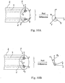
- the invention can make use of additional markers 6 presented hereafter and illustrated in figure 2 . More precisely, it can be based on the use of at least two supplementary EM markers which are stuck on the patient chest, preferably along the sternum, on anatomical points that can be easily found back, and which do not undergo important deformation in case the patient has not the same position during the previous and subsequent exploration (the patient can lay on the supine position and then on a decubitus lateral). It has been found that the jugular notch was a remarkably static anatomical point, as well as the sternum end point.
- the two markers 6 are stuck on the patient 7 during the intervention and their positions are recorded as well.
- the recorded position can be either only one acquisition for both points, or a collection of acquisitions for each marker which is averaged since it is assumed that the patient should not move during the intervention.
- two markers 6 are stuck on the patient 7 at approximately the same anatomical positions, and their positions are recorded.
- a first approach to perform the registration is then to do a 3D/3D point registration (see Arun KS, Huang TS, Blostein SD. "Least-squares fitting of two 3-d point sets. In IEEE Transactions on Pattern Analysis and Machine Intelligence". 1987 May;9(5):698-700 ) using two point triplets identified during each exploration.
- This triplet can be the two marker positions and the point P closest on the oesophagus shape estimation (it is approximately a collection of point with a line shape, curved close to the patient's throat), which is the closest to the marker stuck on the jugular notch.
- the inventors noticed that there is some uncertainty in finding repetitively the sternum end point (along the sternum). Additionally, the inventors also observed that the relative position of the closest point P closest to the jugular notch can change between the previous and the subsequent exploration of several millimeters. The 3D/3D registration may thus not be very accurate and it is proposed instead to register the following frame F that can be defined during the previous and subsequent exploration.
- the frame F center is defined by the marker stuck on the jugular notch, its X axis is defined by the vector M J M s between the jugular notch marker and the sternum end point marker, its Y axis is defined by the cross product between M J M s and the vector between the jugular notch marker and P closest , its Z axis being then computed from the cross product between X and Y axis.
- the invention basically aims at displaying a frame from the previous exploration, which point of view in the oesophagus or similar tubular organ is as close as possible to the point of view of the live/subsequent exploration. Finding the most adapted frame can be computationally expensive. A simpler implementation is possible, which still provides a guidance tool for medical experts but uses only a small or reduced collection of recorded image frames.
- the medical expert annotates the images that he considers relevant.
- R i relevant images
- i belonging to the range [1-50] (containing for instance biopsy information or pathology information), on which he indicates the position of the relevant information (an arrow or a circle can be superimposed on the image to show the relevant information).
- the system will show the live images of the subsequent exploration on one screen, and display the relevant images R i only if the live endoscope position is close to the position associated to the relevant images R i after the registration step (for instance, if the distance between the endoscope position and the position associated to R i is within a threshold value, say 3 mm).
- the system can also indicate in which direction (forward or backward) the endoscope should be moved to get closer to the point associated to R i .
- This indication can be provided by means of an oriented arrow, a letter (for instance, F or B) or a color indicator.
- the invention can also consider the issue of dealing with unsmooth positions of recorded frames.
- the system can display all relevant images in a mosaic representation on a screen, or the system may ask the medical expert, which relevant image among the several relevant images he wants to reach.
- the system repetitively and simultaneously records the endoscope position via the (EM) sensor associated to the corresponding image during the procedure. It is thus possible that many pairs position+image are recorded for a specific depth in the oesophagus or similar tubular organ, but with a different lateral position of the endoscope in said organ.
- EM electromagnetic wave
- a basic approach to select, during the subsequent live exploration, the image from the previous exploration corresponding to the current position of the endoscope may be uncomfortable for the medical expert.
- the displayed images at time t may be very different from the image at time t+1, the point of view of the endoscope in the oesophagus or similar tubular organ being very different: the image flow is then not smooth.
- the lumen can be on the left side of the video images during the forward motion of the endoscope (t 0 - t 200 ) and on the right side of the video images during the backward motion of the endoscope (t 200 - t 400 ), within the same exploration procedure.
- Another issue which can be addressed by the invention is taking the rotation of the endoscope into account.
- the medical expert introduces the endoscope with an orientation around its axis (usually called roll) that may be different from the orientation during the previous exploration.
- An experienced medical expert is usually able, by watching some specific marks on the endoscope, to approximately have the same orientation between the previous and the subsequent exploration. However, this takes time and is not so easy for a novice medical expert. If the orientation of the endoscope is not the same, the method can of course synchronize the two exploration videos but the image orientation will not be the same, and thus it will be less easy for the medical expert to recognize the same anatomical structure in both videos.
- the inventive method can be improved to compute the live orientation error between the previous exploration and the subsequent exploration.
- the system can indicate with an arrow on the live video screen in which direction the endoscope should be rotated so that the rotation error is reduced.
- the rotation error in degree can also be displayed and updated.
- the system can also apply automatically the rotation that compensates the error to the previous exploration video, or to the subsequent exploration video.
- a 6 DOF electromagnetic sensor is set at the tip of the endoscope.
- the recording step during the previous exploration includes not only the 3D position of the endoscope tip, but also its orientation (roll, yaw and pitch).
- the orientation of the endoscope tip is also acquired.
- the orientation error can be given by the roll difference between the 6 DOF sensor orientation of the live endoscope position and the recorded 6 DOF sensor orientation at the position from the previous exploration that has been matched to the live endoscope position.
- the system setup 1 consists of an EM field generator 4 with an adapted working volume which is placed roughly above the chest of the patient 7 and fixed in position using a titanium arm.
- a 6 DOF EM sensor 4' is inserted into the working channel of the flexible endoscope 2 and fixed at the tip 2' of the endoscope.
- the EMTS and the endoscope 2 are connected to a laptop or similar computer concerns 5, that synchronously records the data.
- a recording of an intervention consists of a list of EM sensor poses (trajectory of the endoscope tip 2'), with the corresponding image frames captured from the endoscope 2.
- a corresponding image can be found in the recording, that spatially matches the endoscope's current location in the oesophagus.
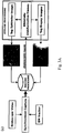
- FIGS 3A to 3C provide an overview of the work-flow of the inventive method.
- the used process is divided into three parts or phases:
- the acquisition phase is schematically illustrated on figure 3A .
- the GI specialist performs the recording of an intervention and tagging of relevant images.
- the flexible endoscope is slowly guided through the oesophagus, while the EM sensor pose and the corresponding image acquired from the endoscope are recorded to the database.
- the recording contains many uninformative frames; with bubbles, motion-blur, specular highlights, and out-of-focus images. Firstly, these uninformative frames are detected and left out from further processing using the method described in: M K Bashar et al. Automatic detection of information frames from wireless capsule endoscopy images. Medical image analysis, 14(3):449-70, June 201 .
- the GI specialist tags the images containing the sphincter as it is used as a landmark in the registration phase.
- the sphincter is used as the anatomical landmark because it is stable and can be reached with good repeatable accuracy.
- the endoscopic frames that contain the biopsy procedure are tagged and in an offline step, the expert reviews the tagged biopsy images and selects those most relevant for the procedure. At this stage, the expert can choose to add supplementary information to the images of the recordings, which will be available during the synchronisation phase.
- the registration phase is illustrated on figure 3B .
- the EM sensor position is recorded while the GI specialist introduces the endoscope into the oesophagus and guides it until the sphincter.
- the contextual knowledge that the oesophagus is fairly linear and exhibits very minimal lateral movement is used.
- the tagged sphincter positions can then be used to obtain translation t, which along with R provides an initialisation for the iterative closest point (ICP) for further registration refinement.
- the synchronisation phase is illustrated on figure 3C .
- a spatial correspondence between the sensor position from the current intervention and the previously recorded intervention is computed.
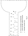
- a search for the closest neighbour (in Euclidean sense) to the current sensor position is made. Due to localized deformations of the oesophagus during navigation, the trajectories are not smooth and exhibit deviations ( figure 6 ) from the central oesophagus line (connecting the throat to the sphincter, along vector z), which can lead to a false match with marked depth difference. Since the trajectories have been aligned along vector z, the search space is constrained to lie within ⁇ z (approximately 2 mm).
- the closest neighbour gives the corresponding best matching image of the region in the oesophagus taken during the previous procedure.
- the matched images that were tagged to contain locations of biopsy sites provide the GI specialist a more localized region for review.
- Figure 7 resents the result of the synchronisation phase. ICP finds the transform up to a rotation along vector z, however, since the search space is constrained along this direction, the determination of the closet neighbour is unaffected by it.
- the inventors performed experiments based on the inventive method and system, using NDI ⁇ copyright Aurora EMTS (accuracy of 0.5 mm in the given working volume).
- the inventive system was tested on three sets of in-vivo sequences on different porcines, with each set consisting of four recordings. Prior marking were made on the oesophagus using a dual knife at every 2 cm to simulate biopsy locations. To replicate realistic surgical procedures conducted at different times, the EM field emitter was randomly repositioned before each recording. The steps described previously were performed, with the first recording as the reference and three other recordings to mimic a follow-up procedure. A qualitative evaluation of the approach was performed by presenting the output of the synchronisation phase to five experts.
- the invention system can also be used and applied to reposition or relocate quickly and precisely, at specific locations already explored, a flexible endoscope in other tubular more or less deformable organs.
- the inventive system takes into consideration the following information about anatomy of the colon: there exist three parts of the colon which are attached to abdominal wall, and thus which can be considered as reference landmarks: the right and the left colic flexures, the caecum to intestine transition. Since these three parts can be easily recognized by the medical expert, and their position recorded during explorations (previous and subsequent), it is then possible to map the size of each intermediate trajectory in the previous exploration to the corresponding one in the subsequent exploration.
- the medical expert (operator) firstly reaches the three fixed points in the current exploration, and the computer matches the three colon parts by applying a ratio to the length of each part, so that the length of each matched part in previous and subsequent exploration have the same value.
- the medical expert can reach the relevant sites annotated or recorded during the previous exploration by slightly removing the endoscope until the inventive method indicates that he is close to the relevant sites. It is important to highlight that if the medical expert tries to reach a target using the inventive method by moving forward in the colon, it is likely that the information provided by the system may not be accurate. Indeed, it is well known that the unpredictable elastic deformation of the colon mainly occurs during the forward motion and much less during the backward motion.
- the invention proposes a method and a system which are quite straightforward, work in real-time (trajectory matching at 50 to 100 Hz), can be used with minimal change to the operating room protocol and most of the offline steps can easily be automated. Moreover, because the system is scalable in time, the recordings can be shared between GI specialists without loss of information. Finally, since the inventive method does not rely on the quality of images, it is robust to typical endoscopic image artefacts for interoperative comparison.
Landscapes
- Engineering & Computer Science (AREA)
- Health & Medical Sciences (AREA)
- Life Sciences & Earth Sciences (AREA)
- Surgery (AREA)
- Physics & Mathematics (AREA)
- Medical Informatics (AREA)
- General Health & Medical Sciences (AREA)
- Biomedical Technology (AREA)
- Heart & Thoracic Surgery (AREA)
- Molecular Biology (AREA)
- Animal Behavior & Ethology (AREA)
- Public Health (AREA)
- Veterinary Medicine (AREA)
- Nuclear Medicine, Radiotherapy & Molecular Imaging (AREA)
- Theoretical Computer Science (AREA)
- General Physics & Mathematics (AREA)
- Pathology (AREA)
- Biophysics (AREA)
- Radiology & Medical Imaging (AREA)
- Computer Vision & Pattern Recognition (AREA)
- Optics & Photonics (AREA)
- Robotics (AREA)
- Human Computer Interaction (AREA)
- Computer Graphics (AREA)
- Computer Hardware Design (AREA)
- General Engineering & Computer Science (AREA)
- Software Systems (AREA)
- Quality & Reliability (AREA)
- Signal Processing (AREA)
- Multimedia (AREA)
- Endoscopes (AREA)
Claims (7)
- Medizinisches System, das eine manuelle oder automatische Repositionierung eines flexiblen Endoskops an einem bestimmten Ort während zumindest einer "neuen" Exploration im Anschluss an eine erste bzw. "Referenz-" Exploration ermöglicht, während der dieser bestimmte Ort in einem tubulären, mehr oder weniger verformbaren Organ bereits mittels dieses Endoskops exploriert wurde,
wobei das medizinische System (1) aufweist:i) ein flexibles Endoskop (2), das zumindest mit einer Bildaufnahmevorrichtung (3) und eventuell zumindest einem Werkzeug oder Instrument ausgestattet ist,ii) diesem Endoskop (2) zugeordnete Anzeige- (3') und Bewegungssteuerungs- (3") Mittel,iii) ein(e) Verfolgungsvorrichtung oder -system (4), die/das die 3D-Position und - Ausrichtung der Endspitze (2') des Endoskops (2) bestimmt, beispielsweise eine elektromagnetische Verfolgungsvorrichtung oder eine fiberoptische Verfolgungsvorrichtung, die an der oder in die Spitze (2') des flexiblen Endoskops (2) befestigt oder eingebaut ist,iiii) Rechnereinrichtungen (5) und diesen zugeordnete Speichereinrichtungen, wobei diese Speichereinrichtungen eingerichtet sind, ein "Referenzexplorations-" Video zu speichern, das durch das flexible Endoskop während seiner "Referenz-" Exploration aufgenommen wurde, wobei jedes der aufgenommenen Videobilder einer synchron aufgenommenen 3D-Position und -Ausrichtung der Endoskopspitze zugeordnet ist, und das "Referenzexplorations-" Video Bilder eines markierten anatomischen Orientierungspunkts des tubulären Organs aufweist,dadurch gekennzeichnet, dass das medizinische System (1) eingerichtet ist, umi) das flexible Endoskop während der "neuen" Exploration zu dem markierten anatomischen Orientierungspunkt zu führen, die 3D-Position und -Ausrichtung des markierten anatomischen Orientierungspunkts aufzunehmen,ii) eine Translation (t) von der 3D-Position und -Ausrichtung des markierten anatomischen Orientierungspunkts zur Ausrichtung der Videobilder der "Referenz-" Exploration entlang einer Mittelachse (z) des tubulären Organs zu verwenden,iii) eine räumliche Entsprechung zwischen der Position der Endoskopspitze der "neuen" Exploration und der Position der Endoskopspitze der "Referenz-" Exploration zu berechnen,iv) das räumlich nächstgelegene benachbarte Bild des "Referenzexplorations-" Videos als am besten passendes Bild der "Referenz-" Exploration zu suchen, undv) parallel auf oder in zwei unterschiedlichen Fenstern auf einem oder mehreren Bildschirm(en) ein Echtzeit-Bild des Videos der "neuen Exploration" und des am besten passenden Bilds der "Referenz-" Exploration anzuzeigen. - Medizinisches System nach Anspruch 1,
wobei es auch zumindest zwei Marker (6) aufweist, deren Positionen mit dem verwendeten Verfolgungssystem (4) oder einem anderen Verfolgungssystem aufgezeichnet werden können, und die an vorgegebenen anatomischen Orten an dem Individuum platziert werden können, wobei diese Orte von dem explorierten tubulären Organ abhängen und vorzugsweise keine merkliche Verformung oder Verlagerung erfahren, wenn das Individuum seine Position verändert, wobei diese Marker (6) Referenzpunkte in den vorangegangenen und nachfolgenden Explorationen bereitstellen. - Medizinisches System nach einem der Ansprüche 1 und 2,
wobei es Mittel für die Durchführung einer Videoendoskopie-Echtzeitanalyse aufweist unter Ausführung einer Extraktion und eines Abgleichs ähnlicher Merkmale, die sowohl während der ersten bzw. der "Referenz-" Exploration als auch während der nachfolgenden oder neuen Exploration sichtbar sind. - Medizinisches System nach einem der Ansprüche 1 bis 3,
wobei es ein Human Interface Device aufweist, das die präzise Auswahl eines Pixels oder einer Gruppe von Pixeln in einem Video ermöglicht, beispielsweise durch eine Maus, ein Pad, einen Trackball, einen Joystick oder optischem Tracking, um ein interaktives virtuelles Markieren eines von mehreren Bildern des ersten bzw. endoskopischen "Referenz-Explorations"-Videos des flexiblen Endoskops durchzuführen, welches konkrete Punkte von Interesse in dem Körper des explorierten Individuums anzeigt, wobei dieses virtuelle Markieren von einem speziellen "Referenzexplorations-" Videolesegerät durchgeführt wird, das das Hinzufügen von Markierungen zu einem beliebigen ausgewählten Bild des Videos ermöglicht. - Medizinisches System nach einem der Ansprüche 1 bis 3,
wobei es ein Algorithmus-Mittel zur Videoanalyse aufweist, das in der Lage ist, sichtbare Punkte von Interesse wie beispielsweise anatomische, pathologische oder chirurgische konkrete Merkmale in dem Körper des explorierten Individuums zu extrahieren, und ein automatisches virtuelles Markieren an einem oder mehreren Bild(ern) des "Referenzexploration" -Videos des flexiblen Endoskops zu verwirklichen, was diese konkreten Punkte von Interesse anzeigt. - Medizinisches System nach Anspruch 4 oder Anspruch 5,
wobei es auch Mittel aufweist, die in der Lage sind, eine Analyse des Videos der nachfolgenden "neuen Exploration" durchzuführen, um in diesem Video denselben konkreten Punkt von Interesse in dem Körper des Individuums zu lokalisieren, der in einem Bild des "Referenzexplorations-" Videos markiert wurde, und ihn an derselben Stelle in dem synchronisierten Video der nachfolgenden "neuen Exploration" mit einer Überlagerung dieser virtuellen Markierungen hinzuzufügen. - Medizinisches System nach einem der Ansprüche 1 bis 6,
wobei es Mittel aufweist, die in der Lage sind, die zwei synchronisierten Videos des flexiblen Endoskops zu vereinigen, durch virtuelle Markierungen erweitert oder nicht, welche konkrete Punkte von Interesse zeigen, unter Verwendung von Techniken erweiterter Realität mit einem virtuellen Rendern der Körper-3D-Modelle der anatomischen und/oder pathologischen Struktur, wobei diese 3D-Modelle aus einem präoperativen medizinischen Bild extrahiert werden, das vor der "neuen" Exploration des Körpers des Individuums aufgenommen wurde.
Applications Claiming Priority (2)
| Application Number | Priority Date | Filing Date | Title |
|---|---|---|---|
| US201361775930P | 2013-03-11 | 2013-03-11 | |
| PCT/IB2014/000838 WO2014140813A1 (en) | 2013-03-11 | 2014-03-11 | Anatomical site relocalisation using dual data synchronisation |
Publications (2)
| Publication Number | Publication Date |
|---|---|
| EP2996557A1 EP2996557A1 (de) | 2016-03-23 |
| EP2996557B1 true EP2996557B1 (de) | 2019-05-01 |
Family
ID=50943347
Family Applications (1)
| Application Number | Title | Priority Date | Filing Date |
|---|---|---|---|
| EP14730578.3A Active EP2996557B1 (de) | 2013-03-11 | 2014-03-11 | Relokalisierung eines anatomischen orts mit dualer datensynchronisierung |
Country Status (4)
| Country | Link |
|---|---|
| US (1) | US10736497B2 (de) |
| EP (1) | EP2996557B1 (de) |
| JP (1) | JP6348130B2 (de) |
| WO (1) | WO2014140813A1 (de) |
Cited By (1)
| Publication number | Priority date | Publication date | Assignee | Title |
|---|---|---|---|---|
| US11642016B2 (en) | 2021-08-10 | 2023-05-09 | Altek Biotechnology Corporation | Image capturing module, endoscope and method of manufacturing image capturing module |
Families Citing this family (32)
| Publication number | Priority date | Publication date | Assignee | Title |
|---|---|---|---|---|
| US10736497B2 (en) * | 2013-03-11 | 2020-08-11 | Institut Hospitalo-Universitaire De Chirurgie Mini-Invasive Guidee Par L'image | Anatomical site relocalisation using dual data synchronisation |
| JP5993515B2 (ja) * | 2013-09-27 | 2016-09-14 | オリンパス株式会社 | 内視鏡システム |
| WO2015126457A1 (en) * | 2014-02-19 | 2015-08-27 | Indiana University Research And Technology Corporation | Tracking real-time assessment of quality monitoring in endoscopy |
| CA2935506A1 (en) | 2014-02-21 | 2015-08-27 | 3Dintegrated Aps | A set comprising a surgical instrument |
| US10013808B2 (en) | 2015-02-03 | 2018-07-03 | Globus Medical, Inc. | Surgeon head-mounted display apparatuses |
| US9905000B2 (en) * | 2015-02-19 | 2018-02-27 | Sony Corporation | Method and system for surgical tool localization during anatomical surgery |
| US10635924B2 (en) * | 2015-05-11 | 2020-04-28 | Siemens Aktiengesellschaft | System and method for surgical guidance and intra-operative pathology through endo-microscopic tissue differentiation |
| US11020144B2 (en) | 2015-07-21 | 2021-06-01 | 3Dintegrated Aps | Minimally invasive surgery system |
| WO2017012624A1 (en) | 2015-07-21 | 2017-01-26 | 3Dintegrated Aps | Cannula assembly kit, trocar assembly kit, sleeve assembly, minimally invasive surgery system and method therefor |
| DK178899B1 (en) | 2015-10-09 | 2017-05-08 | 3Dintegrated Aps | A depiction system |
| CN107613897B (zh) * | 2015-10-14 | 2021-12-17 | 外科手术室公司 | 扩增实境的外科导航 |
| JP6646133B2 (ja) * | 2016-02-23 | 2020-02-14 | オリンパス株式会社 | 画像処理装置および内視鏡 |
| US10792085B2 (en) | 2016-10-24 | 2020-10-06 | Csa Medical, Inc. | Method and apparatus for performing cryotherapy of distal lung lesions |
| WO2018235255A1 (ja) * | 2017-06-23 | 2018-12-27 | オリンパス株式会社 | 医療システムとその作動方法 |
| US20190254753A1 (en) | 2018-02-19 | 2019-08-22 | Globus Medical, Inc. | Augmented reality navigation systems for use with robotic surgical systems and methods of their use |
| CN112074867A (zh) * | 2018-05-11 | 2020-12-11 | 直观外科手术操作公司 | 与用于图像引导手术的配准有关的系统和方法 |
| US10902616B2 (en) * | 2018-08-13 | 2021-01-26 | Nvidia Corporation | Scene embedding for visual navigation |
| EP3860426A4 (de) * | 2018-10-02 | 2022-12-07 | Convergascent LLC | Endoskop mit trägheitsmesseinheiten und/oder haptischen eingabesteuerungen |
| US11514576B2 (en) | 2018-12-14 | 2022-11-29 | Acclarent, Inc. | Surgical system with combination of sensor-based navigation and endoscopy |
| US11992373B2 (en) | 2019-12-10 | 2024-05-28 | Globus Medical, Inc | Augmented reality headset with varied opacity for navigated robotic surgery |
| US11464581B2 (en) | 2020-01-28 | 2022-10-11 | Globus Medical, Inc. | Pose measurement chaining for extended reality surgical navigation in visible and near infrared spectrums |
| US11382699B2 (en) | 2020-02-10 | 2022-07-12 | Globus Medical Inc. | Extended reality visualization of optical tool tracking volume for computer assisted navigation in surgery |
| US11207150B2 (en) | 2020-02-19 | 2021-12-28 | Globus Medical, Inc. | Displaying a virtual model of a planned instrument attachment to ensure correct selection of physical instrument attachment |
| US11607277B2 (en) | 2020-04-29 | 2023-03-21 | Globus Medical, Inc. | Registration of surgical tool with reference array tracked by cameras of an extended reality headset for assisted navigation during surgery |
| US11510750B2 (en) | 2020-05-08 | 2022-11-29 | Globus Medical, Inc. | Leveraging two-dimensional digital imaging and communication in medicine imagery in three-dimensional extended reality applications |
| US11382700B2 (en) | 2020-05-08 | 2022-07-12 | Globus Medical Inc. | Extended reality headset tool tracking and control |
| US11153555B1 (en) | 2020-05-08 | 2021-10-19 | Globus Medical Inc. | Extended reality headset camera system for computer assisted navigation in surgery |
| US11737831B2 (en) | 2020-09-02 | 2023-08-29 | Globus Medical Inc. | Surgical object tracking template generation for computer assisted navigation during surgical procedure |
| USD1022197S1 (en) | 2020-11-19 | 2024-04-09 | Auris Health, Inc. | Endoscope |
| US20230100698A1 (en) | 2021-09-29 | 2023-03-30 | Cilag Gmbh International | Methods for Controlling Cooperative Surgical Instruments |
| KR102637484B1 (ko) * | 2021-10-26 | 2024-02-16 | 주식회사 카이미 | 인공지능 기반의 내시경 진단 보조 시스템 및 이의 제어방법 |
| CN114191078B (zh) * | 2021-12-29 | 2024-04-26 | 上海复旦数字医疗科技股份有限公司 | 一种基于混合现实的内窥镜手术导航机器人系统 |
Family Cites Families (20)
| Publication number | Priority date | Publication date | Assignee | Title |
|---|---|---|---|---|
| US5829444A (en) * | 1994-09-15 | 1998-11-03 | Visualization Technology, Inc. | Position tracking and imaging system for use in medical applications |
| JP2001197485A (ja) * | 2000-01-11 | 2001-07-19 | Asahi Optical Co Ltd | 電子内視鏡システムおよび電子内視鏡用信号切替装置 |
| US20050228250A1 (en) * | 2001-11-21 | 2005-10-13 | Ingmar Bitter | System and method for visualization and navigation of three-dimensional medical images |
| US7505809B2 (en) | 2003-01-13 | 2009-03-17 | Mediguide Ltd. | Method and system for registering a first image with a second image relative to the body of a patient |
| US20050085718A1 (en) | 2003-10-21 | 2005-04-21 | Ramin Shahidi | Systems and methods for intraoperative targetting |
| US7397364B2 (en) * | 2003-11-11 | 2008-07-08 | Biosense Webster, Inc. | Digital wireless position sensor |
| WO2005058137A2 (en) * | 2003-12-12 | 2005-06-30 | University Of Washington | Catheterscope 3d guidance and interface system |
| WO2006005075A2 (en) * | 2004-06-30 | 2006-01-12 | Amir Belson | Apparatus and methods for capsule endoscopy of the esophagus |
| WO2007025081A2 (en) | 2005-08-24 | 2007-03-01 | Traxtal Inc. | System, method and devices for navigated flexible endoscopy |
| US7945310B2 (en) * | 2006-09-18 | 2011-05-17 | Stryker Corporation | Surgical instrument path computation and display for endoluminal surgery |
| US8248414B2 (en) * | 2006-09-18 | 2012-08-21 | Stryker Corporation | Multi-dimensional navigation of endoscopic video |
| US7892165B2 (en) * | 2006-10-23 | 2011-02-22 | Hoya Corporation | Camera calibration for endoscope navigation system |
| US8672836B2 (en) * | 2007-01-31 | 2014-03-18 | The Penn State Research Foundation | Method and apparatus for continuous guidance of endoscopy |
| JP5148227B2 (ja) * | 2007-09-25 | 2013-02-20 | 富士フイルム株式会社 | 内視鏡システム |
| WO2009128055A1 (en) * | 2008-04-15 | 2009-10-22 | Provost Fellows And Scholars Of The College Of The Holy And Undivided Trinity Of Queen Elizabeth Near Dublin | Endoscopy system with motion sensors |
| EP2348954A1 (de) * | 2008-10-20 | 2011-08-03 | Koninklijke Philips Electronics N.V. | Lokalisationsverfahren und system auf bildgebungsbasis |
| EP2348982B1 (de) | 2008-12-03 | 2020-03-25 | St. Jude Medical, Atrial Fibrillation Division, Inc. | System zur bestimmung der position der spitze eines medizinischen katheters im körper eines patienten |
| WO2010128411A1 (en) * | 2009-05-08 | 2010-11-11 | Koninklijke Philips Electronics, N.V. | Real-time scope tracking and branch labeling without electro-magnetic tracking and pre-operative scan roadmaps |
| US20120069167A1 (en) * | 2009-05-18 | 2012-03-22 | Koninklijke Philips Electronics N.V. | Marker-free tracking registration and calibration for em-tracked endoscopic system |
| US10736497B2 (en) * | 2013-03-11 | 2020-08-11 | Institut Hospitalo-Universitaire De Chirurgie Mini-Invasive Guidee Par L'image | Anatomical site relocalisation using dual data synchronisation |
-
2014
- 2014-03-11 US US14/774,516 patent/US10736497B2/en active Active
- 2014-03-11 WO PCT/IB2014/000838 patent/WO2014140813A1/en active Application Filing
- 2014-03-11 JP JP2015562379A patent/JP6348130B2/ja active Active
- 2014-03-11 EP EP14730578.3A patent/EP2996557B1/de active Active
Non-Patent Citations (1)
| Title |
|---|
| None * |
Cited By (2)
| Publication number | Priority date | Publication date | Assignee | Title |
|---|---|---|---|---|
| US11642016B2 (en) | 2021-08-10 | 2023-05-09 | Altek Biotechnology Corporation | Image capturing module, endoscope and method of manufacturing image capturing module |
| TWI815182B (zh) * | 2021-08-10 | 2023-09-11 | 榮晶生物科技股份有限公司 | 影像擷取模組、內視鏡及影像擷取模組之製造方法 |
Also Published As
| Publication number | Publication date |
|---|---|
| WO2014140813A1 (en) | 2014-09-18 |
| JP2016511049A (ja) | 2016-04-14 |
| EP2996557A1 (de) | 2016-03-23 |
| JP6348130B2 (ja) | 2018-06-27 |
| US10736497B2 (en) | 2020-08-11 |
| US20160022125A1 (en) | 2016-01-28 |
Similar Documents
| Publication | Publication Date | Title |
|---|---|---|
| EP2996557B1 (de) | Relokalisierung eines anatomischen orts mit dualer datensynchronisierung | |
| US11350893B2 (en) | Methods and systems for using multi view pose estimation | |
| US20200405433A1 (en) | System and method for dynamic validation, correction of registration for surgical navigation | |
| CN102428496B (zh) | 用于em跟踪内窥镜系统的无标记物跟踪的配准和校准 | |
| US20170084036A1 (en) | Registration of video camera with medical imaging | |
| WO2017027638A1 (en) | 3d reconstruction and registration of endoscopic data | |
| EP3544538B1 (de) | System zur navigation von interventionellen instrumenten | |
| CN111568544B (zh) | 用于使医疗装置相对于目标的导航视觉化的系统和方法 | |
| EP2413777B1 (de) | Assoziation einer sensorposition mit einer bildposition | |
| EP2473973A1 (de) | Vorrichtung und verfahren zur bestimmung eines ortes auf einem zielbild | |
| KR20130108320A (ko) | 관련 애플리케이션들에 대한 일치화된 피하 해부구조 참조의 시각화 | |
| US20220079557A1 (en) | System and method for medical navigation | |
| Liu et al. | A non-invasive navigation system for retargeting gastroscopic lesions | |
| Vemuri et al. | Interoperative biopsy site relocalization in endoluminal surgery | |
| Wang et al. | Dynamic 3D reconstruction of gastric internal surface under gastroscopy | |
| Vemuri et al. | Inter-operative trajectory registration for endoluminal video synchronization: Application to biopsy site re-localization | |
| Serna-Morales et al. | Acquisition of three-dimensional information of brain structures using endoneurosonography | |
| US20240206980A1 (en) | Volumetric filter of fluoroscopic sweep video | |
| Vemuri | Inter-operative biopsy site relocalization in gastroscopy: Application to oesophagus | |
| WO2020181498A1 (zh) | 体内导航系统和方法 | |
| WO2023146902A1 (en) | Two-phase instrument guidance for accurate endoscopic surgical procedures |
Legal Events
| Date | Code | Title | Description |
|---|---|---|---|
| PUAI | Public reference made under article 153(3) epc to a published international application that has entered the european phase |
Free format text: ORIGINAL CODE: 0009012 |
|
| 17P | Request for examination filed |
Effective date: 20151008 |
|
| AK | Designated contracting states |
Kind code of ref document: A1 Designated state(s): AL AT BE BG CH CY CZ DE DK EE ES FI FR GB GR HR HU IE IS IT LI LT LU LV MC MK MT NL NO PL PT RO RS SE SI SK SM TR |
|
| AX | Request for extension of the european patent |
Extension state: BA ME |
|
| DAX | Request for extension of the european patent (deleted) | ||
| STAA | Information on the status of an ep patent application or granted ep patent |
Free format text: STATUS: EXAMINATION IS IN PROGRESS |
|
| 17Q | First examination report despatched |
Effective date: 20170113 |
|
| GRAP | Despatch of communication of intention to grant a patent |
Free format text: ORIGINAL CODE: EPIDOSNIGR1 |
|
| STAA | Information on the status of an ep patent application or granted ep patent |
Free format text: STATUS: GRANT OF PATENT IS INTENDED |
|
| INTG | Intention to grant announced |
Effective date: 20181106 |
|
| GRAS | Grant fee paid |
Free format text: ORIGINAL CODE: EPIDOSNIGR3 |
|
| GRAJ | Information related to disapproval of communication of intention to grant by the applicant or resumption of examination proceedings by the epo deleted |
Free format text: ORIGINAL CODE: EPIDOSDIGR1 |
|
| GRAL | Information related to payment of fee for publishing/printing deleted |
Free format text: ORIGINAL CODE: EPIDOSDIGR3 |
|
| STAA | Information on the status of an ep patent application or granted ep patent |
Free format text: STATUS: EXAMINATION IS IN PROGRESS |
|
| GRAR | Information related to intention to grant a patent recorded |
Free format text: ORIGINAL CODE: EPIDOSNIGR71 |
|
| STAA | Information on the status of an ep patent application or granted ep patent |
Free format text: STATUS: GRANT OF PATENT IS INTENDED |
|
| GRAA | (expected) grant |
Free format text: ORIGINAL CODE: 0009210 |
|
| STAA | Information on the status of an ep patent application or granted ep patent |
Free format text: STATUS: THE PATENT HAS BEEN GRANTED |
|
| INTC | Intention to grant announced (deleted) | ||
| INTG | Intention to grant announced |
Effective date: 20190321 |
|
| AK | Designated contracting states |
Kind code of ref document: B1 Designated state(s): AL AT BE BG CH CY CZ DE DK EE ES FI FR GB GR HR HU IE IS IT LI LT LU LV MC MK MT NL NO PL PT RO RS SE SI SK SM TR |
|
| REG | Reference to a national code |
Ref country code: GB Ref legal event code: FG4D |
|
| REG | Reference to a national code |
Ref country code: CH Ref legal event code: EP Ref country code: AT Ref legal event code: REF Ref document number: 1125815 Country of ref document: AT Kind code of ref document: T Effective date: 20190515 |
|
| REG | Reference to a national code |
Ref country code: DE Ref legal event code: R096 Ref document number: 602014045745 Country of ref document: DE |
|
| REG | Reference to a national code |
Ref country code: IE Ref legal event code: FG4D |
|
| REG | Reference to a national code |
Ref country code: NL Ref legal event code: MP Effective date: 20190501 |
|
| REG | Reference to a national code |
Ref country code: LT Ref legal event code: MG4D |
|
| PG25 | Lapsed in a contracting state [announced via postgrant information from national office to epo] |
Ref country code: FI Free format text: LAPSE BECAUSE OF FAILURE TO SUBMIT A TRANSLATION OF THE DESCRIPTION OR TO PAY THE FEE WITHIN THE PRESCRIBED TIME-LIMIT Effective date: 20190501 Ref country code: NO Free format text: LAPSE BECAUSE OF FAILURE TO SUBMIT A TRANSLATION OF THE DESCRIPTION OR TO PAY THE FEE WITHIN THE PRESCRIBED TIME-LIMIT Effective date: 20190801 Ref country code: HR Free format text: LAPSE BECAUSE OF FAILURE TO SUBMIT A TRANSLATION OF THE DESCRIPTION OR TO PAY THE FEE WITHIN THE PRESCRIBED TIME-LIMIT Effective date: 20190501 Ref country code: SE Free format text: LAPSE BECAUSE OF FAILURE TO SUBMIT A TRANSLATION OF THE DESCRIPTION OR TO PAY THE FEE WITHIN THE PRESCRIBED TIME-LIMIT Effective date: 20190501 Ref country code: AL Free format text: LAPSE BECAUSE OF FAILURE TO SUBMIT A TRANSLATION OF THE DESCRIPTION OR TO PAY THE FEE WITHIN THE PRESCRIBED TIME-LIMIT Effective date: 20190501 Ref country code: PT Free format text: LAPSE BECAUSE OF FAILURE TO SUBMIT A TRANSLATION OF THE DESCRIPTION OR TO PAY THE FEE WITHIN THE PRESCRIBED TIME-LIMIT Effective date: 20190901 Ref country code: ES Free format text: LAPSE BECAUSE OF FAILURE TO SUBMIT A TRANSLATION OF THE DESCRIPTION OR TO PAY THE FEE WITHIN THE PRESCRIBED TIME-LIMIT Effective date: 20190501 Ref country code: NL Free format text: LAPSE BECAUSE OF FAILURE TO SUBMIT A TRANSLATION OF THE DESCRIPTION OR TO PAY THE FEE WITHIN THE PRESCRIBED TIME-LIMIT Effective date: 20190501 Ref country code: LT Free format text: LAPSE BECAUSE OF FAILURE TO SUBMIT A TRANSLATION OF THE DESCRIPTION OR TO PAY THE FEE WITHIN THE PRESCRIBED TIME-LIMIT Effective date: 20190501 |
|
| PG25 | Lapsed in a contracting state [announced via postgrant information from national office to epo] |
Ref country code: BG Free format text: LAPSE BECAUSE OF FAILURE TO SUBMIT A TRANSLATION OF THE DESCRIPTION OR TO PAY THE FEE WITHIN THE PRESCRIBED TIME-LIMIT Effective date: 20190801 Ref country code: RS Free format text: LAPSE BECAUSE OF FAILURE TO SUBMIT A TRANSLATION OF THE DESCRIPTION OR TO PAY THE FEE WITHIN THE PRESCRIBED TIME-LIMIT Effective date: 20190501 Ref country code: LV Free format text: LAPSE BECAUSE OF FAILURE TO SUBMIT A TRANSLATION OF THE DESCRIPTION OR TO PAY THE FEE WITHIN THE PRESCRIBED TIME-LIMIT Effective date: 20190501 Ref country code: GR Free format text: LAPSE BECAUSE OF FAILURE TO SUBMIT A TRANSLATION OF THE DESCRIPTION OR TO PAY THE FEE WITHIN THE PRESCRIBED TIME-LIMIT Effective date: 20190802 |
|
| REG | Reference to a national code |
Ref country code: AT Ref legal event code: MK05 Ref document number: 1125815 Country of ref document: AT Kind code of ref document: T Effective date: 20190501 |
|
| PG25 | Lapsed in a contracting state [announced via postgrant information from national office to epo] |
Ref country code: IS Free format text: LAPSE BECAUSE OF FAILURE TO SUBMIT A TRANSLATION OF THE DESCRIPTION OR TO PAY THE FEE WITHIN THE PRESCRIBED TIME-LIMIT Effective date: 20190901 |
|
| PG25 | Lapsed in a contracting state [announced via postgrant information from national office to epo] |
Ref country code: RO Free format text: LAPSE BECAUSE OF FAILURE TO SUBMIT A TRANSLATION OF THE DESCRIPTION OR TO PAY THE FEE WITHIN THE PRESCRIBED TIME-LIMIT Effective date: 20190501 Ref country code: SK Free format text: LAPSE BECAUSE OF FAILURE TO SUBMIT A TRANSLATION OF THE DESCRIPTION OR TO PAY THE FEE WITHIN THE PRESCRIBED TIME-LIMIT Effective date: 20190501 Ref country code: EE Free format text: LAPSE BECAUSE OF FAILURE TO SUBMIT A TRANSLATION OF THE DESCRIPTION OR TO PAY THE FEE WITHIN THE PRESCRIBED TIME-LIMIT Effective date: 20190501 Ref country code: AT Free format text: LAPSE BECAUSE OF FAILURE TO SUBMIT A TRANSLATION OF THE DESCRIPTION OR TO PAY THE FEE WITHIN THE PRESCRIBED TIME-LIMIT Effective date: 20190501 Ref country code: DK Free format text: LAPSE BECAUSE OF FAILURE TO SUBMIT A TRANSLATION OF THE DESCRIPTION OR TO PAY THE FEE WITHIN THE PRESCRIBED TIME-LIMIT Effective date: 20190501 Ref country code: CZ Free format text: LAPSE BECAUSE OF FAILURE TO SUBMIT A TRANSLATION OF THE DESCRIPTION OR TO PAY THE FEE WITHIN THE PRESCRIBED TIME-LIMIT Effective date: 20190501 |
|
| REG | Reference to a national code |
Ref country code: DE Ref legal event code: R097 Ref document number: 602014045745 Country of ref document: DE |
|
| PG25 | Lapsed in a contracting state [announced via postgrant information from national office to epo] |
Ref country code: SM Free format text: LAPSE BECAUSE OF FAILURE TO SUBMIT A TRANSLATION OF THE DESCRIPTION OR TO PAY THE FEE WITHIN THE PRESCRIBED TIME-LIMIT Effective date: 20190501 |
|
| PLBE | No opposition filed within time limit |
Free format text: ORIGINAL CODE: 0009261 |
|
| STAA | Information on the status of an ep patent application or granted ep patent |
Free format text: STATUS: NO OPPOSITION FILED WITHIN TIME LIMIT |
|
| PG25 | Lapsed in a contracting state [announced via postgrant information from national office to epo] |
Ref country code: TR Free format text: LAPSE BECAUSE OF FAILURE TO SUBMIT A TRANSLATION OF THE DESCRIPTION OR TO PAY THE FEE WITHIN THE PRESCRIBED TIME-LIMIT Effective date: 20190501 |
|
| 26N | No opposition filed |
Effective date: 20200204 |
|
| PG25 | Lapsed in a contracting state [announced via postgrant information from national office to epo] |
Ref country code: PL Free format text: LAPSE BECAUSE OF FAILURE TO SUBMIT A TRANSLATION OF THE DESCRIPTION OR TO PAY THE FEE WITHIN THE PRESCRIBED TIME-LIMIT Effective date: 20190501 |
|
| PG25 | Lapsed in a contracting state [announced via postgrant information from national office to epo] |
Ref country code: SI Free format text: LAPSE BECAUSE OF FAILURE TO SUBMIT A TRANSLATION OF THE DESCRIPTION OR TO PAY THE FEE WITHIN THE PRESCRIBED TIME-LIMIT Effective date: 20190501 |
|
| PG25 | Lapsed in a contracting state [announced via postgrant information from national office to epo] |
Ref country code: MC Free format text: LAPSE BECAUSE OF FAILURE TO SUBMIT A TRANSLATION OF THE DESCRIPTION OR TO PAY THE FEE WITHIN THE PRESCRIBED TIME-LIMIT Effective date: 20190501 |
|
| REG | Reference to a national code |
Ref country code: CH Ref legal event code: PL |
|
| REG | Reference to a national code |
Ref country code: BE Ref legal event code: MM Effective date: 20200331 |
|
| PG25 | Lapsed in a contracting state [announced via postgrant information from national office to epo] |
Ref country code: LU Free format text: LAPSE BECAUSE OF NON-PAYMENT OF DUE FEES Effective date: 20200311 |
|
| PG25 | Lapsed in a contracting state [announced via postgrant information from national office to epo] |
Ref country code: CH Free format text: LAPSE BECAUSE OF NON-PAYMENT OF DUE FEES Effective date: 20200331 Ref country code: LI Free format text: LAPSE BECAUSE OF NON-PAYMENT OF DUE FEES Effective date: 20200331 Ref country code: IE Free format text: LAPSE BECAUSE OF NON-PAYMENT OF DUE FEES Effective date: 20200311 |
|
| PG25 | Lapsed in a contracting state [announced via postgrant information from national office to epo] |
Ref country code: BE Free format text: LAPSE BECAUSE OF NON-PAYMENT OF DUE FEES Effective date: 20200331 |
|
| PG25 | Lapsed in a contracting state [announced via postgrant information from national office to epo] |
Ref country code: MT Free format text: LAPSE BECAUSE OF FAILURE TO SUBMIT A TRANSLATION OF THE DESCRIPTION OR TO PAY THE FEE WITHIN THE PRESCRIBED TIME-LIMIT Effective date: 20190501 Ref country code: CY Free format text: LAPSE BECAUSE OF FAILURE TO SUBMIT A TRANSLATION OF THE DESCRIPTION OR TO PAY THE FEE WITHIN THE PRESCRIBED TIME-LIMIT Effective date: 20190501 |
|
| PG25 | Lapsed in a contracting state [announced via postgrant information from national office to epo] |
Ref country code: MK Free format text: LAPSE BECAUSE OF FAILURE TO SUBMIT A TRANSLATION OF THE DESCRIPTION OR TO PAY THE FEE WITHIN THE PRESCRIBED TIME-LIMIT Effective date: 20190501 |
|
| REG | Reference to a national code |
Ref country code: DE Ref legal event code: R082 Ref document number: 602014045745 Country of ref document: DE Representative=s name: BALS & VOGEL PATENTANWAELTE PARTG MBB, DE |
|
| P01 | Opt-out of the competence of the unified patent court (upc) registered |
Effective date: 20230526 |
|
| PGFP | Annual fee paid to national office [announced via postgrant information from national office to epo] |
Ref country code: GB Payment date: 20240329 Year of fee payment: 11 |
|
| PGFP | Annual fee paid to national office [announced via postgrant information from national office to epo] |
Ref country code: IT Payment date: 20240327 Year of fee payment: 11 Ref country code: FR Payment date: 20240329 Year of fee payment: 11 |
|
| PGFP | Annual fee paid to national office [announced via postgrant information from national office to epo] |
Ref country code: DE Payment date: 20240329 Year of fee payment: 11 |