EP2978497B1 - Focused ultrasound apparatus and methods of use - Google Patents
Focused ultrasound apparatus and methods of use Download PDFInfo
- Publication number
- EP2978497B1 EP2978497B1 EP14723604.6A EP14723604A EP2978497B1 EP 2978497 B1 EP2978497 B1 EP 2978497B1 EP 14723604 A EP14723604 A EP 14723604A EP 2978497 B1 EP2978497 B1 EP 2978497B1
- Authority
- EP
- European Patent Office
- Prior art keywords
- ultrasound
- therapy
- ultrasound apparatus
- tissue
- acoustic lens
- Prior art date
- Legal status (The legal status is an assumption and is not a legal conclusion. Google has not performed a legal analysis and makes no representation as to the accuracy of the status listed.)
- Active
Links
Images


Classifications
-
- A—HUMAN NECESSITIES
- A61—MEDICAL OR VETERINARY SCIENCE; HYGIENE
- A61N—ELECTROTHERAPY; MAGNETOTHERAPY; RADIATION THERAPY; ULTRASOUND THERAPY
- A61N7/00—Ultrasound therapy
-
- A—HUMAN NECESSITIES
- A61—MEDICAL OR VETERINARY SCIENCE; HYGIENE
- A61N—ELECTROTHERAPY; MAGNETOTHERAPY; RADIATION THERAPY; ULTRASOUND THERAPY
- A61N7/00—Ultrasound therapy
- A61N7/02—Localised ultrasound hyperthermia
-
- A—HUMAN NECESSITIES
- A61—MEDICAL OR VETERINARY SCIENCE; HYGIENE
- A61B—DIAGNOSIS; SURGERY; IDENTIFICATION
- A61B8/00—Diagnosis using ultrasonic, sonic or infrasonic waves
- A61B8/08—Clinical applications
- A61B8/0866—Clinical applications involving foetal diagnosis; pre-natal or peri-natal diagnosis of the baby
-
- A—HUMAN NECESSITIES
- A61—MEDICAL OR VETERINARY SCIENCE; HYGIENE
- A61B—DIAGNOSIS; SURGERY; IDENTIFICATION
- A61B8/00—Diagnosis using ultrasonic, sonic or infrasonic waves
- A61B8/44—Constructional features of the ultrasonic, sonic or infrasonic diagnostic device
-
- A—HUMAN NECESSITIES
- A61—MEDICAL OR VETERINARY SCIENCE; HYGIENE
- A61B—DIAGNOSIS; SURGERY; IDENTIFICATION
- A61B8/00—Diagnosis using ultrasonic, sonic or infrasonic waves
- A61B8/44—Constructional features of the ultrasonic, sonic or infrasonic diagnostic device
- A61B8/4444—Constructional features of the ultrasonic, sonic or infrasonic diagnostic device related to the probe
-
- A—HUMAN NECESSITIES
- A61—MEDICAL OR VETERINARY SCIENCE; HYGIENE
- A61B—DIAGNOSIS; SURGERY; IDENTIFICATION
- A61B90/00—Instruments, implements or accessories specially adapted for surgery or diagnosis and not covered by any of the groups A61B1/00 - A61B50/00, e.g. for luxation treatment or for protecting wound edges
- A61B90/36—Image-producing devices or illumination devices not otherwise provided for
- A61B90/37—Surgical systems with images on a monitor during operation
- A61B2090/378—Surgical systems with images on a monitor during operation using ultrasound
- A61B2090/3782—Surgical systems with images on a monitor during operation using ultrasound transmitter or receiver in catheter or minimal invasive instrument
- A61B2090/3784—Surgical systems with images on a monitor during operation using ultrasound transmitter or receiver in catheter or minimal invasive instrument both receiver and transmitter being in the instrument or receiver being also transmitter
-
- A—HUMAN NECESSITIES
- A61—MEDICAL OR VETERINARY SCIENCE; HYGIENE
- A61N—ELECTROTHERAPY; MAGNETOTHERAPY; RADIATION THERAPY; ULTRASOUND THERAPY
- A61N7/00—Ultrasound therapy
- A61N2007/0056—Beam shaping elements
- A61N2007/006—Lenses
-
- A—HUMAN NECESSITIES
- A61—MEDICAL OR VETERINARY SCIENCE; HYGIENE
- A61N—ELECTROTHERAPY; MAGNETOTHERAPY; RADIATION THERAPY; ULTRASOUND THERAPY
- A61N7/00—Ultrasound therapy
- A61N2007/0056—Beam shaping elements
- A61N2007/0065—Concave transducers
-
- A—HUMAN NECESSITIES
- A61—MEDICAL OR VETERINARY SCIENCE; HYGIENE
- A61N—ELECTROTHERAPY; MAGNETOTHERAPY; RADIATION THERAPY; ULTRASOUND THERAPY
- A61N7/00—Ultrasound therapy
- A61N2007/0078—Ultrasound therapy with multiple treatment transducers
-
- A—HUMAN NECESSITIES
- A61—MEDICAL OR VETERINARY SCIENCE; HYGIENE
- A61N—ELECTROTHERAPY; MAGNETOTHERAPY; RADIATION THERAPY; ULTRASOUND THERAPY
- A61N7/00—Ultrasound therapy
- A61N2007/0086—Beam steering
Definitions
- Ureteroceles are a thin cyst-like out-pouching of the ureter opening to the bladder beyond the ureterovesical junction, associated with febrile urinary tract infections, urinary retention, and renal obstruction or injury. Ureteroceles are often detected during fetal development and are heated after birth. Ureteroceles can be detected during prenatal or postnatal ultrasonography, either incidentally or during investigation of related symptoms such as hydronephrosis. The diagnosis of ureteroceles most commonly occurs in the pediatric population, after workup of a urinary tract infection ("UTI"), bladder outlet obstruction, or abdominal pain. Treatment varies with ureterocele form and severity of symptoms.
- UTI urinary tract infection
- Endoscopic puncture is favored over open surgical reconstruction if treatment is needed due to its minimally-invasive approach.
- endoscopic puncture involves inserting an instrument through the urethra to the bladder and utilizes an electrode or laser to create one or more small incisions to decompress the ureteroceles and relieve obstruction of urinary flow. While this technique is less invasive than open repair, instrumentation may seed bacteria into the mine and lead to a UTI.
- general anesthesia in children, especially neonates.
- this procedure is typically done after birth at which point kidney damage, caused by chronic urinary obstruction, may already be evident. Individual cases of treatment in utero have been reported, but are uncommon.
- US 2009/230822 is concerned with a transducer array for lysing an adipose tissue, the transducer array comprising at least one unitary piece of piezoelectric material having first and second opposing surfaces, and one or more conductive layers on each of said first and second opposing surfaces, wherein at least one of said one or more conductive layers comprises a plurality of electrode elements.
- WO 2012/156863 relates to an ultrasonic HIFU transducer that has a threaded opening into which a modular cavitation sensor is removably located, wherein the modular cavitation sensor includes a modular housing containing a piezoelectric transducer.
- WO 2011/055316 relates to a curved HIFU transducer comprising a plurality of curved composite ceramic piezoelectric tiles having opposite convex and concave surfaces.
- US 2012/016239 relates to a dermatological cosmetic treatment and imaging system, which may comprise one or more transducers and various means for focusing a sound field, including mechanical lenses.
- EP 2844343 which subject to priority considerations may be prior art under Article 54(3) EPC, relates to an ultrasound therapy system that can include a plurality of transducer modules that each include an acoustic lens, a substantially flat, single-element transducer, and a matching layer disposed between the lens and the transducer.
- Described herein is a focused ultrasound apparatus and methods of use to perform ureterocele puncture noninvasively using focused ultrasound-generated cavitation or boiling bubbles to controllably erode a hole through the tissue.
- the ultrasound energy may be focused and delivered transcutaneously to the target to cause localized tissue breakdown into subcellular fragments.
- the resulting hole is not just an incision or tear in the wall of the ureteroceles, but is the result of removal of a substantially circular section of the tissue. This has the advantage of reducing incidence of a future re-obstruction.
- the finely focused energy through the skin of the patient may beneficially cause a perforation in tissue without degrading surrounding tissue.
- Ultrasound imaging may provide feedback to an operator for precise position for treatment and detection of the puncture.
- the noninvasive approach described may potentially be performed in utero or after birth to treat ureteroceles minimizing the risks of anesthesia and infections.
- the focused ultrasound apparatus and methods may also be beneficially used to treat newborn babies with congenital heart abnormalities, posterior urethral valves or obstructive uropathy or to treat any patient with a cardiac abnormality, ovarian or renal cysts, cysts in other organs or soft tissues, valves in veins requiring puncturing (i.e., vein stripping), polycystic kidney disease, a posterior urethral valve, obstructive uropathy, acquired or congenital cystic kidney disease, calyceal diverticulum, acquired or congenital urethral stricture disease or ureteral stricture disease, congenital cystic adenomatoid malformation or other cystic lesions in a lung, need for a shunt in the brain or need for a bypass of cerebrospinal fluid or blood in the brain, a blockage in a Eustachi
- the ultrasound apparatus of the present invention can be used to carry out a method for treating a pathologic tissue membrane including the steps of (a) placing a coupling head of the ultrasound apparatus in contact with a subject's skin, where the subject or a subject's in utero fetus has a pathologic tissue membrane in need of puncturing, (b) obtaining image feedback of the tissue via the ultrasound imaging probe, (c) aligning a focal point of the therapy transducer with the tissue based upon at least the image feedback, (d) directing an effective amount of focused ultrasound at the tissue via the therapy transducer and (e) puncturing a hole in the tissue via the focused ultrasound.
- the ultrasound apparatus of the present invention can be used to carry out a method for diagnosing a pathologic tissue membrane including the steps of (a) placing a coupling head of the ultrasound apparatus in contact with a subject's skin, (b) obtaining image feedback of a tissue via the ultrasound imaging probe, and (c) determining whether the subject or a subject's in uterus fetus has a pathologic tissue membrane in need of puncturing.
- Example ultrasound apparatus, methods of treatment and methods of diagnosing are described herein. Any example embodiment or feature described herein is not necessarily to be construed as preferred or advantageous over other embodiments or features. The example embodiments described herein are not meant to be limiting.
- tissue membrane means a thin layer of cells, cellular tissue or connective tissue that covers, separates, and/or lines a tissue or organ, or that contains fluid within an organ.
- the tissue membrane can be one that covers, separates, or hues any tissue or organ, including but not limited to ureter, urethra, cardiac tissue, bladder, prostate, cyst, kidney, liver, nerve, artery, sail, sheath, penis, uterus, vagina, lung, brain, tympanic membrane, valve, and vein.
- puncturing means mechanical erosion of a tissue membrane or localized tissue breakdown into subcellular fragments.
- the puncture may take the form of a single hole, a series of smaller spaced apart holes or a linear incision.
- the size and shape of the punctured hole or incision can be varied based upon the width of the ultrasound at the focal point or high pressure zone.
- the present invention provides an ultrasound apparatus as defined in the claims.
- the present disclosure additionally describes methods for diagnosing a pathologic tissue membrane, as well as a focused ultrasound apparatus and methods of treatment to perform tissue membrane puncture noninvasively using focused ultrasound- generated cavitation or boiling bubbles to controllably erode a hole through the tissue.
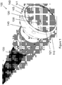
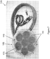
- an ultrasound apparatus 100 including a therapy transducer 105 having a treatment surface 110.
- the therapy transducer 105 includes a plurality of electrically isolated sections 115.
- the electrically isolated sections 115 of the therapy transducer 105 are configured to generate ultrasound radiation in response to the application of electric current thereto.
- the electrically isolated sections may be a piezoelectric ceramic material, and the electric current may be supplied via wires 116 connected to the front and back surfaces of each electrically isolated section 115, as shown in Figures 6C-6F .
- the active transducer material used for the electrically isolated sections 115 may include piezoelectric ceramics, piezoelectric ceramic-composites and/or crystals, among other possibilities.
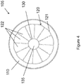
- the ultrasound apparatus 100 also includes a concave acoustic lens 120 defining a therapy aperture 121 in the treatment surface 110 of the therapy transducer 105.
- Figures 1-6H show a single concave acoustic lens 120.
- the single acoustic lens 120 is preferably sized to be coextensive with the treatment surface 110 of the therapy transducer 105.
- This arrangement beneficially utilizes the entire active area of the electrically isolated section 115 of the therapy transducer 105 and is therefore very efficient.
- This arrangement also advantageously allows the ultrasound apparatus 100 to maintain a smaller therapeutic window with an ultrasound focal point 125 that may aid in treating an utero fetus or infant.
- the single acoustic lens 120 includes a plurality of sectors 122 and each sector 122 has a radius of curvature.
- the curvature of each sector 122 of the acoustic lens 120 is defined by a concave elliptical profile such that the major axis is aligned in the direction towards the focal point 125 of the transducer 105.
- the length of each of the major and minor axes of the elliptical profile may be calculated to focus ultrasound generated by the given electrically-isolated section 115 to the focal point 125 of the transducer 105.
- the radius of curvature of the sectors 122 of the acoustic lens 120 may aid in focusing of the ultrasound generated by the electrically isolated sections 115 of the therapy transducer 105.
- the radius of curvature of each of the plurality of sectors 122 of the acoustic lens 120 may be smaller than radius of curvature of the acoustic lens 120 itself. In a further embodiment, the radius of curvature of each of the plurality of sectors 122 of the acoustic lens 120 may be the same. In operation, the radius of curvature of each sector 122 of the acoustic lens 120 may direct the ultrasound and focus the ultrasound on the focal point 125.
- Each of the plurality of electrically isolated sections 115 of the therapy transducer 105 is coupled to one of the plurality of sectors 122 of the single acoustic lens 120, such that there is a 1:1 ratio of electrically isolated sections 115 to sectors 122 of the acoustic lens 120.
- the electrically isolated sections 115 are coupled to the sectors 122 of the acoustic lens 120 via an adhesive 126.
- Each electrically isolated section 115 of the therapy transducer 105 is a flat piezoelectric ceramic, material that is, for example, shaped to substantially match the footprint of each sector 122 of the acoustic lens 120.
- the plurality of sectors 122 of the acoustic lens 120 may be between 4 sectors to 100 sectors.
- each electrically isolated section 115 may be further divided into multiple electrically isolated sub-sections such that each section 115 maintains the same footprint relative to a given sector of the acoustic lens 120.
- the electrically isolated sections 115 may comprise a phased array that provides further control to an operator to adjust the focal point 125.
- a preferred embodiment has an acoustic lens 120 with ten to twelve sectors 122; this arrangement permits efficient distribution of power to each electrically isolated section 115, while at the same time providing the necessary amplitude of ultrasound to each electrically isolated section 115.
- Each of the plurality of sectors 122 of the acoustic lens 120 is configured to direct ultrasound to a local point 125.
- the ultrasound generated in the electrically isolated sections 115 may travel through the adhesive layer 126, through the acoustic lens 120 and out of the therapy transducer 105 to the focal point 125.
- the focal point 125 may lie on the central axis 127 of the therapy aperture 121, and the focal point 125 may range from about 1 cm to about 18 cm from a center of the therapy aperture 121.
- each electrically isolated section 115 of the therapy transducer 105 may be a circular flat piezoelectric ceramic material that is shaped to substantially match the footprint, of each concave acoustic lens 120.
- Still another ultrasound apparatus described herein is one wherein the plurality of electrically isolated sections 115 of the therapy transducer 105 may each have a radius of curvature and together define a single acoustic lens 120.
- the ceramic piezoelectric material is curved instead of flat and thereby define the acoustic lens 120.
- the radius of curvature of each of the plurality of electrically isolated sections 115 of the therapy transducer 105 may be smaller than a radius of curvature of the single acoustic lens 120.
- the plurality of electrically isolated sections 115 of die therapy transducer 105 may be between 4 sections and 100 sections.
- each electrically isolated section 115 may be further divided into multiple electrically isolated sub-sections.
- the electrically isolated sections 115 may comprise a phased array that provides further control to an operator to adjust the focal point 125.
- the ultrasound apparatus 100 may also include an imaging aperture 130 defined by either the treatment surface 110 of the therapy transducer 105 or by a concave acoustic lens 120 that shares a central axis 127 with the therapy aperture 121 (in the present invention, the imaging aperture 130 is defined by the single concave acoustic lens 120). In a preferred embodiment, the imaging aperture 130 may be aligned with the central axis 127 of the therapy aperture 121.
- the ultrasound apparatus 100 further includes an ultrasound imaging probe 135 axially aligned with the central axis 127 of the therapy aperture 121.
- the imaging probe may be configured for B-mode ultrasound or Doppler ultrasound imaging, including color Doppler.
- the imaging probe 135 may permit diagnosis of a tissue structural defect.
- the imaging probe 135 may further permit alignment of the therapy aperture 121 and ultrasound focal point 125 with the tissue structural defect, monitoring of the directed focused ultrasound during treatment and detection of cavitation bubbles from the directed focused ultrasound in a flow channel through a hole eroded through the tissue structural defect.
- the ultrasound apparatus 100 may further include a coupling head 140 coupled to and extending from the treatment surface 110 of the therapy transducer 105.
- the coupling head 140 may be angled inward toward the central axis 127 of the therapy aperture 121.
- the coupling head 140 may circumscribe the single acoustic lens 120 ( Figures 1-3 ) (for reference, also illustrated is an apparatus wherein the coupling head circumscribes the plurality of acoustic lenses 120 ( Figure 7 )).
- the coupling head 140 may include a membrane 141, and a fluid may be enclosed between the acoustic lens 120 and the membrane 141 of the coupling head 140.
- the coupling head 140 may be a solid planar disc. In one embodiment, the thickness of the planar disc may be less than or equal to about 5 cm. In one embodiment, the coupling head may be flexible. In various embodiments, the coupling head 140 may be made of flexible materials such as rubber, hydrogels, or other solid gels, among other possibilities.
- ultrasound may be generated by the electrically isolated sections 115 of the therapy transducer 105 and travel through the sectors 122 of the acoustic lens 120, where the ultrasound becomes focused, and travel through the coupling head 140 and through the skin of the subject to the tissue defect.
- the ultrasound apparatus 100 may include a housing 145 that at. least partially surrounds the plurality of electrically isolated sections 115 of the therapy transducer 105 and the imaging probe 135.
- the electrical coupling or wires 116 extending from the electrically isolated sections 115 of the transducer 105 may be disposed within a flexible cable conduit 150.
- the ultrasound apparatus 100 may include an amplifier electrically coupled to the therapy transducer 105.
- the ultrasound apparatus 100 may include a control system configured to control the amplifier and alter parameters associated with the focused ultrasound as described below with respect to the disclosed methods for treating a pathologic tissue membrane.
- the ultrasound apparatus 100 may be sized and shaped to be hand-held.
- the ultrasound apparatus 100 may be coupled to an articulating arm that may be manually manipulated by an operator or moved into position automatically via a control system to hold the ultrasound apparatus 100 in place during administration of the focused ultrasound.
- a plurality of ultrasound devices 100 may be coupled to a belt to achieve multiple tissue punctures from different ultrasound focal points.
- the ultrasound apparatus of the invention can be utilized in methods for treating a pathologic tissue membrane, comprising:
- the inventors have discovered that such methods can be used, for example, noninvasively to perform tissue membrane puncture to a subject in need thereof using focused ultrasound-generated cavitation or boiling bubbles to controllably erode a hole through the tissue.
- the ultrasound energy may be focused and delivered transcutaneously to the target, to cause localized tissue breakdown into subcellular fragments.
- the resulting hole is not just an incision or tear in the wall of the tissue membrane, but is the result of removal of a substantially circular section of the tissue. This has the advantage of reducing incidence of a future re-obstruction.
- the finely focused energy through the skin of the patient may beneficially cause a perforation in tissue without degrading surrounding tissue.
- the methods can be used to treat any suitable subject, such as a human subject with a pathologic tissue membrane that can benefit from controllably eroding a hole through the tissue with the defect.
- the pathologic tissue membrane may be in a tissue, including but not limited to ureter, urethra, cardiac tissue, bladder, prostate, cyst, kidney, liver, nerve, artery, sail, sheath, penis, uterus, vagina, lung, brain, tympanic membrane, esophagus, trachea, bowel, stomach, sinus, valve, and vein.
- the subject or the subject's in utero fetus may have any disorder involving a pathologic tissue membrane, including but not limited to ureteroceles, a congenital heart abnormality, an ovarian cyst, polycystic kidney disease, a posterior urethral valve, obstructive uropathy, acquired or congenital cystic kidney disease, calyceal diverticulum, acquired or congenital urethral stricture disease or ureteral stricture disease, congenital cystic adenomatoid malformation or other cystic lesions in a lung, need for a shunt in the brain or need for bypass of cerebrospinal fluid or blood in the brain, a blockage in a Eustachian tube of the ear, congenital malformations of the esophagus, trachea, bowel or stomach such as pyloric stenosis, obstructed sinuses in the face or congenital or acquired obstruction of the uterus or vagina.
- the tissue may be a ureter and the subject or the subject's in utero fetus may have ureteroceles.
- the tissue may be cardiac tissue and the subject or the subject's in utero fetus may have a congenital heart abnormality.
- the tissue may be an ovarian cyst.
- the tissue may be a posterior urethral valve and the subject or the subject's in utero fetus may have obstructive uropathy.
- the tissue may include a renal cyst and the subject may have acquired or congenital cystic kidney disease or calyceal diverticulum.
- the tissue may include ureteral or urethral strictures and the subject may have acquired or congenital urethral stricture disease or ureteral stricture disease.
- the tissue may include lung and the subject may have congenital cystic adenomatoid malformation or other cystic lesions in a lung.
- the tissue may include brain and the subject may need shunt or a bypass of cerebrospinal fluid or blood.
- the tissue may include a tympanic membrane and the subject may have a blockage in the Eustachian tube of the ear.
- the tissue may include esophagus, trachea, bowel or stomach and the subject may have a congenital malformation or pyloric stenosis.
- the tissue may include sinus and the subject may have an obstructed sinus.
- the tissue may include uterus or vagina and the subject may have a congenital or acquired obstruction of the uterus or vagina.
- a "pathologic" tissue membrane means a tissue membrane structure that is causing an obstruction, blockage or mass or that is related to, involving or caused by an acquired or congenital disease or anomaly.
- treat or “treating” means accomplishing one or more of the following in an individual that is suffering from a pathologic tissue membrane: (a) reducing the severity of the pathologic tissue membrane; (b) inhibiting worsening of the pathologic tissue membrane; (c) limiting or preventing recurrence of the pathologic tissue membrane; (d) removal of all or a portion of the pathologic tissue membrane; (e) incising, cutting, puncturing, perforating or piercing all or a portion of the pathologic tissue membrane.
- an "effective amount" of the focused ultrasound refers to an amount of the ultrasound that is effective for treating a pathologic tissue membrane.
- the effective amount may vary from subject to subject, depending upon the age, the subject's size and health, the nature and extent of the condition being treated, recommendations of the treating physician, etc.
- a therapeutic amount of the focused ultrasound will be in the range of about 1 J to about 100,000 J, and may preferably be in the range of about 100 J to 3,000 J, which would correspond to about 10 seconds to 30 seconds of ultrasound therapy.
- the subject can be administered as many infrasound treatments as are required to treat or limit the pathologic tissue membrane.
- Figure 13 is a flow chart of a method of treatment according to one example method that can be performed using the apparatus of the invention.
- Example methods such as method 1300 of Figure 13 , may be carried out by an operator or a control system and/or by other components in communication with or disposed on the ultrasound apparatus.
- a control system may take the form of program instructions stored on a non-transitory computer readable medium and a processor that executes the instructions. However, control system may take other forms including software, hardware, and or firmware.
- Example methods may be implemented as part of treating a pathologic tissue membrane.
- method 1300 involves placing a coupling head of an ultrasound apparatus in contact with a subject's skin.
- the coupling head may be placed at any location on the body where a seal may be maintained between the coupling head and the subject's skin through which ultrasound may be directed.
- This location on the body may be external, for example on the subject's abdomen, back, limbs or skull, or internal via the rectum, vagina or esophagus, for example.
- the subject or a subject's in-utero fetus may have a pathologic tissue membrane in need of puncturing.
- the method may be performed using any of the embodiments of the ultrasound apparatus described above.
- the method for treatment may be carried out using a therapeutic focused ultrasound device having a single unitary concave transducer section defining a therapy aperture, where the concave transducer is configured to have a single electronic signal applied to it.
- the method for treatment may be carried out using an ultrasound device having a phased array in the form of a 2D grid that may permit modification of the location of the ultrasound focal point and the width of the focal point.
- the operator may obtain image feedback of the tissue via the ultrasound imaging probe.
- the image feedback may be displayed on, for example, a display device including a monitor, computer, laptop, LCD display, or any other device capable of displaying digital images or electronic files, among other possibilities.
- the operator or the control system may align a focal point of the therapy transducer with the tissue based upon at least the image feedback.
- an indicator showing the focal point of the therapy transducer may be overlaid on the ultrasound image on the display device.
- the operator may then manipulate the angle and/or placement of the therapy transducer relative to the subject in order to align the indicator with the desired region of the tissue.
- the operator may administer a short exposure of focused ultrasound to briefly generate cavitation bubbles that are visible via image feedback and confirm that the bubbles are located in the correct position without significantly affecting the tissue.
- focused ultrasound is directed at the tissue via the therapy transducer.
- At block 1325 at least one hole or at least one linear incision is punctured in the tissue via the focused ultrasound. Puncture may be achieved through focused ultrasound-generated cavitation or boiling bubbles to controllably erode a hole or linear incision through the tissue.
- the punctured hole may have a diameter ranging from about 0.7 mm to about 3.4 mm.
- the hole may be a single eroded hole.
- the punctured hole may be an incised linear incision. The dimensions of the hole(s) or incision may be controlled by modifying ultrasound parameters described below to change the width of the ultrasound focal point.
- the directed focused ultrasound may be monitored via the imaging probe using one or more of a B-mode ultrasound imaging or Doppler ultrasound imaging. Then cavitation bubbles from the directed focused ultrasound may be detected in a flow channel through the eroded hole based upon at least the B-mode or Doppler ultrasound imaging. Once cavitation bubbles are detected, the directed focused ultrasound may be ceased.
- a fluid may be provided between the subject's skin and the coupling head.
- the directed focused ultrasound may then travel through at least the fluid and the patient's skin to the tissue, for example, the meter.
- the fluid may take the form of ultrasound gel. The gel aids in coupling the ultrasound device to the subject's skin to allow the ultrasound an unbroken pathway to the intended focal point of the ultrasound apparatus.
- the focused ultrasound may be directed at the tissue for a duration of about 1 second to about 1 hour. For durations lasting 30 minutes or more, the subject may need to be anesthetized for pain management.
- the operator may set one or more of an ultrasound pulse duration, an ultrasound pulse repetition frequency, an ultrasound pulse time delay and an operating frequency of the therapy transducer.
- the foregoing parameters may be independently set for each of a plurality of electrically isolated sections of the therapy transducer.
- altering the ultrasound pulse delay between the plurality of sections of the therapy transducer may permit the width of the ultrasound focal point or high pressure zone to be modified.
- the signal for each electrically isolated section of the therapy transducer may be delayed more than the signal for the adjacent sections.
- T is the time period of one cycle of the ultrasound frequency
- N is the total number of acoustic lens sectors
- n is the specific order number of an isolated section in a given arrangement
- “m” is an integer.
- the delays between the isolated sections may be larger, making the focal point wider.
- increasing the ultrasound pulse duration may increase the diameter of the puncture or hole.
- the ultrasound pulse repetition frequency may be adjusted to assist in maintaining equal acoustic power distribution to each of the electrically isolated sections.
- the ultrasound pulse duration may range from about 1 ⁇ s to about 100 ms
- the ultrasound pulse repetition frequency may range from about 1 Hz to about 10 kHz
- the transducer operating frequency may range from about 0.2 MHz to about 10 MHz and preferably ranges from about 1 MHz to 3MHz.
- the directed focused ultrasound may be continuous wave ultrasound.
- the directed focused ultrasound may generate positive pressure that may range from about 30 MPa to about 120 MPa and negative pressure that may range from about 4 MPa to about 20 MPa. These pressure measurements are based on ultrasound measurements made in water, not through tissue.
- the positive and negative pressures may be larger depending on how the ultrasound is administered. For example, the pressure amplitudes required to achieve therapeutic effect depend upon the electrical parameters that may be set, as discussed above.
- movement of the subject due to breathing or coughing for example, and movement of the subject's in utero fetus may be detected via the ultrasound imaging probe or a sensor.
- the directed focused ultrasound may be ceased temporarily until realignment is confirmed.
- the ultrasound apparatus of the invention can also be utilized in methods for diagnosing a pathologic tissue membrane, comprising:
- B-Mode ultrasound imaging or Doppler imaging may be used via the imaging probe to obtain images of a tissue suspected of contributing to symptoms exhibited by the subject.
- the methods can be used to diagnose any suitable subject, such as a human subject with a pathologic tissue membrane that may benefit from controllably eroding a hole through the tissue with the detect.
- the pathologic tissue membrane may be in a tissue, including but not limited to ureter, urethra, cardiac tissue, bladder, prostate, cyst, kidney, liver, nerve, artery, sail, sheath, penis, uterus, vagina, lung, brain, tympanic membrane, valve, and vein.
- the subject or the subject's in utero fetus may have any disorder involving a pathologic tissue membrane, including but not limited to ureteroceles, a congenital heart abnormality, an ovarian cyst, polycystic kidney disease, a posterior urethral valve, obstructive uropathy, acquired or congenital cystic kidney disease, calyceal diverticulum, acquired or congenital urethral stricture disease or ureteral stricture disease, congenital cystic adenomatoid malformation or other cystic lesions in a lung, need for a shunt in the brain or need for a bypass of cerebrospinal fluid or blood in the brain, a blockage in a Eustachian tube of the ear, congenital malformations of the esophagus, trachea, bowel or stomach such as pyloric stenosis, obstructed sinuses in the face or congenital or acquired obstruction of the uterus or vagina
- the tissue may be a ureter and the subject or the subject's in utero fetus may have ureteroceles.
- the tissue may be cardiac tissue and the subject or the subject's in utero fetus may have a congenital heart abnormality.
- the tissue may be an ovarian cyst.
- the tissue may be a posterior urethral valve and the subject or the subject's in utero fetus may have obstructive uropathy.
- the tissue may include a renal cyst and the subject may have acquired or congenital cystic kidney disease or calyceal diverticulum.
- the tissue may include ureteral or urethral strictures and the subject may have acquired or congenital urethral stricture disease or ureteral stricture disease.
- the tissue may include lung and the subject may have congenital cystic adenomatoid malformation or other cystic lesions in a lung.
- the tissue may include brain and the subject may need a shunt or a bypass of cerebrospinal fluid or blood.
- the tissue may include a tympanic membrane and the subject may have a blockage in the Eustachian tube of the ear.
- the tissue may include esophagus, trachea, bowel or stomach and the subject may have a congenital malformation or pyloric stenosis.
- the tissue may include sinus and the subject may have an obstructed sinus.
- the tissue may include uterus or vagina and the subject may have a congenital or acquired obstruction of the uterus or vagina.
- Example methods may be implemented as part of diagnosing a pathologic tissue membrane.
- method 1400 involves placing a coupling head of the ultrasound apparatus in contact with subject's skin.
- the coupling head may be placed at any location on the body where a seal may be maintained between the coupling head and the subject's skin through which ultrasound may be directed for diagnosis.
- This location on the body may be external, for example on the subject's abdomen, back, limbs or skull, or internal via the rectum, vagina or esophagus for example.
- the method may be performed using any of the embodiments of the ultrasound apparatus described above .
- the operator may obtain image feedback of a tissue via the ultrasound imaging probe.
- the image feedback may he displayed on, for example, a monitor, computer, laptop, LCD display, or any other device capable of displaying digital images or electronic files, among other possibilities.
- the operator or the control system may determine whether the subject or a subject's in utero fetus has a pathologic tissue membrane in need of puncturing.
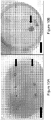
- Focused ultrasound produced erosion of the wall and puncture ( Fig. 8 ) in times between 50-300 seconds, depending on ultrasonic parameters, focal alignment and wall thickness.
- Figure 8 shows a puncture created in a bladder bleb after 180 seconds exposure of high-intensity ultrasound (indicated by white radiating lines) focused onto the bleb wall.
- Dye was visible flowing from the punctures post-treatment, indicating the existence of a patent communication through the wall.
- the feasibility of using histotripsy to generate a mechanical puncture under ultrasound image guidance was evaluate using a tissue model for the ureterocele wall.
- a model of the ureterocele wall was developed using freshly excised bovine bladder tissue.
- the bladder was harvested and maintained in degassed phosphate-buffered saline until use ( ⁇ 12 hours from excision).
- the bladder was sectioned into 5 x 5 cm segments.
- the mucosal and submucosal layers were denuded from the underlying muscle and adventitia to create a membrane 0.5-1mm in thickness.
- the mucosal membrane was placed over a circular opening of a polypropylene chamber containing dyed saline.
- the membrane was fixed with a band around the container such that the membrane sealed the fluid in the chamber without applying Tension to the membrane, aside from that caused by the sample's own weight.
- This model provided an end point for determination of puncture when dyed saline could be observed flowing through the puncture hole in the membrane.
- the therapy transducer was a 1MHz seven-element array with 14.7-cm diameter. Each element of the transducer was focused through a plastic lens, with all lenses focused at the radius of curvature of 14 cm.
- the transducer was electrically driven by a radiofrequency class D amplifier modified to output high amplitude pulse durations up to 10 ms. The amplifier output was controlled by an electronic timing board that specified the ultrasound pulse duration ("PD"), pulse repetition rate ("PRF'X and transducer operating frequency.
- PD ultrasound pulse duration
- PRF'X pulse repetition rate
- the three-dimensional pressure output of the transducer was obtained under free field conditions in a degassed water bath by acoustic holography with a capsule hydrophone (HGL-0085. Onda Corporation, Sunnyvale, CA) recorded at low pressure amplitudes and nonlinear acoustic simulation.
- the focal pressures were continued by measurements with a fiberoptic probe hydrophone (FOPH2000, RP Acoustics, Stuttgart, Germany).
- the linear - 6dB focal pressure beam width was 2.0mm transverse to the acoustic axis by 13.6mm along the acoustic axis.
- the focal peak positive pressure of the ultrasound pulses applied in this experiment was 100-120 MPa, and the peak negative pressure applied was 17-20 MPa. Pulses between 1-5000 cycles duration were used in the experiments, with the pulse rate selected in each case to fix the duty cycle at 0.5% to 0.6%.
- the time-averaged spatial peak intensity of the exposure was between 145-190W/cm 2 .
- a research ultrasound imaging engine (V-1, Verasonics Inc. Redmond, WA) with a linear array probe (L7-4, Philips Healthcare, Andover, MA) operated at 5MHz was used to visualize the treatment area before, during, and after exposure in a subset of the experiments.
- the imaging was triggered by the therapy system such that the images were synchronized to avoid acoustic and electrical interference from the therapy ultrasound pulses. Because of the low duty factor of the therapy output ( ⁇ 1%), the frame rate of the ultrasound imager could be maintained at ⁇ 10 frames per second during treatment.
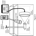
- FIG. 9 shows the experimental alignment of a sample with therapy and imaging transducers during expostire.
- the sample is shown sealed over a chamber of dyed saline and attached to a three-axis motor positioner (Velmex Inc., Bloomfield, NY) to align it with the therapy transducer focus.
- An imaging probe was positioned at an oblique angle in the focal plane to observe the cavitation activity of the sample pre- and post-puncture.
- An alignment laser was used to identify the position of the focus in the water.
- the membrane was translated by the three-axis motorized positioner aligning the tissue with the therapy transducer focus.
- a short, 1-second exposure was then used to confirm alignment by visualization of cavitation on the membrane.
- the imaging probe was aligned off-axis in the water bath in the plane of the therapy focus to observe the treatment region.
- a digital camera (S8000, Nikon USA, Melville, NY) was used to record a video of each exposure.
- the membrane was exposed under a set of ultrasound therapy pulse parameters until a visual puncture through the membrane was observed. In some cases, punctures were falsely identified, and no dye was visualized flowing through the membrane when the therapy was turned off In these cases, the membrane was further treated until a positive result was achieved for up to 300 seconds total treatment time. After treatments, the punctures were photographed and measured while still attached to the container outside of the water bath.
- Figure 10A shows six punctures made in the sample bladder wall membrane by exposures with a short pulse duration (2 ⁇ s) of an ultrasound (the scale bar is 5 mm long), whereas Figure 10B shows a larger puncture generated by a sequential exposure of 1 ⁇ s pulses followed by 5000 ⁇ s pulses (the scale bar is 5 mm long).
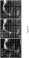
- Ultrasound B-mode images indicated the presence of cavitation as an echogenic region at the focal point ( FIGs. 12A-C ).
- Figure 12A shows the therapy ultrasound orientation and focusing, and the visualization of bladder mucosa by B-mode ultrasound image guidance is shown before treatment (the scale bar is 1 cm). Before puncture, cavitation was limited to the proximal membrane surface (transducer side). Then, cavitation is shown to be localized to the external membrane surface before puncture.
- Figure 12B shows the visualization of bladder mucosa by B-mode ultrasound image guidance during exposure pre-puncture. Finally, cavitation was visualized penetrating into, and eventually through, the membrane, forming a narrow jet in the fluid space distal to the surface.
- FIG. 12C This phenomenon is shown in Figure 12C , which displays the visualization of bladder mucosa by B-mode ultrasound image guidance during exposure post-puncture.
- the puncture was not visible after treatment as a hypoechoic region in the membrane, as is apparent on bulk tissues treated by histotripsy. However, once a channel was created, bubbles could be seen flowing through the membrane, as shown in Figure 12C .
- ultrasound imaging may provide feedback to the operator for precise position of treatment and detection of the puncture. It was possible to detect a perforation when therapy was being delivered with cavitation observed on both sides of the ureterocele. There is a tendency for formation of a cavitation cloud at the fluid-tissue interface that possibly arises from bubbles being preferentially pushed into the membrane by acoustic radiation force. On puncture, this force would cause bubbles to flow through the hole, as observed on B-mode images. The hole was not visible directly under B-mode ultrasonography post-treatment, however. Color Doppler Ultrasound may complement B-mode imaging feedback in providing information on the flow channel created after therapy and help determine a more precise end point In addition to neonatal surgery, another application may be the management of ureteroceles in utero.
- Pulsed focused ultrasound can create a mechanical perforation in denuded bladder mucosa, a tissue model for the ureterocele wall.
- the puncture diameter was repeatable and could be controlled by ultrasound exposure parameters.
- Ultrasound imaging allowed for real-time targeting and visualization to confirm puncture.
Landscapes
- Health & Medical Sciences (AREA)
- Life Sciences & Earth Sciences (AREA)
- Animal Behavior & Ethology (AREA)
- Veterinary Medicine (AREA)
- Nuclear Medicine, Radiotherapy & Molecular Imaging (AREA)
- Public Health (AREA)
- Radiology & Medical Imaging (AREA)
- Engineering & Computer Science (AREA)
- Biomedical Technology (AREA)
- General Health & Medical Sciences (AREA)
- Pathology (AREA)
- Molecular Biology (AREA)
- Surgery (AREA)
- Medical Informatics (AREA)
- Heart & Thoracic Surgery (AREA)
- Physics & Mathematics (AREA)
- Biophysics (AREA)
- Gynecology & Obstetrics (AREA)
- Pregnancy & Childbirth (AREA)
- Surgical Instruments (AREA)
- Ultra Sonic Daignosis Equipment (AREA)
Applications Claiming Priority (2)
| Application Number | Priority Date | Filing Date | Title |
|---|---|---|---|
| US201361806295P | 2013-03-28 | 2013-03-28 | |
| PCT/US2014/032219 WO2014160964A1 (en) | 2013-03-28 | 2014-03-28 | Focused ultrasound apparatus and methods of use |
Publications (2)
| Publication Number | Publication Date |
|---|---|
| EP2978497A1 EP2978497A1 (en) | 2016-02-03 |
| EP2978497B1 true EP2978497B1 (en) | 2021-09-01 |
Family
ID=50694029
Family Applications (1)
| Application Number | Title | Priority Date | Filing Date |
|---|---|---|---|
| EP14723604.6A Active EP2978497B1 (en) | 2013-03-28 | 2014-03-28 | Focused ultrasound apparatus and methods of use |
Country Status (5)
| Country | Link |
|---|---|
| US (2) | US10350439B2 (enExample) |
| EP (1) | EP2978497B1 (enExample) |
| JP (1) | JP6440682B2 (enExample) |
| CN (1) | CN105392529B (enExample) |
| WO (1) | WO2014160964A1 (enExample) |
Families Citing this family (41)
| Publication number | Priority date | Publication date | Assignee | Title |
|---|---|---|---|---|
| EP2903688A4 (en) | 2012-10-05 | 2016-06-15 | Univ Michigan | BLADE-INDUCED COLOR DOPPLER FEEDBACK DURING A HISTOTRIPSIA |
| US11432900B2 (en) | 2013-07-03 | 2022-09-06 | Histosonics, Inc. | Articulating arm limiter for cavitational ultrasound therapy system |
| US10780298B2 (en) | 2013-08-22 | 2020-09-22 | The Regents Of The University Of Michigan | Histotripsy using very short monopolar ultrasound pulses |
| US10639503B2 (en) | 2014-08-27 | 2020-05-05 | Fusmobile Inc. | Handheld devices for projecting focused ultrasound and related methods |
| US9987809B2 (en) * | 2014-12-31 | 2018-06-05 | General Electric Company | System and method for manufacturing an ultrasound probe lens |
| US10098539B2 (en) | 2015-02-10 | 2018-10-16 | The Trustees Of Columbia University In The City Of New York | Systems and methods for non-invasive brain stimulation with ultrasound |
| EP4230262A3 (en) | 2015-06-24 | 2023-11-22 | The Regents Of The University Of Michigan | Histotripsy therapy systems for the treatment of brain tissue |
| WO2017004562A1 (en) | 2015-07-01 | 2017-01-05 | The Trustees Of Columbia University In The City Of New York | Systems and methods for modulation and mapping of brain tissue using an ultrasound assembly |
| WO2017055403A1 (en) * | 2015-09-29 | 2017-04-06 | Institut National De La Sante Et De La Recherche Medicale (Inserm) | Device and system for generating ultrasonic waves in a target region of a soft solid and method for locally treating a tissue |
| JPWO2017126037A1 (ja) * | 2016-01-19 | 2018-08-09 | オリンパス株式会社 | 超音波医療装置 |
| CN105748106B (zh) * | 2016-04-22 | 2018-07-31 | 毛军卫 | 超声探头以及具有该超声探头的超声检测设备 |
| US11020617B2 (en) | 2016-07-27 | 2021-06-01 | The Trustees Of Columbia University In The City Of New York | Methods and systems for peripheral nerve modulation using non ablative focused ultrasound with electromyography (EMG) monitoring |
| EP3490438A4 (en) | 2016-07-27 | 2020-03-18 | The Trustees of Columbia University in the City of New York | METHODS AND SYSTEMS FOR PERIPHERAL NERVOUS MODULATION WITH FOCUSED ULTRASOUND |
| JP6801469B2 (ja) * | 2017-01-20 | 2020-12-16 | コニカミノルタ株式会社 | 超音波診断装置 |
| JP6445083B2 (ja) * | 2017-05-12 | 2018-12-26 | 株式会社リンクス | 超音波装置及び超音波ユニット |
| EP3675959B1 (en) * | 2017-09-01 | 2024-11-27 | Dalhousie University | Transducer assembly for generating focused ultrasound |
| TW202529848A (zh) * | 2018-01-26 | 2025-08-01 | 美商奧賽拉公司 | 用於多個維度中的同時多聚焦超音治療的系統和方法 |
| JP3216192U (ja) * | 2018-03-01 | 2018-05-17 | 有限会社ユーマンネットワーク | 平面形素子を用いた集束式音波治療装置 |
| EP3860711A4 (en) * | 2018-10-03 | 2022-07-27 | MDSG Innovation Ltd. | DEVICE AND METHOD FOR TREATMENT OF KIDNEYS |
| US20220218308A1 (en) * | 2018-11-15 | 2022-07-14 | Osteosys Co., Ltd. | Ultrasonic transducer |
| KR102249727B1 (ko) * | 2018-11-15 | 2021-05-10 | 주식회사 오스테오시스 | 초음파 트랜스듀서 |
| CN113286552B (zh) | 2018-11-28 | 2025-05-02 | 希斯托索尼克斯公司 | 组织摧毁术系统及方法 |
| US12588895B2 (en) | 2019-06-13 | 2026-03-31 | The Trustees Of Columbia University In The City Of New York | System, method, computer-accessible medium and apparatus for flexible two-dimensional ultrasound phased array |
| CN111112037A (zh) * | 2020-01-20 | 2020-05-08 | 重庆医科大学 | 透镜式多频聚焦超声换能器、换能系统及其声焦域轴向长度的确定方法 |
| WO2021155026A1 (en) | 2020-01-28 | 2021-08-05 | The Regents Of The University Of Michigan | Systems and methods for histotripsy immunosensitization |
| CN111632284B (zh) * | 2020-04-27 | 2021-04-20 | 深圳市普罗医学股份有限公司 | 一种用于胸肺部治疗的超声治疗头及超声治疗设备 |
| JP7440188B2 (ja) * | 2020-05-15 | 2024-02-28 | 朝日インテック株式会社 | カテーテル |
| GB202009079D0 (en) | 2020-06-15 | 2020-07-29 | Oxsonics Ltd | Mapping of cavitation activity |
| JP2023530477A (ja) | 2020-06-18 | 2023-07-18 | ヒストソニックス,インコーポレーテッド | 組織破砕音響/患者結合システムおよび方法 |
| EP4204084A4 (en) | 2020-08-27 | 2024-10-09 | The Regents Of The University Of Michigan | ULTRASONIC TRANSDUCER WITH TRANSMIT-RECEIVE CAPABILITY FOR HISTOTRIPSY |
| KR102583983B1 (ko) * | 2021-06-04 | 2023-10-06 | 한국과학기술연구원 | 집속초음파를 이용한 케비테이션 기반 생체조직 분해 장치 |
| EP4351718A4 (en) | 2021-06-07 | 2025-03-26 | The Regents of The University of Michigan | MINIMALLY INVASIVE HISTOTRIPSY METHODS AND SYSTEMS |
| IL308943A (en) | 2021-06-07 | 2024-01-01 | Univ Michigan Regents | All-inclusive ultrasound systems and methods that include histotripsy |
| US12285636B2 (en) | 2021-08-05 | 2025-04-29 | The University Of Washington | Non-planar holographic beam shaping lenses for acoustics |
| CN113457003A (zh) * | 2021-08-05 | 2021-10-01 | 广东光声医疗科技有限公司 | 一种大深度经阴道粘膜的多频振动超声导入装置 |
| CN115350411A (zh) * | 2022-07-29 | 2022-11-18 | 杭州福嵩科技有限责任公司 | 一种用于甲状腺治疗的超声换能器及治疗头 |
| AU2023366591A1 (en) | 2022-10-28 | 2025-04-24 | Histosonics, Inc. | Histotripsy systems and methods |
| KR20250129759A (ko) * | 2022-12-30 | 2025-08-29 | 히스토소닉스, 인크. | 히스토트립시 시스템들 및 방법들 |
| WO2024221001A2 (en) | 2023-04-20 | 2024-10-24 | Histosonics, Inc. | Histotripsy systems and associated methods including user interfaces and workflows for treatment planning and therapy |
| US20250010105A1 (en) * | 2023-07-07 | 2025-01-09 | WaveVision GmbH | Secondary reflector for improvement of shockwave therapies |
| KR102604540B1 (ko) * | 2023-07-10 | 2023-11-22 | 포항공과대학교 산학협력단 | 환부 종합 관찰용 초음파 프로브 |
Family Cites Families (34)
| Publication number | Priority date | Publication date | Assignee | Title |
|---|---|---|---|---|
| US3958559A (en) * | 1974-10-16 | 1976-05-25 | New York Institute Of Technology | Ultrasonic transducer |
| JPH0714410B2 (ja) * | 1986-05-02 | 1995-02-22 | 株式会社日立製作所 | 超音波照射装置 |
| US5065761A (en) * | 1989-07-12 | 1991-11-19 | Diasonics, Inc. | Lithotripsy system |
| JPH06319746A (ja) | 1993-05-11 | 1994-11-22 | Toshiba Corp | 超音波治療装置 |
| US6007499A (en) * | 1997-10-31 | 1999-12-28 | University Of Washington | Method and apparatus for medical procedures using high-intensity focused ultrasound |
| US7722539B2 (en) * | 1998-09-18 | 2010-05-25 | University Of Washington | Treatment of unwanted tissue by the selective destruction of vasculature providing nutrients to the tissue |
| JP2003513691A (ja) * | 1999-10-25 | 2003-04-15 | シーラス、コーポレイション | 血管を封止するための集束超音波の使用 |
| JP2001137256A (ja) | 1999-11-16 | 2001-05-22 | Toshiba Corp | 超音波治療装置 |
| US8221402B2 (en) * | 2000-01-19 | 2012-07-17 | Medtronic, Inc. | Method for guiding a medical device |
| CN2551208Y (zh) | 2002-05-10 | 2003-05-21 | 绵阳索尼克电子有限责任公司 | 肿瘤治疗超声聚焦刀 |
| CN1494933A (zh) * | 2002-09-09 | 2004-05-12 | 株式会社东芝 | 超声辐射设备 |
| US20040059265A1 (en) | 2002-09-12 | 2004-03-25 | The Regents Of The University Of California | Dynamic acoustic focusing utilizing time reversal |
| WO2005025424A1 (en) * | 2003-09-12 | 2005-03-24 | Austin Health | Ultrasound apparatus |
| FR2873299B1 (fr) * | 2004-07-23 | 2006-11-17 | Inst Nat Sante Rech Med | Dispositif et methode de traitement par ultrasons |
| US20120046547A1 (en) | 2004-10-06 | 2012-02-23 | Guided Therapy Systems, Llc | System and method for cosmetic treatment |
| CN1669672A (zh) | 2005-04-20 | 2005-09-21 | 南京航空航天大学 | 压电式多阵元高强度聚焦超声换能器及聚焦方法 |
| US8057408B2 (en) | 2005-09-22 | 2011-11-15 | The Regents Of The University Of Michigan | Pulsed cavitational ultrasound therapy |
| US20070233185A1 (en) * | 2005-10-20 | 2007-10-04 | Thomas Anderson | Systems and methods for sealing a vascular opening |
| US7955281B2 (en) * | 2006-09-07 | 2011-06-07 | Nivasonix, Llc | External ultrasound lipoplasty |
| CN101190436B (zh) * | 2006-11-22 | 2010-09-29 | 中国科学院声学研究所 | 一种相控聚焦超声波波源装置 |
| US8043604B2 (en) * | 2007-04-27 | 2011-10-25 | Pluromed, Inc. | Ultrasonography using time- and temperature-sensitive variable adhesion coupling gels |
| US8323201B2 (en) * | 2007-08-06 | 2012-12-04 | Orison Corporation | System and method for three-dimensional ultrasound imaging |
| US8466605B2 (en) * | 2008-03-13 | 2013-06-18 | Ultrashape Ltd. | Patterned ultrasonic transducers |
| WO2010102302A2 (en) * | 2009-03-06 | 2010-09-10 | Mirabilis Medica, Inc. | Ultrasound treatment and imaging applicator |
| WO2011022085A1 (en) * | 2009-08-19 | 2011-02-24 | Duke University | Acoustic lens for shockwave lithotripsy and related methods |
| CA2779455A1 (en) * | 2009-10-30 | 2011-05-05 | Sound Interventions, Inc. | Method and apparatus for non-invasive treatment of hypertension through ultrasound renal denervation |
| EP2498921B1 (en) | 2009-11-09 | 2016-09-14 | Koninklijke Philips N.V. | Curved ultrasonic hifu transducer formed by tiled segments |
| CN102416225B (zh) * | 2010-09-27 | 2014-07-02 | 重庆融海超声医学工程研究中心有限公司 | 一种超声换能器 |
| WO2012052920A1 (en) | 2010-10-18 | 2012-04-26 | CardioSonic Ltd. | Therapeutics reservoir |
| US8715187B2 (en) * | 2010-12-17 | 2014-05-06 | General Electric Company | Systems and methods for automatically identifying and segmenting different tissue types in ultrasound images |
| WO2012156863A2 (en) | 2011-05-18 | 2012-11-22 | Koninklijke Philips Electronics N.V. | Spherical ultrasonic hifu transducer with modular cavitation sense element |
| KR101355532B1 (ko) | 2011-11-21 | 2014-01-24 | 알피니언메디칼시스템 주식회사 | 고강도 집속 초음파용 트랜스듀서 |
| US20130253387A1 (en) | 2012-03-08 | 2013-09-26 | Sonitec, LLC | Vibratory energy systems and methods for occluded body cavities |
| US9636133B2 (en) * | 2012-04-30 | 2017-05-02 | The Regents Of The University Of Michigan | Method of manufacturing an ultrasound system |
-
2014
- 2014-03-28 US US14/777,949 patent/US10350439B2/en active Active
- 2014-03-28 WO PCT/US2014/032219 patent/WO2014160964A1/en not_active Ceased
- 2014-03-28 EP EP14723604.6A patent/EP2978497B1/en active Active
- 2014-03-28 CN CN201480018473.2A patent/CN105392529B/zh active Active
- 2014-03-28 JP JP2016505604A patent/JP6440682B2/ja active Active
-
2019
- 2019-06-20 US US16/447,771 patent/US20200078608A1/en not_active Abandoned
Also Published As
| Publication number | Publication date |
|---|---|
| JP2016515901A (ja) | 2016-06-02 |
| JP6440682B2 (ja) | 2018-12-19 |
| US20160287909A1 (en) | 2016-10-06 |
| US20200078608A1 (en) | 2020-03-12 |
| CN105392529B (zh) | 2020-03-17 |
| US10350439B2 (en) | 2019-07-16 |
| WO2014160964A1 (en) | 2014-10-02 |
| EP2978497A1 (en) | 2016-02-03 |
| CN105392529A (zh) | 2016-03-09 |
Similar Documents
| Publication | Publication Date | Title |
|---|---|---|
| EP2978497B1 (en) | Focused ultrasound apparatus and methods of use | |
| KR102124422B1 (ko) | 고강도-저강도 집속초음파 치료장치 | |
| US7553284B2 (en) | Focused ultrasound for pain reduction | |
| US8942781B2 (en) | Medical system comprising a percutaneous probe | |
| US6626855B1 (en) | Controlled high efficiency lesion formation using high intensity ultrasound | |
| US7850626B2 (en) | Method and probe for using high intensity focused ultrasound | |
| US8211017B2 (en) | Image guided high intensity focused ultrasound treatment of nerves | |
| US20100036246A1 (en) | Automatic fat thickness measurements | |
| US20100280371A1 (en) | Non-Invasive Device and Method for Locating a Structure Such as a Nerve | |
| JPH06205836A (ja) | 疾病組織処置用のカテーテル | |
| Li et al. | Endoscopic high-intensity focused US: technical aspects and studies in an in vivo porcine model (with video) | |
| PT2409728T (pt) | Sistema para o tratamento de tecidos por ultrassons | |
| WO2007021958A2 (en) | Method and apparatus for preparing organs and tissues for laparoscopic surgery | |
| US11253729B2 (en) | External ultrasound generating treating device for spinal cord and/or spinal nerve treatment, apparatus comprising such device and method | |
| Rahimi et al. | A high-frequency phased array system for transcranial ultrasound delivery in small animals | |
| Simoni et al. | Ex vivo assessment of multiple parameters in high intensity focused ultrasound | |
| KR102490676B1 (ko) | 초음파를 이용한 장치 및 방법 | |
| Jaiswal | Esophageal Damage in Response to Noninvasive Focused Ultrasound for Treatment of Atrial Fibrillation | |
| Vaezy | High Intensity Focused Ultrasound for Therapy in Medicine |
Legal Events
| Date | Code | Title | Description |
|---|---|---|---|
| PUAI | Public reference made under article 153(3) epc to a published international application that has entered the european phase |
Free format text: ORIGINAL CODE: 0009012 |
|
| 17P | Request for examination filed |
Effective date: 20150925 |
|
| AK | Designated contracting states |
Kind code of ref document: A1 Designated state(s): AL AT BE BG CH CY CZ DE DK EE ES FI FR GB GR HR HU IE IS IT LI LT LU LV MC MK MT NL NO PL PT RO RS SE SI SK SM TR |
|
| AX | Request for extension of the european patent |
Extension state: BA ME |
|
| DAX | Request for extension of the european patent (deleted) | ||
| STAA | Information on the status of an ep patent application or granted ep patent |
Free format text: STATUS: EXAMINATION IS IN PROGRESS |
|
| 17Q | First examination report despatched |
Effective date: 20170607 |
|
| GRAP | Despatch of communication of intention to grant a patent |
Free format text: ORIGINAL CODE: EPIDOSNIGR1 |
|
| STAA | Information on the status of an ep patent application or granted ep patent |
Free format text: STATUS: GRANT OF PATENT IS INTENDED |
|
| INTG | Intention to grant announced |
Effective date: 20210318 |
|
| GRAS | Grant fee paid |
Free format text: ORIGINAL CODE: EPIDOSNIGR3 |
|
| GRAA | (expected) grant |
Free format text: ORIGINAL CODE: 0009210 |
|
| STAA | Information on the status of an ep patent application or granted ep patent |
Free format text: STATUS: THE PATENT HAS BEEN GRANTED |
|
| AK | Designated contracting states |
Kind code of ref document: B1 Designated state(s): AL AT BE BG CH CY CZ DE DK EE ES FI FR GB GR HR HU IE IS IT LI LT LU LV MC MK MT NL NO PL PT RO RS SE SI SK SM TR |
|
| REG | Reference to a national code |
Ref country code: GB Ref legal event code: FG4D |
|
| REG | Reference to a national code |
Ref country code: CH Ref legal event code: EP Ref country code: AT Ref legal event code: REF Ref document number: 1425569 Country of ref document: AT Kind code of ref document: T Effective date: 20210915 |
|
| REG | Reference to a national code |
Ref country code: DE Ref legal event code: R096 Ref document number: 602014079818 Country of ref document: DE |
|
| REG | Reference to a national code |
Ref country code: IE Ref legal event code: FG4D |
|
| REG | Reference to a national code |
Ref country code: LT Ref legal event code: MG9D |
|
| REG | Reference to a national code |
Ref country code: NL Ref legal event code: MP Effective date: 20210901 |
|
| PG25 | Lapsed in a contracting state [announced via postgrant information from national office to epo] |
Ref country code: FI Free format text: LAPSE BECAUSE OF FAILURE TO SUBMIT A TRANSLATION OF THE DESCRIPTION OR TO PAY THE FEE WITHIN THE PRESCRIBED TIME-LIMIT Effective date: 20210901 Ref country code: ES Free format text: LAPSE BECAUSE OF FAILURE TO SUBMIT A TRANSLATION OF THE DESCRIPTION OR TO PAY THE FEE WITHIN THE PRESCRIBED TIME-LIMIT Effective date: 20210901 Ref country code: NO Free format text: LAPSE BECAUSE OF FAILURE TO SUBMIT A TRANSLATION OF THE DESCRIPTION OR TO PAY THE FEE WITHIN THE PRESCRIBED TIME-LIMIT Effective date: 20211201 Ref country code: LT Free format text: LAPSE BECAUSE OF FAILURE TO SUBMIT A TRANSLATION OF THE DESCRIPTION OR TO PAY THE FEE WITHIN THE PRESCRIBED TIME-LIMIT Effective date: 20210901 Ref country code: BG Free format text: LAPSE BECAUSE OF FAILURE TO SUBMIT A TRANSLATION OF THE DESCRIPTION OR TO PAY THE FEE WITHIN THE PRESCRIBED TIME-LIMIT Effective date: 20211201 Ref country code: SE Free format text: LAPSE BECAUSE OF FAILURE TO SUBMIT A TRANSLATION OF THE DESCRIPTION OR TO PAY THE FEE WITHIN THE PRESCRIBED TIME-LIMIT Effective date: 20210901 Ref country code: RS Free format text: LAPSE BECAUSE OF FAILURE TO SUBMIT A TRANSLATION OF THE DESCRIPTION OR TO PAY THE FEE WITHIN THE PRESCRIBED TIME-LIMIT Effective date: 20210901 Ref country code: HR Free format text: LAPSE BECAUSE OF FAILURE TO SUBMIT A TRANSLATION OF THE DESCRIPTION OR TO PAY THE FEE WITHIN THE PRESCRIBED TIME-LIMIT Effective date: 20210901 |
|
| REG | Reference to a national code |
Ref country code: AT Ref legal event code: MK05 Ref document number: 1425569 Country of ref document: AT Kind code of ref document: T Effective date: 20210901 |
|
| PG25 | Lapsed in a contracting state [announced via postgrant information from national office to epo] |
Ref country code: PL Free format text: LAPSE BECAUSE OF FAILURE TO SUBMIT A TRANSLATION OF THE DESCRIPTION OR TO PAY THE FEE WITHIN THE PRESCRIBED TIME-LIMIT Effective date: 20210901 Ref country code: LV Free format text: LAPSE BECAUSE OF FAILURE TO SUBMIT A TRANSLATION OF THE DESCRIPTION OR TO PAY THE FEE WITHIN THE PRESCRIBED TIME-LIMIT Effective date: 20210901 Ref country code: GR Free format text: LAPSE BECAUSE OF FAILURE TO SUBMIT A TRANSLATION OF THE DESCRIPTION OR TO PAY THE FEE WITHIN THE PRESCRIBED TIME-LIMIT Effective date: 20211202 |
|
| PG25 | Lapsed in a contracting state [announced via postgrant information from national office to epo] |
Ref country code: AT Free format text: LAPSE BECAUSE OF FAILURE TO SUBMIT A TRANSLATION OF THE DESCRIPTION OR TO PAY THE FEE WITHIN THE PRESCRIBED TIME-LIMIT Effective date: 20210901 |
|
| PG25 | Lapsed in a contracting state [announced via postgrant information from national office to epo] |
Ref country code: IS Free format text: LAPSE BECAUSE OF FAILURE TO SUBMIT A TRANSLATION OF THE DESCRIPTION OR TO PAY THE FEE WITHIN THE PRESCRIBED TIME-LIMIT Effective date: 20220101 Ref country code: SM Free format text: LAPSE BECAUSE OF FAILURE TO SUBMIT A TRANSLATION OF THE DESCRIPTION OR TO PAY THE FEE WITHIN THE PRESCRIBED TIME-LIMIT Effective date: 20210901 Ref country code: SK Free format text: LAPSE BECAUSE OF FAILURE TO SUBMIT A TRANSLATION OF THE DESCRIPTION OR TO PAY THE FEE WITHIN THE PRESCRIBED TIME-LIMIT Effective date: 20210901 Ref country code: RO Free format text: LAPSE BECAUSE OF FAILURE TO SUBMIT A TRANSLATION OF THE DESCRIPTION OR TO PAY THE FEE WITHIN THE PRESCRIBED TIME-LIMIT Effective date: 20210901 Ref country code: PT Free format text: LAPSE BECAUSE OF FAILURE TO SUBMIT A TRANSLATION OF THE DESCRIPTION OR TO PAY THE FEE WITHIN THE PRESCRIBED TIME-LIMIT Effective date: 20220103 Ref country code: NL Free format text: LAPSE BECAUSE OF FAILURE TO SUBMIT A TRANSLATION OF THE DESCRIPTION OR TO PAY THE FEE WITHIN THE PRESCRIBED TIME-LIMIT Effective date: 20210901 Ref country code: EE Free format text: LAPSE BECAUSE OF FAILURE TO SUBMIT A TRANSLATION OF THE DESCRIPTION OR TO PAY THE FEE WITHIN THE PRESCRIBED TIME-LIMIT Effective date: 20210901 Ref country code: CZ Free format text: LAPSE BECAUSE OF FAILURE TO SUBMIT A TRANSLATION OF THE DESCRIPTION OR TO PAY THE FEE WITHIN THE PRESCRIBED TIME-LIMIT Effective date: 20210901 Ref country code: AL Free format text: LAPSE BECAUSE OF FAILURE TO SUBMIT A TRANSLATION OF THE DESCRIPTION OR TO PAY THE FEE WITHIN THE PRESCRIBED TIME-LIMIT Effective date: 20210901 |
|
| REG | Reference to a national code |
Ref country code: DE Ref legal event code: R097 Ref document number: 602014079818 Country of ref document: DE |
|
| PLBE | No opposition filed within time limit |
Free format text: ORIGINAL CODE: 0009261 |
|
| STAA | Information on the status of an ep patent application or granted ep patent |
Free format text: STATUS: NO OPPOSITION FILED WITHIN TIME LIMIT |
|
| PG25 | Lapsed in a contracting state [announced via postgrant information from national office to epo] |
Ref country code: DK Free format text: LAPSE BECAUSE OF FAILURE TO SUBMIT A TRANSLATION OF THE DESCRIPTION OR TO PAY THE FEE WITHIN THE PRESCRIBED TIME-LIMIT Effective date: 20210901 |
|
| 26N | No opposition filed |
Effective date: 20220602 |
|
| PG25 | Lapsed in a contracting state [announced via postgrant information from national office to epo] |
Ref country code: SI Free format text: LAPSE BECAUSE OF FAILURE TO SUBMIT A TRANSLATION OF THE DESCRIPTION OR TO PAY THE FEE WITHIN THE PRESCRIBED TIME-LIMIT Effective date: 20210901 |
|
| PG25 | Lapsed in a contracting state [announced via postgrant information from national office to epo] |
Ref country code: MC Free format text: LAPSE BECAUSE OF FAILURE TO SUBMIT A TRANSLATION OF THE DESCRIPTION OR TO PAY THE FEE WITHIN THE PRESCRIBED TIME-LIMIT Effective date: 20210901 |
|
| REG | Reference to a national code |
Ref country code: CH Ref legal event code: PL |
|
| REG | Reference to a national code |
Ref country code: BE Ref legal event code: MM Effective date: 20220331 |
|
| PG25 | Lapsed in a contracting state [announced via postgrant information from national office to epo] |
Ref country code: LU Free format text: LAPSE BECAUSE OF NON-PAYMENT OF DUE FEES Effective date: 20220328 Ref country code: LI Free format text: LAPSE BECAUSE OF NON-PAYMENT OF DUE FEES Effective date: 20220331 Ref country code: IE Free format text: LAPSE BECAUSE OF NON-PAYMENT OF DUE FEES Effective date: 20220328 Ref country code: CH Free format text: LAPSE BECAUSE OF NON-PAYMENT OF DUE FEES Effective date: 20220331 |
|
| PG25 | Lapsed in a contracting state [announced via postgrant information from national office to epo] |
Ref country code: BE Free format text: LAPSE BECAUSE OF NON-PAYMENT OF DUE FEES Effective date: 20220331 |
|
| P01 | Opt-out of the competence of the unified patent court (upc) registered |
Effective date: 20230519 |
|
| PG25 | Lapsed in a contracting state [announced via postgrant information from national office to epo] |
Ref country code: HU Free format text: LAPSE BECAUSE OF FAILURE TO SUBMIT A TRANSLATION OF THE DESCRIPTION OR TO PAY THE FEE WITHIN THE PRESCRIBED TIME-LIMIT; INVALID AB INITIO Effective date: 20140328 |
|
| PG25 | Lapsed in a contracting state [announced via postgrant information from national office to epo] |
Ref country code: MK Free format text: LAPSE BECAUSE OF FAILURE TO SUBMIT A TRANSLATION OF THE DESCRIPTION OR TO PAY THE FEE WITHIN THE PRESCRIBED TIME-LIMIT Effective date: 20210901 Ref country code: CY Free format text: LAPSE BECAUSE OF FAILURE TO SUBMIT A TRANSLATION OF THE DESCRIPTION OR TO PAY THE FEE WITHIN THE PRESCRIBED TIME-LIMIT Effective date: 20210901 |
|
| PG25 | Lapsed in a contracting state [announced via postgrant information from national office to epo] |
Ref country code: TR Free format text: LAPSE BECAUSE OF FAILURE TO SUBMIT A TRANSLATION OF THE DESCRIPTION OR TO PAY THE FEE WITHIN THE PRESCRIBED TIME-LIMIT Effective date: 20210901 |
|
| PG25 | Lapsed in a contracting state [announced via postgrant information from national office to epo] |
Ref country code: MT Free format text: LAPSE BECAUSE OF FAILURE TO SUBMIT A TRANSLATION OF THE DESCRIPTION OR TO PAY THE FEE WITHIN THE PRESCRIBED TIME-LIMIT Effective date: 20210901 |
|
| PGFP | Annual fee paid to national office [announced via postgrant information from national office to epo] |
Ref country code: GB Payment date: 20260216 Year of fee payment: 13 |
|
| PGFP | Annual fee paid to national office [announced via postgrant information from national office to epo] |
Ref country code: DE Payment date: 20260218 Year of fee payment: 13 |
|
| PGFP | Annual fee paid to national office [announced via postgrant information from national office to epo] |
Ref country code: IT Payment date: 20260226 Year of fee payment: 13 |
|
| PGFP | Annual fee paid to national office [announced via postgrant information from national office to epo] |
Ref country code: FR Payment date: 20260223 Year of fee payment: 13 |