EP2977467A2 - Verfahren, verwendung von marker und bestimmungsvorrichtung zur gewinnung von informationen über eine vielzahl von krebsarten - Google Patents
Verfahren, verwendung von marker und bestimmungsvorrichtung zur gewinnung von informationen über eine vielzahl von krebsarten Download PDFInfo
- Publication number
- EP2977467A2 EP2977467A2 EP15176249.9A EP15176249A EP2977467A2 EP 2977467 A2 EP2977467 A2 EP 2977467A2 EP 15176249 A EP15176249 A EP 15176249A EP 2977467 A2 EP2977467 A2 EP 2977467A2
- Authority
- EP
- European Patent Office
- Prior art keywords
- cancer
- cancers
- plural types
- methylation
- specimens
- Prior art date
- Legal status (The legal status is an assumption and is not a legal conclusion. Google has not performed a legal analysis and makes no representation as to the accuracy of the status listed.)
- Granted
Links
Images


Classifications
-
- C—CHEMISTRY; METALLURGY
- C12—BIOCHEMISTRY; BEER; SPIRITS; WINE; VINEGAR; MICROBIOLOGY; ENZYMOLOGY; MUTATION OR GENETIC ENGINEERING
- C12Q—MEASURING OR TESTING PROCESSES INVOLVING ENZYMES, NUCLEIC ACIDS OR MICROORGANISMS; COMPOSITIONS OR TEST PAPERS THEREFOR; PROCESSES OF PREPARING SUCH COMPOSITIONS; CONDITION-RESPONSIVE CONTROL IN MICROBIOLOGICAL OR ENZYMOLOGICAL PROCESSES
- C12Q1/00—Measuring or testing processes involving enzymes, nucleic acids or microorganisms; Compositions therefor; Processes of preparing such compositions
- C12Q1/68—Measuring or testing processes involving enzymes, nucleic acids or microorganisms; Compositions therefor; Processes of preparing such compositions involving nucleic acids
- C12Q1/6876—Nucleic acid products used in the analysis of nucleic acids, e.g. primers or probes
- C12Q1/6883—Nucleic acid products used in the analysis of nucleic acids, e.g. primers or probes for diseases caused by alterations of genetic material
- C12Q1/6886—Nucleic acid products used in the analysis of nucleic acids, e.g. primers or probes for diseases caused by alterations of genetic material for cancer
-
- C—CHEMISTRY; METALLURGY
- C12—BIOCHEMISTRY; BEER; SPIRITS; WINE; VINEGAR; MICROBIOLOGY; ENZYMOLOGY; MUTATION OR GENETIC ENGINEERING
- C12Q—MEASURING OR TESTING PROCESSES INVOLVING ENZYMES, NUCLEIC ACIDS OR MICROORGANISMS; COMPOSITIONS OR TEST PAPERS THEREFOR; PROCESSES OF PREPARING SUCH COMPOSITIONS; CONDITION-RESPONSIVE CONTROL IN MICROBIOLOGICAL OR ENZYMOLOGICAL PROCESSES
- C12Q2600/00—Oligonucleotides characterized by their use
- C12Q2600/154—Methylation markers
Definitions
- the present invention relates to a method, use of marker, and a determination device for obtaining information on plural types of cancers in a subject.
- US 2012/0190024 A describes that a DNA sample extracted from a biological sample is used to analyze the methylation status of CpG sites in a gene region such as PCDHGA10, PCDHB6, or LBX2 to determine the presence or absence of cells derived from epithelial cancer such as breast cancer in the biological sample.
- US 2012/0178634 A describes that a DNA sample extracted from a biological sample is used to analyze the methylation status of CpG sites in a gene region such as COL4A2, AOX1, DUSP26, ELMO1, STOX2, EDIL3, ZNF447, or EFHD1 to determine the presence or absence of cells derived from colon cancer, breast cancer, or the like in the biological sample.
- a gene region such as COL4A2, AOX1, DUSP26, ELMO1, STOX2, EDIL3, ZNF447, or EFHD1
- US 2013/0130242 A describes that a DNA sample extracted from a biological sample is used to analyze the methylation status of CpG sites in a gene region such as HOXA9, KCNQ1DN, RAPSN, FZD9, or CDH22 to determine the presence or absence of cells derived from colon cancer or the like in the biological sample.
- a DNA sample extracted from a biological sample is used to analyze the methylation status of CpG sites in a gene region such as LBX2, PAX9, ADD3, CCDC61, ZIC4, CGB7, TOX, or TNNI3 to determine the presence or absence of cells derived from breast cancer or the like in the biological sample.
- the present inventors thoroughly conducted researches, and have come to complete the present invention by identifying a genetic region selected from a group consisting of GDF7, ZNF132, CPXM1, RPL39L, DOK1, FUZ, MGAT3 and EHD3 genes as a novel marker.
- the present invention can provide a method, in vitro use of a marker, and a determination device for obtaining information on plural types of cancers in a subject by analyzing methylation status.
- a DNA sample is first prepared from a biological sample collected from a subject.
- cancers include brain tumor, hepatocellular cancer, colon cancer, gastric cancer, uterine body cancer, lung cancer, breast cancer, prostate cancer, and renal cell cancer.
- at least two different types of cancers are selected from these cancers as the plural types of cancers.
- the biological sample is not particularly limited as long as it is a biological sample containing DNA of a subject, but is preferably a sample containing a genomic DNA such as a clinical specimen.
- the clinical specimen include body fluid, urine, and tissues obtained by operations or biopsies.
- the body fluid include blood, serum, plasma, lymph fluid, ascitic fluid, bone marrow fluid, and nipple discharge.
- the biological sample may also be a culture obtained by culturing cells or tissues collected from a subject.
- the DNA sample can be prepared by extracting DNA from the biological sample.
- a method for extracting DNA from a biological sample is well-known in the art.
- DNA can be extracted by, for example, mixing the biological sample with a treatment solution containing a surfactant for solubilization of cells or tissues (such as sodium cholate and sodium dodecyl sulfate) and subjecting the resulting mixture to physical procedure (such as stirring, homogenization, and ultrasonication) to release DNA contained in the biological sample into the mixture.
- a supernatant containing DNA released by centrifuging the mixture to precipitate cell debris is preferably used in a later-described analyzing step.
- the obtained supernatant may be purified by any well-known method in the art.
- DNA can also be extracted from the biological sample and purified by using a commercially-available kit.
- the above-described preparing step further comprises a step of fragmenting the extracted DNA.
- fragmenting the DNA to have appropriate length, methylated DNA immunoprecipitation (MeDIP) and non-methylated cytosine conversion as described below can be effectively performed.
- MeDIP methylated DNA immunoprecipitation
- non-methylated cytosine conversion as described below can be effectively performed.
- Fragmentation of DNA may be performed by ultrasonication, alkaline treatment, restriction enzyme treatment, or the like.
- alkaline treatment for example, a sodium hydroxide solution is added to a DNA solution to obtain a final concentration of 0.1 to 1.0N and the mixture is incubated at 10 to 40°C for 5 to 15 minutes to fragment the DNA.
- restriction enzyme treatment the restriction enzyme is appropriately selected based on the base sequence of DNA, which may be MseI or BamHI, for example.
- the methylation status of a CpG site in a promoter region of at least one gene selected from a group consisting of GDF7, ZNF132, CPXM1, RPL39L, DOK1, FUZ, MGAT3 and EHD3 genes in the DNA obtained in the preparing step is analyzed.
- CpG site means a site of a sequence in which cytosine (C) and guanine (G) are adjacent in this order from 5' to 3' in the base sequence.
- the letter “p” in “CpG” represents a phosphodiester bond between cytosine and guanine.
- analyzing the methylation status means analyzing the presence or absence of methylation of a CpG site located in a promoter region of at least one gene selected from a group consisting of GDF7, ZNF132, CPXM1, RPL39L, DOK1, FUZ, MGAT3 and EHD3 genes or analyzing methylation frequency in the promoter region.
- GDF7 Crowth Differentiating Factor 7
- TGF- ⁇ ⁇ type transforming growth factor family.
- the information on plural types of cancers obtained by analyzing the methylation status of the CpG site in the GDF7 is preferably related to at least two plural types of cancers selected from brain tumor, hepatocellular cancer, colon cancer, gastric cancer, uterine body cancer, lung cancer, breast cancer, prostate cancer, and renal cell cancer.
- ZNF132 Zinc Finger Protein 132
- the information on plural types of cancers obtained by analyzing the methylation status of the CpG site in the ZNF132 is preferably related to at least two plural types of cancers selected from hepatocellular cancer, colon cancer, gastric cancer, uterine body cancer, lung cancer, renal cell cancer, and breast cancer.
- CPXM1 Carboxy Peptidase X Member 1
- the information on plural types of cancers obtained by analyzing the methylation status of the CpG site in the CPXM1 is preferably related to at least two plural types of cancers selected from hepatocellular cancer, colon cancer, gastric cancer, uterine body cancer, lung cancer, renal cell cancer, prostate cancer, and breast cancer.
- RPL39L Ribosomal Protein L-39 Like
- the information on plural types of cancers obtained by analyzing the methylation status of the CpG site in the RPL39L is preferably related to at least two plural types of cancers selected from brain tumor, breast cancer, lung cancer, gastric cancer, colon cancer, renal cell cancer, uterine body cancer, and prostate cancer.
- DOK1 (DOcKing protein 1) is known in the art as a gene that encodes an adapter molecule associated with the induction of Th2 cytokine by coupling to protein kinase C- ⁇ .
- the information on plural types of cancers obtained by analyzing the methylation status of the CpG site in the DOK1 is preferably related to at least two plural types of cancers selected from hepatocellular cancer, breast cancer, and prostate cancer.
- FUZ (FUZzy homolog) is known in the art as a gene that encodes a protein, which function is unknown, in a human.
- the information on plural types of cancers obtained by analyzing the methylation status of the CpG site in the FUZ is preferably related to at least two plural types of cancers selected from uterine body cancer, hepatocellular cancer, colon cancer, and gastric cancer.
- MGAT3 Mannosyl (beta-1,4-) - Glycoprotein beta-1,4 -N-AcetylglucosaminylTransferase
- the information on plural types of cancers obtained by analyzing the methylation status of the CpG site in the MGAT3 is preferably related to at least two plural types of cancers selected from breast cancer, lung cancer, colon cancer, and gastric cancer.
- EHD3 (EH-domain containing 3) is known in the art as a gene that encodes a protein associated with a cell cytoskeleton formation by an epidermal growth factor stimulation.
- the information on plural types of cancers obtained by analyzing the methylation status of the CpG site in the EHD3 is preferably related to colon cancer and gastric cancer.
- the base sequence itself in the promotor region of each gene of GDF7, ZNF132, CPXM1, RPL39L, DOK1, FUZ, MGAT3, and EHD3 is well-known in the art.
- Such a base sequence can be obtained from a well-known database provided by, for example, the National Center for Biotechnology Information (NCBI) (http://www.ncbi.nlm.nih.gov/).
- NCBI National Center for Biotechnology Information
- the ID numbers of the genes described above are shown in Table 1.
- the base sequence of the promotor region of each gene is shown in Table 1 (base sequences having SEQ. ID NOs: 1 to 3 and 5 to 8 are sequences of positive strand, and base sequences having SEQ. ID NO: 4 is sequence of negative strand).
- the analyzing step may be a step of analyzing the presence or absence of methylation of at least one CpG site among CpG sites located in a promoter region of at least one gene selected from a group consisting of GDF7, ZNF132, CPXM1, RPL39L, DOK1, FUZ, MGAT3 and EHD3 genes.
- Presence or absence of methylation means whether or not cytosine in a CpG site located in the promoter region is methylated.
- only one CpG site may be analyzed, but a plurality of CpG sites is preferably analyzed for the presence or absence of methylation.
- the CpG sites may be selected in a promoter region of one gene or in each of promoter regions of a plurality of genes.
- the analyzing step may be a step of analyzing methylation frequency in a promoter region of at least one gene selected from a group consisting of GDF7, ZNF132, CPXM1, RPL39L, DOK1, FUZ, MGAT3 and EHD3 genes.
- methylation frequency means a ratio of the number of methylated CpG sites relative to the number of CpG sites located in the promoter region.
- a target for analysis may be the entire promoter region or a part of the promoter region including at least one CpG site. The target for analysis may contain only one CpG site, but the target for analysis preferably contains a plurality of CpG sites.
- the target for analysis may be selected in a promoter region of any one of the above genes or in promoter regions of the genes.
- the positions and number of CpG sites located in the promoter regions of GDF7, ZNF132, CPXM1, RPL39L, DOK1, FUZ, MGAT3 and EHD3 genes are already known, and thus, in the embodiment, the number of methylated CpG sites itself in the promoter regions can be used as the methylation frequency.
- the methylation frequency may be a "methylation score" obtained by analyzing methylation status of a CpG site in DNA with mass spectrometry such as MassARRAY® as described below.
- MassARRAY® allows calculation of a methylation score based on a ratio between the area of a peak derived from methylated DNA fragment and the area of a peak derived from non-methylated DNA fragment obtained through measurement of DNA fragments.
- the target for analysis may is not particularly limited, and may be any CpG sites or certain regions including the CpG sites in the promoter regions of GDF7, ZNF132, CPXM1, RPL39L, DOK1, FUZ, MGAT3 and EHD3 genes.
- the positions and number of CpG sites located in the promoter regions of these genes are already known.
- the target CpG sites or regions may be selected by a person skilled in the art based on the results of routine experiments according to the well-known analysis method described below.
- the analysis method to be used in the embodiment is not particularly limited, but preferably includes a step of differentiating methylated DNA from non-methylated DNA, a step of amplifying DNA, and a step of detecting methylated DNA and/or non-methylated DNA.
- the step of differentiating methylated DNA from non-methylated DNA may include a step of performing methylation sensitive restriction enzyme treatment, a MeDIP method, non-methylated cytosine converting treatment, or the like.
- the step of amplifying DNA may include a step of performing PCR, quantitative PCR, IVT (in vitro transcription) amplification, SPIATM amplification methods, or the like.
- the step of detecting methylated DNA and/or non-methylated DNA may include a step of performing electrophoresis, sequence analysis, microarray analysis, mass spectrometry, Southern hybridization, or the like.
- the MeDIP method is used to enrich for methylated DNA in a biological sample by immunoprecipitation using an anti-methylated cytosine antibody or an anti-methylated cytidine antibody, or an antibody which specifically recognizes a methylated DNA-binding protein.
- the analyzing step may be a step of enriching for methylated DNA in DNA obtained in the extracting step by the MeDIP method and analyzing methylation status of the obtained methylated DNA.
- the methylated DNA enriched by the MeDIP method may be amplified by, for example, IVT amplification, and the methylation status of the obtained amplified product may be analyzed by using a microarray. This analysis method is referred to as "MeDIP on chip.”
- the non-methylated cytosine converting treatment is used to react DNA extracted from a biological sample with a non-methylated cytosine conversion agent so as to convert non-methylated cytosine in the DNA to a different base (uracil, thymine, adenine or guanine).
- the non-methylated cytosine conversion agent is a substance that can react with DNA and convert non-methylated cytosine in the DNA to a different base (uracil, thymine, adenine or guanine).
- the non-methylated cytosine conversion agent may be, for example, bisulfite such as sodium, potassium, calcium or magnesium bisulfite.
- non-methylated cytosine in DNA is converted to uracil due to deamination reaction, while methylated cytosine does not undergo such a base conversion.
- the difference in methylation status of a CpG site in DNA is converted to the difference in a base sequence (C and U) by the non-methylated cytosine converting treatment using bisulfite.
- the non-methylated cytosine converting treatment using bisulfite is referred to as "bisulfite treatment.”
- the additive amount (concentration) of bisulfite is not specifically limited as long as it can sufficiently convert non-methylated cytosine in DNA.
- the final concentration in a solution containing DNA is 1M or higher, preferably 1M to 15M, and more preferably 3M to 10M.
- the incubation condition (temperature and time) after addition of bisulfite may be appropriately selected depending on the additive amount of bisulfite. For example, when bisulfite is added at a final concentration of 6M, the incubation is carried out at 50 to 80°C for 10 to 90 minutes.
- Methylation status of CpG sites in DNA can be analyzed by analyzing the sequence of DNA after bisulfite treatment and detecting the difference in base sequence from the original sequence. This method is referred to as "bisulfite sequencing.”
- the methylation status of CpG sites can be alternatively analyzed by mass spectrometry. Specifically, DNA after bisulfite treatment as a template is amplified by PCR using a primer set specific for a base sequence which is a target for analysis, and the obtained PCR product is subjected to IVT amplification to convert methylated cytosine and uracil respectively to guanine (G) and adenine (A).
- the obtained IVT amplification product is cleaved with RNase A, and the difference in mass (16 Da) due to difference between G and A in the obtained digested fragments is detected using a MALDI-TOF (matrix assisted laser desorption/ionization time-of-flight) mass spectrometer to analyze methylation status of the DNA.
- MALDI-TOF matrix assisted laser desorption/ionization time-of-flight
- the site of IVT product cleaved with RNase A is between an arbitrary base sequence and the adjacent uracil (U) or thymine (T).
- the base sequence and mass of the IVT product cleaved with RNase A can be predicted based on the base sequence of the template DNA. Accordingly, it is possible to identify a portion of the base sequence of the template DNA from which each peak obtained in MassARRAY® is originated. For example, when one CpG site is methylated in a DNA fragment, a peak obtained in MassARRAY® shifts to the side with an increased mass for 16 Da. In analysis of a DNA fragment containing plural CpG sites, for example, a shift of 32 Da is shown when two CpG sites are methylated, and a shift of 48 Da is shown when three methylated CpG sites are methylated.
- the methylation status of CpG sites can be analyzed by a methylation-specific PCR (MSP) method.
- MSP method is a method of analyzing the methylation status of CpG sites (the presence or absence of methylation) by amplifying DNA after bisulfite treatment by PCR using a primer set described below and determining the presence or absence of a PCR product.
- the MSP method utilizes a primer set (M primer) that can amplify a base sequence where a CpG site to be analyzed is methylated (i.e. cytosine is not converted to uracil), but cannot amplify a base sequence where a CpG site is not methylated (i.e. cytosine is converted to uracil).
- M primer a primer set
- the specific M primer set includes the primer set of SEQ. ID NOs: 23 and 24, the primer set of SEQ. ID NOs: 27 and 28, and the primer set of SEQ. ID NOs: 31 and 32.
- the MSP method may also utilize a primer set (U primer) that cannot amplify a base sequence where cytosine in a CpG site to be analyzed is not converted to uracil, but can amplify a base sequence where cytosine in a CpG site is converted to uracil.
- U primer a primer set
- the specific U primer set includes the primer set of SEQ. ID NOs: 25 and 26, the primer set of SEQ. ID NOs: 29 and 30, and the primer set of SEQ. ID NOs: 33 and 34.
- Each primer in the primer set used for the MSP method may be appropriately designed by a person skilled in the art based on the base sequence including a CpG site to be analyzed, and it is preferably designed so as to contain cytosine of the CpG site to be analyzed at the 3' end of the primer or in the vicinity thereof.
- the methylation status of CpG sites may alternatively be analyzed with a microarray.
- the microarray for analysis may be prepared by immobilizing a nucleic probe complementary to the base sequence of a promoter region of each of GDF7, ZNF132, CPXM1, RPL39L, DOK1, FUZ, MGAT3 and EHD3 genes on a substrate.
- the microarray can be prepared according to a well-known method in the art.
- DNA extracted from a biological sample is preferably labeled with a labeling substance well-known in the art.
- the determination method of the embodiment preferably further includes a step of labeling the extracted DNA.
- the labeling step is advantageously carried out after the DNA amplifying step because all DNA in the biological sample can be labeled.
- the labeling substance include fluorescent substances, haptens such as biotin, and radioactive substances.
- the fluorescent substances include Cy3, Cy5, FITC, and Alexa FluorTM. Labeling of DNA facilitates measurement of a signal from a probe on the microarray.
- the method for labeling DNA with the labeling substance is well-known in the art.
- the above signal may be any suitable signal depending on the type of microarrays.
- the signal may be an electric signal generated when a DNA fragment hybridizes to a probe on the microarray, or a fluorescence or luminescence signal generated from a labeling substance when DNA to be analyzed is labeled as described above.
- the signal can be detected using a scanner included in a normal microarray analyzer. Examples of the scanner include GeneChip® Scanner3000 7G (Affymetrix, Inc.), and Illumina® BeadArray Reader (Illumina, Inc.).
- the information on plural types of cancers in a subject is obtained based on the analysis result obtained in the analyzing step.
- the information on cancer described herein is not particularly limited as long as it may be an index on diagnosis of cancer.
- the information is preferably information indicative of occurrence or state of cancer or both of them in a subject.
- information indicative of high or low possibility of any one of plural types of cancers may be provided.
- the one of the plural types of cancers is not specified.
- the primary lesion is not specified, but the information on the possibility of being affected to cancer can be provided. Such information is useful in screening the cancer.
- the cells derived from the primary lesion are assumed to be circulating in the blood.
- Such information can be also used to monitor relapse for the cancer patient whose type of cancer is already known. The determination made by a doctor or the like on the presence or absence and the extent of cancer is assisted by obtaining the information and providing the information to the doctor or the like.
- the analysis result in the analyzing step indicates the presence of methylated CpG sites
- information indicating the occurrence of any one of plural types of cancers or indicating that the status of any one of plural types of cancers is poor (or aggravated) can be obtained.
- such information can be obtained when the methylation frequency obtained in the analyzing step is higher than or equal to a certain threshold.
- the information indicative of high possibility of occurrence of any one of the plural types of cancers can be obtained.
- the result in the analyzing step indicates the absence of methylated CpG sites
- information suggesting no occurrence of any one of plural types of cancers or information indicating that any one of plural types of cancers is in a preferable status can be obtained.
- such information can be obtained when the methylation frequency obtained in the analyzing step is lower than a certain threshold. For example, information indicative of low possibility of occurrence of any one of the plural types of cancers can be obtained.
- the threshold is not particularly limited and may be empirically set based on accumulated data on various biological samples.
- the threshold may be set as follows. First, methylation frequency is analyzed for DNA extracted from a biological sample which is confirmed to be devoid of cancer cells derived from any one of plural types of cancers (normal tissues or normal cells), and a biological sample containing a cancer cell derived from any one of plural types of cancers. Next, based on the obtained analysis results, a threshold is set within a range that is higher than the methylation frequency of the biological sample devoid of cancer cells derived from any one of plural types of cancers and lower than the methylation frequency of the biological sample containing the cancer cell derived from any one of plural types of cancers. Preferably, the threshold is set as a value that can highly accurately differentiate between the biological sample devoid of cancer cells derived from any one of plural types of cancers and the biological sample containing the cancer cell derived from any one of plural types of cancers.
- kits for obtaining information on plural types of cancers also simply referred to as "kit”
- the kit of the embodiment includes a primer set for analysis of methylation status of at least one CpG site selected from CpG sites located in a promoter region of each gene of GDF7, ZNF132, CPXM1, RPL39L, DOK1, FUZ, MGAT3 and EHD3.
- the primer set included in the kit may be any primer set for analysis of methylation status of CpG sites according to mass spectrometry such as MassARRAY® or an analysis method involving PCR amplification such as the MSP method and the bisulfite sequencing method, but is preferably a primer set used for mass spectrometry such as Mass ARRAY® or for the MSP.
- the base sequence of each primer in the primer set may be appropriately selected by a person skilled in the art based on the base sequence in the promoter region.
- primer set examples include a primer set of primers respectively having base sequences SEQ ID NOs: 9 and 10, a primer set of primers respectively having base sequences SEQ ID NOs: 11 and 12, a primer set of primers respectively having base sequences SEQ ID NOs: 13 and 14, a primer set of primers respectively having base sequences SEQ ID NOs: 15 and 16, a primer set of primers respectively having base sequences SEQ ID NOs: 17 and 18, a primer set of primers respectively having base sequences SEQ ID NOs: 19 and 20, a primer set of primers respectively having base sequences SEQ ID NOs: 21 and 22, a primer set of primers respectively having base sequences SEQ ID NOs: 23 and 24, a primer set of primers respectively having base sequences SEQ ID NOs: 27 and 28, and a primer set of primers respectively having base sequences SEQ ID NOs: 31 and 32.
- the scope of the present invention also encompasses a marker for obtaining information on plural types of cancers by methylation analysis (also simply referred to as "marker").
- the marker of the embodiment is at least one CpG site selected from CpG sites located in a promoter region of each gene of GDF7, ZNF132, CPXM1, RPL39L, DOK1, FUZ, MGAT3, and EHD3.
- the methylation status of the marker in a DNA sample prepared from a biological sample collected from a subject may be analyzed, and information on plural types of cancers in the subject can be obtained based on the analysis result.
- the analysis of methylation status and the obtainment of information on plural types of cancers are the same as previously described.
- the marker in another embodiment, includes a contiguous base sequence in the entire or partial promoter region of the gene.
- the promoter region is a promotor region of any one gene selected from a group consisting of GDF7, ZNF132, CPXM1, RPL39L, DOK1, FUZ, MGAT3 and EHD3.
- the marker is a polynucleotide obtained by bisulfite treatment on an isolated DNA including at least one CpG site in the promoter region and at least one cytosine not included in CpG sites (hereinafter simply referred to as "polynucleotide").
- the cytosine not included in CpG sites described herein may be any cytosine other than those in CpG sites, and may include, for example, cytosine in a base sequence (i.e., CA, CT, or CC) in which cytosine (C), and adenine (A), thymine (T) or the cytosine (C) are adjacent in this order in the direction from 5' to 3'.
- a base sequence i.e., CA, CT, or CC
- cytosine (C) cytosine
- A adenine
- T thymine
- C cytosine
- a non-methylated cytosine in the isolated DNA is converted to uracil by bisulfate treatment, while a methylated cytosine is not converted.
- the methylation status of a CpG site can be analyzed by analyzing the sequence of the CpG site in the polynucleotide. Based on the analysis result, the information on plural types of cancers can be obtained.
- the isolated DNA can be obtained in the same manner as that described for preparation of the DNA sample.
- the bisulfite treatment, the analysis of the methylation status, and the obtainment of information on plural types of cancers are also the same as previously described.
- the size of the polynucleotide of the embodiment is not particularly limited as long as it allows analysis of methylation status by the MSP method, sequencing or mass spectrometry, but is preferably 50 to 200 bases and more preferably 70 to 200 bases.
- Examples of the polynucleotide of the embodiment include a polynucleotide having any of base sequences SEQ ID NOs: 47 to 54.
- the polynucleotide of SEQ ID NO: 47 is obtained by bisulfite treatment on a part of the promoter region of GDF7 gene of a non-cancerous subject.
- the polynucleotide having such sequence can be used for analysis of the methylation status by mass spectrometry.
- the polynucleotide of SEQ ID NO: 48 is obtained by bisulfite treatment on a part of the promoter region of ZNF132 gene of a non-cancerous subject.
- the polynucleotide having such sequence can be used for analysis of the methylation status by mass spectrometry.
- the polynucleotide of SEQ ID NO: 49 is obtained by bisulfite treatment on a part of the promoter region of CPXM1 gene of a non-cancerous subject.
- the polynucleotide having such sequence can be used for analysis of the methylation status by mass spectrometry.
- the polynucleotide of SEQ ID NO: 50 is obtained by bisulfite treatment on a part of the promoter region of RPL39L gene of a non-cancerous subject.
- the polynucleotide having such sequence can be used for analysis of the methylation status by mass spectrometry.
- the polynucleotide of SEQ ID NO: 51 is obtained by bisulfite treatment on a part of the promoter region of DOK1 gene of a non-cancerous subject.
- the polynucleotide having such sequence can be used for analysis of the methylation status by mass spectrometry.
- the polynucleotide of SEQ ID NO: 52 is obtained by bisulfite treatment on a part of the promoter region of FUZ gene of a non-cancerous subject.
- the polynucleotide having such sequence can be used for analysis of the methylation status by the MSP method.
- the polynucleotide of SEQ ID NO: 53 is obtained by bisulfite treatment on a part of the promoter region of EHD3 gene of a non-cancerous subject.
- the polynucleotide having such sequence can be used for analysis of the methylation status by the MSP method
- the polynucleotide of SEQ ID NO: 54 is obtained by bisulfite treatment on a part of the promoter region of MGAT3 gene of a non-cancerous subject.
- the polynucleotide having such sequence can be used for analysis of the methylation status by the MSP method.
- the polynucleotide of SEQ ID NO: 55 is obtained by bisulfite treatment on a part of the promoter region of FUZ gene of a non-cancerous subject.
- the polynucleotide having such sequence can be used for analysis of the methylation status by mass spectrometry.
- the polynucleotide of SEQ ID NO: 56 is obtained by bisulfite treatment on a part of the promoter region of EHD3 gene of a non-cancerous subject.
- the polynucleotide having such sequence can be used for analysis of the methylation status by mass spectrometry.
- the present invention also includes a system suitable for providing information on plural types of cancers in a subject.
- the system of the embodiment includes a computer with a processor and a memory controlled by the processor.
- the memory of the system is recorded with a computer program for causing the computer to execute the steps of:
- the present invention also includes a computer program product for causing a computer to provide information on plural types of cancers in a subject.
- the computer program product includes a program that can be downloaded through the Internet or the like, and a medium recorded with such program.
- an exemplary program causes the computer to execute the steps of:
- FIG. 4 is a schematic view of an example of a determination device for providing information on plural types of cancers in a subject.
- a determination device 1 illustrated in Fig. 4 includes a measurement device 2 and a computer system 3 connected to the measurement device 2.
- the measurement device 2 is a MALDI-TOF mass spectrometer.
- the measurement device 2 obtains mass spectrometric information such as the time of flight or the mass-to-charge ratio (m/z value) of a substance to be analyzed.
- the measurement device 2 may be, when methylation status is analyzed by the MSP method, a gel imaging device such as a fluorescence image scanner.
- the measurement device 2 onto which a gel obtained by electrophoresis of a reaction solution after nucleic acid amplification by the MSP method is mounted, detects amplification products.
- the measurement device 2 then obtains the band intensity data of the amplification products and sends the obtained data to the computer system 3.
- the computer system 3 includes a computer main body 3a, an input device 3b, and a display unit 3c for displaying sample information, determination results and the like.
- the computer system 3 receives the mass spectrometric information from the measurement device 2.
- the processor in the computer system 3 executes, based on the mass spectrometric information, a program for providing information on plural types of cancers in a subject.
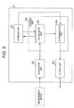
- Fig. 5 is a block diagram illustrating the functionality configuration of the determination device of Fig. 4 .
- the computer system 3 includes an acquisition unit 301, a storage unit 302, a calculation unit 303, a determination unit 304, and an output unit 305.
- the acquisition unit 301 is communicably connected to the measurement device 2 through a network.
- the calculation unit 303 and the determination unit 304 are included in a control unit 306.
- the acquisition unit 301 obtains information from the measurement device 2.
- the storage unit 302 stores a threshold necessary for determination and a formula for calculating a methylation score.
- the calculation unit 303 calculates the methylation score from the information obtained at the acquisition unit 301 according to the formula stored in the storage unit 302.
- the determination unit 304 determines whether or not the methylation score calculated at the calculation unit 303 is lower than the threshold stored at the storage unit 302.
- the output unit 305 outputs the determination result from the determination unit 304 as information on plural types of cancers in the subject (e.g., the presence or absence of cancer cells derived from any of the plural types of cancers in the biological sample collected from the subject).
- Fig. 6 is a block diagram illustrating the hardware configuration of the determination device in Fig. 4 .
- the computer main body 3a includes a central processing unit (CPU) 30, a read only memory (ROM) 31, a random access memory (RAM) 32, a hard disk 33, an input/output interface 34, a readout device 35, a communication interface 36, and an image output interface 37.
- the CPU 30, ROM 31, RAM 32, the hard disk 33, the input/output interface 34, the readout device 35, the communication interface 36, and the image output interface 37 are data-communicably connected via a bus 38.
- the CPU 30 can execute a computer program stored in the ROM 31 and a computer program loaded with the RAM 32.
- the functional blocks described above may be executed. Accordingly, the computer system serves as a terminal that is a determination device for providing information on plural types of cancers in a subject.
- the ROM 31 may be mask ROM, PROM, EPROM, EEPROM, or the like.
- the ROM 31 stores the computer program executed by the CPU 30 and data used for the execution.
- the RAM 32 may be SRAM, DRAM, or the like.
- the RAM 32 is used to read out the computer program stored in the ROM 31 and the hard disk 33.
- the RAM 32 is also used as a work area of the CPU 30 in executing these computer programs.
- the computer programs such as an operating system and an application program (a computer program for providing information on plural types of cancers in a subject), to be executed by the CPU 30, and data for executing the computer programs are installed on the hard disk 33.
- the readout device 35 may be a flexible disk drive, a CD-ROM drive, a DVD-ROM drive, or the like.
- the readout device 35 can read out the computer program or data recorded in a portable recording medium 40.
- the input/output interface 34 may be a serial interface such as USB, IEEE 1394, and RS-232C, a parallel interface such as SCSI, IDE, and IEEE 1284, an analog interface formed by a D/A converter and an A/D converter, and the like.
- An input device 3b such as a keyboard, a mouse, and the like is connected to the input/output interface 34. The operator uses the input device 3b to input data to the computer main body 3a.
- the communication interface 36 is, for example, an Ethernet® interface.
- the computer system 3 can send printing data to a printer by the communication interface 36.
- the image output interface 37 is connected to the display unit 3c including a LCD, a CRT and the like. Accordingly, the display unit 3c can output an image signal according to image data from the CPU 30. The display unit 3c displays an image (on a screen) according to the input image signal.
- Fig. 7 is a flow chart for providing information on plural types of cancers using the determination device of Fig. 4 .
- a peak area is calculated based on mass spectrometric information of a nucleic acid in a measurement sample prepared from a DNA sample derived from a subject, and a methylation score is calculated from the obtained peak area, so as to determine whether or not the methylation score is lower than a threshold.
- the present invention is not limited to this embodiment.
- the acquisition unit 301 in the determination device 1 obtains mass spectrometric information from the measurement device 2.
- the calculation unit 303 calculates a peak area from the mass spectrometric information obtained at the acquisition unit 301 and sends the peak area to the storage unit 302.
- the calculation unit 303 calculates a methylation score based on the peak area stored in the storage unit 302 according to the formula stored in the storage unit 302.
- the determination unit 304 determines whether or not the methylation score calculated at the calculation unit 303 is lower than the threshold stored in the storage unit 302. When the methylation score is lower than the threshold, the process proceeds to the step S1-5 and the determination unit 304 sends, to the output unit 305, a determination result indicating that the biological sample collected from the subject does not contain cancer cells. When the methylation score is not lower than the threshold (i.e., the methylation score is the threshold or more), the determination unit 304 sends, to the output unit 305, a determination result indicating that the biological sample collected from the subject contains cancer cells.
- the output unit 305 outputs the determination result as information on plural types of cancers in the subject, so that the display unit 3c displays the result and/or the printer prints out the result. Accordingly, the determination device can provide, to a physician or the like, information assisting the physician or the like to judge whether or not the subject has cancer.
- the specimens shown in Tables 2 to 4 below were used as biological samples.
- the healthy blood cells were purchased by BioChain Inc.
- Other specimens were collected from patients or healthy subjects. After being collected, the tissues were immediately frozen with liquid nitrogen and stored at -80°C until use.
- the methylation data of cancer tissue specimens derived from gastric cancer, cancer tissue specimens derived from hepatocellular cancer, non-cancerous tissue specimens derived from hepatocellular cancer, and normal specimens were measured by Infinium Methylation Assay using Infinium HumanMethylation27 BeadChip (Illumina, Inc.) in a manner described below in (2) to (3).
- the methylation data of lung cancer, prostate cancer, and peripheral blood from healthy subjects were obtained using Infinium HumanMethylation27 BeadChip (Illumina, Inc.) published in the following documents.
- the methylation data described herein is the methylation rate (mCpG) of CpG sites of GDF7, ZNF132, CPXM1, RPL39L, DOK1, FUZ, MGAT3, and EHD3 obtained by Infinium Methylation Assay using Infinium HumanMethylation27 BeadChip (Illumina, Inc.) in a manner described below in (2) to (3).
- mCpG methylation rate
- Genomic DNA was extracted from each of cancerous tissue specimens derived from gastric cancer, cancerous tissue specimens derived from hepatocellular cancer, non-cancerous tissue specimens derived from hepatocellular cancer, and normal specimens (excluding normal uterus and healthy peripheral blood) using QIAamp DNA Mini Kit (QIAGEN).
- the obtained genomic DNA 500 ng was subjected to bisulfite treatment using EZ DNA Methylation Kit (Zymo Research), and the genomic DNA after the treatment was dissolved in sterilized distilled water (10 ⁇ l).
- the genomic DNA contained in an obtained DNA solution (4 ⁇ l) was fragmented by Bioruptor (COSMO BIO Co., Ltd).
- Methylation data for each specimen was obtained by Infinium Methylation Assay using Infinium HumanMethylation27 BeadChip (Illumina, Inc.) on the fragmented genomic DNA.
- a novel marker which was methylated specifically to cancerous tissue of various types of cancers was detected using the obtained methylation data, and the methylation data published in the documents described above and the data of the TCGA. The specific operation was carried out according to the manual attached to the used chip.
- the Infinium HumanMethylation27 BeadChip includes probes for methylated CpG sites and probes for non-methylated CpG sites of 27,578 CpG sites on human genome.
- the threshold was set to "0.2" for the obtained methylation rate (mCpG) of each gene.
- a specimen with the methylation rate of the threshold value or higher was referred to as "methylation positive specimen.”
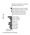
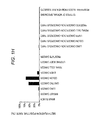
- the promoter region of each gene of GDF7, ZNF132, CPXM1, RPL39L, DOK1, FUZ, MGAT3, and EHD3 was identified as a marker which is highly methylated in cancerous tissue of at least one cancer of the various types of cancers (see Figs. 1A to 1H , and Table 5 below).
- the methylation positive rate of "normal specimens” is an average value of the methylation positive rates of 13 types of normal specimens
- the methylation positive rate of "normal blood” is an average value of the methylation positive rates of peripheral blood from 93 healthy subjects (Salhia B, et al.). According to Figs.
- the average value of the methylation positive rates of the normal specimens and the normal blood was zero in the marker of the embodiment, and thus the methylation was not observed in 13 types of normal specimens and normal blood.
- the methylation positive rate higher than the non-cancerous parts and normal specimens was confirmed in a plurality of predetermined cancers. This suggests that the methylation analysis of the gene in this example is useful as an index for diagnosis of any one of plural types of cancers.
- GDF7 is a gene existing in the chromosome 2, and the probe position in the Infinium HumanMethylation27 BeadChip is 20,729,328.
- Fig. 1A when GDF7 was used, a high methylation positive rate was obtained in cancerous tissue of brain tumor, breast cancer, lung cancer, colon cancer, hepatocellular cancer (liver cancer), gastric cancer, renal cell cancer, uterine body cancer, and prostate cancer.
- ZNF132 is a gene existing in the chromosome 19, and the probe position in the Infinium HumanMethylation27 BeadChip is 63,643,484.
- Fig. 1B when ZNF132 was used, a high methylation positive rate was obtained in cancerous tissue of breast cancer, lung cancer, gastric cancer, colon cancer, hepatocellular cancer, renal cell cancer, and uterine body cancer.
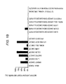
- CPXM1 is a gene existing in the chromosome 20, and the probe position in the Infinium HumanMethylation27 BeadChip is 2,729,489.
- Fig. 1c when CPXM1 was used, a high methylation positive rate was obtained in cancerous tissue of lung cancer, gastric cancer, colon cancer, hepatocellular cancer, renal cell cancer, uterine body cancer, and prostate cancer.
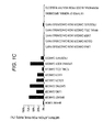
- RPL39L is a gene existing in the chromosome 3, and the probe position in the Infinium HumanMethylation27 BeadChip is 188,340,169.
- Fig. 1D when RPL39L was used, a high methylation positive rate was obtained in cancerous tissue of brain tumor, breast cancer, lung cancer, gastric cancer, colon cancer, hepatocellular cancer, renal cell cancer, uterine body cancer, and prostate cancer.
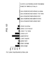
- DOK1 is a gene existing in the chromosome 2, and the probe position in the Infinium HumanMethylation27 BeadChip is 74,634,604. As illustrated in Fig. 1E , when DOK1 was used, a high methylation positive rate was obtained in cancerous tissue of breast cancer, hepatocellular cancer, and prostate cancer. These results suggest that the methylation analysis of the promoter region of DOK1 serves an index of existence of any cancer of breast cancer, hepatocellular cancer, and prostate cancer, and is useful to screen for cancer.
- FUZ is a gene existing in the chromosome 19, and the probe position in the Infinium HumanMethylation27 BeadChip is 55,008,270.
- Fig. 1F when FUZ was used, a high methylation positive rate was obtained in cancerous tissue of gastric cancer, colon cancer, hepatocellular cancer, and uterine body cancer.
- MGAT3 is a gene existing in the chromosome 22, and the probe position in the Infinium HumanMethylation27 BeadChip is 38,183,536.
- Fig. 1G when MGAT3 was used, a high methylation positive rate was obtained in cancerous tissue of breast cancer, lung cancer, gastric cancer, and colon cancer.
- EHD3 is a gene existing in the chromosome 2, and the probe position in the Infinium HumanMethylation27 BeadChip is 31,310,293.
- Fig. 1H when EHD3 was used, a high methylation positive rate was obtained in cancerous tissue of gastric cancer and colon cancer.
- clinical specimens including a cancerous part and non-cancerous tissue specimen of hepatocellular cancer collected by hepatectomy from a patient different from that in Example 1, as well as a cancerous tissue specimen of gastric cancer and a normal stomach tissue specimen collected from a patient different from that in Example 1 were used as biological samples.
- the clinical specimens included normal brain tissue (1 specimen), brain tumor tissue (8 specimens), normal stomach tissue (2 specimens), cancerous tissue of gastric cancer (6 specimens), normal colon tissue (4 specimens), cancerous tissue of colon cancer (5 specimens), normal liver tissue (2 specimens), non-cancerous tissue of hepatocellular cancer (5 specimens), cancerous tissue of hepatocellular cancer (6 specimens), normal uterus tissue (4 specimens), cancerous tissue of uterine body cancer (4 specimens), non-cancerous tissue of breast cancer (3 specimens), and cancerous tissue of breast cancer (4 specimens). After being collected, these tissues were immediately frozen with liquid nitrogen, and stored at -80°C until use.
- Genomic DNA was extracted from each tissue using QIAamp DNA Mini Kit (QIAGEN). The genome DNA contained in an obtained DNA solution was fragmented by Bioruptor (COSMO BIO Co., Ltd). For preparing calibration curves in mass spectrometry, genomic DNA of human peripheral blood lymphocytes was used as the control genomic DNA. The genomic DNA from human peripheral blood lymphocytes was amplified using GenomiPhi v2DNA amplification kit (GE Healthcare Life Sciences). The obtained amplified product consisted of a solution of non-methylated DNA fragments (0% methylated DNA).
- the respective DNA fragments (500 ng) obtained as described above were subjected to bisulfite treatment using EZ DNA Methylation Kit (Zymo Research), and the genome DNA after the treatment was dissolved in sterilized distilled water (80 ⁇ l).
- the methylated cytosine and uracil contained in the respective bisulfate-treated DNA fragments were converted to guanine and adenine by the PCR and IVT amplification.
- the sequence of the primer set for the present marker is shown in Table 5.
- the base sequence (sequence of positive stand) in the region to be analyzed by the primer set is shown in SEQ ID NO: 37.
- the base sequence after the bisulfite conversion in the region amplified with primer set for methylation detection of GDF7 is shown in SEQ ID NO: 47.
- the base sequence of SEQ ID NO: 47 represents the base sequence when all CpG sites existing in the region to be amplified with each primer set for methylation detection.
- tag sequence and T7 promoter sequence were added to the 5' end of the forward and reverse primers of the primer set for IVT reaction.
- a PCR reaction solution was prepared by mixing the following reagents. 10x Hot Star buffer (QIAGEN) 0.5 ⁇ L 25 mM dNTP mix 0.04 ⁇ L Hot Star Taq (5U/ ⁇ l) (QIAGEN) 0.04 ⁇ L Primer mix 2.0 ⁇ L DNA solution 1.0 ⁇ L Water 1.42 ⁇ L Total 5 ⁇ L
- the PCR reaction was carried out in the above reaction solution under the following conditions:
- PCR products were dephosphorylated by SAP (Shrimp Alkaline Phosphatase) contained in a MassCLEAVETM Reagent kit (Sequenom, Inc.). Then, the following reaction solution prepared by the kit was added to the PCR products.
- SAP Shrimp Alkaline Phosphatase
- the obtained mixture was incubated at 37°C for three hours to induce the IVT reaction and urasil- or thymine-specific cleavage.
- the resulting reaction products were purified by Clean Resin (Sequenom, Inc.) to obtain samples for mass spectrometry. These samples, in which samples derived from genomic DNA extracted from the clinical specimens were designated as measurement samples and samples derived from control genomic DNA were designated as control samples, were used for mass spectrometry described later.
- control samples obtained as described above were subjected to the mass spectrometric analysis twice independently.
- the calibration curves were prepared for the respective primer sets based on the obtained analysis results, and correlation coefficients were calculated. Accordingly, it was confirmed that the used primer sets amplified the methylated DNA and non-methylated DNA evenly.
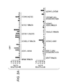
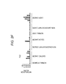
- the obtained measurement samples obtained as described above were subjected to the mass spectrometric analysis to obtain peaks of DNA fragments included in the measurement samples. Then, a portion of the base sequence of GDF7, from which each obtained peak was originated, was identified. A methylation score was calculated from the ratio between the area of the peaks of the fragments including the methylated CpG sites and the area of peaks of the fragments not including the methylated CpG sites in the fragments originating from the same base sequence. The results are illustrated in Fig. 2A .
- the samples having the methylation score exceeded 0.1 in the promoter region of GDF7 gene were, in the cancerous tissue, 4 specimens/8 specimens in brain tumor tissue, 6 specimens/6 specimens in gastric cancer tissue, 5 specimens/5 specimens in colon cancer tissue, 4 specimens/6 specimens in hepatocellular cancer tissue, 4 specimens/4 specimens in uterine body cancer tissue, and 3 specimens/4 specimens in breast cancer tissue.
- non-cancerous tissue such samples were 5 specimens/5 specimens in non-cancerous tissue of hepatocellular cancer, and 0 specimens/3 specimens in non-cancerous tissue of breast cancer.
- the analysis of methylation frequency using GDF7 can be used in the assistance of diagnosis and relapse monitoring of brain tumor, gastric cancer, colon cancer, hepatocellular cancer, uterine body cancer, breast cancer, and the like based on the difference in methylation frequency of the cancerous tissue specimen, the non-cancerous tissue specimen, and the normal specimen.
- the methylation score was calculated in a manner similar to Example 2 except that normal stomach tissue (2 specimens), cancerous tissue (6 specimens) of gastric cancer, non-cancerous tissue (4 specimens) of colon cancer, cancerous tissue (5 specimens) of colon cancer, normal liver tissue (2 specimens), non-cancerous tissue (5 specimens) of hepatocellular cancer, cancerous tissue (6 specimens) of hepatocellular cancer, normal uterus tissue (4 specimens), cancerous tissue (4 specimens) of uterine body cancer, non-cancerous tissue (3 specimens) of breast cancer, and cancerous tissue (4 specimens) of breast cancer were used as clinical specimens, and those shown in Table 8 were used for the MassARRAY analysis (base sequence (sequence of positive strand) of the region analyzed with such primer set is represented by SEQ ID NO: 38).
- the base sequence after bisulfite conversion of the region amplified with the primer set for methylation detection of ZNF132 was represented by SEQ ID NO: 48.
- the base sequence of SEQ ID NO: 48 represents the base sequence when all CpG sites existing in the region amplified with each primer set for methylation detection are methylated.
- the tag sequence of SEQ ID NO: 35 and the T7 promoter sequence of SEQ ID NO: 36 are added to the 5' end of the forward primer and the reverse primer of the primer set for IVT reaction.
- the samples having the methylation score exceeded 0.1 in the promoter region of ZNF12 gene were, in the cancerous tissue, 6 specimens/6 specimens in gastric cancer tissue, 5 specimens /5 specimens in colon cancer tissue, 4 specimens/6 specimens in hepatocellular cancer tissue, 4 specimens/4 specimens in uterine body cancer tissue, and 3 specimens/4 specimens in breast cancer tissue.
- the non-cancerous tissue such samples were 0 specimens/4 specimens in non-cancerous tissue of colon cancer, 0 specimens/5 specimens in non-cancerous tissue of hepatocellular cancer, and 0 specimens/3 specimens in non-cancerous tissue of breast cancer.
- methylation frequency using ZNF132 in the analysis of methylation frequency using ZNF132, it can be used in the assistance of diagnosis and relapse monitoring of gastric cancer, colon cancer, hepatocellular cancer, uterine body cancer, breast cancer, and the like based on the difference in methylation frequency of the cancerous tissue specimen, the non-cancerous tissue specimen, and the normal specimen.
- the methylation score was calculated in a manner similar to Example 2 except that the normal stomach tissue (2 specimens), the cancerous tissue (6 specimens) of gastric cancer, the non-cancerous tissue (4 specimens) of colon cancer, the cancerous tissue (5 specimens) of colon cancer, the normal liver tissue (2 specimens), the non-cancerous tissue (5 specimens) of hepatocellular cancer, the cancerous tissue (6 specimens) of hepatocellular cancer, the normal uterus tissue (4 specimens), the cancerous tissue (4 specimens) of uterine body cancer, the non-cancerous tissue (3 specimens) of breast cancer, and the cancerous tissue (4 specimens) of breast cancer were used as clinical specimens, and those shown in Table 10 were used for the MassARRAY analysis (base sequence (sequence of positive strand) of the region analyzed with the primer set is represented by SEQ ID NO: 39).
- the base sequence after bisulfite conversion of the region amplified with the primer set for methylation detection of the CPXM1 is represented by SEQ ID NO: 49.
- the base sequence of SEQ ID NO: 49 represents the base sequence when all the CpG sites existing in the region amplified with each primer set for methylation detection are methylated.
- the tag sequence of SEQ ID NO: 35 and the T7 promoter sequence of SEQ ID NO: 36 arc added to the 5' end of the forward primer and the reverse primer of the primer set for IVT reaction.
- the samples having the methylation score exceeded 0.1 in the promoter region of CPXM1 gene were, in the cancerous tissue, 6 specimens/6 specimens in gastric cancer tissue, 3 specimens/5 specimens in colon cancer tissue, 5 specimens/6 specimens in hepatocellular cancer tissue, 4 specimens/4 specimens in uterine body cancer tissue, and 3 specimens/4 specimens in breast cancer tissue.
- the non-cancerous tissue such samples were 0 specimens/4 specimens in non-cancerous tissue of colon cancer, 0 specimens/5 specimens in non-cancerous tissue of hepatocellular cancer, and 0 specimens/3 specimens in non-cancerous tissue of breast cancer.
- the methylation score was calculated in a manner similar to Example 2 except that normal stomach tissue (1 specimen), brain tumor tissue (8 specimens), normal stomach tissue (2 specimens), cancerous tissue (6 specimens) of gastric cancer, non-cancerous tissue (4 specimens) of colon cancer, cancerous tissue (5 specimens) of colon cancer, normal liver tissue (2 specimens), non-cancerous tissue (5 specimens) of hepatocellular cancer, cancerous tissue (6 specimens) of hepatocellular cancer, normal uterus tissue (4 specimens), and cancerous tissue (4 specimens) of uterine body cancer were used as clinical specimens, and those shown in Table 12 were used for the MassARRAY analysis (base sequence (sequence of positive strand) of the region analyzed with the primer set is represented by SEQ ID NO: 40).
- the base sequence after bisulfite conversion of the region amplified with the primer set for methylation detection of RPL39L is represented by SEQ ID NO: 50.
- the base sequence of SEQ ID NO: 50 represents the base sequence when all CpG sites existing in the region amplified with each primer set for methylation detection are methylated.
- the tag sequence of SEQ ID NO: 35 and the T7 promoter sequence of SEQ ID NO: 36 are added to the 5' end of the forward primer and the reverse primer of the primer set for IVT reaction.
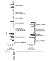
- the samples having the methylation score exceeded 0.1 in the promoter region of RPL39L gene were, in cancerous tissue, 7 specimens/8 specimens in brain tumor tissue, 4 specimens/6 specimens in gastric cancer tissue, 2 specimens/5 specimens in colon cancer tissue, 4 specimens/6 specimens in hepatocellular cancer tissue, and 3 specimens/4 specimens in uterine body cancer tissue.
- non-cancerous tissue such samples were 1 specimens/4 specimens in non-cancerous tissue of colon cancer, and 0 specimens/5 specimens in non-cancerous tissue of hepatocellular cancer.
- Fig. 2D As is evident from Fig. 2D and the results listed in Table 13, in the analysis of the methylation frequency using RPL39L, it can be used in the assistance of diagnosis and relapse monitoring of brain tumor, gastric cancer, colon cancer, hepatocellular cancer, uterine body cancer, and the like based on the difference in methylation frequency of the cancerous tissue specimen, the non-cancerous tissue specimen, and the normal specimen.
- the methylation score was calculated in a manner similar to Example 2 except that that normal liver tissue (2 specimens), non-cancerous tissue (5 specimens) of hepatocellular cancer, cancerous tissue (6 specimens) of hepatocellular cancer, non-cancerous tissue (3 specimens) of breast cancer, and cancerous tissue (4 specimens) of breast cancer were used as clinical specimens, and those shown in Table 14 were used for the MassARRAY analysis (base sequence (sequence of positive strand) of the region analyzed with the primer set is represented by SEQ ID NO: 41).
- the base sequence after bisulfite conversion of the region amplified with the primer set for methylation detection of DOK1 is represented by SEQ ID NO: 51.
- the base sequence of SEQ ID NO: 51 represents the base sequence when all CpG sites existing in the region amplified with each primer set for methylation detection are methylated.
- the tag sequence of SEQ ID NO: 35 and the T7 promoter sequence of SEQ ID NO: 36 are added to the 5' end of the forward primer and the reverse primer of the primer set for IVT reaction.
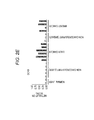
- the samples having the methylation score exceeded 0.1 in the promoter region of the DOK1 gene were, in cancerous tissue, 6 specimens/6 specimens in hepatocellular cancer tissue, and 3 specimens/4 specimens in breast cancer tissue.
- non-cancerous tissue such samples were 0 specimen/5 specimens in non-cancerous tissue of hepatocellular cancer, and 0 specimens/3 specimens in non-cancerous tissue of breast cancer.
- normal tissue the samples were 0 specimens/2 specimens in liver tissue.
- the methylation score was calculated in a manner similar to Example 2 except that that normal stomach tissue (2 specimens), cancerous tissue (6 specimens) of gastric cancer, non-cancerous tissue (4 specimens) of colon cancer, cancerous tissue (5 specimens) of colon cancer, normal liver tissue (2 specimens), non-cancerous tissue (5 specimens) of hepatocellular cancer, and cancerous tissue (6 specimens) of hepatocellular cancer were used as clinical specimens, and those shown in Table 16 were used for the MassARRAY analysis (base sequence (sequence of positive strand) of the region analyzed with the primer set is represented by SEQ ID NO: 42).
- the base sequence after bisulfite conversion of the region amplified with the primer set for methylation detection of FUZ is represented by SEQ ID NO: 55.
- the base sequence of SEQ ID NO: 55 represents the base sequence when all CpG sites existing in the region amplified with each primer set for methylation detection are methylated.
- the tag sequence of SEQ ID NO: 35 and the T7 promoter sequence of SEQ ID NO: 36 are added to the 5' end of the forward primer and the reverse primer of the primer set for IVT reaction.
- the samples having the methylation score exceeded 0.1 in the promoter region of FUZ gene were, in cancerous tissue, 4 specimens/6 specimens in gastric cancer tissue, 3 specimens/5 specimens in colon cancer tissue, and 4 specimens/6 specimens in hepatocellular cancer tissue.
- non-cancerous tissue such samples were 0 specimen/4 specimens in non-cancerous tissue of colon cancer, and 0 specimen/5 specimens in non-cancerous tissue of hepatocellular cancer.
- the samples were 0 specimen/2 specimens in stomach tissue, and 0 specimen/2 specimens in liver tissue.
- the methylation score was calculated in a manner similar to Example 2 except that that normal stomach tissue (2 specimens), cancerous tissue (6 specimens) of gastric cancer, non-cancerous tissue (4 specimens) of colon cancer, and cancerous tissue (5 specimens) of colon cancer were used as clinical specimens, and those shown in Table 18 were used for the MassARRAY analysis (base sequence (sequence of positive strand) of the region analyzed with the primer set is represented by SEQ ID NO: 43).
- the base sequence after bisulfite conversion of the region amplified with the primer set for methylation detection of EHD3 is represented by SEQ ID NO: 56.
- the base sequence of SEQ ID NO: 56 represents the base sequence when all CpG sites existing in the region amplified with each primer set for methylation detection are methylated.
- the tag sequence of SEQ ID NO: 35 and the T7 promoter sequence of SEQ ID NO: 36 are added to the 5' end of the forward primer and the reverse primer of the primer set for IVT reaction.
- the non-cancerous tissue (2 specimens) of gastric cancer, the cancerous tissue (4 specimens) of gastric cancer, the non-cancerous tissue (4 specimens) of colon cancer, the cancerous tissue (5 specimens) of colon cancer, the normal liver tissue (2 specimens), the non-cancerous tissue (5 specimens) of hepatocellular cancer, and the cancerous tissue (6 specimens) of hepatocellular cancer were used as biological samples.
- Genomic DNA was extracted from each tissue described above using QIAamp DNA Mini Kit (QIAGEN). The genomic DNA contained in an obtained DNA solution was fragmented by Bioruptor (COSMO BIO Co., Ltd).
- genomic DNA of human peripheral blood lymphocytes was used as the control genomic DNA.
- the genomic DNA from human peripheral blood lymphocytes was amplified using GenomiPhi v2DNA amplification kit (GE Healthcare Life Sciences). The obtained amplified product consisted of non-methylated DNA.
- the amplified product was then fragmented with Bioruptor (COSMO BIO Co., Ltd.) to obtain a solution of non-methylated DNA fragments (0% methylated DNA).
- a part of the solution of the non-methylated DNA fragments was subjected to reaction with SssI methylase (New England Biolabs) to methylate all cytosines in CG sequences, and a solution of methylated DNA fragments (100% methylated DNA) was obtained.
- the respective DNA fragments (500 ng) obtained as described above were subjected to bisulfite treatment using EZ DNA Methylation Kit (Zymo Research), and the genomic DNA after the treatment was dissolved in sterilized distilled water (80 ⁇ l).
- the MSP was conducted using the measurement samples and the control samples (DNA after bisulfite treatment) obtained as described above in (2).
- the composition of the PCR reagent used in the MSP, the primer set, and the reaction conditions of the PCR are described below.
- DDW (sterilized water) 16.75 ⁇ L 10 ⁇ PCR buffer with MgC12 (Roche) 2.5 ⁇ L 2 mM dNTP mix 2. 2.5 ⁇ L 10 ⁇ M sense primer 1.0 ⁇ L 10 ⁇ M anti-sense primer 1.0 ⁇ L Faststart Taq polymerase (Roche) 0.25 ⁇ L Measurement sample or control sample 1.0 ⁇ L Total 25 ⁇ L
- the primer set used in the MSP is shown in Table 20.
- Such primer sets are primer sets that can obtain the amplified product when DNA in the region to be amplified is methylated.
- the base sequences of the region analyzed with the primer set shown in Table 20 for the promoter region of FUZ gene are respectively represented by SEQ ID NO: 44.
- the base sequence of SEQ ID NO: 44 is the sequence of positive strand.
- the base sequence after bisulfite conversion in the region amplified with the primer set for methylation detection (M primer of SEQ ID NOs: 23 and 24) of FUZ is represented by SEQ ID NO: 52.
- the base sequence of SEQ ID NO: 52 represents the base sequence when all CpG sites existing in the region amplified with each primer set for methylation detection are methylated.
- the base sequence of after bisulfite conversion in the region amplified with the primer set for non-methylation detection (U primer of SEQ ID NOs: 25 and 26) of FUZ is represented by SEQ ID NO: 57.
- SEQ ID NO: 57 The base sequence of after bisulfite conversion in the region amplified with the primer set for non-methylation detection (U primer of SEQ ID NOs: 25 and 26) of FUZ is represented by SEQ ID NO: 57.
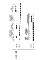
- the amplified product obtained with the MSP described above was checked with 2% agarose gel electrophoresis. The result is shown in Fig. 3A .
- "0" and “100” indicated in “control” represent 0% methylated control sample and 100% methylated control sample, respectively.
- the band derived from the methylated CpG was not detected in the non-cancerous tissue of gastric cancer, the non-cancerous tissue of colon cancer, the normal liver tissue, and the non-cancerous tissue of hepatocellular cancer.
- the band of the methylated DNA was detected in 3 specimens/4 specimens for the cancerous tissue of gastric cancer, 3 specimens/5 specimens for the cancerous tissue of colon cancer, and 4 specimens/6 specimens for the cancerous tissue of hepatocellular cancer. As is evident from Fig.
- the methylation frequency using FUZ in the analysis of the methylation frequency using FUZ, it can be used in the assistance of diagnosis and relapse monitoring of gastric cancer, colon cancer, hepatocellular cancer, and the like based on the difference in methylation frequency of the cancerous tissue specimen and the normal specimen.
- the amplified product was obtained by the MSP in a manner similar to Example 9 except that the non-cancerous tissue (2 specimens) of gastric cancer, the cancerous tissue (4 specimens) of gastric cancer, the non-cancerous tissue (4 specimens) of colon cancer, and the cancerous tissue (5 specimens) of colon cancer were used as biological samples, and that the primer set shown in Table 21 was used (base sequence of region to be analyzed is represented by SEQ ID NO: 45, base sequence of SEQ ID NO: 45 is sequence of positive strand).
- the base sequence after bisulfite conversion of the region amplified with the primer set for methylation detection (M primer of SEQ ID NOs: 27 and 28) of EHD3 is represented by SEQ ID NO: 53.
- the base sequence of SEQ ID NO: 53 represents the base sequence when all CpG sites existing in the region amplified with each primer set for methylation detection are methylated.
- the base sequence after bisulfite conversion of the region amplified with the primer set for non-methylation detection (U primer of SEQ ID NOs: 29 and 30) of EHD3 is represented by SEQ ID NO: 58.
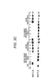
- the band derived from the methylated CpG was not detected in the non-cancerous tissue of gastric cancer and the non-cancerous tissue of colon cancer.
- the band of the methylated DNA was detected in 3 specimens/4 specimens for the cancerous tissue of gastric cancer and 5 specimens/5 specimens for the cancerous tissue of colon cancer.
- Fig. 3B in the analysis of the methylation frequency using the EHD3, it can be used in the assistance of diagnosis and relapse monitoring of gastric cancer, colon cancer, and the like based on the difference in methylation frequency of the cancerous tissue specimen and the non-cancerous tissue specimen.
- the amplified product was obtained by the MSP in a manner similar to Example 9 except that the non-cancerous tissue (2 specimens) of gastric cancer, the cancerous tissue (6 specimens) of gastric cancer, the non-cancerous tissue (4 specimens) of colon cancer, and the cancerous tissue (5 specimens) of colon cancer were used for the biological sample, and that the primer set shown in Table 22 was used (base sequence of region to be analyzed is represented by SEQ ID NO: 46, base sequence of SEQ ID NO: 46 is sequence of positive strand).
- the base sequence after bisulfite conversion of the region amplified with the primer set for methylation detection (M primer of SEQ ID NOs: 31 and 32) of MGAT3 is represented by SEQ ID NO: 54.
- the base sequence of SEQ ID NO: 54 represents the base sequence when all CpG sites existing in the region amplified with each primer set for methylation detection are methylated.
- the base sequence after bisulfite conversion of the region amplified with the detection primer set for non-methylation detection (U primer of SEQ ID NOs: 33 and 34) of MGAT3 is represented by SEQ ID NO: 59.
- the band derived from the methylated CpG was not detected in the non-cancerous tissue of gastric cancer and the non-cancerous tissue of colon cancer.
- the band of the methylated DNA was detected in 5 specimens/6 specimens for the cancerous tissue of gastric cancer and 4 specimens/5 specimens for the cancerous tissue of colon cancer.
- Fig. 3C in the analysis of the methylation frequency using the MGAT3, it can be used in the assistance of diagnosis and relapse monitoring of gastric cancer, colon cancer, and the like based on the difference in methylation frequency of the cancerous tissue specimen and the non-cancerous tissue specimen.
Landscapes
- Chemical & Material Sciences (AREA)
- Life Sciences & Earth Sciences (AREA)
- Health & Medical Sciences (AREA)
- Organic Chemistry (AREA)
- Proteomics, Peptides & Aminoacids (AREA)
- Engineering & Computer Science (AREA)
- Immunology (AREA)
- Pathology (AREA)
- Analytical Chemistry (AREA)
- Zoology (AREA)
- Genetics & Genomics (AREA)
- Wood Science & Technology (AREA)
- Physics & Mathematics (AREA)
- Biotechnology (AREA)
- Microbiology (AREA)
- Molecular Biology (AREA)
- Hospice & Palliative Care (AREA)
- Biophysics (AREA)
- Oncology (AREA)
- Biochemistry (AREA)
- Bioinformatics & Cheminformatics (AREA)
- General Engineering & Computer Science (AREA)
- General Health & Medical Sciences (AREA)
- Measuring Or Testing Involving Enzymes Or Micro-Organisms (AREA)
- Apparatus Associated With Microorganisms And Enzymes (AREA)
Applications Claiming Priority (1)
| Application Number | Priority Date | Filing Date | Title |
|---|---|---|---|
| JP2014143326A JP6418595B2 (ja) | 2014-07-11 | 2014-07-11 | 複数種類の癌に関する情報を取得する方法、システムおよびプログラム |
Publications (3)
| Publication Number | Publication Date |
|---|---|
| EP2977467A2 true EP2977467A2 (de) | 2016-01-27 |
| EP2977467A3 EP2977467A3 (de) | 2016-05-11 |
| EP2977467B1 EP2977467B1 (de) | 2018-04-11 |
Family
ID=53765064
Family Applications (1)
| Application Number | Title | Priority Date | Filing Date |
|---|---|---|---|
| EP15176249.9A Active EP2977467B1 (de) | 2014-07-11 | 2015-07-10 | Verfahren, verwendung von marker und bestimmungsvorrichtung zur gewinnung von informationen über eine vielzahl von krebsarten |
Country Status (2)
| Country | Link |
|---|---|
| EP (1) | EP2977467B1 (de) |
| JP (1) | JP6418595B2 (de) |
Cited By (7)
| Publication number | Priority date | Publication date | Assignee | Title |
|---|---|---|---|---|
| CN108460246A (zh) * | 2018-03-08 | 2018-08-28 | 北京希望组生物科技有限公司 | 一种基于三代测序平台的hla基因分型方法 |
| WO2020239896A1 (en) * | 2019-05-31 | 2020-12-03 | Universal Diagnostics, S.L. | Detection of colorectal cancer |
| US11001898B2 (en) | 2019-05-31 | 2021-05-11 | Universal Diagnostics, S.L. | Detection of colorectal cancer |
| US11396679B2 (en) | 2019-05-31 | 2022-07-26 | Universal Diagnostics, S.L. | Detection of colorectal cancer |
| US11530453B2 (en) | 2020-06-30 | 2022-12-20 | Universal Diagnostics, S.L. | Systems and methods for detection of multiple cancer types |
| US11898199B2 (en) | 2019-11-11 | 2024-02-13 | Universal Diagnostics, S.A. | Detection of colorectal cancer and/or advanced adenomas |
| US12467096B2 (en) | 2020-05-15 | 2025-11-11 | Universal Diagnostics, S.A. | Methods and systems for identifying methylation biomarkers |
Families Citing this family (3)
| Publication number | Priority date | Publication date | Assignee | Title |
|---|---|---|---|---|
| WO2017119510A1 (ja) * | 2016-01-08 | 2017-07-13 | 国立大学法人京都大学 | 乳がんの診断のための検査方法、遺伝子マーカー、および検査薬 |
| JP7627904B2 (ja) * | 2021-01-26 | 2025-02-07 | 学校法人 岩手医科大学 | 腎細胞癌の診断マーカー及びそれを用いた診断方法 |
| EP4306656A4 (de) * | 2021-03-12 | 2025-05-21 | FUJIFILM Corporation | Krebstestreagenzienset, verfahren zur herstellung eines krebstestreagenziensets und krebstestverfahren |
Citations (3)
| Publication number | Priority date | Publication date | Assignee | Title |
|---|---|---|---|---|
| US20120178634A1 (en) | 2009-07-03 | 2012-07-12 | Sysmex Corporation | Method for determination of presence of cancer cell, and method for determination of prognosis of cancer patient |
| US20120190024A1 (en) | 2009-06-30 | 2012-07-26 | Sysmex Corporation | Method for determining presence or absence of epithelial cancer-origin cell in biological sample, and molecular marker and kit therefor |
| US20130130242A1 (en) | 2010-02-26 | 2013-05-23 | Sysmex Corporation | Method for determining presence or absence of cancer cell in biological sample, and molecular marker and kit for determination |
Family Cites Families (3)
| Publication number | Priority date | Publication date | Assignee | Title |
|---|---|---|---|---|
| JP2006008627A (ja) * | 2004-06-28 | 2006-01-12 | Nara Prefecture | 悪性腫瘍の診断のための試薬および診断方法 |
| JPWO2009128453A1 (ja) * | 2008-04-14 | 2011-08-04 | 学校法人日本大学 | 増殖性疾患の検出方法 |
| WO2012167145A2 (en) * | 2011-06-01 | 2012-12-06 | University Of Southern California | Genome-scale analysis of aberrant dna methylation in colorectal cancer |
-
2014
- 2014-07-11 JP JP2014143326A patent/JP6418595B2/ja active Active
-
2015
- 2015-07-10 EP EP15176249.9A patent/EP2977467B1/de active Active
Patent Citations (3)
| Publication number | Priority date | Publication date | Assignee | Title |
|---|---|---|---|---|
| US20120190024A1 (en) | 2009-06-30 | 2012-07-26 | Sysmex Corporation | Method for determining presence or absence of epithelial cancer-origin cell in biological sample, and molecular marker and kit therefor |
| US20120178634A1 (en) | 2009-07-03 | 2012-07-12 | Sysmex Corporation | Method for determination of presence of cancer cell, and method for determination of prognosis of cancer patient |
| US20130130242A1 (en) | 2010-02-26 | 2013-05-23 | Sysmex Corporation | Method for determining presence or absence of cancer cell in biological sample, and molecular marker and kit for determination |
Non-Patent Citations (3)
| Title |
|---|
| KOBAYASHI Y ET AL.: "DNA methylation profiling reveals novel biomarkers and important roles for DNA methyl transferases in prostate cancer", GENOME RES., vol. 21, no. 7, July 2011 (2011-07-01), pages 1017 - 27 |
| SALHIA B ET AL.: "DNA methylation analysis determines the high frequency of genic hypomethylation and low frequency ofhypermethylation events in plasma cell tumors", CANCER RES., vol. 70, no. 17, 1 September 2010 (2010-09-01), pages 6934 - 44 |
| SELAMAT SA ET AL.: "Genome-scale analysis of DNA methylation in lung adenocarcinoma and integration with mRNA expression", GENOME RES., vol. 22, no. 7, July 2012 (2012-07-01), pages 1197 - 211, XP008158437, DOI: doi:10.1101/gr.132662.111 |
Cited By (8)
| Publication number | Priority date | Publication date | Assignee | Title |
|---|---|---|---|---|
| CN108460246A (zh) * | 2018-03-08 | 2018-08-28 | 北京希望组生物科技有限公司 | 一种基于三代测序平台的hla基因分型方法 |
| WO2020239896A1 (en) * | 2019-05-31 | 2020-12-03 | Universal Diagnostics, S.L. | Detection of colorectal cancer |
| US11001898B2 (en) | 2019-05-31 | 2021-05-11 | Universal Diagnostics, S.L. | Detection of colorectal cancer |
| US11396679B2 (en) | 2019-05-31 | 2022-07-26 | Universal Diagnostics, S.L. | Detection of colorectal cancer |
| EP3963111B1 (de) | 2019-05-31 | 2024-04-17 | Universal Diagnostics, S.A. | Nachweis von kolorektalkarzinomen |
| US11898199B2 (en) | 2019-11-11 | 2024-02-13 | Universal Diagnostics, S.A. | Detection of colorectal cancer and/or advanced adenomas |
| US12467096B2 (en) | 2020-05-15 | 2025-11-11 | Universal Diagnostics, S.A. | Methods and systems for identifying methylation biomarkers |
| US11530453B2 (en) | 2020-06-30 | 2022-12-20 | Universal Diagnostics, S.L. | Systems and methods for detection of multiple cancer types |
Also Published As
| Publication number | Publication date |
|---|---|
| EP2977467B1 (de) | 2018-04-11 |
| EP2977467A3 (de) | 2016-05-11 |
| JP2016015963A (ja) | 2016-02-01 |
| JP6418595B2 (ja) | 2018-11-07 |
Similar Documents
| Publication | Publication Date | Title |
|---|---|---|
| EP2977467B1 (de) | Verfahren, verwendung von marker und bestimmungsvorrichtung zur gewinnung von informationen über eine vielzahl von krebsarten | |
| EP2816121B1 (de) | Verfahren zur Gewinnung von Informationen über hepatozelluläres Karzinom | |
| EP2899275B1 (de) | Verfahren zur gewinnung von informationen über endometriumkarzinom sowie marker und kit zur gewinnung von informationen über endometriumkarzinom | |
| EP2808397B1 (de) | Verfahren zur Gewinnung von Informationen über Darmkrebs und Marker und Kit zur Gewinnung von Informationen über Darmkrebs | |
| JP6269492B2 (ja) | 肝細胞癌に関する情報の取得方法、ならびに肝細胞癌に関する情報を取得するためのマーカーおよびキット | |
| EP2966183B1 (de) | Verfahren zur gewinnung von informationen über lungenkrebs und verwendung von marker und bestimmungsvorrichtung zur gewinnung von informationen über lungenkrebs | |
| EP2899272B1 (de) | Verfahren und verwendung zur gewinnung von informationen über kolorektalkrebs | |
| EP2821487B1 (de) | Verfahren zur bestimmung des vorhandenseins oder nichtvorhandenseins von krebszellen aus einem hepatozellulären karzinom sowie bestimmungsmarker und kit | |
| JP6269493B2 (ja) | 脳腫瘍に関する情報の取得方法、ならびに脳腫瘍に関する情報を取得するためのマーカーおよびキット | |
| EP2966182B1 (de) | Verfahren zum erhalt von informationen über uterinkörperkrebs und marker und bestimmungsvorrichtung zum erhalt von informationen über den uterinkörperkrebs | |
| EP3075852B1 (de) | Verfahren zur erfassung von informationen über gynäkologischen krebs und nachweiskit für gynäkologischen krebs | |
| EP3075851A1 (de) | Verfahren zur informationserfassung über magensaft und kit zum nachweis von magenkrebs | |
| JP6636105B2 (ja) | 大腸癌に関する情報の取得方法、ならびに大腸癌に関する情報を取得するためのマーカーおよびキット | |
| EP3075853B1 (de) | Verfahren zur gewinnung von informationen über brustkrebs und kit zum nachweis von brustkrebs | |
| JP6551656B2 (ja) | 卵巣癌に関する情報の取得方法、ならびに卵巣癌に関する情報を取得するためのマーカーおよび卵巣癌検出用キット |
Legal Events
| Date | Code | Title | Description |
|---|---|---|---|
| PUAI | Public reference made under article 153(3) epc to a published international application that has entered the european phase |
Free format text: ORIGINAL CODE: 0009012 |
|
| AK | Designated contracting states |
Kind code of ref document: A2 Designated state(s): AL AT BE BG CH CY CZ DE DK EE ES FI FR GB GR HR HU IE IS IT LI LT LU LV MC MK MT NL NO PL PT RO RS SE SI SK SM TR |
|
| AX | Request for extension of the european patent |
Extension state: BA ME |
|
| PUAL | Search report despatched |
Free format text: ORIGINAL CODE: 0009013 |
|
| AK | Designated contracting states |
Kind code of ref document: A3 Designated state(s): AL AT BE BG CH CY CZ DE DK EE ES FI FR GB GR HR HU IE IS IT LI LT LU LV MC MK MT NL NO PL PT RO RS SE SI SK SM TR |
|
| AX | Request for extension of the european patent |
Extension state: BA ME |
|
| RIC1 | Information provided on ipc code assigned before grant |
Ipc: C12Q 1/68 20060101AFI20160405BHEP |
|
| 17P | Request for examination filed |
Effective date: 20161108 |
|
| RBV | Designated contracting states (corrected) |
Designated state(s): AL AT BE BG CH CY CZ DE DK EE ES FI FR GB GR HR HU IE IS IT LI LT LU LV MC MK MT NL NO PL PT RO RS SE SI SK SM TR |
|
| 17Q | First examination report despatched |
Effective date: 20170418 |
|
| GRAP | Despatch of communication of intention to grant a patent |
Free format text: ORIGINAL CODE: EPIDOSNIGR1 |
|
| INTG | Intention to grant announced |
Effective date: 20170926 |
|
| GRAJ | Information related to disapproval of communication of intention to grant by the applicant or resumption of examination proceedings by the epo deleted |
Free format text: ORIGINAL CODE: EPIDOSDIGR1 |
|
| INTC | Intention to grant announced (deleted) | ||
| GRAR | Information related to intention to grant a patent recorded |
Free format text: ORIGINAL CODE: EPIDOSNIGR71 |
|
| GRAS | Grant fee paid |
Free format text: ORIGINAL CODE: EPIDOSNIGR3 |
|
| GRAA | (expected) grant |
Free format text: ORIGINAL CODE: 0009210 |
|
| INTG | Intention to grant announced |
Effective date: 20180228 |
|
| AK | Designated contracting states |
Kind code of ref document: B1 Designated state(s): AL AT BE BG CH CY CZ DE DK EE ES FI FR GB GR HR HU IE IS IT LI LT LU LV MC MK MT NL NO PL PT RO RS SE SI SK SM TR |
|
| REG | Reference to a national code |
Ref country code: GB Ref legal event code: FG4D |
|
| REG | Reference to a national code |
Ref country code: CH Ref legal event code: EP |
|
| REG | Reference to a national code |
Ref country code: AT Ref legal event code: REF Ref document number: 988089 Country of ref document: AT Kind code of ref document: T Effective date: 20180415 |
|
| REG | Reference to a national code |
Ref country code: IE Ref legal event code: FG4D |
|
| REG | Reference to a national code |
Ref country code: DE Ref legal event code: R096 Ref document number: 602015009750 Country of ref document: DE |
|
| REG | Reference to a national code |
Ref country code: NL Ref legal event code: MP Effective date: 20180411 |
|
| REG | Reference to a national code |
Ref country code: LT Ref legal event code: MG4D |
|
| PG25 | Lapsed in a contracting state [announced via postgrant information from national office to epo] |
Ref country code: NL Free format text: LAPSE BECAUSE OF FAILURE TO SUBMIT A TRANSLATION OF THE DESCRIPTION OR TO PAY THE FEE WITHIN THE PRESCRIBED TIME-LIMIT Effective date: 20180411 |
|
| PG25 | Lapsed in a contracting state [announced via postgrant information from national office to epo] |
Ref country code: PL Free format text: LAPSE BECAUSE OF FAILURE TO SUBMIT A TRANSLATION OF THE DESCRIPTION OR TO PAY THE FEE WITHIN THE PRESCRIBED TIME-LIMIT Effective date: 20180411 Ref country code: LT Free format text: LAPSE BECAUSE OF FAILURE TO SUBMIT A TRANSLATION OF THE DESCRIPTION OR TO PAY THE FEE WITHIN THE PRESCRIBED TIME-LIMIT Effective date: 20180411 Ref country code: NO Free format text: LAPSE BECAUSE OF FAILURE TO SUBMIT A TRANSLATION OF THE DESCRIPTION OR TO PAY THE FEE WITHIN THE PRESCRIBED TIME-LIMIT Effective date: 20180711 Ref country code: SE Free format text: LAPSE BECAUSE OF FAILURE TO SUBMIT A TRANSLATION OF THE DESCRIPTION OR TO PAY THE FEE WITHIN THE PRESCRIBED TIME-LIMIT Effective date: 20180411 Ref country code: ES Free format text: LAPSE BECAUSE OF FAILURE TO SUBMIT A TRANSLATION OF THE DESCRIPTION OR TO PAY THE FEE WITHIN THE PRESCRIBED TIME-LIMIT Effective date: 20180411 Ref country code: AL Free format text: LAPSE BECAUSE OF FAILURE TO SUBMIT A TRANSLATION OF THE DESCRIPTION OR TO PAY THE FEE WITHIN THE PRESCRIBED TIME-LIMIT Effective date: 20180411 Ref country code: FI Free format text: LAPSE BECAUSE OF FAILURE TO SUBMIT A TRANSLATION OF THE DESCRIPTION OR TO PAY THE FEE WITHIN THE PRESCRIBED TIME-LIMIT Effective date: 20180411 Ref country code: BG Free format text: LAPSE BECAUSE OF FAILURE TO SUBMIT A TRANSLATION OF THE DESCRIPTION OR TO PAY THE FEE WITHIN THE PRESCRIBED TIME-LIMIT Effective date: 20180711 |
|
| PG25 | Lapsed in a contracting state [announced via postgrant information from national office to epo] |
Ref country code: RS Free format text: LAPSE BECAUSE OF FAILURE TO SUBMIT A TRANSLATION OF THE DESCRIPTION OR TO PAY THE FEE WITHIN THE PRESCRIBED TIME-LIMIT Effective date: 20180411 Ref country code: GR Free format text: LAPSE BECAUSE OF FAILURE TO SUBMIT A TRANSLATION OF THE DESCRIPTION OR TO PAY THE FEE WITHIN THE PRESCRIBED TIME-LIMIT Effective date: 20180712 Ref country code: LV Free format text: LAPSE BECAUSE OF FAILURE TO SUBMIT A TRANSLATION OF THE DESCRIPTION OR TO PAY THE FEE WITHIN THE PRESCRIBED TIME-LIMIT Effective date: 20180411 Ref country code: HR Free format text: LAPSE BECAUSE OF FAILURE TO SUBMIT A TRANSLATION OF THE DESCRIPTION OR TO PAY THE FEE WITHIN THE PRESCRIBED TIME-LIMIT Effective date: 20180411 |
|
| REG | Reference to a national code |
Ref country code: AT Ref legal event code: MK05 Ref document number: 988089 Country of ref document: AT Kind code of ref document: T Effective date: 20180411 |
|
| PG25 | Lapsed in a contracting state [announced via postgrant information from national office to epo] |
Ref country code: PT Free format text: LAPSE BECAUSE OF FAILURE TO SUBMIT A TRANSLATION OF THE DESCRIPTION OR TO PAY THE FEE WITHIN THE PRESCRIBED TIME-LIMIT Effective date: 20180813 |
|
| REG | Reference to a national code |
Ref country code: DE Ref legal event code: R097 Ref document number: 602015009750 Country of ref document: DE |
|
| PG25 | Lapsed in a contracting state [announced via postgrant information from national office to epo] |
Ref country code: DK Free format text: LAPSE BECAUSE OF FAILURE TO SUBMIT A TRANSLATION OF THE DESCRIPTION OR TO PAY THE FEE WITHIN THE PRESCRIBED TIME-LIMIT Effective date: 20180411 Ref country code: AT Free format text: LAPSE BECAUSE OF FAILURE TO SUBMIT A TRANSLATION OF THE DESCRIPTION OR TO PAY THE FEE WITHIN THE PRESCRIBED TIME-LIMIT Effective date: 20180411 Ref country code: EE Free format text: LAPSE BECAUSE OF FAILURE TO SUBMIT A TRANSLATION OF THE DESCRIPTION OR TO PAY THE FEE WITHIN THE PRESCRIBED TIME-LIMIT Effective date: 20180411 Ref country code: RO Free format text: LAPSE BECAUSE OF FAILURE TO SUBMIT A TRANSLATION OF THE DESCRIPTION OR TO PAY THE FEE WITHIN THE PRESCRIBED TIME-LIMIT Effective date: 20180411 Ref country code: SK Free format text: LAPSE BECAUSE OF FAILURE TO SUBMIT A TRANSLATION OF THE DESCRIPTION OR TO PAY THE FEE WITHIN THE PRESCRIBED TIME-LIMIT Effective date: 20180411 Ref country code: CZ Free format text: LAPSE BECAUSE OF FAILURE TO SUBMIT A TRANSLATION OF THE DESCRIPTION OR TO PAY THE FEE WITHIN THE PRESCRIBED TIME-LIMIT Effective date: 20180411 |
|
| PLBE | No opposition filed within time limit |
Free format text: ORIGINAL CODE: 0009261 |
|
| STAA | Information on the status of an ep patent application or granted ep patent |
Free format text: STATUS: NO OPPOSITION FILED WITHIN TIME LIMIT |
|
| PG25 | Lapsed in a contracting state [announced via postgrant information from national office to epo] |
Ref country code: IT Free format text: LAPSE BECAUSE OF FAILURE TO SUBMIT A TRANSLATION OF THE DESCRIPTION OR TO PAY THE FEE WITHIN THE PRESCRIBED TIME-LIMIT Effective date: 20180411 Ref country code: SM Free format text: LAPSE BECAUSE OF FAILURE TO SUBMIT A TRANSLATION OF THE DESCRIPTION OR TO PAY THE FEE WITHIN THE PRESCRIBED TIME-LIMIT Effective date: 20180411 |
|
| REG | Reference to a national code |
Ref country code: CH Ref legal event code: PL |
|
| 26N | No opposition filed |
Effective date: 20190114 |
|
| PG25 | Lapsed in a contracting state [announced via postgrant information from national office to epo] |
Ref country code: LU Free format text: LAPSE BECAUSE OF NON-PAYMENT OF DUE FEES Effective date: 20180710 Ref country code: MC Free format text: LAPSE BECAUSE OF FAILURE TO SUBMIT A TRANSLATION OF THE DESCRIPTION OR TO PAY THE FEE WITHIN THE PRESCRIBED TIME-LIMIT Effective date: 20180411 |
|
| REG | Reference to a national code |
Ref country code: BE Ref legal event code: MM Effective date: 20180731 |
|
| REG | Reference to a national code |
Ref country code: IE Ref legal event code: MM4A |
|
| PG25 | Lapsed in a contracting state [announced via postgrant information from national office to epo] |
Ref country code: IE Free format text: LAPSE BECAUSE OF NON-PAYMENT OF DUE FEES Effective date: 20180710 Ref country code: LI Free format text: LAPSE BECAUSE OF NON-PAYMENT OF DUE FEES Effective date: 20180731 Ref country code: FR Free format text: LAPSE BECAUSE OF NON-PAYMENT OF DUE FEES Effective date: 20180731 Ref country code: CH Free format text: LAPSE BECAUSE OF NON-PAYMENT OF DUE FEES Effective date: 20180731 |
|
| PG25 | Lapsed in a contracting state [announced via postgrant information from national office to epo] |
Ref country code: BE Free format text: LAPSE BECAUSE OF NON-PAYMENT OF DUE FEES Effective date: 20180731 Ref country code: SI Free format text: LAPSE BECAUSE OF FAILURE TO SUBMIT A TRANSLATION OF THE DESCRIPTION OR TO PAY THE FEE WITHIN THE PRESCRIBED TIME-LIMIT Effective date: 20180411 |
|
| PG25 | Lapsed in a contracting state [announced via postgrant information from national office to epo] |
Ref country code: MT Free format text: LAPSE BECAUSE OF NON-PAYMENT OF DUE FEES Effective date: 20180710 |
|
| GBPC | Gb: european patent ceased through non-payment of renewal fee |
Effective date: 20190710 |
|
| PG25 | Lapsed in a contracting state [announced via postgrant information from national office to epo] |
Ref country code: TR Free format text: LAPSE BECAUSE OF FAILURE TO SUBMIT A TRANSLATION OF THE DESCRIPTION OR TO PAY THE FEE WITHIN THE PRESCRIBED TIME-LIMIT Effective date: 20180411 |
|
| PG25 | Lapsed in a contracting state [announced via postgrant information from national office to epo] |
Ref country code: GB Free format text: LAPSE BECAUSE OF NON-PAYMENT OF DUE FEES Effective date: 20190710 |
|
| PG25 | Lapsed in a contracting state [announced via postgrant information from national office to epo] |
Ref country code: CY Free format text: LAPSE BECAUSE OF FAILURE TO SUBMIT A TRANSLATION OF THE DESCRIPTION OR TO PAY THE FEE WITHIN THE PRESCRIBED TIME-LIMIT Effective date: 20180411 Ref country code: HU Free format text: LAPSE BECAUSE OF FAILURE TO SUBMIT A TRANSLATION OF THE DESCRIPTION OR TO PAY THE FEE WITHIN THE PRESCRIBED TIME-LIMIT; INVALID AB INITIO Effective date: 20150710 Ref country code: MK Free format text: LAPSE BECAUSE OF NON-PAYMENT OF DUE FEES Effective date: 20180411 |
|
| PG25 | Lapsed in a contracting state [announced via postgrant information from national office to epo] |
Ref country code: IS Free format text: LAPSE BECAUSE OF FAILURE TO SUBMIT A TRANSLATION OF THE DESCRIPTION OR TO PAY THE FEE WITHIN THE PRESCRIBED TIME-LIMIT Effective date: 20180811 |
|
| PGFP | Annual fee paid to national office [announced via postgrant information from national office to epo] |
Ref country code: DE Payment date: 20250528 Year of fee payment: 11 |