EP2964201B1 - Mit einer doppellipidschicht beschichtete mesoporöse siliciumdioxidnanoteilchen mit hoher ladungskapazität für ein oder mehrere antikrebsmittel - Google Patents
Mit einer doppellipidschicht beschichtete mesoporöse siliciumdioxidnanoteilchen mit hoher ladungskapazität für ein oder mehrere antikrebsmittel Download PDFInfo
- Publication number
- EP2964201B1 EP2964201B1 EP14760467.2A EP14760467A EP2964201B1 EP 2964201 B1 EP2964201 B1 EP 2964201B1 EP 14760467 A EP14760467 A EP 14760467A EP 2964201 B1 EP2964201 B1 EP 2964201B1
- Authority
- EP
- European Patent Office
- Prior art keywords
- gem
- tumor
- msnp
- cancer
- drug
- Prior art date
- Legal status (The legal status is an assumption and is not a legal conclusion. Google has not performed a legal analysis and makes no representation as to the accuracy of the status listed.)
- Active
Links
Images


Classifications
-
- A—HUMAN NECESSITIES
- A61—MEDICAL OR VETERINARY SCIENCE; HYGIENE
- A61K—PREPARATIONS FOR MEDICAL, DENTAL OR TOILETRY PURPOSES
- A61K9/00—Medicinal preparations characterised by special physical form
- A61K9/10—Dispersions; Emulsions
- A61K9/127—Synthetic bilayered vehicles, e.g. liposomes or liposomes with cholesterol as the only non-phosphatidyl surfactant
-
- A—HUMAN NECESSITIES
- A61—MEDICAL OR VETERINARY SCIENCE; HYGIENE
- A61K—PREPARATIONS FOR MEDICAL, DENTAL OR TOILETRY PURPOSES
- A61K31/00—Medicinal preparations containing organic active ingredients
- A61K31/33—Heterocyclic compounds
- A61K31/335—Heterocyclic compounds having oxygen as the only ring hetero atom, e.g. fungichromin
- A61K31/337—Heterocyclic compounds having oxygen as the only ring hetero atom, e.g. fungichromin having four-membered rings, e.g. taxol
-
- A—HUMAN NECESSITIES
- A61—MEDICAL OR VETERINARY SCIENCE; HYGIENE
- A61K—PREPARATIONS FOR MEDICAL, DENTAL OR TOILETRY PURPOSES
- A61K31/00—Medicinal preparations containing organic active ingredients
- A61K31/33—Heterocyclic compounds
- A61K31/395—Heterocyclic compounds having nitrogen as a ring hetero atom, e.g. guanethidine or rifamycins
- A61K31/435—Heterocyclic compounds having nitrogen as a ring hetero atom, e.g. guanethidine or rifamycins having six-membered rings with one nitrogen as the only ring hetero atom
- A61K31/47—Quinolines; Isoquinolines
- A61K31/4709—Non-condensed quinolines and containing further heterocyclic rings
-
- A—HUMAN NECESSITIES
- A61—MEDICAL OR VETERINARY SCIENCE; HYGIENE
- A61K—PREPARATIONS FOR MEDICAL, DENTAL OR TOILETRY PURPOSES
- A61K31/00—Medicinal preparations containing organic active ingredients
- A61K31/70—Carbohydrates; Sugars; Derivatives thereof
- A61K31/7042—Compounds having saccharide radicals and heterocyclic rings
- A61K31/7052—Compounds having saccharide radicals and heterocyclic rings having nitrogen as a ring hetero atom, e.g. nucleosides, nucleotides
- A61K31/706—Compounds having saccharide radicals and heterocyclic rings having nitrogen as a ring hetero atom, e.g. nucleosides, nucleotides containing six-membered rings with nitrogen as a ring hetero atom
- A61K31/7064—Compounds having saccharide radicals and heterocyclic rings having nitrogen as a ring hetero atom, e.g. nucleosides, nucleotides containing six-membered rings with nitrogen as a ring hetero atom containing condensed or non-condensed pyrimidines
- A61K31/7068—Compounds having saccharide radicals and heterocyclic rings having nitrogen as a ring hetero atom, e.g. nucleosides, nucleotides containing six-membered rings with nitrogen as a ring hetero atom containing condensed or non-condensed pyrimidines having oxo groups directly attached to the pyrimidine ring, e.g. cytidine, cytidylic acid
-
- A—HUMAN NECESSITIES
- A61—MEDICAL OR VETERINARY SCIENCE; HYGIENE
- A61K—PREPARATIONS FOR MEDICAL, DENTAL OR TOILETRY PURPOSES
- A61K31/00—Medicinal preparations containing organic active ingredients
- A61K31/70—Carbohydrates; Sugars; Derivatives thereof
- A61K31/7088—Compounds having three or more nucleosides or nucleotides
- A61K31/7105—Natural ribonucleic acids, i.e. containing only riboses attached to adenine, guanine, cytosine or uracil and having 3'-5' phosphodiester links
-
- A—HUMAN NECESSITIES
- A61—MEDICAL OR VETERINARY SCIENCE; HYGIENE
- A61K—PREPARATIONS FOR MEDICAL, DENTAL OR TOILETRY PURPOSES
- A61K31/00—Medicinal preparations containing organic active ingredients
- A61K31/70—Carbohydrates; Sugars; Derivatives thereof
- A61K31/7088—Compounds having three or more nucleosides or nucleotides
- A61K31/713—Double-stranded nucleic acids or oligonucleotides
-
- A—HUMAN NECESSITIES
- A61—MEDICAL OR VETERINARY SCIENCE; HYGIENE
- A61K—PREPARATIONS FOR MEDICAL, DENTAL OR TOILETRY PURPOSES
- A61K45/00—Medicinal preparations containing active ingredients not provided for in groups A61K31/00 - A61K41/00
- A61K45/06—Mixtures of active ingredients without chemical characterisation, e.g. antiphlogistics and cardiaca
-
- A—HUMAN NECESSITIES
- A61—MEDICAL OR VETERINARY SCIENCE; HYGIENE
- A61K—PREPARATIONS FOR MEDICAL, DENTAL OR TOILETRY PURPOSES
- A61K47/00—Medicinal preparations characterised by the non-active ingredients used, e.g. carriers or inert additives; Targeting or modifying agents chemically bound to the active ingredient
- A61K47/02—Inorganic compounds
-
- A—HUMAN NECESSITIES
- A61—MEDICAL OR VETERINARY SCIENCE; HYGIENE
- A61K—PREPARATIONS FOR MEDICAL, DENTAL OR TOILETRY PURPOSES
- A61K9/00—Medicinal preparations characterised by special physical form
- A61K9/48—Preparations in capsules, e.g. of gelatin, of chocolate
- A61K9/50—Microcapsules having a gas, liquid or semi-solid filling; Solid microparticles or pellets surrounded by a distinct coating layer, e.g. coated microspheres, coated drug crystals
- A61K9/51—Nanocapsules; Nanoparticles
- A61K9/5107—Excipients; Inactive ingredients
- A61K9/5115—Inorganic compounds
-
- A—HUMAN NECESSITIES
- A61—MEDICAL OR VETERINARY SCIENCE; HYGIENE
- A61P—SPECIFIC THERAPEUTIC ACTIVITY OF CHEMICAL COMPOUNDS OR MEDICINAL PREPARATIONS
- A61P35/00—Antineoplastic agents
Definitions
- PDAC Human pancreatic ductal adenocarcinoma
- a GEM encapsulating liposome has been made by a procedure in which the free drug is added in the step of lipid film rehydration.
- This conventional protocol usually leads to relatively low drug loading capacity (a yield of ⁇ 8% (drug/liposome (w/w) drug loading).
- a carrier system with an improved loading capacity for GEM or other agents that are useful for cancer treatment. Furthermore, there is a need for a carrier system into which can be loaded more than one such agent, particularly one agent that is hydrophobic and one which is hydrophilic.
- WO 2010/078569 A1 teaches porous nanoparticle supported lipid bilayer nanostructures.
- WO 2013/012891 A1 teaches intraperitoneally-administered nanocarriers that release their therapeutic load based on the inflammatory environment of cancers.
- WO 2006/032136 A1 teaches free or liposomal gemcitabine alone or in combination with free or liposomal idarubicin.
- WO 2012/149376 A2 teaches porous nanoparticle-supported lipid bilayers (protocells) for targeted delivery and methods of using same.
- the present invention relates, to a submicron structure (also referred to herein as a submicron particle) which exhibits a surprisingly large loading capacity for a variety of substances, including small molecules, siRNAs and miRNAs.
- the submicron structure comprise a silica body defining a plurality of pores that are suitable to receive molecules therein, and having a surface, and a phospholipid bilayer coating the surface, wherein said submicron structure has a maximum dimension of less than one micron (e.g . between about 20 nm and about 300 nm, or between about 50 nm and about 200 nm).
- This submicron structure is sometimes referred to herein as a "mesoporous silica nanoparticle (MSNP)."
- the submicron structure includes a silica body defining a plurality of pores that are suitable to receive molecules therein, and having a surface; and a phospholipid bilayer coating the surface; where the submicron structure has a maximum dimension of less than one micron, and where the phospholipid bilayer stably seals the plurality of pores; and wherein the submicron structure is a member of a monodisperse population (of submicron structures).
- the submicron structure further comprises both the anticancer drug Gemcytabine (GEM) and an agent which leads to inhibition of its degradation, paclitaxel.
- GEM anticancer drug
- the two agents act synergistically.
- One advantage of the submicron particles of the present invention is that they are loaded with both a hydrophilic molecule (GEM) and a hydrophobic molecule (paclitaxel).
- submicron structures coated with a phospholipid bilayer include monodisperse particle size distribution, which can facilitate uniform cellular uptake of the particles; and control over the dose(s) and ratio(s) of agents delivered together in the submicron structure.
- the invention relates to a submicron structure including a silica body defining a plurality of pores that are suitable to receive molecules therein, and having a surface, and a phospholipid bilayer coating the surface, wherein said submicron structure has a maximum dimension of less than one micron, and wherein the phospholipid bilayer stably seals the plurality of pores; and wherein the submicron structure is a member of a monodisperse population.
- 'monodisperse population' refers to a plurality of particles (submicron structures) in a colloidal system in which the suspended plurality of particles have substantially identical size and shape.
- a monodisperse population can exhibit a deviation in diameter of 10% rms or less, or 5% rms or less.
- submicron structures can retain molecules within the pores for extended periods of time without substantial losses.
- molecules can be retained within the submicron structures for 1, 2, 3, 4, 5, 6, or 7 days or more without substantial losses; or for 1 week, 2 weeks, 3 weeks, or 4 weeks or more without substantial losses; or for 1 month, 2 months, 3 months, 4 months, 5 months, or 6 months or more without substantial losses.
- “Without substantial losses” can refer to a loss of 10% or less; 5% or less; or 2% or less of molecules retained within the pores.
- a submicron particle can include about 5% w/w or greater of molecules (for example, therapeutic agents) within the pores; about 10% w/w or greater; about 20% w/w or greater; about 30% w/w or greater; or about 40% w/w or greater.
- the weight percent of molecules retained within the pores can be referred to as the loading capacity of submicron structures.
- the submicron structure includes a silica body that defines a plurality of pores therein.
- the silica body can be a mesoporous silica nanoparticle.
- the fact that we refer to the body as a silica body does not preclude materials other than silica from also being incorporated within the silica body.
- the silica body may be substantially spherical with a plurality of pore openings through the surface providing access to the pores.
- the silica body can have shapes other than substantially spherical shapes in other embodiments of the current invention.
- the silica body defines an outer surface between the pore openings, as well as side walls within the pores.
- the pores can extend through the silica body to another pore opening, or can extend only partially through the silica body such that it has a bottom surface of the pore defined by the silica body.
- the silica body is mesoporous. In other embodiments, the silica body is microporous.
- “mesoporous” means having pores with a diameter between 2 nm and 50 nm, while “microporous” means having pores with a diameter smaller than 2 nm.
- the pores may be of any size, but are large enough to contain one or more therapeutic compounds therein. In such embodiments, the pores allow small molecules, for example, therapeutic compound such as anticancer compounds to adhere or bind to the inside surface of the pores, and to be released from the silica body when used for therapeutic purposes.
- the pores are substantially cylindrical.
- Some embodiments of the invention include nanoparticles having pore diameters between about 1 nm and about 10 nm in diameter. Other embodiments include nanoparticles having pore diameters between about 1 nm and about 5 nm. Other embodiments include particles having pore diameters less than 2.5 nm. In other embodiments, the pore diameters are between 1.5 and 2.5 nm. Silica nanoparticles having other pore sizes may be prepared, for example, by using different surfactants or swelling agents during the preparation of the silica nanoparticles.
- the submicron structures according to some embodiments of the current invention may be referred to as nanoparticles.
- nanoparticles as used herein is intended the include particles as large as about 1000 nm. In general, particles larger than 300 nm may be less effective in entering living cells. Colloidal suspensions may be formed using a plurality of submicron structures. In that case, larger particles can tend to settle rather than remaining suspended in Brownian motion.
- size of the submicron structure refers to the size of the primary particles, as measured by transmission electron microscopy (TEM) or similar visualization technique. Particle size does not refer to agglomerates in solution or suspension.
- Some embodiments include nanoparticles having an average maximum dimension between about 50 nm and about 1000 nm.
- nanoparticles having an average maximum dimension between about 50 nm and about 500 nm include nanoparticles having an average maximum dimension between about 50 nm and about 200 nm. In some embodiments, the average maximum dimension is greater than about 20nm, greater than about 30nm, greater than 40nm, or greater than about 50nm. Other embodiments include nanoparticles having an average maximum dimension less than about 500 nm, less than about 300nm, less than about 200nm, less than about 100nm or less than about 75 nm.
- the surface of the submicron structure or nanoparticle is unmodified.
- an "unmodified” nanoparticle has had no other functional groups added to the surface after formation of the nanoparticle. Unmodified nanoparticles have an anionic charge due to free silyl hydroxide moieties present on the surface.
- the submicron structure may comprise at least one of gold or super-paramagnetic core.
- a variety of submicron structures, and methods of making them, are described in, for example, U.S. Patent Application Nos. 2010-0255103 , 2010-0284924 , 2010-0310465 , 2012-0021034 , 2013-0046274 , and 2012-0207795 .
- compositions comprising a plurality of submicron structures of the invention, wherein the submicron structures are monodisperse with regard to size and uniformity.
- a silica body is prepared according to a sol-gel process (see, for example, Xia et al., ACS Nano, vol. 3, pp. 3273-3286, 2009 ; Jie et al., Small, vol. 3, pp. 1341-1346, 2007 ).
- the pores of the silica body are loaded with molecules (e.g., a therapeutic agent).
- a phospholipid bilayer is then formed on the surface of the silica body, thereby coating the surface.
- the phospholipid bilayer can stably seal the molecules within the pores of the silica body. Because the molecules are stably sealed within the pores, the submicron structures can have a high loading capacity for the molecules, and the high loading can be stably maintained prior to delivery (e.g., administration to a subject).
- Forming the phospholipid bilayer can include contacting a suspension of silica bodies (e.g., pre-loaded silica bodies) with a solution of phospholipids in a suitable solvent.
- the combined mixture can be supplied with energy (e.g., via sonication) to facilitate coating of the silica body surface with a phospholipid bilayer.
- Numerous phospholipids suitable for forming bilayers are known, including, but not limited to, 1,2-dioleoyl-3-trimethylammonium-propane (DOTAP), 1,2-dioleoyl-sn-glycero-3-phospho-L-serine (DOPS) and 1,2-dioleoyl-sn-glycero-3-phosphocholine (DOPC).
- DOTAP 1,2-dioleoyl-3-trimethylammonium-propane
- DOPS 1,2-dioleoyl-sn-glycero-3-phospho-L-serine
- the submicron structure includes two or more different molecules, at least one of which is within the pores of the silica body, and at least one of which is associated with the phospholipid bilayer.
- the submicron structure further comprises one or more therapeutic agents.
- a "therapeutic agent” is an agent that, by itself or in conjunction with one or more other therapeutic agents, elicits a measurable amount of a therapeutic effect (e.g., amelioration of a symptom) when administered to a subject.
- One category of therapeutic agents that can be administered is a conventional drug, or anticancer agent, such as, e.g ., GEM, taxol, doxorubicin, camptothecin, 5-FU, cisplatin, carboplatin or an siRNA or miRNA designed and made by conventional methods to target a nucleic acid which encodes a protein that mediates a cancer.
- a conventional drug, or anticancer agent such as, e.g ., GEM, taxol, doxorubicin, camptothecin, 5-FU, cisplatin, carboplatin or an siRNA or miRNA designed and made by conventional methods to target a nucleic acid which encodes a protein that mediates a cancer.
- Another category of therapeutic agents is an agent which stabilizes the drug as noted above, e.g , against metabolic degradation.
- agents which modulate oxidative stress such as a redox cycling chemicals.
- Other small molecules or siRNAs or miRNAs that target a drug degradation enzyme, such as CDA can also be used.
- Another category of therapeutic agents is an agent which facilitates the delivery of the drug to a target cell, tissue, organ or tumor.
- an inhibitor of the TGF- ⁇ pathway such as inhibitors of the type 1 or type 2 TGF- ⁇ receptors and kinases involved in those pathways can be administered.
- any of a variety of well-known inhibitors of the TGF- ⁇ receptors or post receptor signaling pathways or transcription factors can be used.
- Another category of therapeutic agents is an agent that acts synergistically with a drug.
- other pairs of synergistic agents can be administered. These include, e.g., siRNA and chemodrugs, ( e.g.
- doxorubicin and Pgp siRNA paclitaxel and Bcl-2-targeted siRNA; paclitaxel and VEGF siRNA; doxorubicin and Bc12 siRNA; folfurinox (4drug combination); irinotecan and floxouridine; irinotecan and cisplatin; cytarabine and daunorubicin; doxorubicin and docetaxel; 6-mercaptopurine and daunorubicin; quercetin and vincristine; doxorubicin and phosphatidylinositol-3 kinase inhibitor; gemcitabine and doxorubicin; doxorubicin and a Pgp inhibitor, such as verapamil; cysplatinin or carboplatin plus an aromatase inhibitor; methotrexate and all-trans retinoic acid; and others that will be evident to a skilled worker.
- Pgp inhibitor such as ver
- a submicron structure (particle) of the invention can be "loaded" with one or more therapeutic agents in a variety of ways.
- substances such as hydrophilic substances can be incorporated into the pores, e.g . the substance can be introduced into the silica body during the process of forming the silica body, or the substance can be introduced after the silica body has formed.
- a substance such as a hydrophobic substance can be attached to the phospholipid bilayer which coats the silica particle.
- the pores can also be loaded by phase exchange with one or a combination of hydrophobic drugs (e.g . paclitaxel), allowing additional hydrophobic drugs to be added to the lipid bilayer.
- the "subject” can be any of a variety of animals, including mammals such as domestic animals (pets), laboratory animals, farm animals and humans.
- the subject is a human having a cancer.
- the subject has a cancer with a heavy stroma and pericyte coverage such as, e.g ., PDAC, prostate cancer or a glioblastoma.
- the subject in which an inhibitor of the TFG- ⁇ pathway is delivered with a submicron structure of the invention, the subject can have a condition in which TFG- ⁇ plays an important role in disease pathogenesis, such as, e.g., neocartilage formation, organ fibrosis and aberrant immune response.
- an "effective" amount of a therapeutic agent is an amount that can elicit a measurable amount of a therapeutic effect, such as reduction of a symptom of a disease or condition.
- a submicron structure is administered to a subject systemically.
- routes of administration include, for example, intravenous, intra-arterial, intraperitoneal, intramuscular, or subcutaneous administration.
- a GEM encapsulating liposome has been made by a procedure in which the free drug is added in the step of lipid film rehydration.
- This conventional protocol usually leads to relatively low drug loading capacity (a yield of ⁇ 8% (drug/liposome (w/w) drug loading).
- the present inventors have found that by creating an ammonium sulfate ((NH 4 ) 2 SO 4 ) gradient inside the liposome, under optimal conditions, by an active exchange reaction, it is possible to develop an improved drug loading protocol capable of highly efficient GEM encapsulation that generally results in about 20% loading capacity (drug/liposome, w/w).
- the salt gradient inside the liposome improves GEM loading and leads to a gel-like precipitate of GEM inside the liposome.
- this high loading occurs because the amphipathic GEM molecule can easily diffuse through the liposome bilayer as un-protonated species, and is subsequently trapped inside the liposome due to a protonation reaction that converts the amphipathic into hydrophilic molecules.
- the protonated products of less diffusion ability can be stabilized as gel-like drug precipitate (i.e. (GEM-NH 3 ) 2 SO 4 ) inside the liposomes.
- One method described herein comprises a rehydration procedure using ammonium sulfate containing solution for loading for GEM. Shown herein are analyses of each parameter during the synthesis and GEM loading, including liposome formulation, ammonium sulfate concentration, extent of salt removal, drug loading time, temperature, amount of free GEM, etc.
- the loading approach also leads to improved drug stability inside the liposome.
- Other agents that are structurally similar to GEM can also be efficiently encapsulated into liposomes by a method described herein.
- this liposomal GEM delivery platform Due to the high loading ability including the potential of liposome modification (i.e. PEG, active ligand, fluorescent labeling, etc), this liposomal GEM delivery platform exhibits good cancer killing ability both in vitro and in vivo. Moreover, the GEM-laden liposome will be an ideal "second wave" particle that can be used in the multi-wave PDAC therapy, as described elsewhere herein.
- liposome modification i.e. PEG, active ligand, fluorescent labeling, etc
- Pancreatic cancer elicits a dense stromal response in which pericyte coverage of tumor vasculature presents a barrier that interferes with liposomal delivery of gemcitabine.
- a mesoporous silica nanoparticle to deliver a small molecule inhibitor of the TGF- ⁇ pathway to decrease pericyte coverage and improve gemcitabine delivery to a human xenograft tumor.
- This dual wave approach provided effective tumor cell killing compared to free drug or liposome-encapsulated drug, thereby demonstrating the utility of an engineered approach to stromal drug resistance.
- PDAC Human pancreatic ductal adenocarcinoma
- the desmoplastic stroma is comprised of a dense extracellular matrix, as well as a variety of non-cancerous cells, including the presence of pericytes that blocks vascular fenestrations and prevents vascular access of cancer drugs and other therapeutic agents . 4-8 This includes interference in the delivery of drug-laden nanocarriers in animal PDAC models. 5-8 Pericyte coverage of more than 70% of the tumor vasculature significantly differentiates PDAC from other cancer types that exhibit a less dense stroma, e.g ., glioblastoma or renal carcinoma in which the pericyte coverage is limited to 10-20% of the blood vessels. 4-6 Mammary and colon carcinoma fall somewhere in between. 4-6 Thus, the development of efficacious and safe chemotherapy for PDAC is a big challenge.
- nano carrier such as unilamellar pegylated liposome
- TGF- ⁇ transforming growth factor beta
- TGF- ⁇ stabilizes capillary-like structures during neo-angiogenesis and is also responsible for the differentiation of mesenchymal cells into pericytes (PCs) that cover endothelial cells (ECs), leading to the formation of intact blood vessels.
- PCs pericytes
- ECs endothelial cells
- TGF- ⁇ signaling inhibition presents one of the promising targets to affect change in the vascular access of cancer drugs and nanocarriers to tumor sites.
- Vascular access can also be improved by reducing the collagen content of the vasculature and stroma throughout the tumor interstitium. 19
- TGF- ⁇ a well-known vasculature modulator
- LY364947 a nitrogen heterocyclic compound
- Another advantage of this carrier is that the PEI-PEG coating is stably attached, provides monodispersion of MSNP in blood and decreases update by the reticuloendothelial system, so as to allow a long circulatory half-life and effective delivery of drugs and/or siRNA to breast and cervical tumor sites. 28, 31
- the loading capacity was quantitatively determined by using the LY364947 OD value of 269 nm.
- the soft structure of PEI facilitates conformational changes that allow strong hydrogen bonding and incorporation of the drug on the particle surface ( Fig. 1B ). This leads to a slight increase in the hydrodynamic particle size from 120 nm to 130 nm at the maximum loading capacity for the inhibitor.
- Fig. 1B demonstrates that the drug-bound particles are stably suspended in water, saline (plus 2% serum) and cell culture medium for 72 h.
- Fig. 1D demonstrates that the TGF ⁇ i could be released from the MSNP in a time-dependent manner by lowering of the pH of the solution to 5.5 ( Fig. 1D ). Approximately 40% weight percentage TGF ⁇ i could be released within 24 h.
- TGF ⁇ i-loaded MSNP disrupts PC interactions with EC in vitro and in vivo
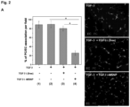
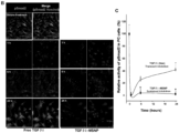
- Fig. 2 To investigate the effects of TGF ⁇ i on the co-migration of cultured human vascular smooth muscle cells (used as a surrogate PC) with human microvascular EC, we used a Matrigel assay 33 to compare the effect of TGF ⁇ i-loaded MSNP with the free inhibitor ( Fig. 2 ). ECs and PCs were stained with CellTracker TM Green and CellTracker TM Red, respectively. Fig. 2A demonstrates that the percentage of PC/EC association was significantly inhibited if the TGF ⁇ i was delivered by MSNP as compared to the inhibitory effect of free inhibitor at 1 ⁇ M. Representative fluorescent images of the cellular co-migration are shown on the right hand side of the figure.
- the growth factor Upon binding to type I/II TGF- ⁇ receptors, the growth factor induces the phosphorylation of the C-terminal SXS motif of the-associated kinases, Smad2 and Smad3. 34 Looking at Smad2 phosphorylation in PCs, we used an immunochemical technique that discerns anti-pSmad2 by a secondary FITC-conjugated antibody under a confocal microscope ( Fig. 2B ). 35 This demonstrated efficient and sustained inhibition of Smad2 phosphorylation for up to 24 h in PCs treated with TGF ⁇ i-MSNP compared to cells exposed to free inhibitor, which only suppressed pSmad2 for 6 h ( Fig. 2B ). Quantitative assessment of the green fluorescence intensity by Image J software confirmed a statistically significant and sustained inhibition of Smad2 phosphorylation by TGF ⁇ i-MSNP ( Fig. 2C ).
- TGF ⁇ i-MSNP was injected intravenously at inhibitor dose of 1 mg/kg (equivalent to a MSNP dose of 2 mg/kg) in nude mice expressing tumors ranging from 0.8 ⁇ 1.0 cm in diameter.
- inhibitor dose 1 mg/kg (equivalent to a MSNP dose of 2 mg/kg)
- tail vein injections of saline or the free inhibitor were used as controls.
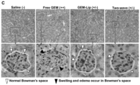
- dual-color immunohistochemistry was used for detecting CD31 staining in ECs with a green fluorescent dye (FITC), and NG2 in PCs with a red fluorescence marker (Alexa fluor 594) ( Fig. 3 ).
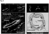
- TGFpi-loaded MSNP improves PDAC access of i.v . injected "hard” and “soft” nanoparticles in BxPC3 xenografts
- TGF ⁇ i-MSNP could improve the egress of nanocarriers at the BxPC3 xenograft site.
- 39 We tested this possibility through the use of "hard” (100 nm PEI-PEG coated MSNP) and “soft” (130 nm liposome) nanocarriers in an imaginable biodistribution experiment in nude mice.
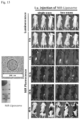
- These 2 nd wave particles were designed with near-infrared (NIR) tags to provide high photon penetration in animal tissues, as described previously by us. 28, 31 TEM or cryoEM images of the particles are provided in Figs. 5A and 5B . Detailed characterization is provided in Fig. 11 .
- BxPC-3 cells were stably transfected with a luciferase vector and used for obtaining bioluminescence images in the mice following intraperitoneal (i.p .) injection of d-Luciferin (Figs. 5A and 5B , first row).
- Initial reference images showed very low NIR background in the tumor-bearing animals ( Figs. 5A and 5B , second row).
- the tumor-bearing animals were i.v. injected with TGF ⁇ i-MSNP (containing 1 mg/kg of the inhibitor), followed after 1-2 h interval, by i.v. injection of 50 mg/kg NIR-labeled MSNPs or liposomes.
- prior TGF ⁇ i-MSNP administration resulted in a significant increase in the fluorescence intensity by 40 h, whereupon the signal was sustained for at least 60 h. Very little change in fluorescence intensity was observed in the tumor tissue receiving NIR-MSNP alone. Similar enhanced retention of a 50 nm amine-modified, PEGylated MSNP at the xenograft site was observed following 1 st wave TGF ⁇ i-MSNP administration as shown in Fig. 12 .
- Dylight 680-DMPE ⁇ 0.1%, w/w was incorporated into the lipid mixture.
- Fig. 5B first column
- there was a significant increase in fluorescence intensity at tumor site in the mice that were injected with TGF ⁇ i-MSNP Fig. 5B , second column).
- mice receiving the NIR-labeled MSNPs were sacrificed at 60 h post injection, and ex vivo fluorescence images were obtained for the tumor tissue as well as major organs ( FIG. 5C , upper panel). Consistent with the live animal imaging results, prior TGF ⁇ i-MSNP treatment was associated with increased fluorescence intensity in tumor tissue compared to animals receiving the 2 nd wave treatment alone. Both animal groups showed abundant particle distribution to the liver, spleen, lung, and the kidney. Following ex vivo imaging, the collected organs were weighed and used for Si elemental analysis by inductively coupled plasma optical emission spectrometry (ICP-OES). This allowed quantitative analysis of the particle distribution, expressed as a percentage (%) of the total mass of administered particles.
- ICP-OES inductively coupled plasma optical emission spectrometry
- TGF ⁇ i-MSNP improve the extent of liposome intratumoral distribution in BxPC3 xenografts
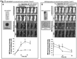
- Two-wave treatment improves the efficacy of gemcitabine treatment of BxPC3 tumors
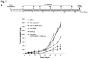
- xenograft-bearing nude mice were i.v . injected with 101 mg/kg of the liposomes (GEM dose: 20 mg/kg) 1-2 h after the i.v . injection of TGF ⁇ i-MSNP (TGF ⁇ i dose of 1 mg/kg), every 3-6 days for 38 days ( Fig. 7A ).
- the controls included animals injected with saline, free GEM, empty liposomes, TGF ⁇ i-MSNP alone, and GEM-liposomes alone. Since our previous studies have shown that empty MSNP lacks anticancer activity, 28, 31 we did not include this as a negative control in our animal experiments.
- the GEM liposome When comparing the effect on tumor size, the GEM liposome showed a significantly higher rate of tumor shrinkage than the free drug ( Fig. 7A ).
- This delay in observing the effect of prior TGF ⁇ i-MSNP treatment could be due to the effect of tumor stage, with the stromal effects and vascular access becoming a problem beyond 25 days. No tumor inhibition was found with saline treatment, TGF ⁇ i-MSNP alone or the use of empty liposomes ( Fig. 7A ).
- the safety of nanocarrier delivery system is of key importance in the assessment of this therapeutic platform. This includes the inherent safety of the carrier as well as the possible benefits that may accrue due to drug encapsulation. Safety assessment was performed by monitoring total body weight, blood chemistry, and histological examination of major organs. Compared to saline-treated BxPC3 tumor-bearing mice, no significant body weight changes were observed during the administration of empty liposomes, GEM-liposomes, or TGF ⁇ i-MSNP plus GEM-liposomes. In contrast, animals receiving free GEM administration showed a reduced weight gain ( Fig. 7B ).
- TGF ⁇ i-MSNP treatment was used to initially target the tumor stroma to decrease PC coverage of EC, followed by the delivery of GEM-laden liposomes that were effectively distributed throughout the tumor tissue, resulting in enhanced killing of the cancer cells after a window of 25 days following treatment.
- both particle waves were optimally designed to prolong circulation time in the blood, reduce RES uptake, and carry an effective drug payload to the cancer site.
- the co-polymer coated MSNP could deliver a high load of a TGF ⁇ i, which was supramolecularly attached to PEI, and through slow release could interfere in PCs adherence to the tumor vasculature at the xenograft site.
- nanocarriers for drug delivery, including the consideration of an engineered approach towards specific barriers, which can be targeted by independent waves of therapy that ultimately provide effective of killing and elimination of the cancer tissue.
- nanotherapeutics 44,45 or combination therapy 7, 14, 46, 47 with the view to improve systemic drug delivery through increased blood vessel permeability, tumor penetration or reducing the effect of drug inactivation enzymes, most of the research efforts concentrated on cancer cell killing with few efforts being directed to the cancer microenvironment. 31
- the tumor microenvironment is a very complex system that differs from the normal tissue environment and significantly influences the efficacy of nano delivery systems.
- biophysicochemical factors hypooxia, acidosis, high interstitial fluid pressure
- heterogeneous cellular components other than cancer cells endothelial cells and pericytes, cancer-associated fibroblasts, and inflammatory cells
- non-cellular components extracellular matrix, matrix metalloproteinase, soluble growth factors and their receptors, and integrins.
- these heterogeneous components could be the target(s) of an engineered approach.
- One example is the use of macrolide-modified gold nanorods that were designed to target and activate antitumor potential of macrophages.
- PEGPH20 PEGylated hyaluronidase PH20
- IFP interstitial fluid pressure
- the TGF- ⁇ superfamily plays an important role in cancer biology. 24 This includes a role in tumor neo-angiogenesis in which the interaction of PCs with ECs play a role in formation of intact blood vessels. 17
- the effects of inhibiting the TGF- ⁇ signaling pathway has been demonstrated in multiple in vitro and in vivo models, i.e. tumor xenograft models, a retinal vascular model, and a 3D PC/EC co-culture model. 7, 18, 22, 55
- these studies indicate that TGF- ⁇ maintains the integrity and function of the microvasculature while interference in this pathway often leads to dissociation of EC from PC and impaired EC barrier function.
- TGF ⁇ i promotes vascular access and accumulation of nanoparticles and macromolecules in BxPC3 subcutaneous xenograft model and OCUM-2MLN orthotopic gastric cancer model. 7, 18
- TGF- ⁇ negatively regulates local tumor immune responses and one can envisage that TGF ⁇ i-MSNP may promote the function of tumor antigen specific CD8 + T cells in the local immunosuppressive tumor microenvironment.
- a liposomal carrier In the case of a liposomal carrier, a high loading capacity ( ⁇ 20%) for GEM was achieved by creating an ammonium sulfate gradient in the liposome. This allowed intra-liposomal retention of the drug, which is protonated after diffusion through the liposomal membrane. 40 It has also been shown that the encapsulated GEM is stabilized as a gel-like precipitate inside the liposome ( Fig. 10 ). 40
- Example II drug(s)-laden lipid bilayer coated MSNP
- near infrared labeled particles are synthesized for in vivo biodistribution studies in tumor xenograft bearing nude mice, and in vivo efficacy tests are carried out. It is expected that the in vivo efficacy can be at least as effective as for the subunit structures described in present Example I.
- the synthesis of the 50 nm MSNP core was carried out as previously described by us, using a sol-gel chemistry procedure. 28, 31
- the particle surface was further modified using electrostatic attachment of a 1.8 kD PEI polymer, which was subsequently used for covalent attachment of 5 kD PEG.
- To perform PEI coating 10 mg of MSNP was suspended in 1 mL of 2.5 mg/mL PEI 1.8 kD ethanolic solution. The solution was sonicated and stirred for 30 min. The particles were further washed in ethanol to remove excess PEI and trace amount of surfactant.
- the PEI-coated particle was subsequently transferred into 1.5 mL of DMF, mixed with 50 mg of activated poly(ethylene glycol) methyl ether (m-PEG, MW 5 kD), and stirred for 24 h.
- the nanoparticles were washed with DMF and ethanol and resuspended in water.
- the NIR fluorescent dye DyLight 680 NHS ester was used for particle labeling. 10 mg particles were suspended in 1mL of DMF and mixed with 0.1 mg of Dylight 680. The reaction took place under an inert atmosphere during stirring at room temperature for 12 h. The particles were centrifuged and washed with deionized water. 28
- Loading capacity (%, w/w) [(Total minus non-encapsulated weight of LY364947)/(weight of MSNPs)] ⁇ 100%.
- Loading capacity (%, w/w) [(Total minus non-encapsulated weight of LY364947)/(weight of MSNPs)] ⁇ 100%.
- the drug release was studied in deionized water, saline containing 2% fetal calf serum or DMEM supplemented with 10% FCS for time periods ranging from 0-72 h at 37°C.
- Release percentage (%) [(the weight of LY364947 in the supematants)/(the total weight of attached LY364947 at the starting point)] ⁇ 100%.
- HDME Human microvascular endothelial cell
- ECM endothelial cell medium
- ScienCell Carlsbad, CA
- penicillin 100 ⁇ g/mL streptomycin
- HSM Human smooth muscle
- the PCs were cultured in ATCC-formulated F-12K medium containing 0.05 mg/mL ascorbic acid, 0.01 mg/mL insulin, 0.01 mg/mL transferring, 10 ng/mL sodium selenite, 0.03 mg/mL endothelial cell growth supplement, 10 mM 4-(2-hydroxyethyl)-1-piperazineethanesulfonic acid (HEPES), 10 mM 2-[(2-Hydroxy-1,1-bis(hydroxymethyl)ethyl)amino]ethanesulfonic acid (TES), and 10% FBS.
- HEPES 4-(2-hydroxyethyl)-1-piperazineethanesulfonic acid
- TES 2-[(2-Hydroxy-1,1-bis(hydroxymethyl)ethyl)amino]ethanesulfonic acid
- FBS FBS
- BxPC-3 cells were purchased from ATCC and cultured in Dulbecco's modified eagle medium (DMEM) (Carlsbad, CA) containing 10% FBS, 100 U/mL penicillin, 100 ⁇ g/mL streptomycin, and 2 mM L-glutamine.
- DMEM Dulbecco's modified eagle medium
- the Matrigel assay was performed using a modified method in the literature. 33 In order to distinguish the PCs and ECs in the Matrigel assay, HOME cells (10 4 cells/mL) and HSM cells (5 ⁇ 10 3 cells/mL) were first stained by CellTracker TM Green CMFDA (Invitrogen, Grand Island, NY) and CellTracker TM Red CMTPX (Invitrogen, Grand Island, NY) according the manufacture's instruction 24 h before experiment.
- ECs were treated with 2 ng/mL of TGF- ⁇ for 3 h and PCs were treated with free TGF- ⁇ or TGF ⁇ i-MSNP at inhibitor dose of 1 ⁇ M for 3 h. Subsequently, both cell types were co-cultured in Matrigel-coated 6-well plates for further incubation of 16 h at 37°C. PC/EC adhesions were quantitatively determined from five fields of three independent samples with the fluorescent microscope (Zeiss, Germany).
- Smad2 activation was determined using an immunofluorescent staining in 8-well chamber slides in which 4 ⁇ 10 4 PCs were cultured in each well containing 0.4 mL culture medium. 16 h post cell seeding, PCs were treated with 2 ng/mL TGF- ⁇ for 3 h. Subsequently, the cells were treated with TGF ⁇ i-laden MSNP at the inhibitor dose of 1 ⁇ M for 1-24 h. For comparison, free TGF ⁇ i was used to treat the cells at identical dose. Subsequently, PCs cells grown on chamber slides were fixed, permeabilized, and stained for pSmad2 with a standard immunocytochemistry protocol.
- pSmad2 staining was performed by using a 1:500 dilution of primary anti-pSmad2 antibody (Abeam, Cambridge, MA) for 16 h at 4 °C. This was followed by a 1:500 diluted FITC-conjugated secondary antibody (Santa Cruz, USA) for 1 h at room temperature. The nuclei were stained with Hoechst 33342. Slides were visualized under a confocal microscope (Leica Confocal 1P/FCS). The signal intensity of green channel, revealing activated Smad2, was calculated by Image J software (version 1.37c, NIH).
- Athymic BALB/c nu/nu female mice (6 weeks) were purchased from the Charles River Laboratory and maintained under pathogen-free conditions. All animal experiments were performed using protocols approved by the UCLA Animal Research Committee. For tumor visualization in mice using optical imaging, permanent luciferase transfection using lentivirus was performed in BxPC3 cells. To grow tumor xenograft, BxPC3-luc cell suspension (0.1 mL, 5 ⁇ 10 6 cells/mL) was injected subcutaneously into nude mice. For efficacy experiment, the mice were used for various treatments 7 days post tumor implantation. To perform imaging experiment, the tumor bearing animals were used 3-4 weeks after tumor implantation of the tumor size of 0.8-1 cm in diameter.
- the NIR-MSNP or NIR-liposome were used.
- One to two hour after TGFpi-laden MSNP injection the mice were intravenously administrated with 50 mg/kg of NIR dye-labeled particles. The fluorescence images were taken at indicated time points. This treatment was compared to the mice received i.v. injection of NIR dye-labeled MSNP or liposome alone at 50 mg/kg.
- the tumor tissue together with major organs (heart, lung, spleen, liver, kidney, brain and cardiac muscle) were collected and used for ex vivo image.
- BxPC3 tumor-bearing mice (tumor size: 0.8-1 cm in diameter) were intravenously treated with TGF ⁇ i-laden MSNP at inhibitor dose of 1 mg/kg (MSNP dose: 2 mg/kg).
- the tumor biopsies were rapidly collected 2 h post injection, washed in PBS and immediately fixed with 2.5% glutaraldehyde in PBS at room temperature for 2 h and stored at 4 °C for overnight. Further sample preparation and sectioning were performed by Electron Microscopy Services Center in Brain Research Institute at UCLA. Briefly, after secondary fixation in 1% O s O 4 in PBS, the samples were dehydrated in a graded ethanol series, treated with propylene oxide, and embedded in resin. Approximately 60-70 nm thick sections were cut on a Leica ultramicrotome and picked up on Formvar-coated copper grids. The sections were examined in a CM120 electron microscope (Philips).
- the tumor tissues were rapidly embedded by OCT reagent before sectioning to provide 4 ⁇ m thick slices.
- the slices were washed three times in PBS and fixed.
- For ECs staining, the sections were first incubated with rat-anti-mouse CD31 monoclonal antibody (1:500) at 4 °C overnight. After removal of the primary antibody and washing in PBS for three times, FITC-conjugated goat-anti-rat IgG (1:500) was added and incubated at room temperature for 1 h.
- mice were randomly divided into six groups. These groups were used for comparing the effects of saline, free liposome, TGF ⁇ i-MSNP alone, free GEM, GEM-Lip alone, and two-wave treatment, respectively.
- Each animal in two-wave group received i.v . injection of TGF ⁇ i-MSNP at inhibitor dose of 1 mg/kg (MSNP dose: 2 mg/kg) followed by a liposome dose of 101 mg/kg (GEM dose: 20 mg/kg) with 1-2 h interval, during each injection, 6 injections in a 38 days time period ( Fig. 6A ).
- the free GEM and GEM loaded liposome groups received the same drug dose in the absence of TGF ⁇ i-MSNP pre-treatment.
- phospholipids products were purchased from Avanti Polar Lipids, either powder form or chloroform solution without further purification.
- Gemcitabine (GEM) was purchased from Sigma Aldrich.
- the liposomal mini-extruder, holder/heating block, and different size PC Membranes (0.4 and 0.1 ⁇ m) were purchased from Avanti Polar Lipids.
- the liposomal composition is shown in Table 1.
- Liposomes were prepared through a thin film-rehydration procedure.
- GEM was encapsulated using an equilibrium exchange method for liposomal trapping ( BMC Cancer 2004, 4, 63 ).
- Inclusion of ammonium sulfate inside the liposome generates a transmembrane gradient, which is responsible for protonation of the amphipathic GEM molecules which can freely diffuse through the liposome bilayer.
- the GEM molecules become hydrophilic, which prevents their escape from the liposome.
- the drug becomes stabilized as gel-like drug precipitate [i.e. (GEM-NH3)2SO4] inside the liposomes.
- FIG. 10A provides a flow chart showing the major steps of GEM loading, and in order to obtain optimal drug loading, each step had to be systematically investigated through to find the best possible liposome formulation, ammonium sulfate concentration, extent of salt removal, drug loading time, temperature, and amount of free GEM, etc ( Fig. 10B ).
- Table 1 Formulation of different linosome for GEM loading.
- lipid mixture for each formulation (#1-#6) was dissolved in a round-bottomed flask, using chloroform as solvent (concentration: 2.5 ⁇ 10 mg/mL).
- chloroform as solvent
- lipid films were placed in a chemical hood overnight to remove trace amounts of organic solvent impurities.
- the lipid films can be stored at -80 °C under inert atmosphere (i.e. argon, nitrogen) for at least 2 months.
- fluorescein-DHPE i.e. texas red
- lipid films were incubated with indicated concentrations of ammonium sulfate solution (ranging from 0-360 mM) at 60 °C for 1 h, with vigorous stirring.
- the multi-lamellar particles were repeatedly extruded, first at a 400 nm pore size (3 times), then at a 100 nm pore size 11 times, while being kept at 60 °C on a heating block.
- ultra-speed centrifugation at 100,000 rpm or repeated dialysis against isotonic glucose solution was performed.
- the resulting mono-disperse, unilamellar vesicles were suspended in an isotonic solution of GEM hydrochloride at different free GEM concentrations (0.2-5 mg/mL).
- the encapsulated GEM was quantified by UV absorption at 270 nm using a microplate reader (Molecular Device).
- the elemental phosphorus (P) was quantified by ICP-OES (ICPE-9000, Shimadzu).
- Drug loading capacity (%) was determined as (amount of GEM)/(amount of liposome) ⁇ 100%.
- the size of the drug-laden liposomes was characterized by dynamic light scattering at a liposome concentration of 100 ⁇ g/mL (ZetaPALS, Brookhaven Instruments Co.).
- the zeta potential of the liposome was measured by a ZetaPALS (Brookhaven Instruments Co.).
- the morphology of drug-laden liposome was visualized by cyroEM (TF20, FET).
- FIG. 10 D. FULL PANEL OF NIR IMAGES TO COVER ALL THE TIME POINTS IN MICE INJECTED WITH SECOND WAVE NIR-MNSP AS SHOWN IN FIG. 5A are shown in Fig. 10
- TGF ⁇ i-MSNP treatment 50 nm amine-modified, PEGylated MSNP with a Dylight680 NIR tag were tested in xenograft tumors.
- the same experiment was performed as described in Fig. 5A . Briefly, the BxPC3-luc tumor-bearing animals were pre-treated by i.v. injection of TGF ⁇ i-MSNP (inhibitor: 1 mg/kg; particle: 2 mg/kg) followed by i.v. injection of 50 mg/kg of the 50 nm MSNP after a time lag of 1-2 h. The in vivo biodistribution was compared with the mice receiving i.v.
- the biochemical parameters were assayed by the UCLA Division of Laboratory Animal Medicine (DLAM) diagnostic laboratory services. These parameters include bicarbonate (CO2), cholesterol (CHOL), inorganic phosphorus (PHOS), aspartate aminotransferase (AST), direct serum bilirubin (DBILI), total bilirubin (TBILI), blood urea nitrogen (BUN), creatine kinase (CK), creatinine (CREAT), gamma glutamyl transferase (GGT), glucose (GLU), total protein (TP), albumin (ALB), alkaline phosphatase (ALP), calcium (CA), alanine aminotransferase (ALT), and BUN-to-creatinine ratio (BUN-CR).
- CO2 bicarbonate
- PHOS inorganic phosphorus
- AST aspartate aminotransferase
- DBILI direct serum bilirubin
- TBILI total bilirubin
- BUN blood urea nitrogen
- N-(2-Aminoethyl)-3-aminopropyltrimethoxysilane was purchased from Gelest (Morrisville, PA). Cetyl trimethylammonium bromide (CTAB, 95%), tetraorthoethylsilicate (TEOS, 98%), 3-(trihydroxysilyl) propyl methylphosphonate (42% in H 2 O), Pluronic F127, polyethyleneimine (PEI, 1.2 kD), 4-(dimethylamino)pyridine (99%), N,N'-disuccinimidyl carbonate (95%), poly(ethylene glycol) methyl ether (m-PEG, MW 5 kD), phthalic anhydride (99%), transforming growth factor- ⁇ 1 (TGF- ⁇ ) and gemcitabine hydrochloride (purity: ⁇ 98%) were purchased from Sigma Aldrich (St.
- Amine-reactive near-infrared Fluor Dylight 680 NHS ester was purchased from Thermo Scientific (Rockford, IL).
- D-Luciferin was purchased from Xenogen (Alameda, CA).
- Cell Tracker TM Red CMTPX, Cell Tracker TM Green CMFDA (5-Chloromethylfluorescein Diacetate), DPBS solution, L-glutamine, penicillin, streptomycin, and DMEM culture medium were obtained from Invitrogen.
- Fetal bovine serum (FBS) was purchased from Atlanta Biologicals.
- Anti-Smad2 (phospho S467) antibody was purchased from Abeam.
- Anti-CD31 antibody and Matrigel TM -Basement Membrane Matrix was purchased from BD Bioscience.
- TGF ⁇ i Transforming growth factor type I receptors kinas inhibitor
- Phospholipids and cholesterol were purchased from Avanti Polar Lipids (Alabaster, Alabama). All reagents were used without further purification.
- Samples were characterized for morphology, size distribution and surface charge.
- the morphologies and primary sizes of MSNP particle were characterized using a transmission electron microscope (JEOL JEM 2010, JEOL USA, Inc., Peabody, MA).
- the morphologies of liposome were characterized using cyroEM (TF20, FET).
- Hydrodynamic size and zeta potential in solution were measured by ZetaSizer Nano (Malvern Instruments Ltd., Worcestershire, UK). All of the measurements were performed with the samples suspended in filtered water or saline at 100 ⁇ g/mL nanoparticle concentration.
- the collected tumor and organs were used for Si elemental analysis using ICP-OES. Briefly, each tissue was accurately weighed and soaked in concentrated 1 mL HNO 3 and 0.5 mL 30% H 2 O 2 for overnight. This yellow color digestion solution was heated at 80 °C for 1 h in the subsequent day. Dropwise addition of H 2 O 2 solution was used to drive off nitrogen oxide vapor until the digestion lipid turns colorless. 2% HNO 3 was used to dilute the sample into 10 mL volume and the resulting sample was analyzed by ICP-OES.
- mice were sacrificed on the 38 th day and serum was collected by centrifuging the whole blood at 5,000 rpm for 15 min.
- the biochemical parameters were assayed by UCLA Division of Laboratory Animal Medicine (DLAM) diagnostic laboratory services. Appropriate size sections of the tumor, liver, kidney, and spleen were fixed in 10% formalin and then embedded into paraffin. Tissue sections of 4 ⁇ m thickness were mounted on glass slides by the UCLA Division of Laboratory Animal Medicine (DLAM) diagnostic laboratory services. The sections were stained with hematoxylin-eosin (H&E) and examined by light microscopy.
- H&E hematoxylin-eosin
- An MSNP coated with a phospholipid bilayer which can provide a GEM loading capacity of ⁇ 40% (drug/particle, w/w).
- the MSNP core is synthesized by a modified surfactant-templated sol-gel method in aqueous solution at relatively low temperature.
- a lipid membrane was dehydrated with a GEM-containing MSNP suspension, using controlled energy input (e.g. sonication). This led to rapid coating and sealing of the MSNPs, encapsulating a high w/w content GEM in one step.
- pancreatic cancer can in many cases be resistant to individual chemotherapeutic agents, including GEM, via acquired or de novo mechanisms, there is a need to consider drugs that provide a synergistic effect with GEM, e.g., when co-administered with GEM.
- a recent successful clinical trial has allowed Abraxane R (paclitaxel/albumin complex) to be combined with GEM in untreated pancreatic patients with metastatic disease. This combination has resulted in a statistically significant improvement in overall survival compared to patients receiving GEM alone.
- paclitaxel is believed to be capable of increasing the intratumoral GEM content by reducing the activity of cytidine deaminase (CDA), a key enzyme that metabolically inactivates GEM and reduces its circulatory half-life to minutes. Since the hydrophobic paclitaxel molecules can be co-dissolved in a lipophilic organic solution, the presence of a lipid coat on MSNP allows co-packaging of paclitaxel in a phospholipid bilayer coating on GEM-laden particles.
- CDA cytidine deaminase
- a low temperature sol-gel chemistry procedure is used to obtain highly uniform (e.g., monodisperse) and colloidally stable MSNPs.
- MSNPs prepared via a low temperature sol-gel method can exhibit improved size control than those prepared by, for example, an aerosol-assisted self-assembly method.
- Particles prepared by an aerosol-assisted self-assembly method may exhibit a wide size distribution, and may not be uniformly bio-available, e.g., at tumor sites.
- the monodisperse and size-controlled MSNPs prepared by a sol-gel method may show greater potential for in vivo use.
- a submicron structure (such as an MSNP coated with a phospholipid bilayer) can provide simultaneous delivery of a drug, and: an agent which stabilizes the drug against metabolic degradation; an agent which facilitates the delivery of the drug to a target cell, tissue, organ or tumor; an agent which acts synergistically with the drug; one or more additional therapeutic agents; or a combination thereof.
- a GEM-laden MSNP provides for simultaneous delivery of paclitaxel in a single carrier, i.e., a submicron structure which includes both GEM (e.g., within the pores) and paclitaxel (e.g., associated with the phospholipid bilayer). Including more than one therapeutic agent in a single particle allows precise control over the doses and dosage ratios of the therapeutic agents delivered to the site of release (e.g., a tumor cell).
- the chemicals were obtained from Sigma Aldrich and used without further purification.
- CCTAC cetyltrimethylammonium chloride
- MSNP 10 mg MSNP was suspended in 20 mg GEM ethanol/water (7:3, v/v). The mixture was shaken for at least 24 hour at room temperature. The drug-laden particles were collected by centrifugation (prior to pore sealing) and immediately used for lipid coating. Particles were not washed between drug loading and lipid coating.
- the lipid membrane was dehydrated using GEM-containing MSNP suspension with controlled energy input (e.g. sonication), leading to lipid-coated and pore-sealed MSNPs that contained high GEM content in one step .
- Lipid membrane Lipid mixture was dissolved in a round-bottomed flask, using chloroform as solvent (concentration: 2.5 ⁇ 10 mg/mL). Different liposomal compositions can be selected based on drug, targeting purpose, and other considerations. Paclitaxel can be co-dissolved in the organic solution. Lipid films were made by evaporation for ⁇ 1 h, using a rotary evaporator connected to a vacuum system at room temperature.
- lipid films were placed in a chemical hood for at least 2 hours to remove trace amounts of organic solvent impurities.
- the lipid films can be stored at -80 °C under an inert atmosphere for at least 2 months.
- Fluorescently labeled lipid film can be made by co-dissolving 0.1% w/w fluorescein-DHPE (i.e. Texas red) with the lipids.
- lipid films were incubated with the GEM-laden MSNP aqueous solution at 40 °C for 20 min, with continuous water-bath sonication. The mixture was spun at 1500 rpm for 5 min and the supernatant collected, which contain lipid-coated MSNP, free GEM, and free liposome. A centrifugal filter unit with 10,000 MW cutting off size was used to remove any un-encapsulated GEM.
- Sample characterization The samples were fully characterized for morphology using TEM and cryoEM, size and zeta potential, surface area, loading capacity and release profile, and Si/P elemental ratio using ICP-OES.
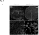
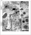
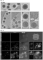
- FIG. 14 shows a cryoEM image (TF20, FET) of lipid-coated MSNP.
- the upper box shows the ⁇ 70 nm MSNP synthesized using the procedure in section D.
- the zoom-in image (region 1) shows an ordered mesoporous structure and primary particle size of ⁇ 60 nm.
- the lower panel shows an intact lipid coating on the silica surface.
- the zoom-in images (regions of ii ⁇ iv) showed a lipid thickness of 7.0 nm on the silica surface, which is very close to the thickness of lipid bilayer in liposome (7.1 nm).
- the HPLC analysis and microplate reader analysis showed that the loading capacity of GEM in lipid coated MSNP was ⁇ 40% (w/w). This is an approximately 2-fold improvement compared to GEM-laden liposome.
- Fig. 15 demonstrates cellular uptake of the lipid bilayer-coated MSNP. Confocal microscopy was used to demonstrate the cellular uptake of FITC-labeled paclitaxel (in green) loaded DHPE (red)-labeled lipid-coated MSNP in Panc1 cells. Panc1 cells were treated with 40 ⁇ g/mL nanoparticles for the indicated time periods. The merged image at 3 hours showed the red-labeled lipid bilayer in association with green-labeled paclitaxel, which indicated that the particle successfully delivered paclitaxel into the cells, while the lipid coating remained intact.
- the merged images at 20 hours show amore dispersed pattern of intracellular paclitaxel distribution and lower level of co-localization, providing evidence of intracellular paclitaxel release from the MSNP's lipid coating where the hydrophobic drug was packaged.
- the nuclear were stained by Hoechst 33342 in blue.
- C3 Paclitaxel-laden lipid coated MSNP down-regulated the expression of cytidine deaminase (CDA), a key enzyme in metabolic inactivation of GEM in pancreatic cancer cells.
- CDA cytidine deaminase
- Panc1 cells were treated with paclitaxel-laden lipid coated MSNP at 25 ⁇ g/mL for different lengths of time (0-24 hours), or for 24 hours using different particle concentrations (0-200 ⁇ g/mL).
- the expression of CDA and heme oxygenase-1 (HO-1, which is an oxidative stress protein induced in response to paclitaxel induced oxidative challenge) were determined by Western blotting.
- the data demonstrated that paclitaxel-laden lipid coated MSNP significantly lowered the CDA expression and induced HO-1 level in a dose- and time-dependent manner.
- the data showed that 24-hour incubation at particle concentration of 25 ⁇ g/mL could lead to the maximal effects in Panel cells.
Landscapes
- Health & Medical Sciences (AREA)
- Chemical & Material Sciences (AREA)
- Life Sciences & Earth Sciences (AREA)
- Veterinary Medicine (AREA)
- Animal Behavior & Ethology (AREA)
- General Health & Medical Sciences (AREA)
- Public Health (AREA)
- Pharmacology & Pharmacy (AREA)
- Medicinal Chemistry (AREA)
- Epidemiology (AREA)
- Molecular Biology (AREA)
- Biochemistry (AREA)
- Engineering & Computer Science (AREA)
- Bioinformatics & Cheminformatics (AREA)
- Inorganic Chemistry (AREA)
- Dispersion Chemistry (AREA)
- Physics & Mathematics (AREA)
- Biomedical Technology (AREA)
- Nanotechnology (AREA)
- Optics & Photonics (AREA)
- Chemical Kinetics & Catalysis (AREA)
- General Chemical & Material Sciences (AREA)
- Nuclear Medicine, Radiotherapy & Molecular Imaging (AREA)
- Organic Chemistry (AREA)
- Medicinal Preparation (AREA)
- Pharmaceuticals Containing Other Organic And Inorganic Compounds (AREA)
- Silicates, Zeolites, And Molecular Sieves (AREA)
Claims (10)
- Partikel mit einer Größe im Submikron-Bereich, das Folgendes umfasst:einen Siliciumdioxid-Körper, der eine Oberfläche aufweist und mehrere Poren definiert, die zur Aufnahme von Molekülen darin geeignet sind; undeine Phospholipid-Doppelschicht, mit der die Oberfläche überzogen ist, wobei die Phospholipid-Doppelschicht aufgetragen wird, indem der Siliciumdioxid-Körper mit einem Phospholipid-Film in Kontakt gebracht wird, wodurch die Phospholipid-Doppelschicht die mehreren Poren stabil versiegelt;wobei das Partikel mit einer Größe im Submikron-Bereich ein erstes Therapeutikum innerhalb der Poren einschließt, wobei das erste Therapeutikum Gemcitabin (GEM) ist;wobei das Partikel mit einer Größe im Submikron-Bereich ein zweites, verschiedenes Therapeutikum einschließt, das mit der Phospholipid-Doppelschicht assoziiert ist, wobei das zweite Therapeutikum Paclitaxel ist;wobei die Dosis und das Verhältnis von Gemcitabin zu Paclitaxel gesteuert werden, indem der Siliciumdioxid-Körper mit Gemcitabin beladen wird, bevor die Phospholipid-Doppelschicht aufgetragen wird, und der Phospholipid-Film vor dem Auftragen auf den Siliciumdioxid-Körper das Paclitaxel enthält;wobei das Partikel mit einer Größe im Submikron-Bereich eine maximale Abmessung von weniger als einem Mikrometer aufweist undwobei das Partikel mit einer Größe im Submikron-Bereich ein Element einer monodispersen Population von Partikeln mit einer Größe im Submikron-Bereich ist.
- Partikel mit einer Größe im Submikron-Bereich nach Anspruch 1, wobei das Gemcitabin und das Paclitaxel, das erste Therapeutikum, synergistisch wirken.
- Partikel mit einer Größe im Submikron-Bereich nach Anspruch 1, wobei das Partikel eine GEM-Beladungskapazität von ~40 % Arzneimittel/Partikel (w/w) bereitstellt.
- Partikel mit einer Größe im Submikron-Bereich nach einem der vorhergehenden Ansprüche, wobei das Partikel mit einer Größe im Submikron-Bereich mit einer systemischen Verabreichung kompatibel ist.
- Partikel mit einer Größe im Submikron-Bereich nach einem der Ansprüche 1 bis 4 zur Verwendung als Therapeutikum.
- Partikel mit einer Größe im Submikron-Bereich nach einem der Ansprüche 1 bis 4 zur Verwendung bei der Behandlung eines Krebses.
- Partikel mit einer Größe im Submikron-Bereich zur Verwendung nach Anspruch 6, wobei der Krebs ein Krebs ist, der ausgewählt ist aus der Gruppe bestehend aus einem duktalen Adenokarzinom des Pankreas (PDAC), Prostatakrebs und einem Glioblastom.
- Partikel mit einer Größe im Submikron-Bereich zur Verwendung nach Anspruch 6, wobei der Krebs ein Pankreaskrebs ist.
- Partikel mit einer Größe im Submikron-Bereich zur Verwendung nach einem der Ansprüche 6 bis 8, wobei das Partikel mit einer Größe im Submikron-Bereich einem Individuum systemisch verabreicht wird.
- Partikel mit einer Größe im Submikron-Bereich zur Verwendung nach einem der Ansprüche 6 bis 8, wobei das Partikel mit einer Größe im Submikron-Bereich einem Individuum über einen Weg verabreicht wird, der ausgewählt ist aus der Gruppe bestehend aus einer intravenösen, intraarteriellen, intraperitonealen, intramuskulären und subkutanen Verabreichung.
Priority Applications (1)
| Application Number | Priority Date | Filing Date | Title |
|---|---|---|---|
| EP24156880.7A EP4378461A3 (de) | 2013-03-05 | 2014-03-05 | Mit einer lipiddoppelschicht beschichtete mesoporöse siliciumdioxidnanoteilchen mit hoher ladekapazität für ein oder mehrere antikrebsmittel |
Applications Claiming Priority (3)
| Application Number | Priority Date | Filing Date | Title |
|---|---|---|---|
| US201361773013P | 2013-03-05 | 2013-03-05 | |
| US201361858388P | 2013-07-25 | 2013-07-25 | |
| PCT/US2014/020857 WO2014138278A1 (en) | 2013-03-05 | 2014-03-05 | Lipid bilayer coated mesoporous silica nanoparticles with a high loading capacity for one or more anticancer agents |
Related Child Applications (1)
| Application Number | Title | Priority Date | Filing Date |
|---|---|---|---|
| EP24156880.7A Division EP4378461A3 (de) | 2013-03-05 | 2014-03-05 | Mit einer lipiddoppelschicht beschichtete mesoporöse siliciumdioxidnanoteilchen mit hoher ladekapazität für ein oder mehrere antikrebsmittel |
Publications (3)
| Publication Number | Publication Date |
|---|---|
| EP2964201A1 EP2964201A1 (de) | 2016-01-13 |
| EP2964201A4 EP2964201A4 (de) | 2016-08-24 |
| EP2964201B1 true EP2964201B1 (de) | 2024-02-14 |
Family
ID=51491913
Family Applications (2)
| Application Number | Title | Priority Date | Filing Date |
|---|---|---|---|
| EP14760467.2A Active EP2964201B1 (de) | 2013-03-05 | 2014-03-05 | Mit einer doppellipidschicht beschichtete mesoporöse siliciumdioxidnanoteilchen mit hoher ladungskapazität für ein oder mehrere antikrebsmittel |
| EP24156880.7A Pending EP4378461A3 (de) | 2013-03-05 | 2014-03-05 | Mit einer lipiddoppelschicht beschichtete mesoporöse siliciumdioxidnanoteilchen mit hoher ladekapazität für ein oder mehrere antikrebsmittel |
Family Applications After (1)
| Application Number | Title | Priority Date | Filing Date |
|---|---|---|---|
| EP24156880.7A Pending EP4378461A3 (de) | 2013-03-05 | 2014-03-05 | Mit einer lipiddoppelschicht beschichtete mesoporöse siliciumdioxidnanoteilchen mit hoher ladekapazität für ein oder mehrere antikrebsmittel |
Country Status (3)
| Country | Link |
|---|---|
| US (3) | US10828255B2 (de) |
| EP (2) | EP2964201B1 (de) |
| WO (1) | WO2014138278A1 (de) |
Families Citing this family (28)
| Publication number | Priority date | Publication date | Assignee | Title |
|---|---|---|---|---|
| US9993437B2 (en) | 2007-12-06 | 2018-06-12 | The Regents Of The University Of California | Mesoporous silica nanoparticles for biomedical applications |
| US20120207795A1 (en) | 2010-07-13 | 2012-08-16 | The Regents Of The University Of California | Cationic polymer coated mesoporous silica nanoparticles and uses thereof |
| US10220004B2 (en) | 2011-07-14 | 2019-03-05 | The Regents Of The University Of California | Method of controlled delivery using sub-micron-scale machines |
| EP2964201B1 (de) | 2013-03-05 | 2024-02-14 | The Regents of the University of California | Mit einer doppellipidschicht beschichtete mesoporöse siliciumdioxidnanoteilchen mit hoher ladungskapazität für ein oder mehrere antikrebsmittel |
| US9574135B2 (en) * | 2013-08-22 | 2017-02-21 | Nanoco Technologies Ltd. | Gas phase enhancement of emission color quality in solid state LEDs |
| LT3138555T (lt) | 2014-04-30 | 2021-03-25 | Fujifilm Corporation | Liposomų kompozicija ir jų gamybos būdas |
| CN104983716B (zh) * | 2015-07-20 | 2018-04-27 | 广西医科大学 | 肿瘤细胞膜/核膜双靶向肿瘤纳米药物缓释系统及其制备与应用 |
| LU92784B1 (en) * | 2015-07-22 | 2017-01-31 | Luxembourg Inst Of Science And Tech (List) | Negatively charged self-assembling supported lipid bilayer on mesoporous silica nanoparticles, method of synthesis and use as a nanovector |
| WO2017078008A1 (ja) * | 2015-11-02 | 2017-05-11 | 富士フイルム株式会社 | ゲムシタビンリポソーム組成物を含む腫瘍治療剤およびキット |
| EP3399966B1 (de) * | 2016-01-08 | 2023-03-29 | The Regents of the University of California | Mesoporöse siliciumdioxidnanoteilchen mit lipiddoppelbeschichtung zur frachtlieferung |
| LU100023B1 (en) * | 2017-01-20 | 2018-07-30 | Luxembourg Inst Science & Tech List | Nanocapsules and method for manufacturing thereof |
| US11433143B2 (en) | 2017-05-18 | 2022-09-06 | The Regents Of The University Of California | Nano-enabled immunotherapy in cancer |
| EP3624810A4 (de) * | 2017-05-18 | 2021-02-17 | The Regents of The University of California | Nano-aktivierte immuntherapie bei krebserkrankungen |
| WO2019027905A1 (en) | 2017-07-31 | 2019-02-07 | January Therapeutics, Inc. | ORGANOPHOSPHATE DERIVATIVES |
| WO2019028387A1 (en) * | 2017-08-03 | 2019-02-07 | Rita Elena Serda | LIPOSOMAL COATED NANOPARTICLES FOR IMMUNOTHERAPY APPLICATIONS |
| EP3735250A4 (de) | 2018-01-01 | 2022-04-13 | The Regents Of The University Of California | Aufskalierungssynthese von silicasom-nanoträgern |
| JP2021514985A (ja) * | 2018-03-02 | 2021-06-17 | ジャニュアリー セラピューティクス,インク. | ナノ粒子組成物 |
| CN112334140A (zh) | 2018-06-20 | 2021-02-05 | 富士胶片株式会社 | 包含内含吉西他滨的脂质体组合物及免疫检查点抑制剂的组合医药 |
| WO2020068798A1 (en) | 2018-09-24 | 2020-04-02 | Guo Jimin | Living mammalian cells modified with functional modular nanoparticles |
| WO2020077451A1 (en) | 2018-10-16 | 2020-04-23 | Silicycle Inc. | Tunable process for silica capsules/spheres preparation and their use |
| US12208164B2 (en) | 2019-02-28 | 2025-01-28 | Unm Rainforest Innovations | Modular metal-organic polyhedra superassembly compositions |
| WO2020214741A1 (en) * | 2019-04-15 | 2020-10-22 | Cornell University | Functionalized silica nanorings, methods of making same, and uses thereof |
| US11708637B2 (en) | 2019-08-13 | 2023-07-25 | The Regents Of The University Of California | Methods of supporting a graphene sheet disposed on a frame support |
| AU2020367786A1 (en) * | 2019-10-14 | 2022-05-26 | The Regents Of The University Of California | Nano-enabled immunotherapy in cancer |
| WO2021173870A1 (en) * | 2020-02-27 | 2021-09-02 | University Of Washington | Composition and method to prepare long-acting injectable suspension containing multiple cancer drugs |
| LU102356B1 (en) * | 2020-12-30 | 2022-06-30 | Luxembourg Inst Science & Tech List | Stabilisation of carbonate calcium nanoparticles |
| WO2023147596A1 (en) * | 2022-01-31 | 2023-08-03 | Noureddine Achraf | Triplex nanoparticles |
| CN119236105B (zh) * | 2024-09-29 | 2025-09-05 | 中南大学 | 一种基于铜死亡的多功能抗肿瘤纳米药物递送系统及其制备方法与应用 |
Citations (1)
| Publication number | Priority date | Publication date | Assignee | Title |
|---|---|---|---|---|
| WO2012149376A2 (en) * | 2011-04-28 | 2012-11-01 | Stc.Unm | Porous nanoparticle-supported lipid bilayers (protocells) for targeted delivery and methods of using same |
Family Cites Families (33)
| Publication number | Priority date | Publication date | Assignee | Title |
|---|---|---|---|---|
| US2103412A (en) | 1936-10-01 | 1937-12-28 | Frederick J Gaertner | Finger ring |
| US4627413A (en) | 1985-10-29 | 1986-12-09 | Roca Technologies, Inc. | Three position fireplace cooker |
| US4737323A (en) | 1986-02-13 | 1988-04-12 | Liposome Technology, Inc. | Liposome extrusion method |
| DE4217353B4 (de) | 1992-05-26 | 2008-02-21 | Nimbus Biotechnologie Gmbh | Verfahren zur säulenchromatographischen Trennung von Proteinen mittels Lipidbilayer-beschichteter Silicagele |
| US5827533A (en) * | 1997-02-06 | 1998-10-27 | Duke University | Liposomes containing active agents aggregated with lipid surfactants |
| DE19814775C2 (de) | 1998-04-02 | 2001-04-19 | Nimbus Biotechnologie Gmbh | Verfahren zur Bestimmung der Lipid-Bindungskonstanten von Substanzen in wässriger Lösung an Oberflächen aus amphiphilen Molekülen |
| US7618565B2 (en) | 2001-08-16 | 2009-11-17 | Polytechnic Institute Of New York University | Lipobeads and their production |
| US20040005352A1 (en) * | 2002-04-16 | 2004-01-08 | Lopez Gabriel P. | Biologically functionalized porous microspheres |
| AU2003268087A1 (en) * | 2002-08-23 | 2004-03-11 | Ian Ma | Liposomal gemcitabine compositions for better drug delivery |
| US7563451B2 (en) * | 2003-07-22 | 2009-07-21 | Iowa State University Research Foundation, Inc. | Capped mesoporous silicates |
| CN103948545B (zh) | 2004-05-03 | 2017-10-03 | 益普生生物制药公司 | 用于药物输送的脂质体 |
| MX2007001362A (es) | 2004-08-03 | 2007-04-16 | Chemetall Gmbh | Procedimiento para revestir superficies metalicas con un revestimiento anticorrosivo. |
| WO2006032136A1 (en) | 2004-09-20 | 2006-03-30 | British Columbia Cancer Agency Branch | Free or liposomal gemcitabine alone or in combination with free or liposomal idarubicin |
| US20110104073A1 (en) | 2007-01-18 | 2011-05-05 | Qi Zeng | Iron/Iron Oxide Nanoparticle and Use Thereof |
| US9993437B2 (en) | 2007-12-06 | 2018-06-12 | The Regents Of The University Of California | Mesoporous silica nanoparticles for biomedical applications |
| US20100310465A1 (en) | 2008-01-23 | 2010-12-09 | The Regents Of The University Of California | Nano-devices having releasable seals for controlled release of molecules |
| WO2009094568A1 (en) | 2008-01-23 | 2009-07-30 | The Regents Of The University Of California | Nano-devices having impellers for capture and release of molecules |
| US20120021034A1 (en) | 2008-12-19 | 2012-01-26 | The Regents Of The University Of California | Structured silver-mesoporous silica nanoparticles having antimicrobial activity |
| WO2010078569A2 (en) * | 2009-01-05 | 2010-07-08 | Stc.Unm | Porous nanoparticle supported lipid bilayer nanostructures |
| US8992984B1 (en) | 2009-10-21 | 2015-03-31 | Stc.Unm | Protocells and their use for targeted delivery of multicomponent cargos to cancer cells |
| TWI383808B (zh) | 2009-11-23 | 2013-02-01 | Nat Univ Tsing Hua | 磷脂質包覆中孔洞二氧化矽奈米球的合成與其生醫應用 |
| WO2012009448A2 (en) | 2010-07-13 | 2012-01-19 | The Regents Of The University Of California | Cationic polymer coated mesoporous silica nanoparticles and uses thereof |
| US20120207795A1 (en) | 2010-07-13 | 2012-08-16 | The Regents Of The University Of California | Cationic polymer coated mesoporous silica nanoparticles and uses thereof |
| US10220004B2 (en) | 2011-07-14 | 2019-03-05 | The Regents Of The University Of California | Method of controlled delivery using sub-micron-scale machines |
| AU2012284147A1 (en) | 2011-07-19 | 2014-02-27 | Stc. Unm | Intraperitoneally-administered nanocarriers that release their therapeutic load based on the inflammatory environment of cancers |
| WO2013056132A2 (en) | 2011-10-14 | 2013-04-18 | Stc.Unm | Porous nanoparticle-supported lipid bilayers (protocells) for targeted delivery including transdermal delivery of cargo and methods thereof |
| US8926994B2 (en) * | 2011-12-07 | 2015-01-06 | The Methodist Hospital Research Institute | Mesoporous silicon particles for the presentation of tumor antigens and adjuvant for anti-cancer immunity |
| US10319058B2 (en) | 2012-11-20 | 2019-06-11 | Mpt, Inc. | Method for applying advertising media to packaging, method of advertising, and system for applying a communication member on a packaging material |
| EP2964201B1 (de) | 2013-03-05 | 2024-02-14 | The Regents of the University of California | Mit einer doppellipidschicht beschichtete mesoporöse siliciumdioxidnanoteilchen mit hoher ladungskapazität für ein oder mehrere antikrebsmittel |
| EP4480902A3 (de) | 2015-03-16 | 2025-03-12 | PDX Pharmaceuticals, Inc. | Vernetzte polymermodifizierte nanopartikel |
| LU92784B1 (en) | 2015-07-22 | 2017-01-31 | Luxembourg Inst Of Science And Tech (List) | Negatively charged self-assembling supported lipid bilayer on mesoporous silica nanoparticles, method of synthesis and use as a nanovector |
| EP3399966B1 (de) | 2016-01-08 | 2023-03-29 | The Regents of the University of California | Mesoporöse siliciumdioxidnanoteilchen mit lipiddoppelbeschichtung zur frachtlieferung |
| EP3735250A4 (de) | 2018-01-01 | 2022-04-13 | The Regents Of The University Of California | Aufskalierungssynthese von silicasom-nanoträgern |
-
2014
- 2014-03-05 EP EP14760467.2A patent/EP2964201B1/de active Active
- 2014-03-05 EP EP24156880.7A patent/EP4378461A3/de active Pending
- 2014-03-05 WO PCT/US2014/020857 patent/WO2014138278A1/en not_active Ceased
- 2014-03-05 US US14/772,740 patent/US10828255B2/en active Active
-
2020
- 2020-09-21 US US16/948,498 patent/US11918686B2/en active Active
-
2024
- 2024-01-31 US US18/429,272 patent/US20240285531A1/en active Pending
Patent Citations (1)
| Publication number | Priority date | Publication date | Assignee | Title |
|---|---|---|---|---|
| WO2012149376A2 (en) * | 2011-04-28 | 2012-11-01 | Stc.Unm | Porous nanoparticle-supported lipid bilayers (protocells) for targeted delivery and methods of using same |
Also Published As
| Publication number | Publication date |
|---|---|
| EP2964201A1 (de) | 2016-01-13 |
| US20240285531A1 (en) | 2024-08-29 |
| WO2014138278A1 (en) | 2014-09-12 |
| EP2964201A4 (de) | 2016-08-24 |
| US10828255B2 (en) | 2020-11-10 |
| EP4378461A3 (de) | 2024-09-11 |
| US20160008283A1 (en) | 2016-01-14 |
| US11918686B2 (en) | 2024-03-05 |
| EP4378461A2 (de) | 2024-06-05 |
| US20210077397A1 (en) | 2021-03-18 |
Similar Documents
| Publication | Publication Date | Title |
|---|---|---|
| US20240285531A1 (en) | Lipid bilayer coated mesoporous silica nanoparticles with a high loading capacity for one or more anticancer agents | |
| Zhang et al. | pH and redox dual-responsive nanoparticles based on disulfide-containing poly (β-amino ester) for combining chemotherapy and COX-2 inhibitor to overcome drug resistance in breast cancer | |
| Li et al. | Nitroimidazole derivative incorporated liposomes for hypoxia-triggered drug delivery and enhanced therapeutic efficacy in patient-derived tumor xenografts | |
| Meng et al. | Two-wave nanotherapy to target the stroma and optimize gemcitabine delivery to a human pancreatic cancer model in mice | |
| Wang et al. | Hyaluronic acid-decorated dual responsive nanoparticles of Pluronic F127, PLGA, and chitosan for targeted co-delivery of doxorubicin and irinotecan to eliminate cancer stem-like cells | |
| Liu et al. | Irinotecan delivery by lipid-coated mesoporous silica nanoparticles shows improved efficacy and safety over liposomes for pancreatic cancer | |
| Xiong et al. | Co-delivery of polymeric metformin and cisplatin by self-assembled core-membrane nanoparticles to treat non-small cell lung cancer | |
| Wei et al. | Codelivery of mTERT siRNA and paclitaxel by chitosan-based nanoparticles promoted synergistic tumor suppression | |
| Guo et al. | Biomimetic cell membrane-coated glucose/oxygen-exhausting nanoreactor for remodeling tumor microenvironment in targeted hypoxic tumor therapy | |
| Yang et al. | MiRNA-204-5p and oxaliplatin-loaded silica nanoparticles for enhanced tumor suppression effect in CD44-overexpressed colon adenocarcinoma | |
| Dai et al. | Enhancement of gemcitabine against pancreatic cancer by loading in mesoporous silica vesicles | |
| Li et al. | Preparation, characterization and evaluation of bufalin liposomes coated with citrus pectin | |
| Liu et al. | Biomineralization-inspired dasatinib nanodrug with sequential infiltration for effective solid tumor treatment | |
| TWI724578B (zh) | 酸鹼敏感型脂質奈米粒子用於包覆抗癌藥物和微小核糖核酸及其用途 | |
| Popilski et al. | Doxorubicin liposomes cell penetration enhancement and its potential drawbacks for the tumor targeting efficiency | |
| Kim et al. | Nanoparticle platform comprising lipid-tailed pH-sensitive carbon dots with minimal drug loss | |
| Li et al. | Engineering Lipusu with lysophosphatidylcholine for improved tumor cellular uptake and anticancer efficacy | |
| Fang et al. | Enhanced lymphatic delivery of nanomicelles encapsulating CXCR4-recognizing peptide and doxorubicin for the treatment of breast cancer | |
| Luo et al. | Reprogramming the pancreatic cancer stroma and immune landscape by a silicasome nanocarrier delivering nintedanib, a protein tyrosine kinase inhibitor | |
| Yu et al. | Stromal and tumor immune microenvironment reprogramming through multifunctional cisplatin-based liposomes boosts the efficacy of anti-PD-1 immunotherapy in pancreatic cancer | |
| Chen et al. | Ultrasound-propelled liposome circumvention and siRNA silencing reverse BRAF mutation-arised cancer resistance to trametinib | |
| CN102755292A (zh) | 一种含有二甲双胍囊泡类给药系统及其应用 | |
| Xu et al. | Anti-tumor therapy of glycyrrhetinic acid targeted liposome co-delivery of doxorubicin and berberine for hepatocellular carcinoma | |
| Imanimoghadam et al. | Improving Chemotherapy Effectiveness: Utilizing CuS Nanoparticles Coated with AS1411 Aptamer and Chitosan for Targeted Delivery of Doxorubicin to Cancerous Cells | |
| Tu et al. | The siEGFR nanoplexes for the enhanced brain glioma treatment: Endoplasmic reticulum biomimetic strategy to induce homing effect and non-degradable intracellular transport |
Legal Events
| Date | Code | Title | Description |
|---|---|---|---|
| PUAI | Public reference made under article 153(3) epc to a published international application that has entered the european phase |
Free format text: ORIGINAL CODE: 0009012 |
|
| 17P | Request for examination filed |
Effective date: 20150914 |
|
| AK | Designated contracting states |
Kind code of ref document: A1 Designated state(s): AL AT BE BG CH CY CZ DE DK EE ES FI FR GB GR HR HU IE IS IT LI LT LU LV MC MK MT NL NO PL PT RO RS SE SI SK SM TR |
|
| AX | Request for extension of the european patent |
Extension state: BA ME |
|
| DAX | Request for extension of the european patent (deleted) | ||
| A4 | Supplementary search report drawn up and despatched |
Effective date: 20160727 |
|
| RIC1 | Information provided on ipc code assigned before grant |
Ipc: A61K 9/127 20060101ALI20160721BHEP Ipc: A61P 35/00 20060101ALI20160721BHEP Ipc: A61K 9/16 20060101AFI20160721BHEP Ipc: A61K 47/02 20060101ALI20160721BHEP |
|
| STAA | Information on the status of an ep patent application or granted ep patent |
Free format text: STATUS: REQUEST FOR EXAMINATION WAS MADE |
|
| STAA | Information on the status of an ep patent application or granted ep patent |
Free format text: STATUS: EXAMINATION IS IN PROGRESS |
|
| 17Q | First examination report despatched |
Effective date: 20180823 |
|
| P01 | Opt-out of the competence of the unified patent court (upc) registered |
Effective date: 20230530 |
|
| RIC1 | Information provided on ipc code assigned before grant |
Ipc: A61K 31/4709 20060101ALI20230626BHEP Ipc: A61K 31/337 20060101ALI20230626BHEP Ipc: A61P 35/00 20060101ALI20230626BHEP Ipc: A61K 47/02 20060101ALI20230626BHEP Ipc: A61K 9/127 20060101ALI20230626BHEP Ipc: A61K 9/16 20060101AFI20230626BHEP |
|
| GRAP | Despatch of communication of intention to grant a patent |
Free format text: ORIGINAL CODE: EPIDOSNIGR1 |
|
| STAA | Information on the status of an ep patent application or granted ep patent |
Free format text: STATUS: GRANT OF PATENT IS INTENDED |
|
| INTG | Intention to grant announced |
Effective date: 20230907 |
|
| RAP3 | Party data changed (applicant data changed or rights of an application transferred) |
Owner name: THE REGENTS OF THE UNIVERSITY OF CALIFORNIA |
|
| GRAS | Grant fee paid |
Free format text: ORIGINAL CODE: EPIDOSNIGR3 |
|
| GRAA | (expected) grant |
Free format text: ORIGINAL CODE: 0009210 |
|
| STAA | Information on the status of an ep patent application or granted ep patent |
Free format text: STATUS: THE PATENT HAS BEEN GRANTED |
|
| AK | Designated contracting states |
Kind code of ref document: B1 Designated state(s): AL AT BE BG CH CY CZ DE DK EE ES FI FR GB GR HR HU IE IS IT LI LT LU LV MC MK MT NL NO PL PT RO RS SE SI SK SM TR |
|
| REG | Reference to a national code |
Ref country code: GB Ref legal event code: FG4D |
|
| REG | Reference to a national code |
Ref country code: CH Ref legal event code: EP |
|
| REG | Reference to a national code |
Ref country code: DE Ref legal event code: R096 Ref document number: 602014089483 Country of ref document: DE |
|
| REG | Reference to a national code |
Ref country code: IE Ref legal event code: FG4D |
|
| REG | Reference to a national code |
Ref country code: LT Ref legal event code: MG9D |
|
| REG | Reference to a national code |
Ref country code: NL Ref legal event code: MP Effective date: 20240214 |
|
| PG25 | Lapsed in a contracting state [announced via postgrant information from national office to epo] |
Ref country code: IS Free format text: LAPSE BECAUSE OF FAILURE TO SUBMIT A TRANSLATION OF THE DESCRIPTION OR TO PAY THE FEE WITHIN THE PRESCRIBED TIME-LIMIT Effective date: 20240614 |
|
| PG25 | Lapsed in a contracting state [announced via postgrant information from national office to epo] |
Ref country code: LT Free format text: LAPSE BECAUSE OF FAILURE TO SUBMIT A TRANSLATION OF THE DESCRIPTION OR TO PAY THE FEE WITHIN THE PRESCRIBED TIME-LIMIT Effective date: 20240214 |
|
| PG25 | Lapsed in a contracting state [announced via postgrant information from national office to epo] |
Ref country code: GR Free format text: LAPSE BECAUSE OF FAILURE TO SUBMIT A TRANSLATION OF THE DESCRIPTION OR TO PAY THE FEE WITHIN THE PRESCRIBED TIME-LIMIT Effective date: 20240515 |
|
| REG | Reference to a national code |
Ref country code: AT Ref legal event code: MK05 Ref document number: 1656452 Country of ref document: AT Kind code of ref document: T Effective date: 20240214 |
|
| PG25 | Lapsed in a contracting state [announced via postgrant information from national office to epo] |
Ref country code: RS Free format text: LAPSE BECAUSE OF FAILURE TO SUBMIT A TRANSLATION OF THE DESCRIPTION OR TO PAY THE FEE WITHIN THE PRESCRIBED TIME-LIMIT Effective date: 20240514 Ref country code: HR Free format text: LAPSE BECAUSE OF FAILURE TO SUBMIT A TRANSLATION OF THE DESCRIPTION OR TO PAY THE FEE WITHIN THE PRESCRIBED TIME-LIMIT Effective date: 20240214 Ref country code: NL Free format text: LAPSE BECAUSE OF FAILURE TO SUBMIT A TRANSLATION OF THE DESCRIPTION OR TO PAY THE FEE WITHIN THE PRESCRIBED TIME-LIMIT Effective date: 20240214 |
|
| PG25 | Lapsed in a contracting state [announced via postgrant information from national office to epo] |
Ref country code: ES Free format text: LAPSE BECAUSE OF FAILURE TO SUBMIT A TRANSLATION OF THE DESCRIPTION OR TO PAY THE FEE WITHIN THE PRESCRIBED TIME-LIMIT Effective date: 20240214 |
|
| PG25 | Lapsed in a contracting state [announced via postgrant information from national office to epo] |
Ref country code: AT Free format text: LAPSE BECAUSE OF FAILURE TO SUBMIT A TRANSLATION OF THE DESCRIPTION OR TO PAY THE FEE WITHIN THE PRESCRIBED TIME-LIMIT Effective date: 20240214 |
|
| PG25 | Lapsed in a contracting state [announced via postgrant information from national office to epo] |
Ref country code: RS Free format text: LAPSE BECAUSE OF FAILURE TO SUBMIT A TRANSLATION OF THE DESCRIPTION OR TO PAY THE FEE WITHIN THE PRESCRIBED TIME-LIMIT Effective date: 20240514 Ref country code: NO Free format text: LAPSE BECAUSE OF FAILURE TO SUBMIT A TRANSLATION OF THE DESCRIPTION OR TO PAY THE FEE WITHIN THE PRESCRIBED TIME-LIMIT Effective date: 20240514 Ref country code: NL Free format text: LAPSE BECAUSE OF FAILURE TO SUBMIT A TRANSLATION OF THE DESCRIPTION OR TO PAY THE FEE WITHIN THE PRESCRIBED TIME-LIMIT Effective date: 20240214 Ref country code: LT Free format text: LAPSE BECAUSE OF FAILURE TO SUBMIT A TRANSLATION OF THE DESCRIPTION OR TO PAY THE FEE WITHIN THE PRESCRIBED TIME-LIMIT Effective date: 20240214 Ref country code: IS Free format text: LAPSE BECAUSE OF FAILURE TO SUBMIT A TRANSLATION OF THE DESCRIPTION OR TO PAY THE FEE WITHIN THE PRESCRIBED TIME-LIMIT Effective date: 20240614 Ref country code: HR Free format text: LAPSE BECAUSE OF FAILURE TO SUBMIT A TRANSLATION OF THE DESCRIPTION OR TO PAY THE FEE WITHIN THE PRESCRIBED TIME-LIMIT Effective date: 20240214 Ref country code: GR Free format text: LAPSE BECAUSE OF FAILURE TO SUBMIT A TRANSLATION OF THE DESCRIPTION OR TO PAY THE FEE WITHIN THE PRESCRIBED TIME-LIMIT Effective date: 20240515 Ref country code: FI Free format text: LAPSE BECAUSE OF FAILURE TO SUBMIT A TRANSLATION OF THE DESCRIPTION OR TO PAY THE FEE WITHIN THE PRESCRIBED TIME-LIMIT Effective date: 20240214 Ref country code: ES Free format text: LAPSE BECAUSE OF FAILURE TO SUBMIT A TRANSLATION OF THE DESCRIPTION OR TO PAY THE FEE WITHIN THE PRESCRIBED TIME-LIMIT Effective date: 20240214 Ref country code: BG Free format text: LAPSE BECAUSE OF FAILURE TO SUBMIT A TRANSLATION OF THE DESCRIPTION OR TO PAY THE FEE WITHIN THE PRESCRIBED TIME-LIMIT Effective date: 20240214 Ref country code: AT Free format text: LAPSE BECAUSE OF FAILURE TO SUBMIT A TRANSLATION OF THE DESCRIPTION OR TO PAY THE FEE WITHIN THE PRESCRIBED TIME-LIMIT Effective date: 20240214 |
|
| PG25 | Lapsed in a contracting state [announced via postgrant information from national office to epo] |
Ref country code: PT Free format text: LAPSE BECAUSE OF FAILURE TO SUBMIT A TRANSLATION OF THE DESCRIPTION OR TO PAY THE FEE WITHIN THE PRESCRIBED TIME-LIMIT Effective date: 20240614 Ref country code: PL Free format text: LAPSE BECAUSE OF FAILURE TO SUBMIT A TRANSLATION OF THE DESCRIPTION OR TO PAY THE FEE WITHIN THE PRESCRIBED TIME-LIMIT Effective date: 20240214 |
|
| PG25 | Lapsed in a contracting state [announced via postgrant information from national office to epo] |
Ref country code: SE Free format text: LAPSE BECAUSE OF FAILURE TO SUBMIT A TRANSLATION OF THE DESCRIPTION OR TO PAY THE FEE WITHIN THE PRESCRIBED TIME-LIMIT Effective date: 20240214 Ref country code: PT Free format text: LAPSE BECAUSE OF FAILURE TO SUBMIT A TRANSLATION OF THE DESCRIPTION OR TO PAY THE FEE WITHIN THE PRESCRIBED TIME-LIMIT Effective date: 20240614 Ref country code: PL Free format text: LAPSE BECAUSE OF FAILURE TO SUBMIT A TRANSLATION OF THE DESCRIPTION OR TO PAY THE FEE WITHIN THE PRESCRIBED TIME-LIMIT Effective date: 20240214 Ref country code: LV Free format text: LAPSE BECAUSE OF FAILURE TO SUBMIT A TRANSLATION OF THE DESCRIPTION OR TO PAY THE FEE WITHIN THE PRESCRIBED TIME-LIMIT Effective date: 20240214 |
|
| PG25 | Lapsed in a contracting state [announced via postgrant information from national office to epo] |
Ref country code: DK Free format text: LAPSE BECAUSE OF FAILURE TO SUBMIT A TRANSLATION OF THE DESCRIPTION OR TO PAY THE FEE WITHIN THE PRESCRIBED TIME-LIMIT Effective date: 20240214 |
|
| PG25 | Lapsed in a contracting state [announced via postgrant information from national office to epo] |
Ref country code: SM Free format text: LAPSE BECAUSE OF FAILURE TO SUBMIT A TRANSLATION OF THE DESCRIPTION OR TO PAY THE FEE WITHIN THE PRESCRIBED TIME-LIMIT Effective date: 20240214 |
|
| PG25 | Lapsed in a contracting state [announced via postgrant information from national office to epo] |
Ref country code: CZ Free format text: LAPSE BECAUSE OF FAILURE TO SUBMIT A TRANSLATION OF THE DESCRIPTION OR TO PAY THE FEE WITHIN THE PRESCRIBED TIME-LIMIT Effective date: 20240214 Ref country code: EE Free format text: LAPSE BECAUSE OF FAILURE TO SUBMIT A TRANSLATION OF THE DESCRIPTION OR TO PAY THE FEE WITHIN THE PRESCRIBED TIME-LIMIT Effective date: 20240214 |
|
| PG25 | Lapsed in a contracting state [announced via postgrant information from national office to epo] |
Ref country code: SK Free format text: LAPSE BECAUSE OF FAILURE TO SUBMIT A TRANSLATION OF THE DESCRIPTION OR TO PAY THE FEE WITHIN THE PRESCRIBED TIME-LIMIT Effective date: 20240214 |
|
| PG25 | Lapsed in a contracting state [announced via postgrant information from national office to epo] |
Ref country code: SM Free format text: LAPSE BECAUSE OF FAILURE TO SUBMIT A TRANSLATION OF THE DESCRIPTION OR TO PAY THE FEE WITHIN THE PRESCRIBED TIME-LIMIT Effective date: 20240214 Ref country code: SK Free format text: LAPSE BECAUSE OF FAILURE TO SUBMIT A TRANSLATION OF THE DESCRIPTION OR TO PAY THE FEE WITHIN THE PRESCRIBED TIME-LIMIT Effective date: 20240214 Ref country code: RO Free format text: LAPSE BECAUSE OF FAILURE TO SUBMIT A TRANSLATION OF THE DESCRIPTION OR TO PAY THE FEE WITHIN THE PRESCRIBED TIME-LIMIT Effective date: 20240214 Ref country code: EE Free format text: LAPSE BECAUSE OF FAILURE TO SUBMIT A TRANSLATION OF THE DESCRIPTION OR TO PAY THE FEE WITHIN THE PRESCRIBED TIME-LIMIT Effective date: 20240214 Ref country code: DK Free format text: LAPSE BECAUSE OF FAILURE TO SUBMIT A TRANSLATION OF THE DESCRIPTION OR TO PAY THE FEE WITHIN THE PRESCRIBED TIME-LIMIT Effective date: 20240214 Ref country code: CZ Free format text: LAPSE BECAUSE OF FAILURE TO SUBMIT A TRANSLATION OF THE DESCRIPTION OR TO PAY THE FEE WITHIN THE PRESCRIBED TIME-LIMIT Effective date: 20240214 |
|
| REG | Reference to a national code |
Ref country code: DE Ref legal event code: R097 Ref document number: 602014089483 Country of ref document: DE |
|
| PG25 | Lapsed in a contracting state [announced via postgrant information from national office to epo] |
Ref country code: LU Free format text: LAPSE BECAUSE OF NON-PAYMENT OF DUE FEES Effective date: 20240305 |
|
| PG25 | Lapsed in a contracting state [announced via postgrant information from national office to epo] |
Ref country code: MC Free format text: LAPSE BECAUSE OF FAILURE TO SUBMIT A TRANSLATION OF THE DESCRIPTION OR TO PAY THE FEE WITHIN THE PRESCRIBED TIME-LIMIT Effective date: 20240214 |
|
| PG25 | Lapsed in a contracting state [announced via postgrant information from national office to epo] |
Ref country code: MC Free format text: LAPSE BECAUSE OF FAILURE TO SUBMIT A TRANSLATION OF THE DESCRIPTION OR TO PAY THE FEE WITHIN THE PRESCRIBED TIME-LIMIT Effective date: 20240214 Ref country code: LU Free format text: LAPSE BECAUSE OF NON-PAYMENT OF DUE FEES Effective date: 20240305 |
|
| PG25 | Lapsed in a contracting state [announced via postgrant information from national office to epo] |
Ref country code: IT Free format text: LAPSE BECAUSE OF FAILURE TO SUBMIT A TRANSLATION OF THE DESCRIPTION OR TO PAY THE FEE WITHIN THE PRESCRIBED TIME-LIMIT Effective date: 20240214 |
|
| PLBE | No opposition filed within time limit |
Free format text: ORIGINAL CODE: 0009261 |
|
| STAA | Information on the status of an ep patent application or granted ep patent |
Free format text: STATUS: NO OPPOSITION FILED WITHIN TIME LIMIT |
|
| PG25 | Lapsed in a contracting state [announced via postgrant information from national office to epo] |
Ref country code: IT Free format text: LAPSE BECAUSE OF FAILURE TO SUBMIT A TRANSLATION OF THE DESCRIPTION OR TO PAY THE FEE WITHIN THE PRESCRIBED TIME-LIMIT Effective date: 20240214 |
|
| 26N | No opposition filed |
Effective date: 20241115 |
|
| PGFP | Annual fee paid to national office [announced via postgrant information from national office to epo] |
Ref country code: DE Payment date: 20250327 Year of fee payment: 12 |
|
| PGFP | Annual fee paid to national office [announced via postgrant information from national office to epo] |
Ref country code: IE Payment date: 20250327 Year of fee payment: 12 |
|
| PG25 | Lapsed in a contracting state [announced via postgrant information from national office to epo] |
Ref country code: SI Free format text: LAPSE BECAUSE OF FAILURE TO SUBMIT A TRANSLATION OF THE DESCRIPTION OR TO PAY THE FEE WITHIN THE PRESCRIBED TIME-LIMIT Effective date: 20240214 |
|
| PGFP | Annual fee paid to national office [announced via postgrant information from national office to epo] |
Ref country code: BE Payment date: 20250327 Year of fee payment: 12 |
|
| PGFP | Annual fee paid to national office [announced via postgrant information from national office to epo] |
Ref country code: FR Payment date: 20250325 Year of fee payment: 12 |
|
| PGFP | Annual fee paid to national office [announced via postgrant information from national office to epo] |
Ref country code: GB Payment date: 20250327 Year of fee payment: 12 |
|
| PGFP | Annual fee paid to national office [announced via postgrant information from national office to epo] |
Ref country code: CH Payment date: 20250401 Year of fee payment: 12 |
|
| PG25 | Lapsed in a contracting state [announced via postgrant information from national office to epo] |
Ref country code: CY Free format text: LAPSE BECAUSE OF FAILURE TO SUBMIT A TRANSLATION OF THE DESCRIPTION OR TO PAY THE FEE WITHIN THE PRESCRIBED TIME-LIMIT; INVALID AB INITIO Effective date: 20140305 |
|
| PG25 | Lapsed in a contracting state [announced via postgrant information from national office to epo] |
Ref country code: HU Free format text: LAPSE BECAUSE OF FAILURE TO SUBMIT A TRANSLATION OF THE DESCRIPTION OR TO PAY THE FEE WITHIN THE PRESCRIBED TIME-LIMIT; INVALID AB INITIO Effective date: 20140305 |
|
| PG25 | Lapsed in a contracting state [announced via postgrant information from national office to epo] |
Ref country code: TR Free format text: LAPSE BECAUSE OF FAILURE TO SUBMIT A TRANSLATION OF THE DESCRIPTION OR TO PAY THE FEE WITHIN THE PRESCRIBED TIME-LIMIT Effective date: 20240214 |