EP2948541B1 - Wachstumsmatrizen für stammzellenfortpflanzung in vitro und in einer geweberegeneration - Google Patents
Wachstumsmatrizen für stammzellenfortpflanzung in vitro und in einer geweberegeneration Download PDFInfo
- Publication number
- EP2948541B1 EP2948541B1 EP14743406.2A EP14743406A EP2948541B1 EP 2948541 B1 EP2948541 B1 EP 2948541B1 EP 14743406 A EP14743406 A EP 14743406A EP 2948541 B1 EP2948541 B1 EP 2948541B1
- Authority
- EP
- European Patent Office
- Prior art keywords
- stem cells
- growth
- chitosan
- fgf
- cells
- Prior art date
- Legal status (The legal status is an assumption and is not a legal conclusion. Google has not performed a legal analysis and makes no representation as to the accuracy of the status listed.)
- Active
Links
Images


Classifications
-
- C—CHEMISTRY; METALLURGY
- C12—BIOCHEMISTRY; BEER; SPIRITS; WINE; VINEGAR; MICROBIOLOGY; ENZYMOLOGY; MUTATION OR GENETIC ENGINEERING
- C12N—MICROORGANISMS OR ENZYMES; COMPOSITIONS THEREOF; PROPAGATING, PRESERVING, OR MAINTAINING MICROORGANISMS; MUTATION OR GENETIC ENGINEERING; CULTURE MEDIA
- C12N5/00—Undifferentiated human, animal or plant cells, e.g. cell lines; Tissues; Cultivation or maintenance thereof; Culture media therefor
- C12N5/06—Animal cells or tissues; Human cells or tissues
- C12N5/0602—Vertebrate cells
- C12N5/0618—Cells of the nervous system
- C12N5/0623—Stem cells
-
- A—HUMAN NECESSITIES
- A61—MEDICAL OR VETERINARY SCIENCE; HYGIENE
- A61K—PREPARATIONS FOR MEDICAL, DENTAL OR TOILETRY PURPOSES
- A61K35/00—Medicinal preparations containing materials or reaction products thereof with undetermined constitution
- A61K35/12—Materials from mammals; Compositions comprising non-specified tissues or cells; Compositions comprising non-embryonic stem cells; Genetically modified cells
- A61K35/28—Bone marrow; Haematopoietic stem cells; Mesenchymal stem cells of any origin, e.g. adipose-derived stem cells
-
- A—HUMAN NECESSITIES
- A61—MEDICAL OR VETERINARY SCIENCE; HYGIENE
- A61K—PREPARATIONS FOR MEDICAL, DENTAL OR TOILETRY PURPOSES
- A61K35/00—Medicinal preparations containing materials or reaction products thereof with undetermined constitution
- A61K35/12—Materials from mammals; Compositions comprising non-specified tissues or cells; Compositions comprising non-embryonic stem cells; Genetically modified cells
- A61K35/30—Nerves; Brain; Eyes; Corneal cells; Cerebrospinal fluid; Neuronal stem cells; Neuronal precursor cells; Glial cells; Oligodendrocytes; Schwann cells; Astroglia; Astrocytes; Choroid plexus; Spinal cord tissue
-
- A—HUMAN NECESSITIES
- A61—MEDICAL OR VETERINARY SCIENCE; HYGIENE
- A61K—PREPARATIONS FOR MEDICAL, DENTAL OR TOILETRY PURPOSES
- A61K35/00—Medicinal preparations containing materials or reaction products thereof with undetermined constitution
- A61K35/12—Materials from mammals; Compositions comprising non-specified tissues or cells; Compositions comprising non-embryonic stem cells; Genetically modified cells
- A61K35/48—Reproductive organs
- A61K35/51—Umbilical cord; Umbilical cord blood; Umbilical stem cells
-
- A—HUMAN NECESSITIES
- A61—MEDICAL OR VETERINARY SCIENCE; HYGIENE
- A61K—PREPARATIONS FOR MEDICAL, DENTAL OR TOILETRY PURPOSES
- A61K35/00—Medicinal preparations containing materials or reaction products thereof with undetermined constitution
- A61K35/12—Materials from mammals; Compositions comprising non-specified tissues or cells; Compositions comprising non-embryonic stem cells; Genetically modified cells
- A61K35/48—Reproductive organs
- A61K35/54—Ovaries; Ova; Ovules; Embryos; Foetal cells; Germ cells
- A61K35/545—Embryonic stem cells; Pluripotent stem cells; Induced pluripotent stem cells; Uncharacterised stem cells
-
- A—HUMAN NECESSITIES
- A61—MEDICAL OR VETERINARY SCIENCE; HYGIENE
- A61K—PREPARATIONS FOR MEDICAL, DENTAL OR TOILETRY PURPOSES
- A61K9/00—Medicinal preparations characterised by special physical form
- A61K9/0012—Galenical forms characterised by the site of application
- A61K9/0085—Brain, e.g. brain implants; Spinal cord
-
- A—HUMAN NECESSITIES
- A61—MEDICAL OR VETERINARY SCIENCE; HYGIENE
- A61K—PREPARATIONS FOR MEDICAL, DENTAL OR TOILETRY PURPOSES
- A61K9/00—Medicinal preparations characterised by special physical form
- A61K9/48—Preparations in capsules, e.g. of gelatin, of chocolate
- A61K9/50—Microcapsules having a gas, liquid or semi-solid filling; Solid microparticles or pellets surrounded by a distinct coating layer, e.g. coated microspheres, coated drug crystals
- A61K9/5073—Microcapsules having a gas, liquid or semi-solid filling; Solid microparticles or pellets surrounded by a distinct coating layer, e.g. coated microspheres, coated drug crystals having two or more different coatings optionally including drug-containing subcoatings
- A61K9/5078—Microcapsules having a gas, liquid or semi-solid filling; Solid microparticles or pellets surrounded by a distinct coating layer, e.g. coated microspheres, coated drug crystals having two or more different coatings optionally including drug-containing subcoatings with drug-free core
-
- C—CHEMISTRY; METALLURGY
- C12—BIOCHEMISTRY; BEER; SPIRITS; WINE; VINEGAR; MICROBIOLOGY; ENZYMOLOGY; MUTATION OR GENETIC ENGINEERING
- C12N—MICROORGANISMS OR ENZYMES; COMPOSITIONS THEREOF; PROPAGATING, PRESERVING, OR MAINTAINING MICROORGANISMS; MUTATION OR GENETIC ENGINEERING; CULTURE MEDIA
- C12N5/00—Undifferentiated human, animal or plant cells, e.g. cell lines; Tissues; Cultivation or maintenance thereof; Culture media therefor
- C12N5/0068—General culture methods using substrates
-
- C—CHEMISTRY; METALLURGY
- C12—BIOCHEMISTRY; BEER; SPIRITS; WINE; VINEGAR; MICROBIOLOGY; ENZYMOLOGY; MUTATION OR GENETIC ENGINEERING
- C12N—MICROORGANISMS OR ENZYMES; COMPOSITIONS THEREOF; PROPAGATING, PRESERVING, OR MAINTAINING MICROORGANISMS; MUTATION OR GENETIC ENGINEERING; CULTURE MEDIA
- C12N2501/00—Active agents used in cell culture processes, e.g. differentation
- C12N2501/10—Growth factors
- C12N2501/115—Basic fibroblast growth factor (bFGF, FGF-2)
-
- C—CHEMISTRY; METALLURGY
- C12—BIOCHEMISTRY; BEER; SPIRITS; WINE; VINEGAR; MICROBIOLOGY; ENZYMOLOGY; MUTATION OR GENETIC ENGINEERING
- C12N—MICROORGANISMS OR ENZYMES; COMPOSITIONS THEREOF; PROPAGATING, PRESERVING, OR MAINTAINING MICROORGANISMS; MUTATION OR GENETIC ENGINEERING; CULTURE MEDIA
- C12N2533/00—Supports or coatings for cell culture, characterised by material
- C12N2533/50—Proteins
- C12N2533/52—Fibronectin; Laminin
-
- C—CHEMISTRY; METALLURGY
- C12—BIOCHEMISTRY; BEER; SPIRITS; WINE; VINEGAR; MICROBIOLOGY; ENZYMOLOGY; MUTATION OR GENETIC ENGINEERING
- C12N—MICROORGANISMS OR ENZYMES; COMPOSITIONS THEREOF; PROPAGATING, PRESERVING, OR MAINTAINING MICROORGANISMS; MUTATION OR GENETIC ENGINEERING; CULTURE MEDIA
- C12N2533/00—Supports or coatings for cell culture, characterised by material
- C12N2533/70—Polysaccharides
- C12N2533/72—Chitin, chitosan
Definitions
- the present invention was made with government support under contracts 09-3207-BIR-E-2 and CBIR12FEL025 awarded by the New Jersey Commission on Brain Injury Research.
- This invention relates generally to a field of tissue engineering.
- the present invention relates to the stabilization of a recombinant protein, fibroblast growth factor 2 (FOF-2), to promote its biological activity on stem cells and a method for culturing stem cells.
- FEF-2 fibroblast growth factor 2
- CNS central nervous system
- NSCs endogenous neural stem cells
- TBI traumatic brain injury
- Salman et al.
- NPs neural precursors
- the SVZ cells proximal to the injured area produced a very small percentage of new neurons (not quantified), with the majority of the transplanted cells becoming astrocytes.
- Direct transplantation of NPs into the penumbra of brain lesions has yielded minor advancements ( Sanberg et al., Br Med Bull 101 : 163- 181, 2012 ). Most of the transplanted cells either do not. survive ( Shindo et al., J Med Invest 53: 42-51 , 2006 ) or differentiate into glial cells instead of neurons ( Shear et. al., Brain Res 1026: 1 1-22, 2004 ). Shear et al.
- US 2012/282324 discloses methods of promoting the revascularization and/or regeneration of central nervous system lesions using an in-site crosslinkable hydrogel.
- US 2012/282324 also discloses methods of treating a spinal cord injury by topically delivering to the spinal cord injury site a vehicle comprising a neurotrophic factor and/or anti-inflammatory agent.
- the document also discloses methods of treating a spinal cord injury by topically administering or delivering a hydrogel to the injury site.
- tissue engineers are developing organic substitutes to support or replace portions of malfunctioning tissues or organs to create substitutes.
- the common approach to create these substitutes is to use living cells, scaffolding and signaling molecules.
- Evans (Semin Surg Oncol 1.9: 312-3.18, 2000 ) identified four components necessary tor nervous tissue scaffolds; growth-promoting proteins, extracellular matrix (ECM), support cells (typically stem cells) and molecules that will promote axonal regeneration.
- ECM extracellular matrix
- stem cells require both contact with extracellular matrices as well as growth-promoting proteins to proliferate and retain the cardinal characteristics of stem cells (stemness).
- Extracellular matrix factors such as laminin and fibronectin, acting through integrin receptors, have been shown to be important for stem cell self-renewal.
- FGF-2 has been shown to be critical.
- a scaffold can serve as an artificial matrix and supportive network for engrafted cells as well as for the host tissue. Furthermore, it serves as both a physical and chemical barrier against glial scarring, which is well known to inhibit axonal regeneration.
- the ECM is also an important regulator of cell function. Interactions of ECM and integrins govern cellular processes such as proliferation, survival, migration and differentiation.
- a plurality of stem cells can be maintained on 2-D and 3-D matrices that have been modified to stabilize growth-promoting proteins within them.
- these matrices or biomaterial scaffolds
- these matrices can be deployed, for example, to promote CNS regeneration.
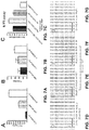
- a method of culturing stem cells comprising preparing a multifunctional matrix having the steps of drying a chitosan solution to form a chitosan layer; neutralizing the acidity of chitosan layer with a basic solution; attaching a growth-promoting protein binding partner to the chitosan layer, wherein the growth-promoting protein binding partner is heparin, heparan sulfate or an analogue of heparan sulfate; cross-linking the growth-promoting protein binding partner to the chitosan layer with genipin; applying a solution of a growth-promoting protein and allowing the growth-promoting protein to attach to the chitosan layer such that the growth-promoting protein is immobilized; seeding one or more stem cells onto the multifunctional matrix; and culturing the stem cells thereon under limited feeding conditions.
- culturing a population of stem cells relies upon immobilizing fibroblast growth factor (FGP) to a surface of a culture plate.
- the method generally has the steps of: (i) coating a bottom of a chamber with a chitosan solution; (ii) drying the chitosan solution to form a chitosan layer; (iii) neutralizing the acidity of the chitosan layer; (iv) binding a heparin to the chitosan layer; (v) cross-linking the heparin to the chitosan layer using genipin; (vi) binding a growth-promoting protein; (vii) applying a solution of an adhesive component (e.g., extracellular matrix protein or extracellular matrix peptide); (viii) binding the adhesive component to the chitosan, which creates a multifunctional film; and (ix) seeding the population of stem cells onto the chamber and culturing the stem cells.
- an adhesive component e.g., extra
- the method can also have washing/rinsing steps between the additions of each component.
- the growth-promoting protein is one or more growth factors, such as fibroblast growth factor-2 (FGF-2).
- FGF-2 fibroblast growth factor-2
- stem cells plated onto the disclosed multifunctional matrix remain in a multipotent and proliferative state without providing soluble growth-promoting proteins.
- the stem cells can remain less mature and more highly proliferative than ceils maintained on a fibronectin-coated substrate in a culture medium supplemented with soluble growth- promoting proteins, such as FGF-2.
- the cultured stem cells using the disclosed method are highly suitable for cell transplantation to repair tissue damage, such as traumatic brain injury.
- a method to repair injured mammalian tissue by administration of a multifunctional matrix into a subject in need of treatment.
- the multifunctional matrix comprises a chitosan-(genipin)-growth-promoting protein binding partner, an immobilized thereon growth factor, preferably FGF-2, alone or in combination with an adhesive component.
- the adhesive component is fibronectin.
- the adhesive component is a peptide sequence arginine-glycine-aspartic acid (RGD) or isoleucine-lysine-valine-alanine-valine (IKVAV),
- the multifunctional matrix is provided having a scaffold with immobilized FGF-2, heparin, genipin and chitosan.
- the multifunctional matrix is provided having a scaffold of chitosan, genipin- linked heparin with immobilized FGF-2 and fibronectin.
- an injectable multifunctional microsphere scaffold to achieve a scaffold that is highly suitable as a vehicle for cell transplantation to repair traumatic CNS injuries.
- a chitosan solution is electrosprayed into a coagulation bath to generate microspheres (range: 30-100 ⁇ m) that can be subsequently modified.
- Primitive neural precursors seeded onto the multifunctional microspheres can be propagated in culture, and the microspheres containing cells are small enough to be injected using a 26 gauge Hamilton syringe into the region of the CNS that had previously sustained a contusion injury.
- this multifunctional scaffold can be used as a cellular and growth factor delivery vehicle to promote the regeneration of nervous tissue injury after CNS injuries.
- a novel multi-functional growth matrix is disclosed that can be used as a biomaterial scaffold for mammalian tissue regeneration and repair.
- the growth matrix is a 2-dimensional multi-functional scaffold that supports the expansion of stem cells.
- the 2-dimensional multi-functional scaffold can be used to grow human embryonic stem cells (hESCs) and induced pluripotential stem cells (iPSCs).
- the growth matrix is a 3-dimensional multi-functional scaffold that supports the expansion of stem cells.
- such 3-D scaffolds can be used as a vehicle for transplanting stem cells.
- the multi-functional matrix disclosed herein promotes both the proliferation and pluripotency of stem cells by attaching a growth-promoting protein to a surface of a culture plate, thereby stabilizing the growth-promoting protein.
- the matrix can also be further modified by attaching an adhesive component, such as an extracellular matrix protein.
- Such a biomaterial scaffold promotes tissue regeneration from primitive cells.
- the disclosed matrix may be highly suitable for propagating neural stem cells in vitro.
- the disclosed matrix promotes both the proliferation and pluripotency of stem cells at a reduced frequency of cell feedings (no need for daily feedings) while the proportion of proliferating and undifferentiated cells is significantly greater than cells propagated under standard growth conditions.
- matrix and sinaffold are used herein interchangeably and refer to a structure capable of supporting 2D and/or 3D cell growth.
- Stem cells that can be supported by the disclosed growth matrix include, but not limited to, embryonic stem cells, pluripotential stem cells, somatic stem cells, adipose-derived stem cells, mesenchymal stem cells, hematopoietic stem cells or umbilical cord blood stem cells, oligodendrocyte progenitors, or FGF responsive progenitors that will grow on an adherent substrate, in the presence or absence of other growth-promoting proteins.
- the stem cells are mammalian and more preferably limited to rodent, primate or human.
- the stem cells that can be supported by the disclosed growth matrix also include the induced pluripotential stem cells (iPSCs) or the stem cells derived from iPSCs.
- iPSCs induced pluripotential stem cells
- the 3-D scaffold may be used as a vehicle for transplanting stem cells on a supportive matrix.
- Stem cells may be attached to a scaffold and transplanted into the lesions created by a spinal cord, traumatic brain injury or a stroke to reconstruct critical neural circuits.
- Stem cells may be attached to a scaffold and transplanted into non-neural tissues.
- the stem cells are mesenchymal stem cells.
- the non-neural tissue is bone, cartilage,, liver, pancreas, heart, skin, bladder, skeletal muscle, lung or kidney.
- the backbone of the disclosed matrix or scaffold is made from chitosan, which is derived by, alkaline deacetylation of chitin. This chitosan backbone yields repeating units of glucosamine and N- acetylglycosamine. Chitosan is a natural polysaccharide similar in structure to glycosaminoglycans which allows for easy modification.
- Chitosan dissolves easily in a weak acid solution such as acetic acid. Once dissolved as a viscous liquid, chitosan can be used to coat the surfaces of cell culture dishes or used to form complex 2D/3D scaffolds. CNS injuries are not uniform in shape or size; therefore a scaffold that is injectable and will mold to the injured tissue is necessary. The chemical structure of chitosan easily allows for modification of its chemical structure, thus making it a very attractive and versatile material.
- the disclosed matrix having chitosan backbone therein is further modified to immobilize a growth-promoting protein in a biological and stable form by covalently linking a growth-promoting protein binding partner to the chitosan using genipin, a naturally occurring and biologically safe cross-linking agent.
- the disclosed matrix is made from (i) chitosan, (ii) genipin, (iii) growth-promoting protein binding partner, and (iv) growth-promoting protein.
- the disclosed matrix further contains (v) an adhesive component.
- the matrix comprises (i) chitosan, (ii) growth-promoting protein binding partner, (iii) genipin, (iv) growth-promoting protein, and (v) adhesive component.
- the method also has one or more washing/rinsing steps between the additions of one or more components.
- the matrix comprises a scaffold with fibronectin, immobilized FGF-2, heparin, genipin and chitosan.
- the matrix comprises a scaffold of chitosan, linked heparin to chitosan via genipin, and immobilized thereon FGF-2 and fibronectin.
- the FGF-2 and fibronectin are immobilized on heparin by forming one or more non-covenant bridges, such as electrostatic, van der Waals, and hydrophobic.
- the crosslinking agent used in the disclosed matrix is genipin.
- the matrix can also be prepared with glutaraldehyde, formaldehyde, tripolyphosphate (TPP), 1-ethyl-3-(3-dimethylaminopropyl) carbodiimide (EDC), ⁇ -glycerophosphate ( ⁇ GP), calcium phosphate, citrate, sulfate, Fe(III), Ho-166, poly ethylene glycol dialdehyde diethyl acetal (PEGDDA), oxalic acid, glyoxal.
- TPP tripolyphosphate
- EDC 1-ethyl-3-(3-dimethylaminopropyl) carbodiimide
- ⁇ GP ⁇ -glycerophosphate
- CaGP calcium phosphate
- citrate citrate
- sulfate Fe(III)
- Ho-166 poly ethylene glycol dialdehyde diethyl acetal
- PEGDDA poly
- the growth-promoting protein binding partner is selected from heparin, heparan sulfate or a heparan sulfate analogue (see e.g ., Guimond. S.E., et al. Int J Exp Pathol. 2004 August; 85(4): A62-A63 ; .
- the growth-promoting protein binding partner is heparin.
- the growth-promoting protein is not particularly limited as long as it can promote growth of the stem cells applied therein.
- the growth-promoting protein is selected from an Activin, an Adrenomedullin (AM), an Angiopoietin (Ang), an Autocrine motility factor (AMF), a Cadherin, a Ciliary neurotrophic factor (CNTF), an Epiregulin, an Erythropoietin (EPO), a Follistatin, a fibroblast growth factor (FGF), platelet-derived growth factor (PDGF), epidermal growth factor (EGF), heparin binding epidermal growth factor (HB-EGF), a Glial cell line-derived neurotrophic factor (GDNF), a Granulocyte colony-stimulating factor (G-CSF), a Granulocyte macrophage colony- stimulating factor (GM-CSF), a Growth differentiation factor (GDF), a Hepatoma-derived growth factor (HDGF), an Interleukin, an Insulin-like growth
- the growth-promoting proteins can readily form non-covalent interactions with heparin include, but not limited to. Brain-derived neurotrophic factor (BDNF), Bone morphogenetic protein (BMP), Epidermal growth factor (EGF), one of the Fibroblast growth factors (FGF-2, 4, 7, 8, 10, 18), Heparin Binding Epidermal Growth Factor (HbEGF), Hepatocyte growth factor (HGF), Keratinocyte growth factor (KGF), Neuregulin (NRG), Neurite growth promoting factor (NEGF 1/2), Platelet-derived growth factor (PDGF) or Vascular endothelial growth factor (VEGF).
- BDNF Brain-derived neurotrophic factor
- BMP Bone morphogenetic protein
- EGF Epidermal growth factor
- FGF-2 Fibroblast growth factors
- HbEGF Heparin Binding Epidermal Growth Factor
- HGF Hepatocyte growth factor
- KGF Keratinocyte growth factor
- NGF 1/2 Neuregulin
- NEGF 1/2 Neurite growth promoting
- the growth-promoting protein is selected from fibroblast growth factor (FCF), platelet-derived growth factor (PDGF), epidermal growth factor (EGF) or heparin binding epidermal growth factor (HB-EGF).
- FCF fibroblast growth factor
- PDGF platelet-derived growth factor
- EGF epidermal growth factor
- HB-EGF heparin binding epidermal growth factor
- FGF fibroblast growth factor
- FGF-2 fibroblast growth factor-2.
- the adhesive component is an extracellular matrix protein, an extracellular matrix peptide, an adhesive saccharide, or combinations thereof.
- the extracellular matrix protein is selected from Fibronectin, Laminin, Vitronectin, Fibrillin, Fibrinogen, Plasminogen, Plasmin, Aggrecan, Brevican, Tenascin, Collagen, Elastin, Hyaluronic acid proteoglycan, Keratan sulfate proteoglycan, Heparan sulfate proteoglycan, Chondroitin sulfate proteoglycan, Syndecan-1 (proteoglycan), IGF Binding Protein or combinations thereof.
- the adhesive saccharide is preferably cellulose.
- the extracellular matrix peptide is a peptide selected from poly-DL-lysine, such as PDL or PLL, poly-DL-ornithine, a short peptide sequence RGD (Arg-Gly-Asp), or a short peptide sequence IKVAV (Ile-Lys-Val-Ala-Val, SEQ ID NO: 1).
- the extracellular matrix peptide is RGD or IKVAV (SEQ ID NO: 1).
- RGD also includes sequences RGD-S (SEQ ID NO: 2).
- IKVAV also includes sequences CSRARKQAAS-IKVAV-SADR (SEQ ID NO: 10) (also trifluoroacetate) and S-IKVAV (SEQ ID NO; 11).
- xenogeneie-free culture system As researchers move from the bench to the clinic, there is a real need for a xenogeneie-free culture system. Ideally, stem cell derivation, establishment, cell banking and undifferentiated expansion should all be done using xenogeneic free components ( i.e., not derived from living tissue).
- peptide sequences of extracellular matrix peptides such as RGD and IKVAV (SEQ ID NO: 1), those skilled in the art can avoid costs and potential contamination associated with using extracellular matrix components extracted from tissue or cultured cells.
- the goal was to enable the growth-promoting protein to be slowly released into the medium to stimulate growth.
- stem cells plated onto the multi-functional matrix remained in a multipotent, proliferating state for at least 3 days without renewing the growth-promoting protein.
- the cells grown on the matrix also remained less mature and more highly proliferative than cells maintained on a similar scaffold that lacked the tethered growth-promoting protein.
- the growth-promoting protein can remain biologically active while bound within the disclosed matrix for about 7 days.
- the matrices of prior art do not have stably immobilized, growth-promoting proteins of defined identify and are, thus, not suitable for therapeutic use Therefore a major advantage of the matrix herein is that it can be formulated using defined components, that it will stabilize the biological activity of those components and that it can maintain stem cells in a more uniform, proliferating and primitive state, It is also well known to those knowledgeable in the art that stem cell propagation using presently available methods require that the cells be fed on a daily basis, whereas the disclosed matrix reduces the time-consuming and tedious task of feeding the cells on a daily basis, while maintaining their stemness.
- the disclosed matrix can be prepared by (i) coating the bottom of a chamber with a chitosan solution; (ii) drying the chitosan solution to form a chitosan layer: (iii) neutralizing the acidity of the chitosan layer by applying a basic aqueous solution (typically sodium hydroxide or ammonium hydroxide) with rinses to remove the basic aqueous solution (iv) binding a growth-promoting protein binding partner to the chitosan layer, (v) cross-linking the growth-promoting protein binding partner to the chitosan layer using genipin; (vi) applying a solution of a growth-promoting protein; (vii) allowing the growth-promoting protein to adhere, (viii) applying a solution of an adhesive component; and (ix) allowing the adhesive component to adhere, which creates a multifunctional film, To support the expansion of stem cells, the method further has a step of (x) seeding the population of stem cells onto the chamber and culturing the stem cells
- Stem cells plated onto the disclosed matrix remain in a multipotent and proliferative state without a need of providing soluble growth-promoting proteins. Moreover, they remain less mature and more highly proliferative than cells maintained on a fibronectin-coated substrate in a culture medium supplemented with soluble growth-promoting proteins (e.g., soluble FGF-2).
- the growth-promoting protein binding partner is heparin and the growth-promoting protein is one or more growth factors, such as fibroblast growth factor-2 (FGF-2).
- the matrix is made from a scaffold of chitosan, genipin-linked heparin with immobilized FGF-2 and fibronectin.
- the disclosed method can produce a scaffold that is highly suitable as a vehicle for cell transplantation to repair tissue damage, such as CNS injury.
- the present invention provides a method to grow stem cells in culture which may then be used to repair injured mammalian tissue.
- the present disclosure includes a method to repair an injured tissue by delivering the stem cells upon a scaffold composition into a subject.
- the scaffold composition is made from chitosan, a genipin immobilized growth-promoting factor binding partner, a growth-promoting protein, and an adhesive component.
- the scaffold composition is comprised of chitosan, genipin-linked heparin with immobilized and fibronectin.
- a method to manufacture an injectable multifunctional microsphere scaffold is provided to achieve a scaffold that is highly suitable as a vehicle for cell transplantation to repair brain injuries.
- a chitosan solution is electrosprayed into a coagulation bath to generate microspheres (range: 30-100 ⁇ m) that can be subsequently modified.
- microspheres range: 30-100 ⁇ m
- Neural stem cells seeded onto the multifunctional microspheres can be propagated in culture, and the microspheres containing the cells are small enough to be injected using a 26 gauge Hamilton syringe into the brain that had previously sustained cortical contusion injuries.
- this multifunctional scaffold can be used as a cellular and growth factor delivery vehicle to promote the regeneration of nervous tissue injury after brain injuries.
- the microspheres are modified by the addition of fibronectin.
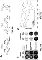
- Fibronectin not only aids in the adherence of the cells to chitosan, but may also enhance their proliferation as shown in FIGs. 1 and 2 and maintain their sternness as shown in FIG. 3 , due to the molecular structure of fibronectin.
- Arginine-glycine-aspartic acid (RGD) first identified in fibronectin, is contained within other ECM proteins such as collagen, vitronectin, thrombospondin, von Willebrand factor, fibrinogen, gelatin and some laminins.
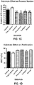
- RG3.6 cells grown on fibronectin coated substrates exhibit a higher mitotic index (see. FIG . 1 ).
- Collagen also possesses an RGD sequence, but the cells do not proliferate as significantly when attached to collagen. This may be due to collagen's negative charge and the resulting gel-like coating formed when bound to chitosan.
- the RGD sequence binds to the ⁇ 5 ⁇ 1 integral receptor whose intracellular amino terminus influences cellular migration, proliferation, self-renewal and differentiation. It has been observed that reducing ⁇ 5 ⁇ 1 expression in cortical progenitors increased their differentiation. A similar effect of specific proteins on ⁇ 6 ⁇ 1 was observed.
- This receptor is activated by netrin-4 and laminin- ⁇ 1. A reduction in these proteins increases the differentiation of NSC.
- This concept was applied to an in vivo application using a hyaluronic acid based hydrogel with immobilized RGD for brain tissue engineering. Transplantations after cortical damage using hyaluronic acid-RGD scaffolds enhanced cell infiltration and angiogenesis into the matrix, while simultaneously inhibiting glial scar formation. An increase in neurite extension was also observed. As shown in FIG. 8 , the fibronectin may be substituted with the RGD peptide with comparable performance.
- the modified chitosan microspheres disclosed herein can be designed to allow FGF-2 to be tethered to the surface of the scaffold, which differs from systems of prior art that have used the spheres as a method for either encapsulating growth factors or transplantable cells.
- FGF-2 is a known survival factor for neural precursors and maintains these cells in a primitive state.
- FGF-2 has been shown to increase the numbers of stem/progenitor cells in the subventricular zone following TBI.
- the FGF-2 By immobilizing the FGF-2 to the surface of the chitosan, it is presented to the cells in a more biologically active form (due to heparin binding) and is more available to adherent cells when bound rather than in soluble form either supplied through media or released from encapsulation.
- the inventors have discovered that cells maintained on the multifunctional film (matrix) do not need to be fed for at least 3 days after plating, and yet the proportion of proliferating and undifferentiated cells is significantly greater than cells propagated under standard growth conditions. Stem cells normally require feeding on a daily basis; however, with the disclosed matrix, the cells can clearly be left untended for at least 3 days.
- Neural Stem Cells Two cell types were used to evaluate the functionality of the modified chitosan films and microspheres: an immortalized cell line, RG3.6, that was created from embryonic day 13.5 green fluorescent protein positive (GFP + ) rat neocortical cells; and primary NSCs harvested from embryonic day 13.5 EGFP (SD- Tg(GFP)Bal/2Rrrc (RRRC:0065) rat neocortex.
- the RG3.6 cell line was used instead of primary cells for specific experiments to eliminate several variables seen with heterogenous primary cell cultures and to achieve greater consistency during substrate optimization.
- RG3.6 and primary NSCs were maintained in DMEM/F12 media supplemented with B27, gentamycin (50 ⁇ g/ml), apo-transferrin (50 ⁇ g/ml) and rhFGF-2 (10 ng/ml +1 ng/ml Heparan Sulfate). They were either grown as neurospheres or as an attached monolayer on poly-ornithine/fibronectin coated petri dishes.
- Chitosan Films A 3% w/v low molecular weight chitosan solution was prepared in 2% acetic acid v/v. Chitosan (low molecular weight ⁇ 50kDa) was purchased from Sigma (St Louis, MO). The solution was pipetted into two-well glass chamber slides (NUNC, Rochester, NY) to coat the bottom of chamber. The remaining solution was removed and the slides were set to dry for 2-3 h at room temperature. Chitosan coatings were neutralized in 0.5 M NaOH (Sigma; St Louis, MO) for 10 minutes and then rinsed 3 times in sterile deionized water for 5 minutes each.
- the chitosan was subsequently adsorbed with solutions of fibronectin (10 ⁇ g/mL), laminin (20 ⁇ g/mL), gelatin (0.1%), collagen type I (0.1 mg/mL), or poly(L-lysine) (0.05 mg/mL) prepared in dH 2 O.
- RG3.6 cell line was seeded onto chitosan adsorbed with fibronectin, laminin, gelatin, type I collagen, or poly(L-lysine) and compared to RG3.6 cells grown on chitosan without adsorbed matrix factors or polymer. After 4 days of growth the cells were fixed and stained for actin and counterstained using DAPI. NSCs are morphologically distinct, with a few long processes. RG3.6 cells responded differently when grown on chitosan substrates with varying adhesive proteins. Substrates coated with fibronectin promoted the NSC morphology ( see FIGs.
- RG3.6 cells maintained on fibronectin had l-3 processes that were quite long and straight (49 ⁇ 5 ⁇ m) and they lacked branches.
- RG3.6 cells grown on laminin also possessed processes that were few in number and long (35 ⁇ 3 ⁇ m), but they were shorter than the processes of the cells grown on fibronectin.
- Cells grown on type 1 collagen (11 ⁇ 1 ⁇ m), gelatin (31 ⁇ 2 ⁇ m) and unmodified chitosan (13 ⁇ 1 ⁇ m) had short processes.
- Cells grown on poly(L-lysine) had numerous, short processes (17 ⁇ 1 ⁇ m) that branched frequently.
- RG3.6 cells were stained for Ki67, which is a marker of cells undergoing mitosis. These studies showed that the mitotic indexes of RG3.6 cells grown on all substrate conditions were high and differed only slightly, but two ECM proteins fibronectin and gelatin resulted in the highest mitotic index at 92 ⁇ 1% and 93 ⁇ 2%, respectively ( see FIG. 1D ). The mitotic indices of cells grown on the other substrates were: laminin, 89 ⁇ 2%; collagen, 87 ⁇ 4%; poly(L-lysine), 77 ⁇ 5%; and unmodified chitosan, 82 ⁇ 1%.
- chitosan was non-toxic to NSCs (data not shown) but the cells required adhesive peptides to grow efficiently on the scaffold. Proliferative rates were high on all chitosan substrates, however they were noticeably higher when the chitosan was coated with fibronectin or gelatin.
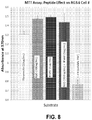
- the biological activity of the human fibroblast growth factor-2 (hFGF-2) bound to the scaffold was verified by measuring cell growth using the MTT assay and analyzing the morphology of NSCs.
- Heparin sodium salt from bovine intestinal mucosa was purchased from Sigma (St Louis, MO).
- Recombinant human-fibroblast growth factor-2 (rhFGF-2) was purchased from Peprotech (Rocky Hill, NJ).
- Genipin was purchased from Wako Pure Chemical Industries, Ltd. (Osaka, Japan).
- FGF-2 was added to half the chitosan-heparin-genipin wells. Other wells were untreated. NSCs were plated into each condition at high (5x10 4 cells/well) and low (2x10 4 cells/well) densities. Cells in the chitosan-fibronectin condition received FGF-2 in the media daily as these cells would normally when propagating in vitro . Cells on the multifunctional scaffold did not receive medium supplemented with FGF-2. The former condition served as a control. Cultures were grown for three days before being analyzed for cell growth using the MTT Assay.
- NSCs are a morphologically distinct cell type with typically, 2 very long processes.
- Cell process lengths were measured using Sigma Scan Pro software (Systat Software, San Jose, CA). The average number of processes extending from each cell was manually calculated.
- Statistical analyses were performed using an ANOVA with Tukey post-hoc . Data are expressed as the mean ⁇ standard error of mean (SEM).
- MTT assay is a colorimetric assay that measures the reduction of a yellow substrate 3-[4,5- dimetylthiazol-2-yl]-2,5-dipheniltetrazolium (MTT, purchased from Sigma, St Louis, MO) in the cell into an insoluble purple formazan product. Briefly, 10 ⁇ l of a 5mg/mi MTT solution in PBS was added to 100 ⁇ L of medium and incubated for 2-4 h in the cell incubator at 37° C. The reaction was stopped by adding 100 ⁇ l of a solution containing 50% (w/v) N,Ndimethylformamide and 20% SDS (pH 4.8). The plates were maintained overnight in the incubator at 37° C and the absorption at 560-690 nm was determined using a microtiter plate reader (PowerWave 200, Bio-tek Instruments).
- NSCs were seeded onto poly-d-lysine and laminin coated dishes and maintained for 24h in medium, then the FGF-2 was removed from the medium whereupon the cells differentiated over the following four days.
- T ⁇ evaluate proliferating cells using standard culture conditions, NSCs were seeded in medium onto chitosan-coated dishes with adsorbed fibronectin (10 ⁇ g/mL). Ten percent of the medium was changed every day and replaced with equal volume of 10X FGF-2 containing media (100 ng/mL).
- NSCs were seeded onto chitosan-coated dishes with covalently bound heparin and adsorbed fibronectin.
- FGF-2 in 1 mg/mL BSA solution
- the FGF-2 solution was then aspirated and rinsed gently, twice to remove any unbound growth factor.
- NSCs were seeded onto the plates and maintained for 4 days in the basal growth medium (which lacked soluble FGF-2). Cells were scraped from the plates in lysis buffer and stored until Western Blot protein analyses.
- Protein concentrations were determined using the BCA assay (ThermoScientific, Rockford, IL).
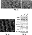
- Western blots were analyzed for the stem cell and progenitor markers brain lipid binding protein (BLBP) using a rabbit anti-BLBP antiserum at 1:1000 (Abcam, Cambridge MA); sex determining region Y-box 2 (Sox2) using rabbit anti-Sox2 antisera 1:200 (Chemicon, Temecula, CA); beta tubulin (TUJI) 1:1000 (Covance, Princeton, NJ), microtubule associated protein-2 (MAP2) using a rabbit anti-MAP2 1:200 (Sigma).
- BLBP brain lipid binding protein
- Sox2 sex determining region Y-box 2
- Sox2 sex determining region Y-box 2
- MAP2 microtubule associated protein-2
- RG3.6 cells and primary NSCs responded differently when grown on the more highly modified chitosan substrate.
- the cells preferred the addition of genipin cross-linked heparin, fibronectin and immobilized FGF-2 to the chitosan substrate.
- the MTT assay returned a 2.5 fold higher value than NSCs grown on the same complex without immobilized FGF-2 ( see FIG. 2A ).
- the higher number of cells growing on the scaffold condition with immobilized FGF than on the chitosan-flbronectin condition, which, received FGF-2 in the media, daily was also evident microscopically ( see FIG . 2B-2D ).
- the immobilized FGF-2 was superior in promoting NSC cell proliferation compared to the standard growth conditions used to propagate NSCs.
- NSCs cells are typically grown in culture as a monolayer, receiving soluble FGF in media changes daily.
- NSCs were grown on fibronectin or on a multifunctional film comprised of chitosan, genipin-linked heparin, FGF-2 and fibronectin.
- This multifunctional film promoted both the proliferation and pluripotency of NSCs while reducing the frequency of feeding the cells (see FIG. 3A-3B ).
- BLBP brain lipid binding protein
- PCNA proliferating cell nuclear antigen
- TUJI class III beta tubulin
- MAP-2 microtubule associated protein-2
- GFAP glial fibrillary acidic protein
- SOX2 SRY box-2 binding protein
- Chitosan powder (1.5g) was dispersed in 50 mL of water containing 2.0% v/v acetic acid to create a 3% chitosan solution.
- the chitosan solution was mechanically stirred at 700 rpm until completely dissolved.
- the resulting solution was collected and centrifuged at 2,000 rpm for 10 minutes. Subsequently, the supernatant was collected and the remaining impurities that pelleted were discarded.
- Chitosan microspheres were formed by extruding the acid chitosan solution through a syringe at a flow rate of 5ml/hr into a basic coagulation bath, consisting of 2.5 M sodium hydroxide: methanol: water (20:30:50 v/v). To reduce the surface tension on the end of the needle and thus reduce the size of the microspheres to a desired range, a 25kV electric current was applied. Next, the spheres were filtered through a 100 ⁇ m strainer to remove any oversized spheres. They were removed from the ionic solution and rinsed four times in distilled water to eliminate any residual sodium hydroxide and methanol. They were then sterilized in 70% ethanol for 30 minutes.
- Microsphere size was measured using Sigma Scan Pro 5 software. Frequency distribution of microsphere diameter was also quantified. Following rinses in distilled water, microspheres were coated overnight with heparin (0.5 mg/ml) and genipin (0.45 mM) in HBS to cross-link the heparin to the microspheres, as described previously. The following day the heparin cross-linked spheres were rinsed 3 times 10 minutes in HBS and incubated for 4 hours with fibronectin (10 ⁇ g/mL) and 2 h in 1 ⁇ g/mL rhbFGF.
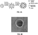
- FIG. 4A This example illustrates a manufacture of a vehicle to facilitate the delivery of neural precursors into brain injuries with the cells adhered to the surface of the microspheres.
- a schematic for the vehicle design is presented in FIG. 4A .
- Previously generated microspheres using a coaxial airflow ranged in size from 200-500 ⁇ M in diameter. Such spheres are too large for injections using 23 gauge needles.
- the optimal sphere size for transplantation through a 23 gauge Hamilton syringe is between 20-100 ⁇ m.
- the size of the microspheres can be decreased by using an electric current. The high flow rate and high voltage reduced the surface tension on the syringe allowing for smaller droplets to be "sprayed" into the coagulation solution.
- Microspheres between 10-100 ⁇ m were formed using 22 kV and a flow rate of 90 ml/h with, on average 64 ⁇ m in diameter ( see FIGs. 4C and 4D ). The majority of the spheres fell within the 30-100 ⁇ m range as seen in the frequency distribution.
- the microspheres were modified by covalently cross-linking heparin to chitosan through genipin to allow the immobilization of FGF-2. Fibronectin was also added for cell adhesion. Toluidine blue stain for heparin was evident on the microspheres ( see FIG. 4E ).
- RG3.6 cells were mixed with microspheres and incubated overnight, whereupon they attached to the spheres ( see FIG. 4F ). These 3-D cultures were maintained for 10 days in vitro with daily media changes. RG3.6 proliferated well on the scaffold, with limited cell death.
- Cold PBS was suffused onto the surface of the skull during the craniotomy to reduce the generation of heat that could cause damage to the underlying dura mater and neocortex.
- the skull flap was removed and the animal placed into a stereotactic apparatus under the controlled cortical impactor (CCI) (eCCI 6.3 device built by Custom Design and Fabrication, Richmond, VA).
- CCI cortical impactor
- the tip of a 3.5 mm diameter anvil was zeroed by bringing it into contact with the exposed dura mater.
- the velocity of the impactor was set at 4,0 ⁇ 0.2 m/s, the depth of penetration to 1.5 mm and the duration of deformation to 150 msec. After impact, the integrity of the dura mater was confirmed and the scalp incision sutured with 3-0 nylon thread.
- Buprenorphine (0.05 mg/kg, SC) was administered post-operatively and the rats were placed on heating pads maintained at 37° and monitored continuously for 2 h after surgery. In addition, immediately after surgery, all subjects received 3% body weight of 0.9% saline subcutaneously (SC) to prevent dehydration.
- SC subcutaneously
- Transplantation (illustrative example): Subacute transplantations were performed 7 days after CCI injury. The animals were anesthetized again using a ketamine/xylazine mixture and the sutures were removed to expose the skull. Cell-sphere complexes were collected from culture dishes and resuspended in phenol-free media without supplements. A 26 gauge Hamilton syringe was used to inject the scaffold at three different depths: 1.5, 1.0 and 0.5 mm below the dura mater. One ⁇ L was injected at each depth over 5 minutes, with 5 minute intervals between each injection and 10 minutes following the final injection. The scalp incision was sutured with 3-0 nylon thread and the animals placed onto a 37 °C heating pad until they were fully awake. All of the procedures performed on animals in this report were approved by the New Jersey Medical School IACUC under animal protocol ## 08056.
- the brains were rinsed with PBS and cryoprotected by immersion in 30% sucrose in dH2O. After one change of sucrose solution, the brains were placed into plastic cryomolds and frozen in OCT on a dry-ice-ethanol slush. The brains were cryosectioned at 40 and 15 ⁇ m thickness and stained using mouse anti-Nestin antibody (Developmental Studies Hybridoma Bank, Iowa, 1:5). Sections were incubated in secondary antibodies for 2 h at room temperature (all from Jackson Immunoresearch, West Grove, PA; 1:200). All secondary antibody combinations were carefully examined to ensure that there was no cross-talk between fluorescent dyes or cross-reactivity between secondary antibodies.
- Stem Cell Engraftment Numerous studies have encapsulated cells inside spheres or other delivery vehicles to enable the cells to produce soluble growth and trophic factors. In contrast, a delivery vehicle described herein has the cells adhered to the surface of the microspheres. This configuration enabled the progeny of the stem cells to migrate off of the scaffold into the adjacent tissue, which is crucial to reconstruct a damaged brain. To test this approach, multifunctional microspheres containing the NSC cell line attached to the multifunctional microspheres were transplanted into the lesion cavity at 7 days of recovery from CCL As the NSCs express GFP they could be distinguished from the host cells using fluorescence microscopy. When the NSCs were differentiated in vitro they formed neurons and glia ( see FIG. 5A ).
- FIG. 9 A schematic depicting a method for CNS repair after TBI is provided as FIG. 9 .
- the initial injury and subsequent inflammation causes a loss of cortical tissue, including a loss of many laminar neurons.
- microsphere complexes containing adherent NSC can be transplanted.
- the cells begin to proliferate and form processes that extend to the pial surface, mimicking embryonic neurogensis.
- Neuroblasts and other progeny can migrate along their processes, ultimately generating neurons appropriate to each cortical layer and supportive glia.
- the scaffold degrades over time revealing a regenerated, and ordered cortex.
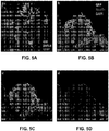
- Ionic and covalent heparin immobilization on chitosan films and microspheres 2-D chitosan films and 3-D chitosan microspheres were prepared for ionic and covalent heparin immobilization.
- To prepare chitosan films 24 well plates were coated with a thin layer of 3% chitosan solution. Wells were allowed to dry overnight and subsequently the acidity was neutralized using 0.5 M sodium hydroxide. Afterwards, plates were rinsed three times with distilled water and incubated overnight with 0.5 mg/mL heparin in HEPES buffered solution (HBS) for ionic binding and in 0.45 mM genipin in HBS for covalent binding.
- HBS HEPES buffered solution
- FTIR analysis The genipin cross-linked chitosan-heparin films were analyzed with Attenuated Total Reflectance Fourier Transform Infrared Spectroscopy (ATR-FTIR; Perkin Elmer) to detect heparin binding. As controls, the chitosan film and heparin powder were measured by FTIR.
- ATR-FTIR Attenuated Total Reflectance Fourier Transform Infrared Spectroscopy
- the FGF solutions contained 1 mg/mL BSA to maintain growth factor stability. After allowing the FGF-2 to bind, the solutions were collected in separate eppendorf tubes to determine the unbound FGF-2. Each well was washed gently two times with 50 ⁇ L of HBS, which was also added to each respective collection tube. To test long term release, wells were refilled with 100 ⁇ L of PBS and collected 7 days later. A sandwich ELISA was used to measure FGF-2 that was released over time. Subtracting the amount released on day 0 from the total amount of FGF-2 added to the scaffold revealed the percentage of bound growth factor.
- Heparin retention on chitosan Heparin promotes angiogenesis, has been demonstrated to reduce inflammation and it has high affinity for fibroblast growth factors, thus we reasoned that it would be highly advantageous to covalently attach heparin to the microspheres.
- Figure 3A shows the chemical structure of chitosan, heparin, and genipin. Heparin binding to chitosan was tested in two ways, via colorimetric dye staining and FTIR analyses. Positively charged toluidine blue stain, which stains for negatively charged heparin, was strong on chilosan-heparin 2-D films and 3-D microspheres ( see FIG. 6B ).
- Toluidine blue also strongly stained the chitosan-heparin-genipin films and microspheres.
- chitosan alone did not demonstrate any toluidine blue stain on films and microspheres.
- the blue staining on the run of the chitosan alone condition is due to the toluidine blue getting trapped underneath the thin edges of the chitosan coating. This was difficult to avoid, thus, only the center of the films were considered for analyses.
- chitosan alone were immersed and washed in 1.5M NaCl instead of HBS, chitosan alone, as expected, remained unstained.
- the chitosan-heparin complex also displayed peaks of chitosan functional groups, including N-H bending at 1560 nm and CH 2 bending at 1,380 nm.
- FGF-2 Immobilization and Release Heparin has binding sites for several growth factors, including but not limited to FGFs, VEGF, HGF and BMP. Therefore we investigated the levels of FCF-2 that could be immobilized to chitosan-heparin complexes. Three different concentrations (100, 500, and 1,000 ng/ml) of FGF-2 were evaluated for binding to chitosan-heparin-genipin film scaffolds. As would be predicted, as the concentration of FGF-2 increased, the amount of bound FGF-2 increased ( see FIG. 7A ) At each concentration approximately 70-80% of the FGF-2 bound to the scaffold ( see FIG. 7B ).
- RG3.6 cells were seeded onto a 2-D chitosan-heparin-genipin crosslinked scaffold and the MTT assay was performed to assess cell viability and growth after 2 days in vitro, Cells were tested under 4 conditions, they were seeded onto; the complex film with FGF-2 in the medium (Control); onto the complex film with freshly bound FGF-2 (Bound, Day 0); or onto the complex film where FGF-2 had been bound earlier and incubated at 37°C for 3 days (Bound Day 3); or onto the complex film without added FGF-2 in the medium.
- Neural stem cell growth and viability as reflected by the MTT assay was highest on the FGF-2 bound to the scaffold immediately prior to cell seeding.
- Cell growth on the bound FGF-2 condition was superior to cell growth on the scaffold with FGF-2 provided in the medium.
- cell growth on the complex film that had immobilized FGF-2 attached to the scaffold three days prior to seeding was comparable to the control, which received FGF-2 added to the media daily ( see FIG. 7C ).
- Cell growth on both FGF-2 containing scaffolds, as well as the control was significantly higher than cells grown on the complex film lacking FGF-2 in the medium.
- the RGD peptide first identified in fibronectin, binds to integrin receptors, present on the surface of many types of cells. As reviewed earlier, studies have shown that these receptors are essential for maintaining neural stem cells in a primitive state. Therefore, we hypothesized that the RGD peptide would be sufficient as an adhesive peptide on the scaffold. Confirming the validity of this hypothesis, scaffolds produced with an RGD peptide enhanced the growth of the RG3.6 cells, and when used at an equimolar concentration as fibronectin, produced more robust growth ( see FIG. 8 ).
Landscapes
- Health & Medical Sciences (AREA)
- Life Sciences & Earth Sciences (AREA)
- Engineering & Computer Science (AREA)
- Biomedical Technology (AREA)
- Cell Biology (AREA)
- Developmental Biology & Embryology (AREA)
- Chemical & Material Sciences (AREA)
- Biotechnology (AREA)
- Zoology (AREA)
- General Health & Medical Sciences (AREA)
- Bioinformatics & Cheminformatics (AREA)
- Public Health (AREA)
- Veterinary Medicine (AREA)
- Medicinal Chemistry (AREA)
- Pharmacology & Pharmacy (AREA)
- Epidemiology (AREA)
- Animal Behavior & Ethology (AREA)
- Immunology (AREA)
- Genetics & Genomics (AREA)
- Organic Chemistry (AREA)
- Wood Science & Technology (AREA)
- Virology (AREA)
- Reproductive Health (AREA)
- Neurology (AREA)
- Neurosurgery (AREA)
- Microbiology (AREA)
- Biochemistry (AREA)
- General Engineering & Computer Science (AREA)
- Hematology (AREA)
- Gynecology & Obstetrics (AREA)
- Psychology (AREA)
- Orthopedic Medicine & Surgery (AREA)
- Ophthalmology & Optometry (AREA)
- Materials For Medical Uses (AREA)
- Micro-Organisms Or Cultivation Processes Thereof (AREA)
- Medicinal Preparation (AREA)
- Medicines Containing Material From Animals Or Micro-Organisms (AREA)
Claims (14)
- Verfahren zur Kultivierung von Stammzellen, das Folgendes umfasst:
Herstellen einer multifunktionalen Matrix, was die folgenden Schritte aufweist:Trocknen einer Chitosanlösung, um eine Chitosanschicht zu bilden;Neutralisieren der Acidität der Chitosanschicht mit einer basischen Lösung;Verbinden eines wachstumsfördernden Proteinbindungspartners mit der Chitosanschicht, wobei der wachstumsfördernde Proteinbindungspartner Heparin, Heparansulfat oder ein Analogon von Heparansulfat ist;Vernetzen des wachstumsfördernden Proteinbindungspartners zu der Chitosanschicht mit Genipin;Anwenden einer Lösung eines wachstumsfördernden Proteins und Ermöglichen, dass sich das wachstumsfördernde Protein mit der Chitosanschicht derart verbindet, dass das wachstumsfördernde Protein immobilisiert wird;Besiedeln mit einer oder mehreren Stammzellen auf der multifunktionalen Matrix; undKultivieren der Stammzellen darauf unter eingeschränkten Fütterungsbedingungen. - Verfahren nach Anspruch 1, wobei das Herstellen einer multifunktionalen Matrix weiter die folgenden Schritte umfasst:Anwenden einer Lösung einer adhäsiven Komponente; undErmöglichen, dass sich die adhäsive Komponente mit der Chitosanschicht verbindet.
- Verfahren nach Anspruch 1, wobei der wachstumsfördernde Proteinbindungspartner Heparin ist.
- Verfahren nach Anspruch 1, wobei Stammzellen, die auf der multifunktionalen Matrix kultiviert werden, in einem multipotenten und proliferativen Zustand, frei von jedweden löslichen wachstumsfördernden Proteinen verbleiben.
- Verfahren nach Anspruch 1, wobei Stammzellen aus einer Gruppe ausgewählt sind, die aus Folgenden besteht: embryonalen Stammzellen, pluripotenten Stammzellen, somatischen Stammzellen, aus Fettgewebe stammenden Stammzellen, mesenchymalen Stammzellen, hämatopoetischen Stammzellen, Nabelschnurblut-Stammzellen, Oligodendrozyten-Vorläufern, FGF-responsiven Vorläufern, induzierten pluripotenten Stammzellen (iPSCs) und Stammzellen, die von iPSCs stammen.
- Verfahren nach Anspruch 1, wobei die multifunktionale Matrix 2-dimensional ist und Stammzellen aus hESCs und iPSCs ausgewählt sind.
- Verfahren nach Anspruch 1, wobei die multifunktionale Matrix 3-dimensional ist.
- Verfahren nach Anspruch 1, wobei die Matrix xenogenfreie Komponenten enthält.
- Verfahren nach Anspruch 5, wobei die Stammzellen vom Säugetier und bevorzugt Nagetier, Primaten oder Menschen stammen.
- Verfahren nach Anspruch 1, wobei das Wachstums fordernde Protein aus einer Gruppe ausgewählt ist, die aus FGF, PDGF, EGF und HB-EGF besteht.
- Verfahren nach Anspruch 1, wobei FGF FGF-2 ist.
- Verfahren nach Anspruch 2, wobei die adhäsive Komponente ein extrazelluläres Matrixprotein ist, das aus einer Gruppe ausgewählt ist, die aus Folgenden besteht: Fibronectin, Laminin, Vitronectin, Fibrillin, Fibrinogen, Plasminogen, Plasmin, Aggrecan, Brevican, Tenascin, Kollagen, Elastin, Hyaluronsäure-Proteoglykan, Keratansulfat-Proteoglykan, Heparansulfat-Proteoglykan, Chondroitinsulfat-Proteoglykan, Syndecan-1-(Proteoglykan) und IGF-Bindungsprotein.
- Verfahren nach Anspruch 2, wobei die adhäsive Komponente ein extrazelluläres Matrixpeptid ist, das eine Peptidsequenz aufweist, die RGD oder IKVAV umfasst.
- Verfahren nach Anspruch 1, wobei die Stammzellen höchstens einmal in 3 Tagen gefüttert werden.
Applications Claiming Priority (2)
| Application Number | Priority Date | Filing Date | Title |
|---|---|---|---|
| US201361757378P | 2013-01-28 | 2013-01-28 | |
| PCT/US2014/013381 WO2014117146A1 (en) | 2013-01-28 | 2014-01-28 | Growth matrices for stem cell propagation in vitro and in tissue regeneration |
Publications (3)
| Publication Number | Publication Date |
|---|---|
| EP2948541A1 EP2948541A1 (de) | 2015-12-02 |
| EP2948541A4 EP2948541A4 (de) | 2016-07-27 |
| EP2948541B1 true EP2948541B1 (de) | 2019-09-18 |
Family
ID=51228132
Family Applications (1)
| Application Number | Title | Priority Date | Filing Date |
|---|---|---|---|
| EP14743406.2A Active EP2948541B1 (de) | 2013-01-28 | 2014-01-28 | Wachstumsmatrizen für stammzellenfortpflanzung in vitro und in einer geweberegeneration |
Country Status (6)
| Country | Link |
|---|---|
| US (1) | US10465165B2 (de) |
| EP (1) | EP2948541B1 (de) |
| CN (1) | CN105143442A (de) |
| CA (1) | CA2899638A1 (de) |
| IL (1) | IL240145A0 (de) |
| WO (1) | WO2014117146A1 (de) |
Families Citing this family (9)
| Publication number | Priority date | Publication date | Assignee | Title |
|---|---|---|---|---|
| WO2015120388A1 (en) * | 2014-02-10 | 2015-08-13 | Cytori Therapeutics, Inc. | Regenerative cell therapy for central nervous system (cns) disorders and ptsd |
| CN107849539A (zh) * | 2015-08-13 | 2018-03-27 | 富士胶片株式会社 | 多能干细胞的培养方法、培养容器的制造方法、培养容器以及细胞培养用支架材料 |
| WO2017036533A1 (en) | 2015-09-03 | 2017-03-09 | Ecole Polytechnique Federale De Lausanne (Epfl) | Three-dimensional hydrogels for culturing adult epithelial stem cells and organoids |
| CN105602893A (zh) * | 2015-12-28 | 2016-05-25 | 上海吉泉生物技术有限公司 | 一种无血清培养脐带间充质干细胞方法及应用 |
| SG11201810936VA (en) * | 2016-12-01 | 2019-01-30 | Univ Ramot | Combined treatment for nerve injuries |
| EP4164706A1 (de) | 2019-12-04 | 2023-04-19 | Centre Hospitalier Universitaire Vaudois (CHUV) | Vorrichtung und verfahren zur gewebezüchtung und regenerativen medizin |
| CN114276988B (zh) * | 2021-11-19 | 2022-08-05 | 珠海贝索细胞科学技术有限公司 | 一种赋能型间充质干细胞添加剂及其应用 |
| WO2025080462A1 (en) * | 2023-10-12 | 2025-04-17 | Corning Incorporated | Chitosan-based hydrogels for 3d cell culture |
| CN117821382A (zh) * | 2023-12-29 | 2024-04-05 | 广州赛莱拉干细胞科技股份有限公司 | 一种毛囊干细胞无血清培养基和培养方法 |
Family Cites Families (7)
| Publication number | Priority date | Publication date | Assignee | Title |
|---|---|---|---|---|
| US20100254900A1 (en) * | 2002-03-18 | 2010-10-07 | Campbell Phil G | Biocompatible polymers and Methods of use |
| CA2672495C (en) * | 2006-12-11 | 2017-01-17 | Chi2Gel Ltd. | Novel injectable chitosan mixtures forming hydrogels |
| EP2229147A2 (de) * | 2007-12-03 | 2010-09-22 | The Johns Hopkins University | Synthese- und anwendungsverfahren für chemosphären |
| US8680182B2 (en) * | 2009-06-04 | 2014-03-25 | Clemson University Research Foundation | Methods for promoting the revascularization and reenervation of CNS lesions |
| WO2011156586A2 (en) * | 2010-06-09 | 2011-12-15 | Trustees Of Tufts College | Multilayered silk scaffolds for meniscus tissue engineering |
| WO2012087965A2 (en) | 2010-12-22 | 2012-06-28 | Fate Therapauetics, Inc. | Cell culture platform for single cell sorting and enhanced reprogramming of ipscs |
| WO2012119012A1 (en) * | 2011-03-02 | 2012-09-07 | New Jersey Institute Of Technology | System and method for vascularized biomimetic 3-d tissue models |
-
2014
- 2014-01-28 EP EP14743406.2A patent/EP2948541B1/de active Active
- 2014-01-28 CN CN201480018759.0A patent/CN105143442A/zh active Pending
- 2014-01-28 WO PCT/US2014/013381 patent/WO2014117146A1/en not_active Ceased
- 2014-01-28 CA CA2899638A patent/CA2899638A1/en not_active Abandoned
- 2014-01-28 US US14/763,944 patent/US10465165B2/en active Active
-
2015
- 2015-07-26 IL IL240145A patent/IL240145A0/en unknown
Non-Patent Citations (1)
| Title |
|---|
| None * |
Also Published As
| Publication number | Publication date |
|---|---|
| CN105143442A (zh) | 2015-12-09 |
| IL240145A0 (en) | 2015-09-24 |
| EP2948541A4 (de) | 2016-07-27 |
| WO2014117146A1 (en) | 2014-07-31 |
| US10465165B2 (en) | 2019-11-05 |
| WO2014117146A8 (en) | 2015-02-19 |
| US20150361395A1 (en) | 2015-12-17 |
| CA2899638A1 (en) | 2014-07-31 |
| EP2948541A1 (de) | 2015-12-02 |
Similar Documents
| Publication | Publication Date | Title |
|---|---|---|
| EP2948541B1 (de) | Wachstumsmatrizen für stammzellenfortpflanzung in vitro und in einer geweberegeneration | |
| Skop et al. | Heparin crosslinked chitosan microspheres for the delivery of neural stem cells and growth factors for central nervous system repair | |
| US20250186657A1 (en) | Neuronal replacement and reestablishment of axonal connections | |
| Kijeńska et al. | Interaction of Schwann cells with laminin encapsulated PLCL core–shell nanofibers for nerve tissue engineering | |
| Tam et al. | Regenerative therapies for central nervous system diseases: a biomaterials approach | |
| US9192655B2 (en) | System and method for a hydrogel and hydrogel composite for cartilage repair applications | |
| Mashinchian et al. | Regulation of stem cell fate by nanomaterial substrates | |
| CN102908207B (zh) | 生物打印技术制备的组织工程神经移植物及其制备方法 | |
| Skop et al. | Optimizing a multifunctional microsphere scaffold to improve neural precursor cell transplantation for traumatic brain injury repair | |
| Yaylaci et al. | Chondrogenic differentiation of mesenchymal stem cells on glycosaminoglycan-mimetic peptide nanofibers | |
| KR102041360B1 (ko) | 탈세포화된 세포외 기질을 포함하는 직접교차분화 촉진용 조성물 및 이의 용도 | |
| JP2009502242A (ja) | ポリマーがコーティングされたナノフィブリル構造体、並びに細胞維持及び分化のための方法 | |
| KR20150009669A (ko) | 폴리도파민이 결합된 생리활성 펩타이드로 고정화된 고분자 지지체 및 이의 제조방법 | |
| Ren et al. | Hyaluronic acid/polylysine hydrogel as a transfer system for transplantation of neural stem cells | |
| CN104623738B (zh) | 带有悬丝纤维支架的组织工程神经移植物及其制备方法 | |
| KR20200052468A (ko) | 혈관내피세포 성장인자를 포함하며 혈관신생을 유도하는 기능성 바이오 잉크 조성물 | |
| WO2014153610A1 (en) | Growth factor binding surfaces and uses thereof | |
| US20240307591A1 (en) | Accelerated Development of Functional Three-Dimensional Tissue Moduli | |
| US20130288366A1 (en) | Synthetic culture platform and methods of using | |
| Bean et al. | Stem cells and nanotechnology in tissue engineering and regenerative medicine | |
| Khanna et al. | Extracellular matrix bioactive molecules and cell behavior modeling | |
| Jalili‐Firoozinezhad et al. | Nanotissue engineering of neural cells | |
| CN118490892B (zh) | 缓释Apt19S募集内源性干细胞和引导中枢神经轴突直行再生的生物活性支架及其应用 | |
| Wang et al. | Interactions between neural stem cells and biomaterials combined with biomolecules | |
| US20240139379A1 (en) | Compositions, apparatuses and methods for making and using bioscaffolds |
Legal Events
| Date | Code | Title | Description |
|---|---|---|---|
| PUAI | Public reference made under article 153(3) epc to a published international application that has entered the european phase |
Free format text: ORIGINAL CODE: 0009012 |
|
| 17P | Request for examination filed |
Effective date: 20150727 |
|
| AK | Designated contracting states |
Kind code of ref document: A1 Designated state(s): AL AT BE BG CH CY CZ DE DK EE ES FI FR GB GR HR HU IE IS IT LI LT LU LV MC MK MT NL NO PL PT RO RS SE SI SK SM TR |
|
| AX | Request for extension of the european patent |
Extension state: BA ME |
|
| DAX | Request for extension of the european patent (deleted) | ||
| REG | Reference to a national code |
Ref country code: DE Ref legal event code: R079 Ref document number: 602014053852 Country of ref document: DE Free format text: PREVIOUS MAIN CLASS: C12N0005000000 Ipc: A61K0035280000 |
|
| A4 | Supplementary search report drawn up and despatched |
Effective date: 20160629 |
|
| RIC1 | Information provided on ipc code assigned before grant |
Ipc: C12N 5/00 20060101ALI20160623BHEP Ipc: A61K 35/28 20060101AFI20160623BHEP |
|
| STAA | Information on the status of an ep patent application or granted ep patent |
Free format text: STATUS: EXAMINATION IS IN PROGRESS |
|
| 17Q | First examination report despatched |
Effective date: 20180206 |
|
| GRAP | Despatch of communication of intention to grant a patent |
Free format text: ORIGINAL CODE: EPIDOSNIGR1 |
|
| STAA | Information on the status of an ep patent application or granted ep patent |
Free format text: STATUS: GRANT OF PATENT IS INTENDED |
|
| INTG | Intention to grant announced |
Effective date: 20190408 |
|
| GRAS | Grant fee paid |
Free format text: ORIGINAL CODE: EPIDOSNIGR3 |
|
| GRAA | (expected) grant |
Free format text: ORIGINAL CODE: 0009210 |
|
| STAA | Information on the status of an ep patent application or granted ep patent |
Free format text: STATUS: THE PATENT HAS BEEN GRANTED |
|
| AK | Designated contracting states |
Kind code of ref document: B1 Designated state(s): AL AT BE BG CH CY CZ DE DK EE ES FI FR GB GR HR HU IE IS IT LI LT LU LV MC MK MT NL NO PL PT RO RS SE SI SK SM TR |
|
| REG | Reference to a national code |
Ref country code: GB Ref legal event code: FG4D |
|
| REG | Reference to a national code |
Ref country code: CH Ref legal event code: EP |
|
| REG | Reference to a national code |
Ref country code: DE Ref legal event code: R096 Ref document number: 602014053852 Country of ref document: DE |
|
| REG | Reference to a national code |
Ref country code: AT Ref legal event code: REF Ref document number: 1180497 Country of ref document: AT Kind code of ref document: T Effective date: 20191015 |
|
| REG | Reference to a national code |
Ref country code: IE Ref legal event code: FG4D |
|
| REG | Reference to a national code |
Ref country code: NL Ref legal event code: MP Effective date: 20190918 |
|
| PG25 | Lapsed in a contracting state [announced via postgrant information from national office to epo] |
Ref country code: FI Free format text: LAPSE BECAUSE OF FAILURE TO SUBMIT A TRANSLATION OF THE DESCRIPTION OR TO PAY THE FEE WITHIN THE PRESCRIBED TIME-LIMIT Effective date: 20190918 Ref country code: NO Free format text: LAPSE BECAUSE OF FAILURE TO SUBMIT A TRANSLATION OF THE DESCRIPTION OR TO PAY THE FEE WITHIN THE PRESCRIBED TIME-LIMIT Effective date: 20191218 Ref country code: BG Free format text: LAPSE BECAUSE OF FAILURE TO SUBMIT A TRANSLATION OF THE DESCRIPTION OR TO PAY THE FEE WITHIN THE PRESCRIBED TIME-LIMIT Effective date: 20191218 Ref country code: SE Free format text: LAPSE BECAUSE OF FAILURE TO SUBMIT A TRANSLATION OF THE DESCRIPTION OR TO PAY THE FEE WITHIN THE PRESCRIBED TIME-LIMIT Effective date: 20190918 Ref country code: HR Free format text: LAPSE BECAUSE OF FAILURE TO SUBMIT A TRANSLATION OF THE DESCRIPTION OR TO PAY THE FEE WITHIN THE PRESCRIBED TIME-LIMIT Effective date: 20190918 Ref country code: LT Free format text: LAPSE BECAUSE OF FAILURE TO SUBMIT A TRANSLATION OF THE DESCRIPTION OR TO PAY THE FEE WITHIN THE PRESCRIBED TIME-LIMIT Effective date: 20190918 |
|
| REG | Reference to a national code |
Ref country code: LT Ref legal event code: MG4D |
|
| PG25 | Lapsed in a contracting state [announced via postgrant information from national office to epo] |
Ref country code: RS Free format text: LAPSE BECAUSE OF FAILURE TO SUBMIT A TRANSLATION OF THE DESCRIPTION OR TO PAY THE FEE WITHIN THE PRESCRIBED TIME-LIMIT Effective date: 20190918 Ref country code: LV Free format text: LAPSE BECAUSE OF FAILURE TO SUBMIT A TRANSLATION OF THE DESCRIPTION OR TO PAY THE FEE WITHIN THE PRESCRIBED TIME-LIMIT Effective date: 20190918 Ref country code: AL Free format text: LAPSE BECAUSE OF FAILURE TO SUBMIT A TRANSLATION OF THE DESCRIPTION OR TO PAY THE FEE WITHIN THE PRESCRIBED TIME-LIMIT Effective date: 20190918 Ref country code: GR Free format text: LAPSE BECAUSE OF FAILURE TO SUBMIT A TRANSLATION OF THE DESCRIPTION OR TO PAY THE FEE WITHIN THE PRESCRIBED TIME-LIMIT Effective date: 20191219 |
|
| REG | Reference to a national code |
Ref country code: AT Ref legal event code: MK05 Ref document number: 1180497 Country of ref document: AT Kind code of ref document: T Effective date: 20190918 |
|
| PG25 | Lapsed in a contracting state [announced via postgrant information from national office to epo] |
Ref country code: EE Free format text: LAPSE BECAUSE OF FAILURE TO SUBMIT A TRANSLATION OF THE DESCRIPTION OR TO PAY THE FEE WITHIN THE PRESCRIBED TIME-LIMIT Effective date: 20190918 Ref country code: PL Free format text: LAPSE BECAUSE OF FAILURE TO SUBMIT A TRANSLATION OF THE DESCRIPTION OR TO PAY THE FEE WITHIN THE PRESCRIBED TIME-LIMIT Effective date: 20190918 Ref country code: PT Free format text: LAPSE BECAUSE OF FAILURE TO SUBMIT A TRANSLATION OF THE DESCRIPTION OR TO PAY THE FEE WITHIN THE PRESCRIBED TIME-LIMIT Effective date: 20200120 Ref country code: ES Free format text: LAPSE BECAUSE OF FAILURE TO SUBMIT A TRANSLATION OF THE DESCRIPTION OR TO PAY THE FEE WITHIN THE PRESCRIBED TIME-LIMIT Effective date: 20190918 Ref country code: IT Free format text: LAPSE BECAUSE OF FAILURE TO SUBMIT A TRANSLATION OF THE DESCRIPTION OR TO PAY THE FEE WITHIN THE PRESCRIBED TIME-LIMIT Effective date: 20190918 Ref country code: RO Free format text: LAPSE BECAUSE OF FAILURE TO SUBMIT A TRANSLATION OF THE DESCRIPTION OR TO PAY THE FEE WITHIN THE PRESCRIBED TIME-LIMIT Effective date: 20190918 Ref country code: AT Free format text: LAPSE BECAUSE OF FAILURE TO SUBMIT A TRANSLATION OF THE DESCRIPTION OR TO PAY THE FEE WITHIN THE PRESCRIBED TIME-LIMIT Effective date: 20190918 Ref country code: NL Free format text: LAPSE BECAUSE OF FAILURE TO SUBMIT A TRANSLATION OF THE DESCRIPTION OR TO PAY THE FEE WITHIN THE PRESCRIBED TIME-LIMIT Effective date: 20190918 |
|
| PG25 | Lapsed in a contracting state [announced via postgrant information from national office to epo] |
Ref country code: SK Free format text: LAPSE BECAUSE OF FAILURE TO SUBMIT A TRANSLATION OF THE DESCRIPTION OR TO PAY THE FEE WITHIN THE PRESCRIBED TIME-LIMIT Effective date: 20190918 Ref country code: SM Free format text: LAPSE BECAUSE OF FAILURE TO SUBMIT A TRANSLATION OF THE DESCRIPTION OR TO PAY THE FEE WITHIN THE PRESCRIBED TIME-LIMIT Effective date: 20190918 Ref country code: CZ Free format text: LAPSE BECAUSE OF FAILURE TO SUBMIT A TRANSLATION OF THE DESCRIPTION OR TO PAY THE FEE WITHIN THE PRESCRIBED TIME-LIMIT Effective date: 20190918 Ref country code: IS Free format text: LAPSE BECAUSE OF FAILURE TO SUBMIT A TRANSLATION OF THE DESCRIPTION OR TO PAY THE FEE WITHIN THE PRESCRIBED TIME-LIMIT Effective date: 20200224 |
|
| REG | Reference to a national code |
Ref country code: DE Ref legal event code: R097 Ref document number: 602014053852 Country of ref document: DE |
|
| PLBE | No opposition filed within time limit |
Free format text: ORIGINAL CODE: 0009261 |
|
| STAA | Information on the status of an ep patent application or granted ep patent |
Free format text: STATUS: NO OPPOSITION FILED WITHIN TIME LIMIT |
|
| PG2D | Information on lapse in contracting state deleted |
Ref country code: IS |
|
| PG25 | Lapsed in a contracting state [announced via postgrant information from national office to epo] |
Ref country code: DK Free format text: LAPSE BECAUSE OF FAILURE TO SUBMIT A TRANSLATION OF THE DESCRIPTION OR TO PAY THE FEE WITHIN THE PRESCRIBED TIME-LIMIT Effective date: 20190918 Ref country code: IS Free format text: LAPSE BECAUSE OF FAILURE TO SUBMIT A TRANSLATION OF THE DESCRIPTION OR TO PAY THE FEE WITHIN THE PRESCRIBED TIME-LIMIT Effective date: 20200119 |
|
| REG | Reference to a national code |
Ref country code: DE Ref legal event code: R119 Ref document number: 602014053852 Country of ref document: DE |
|
| 26N | No opposition filed |
Effective date: 20200619 |
|
| PG25 | Lapsed in a contracting state [announced via postgrant information from national office to epo] |
Ref country code: MC Free format text: LAPSE BECAUSE OF FAILURE TO SUBMIT A TRANSLATION OF THE DESCRIPTION OR TO PAY THE FEE WITHIN THE PRESCRIBED TIME-LIMIT Effective date: 20190918 Ref country code: SI Free format text: LAPSE BECAUSE OF FAILURE TO SUBMIT A TRANSLATION OF THE DESCRIPTION OR TO PAY THE FEE WITHIN THE PRESCRIBED TIME-LIMIT Effective date: 20190918 |
|
| REG | Reference to a national code |
Ref country code: CH Ref legal event code: PL |
|
| REG | Reference to a national code |
Ref country code: BE Ref legal event code: MM Effective date: 20200131 |
|
| PG25 | Lapsed in a contracting state [announced via postgrant information from national office to epo] |
Ref country code: LU Free format text: LAPSE BECAUSE OF NON-PAYMENT OF DUE FEES Effective date: 20200128 Ref country code: DE Free format text: LAPSE BECAUSE OF NON-PAYMENT OF DUE FEES Effective date: 20200801 Ref country code: FR Free format text: LAPSE BECAUSE OF NON-PAYMENT OF DUE FEES Effective date: 20200131 |
|
| PG25 | Lapsed in a contracting state [announced via postgrant information from national office to epo] |
Ref country code: CH Free format text: LAPSE BECAUSE OF NON-PAYMENT OF DUE FEES Effective date: 20200131 Ref country code: LI Free format text: LAPSE BECAUSE OF NON-PAYMENT OF DUE FEES Effective date: 20200131 Ref country code: BE Free format text: LAPSE BECAUSE OF NON-PAYMENT OF DUE FEES Effective date: 20200131 |
|
| PG25 | Lapsed in a contracting state [announced via postgrant information from national office to epo] |
Ref country code: IE Free format text: LAPSE BECAUSE OF NON-PAYMENT OF DUE FEES Effective date: 20200128 |
|
| PG25 | Lapsed in a contracting state [announced via postgrant information from national office to epo] |
Ref country code: TR Free format text: LAPSE BECAUSE OF FAILURE TO SUBMIT A TRANSLATION OF THE DESCRIPTION OR TO PAY THE FEE WITHIN THE PRESCRIBED TIME-LIMIT Effective date: 20190918 Ref country code: MT Free format text: LAPSE BECAUSE OF FAILURE TO SUBMIT A TRANSLATION OF THE DESCRIPTION OR TO PAY THE FEE WITHIN THE PRESCRIBED TIME-LIMIT Effective date: 20190918 Ref country code: CY Free format text: LAPSE BECAUSE OF FAILURE TO SUBMIT A TRANSLATION OF THE DESCRIPTION OR TO PAY THE FEE WITHIN THE PRESCRIBED TIME-LIMIT Effective date: 20190918 |
|
| PG25 | Lapsed in a contracting state [announced via postgrant information from national office to epo] |
Ref country code: MK Free format text: LAPSE BECAUSE OF FAILURE TO SUBMIT A TRANSLATION OF THE DESCRIPTION OR TO PAY THE FEE WITHIN THE PRESCRIBED TIME-LIMIT Effective date: 20190918 |
|
| PGFP | Annual fee paid to national office [announced via postgrant information from national office to epo] |
Ref country code: GB Payment date: 20260127 Year of fee payment: 13 |