EP2948042B1 - Évaluation de déficit circulatoire - Google Patents
Évaluation de déficit circulatoire Download PDFInfo
- Publication number
- EP2948042B1 EP2948042B1 EP14701757.8A EP14701757A EP2948042B1 EP 2948042 B1 EP2948042 B1 EP 2948042B1 EP 14701757 A EP14701757 A EP 14701757A EP 2948042 B1 EP2948042 B1 EP 2948042B1
- Authority
- EP
- European Patent Office
- Prior art keywords
- skin
- heterogeneity
- fcd
- microcirculation
- patients
- Prior art date
- Legal status (The legal status is an assumption and is not a legal conclusion. Google has not performed a legal analysis and makes no representation as to the accuracy of the status listed.)
- Active
Links
- 206010009192 Circulatory collapse Diseases 0.000 title claims description 73
- 206010040560 shock Diseases 0.000 title claims description 73
- 238000000034 method Methods 0.000 claims description 94
- 238000002618 extracorporeal membrane oxygenation Methods 0.000 claims description 89
- 238000005259 measurement Methods 0.000 claims description 73
- QVGXLLKOCUKJST-UHFFFAOYSA-N atomic oxygen Chemical compound [O] QVGXLLKOCUKJST-UHFFFAOYSA-N 0.000 claims description 71
- 229910052760 oxygen Inorganic materials 0.000 claims description 71
- 239000001301 oxygen Substances 0.000 claims description 71
- 230000004089 microcirculation Effects 0.000 claims description 66
- 238000004458 analytical method Methods 0.000 claims description 58
- 210000003743 erythrocyte Anatomy 0.000 claims description 32
- 238000011282 treatment Methods 0.000 claims description 31
- 238000004393 prognosis Methods 0.000 claims description 25
- 238000001228 spectrum Methods 0.000 claims description 20
- 238000002560 therapeutic procedure Methods 0.000 claims description 19
- 230000007170 pathology Effects 0.000 claims description 17
- 238000001055 reflectance spectroscopy Methods 0.000 claims description 17
- 238000000386 microscopy Methods 0.000 claims description 15
- 238000010865 video microscopy Methods 0.000 claims description 14
- 206010040047 Sepsis Diseases 0.000 claims description 13
- 201000004193 respiratory failure Diseases 0.000 claims description 13
- 238000012545 processing Methods 0.000 claims description 12
- 238000000605 extraction Methods 0.000 claims description 10
- 201000011384 erythromelalgia Diseases 0.000 claims description 9
- 230000001225 therapeutic effect Effects 0.000 claims description 9
- 230000001154 acute effect Effects 0.000 claims description 6
- 208000028867 ischemia Diseases 0.000 claims description 6
- 210000000056 organ Anatomy 0.000 claims description 6
- 230000008569 process Effects 0.000 claims description 5
- 230000001684 chronic effect Effects 0.000 claims description 4
- 238000004519 manufacturing process Methods 0.000 claims description 4
- 206010007556 Cardiac failure acute Diseases 0.000 claims description 2
- 206010007558 Cardiac failure chronic Diseases 0.000 claims description 2
- 206010009126 Chronic respiratory failure Diseases 0.000 claims 1
- 206010072170 Skin wound Diseases 0.000 claims 1
- 206010001053 acute respiratory failure Diseases 0.000 claims 1
- 201000011461 pre-eclampsia Diseases 0.000 claims 1
- 210000003491 skin Anatomy 0.000 description 83
- 230000010412 perfusion Effects 0.000 description 46
- 210000004027 cell Anatomy 0.000 description 33
- 210000000038 chest Anatomy 0.000 description 24
- 210000004088 microvessel Anatomy 0.000 description 24
- 210000001519 tissue Anatomy 0.000 description 24
- 208000032843 Hemorrhage Diseases 0.000 description 21
- 208000034158 bleeding Diseases 0.000 description 21
- 231100000319 bleeding Toxicity 0.000 description 21
- 230000000740 bleeding effect Effects 0.000 description 21
- 125000001475 halogen functional group Chemical group 0.000 description 18
- 239000000835 fiber Substances 0.000 description 16
- 238000012544 monitoring process Methods 0.000 description 16
- 239000000523 sample Substances 0.000 description 16
- 210000003414 extremity Anatomy 0.000 description 15
- 210000002216 heart Anatomy 0.000 description 14
- 230000006870 function Effects 0.000 description 13
- 210000002683 foot Anatomy 0.000 description 12
- 206010021143 Hypoxia Diseases 0.000 description 11
- 208000004756 Respiratory Insufficiency Diseases 0.000 description 11
- 208000027418 Wounds and injury Diseases 0.000 description 11
- 230000004087 circulation Effects 0.000 description 11
- 230000034994 death Effects 0.000 description 11
- 208000014674 injury Diseases 0.000 description 11
- 230000009885 systemic effect Effects 0.000 description 11
- 206010007625 cardiogenic shock Diseases 0.000 description 10
- 230000007954 hypoxia Effects 0.000 description 10
- 230000000050 nutritive effect Effects 0.000 description 10
- 230000035939 shock Effects 0.000 description 10
- 238000012360 testing method Methods 0.000 description 10
- 210000004369 blood Anatomy 0.000 description 9
- 239000008280 blood Substances 0.000 description 9
- 230000000875 corresponding effect Effects 0.000 description 9
- 230000006378 damage Effects 0.000 description 9
- 230000004907 flux Effects 0.000 description 9
- 230000033001 locomotion Effects 0.000 description 9
- BPYKTIZUTYGOLE-IFADSCNNSA-N Bilirubin Chemical compound N1C(=O)C(C)=C(C=C)\C1=C\C1=C(C)C(CCC(O)=O)=C(CC2=C(C(C)=C(\C=C/3C(=C(C=C)C(=O)N\3)C)N2)CCC(O)=O)N1 BPYKTIZUTYGOLE-IFADSCNNSA-N 0.000 description 8
- 230000000694 effects Effects 0.000 description 8
- 210000003128 head Anatomy 0.000 description 8
- 238000003384 imaging method Methods 0.000 description 8
- 238000010837 poor prognosis Methods 0.000 description 8
- 238000002266 amputation Methods 0.000 description 7
- 230000000747 cardiac effect Effects 0.000 description 7
- 238000009826 distribution Methods 0.000 description 7
- 230000000004 hemodynamic effect Effects 0.000 description 7
- 210000003205 muscle Anatomy 0.000 description 7
- 230000004083 survival effect Effects 0.000 description 7
- 230000017531 blood circulation Effects 0.000 description 6
- 239000003814 drug Substances 0.000 description 6
- 230000002503 metabolic effect Effects 0.000 description 6
- 235000016709 nutrition Nutrition 0.000 description 6
- 230000003595 spectral effect Effects 0.000 description 6
- 230000001331 thermoregulatory effect Effects 0.000 description 6
- 108010054147 Hemoglobins Proteins 0.000 description 5
- 102000001554 Hemoglobins Human genes 0.000 description 5
- 238000000354 decomposition reaction Methods 0.000 description 5
- 230000001419 dependent effect Effects 0.000 description 5
- 238000011161 development Methods 0.000 description 5
- 238000009792 diffusion process Methods 0.000 description 5
- 201000010099 disease Diseases 0.000 description 5
- 208000037265 diseases, disorders, signs and symptoms Diseases 0.000 description 5
- 210000004247 hand Anatomy 0.000 description 5
- 210000000265 leukocyte Anatomy 0.000 description 5
- 210000004072 lung Anatomy 0.000 description 5
- 230000035479 physiological effects, processes and functions Effects 0.000 description 5
- 230000009467 reduction Effects 0.000 description 5
- 230000010410 reperfusion Effects 0.000 description 5
- 230000003319 supportive effect Effects 0.000 description 5
- 238000001356 surgical procedure Methods 0.000 description 5
- 230000008733 trauma Effects 0.000 description 5
- 230000000472 traumatic effect Effects 0.000 description 5
- 230000002792 vascular Effects 0.000 description 5
- 206010017076 Fracture Diseases 0.000 description 4
- JVTAAEKCZFNVCJ-UHFFFAOYSA-M Lactate Chemical compound CC(O)C([O-])=O JVTAAEKCZFNVCJ-UHFFFAOYSA-M 0.000 description 4
- XUMBMVFBXHLACL-UHFFFAOYSA-N Melanin Chemical class O=C1C(=O)C(C2=CNC3=C(C(C(=O)C4=C32)=O)C)=C2C4=CNC2=C1C XUMBMVFBXHLACL-UHFFFAOYSA-N 0.000 description 4
- 208000034486 Multi-organ failure Diseases 0.000 description 4
- 238000004497 NIR spectroscopy Methods 0.000 description 4
- 206010053159 Organ failure Diseases 0.000 description 4
- 206010052428 Wound Diseases 0.000 description 4
- 210000000601 blood cell Anatomy 0.000 description 4
- 238000004820 blood count Methods 0.000 description 4
- 238000004422 calculation algorithm Methods 0.000 description 4
- 238000003745 diagnosis Methods 0.000 description 4
- 229940079593 drug Drugs 0.000 description 4
- 238000005516 engineering process Methods 0.000 description 4
- 238000007654 immersion Methods 0.000 description 4
- 238000010988 intraclass correlation coefficient Methods 0.000 description 4
- 230000007774 longterm Effects 0.000 description 4
- 239000000463 material Substances 0.000 description 4
- 239000000203 mixture Substances 0.000 description 4
- 208000029744 multiple organ dysfunction syndrome Diseases 0.000 description 4
- 230000035764 nutrition Effects 0.000 description 4
- 230000002980 postoperative effect Effects 0.000 description 4
- 239000000047 product Substances 0.000 description 4
- 238000011160 research Methods 0.000 description 4
- 230000000241 respiratory effect Effects 0.000 description 4
- 210000003462 vein Anatomy 0.000 description 4
- 230000000007 visual effect Effects 0.000 description 4
- 238000010989 Bland-Altman Methods 0.000 description 3
- 102100032752 C-reactive protein Human genes 0.000 description 3
- 208000002251 Dissecting Aneurysm Diseases 0.000 description 3
- 206010019280 Heart failures Diseases 0.000 description 3
- 206010021137 Hypovolaemia Diseases 0.000 description 3
- 208000002193 Pain Diseases 0.000 description 3
- 206010002895 aortic dissection Diseases 0.000 description 3
- 210000001367 artery Anatomy 0.000 description 3
- 230000008901 benefit Effects 0.000 description 3
- 238000009534 blood test Methods 0.000 description 3
- 210000004556 brain Anatomy 0.000 description 3
- 230000009087 cell motility Effects 0.000 description 3
- 210000000795 conjunctiva Anatomy 0.000 description 3
- 238000001816 cooling Methods 0.000 description 3
- 208000028659 discharge Diseases 0.000 description 3
- 230000001605 fetal effect Effects 0.000 description 3
- 229910052736 halogen Inorganic materials 0.000 description 3
- 208000015181 infectious disease Diseases 0.000 description 3
- 230000002427 irreversible effect Effects 0.000 description 3
- 201000006370 kidney failure Diseases 0.000 description 3
- 201000002818 limb ischemia Diseases 0.000 description 3
- 208000010125 myocardial infarction Diseases 0.000 description 3
- 210000005036 nerve Anatomy 0.000 description 3
- 230000008506 pathogenesis Effects 0.000 description 3
- 230000001575 pathological effect Effects 0.000 description 3
- 238000013146 percutaneous coronary intervention Methods 0.000 description 3
- 239000004033 plastic Substances 0.000 description 3
- 230000035935 pregnancy Effects 0.000 description 3
- 230000002028 premature Effects 0.000 description 3
- 238000011084 recovery Methods 0.000 description 3
- 208000024891 symptom Diseases 0.000 description 3
- 229910052721 tungsten Inorganic materials 0.000 description 3
- 239000010937 tungsten Substances 0.000 description 3
- -1 tungsten halogen Chemical class 0.000 description 3
- 238000009564 veno-arterial ECMO Methods 0.000 description 3
- INGWEZCOABYORO-UHFFFAOYSA-N 2-(furan-2-yl)-7-methyl-1h-1,8-naphthyridin-4-one Chemical compound N=1C2=NC(C)=CC=C2C(O)=CC=1C1=CC=CO1 INGWEZCOABYORO-UHFFFAOYSA-N 0.000 description 2
- 206010001052 Acute respiratory distress syndrome Diseases 0.000 description 2
- 108010074051 C-Reactive Protein Proteins 0.000 description 2
- 241000661823 Canopus Species 0.000 description 2
- 102000004420 Creatine Kinase Human genes 0.000 description 2
- 108010042126 Creatine kinase Proteins 0.000 description 2
- 208000028399 Critical Illness Diseases 0.000 description 2
- 208000005189 Embolism Diseases 0.000 description 2
- 206010020565 Hyperaemia Diseases 0.000 description 2
- 206010030113 Oedema Diseases 0.000 description 2
- 108010064719 Oxyhemoglobins Proteins 0.000 description 2
- 206010037660 Pyrexia Diseases 0.000 description 2
- 208000001647 Renal Insufficiency Diseases 0.000 description 2
- 208000013616 Respiratory Distress Syndrome Diseases 0.000 description 2
- 206010040893 Skin necrosis Diseases 0.000 description 2
- 208000006011 Stroke Diseases 0.000 description 2
- 239000002253 acid Substances 0.000 description 2
- 210000003484 anatomy Anatomy 0.000 description 2
- 239000003146 anticoagulant agent Substances 0.000 description 2
- 229940127219 anticoagulant drug Drugs 0.000 description 2
- 206010002906 aortic stenosis Diseases 0.000 description 2
- 238000013459 approach Methods 0.000 description 2
- 230000004872 arterial blood pressure Effects 0.000 description 2
- 210000001142 back Anatomy 0.000 description 2
- 238000009530 blood pressure measurement Methods 0.000 description 2
- 210000000988 bone and bone Anatomy 0.000 description 2
- 230000002612 cardiopulmonary effect Effects 0.000 description 2
- 210000000748 cardiovascular system Anatomy 0.000 description 2
- 230000002490 cerebral effect Effects 0.000 description 2
- 230000001447 compensatory effect Effects 0.000 description 2
- 230000007423 decrease Effects 0.000 description 2
- 108010002255 deoxyhemoglobin Proteins 0.000 description 2
- 238000013461 design Methods 0.000 description 2
- 206010014665 endocarditis Diseases 0.000 description 2
- 230000008508 epithelial proliferation Effects 0.000 description 2
- 210000000981 epithelium Anatomy 0.000 description 2
- 239000012530 fluid Substances 0.000 description 2
- 210000001255 hallux Anatomy 0.000 description 2
- 230000036541 health Effects 0.000 description 2
- 230000004217 heart function Effects 0.000 description 2
- 230000006872 improvement Effects 0.000 description 2
- 238000009115 maintenance therapy Methods 0.000 description 2
- 201000004792 malaria Diseases 0.000 description 2
- 238000013208 measuring procedure Methods 0.000 description 2
- 230000004060 metabolic process Effects 0.000 description 2
- 230000037323 metabolic rate Effects 0.000 description 2
- 210000001037 metacarpus Anatomy 0.000 description 2
- 238000001000 micrograph Methods 0.000 description 2
- 230000001338 necrotic effect Effects 0.000 description 2
- 210000000944 nerve tissue Anatomy 0.000 description 2
- 235000015097 nutrients Nutrition 0.000 description 2
- 239000013307 optical fiber Substances 0.000 description 2
- 229940127234 oral contraceptive Drugs 0.000 description 2
- 239000003539 oral contraceptive agent Substances 0.000 description 2
- 238000006213 oxygenation reaction Methods 0.000 description 2
- 230000002085 persistent effect Effects 0.000 description 2
- 230000010287 polarization Effects 0.000 description 2
- 208000002815 pulmonary hypertension Diseases 0.000 description 2
- 238000013102 re-test Methods 0.000 description 2
- YGSDEFSMJLZEOE-UHFFFAOYSA-N salicylic acid Chemical compound OC(=O)C1=CC=CC=C1O YGSDEFSMJLZEOE-UHFFFAOYSA-N 0.000 description 2
- 230000035945 sensitivity Effects 0.000 description 2
- 230000037152 sensory function Effects 0.000 description 2
- 210000004927 skin cell Anatomy 0.000 description 2
- 210000003270 subclavian artery Anatomy 0.000 description 2
- 208000011580 syndromic disease Diseases 0.000 description 2
- 230000001839 systemic circulation Effects 0.000 description 2
- 238000013334 tissue model Methods 0.000 description 2
- 230000000287 tissue oxygenation Effects 0.000 description 2
- 230000007704 transition Effects 0.000 description 2
- 238000002604 ultrasonography Methods 0.000 description 2
- 230000002227 vasoactive effect Effects 0.000 description 2
- 238000009565 veno-venous ECMO Methods 0.000 description 2
- XLYOFNOQVPJJNP-UHFFFAOYSA-N water Substances O XLYOFNOQVPJJNP-UHFFFAOYSA-N 0.000 description 2
- SFLSHLFXELFNJZ-QMMMGPOBSA-N (-)-norepinephrine Chemical compound NC[C@H](O)C1=CC=C(O)C(O)=C1 SFLSHLFXELFNJZ-QMMMGPOBSA-N 0.000 description 1
- VZSRBBMJRBPUNF-UHFFFAOYSA-N 2-(2,3-dihydro-1H-inden-2-ylamino)-N-[3-oxo-3-(2,4,6,7-tetrahydrotriazolo[4,5-c]pyridin-5-yl)propyl]pyrimidine-5-carboxamide Chemical compound C1C(CC2=CC=CC=C12)NC1=NC=C(C=N1)C(=O)NCCC(N1CC2=C(CC1)NN=N2)=O VZSRBBMJRBPUNF-UHFFFAOYSA-N 0.000 description 1
- 208000003017 Aortic Valve Stenosis Diseases 0.000 description 1
- 208000007333 Brain Concussion Diseases 0.000 description 1
- 206010065929 Cardiovascular insufficiency Diseases 0.000 description 1
- 206010053567 Coagulopathies Diseases 0.000 description 1
- 206010010071 Coma Diseases 0.000 description 1
- 206010010356 Congenital anomaly Diseases 0.000 description 1
- 229920004934 Dacron® Polymers 0.000 description 1
- 206010052337 Diastolic dysfunction Diseases 0.000 description 1
- 206010015150 Erythema Diseases 0.000 description 1
- 206010053172 Fatal outcomes Diseases 0.000 description 1
- 206010018910 Haemolysis Diseases 0.000 description 1
- 206010062713 Haemorrhagic diathesis Diseases 0.000 description 1
- HTTJABKRGRZYRN-UHFFFAOYSA-N Heparin Chemical compound OC1C(NC(=O)C)C(O)OC(COS(O)(=O)=O)C1OC1C(OS(O)(=O)=O)C(O)C(OC2C(C(OS(O)(=O)=O)C(OC3C(C(O)C(O)C(O3)C(O)=O)OS(O)(=O)=O)C(CO)O2)NS(O)(=O)=O)C(C(O)=O)O1 HTTJABKRGRZYRN-UHFFFAOYSA-N 0.000 description 1
- 206010020919 Hypervolaemia Diseases 0.000 description 1
- 206010023204 Joint dislocation Diseases 0.000 description 1
- 208000032376 Lung infection Diseases 0.000 description 1
- 241000780272 Mammilla Species 0.000 description 1
- 201000009906 Meningitis Diseases 0.000 description 1
- 241001465754 Metazoa Species 0.000 description 1
- 208000020128 Mitral stenosis Diseases 0.000 description 1
- 206010027724 Mitral valve disease mixed Diseases 0.000 description 1
- 206010027727 Mitral valve incompetence Diseases 0.000 description 1
- 206010028851 Necrosis Diseases 0.000 description 1
- 206010028980 Neoplasm Diseases 0.000 description 1
- 206010036590 Premature baby Diseases 0.000 description 1
- 201000004681 Psoriasis Diseases 0.000 description 1
- 208000010378 Pulmonary Embolism Diseases 0.000 description 1
- 241001074085 Scophthalmus aquosus Species 0.000 description 1
- 206010040070 Septic Shock Diseases 0.000 description 1
- 238000000692 Student's t-test Methods 0.000 description 1
- 241000282887 Suidae Species 0.000 description 1
- 208000001871 Tachycardia Diseases 0.000 description 1
- 206010052779 Transplant rejections Diseases 0.000 description 1
- 208000025865 Ulcer Diseases 0.000 description 1
- 208000031294 Upper limb fractures Diseases 0.000 description 1
- 208000027276 Von Willebrand disease Diseases 0.000 description 1
- 238000000862 absorption spectrum Methods 0.000 description 1
- 210000000142 acromioclavicular joint Anatomy 0.000 description 1
- 230000006978 adaptation Effects 0.000 description 1
- 230000008649 adaptation response Effects 0.000 description 1
- 230000003044 adaptive effect Effects 0.000 description 1
- 230000009285 allergic inflammation Effects 0.000 description 1
- 230000003872 anastomosis Effects 0.000 description 1
- 208000007502 anemia Diseases 0.000 description 1
- 238000002583 angiography Methods 0.000 description 1
- 230000010100 anticoagulation Effects 0.000 description 1
- 239000000427 antigen Substances 0.000 description 1
- 102000036639 antigens Human genes 0.000 description 1
- 108091007433 antigens Proteins 0.000 description 1
- 210000002376 aorta thoracic Anatomy 0.000 description 1
- 210000002565 arteriole Anatomy 0.000 description 1
- 230000000721 bacterilogical effect Effects 0.000 description 1
- 238000010009 beating Methods 0.000 description 1
- 230000009286 beneficial effect Effects 0.000 description 1
- 230000033228 biological regulation Effects 0.000 description 1
- 239000010836 blood and blood product Substances 0.000 description 1
- 208000015294 blood coagulation disease Diseases 0.000 description 1
- 230000036772 blood pressure Effects 0.000 description 1
- 229940125691 blood product Drugs 0.000 description 1
- 230000036760 body temperature Effects 0.000 description 1
- 238000009529 body temperature measurement Methods 0.000 description 1
- 210000000476 body water Anatomy 0.000 description 1
- 230000037396 body weight Effects 0.000 description 1
- 210000002302 brachial artery Anatomy 0.000 description 1
- 230000006931 brain damage Effects 0.000 description 1
- 231100000874 brain damage Toxicity 0.000 description 1
- 208000029028 brain injury Diseases 0.000 description 1
- 229940124630 bronchodilator Drugs 0.000 description 1
- 239000000168 bronchodilator agent Substances 0.000 description 1
- 210000001217 buttock Anatomy 0.000 description 1
- 238000004364 calculation method Methods 0.000 description 1
- 201000011510 cancer Diseases 0.000 description 1
- 230000001413 cellular effect Effects 0.000 description 1
- 230000003727 cerebral blood flow Effects 0.000 description 1
- 238000000701 chemical imaging Methods 0.000 description 1
- 238000006243 chemical reaction Methods 0.000 description 1
- 238000003759 clinical diagnosis Methods 0.000 description 1
- 208000035850 clinical syndrome Diseases 0.000 description 1
- 239000003086 colorant Substances 0.000 description 1
- 230000006835 compression Effects 0.000 description 1
- 238000007906 compression Methods 0.000 description 1
- 208000016569 congenital mitral valve insufficiency Diseases 0.000 description 1
- 208000018631 connective tissue disease Diseases 0.000 description 1
- 229940039231 contrast media Drugs 0.000 description 1
- 239000002872 contrast media Substances 0.000 description 1
- 230000002596 correlated effect Effects 0.000 description 1
- 230000001186 cumulative effect Effects 0.000 description 1
- 230000003247 decreasing effect Effects 0.000 description 1
- 239000007857 degradation product Substances 0.000 description 1
- 238000001739 density measurement Methods 0.000 description 1
- 238000009111 destination therapy Methods 0.000 description 1
- 230000006866 deterioration Effects 0.000 description 1
- 206010012601 diabetes mellitus Diseases 0.000 description 1
- 238000012631 diagnostic technique Methods 0.000 description 1
- 238000000502 dialysis Methods 0.000 description 1
- 230000004069 differentiation Effects 0.000 description 1
- 208000009190 disseminated intravascular coagulation Diseases 0.000 description 1
- 230000004064 dysfunction Effects 0.000 description 1
- 230000002526 effect on cardiovascular system Effects 0.000 description 1
- 231100000321 erythema Toxicity 0.000 description 1
- 210000003191 femoral vein Anatomy 0.000 description 1
- 238000001914 filtration Methods 0.000 description 1
- 210000004904 fingernail bed Anatomy 0.000 description 1
- 210000000245 forearm Anatomy 0.000 description 1
- 210000001061 forehead Anatomy 0.000 description 1
- 239000007789 gas Substances 0.000 description 1
- 230000002068 genetic effect Effects 0.000 description 1
- PCHJSUWPFVWCPO-UHFFFAOYSA-N gold Chemical compound [Au] PCHJSUWPFVWCPO-UHFFFAOYSA-N 0.000 description 1
- 210000004013 groin Anatomy 0.000 description 1
- 230000012010 growth Effects 0.000 description 1
- 238000001631 haemodialysis Methods 0.000 description 1
- 210000003709 heart valve Anatomy 0.000 description 1
- 238000010438 heat treatment Methods 0.000 description 1
- 238000005534 hematocrit Methods 0.000 description 1
- 230000002489 hematologic effect Effects 0.000 description 1
- 230000000322 hemodialysis Effects 0.000 description 1
- 230000008588 hemolysis Effects 0.000 description 1
- 208000031169 hemorrhagic disease Diseases 0.000 description 1
- 229960002897 heparin Drugs 0.000 description 1
- 229920000669 heparin Polymers 0.000 description 1
- 210000002758 humerus Anatomy 0.000 description 1
- 230000001146 hypoxic effect Effects 0.000 description 1
- 238000001727 in vivo Methods 0.000 description 1
- 230000002458 infectious effect Effects 0.000 description 1
- 230000000977 initiatory effect Effects 0.000 description 1
- 238000002347 injection Methods 0.000 description 1
- 239000007924 injection Substances 0.000 description 1
- 229940124975 inotropic drug Drugs 0.000 description 1
- 238000007689 inspection Methods 0.000 description 1
- 238000011835 investigation Methods 0.000 description 1
- 230000000302 ischemic effect Effects 0.000 description 1
- 210000003734 kidney Anatomy 0.000 description 1
- 210000003127 knee Anatomy 0.000 description 1
- 210000002414 leg Anatomy 0.000 description 1
- 210000003141 lower extremity Anatomy 0.000 description 1
- 206010025482 malaise Diseases 0.000 description 1
- 230000036244 malformation Effects 0.000 description 1
- 230000036210 malignancy Effects 0.000 description 1
- 230000007246 mechanism Effects 0.000 description 1
- 210000001617 median nerve Anatomy 0.000 description 1
- 238000002483 medication Methods 0.000 description 1
- 210000004379 membrane Anatomy 0.000 description 1
- 239000012528 membrane Substances 0.000 description 1
- 230000003821 menstrual periods Effects 0.000 description 1
- 230000007102 metabolic function Effects 0.000 description 1
- 210000003789 metatarsus Anatomy 0.000 description 1
- OJLOPKGSLYJEMD-URPKTTJQSA-N methyl 7-[(1r,2r,3r)-3-hydroxy-2-[(1e)-4-hydroxy-4-methyloct-1-en-1-yl]-5-oxocyclopentyl]heptanoate Chemical compound CCCCC(C)(O)C\C=C\[C@H]1[C@H](O)CC(=O)[C@@H]1CCCCCCC(=O)OC OJLOPKGSLYJEMD-URPKTTJQSA-N 0.000 description 1
- 229960005249 misoprostol Drugs 0.000 description 1
- 208000005907 mitral valve insufficiency Diseases 0.000 description 1
- 208000006887 mitral valve stenosis Diseases 0.000 description 1
- 210000004877 mucosa Anatomy 0.000 description 1
- 210000004400 mucous membrane Anatomy 0.000 description 1
- 230000035772 mutation Effects 0.000 description 1
- 210000000282 nail Anatomy 0.000 description 1
- 230000017074 necrotic cell death Effects 0.000 description 1
- 238000013188 needle biopsy Methods 0.000 description 1
- 229960002748 norepinephrine Drugs 0.000 description 1
- SFLSHLFXELFNJZ-UHFFFAOYSA-N norepinephrine Natural products NCC(O)C1=CC=C(O)C(O)=C1 SFLSHLFXELFNJZ-UHFFFAOYSA-N 0.000 description 1
- 230000003287 optical effect Effects 0.000 description 1
- 230000004768 organ dysfunction Effects 0.000 description 1
- 230000027758 ovulation cycle Effects 0.000 description 1
- 238000007427 paired t-test Methods 0.000 description 1
- FJKROLUGYXJWQN-UHFFFAOYSA-N papa-hydroxy-benzoic acid Natural products OC(=O)C1=CC=C(O)C=C1 FJKROLUGYXJWQN-UHFFFAOYSA-N 0.000 description 1
- 230000000803 paradoxical effect Effects 0.000 description 1
- 230000037361 pathway Effects 0.000 description 1
- 230000002093 peripheral effect Effects 0.000 description 1
- 239000006187 pill Substances 0.000 description 1
- 239000000902 placebo Substances 0.000 description 1
- 229940068196 placebo Drugs 0.000 description 1
- 239000005020 polyethylene terephthalate Substances 0.000 description 1
- 230000000750 progressive effect Effects 0.000 description 1
- 230000002035 prolonged effect Effects 0.000 description 1
- 230000000069 prophylactic effect Effects 0.000 description 1
- 102000004169 proteins and genes Human genes 0.000 description 1
- 108090000623 proteins and genes Proteins 0.000 description 1
- 230000009325 pulmonary function Effects 0.000 description 1
- 238000002106 pulse oximetry Methods 0.000 description 1
- 238000005086 pumping Methods 0.000 description 1
- 238000011002 quantification Methods 0.000 description 1
- 238000010223 real-time analysis Methods 0.000 description 1
- 238000002278 reconstructive surgery Methods 0.000 description 1
- 238000000985 reflectance spectrum Methods 0.000 description 1
- 230000001105 regulatory effect Effects 0.000 description 1
- 230000008844 regulatory mechanism Effects 0.000 description 1
- 230000008439 repair process Effects 0.000 description 1
- 238000009256 replacement therapy Methods 0.000 description 1
- 230000004044 response Effects 0.000 description 1
- 210000001525 retina Anatomy 0.000 description 1
- 210000005245 right atrium Anatomy 0.000 description 1
- 229960004889 salicylic acid Drugs 0.000 description 1
- 210000002966 serum Anatomy 0.000 description 1
- 230000009528 severe injury Effects 0.000 description 1
- 230000036555 skin type Effects 0.000 description 1
- 230000000391 smoking effect Effects 0.000 description 1
- 239000000243 solution Substances 0.000 description 1
- 241000894007 species Species 0.000 description 1
- 238000004611 spectroscopical analysis Methods 0.000 description 1
- 238000010183 spectrum analysis Methods 0.000 description 1
- 230000002269 spontaneous effect Effects 0.000 description 1
- 238000007619 statistical method Methods 0.000 description 1
- 238000003860 storage Methods 0.000 description 1
- 230000035882 stress Effects 0.000 description 1
- 230000008343 sublingual microcirculation Effects 0.000 description 1
- 238000006467 substitution reaction Methods 0.000 description 1
- 238000009120 supportive therapy Methods 0.000 description 1
- 239000004094 surface-active agent Substances 0.000 description 1
- 230000002889 sympathetic effect Effects 0.000 description 1
- 201000000596 systemic lupus erythematosus Diseases 0.000 description 1
- 238000012353 t test Methods 0.000 description 1
- 230000006794 tachycardia Effects 0.000 description 1
- 230000028016 temperature homeostasis Effects 0.000 description 1
- 230000000451 tissue damage Effects 0.000 description 1
- 231100000827 tissue damage Toxicity 0.000 description 1
- 210000003371 toe Anatomy 0.000 description 1
- 238000012549 training Methods 0.000 description 1
- 238000002054 transplantation Methods 0.000 description 1
- 238000011269 treatment regimen Methods 0.000 description 1
- 230000036269 ulceration Effects 0.000 description 1
- 210000000658 ulnar nerve Anatomy 0.000 description 1
- 210000001364 upper extremity Anatomy 0.000 description 1
- 210000000689 upper leg Anatomy 0.000 description 1
- 230000002485 urinary effect Effects 0.000 description 1
- 230000004218 vascular function Effects 0.000 description 1
- 238000009423 ventilation Methods 0.000 description 1
- 230000002861 ventricular Effects 0.000 description 1
- 210000000264 venule Anatomy 0.000 description 1
- 230000021542 voluntary musculoskeletal movement Effects 0.000 description 1
- 208000012137 von Willebrand disease (hereditary or acquired) Diseases 0.000 description 1
- 238000010792 warming Methods 0.000 description 1
- 230000004580 weight loss Effects 0.000 description 1
Images


Classifications
-
- A—HUMAN NECESSITIES
- A61—MEDICAL OR VETERINARY SCIENCE; HYGIENE
- A61B—DIAGNOSIS; SURGERY; IDENTIFICATION
- A61B5/00—Measuring for diagnostic purposes; Identification of persons
- A61B5/02—Detecting, measuring or recording pulse, heart rate, blood pressure or blood flow; Combined pulse/heart-rate/blood pressure determination; Evaluating a cardiovascular condition not otherwise provided for, e.g. using combinations of techniques provided for in this group with electrocardiography or electroauscultation; Heart catheters for measuring blood pressure
- A61B5/026—Measuring blood flow
- A61B5/0261—Measuring blood flow using optical means, e.g. infrared light
-
- A—HUMAN NECESSITIES
- A61—MEDICAL OR VETERINARY SCIENCE; HYGIENE
- A61B—DIAGNOSIS; SURGERY; IDENTIFICATION
- A61B3/00—Apparatus for testing the eyes; Instruments for examining the eyes
- A61B3/10—Objective types, i.e. instruments for examining the eyes independent of the patients' perceptions or reactions
- A61B3/13—Ophthalmic microscopes
-
- A—HUMAN NECESSITIES
- A61—MEDICAL OR VETERINARY SCIENCE; HYGIENE
- A61B—DIAGNOSIS; SURGERY; IDENTIFICATION
- A61B5/00—Measuring for diagnostic purposes; Identification of persons
- A61B5/0059—Measuring for diagnostic purposes; Identification of persons using light, e.g. diagnosis by transillumination, diascopy, fluorescence
- A61B5/0075—Measuring for diagnostic purposes; Identification of persons using light, e.g. diagnosis by transillumination, diascopy, fluorescence by spectroscopy, i.e. measuring spectra, e.g. Raman spectroscopy, infrared absorption spectroscopy
-
- A—HUMAN NECESSITIES
- A61—MEDICAL OR VETERINARY SCIENCE; HYGIENE
- A61B—DIAGNOSIS; SURGERY; IDENTIFICATION
- A61B5/00—Measuring for diagnostic purposes; Identification of persons
- A61B5/0059—Measuring for diagnostic purposes; Identification of persons using light, e.g. diagnosis by transillumination, diascopy, fluorescence
- A61B5/0077—Devices for viewing the surface of the body, e.g. camera, magnifying lens
-
- A—HUMAN NECESSITIES
- A61—MEDICAL OR VETERINARY SCIENCE; HYGIENE
- A61B—DIAGNOSIS; SURGERY; IDENTIFICATION
- A61B5/00—Measuring for diagnostic purposes; Identification of persons
- A61B5/02—Detecting, measuring or recording pulse, heart rate, blood pressure or blood flow; Combined pulse/heart-rate/blood pressure determination; Evaluating a cardiovascular condition not otherwise provided for, e.g. using combinations of techniques provided for in this group with electrocardiography or electroauscultation; Heart catheters for measuring blood pressure
- A61B5/02007—Evaluating blood vessel condition, e.g. elasticity, compliance
-
- A—HUMAN NECESSITIES
- A61—MEDICAL OR VETERINARY SCIENCE; HYGIENE
- A61B—DIAGNOSIS; SURGERY; IDENTIFICATION
- A61B5/00—Measuring for diagnostic purposes; Identification of persons
- A61B5/02—Detecting, measuring or recording pulse, heart rate, blood pressure or blood flow; Combined pulse/heart-rate/blood pressure determination; Evaluating a cardiovascular condition not otherwise provided for, e.g. using combinations of techniques provided for in this group with electrocardiography or electroauscultation; Heart catheters for measuring blood pressure
- A61B5/0205—Simultaneously evaluating both cardiovascular conditions and different types of body conditions, e.g. heart and respiratory condition
-
- A—HUMAN NECESSITIES
- A61—MEDICAL OR VETERINARY SCIENCE; HYGIENE
- A61B—DIAGNOSIS; SURGERY; IDENTIFICATION
- A61B5/00—Measuring for diagnostic purposes; Identification of persons
- A61B5/145—Measuring characteristics of blood in vivo, e.g. gas concentration, pH value; Measuring characteristics of body fluids or tissues, e.g. interstitial fluid, cerebral tissue
- A61B5/1455—Measuring characteristics of blood in vivo, e.g. gas concentration, pH value; Measuring characteristics of body fluids or tissues, e.g. interstitial fluid, cerebral tissue using optical sensors, e.g. spectral photometrical oximeters
- A61B5/14551—Measuring characteristics of blood in vivo, e.g. gas concentration, pH value; Measuring characteristics of body fluids or tissues, e.g. interstitial fluid, cerebral tissue using optical sensors, e.g. spectral photometrical oximeters for measuring blood gases
- A61B5/14552—Details of sensors specially adapted therefor
-
- A—HUMAN NECESSITIES
- A61—MEDICAL OR VETERINARY SCIENCE; HYGIENE
- A61B—DIAGNOSIS; SURGERY; IDENTIFICATION
- A61B5/00—Measuring for diagnostic purposes; Identification of persons
- A61B5/145—Measuring characteristics of blood in vivo, e.g. gas concentration, pH value; Measuring characteristics of body fluids or tissues, e.g. interstitial fluid, cerebral tissue
- A61B5/1455—Measuring characteristics of blood in vivo, e.g. gas concentration, pH value; Measuring characteristics of body fluids or tissues, e.g. interstitial fluid, cerebral tissue using optical sensors, e.g. spectral photometrical oximeters
- A61B5/14558—Measuring characteristics of blood in vivo, e.g. gas concentration, pH value; Measuring characteristics of body fluids or tissues, e.g. interstitial fluid, cerebral tissue using optical sensors, e.g. spectral photometrical oximeters by polarisation
-
- A—HUMAN NECESSITIES
- A61—MEDICAL OR VETERINARY SCIENCE; HYGIENE
- A61B—DIAGNOSIS; SURGERY; IDENTIFICATION
- A61B5/00—Measuring for diagnostic purposes; Identification of persons
- A61B5/44—Detecting, measuring or recording for evaluating the integumentary system, e.g. skin, hair or nails
- A61B5/441—Skin evaluation, e.g. for skin disorder diagnosis
- A61B5/445—Evaluating skin irritation or skin trauma, e.g. rash, eczema, wound, bed sore
-
- A—HUMAN NECESSITIES
- A61—MEDICAL OR VETERINARY SCIENCE; HYGIENE
- A61B—DIAGNOSIS; SURGERY; IDENTIFICATION
- A61B5/00—Measuring for diagnostic purposes; Identification of persons
- A61B5/48—Other medical applications
- A61B5/4848—Monitoring or testing the effects of treatment, e.g. of medication
-
- A—HUMAN NECESSITIES
- A61—MEDICAL OR VETERINARY SCIENCE; HYGIENE
- A61B—DIAGNOSIS; SURGERY; IDENTIFICATION
- A61B5/00—Measuring for diagnostic purposes; Identification of persons
- A61B5/48—Other medical applications
- A61B5/4887—Locating particular structures in or on the body
- A61B5/489—Blood vessels
-
- A—HUMAN NECESSITIES
- A61—MEDICAL OR VETERINARY SCIENCE; HYGIENE
- A61B—DIAGNOSIS; SURGERY; IDENTIFICATION
- A61B2503/00—Evaluating a particular growth phase or type of persons or animals
- A61B2503/04—Babies, e.g. for SIDS detection
- A61B2503/045—Newborns, e.g. premature baby monitoring
-
- A—HUMAN NECESSITIES
- A61—MEDICAL OR VETERINARY SCIENCE; HYGIENE
- A61B—DIAGNOSIS; SURGERY; IDENTIFICATION
- A61B2505/00—Evaluating, monitoring or diagnosing in the context of a particular type of medical care
- A61B2505/03—Intensive care
-
- A—HUMAN NECESSITIES
- A61—MEDICAL OR VETERINARY SCIENCE; HYGIENE
- A61B—DIAGNOSIS; SURGERY; IDENTIFICATION
- A61B2576/00—Medical imaging apparatus involving image processing or analysis
-
- A—HUMAN NECESSITIES
- A61—MEDICAL OR VETERINARY SCIENCE; HYGIENE
- A61B—DIAGNOSIS; SURGERY; IDENTIFICATION
- A61B5/00—Measuring for diagnostic purposes; Identification of persons
- A61B5/145—Measuring characteristics of blood in vivo, e.g. gas concentration, pH value; Measuring characteristics of body fluids or tissues, e.g. interstitial fluid, cerebral tissue
- A61B5/1455—Measuring characteristics of blood in vivo, e.g. gas concentration, pH value; Measuring characteristics of body fluids or tissues, e.g. interstitial fluid, cerebral tissue using optical sensors, e.g. spectral photometrical oximeters
- A61B5/1464—Measuring characteristics of blood in vivo, e.g. gas concentration, pH value; Measuring characteristics of body fluids or tissues, e.g. interstitial fluid, cerebral tissue using optical sensors, e.g. spectral photometrical oximeters specially adapted for foetal tissue
Definitions
- the present invention relates to the analysis of the microcirculation of a subject, and in particular to methods of, and apparatus for, such analysis and to the use of data obtained thereby.
- data obtained by means of the present invention may be used to assess the prognosis of subjects presenting with symptoms of circulatory failure, and to assess the effects of treatment of circulatory failure in a patent.
- the methods of the invention can also provide an early warning of circulatory problems prior to a clinical diagnosis thereof.
- Circulatory failure can be defined as the inability of the cardiovascular system to supply sufficient amounts of oxygen to meet the metabolic demands of the cells of the body. In clinical medicine, unfortunately, there is no gold standard for monitoring of tissue oxygenation. ( Arnaldo Dubin. Rev Bras Ter Intensiva. 2011; 23(3):249-251 )
- Circulatory failure can be local or systemic.
- Generalized (i.e. systemic) and clinically evident failure, i.e. shock may be central (e.g. caused by heart failure or hypervolemia) or peripheral (e.g. distributive failure caused by sepsis).
- ECMO Veno-arterial extra-corporeal membrane oxygenation
- ECMO treatment is resource-demanding and the assumption underlying its use, that improved technological solutions and central hemodynamics - i.e. improved blood pressure and cardiac output - translates into improved survival, may not be completely valid.
- Mortality in ECMO patients is most often caused by sepsis, multi-organ failure or bleeding complications.
- Clinical examination of arterial and venous circulation may give valuable information, but conclusions are often wrongly extrapolated to be valid for conclusions of microvascular function.
- a large number of technologies like blood gas analyses and assessment of metabolic products in blood samples, pressure- and cardiac output measurements, as well as imaging techniques, are used to diagnose and guide treatment of circulatory failure. These techniques collect data assessing function of the heart, veins and arteries, as well an average index of the metabolic function of the body.
- measured values within the reference spectrum can coexist with critical systemic or local circulatory failure.
- the challenge is therefore to improve technologies to measure reproducible and relevant microvascular parameters that can be used to assess oxygen delivery to the cells, because if this delivery fails, cells will not function and eventually die.
- MR magnetic resonance
- Tissue perfusion may be assessed, for example through isotope washout, Doppler ultrasound or laser Doppler (LD).
- LD laser Doppler
- metabolic parameters which can also be measured, such as oxygen saturation in the muscle or brain using near infrared spectroscopy or transcutaneous diffusion of oxygen or CO 2 .
- OPS orthogonal polarization spectral
- SDF sidestream dark field
- CAVM computer assisted video microscopy
- Dubin concludes that a suitable approach may be to use (i) sublingual SDF imaging, (ii) muscle ⁇ StO 2 (the slope of the recovery of muscle oxygen saturation after an occlusion test) and tissue capriometry.
- DRS diffuse reflectance spectroscopy
- the field does not lack parameters or tools to investigate circulatory failure but lacks a reliable test or framework to assess systemic or localised circulatory failure, preferably one that is appropriate for many patient groups and clinical scenarios. There is no accepted standard and data can conflict.
- the present inventors have developed a method of analysing and evaluating circulatory failure or possible circulatory failure which relies on six key parameters. These parameters, taken together, are surprisingly effective at predicting clinical outcome, in particular in determining whether a subject has circulatory failure and whether, in severe cases, their chances of survival are good or not. It is also of utility in assessing localised circulatory failure.
- the present invention provides a method of identifying or monitoring circulatory failure in a subject, as defined in claim 1.
- Circulatory failure does not imply total failure, but the insufficient delivery of oxygen and nutrients to the organs and cells of the body, usually in spite of full oxygen saturation of erythrocytes in the arteries. Circulatory failure can be defined as the inability of the cardiovascular system to supply sufficient oxygen to meet the metabolic demands of the cells of the body.
- microvasculature or “microcirculation” include the capillaries, metarterioles, sinusoids and venules.
- Circulatory failure may be systemic or localised and the invention is particularly suitable and useful for identifying systemic circulatory failure.
- Localised failure means that it does not affect all (or substantially all) cells of the body. Examples of localised failure or potential failure include limb ischaemia, erythromelalgia, wounds, Reynaud's syndrome, psoriasis, allergic inflammation of the retina, failure associated with organ transplant (not the heart).
- Systemic failure may be due, inter alia, to sepsis, malaria, cardiogenic shock or bleeding after trauma (hypovolemia). Long standing diabetes mellitus may also be accompanied by systemic circulatory failure.
- Assessment of the microcirculation includes analysis of the microvascular morphology and physiology, in particular to assess the capability of the microcirculation to deliver oxygen and/or nutrition to the surrounding cells.
- the invention is based on the hypothesis that pathological microvascular morphology and physiology correlates to a poor prognosis and specifically to circulatory failure, particularly to life threatening circulatory failure. Therefore the results of these microvascular examinations can be used to improve selection of appropriate treatment and/or to guide or monitor therapy.
- trend analyses from repeated assessments taken before and after start of a specific treatment for identified circulatory failure the responders to the treatment can be identified and in non-responders the treatment can be stopped. In this way subjects who benefit from ECLS (Extra corporeal life support (this includes ECMO) may be identified.
- the present invention can also provide "stop criteria" for when to withdraw a certain treatment, in particular a life support treatment.
- the examination of the microcirculation need only relate to localised area(s) of the subject since, in the case of a subject with systemic circulation failure, the local state of the microcirculation is considered to provide a good indication of the systemic microcirculation. Nevertheless, assessments may be made in relation to multiple areas.
- Assessment of the microcirculation may also comprise analysis of pericapillary pathology, such as determining whether pericapillary bleedings and/or dark haloes are visible.
- the invention is performed by analysing the microcirculation of the skin.
- data may also be obtained from the microcirculation of any accessible microvascular bed, for example, sublingual or from the microcirculation in the conjunctiva.
- the skin does not include the nail fold.
- the body or a region thereof has experienced hypoxia and the method of the invention serves to investigate whether the hypoxia caused by a period of circulatory failure has resulted in irreversible damage, for example in the case of limb ischaemia.
- Different tissues can cope with different periods of hypoxia, with nerves the most sensitive.
- damage to the nerves may be the limiting factor, according to the present invention it is not necessary to monitor the nerves, analysis of the microvasculature, e.g. of the skin, can give relevant clinical, e.g. prognostic information, including whether reperfusion of a damaged limb has been successful.
- the visual assessment of the microcirculation comprises the use of a microscope to provide images, and preferably to the use of a video microscope, and/or the use of images (still and/or moving) obtained by (video) microscopy.
- the microscope is preferably digital and is preferably computer assisted video microscopy (CAVM).
- the microscope uses unpolarised light.
- the microscope white light such as produced by a conventional microscope light source.
- Films and single frames (images) may be analysed off-line, if necessary, remotely from the patient.
- off-line means without the microscope being in contact with or connected to the subject, e.g. after the completion of the gathering of the images).
- the methods of the invention comprise assessing the subject's microcirculation through the analysis of (video)-microscopy images thereof.
- the present invention also provides a method of identifying or monitoring circulatory failure in a subject, which method comprises assessing the subject's microcirculation visually by analysis of images thereof obtained using a microscope (parameters (a)-(d) as described herein) and by analysis of spectra obtained by DRS performed on the microvessels of the subject.
- DRS spectra provide information about the amount of oxy- and deoxy-haemoglobin in the microvessels. This information can be used to estimate oxygen saturation of erythrocytes within the microvessels.
- the images are preferably obtained by applying the video microscope gently to the surface of the skin (or other tissue like the tongue or the conjunctiva of the eye), preferably using immersion oil, with the images being transmitted to a computer for storage, i.e. preferably computer-assisted video microscopy (CAVM) is employed.
- CAVM computer-assisted video microscopy
- the methods of the invention may provide an early indication of circulatory failure.
- the techniques can be used prognostically.
- the invention provides a method of making a prognosis for a subject with circulatory failure, as defined in claim 4.
- the subject can be given all possible interventions as part of Intensive Care therapy.
- the care response is selected to match the prognosis provided by the method of the invention.
- Pathological microvascular morphology and/or perfusion correlate to poor prognosis, with the prognosis becoming worse as the degree of pathology increases.
- the degree of pathology may be quantified by comparison to the microcirculation of healthy subjects and determining the degree of deviation.
- circulatory failure can be identified, and the severity thereof established, through observation of a pathological microcirculation and the degree of deviation from a healthy microcirculation.
- Analysis of images obtained by microscopy may comprise selecting one or more images based on pre-determined criteria (e.g. to ensure suitable quality) and optionally applying a grid to facilitate analysis.
- results of analysis may be quantified by comparison to reference values based upon values obtained from corresponding examinations of healthy subjects, with a significant deviation from those values being indicative of circulatory failure.
- Those reference values may be generated as described in the Examples herein.
- test results can be compared against a database of reference values or against threshold values obtained from a database.
- a database may include healthy reference values and values from subjects determined to have or have had circulatory failure.
- Circulatory failure may be identified or a poor prognosis given even if only 1 or 2 of the parameters (a)-(f) are outside reference values. Generally, the more parameters that fall outside healthy reference values the worse the prognosis or the more severe the circulatory failure or the longer the period of hypoxia.
- the number per unit area and/or proportion of capillaries affected may be calculated.
- FCD may, at least in relation to skin-based measurements, be defined as the number of visible capillary loops per unit area, e.g. per square millimetre, or if capillaries are organised parallel to the tissue surface, the number of capillaries crossing a grid of lines per mm line. For example, subjects with a FCD below 8 crossings/mm line in a grid of lines are likely to have circulatory failure, when measurements are done in areas where the microvessels are parallel to the tissue surface. Values above 9 crossings/mm indicate a good prognosis.
- the capillary density is "functional" in that the measured capillaries are observed to contain erythrocytes.
- FCD is preferably provided as a mean value, e.g. based on 4 to 20, more preferably 5-10, most preferably 7-10 repeated measurements.
- a mean value e.g. based on 4 to 20, more preferably 5-10, most preferably 7-10 repeated measurements.
- Heterogeneity as determined according to parameters (b), (d) and (f) is an indication of the variation seen between multiple values. In general, larger variations are a bad sign. Heterogeneity is preferably found by determining the coefficient of variance over a plurality of different locations in the same area (the coefficient of variance - CoV - is the standard deviation divided by the mean). These are most conveniently provided by analysing a plurality of images of randomly selected areas of the skin.
- Neonates with a CoV for FCD above 0.35 are likely to have circulatory failure, with healthy values lying below 0.35.
- those with a CoV for FCD above 0.38 have a poor prognosis/are likely to have circulatory failure, with ideal values lying within the range of 0.15 to 0.3.
- Heterogeneity preferably depends on analysis of at least 4 images, preferably 5-10 images, e.g. 6-8 images.
- a mean flow-categorical velocity is determined.
- capillary flow velocity may be assessed in terms of the number or proportion of capillaries categorised as 0 or 1 or 0 or 4 (negative sign when proportion is high) or the number or proportion categorised as 2 or 3 (positive when high).
- An optimal distribution of capillary flow velocities and low variation of flow velocities among capillaries generally correlate to good circulatory function. Ideally there should be 20-30% in category 2 and 70-80% in category 3, and 95% in categories 2 and 3 combined. The best prognosis arises when the figures are closest to 25% in category 2 and 75% in category 3. (see controls in Figure 8 ).
- the methods and assessments of the invention comprise an assessment of the proportion of microvessels sampled which fall into each flow category.
- CFV or mean flow-categorical velocity is preferably provided as a mean value, e.g. based on analysis of flow patterns in up to 40-60 microvessels, more preferably from 3-5, or possibly 3-8 video recordings over a plurality of different locations.
- MFCV mean flow-categorical velocity
- Heterogeneity of the CFV or MFCV is assessed using the same approach as for heterogeneity of the FCD.
- Analysis of the images may be automated, whereby each image is scanned to identify the above-mentioned characteristics, e.g. using conventional recognition techniques. The values/numbers of the respective characteristics per unit area may then be calculated. In the case of CAVM, such processing may be carried out by the same computer to which the images were uploaded, or they may be transmitted to a computer for processing.
- parameters (a) to (d) are assessed using the same instrument.
- OPS or SDF may be suitable in some embodiments, not according to the invention, particularly when it is desired to assess the microcirculation of the skin, a white light microscope, preferably CAVM is used.
- Diffuse reflectance spectroscopy is employed to provide a measure of oxygen saturation of erythrocytes in the microcirculation. Saturation values similar to healthy control subjects indicate no circulatory failure and/or a good prognosis.
- the oxygen saturation of microvascular erythocytes is measured and preferably a mean value obtained. The technique is described in Awan, supra.
- oxygen extraction by the microvessels is also determined; this is calculated as follows: arterial oxygen saturation (SaO 2 ) - SmvO 2
- Arterial oxygen saturation is suitably measured using pulse oximetry.
- a % oxygen saturation (SmvO 2 ) of less than 70% in the skin of a subject is indicative of circulatory failure or a poor prognosis; SmvO 2 which is less than 75% suggests there may be circulatory failure and would warrant further monitoring and increased intervention.
- a mean SmvO 2 value based on 4 to 20, more usually 5-10 or 7-10 readings, may be obtained.
- the methods of the invention also comprise an assessment of the heterogeneity of SmvO 2 and, where appropriate, heterogeneity of oxygen extraction.
- the heterogeneity of SmvO 2 is preferably found by determining the coefficient of variance of SmvO 2 over a plurality of different locations, i.e. spatial heterogeneity. Patients with a CoV above 20% are likely to have circulatory, with healthy values lying within the range of 9 - 18%, based on around 10 repeated DRS assessments in the same skin area. Since each of the measuring volumes have a volume of fractions of 1mm 3 , placing the probe on the skin, removing the probe and placing the probe on the skin again for a second assessment in the same area, is sufficient to obtain data from different measuring volumes.
- the output of the above analysis may comprise a separate measure for each of the characteristics (a) to (f) that were determined. These may be displayed on a monitor associated with the computer, sent to a printer, etc. Alternatively, the outputs may be combined to provide one or more scores indicative of the pathology of the microcirculation. For example, a weighted sum or average of the individual characteristics may be determined and displayed as mentioned above. An algorithm may be used to give a single output value based on a weighting applied to each parameter which may vary depending on clinical setting and patient characteristics.
- the invention may also comprise the use of Laser Doppler perfusion measurements, in addition to the other measurements discussed above, in particular if the subject is a neonate.
- a method of providing clinically relevant information about a subject with or suspected of having circulatory failure comprising assessing the subject's microcirculation, is provided, in respect of the following parameters:
- All assessments by microscopy and DRS not according to the invention are performed in real time, i.e. with the patient present.
- the methods of the invention are performed on data obtained from the patient and the patient is not still undergoing monitoring or required to be present for the analysis to take place. This applies to all the methods of the invention.
- the invention extends to obtaining information that may be useful when monitoring the effect of supportive treatment for the subject. For example, it may be used in order to assess the effects on oxygen delivery to cells of a therapeutic intervention in a specific patient.
- the supportive treatment may comprise treatment with vasoactive or inotropic drugs, blood products and volume substitution, and even extra-corporeal life support treatment (ECLS), e.g. extra-corporeal membrane oxygenation (ECMO).
- ECLS extra-corporeal life support treatment
- surfactants and bronchodilators may be administered and oxygen replacement therapy performed.
- a method of assessing the effectiveness of a therapeutic intervention on a subject with or suspected of having circulatory failure comprising assessing the subject's microcirculation visually by microscopy and by diffuse reflectance spectroscopy (DRS) to determine the oxygen saturation of erythrocytes within the micro-vessels, is provided as described above.
- the assessment will require an assessment before and after (optionally also during) intervention and comparison of the results obtained with one another and/or with reference values.
- the effectiveness of the intervention will generally be positively correlated with its ability to result in, or tend towards, a normal microcirculation.
- All the methods of the invention may advantageously be repeated one or more hours or one or more days apart over several hours, days or weeks.
- an individual subject may be assessed more than 3, 5, 10 or 20 times and trend analysis performed to refine the diagnosis, prognosis or, in particular, assess the effectiveness of treatment.
- the methods of the invention described herein; the methods of identifying or monitoring circulatory failure, the methods of making a prognosis and of providing clinically relevant information comprise assessment steps wherein a microscope and spectrometer are used to analyse the microcirculation of the subject, for example by applying light and a probe to the skin of the subject.
- assessments utilising a comparison with reference values, information regarding circulatory failure and likely clinical outcome is obtained.
- Such information may give or may contribute to a diagnosis or prognosis for the subject.
- a therapeutic step may be taken, in particular to cease, continue or alter a therapeutic intervention or regimen, for example life support treatment such as ECMO.
- Obtaining information, making a diagnosis or prognosis and a consequential therapeutic step are steps which make up further embodiments of the methods of the disclosure.
- a method of identifying or monitoring circulatory failure in a subject comprises assessing the subject's microcirculation in respect of the following parameters:
- the patient may be a non-human animal or a human, but is preferably a human.
- the present inventor has surprisingly shown that microscopy and DRS together can be used to generate meaningful data regarding the health of neonates through analysis of their microcirculation. It was not expected that neonates, whose skin is not fully developed, would show such similarities in their microcirculation to adults and that reproducible and reliable information about newborn microvasculature/microcirculation could be obtained using non-invasive techniques compatible with caring for vulnerable and possibly very sick babies. Such information permits diagnostic and prognostic conclusions, in particular regarding circulatory or respiratory failure.
- the inventor has recognised that the state of a neonate's microcirculation provides valuable insight when seeking to optimize therapy for sick and/or premature neonates.
- signs of circulation failure are clinically recognized now, the failure has already become severe.
- the body has regulatory mechanism which give priority to some tissues and cells over others, for example the brain and the heart (coronary) circulation has priority over skin cell and thermoregulatory skin perfusion.
- the present invention can look at the quality of perfusion to the cells in the skin. With increasing failure more cells will be affected and lead to organ failure, until insufficient oxygen delivery to vital organs like the brain and the heart leads to death.
- the present invention provides a method of identifying circulatory or respiratory failure in a neonate, which method comprises assessing the neonate's microcirculation visually by microscopy and by diffuse reflectance spectroscopy (DRS) to determine the oxygen saturation of erythrocytes within the microvessels.
- DRS diffuse reflectance spectroscopy
- Any condition which causes respiratory or circulatory failure may result in retarded development, sickness, permanent organ dysfunction or even death of a neonate.
- Many conditions and complications which can lead to respiratory or circulatory failure are associated with the transition from intra- to extrauterine life, in particular the changes that occur in the transition from fetal to neonatal circulation, these may all be exacerbated in premature babies.
- Conditions which can cause respiratory or circulatory failure include respiratory distress syndrome (RDS), persistent pulmonary hypertension (PPHN), anaemia, hypo-volemia, infectious conditions (sepsis) as well as congenital malformations in the respiratory or circulatory systems.
- the invention is particularly applicable to premature neonates, i.e. born after less than 37 weeks of pregnancy (from the first day of the last menstrual period), more particularly to neonates born after less than 32 weeks of pregnancy.
- it may be desirably to perform the assessment method described herein even when there are no symptoms suggesting circulatory or respiratory failure.
- the scores obtained by the present assessments can give an early warning of circulatory and/or respiratory failure as the quality of the microcirculation reveals itself to be below healthy levels.
- such early indications also constitute 'circulatory or respiratory failure' as defined herein.
- the invention also extends to an apparatus for carrying out the methods discussed herein.
- apparatus for assessing a subject's microcirculation as defined in claim 7.
- Software for analysis of the collected frames/films and the spectrometer curves can be installed on the same computer, but analysis may also be performed on a separate computer after the collected files has been transferred to this other computer.
- the fist computer (receiving computer) can have installed software for real time analysis, both for DRS and for CAVM files.
- offline analysis is performed on another computer (processing computer), just as in a professional biochemical lab that receives blood samples from a (general practitioner) GP.
- analysis of the images and data will involve some computer processing and some analysis by individuals who are experienced in interpretation of the images/data.
- the microscope is preferably used to obtain the image(s) from the skin or other part of a patient's body, and is then removed therefrom prior to the processing steps.
- a probe attached to the spectrometer is preferably used on the body and then removed prior to the processing steps.
- apparatus for assessing a subject's microcirculation comprising a computer arranged to receive image(s) of the microcirculation of a neonate obtained using a microscope and data relating to SmvO 2 from a spectrometer and to process the image(s) and data to identify and/or determine characteristics/parameters associated with pathology, wherein the image(s) and data relate to the following parameters:
- the apparatus preferably further comprises a means for outputting values corresponding to the characteristics/parameters and/or a value based on them in combination, such as a weighted sum or average.
- a value may be regarded as a microvascular pathology score.
- An algorithm can be used to process the raw data such that each of the parameters defined herein is given a weight, weights may be optimised for different patient cohorts, e.g. for premature or for full term neonates.
- the output value of the algorithm typically corresponding to a weighted sum or average.
- the invention also extends to software, whether in tangible form on a data carrier, or downloadable via a network, comprising instructions to cause a computer to carry out the processing and/or output steps mentioned above, as defined in claim 10.
- the invention further extends to the use of such an apparatus and/or such software to assess the subject's microcirculation and to identify circulatory failure, to provide a prognosis and/or to monitor the efficiency of a treatment regimen or intervention based on that measurement/those measurements.
- the present invention provides a computer implemented method of identifying or monitoring circulatory failure in a subject, which method comprises assessing the subject's microcirculation in respect of the following parameters:
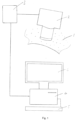
- FIG. 1 With reference to Figure 1 , there is shown a part of a patient's body 1, a hand-held video microscope head 2, video microscope controller 3, computer 4, keyboard 5 and display unit 6.
- the microscope head 2 is shown in contact with the patient's body 1 as it would be when acquiring images therefrom under the control of microscope controller 3. Once images have been obtained, the microscope head 2 is removed from the patient's body 1.
- Images are passed via the microscope controller 3 to the computer 4 for processing. This involves analysis of the images to identify and measure/quantify the following:-
- these characteristics are displayed on a screen for identification/analysis by a human operator who then makes appropriate entries of representative values via the keyboard 5.
- image recognition software identifies capillaries (and associated bleedings and haloes) and speed of blood flow therein and assigns values automatically
- the computer then calculates a weighted sum of these values and outputs this to the display 6, along with the values on which it is based.
- This score, together with scores for SmvO 2 is indicative of the degree of pathology of the neonate's microcirculation.
- FIG. 2 With reference to Figure 2 , there is shown a part of a patient's body 1, a light source 7, a probe 8, a spectrometer 9, computer 4, keyboard 5 and display unit 6.
- the probe 8 is shown in contact with the patient's body 1 as it would be when it is emitting light received by an optical fibre from the light source 7 and receiving reflected light from the body, this reflected light being transmitted via an optical fibre to the spectrometer 9. Once the reflected light has been processed by the spectrometer 9, the probe 8 is removed from the patient's body 1.
- Data from the spectrometer 9 are passed to the computer 4 for recordal and processing.
- the spectrometer 9 generates data in the form of reflectance spectra, decomposition of the spectra is performed by the computer 4 to estimate SmvO 2 and the heterogeneity of SmvO 2 .
- the computer then calculates a weighted sum of these values and outputs this to the display 6, together with the values on which it is based. This score, together with the score based on visual analysis is indicative of the degree of pathology of the neonate's microcirculation.
- the spectrometer 9 and microscope controller 3 are connected to the same computer 4.
- the computer 4 may analyse the collected frames/films and the spectra.
- the frames/films and spectra may be transferred to another computer for analysis and computer(s) 4 act only to receive the data from the microscope controller 3 and spectrometer 9.
- the present inventor was the first to appreciate the prognostic utility of analysis of the microcirculation of ICU patients.
- the present invention provides a method of making a prognosis for a patient with circulation failure being considered for, or undergoing, intensive care therapy, comprising assessing the state of the patient's microcirculation.
- This aspect of the invention is particularly applicable to patients with systemic circulation failure, for example following acute cardiac pump failure, hypovolemia or sepsis, and the supportive treatment may comprise extra-corporeal life support treatment (ECLS), e.g. extra-corporeal membrane oxygenation (ECMO).
- ECLS extra-corporeal life support treatment
- ECMO extra-corporeal membrane oxygenation
- Examinations according to the invention can be used to make a prognosis and hence improve selection of patients for life supporting treatments such as ECMO/ECLS and/or to guide such therapy as well as providing an indication of the effect of additional supportive therapy and the benefit in continuing with treatment i.e. to provide stop criteria for life supporting treatment.
- FIG. 1 With reference to Figure 1 , there is shown a part of a patient's body 1, a hand-held video microscope head 2, video microscope controller 3, computer 4, keyboard 5 and display unit 6.
- the microscope head 2 is shown in contact with the patient's body 1 as it would be when acquiring images therefrom under the control of microscope controller 3. Once images have been obtained, the microscope head 2 is removed from the patient's body 1.
- Images are passed via the microscope controller 3 to the computer 4 for processing. This involves analysis of the images to identify and measure/quantify the following:-
- these characteristics are displayed on a screen for identification/analysis by a human operator who then makes appropriate entries of representative values via the keyboard 5.
- image recognition software identifies capillaries (and associated bleedings and haloes) and speed of blood flow therein and assigns values automatically.
- the computer then calculates a weighted sum of these values and outputs this to the display 6, along with the values on which it is based. This score is indicative of the degree of pathology of the microcirculation.
- Figure 9(a) and (b) are graphs showing (a) oxygen saturation as a mean % value over time as measured by DRS and described in Example 5; and (b) the CoV of the values for oxygen saturation presented in (a).
- Arterial cannulation was performed in the groin in seven patients, in two of them via an end to side Dacron graft.
- One patient had arterial cannulation via the right subclavian artery.
- Seven patients had vein drainage via the femoral vein and one via the right atrium.
- the ECMO-circuit consisted of a centrifugal pump (Medtronics Incorporated, Minneapolis, Minnesota, USA), a heparin coated membrane oxygenator and tubes (Maquet Cardiovascular 72145 Hirrlingen, Germany). After establishing ECMO, all patients were initially treated with a flow of at least 4.0 l/min. Maintenance therapy on ECMO was guided by a standard protocol for the unit [38]. Weaning from ECMO was guided by trans-oesophageal ECCO Doppler in addition to clinical parameters.
- Skin microcirculation was evaluated with video microscopic measurements in eight patients and Laser Doppler measurements in six.
- CAVM Computer assisted video microscopy
- This technique involves a hand-held video-microscope applied gently on the surface of the region of interest (ROI). Immersion oil is used. Pictures or film sequences are projected and stored on a computer. For the first patient, E1, a less advanced microscope, with a 1.3 megapixel CCD, (ProScan, Bodelin technologies, OR, USA), magnifying 200 times was used. With this microscope pericapillary pathology, functional capillary density (FCD) and heterogeneity could be evaluated, but not capillary flow patterns. The remaining patients (E2-E8) were examined with another microscope (Microvision 2100, Finlay Microvision Co. Ltd., Warwickshire, UK) with higher resolution and a 500 times magnifying lens. An analogue to digital converter (Canopus, Kobe, Japan) was used to project and store the film sequences on a Mac Book pro, using the software iMovie (both Apple, Cupertino, USA).
- FCD functional capillary density
- LDPM is a technique for quantification of microvascular perfusion.
- the output of the technique is given in a semi-quantitative scale of flux, defined as the product of number of moving blood cells and their mean velocity in the measured volume ( ⁇ 1 mm 3 ).
- a Moor Blood Flow Monitor (MBF 3D) for perfusion measurements with Moorsoft both Moor instruments, Axminster, Devon, England was used for recording and analysis. Flux in a ROI was given as the mean value of seven consecutive measurements of a ten-second duration.
- Figure 6 shows skin microcirculation in fossa tabatière in two ECMO patients. To the left (a) several pericapillary bleedings are seen, and hardly no dot-liked capillaries are present (Patient E1, Proscan (x 200)). To the right (b) circular dark haloes are surrounding capillaries with no flow or extremely slow sluggish flow are seen (Patient E2 Microvision 2100 (x500)).
- the first microvascular examinations were performed as soon as possible, usually within 24 hours after establishing ECMO.
- the second measurement was performed on day 3 when possible.
- E4 was examined twice with an interval of only two hours before the ECMO was turned off due to ceased cerebral blood flow.
- Four survivors (Group 2) were controlled 18-65 days after weaning off ECMO.
- E3 was examined seven times.
- the skin on the dorsum of the hand, between the first and second metacarpus (fossa tabatière) was examined by CAVM in all patients. From patient 3 and onwards we also assessed the skin perfusion of the medial side of the right foot, with the ROI being located one third of the distance on an imaginary line from the medial malleolus to the caput of the first metatarsus.
- E1 pericapillary bleedings
- additional skin areas such as over arm, thigh, chest, and face were also examined.
- Six patients were measured by LDPM.
- Patient E1 was only measured in fossa tabatière. From patient 4 and onwards LDPM was measured on the foot as well.
- CAVM and Laser Doppler measurements were performed in both locations in all controls. At the time of microvascular measurements, the corresponding central hemodynamic parameters, results from blood tests and clinical parameters were noted.
- CoV coefficient of variation
- Figure 5 is an overview over the eight ECMO patients, from start of the ECMO treatment till death or last control measurement. Measurement periods are marked according to measuring technique.
- the patients were grouped according to clinical outcome into two groups: patients dying on ECMO (Group 1) and patients surviving ECMO (Group 2).
- Mean age for group 1 was 44 years, for group 2 it was 58 years. Both sexes were present in each group. In group 2, one patient had a recovery of heart function and a maintained cerebral function but died from bleeding complications in the intensive care unit.
- CAVM Computer assisted video microscopy
- E1 had numerous pericapillary bleedings in several skin locations (fossa tabatière, volar side of forearm, leg and face) on both examinations, figure 3 (a) . No other patients had any visible pericapillary bleedings. Patients E2 and E4 had circular dark haloes at a distance of 12 ⁇ 1 microns around some skin capillaries (65% in E2 and in 14% in E4), see figure 3 (b) .
- FCD Functional capillary density
- Figure 4 shows functional capillary density (in capillaries/mm 2 ) for three groups of ECMO patients and eight healthy controls (b) and coefficient of variation of functional capillary density for the same patients (a).
- the first, second and follow-up measurements are given.
- E2 pericapillary dark haloes
- erythrocyte movement in the capillaries was hardly detected in any of the film sequences, although the ECMO circuit gave an output of 4.5 litre/min.
- E3 had a prolonged and complicated stay in the ICU with lung infections, sepsis episodes, and progressive renal failure after ECMO treatment.
- the patient's final microscopy assessments on day 44 showed that FCD was reduced with 30%, CoV was increased with 64% and the mean flow-categorial velocity was reduced to 1.75, a reduction of 34%, compared with the initial measurements.
- the patient was examined during an episode with gram negative sepsis (day 24); at that time the FCD was markedly reduced to 49 capillaries/mm2 (-29%), the CoV was 0.17 (-23%), and 22% of the skin capillaries had brisk flow (category 4). Capillaries in this flow category were not observed for any other patients at any time.
- the controls had a mean flux value of 48 Au (range 22-96) in fossa Tabatière, while the corresponding values at the medial side of the foot were 31.5 (range 17.4-60.1).
- the mean coefficient of variation was 0.20 (range 0.13-0.35) in fossa Tabatière, and 0.25 (range 0.12-0.34) at the medial side of the foot.
- CAVM computer assisted video microscopy
- the patients that died on ECMO had reduced FCD and increased CoV of FCD compared with patients surviving ECMO and healthy controls.
- the patients surviving ECMO had stable FCD and CoV within the reference levels for the controls ( figure 7 ).
- the exception was patient E3, who had a complicated post ECMO course with several septic episodes.
- Freedlander used a microscope to show a decreased capillary density in skin in septic patients. Later studies confirmed Freedlanders findings and demonstrated increased heterogeneity of FCD in different tissues and mammalian species with systemic diseases. In patients with septic and cardiogenic shock a persistent severely reduced FCD for 24 hours in the sublingual area, is associated with increased mortality. Reduced FCD in the rectal mucosa is associated with poor prognosis in patients with severe malaria.
- Figure 7 shows distribution of capillaries in each of five flow categories for patients dying on- (group 1), and patients successfully weaned off ECMO (group 2) as compared to eight healthy controls.
- E2 and E4 Dark haloes were found in two of the patients who died on ECMO (E2 and E4), with the halo edges 12 ⁇ 1 microns from the capillaries. In E2 numerous haloes were present, prominently around capillaries with "no flow”. E4 had fewer haloes, and they were also seen surrounding perfused capillaries. The cause of these haloes is uncertain, but one possibility is that they represent precipitated proteins or erythrocyte degradation products leaking from injured capillaries. Another possibility is that they are caused by pericapillary oedema.
- thermoregulatory plexuses where perfusion is mainly regulated by sympathetic activity. Since perfusion in these plexuses mainly serve a thermoregulatory function, skin nutrition can not be assessed by the LDPM technique. Even though the two patients that died on ECMO (E1 and E4) had the lowest perfusion values of all in both locations and measuring periods, no significant differences were demonstrated du to small numbers and large data variation.
- the number of included patients is small and represents a heterogeneous group with acute heart failure, while the reference data were collected from healthy young male students. Since capillary erythrocyte velocities in healthy subjects are not age dependent, we assume that our control group of young students can be used.
- Microvascular examinations of skin nutritive capillaries in patients on ECMO show major structural and functional pathology in patients dying on ECMO, while patients surviving ECMO have results similar to healthy controls.
- the finding of intact skin microcirculatory morphology and function in survivors early after establishment of ECMO appears to be a robust and clinically useful finding implying a good prognosis.
- Pericapillary bleedings or dark haloes, micro-thrombi/capillaries with "no flow”, low capillary flow velocity and low functional capillary density are associated with poor prognosis.
- Table 3 During a six month period twenty-five healthy term newborns of Caucasian race with healthy mothers were enrolled within the first twenty-four hours after delivery (Table 3).
- CAVM Computer Assisted Video Microscopy
- FCD Functional capillary density
- Microvascular perfusion was assessed with a Moor Blood Flow Monitor (MBF 3D) with Moorsoft (Both Moor instruments, Axminster, Devon, England) for recordings and analyses.
- MPF 3D Moor Blood Flow Monitor
- Moorsoft Both Moor instruments, Axminster, Devon, England
- the output was given in a semi quantitative scale of flux (arbitrary unit, AU) defined as the product of the number of moving blood cells and their mean velocity in the measured volume (approximately 1 mm 3 ). Seven ten-second sequences with no or limited movement artifacts were taken from each ROI.
- Diffuse Reflectance Spectroscopy DRS
- a setup consisting of a spectrometer operating in the visible wavelength region (S2000, Avantes, The Netherlands) and a tungsten halogen light source (AvaLight-HAL, The Netherlands) having an effective spectral range of 450 to 800 nm was used.
- a polytetrafluoretylene tile (WS-2, Avantes, Netherland) enclosed in a black plastic housing was used as reference.
- a custom-built fiber optic probe was used for measurements with a fiber composition of three adjacent illuminating fibers (fiber diameter 400 ⁇ m) and one receiving fiber (fiber diameter 400 ⁇ m) resulting in an emitting-receiving distance for the probe of approximately 800 ⁇ m ( Meglinsky et al. Med Biol Eng Comput, 2001. 39(1): pp. 44-50 ). Twelve spectra were collected from each ROI in 20 neonates.
- Analyses of the spectra were done by adapting a tissue model based on a diffusion approximation ( Farrell et al., Med Phys, 1992. 19(4): pp. 879-88 ; Jacques, IEEE Transactions on Bio-Medical Engineering, 1989. 36(12): pp. 1155-1161 ).
- the model included the chromophores melanin, hemoglobin derivatives , water and a Mie and Rayleigh scattering factor.
- Decomposition of the spectral signature was done by a least square fit of the model to the measured spectra. The decomposition of reflected light spectra was then used to estimate the apparent content of oxy- and deoxy-hemoglobin. Microvascular oxygen saturation was compared with arterial oxygen saturation to estimate oxygen extraction.
- the arterial oxygen saturation was measured using a pulse oxymeter (Masimo Set, Rad-5v, Irvine, USA) with the probe located on the right hand.
- Skin temperature was measured using a surface temperature scanner (Omega Medical, Model no. STS-101-C, USA) attached to the ROI just before measurements with the microvascular techniques. Axillary temperature was taken with an ordinary thermometer (Digitemp, Microlife Asia, Mt1671, Taiwan).
- a transcutaneous bilimeter (Dräger, JM 103, Dräger medical, Lübeck, Made in Japan) was used to estimate bilirubin values, a factor in the DRS analysing algorithm.
- Demographic data are presented as mean with range. All other variables are reported as mean with standard deviation (SD).
- SD standard deviation
- paired t-test was conducted to compare means. P-value ⁇ 0.05 was considered significant.
- Statistical analyses were performed using SPSS for Windows (Statistical Package for the Social Sciences, version 18.0 SPSS Inc., Chicago, IL, USA).
- FCD Functional capillary density
- the dominant capillary flow category was category three (continuous high flow), but flow category two (continuous low flow) was also represented (Table 2, Fig. 4a and b ). Flow categories zero (no flow) and four (brisk flow) were not found.
- the mean flow-categorical velocity (MFCV) was similar both in the chest and in the hand at all the three days, varying between 2.57 (0.10) and 2.71 (0.11).
- Oxygen extraction defined as SaO 2 minus SmvO 2 showed significant difference between chest and hand with higher oxygen extraction in the hand on all three days (mean (SD)); Chest day 1-3: 14.5 (1.6), 14.1 (3.1), 11.7 (2.7); Hand day 1-3: 23.5 (3.4), 20.2 (2.8), 24.3 (3.5), There were no changes with time.
- the cardio-pulmonary adaptation in neonates with closure of the fetal shunts mainly occurs during the first hours of life, but is not completed until days to months after birth.
- Other adaptive responses such as reduction of total body water accompanied by 4 -7% weight loss, hemolysis of fetal erythrocytes and production of erythrocytes with adult hemoglobin, occurs during the first days to weeks.
- the hematocrit peaks at two hours of age and then decreases steadily over the next weeks.
- Our neonates were examined the first, second and third day of life when many of these adaptive processes affecting the central hemodynamics and hemorheology, and thereby microvascular perfusion, occur.
- FCD Functional capillary density
- FCD In the chest FCD varied between 10.7 and 11.3 c/mm, and in the hand between 12.7 and 13.2 c/mm, significantly higher in the hand compared to the chest on all three days (Table 3). There was also a clear tendency towards reduction in FCD from day one to day three for both locations.
- MFCV capillary flow-categorical velocity
- OPS Orthogonal Polarization Spectral Imaging
- SDF System-stream dark field imaging
- the laser Doppler principle for quantifying microvascular perfusion in tissue volumes in the range of 1 mm 3 has been commercially available for nearly 40 years, but the technique has hardly any routine applications in clinical medicine. This is partly explained by the fact that the method assesses a mix of both superficial papillary capillaries, mainly serving the nutritive perfusion, and deeper plexuses, mainly serving the thermoregulatory function.
- Diffuse Reflectance Spectroscopy was used to assess the oxygen saturation of erythrocytes in the microcirculation (SmvO 2 ).
- the measuring volume of DRS is dependent on the emitting light spectrum, the optical properties of the tissue and the design of the measuring probe.
- the equipment used in this study is estimated to have a measuring volume of ⁇ 1 mm 3 corresponding to a measuring depth of approximately 0.8 mm in skin (Meglinsky et al., supra).
- Microvascular oxygen saturation represents the balance between oxygen supply and consumption in the measuring volume.
- the supply is again dependent on the product of perfusion and arterial oxygen saturation, while the consumption is dependent on the metabolic rate of the tissue. Since SaO 2 in our newborns was near to 100%, our SvmO 2 data showed extraction between 12 and 20%.
- both thermoregulatory and nutritive perfusion takes place in the DRS measuring volume in contrast to in adult skin where only the papillary nutritive perfusion is assessed.
- Parts of the perfusion in the measuring volume in the newborn may also have a transport function (the horizontal structure in newborns as compared with the vertical papillary loops in the DRS measuring volumes of adult skin).
- microscopy and spectroscopy are the most useful techniques for assessing skin microcirculation in neonates. They both have measuring volumes of fractions of 1mm 3 and a resolution corresponding to individual microvessels. Small measuring volumes give high resolution, but at the price of a larger variation in measured values, i.e. a larger spread in repeated measurements. This problem can be handled by using the mean of repeated measurements to express an average value from a tissue, and the spread of repeated measurements can be used to describe the heterogeneity of the microcirculation.
- Erythromelalgia is a clinical syndrome characterized by erythema, increased skin temperature and burning pain in the extremities. The pain is relieved by cooling and aggravated by warming. EM is commonly divided into primary and secondary cases, depending on whether or not there is an underlying disease. Symptoms vary from mild discomfort to limbs threatening hypoxia and amputation.
- the pathogenesis of EM is debated.
- the inventor and others have proposed a hypothesis of a common final pathway of the pathogenesis: maldistribution of skin microvascular perfusion through anatomical or functional microvascular arterio-venous shunts, with increased thermoregulatory perfusion and a relative lack of nutritive capillary perfusion in affected skin.
- the tissue consequently becomes hypoxic, causing supplying arterioles to dilate, leading to a paradoxical situation with coexistence of hyperaemia and hypoxia.
- This hypothesis gives an explanation for why cooling universally reduces pain.
- the cooling reduces metabolism, and thereby the hypoxia; the improvement of tissue oxygenation reduces the arteriolar dilatation, and hyperaemia is less pronounced: the vicious cycle is reversed.
- CAVM Computer Assisted Video Microscopy
- This technique involves a hand held video-microscope applied gently on the surface of the region of interest. Pictures or film sequences are projected and stored on a computer.
- a digital video microscope (Optilia, D1, Sundbyberg, Sweden) with enlargement 250x, resolution 640x480 and frame rate 15 fps (frames per second) is used.
- An analogue to digital converter (Canopus, Kobe, Japan) is used to project and store the film sequences on a Mac Book pro, using the software iMovie (all Apple, Cupertino, USA).
- FCD vessel density
- the red blood cells' flow pattern in the capillaries varies over time.
- the heterogeneity of the mean flow values is expressed as CoV (Wester et al., supra ) .
- Diffuse Reflectance Spectroscopy DRS
- a spectrometric set-up is used with a spectrometer operating in the visible wavelength region (S2000, Avantes, Netherland) and a tungsten halogen light source (AvaLight-HAL, Netherland) having an effective spectral range of 450 to 800 nm.
- a polytetrafluoretylene tile (WS-2, Avantes, Netherland) enclosed in a black plastic housing was used as reference.
- a custom-built fiber optic probe is used for measurements with a fiber composition of three adjacent illuminating fibers ( ⁇ 400 ⁇ m) and a receiving fiber ( ⁇ 400 ⁇ m) resulting in an emitting-receiving distance for the probe of approximately 800 ⁇ m (Meglinsky I et al.
- Spectral analysis are done by adapting a tissue model based on a diffusion approximation (Farrell J, et al. supra ) .
- the model included the chromophores melanin, hemoglobin derivatives ( Zijlistra WG, et al.. Visible and Near Infrared Absorption Spectra of Human and Animal Haemoglobin: Determination and Application: Brill Academic Publishers; 2000 ), water ( Hale GM, et al. Appl Opt. 1973;12:555-63 ) and a scattering factor accounting for both Mie and Rayleigh components.
- Decomposition of the spectral signature is made by a least square fit of the model to the measured spectra. The decomposition of reflected light spectra is then used to estimate the apparent content of oxy- and deoxyhemoglobin.
- Acetylic salicylic acid acid is discontinued one week before the examinations and misoprostol is optimally discontinued for 4 weeks.
- Acute traumatic limb ischemia is a challenge for trauma surgeons. Is the life and function of the limb salvageable or has the period of ischemia given irreversible tissue damage? The surgeons may have to make quick decisions being aware that wrong decisions may cause either amputation of a savable limb or development of necrosis and sepsis in a non-savable limb where a salvage procedure was tried. In the worst case the latter may contribute to a fatal outcome in a multi-traumatized patient. Currently surgeons lack predictors for limb prognosis. A way of monitoring the circulation of a re-perfused limb is therefore of great importance.
- a 33 year old female patient was involved in a high-energy car crash. At the site of injury she could move the fingers of the injured arm and has maintained intact sensory function. She had a moderate severe brain concussion corresponding to a Glasgow Coma scale of 12, sixty minutes after the initial injury. She arrived at our hospital (a level I trauma center), nearly 120 minutes after the accident and was examined according to guidelines for advanced trauma and life support (ATLS).
- ATLS advanced trauma and life support
- CAVM Computer assisted video microscope
- DRS diffuse reflectance spectroscopy
- CAVM Computer assisted video microscopy
- a hand-held digital microscope (Mediscope, OP-120 011), (Optilia Instruments AB Sollentuna, Sweden) with a 200 x magnifying lens, a resolution 640 ⁇ 480 and frame rate of 15 frames per second was used. The use of the microscope and analysis of parameters is described elsewhere.
- CAVM parameters of interest are functional capillary density (FCD), heterogeneity of FCD, microvascular flow-patterns and pericapillary pathology.
- FCD functional capillary density
- FCD heterogeneity of FCD
- microvascular flow-patterns microvascular flow-patterns
- pericapillary pathology CAVM films were analyzed blindly by an experienced investigator and also by the software Java Cap (Eye catcher technologies, Linköping, Sweden). Baby oil (Natusan) was used as immersion oil for the video-microscope. All equipment was gently applied on the skin surface.
- Diffuse Reflectance Spectroscopy DRS
- a spectrometric setup consisting of a spectrometer operating in the visible wavelength region (S2000, Avantes, The Netherlands) and a tungsten halogen light source (AvaLight-HAL, The Netherlands) was used.
- the setup has an effective spectral range of 450 to 800 nm.
- a polytetrafluoretylene tile (WS-2, Avantes, Netherland) enclosed in a black plastic housing was used as reference.
- a custom-built fiber optic probe was used for measurements with a fiber composition of three adjacent illuminating fibers (fiber diameter 400 ⁇ m) and one receiving fiber (fiber diameter 400 ⁇ m) resulting in an emitting-receiving distance for the probe of approximately 800 ⁇ m.
- the estimated measuring volume of the equipment has been estimated to be in the range of 0.1mm 3 . Seven spectra were collected from each hand at each measurement.
- results from one hand were compared to the results from the other hand taken the same day. Results from one hand were also compared to results from the same hand taken another day. A group of eight healthy male students serve as a control group.
- the patient was treated in the intensive care unit. Except for wounds in her forehead and knee, the only injuries needing surgery were in her left arm. She was hemodynamically stable through the entire course, but received norepinephrine initially to maintain a mean arterial pressure above 70 mm Hg. She did not suffer from lung ventilation problems. She did not develop kidney failure or other organ failures. Attempts were made to wake her up at day 3, but due to confusion and exaggeration she was slept down again. On day 5 she had a fever episode (maximum temp 39,2°C), but no other signs of infection.
- CAVM Computer assisted video microscopy
- the heterogeneity of the FCD The coefficient of variation varied between 0.20 and 0.36 on the right arm and between 0.15 and 0.35 on the left side, for both hands the highest value was the day after the accident and reperfusion.
- Capillary flow pattern For all measurements except on the left arm on day 9, an overwhelming majority of capillaries were in category 2 and 3 (continuous flow). The flow pattern and mean-categorial flow index were in the same range as our healthy controls.
- Example 5 Case study and prognosis of a patient receiving ECMO
- HTx heart transplantation
- Clinical course The patient was treated with veno-arterial ECMO for 10 days. The ECMO was then converted to veno-venous ECMO and the patient died during this circulatory support on day 11. During this course she had multiple surgical revisions due to a bleeding tendency, partly related to the anticoagulation needed for the two mechanical heart valves, partly to a multi-transfusion syndrome with accompanying coagulopathy. The standard monitoring techniques showed acceptable values, and only on day 10 she was taken off the transplant list due lack of clinical progress and development of extensive ulcerations, skin necrosis, on the buttocks and the back.
- the microvascular data were continually reviewed shortly after collection.
- the CAVM collected frames and films were scored by two independent and experienced examiners according to the impression of FCD (parameter (a)), heterogeneity of FCD (parameter (b)), CFV (parameter (c)) and heterogeneity of FCV (parameter (d)), and a written report was made.
- FCD paraffin wax
- CFV paraffin wax
- d heterogeneity of FCV
- the DRS data, parameter (e) and (f), are shown in Figure 9(a) and (b ).
Landscapes
- Health & Medical Sciences (AREA)
- Life Sciences & Earth Sciences (AREA)
- Physics & Mathematics (AREA)
- Veterinary Medicine (AREA)
- Surgery (AREA)
- Engineering & Computer Science (AREA)
- Biomedical Technology (AREA)
- Heart & Thoracic Surgery (AREA)
- Medical Informatics (AREA)
- Molecular Biology (AREA)
- Biophysics (AREA)
- Animal Behavior & Ethology (AREA)
- General Health & Medical Sciences (AREA)
- Public Health (AREA)
- Pathology (AREA)
- Spectroscopy & Molecular Physics (AREA)
- Cardiology (AREA)
- Physiology (AREA)
- Optics & Photonics (AREA)
- Vascular Medicine (AREA)
- Hematology (AREA)
- Ophthalmology & Optometry (AREA)
- Dermatology (AREA)
- Pulmonology (AREA)
- Investigating Or Analysing Biological Materials (AREA)
- Measuring Pulse, Heart Rate, Blood Pressure Or Blood Flow (AREA)
- Measurement Of The Respiration, Hearing Ability, Form, And Blood Characteristics Of Living Organisms (AREA)
- Measuring And Recording Apparatus For Diagnosis (AREA)
- Liquid Crystal Substances (AREA)
- External Artificial Organs (AREA)
Claims (11)
- Procédé d'identification ou d'évaluation d'une insuffisance circulatoire chez un sujet (1), lequel procédé comprend l'étude de la microcirculation du sujet en fonction des paramètres suivants :(a) la densité capillaire fonctionnelle (FCD) ;(b) l'hétérogénéité de la FCD ;(c) la vitesse du flux capillaire ;(d) l'hétérogénéité de la vitesse du flux capillaire ;(e) la saturation en oxygène des érythrocytes microvasculaires (SmvO2) ; et(f) l'hétérogénéité de la SmvO2 ;dans lequel les paramètres (a) à (d) sont étudiés par analyse d'images de ceux-ci obtenues en utilisant un microscope (2, 3) dans lequel le microscope utilise une lumière blanche non polarisée, et les paramètres (e) et (f) sont étudiés par analyse de spectres obtenus par spectroscopie par réflectance diffuse (DRS) ;dans lequel l'analyse est réalisée sur un ordinateur (4) comprenant un logiciel destiné à l'analyse desdites images et desdits spectres ;dans lequel le procédé comprend en outre la comparaison des résultats de l'étude avec des valeurs de référence afin d'identifier ou d'évaluer une insuffisance circulatoire chez ledit sujet ;dans lequel la microcirculation de la peau est étudiée, dans lequel la peau n'inclut pas le sillon latéral de l'ongle ; etdans lequel le procédé est réalisé sur des données obtenues auprès du patient et le patient n'est pas présent pendant le procédé.
- Procédé selon la revendication 1, dans lequel la microscopie est une microscopie vidéo assistée par ordinateur (CAVM).
- Procédé selon la revendication 1, dans lequel le procédé est mis en oeuvre par ordinateur.
- Procédé d'établissement d'un pronostic pour un sujet atteint d'une insuffisance circulatoire (1), le procédé comprenant l'étude de la microcirculation du sujet en fonction des paramètres suivants :(a) la densité capillaire fonctionnelle (FCD) ;(b) l'hétérogénéité de la FCD ;(c) la vitesse du flux capillaire ;(d) l'hétérogénéité de la vitesse du flux capillaire ;(e) la saturation en oxygène des érythrocytes microvasculaires (SmvO2) ; et(f) l'hétérogénéité de la SmvO2 ;dans lequel les paramètres (a) à (d) sont étudiés visuellement par analyse d'images de ceux-ci obtenues en utilisant un microscope (2, 3) dans lequel le microscope utilise une lumière blanche non polarisée, et les paramètres (e) et (f) sont étudiés par analyse de spectres obtenus par spectroscopie par réflectance diffuse (DRS) ;dans lequel l'analyse est réalisée sur un ordinateur (4) comprenant un logiciel destiné à l'analyse desdites images et desdits spectres ;dans lequel le procédé comprend en outre la comparaison des résultats de l'étude avec des valeurs de référence afin d'établir un pronostic pour ledit sujet ;dans lequel la microcirculation de la peau est étudiée, dans lequel la peau n'inclut pas le sillon latéral de l'ongle ; etdans lequel le procédé est réalisé sur des données obtenues auprès du patient et le patient n'est pas présent pendant le procédé.
- Procédé selon une quelconque revendication précédente, comprenant en outre la mesure de l'extraction d'oxygène par la microvascularisation et, éventuellement, de l'hétérogénéité de ladite extraction.
- Procédé selon la revendication 5, dans lequel le procédé est employé pour étudier l'efficacité d'une intervention thérapeutique sur le sujet (1).
- Appareil d'étude d'une microcirculation d'un sujet comprenant un ordinateur (4) ; dans lequel l'ordinateur est agencé pour recevoir et traiter :(i) des images de la microcirculation du sujet (1) obtenues en utilisant un microscope (2, 3) utilisant une lumière blanche non polarisée pour déterminer les paramètres suivants associés à une pathologie :(a) la densité capillaire fonctionnelle (FCD) ;(b) l'hétérogénéité de la FCD ;(c) la vitesse du flux capillaire ;(d) l'hétérogénéité de la vitesse du flux capillaire ; et(ii) des données concernant la SmvO2 à partir d'un spectromètre (9) pour déterminer les paramètres suivants associés à une pathologie :(e) la saturation en oxygène des érythrocytes microvasculaires (SmvO2) ; et(f) l'hétérogénéité de la SmvO2 ;dans lequel l'ordinateur est en outre agencé pour comparer les résultats de l'étude avec des valeurs de référence ; etdans lequel les images et les données proviennent de la microcirculation de la peau, dans lequel la peau n'inclut pas le sillon latéral de l'ongle.
- Appareil selon la revendication 7, comprenant en outre ledit microscope (2, 3) et ledit spectromètre (9).
- Appareil selon la revendication 7 ou 8 comprenant en outre un moyen pour délivrer des valeurs correspondant aux paramètres et/ou une valeur basée sur ceux-ci en combinaison, par exemple une somme ou une moyenne pondérée.
- Logiciel comprenant des instructions pour amener un ordinateur (4) à exécuter les étapes de traitement d'images et de données définies dans l'une quelconque des revendications 1 à 6.
- Procédé selon l'une quelconque des revendications 1 à 6, dans lequel le sujet (1) :(i) est examiné en vue de ou suit des soins intensifs, tels qu'une oxygénation par membrane extracorporelle (ECMO) et/ou un traitement d'assistance respiratoire extracorporelle (ECLS) ; ou(ii) souffre d'une prééclampsie ; ou(iii) souffre d'une sepsie ; ou(iv) souffre d'une insuffisance cardiaque chronique ou aiguë ; ou(v) présente une plaie chronique de la peau ; ou(vi) est asphyxié ; ou(vii) présente une insuffisance respiratoire aiguë ou chronique ; ou(viii) présente une ischémie des membres aiguë ou chronique ;(ix) a subi une greffe d'organes ;(x) est atteint d'érythromélalgie.
Priority Applications (1)
| Application Number | Priority Date | Filing Date | Title |
|---|---|---|---|
| EP23177112.2A EP4235152A3 (fr) | 2013-01-28 | 2014-01-28 | Évaluation de déficit circulatoire |
Applications Claiming Priority (3)
| Application Number | Priority Date | Filing Date | Title |
|---|---|---|---|
| GB1301490.7A GB2510176A (en) | 2013-01-28 | 2013-01-28 | Determining patient prognosis by microcirculation analysis |
| GB201312796A GB201312796D0 (en) | 2013-07-17 | 2013-07-17 | Neonatal microcirculation analysis |
| PCT/EP2014/051661 WO2014114814A1 (fr) | 2013-01-28 | 2014-01-28 | Évaluation de déficit circulatoire |
Related Child Applications (1)
| Application Number | Title | Priority Date | Filing Date |
|---|---|---|---|
| EP23177112.2A Division EP4235152A3 (fr) | 2013-01-28 | 2014-01-28 | Évaluation de déficit circulatoire |
Publications (2)
| Publication Number | Publication Date |
|---|---|
| EP2948042A1 EP2948042A1 (fr) | 2015-12-02 |
| EP2948042B1 true EP2948042B1 (fr) | 2023-06-07 |
Family
ID=50029038
Family Applications (2)
| Application Number | Title | Priority Date | Filing Date |
|---|---|---|---|
| EP23177112.2A Pending EP4235152A3 (fr) | 2013-01-28 | 2014-01-28 | Évaluation de déficit circulatoire |
| EP14701757.8A Active EP2948042B1 (fr) | 2013-01-28 | 2014-01-28 | Évaluation de déficit circulatoire |
Family Applications Before (1)
| Application Number | Title | Priority Date | Filing Date |
|---|---|---|---|
| EP23177112.2A Pending EP4235152A3 (fr) | 2013-01-28 | 2014-01-28 | Évaluation de déficit circulatoire |
Country Status (5)
| Country | Link |
|---|---|
| US (1) | US10987011B2 (fr) |
| EP (2) | EP4235152A3 (fr) |
| JP (2) | JP6542130B2 (fr) |
| ES (1) | ES2949276T3 (fr) |
| WO (1) | WO2014114814A1 (fr) |
Families Citing this family (14)
| Publication number | Priority date | Publication date | Assignee | Title |
|---|---|---|---|---|
| US20210350927A1 (en) * | 2013-01-28 | 2021-11-11 | Oslo Universitetssykehus Hf | Assessing circulatory failure |
| JP6336949B2 (ja) | 2015-01-29 | 2018-06-06 | 富士フイルム株式会社 | 画像処理装置及び画像処理方法、並びに内視鏡システム |
| JP6688164B2 (ja) * | 2016-06-09 | 2020-04-28 | 花王株式会社 | 皮膚毛細血管の観察方法 |
| JP2020537279A (ja) | 2017-10-16 | 2020-12-17 | マサチューセッツ インスティテュート オブ テクノロジー | 非侵襲的な血液学的測定のためのシステム、デバイス、および方法 |
| CN111902080B (zh) * | 2018-03-26 | 2024-04-12 | 泰尔茂株式会社 | 支援系统、支援方法、支援程序及记录有支援程序的记录介质 |
| US10485552B1 (en) * | 2018-12-28 | 2019-11-26 | Imad R. Makhoul | Apparatus and method for controlling systemic blood pressure in patients |
| US11754824B2 (en) | 2019-03-26 | 2023-09-12 | Active Medical, BV | Method and apparatus for diagnostic analysis of the function and morphology of microcirculation alterations |
| US20220296108A1 (en) * | 2019-05-24 | 2022-09-22 | Seoul National University Hospital | Method and apparatus for quantitation of microcirculation |
| EP4048151A4 (fr) * | 2019-10-23 | 2023-04-05 | The Johns Hopkins University | Capillaroscope à éclairage décalé |
| GB2589068A (en) | 2019-10-31 | 2021-05-26 | Odi Medical As | Probe |
| JPWO2021095472A1 (fr) * | 2019-11-12 | 2021-05-20 | ||
| CN111657861B (zh) * | 2020-06-04 | 2022-02-25 | 浙江大学 | 基于双光子显微镜技术的溶栓药效评价方法 |
| CN112587105B (zh) * | 2020-12-11 | 2023-10-27 | 复旦大学附属中山医院 | 一种人体循环灌注状态评估系统 |
| WO2023011743A1 (fr) | 2021-08-05 | 2023-02-09 | Odi Medical As | Évaluation de la microcirculation |
Citations (1)
| Publication number | Priority date | Publication date | Assignee | Title |
|---|---|---|---|---|
| GB2510176A (en) * | 2013-01-28 | 2014-07-30 | Univ Oslo Hf | Determining patient prognosis by microcirculation analysis |
Family Cites Families (25)
| Publication number | Priority date | Publication date | Assignee | Title |
|---|---|---|---|---|
| CA2042075C (fr) * | 1991-05-08 | 2001-01-23 | Branko Palcic | Systeme d'imagerie endoscopique |
| US5769792A (en) * | 1991-07-03 | 1998-06-23 | Xillix Technologies Corp. | Endoscopic imaging system for diseased tissue |
| US6420709B1 (en) * | 1992-07-15 | 2002-07-16 | Optix Lp | Methods of minimizing scattering and improving tissue sampling in non-invasive testing and imaging |
| US5699797A (en) * | 1992-10-05 | 1997-12-23 | Dynamics Imaging, Inc. | Method of investigation of microcirculation functional dynamics of physiological liquids in skin and apparatus for its realization |
| US5553613A (en) * | 1994-08-17 | 1996-09-10 | Pfizer Inc. | Non invasive blood analyte sensor |
| US5713364A (en) * | 1995-08-01 | 1998-02-03 | Medispectra, Inc. | Spectral volume microprobe analysis of materials |
| US20010041843A1 (en) * | 1999-02-02 | 2001-11-15 | Mark Modell | Spectral volume microprobe arrays |
| US6104945A (en) * | 1995-08-01 | 2000-08-15 | Medispectra, Inc. | Spectral volume microprobe arrays |
| EP1148810B1 (fr) * | 1999-01-26 | 2005-11-16 | Newton Laboratories, Inc. | Systeme d'imagerie a autofluorescence pour endoscopie |
| CA2385849A1 (fr) * | 1999-09-23 | 2001-03-29 | Richard G. Nadeau | Applications medicales de formations d'images spectrales par polarisation croisee |
| US20020007122A1 (en) * | 1999-12-15 | 2002-01-17 | Howard Kaufman | Methods of diagnosing disease |
| US6377841B1 (en) * | 2000-03-31 | 2002-04-23 | Vanderbilt University | Tumor demarcation using optical spectroscopy |
| US20020165456A1 (en) * | 2001-03-26 | 2002-11-07 | Murat Canpolat | Estimation of the average size of white light scatterers in normal and cancerous tissue using light scattering spectrum |
| US20050154319A1 (en) * | 2002-01-15 | 2005-07-14 | Xillix Technologies Corporation | Fluorescence endoscopy video systems with no moving parts in the camera |
| US6899675B2 (en) * | 2002-01-15 | 2005-05-31 | Xillix Technologies Corp. | Fluorescence endoscopy video systems with no moving parts in the camera |
| US7706862B2 (en) * | 2003-04-17 | 2010-04-27 | Research Foundation Of The City University Of New York | Detecting human cancer through spectral optical imaging using key water absorption wavelengths |
| US8064976B2 (en) | 2003-10-03 | 2011-11-22 | Can Ince | Systems and methods for sidesstream dark field imaging |
| US20060241364A1 (en) * | 2004-10-01 | 2006-10-26 | Academisch Medisch Centrum Of The University Van Amsterdam | System and method for imaging the reflectance of a substrate |
| US9131861B2 (en) | 2004-11-30 | 2015-09-15 | Academisch Medisch Centrum | Pulsed lighting imaging systems and methods |
| JP4743486B2 (ja) | 2005-05-24 | 2011-08-10 | 微小循環研究所 有限会社 | 指先の毛細血管血流による医療診断支援システム。 |
| US8214023B2 (en) * | 2006-09-21 | 2012-07-03 | Institute Of Critical Care Medicine | Microcirculation imaging |
| WO2010093503A2 (fr) * | 2007-01-05 | 2010-08-19 | Myskin, Inc. | Procédés d'analyse de la peau |
| US8903247B2 (en) | 2009-10-30 | 2014-12-02 | Cisco Technology, Inc. | Bidirectional multi-mode fiber interface |
| US9134243B2 (en) * | 2009-12-18 | 2015-09-15 | University Health Network | System and method for sub-surface fluorescence imaging |
| JP5797921B2 (ja) | 2011-03-30 | 2015-10-21 | 花王株式会社 | 体表評価方法および体表評価装置 |
-
2014
- 2014-01-28 EP EP23177112.2A patent/EP4235152A3/fr active Pending
- 2014-01-28 EP EP14701757.8A patent/EP2948042B1/fr active Active
- 2014-01-28 ES ES14701757T patent/ES2949276T3/es active Active
- 2014-01-28 US US14/763,872 patent/US10987011B2/en active Active
- 2014-01-28 WO PCT/EP2014/051661 patent/WO2014114814A1/fr active Application Filing
- 2014-01-28 JP JP2015554205A patent/JP6542130B2/ja active Active
-
2019
- 2019-06-12 JP JP2019109448A patent/JP6785339B2/ja active Active
Patent Citations (1)
| Publication number | Priority date | Publication date | Assignee | Title |
|---|---|---|---|---|
| GB2510176A (en) * | 2013-01-28 | 2014-07-30 | Univ Oslo Hf | Determining patient prognosis by microcirculation analysis |
Non-Patent Citations (3)
| Title |
|---|
| AWAN Z A ET AL: "Diffuse reflectance spectroscopy: Systemic and microvascular oxygen saturation is linearly correlated and hypoxia leads to increased spatial heterogeneity of microvascular saturation", MICROVASCULAR RESEARCH, ACADEMIC PRESS, US, vol. 81, no. 3, 24 February 2011 (2011-02-24), pages 245 - 251, XP028192943, ISSN: 0026-2862, [retrieved on 20110302], DOI: 10.1016/J.MVR.2011.02.004 * |
| FREDLY S ET AL: "Poster Presentation Abstracts 324 SKIN MICROCIRCULATION IN HEALTHY NEWBORNS -ASSESSMENTS OF MORPHOLOGY, PERFUSION AND OXYGENATION", 30 November 2010 (2010-11-30), XP055907344, Retrieved from the Internet <URL:https://www.nature.com/articles/pr2010533.pdf> [retrieved on 20220330], DOI: 10.1203/00006450-201011001-00324 * |
| Z. A. AWAN ET AL: "Human microvascular imaging: a review of skin and tongue videomicroscopy techniques and analysing variables", CLINICAL PHYSIOLOGY AND FUNCTIONAL IMAGING, vol. 30, no. 2, 1 March 2010 (2010-03-01), GB, pages 79 - 88, XP055713029, ISSN: 1475-0961, DOI: 10.1111/j.1475-097X.2009.00913.x * |
Also Published As
| Publication number | Publication date |
|---|---|
| JP6785339B2 (ja) | 2020-11-18 |
| EP2948042A1 (fr) | 2015-12-02 |
| EP4235152A3 (fr) | 2023-09-27 |
| EP4235152A2 (fr) | 2023-08-30 |
| JP2019188172A (ja) | 2019-10-31 |
| WO2014114814A1 (fr) | 2014-07-31 |
| US20150359440A1 (en) | 2015-12-17 |
| ES2949276T3 (es) | 2023-09-27 |
| JP6542130B2 (ja) | 2019-07-10 |
| US10987011B2 (en) | 2021-04-27 |
| JP2016509505A (ja) | 2016-03-31 |
Similar Documents
| Publication | Publication Date | Title |
|---|---|---|
| EP2948042B1 (fr) | Évaluation de déficit circulatoire | |
| Nielsen et al. | The effects of red blood cell transfusion on tissue oxygenation and the microcirculation in the intensive care unit: a systematic review | |
| Awan et al. | Human microvascular imaging: a review of skin and tongue videomicroscopy techniques and analysing variables | |
| JP2003510112A (ja) | 直交偏光スペクトルイメージングの医学的応用 | |
| JP2013544588A (ja) | invivoにおける組織酸素化の判定 | |
| Wester et al. | Skin microvascular morphology and hemodynamics during treatment with veno-arterial extra-corporeal membrane oxygenation | |
| Banerjee et al. | Cerebral blood flow and oximetry response to blood transfusion in relation to chronological age in preterm infants | |
| Jung et al. | Evaluation of the microcirculation during extracorporeal membrane-oxygenation | |
| RU2580895C2 (ru) | Способ оценки регионарного кровообращения, тканевой микроциркуляции и насыщения крови кислородом и устройство для оценки регионарного кровообращения, тканевой микроциркуляции и насыщения крови кислородом | |
| Marcinkevics et al. | Hyperspectral evaluation of skin blood oxygen saturation at baseline and during arterial occlusion | |
| US20210350927A1 (en) | Assessing circulatory failure | |
| GB2510176A (en) | Determining patient prognosis by microcirculation analysis | |
| Kvernebo | Microcirculation and Tissue Perfusion Assessments for Complex Cardiovascular Disease Care | |
| Saknite et al. | Novel hybrid technology for early diagnostics of sepsis | |
| Weaver | Electrical Cardiometry During Mechanical Ventilation: An Exploratory Study | |
| Kvernebo | Ocular surface microcirculation. Studies using in vivo computer assisted microscopy and diffuse reflectance spectroscopy | |
| Müller | The influence of a patent ductus arteriosus on the peripheral muscle oxygenation and perfusion in neonates | |
| Robinson et al. | In vivo performance of a visible wavelength optical sensor for monitoring intestinal perfusion and oxygenation | |
| Kuryata et al. | Chest pain in patients with diabetes mellitus is associated with severe coronary artery calcification | |
| Sun et al. | Fetal Acid-Base Balance | |
| WO2023011743A1 (fr) | Évaluation de la microcirculation | |
| Kvernebo et al. | Human ocular surface microcirculation quantified by in vivo computer assisted video microscopy and diffuse reflectance spectroscopy | |
| Arslan et al. | Association between peripheral perfusion index and five vital signs in patients admitted to the emergency department Association between perfusion index and vital signs | |
| Kaneko et al. | Factors contributing to the accuracy of initial screening values measured by noninvasive spectrophotometric hemoglobin monitoring in an Emergency Department. | |
| Ceylan et al. | Monitoring of near-infrared spectrum values during packed red blood cell transfusion in pediatric intensive care unit |
Legal Events
| Date | Code | Title | Description |
|---|---|---|---|
| PUAI | Public reference made under article 153(3) epc to a published international application that has entered the european phase |
Free format text: ORIGINAL CODE: 0009012 |
|
| 17P | Request for examination filed |
Effective date: 20150807 |
|
| AK | Designated contracting states |
Kind code of ref document: A1 Designated state(s): AL AT BE BG CH CY CZ DE DK EE ES FI FR GB GR HR HU IE IS IT LI LT LU LV MC MK MT NL NO PL PT RO RS SE SI SK SM TR |
|
| AX | Request for extension of the european patent |
Extension state: BA ME |
|
| DAX | Request for extension of the european patent (deleted) | ||
| STAA | Information on the status of an ep patent application or granted ep patent |
Free format text: STATUS: EXAMINATION IS IN PROGRESS |
|
| 17Q | First examination report despatched |
Effective date: 20190828 |
|
| STAA | Information on the status of an ep patent application or granted ep patent |
Free format text: STATUS: EXAMINATION IS IN PROGRESS |
|
| GRAP | Despatch of communication of intention to grant a patent |
Free format text: ORIGINAL CODE: EPIDOSNIGR1 |
|
| STAA | Information on the status of an ep patent application or granted ep patent |
Free format text: STATUS: GRANT OF PATENT IS INTENDED |
|
| INTG | Intention to grant announced |
Effective date: 20221220 |
|
| GRAS | Grant fee paid |
Free format text: ORIGINAL CODE: EPIDOSNIGR3 |
|
| GRAA | (expected) grant |
Free format text: ORIGINAL CODE: 0009210 |
|
| STAA | Information on the status of an ep patent application or granted ep patent |
Free format text: STATUS: THE PATENT HAS BEEN GRANTED |
|
| AK | Designated contracting states |
Kind code of ref document: B1 Designated state(s): AL AT BE BG CH CY CZ DE DK EE ES FI FR GB GR HR HU IE IS IT LI LT LU LV MC MK MT NL NO PL PT RO RS SE SI SK SM TR |
|
| REG | Reference to a national code |
Ref country code: GB Ref legal event code: FG4D |
|
| REG | Reference to a national code |
Ref country code: CH Ref legal event code: EP Ref country code: AT Ref legal event code: REF Ref document number: 1572298 Country of ref document: AT Kind code of ref document: T Effective date: 20230615 |
|
| REG | Reference to a national code |
Ref country code: DE Ref legal event code: R096 Ref document number: 602014087126 Country of ref document: DE |
|
| P01 | Opt-out of the competence of the unified patent court (upc) registered |
Effective date: 20230602 |
|
| REG | Reference to a national code |
Ref country code: LT Ref legal event code: MG9D |
|
| REG | Reference to a national code |
Ref country code: ES Ref legal event code: FG2A Ref document number: 2949276 Country of ref document: ES Kind code of ref document: T3 Effective date: 20230927 |
|
| REG | Reference to a national code |
Ref country code: NL Ref legal event code: MP Effective date: 20230607 |
|
| PG25 | Lapsed in a contracting state [announced via postgrant information from national office to epo] |
Ref country code: SE Free format text: LAPSE BECAUSE OF FAILURE TO SUBMIT A TRANSLATION OF THE DESCRIPTION OR TO PAY THE FEE WITHIN THE PRESCRIBED TIME-LIMIT Effective date: 20230607 Ref country code: NO Free format text: LAPSE BECAUSE OF FAILURE TO SUBMIT A TRANSLATION OF THE DESCRIPTION OR TO PAY THE FEE WITHIN THE PRESCRIBED TIME-LIMIT Effective date: 20230907 |
|
| REG | Reference to a national code |
Ref country code: AT Ref legal event code: MK05 Ref document number: 1572298 Country of ref document: AT Kind code of ref document: T Effective date: 20230607 |
|
| PG25 | Lapsed in a contracting state [announced via postgrant information from national office to epo] |
Ref country code: RS Free format text: LAPSE BECAUSE OF FAILURE TO SUBMIT A TRANSLATION OF THE DESCRIPTION OR TO PAY THE FEE WITHIN THE PRESCRIBED TIME-LIMIT Effective date: 20230607 Ref country code: NL Free format text: LAPSE BECAUSE OF FAILURE TO SUBMIT A TRANSLATION OF THE DESCRIPTION OR TO PAY THE FEE WITHIN THE PRESCRIBED TIME-LIMIT Effective date: 20230607 Ref country code: LV Free format text: LAPSE BECAUSE OF FAILURE TO SUBMIT A TRANSLATION OF THE DESCRIPTION OR TO PAY THE FEE WITHIN THE PRESCRIBED TIME-LIMIT Effective date: 20230607 Ref country code: LT Free format text: LAPSE BECAUSE OF FAILURE TO SUBMIT A TRANSLATION OF THE DESCRIPTION OR TO PAY THE FEE WITHIN THE PRESCRIBED TIME-LIMIT Effective date: 20230607 Ref country code: HR Free format text: LAPSE BECAUSE OF FAILURE TO SUBMIT A TRANSLATION OF THE DESCRIPTION OR TO PAY THE FEE WITHIN THE PRESCRIBED TIME-LIMIT Effective date: 20230607 Ref country code: GR Free format text: LAPSE BECAUSE OF FAILURE TO SUBMIT A TRANSLATION OF THE DESCRIPTION OR TO PAY THE FEE WITHIN THE PRESCRIBED TIME-LIMIT Effective date: 20230908 |
|
| PG25 | Lapsed in a contracting state [announced via postgrant information from national office to epo] |
Ref country code: FI Free format text: LAPSE BECAUSE OF FAILURE TO SUBMIT A TRANSLATION OF THE DESCRIPTION OR TO PAY THE FEE WITHIN THE PRESCRIBED TIME-LIMIT Effective date: 20230607 |
|
| PG25 | Lapsed in a contracting state [announced via postgrant information from national office to epo] |
Ref country code: SK Free format text: LAPSE BECAUSE OF FAILURE TO SUBMIT A TRANSLATION OF THE DESCRIPTION OR TO PAY THE FEE WITHIN THE PRESCRIBED TIME-LIMIT Effective date: 20230607 |
|
| PG25 | Lapsed in a contracting state [announced via postgrant information from national office to epo] |
Ref country code: IS Free format text: LAPSE BECAUSE OF FAILURE TO SUBMIT A TRANSLATION OF THE DESCRIPTION OR TO PAY THE FEE WITHIN THE PRESCRIBED TIME-LIMIT Effective date: 20231007 |
|
| PG25 | Lapsed in a contracting state [announced via postgrant information from national office to epo] |
Ref country code: SM Free format text: LAPSE BECAUSE OF FAILURE TO SUBMIT A TRANSLATION OF THE DESCRIPTION OR TO PAY THE FEE WITHIN THE PRESCRIBED TIME-LIMIT Effective date: 20230607 Ref country code: SK Free format text: LAPSE BECAUSE OF FAILURE TO SUBMIT A TRANSLATION OF THE DESCRIPTION OR TO PAY THE FEE WITHIN THE PRESCRIBED TIME-LIMIT Effective date: 20230607 Ref country code: RO Free format text: LAPSE BECAUSE OF FAILURE TO SUBMIT A TRANSLATION OF THE DESCRIPTION OR TO PAY THE FEE WITHIN THE PRESCRIBED TIME-LIMIT Effective date: 20230607 Ref country code: PT Free format text: LAPSE BECAUSE OF FAILURE TO SUBMIT A TRANSLATION OF THE DESCRIPTION OR TO PAY THE FEE WITHIN THE PRESCRIBED TIME-LIMIT Effective date: 20231009 Ref country code: IS Free format text: LAPSE BECAUSE OF FAILURE TO SUBMIT A TRANSLATION OF THE DESCRIPTION OR TO PAY THE FEE WITHIN THE PRESCRIBED TIME-LIMIT Effective date: 20231007 Ref country code: EE Free format text: LAPSE BECAUSE OF FAILURE TO SUBMIT A TRANSLATION OF THE DESCRIPTION OR TO PAY THE FEE WITHIN THE PRESCRIBED TIME-LIMIT Effective date: 20230607 Ref country code: CZ Free format text: LAPSE BECAUSE OF FAILURE TO SUBMIT A TRANSLATION OF THE DESCRIPTION OR TO PAY THE FEE WITHIN THE PRESCRIBED TIME-LIMIT Effective date: 20230607 Ref country code: AT Free format text: LAPSE BECAUSE OF FAILURE TO SUBMIT A TRANSLATION OF THE DESCRIPTION OR TO PAY THE FEE WITHIN THE PRESCRIBED TIME-LIMIT Effective date: 20230607 |
|
| PG25 | Lapsed in a contracting state [announced via postgrant information from national office to epo] |
Ref country code: PL Free format text: LAPSE BECAUSE OF FAILURE TO SUBMIT A TRANSLATION OF THE DESCRIPTION OR TO PAY THE FEE WITHIN THE PRESCRIBED TIME-LIMIT Effective date: 20230607 |
|
| REG | Reference to a national code |
Ref country code: DE Ref legal event code: R097 Ref document number: 602014087126 Country of ref document: DE |
|
| PLBE | No opposition filed within time limit |
Free format text: ORIGINAL CODE: 0009261 |
|
| STAA | Information on the status of an ep patent application or granted ep patent |
Free format text: STATUS: NO OPPOSITION FILED WITHIN TIME LIMIT |
|
| PGFP | Annual fee paid to national office [announced via postgrant information from national office to epo] |
Ref country code: ES Payment date: 20240208 Year of fee payment: 11 |
|
| PG25 | Lapsed in a contracting state [announced via postgrant information from national office to epo] |
Ref country code: DK Free format text: LAPSE BECAUSE OF FAILURE TO SUBMIT A TRANSLATION OF THE DESCRIPTION OR TO PAY THE FEE WITHIN THE PRESCRIBED TIME-LIMIT Effective date: 20230607 |
|
| PGFP | Annual fee paid to national office [announced via postgrant information from national office to epo] |
Ref country code: DE Payment date: 20240123 Year of fee payment: 11 Ref country code: GB Payment date: 20240118 Year of fee payment: 11 |
|
| PG25 | Lapsed in a contracting state [announced via postgrant information from national office to epo] |
Ref country code: SI Free format text: LAPSE BECAUSE OF FAILURE TO SUBMIT A TRANSLATION OF THE DESCRIPTION OR TO PAY THE FEE WITHIN THE PRESCRIBED TIME-LIMIT Effective date: 20230607 |
|
| 26N | No opposition filed |
Effective date: 20240308 |