EP2932983A1 - Kernspinresonanzdiagnosemittel und verfahren zur detektion oder diagnose des zustands einer zelle, eines gewebes oder eines organs in einer person damit - Google Patents
Kernspinresonanzdiagnosemittel und verfahren zur detektion oder diagnose des zustands einer zelle, eines gewebes oder eines organs in einer person damit Download PDFInfo
- Publication number
- EP2932983A1 EP2932983A1 EP13863271.6A EP13863271A EP2932983A1 EP 2932983 A1 EP2932983 A1 EP 2932983A1 EP 13863271 A EP13863271 A EP 13863271A EP 2932983 A1 EP2932983 A1 EP 2932983A1
- Authority
- EP
- European Patent Office
- Prior art keywords
- subject
- magnetic resonance
- nuclear magnetic
- ala
- organ
- Prior art date
- Legal status (The legal status is an assumption and is not a legal conclusion. Google has not performed a legal analysis and makes no representation as to the accuracy of the status listed.)
- Granted
Links
- 0 COC(CCC(O*)=O)CN** Chemical compound COC(CCC(O*)=O)CN** 0.000 description 1
Images

Classifications
-
- A—HUMAN NECESSITIES
- A61—MEDICAL OR VETERINARY SCIENCE; HYGIENE
- A61B—DIAGNOSIS; SURGERY; IDENTIFICATION
- A61B5/00—Measuring for diagnostic purposes; Identification of persons
- A61B5/05—Detecting, measuring or recording for diagnosis by means of electric currents or magnetic fields; Measuring using microwaves or radio waves
- A61B5/055—Detecting, measuring or recording for diagnosis by means of electric currents or magnetic fields; Measuring using microwaves or radio waves involving electronic [EMR] or nuclear [NMR] magnetic resonance, e.g. magnetic resonance imaging
-
- A—HUMAN NECESSITIES
- A61—MEDICAL OR VETERINARY SCIENCE; HYGIENE
- A61K—PREPARATIONS FOR MEDICAL, DENTAL OR TOILETRY PURPOSES
- A61K49/00—Preparations for testing in vivo
- A61K49/06—Nuclear magnetic resonance [NMR] contrast preparations; Magnetic resonance imaging [MRI] contrast preparations
- A61K49/08—Nuclear magnetic resonance [NMR] contrast preparations; Magnetic resonance imaging [MRI] contrast preparations characterised by the carrier
- A61K49/10—Organic compounds
- A61K49/101—Organic compounds the carrier being a complex-forming compound able to form MRI-active complexes with paramagnetic metals
- A61K49/103—Organic compounds the carrier being a complex-forming compound able to form MRI-active complexes with paramagnetic metals the complex-forming compound being acyclic, e.g. DTPA
-
- A—HUMAN NECESSITIES
- A61—MEDICAL OR VETERINARY SCIENCE; HYGIENE
- A61K—PREPARATIONS FOR MEDICAL, DENTAL OR TOILETRY PURPOSES
- A61K49/00—Preparations for testing in vivo
- A61K49/06—Nuclear magnetic resonance [NMR] contrast preparations; Magnetic resonance imaging [MRI] contrast preparations
- A61K49/08—Nuclear magnetic resonance [NMR] contrast preparations; Magnetic resonance imaging [MRI] contrast preparations characterised by the carrier
- A61K49/10—Organic compounds
-
- A—HUMAN NECESSITIES
- A61—MEDICAL OR VETERINARY SCIENCE; HYGIENE
- A61K—PREPARATIONS FOR MEDICAL, DENTAL OR TOILETRY PURPOSES
- A61K49/00—Preparations for testing in vivo
- A61K49/06—Nuclear magnetic resonance [NMR] contrast preparations; Magnetic resonance imaging [MRI] contrast preparations
- A61K49/18—Nuclear magnetic resonance [NMR] contrast preparations; Magnetic resonance imaging [MRI] contrast preparations characterised by a special physical form, e.g. emulsions, microcapsules, liposomes
- A61K49/1818—Nuclear magnetic resonance [NMR] contrast preparations; Magnetic resonance imaging [MRI] contrast preparations characterised by a special physical form, e.g. emulsions, microcapsules, liposomes particles, e.g. uncoated or non-functionalised microparticles or nanoparticles
- A61K49/1821—Nuclear magnetic resonance [NMR] contrast preparations; Magnetic resonance imaging [MRI] contrast preparations characterised by a special physical form, e.g. emulsions, microcapsules, liposomes particles, e.g. uncoated or non-functionalised microparticles or nanoparticles coated or functionalised microparticles or nanoparticles
- A61K49/1824—Nuclear magnetic resonance [NMR] contrast preparations; Magnetic resonance imaging [MRI] contrast preparations characterised by a special physical form, e.g. emulsions, microcapsules, liposomes particles, e.g. uncoated or non-functionalised microparticles or nanoparticles coated or functionalised microparticles or nanoparticles coated or functionalised nanoparticles
- A61K49/1827—Nuclear magnetic resonance [NMR] contrast preparations; Magnetic resonance imaging [MRI] contrast preparations characterised by a special physical form, e.g. emulsions, microcapsules, liposomes particles, e.g. uncoated or non-functionalised microparticles or nanoparticles coated or functionalised microparticles or nanoparticles coated or functionalised nanoparticles having a (super)(para)magnetic core, being a solid MRI-active material, e.g. magnetite, or composed of a plurality of MRI-active, organic agents, e.g. Gd-chelates, or nuclei, e.g. Eu3+, encapsulated or entrapped in the core of the coated or functionalised nanoparticle
- A61K49/1833—Nuclear magnetic resonance [NMR] contrast preparations; Magnetic resonance imaging [MRI] contrast preparations characterised by a special physical form, e.g. emulsions, microcapsules, liposomes particles, e.g. uncoated or non-functionalised microparticles or nanoparticles coated or functionalised microparticles or nanoparticles coated or functionalised nanoparticles having a (super)(para)magnetic core, being a solid MRI-active material, e.g. magnetite, or composed of a plurality of MRI-active, organic agents, e.g. Gd-chelates, or nuclei, e.g. Eu3+, encapsulated or entrapped in the core of the coated or functionalised nanoparticle having a (super)(para)magnetic core coated or functionalised with a small organic molecule
- A61K49/1836—Nuclear magnetic resonance [NMR] contrast preparations; Magnetic resonance imaging [MRI] contrast preparations characterised by a special physical form, e.g. emulsions, microcapsules, liposomes particles, e.g. uncoated or non-functionalised microparticles or nanoparticles coated or functionalised microparticles or nanoparticles coated or functionalised nanoparticles having a (super)(para)magnetic core, being a solid MRI-active material, e.g. magnetite, or composed of a plurality of MRI-active, organic agents, e.g. Gd-chelates, or nuclei, e.g. Eu3+, encapsulated or entrapped in the core of the coated or functionalised nanoparticle having a (super)(para)magnetic core coated or functionalised with a small organic molecule the small organic molecule being a carboxylic acid having less than 8 carbon atoms in the main chain
-
- C—CHEMISTRY; METALLURGY
- C07—ORGANIC CHEMISTRY
- C07C—ACYCLIC OR CARBOCYCLIC COMPOUNDS
- C07C229/00—Compounds containing amino and carboxyl groups bound to the same carbon skeleton
- C07C229/02—Compounds containing amino and carboxyl groups bound to the same carbon skeleton having amino and carboxyl groups bound to acyclic carbon atoms of the same carbon skeleton
- C07C229/04—Compounds containing amino and carboxyl groups bound to the same carbon skeleton having amino and carboxyl groups bound to acyclic carbon atoms of the same carbon skeleton the carbon skeleton being acyclic and saturated
- C07C229/22—Compounds containing amino and carboxyl groups bound to the same carbon skeleton having amino and carboxyl groups bound to acyclic carbon atoms of the same carbon skeleton the carbon skeleton being acyclic and saturated the carbon skeleton being further substituted by oxygen atoms
-
- G—PHYSICS
- G01—MEASURING; TESTING
- G01R—MEASURING ELECTRIC VARIABLES; MEASURING MAGNETIC VARIABLES
- G01R33/00—Arrangements or instruments for measuring magnetic variables
- G01R33/20—Arrangements or instruments for measuring magnetic variables involving magnetic resonance
- G01R33/44—Arrangements or instruments for measuring magnetic variables involving magnetic resonance using nuclear magnetic resonance [NMR]
- G01R33/48—NMR imaging systems
- G01R33/54—Signal processing systems, e.g. using pulse sequences ; Generation or control of pulse sequences; Operator console
- G01R33/56—Image enhancement or correction, e.g. subtraction or averaging techniques, e.g. improvement of signal-to-noise ratio and resolution
- G01R33/5601—Image enhancement or correction, e.g. subtraction or averaging techniques, e.g. improvement of signal-to-noise ratio and resolution involving use of a contrast agent for contrast manipulation, e.g. a paramagnetic, super-paramagnetic, ferromagnetic or hyperpolarised contrast agent
Definitions
- the present invention relates to a nuclear magnetic resonance diagnostic agent, and more specifically to a nuclear magnetic resonance diagnostic agent comprising 5-aminolevulinic acid or a derivative thereof, or a salt thereof. Moreover, the present invention also relates to a method for detecting or diagnosing a state of a cell, tissue or organ in a subject, using the aforementioned nuclear magnetic resonance diagnostic agent, or a method for obtaining a T2-weighted image in which the contrast is increased in MRI.
- X-ray CT computed tomography
- PET positron emission tomography
- MRI magnetic resonance imaging
- X-ray CT has been problematic in terms of external exposure to X-ray irradiation upon examination.
- PET requires administration of a radioisotope such as 11 C-methionine as a tracer into a living body.
- a radioisotope such as 11 C-methionine
- PET has been problematic in terms of the management of such a radioisotope or internal exposure.
- PET has been known to have low spatial resolution.
- MRI is a technique of applying a nuclear magnetic resonance (NMR) phenomenon to computed tomography, and thus, it is considered that MRI does not cause radiation exposure, differing from X-ray CT or PET. Moreover, MRI has been known to have higher tissue-specificity, compared with other modalities such as X-ray CT.
- NMR nuclear magnetic resonance
- T1-weighted image mainly, an image created from a nuclear magnetization distribution contrasted with longitudinal relaxation
- T2-weighted image mainly, an image created from a nuclear magnetization distribution contrasted with transverse relaxation
- TE echo time
- TR repetition time
- a T1-weighted image and/or a T2-weighted image each having different image contrast are obtained.
- images are processed into and/or outputted as monochrome images having contrasting density.
- T1 relaxation time When a T1 value (T1 relaxation time) is measured, the shorter the time, the whiter the color that can be obtained (becomes higher signals), and the longer the time, the blacker the color that can be obtained (becomes lower signals), so that the color can be contrasted to create an image (T1-weighted image).
- T2 relaxation time the longer the time, the whiter the color that can be obtained (becomes higher signals),and the shorter the time, the blacker the color that can be obtained (becomes lower signals), so that the color can be contrasted to create an image (T2-weighted image).
- Typical contrast agents are, for example, gadolinium complexes such as Gd-DTPA (gadolinium-diethylenetriaminepentaacetic acid), which have an action to reduce the T1 relaxation time and increase the contrast (signal intensity) in a T1-weighted image.
- Gd-DTPA gadolinium-diethylenetriaminepentaacetic acid
- Gd-DTPA is used, for example, in detection of glioma. Since Gd-DTPA is chelated, the side effects thereof are reduced compared with a free gadolinium. However, Gd-DTPA still has side effects such as allergic reaction, liver toxicity or renal toxicity. In addition, since Gd-DTPA does not have site-specificity (e.g., tumor selectivity), a large amount of Gd-DTPA has needed to be administered in order to, for example, clarify the contrast between a tumor and other tissues (Patent Literature 1).
- site-specificity e.g., tumor selectivity
- Patent Literature 1 International Publication No. WO 2009/157561
- Protoporphyrin IX (hereinafter also referred to as "PpIX”) has been known as a precursor for pigments such as heme and chlorophyll, and this precursor has been known to have the property of being site-specifically accumulated, for example, in cancer tissues.
- PpIX 5-aminolevulinic acid
- ALA 5-aminolevulinic acid
- ALA 5-aminolevulinic acid
- ALA 5-aminolevulinic acid
- ALA 5-aminolevulinic acid
- ALA 5-aminolevulinic acid
- ALA 5-aminolevulinic acid
- ALA 5-aminolevulinic acid
- ALA 5-aminolevulinic acid
- ALA 5-aminolevulinic acid
- ALA 5-aminolevulinic acid
- ALA 5-aminolevulinic acid
- ALA 5-aminolevulinic acid
- ALA 5-aminolevulinic acid
- the present inventors have conducted intensive studies. As a result, the inventors have focused on a phenomenon in which metabolites of ALA or a derivative thereof administered into a living body (in particular, PpIX) are accumulated in a specific cell, tissue or organ, etc., and they have then analyzed a site in which the metabolites would be accumulated after administration of ALA to the living body, utilizing nuclear magnetic resonance. As a result, the present inventors have surprisingly found that ALA and a derivative thereof are useful as diagnostic agents for solving the aforementioned problem, thereby completing the present invention. To date, an example of combining administration of ALA or a derivative thereof to a living body with a nuclear magnetic resonance method has not yet been reported.
- the object of the present invention can be achieved by providing a nuclear magnetic resonance diagnostic agent containing a compound represented by the following formula (I) or a salt thereof: wherein R 1 represents a hydrogen atom or an acyl group, and R 2 represents a hydrogen atom, a linear or branched alkyl group, a cycloalkyl group, an aryl group, or an aralkyl group.
- R 1 represents a hydrogen atom or an acyl group
- R 2 represents a hydrogen atom, a linear or branched alkyl group, a cycloalkyl group, an aryl group, or an aralkyl group.
- R 1 and R 2 preferably each represent a hydrogen atom.
- the nuclear magnetic resonance diagnostic agent of the present invention is preferably a contrast agent used in MRI.
- the nuclear magnetic resonance diagnostic agent of the present invention preferably increases the contrast of a T2-weighted image, utilizing nuclear magnetic resonance.
- the nuclear magnetic resonance diagnostic agent of the present invention is preferably used to diagnose a disease or a condition selected from the group consisting of neoplastic diseases, infectious diseases, inflammatory diseases, autoimmune diseases, demyelinating diseases, metabolic diseases, degenerative diseases, vascular disorders, and injuries.
- the nuclear magnetic resonance diagnostic agent of the present invention preferably further contains one or two or more metal-containing compounds.
- the nuclear magnetic resonance diagnostic agent of the present invention preferably contains an iron-containing compound as the metal-containing compound.
- the present invention provides a method for detecting a condition of a cell, tissue or organ in a subject, the method comprising a step of detecting the condition of a cell, tissue or organ in a subject to which a compound represented by the following formula (I) or a salt thereof has been administered, utilizing nuclear magnetic resonance: wherein R 1 represents a hydrogen atom or an acyl group, and R 2 represents a hydrogen atom, a linear or branched alkyl group, a cycloalkyl group, an aryl group, or an aralkyl group.
- the present invention provides a method for diagnosing a condition of a cell, tissue or organ in a subject, comprising:
- the cell, tissue or organ in the subject is preferably a site in which the metabolite of the compound represented by the above formula (I) or a salt thereof is accumulated.
- the cell, tissue or organ in the subject is preferably a tumor, that is, a site in which a metabolite of the compound represented by the above formula (I) or a salt thereof is accumulated.
- the present invention provides a method for obtaining a T2-weighted image in which the contrast is increased in MRI, comprising:
- the nuclear magnetic resonance diagnostic agent of the present invention it becomes possible to further reduce toxicity on a living body and side effects, and also to site-specifically detect or diagnose a condition of a cell, tissue or organ in a subject, and the like.
- the nuclear magnetic resonance diagnostic agent of the present invention has reduced toxicity on a living body and causes fewer side effects, when compared with conventional in vivo diagnostic agents (for example, gadolinium contrast agents or radioisotopes).
- the compound contained as an active ingredient in the nuclear magnetic resonance diagnostic agent of the present invention is ALA or a derivative thereof, or a salt thereof, and the compound represented by the above formula (I) or a salt thereof (hereinafter, they are also referred to as "ALA member(s)", collectively) can be shown as examples.
- ALA that is also referred to as ⁇ -aminolevulinic acid is one of amino acids, wherein R 1 and R 2 in the above formula (I) each represent a hydrogen atom.
- the ALA derivative may be a compound other than ALA, wherein R 1 in the above formula (I) represents a hydrogen atom or an acyl group and R 2 in the above formula (I) represents a hydrogen atom, a linear or branched alkyl group, a cycloalkyl group, an aryl group, or an aralkyl group.
- the acyl group in the above formula (I) is not limited.
- the acyl group may include: a linear or branched alkanoyl group having 1 to 8 carbon atoms, such as formyl, acetyl, propionyl, butyryl, isobutyryl, valeryl, isovaleryl, pivaloyl, hexanoyl, octanoyl and benzylcarbonyl groups; and an aroyl group having 7 to 14 carbon atoms, such as benzoyl, 1-naphtoyl and 2-naphthoyl groups.
- the alkyl group in the above formula (I) is not limited.
- Examples of the alkyl group may include: a linear or branched alkyl group having 1 to 8 carbon atoms, such as methyl, ethyl, propyl, isopropyl, butyl, isobutyl, sec-butyl, tert-butyl, pentyl, isopentyl, neopentyl, hexyl, heptyl and octyl groups.
- the cycloalkyl group in the above formula (I) is not limited.
- Examples of the cycloalkyl group may include: a saturated or optionally partially unsaturated cycloalkyl group having 3 to 8 carbon atoms, such as cyclopropyl, cyclobutyl, cyclopentyl, cyclohexyl, cycloheptyl, cyclooctyl, cyclododecyl and 1-cyclohexenyl groups.
- the aryl group in the above formula (I) is not limited.
- Examples of the aryl group may include: an aryl group having 6 to 14 carbon atoms, such as phenyl, naphthyl, anthryl and phenanthryl groups.
- the aralkyl group in the above formula (I) is not limited.
- the same groups as those for the above described aryl group can be exemplified as an aryl portion of the aralkyl group, and the same groups as those for the above described alkyl group can be exemplified as an alkyl portion of the aralkyl group.
- Specific examples may include an aralkyl group having 7 to 15 carbon atoms, such as benzyl, phenethyl, phenylpropyl, phenylbutyl, benzhydryl, trityl, naphthylmethyl and naphthylethyl groups.
- the above described ALA derivative is not limited.
- a compound wherein the above R 1 represents a formyl, acetyl, propionyl or butyryl group, etc., and a compound wherein the above R 2 represents a methyl, ethyl, propyl, butyl or pentyl group, etc. are preferable.
- a compound wherein a combination of the above R 1 and the above R 2 is formyl and methyl, acetyl and methyl, propionyl and methyl, butyryl and methyl, formyl and ethyl, acetyl and ethyl, propionyl and ethyl, or butyryl and ethyl, is preferable, but examples of the compound are not limited thereto.
- an ALA member may be administered, for example, in the form of various salts, esters, or prodrugs (precursors) that are decomposed by enzymes in living bodies, in order to increase the solubility thereof.
- the salt of ALA or a derivative thereof may include pharmacologically acceptable acid addition salts, metal salts, ammonium salts, and organic amine addition salts.
- Examples of the acid addition salts may include: various inorganic acid salts such as hydrochloride, hydrobromide, hydroiodide, phosphate, nitrate and sulfate; and various organic acid addition salts such as formate, acetate, propionate, toluenesulfonate, succinate, oxalate, lactate, tartrate, glycolate, methanesulfonate, butyrate, valerate, citrate, fumarate, maleate and malate.
- Examples of the metal salts may include: various alkaline metal salts such as lithium salts, sodium salts, and potassium salts; various alkaline-earth metal salts such as magnesium salts and calcium salts; and various metal salts such as aluminum and zinc.
- ammonium salts may include alkyl ammonium salts such as ammonium salts and tetramethylammonium salts.
- organic amine salts may include various salts such as triethylamine salts, piperidine salts, morpholine salts and toluidine salts. It is to be noted that these salts can also be used in the form of a solution upon the use thereof.
- ALA aforementioned ALA members
- preferred examples include: ALA; various esters such as ALA methyl ester, ALA ethyl ester, ALA propyl ester, ALA butyl ester and ALA pentyl ester; and hydrochlorides, phosphates, and sulfates thereof.
- ALA hydrochloride and ALA phosphate are more preferable.
- the ALA members can be produced by known methods such as chemical synthesis, production using microorganisms, and production using enzyme.
- the ALA members may also form a hydrate or a solvate.
- the ALA members can be used alone or by appropriately combining two or more types of ALA.
- the nuclear magnetic resonance diagnostic agent of the present invention may comprise a metal-containing compound, as long as it does not cause unacceptable adverse effects on living bodies and the like and the object and problem of the present invention can be achieved.
- a metal portion in such a metal-containing compound is not limited, and examples of such a metal portion may include iron, magnesium, zinc, nickel, vanadium, cobalt, copper, chromium, and molybdenum.
- a person skilled in the art could appropriately select a suitable dose of the metal-containing compound and could administer such a suitable dose of metal-containing compound, together with the ALA member, to a subject.
- PpIX is a precursor of a heme pigment.
- iron is administered, together with the ALA member, to a living body, PpIX that is a metabolite of the ALA member, which is selectively accumulated in a specific site in the living body, is further metabolized to heme.
- accumulation of metabolites of the ALA member (in particular, PpIX) in normal tissues is suppressed by further metabolism to heme.
- the nuclear magnetic resonance diagnostic agent of the present invention may preferably contain an iron-containing compound, as long as it does not cause unacceptable adverse effects on living bodies and the like and the object and problem of the present invention can be achieved.
- the above described iron compound may be either an organic salt or an inorganic salt.
- the inorganic salt is not limited, and examples of the inorganic salt may include ferric chloride, iron sesquioxide, ferrous sulfate and ferrous pyrophosphate.
- the organic salt is not limited, and examples of the organic salt may include: carboxylic acid salts, for example, citric acid salts that are hydroxycarboxylic acid salts, such as ferrous citrate, sodium iron citrate, sodium ferrous citrate, and ammonium iron citrate; organic acid salts such as ferric pyrophosphate, iron lactate, ferrous gluconate, sodium iron diethylenetriaminepentaacetate, ammonium iron diethylenetriaminepentaacetate, sodium iron ethylenediaminetetraacetate, ammonium iron ethylenediaminepentaacetate, sodium iron dicarboxymethyl glutamate, ammonium iron dicarboxymethyl glutamate, ferrous fumarate, ferrous acetate, iron oxalate, ferrous succinate and sodium iron citrate succinate; and heme iron, dextran iron, triethylenetetramine iron, lactoferrin iron, transferrin iron, iron chlorophyllin sodium, ferritin iron, saccharated iron oxide, and
- the dose of the above described iron-containing compound is not limited with respect to the dose of the ALA member (in ALA conversion).
- the iron-containing compound may be administered at a molar ratio of 0.01 : 1 to 100 : 1, preferably of 0.05 : 1 to 10 : 1, and more preferably of 0 1 : 1 to 8 : 1, with respect to the ALA member.
- a person skilled in the art could appropriately select a suitable dose of the iron-containing compound and could administer such a suitable dose of the iron-containing compound, together with ALA, to a subject.
- the doses of metal-containing compounds other than the iron-containing compound such a skilled person could appropriately determine them, in light of the object and problem of the present invention.
- the ALA member and a metal-containing compound, which are comprised in the nuclear magnetic resonance diagnostic agent of the present invention can be administered in the form of a composition comprising both the ALA member and the metal-containing compound, or each alone.
- the ALA member and the metal-containing compound are administered each alone, it is preferable that they be administered simultaneously.
- the term "simultaneously” means not only that administration is performed at the same time, but it also means that although administration is not performed at the same time, it is performed without having considerable intervals so that the administration of ALA member and the metal-containing compound provides an added effect, and preferably a synergistic effect.
- the nuclear magnetic resonance diagnostic agent of the present invention may be used, not be particularly limited, as long as it utilizes a nuclear magnetic resonance phenomenon.
- the nuclear magnetic resonance diagnostic agent of the present invention may be used in an MRI apparatus, an MRS (magnetic resonance spectroscopy) apparatus, an NMR apparatus, and the like.
- these apparatuses may be used based on findings known to a person skilled in the art, depending on the purpose of the present invention.
- the nuclear magnetic resonance imaging (MRI) method is suitable for the analysis of anatomical information.
- the MRS method is one of imaging methods carried out in MRI, and this method is excellent in terms of the analysis of chemical information of metabolic substances.
- the "nuclear magnetic resonance diagnostic agent” is not limited, as long as it is an agent or a composition that utilizes the principles of a nuclear magnetic resonance phenomenon and can be applied to diagnostic purpose.
- the nuclear magnetic resonance diagnostic agent of the present invention can be understood to be an agent, a drug or a composition utilized in: detection or analysis of the dynamic condition (distribution) or localization, etc.
- ALA member and a metabolite thereof in a subject detection of the clinical and pharmacological characteristics of cells, tissues or organs in a subject, or clinical or pharmacological diagnosis thereof; detection of information serving as an indicator for identifying a disease or a condition, or identification or diagnosis of a disease or a condition; detection, identification or diagnosis of the shape or position of a disease or a condition; confirmation of therapeutic effects (for example, confirmation of therapeutic effectiveness after administration of an anticancer agent, etc.); combined diagnosis upon the surgical operation; and the like.
- the nuclear magnetic resonance diagnostic agent of the present invention when used in MRI examination/diagnosis, it can be used as a contrast agent (which indicates an agent or a composition administered to a living body in order to emphasize in obtaining an image contrast in the MRI image, for example, in a specific cell, tissue or organ, etc. in the living body).
- a contrast agent which indicates an agent or a composition administered to a living body in order to emphasize in obtaining an image contrast in the MRI image, for example, in a specific cell, tissue or organ, etc. in the living body.
- MRS detection/diagnosis when the nuclear magnetic resonance diagnostic agent of the present invention is used in MRS detection/diagnosis, it can be used as an agent, a drug or a composition that is administered to a living body in order to change a waveform pattern measured by the MRS apparatus, with regard to a specific cell, tissue or organ, etc. in the living body.
- an apparatus using a superconducting electromagnet, a permanent magnet or the like As an MRI apparatus, an apparatus using a superconducting electromagnet, a permanent magnet or the like has been known.
- the MRI apparatus using a superconducting electromagnet generates a strong magnetic field, so as to configure a precise, high contrast image.
- the subject to be imaged e.g., a human, etc.
- the subject to be imaged may be a subject having a magnetic field such as 10, 9.4, 8, 7, 6, 5, 4, 3, 2, 1.5, 1 or 0.5 tesla.
- a low magnetic field is preferable from the viewpoint of costs, simplicity and the like
- a high magnetic field is preferable from the viewpoint of spatial resolution and the like.
- the nuclear magnetic resonance imaging method is not limited.
- various methods such as a spin echo method (SE), a gradient echo method (GRE), an echo planar method (EPI), proton density-weighted imaging, a flare method (FLAIR; fluid attenuated inversion recovery), diffusion weighted imaging (DWI), a fat suppression method, and a RARE method (Rapid Acquisition with Relaxation Enhancement) have been known to a person skilled in the art. All of these methods can be used for the nuclear magnetic resonance diagnostic agent of the present invention.
- a suitable method can be appropriately selected, and an optimal MRI contrast images (a T1-weighted image, and particularly, a T2-weighted image) can be obtained.
- the contrast of a T2-weighted image is increased in the site-specifically accumulated sites (e.g. tumor tissues, etc.) of ALA member metabolites (in particular, PpIX), compared with the case of not administering the ALA member, and the accumulated site is outputted as a white portion (becomes high signals) (in other words, the T2 relaxation time is prolonged).
- TR, TE, a magnetic field and the like may be adjusted, so that the image contrast may be optimized.
- the T2 relaxation time be significantly prolonged, in particular, in tumor tissues and the like, compared to the case of not administering the ALA member, although the embodiment is not limited thereto.
- the T2 relaxation time is more preferably prolonged at a magnification of 1.1 to 10 times or more (for example, 1.2 to 5 times or more).
- the administration route of the nuclear magnetic resonance diagnostic agent of the present invention to a subject may be either systemic administration or local administration.
- Examples of the administration route of the present nuclear magnetic resonance diagnostic agent may include, but are not limited to, oral administration including sublingual administration, and parenteral administration such as inhalation administration, direct administration to target tissues or organs, which uses a catheter, intravenous administration including infusion, transdermal administration using a poultice and the like, and administration involving forcing enteral nutrition, which uses suppositories, a nasogastric tube, a nasal intestinal tube, a gastrostomy tube or a jejunostomy tube.
- the dosage form of the nuclear magnetic resonance diagnostic agent of the present invention is not limited, and it may be appropriately determined depending on the above described administration route.
- Examples of the dosage form may include an injection, drops, a tablet, a capsule, fine granules, a powder agent, a liquid agent, a water agent dissolved in syrup or the like, a poultice, and a suppository.
- the nuclear magnetic resonance diagnostic agent of the present invention for preparation of the nuclear magnetic resonance diagnostic agent of the present invention, for example, pharmacologically acceptable carriers, excipients, diluents, additives, disintegrators, binders, coating agents, lubricating agents, gliding agents, lubricants, flavors, sweeteners, solubilizers, solvents, gelling agents, nutrients, and the like may be added, as necessary. Further, addition of these agents may have an effect on the absorbency and blood concentration of the nuclear magnetic resonance diagnostic agent of the present invention, and may result in a change in the in vivo kinetics thereof.
- the aforementioned agents may include water, saline, animal fats and oils, vegetable oils, lactose, starch, gelatin, crystalline cellulose, rubber, talc, magnesium stearate, hydroxypropyl cellulose, polyalkylene glycol, polyvinyl alcohol, and glycerin.
- the nuclear magnetic resonance diagnostic agent of the present invention is prepared in the form of an aqueous solution, in order to prevent the decomposition of the ALA member, attention should be paid to prevent the aqueous solution from being converted to an alkaline solution. If the aqueous solution is converted to an alkaline solution, the decomposition can also be prevented by removing oxygen.
- the applied dose to the subject, timing, frequency and period may be different depending on the age and body weight of the subject, or the condition of a cell, tissue or organ in the subject to be detected, identified or diagnosed, etc., or the symptom, condition or the like in the subject to be detected, identified or diagnosed.
- the subject to which the nuclear magnetic resonance diagnostic agent of the present invention is administered (applied) is not limited.
- the subject may include mammals (humans, non-human mammals (e.g., a mouse, a rat, a dog, a cat, a rabbit, a bovine, a horse, a sheep, a goat, a swine, etc.), or non-mammals (e.g., fish, reptiles, amphibian or avian)), plants, insects, bacteria, and cells (including cultured cells), tissues and organs, etc., which are derived from these organisms.
- the subject may also be an artificial environment (e.g., an in vitro reaction system, etc.).
- the subject used in the present invention is preferably a mammal, and particularly preferably a human.
- the dose of the ALA member, timing, frequency, period and the like may be adjusted, as appropriate.
- the target site may be a site (tissues, organs, etc.) in a subject, in which metabolites of the administered ALA member (in particular, PpIX) are accumulated.
- the dose of the ALA member per adult may be 0.1 mmol to 120 mmol/day, preferably 0.2 mmol to 90 mmol/day, more preferably 0.3 mmol to 60 mmol/day, and further preferably 0.35 mmol to 40 mmol/day, in terms of the mole of ALA.
- a preferred dose can be calculated by conversion to moles.
- local administration would require a smaller amount of the ALA member than the case of systemic administration.
- Examples of the administration frequency of the ALA member may include administration once or several times per day, and continuous administration involving infusions and the like.
- administration frequency of the ALA member may include administration once or several times per day, and continuous administration involving infusions and the like.
- the administration period of the ALA member can be determined by a method that has been conventionally known to pharmacologists or clinicians in the technical field, based on various clinical guidelines and the like.
- the accumulation (amount) of metabolites of the ALA member (in particular, PpIX) in a desired cell, tissue or organ in the subject reaches a peak in approximately 2 hours, and thereafter, the peak accumulation (amount) is maintained for several hours (e.g., 8 hours) and then decreased. This behavior can be confirmed by observing the fluorescent intensity of PpIX in the living body.
- the condition of a desired cell, tissue or organ in the subject may be detected or diagnosed, using an MRI apparatus, an MRS apparatus, an NMR apparatus or the like, although the embodiment is not limited thereto.
- an MRI apparatus an MRS apparatus, an NMR apparatus or the like
- a person skilled in the art could understand that, taking into consideration the time at which metabolites of the ALA member arrive at a desired cell, tissue, organ, etc.
- the accumulation time, the time at which the metabolites of the ALA member are saturated (reach a peak) in the desired cell, tissue or organ, and the like, an optimal measurement timing can be determined, as appropriate.
- the site in which the metabolites of the administered ALA members are accumulated may be specified by imaging a cell, tissue or organ in a subject, using the nuclear magnetic resonance diagnostic agent of the present invention as an MRI contrast agent, and then obtaining an image data (in particular, a T2-weighted image data in which the contrast is increased).
- the nuclear magnetic resonance diagnostic agent of the present invention may be used for detection (in the present invention, the detection means, for example, that the condition is detected as (a signal intensity) in) an image utilizing an MRI apparatus, or that the condition is detected as a waveform utilizing an MRS apparatus, etc.), diagnosis, and the like of the condition of a cell, tissue or organ in a subject, in which the principles of nuclear magnetic resonance are utilized.
- the detection means for example, that the condition is detected as (a signal intensity) in) an image utilizing an MRI apparatus, or that the condition is detected as a waveform utilizing an MRS apparatus, etc.
- diagnosis, and the like of the condition of a cell, tissue or organ in a subject in which the principles of nuclear magnetic resonance are utilized.
- the present invention may be carried out according to any one or an appropriate combination of several aspects selected from all the aspects described in the present description, unless they are technically inconsistent with one another.
- the nuclear magnetic resonance diagnostic agent of the present invention may be used for, but not limited to, the detection, diagnosis, analysis, combined use with surgical operation, etc. of a disease or a condition, such as neoplastic disease, infectious disease, inflammatory disease, autoimmune disease, demyelinating disease, metabolic disease, degenerative disease, vascular disorder, and injury.
- a disease or a condition such as neoplastic disease, infectious disease, inflammatory disease, autoimmune disease, demyelinating disease, metabolic disease, degenerative disease, vascular disorder, and injury.
- the above described disease or condition is preferably a disease or a condition selected from the group consisting of neoplastic disease, degenerative disease, inflammatory disease, autoimmune disease, and demyelinating disease, and is more preferably a disease or a condition selected from the group consisting of neoplastic diseases and degenerative diseases, although the embodiment is not limited thereto.
- the nuclear magnetic resonance diagnostic agent of the present invention is preferably used as an MRI contrast agent for the detection, diagnosis,
- examples of the tumor may include, but are not limited to, benign tumors such as hemangioma, adenoma, papilloma, polyp, cystadenomas, fibroma, myxoma, lipoma, osteoma, chondroma, leiomyoma, and rhabdomyoma, or primary or metastatic, and invasive or non-invasive cancer, sarcoma and the like, including brain tumor, spinal cord tumor, maxillary carcinoma of antral origin, pancreatic juice adenocarcinoma, carcinoma of gingiva, tongue cancer, lip cancer, nasopharyngeal cancer, oropharynx cancer, hypopharynx cancer, laryngeal cancer, thyroid cancer, parathyroid cancer, lung cancer, pleural tumor, cancer sexual peritonitis, carcinomatous pleuritis, esophageal cancer, stomach cancer, colon cancer, bile duct cancer, gall
- Preferred examples of the above described tumor include, but are not limited to, brain tumor, spinal cord tumor (e.g., glioma such as glioblastoma), and hemangioma.
- the brain tumor may also be a metastatic brain tumor.
- infectious disease is not particularly limited, as long as it is a disease or a condition, such as bacterial infection, fungal infection, or viral infection, which is developed by the entering of virus, mycoplasma, bacteria, parasites, etc. into a body.
- Specific examples of the above described infectious disease may be the same as those as listed for the following inflammatory disease.
- inflammatory disease examples include, but are not limited to, meningitis, encephalitis, rhinitis, sinusitis, pharyngitis, laryngitis, orbital cellulitis, epiglottitis, retropharyngeal abscess, subacute thyroiditis, Lemierre's syndrome, pneumonia, bronchitis, tuberculosis, infective endocarditis, pericarditis, myocarditis, infectious aortitis, sepsis, cholecystitis, cholangitis, hepatitis, liver abscess, pancreatitis, splenic abscess, gastritis and/or gastric ulcer, enteritis, appendicitis, iliopsoas abscess, urinary organ, pyelonephritis, cystitis, prostatitis, vaginitis, pelvic infections, infectious arthritis, osteomyelitis, fasciit
- the above described autoimmune disease may mean a disease or a condition developed by a phenomenon in which an immune system having a role in recognizing foreign matters and eliminating them from a body excessively reacts with its own normal cells or tissues and makes an attack on them, although the definition is not limited thereto.
- autoimmune disease may include, but are not limited to, organ-specific autoimmune diseases such as Guillain-Barre syndrome, myasthenia gravis, chronic gastritis, autoimmune hepatitis, autoimmune pancreatitis, rapidly progressive glomerulonephritis, and autoimmune optic neuropathy, or systemic autoimmune diseases such as rheumatoid arthritis, systemic lupus erythematosus, polymyositis, Sjogren's syndrome, and vasculitis syndrome.
- organ-specific autoimmune diseases such as Guillain-Barre syndrome, myasthenia gravis, chronic gastritis, autoimmune hepatitis, autoimmune pancreatitis, rapidly progressive glomerulonephritis, and autoimmune optic neuropathy
- systemic autoimmune diseases such as rheumatoid arthritis, systemic lupus erythematosus, polymyositis, Sjogren's syndrome, and vasculitis syndrome.
- the above described demyelinating disease is one of neurological diseases, and it may mean a disease or a condition developed by damaging the myelin sheath of myelinated nerve, although the definition is not limited thereto.
- Examples of the above described demyelinating disease include, but are not limited to, diseases or conditions developed in the central nervous system, such as multiple sclerosis, neuromyelitis optica (Devic's syndrome), concentric sclerosis (Balo's disease), acute disseminated encephalomyelitis (ADEM), inflammatory diffuse sclerosis (Schilder's disease), and progressive multifocal leukoencephalopathy (PML), or diseases or conditions developed in the peripheral nervous system, such as Guillain-Barre syndrome, Fisher syndrome, and chronic inflammatory demyelinating root neuritis.
- diseases or conditions developed in the central nervous system such as multiple sclerosis, neuromyelitis optica (Devic's syndrome), concentric sclerosis (Balo's disease), acute disseminated
- the above described metabolic disease may mean a disease or a condition caused by impaired metabolism in a living body, although the definition is not limited thereto.
- Examples of the above described metabolic disease include, but are not limited to, diseases or conditions caused by amino-acid metabolism abnormality, organic acid metabolism abnormality, carbohydrate metabolism abnormality, mucopolysaccharide metabolism abnormality, lipid metabolism abnormality, nucleic acid metabolism abnormality, vitamin metabolism abnormality, and the like.
- the above described degenerative disease may mean a disease or a condition in which gradual degeneration is observed in nerve cells, nerve tissues and the like, although the definition is not limited thereto.
- Examples of the above described degenerative disease include, but are not limited to, Alzheimer's disease, Parkinson's disease, Pick's disease, Huntington's disease, amyotrophic lateral sclerosis (ALS), progressive supranuclear palsy (PSP), multiple system atrophy (MSA), and spinocerebellar degeneration (SCD).
- vascular disorder examples include, but are not limited to, cerebral vascular disorders such as cerebral infarction, cerebral hemorrhage, subarachnoid hemorrhage, chronic subdural hematoma, and acute epidural hematoma, or various peripheral vascular disorders including arteriosclerosis.
- cerebral vascular disorders such as cerebral infarction, cerebral hemorrhage, subarachnoid hemorrhage, chronic subdural hematoma, and acute epidural hematoma
- peripheral vascular disorders including arteriosclerosis.
- the above described injury may mean the condition of tissues, organs or the like, which are damaged by external factors, although the definition is not limited thereto.
- Examples of the above described injury include, but are not limited to, bleeding, bone fractures, and visceral cleft, which are caused by a physical factor, a radiation factor, a chemical factor, or the like.
- the method for detecting a condition of a cell, tissue or organ in a subject of the present invention may be carried out according to any one or an appropriate combination of several aspects selected from all the aspects described in the present description, unless they are technically inconsistent with one another.
- the aforementioned method may be a method comprising a step of detecting the condition of a desired cell, tissue, organ, etc. in a subject, to which the compound represented by the above formula (I) or a salt thereof, or in some cases, the compound represented by the above formula (I) or a salt thereof and a metal-containing compound (in particular, an iron-containing compound) have been administered in detectably effective amounts.
- the detection is not limited, and it may be based on all types of information that can be detected utilizing the principles of nuclear magnetic resonance.
- Examples of the detection include: detection of the dynamic condition (distribution) of the ALA member and a metabolite thereof in a subject, and detection of localization and the like; detection of the clinical and pharmacological characteristics of a cell, tissue or organ in a subject; detection of information serving as an indicator for identifying a disease or a condition; and detection of information serving as an indicator for identifying the shape or position of a disease or a condition.
- the present detection method may be a non-invasive detection method.
- An example of the detection method is, but is not limited to, detection (for example, detection by imaging) of a site in which metabolites of the ALA member (in particular, PpIX) are accumulated, by utilizing an MRI apparatus, and using, as an indicator, an increase in the image contract in a T2-weighted image (an increase in the T2 value, namely, prolongation of the T2 relaxation time).
- An increase in the image contrast in the T2-weighted image (an increase in the T2 value) in a specific region of a cell, tissue or organ in a subject indicates that metabolites of the ALA member (in particular, PpIX) have been accumulated in the region.
- the detection may be performed in either in vivo or in vitro environment. For example, the detection may be performed on a cell, tissue, organ or the like, which has been excised from a living body after administration of ALA to the body.
- the method for diagnosing a condition of a cell, tissue or organ in a subject of the present invention may be carried out according to any one or an appropriate combination of several aspects selected from all the aspects described in the present description, unless they are technically inconsistent with one another.
- the aforementioned method may comprise, for example, the following steps:
- An example of the above described step (B) is, but is not limited to, detection (for example, detection by imaging) of a site in which metabolites of the ALA member (in particular, PpIX) are accumulated, by utilizing an MRI apparatus, and using, as an indicator, an increase in the image contract in a T2-weighted image (prolongation of the T2 relaxation time).
- An increase in the image contrast in the T2-weighted image in a specific region of a cell, tissue or organ in a subject indicates that metabolites of the ALA member (in particular, PpIX) have been accumulated in the region.
- the present detection may be performed in either in vivo or in vitro environment.
- the above described diagnosis in the above described step (C) may be based on all types of information that can be detected utilizing the principles of nuclear magnetic resonance, although it is not limited thereto.
- Examples of the diagnosis include: the analysis of the dynamic condition (distribution) of the administered ALA member and a metabolite thereof in a subject, and the analysis of localization and the like; the clinical and pharmacological diagnoses of a cell, tissue or organ in a subject; identification or diagnosis of a disease or a condition; identification or diagnosis of the shape or position of a disease or a condition; confirmation of therapeutic effects (for example, confirmation of therapeutic effectiveness after administration of an anticancer agent, etc.); and confirmation of the position or condition of a target brain tumor upon performing surgical operation on the brain tumor, with a combined use of MRI.
- the accumulated site of metabolites of the ALA member may be compared with the non-accumulated site thereof in a single subject, or the same types of cells, tissues, organs or the like in different subjects may be compared with each other.
- the above described "cell, tissue or organ in a subject” may mean all types of targets in the above described subject to be detected or diagnosed using the nuclear magnetic resonance diagnostic agent of the present invention.
- the method for detecting a condition of a cell, tissue or organ in a subject of the present invention comprises, for example, the following steps:
- a step of detecting a desired number of T2 values in the cell, tissue or organ in the subject, as measurement values (a third, a forth ... measurement values), utilizing MRI may be established between the above described step (R) and the above described step (S), and the therapeutic effects of the above described therapeutic agent may be indicated based on a fluctuation in individual T2 measurement values.
- Each step is desirably carried out without interfering other steps and the obtained results.
- a therapeutic agent that is identical to or different from the therapeutic agent used in the above described step (Q) may be administered to the subject before the above described step (O).
- the method for diagnosing a condition of a cell, tissue or organ in a subject of the present invention which comprises a step of diagnosing the condition of the cell, tissue or organ in the subject, based on the above described detection results, may also be carried out.
- the present invention relates to a method for obtaining a T2-weighted image in which the contrast is increased in MRI (imaging method).
- This method may be carried out according to any one or an appropriate combination of several aspects selected from all the aspects described in the present description, unless they are technically inconsistent with one another.
- the present invention relates to (use of) the compound represented by the above formula (I) or a salt thereof, or in some cases, (use of) the compound represented by the above formula (I) or a salt thereof and a metal-containing compound (in particular, an iron-containing compound), for nuclear magnetic resonance diagnosis.
- a metal-containing compound in particular, an iron-containing compound
- the present invention relates to use of the compound represented by the above formula (I) or a salt thereof, or in some cases, use of the compound represented by the above formula (I) or a salt thereof and a metal-containing compound (in particular, an iron-containing compound), for preparation or manufacture of a nuclear magnetic resonance diagnostic agent.
- a metal-containing compound in particular, an iron-containing compound
- the present invention may be carried out according to any one or an appropriate combination of several aspects selected from all the aspects described in the present description, unless they are technically inconsistent with one another.
- the nuclear magnetic resonance diagnostic agent of the present invention will have a high detection limit and will be a simple and commonly used agent, compared to PET.
- the present inventors have surprisingly discovered that when the ALA member is administered as a contrast agent for MRI, the T2 relaxation time is significantly prolonged particularly in ROI (region of interest) in tumor tissues or the like, compared with an ALA member-non-administration group.
- contrast agents are classified into any one of the following contrast agents:
- the contrast of the T1-weighted image in the tumor portion can be increased by accumulation of gadolinium in the tumor as a result of the collapse of the blood brain barrier (BBB).
- BBB blood brain barrier
- Such contrast effects are only additional effects.
- the imaged tumor portion becomes totally white on the image contrast, and thus, it does not necessarily reflect the vascular distribution condition of the brain tumor.
- the tumor portion does not become too white on the T2-weighted image, not like the case of using a gadolinium complex, and thus, it is also possible to confirm blood vessels in the tumor. It is not only expected that the ALA member can be utilized to detect the presence of a tumor or the like on the T2-weighted image, but also that even the shape of the tumor or the like can be analyzed. Moreover, since a metabolite of the ALA member has site-specificity (for example, tumor selectivity), it is possible to administer ALA member to a subject in an amount smaller than that of a contrast agent having no site-specificity.
- site-specificity for example, tumor selectivity
- the T2 value would be increased in the peripheral portion of the tumor, and the T2 value would be decreased in the central portion of the tumor.
- the ALA member can also be utilized for detection, determination, etc. of the life or death of tumor cells or the activity factor of metabolism in a tumor portion, utilizing nuclear magnetic resonance.
- ALA member when used as an MRI contrast agent, metabolites of the ALA member (for example, PpIX) are accumulated in the site of a tumor or the like, and bound water is changed to free water for some reason, in other words, the Hamiltonian change has effects on protons of water molecules, so that the T2 relaxation time is prolonged.
- metabolites of the ALA member for example, PpIX
- first means for express various elements. However, it is understood that these elements should not be limited by such terms. These terms are only used to distinguish one element from other elements. It is understood that it is possible without deviating from the scope of the present invention that, for example, a first element is described as a second element, and likewise, the second element is described as the first element, unless they are technically inconsistent with each other.
- alkyl group having 1 to 8 carbon atoms when used, a person skilled in the art could understand that this expression specifically indicates each of alkyl groups having 1, 2, 3, 4, 5, 6, 7 or 8 carbon atoms.
- Brain tumor (9L gliosarcoma: derived from Fisher 344 rat glial cells) was subcutaneously injected into the back of Fisher 344 rats (male, 8 weeks old) and was fixed therein, and the rat were then allowed to grow for 13 days (tumor diameter: approximately 1 cm). Thereafter, ALA (5-aminolevulinic acid) was administered at a dose of 100 mg/kg to each rat. Three hours after the administration of ALA, the growing tumor portion was excised, and was then preserved in a phosphate buffered saline. This sample was used as the sample group of the present invention (which is also referred to as "ALA administration group" or "ALA+").
- tumor portions which were derived from the ALA administration group and the control group of the present invention, were each placed in a Falcon vessel with a diameter of approximately 3 cm, together with a phosphate buffered saline.
- 7T-MRI BioSpec70/20, Bruker BioSpin GmbH, Germany
- imaging was performed according to the 2D-RARE under the below-mentioned conditions.
- the 2D-RARE indicates an RARE method in which an image is obtained for 2D, namely, for every slice, and that this method is well known to a person skilled in the art.
- a T1 value was measured, using a saturation recovery method that utilizes a phenomenon in which if TR is changed with respect to the fixed TE using the obtained image data (DICOM format), the signal value theoretically becomes 1-exp (-TR/T1) (wherein the saturation recovery method comprises forming a non-equilibrium condition with 90° pulse and observing a process of returning to an equilibrium magnetization by applying an excitation pulse).
- the saturation recovery method comprises forming a non-equilibrium condition with 90° pulse and observing a process of returning to an equilibrium magnetization by applying an excitation pulse.
- the T1 value and T2 value were determined for each pixel of MRI images obtained from an ALA administration group and a control group, and thereafter, a T1 map and a T2 map were prepared from the obtained T1 and T2 values. Subsequently, a portion that could be identified as a tumor on the Magnitude image was defined as ROI and was enclosed by a circle. The average T1 value and average T2 value in the ROI enclosed by the circle, and their standard deviations were determined.
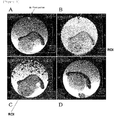
- Figure 1 illustrates an example of the setting of ROI used in the measurement of T1 value/T2 value with regard to a tumor portion collected from an ALA administration group.
- Figure 1A and Figure 1D each relate to an MRI image obtained by the 2D-RARE method, and these views are shown as reference images.
- Figure 1B relates to a parametric image regarding T1 saturation recovery.
- Figure 1C relates to a parametric image regarding T2 relaxation.
- Figure 1A to Figure 1C each show an image regarding the section of an identical tumor portion sample that was collected from the ALA administration group.
- Figure 1D shows an image regarding the section of a tumor portion sample collected from the control group.
- the regions in which the ROI was set in Figure 1B and Figure 1C correspond to a T1 map and a T2 map, respectively. Since the image signal itself indicates a T1 value or a T2 value, it is possible to, for example, measure the T1 value or the T2 value for each pixel on these maps.
- the T2 value was increased (the T2 relaxation time was prolonged) in the ALA administration group, compared to the control group.
- the signal ratio (ALA administration group/control group) was approximately 1.4 times (first measurement), and approximately 1.3 times (second measurement).
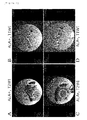
- the region enclosed by a white square in each of Figure 2A and Figure 2C indicates a portion of the section of non-tumor tissues (skin tissues, etc.) derived from a rat, into which tumor cells have been transplanted.
- the black arrow in Figure 2A indicates a circle portion, which was imaged in dark (at low signals) in the T2-weighted image although it was a tumor portion section, wherein the peripheral portion thereof was observed in a high signal zone. Such a change was not observed in the tumor portion section in the control group. Accordingly, it is considered that the region of the high signal zone in the ALA administration group, which is shown with the black arrow, suggests that tumor in the peripheral portion of the circle portion is sufficiently activated.
- Figure 3A and Figure 3C ALA administration group, T2-weighted image
- Figure 3B and Figure 3D control group, T2-weighted image
- the section of the tumor portion for example, the region enclosed by a white circle
- Figure 3A and Figure 3C imaged in white (at high signals)
- Figure 3B and Figure 3D control group, T2-weighted image
- the nuclear magnetic resonance diagnostic agent of the present invention can be advantageously used in the medical field.
Landscapes
- Health & Medical Sciences (AREA)
- Chemical & Material Sciences (AREA)
- Nuclear Medicine, Radiotherapy & Molecular Imaging (AREA)
- Life Sciences & Earth Sciences (AREA)
- Physics & Mathematics (AREA)
- Engineering & Computer Science (AREA)
- Radiology & Medical Imaging (AREA)
- General Health & Medical Sciences (AREA)
- Nanotechnology (AREA)
- Veterinary Medicine (AREA)
- Public Health (AREA)
- Animal Behavior & Ethology (AREA)
- Epidemiology (AREA)
- High Energy & Nuclear Physics (AREA)
- Organic Chemistry (AREA)
- Medicinal Chemistry (AREA)
- Pathology (AREA)
- Signal Processing (AREA)
- Condensed Matter Physics & Semiconductors (AREA)
- Biophysics (AREA)
- General Physics & Mathematics (AREA)
- Biomedical Technology (AREA)
- Heart & Thoracic Surgery (AREA)
- Medical Informatics (AREA)
- Molecular Biology (AREA)
- Surgery (AREA)
- Magnetic Resonance Imaging Apparatus (AREA)
- Medicines Containing Antibodies Or Antigens For Use As Internal Diagnostic Agents (AREA)
Applications Claiming Priority (2)
| Application Number | Priority Date | Filing Date | Title |
|---|---|---|---|
| JP2012270844 | 2012-12-11 | ||
| PCT/JP2013/082974 WO2014092051A1 (ja) | 2012-12-11 | 2013-12-09 | 核磁気共鳴診断剤、及び、それを用いた、対象内の細胞、組織又は臓器の状態を検出又は診断する方法 |
Publications (3)
| Publication Number | Publication Date |
|---|---|
| EP2932983A1 true EP2932983A1 (de) | 2015-10-21 |
| EP2932983A4 EP2932983A4 (de) | 2016-08-17 |
| EP2932983B1 EP2932983B1 (de) | 2018-09-12 |
Family
ID=50934339
Family Applications (1)
| Application Number | Title | Priority Date | Filing Date |
|---|---|---|---|
| EP13863271.6A Active EP2932983B1 (de) | 2012-12-11 | 2013-12-09 | Kernspinresonanzdiagnosemittel und verwendung davon zur detektion oder diagnose des zustands einer zelle, eines gewebes oder eines organs in einer person |
Country Status (5)
| Country | Link |
|---|---|
| US (1) | US11633505B2 (de) |
| EP (1) | EP2932983B1 (de) |
| JP (1) | JP5980953B2 (de) |
| CN (1) | CN104981255B (de) |
| WO (1) | WO2014092051A1 (de) |
Cited By (1)
| Publication number | Priority date | Publication date | Assignee | Title |
|---|---|---|---|---|
| EP3593823A4 (de) * | 2017-03-09 | 2020-12-02 | Baek, Jung-Eun | Zusammensetzung zur rauschentfernung aus mrt und pad damit |
Families Citing this family (8)
| Publication number | Priority date | Publication date | Assignee | Title |
|---|---|---|---|---|
| US11633505B2 (en) | 2012-12-11 | 2023-04-25 | National University Corporation Kumamoto University | Nuclear magnetic resonance diagnostic agent, and method for detecting or diagnosing state of cell, tissue or organ in subject using same |
| US9494503B2 (en) | 2013-11-06 | 2016-11-15 | Aspect Imaging Ltd. | Inline rheology/viscosity, density, and flow rate measurement |
| CN104635186A (zh) * | 2014-12-26 | 2015-05-20 | 苏州露宇电子科技有限公司 | 移动式核磁共振分析仪驱动机构 |
| US10670574B2 (en) | 2015-01-19 | 2020-06-02 | Aspect International (2015) Private Limited | NMR-based systems for crude oil enhancement and methods thereof |
| CN106053299B (zh) | 2015-04-12 | 2020-10-30 | 艾斯拜克特Ai有限公司 | 非圆形横截面管道中的流体的nmr成像 |
| CN106324010A (zh) | 2015-07-02 | 2017-01-11 | 艾斯拜克特Ai有限公司 | 使用mr设备对在管道中流动的流体的分析 |
| KR101807392B1 (ko) | 2016-03-10 | 2017-12-12 | 연세대학교 산학협력단 | 자기공명 이미지에서 조영 증강이 없는 뇌실벽에서 수술 중 광감작제-유도 형광에 의한 신경교종 탐지 |
| US10655996B2 (en) | 2016-04-12 | 2020-05-19 | Aspect Imaging Ltd. | System and method for measuring velocity profiles |
Family Cites Families (14)
| Publication number | Priority date | Publication date | Assignee | Title |
|---|---|---|---|---|
| US4986256A (en) * | 1985-02-28 | 1991-01-22 | The United States Of America As Represented By The Department Of Health And Human Services | Use of paramagnetic metalloporphyrins as contrast agents for tumors in MRI imaging |
| US5079262A (en) | 1989-07-28 | 1992-01-07 | Queen's University At Kingston | Method of detection and treatment of malignant and non-malignant lesions utilizing 5-aminolevulinic acid |
| US5169944A (en) | 1991-04-12 | 1992-12-08 | Board Of Regents Of The University Of Washington | Methods and compositions for the enteral administration of hepatobiliary MRI contrast agents |
| JPH1112197A (ja) | 1997-06-18 | 1999-01-19 | Cosmo Sogo Kenkyusho:Kk | 悪性腫瘍診断剤及び治療剤 |
| AU2003230164B2 (en) * | 2002-05-08 | 2008-05-01 | Yeda Research And Development Co. Ltd. | Sensitized online BOLD-MRI imaging method |
| JP5034032B2 (ja) * | 2004-09-29 | 2012-09-26 | Sbiファーマ株式会社 | 腫瘍診断剤 |
| US8609073B2 (en) * | 2005-03-04 | 2013-12-17 | Dusa Pharmaceuticals, Inc. | Compositions and methods for reducing photosensitivity associated with photodynamic therapy |
| JP5098051B2 (ja) * | 2007-04-05 | 2012-12-12 | Sbiファーマ株式会社 | ミトコンドリア障害脳疾患治療剤及び診断剤 |
| EP2272538B1 (de) * | 2008-04-22 | 2020-09-02 | SBI Pharmaceuticals Co., Ltd. | Verfahren zur erkennung von blasenkrebs |
| JP5295652B2 (ja) | 2008-06-16 | 2013-09-18 | Sbiファーマ株式会社 | 光障害の軽減剤 |
| CA2728960C (en) | 2008-06-26 | 2016-04-12 | Japan Science And Technology Agency | Polymer-metal complex composite having mri contrast ability and mri contrasting and/or antitumor composition using the same |
| US8948851B2 (en) * | 2009-01-20 | 2015-02-03 | The Trustees Of Dartmouth College | Method and apparatus for depth-resolved fluorescence, chromophore, and oximetry imaging for lesion identification during surgery |
| JP2011026221A (ja) | 2009-07-22 | 2011-02-10 | Sbi Alapromo Co Ltd | プロトポルフィリンix細胞内集積増強組成物 |
| US11633505B2 (en) | 2012-12-11 | 2023-04-25 | National University Corporation Kumamoto University | Nuclear magnetic resonance diagnostic agent, and method for detecting or diagnosing state of cell, tissue or organ in subject using same |
-
2013
- 2013-12-09 US US14/650,530 patent/US11633505B2/en active Active
- 2013-12-09 JP JP2014552034A patent/JP5980953B2/ja active Active
- 2013-12-09 CN CN201380072755.6A patent/CN104981255B/zh active Active
- 2013-12-09 WO PCT/JP2013/082974 patent/WO2014092051A1/ja not_active Ceased
- 2013-12-09 EP EP13863271.6A patent/EP2932983B1/de active Active
Cited By (1)
| Publication number | Priority date | Publication date | Assignee | Title |
|---|---|---|---|---|
| EP3593823A4 (de) * | 2017-03-09 | 2020-12-02 | Baek, Jung-Eun | Zusammensetzung zur rauschentfernung aus mrt und pad damit |
Also Published As
| Publication number | Publication date |
|---|---|
| CN104981255B (zh) | 2018-11-30 |
| JPWO2014092051A1 (ja) | 2017-01-12 |
| WO2014092051A1 (ja) | 2014-06-19 |
| JP5980953B2 (ja) | 2016-08-31 |
| HK1210943A1 (zh) | 2016-05-13 |
| EP2932983A4 (de) | 2016-08-17 |
| US11633505B2 (en) | 2023-04-25 |
| HK1214500A1 (en) | 2016-07-29 |
| CN104981255A (zh) | 2015-10-14 |
| US20150320888A1 (en) | 2015-11-12 |
| EP2932983B1 (de) | 2018-09-12 |
Similar Documents
| Publication | Publication Date | Title |
|---|---|---|
| EP2932983B1 (de) | Kernspinresonanzdiagnosemittel und verwendung davon zur detektion oder diagnose des zustands einer zelle, eines gewebes oder eines organs in einer person | |
| Febo et al. | Imaging cocaine-induced changes in the mesocorticolimbic dopaminergic system of conscious rats | |
| US10188754B2 (en) | Compositions and methods for chemical exchange saturation transfer (CEST) based magnetic resonance imaging (MRI) | |
| Ip et al. | Mapping of exogenous choline uptake and metabolism in rat glioblastoma using deuterium metabolic imaging (DMI) | |
| EP3058375B1 (de) | Ph-biosensoren auf der basis von verbindungen aus brenztraubensäure für magnetresonanzbildgebung und spektroskopie sowie deren verwendungen | |
| EP3062783A1 (de) | Bildgebung von histondeacetylasen mit einem radiotracer mittels positronenemissionstomografie | |
| US8664279B2 (en) | Compositions and methods for metabolic imaging | |
| CA3083111C (en) | Preparation for magnetic resonance diagnostics for oncological diseases, comprising deuterated 2-amino-2-methylpropionic acid and/or 2-(n-methylamino)-2-methylpropionic acid, and diagnostic method using said preparation | |
| Zhang et al. | Large-scale synthesis of non-ionic bismuth chelate for computed tomography imaging in vivo | |
| HK1214500B (en) | Nuclear magnetic resonance diagnostic agent, and use thereof in a method for detecting or diagnosing state of cell, tissue or organ in subject | |
| RU2663286C1 (ru) | Препарат для магнитно-резонансной диагностики онкологических заболеваний, содержащий дейтерированную 2-амино-2-метилпропионовую кислоту и/или 2-(n-метиламино)-2-метилпропионовую кислоту, и способ диагностики с использованием этого препарата | |
| Joo et al. | L‐glutamine as a T2 exchange contrast agent | |
| US12115232B2 (en) | Preparation for magnetic resonance diagnostics for oncological diseases, comprising deuterated 3-O-methylglucose, and diagnostic method using said preparation | |
| HK1210943B (zh) | 核磁共振诊断剂以及使用所述核磁共振诊断剂来检测或诊断对象中细胞、组织或器官状态的方法 | |
| US20170165380A1 (en) | Nanosensor Compositions and Methods of use Thereof | |
| RU2718052C2 (ru) | Препарат для магнитно-резонансной диагностики онкологических заболеваний, содержащий дейтерированную 3-о-метилглюкозу, и способ диагностики с использованием этого препарата | |
| JP5088854B2 (ja) | 拡散強調画像撮影のための造影用組成物 | |
| US20130149250A1 (en) | Hyperpolarized agents for mri characterization of redox systems in vivo | |
| US20230158177A1 (en) | Hypoxia-targeting contrast agent and methods of use thereof | |
| WO2020139127A1 (ru) | Препарат для магнитно-резонансной томографии, содержащий дейтерированный саркозин, и способ диагностики с использованием этого препарата | |
| Jirát-Ziółkowska | Functional nanodiagnostics for 31P-magnetic resonance imaging | |
| CN121015917A (zh) | 诊疗一体化的脑靶向磁性粒子成像示踪剂及其制备方法和应用 | |
| KR20220086973A (ko) | 측쇄 아미노산을 유효성분으로 포함하는 신규한 조영제 조성물 | |
| Mishra | Design, synthesis and characterization of novel exogenous smart/bioresponsive contrast agents for magnetic resonance and optical imaging |
Legal Events
| Date | Code | Title | Description |
|---|---|---|---|
| PUAI | Public reference made under article 153(3) epc to a published international application that has entered the european phase |
Free format text: ORIGINAL CODE: 0009012 |
|
| 17P | Request for examination filed |
Effective date: 20150602 |
|
| AK | Designated contracting states |
Kind code of ref document: A1 Designated state(s): AL AT BE BG CH CY CZ DE DK EE ES FI FR GB GR HR HU IE IS IT LI LT LU LV MC MK MT NL NO PL PT RO RS SE SI SK SM TR |
|
| AX | Request for extension of the european patent |
Extension state: BA ME |
|
| DAX | Request for extension of the european patent (deleted) | ||
| REG | Reference to a national code |
Ref country code: DE Ref legal event code: R079 Ref document number: 602013043695 Country of ref document: DE Free format text: PREVIOUS MAIN CLASS: A61K0049000000 Ipc: A61K0049180000 |
|
| REG | Reference to a national code |
Ref country code: HK Ref legal event code: DE Ref document number: 1214500 Country of ref document: HK |
|
| A4 | Supplementary search report drawn up and despatched |
Effective date: 20160715 |
|
| RIC1 | Information provided on ipc code assigned before grant |
Ipc: A61B 5/055 20060101ALI20160712BHEP Ipc: A61K 49/18 20060101AFI20160712BHEP Ipc: A61K 49/10 20060101ALI20160712BHEP Ipc: G01R 33/28 20060101ALI20160712BHEP Ipc: G01R 33/56 20060101ALI20160712BHEP Ipc: A61K 49/00 20060101ALI20160712BHEP |
|
| 17Q | First examination report despatched |
Effective date: 20171012 |
|
| GRAP | Despatch of communication of intention to grant a patent |
Free format text: ORIGINAL CODE: EPIDOSNIGR1 |
|
| INTG | Intention to grant announced |
Effective date: 20180403 |
|
| GRAS | Grant fee paid |
Free format text: ORIGINAL CODE: EPIDOSNIGR3 |
|
| GRAA | (expected) grant |
Free format text: ORIGINAL CODE: 0009210 |
|
| AK | Designated contracting states |
Kind code of ref document: B1 Designated state(s): AL AT BE BG CH CY CZ DE DK EE ES FI FR GB GR HR HU IE IS IT LI LT LU LV MC MK MT NL NO PL PT RO RS SE SI SK SM TR |
|
| REG | Reference to a national code |
Ref country code: GB Ref legal event code: FG4D |
|
| REG | Reference to a national code |
Ref country code: CH Ref legal event code: EP |
|
| REG | Reference to a national code |
Ref country code: CH Ref legal event code: NV Representative=s name: SERVOPATENT GMBH, CH |
|
| REG | Reference to a national code |
Ref country code: IE Ref legal event code: FG4D |
|
| REG | Reference to a national code |
Ref country code: DE Ref legal event code: R096 Ref document number: 602013043695 Country of ref document: DE |
|
| REG | Reference to a national code |
Ref country code: AT Ref legal event code: REF Ref document number: 1039790 Country of ref document: AT Kind code of ref document: T Effective date: 20181015 |
|
| REG | Reference to a national code |
Ref country code: NL Ref legal event code: MP Effective date: 20180912 |
|
| REG | Reference to a national code |
Ref country code: LT Ref legal event code: MG4D |
|
| PG25 | Lapsed in a contracting state [announced via postgrant information from national office to epo] |
Ref country code: FI Free format text: LAPSE BECAUSE OF FAILURE TO SUBMIT A TRANSLATION OF THE DESCRIPTION OR TO PAY THE FEE WITHIN THE PRESCRIBED TIME-LIMIT Effective date: 20180912 Ref country code: RS Free format text: LAPSE BECAUSE OF FAILURE TO SUBMIT A TRANSLATION OF THE DESCRIPTION OR TO PAY THE FEE WITHIN THE PRESCRIBED TIME-LIMIT Effective date: 20180912 Ref country code: NO Free format text: LAPSE BECAUSE OF FAILURE TO SUBMIT A TRANSLATION OF THE DESCRIPTION OR TO PAY THE FEE WITHIN THE PRESCRIBED TIME-LIMIT Effective date: 20181212 Ref country code: SE Free format text: LAPSE BECAUSE OF FAILURE TO SUBMIT A TRANSLATION OF THE DESCRIPTION OR TO PAY THE FEE WITHIN THE PRESCRIBED TIME-LIMIT Effective date: 20180912 Ref country code: GR Free format text: LAPSE BECAUSE OF FAILURE TO SUBMIT A TRANSLATION OF THE DESCRIPTION OR TO PAY THE FEE WITHIN THE PRESCRIBED TIME-LIMIT Effective date: 20181213 Ref country code: BG Free format text: LAPSE BECAUSE OF FAILURE TO SUBMIT A TRANSLATION OF THE DESCRIPTION OR TO PAY THE FEE WITHIN THE PRESCRIBED TIME-LIMIT Effective date: 20181212 Ref country code: LT Free format text: LAPSE BECAUSE OF FAILURE TO SUBMIT A TRANSLATION OF THE DESCRIPTION OR TO PAY THE FEE WITHIN THE PRESCRIBED TIME-LIMIT Effective date: 20180912 |
|
| PG25 | Lapsed in a contracting state [announced via postgrant information from national office to epo] |
Ref country code: AL Free format text: LAPSE BECAUSE OF FAILURE TO SUBMIT A TRANSLATION OF THE DESCRIPTION OR TO PAY THE FEE WITHIN THE PRESCRIBED TIME-LIMIT Effective date: 20180912 Ref country code: LV Free format text: LAPSE BECAUSE OF FAILURE TO SUBMIT A TRANSLATION OF THE DESCRIPTION OR TO PAY THE FEE WITHIN THE PRESCRIBED TIME-LIMIT Effective date: 20180912 Ref country code: HR Free format text: LAPSE BECAUSE OF FAILURE TO SUBMIT A TRANSLATION OF THE DESCRIPTION OR TO PAY THE FEE WITHIN THE PRESCRIBED TIME-LIMIT Effective date: 20180912 |
|
| REG | Reference to a national code |
Ref country code: AT Ref legal event code: MK05 Ref document number: 1039790 Country of ref document: AT Kind code of ref document: T Effective date: 20180912 |
|
| PG25 | Lapsed in a contracting state [announced via postgrant information from national office to epo] |
Ref country code: ES Free format text: LAPSE BECAUSE OF FAILURE TO SUBMIT A TRANSLATION OF THE DESCRIPTION OR TO PAY THE FEE WITHIN THE PRESCRIBED TIME-LIMIT Effective date: 20180912 Ref country code: NL Free format text: LAPSE BECAUSE OF FAILURE TO SUBMIT A TRANSLATION OF THE DESCRIPTION OR TO PAY THE FEE WITHIN THE PRESCRIBED TIME-LIMIT Effective date: 20180912 Ref country code: IS Free format text: LAPSE BECAUSE OF FAILURE TO SUBMIT A TRANSLATION OF THE DESCRIPTION OR TO PAY THE FEE WITHIN THE PRESCRIBED TIME-LIMIT Effective date: 20190112 Ref country code: PL Free format text: LAPSE BECAUSE OF FAILURE TO SUBMIT A TRANSLATION OF THE DESCRIPTION OR TO PAY THE FEE WITHIN THE PRESCRIBED TIME-LIMIT Effective date: 20180912 Ref country code: RO Free format text: LAPSE BECAUSE OF FAILURE TO SUBMIT A TRANSLATION OF THE DESCRIPTION OR TO PAY THE FEE WITHIN THE PRESCRIBED TIME-LIMIT Effective date: 20180912 Ref country code: CZ Free format text: LAPSE BECAUSE OF FAILURE TO SUBMIT A TRANSLATION OF THE DESCRIPTION OR TO PAY THE FEE WITHIN THE PRESCRIBED TIME-LIMIT Effective date: 20180912 Ref country code: IT Free format text: LAPSE BECAUSE OF FAILURE TO SUBMIT A TRANSLATION OF THE DESCRIPTION OR TO PAY THE FEE WITHIN THE PRESCRIBED TIME-LIMIT Effective date: 20180912 Ref country code: AT Free format text: LAPSE BECAUSE OF FAILURE TO SUBMIT A TRANSLATION OF THE DESCRIPTION OR TO PAY THE FEE WITHIN THE PRESCRIBED TIME-LIMIT Effective date: 20180912 Ref country code: EE Free format text: LAPSE BECAUSE OF FAILURE TO SUBMIT A TRANSLATION OF THE DESCRIPTION OR TO PAY THE FEE WITHIN THE PRESCRIBED TIME-LIMIT Effective date: 20180912 |
|
| PG25 | Lapsed in a contracting state [announced via postgrant information from national office to epo] |
Ref country code: SM Free format text: LAPSE BECAUSE OF FAILURE TO SUBMIT A TRANSLATION OF THE DESCRIPTION OR TO PAY THE FEE WITHIN THE PRESCRIBED TIME-LIMIT Effective date: 20180912 Ref country code: SK Free format text: LAPSE BECAUSE OF FAILURE TO SUBMIT A TRANSLATION OF THE DESCRIPTION OR TO PAY THE FEE WITHIN THE PRESCRIBED TIME-LIMIT Effective date: 20180912 Ref country code: PT Free format text: LAPSE BECAUSE OF FAILURE TO SUBMIT A TRANSLATION OF THE DESCRIPTION OR TO PAY THE FEE WITHIN THE PRESCRIBED TIME-LIMIT Effective date: 20190112 |
|
| REG | Reference to a national code |
Ref country code: DE Ref legal event code: R097 Ref document number: 602013043695 Country of ref document: DE |
|
| PLBE | No opposition filed within time limit |
Free format text: ORIGINAL CODE: 0009261 |
|
| STAA | Information on the status of an ep patent application or granted ep patent |
Free format text: STATUS: NO OPPOSITION FILED WITHIN TIME LIMIT |
|
| PG25 | Lapsed in a contracting state [announced via postgrant information from national office to epo] |
Ref country code: DK Free format text: LAPSE BECAUSE OF FAILURE TO SUBMIT A TRANSLATION OF THE DESCRIPTION OR TO PAY THE FEE WITHIN THE PRESCRIBED TIME-LIMIT Effective date: 20180912 |
|
| 26N | No opposition filed |
Effective date: 20190613 |
|
| PG25 | Lapsed in a contracting state [announced via postgrant information from national office to epo] |
Ref country code: LU Free format text: LAPSE BECAUSE OF NON-PAYMENT OF DUE FEES Effective date: 20181209 Ref country code: SI Free format text: LAPSE BECAUSE OF FAILURE TO SUBMIT A TRANSLATION OF THE DESCRIPTION OR TO PAY THE FEE WITHIN THE PRESCRIBED TIME-LIMIT Effective date: 20180912 Ref country code: MC Free format text: LAPSE BECAUSE OF FAILURE TO SUBMIT A TRANSLATION OF THE DESCRIPTION OR TO PAY THE FEE WITHIN THE PRESCRIBED TIME-LIMIT Effective date: 20180912 |
|
| REG | Reference to a national code |
Ref country code: IE Ref legal event code: MM4A |
|
| REG | Reference to a national code |
Ref country code: BE Ref legal event code: MM Effective date: 20181231 |
|
| PG25 | Lapsed in a contracting state [announced via postgrant information from national office to epo] |
Ref country code: IE Free format text: LAPSE BECAUSE OF NON-PAYMENT OF DUE FEES Effective date: 20181209 |
|
| PG25 | Lapsed in a contracting state [announced via postgrant information from national office to epo] |
Ref country code: BE Free format text: LAPSE BECAUSE OF NON-PAYMENT OF DUE FEES Effective date: 20181231 |
|
| PG25 | Lapsed in a contracting state [announced via postgrant information from national office to epo] |
Ref country code: MT Free format text: LAPSE BECAUSE OF NON-PAYMENT OF DUE FEES Effective date: 20181209 |
|
| PG25 | Lapsed in a contracting state [announced via postgrant information from national office to epo] |
Ref country code: TR Free format text: LAPSE BECAUSE OF FAILURE TO SUBMIT A TRANSLATION OF THE DESCRIPTION OR TO PAY THE FEE WITHIN THE PRESCRIBED TIME-LIMIT Effective date: 20180912 |
|
| REG | Reference to a national code |
Ref country code: CH Ref legal event code: PCAR Free format text: NEW ADDRESS: WANNERSTRASSE 9/1, 8045 ZUERICH (CH) |
|
| PG25 | Lapsed in a contracting state [announced via postgrant information from national office to epo] |
Ref country code: CY Free format text: LAPSE BECAUSE OF FAILURE TO SUBMIT A TRANSLATION OF THE DESCRIPTION OR TO PAY THE FEE WITHIN THE PRESCRIBED TIME-LIMIT Effective date: 20180912 Ref country code: HU Free format text: LAPSE BECAUSE OF FAILURE TO SUBMIT A TRANSLATION OF THE DESCRIPTION OR TO PAY THE FEE WITHIN THE PRESCRIBED TIME-LIMIT; INVALID AB INITIO Effective date: 20131209 Ref country code: MK Free format text: LAPSE BECAUSE OF NON-PAYMENT OF DUE FEES Effective date: 20180912 |
|
| REG | Reference to a national code |
Ref country code: CH Ref legal event code: U11 Free format text: ST27 STATUS EVENT CODE: U-0-0-U10-U11 (AS PROVIDED BY THE NATIONAL OFFICE) Effective date: 20260101 |
|
| PGFP | Annual fee paid to national office [announced via postgrant information from national office to epo] |
Ref country code: DE Payment date: 20251028 Year of fee payment: 13 |
|
| PGFP | Annual fee paid to national office [announced via postgrant information from national office to epo] |
Ref country code: GB Payment date: 20251030 Year of fee payment: 13 |
|
| PGFP | Annual fee paid to national office [announced via postgrant information from national office to epo] |
Ref country code: FR Payment date: 20251110 Year of fee payment: 13 |
|
| PGFP | Annual fee paid to national office [announced via postgrant information from national office to epo] |
Ref country code: CH Payment date: 20260101 Year of fee payment: 13 |