EP2932274B1 - Procédé pour le diagnostic de la leucodystrophie métachromatique - Google Patents
Procédé pour le diagnostic de la leucodystrophie métachromatique Download PDFInfo
- Publication number
- EP2932274B1 EP2932274B1 EP13815378.8A EP13815378A EP2932274B1 EP 2932274 B1 EP2932274 B1 EP 2932274B1 EP 13815378 A EP13815378 A EP 13815378A EP 2932274 B1 EP2932274 B1 EP 2932274B1
- Authority
- EP
- European Patent Office
- Prior art keywords
- subject
- sample
- biomarker
- sulfatide
- mld
- Prior art date
- Legal status (The legal status is an assumption and is not a legal conclusion. Google has not performed a legal analysis and makes no representation as to the accuracy of the status listed.)
- Active
Links
- 0 C[C@]([C@](CO[C@@](COC)O)*(C)=N)C=CN Chemical compound C[C@]([C@](CO[C@@](COC)O)*(C)=N)C=CN 0.000 description 1
Images


Classifications
-
- G—PHYSICS
- G01—MEASURING; TESTING
- G01N—INVESTIGATING OR ANALYSING MATERIALS BY DETERMINING THEIR CHEMICAL OR PHYSICAL PROPERTIES
- G01N33/00—Investigating or analysing materials by specific methods not covered by groups G01N1/00 - G01N31/00
- G01N33/48—Biological material, e.g. blood, urine; Haemocytometers
- G01N33/50—Chemical analysis of biological material, e.g. blood, urine; Testing involving biospecific ligand binding methods; Immunological testing
- G01N33/68—Chemical analysis of biological material, e.g. blood, urine; Testing involving biospecific ligand binding methods; Immunological testing involving proteins, peptides or amino acids
- G01N33/6893—Chemical analysis of biological material, e.g. blood, urine; Testing involving biospecific ligand binding methods; Immunological testing involving proteins, peptides or amino acids related to diseases not provided for elsewhere
-
- G—PHYSICS
- G01—MEASURING; TESTING
- G01N—INVESTIGATING OR ANALYSING MATERIALS BY DETERMINING THEIR CHEMICAL OR PHYSICAL PROPERTIES
- G01N2800/00—Detection or diagnosis of diseases
- G01N2800/04—Endocrine or metabolic disorders
Definitions
- the present invention is related to a method for diagnosing metachromatic leukodystrophy, the use of a biomarker for the diagnosis of metachromatic leukodystrophy, and the use of mass spectrometric analysis for the detection of a biomarker.
- Lysosomal storage diseases also referred to herein as lysosomal storage disorders or LSDs, are a group of rare inherited metabolic disorders that result from defects in lysosomal function. LSDs result when a specific organelle in the body's cells - the lysosome - malfunctions. Some of the more prominent lysosomal storage diseases are Gaucher's disease and Fabry disease.
- LSDs are caused by lysosomal dysfunction usually as a consequence of deficiency of a single enzyme required for the metabolism of lipids, glycoproteins or so-called mucopolysaccharides. Individually, LSDs occur with frequencies of about 1:10,000 to 1:250,000, however, as a group the incidence is about 1:5,000. Most of these disorders are autosomal recessively inherited; however, a few are X-linked inherited, such as Fabry disease and Hunter syndrome (MPS II).
- Lysosomal storage diseases affect mostly children and they often die at a young and unpredictable age, many within a few months or years of birth. Many other children die of this disease following years of suffering from various symptoms of their particular disorder.
- the symptoms of lysosomal storage disease vary, depending on the particular disorder and other variables like the age of onset, and can be mild to severe. They can include developmental delay, movement disorders, seizures, dementia, deafness and/or blindness.
- Some people with lysosomal storage disease have enlarged livers (hepatomegaly) and enlarged spleens (splenomegaly), pulmonary and cardiac problems, and bones that develop abnormally.
- Metachromatic leukodystrophy also referred to herein as MLD or Arylsulfatase A deficiency, as used herein, is an LSD which is caused by a deficiency in lysosomal arylsulfatase A, also referred to herein as ARSA.
- ARSA catabolizes sulfatides ( Von Figura et al, "Metachromatic leukodystrophy” In: Scriver CR et al. "The Metabolic and Molecular Bases of Inherited Disease” 8th edn ). Sulfatides accumulate in multiple tissues including oligodendrocytes and Schwann cells, provoking demyelination in both the central and peripheral nervous system ( Sedel et al., J Inherit Metab Dis., 2008 Jun ).
- MLD lysosomal arylsulfatase A gene
- biomarkers for the early detection and diagnosis of MLD holds great promise to improve the clinical outcome of patients. It is especially important for patients with vague or no symptoms or to detect patients which fail to respond to a therapy.
- a biomarker should be technically feasible in many hands, easy to measure; useful, with a consistent, relative magnitude between affected and controls, or treated and untreated; reliable, and accurate clinically, and classifiable as strongly predictive or prognostic.
- chitotriosidase as a biomarker for Gaucher's disease, said enzyme accumulates independent of a direct link to the pathology of Gaucher's disease. Furthermore, up to 35 % of given ethnicities demonstrate a defect of the gene coding for chitotriosidase resulting in an artificially reduced or unmeasurable chitotriosidase activity.
- Mirzaian M. (Master Thesis 5842131, University of Amsterdam, Faculty of Science) describes the development of an UPLC-MS/MS method for determination of lysoglobotriaosylceramide (lysoGb3) and (lyso)-sulfatide in body fluids and tissues from patients with Fabry disease and metachromatic leukodystrophy (MLD).
- lysoGb3 lysoglobotriaosylceramide
- MLD metachromatic leukodystrophy
- Rosengren B et al. (Jounral of Neurochemistry, vol. 52, no. 4, pp. 1035-1041 ) report on lysosulfatide (galactosylsphingosine-3-O-sulfate) from metachromatic leukodystrophy and normal human brain.
- the problem underlying the present invention is to provide a method for the diagnosis of MLD.
- a further problem underlying the present invention is to provide a method for determining the course and prognosis of MLD.
- a still further problem underlying the present invention is to provide a method for determining rather quickly the effectiveness of at least one treatment applied to a subject being positively tested for suffering from or being at risk of developing MLD.
- a further problem underlying the present invention is to provide a method for determining the effectiveness of a compound for the treatment of MLD.
- Another problem underlying the present invention is to provide a biomarker which allows the specific and sensitive diagnosis of MLD.
- the biomarker of each and any problem underlying the present invention and the above problems in particular is different from an enzyme, more preferably the biomarker is different from an enzyme selected form the group comprising arylsulfatase A, N-acetyl-alpha-glucosaminidase, arylsulfatase and beta-glucuronidase.
- a method for diagnosing metachromatic leukodystrophy in a subject comprising a step a), wherein the step a) comprises detecting a biomarker in a sample from the subject, wherein the sample is selected from the group consisting of blood, dried blood, serum and plasma and wherein the biomarker is free lyso-Gb1-sulfatide, wherein lyso-Gb1-sulfatide is of formula (I)
- the sample is a serum sample of the subject, a plasma sample of the subject or a dried blood sample of the subject, preferably a plasma sample of the subject or a serum sample of the subject.
- the method comprises a step b) wherein the step b) comprises determining a level of the biomarker present in the sample.
- the level of the biomarker is indicative whether or not the subject is suffering from metachromatic leukodystrophy or whether or not the subject is at risk of suffering from metachromatic leukodystrophy.
- the biomarker is detected by means of immunoassay, mass spectrometric analysis, biochip array, functional nucleic acids and/or a fluorescent derivative of the biomarker, preferably the biomarker is detected by means of mass spectrometric analysis, more preferably mass spectrometry is combined with HPLC.
- mass spectrometric analysis is selected from the group comprising SELDI, MALDI, MALDI-Q TOF, MS/MS, TOF-TOF and ESI-O-TOF, preferably the mass spectrometric analysis comprises or uses MS/MS.
- step b) comprises comparing the level of the biomarker in the sample from the subject with a cut-off value, preferably the cut-off value for free lyso-Gb1-sulfatide is 0.05 ng/ml.
- the level of the biomarker in the sample from the subject is higher than the cut-off value, this is indicative that the subject is suffering from metachromatic leukodystrophy or is at risk of suffering from metachromatic leukodystrophy; whereas if the level of the biomarker in the sample from the subject is lower than the cut-off value, this is indicative that the subject is not suffering from or is not at risk of suffering from metachromatic leukodystrophy.
- the method preferably the analytical method used in the detection and/or determination of the level of the biomarker present in the sample, has a limit of determination for free lyso-Gb1-sulfatide of 0.05 ng/ml.
- the problem underlying the present invention is solved in a second aspect by the use of a biomarker for the diagnosis of metachromatic leukodystrophy in a method according to the first aspect, including any embodiment thereof, wherein the biomarker is free lyso-Gb1-sulfatide.
- the problem underlying the present invention is solved in a third aspect by the use of mass spectrometric analysis for the detection of a biomarker for the diagnosis of metcahromatic leukodystrophy, wherein the biomarker is free lyso-Gb1-sulfatide, the biomarker is detected in a sample from a subject, wherein the sample is selected from the group consisting of a plasma sample from the subject, a serum sample from the subject and a dried blood sample from the subject and wherein the mass spectrometric analysis preferably comprises or uses MS/MS.
- mass spectrometric analysis is combined with HPLC.
- the sample from the subject is a sample from a subject who has previously been treated for metachromatic leukodystrophy or a sample from a subject who has previously been diagnosed for metachromatic leukodystrophy.
- the sample from the subject is a sample from a subject who has not previously been treated for metachromatic leukodystrophy or a sample from a subject who has not been previously diagnosed for metachromatic leukodystrophy.
- the method comprises a step c), wherein in the step c) it is decided, based on whether the subject is suffering from metachromatic leukodystrophy or is at risk of suffering from metachromatic leukodystrophy, whether a therapy is to be applied, maintained, reduced, elevated or not applied.
- the method comprises a step d), wherein the step d) comprises detecting the biomarker in a sample from the subject after a therapy has been applied, maintained, reduced, elevated or not applied in step c).
- the method comprises a step e), wherein the step e) comprises determining a level of the biomarker in the sample from the subject after a therapy has been applied, maintained, reduced, elevated or not applied in step c).
- the method comprises a step f), wherein the step f) comprises determining whether the level of the biomarker determined in step b) is lower than the level of the biomarker determined in step e).
- the method comprises a step g), wherein the step g) comprises applying, maintaining, reducing, elevating or not applying a therapy based on step f).
- the biomarker is detected by means of mass spectrometric analysis.
- the method comprises protein precipitation and/or HPLC.
- the method comprises protein precipitation, HPLC and MS/MS.
- the subject is a human.
- step d) comprises detecting the biomarker in a sample comprises subjecting the sample to a protein precipitation step, precipitating protein from the sample, providing a supernatant of the sample, subjecting the supernatant of the sample to HPLC and MS/MS and determining the level of the biomarker that is present in the supernatant of the sample.
- the method comprises the following steps:
- the internal standard comprises lyso-Gb2.
- step b), step c) and/or step e) comprises comparing the level of the biomarker in the sample from the subject with a cut-off value.
- the level of the biomarker in the sample from the subject is higher than the cut-off value this is indicative that the subject is suffering from metachromatic leukodystrophy or is at risk of suffering from metachromatic leukodystrophy.
- the level of the biomarker in the sample from the subject is lower than the cut-off value this is indicative that the subject is not suffering from or is not at risk of suffering from metachromatic leukodystrophy.
- the cut-off value is such that a or the sensitivity for diagnosing metachromatic leukodystrophy in a subject is preferably from about 98.5% to 100%, more preferably 99,5% to 100%, and/or such that a or the specificity for diagnosing metachromatic leukodystrophy in a subject is from 99.4% to 100%, preferably 100%.
- step b) and/or step c) and/or step e) comprise(s) that a level of the biomarker in said subject is compared to a level of the biomarker detected in a sample from a control sample.
- control sample is a sample from a subject not having metachromatic leukodystrophy.
- control sample is a sample from a subject having metachromatic leukodystrophy carrier.
- the level of the biomarker in the sample from the subject is higher than the level of the biomarker in the control sample this is indicative that the subject is suffering from and/or is at risk of suffering from metachromatic leukodystrophy.
- the sample from the subject is selected from the group comprising blood, a blood product, urine, saliva, cerebrospinal fluid, stool, tissue sample and lymph.
- the sample from the sample from the subject is selected from the group comprising blood and a blood product.
- the blood product is selected from the group comprising plasma, serum and dried blood.
- the method has a limit of determination for free lyso-Gb1-sulfatide of 0.05ng/ml.
- the method is for the diagnosis of metachromatic leukodystrophy carrier and wherein the biomarker is free lyso-Gb1-sulfatide and the cut-off value is 0.05 ng/ml, and wherein the sample from the subject is preferably serum or plasma.
- the blood is whole blood, plasma, serum or dried blood, preferably plasma or serum.
- the whole blood, plasma or serum is collected on a dry blood filter card.
- Also disclosed is a method for determining the course of metachromatic leukodystrophy in a subject comprising a step a), wherein the step a) comprises determining at several points in time a level of a biomarker present in a sample from the subject.
- the subject has been previously treated for metachromatic leukodystrophy and/or wherein the subject has been previously diagnosed for metachromatic leukodystrophy.
- the subject has not been previously treated for metachromatic leukodystrophy and/or wherein the subject has not been previously diagnosed for metachromatic leukodystrophy.
- the method comprises a step b), wherein the step b) comprises applying, maintaining, reducing, elevating or not applying a therapy based on whether the subject is suffering from metachromatic leukodystrophy or is at risk of suffering from metachromatic leukodystrophy.
- the method comprises a step c), wherein the step c) comprises detecting the biomarker in a sample from the subject after a therapy has been applied, maintained, reduced, elevated or not applied in step b).
- the method comprises a step d), wherein the step d) comprises determining a level of the biomarker in the sample from the subject after a therapy has been applied, maintained, reduced, elevated or not applied in step b).
- the method comprises a step e), wherein the step e) comprises determining whether the level of the biomarker determined in step a) is lower than the level of the biomarker determined in step d).
- the method comprises a step f), wherein the step f) comprises applying, maintaining, reducing, elevating or not applying a therapy based on step e).
- the biomarker is free lyso-Gb1-sulfatide.
- the method comprises determining the level of free lyso-Gb1-sulfatide.
- the method comprises detecting free lyso-Gb1-sulfatide in the sample from the subject.
- the biomarker is detected by means of immunoassay, mass spectrometric analysis, biochip array, functional nucleic acids and/or a fluorescent derivative of the biomarker.
- the biomarker is detected by means of mass spectrometric analysis.
- mass spectrometric analysis is selected from the group consisting of SELDI, MALDI, MALDI-Q TOF, MS/MS, TOF-TOF and ESI-O-TOF.
- the mass spectrometric analysis comprises or uses MS/MS.
- the method comprises protein precipitation and/or HPLC.
- the method comprises protein precipitation, HPLC and MS/MS.
- the subject is a human.
- step d) comprises detecting the biomarker in a sample comprises subjecting the sample to a protein precipitation step, precipitating protein from the sample, providing a supernatant of the sample, subjecting the supernatant of the sample to HPLC and MS/MS and determining the level of the biomarker that is present in the supernatant of the sample.
- a method for determining the effectiveness of at least one treatment applied to a subject being positively tested for suffering from or being at risk of suffering from metachromatic leukodystrophy comprising a step a), wherein the step a) comprises detecting at several points in time a level of a biomarker present in a sample from the subject.
- the method comprises a step b), wherein the step b) comprises determining at several points in time a level of a biomarker present in a sample from the subject.
- the biomarker is free lyso-Gb1-sulfatide.
- the subject has been previously treated for metachromatic leukodystrophy or diagnosed for metachromatic leukodystrophy.
- the subject has not been previously treated for metachromatic leukodystrophy or wherein the subject has not been previously diagnosed for metachromatic leukodystrophy.
- the method comprises a step d), wherein the step d) comprises applying, maintaining, reducing, elevating or not applying at least one treatment applied to the subject based on the decrease in the level of the biomarker as determined in step b).
- the method comprises a step e), wherein the step e) comprises detecting the biomarker in the sample from the subject, wherein the sample has been taken prior to the beginning of the treatment after applying, maintaining, reducing, elevating or not applying at least one treatment in step d) and, optionally determining a level of a biomarker present in a sample from the subject.
- the treatment is selected from the group comprising enzyme replacement therapy, substrate reduction therapy, chaperone therapy, gene therapy, stem cell transplantation of DNA/RNA skipping.
- the method comprises a step f), wherein the step f) comprises determining whether the level of the biomarker determined in step b) is lower than the level of the biomarker determined in step e).
- the method comprises a step g) wherein step g) comprises applying, maintaining, reducing, elevating or not applying at least one treatment applied to the subject based on step f).
- the biomarker is detected by means of immunoassay, mass spectrometric analysis, biochip array, functional nucleic acids and/or a fluorescent derivative of the biomarker.
- the biomarker is detected by means of mass spectrometric analysis.
- mass spectrometric analysis is selected from the group consisting of SELDI, MALDI, MALDI-Q TOF, MS/MS, TOF-TOF and ESI-O-TOF.
- the mass spectrometric analysis comprises or uses MS/MS.
- the method comprises protein precipitation and/or HPLC.
- the method comprises protein precipitation, HPLC and MS/MS.
- the subject is a human.
- the step of detecting the biomarker in the sample from the subject comprises precipitating protein from the sample from the subject, wherein precipitating protein from the sample provides a supernatant of the sample; subjecting a volume of the supernatant to HPLC and MS/MS and determining the level of the biomarker that is present in the sample from the subject.
- the biomarker is free lyso-Gbl-sulfatide.
- the method comprises determining a level of the biomarker in a control sample.
- the biomarker is free lyso-Gb1-sulfatide, wherein if the level of the biomarker in the sample from the subject is higher than the cut-off value of 0.05 ng/ml this is indicative that the subject is suffering from metachromatic leukodystrophy.
- detection comprises the use of HPLC.
- mass spectrometric analysis comprises or uses MS/MS.
- the kit is for
- the biomarker as used or referred to in connection with each and any aspect of the invention and each and any embodiment of the invention is preferably different from an enzyme, more preferably the biomarker is different from an enzyme selected form the group comprising arylsulfatase A, N-acetyl-alpha-glucosaminidase, arylsulfatase and beta-glucuronidase.
- each and any aspect of the invention and each and any embodiment of the invention is one suffering from a mutation selected form the group comprising the following MLD mutations c.465+1G>A (5) p.G309S (4) p.T393G (3) p.L113P (2) p.Q159X (2) p.D257E (2) p.K304R (2) p.G311S (2) p.R313Q (2) p.A316D (2) p.Y381fs (2) p.F387fs (2) p.H397Y (2) p.F400L (2) p.P426L (2) c.979+1G>A (2) p.P84L (1) p.S98F (1) p.G124S (1) p.G129R (1) p.C156Y (1) p.C158X (1) p.W195C (1) p.R246H (1) p.T276M (1) p.T306M (1) p.G309D (1) p.
- free lyso-Gbl-sulfatide also referred to herein preferably as free lyso-glycocerebroside-sulfatide, constitutes a biomarker which allows for a method for diagnosing MLD in a subject, more specifically diagnosing MLD in a subject with high specificity and sensitivity using said free lyso-Gb1-sulfatide as the biomarker.
- Reliance on free lyso-Gb1-sulfatide as a biomarker in the diagnosis of MLD is superior over any method for the diagnosis of MLD based on an enzyme and arysulfatase A in particular.
- Such superiority of a diagnosis based on free lyso-Gb1-sulfatide is shown in terms of both sensitivity and specificity.
- One of the reasons for the observed superiority of a free lyso-Gb1-sulfatide based diagnosis over an arylsulfatase A based diagnosis of MLD is the existence arylsulfatase A pseudodeficiencies, where a decrease in arylsulfatase A activity is - incorrectly - taken as an indication of MLD.
- Similar short-comings are observed in case of MLD diagnosis based on genetics: Some of the observed mutations of arylsulfatase A result in non-pathogenic pseudodeficiencies of arylsulfatase A.
- the present inventors have also surprisingly found that free lyso-Gb1-sulfatide, which can be detected by the methods of the present invention, is circulating in the blood, and plasma and serum in particular, of a subject in a concentration of approximately 1/1000 of total Gb1-sulfatide. Moreover, the present inventors have surprisingly found that, unlike total Gb1-sulfatide, free lyso-Gb1-sulfatide which is present in the blood, and plasma and serum in particular, of a subject is useful in a method for diagnosing MLD in a subject comprising a step of detecting a biomarker in a sample from the subject, wherein the biomarker is free lyso-Gb1-sulfatide. The present inventors have also surprisingly found that the level of free lyso-Gb1-sulfatide determined in the sample from a subject by the methods of the present invention allows for diagnosing MLD with high sensitivity and high specificity.
- the present invention turns away from the teaching of the prior art in that the method of the present invention comprises determining the level of a lyso-compound and using said lyso-compound as a biomarker for diagnosis of a LSD. More specifically, the present inventors have surprisingly found that determining the level of free lyso-Gb1-sulfatide in a sample from a subject allows for diagnosing MLD with high sensitivity and high specificity.
- lysosomal storage disorder also referred to herein as “lysosomal storage disease” or “LSD”, as preferably used herein, refers to genetic diseases and metabolic disorders that result from defects in lysosomal function. Lysosomal storage disorders are caused by lysosomal dysfunction usually as a consequence of deficiency of a single enzyme required for the metabolism of lipids, glycoproteins or so-called mucopolysaccharides. Like other genetic diseases, individuals inherit lysosomal storage diseases from their parents. Although each disorder results from different gene mutations that translate into a deficiency in enzyme activity, they all share a common biochemical characteristic - all lysosomal disorders originate from an abnormal accumulation of substances inside the lysosome.
- MLD is an autosomal recessively inherited LSD which is commonly listed in the family of leukoencephalopathies.
- leukoencephalopathy means a disease or disorder that selectively or predominantly involves the white matter of the brain. It is associated with a group of diseases that affect the myelin itself, oligodendrocytes, astrocytes or even axons.
- the main acquired causes of leukoencephalopathies include inflammatory diseases, vascular diseases, infections, neoplasias and toxic causes (reviewed in Filley and Kleinschmidt-DeMasters, 2001, N Engl J Med ).
- Hereditary leukoencephalopathies can be separated into three categories ( Baumann and Turpin, 2000, J Neurol ; Schiffmann and van der Knaap, 2004,Curr Opin Neurol ; Sedel et al, 2005, Rev Neurol ): (1) leukoencephalopathies characterized clinically, radiologically or pathologically but for which the gene causing the leukoencephalopathy is still unknown; (2) leukoencephalopathies caused by genes coding for proteins not directly involved in metabolic pathways and for which the diagnosis relies directly on gene analysis; and (3) leukoencephalopathies caused by genes coding for enzymes or proteins involved in the cell metabolism and for which the diagnosis relies mostly on biochemical analysis of plasma and urines samples.
- the third category corresponds to inborn errors of metabolism, also referred to herein as IEMs, which are important to recognize because specific treatments often exist ( Sedel et al., 2007, Nat Clin Pract Neurol ).
- IEMs inborn errors of metabolism
- late-onset forms also exist that display different clinical and radiological features, sometimes very far from the classical paediatric description.
- neurologists are usually poorly familiar with IEMs.
- MLD belongs to the third group of leukoencephalopathies, i.e. leukoencephalopathies caused by genes coding for enzymes or proteins involved in the cell metabolism. MLD is caused by a deficiency in lysosomal arylsulfatase A, also referred to herein as ARSA. ARSA catabolizes sulfatides (Von Figura et al, 2001, supra).
- Magnetic Resonance Imaging shows a bilateral periventricular leukoencephalopathy with frontal predominance and cerebral atrophy. Importantly U-fibres are relatively spared at least at early stages of the disease (Sedel et al, 2001, supra).
- Diagnosis of MLD is based on the measurement of ARSA activity on leukocytes. However, about 15% of people in Europe and the United States display low ARSA activity without clinical symptoms and no tissue or urine accumulation of sulfatides (Von Figura et al., 2001, supra). These pseudodeficiencies are caused by certain polymorphism within the ARSA coding gene (Von Figura et al., 2001, supra).
- saposin B an activator necessary to activate sulfatides degradation, can cause MLD despite normal ARSA activity ( Deconinck et al., 2007, Eur J Paediatr Neurol ). Although such deficiency has not been described in adults to our knowledge, it should be suspected in patients with leukodystrophy and high urinary excretion of sulfatides.
- MLD Middle loss virus
- MLD childhood with the juvenile form of MLD (onset between 3 and 10 years of age) usually begin with impaired school performance, mental deterioration, and dementia and then develop symptoms similar to the late infantile form but with slower progression. Age of death is variable, but normally within 10 to 15 years of symptom onset although some juveniles can live for several decades or longer after onset.
- the adult form commonly begins after age 16 as a psychiatric disorder or progressive dementia.
- Adult-onset MLD progresses more slowly than the late infantile and juvenile forms, with a protracted course of a decade or more.
- Palliative care can help with many of the symptoms and usually improves quality and longevity of life.
- That MLD is inherited in an autosomal recessive pattern means that both copies or both alleles of the gene must be mutated or altered in such a way that function is impaired, in contrast to a polymorphism, in which the nucleotide sequence is altered but causes no functional disruption, for a person to be affected by the disorder. Most often, the parents of a child with an autosomal recessive disorder are not affected but are carriers of one copy of the altered gene. Such carrier is referred to herein as MLD carrier. If both parents are carriers, there is a 25% chance with each pregnancy for an affected child. Genetic counseling and genetic testing is recommended for families who may be carriers of MLD.
- Sulfatides accumulate in multiple tissues including oligodendrocytes and Schwann cells, provoking demyelination in both the central and peripheral nervous system (Sedel et al., supra). Sulfatides are a class of sulfated galactosylceramides synthesized primarily in the oligodendrocytes in the central nervous system. Sulfatides are a type of sulfolipid.
- Gb1-sulfatide consists of a ceramide core, i.e. sphingosine bound to a fatty acid via an amide linkage and one glycosyl residue at the 1-hydroxyl moiety.
- a sulfate moiety is present at the C 3 -atom of the glycosyl moiety.
- the glycosyl moiety is either a galactosyl moiety or a glucosyl moiety. More preferably, the glycosyl moiety is a galactosyl moiety.
- lyso-Gb1-sulfatide as used herein, preferably in connection with the invention as defined in the claims, preferably means that the molecule is present in its free amino form. More precisely, lyso-Gb1-sulfatide as used herein, preferably differs from Gb1-sulfatide in that no fatty acid moiety is linked to the - primary - amino group of the sphingosine moiety of the molecule. Furthermore, lyso-Gb1-sulfatide is also referred to herein as glycosylsphingosine-sulfatide.
- lyso-Gb1 sulfatide is of formula (I) and is also referred to as lyso-Galactosyl-ceramide or lyso-galactosyl ceramide sulfatide or galactosyl sphingosine sulfatide.
- lyso-Gb1 sulfatide encompasses in its more general meaning as used herein galactosylsphingosine-sulfatide and/or glucosylsphingosine-sulfatide, whereby preferably the term lyso-Gb1 sulfatide means a compound of formula (I).
- free lyso-Gb1-sulfatide preferably refers to lyso-Gb1-sulfatide which is as such present in a sample from a or the subject, such as blood including dried blood or plasma or serum, and, preferably, is not the result of a manipulation of the sample of said subject.
- Such manipulation of a sample can be the one described by Groener et al. ( Groener et al., Biochimica et Biophysica Acta 1781(2908)72 - 78, 2007 ).
- free lyso-Gbl-sulfatide which is present as such in the blood including dried blood or plasma or serum of a subject from whom the sample is taken, is more particularly not a lyso-Gb1-sulfatide which is generated by chemical, biochemical or physical treatment of the sample contained in the blood sample, plasma sample and/or serum sample, preferably outside of the body of the patient.
- free lyso-Gb1-sulfatide is not a lyso-Gb1-sulfatide prepared by chemical treatment of free lyso-Gb1-sulfatide as contained in a sample of a subject, whereby the sample is preferably a sample selected from the group consisting of plasma, serum and blood including dried blood.
- free lyso-Gb1-sulfatide as used herein is different from lyso-Gb1-sulfatide prepared by Toda K. et al ( Toda K. et al., (1989) Biochemical and Biophysical Research Communications, Vol. 159, No. 2, pp 605-611 ); in other words, Toda K et al.
- free lyso-Gb1-sulfatide as used herein, preferably is present in addition to Gb1-sulfatide and is a compound produced by the subject's metabolic activities. Accordingly, Gb1-sulfatide, which is the molecule that is accumulated in connection with MLD is present in the sample from the subject and has compared to the molecule in a free lyso form, i.e.
- free- lyso-Gb1-sulfatide, present in the blood, and plasma and/or the serum of the subject at least a fatty acid moiety linked to the - primary - amino group of the sphingosine moiety of lyso-Gb1-sulfatide.
- the biomarker is detected by means of immunoassay, mass spectrometric analysis, biochip array, functional nucleic acids and/or a fluorescent derivative of the biomarker.
- detection allows for the selective detection of the biomarker as present in the blood of a subject as such and particularly is not the result of a manipulation of the sample of said subject resulting in a change of the concentration of the biomarker, such as the derivatization of Gb1 into lyso-Gb1 according to the method of the prior art as described above.
- Such manipulation may result in the inability to distinguish the biomarker which is present in its free-lyso-form from the substance which is the result of said manipulation, for example the result of said derivatization.
- the biomarker of the present invention cannot be detected as such and the level of said biomarker cannot be determined as such, respectively, without detecting the manipulated further substance, e.g. Gb1 converted into lyso-Gb1 according to the method of the prior art.
- the biomarker present in the blood of the subject such as free- lyso-Gb1-sulfatide present as such in the blood of the subject, is also present in the sample of the subject as such and may, nevertheless, be selectively labeled with and/or linked to a means such as a fluorescent dye or a nucleic acid molecule specifically binding the biomarker.
- a means such as a fluorescent dye or a nucleic acid molecule specifically binding the biomarker.
- Such selective labeling or linking allows detecting and/or determining the level of the labeled or linked biomarker, without labeling of, linking to or converting a further substance, such as the converted lyso-Gb1 of the prior art, which cannot be distinguished from the biomarker, more precisely the labeled or linked biomarker.
- a means such as a fluorescent dye or a nucleic acid molecule specifically binding the biomarker.
- a fluorescent derivative of the biomarker of the present invention concerns a biomarker which is labeled with and/or bound to a fluorescence dye or molecule, i.e. resulting in a fluorescent derivative of the biomarker, which allows for detecting the fluorescent derivative and/or determining the level of the fluorescent derivative of the biomarker of the invention.
- sample means preferably a limited quantity of a subject's material, wherein said subject's material is part of or has been taken from a subject and/or a subject's body.
- said material is selected from the group comprising body fluids such as blood, a blood product such as preferably plasma, serum, urine, saliva, cerebrospinal fluid and lymph, as well as stool or any kind of tissue and or cell material being part of a subject and/or a subject's body.
- a particular preferred sample is a serum sample or a plasma sample which is used or for use in the invention as defined in the claims.
- a further preferred sample is a dried blood sample.
- a level of a biomarker of the invention in said sample is intended to be similar to and represent the presence and/or the level of the biomarker in a larger amount of that subject's material. More precisely and as an illustrative, non-limiting example, a level of a biomarker of the invention determined in a sample of, e.g., some ml of blood from a subject also represents a level of said biomarker in the blood of the subject's body.
- a sample from the subject comprises said subject's material in a form, for example processed, fixed and/or preserved such that said sample is suitable for use in the method of the invention as defined in the claims, whereby such processing, fixing and/or preserving preferably does not generate lyso-Gb1-sulfatide which was not as such present in the blood of the patient.
- the subject's material in the sample may thus be diluted, for example with a solvent suitable for the method of the invention as defined in the claims such as methanol and/or water, may be dried, for example on a filter card, may be resolved after having been dried such, for example with a solvent suitable for the method of the invention as defined in the claims such as methanol and/or water, or a substance may be added, wherein said substance prevents blood from coagulation such as for example EDTA, citrate or heparin.
- a solvent suitable for the method of the invention as defined in the claims such as methanol and/or water
- the method of the invention as defined in the claims comprises that said subject's material is separated into single components of said subject's material and/or single components of said subject's material are extracted from said subject's material, for example blood is separated into plasma or serum and cellular blood components or protein is precipitated from the sample.
- precipitation of protein preferably results in a) a precipitation of cellular blood components and/or protein, more preferably forming a pellet after a step of centrifugation, and b) the biomarker being not precipitated and being present in the supernatant after a step of centrifugation.
- the method comprises HPLC a supernatant containing the biomarker(s) of the present invention or a part thereof is subjected to HPLC.
- the supernatant or a part thereof which is subjected to HPLC comprises the biomarker to be detected as well as, preferably, an internal standard.
- the internal standard may be added to the sample before or after a precipitation step, i.e.
- the internal standard may be added into the sample immediately after the sample is taken from the subject or after thawing of the sample such as the blood, plasma or serum before analysis, or may be added to the supernatant which is subjected to HPLC, as well as in between these time points.
- the sample such as the blood, plasma or serum before analysis
- HPLC HPLC
- glucosylceramide and its precursor ceramide were used in the prior art to correlate their presence in plasma with the severity of Gaucher's disease type I and the response to the application of therapy ( Groener et al., Biochimica et Biophysica Acta 1781(2908)72 - 78, 2007 ). Thereby, the level of lyso glucosylceramide was found to be different although ceramide levels were not significantly different in the plasma of treated and untreated Gaucher's disease type I patients.
- glucosylceramide present in the plasma mainly consists of a sugar moiety and a ceramide moiety.
- the ceramide moiety comprising a sphingosine and a fatty acid moiety.
- lipids are extracted and ceramide and glucosylceramide are deacetylated by alkaline hydrolysis thus forming the lyso form, i.e. lyso- glucosylceramide ( T. Taketomi et al., J. Biochem. (Tokyo) 120 (1996) 573-579 ).
- the thus produced lyso-glucosylceramide is labeled with a fluorescence dye by derivatization with O-phthaldialdehyde (OPA) at the primary amine group.
- OPA O-phthaldialdehyde
- said method of the prior art is able to detect total glucosylceramide consisting of free lyso-glucosylceramide and glucosylceramide and is not able to distinguish a level of free lyso-glucosylceramide from a level of glucosylceramide in a sample from a subject.
- the level of said total glucosylceramide after cleavage of the various fatty acid moieties from the NH 2 -group of the glucosylceramide is usually in a range of from 5 to 30 ⁇ g per mL plasma or serum. From this it is evident that in the method of Groener et al.
- the total glucosylceramide which can be prepared and obtained, respectively, from a sample, preferably a blood sample, from a subject is used as a biomarker rather than the free lyso-glucosylceramide contained in the blood and accordingly also in the sample without performing a cleavage of the fatty acid moiety/moieties, preferably a cleavage performed by an operator handling the sample.
- the present invention is related to the detection of free lyso-Gb1-sulfatide rather than total-Gb1-sulfatide.
- Gb1-sulfatide and/or a level of Gb1-sulfatide is detected/determined in addition to the detection of and/or the determining of a level of free lyso-Gb1-sulfatide.
- each primary amine circulating in the plasma and being sufficiently lipophilic to be extracted concomitantly with Gb1-sulfatide using an organic solvent according to said method of the prior art is labeled accordingly and is thus able to disturb the detection of cleaved lyso-Gb1-sulfatide.
- Gaucher's disease for diagnosing Gaucher's disease (Groener et al., supra) is prejudicial compared to the methods of the present invention as defined in the claims in that diagnosing of MLD based on such method of the prior art using total Gb1-sulfatide rather than free lyso-Gb1-sulfatide as the method of the prior art using total glucosylceramide rather than free lyso- glucosylceramide is not suitable for reliable clinical application thereof, i.e. the method has no sensitivity and specificity sufficient to diagnose Gaucher's disease by a reliable statistically secured prediction.
- the present invention provides methods for the diagnosis of MLD and biomarkers used in said methods which allow the diagnosis of MLD with high sensitivity and high specificity.
- MLD status also referred to herein as “MLD status”
- MLD status preferably refers to the status of the disease in the subject.
- types of MLD statuses include, but are not limited to, the subject's risk of suffering or developing MLD, the stage of the disease in a subject and the effectiveness of treatment of the disease. Other statuses and degrees of each status are known in the art.
- the MLD status comprises a severe, mild, or healthy MLD status.
- diagnosis means determining the presence or the absence of a disease or disorder in a subject and/or determining whether a subject is at risk for developing a disease, a disorder or symptoms related to a disease or disorder as well as predicting a status of a disease.
- diagnosis or “diagnosing” as used herein also preferably means that a cause of symptoms of a disease which are present or will be present is identified.
- the method comprises giving a recommendation whether a therapy should be applied, maintained, reduced, elevated or not applied.
- detecting in the context of the present invention preferably means a method which includes detecting the presence or absence of a substance in a sample and/or qualifying the type of said substance. Detecting can be accomplished by methods known in the art and those further described herein, including, but not limited to, the direct measurement of the affected protein(s) e.g. the sequencing of the gene coding for ARSA. Any suitable method can be used to detect one or more of the biomarkers described herein. These methods include, without limitation, mass spectrometry (e.g. HPLC-MS/MS), fluorescence (e.g. sandwich immunoassay), HPLC-fluorescence or HPLC-UV preferably after derivatization of free lyso-Gb1-sulfatide.
- mass spectrometry e.g. HPLC-MS/MS
- fluorescence e.g. sandwich immunoassay
- HPLC-fluorescence e.g. sandwich immunoassay
- HPLC-fluorescence e.g. sandwich immunoassay
- a biomarker as preferably used herein is any biological compound, such as a protein and a fragment thereof, a peptide, a polypeptide, a proteoglycan, a glycoprotein, a lipoprotein, a carbohydrate, a lipid, a nucleic acid, an organic or inorganic chemical, a natural polymer, and a small molecule, which is differentially present in a sample from a subject of one phenotypic status (e.g. having a disease) as compared with another phenotypic status (e.g. not having the disease) and which may be isolated from, or measured in the sample from the subject.
- phenotypic status e.g. having a disease
- another phenotypic status e.g. not having the disease
- the biomarker can be the entire intact molecule, or it can be a portion thereof which is preferably detected by mass spectrometric analysis, an antibody, another protein specifically binding the biomarker, functional nucleic acids specifically binding the biomarker and/or a fluorescent label.
- a biomarker is furthermore considered to be informative if a measurable aspect of the biomarker is associated with a given status of the patient, such as a particular status of MLD. Such a measurable aspect may include, for example, the presence, absence, or the level of the biomarker in the sample from the subject and/or its presence as part of a profile of biomarkers.
- a measurable aspect may also be a ratio of two or more measurable aspects of biomarkers, which biomarkers may or may not be of known identity, for example.
- a profile of biomarkers comprises at least two such measurable aspects, where the measurable aspects can correspond to the same or different classes of biomarkers such as, for example, a nucleic acid and a carbohydrate.
- a biomarker profile may also comprise at least three, four, five, 10, 20, 30 or more measurable aspects.
- a biomarker profile comprises hundreds, or even thousands, of measurable aspects.
- the biomarker profile comprises at least one measurable aspect of at least one biomarker and at least one measurable aspect of at least one internal standard.
- an internal standard is added to a sample from a subject.
- internal standard also referred to herein as IS
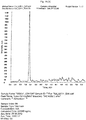
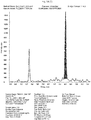
- the concentration of IS in the sample is known and, e.g., by determining the area under the peak, i.e. the peak area, of the internal standard in, e.g., an HPLC-mass spectrometric chromatogram the relation between a peak area and a concentration of a substance, e.g. of IS and/or the biomarker of the present invention, e.g.
- free lyso-Gbl-sulfatide can thus be calculated, e.g., by calculating the ratio of the peak area of free lyso-Gb1-sulfatide and the peak area of IS.
- an IS having a similar chemical structure or an isotopically labeled lyso-Gbl-sulfatide compared to the molecule such as the biomarker, e.g. free lyso-Gb1-sulfatide, is preferable.
- the present inventors have in an embodiment chosen lyso-Gb2 which is not present as such in nature in concentrations which could influence the precise determination of lyso-Gb1-sulfatide.
- the molecule being the IS can be distinguished from the biomarker or the biomarkers of the present invention, e.g. free lyso-Gb1-sulfatide, in the method of the present invention.
- the IS is selected such that a molecule which is ideally not present or rare in nature.
- the internal standard is added to a sample from a subject
- the IS is added such that it is dissolved in a solvent, e.g. ethanol, prior to said addition to the sample.
- the solvent is selected such that said solvent is capable of causing protein precipitation, preferably is capable of causing the protein precipitation step as subject to the method of the present invention as defined in the claims.
- a protein precipitation and/or protein precipitation step is part of the method of the present invention.
- precipitation as used herein, preferably means the formation of a solid in a solution, i.e. for example the formation of a protein precipitate in a sample, e.g. serum, from a subject.
- a sample e.g. serum
- precipitation e.g. protein precipitation
- the solid formed is called the precipitate, or when compacted by a centrifuge, a pellet.
- the liquid remaining above the solid is in either case called the supernatant.
- the present invention contemplates different methods of precipitation and/or separating said supernatant and said precipitate or pellet, comprising, among others, settling or sedimentation and centrifugation.
- a person skilled in the art will know further methods for protein precipitation and/or for separating a supernatant and a protein precipitate, nevertheless said skilled person will acknowledge that if a method, preferably a method of the invention as defined in the claims, is applied were precipitated protein will disable a device such as a column or HPLC-column used in connection with the present invention the precipitated protein is preferably separated from the solvent and/or the sample.
- a level of a biomarker of the present invention e.g. free lyso-Gb1-sulfatide, determined by a method of the present invention as defined in the claims in a sample is compared to a level of the same or another biomarker of the present invention determined by a method of the present invention as defined in the claims in another sample, e.g. from the same patient, from another patient, from a control and/or from the same or different time points, and/or a cut-off value, and/or a level of a control and/or a level of an IS.
- a biomarker of the present invention e.g. free lyso-Gb1-sulfatide
- comparing or “compared to” as used herein, preferably means the mathematical comparison of the two or more values of the levels of the biomarker(s). It will thus be immediately evident whether one of said values is higher, lower or identical if at least two of such values are compared with each other.
- cut-off value refers to a level, concentration and/or a titer of a biomarker of the present invention, more preferably a level range, concentration range and/or titer range of the biomarker.
- the level of the biomarker is also determined in a control.
- a control is preferably a sample from a subject wherein the MLD status of said subject is known.
- a control is a sample of a healthy patient.
- an amount of said biomarker is added to said sample of a healthy patient prior to determining the level of said biomarker in said sample of a healthy patient comprising said added biomarker with a method of the present invention as defined in the claims.
- the control is a sample from at least one subject having a known MLD status, such known MLD status comprising severe, mild, or healthy MLD status, e.g. a control patient.
- the MLD status also comprises the genetic status with regard to mutations of the gene or genes, affected in said disease, comprising the gene coding for ARSA, i.e. comprising the subject having homozygous and/or compound heterozygous mutations, the subject being a carrier of a mutation.
- control is a sample from a subject not being treated for MLD.
- control is a sample from a single subject or a pool of samples from different subjects and/or samples taken from the subject(s) at different time points.
- level or "level of a biomarker” as used herein, preferably means the concentration of a substance and/or titer of a substance, preferably of a biomarker of the invention and more preferably of free lyso-Gbl-sulfatide, within a sample of a subject. It will be understood by a skilled person that in certain embodiments of the invention as defined in the claims, said sample is not necessarily subjected to a method of the invention as a non-processed sample, the method comprising determining a level of said biomarker, i.e. said sample may be subjected, e.g. to a step of protein precipitation, separation, e.g.
- a level of a biomarker is used in connection with a level of the biomarker of the invention which is to be determined according to the present invention, "the” level of the biomarker of the present invention which is to be determined by the methods of to the present invention and which is contained in the sample subjected to the method(s) of the invention is meant.
- a preferred sample is either a blood sample, a serum sample or a plasma sample.
- the level of a biomarker is different between different statuses of MLD if the mean or median level of the biomarker in the different groups is calculated to be statistically significant. Common tests for statistical significance include, among others, t-test, ANOVA, Wilcoxon, Mann-Whitney, odds ratio and Kruskal-Wallis. Biomarkers, alone or in combination, provide measures of relative risk that a subject belongs to one phenotypic status or another. Therefore, biomarkers of the present invention are useful in an embodiment of the present invention as markers for disease, therapeutic effectiveness of a drug or a treatment.
- determining the level of a biomarker preferably means methods which include quantifying an amount of at least one substance in a sample from a subject and/or quantifying an amount of said substance contained in a part of the body of the subject, such as saliva, blood, lymph, serum, plasma or liquor and/or quantifying an amount of said substance in the subject, the substance being selected from the group comprising a biomarker.
- detecting and/or determining the level of free lyso-Gb1-sulfatide in a sample from the subject thus preferably comprises that Gb1-sulfatide present in the blood of a subject is not chemically converted, transformed or derivatized such that free lyso-Gb1-sulfatide cannot be detected and/or the level thereof cannot be determined separate from and/or apart from Gb1-sulfatide.
- Gb1-sulfatide present in a sample from a subject which is subjected to a step of deacylation e.g.
- the method is for detecting and/or determining the level of free lyso-Gb1-sulfatide in a sample from a subject, wherein Gb1-sulfatide present in the sample from the subject is not subjected to a step resulting in deacylation of Gb1-sulfatide, preferably is not subjected to a step resulting in cleavage off of a fatty acid moiety from the Gb1-sulfatide contained in the sample.
- Gb1-sulfatide present in the sample from the subject is not chemically converted, transformed or derivatized.
- free lyso-Gb1-sulfatide present in the sample from the subject is separated from Gb1-sulfatide present in the sample from the subject prior to a step that would result in cleavage of a fatty acid moiety from the Gb1-sulfatide and/or prior to a step in which Gb1-sulfatide is chemically converted, transformed or derivatized.
- a step of detecting and/or determining the level of a biomarker in a sample from the subject, wherein the biomarker is free lyso-Gb1-sulfatide is performed subsequent to separation using HPLC by application of mass spectrometric analysis.
- a subject will be considered to be healthy regarding MLD if it has no mutation of the functional parts of the gene coding for ARSA and/or no mutation of the gene coding for ARSA resulting in a reduction of or deficiency of the respective protein or the activity thereof, resulting in symptoms associated with MLD.
- a subject is considered to be a healthy subject with regard to MLD if the subject does not suffer from symptoms associated with MLD. Moreover, in an embodiment of the methods of the invention as defined in the claims a subject will be considered to be healthy regarding MLD if it has no mutation of the functional parts of the gene coding for ARSA and/or no mutation of the gene coding for ARSA resulting in a reduction of or deficiency of the respective proteins or the activity thereof, resulting in symptoms associated with MLD.
- MLD also comprises MLD carrier. It is important to note that the methods of the invention as defined in the claims, are equally suitable to identify an MLD carrier. The methods of the invention as defined in the claims, are suitable to diagnose whether or whether not a subject is an MLD carrier. The method of the invention as defined in the claims, is further suitable for differentiating, diagnosing and/or differentially diagnosing whether a subject is healthy, is an MLD carrier or is an MLD patient.
- Said mutations i.e. mutations of the gene encoding ARSA, will be detected if a sample from the subject is subjected to a genetic testing for such mutations as described herein.
- a sample from a healthy subject is used as a control sample or as a blank matrix in the methods of the present invention as defined in the claims.
- a blank matrix as used herein is preferably a sample from a healthy subject. Nevertheless, it will be understood that such a blank matrix may contain a native level of free lyso-Gb1-sulfatide.
- the level of a biomarker is indicative for the subject for suffering from or for being at risk for developing a disease or disorder.
- the level of the biomarker determined by the method according to the invention as defined in the claims is compared to a control level of the biomarker, wherein the result of said comparison allows for diagnosing a disease.
- comparing the level of the biomarker in the sample from the subject to the control level of the biomarker comprises comparing the level of the biomarker in the sample from the subject to a cut-off value, wherein if a level of the biomarker in the sample from the subject is higher than the cut-off value, this is indicative that the subject is suffering from or is at risk for developing MLD; and/or wherein if a level of the biomarker in the sample from the subject is lower compared to the cut-off value this is indicative that the subject is not suffering from or is not at risk for developing MLD.
- being at risk for developing a disease preferably means that it is likely that a subject suffers from said disease and/or will develop said disease or symptoms associated with said disease, particularly if no treatment is applied.
- LSDs are genetic disorders and thus the occurrence of relatives, particularly parents having said disease or having a mutation known to be the cause of said disease are indicative for a subject, e.g. the child of two MLD patients, to be at risk for developing said disease. It will be furthermore acknowledged that the progression of a disease is linked to the occurrence of symptoms as well as the severity of said symptoms.
- a person not suffering from symptoms at present may be at risk for developing the disease, for example, because although genetically mutations of a gene, known to cause a disease are present, no symptoms or no severe symptoms occur.
- the methods and biomarkers of the present invention particularly if the level(s) of said biomarker(s) according to the present invention are elevated, allow for diagnosing that such subject is at risk for developing the disease independent from the presence or absence of symptoms.
- the methods according to the invention as defined in the claims allows for determining whether a subject is at risk of suffering from MLD. It is also within the present invention that a therapy is to be applied, maintained, reduced, elevated or not to be applied based on whether the subject is at risk of suffering from MLD or not.
- comparing the level of the biomarker in the sample from the subject to a control level allows for determining the severity of MLD, wherein if a level of the biomarker in the sample from the subject is within the cut-off value that is indicative that the subject is suffering from or is at risk for developing MLD of a more severe status or progression; and wherein if a level of the biomarker in the sample from the subject is lower or higher compared to the control level, i.e. the cut-off value, that is indicative that the subject is not suffering from or is not at risk for developing MLD of a less severe status or progression.
- comparing the level of the biomarker in the sample from the subject to the control level comprises comparing a level of the biomarker in said subject to a level of the biomarker detected in a sample from a control, wherein if a level of the biomarker in the sample from the subject is elevated, increased or higher compared to the control sample this is indicative that the subject is suffering from and/or is at risk for developing MLD; and/or a level of the biomarker in the sample from the subject is elevated, increased or higher compared to the control sample this is indicative that the subject is suffering from or is at risk for developing MLD of a more severe status or progression.
- Said control preferably is selected from the group comprising healthy subjects, subjects suffering from MLD or being at risk of suffering from MLD symptoms, subjects being positively tested for a mutation or a combination of mutations of the gene coding for ARSA, wherein the mutation or the combination of mutations of the gene coding for ARSA are indicative for a perspective of the subject to develop MLD of a more severe or less severe status or progression.
- a control level is determined in a sample from a control, wherein optionally free lyso-Gbl-sulfatide is added to the sample from the control in a specific quantity prior to determining the level of free lyso-Gb1-sulfatide in the sample from the control.
- a method for diagnosing MLD in a subject comprising detecting a biomarker in a sample from a subject, wherein the biomarker is free lyso-Gb1-sulfatide, preferably further comprising determining a level of the biomarker in the sample from the subject, and more preferably further comprising comparing the level of the biomarker in the sample from the subject to a cut-off value, which shows high sensitivity, i.e. a sensitivity of at least 99,0%, 99,1%, 99,2%, 99,3%, 99,4%, 99,5%, 99,6%, 99,7%, 99,8%, 99,9% or 100%.
- the sensitivity which means the proportion of actual positives which are correctly identified as such is high, which means that the percentage of MLD patients correctly identified as having the disease is as high as has been outlined above.
- specificity means the proportion of negatives which are correctly identified as negatives, in other words the percentage of healthy patients correctly identified as not having MLD.
- a specificity of at least 80.0%, 85.0%, 90.0%, 95.0%, 97.5%, 99.0%, 99.1%, 99.2%, 99.3%, 99.4%, 99.5%, 99.6%, 99.7%, 99.8%, 99.9% or 100% is preferred.
- the methods allow for diagnosing MLD in a subject independent from a progression status of MLD in the subject. Such specificity can be achieved with the cut-off value for free lyso-Gbl-sulfatide being 0.05 ng/ml plasma or serum.
- the methods of the invention as defined in the claims allow for diagnosing MLD in a subject having an early status of MLD as well as in a subject having an advanced or progressed status of MLD.
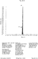
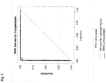
- the power of a method to correctly diagnose MLD is commonly measured as the sensitivity of the method, the specificity of the method or the area under a receiver operated characteristic curve (also referred to herein as "ROC curve").
- An ROC curve is a plot of the true positive rate against the false positive rate for the different possible cut-off values of a diagnostic method.
- An ROC curve shows the relationship between sensitivity and specificity. Sensitivity is the percentage of true positives that are predicted by a test to be positive, while specificity is the percentage of true negatives that are predicted by a test to be negative.
- An ROC-curve provides the sensitivity of a test as a function of 1-specificity. The greater the area under the ROC-curve the more powerful the predictive value of the test.
- the area under the ROC is a measure of test accuracy.
- the accuracy of the test depends on how well the test separates the group being tested into those with and without the disease in question.
- An area under the curve (also referred to herein as "AUC") of 1 represents a perfect method, while an area of 0.5 represents a less useful method.
- a positive predictive value is the percentage of actual positives that test as positive.
- a negative predictive value is the percentage of actual negatives that test as negative.
- a diseased subject tested false negative applying the methods of the present invention as defined in the claims is tested false negative for the reason that a level of the biomarker in a sample from said false negative tested diseased subject is as high as the level of the biomarker in a sample from a healthy subject.
- said false negative tested subject is not tested negative for the reason that the level of the biomarker was too low to be determined by the method of the present invention as defined in the claims.
- a "limit of detection” or “limit of determination” - both terms are used herein in a synonymous manner - of a substance such as free lyso-Gb1-sulfatide, as used herein, preferably is a level of the substance determined by a method for determining a level of the substance, wherein a level less then or lower then said limit of detection cannot be determined by said method. It is thus immediately clear that a "cut-off value” and a “limit of detection”, as used herein, are preferably not necessarily identical, although both reflect a certain level of a substance, e.g. of a biomarker of the present invention.
- a limit of detection represents an absolute level of the biomarker of the present invention which reflects the minimum level of biomarker which can be detected with a method for determining the level of said biomarker. It is thus immediately clear that a limit of detection depends on the method for determining a level of a substance and on the substance the level of which is to be determined by the method. A skilled person will immediately understand that a high limit of detection, e.g.
- a lower limit of detection allows for a method for diagnosing MLD in a subject comprising a step of determining a level of a biomarker present in the sample with higher selectivity and sensitivity.
- An "ideal cut-off value" as used herein preferably is the cut-off value as described herein the method using said ideal cut-off value has the highest selectivity and sensitivity.
- the method comprises a step of validating said method by diagnosing a disease or disorder, preferably MLD in a subject by the method of the present invention as defined in the claims; a step of diagnosing the disease or disorder, preferably MLD, in a subject by a genetic testing, comprising sequencing of a gene, preferably sequencing of a gene a mutation of which is known to the one skilled in the art to cause the disease or disorder, more preferably sequencing the gene coding for ARSA in case of MLD ; and comparing the results of said method and said genetic testing.
- a healthy subject as used herein preferably is considered to be healthy with regard to a disease or disorder if said subject is not suffering from symptoms associated with said disease or disorder and if the result of a genetic testing reveals no mutations of a gene a mutation of which is known to the one skilled in the art to cause the disease or disorder.
- a healthy subject also is understood to be a subject being positively tested for not having MLD.
- a healthy subject is a subject not being a carrier of MLD.
- qualifying MLD status in a subject preferably means a classification of a subject's biomarker profile selected from the group comprising to identify or detect the presence or absence of MLD in the subject, to predict the onset of or the risk for developing of MLD in the subject, to determine the course of MLD in a subject, to determine and/or predict the severity of MLD in a subject, to determine whether a subject suffers from an early status of MLD or an advanced or progressed status of MLD or to determine whether a level of a biomarker in a subject has significantly changed over time.
- managing subject treatment comprises titrating of a dose of a drug applied as a treatment for MLD, e.g. units of recombinant enzyme applied in ERT, administered to a patient.
- a level of a biomarker present in a sample from a subject is determined at several points in time, or is compared to other levels of the biomarker, a cut-off value and/or a level of said biomarker in a control
- a skilled person will apply or not apply a therapy, or amend a therapy already applied in order to treat or not to treat, or to continue treating MLD.
- a skilled person will elect a dosage and/or maintain a dosage or amend a dosage, e.g. a higher dosage, i.e. elevate a dosage, if such a comparison of the level of a biomarker shows e.g. that the level of said biomarker is higher than for example, a cut-off value, i.e. the patient is diagnosed to have MLD ; or that a level determined in the same patient earlier in time is lower or the same, i.e. a therapy applied is not sufficient, i.e. does not result in a decrease in the level.
- skilled person will apply or not apply a dosage or maintain or reduce a dosage, e.g.
- a dosage i.e. decrease a dosage
- a comparison of the level of a biomarker shows e.g. that the level of said biomarker is lower than for example, a cut-off value, i.e. the patient is diagnosed not to have MLD disease; or that a level determined in the same patient earlier in time is higher, i.e. a therapy applied is sufficient, i.e. does result in a decrease in the level.
- a relatively high level of free lyso-Gbl-sulfatide based on such a comparison is indicative for applying a high dosage of recombinant enzyme applied in ERT and/or a relatively low level of free lyso-Gbl based on such a comparison is indicative for applying a low dosage of recombinant enzyme applied in ERT.
- the course of MLD may be determined by the method according to the present invention as defined in the claims by determining a level of the biomarker in the sample from the subject at different time points in the course of the disease. It is important to note that a single application of a method for diagnosing MLD according to the present invention allows for diagnosing MLD and in certain embodiments comprises a step of managing subject treatment based on the diagnosis of whether the subject is suffering from or for being at risk for developing MLD. If a subject a sample of which is thus subjected to the method of the present invention as defined in the claims is tested positive for suffering from or to be at risk for developing MLD a skilled clinician will know how to decide concerning managing subject treatment, i.e. how the subject will be treated, e.g.
- the skilled clinician may decide for at least one additional application of the method according to the present invention as defined in the claims on a later time point. It is thus an embodiment of the present invention as defined in the claims that the levels of the biomarker determined at the different time points, wherein different time points means at least two time points, may be compared. Without wishing to be bound by any theory the present inventors have found that the level of the biomarker of the present invention in samples form one particular patient may be correlated to the severity of the disease in said patient at the time point the sample from the patient is taken.
- an elevated level of the biomarker determined in the sample of a later time point compared to the level of the biomarker determined in the sample of an earlier time point is indicative for a more severe status of the subject at the later time point compared to the status of the subject at the earlier time point.
- a decreased level of the biomarker determined in the sample of a later time point compared to the level of the biomarker determined in the sample of an earlier time point is indicative for a less severe status of the subject at the later time point compared to the status of the subject at the earlier time point.
- the present disclosure provides a method for determining the course of MLD in a subject comprising the step of determining at several points in time a level of a biomarker present in a sample from the subject, wherein the biomarker is free lyso-Gb1-sulfatide
- the disclosure concerns a method for determining the effectiveness of at least one treatment applied to a subject being positively tested for suffering from or being at risk for developing MLD comprising the step of determining at several points in time a level of a biomarker present in a sample from the subject, wherein the biomarker is free lyso-Gb1-sulfatide.
- the methods of the present disclosure thus allow for selecting a therapy and/or adjusting the doses and/or dosage of a selected therapy based on the results of the method of the invention as defined in the claims. If, for example, the subject is scheduled for treating for MLD the method for diagnosing MLD in a subject according to the present invention as defined in the claims may be applied every 3 months and levels of the biomarker thus determined will be compared in order to determine the effectiveness of the treatment(s) and/or therapy/therapies applied to the subject.
- the frequency of application of the method for diagnosing MLD in a subject according to the present invention as defined in the claims may be reduced to every 6 months. If the dosage of the therapy is changed, e.g. the units of recombinant enzyme applied in ERT are reduced or increased, the frequency of application of the method for diagnosing MLD in a subject according to the present invention as defined in the claims may be set back to every 3 months.
- the skilled physician may decide to reduce the dosage of the therapy, e.g. the units of recombinant enzyme applied in ERT; to increase the dosage of the therapy; or to maintain the dosage of the therapy according to the comparison of the levels of the biomarker determined with the method according to the present invention as defined in the claims.
- the dosage of the therapy e.g. the units of recombinant enzyme applied in ERT
- the skilled physician may decide to reduce the dosage of the therapy, e.g. the units of recombinant enzyme applied in ERT; to increase the dosage of the therapy; or to maintain the dosage of the therapy according to the comparison of the levels of the biomarker determined with the method according to the present invention as defined in the claims.
- a significant reduction of the level of free lyso-Gb1-sulfatide within a period of 12 month is indicative for a successful therapy for MLD, wherein reduction as used herein, preferably means that the level of free lyso-Gbl-sulfatide determined by the method of the present invention as defined in the claims determined at the end of a time period is compared to the level of free lyso-Gb1-sulfatide determined by the method of the present invention as defined in the claims determined at the beginning of said time period. Accordingly, the skilled physician may decide to reduce the dosage of the applied therapy or to maintain the dosage of the therapy.
- the reduction of the level of free lyso-Gb1-sulfatide is significantly weaker the skilled physician may decide to increase the dosage of the therapy. It is also a merit of the present inventors to have recognized that the reduction of the level of free lyso-Gbl-sulfatide correlates with the effectiveness of a therapy. The stronger the reduction of the level of the free lyso-Gbl-sulfatide within a time period, e.g. 12 months, the more successful is a therapy, such as for example ERT, SRT or a chaperone based therapy. It is thus a further embodiment of the present invention that the method of the present invention as defined in the claims is for comparing the effectiveness of a therapy or of at least two therapies applied to a subject.
- the disclosure concerns a method for determining the effectiveness of at least one treatment applied to a subject being positively tested for suffering from or being at risk for developing MLD comprising the step of determining at several points in time a level of a biomarker present in a sample from the subject, wherein the biomarker is free lyso-Gbl-sulfatide
- a biomarker is free lyso-Gbl-sulfatide
- the method for diagnosing MLD is independent of whether the subject has or has not been previously treated for MLD.
- the sample from the subject may be a sample from a subject who has been previously treated for MLD as well as a sample from a subject who has not been previously treated for MLD. It is thus a further embodiment of the present invention as defined in the claims that the method of the present invention comprises a step of managing subject treatment and/or determining a level of the biomarker in the sample from the subject after subject management.
- Said subject treatment can be based on the diagnosis of whether the subject is suffering from or for being at risk for developing MLD; on the detection of the biomarker in a sample from the subject after subject management; or on the determining of the level of the biomarker in the sample from the subject after subject management. Nevertheless, a person skilled in the art will understand that a sample of some patients not having MLD or of some patients being successfully treated for MLD will show a level of free lyso-Gb1-sulfatide lower than the limit of detection.
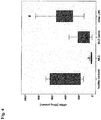
- the present inventors assume that the level of free lyso-Gb1-sulfatide present in a sample from a subject further correlates with the severity of the disease in a subject suffering from MLD.
- the present inventors found by evaluating the results provided herein (e.g. shown in Fig.2 herein) assume that although, in principle, the level of free lyso-Gbl-sulfatide is different in particular individuals, and more specifically may be different in particular individuals having the same mutation(s), that the higher is a level of free lyso-Gb1-sulfatide, the higher is the severity of a course of MLD in terms of a statistical mean according to a clinical score.
- the level of free lyso-Gb1-sulfatide correlates with the severity of MLD in that in patients being positively tested for distinct mutations of the gene coding for ARSA being known to generally causes a mild or a more severe course of MLD, a level of free lyso-Gbl-sulfatide determined in said patients statistically correlates with the severity generally related to such mutation.
- a further embodiment of the present invention as defined in the claims encompasses a method for determining the severity of MLD in a subject comprising a step of
- a level of free lyso-Gbl-sulfatide is determined in samples from the patients suffering from MLD showing the L444Pa mutation usually linked to a more severe course of MLD upon sequencing of the respective gene (homozygous and compound heterozygous) subjected to a method of the present invention as defined in the claims a mean-level of free lyso-Gbl-sulfatide is higher than the mean-level of the free lyso-Gbl-sulfatide determined in samples from the patients suffering from MLD showing a mutation usually linked to a more mild course of MLD upon sequencing of the respective gene, applying the same method.
- a "mutation usually linked to a more severe course of MLD” as used herein preferably is known to cause a more severe course of MLD - this is especially true in case the subject is homozygous as to said mutation.
- a higher mean-level of free lyso-Gb1-sulfatide is determined in the homozygous compared to the homozygous mutation usually linked to a milder course of MLD.
- patients having a compound heterozygous usually linked to a more severe course of MLD have a significantly lower free lyso-Gb1-sulfatide level, than homozygous ones.
- a person skilled in the art will know clinical scores to categorize the severity of MLD or symptoms or an entirety of symptoms thereof.
- a level of the biomarker of the present invention determined in a sample from a subject wherein said level of the biomarker is correlated with the severity of MLD as described above will be indicative for applying a certain therapy and/or dose or dosage of said therapy.
- the level of the biomarker determined according to the methods of the invention as defined in the claims is correlated with "severe" MLD status the subject is scheduled for treatment of MLD and the method for diagnosing MLD in a subject according to the present invention as defined in the claims may be applied every 3 months and levels of the biomarker thus determined will be compared in order to determine the effectiveness of the treatment(s) and/or therapy/therapies applied to the subject.
- the frequency of application of the method for diagnosing MLD in a subject according to the present invention as defined in the claims may be reduced to every 6 months.
- the present disclosure is related to a method of determining the effectiveness of a composition for the treatment of MLD.
- Such method may comprise the steps of determining a level of free lyso-Gb1-sulfatide in a subject having MLD; administering to said subject said compound in an amount sufficient to determine the effectiveness of said compound; re-determining the level of free lyso-Gb1-sulfatide in said subject; comparing the level of free lyso-Gbl-sulfatide determined before and after administering said composition, wherein a lower level of free lyso-Gb1-sulfatide determined after administering said composition compared to the level of free lyso-Gb1-sulfatide determined after administering said composition indicates the effectiveness of said compound for treating MLD .
- MLD myeloma
- MLD myeloma
- a preferable biomarker for the diagnosis of MLD would allow for diagnosis of MLD with high sensitivity and high specificity independent from the age of the subject.
- the biomarkers of the present invention are useful for the diagnosis of MLD in a subject independent from the age of the subject. It is thus an embodiment of the present invention that the method of the present invention as defined in the claims allows for diagnosing MLD in a subject independent from age, preferably under the proviso that the subject is at least 2 months old.
- the method of the present invention as defined in the claims the subject is a subject of young age.
- a subject of young age as used herein preferably is a subject of less than 30 years of age, more preferably of less than 20 years of age and most preferably of less than 10 years of age.
- a patient or subject is preferably a human of at least 2 months age.
- the methods of the invention as defined in the claims and in particular the method of the invention as defined in the claims for diagnosing metachromatic leucodystrophy is one for diagnosing juvenile form of metachromatic leucodystrophy and/or adult form of metachromatic leucodystrophy.
- the cut-off value for free lyso-Gb1-sulfatide of 0.05 ng/ml plasma or serum is one which is preferably applied in case the limit of determination, which is also referred to herein as limit of detection, of the analytical method used in determining the level or concentration of free lyso-Gb1-sulfatide is 0.05 ng free lyso-Gb1-sulfatide/ml plasma or serum, or in case the limit of determination of the analytical method used in determining the level or concentration of free lyso-Gb1-sulfatide is set to 0.05 ng free lyso-Gb1-sulfatide/ml plasma or serum.
- the present inventors have found that using a sample of human serum in the method as described below instead of a sample of human plasma will lead to identical results in terms of the level of free lyso-Gb1-sulfatide, if the sample of human serum and the sample of human plasma are taken from the same subject, at the same time point, and if the samples were measured in parallel; and, more particularly, will lead to the same cut-off value.
- Example 1 Method for the detection of free lyso-Gbl-sulfatide in human plasma/serum
- purum as used herein, preferably means a commercial grade of a chemical compound having a purity of the above specified value.
- ASTM-I refers to a water grade standard purity achieved by purification methods comprising Reverse Osmosis and Ultraviolet (UV) Oxidation.
- a lyso-Gbl-sulfatide stock solution was prepared dissolving 0.62 mg lyso-Gbl-sulfatide (as delivered by Matreya) in 5 mL of 50 % MeOH.
- the solution V1-A-626 was prepared as a mixture of 50 ⁇ L of lyso-Gbl-sulfatide stock solution and 10 mL 50 % MeOH as displayed in the following: Label of resulting solution exp.conc. [ ⁇ g/mL] Volume of solution [ ⁇ L] solution volume of solvent [mL] solvent V1-A-626 0.60760 50 lyso-Gbl-sulfatide-stock 10 50 % MeOH
- calibration standards having four concentration levels between 0.05 and 5.00 ng/mL were used, namely Calibration Standards Std1B-626, Std2B-626, Std3B-626 and Std4B-626.
- Control samples were prepared by spiking solution V1-A-626 or a higher concentrated control sample into a blank human plasma.
- the Internal Standard (IS1) stock solution was prepared dissolving 1.00 mg of lyso-Gb2 (as delivered by Matreya) in 2 mL of DMSO / MeOH (1/1; vol/vol).
- the Internal Standard Working Solution was prepared as a mixture of 82 ⁇ L of IS1 stock solution and 500 mL of ethanol.
- the ethanol may be purchased from any commercial source, wherein the ethanol is absolute ethanol having a grade suitable for the methods described herein.
- proteins contained in 50 ⁇ l of a sample have to precipitate if 100 ⁇ L of said Internal Standard working solution are added to the sample.
- Control samples or study samples either were immediately stored below -20°C at once or aliquots were transferred into new glass vials before storing under the same conditions.
- lyso-Gb1-sulfatide is stable in the above mentioned solutions. lyso-Gb1-Sulfatide is also stable at least for several weeks of storage in plasma/serum below -20°C and over several freeze/thaw cycles in plasma/serum.
- the thus obtained mixture was subsequently mixed using a DVX-2500 Multi-tube vortex device at 2500 rpm for about 30 seconds
- the thus obtained mixture was centrifuged for phase separation at 4000 rpm for 2 minutes.
- the YMC Pro C8 column (Nr. OS12S031003QT) used herein has been purchased from YMC, Germany.
- parameters where a " ⁇ " range is indicated represent parameters which may be adjusted between sequences.
- a sequence as used herein preferably is a batch of defined numbers of samples, preferably 150 in maximum analyzed sequentially, wherein parameters comprising flow and temperature remain unchanged. Adjustments and calibrations performed between sequences are known to those skilled in the art and comprise exchange of the column.
- MS Ionisation mode Electrospray Ionisation (ESI)
- MS polarity positive MS detection mode: Multiple reaction monitoring (MRM)
- Vaporizer temperature 550°C ⁇ 50°C Ionisation voltage: 5.5 kV
- Concentration data fed into and retrieved from the chromatographic data system were rounded to five significant digits. Further calculations in the spreadsheet were performed to full computational accuracy and subsequently rounded to the significant digits / decimal places to be reported. Hence, deviations of intermediate results might occur caused by rounding. Accuracy and coefficients of variation (CV) will be reported with one and two decimal places, respectively.
- HPLC-mass spectrometric chromatograms of 150 plasma samples derived from the 150 subjects described in more details in Example 2.
- Exemplary HPLC-mass spectrometric chromatograms displaying peak intensity of free lyso-Gb1-sulfatide and IS of a healthy control person and of a patient diagnosed as MDL-positive are depicted in Fig. 1A and Fig. 1B .
- Fig. 1A shows HPLC-mass spectrometric chromatograms displaying peak intensity in cps of free lyso-Gbl-sulfatide (upper panel) and IS (lower panel) of a sample from a healthy subject as a function over the retention time in minutes.
- Fig. 1B shows HPLC-mass spectrometric chromatograms displaying peak intensity in cps of free lyso-Gb1-sulfatide (upper panel) and IS (lower panel) of a sample from an MLD patient as a function over the retention time in minutes.
- the retention time of a substance as used herein preferably is depicted on the x-axis and is the elapsed time between the time of injection of a solute, e.g. a biomarker according to the present invention and/or an internal standard, and the time of elution of the peak maximum of said solute.
- a solute e.g. a biomarker according to the present invention and/or an internal standard
- the retention time of a substance according to the herein described methods is a unique characteristic of said solute and can be used for identification purposes.
- Internal Standard working solution comprising lyso-Gb2 as an internal standard was added to the sample as described above. It is important to understand that by said addition of IS to the sample, i.e.
- the concentration of IS in the sample is known and by determining the area under the peak, i.e. the peak area, of the internal standard in said HPLC-mass spectrometric chromatogram the relation between a peak area and a concentration of a substance, e.g. of IS and/or a biomarker thus can be calculated. More precisely, a person skilled in the art will acknowledge that a peak area of a substance depicted in an HPLC-mass spectrometric chromatogram, such as the HPLC-mass spectrometric chromatogram depicted in Fig. 1A or Fig.
- 1B represents a measure for an amount of said substance subjected to an HPLC-mass spectrometric analysis.
- a person skilled in the art will be able to calculate the amount of the substance in a sample from a subject subjected to an HPLC-mass spectrometric analysis, e.g. the amount of free lyso-Gb1-sulfatide in a sample subjected to the method of the present invention, using a ratio of the peak area of free lyso-Gb1-sulfatide, the amount of which is to be determined by said method and the peak area of IS, e.g.
- Plasma samples from a first cohort of 93 subjects were analyzed.
- the level of free lyso-Gb1-sulfatide in samples of said 93 subjects was determined according to the method described in Example 1.
- Table 1 shows the median and minimum and maximum levels of free lyso-Gb1-sulfatide in said samples of said 93 subjects.
- MLD patients could be clearly distinguished from subjects suffering from other LSDs.
- Such other LSDs included Fabry disease, Gaucher's disease and Nieman-Pick disease type C.
- Plasma samples from a second cohort of 150 subjects were analyzed, whereby such second cohort comprised the first cohort.
- the level of free lyso-Gb1-sulfatide in samples of said 150 subjects was determined according to the method described in Example 1.
- That total group comprises 25 MLD patients, 3 carriers, 6 healthy controls and 116 patients with other lysosomal storage disorders: 5 Gaucher patients (27 samples), 20 Fabry unclear patients (20 samples), 11 male Fabry patients (15 samples), 9 Female Fabry patients (11 samples), 7 NPC1 patients (9 samples), 6 NPC1 carriers (9 samples), 5 NPA/B patients (5 samples), 6 Krabbe disease patients (6 samples), 8 male Hunter disease patients (8 samples), 2 female Hunter disease patients (2 samples), 3 GM1 gangliosidosis patients (3 samples), 3 San Filippo B patients (MPS3b) (3 samples), 9 MPS6 patients (9 samples), 1 MPS6 carrier (1 sample), 1 Tay-Sachs disease patient (1 sample), 4 MPS1 (4 samples), 3 San Filippo A patients (MPS3a) (3 samples), 12 MPS3a patients (Mor
- Fig. 2 is a boxplot indicating levels of free lyso-Gb1 sulfatide; the y-axis indicates the absolute levels of free lyso-Gb1-sulfatide in ng/ml determined in plasma of patients by the method according to the present invention, wherein the x-axis depicts groups of patients, which have been grouped as described.
- the boxplot represents the 25th and 75th percentile of each group of patients by the bottom and top of the box, respectively; the band near the middle of the box represents the 50th percentile (i.e. the median) of each group;
- the whiskers represent one standard deviation above and below the mean of the data; Any data not included between the whiskers is shown as an outlier with a small circle
- Example 3 Diagnosis of MLD using free lyso-Gbl-sulfatide as a biomarker
- Plasma samples from the first cohort of patients defined in Example 1 were subject to the methods and analysis described in Example 1. The results from said analysis are depicted in Fig. 2
- Fig. 2 is a boxplot indicating levels of free lyso-Gb1-sulfatide in patients diagnosed either as MLD positive or as suffering from other LSDs.
- the y-axis demonstrates the levels of free lyso-Gb1-sulfatide in ng/ml determined in plasma of patients by the method of Example 1, wherein the x-axis depicts groups of patients, which have been grouped as described in Example 2.
- the boxplot represents the 25 th and 75 th percentile of each group of patients by the bottom and top of the box, respectively; the band near the middle of the box represents the 50 th percentile (i.e. the median) of each group; the whiskers represent one standard deviation above and below the mean of the data. Any data not included between the whiskers is shown as an outlier with a small circle or star.
- Plasma samples from the second cohort of patients defined in Example 1 were subject to the methods and analysis described in Example 1. The results from said analysis are depicted in Fig. 3
- Fig. 3 is a boxplot indicating levels of free lyso-Gb1 sulfatide;
- the y-axis indicates the absolute levels of free lyso-Gb1-sulfatide in ng/ml determined in plasma of patients by the method of Example 1, wherein the x-axis depicts groups of patients, which have been grouped as described (healthy controls, patients suffering from MLD, patients being MLD carrier and patients suffering from LSD different from MLD).
- the boxplot represents the 25th and 75th percentile of each group of patients by the bottom and top of the box, respectively; the band near the middle of the box represents the 50th percentile (i.e. the median) of each group.
- the whiskers represent one standard deviation above and below the mean of the data. Any data not included between the whiskers is shown as an outlier with a small circle.
- Fig. 4 is a boxplot indicating the arylsulfatase enzyme activity in the groups defined above.
- the y-axis demonstrates the arylsulfatase enzyme given as U/mg protein, wherein the x-axis depicts groups of patients, which have been grouped as described above.
- the boxplot represents the 25th and 75th percentile of each group of patients by the bottom and top of the box, respectively; the band near the middle of the box represents the 50th percentile (i.e. the median) of each group; the whiskers represent one standard deviation above and below the mean of the data; any data not included between the whiskers is shown as an outlier with a small circle or star.
- the data obtained by the method described above was first aggregated by using the first measured value of every marker for MLD patient.
- ROC curves were calculated using PASW Statistics 18, Release Version 18.0.2 ( ⁇ SPSS, Inc., 2009, Chicago, IL, www.spss.com). The comparisons of ROC curves and the linear mixed models were done using SAS software, Version 9.2 of the SAS System for Windows. ( ⁇ 2008 SAS Institute Inc., Cary, NC, USA).
- the respective ROC curve is shown in Fig. 5 .
- Fig. 5 is a graph showing receiver operating characteristics (ROC) curves of free lyso-Gbl - sulfatide and arylsulfatase A.
- the x-axis represents "1-specificity" and the y-axis represents the sensitivity.
- Free lyso-Gbl-sulfatide demonstrates a 98 % sensitivity and 99.1% specificity, wherein arylsulfatase A has at best a sensitivity of 92.1 % and 94 % specificity, respectively.
- Example 4 Incorrect diagnosis of a patient as MLD positive based on enzymatic activity of arylsulfatase A
- Gieselmann 1989 shows as well that ARSA activity in tissues from compound heterozygous individuals is lower than homozygous p.N352S individuals. This is a disease-associated polymorphism in HGMD and, most importantly, in dbSNP (rs2071421) the minor allele frequency for this SNP is 20% in the general population and varies between 13-39% in the several hapmap populations. Finally, PolyPhen prediction is "benign" (probability of being a mutation is 1.2%).
Landscapes
- Life Sciences & Earth Sciences (AREA)
- Health & Medical Sciences (AREA)
- Engineering & Computer Science (AREA)
- Molecular Biology (AREA)
- Chemical & Material Sciences (AREA)
- Biomedical Technology (AREA)
- Urology & Nephrology (AREA)
- Hematology (AREA)
- Immunology (AREA)
- Cell Biology (AREA)
- Analytical Chemistry (AREA)
- Biotechnology (AREA)
- Proteomics, Peptides & Aminoacids (AREA)
- Food Science & Technology (AREA)
- Medicinal Chemistry (AREA)
- Physics & Mathematics (AREA)
- Microbiology (AREA)
- Biochemistry (AREA)
- General Health & Medical Sciences (AREA)
- General Physics & Mathematics (AREA)
- Pathology (AREA)
- Investigating Or Analysing Biological Materials (AREA)
- Measuring Or Testing Involving Enzymes Or Micro-Organisms (AREA)
- Other Investigation Or Analysis Of Materials By Electrical Means (AREA)
Claims (12)
- Procédé pour le diagnostic de la leucodystrophie métachromatique chez un sujet, comprenant une étape a), dans lequel l'étape a) comprend la détection d'un biomarqueur dans un échantillon prélevé chez le sujet, dans lequel l'échantillon est choisi dans le groupe constitué du sang, du sang séché, du sérum et du plasma, et dans lequel le biomarqueur est un lyso-Gb1-sulfatide libre, dans lequel le lyso-Gb1-sulfatide est de formule (I)

- Procédé selon la revendication 1, dans lequel l'échantillon est un échantillon de sérum du sujet, un échantillon de plasma du sujet ou un échantillon de sang séché du sujet, de préférence un échantillon de plasma du sujet ou un échantillon de sérum du sujet.
- Procédé selon l'une quelconque des revendications 1 à 2, dans lequel le procédé comprend une étape b), dans lequel l'étape b) comprend la détermination d'un niveau du biomarqueur présent dans l'échantillon.
- Procédé selon la revendication 3, dans lequel le niveau du biomarqueur indique si le sujet souffre ou non de leucodystrophie métachromatique ou si le sujet est à risque ou non de souffrir de leucodystrophie métachromatique.
- Procédé selon l'une quelconque des revendications 1 à 4, dans lequel le biomarqueur est détecté par dosage immunologique, analyse par spectrométrie de masse, réseau de puces à ADN, acides nucléiques fonctionnels et/ou un dérivé fluorescent du biomarqueur, de préférence le biomarqueur est détecté par analyse par spectrométrie de masse, mieux encore la spectrométrie de masse est combinée à une CLHP.
- Procédé selon la revendication 5, dans lequel l'analyse par spectrométrie de masse est choisie dans le groupe comprenant SELDI, MALDI, MALDI-Q TOF, MS/MS, TOF-TOF et ESI-O-TOF, de préférence l'analyse par spectrométrie de masse comprend ou utilise MS/MS.
- Procédé selon l'une quelconque des revendications 3 à 6, dans lequel l'étape b) comprend la comparaison du niveau du biomarqueur dans l'échantillon prélevé chez le sujet à une valeur seuil, la valeur seuil pour le lyso-Gb1-sulfatide libre étant de préférence de 0,05 ng/ml.
- Procédé selon la revendication 7, dans lequel, si le niveau du biomarqueur dans l'échantillon prélevé chez le sujet est supérieur à la valeur seuil, cela indique que le sujet souffre de leucodystrophie métachromatique ou est à risque de souffrir de leucodystrophie métachromatique ; tandis que, si le niveau du biomarqueur dans l'échantillon prélevé chez le sujet est inférieur à la valeur seuil, cela indique que le sujet ne souffre pas ou n'est pas à risque de souffrir de leucodystrophie métachromatique.
- Procédé selon l'une quelconque des revendications 1 à 8, dans lequel le procédé, de préférence le procédé analytique utilisé dans la détection et/ou la détermination du niveau du biomarqueur présent dans l'échantillon, a une limite de détermination du lyso-Gb1-sulfatide libre de 0,05 ng/ml.
- Utilisation d'un biomarqueur pour le diagnostic de la leucodystrophie monochromatique dans un procédé selon l'une quelconque des revendications 1 à 9, dans laquelle le biomarqueur est un lyso-Gb1-sulfatide libre.
- Utilisation de l'analyse par spectrométrie de masse pour la détection d'un biomarqueur pour le diagnostic de la leucodystrophie métachromatique dans un procédé selon l'une quelconque des revendications 1 à 9, dans laquelle le biomarqueur est un lyso-Gb1-sulfatide libre, le biomarqueur est détecté dans un échantillon prélevé chez un sujet, dans lequel l'échantillon est choisi dans le groupe constitué d'un échantillon de plasma prélevé chez le sujet, d'un échantillon de sérum prélevé chez le sujet et d'un échantillon de sang séché prélevé chez le sujet, et dans laquelle l'analyse par spectrométrie de masse comprend ou utilise de préférence MS/MS.
- Utilisation selon la revendication 11, dans laquelle l'analyse par spectrométrie de masse est combinée à une CLHP.
Priority Applications (1)
| Application Number | Priority Date | Filing Date | Title |
|---|---|---|---|
| EP13815378.8A EP2932274B1 (fr) | 2012-12-11 | 2013-12-11 | Procédé pour le diagnostic de la leucodystrophie métachromatique |
Applications Claiming Priority (5)
| Application Number | Priority Date | Filing Date | Title |
|---|---|---|---|
| EP12008257 | 2012-12-11 | ||
| EP12008293.8A EP2743700A1 (fr) | 2012-12-12 | 2012-12-12 | Procédé pour le diagnostic de la leucodystrophie métachromatique |
| EP13001454 | 2013-03-21 | ||
| EP13815378.8A EP2932274B1 (fr) | 2012-12-11 | 2013-12-11 | Procédé pour le diagnostic de la leucodystrophie métachromatique |
| PCT/EP2013/003750 WO2014090404A1 (fr) | 2012-12-11 | 2013-12-11 | Procédé pour le diagnostic de la leucodystrophie métachromatique |
Publications (2)
| Publication Number | Publication Date |
|---|---|
| EP2932274A1 EP2932274A1 (fr) | 2015-10-21 |
| EP2932274B1 true EP2932274B1 (fr) | 2020-01-15 |
Family
ID=49911451
Family Applications (1)
| Application Number | Title | Priority Date | Filing Date |
|---|---|---|---|
| EP13815378.8A Active EP2932274B1 (fr) | 2012-12-11 | 2013-12-11 | Procédé pour le diagnostic de la leucodystrophie métachromatique |
Country Status (9)
| Country | Link |
|---|---|
| US (2) | US10690682B2 (fr) |
| EP (1) | EP2932274B1 (fr) |
| JP (1) | JP2016506501A (fr) |
| AU (1) | AU2013357812B2 (fr) |
| BR (1) | BR112015013498A2 (fr) |
| CA (1) | CA2893609A1 (fr) |
| IL (1) | IL239117B (fr) |
| MX (1) | MX2015007494A (fr) |
| WO (1) | WO2014090404A1 (fr) |
Families Citing this family (4)
| Publication number | Priority date | Publication date | Assignee | Title |
|---|---|---|---|---|
| WO2017066780A1 (fr) * | 2015-10-16 | 2017-04-20 | University Of Washington | Analyse des sulfatides pour le dépistage et le diagnostic chez le nouveau-né de la leucodystrophie métachromatique |
| WO2019185738A1 (fr) * | 2018-03-27 | 2019-10-03 | Universiteit Maastricht | Procédé de diagnostic de maladies des voies biliaires |
| WO2021039644A1 (fr) * | 2019-08-23 | 2021-03-04 | Jcrファーマ株式会社 | PROCÉDÉ DE QUANTIFICATION D'Hex4, lyso-GM1, Fuc-GlcNAc-Asn, ET lyso-sulfataide INCLUS DANS LE LIQUIDE CÉPHALORACHIDIEN |
| RU2769577C1 (ru) * | 2021-06-01 | 2022-04-04 | федеральное государственное автономное образовательное учреждение высшего образования "Казанский (Приволжский) федеральный университет" (ФГАОУ ВО КФУ) | Препарат для лечения метахроматической лейкодистрофии и способ ее лечения |
Citations (2)
| Publication number | Priority date | Publication date | Assignee | Title |
|---|---|---|---|---|
| EP2533051A1 (fr) * | 2011-06-06 | 2012-12-12 | Centogene GmbH | Procédé de diagnostic de la maladie de Gaucher |
| EP2743700A1 (fr) * | 2012-12-12 | 2014-06-18 | Centogene GmbH | Procédé pour le diagnostic de la leucodystrophie métachromatique |
Family Cites Families (3)
| Publication number | Priority date | Publication date | Assignee | Title |
|---|---|---|---|---|
| AU2002366555A1 (en) * | 2001-12-10 | 2003-06-23 | Washington University | Diagnostic for early stage alzheimer's disease |
| ES2928096T3 (es) * | 2006-04-04 | 2022-11-15 | Takeda Pharmaceuticals Co | Un proceso para la concentración de un polipéptido |
| JP5977174B2 (ja) * | 2010-12-13 | 2016-08-24 | Jcrファーマ株式会社 | スフィンゴ糖脂質の定量法 |
-
2013
- 2013-12-11 AU AU2013357812A patent/AU2013357812B2/en not_active Ceased
- 2013-12-11 MX MX2015007494A patent/MX2015007494A/es unknown
- 2013-12-11 WO PCT/EP2013/003750 patent/WO2014090404A1/fr not_active Ceased
- 2013-12-11 JP JP2015545701A patent/JP2016506501A/ja not_active Ceased
- 2013-12-11 BR BR112015013498A patent/BR112015013498A2/pt not_active IP Right Cessation
- 2013-12-11 US US14/651,450 patent/US10690682B2/en not_active Expired - Fee Related
- 2013-12-11 CA CA2893609A patent/CA2893609A1/fr not_active Abandoned
- 2013-12-11 EP EP13815378.8A patent/EP2932274B1/fr active Active
-
2015
- 2015-06-01 IL IL239117A patent/IL239117B/en active IP Right Grant
-
2020
- 2020-05-22 US US16/881,472 patent/US20200284806A1/en not_active Abandoned
Patent Citations (2)
| Publication number | Priority date | Publication date | Assignee | Title |
|---|---|---|---|---|
| EP2533051A1 (fr) * | 2011-06-06 | 2012-12-12 | Centogene GmbH | Procédé de diagnostic de la maladie de Gaucher |
| EP2743700A1 (fr) * | 2012-12-12 | 2014-06-18 | Centogene GmbH | Procédé pour le diagnostic de la leucodystrophie métachromatique |
Also Published As
| Publication number | Publication date |
|---|---|
| BR112015013498A2 (pt) | 2017-07-11 |
| AU2013357812B2 (en) | 2019-10-10 |
| CA2893609A1 (fr) | 2014-06-19 |
| EP2932274A1 (fr) | 2015-10-21 |
| US20150316563A1 (en) | 2015-11-05 |
| AU2013357812A1 (en) | 2015-05-28 |
| US10690682B2 (en) | 2020-06-23 |
| JP2016506501A (ja) | 2016-03-03 |
| WO2014090404A1 (fr) | 2014-06-19 |
| IL239117B (en) | 2018-01-31 |
| US20200284806A1 (en) | 2020-09-10 |
| MX2015007494A (es) | 2015-12-15 |
| IL239117A0 (en) | 2015-07-30 |
Similar Documents
| Publication | Publication Date | Title |
|---|---|---|
| US11841362B2 (en) | Method for the diagnosis of Niemann-Pick disease | |
| US20200284806A1 (en) | Method for the Diagnosis of Metachromatic Leukodystrophy | |
| JP6989636B2 (ja) | ニーマン・ピック病の診断のための方法 | |
| US11204357B2 (en) | Method for the diagnosis of Farber's disease | |
| US20180348239A1 (en) | Method for the Diagnosis of Niemann-Pick Disease Using a Biomarker | |
| EP2743700A1 (fr) | Procédé pour le diagnostic de la leucodystrophie métachromatique |
Legal Events
| Date | Code | Title | Description |
|---|---|---|---|
| PUAI | Public reference made under article 153(3) epc to a published international application that has entered the european phase |
Free format text: ORIGINAL CODE: 0009012 |
|
| 17P | Request for examination filed |
Effective date: 20150515 |
|
| AK | Designated contracting states |
Kind code of ref document: A1 Designated state(s): AL AT BE BG CH CY CZ DE DK EE ES FI FR GB GR HR HU IE IS IT LI LT LU LV MC MK MT NL NO PL PT RO RS SE SI SK SM TR |
|
| AX | Request for extension of the european patent |
Extension state: BA ME |
|
| DAX | Request for extension of the european patent (deleted) | ||
| 17Q | First examination report despatched |
Effective date: 20160315 |
|
| STAA | Information on the status of an ep patent application or granted ep patent |
Free format text: STATUS: EXAMINATION IS IN PROGRESS |
|
| RAP1 | Party data changed (applicant data changed or rights of an application transferred) |
Owner name: CENTOGENE AG |
|
| RAP1 | Party data changed (applicant data changed or rights of an application transferred) |
Owner name: CENTOGENE AG |
|
| GRAP | Despatch of communication of intention to grant a patent |
Free format text: ORIGINAL CODE: EPIDOSNIGR1 |
|
| STAA | Information on the status of an ep patent application or granted ep patent |
Free format text: STATUS: GRANT OF PATENT IS INTENDED |
|
| INTG | Intention to grant announced |
Effective date: 20190806 |
|
| GRAS | Grant fee paid |
Free format text: ORIGINAL CODE: EPIDOSNIGR3 |
|
| GRAA | (expected) grant |
Free format text: ORIGINAL CODE: 0009210 |
|
| STAA | Information on the status of an ep patent application or granted ep patent |
Free format text: STATUS: THE PATENT HAS BEEN GRANTED |
|
| AK | Designated contracting states |
Kind code of ref document: B1 Designated state(s): AL AT BE BG CH CY CZ DE DK EE ES FI FR GB GR HR HU IE IS IT LI LT LU LV MC MK MT NL NO PL PT RO RS SE SI SK SM TR |
|
| REG | Reference to a national code |
Ref country code: GB Ref legal event code: FG4D Ref country code: CH Ref legal event code: EP |
|
| RIN1 | Information on inventor provided before grant (corrected) |
Inventor name: ROLFS, ARNDT Inventor name: MASCHER, HERMANN |
|
| REG | Reference to a national code |
Ref country code: IE Ref legal event code: FG4D |
|
| REG | Reference to a national code |
Ref country code: DE Ref legal event code: R096 Ref document number: 602013065170 Country of ref document: DE |
|
| REG | Reference to a national code |
Ref country code: AT Ref legal event code: REF Ref document number: 1225602 Country of ref document: AT Kind code of ref document: T Effective date: 20200215 |
|
| REG | Reference to a national code |
Ref country code: NL Ref legal event code: MP Effective date: 20200115 |
|
| REG | Reference to a national code |
Ref country code: LT Ref legal event code: MG4D |
|
| PG25 | Lapsed in a contracting state [announced via postgrant information from national office to epo] |
Ref country code: FI Free format text: LAPSE BECAUSE OF FAILURE TO SUBMIT A TRANSLATION OF THE DESCRIPTION OR TO PAY THE FEE WITHIN THE PRESCRIBED TIME-LIMIT Effective date: 20200115 Ref country code: NO Free format text: LAPSE BECAUSE OF FAILURE TO SUBMIT A TRANSLATION OF THE DESCRIPTION OR TO PAY THE FEE WITHIN THE PRESCRIBED TIME-LIMIT Effective date: 20200415 Ref country code: NL Free format text: LAPSE BECAUSE OF FAILURE TO SUBMIT A TRANSLATION OF THE DESCRIPTION OR TO PAY THE FEE WITHIN THE PRESCRIBED TIME-LIMIT Effective date: 20200115 Ref country code: PT Free format text: LAPSE BECAUSE OF FAILURE TO SUBMIT A TRANSLATION OF THE DESCRIPTION OR TO PAY THE FEE WITHIN THE PRESCRIBED TIME-LIMIT Effective date: 20200607 Ref country code: RS Free format text: LAPSE BECAUSE OF FAILURE TO SUBMIT A TRANSLATION OF THE DESCRIPTION OR TO PAY THE FEE WITHIN THE PRESCRIBED TIME-LIMIT Effective date: 20200115 |
|
| PG25 | Lapsed in a contracting state [announced via postgrant information from national office to epo] |
Ref country code: GR Free format text: LAPSE BECAUSE OF FAILURE TO SUBMIT A TRANSLATION OF THE DESCRIPTION OR TO PAY THE FEE WITHIN THE PRESCRIBED TIME-LIMIT Effective date: 20200416 Ref country code: IS Free format text: LAPSE BECAUSE OF FAILURE TO SUBMIT A TRANSLATION OF THE DESCRIPTION OR TO PAY THE FEE WITHIN THE PRESCRIBED TIME-LIMIT Effective date: 20200515 Ref country code: BG Free format text: LAPSE BECAUSE OF FAILURE TO SUBMIT A TRANSLATION OF THE DESCRIPTION OR TO PAY THE FEE WITHIN THE PRESCRIBED TIME-LIMIT Effective date: 20200415 Ref country code: LV Free format text: LAPSE BECAUSE OF FAILURE TO SUBMIT A TRANSLATION OF THE DESCRIPTION OR TO PAY THE FEE WITHIN THE PRESCRIBED TIME-LIMIT Effective date: 20200115 Ref country code: SE Free format text: LAPSE BECAUSE OF FAILURE TO SUBMIT A TRANSLATION OF THE DESCRIPTION OR TO PAY THE FEE WITHIN THE PRESCRIBED TIME-LIMIT Effective date: 20200115 Ref country code: HR Free format text: LAPSE BECAUSE OF FAILURE TO SUBMIT A TRANSLATION OF THE DESCRIPTION OR TO PAY THE FEE WITHIN THE PRESCRIBED TIME-LIMIT Effective date: 20200115 |
|
| RAP2 | Party data changed (patent owner data changed or rights of a patent transferred) |
Owner name: CENTOGENE GMBH |
|
| REG | Reference to a national code |
Ref country code: DE Ref legal event code: R082 Ref document number: 602013065170 Country of ref document: DE Representative=s name: BOHMANN, ARMIN, DIPL.-BIOL.UNIV. DR.RER.NAT., DE Ref country code: DE Ref legal event code: R081 Ref document number: 602013065170 Country of ref document: DE Owner name: CENTOGENE GMBH, DE Free format text: FORMER OWNER: CENTOGENE AG, 18055 ROSTOCK, DE |
|
| RAP2 | Party data changed (patent owner data changed or rights of a patent transferred) |
Owner name: CENTOGENE GMBH |
|
| REG | Reference to a national code |
Ref country code: DE Ref legal event code: R097 Ref document number: 602013065170 Country of ref document: DE |
|
| PG25 | Lapsed in a contracting state [announced via postgrant information from national office to epo] |
Ref country code: CZ Free format text: LAPSE BECAUSE OF FAILURE TO SUBMIT A TRANSLATION OF THE DESCRIPTION OR TO PAY THE FEE WITHIN THE PRESCRIBED TIME-LIMIT Effective date: 20200115 Ref country code: LT Free format text: LAPSE BECAUSE OF FAILURE TO SUBMIT A TRANSLATION OF THE DESCRIPTION OR TO PAY THE FEE WITHIN THE PRESCRIBED TIME-LIMIT Effective date: 20200115 Ref country code: ES Free format text: LAPSE BECAUSE OF FAILURE TO SUBMIT A TRANSLATION OF THE DESCRIPTION OR TO PAY THE FEE WITHIN THE PRESCRIBED TIME-LIMIT Effective date: 20200115 Ref country code: SM Free format text: LAPSE BECAUSE OF FAILURE TO SUBMIT A TRANSLATION OF THE DESCRIPTION OR TO PAY THE FEE WITHIN THE PRESCRIBED TIME-LIMIT Effective date: 20200115 Ref country code: EE Free format text: LAPSE BECAUSE OF FAILURE TO SUBMIT A TRANSLATION OF THE DESCRIPTION OR TO PAY THE FEE WITHIN THE PRESCRIBED TIME-LIMIT Effective date: 20200115 Ref country code: DK Free format text: LAPSE BECAUSE OF FAILURE TO SUBMIT A TRANSLATION OF THE DESCRIPTION OR TO PAY THE FEE WITHIN THE PRESCRIBED TIME-LIMIT Effective date: 20200115 Ref country code: SK Free format text: LAPSE BECAUSE OF FAILURE TO SUBMIT A TRANSLATION OF THE DESCRIPTION OR TO PAY THE FEE WITHIN THE PRESCRIBED TIME-LIMIT Effective date: 20200115 Ref country code: RO Free format text: LAPSE BECAUSE OF FAILURE TO SUBMIT A TRANSLATION OF THE DESCRIPTION OR TO PAY THE FEE WITHIN THE PRESCRIBED TIME-LIMIT Effective date: 20200115 |
|
| REG | Reference to a national code |
Ref country code: AT Ref legal event code: MK05 Ref document number: 1225602 Country of ref document: AT Kind code of ref document: T Effective date: 20200115 |
|
| PLBE | No opposition filed within time limit |
Free format text: ORIGINAL CODE: 0009261 |
|
| STAA | Information on the status of an ep patent application or granted ep patent |
Free format text: STATUS: NO OPPOSITION FILED WITHIN TIME LIMIT |
|
| 26N | No opposition filed |
Effective date: 20201016 |
|
| PG25 | Lapsed in a contracting state [announced via postgrant information from national office to epo] |
Ref country code: IT Free format text: LAPSE BECAUSE OF FAILURE TO SUBMIT A TRANSLATION OF THE DESCRIPTION OR TO PAY THE FEE WITHIN THE PRESCRIBED TIME-LIMIT Effective date: 20200115 Ref country code: AT Free format text: LAPSE BECAUSE OF FAILURE TO SUBMIT A TRANSLATION OF THE DESCRIPTION OR TO PAY THE FEE WITHIN THE PRESCRIBED TIME-LIMIT Effective date: 20200115 |
|
| PGFP | Annual fee paid to national office [announced via postgrant information from national office to epo] |
Ref country code: IE Payment date: 20201027 Year of fee payment: 8 Ref country code: DE Payment date: 20201106 Year of fee payment: 8 Ref country code: GB Payment date: 20201218 Year of fee payment: 8 Ref country code: FR Payment date: 20201220 Year of fee payment: 8 |
|
| PG25 | Lapsed in a contracting state [announced via postgrant information from national office to epo] |
Ref country code: PL Free format text: LAPSE BECAUSE OF FAILURE TO SUBMIT A TRANSLATION OF THE DESCRIPTION OR TO PAY THE FEE WITHIN THE PRESCRIBED TIME-LIMIT Effective date: 20200115 Ref country code: SI Free format text: LAPSE BECAUSE OF FAILURE TO SUBMIT A TRANSLATION OF THE DESCRIPTION OR TO PAY THE FEE WITHIN THE PRESCRIBED TIME-LIMIT Effective date: 20200115 |
|
| REG | Reference to a national code |
Ref country code: CH Ref legal event code: PL |
|
| PG25 | Lapsed in a contracting state [announced via postgrant information from national office to epo] |
Ref country code: MC Free format text: LAPSE BECAUSE OF FAILURE TO SUBMIT A TRANSLATION OF THE DESCRIPTION OR TO PAY THE FEE WITHIN THE PRESCRIBED TIME-LIMIT Effective date: 20200115 |
|
| REG | Reference to a national code |
Ref country code: BE Ref legal event code: MM Effective date: 20201231 |
|
| PG25 | Lapsed in a contracting state [announced via postgrant information from national office to epo] |
Ref country code: LU Free format text: LAPSE BECAUSE OF NON-PAYMENT OF DUE FEES Effective date: 20201211 |
|
| PG25 | Lapsed in a contracting state [announced via postgrant information from national office to epo] |
Ref country code: CH Free format text: LAPSE BECAUSE OF NON-PAYMENT OF DUE FEES Effective date: 20201231 Ref country code: LI Free format text: LAPSE BECAUSE OF NON-PAYMENT OF DUE FEES Effective date: 20201231 |
|
| PG25 | Lapsed in a contracting state [announced via postgrant information from national office to epo] |
Ref country code: TR Free format text: LAPSE BECAUSE OF FAILURE TO SUBMIT A TRANSLATION OF THE DESCRIPTION OR TO PAY THE FEE WITHIN THE PRESCRIBED TIME-LIMIT Effective date: 20200115 Ref country code: MT Free format text: LAPSE BECAUSE OF FAILURE TO SUBMIT A TRANSLATION OF THE DESCRIPTION OR TO PAY THE FEE WITHIN THE PRESCRIBED TIME-LIMIT Effective date: 20200115 Ref country code: CY Free format text: LAPSE BECAUSE OF FAILURE TO SUBMIT A TRANSLATION OF THE DESCRIPTION OR TO PAY THE FEE WITHIN THE PRESCRIBED TIME-LIMIT Effective date: 20200115 |
|
| PG25 | Lapsed in a contracting state [announced via postgrant information from national office to epo] |
Ref country code: MK Free format text: LAPSE BECAUSE OF FAILURE TO SUBMIT A TRANSLATION OF THE DESCRIPTION OR TO PAY THE FEE WITHIN THE PRESCRIBED TIME-LIMIT Effective date: 20200115 Ref country code: AL Free format text: LAPSE BECAUSE OF FAILURE TO SUBMIT A TRANSLATION OF THE DESCRIPTION OR TO PAY THE FEE WITHIN THE PRESCRIBED TIME-LIMIT Effective date: 20200115 |
|
| REG | Reference to a national code |
Ref country code: DE Ref legal event code: R119 Ref document number: 602013065170 Country of ref document: DE |
|
| PG25 | Lapsed in a contracting state [announced via postgrant information from national office to epo] |
Ref country code: BE Free format text: LAPSE BECAUSE OF NON-PAYMENT OF DUE FEES Effective date: 20201231 |
|
| GBPC | Gb: european patent ceased through non-payment of renewal fee |
Effective date: 20211211 |
|
| PG25 | Lapsed in a contracting state [announced via postgrant information from national office to epo] |
Ref country code: IE Free format text: LAPSE BECAUSE OF NON-PAYMENT OF DUE FEES Effective date: 20211211 Ref country code: GB Free format text: LAPSE BECAUSE OF NON-PAYMENT OF DUE FEES Effective date: 20211211 Ref country code: DE Free format text: LAPSE BECAUSE OF NON-PAYMENT OF DUE FEES Effective date: 20220701 |
|
| PG25 | Lapsed in a contracting state [announced via postgrant information from national office to epo] |
Ref country code: FR Free format text: LAPSE BECAUSE OF NON-PAYMENT OF DUE FEES Effective date: 20211231 |