EP2931123B1 - Ultrasound tracking imaging device with local view and stationary overall view - Google Patents
Ultrasound tracking imaging device with local view and stationary overall view Download PDFInfo
- Publication number
- EP2931123B1 EP2931123B1 EP13821171.9A EP13821171A EP2931123B1 EP 2931123 B1 EP2931123 B1 EP 2931123B1 EP 13821171 A EP13821171 A EP 13821171A EP 2931123 B1 EP2931123 B1 EP 2931123B1
- Authority
- EP
- European Patent Office
- Prior art keywords
- view
- local
- real
- tip
- time
- Prior art date
- Legal status (The legal status is an assumption and is not a legal conclusion. Google has not performed a legal analysis and makes no representation as to the accuracy of the status listed.)
- Active
Links
Images

Classifications
-
- A—HUMAN NECESSITIES
- A61—MEDICAL OR VETERINARY SCIENCE; HYGIENE
- A61B—DIAGNOSIS; SURGERY; IDENTIFICATION
- A61B8/00—Diagnosis using ultrasonic, sonic or infrasonic waves
- A61B8/08—Clinical applications
- A61B8/0833—Clinical applications involving detecting or locating foreign bodies or organic structures
- A61B8/0841—Clinical applications involving detecting or locating foreign bodies or organic structures for locating instruments
-
- A—HUMAN NECESSITIES
- A61—MEDICAL OR VETERINARY SCIENCE; HYGIENE
- A61B—DIAGNOSIS; SURGERY; IDENTIFICATION
- A61B34/00—Computer-aided surgery; Manipulators or robots specially adapted for use in surgery
- A61B34/20—Surgical navigation systems; Devices for tracking or guiding surgical instruments, e.g. for frameless stereotaxis
-
- A—HUMAN NECESSITIES
- A61—MEDICAL OR VETERINARY SCIENCE; HYGIENE
- A61B—DIAGNOSIS; SURGERY; IDENTIFICATION
- A61B5/00—Measuring for diagnostic purposes; Identification of persons
- A61B5/06—Devices, other than using radiation, for detecting or locating foreign bodies ; Determining position of diagnostic devices within or on the body of the patient
-
- A—HUMAN NECESSITIES
- A61—MEDICAL OR VETERINARY SCIENCE; HYGIENE
- A61B—DIAGNOSIS; SURGERY; IDENTIFICATION
- A61B8/00—Diagnosis using ultrasonic, sonic or infrasonic waves
- A61B8/08—Clinical applications
- A61B8/0883—Clinical applications for diagnosis of the heart
-
- A—HUMAN NECESSITIES
- A61—MEDICAL OR VETERINARY SCIENCE; HYGIENE
- A61B—DIAGNOSIS; SURGERY; IDENTIFICATION
- A61B8/00—Diagnosis using ultrasonic, sonic or infrasonic waves
- A61B8/12—Diagnosis using ultrasonic, sonic or infrasonic waves in body cavities or body tracts, e.g. by using catheters
-
- A—HUMAN NECESSITIES
- A61—MEDICAL OR VETERINARY SCIENCE; HYGIENE
- A61B—DIAGNOSIS; SURGERY; IDENTIFICATION
- A61B8/00—Diagnosis using ultrasonic, sonic or infrasonic waves
- A61B8/44—Constructional features of the ultrasonic, sonic or infrasonic diagnostic device
- A61B8/4444—Constructional features of the ultrasonic, sonic or infrasonic diagnostic device related to the probe
- A61B8/445—Details of catheter construction
-
- A—HUMAN NECESSITIES
- A61—MEDICAL OR VETERINARY SCIENCE; HYGIENE
- A61B—DIAGNOSIS; SURGERY; IDENTIFICATION
- A61B8/00—Diagnosis using ultrasonic, sonic or infrasonic waves
- A61B8/46—Ultrasonic, sonic or infrasonic diagnostic devices with special arrangements for interfacing with the operator or the patient
- A61B8/461—Displaying means of special interest
- A61B8/463—Displaying means of special interest characterised by displaying multiple images or images and diagnostic data on one display
-
- A—HUMAN NECESSITIES
- A61—MEDICAL OR VETERINARY SCIENCE; HYGIENE
- A61B—DIAGNOSIS; SURGERY; IDENTIFICATION
- A61B8/00—Diagnosis using ultrasonic, sonic or infrasonic waves
- A61B8/46—Ultrasonic, sonic or infrasonic diagnostic devices with special arrangements for interfacing with the operator or the patient
- A61B8/467—Ultrasonic, sonic or infrasonic diagnostic devices with special arrangements for interfacing with the operator or the patient characterised by special input means
- A61B8/469—Ultrasonic, sonic or infrasonic diagnostic devices with special arrangements for interfacing with the operator or the patient characterised by special input means for selection of a region of interest
-
- A—HUMAN NECESSITIES
- A61—MEDICAL OR VETERINARY SCIENCE; HYGIENE
- A61B—DIAGNOSIS; SURGERY; IDENTIFICATION
- A61B8/00—Diagnosis using ultrasonic, sonic or infrasonic waves
- A61B8/52—Devices using data or image processing specially adapted for diagnosis using ultrasonic, sonic or infrasonic waves
- A61B8/5215—Devices using data or image processing specially adapted for diagnosis using ultrasonic, sonic or infrasonic waves involving processing of medical diagnostic data
- A61B8/5223—Devices using data or image processing specially adapted for diagnosis using ultrasonic, sonic or infrasonic waves involving processing of medical diagnostic data for extracting a diagnostic or physiological parameter from medical diagnostic data
-
- A—HUMAN NECESSITIES
- A61—MEDICAL OR VETERINARY SCIENCE; HYGIENE
- A61B—DIAGNOSIS; SURGERY; IDENTIFICATION
- A61B34/00—Computer-aided surgery; Manipulators or robots specially adapted for use in surgery
- A61B34/20—Surgical navigation systems; Devices for tracking or guiding surgical instruments, e.g. for frameless stereotaxis
- A61B2034/2046—Tracking techniques
- A61B2034/2051—Electromagnetic tracking systems
-
- A—HUMAN NECESSITIES
- A61—MEDICAL OR VETERINARY SCIENCE; HYGIENE
- A61B—DIAGNOSIS; SURGERY; IDENTIFICATION
- A61B34/00—Computer-aided surgery; Manipulators or robots specially adapted for use in surgery
- A61B34/20—Surgical navigation systems; Devices for tracking or guiding surgical instruments, e.g. for frameless stereotaxis
- A61B2034/2046—Tracking techniques
- A61B2034/2063—Acoustic tracking systems, e.g. using ultrasound
-
- A—HUMAN NECESSITIES
- A61—MEDICAL OR VETERINARY SCIENCE; HYGIENE
- A61B—DIAGNOSIS; SURGERY; IDENTIFICATION
- A61B90/00—Instruments, implements or accessories specially adapted for surgery or diagnosis and not covered by any of the groups A61B1/00 - A61B50/00, e.g. for luxation treatment or for protecting wound edges
- A61B90/36—Image-producing devices or illumination devices not otherwise provided for
- A61B90/37—Surgical systems with images on a monitor during operation
- A61B2090/378—Surgical systems with images on a monitor during operation using ultrasound
- A61B2090/3782—Surgical systems with images on a monitor during operation using ultrasound transmitter or receiver in catheter or minimal invasive instrument
Definitions
- the present invention relates to tracking an object and to local imaging from the object and, more particularly, to local imaging from a location on the object.
- Ultrasound-assisted surgery using three-dimensional (3D) ultrasound images is developing rapidly, with advances in transducer technology being paralleled by advances in catheter technology.
- Minimally invasive intravascular surgery can be performed using a variety of possible devices disposed at the distal end of a catheter.
- the clinician advances the catheter into the body through an incision and up through a vein.
- Control exists at the proximal end, as on a handle, for steering the catheter through a tortuous path.
- the device or "micromanipulator" is manipulated proximally to carry out the surgical procedure.
- Other types of surgical procedures carried out by means of a catheter are laparoscopy, thoracoscopy, pleuroscopy, atherectomy, laser ablation, etc.
- Lockhart U.S. Patent No. 6,226,547 to Lockhart et al. discloses using a magnetic field to track a catheter. Lockhart displays the location of the catheter's head, but does not mention imaging.
- Tgavalekos discloses an ablation catheter, and an imaging catheter for monitoring the ablation. Both catheters are magnetically tracked, and respective representations are superimposed, for display, on pre-operative or intra-operative imaging of the region undergoing ablation. The imaging is driven by a motor. To avoid motor noise interference with the tracking, multiple tracking devices are placed together on the imaging catheter.
- U.S. Patent No. 5,217,456 to Narciso, Jr. (hereinafter “Narciso") is configured for imaging from the catheter distal tip by means of light carried on an optical fiber, the light entering and exiting through an axially rotating side-looking window.
- US 6016439 discloses a medical apparatus for synthetic view point imaging which includes an instrument insertable into the body of a patient. Synthetic images of the patient's body are synthesized having a viewpoint with a defined spatial relationship to the distal end of the instrument, based on tissue image information and a determined position of the instrument.
- US 2012/0046521 discloses a surgical instrument navigation system which visually simulates a virtual volumetric scene of a body cavity of a patient from a point of view of a surgical instrument residing in said cavity of a patient.
- the design of the imaging catheter in Tgavalekos is complicated by the need for the multiple tracking devices. Also, there is no local imaging from the point of view of the ablation catheter tip. Nor is there any such imaging that dynamically moves with the tip to thereby, in concert with an overall view, relieve the surgeon from operating the imaging controls during the procedure. Nor is there any such imaging that offers the advantages of ultrasound in differentially imaging soft tissue.
- catheters for surgery include a local imaging device at the distal tip, as in Narciso, it is burdensome to include all the functionality needed at the tip, e.g., the imaging device, micromanipulator, cooling mechanism in the case of thermal ablation, and steering cable connections.
- a real-time ultrasound tracking imaging device configured to acquire in real-time volumetric ultrasound image data of a distal tip of an elongated instrument and a surrounding anatomy.
- the device comprises: a position and orientation tracker arranged to derive a position of, and a local viewing direction from, a location at the distal tip; a local-view forming software including a coordinate system transformation module, which is arranged to perform a coordinate system transformation in accordance with the derived position and derived direction and to form a local view associated with an orientation of the tip about an axis in the local viewing direction, the local view being formed from said location based on a result of said transformation, and moving with said tip; and a user control including a local-view orientation initialization control.
- the elongated instrument further includes a plurality of echoic structures located at said distal tip.
- the deriving of said position of the distal tip and the local viewing direction is based on real-time ultrasound image data, from said acquired real-time image data, of said plurality of echoic structures.
- the instrument serves as a component of the device.
- the instrument is outfitted for delivering, at the tip, medical treatment and/or a medical apparatus.
- the tip is configured specifically for manipulating body tissue for a medical purpose.
- the instrument includes a catheter.
- the tip is disposed intracorporeally.
- the device is configured for keeping, with the movement, a field of view of the local view fixed but the local view otherwise in synchrony with the position and the direction.
- the device is further configured for dynamically detecting an update in the position and/or direction and for repeating, dynamically in response to the detecting of an update, the deriving, the performing and the forming.
- the device configured for real-time imaging, includes a display.
- the device is further configured for displaying, on the display and from the real-time imaging, the local view and a more overall view that includes the tip but which does not move with the tip.
- the device is further configured for repeating as an update, at least once per second, the deriving, the performing and the forming.
- the device is configured for real-time ultrasound imaging, and the forming is based on data acquired in the real-time ultrasound imaging.
- the forming is based on data acquired in the real-time imaging of the tip.
- the deriving entails determining the position and/or direction based on content of the imaging of the tip.
- the deriving of direction includes doing so from the real-time imaging of structures at the tip.
- the device further includes a display and is configured for displaying the local view on the display.
- the device is further configured for real-time imaging and for switching between the local view and a more overall view that is formed from the real-time imaging.
- the switching is responsive to user actuation of a control.
- the device is further configured for real-time imaging and for simultaneously displaying the local view and a more overall view that is formed from the real-time imaging
- the device further includes a display, and is further configured for real-time imaging and for displaying, from the real-time imaging, a more overall view that includes the tip but which does not move with the tip.
- the device includes a transesophageal echocardiography (TEE) probe configured for outputting information from which the forming occurs.
- TEE transesophageal echocardiography
- the location moves with, but is fixed with respect to, the tip.
- the device is configured for adjusting the transformation by conforming an orientation about an axis in the direction with feedback from the facility.
- the surgeon can switch between a "landscape" view of the heart including the manipulator (like “birds-eye view” in a computer game) versus a “local” view (like “avatar view” in a computer game) showing the part of the heart directly in front of the manipulator.
- the computer game metaphor is meaningful - these two types of views have evolved as the most desirable views during an action game - which has a lot of similarity to the activity during a cardiac intervention surgery.
- the general problem is to support hand-eye coordination of the surgeon during ultrasound-assisted cardiac intervention procedures. But in particular, what is proposed herein addresses the problem of providing both a local view and a landscape view of the region of the heart being operated on.
- the local view is used for performing the procedure.
- the landscape view is used for positioning the micromanipulator catheter. Hence a good system should provide both.
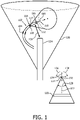
- FIG. 1 depicts exemplary surgical-instrument tracking and point-of-view-based imaging.
- the body of an elongated surgical instrument 102 such as a catheter for internal use, is shown disposed within a blood vessel 104 or organ cavity.
- an electrode 108 At the distal tip 106 of instrument 102 is an electrode 108, for ablation or electrophysiology for example.
- the electrode 108 may be retractable and extendable.
- the retraction/extension and steering are controlled proximally at a handle (not shown) of the instrument 102.
- two echoic structures 110, 112 usable for ultrasonically tracking the tip.
- the structures 110, 112 may be hollowly annular and radially symmetric. The hollowness keeps free the axial center of the catheter body, for functions like stent advancement.
- the symmetry aids in identifying the center of each structure 110, 112.
- a position 114 of a location 116 at the distal tip 106 is central within the proximal structure 110, and a local viewing direction 118 from the location corresponds to a line from the location to the center of the distal structure 112.
- a local view 120 is from that location 116 and in the local viewing direction 118.
- the local view 120 is reconstructed from live imaging, as discussed in detail further below.
- an orientation of the tip 106 about an axis 122 in the local viewing direction 118 is relevant for a catheter that, for example, has, for steering, a "left" pull wire and a "right” pull wire, as also discussed in more detail further below.
- One or both of the structures 110, 112 at the tip 106 may be circumferentially non-symmetrical to facilitate ultrasonic determination of the orientation.
- the distal tip 106 could include one or more magnetic field transducers for tracking as in Lockhart and Tgavalekos.
- the local viewing direction 118 is shown in FIG. 1 to be longitudinally straight ahead from the distal tip 106, it may alternatively be in another direction, such as a side-looking direction.
- a transesophageal (TEE) probe 124 is shown, by way of illustrative and non-limitative example, with forward-looking optics, such as those commonly provided for intravascular ultrasound (IVUS) catheters. However, the optics may instead, or in addition, include side-looking capability. TEE volumetric data 126 acquisition is in real time.
- the live imaging includes imaging of the distal tip 106 and surrounding anatomy.
- the local view 120 is formed whose field of view (FOV) 128 is denoted in FIG. 1 by the angular range shown.
- the local view 120 depicted in FIG. 1 is formed from real-time C-plane imaging, although any subset, thick or thin, of the data 126 may constitute the local view.
- Forming the local view 120 entails a coordinate system transformation 130 of at least the corresponding TEE volumetric data 126 to the local view.
- the local view 120 moves with the distal tip 106, with the FOV 128 remaining fixed.
- the FOV 128 is shown in FIG. 1 to be about 30 degrees, but any angular sector could be used.
- the movement can be translational as seen from the orthogonal broken double-headed straight arrows 132, 134 in FIG. 1 .
- the third orthogonal direction is another type of movement, but is not shown in FIG. 1 .
- the movement can also be rotational, as seen from the pair of curved arrows 136, 138, although, in the two other orthogonal orientations, rotations in either direction are possible.
- the movement of the local view 120 as a result of operation of the catheter controls in the handle, can be likened to moving a flashlight in one's grasp.
- the local view 120 is best used in conjunction with a more overall view that includes the catheter distal tip 106 and surrounding anatomy, for example a live image of the heart.
- the local view 120 in FIG. 2 is seen on a display, accompanied by a more overall or global view 204.
- the bracket 206 in FIG. 2 signifies that the two views 120, 204 appear simultaneously.
- the surgeon need merely switch his or her gaze between the two views 120, 204 in performing the surgical procedure.
- the surgeon manually operates user controls on the catheter handle to operate the micromanipulator situated at the catheter's distal tip 106.
- Both views 120, 204 are preferably updated in real-time, i.e., at least once per second or at another frequency such as between 20 and 60 Hz.
- the views 120, 204 can alternate onscreen as indicated in FIG. 2 by the sector-sweeping arrow configuration 208, optionally each view sized for filling up available screen space.
- Each alternation can be user-actuated as by a toggle switch, or automatic without user intervention.
- the clinician's hands can be kept free for the surgery if a foot pedal is used.
- the toggle swith could instead reside on the catheter handle. Whether the switch is on the catheter handle or elsewhere such as on the ultrasound console, if switching is done manually, this can be done at a convenient time during the surgery - the surgeon is still relieved of having to operate ultrasound controls responsive to imaging changes that accompany movement of the distal tip 106.
- a local view 210 and simultaneous more overall view 212 show that the local view moves with the distal tip 106 whereas the more overall view does not.
- the distal tip 106 moves into one of two branches of the blood vessel 104, but the view has not moved.
- a branching point 214 seen at the earlier time in the local view 120 is no longer visible, as the distal tip 106 has entered the left-hand branch and offers now the new local view 210.
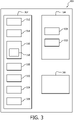
- FIG. 3 provides an overview of a particular, exemplary surgical-instrument tracking, and point-of-view-based imaging, device 300. It includes a host system 302 and an elongated surgical instrument 304. It further includes a transesophageal (TEE) probe 306 having a 2D ultrasound transducer, and a beamformer, for real-time 3D imaging.
- TEE transesophageal
- the elongated surgical instrument 304 is made up of a proximal handle and an elongated body.
- the elongated body includes a catheter 308 and, at the distal tip 106 of the catheter, a micromanipulator 310 such as a pair of scissors or plyers, a stent placement mechanism, or any other device.
- the distal tip 106 can be designed for manipulating or transforming body tissue. Or, it can be designed merely, or in addition, for monitoring through some type of sensing, for dispensing a substance, endogenous or exogenous, such as a medicine, chemical or agent, or it can be designed for any other medical use.
- the host system 302 includes a microcontroller 312, a position and orientation tracker 314, local-view forming software 316 that includes a coordinate system transformation module 318, and a console.
- the console features a user display 320 such as a screen for presenting images, and user controls 322 for imaging which include a local-view orientation initialization control 324 and a view toggling control 326.
- the position and orientation tracker 314 uses the volumetric data acquired in real time by the TEE probe 306 to localize the echoic structures 110, 112 at the tip 106 of the catheter 308.
- other tracking methods may involve one or more magnetic field transducers at the catheter tip 106, and a reference tracking element which may be introduced into the patient by means of a separate catheter, as described in Tgavalekos.
- the reference tracking element would be communicatively coupled, wirelessly or by wireline, to the tracker 314 in the host system 302.
- the host system 302 can be realized in software, hardware, firmware or any combination thereof.
- the microcontroller 312 can be designed to exchange signals with an electronic control in the handle of the catheter 308.
- the microcontroller device 312 can be implemented as one or more integrated circuits.
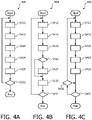
- track and point-of-view-based imaging is realizable according to a movement-independent process 400 or alternative, movement-dependent processes 404, 408.
- the processes 400, 404, 408 are managed by the microcontroller 312 in the host system 302.
- the overall view 204 is formed from acquired live ultrasound radio frequency (RF) data (step S412).
- the overall view 204 is associated with a specific geometric configuration that constitutes a volumetric set of RF data.
- the configuration may be confined to an organ such as the heart or portion of the heart.
- the echoic structures 110, 112 at the catheter tip 106 are detected (step S416).
- the position 114 is derived, and from both structures 110, 112, the local viewing direction 118 is derived (step S420).
- the echogenecity of structures is such that their brightness is automatically distinguishable. The distinguishing is done, by the position and orientation tracker 314, in the imaging that constitutes the overall view.
- the structures 110, 112 could each be annular, gas-filled gaps sandwiched within the walls of the catheter 308.
- the orientation of the tip 106 may also be derived. However, the orientation of the catheter may not be needed or desired, in which case any arbitrary orientation can be supplied for coordinate system transformation purposes.
- an alternative to implementing the echoic structures 110, 112 is electromagnetic tracking. The results of which are derived by the host system 302. The derivation occurs by virtue of reception from a discrete tracking facility. Or, the derivation occurs as a result of calculations performed by the position and orientation tracker 314 based on input from the reference tracking element.
- step S424 coordinates of the overall view 204 are transformed to local coordinates by the coordinate system transformation module 318 of the local-view forming software 316. This forms the local view 120 (step S424).
- the local and overall views 120, 204 are displayed on the display 320 (step S428). If imaging is to continue (step S432), processing returns to step S412.
- the process 400 can operate in real time, iterating at least once per second.
- the first movement-dependent process 404 is similar, but includes a conditional branch between the third and fourth steps S420, S424. If movement of the distal tip 106 is not detected (step S422), processing skips the coordinate transforming step S424 to display the local and overall views 120, 204 (step S428). Tip movement is detected if either the position 114, the direction 118, or the orientation has changed since the previous iteration.
- the handle controls are communicatively coupled, wirelessly or by wireline, to the microcontroller 312 in the host system 302.
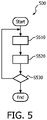
- FIG. 5 An optional, local-view display initialization process 500 is exemplified in FIG. 5 .
- the surgical-instrument tracking, and point-of-view-based imaging, device 300 features a steering facility for the elongated surgical instrument 304 and is configured for adjusting the transformation by conforming an orientation about an axis in the direction 118 with feedback from the facility.
- This embodiment is intended for when the user needs or desires the orientation, e.g., so that the local view 120 displayed features rightward motion when pulling on the "right" catheter steering cable and leftward motion when pulling on the "left” steering cable.
- a user manipulates the local-view orientation initialization control 324, by means of an onscreen slide bar for example, interactively to rotate the local view 120 (step S510).
- step S520 This involves the local-view forming software 316 adjusting the transformation according to the user-induced rotation.
- the user then tests the rotational alignment by operating the catheter handle to slightly steer the catheter distal tip 106 (step S520). If the user sees onscreen that the local view 120 is not yet aligned with the steering (step S530), return is made to step S510; otherwise, alignment is complete.
- a tracking, and point-of-view-based imaging, device is configured for deriving a position of, and a direction from, a location at a distal tip of an elongated instrument, for performing coordinate system transformation in accordance with the position and direction, and for forming, from the location and based on a result of the transformation, a local view that moves with the tip.
- the device can keep, with the movement, a field of view of the local view fixed but the local view otherwise in synchrony with the position and the direction. From real-time ultrasound imaging, the local view and a more overall view that includes the tip but which does not move with said tip can be displayed.
- the distal tip can be that of a catheter and can be outfitted with a micromanipulator for surgery aided interactively by the combination of dynamic local and overall imaging.
- a stationary TEE probe is used in the illustrative embodiments
- a stationary transthoracic echocardiography (TTE) probe may instead be utilized.
- a catheter such as a cardiac catheter is the elongated instrument used in the illustrative embodiments
- other elongated instruments such as a laparascope, endoscope, colonoscope or speculum are within the intended scope of what is proposed herein above.
- tracking and point-of-view-based imaging may be used in an autopsy, with the micromanipulator moving tissue to reveal a structure.
- a computer program can be stored momentarily, temporarily or for a longer period of time on a suitable computer-readable medium, such as an optical storage medium or a solid-state medium.
- a suitable computer-readable medium such as an optical storage medium or a solid-state medium.
- Such a medium is non-transitory only in the sense of not being a transitory, propagating signal, but includes other forms of computer-readable media such as register memory, processor cache, RAM and other volatile memory.
- a single processor or other unit may fulfill the functions of several items recited in the claims.
- the mere fact that certain measures are recited in mutually different dependent claims does not indicate that a combination of these measures cannot be used to advantage.
Landscapes
- Health & Medical Sciences (AREA)
- Life Sciences & Earth Sciences (AREA)
- Engineering & Computer Science (AREA)
- Surgery (AREA)
- Public Health (AREA)
- Animal Behavior & Ethology (AREA)
- Veterinary Medicine (AREA)
- General Health & Medical Sciences (AREA)
- Biomedical Technology (AREA)
- Heart & Thoracic Surgery (AREA)
- Medical Informatics (AREA)
- Molecular Biology (AREA)
- Biophysics (AREA)
- Pathology (AREA)
- Nuclear Medicine, Radiotherapy & Molecular Imaging (AREA)
- Physics & Mathematics (AREA)
- Radiology & Medical Imaging (AREA)
- Robotics (AREA)
- Human Computer Interaction (AREA)
- Physiology (AREA)
- Computer Vision & Pattern Recognition (AREA)
- Cardiology (AREA)
- Ultra Sonic Daignosis Equipment (AREA)
Applications Claiming Priority (2)
| Application Number | Priority Date | Filing Date | Title |
|---|---|---|---|
| US201261737980P | 2012-12-17 | 2012-12-17 | |
| PCT/IB2013/060262 WO2014097014A1 (en) | 2012-12-17 | 2013-11-20 | Micromanipulator-controlled local view with stationary overall view |
Publications (2)
| Publication Number | Publication Date |
|---|---|
| EP2931123A1 EP2931123A1 (en) | 2015-10-21 |
| EP2931123B1 true EP2931123B1 (en) | 2022-09-21 |
Family
ID=49956268
Family Applications (1)
| Application Number | Title | Priority Date | Filing Date |
|---|---|---|---|
| EP13821171.9A Active EP2931123B1 (en) | 2012-12-17 | 2013-11-20 | Ultrasound tracking imaging device with local view and stationary overall view |
Country Status (7)
| Country | Link |
|---|---|
| US (2) | US10792010B2 (enExample) |
| EP (1) | EP2931123B1 (enExample) |
| JP (1) | JP6243441B2 (enExample) |
| CN (1) | CN104869899B (enExample) |
| BR (1) | BR112015013808A2 (enExample) |
| RU (1) | RU2653836C2 (enExample) |
| WO (1) | WO2014097014A1 (enExample) |
Families Citing this family (6)
| Publication number | Priority date | Publication date | Assignee | Title |
|---|---|---|---|---|
| CN108472096B (zh) | 2015-12-31 | 2021-11-16 | 史赛克公司 | 用于在由虚拟对象限定的目标部位处对患者执行手术的系统和方法 |
| JP7014517B2 (ja) * | 2016-02-26 | 2022-02-01 | キヤノンメディカルシステムズ株式会社 | 超音波診断装置及び画像処理プログラム |
| EP3471624B1 (en) | 2016-06-17 | 2022-08-10 | Koninklijke Philips N.V. | System for determining hemodynamic parameters of a patient |
| US10646197B2 (en) | 2016-07-06 | 2020-05-12 | Biosense Webster (Israel) Ltd. | Ascertaining tissue thickness |
| KR102729989B1 (ko) | 2019-07-03 | 2024-11-15 | 스트리커 코포레이션 | 수술 내비게이션을 위한 장애물 회피 기술 |
| JP7401679B2 (ja) * | 2020-07-10 | 2023-12-19 | 朝日インテック株式会社 | 医療装置、及び、画像生成方法 |
Family Cites Families (23)
| Publication number | Priority date | Publication date | Assignee | Title |
|---|---|---|---|---|
| US5217456A (en) | 1992-02-24 | 1993-06-08 | Pdt Cardiovascular, Inc. | Device and method for intra-vascular optical radial imaging |
| US6016439A (en) * | 1996-10-15 | 2000-01-18 | Biosense, Inc. | Method and apparatus for synthetic viewpoint imaging |
| GB2331807B (en) | 1997-11-15 | 2002-05-29 | Roke Manor Research | Catheter tracking system |
| US6778689B1 (en) * | 2000-03-29 | 2004-08-17 | General Electric Company | System and method of real-time multiple field-of-view imaging |
| US8517923B2 (en) | 2000-04-03 | 2013-08-27 | Intuitive Surgical Operations, Inc. | Apparatus and methods for facilitating treatment of tissue via improved delivery of energy based and non-energy based modalities |
| US6892090B2 (en) * | 2002-08-19 | 2005-05-10 | Surgical Navigation Technologies, Inc. | Method and apparatus for virtual endoscopy |
| US6896657B2 (en) | 2003-05-23 | 2005-05-24 | Scimed Life Systems, Inc. | Method and system for registering ultrasound image in three-dimensional coordinate system |
| JP2006014928A (ja) | 2004-07-01 | 2006-01-19 | Fuji Photo Film Co Ltd | 画像表示方法および装置並びにプログラム |
| RU2256169C1 (ru) * | 2004-02-13 | 2005-07-10 | Общестов с ограниченной ответственностью "Институт рентгеновской оптики" | Способ и устройство для исследования объекта в рассеянном и/или прошедшем излучении |
| US8185199B2 (en) * | 2005-02-10 | 2012-05-22 | Zoll Medical Corporation | Monitoring physiological signals during external electrical stimulation |
| US8303505B2 (en) * | 2005-12-02 | 2012-11-06 | Abbott Cardiovascular Systems Inc. | Methods and apparatuses for image guided medical procedures |
| US8417318B2 (en) * | 2007-02-22 | 2013-04-09 | Accuray Incorporated | Calibrating tracking systems to remove position-dependent bias |
| US8821376B2 (en) * | 2007-03-12 | 2014-09-02 | David Tolkowsky | Devices and methods for performing medical procedures in tree-like luminal structures |
| US8073215B2 (en) | 2007-09-18 | 2011-12-06 | Siemens Medical Solutions Usa, Inc. | Automated detection of planes from three-dimensional echocardiographic data |
| US20090105579A1 (en) * | 2007-10-19 | 2009-04-23 | Garibaldi Jeffrey M | Method and apparatus for remotely controlled navigation using diagnostically enhanced intra-operative three-dimensional image data |
| US20090118620A1 (en) | 2007-11-06 | 2009-05-07 | General Electric Company | System and method for tracking an ultrasound catheter |
| CN101336831B (zh) * | 2008-08-13 | 2010-09-01 | 汕头市超声仪器研究所有限公司 | 实时三维医学超声图像的重建方法 |
| US8690776B2 (en) * | 2009-02-17 | 2014-04-08 | Inneroptic Technology, Inc. | Systems, methods, apparatuses, and computer-readable media for image guided surgery |
| US9439736B2 (en) * | 2009-07-22 | 2016-09-13 | St. Jude Medical, Atrial Fibrillation Division, Inc. | System and method for controlling a remote medical device guidance system in three-dimensions using gestures |
| JP2011055901A (ja) * | 2009-09-07 | 2011-03-24 | Toshiba Corp | 経食道心エコー用超音波プローブ |
| WO2012024686A2 (en) | 2010-08-20 | 2012-02-23 | Veran Medical Technologies, Inc. | Apparatus and method for four dimensional soft tissue navigation |
| JP2013111327A (ja) * | 2011-11-30 | 2013-06-10 | Sony Corp | 信号処理装置および方法 |
| EP2785256A4 (en) * | 2011-12-01 | 2016-03-30 | Neochord Inc | SURGICAL NAVIGATION FOR THE REPAIR OF CARDIAC VALVES |
-
2013
- 2013-11-20 WO PCT/IB2013/060262 patent/WO2014097014A1/en not_active Ceased
- 2013-11-20 BR BR112015013808A patent/BR112015013808A2/pt not_active IP Right Cessation
- 2013-11-20 JP JP2015547207A patent/JP6243441B2/ja active Active
- 2013-11-20 EP EP13821171.9A patent/EP2931123B1/en active Active
- 2013-11-20 US US14/652,232 patent/US10792010B2/en active Active
- 2013-11-20 CN CN201380066156.3A patent/CN104869899B/zh active Active
- 2013-11-20 RU RU2015129035A patent/RU2653836C2/ru not_active IP Right Cessation
-
2020
- 2020-09-03 US US17/011,189 patent/US11684337B2/en active Active
Also Published As
| Publication number | Publication date |
|---|---|
| CN104869899A (zh) | 2015-08-26 |
| US20160183911A1 (en) | 2016-06-30 |
| CN104869899B (zh) | 2017-10-27 |
| RU2653836C2 (ru) | 2018-05-14 |
| WO2014097014A1 (en) | 2014-06-26 |
| JP2015536785A (ja) | 2015-12-24 |
| US10792010B2 (en) | 2020-10-06 |
| EP2931123A1 (en) | 2015-10-21 |
| US20200397401A1 (en) | 2020-12-24 |
| RU2015129035A (ru) | 2017-01-25 |
| US11684337B2 (en) | 2023-06-27 |
| JP6243441B2 (ja) | 2017-12-06 |
| BR112015013808A2 (pt) | 2017-07-11 |
Similar Documents
| Publication | Publication Date | Title |
|---|---|---|
| US11684337B2 (en) | Micromanipulator-controlled local view with stationary overall views | |
| KR101759534B1 (ko) | 외과 수술 시 임상적으로 중요한 해부상의 랜드마크에 대한 시각 추적 및 주석 달기 | |
| JP5250234B2 (ja) | 超音波映像と外部医療映像との合成映像をディスプレイする超音波システム | |
| JP4965042B2 (ja) | 医療画像をリアルタイムに描画する方法 | |
| US8213693B1 (en) | System and method to track and navigate a tool through an imaged subject | |
| US11020016B2 (en) | System and method for displaying anatomy and devices on a movable display | |
| RU2668490C2 (ru) | Инструменты наведения для ручного управления эндоскопом с помощью 3d-изображений, полученных до операции и во время операции | |
| JP2021514761A (ja) | マッピング及びナビゲーションのための方法及びシステム | |
| JP2020028718A (ja) | 光学形状検出装置の視点を伴う仮想画像 | |
| US11406255B2 (en) | System and method for detecting abnormal tissue using vascular features | |
| US20080243142A1 (en) | Videotactic and audiotactic assisted surgical methods and procedures | |
| JP2023519714A (ja) | 標的解剖学的特徴の位置特定 | |
| US12016652B2 (en) | System and method for real-time creation of cardiac electro-physiology signals in the heart | |
| WO1996025881A1 (en) | Method for ultrasound guidance during clinical procedures | |
| JP2007152114A (ja) | カテーテル施術のための超音波システム | |
| Sauer | Image registration: enabling technology for image guided surgery and therapy | |
| CN116234515A (zh) | 用于对准机器人臂的触觉反馈 | |
| CN107847111A (zh) | 根据体积图像的交互式平面切片的内窥镜引导 | |
| WO2015091226A1 (en) | Laparoscopic view extended with x-ray vision | |
| JP2014204904A (ja) | 医用ガイドシステム | |
| US20230263580A1 (en) | Method and system for tracking and visualizing medical devices | |
| CN116075276A (zh) | 机器人碰撞边界确定 | |
| Martins et al. | Input system interface for image-guided surgery based on augmented reality | |
| US20250339175A1 (en) | Percutaneous access guidance | |
| WO2020106664A1 (en) | System and method for volumetric display of anatomy with periodic motion |
Legal Events
| Date | Code | Title | Description |
|---|---|---|---|
| PUAI | Public reference made under article 153(3) epc to a published international application that has entered the european phase |
Free format text: ORIGINAL CODE: 0009012 |
|
| 17P | Request for examination filed |
Effective date: 20150717 |
|
| AK | Designated contracting states |
Kind code of ref document: A1 Designated state(s): AL AT BE BG CH CY CZ DE DK EE ES FI FR GB GR HR HU IE IS IT LI LT LU LV MC MK MT NL NO PL PT RO RS SE SI SK SM TR |
|
| AX | Request for extension of the european patent |
Extension state: BA ME |
|
| DAX | Request for extension of the european patent (deleted) | ||
| STAA | Information on the status of an ep patent application or granted ep patent |
Free format text: STATUS: EXAMINATION IS IN PROGRESS |
|
| 17Q | First examination report despatched |
Effective date: 20170718 |
|
| RAP1 | Party data changed (applicant data changed or rights of an application transferred) |
Owner name: KONINKLIJKE PHILIPS N.V. |
|
| GRAP | Despatch of communication of intention to grant a patent |
Free format text: ORIGINAL CODE: EPIDOSNIGR1 |
|
| STAA | Information on the status of an ep patent application or granted ep patent |
Free format text: STATUS: GRANT OF PATENT IS INTENDED |
|
| RIC1 | Information provided on ipc code assigned before grant |
Ipc: A61B 34/20 20160101ALI20220330BHEP Ipc: A61B 8/08 20060101ALI20220330BHEP Ipc: A61B 5/06 20060101AFI20220330BHEP |
|
| INTG | Intention to grant announced |
Effective date: 20220420 |
|
| GRAS | Grant fee paid |
Free format text: ORIGINAL CODE: EPIDOSNIGR3 |
|
| GRAA | (expected) grant |
Free format text: ORIGINAL CODE: 0009210 |
|
| STAA | Information on the status of an ep patent application or granted ep patent |
Free format text: STATUS: THE PATENT HAS BEEN GRANTED |
|
| AK | Designated contracting states |
Kind code of ref document: B1 Designated state(s): AL AT BE BG CH CY CZ DE DK EE ES FI FR GB GR HR HU IE IS IT LI LT LU LV MC MK MT NL NO PL PT RO RS SE SI SK SM TR |
|
| REG | Reference to a national code |
Ref country code: GB Ref legal event code: FG4D |
|
| REG | Reference to a national code |
Ref country code: CH Ref legal event code: EP |
|
| REG | Reference to a national code |
Ref country code: IE Ref legal event code: FG4D |
|
| REG | Reference to a national code |
Ref country code: DE Ref legal event code: R096 Ref document number: 602013082559 Country of ref document: DE |
|
| REG | Reference to a national code |
Ref country code: AT Ref legal event code: REF Ref document number: 1519563 Country of ref document: AT Kind code of ref document: T Effective date: 20221015 |
|
| REG | Reference to a national code |
Ref country code: DE Ref legal event code: R084 Ref document number: 602013082559 Country of ref document: DE |
|
| REG | Reference to a national code |
Ref country code: GB Ref legal event code: 746 Effective date: 20221129 |
|
| REG | Reference to a national code |
Ref country code: LT Ref legal event code: MG9D |
|
| REG | Reference to a national code |
Ref country code: NL Ref legal event code: MP Effective date: 20220921 |
|
| PG25 | Lapsed in a contracting state [announced via postgrant information from national office to epo] |
Ref country code: SE Free format text: LAPSE BECAUSE OF FAILURE TO SUBMIT A TRANSLATION OF THE DESCRIPTION OR TO PAY THE FEE WITHIN THE PRESCRIBED TIME-LIMIT Effective date: 20220921 Ref country code: RS Free format text: LAPSE BECAUSE OF FAILURE TO SUBMIT A TRANSLATION OF THE DESCRIPTION OR TO PAY THE FEE WITHIN THE PRESCRIBED TIME-LIMIT Effective date: 20220921 Ref country code: NO Free format text: LAPSE BECAUSE OF FAILURE TO SUBMIT A TRANSLATION OF THE DESCRIPTION OR TO PAY THE FEE WITHIN THE PRESCRIBED TIME-LIMIT Effective date: 20221221 Ref country code: LV Free format text: LAPSE BECAUSE OF FAILURE TO SUBMIT A TRANSLATION OF THE DESCRIPTION OR TO PAY THE FEE WITHIN THE PRESCRIBED TIME-LIMIT Effective date: 20220921 Ref country code: LT Free format text: LAPSE BECAUSE OF FAILURE TO SUBMIT A TRANSLATION OF THE DESCRIPTION OR TO PAY THE FEE WITHIN THE PRESCRIBED TIME-LIMIT Effective date: 20220921 Ref country code: FI Free format text: LAPSE BECAUSE OF FAILURE TO SUBMIT A TRANSLATION OF THE DESCRIPTION OR TO PAY THE FEE WITHIN THE PRESCRIBED TIME-LIMIT Effective date: 20220921 |
|
| REG | Reference to a national code |
Ref country code: AT Ref legal event code: MK05 Ref document number: 1519563 Country of ref document: AT Kind code of ref document: T Effective date: 20220921 |
|
| PG25 | Lapsed in a contracting state [announced via postgrant information from national office to epo] |
Ref country code: HR Free format text: LAPSE BECAUSE OF FAILURE TO SUBMIT A TRANSLATION OF THE DESCRIPTION OR TO PAY THE FEE WITHIN THE PRESCRIBED TIME-LIMIT Effective date: 20220921 Ref country code: GR Free format text: LAPSE BECAUSE OF FAILURE TO SUBMIT A TRANSLATION OF THE DESCRIPTION OR TO PAY THE FEE WITHIN THE PRESCRIBED TIME-LIMIT Effective date: 20221222 |
|
| PG25 | Lapsed in a contracting state [announced via postgrant information from national office to epo] |
Ref country code: SM Free format text: LAPSE BECAUSE OF FAILURE TO SUBMIT A TRANSLATION OF THE DESCRIPTION OR TO PAY THE FEE WITHIN THE PRESCRIBED TIME-LIMIT Effective date: 20220921 Ref country code: RO Free format text: LAPSE BECAUSE OF FAILURE TO SUBMIT A TRANSLATION OF THE DESCRIPTION OR TO PAY THE FEE WITHIN THE PRESCRIBED TIME-LIMIT Effective date: 20220921 Ref country code: PT Free format text: LAPSE BECAUSE OF FAILURE TO SUBMIT A TRANSLATION OF THE DESCRIPTION OR TO PAY THE FEE WITHIN THE PRESCRIBED TIME-LIMIT Effective date: 20230123 Ref country code: ES Free format text: LAPSE BECAUSE OF FAILURE TO SUBMIT A TRANSLATION OF THE DESCRIPTION OR TO PAY THE FEE WITHIN THE PRESCRIBED TIME-LIMIT Effective date: 20220921 Ref country code: CZ Free format text: LAPSE BECAUSE OF FAILURE TO SUBMIT A TRANSLATION OF THE DESCRIPTION OR TO PAY THE FEE WITHIN THE PRESCRIBED TIME-LIMIT Effective date: 20220921 Ref country code: AT Free format text: LAPSE BECAUSE OF FAILURE TO SUBMIT A TRANSLATION OF THE DESCRIPTION OR TO PAY THE FEE WITHIN THE PRESCRIBED TIME-LIMIT Effective date: 20220921 |
|
| PG25 | Lapsed in a contracting state [announced via postgrant information from national office to epo] |
Ref country code: SK Free format text: LAPSE BECAUSE OF FAILURE TO SUBMIT A TRANSLATION OF THE DESCRIPTION OR TO PAY THE FEE WITHIN THE PRESCRIBED TIME-LIMIT Effective date: 20220921 Ref country code: PL Free format text: LAPSE BECAUSE OF FAILURE TO SUBMIT A TRANSLATION OF THE DESCRIPTION OR TO PAY THE FEE WITHIN THE PRESCRIBED TIME-LIMIT Effective date: 20220921 Ref country code: IS Free format text: LAPSE BECAUSE OF FAILURE TO SUBMIT A TRANSLATION OF THE DESCRIPTION OR TO PAY THE FEE WITHIN THE PRESCRIBED TIME-LIMIT Effective date: 20230121 Ref country code: EE Free format text: LAPSE BECAUSE OF FAILURE TO SUBMIT A TRANSLATION OF THE DESCRIPTION OR TO PAY THE FEE WITHIN THE PRESCRIBED TIME-LIMIT Effective date: 20220921 |
|
| REG | Reference to a national code |
Ref country code: DE Ref legal event code: R097 Ref document number: 602013082559 Country of ref document: DE |
|
| PG25 | Lapsed in a contracting state [announced via postgrant information from national office to epo] |
Ref country code: NL Free format text: LAPSE BECAUSE OF FAILURE TO SUBMIT A TRANSLATION OF THE DESCRIPTION OR TO PAY THE FEE WITHIN THE PRESCRIBED TIME-LIMIT Effective date: 20220921 Ref country code: MC Free format text: LAPSE BECAUSE OF FAILURE TO SUBMIT A TRANSLATION OF THE DESCRIPTION OR TO PAY THE FEE WITHIN THE PRESCRIBED TIME-LIMIT Effective date: 20220921 Ref country code: AL Free format text: LAPSE BECAUSE OF FAILURE TO SUBMIT A TRANSLATION OF THE DESCRIPTION OR TO PAY THE FEE WITHIN THE PRESCRIBED TIME-LIMIT Effective date: 20220921 |
|
| REG | Reference to a national code |
Ref country code: CH Ref legal event code: PL |
|
| REG | Reference to a national code |
Ref country code: BE Ref legal event code: MM Effective date: 20221130 |
|
| PLBE | No opposition filed within time limit |
Free format text: ORIGINAL CODE: 0009261 |
|
| STAA | Information on the status of an ep patent application or granted ep patent |
Free format text: STATUS: NO OPPOSITION FILED WITHIN TIME LIMIT |
|
| PG25 | Lapsed in a contracting state [announced via postgrant information from national office to epo] |
Ref country code: LI Free format text: LAPSE BECAUSE OF NON-PAYMENT OF DUE FEES Effective date: 20221130 Ref country code: DK Free format text: LAPSE BECAUSE OF FAILURE TO SUBMIT A TRANSLATION OF THE DESCRIPTION OR TO PAY THE FEE WITHIN THE PRESCRIBED TIME-LIMIT Effective date: 20220921 Ref country code: CH Free format text: LAPSE BECAUSE OF NON-PAYMENT OF DUE FEES Effective date: 20221130 |
|
| 26N | No opposition filed |
Effective date: 20230622 |
|
| PG25 | Lapsed in a contracting state [announced via postgrant information from national office to epo] |
Ref country code: SI Free format text: LAPSE BECAUSE OF FAILURE TO SUBMIT A TRANSLATION OF THE DESCRIPTION OR TO PAY THE FEE WITHIN THE PRESCRIBED TIME-LIMIT Effective date: 20220921 Ref country code: LU Free format text: LAPSE BECAUSE OF NON-PAYMENT OF DUE FEES Effective date: 20221120 |
|
| PG25 | Lapsed in a contracting state [announced via postgrant information from national office to epo] |
Ref country code: IE Free format text: LAPSE BECAUSE OF NON-PAYMENT OF DUE FEES Effective date: 20221120 |
|
| PG25 | Lapsed in a contracting state [announced via postgrant information from national office to epo] |
Ref country code: FR Free format text: LAPSE BECAUSE OF NON-PAYMENT OF DUE FEES Effective date: 20221121 Ref country code: BE Free format text: LAPSE BECAUSE OF NON-PAYMENT OF DUE FEES Effective date: 20221130 |
|
| PG25 | Lapsed in a contracting state [announced via postgrant information from national office to epo] |
Ref country code: HU Free format text: LAPSE BECAUSE OF FAILURE TO SUBMIT A TRANSLATION OF THE DESCRIPTION OR TO PAY THE FEE WITHIN THE PRESCRIBED TIME-LIMIT; INVALID AB INITIO Effective date: 20131120 |
|
| PG25 | Lapsed in a contracting state [announced via postgrant information from national office to epo] |
Ref country code: CY Free format text: LAPSE BECAUSE OF FAILURE TO SUBMIT A TRANSLATION OF THE DESCRIPTION OR TO PAY THE FEE WITHIN THE PRESCRIBED TIME-LIMIT Effective date: 20220921 |
|
| PG25 | Lapsed in a contracting state [announced via postgrant information from national office to epo] |
Ref country code: MK Free format text: LAPSE BECAUSE OF FAILURE TO SUBMIT A TRANSLATION OF THE DESCRIPTION OR TO PAY THE FEE WITHIN THE PRESCRIBED TIME-LIMIT Effective date: 20220921 Ref country code: IT Free format text: LAPSE BECAUSE OF FAILURE TO SUBMIT A TRANSLATION OF THE DESCRIPTION OR TO PAY THE FEE WITHIN THE PRESCRIBED TIME-LIMIT Effective date: 20220921 |
|
| PG25 | Lapsed in a contracting state [announced via postgrant information from national office to epo] |
Ref country code: BG Free format text: LAPSE BECAUSE OF FAILURE TO SUBMIT A TRANSLATION OF THE DESCRIPTION OR TO PAY THE FEE WITHIN THE PRESCRIBED TIME-LIMIT Effective date: 20220921 |
|
| PG25 | Lapsed in a contracting state [announced via postgrant information from national office to epo] |
Ref country code: MT Free format text: LAPSE BECAUSE OF FAILURE TO SUBMIT A TRANSLATION OF THE DESCRIPTION OR TO PAY THE FEE WITHIN THE PRESCRIBED TIME-LIMIT Effective date: 20220921 |
|
| PGFP | Annual fee paid to national office [announced via postgrant information from national office to epo] |
Ref country code: DE Payment date: 20241128 Year of fee payment: 12 |
|
| PGFP | Annual fee paid to national office [announced via postgrant information from national office to epo] |
Ref country code: GB Payment date: 20241126 Year of fee payment: 12 |
|
| PG25 | Lapsed in a contracting state [announced via postgrant information from national office to epo] |
Ref country code: TR Free format text: LAPSE BECAUSE OF FAILURE TO SUBMIT A TRANSLATION OF THE DESCRIPTION OR TO PAY THE FEE WITHIN THE PRESCRIBED TIME-LIMIT Effective date: 20220921 |