EP2922477B1 - Automatic positioning of standard planes for real-time fetal heart evaluation - Google Patents
Automatic positioning of standard planes for real-time fetal heart evaluation Download PDFInfo
- Publication number
- EP2922477B1 EP2922477B1 EP13799689.8A EP13799689A EP2922477B1 EP 2922477 B1 EP2922477 B1 EP 2922477B1 EP 13799689 A EP13799689 A EP 13799689A EP 2922477 B1 EP2922477 B1 EP 2922477B1
- Authority
- EP
- European Patent Office
- Prior art keywords
- image
- fetal heart
- image plane
- plane
- planes
- Prior art date
- Legal status (The legal status is an assumption and is not a legal conclusion. Google has not performed a legal analysis and makes no representation as to the accuracy of the status listed.)
- Active
Links
- 210000002458 fetal heart Anatomy 0.000 title claims description 87
- 238000011156 evaluation Methods 0.000 title 1
- 238000002604 ultrasonography Methods 0.000 claims description 48
- 239000000523 sample Substances 0.000 claims description 41
- 238000000034 method Methods 0.000 claims description 31
- 239000011159 matrix material Substances 0.000 claims description 26
- 238000003384 imaging method Methods 0.000 claims description 20
- 230000004044 response Effects 0.000 claims description 9
- 230000001605 fetal effect Effects 0.000 description 11
- 210000003754 fetus Anatomy 0.000 description 10
- 210000005240 left ventricle Anatomy 0.000 description 8
- 210000003484 anatomy Anatomy 0.000 description 7
- 230000008569 process Effects 0.000 description 6
- 238000012545 processing Methods 0.000 description 6
- 210000005241 right ventricle Anatomy 0.000 description 4
- 230000002123 temporal effect Effects 0.000 description 4
- 230000006978 adaptation Effects 0.000 description 3
- 238000003745 diagnosis Methods 0.000 description 3
- 208000002330 Congenital Heart Defects Diseases 0.000 description 2
- 210000001015 abdomen Anatomy 0.000 description 2
- 208000028831 congenital heart disease Diseases 0.000 description 2
- 238000001514 detection method Methods 0.000 description 2
- 238000011161 development Methods 0.000 description 2
- 238000010586 diagram Methods 0.000 description 2
- 238000002592 echocardiography Methods 0.000 description 2
- 238000012935 Averaging Methods 0.000 description 1
- 230000005856 abnormality Effects 0.000 description 1
- 210000002376 aorta thoracic Anatomy 0.000 description 1
- 238000013459 approach Methods 0.000 description 1
- 230000008901 benefit Effects 0.000 description 1
- 210000004204 blood vessel Anatomy 0.000 description 1
- 210000005242 cardiac chamber Anatomy 0.000 description 1
- 230000000747 cardiac effect Effects 0.000 description 1
- 238000006243 chemical reaction Methods 0.000 description 1
- 239000004020 conductor Substances 0.000 description 1
- 230000007547 defect Effects 0.000 description 1
- 230000001419 dependent effect Effects 0.000 description 1
- 239000000284 extract Substances 0.000 description 1
- 238000001914 filtration Methods 0.000 description 1
- 230000005831 heart abnormality Effects 0.000 description 1
- 230000006872 improvement Effects 0.000 description 1
- 230000003993 interaction Effects 0.000 description 1
- 238000010859 live-cell imaging Methods 0.000 description 1
- 238000004519 manufacturing process Methods 0.000 description 1
- 230000002688 persistence Effects 0.000 description 1
- 230000035935 pregnancy Effects 0.000 description 1
- 230000009467 reduction Effects 0.000 description 1
- 238000005070 sampling Methods 0.000 description 1
- 238000012216 screening Methods 0.000 description 1
- 238000000926 separation method Methods 0.000 description 1
Images


Classifications
-
- G—PHYSICS
- G06—COMPUTING; CALCULATING OR COUNTING
- G06T—IMAGE DATA PROCESSING OR GENERATION, IN GENERAL
- G06T17/00—Three dimensional [3D] modelling, e.g. data description of 3D objects
-
- A—HUMAN NECESSITIES
- A61—MEDICAL OR VETERINARY SCIENCE; HYGIENE
- A61B—DIAGNOSIS; SURGERY; IDENTIFICATION
- A61B8/00—Diagnosis using ultrasonic, sonic or infrasonic waves
- A61B8/54—Control of the diagnostic device
-
- A—HUMAN NECESSITIES
- A61—MEDICAL OR VETERINARY SCIENCE; HYGIENE
- A61B—DIAGNOSIS; SURGERY; IDENTIFICATION
- A61B8/00—Diagnosis using ultrasonic, sonic or infrasonic waves
- A61B8/08—Detecting organic movements or changes, e.g. tumours, cysts, swellings
- A61B8/0866—Detecting organic movements or changes, e.g. tumours, cysts, swellings involving foetal diagnosis; pre-natal or peri-natal diagnosis of the baby
-
- A—HUMAN NECESSITIES
- A61—MEDICAL OR VETERINARY SCIENCE; HYGIENE
- A61B—DIAGNOSIS; SURGERY; IDENTIFICATION
- A61B8/00—Diagnosis using ultrasonic, sonic or infrasonic waves
- A61B8/13—Tomography
- A61B8/14—Echo-tomography
- A61B8/145—Echo-tomography characterised by scanning multiple planes
-
- A—HUMAN NECESSITIES
- A61—MEDICAL OR VETERINARY SCIENCE; HYGIENE
- A61B—DIAGNOSIS; SURGERY; IDENTIFICATION
- A61B8/00—Diagnosis using ultrasonic, sonic or infrasonic waves
- A61B8/48—Diagnostic techniques
- A61B8/483—Diagnostic techniques involving the acquisition of a 3D volume of data
-
- G—PHYSICS
- G06—COMPUTING; CALCULATING OR COUNTING
- G06T—IMAGE DATA PROCESSING OR GENERATION, IN GENERAL
- G06T15/00—3D [Three Dimensional] image rendering
- G06T15/08—Volume rendering
-
- G—PHYSICS
- G06—COMPUTING; CALCULATING OR COUNTING
- G06T—IMAGE DATA PROCESSING OR GENERATION, IN GENERAL
- G06T19/00—Manipulating 3D models or images for computer graphics
-
- G—PHYSICS
- G06—COMPUTING; CALCULATING OR COUNTING
- G06T—IMAGE DATA PROCESSING OR GENERATION, IN GENERAL
- G06T7/00—Image analysis
- G06T7/30—Determination of transform parameters for the alignment of images, i.e. image registration
-
- F—MECHANICAL ENGINEERING; LIGHTING; HEATING; WEAPONS; BLASTING
- F04—POSITIVE - DISPLACEMENT MACHINES FOR LIQUIDS; PUMPS FOR LIQUIDS OR ELASTIC FLUIDS
- F04C—ROTARY-PISTON, OR OSCILLATING-PISTON, POSITIVE-DISPLACEMENT MACHINES FOR LIQUIDS; ROTARY-PISTON, OR OSCILLATING-PISTON, POSITIVE-DISPLACEMENT PUMPS
- F04C2270/00—Control; Monitoring or safety arrangements
- F04C2270/04—Force
- F04C2270/042—Force radial
- F04C2270/0421—Controlled or regulated
-
- G—PHYSICS
- G06—COMPUTING; CALCULATING OR COUNTING
- G06T—IMAGE DATA PROCESSING OR GENERATION, IN GENERAL
- G06T2207/00—Indexing scheme for image analysis or image enhancement
- G06T2207/10—Image acquisition modality
- G06T2207/10132—Ultrasound image
-
- G—PHYSICS
- G06—COMPUTING; CALCULATING OR COUNTING
- G06T—IMAGE DATA PROCESSING OR GENERATION, IN GENERAL
- G06T2207/00—Indexing scheme for image analysis or image enhancement
- G06T2207/30—Subject of image; Context of image processing
- G06T2207/30004—Biomedical image processing
- G06T2207/30048—Heart; Cardiac
-
- G—PHYSICS
- G06—COMPUTING; CALCULATING OR COUNTING
- G06T—IMAGE DATA PROCESSING OR GENERATION, IN GENERAL
- G06T2210/00—Indexing scheme for image generation or computer graphics
- G06T2210/41—Medical
-
- G—PHYSICS
- G06—COMPUTING; CALCULATING OR COUNTING
- G06T—IMAGE DATA PROCESSING OR GENERATION, IN GENERAL
- G06T2219/00—Indexing scheme for manipulating 3D models or images for computer graphics
- G06T2219/028—Multiple view windows (top-side-front-sagittal-orthogonal)
Definitions
- This invention relates to medical diagnostic ultrasound systems and, in particular, to ultrasound systems which can perform diagnosis of the fetal heart by real-time imaging of diagnostically useful image planes.
- Fetal cardiac ultrasound screening is intended for the detection of structural anomalies (generally congenital heart defects, or CHD) and includes the analysis of standard two dimensional (2D) image views of the fetal heart.
- Typical standard views include the 4-chamber view and views which enable the assessment of the left ventricle (LV) and right ventricle (RV) outflow tracts.
- Other views which may be required include a 5-chamber view, a 3-vessel view, and a tracheal view. In practice, these views usually reveal most CHDs.
- the traditional way for the clinician to acquire an image of each required view is to manipulate the ultrasound probe while in acoustic contact with the abdomen until a desired anatomical orientation is in the plane of the 2D imaging probe. For instance, the clinician first manipulates the probe until the fetal heart is seen in a 4-chamber view. The clinician then stores an image of that view or a sequence of images over one or more heartbeats. Image storage is ended and the clinician manipulates the probe again, this time trying to align the image plane with a view of the LV outflow tract. When the clinician has successfully aligned the image plane with this view, another image or sequence of images are stored. The process of probe manipulation and storage is repeated for a third and other views as required.
- fetal echocardiography is very operator-dependent.
- the fetus may be moving during the procedure, requiring the clinician to reorient herself with the fetal heart each time the fetus moves.
- 3D ultrasound image acquisition notably the Spatial Temporal Image Correlation, or STIC, protocol
- STIC Spatial Temporal Image Correlation
- the STIC procedure is conducted by making a slow sweep of the 2D image plane over the fetal heart, which may take 10 seconds or more.
- the objective is to acquire an image of each adjacent anatomical plane of the fetal heart at each phase of the fetal heart cycle. This is done by acquiring a large number of images over many heart cycles as the image plane is swept over the heart.
- User-directed image processing is then used to extract the predominant temporal cycle of the entire fetal heart from the acquired 2D frames. This information is then used to re-assemble the frames into a series a volume images, each at a different phase of the heart cycle.
- the standard 2D views are then extracted from the volumes by the user by a process known as multiplanar reconstruction (MPR).
- MPR multiplanar reconstruction
- the clinician must search through the volumes at different plane orientations, searching for each standard view. Some views may be distorted or anatomically incorrect, depending on the speed and uniformity of the sweep of the image plane.
- the quality and consistency of the STIC volumes can vary greatly from one exam to the next.
- the STIC images are not in real time, but are synthesized retrospective reconstructions of the anatomical views constructed from multiple, different heart cycles.
- 3D fetal heart exams can potentially reduce misdiagnosis rate and improve workflow and operator dependency (less skills), provided that the 3D workflow is intuitive and adequate tools (e.g., MPR) are provided to explore the volumetric image data.
- a diagnostic ultrasound system and method which enables acquisition of multiple standard views of target anatomy such as the fetal heart in real-time.
- a matrix array probe is placed in contact with a suitable acoustic window on the mother's body to view the fetal heart.
- a matrix array probe is capable of scanning selectable, differently oriented image planes in rapid succession, enabling real-time imaging of the selected image planes.
- the probe is first manipulated while imaging one plane in real-time until a first reference plane such as a 4-chamber view is acquired.
- a model of the target anatomy such as a heart model is then used to match the ultrasound image of the 4-chamber view with a corresponding 4-chamber view in the heart model.
- the heart model provides information as to the relative orientations of other standard views in relation to the acquired reference plane. This information is used to control the matrix array probe to additionally scan the image planes of one or more other views in real-time. The user can then display the reference standard view and one or more other desired views simultaneously in real-time.
- an ultrasound system 10 constructed in accordance with the principles of the present invention is shown in block diagram form.
- the ultrasound system is configured by two subsystems, a front end acquisition subsystem 10A and a display subsystem 10B.
- An ultrasound probe is coupled to the acquisition subsystem which includes a two-dimensional matrix array transducer 70 and a micro-beamformer 72.
- the micro-beamformer contains circuitry which control the signals applied to groups of elements ("patches") of the array transducer 70 and does initial processing of the echo signals received by elements of each group.
- Micro-beamforming in the probe advantageously reduces the number of conductors in the cable between the probe and the ultrasound system and is described in US Pat. 5,997,479 (Savord et al. ) and in US Pat. 6,436,048 (Pesque ).
- the probe is coupled to the acquisition subsystem 10A of the ultrasound system.
- the acquisition subsystem includes a beamform controller 74 which is responsive to a gating signal produced as described below and provides control signals to the microbeamformer 72, instructing the probe as to the timing, frequency, direction and focusing of transmit beams in 2D image planes or 3D volumes.
- the beamform controller also controls the beamforming of echo signals received by the acquisition subsystem by its control of analog-to-digital (A/D) converters 18 and a beamformer 20. Echo signals received by the probe are amplified by preamplifier and TGC (time gain control) circuitry 16 in the acquisition subsystem, then digitized by the A/D converters 18.
- A/D analog-to-digital
- the digitized echo signals are then formed into fully steered and focused beams by a beamformer 20.
- the echo signals are then processed by an image processor 22 which performs digital filtering, B mode detection, and Doppler processing, and can also perform other signal processing such as harmonic separation, speckle reduction, and other desired image signal processing.
- the echo signals produced by the acquisition subsystem 10A are coupled to the display subsystem 10B, which processes the echo signals for display in the desired image format.
- the echo signals are processed by an image line processor 24, which is capable of sampling the echo signals, splicing segments of beams into complete line signals, and averaging line signals for signal-to-noise improvement or flow persistence.
- the image lines for a 2D image are scan converted into the desired image format by a scan converter 26 which performs R-theta conversion as is known in the art.
- the image is then stored in an image memory 28 from which it can be displayed on a display 38.
- the image in memory is also overlaid with graphics to be displayed with the image, which are generated by a graphics generator 34. Individual images or image sequences can be stored in a cine memory 30 during capture of image loops or sequences.
- a fetal heart rate generator 54 provides this capability.
- the fetal heart rate generator synthesizes the periodicity of the fetal heart cycle as described in international patent publications WO 2011/001309 (Jago et al. ) and WO 2011/158136 (Schauf ).
- the fetal heart rate generator produces a gating signal at a selected phase of the fetal heart which can be used to gate image acquisition or processing as described below.
- the display subsystem includes heart model data 32 of the fetal heart.
- the heart model data is that of a 3D anatomical mesh model of the fetal heart as described in US patent pub. no. 2008/0304744 (Peters et al. ) and in US provisional application no. 61/569,450, filed December 12, 2011 (Radulescu et al. ), forming the priority application for the international application PCT/ IB2012/057137 , published as WO 2013/088326 A2 .
- Such a model represents the structure of the heart including its interior and exterior structure such as blood vessels and valves.
- the heart model may be of a single phase of the heart such as end diastole, or it may comprises multiple models of the heart at different phases of the heart cycle.
- individual planes can be extracted from the heart model data and matched or registered to actual fetal ultrasound 2D images. This registration is performed by an image registration processor 36, which receives ultrasound images of a fetal heart produced by the scan converter 26 and registers them with a corresponding plane of the heart model data.
- a match with a plane of the heart model triggers the production of image plane orientation data 44, which in turn couples image plane coordinates or orientation data in a gating signal to the beamform controller 74 which in response directs the matrix array probe to scan an identified image plane.
- Plane coordinate information for the image plane orientation is provided from the heart model data.
- the heart model data 32 is accessed for selected image planes 42 which are selected from the user control panel 40.
- the ECG trigger signal generator 54 is coupled to the image registration processor 36.
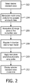
- FIGURE 2 One method for using the ultrasound system of FIGURE 1 to acquire standard images for a fetal heart exam is illustrated in FIGURE 2 .
- the user selects the standard views that are desired for this particular fetal heart exam.
- typical standard views include a 4-chamber view, LV outflow tracts, RV outflow tracts, 5-chamber view, 3-vessel view, and the tracheal view.
- the user may select these standard views from a list of standard views accessed as a pull-down list on the system display.
- the user selects the desired views with a control of control panel 40, and the image planes of the selected views are stored at 42 where they are used to select the views from the planes of the heart model data 32.
- step 204 the user presses the matrix array probe aperture against the abdomen of the patient and manipulates the probe until a suitable acoustic window is found, one from which the user can image the fetal heart.
- step 206 the user manipulates the matrix array probe, which at this time is only scanning a single 2D image plane, until the image plane is intersecting the fetal heart and an image of the first selected view of the heart is obtained.
- this first standard view will be a 4-chamber view of the fetal heart.
- This first standard view is coupled to the image registration processor 36 which, in step 208, registers the ultrasound image with a plane of the heart model. Registration of an ultrasound image with a heart model is described in the above Peters et al. publication and Radulescu et al.
- FIG. 5 On the left side of position a) is a 4-chamber ultrasound image 502 of the fetal heart, and on the right side of position a) is a 4-chamber plane 504 of a 3D fetal heart model.
- the heart model plane 504 is oriented with the cross-section of the descending aorta 122' at the bottom, in correspondence with its location at 122 in the ultrasound image 502.
- the heart chambers in the ultrasound image have been labeled (RA, LA, RV, LV), and their correspondence with those of the heart model plane 504 is readily apparent.
- the image registration processor identifies a plane of the heart model which most closely matches the anatomy of the 4-chamber view 502, using tools such as block matching of the pixels of the ultrasound image to different planes through the heart model data.
- the arrow in the heart model plane 504 and in other illustrated heart model planes indicate anatomical landmarks in the heart model planes which can be most readily found and matched in ultrasound images, speeding the registration process.
- FIGURE 5 also illustrates the matching of other standard views to a heart model.
- Position b) illustrates an image 506 of the LV outflow tract in correspondence with an LV outflow tract plane 508 of a fetal heart model
- position c) illustrates an image 510 of a ductal arch view in correspondence with a ductal arch plane 512 of the heart model.
- the ductal arch image 510 is seen to be reversed in relation to the plane 512 of the heart model. This can be resolved by reversing the ultrasound image as described in U.S. patents 6,669,641 (Poland et al. ), or by reversing the heart model data during the registration process as described in the above-referenced Peters et al. publication and Radulescu et al. application.
- the orientations of the other selected standard views can be readily identified using the heart model.
- the relative orientations of other standard view planes are known from the heart model, based on a priori statistical knowledge about the normal geometrical relationships of structures within the fetal heart. For example, when a particular plane of the heart model is identified as the 4-chamber view, the next standard view may be in a plane that is rotated 35° and tilted 15° with respect to the 4-chamber view plane in the model, for instance.
- the coordinates of this rotated and tilted plane are coupled to the image plane orientation data 44 and used to control the matrix array probe to scan a second image plane in this particular orientation relative to the first standard view plane.
- the plane coordinates of other selected standard planes are found in the heart model data, coupled to the image plane orientation data, and used to gate the scanning of additional image planes in all selected standard view planes, as indicated in step 210.
- step 212 the selected standard views are displayed in real-time on the ultrasound display 38.
- FIGURE 6 illustrates the display screen of an ultrasound system which is displaying three such standard views in real-time.
- a live 4-chamber view 602 is shown
- a live ductal arch view 610 is shown
- a LV outflow view 606 is shown.
- the three views can be simultaneously displayed in real-time because only three image planes need to be scanned in alternating succession, rather than an entire 3D volume from which MPR frames must be identified and extracted.
- FIGURE 6 contains an icon graphic 610 of a generic fetal heart 612 which shows the user how the planes 614 of the images intersect the heart.
- the outline of the fetal heart graphic 612 can be provided by the fetal heart model 32.
- Each image in FIGURE 6 is outlined by a box.
- each box can be outlined by a different color, and multiple like-colored plane graphics 614 can be displayed in the graphic simultaneously.
- Another display option is to render the three planes in three dimensions, depicting the relative orientation of all three real-time images to the user.
- the real-time images can also be overlaid with adjustable cursors 13, 14, and 15 (see FIGURE 5 ) which can be repositioned by the user and clicked on using image adjustment controls 12 to view image planes orthogonal to the standard view planes.
- Another graphic which is commonly displayed with the images is the fetal heart rate, produced by the fetal heart rate generator 54, which is an important factor in many diagnoses.
- the operation and control of the matrix array probe in an implementation of the present invention can use elements of the functionality of a matrix array probe when operated in the biplane mode.
- U.S. patents 6,669,641 (Poland et al. ), 6,709,394 (Frisa et al. ) and 6,755,786 (Frisa et al. ) describe ultrasonic biplane imaging.
- biplane imaging a two-dimensional matrix array transducer probe scans two different 2D image planes in rapid alternating succession, thereby producing live real-time images of both planes.
- One of the image planes is denominated as the reference image plane.
- This image plane is generally oriented perpendicular to the plane of the matrix array transducer, extending straight out from the probe around a center orthogonal axis to the array.
- the reference image orientation is usually maintained stationary and the second image plane is movable by the user in relation to this reference plane.
- the '394 patent describes biplane imaging in which the second image plane can be tilted or rotated with respect to the reference plane.
- the tilted image plane has a nominal orientation with its center axis in alignment with the center axis of the reference plane.
- the tilt plane can be moved (tilted) so that it is oriented at different angles in relation to the center axis of the reference plane but with its center axis always located in the reference plane.
- the rotational biplane implementation again has the center axis of the second (rotating) image plane aligned at the start with the center axis of the reference image and the second image orientation is orthogonal to the plane of the reference image. From this starting position the rotating plane can be rotated about its center axis at angles with respect to the reference image which vary from orthogonal.
- the '786 patent describes what is known as elevation tilt biplane imaging. In elevation tilt imaging the second image has a starting position in alignment with the reference image.
- the second image is then moved away from the reference image plane in the elevation dimension, and can be moved to different planes which do not intersect the reference image plane in the region of interest.
- the two planes can thus be perfectly parallel or angularly parallel, the latter being a condition where the second plane has a common apex location with the reference plane or intersects the reference plane above the top (shallowest depth) of the images.
- Biplane images allow a clinician to position the reference plane to view a target anatomy or region of interest, then move the second plane to observe other planar images of the target anatomy. As shown in the foregoing patents, the two biplane images are displayed side-by-side at the same time, so that the clinician can constantly view the reference image while moving the second plane.
- Biplane imaging allows the clinician to scan and observe two image planes at the same time, while constantly maintaining his or her navigational bearings of the image locations within the three dimensional volume being scanned.
- a single image or a loop (sequence of live images) can be captured or save and displayed or replayed later when making a definitive diagnosis.
- An implementation of the present invention can use two (biplane) images when only two standard views are required, and is extended to image further planes when three or more standard views are needed.
- the central reference image plane of biplane imaging can be used to acquire the starting standard view such as the 4-chamber view.
- the image registration processor 36 matches the image to the closest 4-chamber plane of the heart model and the heart model provides the relative orientations of the other desired standard views from their positions in the heart model. These plane orientations are coupled to the beamform controller 74, which then automatically begins scanning and displaying images of these other views in real-time. If the fetus moves during the exam, the user only has to reposition the probe to reacquire the 4-chamber view in the reference image plane, the image registration processor again matches the image to a 4-chamber plane of the heart model, the orientations of the other standard views are identified in the heart model, coupled to the beamform controller, and live imaging of all of the standard views resumes.
- An implementation of the present invention can operate with a fixed heart model or one that is adaptable.
- the heart model may be one which represents the heart at the end diastole phase of the heart cycle.
- the image registration process is then done with an image captured at that phase of the fetal heartbeat. This can be done by use of the fetal heart rate generator 54, which can gate images at selected phases of the fetal heart cycle, which enables the registration processor to perform a match at the moment when an end diastole image has been acquired.
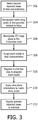
- FIGURE 3 illustrates a method of the present invention where the heart model is adapted to the particular fetus.
- the user not only selects the standard views that are desired, but also enters fetal characteristics such as fetal age, shape and development of the fetal heart, known or suspected heart abnormalities or defects, or other information which more particularly describes the heart of the fetus to be examined.
- This information is then used in step 308 to adapt the heart model to the particular fetus.
- One adaptation technique is to have a library of different fetal heart models for different fetal ages. Entry of fetal age data enables the selection of the heart model which most closely matches the age of the fetus being examined, making image registration easier and more reliable.
- Another adaptation technique is to morph or warp the heart model to the size and shape of the features in an acquired fetal heart image, as described in the above-referenced Peters et al. publication and Radulescu et al. application. See also international patent publication WO 2007/034425 (Ecabert et al. )
- a given heart model can be adapted to virtually any fetal heart images and to different phases of the heart cycle.
- a starting standard view image is registered to the adapted heart model data in step 310 and identification, scanning and display of the other standard view images proceeds in steps 210 and 212 as before.
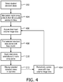
- FIGURE 4 illustrates an even more highly automated implementation of the present invention.
- the user begins imaging, not in 2D, but in 3D acquisition mode.
- the user finds an appropriate 3D acoustic window from which to image the entire fetal heart in 3D, and in step 406 a volume image of the fetal heart is acquired.
- the registration processor matches at least one plane of the volume image, as by MPR reconstruction, for instance, with a corresponding plane of the heart model 32.
- the standard view planes are then identified in the heart model, either by matching all of the standard view planes of the volume ultrasound image to corresponding planes of the heart model, or by the relative orientations of standard view planes of the heart model to one ultrasound image-heart model plane match.
- the orientations of the standard view planes are supplied to the matrix array probe (step 210), which begins scanning and displaying the selected standard view 2D planes (step 212).
- the user does not have to find even an initial standard view. It is only necessary to capture volume image data of the fetal heart, and the ultrasound system identifies and extracts the desired standard view planes and begins imaging them without further user input.
- the method of FIGURE 4 can also incorporate adaptation of the heart model as discussed in the description of FIGURE 3 .
- the matrix array probe periodically acquires another volume image dataset in step 414.
- the new volume image data is used again starting with step 406 to re-identify the orientations of the standard view planes and updates the control of the matrix array probe to image the desired standard view planes. It is only necessary for the user to position the probe so that a full volume image dataset can be continually acquired as needed.
- the system can continue to track and display the desired standard views in real-time without any user interaction, simplifying and accelerating the ability to make a diagnosis of the fetus.
Landscapes
- Health & Medical Sciences (AREA)
- Life Sciences & Earth Sciences (AREA)
- Engineering & Computer Science (AREA)
- Physics & Mathematics (AREA)
- General Health & Medical Sciences (AREA)
- Public Health (AREA)
- Radiology & Medical Imaging (AREA)
- Nuclear Medicine, Radiotherapy & Molecular Imaging (AREA)
- Biomedical Technology (AREA)
- Heart & Thoracic Surgery (AREA)
- Medical Informatics (AREA)
- Molecular Biology (AREA)
- Surgery (AREA)
- Animal Behavior & Ethology (AREA)
- Biophysics (AREA)
- Pathology (AREA)
- Veterinary Medicine (AREA)
- General Physics & Mathematics (AREA)
- Theoretical Computer Science (AREA)
- Computer Graphics (AREA)
- Software Systems (AREA)
- Computer Hardware Design (AREA)
- General Engineering & Computer Science (AREA)
- Gynecology & Obstetrics (AREA)
- Pregnancy & Childbirth (AREA)
- Computer Vision & Pattern Recognition (AREA)
- Geometry (AREA)
- Ultra Sonic Daignosis Equipment (AREA)
Applications Claiming Priority (2)
| Application Number | Priority Date | Filing Date | Title |
|---|---|---|---|
| US201261728566P | 2012-11-20 | 2012-11-20 | |
| PCT/IB2013/060105 WO2014080319A1 (en) | 2012-11-20 | 2013-11-13 | Automatic positioning of standard planes for real-time fetal heart evaluation |
Publications (2)
| Publication Number | Publication Date |
|---|---|
| EP2922477A1 EP2922477A1 (en) | 2015-09-30 |
| EP2922477B1 true EP2922477B1 (en) | 2018-01-10 |
Family
ID=49713434
Family Applications (1)
| Application Number | Title | Priority Date | Filing Date |
|---|---|---|---|
| EP13799689.8A Active EP2922477B1 (en) | 2012-11-20 | 2013-11-13 | Automatic positioning of standard planes for real-time fetal heart evaluation |
Country Status (7)
| Country | Link |
|---|---|
| US (2) | US9734626B2 (pt) |
| EP (1) | EP2922477B1 (pt) |
| JP (1) | JP6180539B2 (pt) |
| CN (1) | CN104797199B (pt) |
| BR (1) | BR112015011288B1 (pt) |
| RU (1) | RU2654611C2 (pt) |
| WO (1) | WO2014080319A1 (pt) |
Families Citing this family (33)
| Publication number | Priority date | Publication date | Assignee | Title |
|---|---|---|---|---|
| JP6272618B2 (ja) * | 2013-09-25 | 2018-01-31 | ハートフロー, インコーポレイテッド | 自動医療画像注釈の検証及び修正のためのシステム、方法及びコンピュータ可読媒体 |
| CN105900140B (zh) | 2013-11-05 | 2019-02-05 | 皇家飞利浦有限公司 | 用于实时超声成像的三平面图像的自动分割 |
| KR20150074304A (ko) * | 2013-12-23 | 2015-07-02 | 삼성전자주식회사 | 의료 영상 정보를 제공하는 방법 및 그 장치 |
| JP6517248B2 (ja) | 2014-06-30 | 2019-05-22 | コーニンクレッカ フィリップス エヌ ヴェKoninklijke Philips N.V. | 解剖学的方向に応じた超音波アレイの並進 |
| JP5918324B2 (ja) * | 2014-09-08 | 2016-05-18 | 日立アロカメディカル株式会社 | 超音波診断装置 |
| WO2016083147A1 (en) * | 2014-11-28 | 2016-06-02 | Koninklijke Philips N.V. | Model-based segmentation of an anatomical structure |
| KR102446343B1 (ko) * | 2015-06-15 | 2022-09-22 | 삼성메디슨 주식회사 | 초음파 장치 및 그 제어방법 |
| US11553892B2 (en) * | 2016-03-01 | 2023-01-17 | Koninklijke Philips N.V. | Automated ultrasonic measurement of nuchal fold translucency |
| US11129591B2 (en) | 2016-04-21 | 2021-09-28 | The University Of British Columbia | Echocardiographic image analysis |
| US11298104B2 (en) * | 2016-08-10 | 2022-04-12 | Canon Medical Systems Corporation | Medical processing apparatus, ultrasound diagnostic apparatus, and medical processing method |
| JP6987558B2 (ja) * | 2016-08-10 | 2022-01-05 | キヤノンメディカルシステムズ株式会社 | 医用処理装置、超音波診断装置および医用処理プログラム |
| US11696745B2 (en) * | 2017-03-16 | 2023-07-11 | Koninklijke Philips N.V. | Optimal scan plane selection for organ viewing |
| US20180322627A1 (en) * | 2017-05-05 | 2018-11-08 | General Electric Company | Methods and systems for acquisition of medical images for an ultrasound exam |
| EP3422048A1 (en) * | 2017-06-26 | 2019-01-02 | Koninklijke Philips N.V. | Ultrasound imaging method and system |
| WO2019045144A1 (ko) * | 2017-08-31 | 2019-03-07 | (주)레벨소프트 | 의료용 항법 장치를 위한 의료 영상 처리 장치 및 의료 영상 처리 방법 |
| TW201923776A (zh) * | 2017-10-27 | 2019-06-16 | 美商蝴蝶網路公司 | 超音波影像上的自動化測量及用於自動化測量的收集的品質指示器 |
| WO2019154667A1 (en) | 2018-02-12 | 2019-08-15 | Koninklijke Philips N.V. | Workflow assistance for medical doppler ultrasound evaluation |
| EP3549528A1 (en) * | 2018-04-05 | 2019-10-09 | Koninklijke Philips N.V. | Ultrasound imaging system and method |
| US10751029B2 (en) | 2018-08-31 | 2020-08-25 | The University Of British Columbia | Ultrasonic image analysis |
| KR20200099910A (ko) | 2019-02-15 | 2020-08-25 | 삼성메디슨 주식회사 | 초음파 영상을 표시하는 방법, 장치 및 컴퓨터 프로그램 제품 |
| US11517290B2 (en) * | 2019-03-13 | 2022-12-06 | GE Precision Healthcare LLC | Method and system for providing standard ultrasound scan plane views using automatic scan acquisition rotation and view detection |
| EP3711673A1 (en) | 2019-03-18 | 2020-09-23 | Koninklijke Philips N.V. | Methods and systems for adjusting the field of view of an ultrasound probe |
| US20210015449A1 (en) * | 2019-07-16 | 2021-01-21 | GE Precision Healthcare LLC | Methods and systems for processing and displaying fetal images from ultrasound imaging data |
| CN111127305B (zh) * | 2019-10-14 | 2021-01-26 | 李胜利 | 基于早孕期胎儿颅面部三维容积自动获取标准切面的方法 |
| US20210204908A1 (en) * | 2020-01-08 | 2021-07-08 | GE Precision Healthcare LLC | Method and system for assisted ultrasound scan plane identification based on m-mode analysis |
| CN113628158A (zh) * | 2020-05-08 | 2021-11-09 | 通用电气精准医疗有限责任公司 | 用于利用生成模型来管理图像质量的方法和系统 |
| JP6874199B1 (ja) * | 2020-12-01 | 2021-05-19 | 信二 福家 | 心臓モデル導入装置、心臓診断支援システム、プログラム、および心臓モデル導入方法 |
| JP7538705B2 (ja) * | 2020-12-08 | 2024-08-22 | 富士フイルムヘルスケア株式会社 | 超音波診断システム及び操作支援方法 |
| US11593936B2 (en) | 2020-12-28 | 2023-02-28 | GE Precision Healthcare LLC | Ultrasound imaging system and method for providing feedback regarding acquisition quality |
| US20220301240A1 (en) * | 2021-03-22 | 2022-09-22 | GE Precision Healthcare LLC | Automatic Model-Based Navigation System And Method For Ultrasound Images |
| JP2022175748A (ja) * | 2021-05-14 | 2022-11-25 | キヤノン株式会社 | 画像処理装置、画像処理方法、およびプログラム |
| US12082969B1 (en) * | 2023-02-22 | 2024-09-10 | BrightHeart SAS | Systems and methods for improving detection of fetal congenital heart defects |
| CN116687445B (zh) * | 2023-07-31 | 2024-01-30 | 深圳华声医疗技术股份有限公司 | 超声胎儿心脏自动定位跟踪方法、装置、设备及存储介质 |
Family Cites Families (54)
| Publication number | Priority date | Publication date | Assignee | Title |
|---|---|---|---|---|
| US5207225A (en) * | 1990-11-14 | 1993-05-04 | Advanced Technology Laboratories, Inc. | Transesophageal ultrasonic scanhead |
| US5050610A (en) * | 1990-11-14 | 1991-09-24 | Advanced Technology Laboratories, Inc. | Transesophageal ultrasonic scanhead |
| US6106466A (en) * | 1997-04-24 | 2000-08-22 | University Of Washington | Automated delineation of heart contours from images using reconstruction-based modeling |
| US5997479A (en) | 1998-05-28 | 1999-12-07 | Hewlett-Packard Company | Phased array acoustic systems with intra-group processors |
| US6174285B1 (en) * | 1999-02-02 | 2001-01-16 | Agilent Technologies, Inc. | 3-D ultrasound imaging system with pre-set, user-selectable anatomical images |
| US6709394B2 (en) | 2000-08-17 | 2004-03-23 | Koninklijke Philips Electronics N.V. | Biplane ultrasonic imaging |
| US7037264B2 (en) * | 2000-08-17 | 2006-05-02 | Koninklijke Philips Electronics N.V. | Ultrasonic diagnostic imaging with steered image plane |
| US6669641B2 (en) | 2000-08-17 | 2003-12-30 | Koninklijke Philips Electronics N.V. | Method of and system for ultrasound imaging |
| US6755788B2 (en) | 2000-08-17 | 2004-06-29 | Koninklijke Philips Electronics N. V. | Image orientation display for a three dimensional ultrasonic imaging system |
| US6468216B1 (en) | 2000-08-24 | 2002-10-22 | Kininklijke Philips Electronics N.V. | Ultrasonic diagnostic imaging of the coronary arteries |
| US20030160786A1 (en) * | 2002-02-28 | 2003-08-28 | Johnson Richard K. | Automatic determination of borders of body structures |
| US7599730B2 (en) * | 2002-11-19 | 2009-10-06 | Medtronic Navigation, Inc. | Navigation system for cardiac therapies |
| MXPA05011120A (es) | 2003-04-16 | 2005-12-15 | Eastern Viriginai Medical Scho | Sistema y metodo para generar imagenes de ultrasonido independientes del operador. |
| US8083678B2 (en) * | 2003-04-16 | 2011-12-27 | Eastern Virginia Medical School | System, method and medium for acquiring and generating standardized operator independent ultrasound images of fetal, neonatal and adult organs |
| US7092749B2 (en) * | 2003-06-11 | 2006-08-15 | Siemens Medical Solutions Usa, Inc. | System and method for adapting the behavior of a diagnostic medical ultrasound system based on anatomic features present in ultrasound images |
| WO2005004065A1 (en) * | 2003-06-25 | 2005-01-13 | Siemens Corporate Research, Inc. | Model assisted planning of medical imaging |
| US20050028307A1 (en) * | 2003-08-06 | 2005-02-10 | Kuei-Kun Wu | Snake |
| US7604595B2 (en) * | 2004-06-22 | 2009-10-20 | General Electric Company | Method and system for performing real time navigation of ultrasound volumetric data |
| US7599136B2 (en) * | 2005-07-27 | 2009-10-06 | Honda Motor Co., Ltd. | Ambient light lens |
| EP1929444B1 (en) | 2005-09-23 | 2011-11-16 | Philips Intellectual Property & Standards GmbH | A method of and a system for adapting a geometric model using multiple partial transformations |
| DE602006008040D1 (de) | 2005-12-19 | 2009-09-03 | Koninkl Philips Electronics Nv | Verfahren zur erleichterung der nachbearbeitung von bildern über verformbare netze |
| US20070249935A1 (en) * | 2006-04-20 | 2007-10-25 | General Electric Company | System and method for automatically obtaining ultrasound image planes based on patient specific information |
| US20080009722A1 (en) * | 2006-05-11 | 2008-01-10 | Constantine Simopoulos | Multi-planar reconstruction for ultrasound volume data |
| KR20090088404A (ko) * | 2006-12-12 | 2009-08-19 | 코닌클리케 필립스 일렉트로닉스 엔.브이. | 의료 이미징 시스템 |
| WO2008136008A2 (en) * | 2007-05-08 | 2008-11-13 | Mediguide Ltd. | Method for producing an electrophysiological map of the heart |
| US8057394B2 (en) * | 2007-06-30 | 2011-11-15 | St. Jude Medical, Atrial Fibrillation Division, Inc. | Ultrasound image processing to render three-dimensional images from two-dimensional images |
| US8073215B2 (en) * | 2007-09-18 | 2011-12-06 | Siemens Medical Solutions Usa, Inc. | Automated detection of planes from three-dimensional echocardiographic data |
| US8172753B2 (en) * | 2008-07-11 | 2012-05-08 | General Electric Company | Systems and methods for visualization of an ultrasound probe relative to an object |
| WO2010046819A1 (en) * | 2008-10-22 | 2010-04-29 | Koninklijke Philips Electronics N.V. | 3-d ultrasound imaging |
| CN102197413B (zh) * | 2008-10-29 | 2017-03-22 | 皇家飞利浦电子股份有限公司 | 分析至少三维医学图像 |
| US20100172559A1 (en) * | 2008-11-11 | 2010-07-08 | Eigen, Inc | System and method for prostate biopsy |
| RU2539006C2 (ru) * | 2009-06-30 | 2015-01-10 | Конинклейке Филипс Электроникс Н.В. | Формирование трехмерного изображения сердца плода посредством физиологически стробированного получения данных, не связанного с экг |
| KR101121353B1 (ko) * | 2009-08-03 | 2012-03-09 | 한국과학기술원 | 2차원 초음파 영상에 대응하는 2차원 ct 영상을 제공하는 시스템 및 방법 |
| ITGE20090070A1 (it) * | 2009-08-31 | 2011-03-01 | Esaote Spa | Metodo e dispositivo per il rilevamento e la visualizzazione di informazioni emodinamiche in particolare del flusso ematico nelle vene, mediante ultrasoni |
| DE102009053471B4 (de) * | 2009-11-16 | 2018-08-02 | Siemens Healthcare Gmbh | Verfahren und Vorrichtung zur Identifizierung und Zuordnung von Koronarkalk zu einem Herzkranzgefäß sowie Computerprogrammprodukt |
| JP5394299B2 (ja) * | 2010-03-30 | 2014-01-22 | 富士フイルム株式会社 | 超音波診断装置 |
| JP5889886B2 (ja) | 2010-06-17 | 2016-03-22 | コーニンクレッカ フィリップス エヌ ヴェKoninklijke Philips N.V. | 3d超音波胎児イメージングのための自動心拍数検出 |
| US20110317897A1 (en) * | 2010-06-29 | 2011-12-29 | General Electric Company | Method and apparatus for automated localization of a moving structure |
| US8633907B2 (en) * | 2010-07-06 | 2014-01-21 | Padmanabhan Mahalingam | Touch screen overlay for visually impaired persons |
| TWI483017B (zh) * | 2010-07-07 | 2015-05-01 | Hon Hai Prec Ind Co Ltd | 光纖耦合連接器 |
| US10321892B2 (en) * | 2010-09-27 | 2019-06-18 | Siemens Medical Solutions Usa, Inc. | Computerized characterization of cardiac motion in medical diagnostic ultrasound |
| US9668716B2 (en) * | 2010-12-10 | 2017-06-06 | General Electric Company | Ultrasound imaging system and method for ultrasound imaging a three dimensional volume |
| EP2651307B1 (en) * | 2010-12-13 | 2017-11-15 | Koninklijke Philips N.V. | Adjusting measurements of the effects of acoustic radiation force for background motion effects |
| US8657750B2 (en) * | 2010-12-20 | 2014-02-25 | General Electric Company | Method and apparatus for motion-compensated ultrasound imaging |
| US20120165671A1 (en) * | 2010-12-27 | 2012-06-28 | Hill Anthony D | Identification of objects in ultrasound |
| KR101792590B1 (ko) * | 2011-04-26 | 2017-11-01 | 삼성전자주식회사 | 빔포밍 방법, 이를 수행하는 장치 및 의료영상시스템 |
| US20130007099A1 (en) * | 2011-06-30 | 2013-01-03 | Michael Lee | System and Method for Interactive Identification and Matching of Funding and/or Advisory Seekers and Funding and/or Advisory Providers |
| US9204862B2 (en) * | 2011-07-08 | 2015-12-08 | General Electric Company | Method and apparatus for performing ultrasound elevation compounding |
| KR101286222B1 (ko) * | 2011-09-19 | 2013-07-15 | 삼성메디슨 주식회사 | 영상을 처리하는 방법, 장치, 초음파 진단장치 및 의료영상시스템 |
| US8867822B2 (en) * | 2011-10-14 | 2014-10-21 | Fujifilm Corporation | Model-based coronary artery calcium scoring |
| WO2013105042A2 (en) * | 2012-01-10 | 2013-07-18 | Koninklijke Philips Electronics N.V. | Image processing apparatus |
| US9684972B2 (en) * | 2012-02-03 | 2017-06-20 | Koninklijke Philips N.V. | Imaging apparatus for imaging an object |
| US9427211B2 (en) * | 2012-07-10 | 2016-08-30 | General Electric Company | Ultrasound imaging system and method |
| KR20140024190A (ko) * | 2012-08-20 | 2014-02-28 | 삼성메디슨 주식회사 | 초음파 영상 관리 방법, 표시 방법 및 그 장치 |
-
2013
- 2013-11-13 RU RU2015124063A patent/RU2654611C2/ru active
- 2013-11-13 BR BR112015011288-9A patent/BR112015011288B1/pt not_active IP Right Cessation
- 2013-11-13 JP JP2015542393A patent/JP6180539B2/ja active Active
- 2013-11-13 EP EP13799689.8A patent/EP2922477B1/en active Active
- 2013-11-13 CN CN201380060524.3A patent/CN104797199B/zh active Active
- 2013-11-13 US US14/646,048 patent/US9734626B2/en active Active
- 2013-11-13 WO PCT/IB2013/060105 patent/WO2014080319A1/en active Application Filing
-
2017
- 2017-07-26 US US15/659,934 patent/US10410409B2/en active Active
Also Published As
| Publication number | Publication date |
|---|---|
| EP2922477A1 (en) | 2015-09-30 |
| JP2015534872A (ja) | 2015-12-07 |
| CN104797199B (zh) | 2018-02-23 |
| WO2014080319A1 (en) | 2014-05-30 |
| BR112015011288B1 (pt) | 2022-06-21 |
| CN104797199A (zh) | 2015-07-22 |
| RU2015124063A (ru) | 2017-01-10 |
| JP6180539B2 (ja) | 2017-08-16 |
| US9734626B2 (en) | 2017-08-15 |
| US10410409B2 (en) | 2019-09-10 |
| RU2654611C2 (ru) | 2018-05-21 |
| US20150302638A1 (en) | 2015-10-22 |
| BR112015011288A2 (pt) | 2017-07-11 |
| US20170337731A1 (en) | 2017-11-23 |
Similar Documents
| Publication | Publication Date | Title |
|---|---|---|
| US10410409B2 (en) | Automatic positioning of standard planes for real-time fetal heart evaluation | |
| EP2448495B1 (en) | Three dimensional fetal heart imaging by non-ecg physiological gated acquisition | |
| EP1609422B1 (en) | Apparatus for real time ultrasound multi-plane imaging | |
| EP2582302B1 (en) | Automated heart rate detection for 3d ultrasonic fetal imaging | |
| US7794398B2 (en) | Real-time volumetric bi-plane ultrasound imaging and quantification | |
| EP1609421A1 (en) | Methods and apparatus for defining a protocol for ultrasound machine | |
| CN109310399B (zh) | 医学超声图像处理设备 | |
| US9437043B2 (en) | Display and export of individual biplane images | |
| US10398411B2 (en) | Automatic alignment of ultrasound volumes | |
| US20200196981A1 (en) | Automated sweep and export of 2d ultrasound images of 3d volumes | |
| CN116421216A (zh) | 使用心舒张后期3d成像显示超声探头位置的系统和方法 |
Legal Events
| Date | Code | Title | Description |
|---|---|---|---|
| PUAI | Public reference made under article 153(3) epc to a published international application that has entered the european phase |
Free format text: ORIGINAL CODE: 0009012 |
|
| 17P | Request for examination filed |
Effective date: 20150622 |
|
| AK | Designated contracting states |
Kind code of ref document: A1 Designated state(s): AL AT BE BG CH CY CZ DE DK EE ES FI FR GB GR HR HU IE IS IT LI LT LU LV MC MK MT NL NO PL PT RO RS SE SI SK SM TR |
|
| AX | Request for extension of the european patent |
Extension state: BA ME |
|
| DAX | Request for extension of the european patent (deleted) | ||
| 17Q | First examination report despatched |
Effective date: 20161017 |
|
| GRAP | Despatch of communication of intention to grant a patent |
Free format text: ORIGINAL CODE: EPIDOSNIGR1 |
|
| INTG | Intention to grant announced |
Effective date: 20170714 |
|
| GRAS | Grant fee paid |
Free format text: ORIGINAL CODE: EPIDOSNIGR3 |
|
| GRAA | (expected) grant |
Free format text: ORIGINAL CODE: 0009210 |
|
| AK | Designated contracting states |
Kind code of ref document: B1 Designated state(s): AL AT BE BG CH CY CZ DE DK EE ES FI FR GB GR HR HU IE IS IT LI LT LU LV MC MK MT NL NO PL PT RO RS SE SI SK SM TR |
|
| REG | Reference to a national code |
Ref country code: CH Ref legal event code: EP Ref country code: AT Ref legal event code: REF Ref document number: 961519 Country of ref document: AT Kind code of ref document: T Effective date: 20180115 |
|
| REG | Reference to a national code |
Ref country code: IE Ref legal event code: FG4D |
|
| REG | Reference to a national code |
Ref country code: DE Ref legal event code: R096 Ref document number: 602013032130 Country of ref document: DE |
|
| REG | Reference to a national code |
Ref country code: DE Ref legal event code: R084 Ref document number: 602013032130 Country of ref document: DE |
|
| REG | Reference to a national code |
Ref country code: NL Ref legal event code: MP Effective date: 20180110 |
|
| REG | Reference to a national code |
Ref country code: AT Ref legal event code: MK05 Ref document number: 961519 Country of ref document: AT Kind code of ref document: T Effective date: 20180110 |
|
| PG25 | Lapsed in a contracting state [announced via postgrant information from national office to epo] |
Ref country code: NL Free format text: LAPSE BECAUSE OF FAILURE TO SUBMIT A TRANSLATION OF THE DESCRIPTION OR TO PAY THE FEE WITHIN THE PRESCRIBED TIME-LIMIT Effective date: 20180110 |
|
| PG25 | Lapsed in a contracting state [announced via postgrant information from national office to epo] |
Ref country code: FI Free format text: LAPSE BECAUSE OF FAILURE TO SUBMIT A TRANSLATION OF THE DESCRIPTION OR TO PAY THE FEE WITHIN THE PRESCRIBED TIME-LIMIT Effective date: 20180110 Ref country code: CY Free format text: LAPSE BECAUSE OF FAILURE TO SUBMIT A TRANSLATION OF THE DESCRIPTION OR TO PAY THE FEE WITHIN THE PRESCRIBED TIME-LIMIT Effective date: 20180110 Ref country code: ES Free format text: LAPSE BECAUSE OF FAILURE TO SUBMIT A TRANSLATION OF THE DESCRIPTION OR TO PAY THE FEE WITHIN THE PRESCRIBED TIME-LIMIT Effective date: 20180110 Ref country code: LT Free format text: LAPSE BECAUSE OF FAILURE TO SUBMIT A TRANSLATION OF THE DESCRIPTION OR TO PAY THE FEE WITHIN THE PRESCRIBED TIME-LIMIT Effective date: 20180110 Ref country code: NO Free format text: LAPSE BECAUSE OF FAILURE TO SUBMIT A TRANSLATION OF THE DESCRIPTION OR TO PAY THE FEE WITHIN THE PRESCRIBED TIME-LIMIT Effective date: 20180410 Ref country code: HR Free format text: LAPSE BECAUSE OF FAILURE TO SUBMIT A TRANSLATION OF THE DESCRIPTION OR TO PAY THE FEE WITHIN THE PRESCRIBED TIME-LIMIT Effective date: 20180110 |
|
| PG25 | Lapsed in a contracting state [announced via postgrant information from national office to epo] |
Ref country code: BG Free format text: LAPSE BECAUSE OF FAILURE TO SUBMIT A TRANSLATION OF THE DESCRIPTION OR TO PAY THE FEE WITHIN THE PRESCRIBED TIME-LIMIT Effective date: 20180410 Ref country code: RS Free format text: LAPSE BECAUSE OF FAILURE TO SUBMIT A TRANSLATION OF THE DESCRIPTION OR TO PAY THE FEE WITHIN THE PRESCRIBED TIME-LIMIT Effective date: 20180110 Ref country code: AT Free format text: LAPSE BECAUSE OF FAILURE TO SUBMIT A TRANSLATION OF THE DESCRIPTION OR TO PAY THE FEE WITHIN THE PRESCRIBED TIME-LIMIT Effective date: 20180110 Ref country code: LV Free format text: LAPSE BECAUSE OF FAILURE TO SUBMIT A TRANSLATION OF THE DESCRIPTION OR TO PAY THE FEE WITHIN THE PRESCRIBED TIME-LIMIT Effective date: 20180110 Ref country code: SE Free format text: LAPSE BECAUSE OF FAILURE TO SUBMIT A TRANSLATION OF THE DESCRIPTION OR TO PAY THE FEE WITHIN THE PRESCRIBED TIME-LIMIT Effective date: 20180110 Ref country code: PL Free format text: LAPSE BECAUSE OF FAILURE TO SUBMIT A TRANSLATION OF THE DESCRIPTION OR TO PAY THE FEE WITHIN THE PRESCRIBED TIME-LIMIT Effective date: 20180110 Ref country code: IS Free format text: LAPSE BECAUSE OF FAILURE TO SUBMIT A TRANSLATION OF THE DESCRIPTION OR TO PAY THE FEE WITHIN THE PRESCRIBED TIME-LIMIT Effective date: 20180510 Ref country code: GR Free format text: LAPSE BECAUSE OF FAILURE TO SUBMIT A TRANSLATION OF THE DESCRIPTION OR TO PAY THE FEE WITHIN THE PRESCRIBED TIME-LIMIT Effective date: 20180411 |
|
| REG | Reference to a national code |
Ref country code: DE Ref legal event code: R097 Ref document number: 602013032130 Country of ref document: DE |
|
| PG25 | Lapsed in a contracting state [announced via postgrant information from national office to epo] |
Ref country code: EE Free format text: LAPSE BECAUSE OF FAILURE TO SUBMIT A TRANSLATION OF THE DESCRIPTION OR TO PAY THE FEE WITHIN THE PRESCRIBED TIME-LIMIT Effective date: 20180110 Ref country code: RO Free format text: LAPSE BECAUSE OF FAILURE TO SUBMIT A TRANSLATION OF THE DESCRIPTION OR TO PAY THE FEE WITHIN THE PRESCRIBED TIME-LIMIT Effective date: 20180110 Ref country code: AL Free format text: LAPSE BECAUSE OF FAILURE TO SUBMIT A TRANSLATION OF THE DESCRIPTION OR TO PAY THE FEE WITHIN THE PRESCRIBED TIME-LIMIT Effective date: 20180110 |
|
| PLBE | No opposition filed within time limit |
Free format text: ORIGINAL CODE: 0009261 |
|
| STAA | Information on the status of an ep patent application or granted ep patent |
Free format text: STATUS: NO OPPOSITION FILED WITHIN TIME LIMIT |
|
| PG25 | Lapsed in a contracting state [announced via postgrant information from national office to epo] |
Ref country code: SK Free format text: LAPSE BECAUSE OF FAILURE TO SUBMIT A TRANSLATION OF THE DESCRIPTION OR TO PAY THE FEE WITHIN THE PRESCRIBED TIME-LIMIT Effective date: 20180110 Ref country code: DK Free format text: LAPSE BECAUSE OF FAILURE TO SUBMIT A TRANSLATION OF THE DESCRIPTION OR TO PAY THE FEE WITHIN THE PRESCRIBED TIME-LIMIT Effective date: 20180110 Ref country code: SM Free format text: LAPSE BECAUSE OF FAILURE TO SUBMIT A TRANSLATION OF THE DESCRIPTION OR TO PAY THE FEE WITHIN THE PRESCRIBED TIME-LIMIT Effective date: 20180110 Ref country code: CZ Free format text: LAPSE BECAUSE OF FAILURE TO SUBMIT A TRANSLATION OF THE DESCRIPTION OR TO PAY THE FEE WITHIN THE PRESCRIBED TIME-LIMIT Effective date: 20180110 |
|
| 26N | No opposition filed |
Effective date: 20181011 |
|
| PG25 | Lapsed in a contracting state [announced via postgrant information from national office to epo] |
Ref country code: SI Free format text: LAPSE BECAUSE OF FAILURE TO SUBMIT A TRANSLATION OF THE DESCRIPTION OR TO PAY THE FEE WITHIN THE PRESCRIBED TIME-LIMIT Effective date: 20180110 |
|
| REG | Reference to a national code |
Ref country code: CH Ref legal event code: PL |
|
| GBPC | Gb: european patent ceased through non-payment of renewal fee |
Effective date: 20181113 |
|
| PG25 | Lapsed in a contracting state [announced via postgrant information from national office to epo] |
Ref country code: LU Free format text: LAPSE BECAUSE OF NON-PAYMENT OF DUE FEES Effective date: 20181113 Ref country code: MC Free format text: LAPSE BECAUSE OF FAILURE TO SUBMIT A TRANSLATION OF THE DESCRIPTION OR TO PAY THE FEE WITHIN THE PRESCRIBED TIME-LIMIT Effective date: 20180110 |
|
| REG | Reference to a national code |
Ref country code: BE Ref legal event code: MM Effective date: 20181130 |
|
| REG | Reference to a national code |
Ref country code: IE Ref legal event code: MM4A |
|
| PG25 | Lapsed in a contracting state [announced via postgrant information from national office to epo] |
Ref country code: LI Free format text: LAPSE BECAUSE OF NON-PAYMENT OF DUE FEES Effective date: 20181130 Ref country code: CH Free format text: LAPSE BECAUSE OF NON-PAYMENT OF DUE FEES Effective date: 20181130 |
|
| PG25 | Lapsed in a contracting state [announced via postgrant information from national office to epo] |
Ref country code: IE Free format text: LAPSE BECAUSE OF NON-PAYMENT OF DUE FEES Effective date: 20181113 |
|
| PG25 | Lapsed in a contracting state [announced via postgrant information from national office to epo] |
Ref country code: BE Free format text: LAPSE BECAUSE OF NON-PAYMENT OF DUE FEES Effective date: 20181130 |
|
| PG25 | Lapsed in a contracting state [announced via postgrant information from national office to epo] |
Ref country code: GB Free format text: LAPSE BECAUSE OF NON-PAYMENT OF DUE FEES Effective date: 20181113 |
|
| PG25 | Lapsed in a contracting state [announced via postgrant information from national office to epo] |
Ref country code: MT Free format text: LAPSE BECAUSE OF NON-PAYMENT OF DUE FEES Effective date: 20181113 |
|
| PG25 | Lapsed in a contracting state [announced via postgrant information from national office to epo] |
Ref country code: TR Free format text: LAPSE BECAUSE OF FAILURE TO SUBMIT A TRANSLATION OF THE DESCRIPTION OR TO PAY THE FEE WITHIN THE PRESCRIBED TIME-LIMIT Effective date: 20180110 |
|
| PG25 | Lapsed in a contracting state [announced via postgrant information from national office to epo] |
Ref country code: PT Free format text: LAPSE BECAUSE OF FAILURE TO SUBMIT A TRANSLATION OF THE DESCRIPTION OR TO PAY THE FEE WITHIN THE PRESCRIBED TIME-LIMIT Effective date: 20180110 |
|
| PG25 | Lapsed in a contracting state [announced via postgrant information from national office to epo] |
Ref country code: MK Free format text: LAPSE BECAUSE OF NON-PAYMENT OF DUE FEES Effective date: 20180110 Ref country code: HU Free format text: LAPSE BECAUSE OF FAILURE TO SUBMIT A TRANSLATION OF THE DESCRIPTION OR TO PAY THE FEE WITHIN THE PRESCRIBED TIME-LIMIT; INVALID AB INITIO Effective date: 20131113 |
|
| PGFP | Annual fee paid to national office [announced via postgrant information from national office to epo] |
Ref country code: IT Payment date: 20231124 Year of fee payment: 11 Ref country code: FR Payment date: 20231123 Year of fee payment: 11 Ref country code: DE Payment date: 20231127 Year of fee payment: 11 |