EP2855707B1 - Procédé de séquençage précis d'adn - Google Patents
Procédé de séquençage précis d'adn Download PDFInfo
- Publication number
- EP2855707B1 EP2855707B1 EP13797483.8A EP13797483A EP2855707B1 EP 2855707 B1 EP2855707 B1 EP 2855707B1 EP 13797483 A EP13797483 A EP 13797483A EP 2855707 B1 EP2855707 B1 EP 2855707B1
- Authority
- EP
- European Patent Office
- Prior art keywords
- sequencing
- dna
- mutations
- errors
- reads
- Prior art date
- Legal status (The legal status is an assumption and is not a legal conclusion. Google has not performed a legal analysis and makes no representation as to the accuracy of the status listed.)
- Active
Links
Images


Classifications
-
- C—CHEMISTRY; METALLURGY
- C12—BIOCHEMISTRY; BEER; SPIRITS; WINE; VINEGAR; MICROBIOLOGY; ENZYMOLOGY; MUTATION OR GENETIC ENGINEERING
- C12Q—MEASURING OR TESTING PROCESSES INVOLVING ENZYMES, NUCLEIC ACIDS OR MICROORGANISMS; COMPOSITIONS OR TEST PAPERS THEREFOR; PROCESSES OF PREPARING SUCH COMPOSITIONS; CONDITION-RESPONSIVE CONTROL IN MICROBIOLOGICAL OR ENZYMOLOGICAL PROCESSES
- C12Q1/00—Measuring or testing processes involving enzymes, nucleic acids or microorganisms; Compositions therefor; Processes of preparing such compositions
- C12Q1/68—Measuring or testing processes involving enzymes, nucleic acids or microorganisms; Compositions therefor; Processes of preparing such compositions involving nucleic acids
- C12Q1/6869—Methods for sequencing
-
- C—CHEMISTRY; METALLURGY
- C12—BIOCHEMISTRY; BEER; SPIRITS; WINE; VINEGAR; MICROBIOLOGY; ENZYMOLOGY; MUTATION OR GENETIC ENGINEERING
- C12Q—MEASURING OR TESTING PROCESSES INVOLVING ENZYMES, NUCLEIC ACIDS OR MICROORGANISMS; COMPOSITIONS OR TEST PAPERS THEREFOR; PROCESSES OF PREPARING SUCH COMPOSITIONS; CONDITION-RESPONSIVE CONTROL IN MICROBIOLOGICAL OR ENZYMOLOGICAL PROCESSES
- C12Q1/00—Measuring or testing processes involving enzymes, nucleic acids or microorganisms; Compositions therefor; Processes of preparing such compositions
- C12Q1/68—Measuring or testing processes involving enzymes, nucleic acids or microorganisms; Compositions therefor; Processes of preparing such compositions involving nucleic acids
- C12Q1/6844—Nucleic acid amplification reactions
Definitions
- somatic mutations Uncorrected lesions in DNA lead to somatic mutations, which accumulate over the life of organism.
- the accumulation of somatic mutations contributes to dispersive cellular processes such as aging and the onset, development, and evolution of malignancies.
- the understanding of the exact nature of this contribution requires precise quantification of the levels of somatic mutations and the rates of accumulation, together with precise mapping of the mutational spectra.
- the pattern of somatic mutations is different in each cell, and the high fidelity of DNA replication and repair results in a very low number of somatic mutations per single cell.
- the number of somatic mutations accumulated across the life span of the particular cell is in the range of 100-10,000, depending on the number of cell divisions, the metabolic load and environmental insults.
- This inventions enables the establishment of baselines of somatic mutations for humans and also determine the tissue-specific spectra of somatic mutations and the rates of mutation accumulation associated with them, such as mutation patterns associated with frequent cellular replications, with metabolic load, and with interactions with environment.
- the invention provides methods for sequencing DNA. We colloquially refer to some embodiments of subject methods as "Twin-Seq”.
- the invention is a method for sequencing DNA comprising steps: (a) independently sequencing first and second strands of a dsDNA to obtain corresponding first and second sequences; and (b) combining the first and second sequences to generate a consensus sequence of the dsDNA, wherein the sequencing step (a) comprises: amplifying the dsDNA to obtain multiple copies of the first and second strands; sequencing the copies to obtain copy sequences, wherein each of the copy sequences is optionally a composite sequences generated from multiple reads of each copy; and combining the copy sequences to obtain first and second strand consensus sequences that are the first and second sequences.
- the error probability of the consensus sequence approximates a multiplication of those of the first and second sequences.
- the first and second sequences may derive from single molecule reads or composite reads of clonal amplification products of the first and second strands.
- paired-end sequencing uses sequencing reads long enough to cover the whole length of dsDNA insert and/or sequencing reads that have the same quality information about both ends of the insert (paired-end sequencing).
- paired-end sequencing the same quality means the quality of readout and the length of both reads. Therefore, Illumina sequencing is well-adapted to Twin-seq, and SOLID less so, unless the second read in paired-end sequencing is improved.
- Suitable sequencing methodologies generally comprise: library preparation, sequencing process, and type of readout (detection).
- Twin-seq accommodates a wide variety of readout types (e.g. eelectronic or fluorescent), and protocols (e.g. single-molecule or polonies, specific chemistry during amplification); however, the length of the sequencing reads and ability to do paired-end sequencing if length is limited is important.
- the sequencing of the subject methods may be performed by any compatible methodology, including: a. massively parallel signature sequencing (Lynx MPSS; b. polony sequencing; c. parallelized pyrosequencing (454 Life Sciences); d. reversible dye terminator-based sequencing (Solexa); e. ligation-based sequencing (SOLiD, ABI); f. ion-semiconductor sequencing; g. DNA nanoball sequencing; h. extension-based single molecule sequencing (Helicose); i. single molecule real time sequencing (RNAP); j. nanopore sequencing; k. FRET-donor-polymerase-based sequencing (VisiGen Biotechnologies); and 1. hybridization sequencing.
- the subject methods further comprise the steps of: (i) PCR amplifying a dsDNA fragment having a sequence flanked by Y-adapters to produce asymmetrical, amplified dsDNA; (ii) denaturing the amplified dsDNA and attaching and then bridge amplifying resultant amplified ssDNA at discrete locations of a flow cell to produce polonies; (iii) reading the sequences of the polonies; and (iv) identifying same-sized complementary sequences, which provide the first and second sequences corresponding to the first and second strands of the dsDNA.
- Asymmetry is provided by Y-adapters, however we can also add barcodes and/or randomization to increase analysis confidence that two different reads obtained from sequencing were part of the same dsDNA fragment/insert before PCR, and not two different fragments. Additionally or alternatively we can also employ different adapters produced with two consecutive ligations, which ligations could be separated by fragmentation and amplification steps.
- a sample that has a low complexity for example mitochondrial DNA or exome-selected DNA
- several reads can have the same sequence and length.
- they should have the same sequence of randomized barcode. This aspect differentiating coincidences from clonally amplified sequences is particularly important for validation.
- the subject methods further comprise the step of identifying one or more correspondences between the first and second sequences and a genomic DNA sequence, which may be effected by mapping the first and second sequences to a genomic DNA sequence ; by metagenomic analysis; by relating one or more mutations of the genomic DNA sequence to the first and/or second sequence; etc.
- the invention includes all combinations of recited particular embodiments as if each combination had been laboriously recited.
- One aspect of the invention employs a combination of methods to obtain highly accurate results of high-throughput sequencing utilizing clonally amplified templates (reviewed in, Metzker, M. L. (2010). “Sequencing technologies - the next generation.” Nature Reviews Genetics 11(1): 31-46 ), such as that obtained with the Illumina platform ( Bentley, D. R., S. Balasubramanian, et al. (2008) “Accurate whole human genome sequencing using reversible terminator chemistry.” Nature 456(7218): 53-59 .).
- the invention allows for differentiation, with very high certainty, between the correct DNA sequences, which have been present in the DNA in the cell, and the erroneous DNA sequences, which are generated when: (1) any of the steps in sequencing procedure introduces a change or a modification into the original DNA sequence, or (2) due to other instrumental or software issues.
- the error-generating events take place, among others, during experimental isolation of the DNA, during preparation of the isolated DNA for sequencing, and during the sequencing process itself.
- the sequencing technologies use a complex instrumentation and multi-step chemical processes, and every part of the sequencing technology may separately contribute to the accumulation of errors and damage in the DNA molecules.
- the Twin-seq design has a built-in validation that allows for a clear separation of real changes in DNA sequence from the DNA damage, sequencing errors and other effects that can result in changes mimicking a DNA sequence change at the source.
- the advantage of the Twin-seq method is that the validation is based on internal consistency of each experimental data set, so the reliability of sequencing data can be robustly ascertained, which is of particular significance when the quality of the result is subject to certification or regulation.
- the Twin-seq method can be used to replace the widely used Ames test and its derivatives, and due to its built-in internal validation may become the inherent part of:
- the Twin-seq method utilizes the following unusual features, which decrease the error level and provide highly reliable information about the type and sources of sequencing errors:
- Y sequencing adapters allows for the identification of the original strands in DNA sequencing; it is used in RNA sequencing, where it has no consequences for reliability analysis, since RNA has only one strand. It has not been noticed that, when sequencing double-stranded DNA, it is possible to achieve full independence of chemical errors from the two initial, complementary DNA strands. Either by fully sequencing the original DNA strands, or by paired-end sequencing of the same length from both ends of the sequenced fragments, data from both original strands can be combined for statistical analysis. By eliminating the DNA repair step, in Twin-seq procedure each strand of the duplex accumulates sequencing errors and chemical damage independently of its partner.
- the library preparation protocols contain multiple steps that may introduce damage to DNA template, such as depurination, deamidation, or oxidative damage to bases. These modifications may increase error levels in sequencing, and some of them may result in correlated patterns of errors that mimic mutational changes.
- damage to DNA template such as depurination, deamidation, or oxidative damage to bases.
- the damage of a particular base in the single-stranded extension changes the template that T4 polymerase uses for generating double-stranded, blunt ends, which is the next step of the preparation of sequencing library.
- T4 polymerase encounters a damaged base in the template and introduces its partner - a complementary base.
- the damage may change the pattern of the preferred hydrogen bonds, which results in the stabilization of interactions with an incorrect nucleotide in the complementary position.
- this error is further amplified in PCR, and may result in a false mutation call.
- Ligation of dsDNA fragments (inserts) to the defined Y-type adapters is a part of the standard sequencing library preparation.
- the partially single-stranded nature of Y adapters when ligated to the original, unamplified double-stranded DNA molecule, allows, after obtaining sequencing reads, for the identification of complementary strands originating from the same dsDNA fragment. Since this identification relies on similarities between short DNA sequences, there is a possibility of accidental sequence coincidences of different DNA fragments that happen to have very similar or identical sequences. In the case of identical sequences, the inability to identify their origin from the same or different fragment does not create much of an issue; however, a problem arises when very similar fragments from different DNA pieces appear in sequencing results.
- the clonal amplification of DNA fragments ligated into Y-type adapters provides a built-in validation through quantification of distinct error sources present in particular experimental conditions and results in highly confident estimates of errors levels and their types, which in turn leads to calling mutations with high certainty.
- the clonal amplification of DNA fragments in our approach is achieved by limiting the amount of the DNA template before the PCR amplification.
- the PCR amplification selects for properly ligated DNA fragments, i.e. fragments that have Y-adapter ligated on both ends, with the right amount of the template, for Twin-seq typically in low pictograms, which is orders of magnitude less than in the standard approach.
- the number of PCR steps is precisely calculated to avoid depletion of nucleotide substrate for polymerization. This minimizes misincorporation of nucleotides, uneven amplification jumping PCR and other consequences of mispriming and other artifacts like rolling-circle amplification.
- the amount of template used for PCR is in the range of picograms, whereas the amount of single-stranded oligos used in PCR amplification is many orders of magnitude higher.
- the single-stranded oligos used by Illumina are long and their sequences are designed to be non-complementary, which should be sufficient to obtain the desired PCR product.
- due to very high concentration of oligos in comparison with the very low concentration of template even imperfect complementarity between two oligonucleotides used as PCR primers, results in amplification of the so-called primer-dimer product and affecting so amplification of proper template. Therefore, we modified the standard Illumina PCR amplification procedure to solve this problem.
- This assembly undergoes the PCR amplification using standard PCR conditions with the number of PCR cycles adjusted to the amount of the PCR template. This procedure prevents the formation of primer-dimers between two long PCR primers.
- the mispriming leading to primer-dimers is due to the products of annealing between PE1 and PE2 during ramping up of the temperature, therefore PE1 and PE2 have to be initially separated. the products of annealing between PE1 and PE2, therefore PE1 and PE2 have to be separated.
- the sequencing instrument outputs electronic signal that undergoes multiple stages of analysis. After the identification of the signal arising from a single DNA fragment, e.g, fluorescence from a polony cluster in Illumina, converting the measured value into DNA sequence is called base-calling.
- base-calling converting the measured value into DNA sequence is called base-calling.
- the readout is one base position in each sequencing cycle. This procedure depends on the synchronization of the synthesis to advance by one base position in all places across the sequencing flow cell. However, the synthesis is performed enzymatically and its completeness depends on the local properties of DNA sequences that may prevent the advancement along the DNA strand in all the copies making a particular polony cluster. Such desynchronization of the DNA synthesis is called a phasing error. This problem is highly significant for Solexa/Illumina approach. Several calculation methods were proposed and implemented to improve upon the standard Illumina base-calling.
- the base-calling in Twin-seq differs in at least three points from the previously introduced approaches: (1) improved base-calling is performed as a second round of the base-calling; (2) base-calling is performed on the fluorescence intensities averaged over all the reads in the clonal cluster; (3) genome-wide mapping of the phasing error can also be employed.
- the base-calling performed the second time can be limited to regions that are identified in the first round of analysis as problematic, making it much faster compared to the repeating the procedure on the whole data set. These genomic regions are identified either in a priori analysis of the genome, when the reference genome is known, or by analysis of the consistency of fluorescence intensities within all the reads in a particular clonal cluster.
- the sequencing reads that belong to a particular clonal cluster are analyzed with respect to their internal consistency without using the genomic sequence as a reference point.
- Mutation calling can be performed at the level of clonal cluster by comparison with reference genome
- the calling for mutations is performed by comparing multiple sequencing reads originating from different fragments in DNA library with the reference genome.
- the current statistical criteria used for deciding how many reads have to contain the change in the sequence to call a genomic position mutated are quite arbitrary.
- Twin-seq the internal consistency of sequences in the clonal cluster can be used to call mutations. For instance, if all clonally amplified reads in the clonal cluster contain the sequence change in comparison with the reference genome, it means that the original dsDNA fragment contained this change as well. Even when many more fragments mapping to this genomic location will not contain this change, it simply means that this mutation was a rare somatic mutation or, in the case of repetitive regions, happened in one of many repeats. We can still identify it and analyze it further.
- the next unique feature is Twin-seq's ability to differentiate the changes in the sequence from the chemical damage and amplification errors introduced earlier.
- the amplification errors have a distinct signature as well.
- the PCR errors will be independent for both strands of the duplex, with the probability of a coincident error happening on both strands in the same place being below 10 -10 . Therefore, in the internal consistency analysis, we observe the effects of a PCR error only in sequencing reads arising from one strand of the duplex. The pattern of PCR errors in this group of reads will follow an ABC distribution. Depending on the PCR cycle, in which the error happened, we will observe a specific binomial distribution of sequencing reads containing the changed base. In standard approaches, we cannot analyze it because each sequencing read represents a separate duplex, whereas in Safe-seq approach we could use internal consistency but only within one strand of the duplex, therefore we cannot differentiate PCR errors from other sequence changes reliably.
- the chemical damage to DNA has a distinct signature as well: the undamaged strand gives rise to a group of clonal reads with unchanged sequences, whereas the damaged strand of a particular duplex leads to a group of clonal reads with a characteristic pattern of sequence substitutions.
- the group of unchanged reads is compared in our analysis with the group of reads containing substitutions.
- the pattern of substitutions represents preferences of DNA polymerases used in sequencing, each of them have specific probability of introducing incorrect base against the damaged base.
- This feature allows for determining not only mutations but also patterns of DNA damage, as well as characterizing the specificity of the enzyme used in DNA amplification.
- the Twin-seq approach allows for achieving highly reliable NGS sequencing results with very low level of errors. Both error level and certifiable reliability do not have precedent in any other approach. Due to clear separation of error sources from mutational signals, it is also possible to analyze mutagenic substances or other agents damaging DNA.
- next generation sequencing technologies are very promising in respect of providing information about whole genomes, however without the full control over errors introduced either when DNA is isolated and stored, or during sequencing protocols and sequencing process, these methods cannot be fully utilized.
- the even bigger problem is uncertainty of sequencing results, and related to it, lack of trust in sequencing results.
- the thresholds for the decisions whether a sequence change observed in next-generation sequencing results is a mutation are arbitrary, and until now there were no method to decide if the observed change may be a result of systematic problem in sequencing, because signatures of systematic problems could not be analyzed with the standard sequencing approach.
- Twin-seq addresses both problems.
- the low level of errors is achieved by multi-step procedure that controls the process from the DNA isolation to data analysis step.
- the low level of errors allows for use of the next-generation sequencing in context previously unavailable to genome-wide studies, for instance studies of somatic mutations, genomic heterogeneity in cancer or mitochondrial disorders are possible.
- somatic mutations There are many processes that introduce lesions into DNA. These lesions, when uncorrected, lead to somatic mutations. In the normal cellular state, the fidelity of DNA replication and repair is extremely high, thus the number of somatic mutations per cell is very low. However, over the life of an organism somatic mutations slowly accumulate, which results in modulation and alteration of all cellular functions. Learning how this happens is central to our understanding of many processes, e.g. immune defense [ 1-3 ], aging [ 4-8 ], and the onset and evolution of cancers [ 9-11 ]. Despite the importance of somatic mutations, our knowledge of them is derived from indirect or fragmentary measurements, causing standing controversies and problems with interpretations of the mechanisms involved in aging and cancer.
- somatic mutations The low levels of somatic mutations and their dispersive nature are the main reasons why the studies carried out so far have relied on either a phenotypic selection of mutations or were limited to special types of mutations: in microsatellites, at restriction sites or in cellular populations, for example in tumor tissues, where selection leads to somatic mutations being locally amplified.
- Error-free DNA sequencing conducted at a sufficient scale could overcome these limitations and generate comprehensive information about the levels and patterns of somatic mutations.
- Current, massive parallel sequencing methods generate data at a reasonable cost, but their error level is far above the expected level of the mutations, which in humans is estimated to be in the range of 100-10,000 somatic mutations accumulated per cell, depending on the number of cell divisions, metabolic load and environmental stress.
- Our invention provides a comprehensive quantification of the level and types of somatic mutations accumulated during the human lifetime in different tissues.
- the accumulation of somatic mutations has been assessed as a critical factor contributing to aging, cancer development and many other disorders.
- biases, incompleteness of data, and inconsistencies between the results hinder our understanding of the impact of somatic mutations.
- We address this challenge by using a novel and innovative approach which determines somatic mutations directly by high-throughput sequencing. It combines several advances in sample preparation and data analysis to achieve an unprecedented accuracy of 1 error per billion base pairs.
- the results have high transformative impact on the understanding of aging, cancer processes, and other diseases where somatic mutations have been implicated.
- the method is ideally suited to address mutator phenotypes and the somatic evolution of cancer.
- a breakthrough aspect of our Twin-Seq method is the change in the experiment that provides fully independent sequencing information for each of the two complementary strands of the dsDNA fragment used to prepare a sequencing library.
- somatic mutations have been implicated not only in aging and cancer [ 4 , 5 , 31 ], but also in heart disease [ 32 ], autoimmune diseases [ 33 ], and other disorders [ 34 ].
- somatic mutations have been implicated not only in aging and cancer [ 4 , 5 , 31 ], but also in heart disease [ 32 ], autoimmune diseases [ 33 ], and other disorders [ 34 ].
- the obvious implication will be in diagnostics, and possibly also in treatment, where identification of mutator phenotypes may lead to the design of individualized treatments based on the principle of synthetic lethality.
- the central issue is knowing the number and type of somatic mutations that can drive these processes.
- the baseline level of somatic mutations in different tissues is highly uncertain [ 35 ].
- the relative contributions of DNA damage and somatic mutation in aging is unknown [ 36 ]. This realization directed our choice of the subject matter.
- somatic mutations Such methods are difficult to develop because of the very low level and high dispersion of somatic mutations.
- the estimated frequency of somatic mutations is 10 -8 to 10 -10 per base [ 44 ].
- a particular somatic mutation will be present on average in very few copies in a large population of cells. That means that the expected biological signal for measuring somatic mutations is very low, making its detection difficult in the presence of the high background generated by experimental errors and unaccounted systematic effects.
- the first approach uses Sanger sequencing, which severely limits the sensitivity of the method to levels worse than 10 -4 [ 29 ].
- the other two approaches perform high-throughput Illumina sequencing on libraries generated from DNA either synchronously [ 30 ] or asynchronously (WGA) [ 27 , 28 ] amplified prior to the preparation of the sequencing libraries. This is followed with analyzing the consensus between amplified reads. However, polymerase errors may also be amplified, and may appear simultaneously in multiple reads.
- the article describing the method using synchronous PCR amplification identified 3 to 9 sequence changes in PCR clonal clusters per 1 million bases [ 30 ], without differentiating which part was generated by somatic mutations and which by false positives.
- the sequencing adapters used in ligation are not double-stranded; they have a special, Y-structure design.
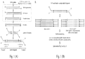
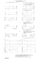
- the adapters' design in combination with the polarity of the complementary strands results in a ligation product that has built-in asymmetry. Therefore, even after PCR amplification, a sequencing read contains information about the polarity of the original strand ( Fig. 1 ).
- This feature is used in the strand-specific RNA sequencing, but was never employed in the context of uncertainty analysis in dsDNA. An essence of our method is to use this feature to generate a hierarchy of consensuses between the sequencing reads and use those consensuses as validation criteria.
- PCR clonal amplification should be sufficiently high so as: (1) to increase the probability that the reads from both complementary strands are observed, (2) to reach the desired reliability of the result, (3) to improve the uncertainty estimates and the diagnosis of problems.
- the second category consists of errors introduced during PCR amplification. This will result in the same error in a sequence in one or more reads of a clonal cluster.
- the PCR amplification errors will have a characteristic probability distribution, discussed later.
- the errors in the third category result from chemical alterations of base in a single strand in a double-stranded fragment of DNA. This alteration can happen inside the living cell or later, during sample handling, but prior to the PCR amplification step. This category will have a distinct probability distribution of a single chemical event generating multiple errors in the PCR product.
- Standard sample preparation can introduce a coupled error on complementary strands, which will have the same signature as a somatic mutation.
- Standard Illumina sample preparation that disrupts DNA by sonication or nebulization introduces uneven breaks on both strands, which need to be repaired enzymatically.
- the process is performed using a T4 DNA polymerase, which uses the single stranded overhang as a template.
- the overhang sequence may contain a chemically altered base, e.g. 7,8-dihydro-8-oxo-2-deoxyguanosine (8-oxo-dG) and in in vitro conditions the polymerase will insert with some frequency an incorrect nucleotide in the complementary strand ( Fig.
- the damaged base may with some frequency also induce the same mispairing later during PCR amplification. Consequently, the same chemical event causes a coupled sequencing error that appears in two groups of reads corresponding to two complementary strands.
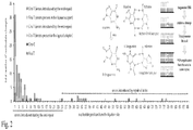
- the same type of coupled error may arise when the original DNA duplex contains a nick on one of its strands, which is then repaired by the combined actions of exonuclease digestion and strand copying, starting from the nick. In our preliminary studies, these repair effects manifest themselves as a surplus of coupled errors in both strands, concentrated close to the ligation site of sequencing adapters ( Fig. 2 ).
- the types of coupled substitution are defined with respect to the strand with putative DNA damage.
- the strands with putative DNA damage correspond to the start of the first paired-end read for both groups of reads, each representing one of two complementary strands.
- the results of the fill-in reaction will be present at the start of the second paired-end read at complementary positions.
- the chemistries generating damaged bases are very diverse, and we expect for errors of this type to observe very different frequencies across all 12 possible types of mutational substitution.
- the frequency for complementary types of substitution e.g.
- G ⁇ T and C ⁇ A) for somatic mutation should be the same, because the mutated positions are only differentiated by the direction in which sequencing is performed. Additionally, while fill-in originating errors should concentrate close to the ends of sequenced DNA, somatic mutations should be randomly distributed along the reads.
- a critical improvement to enable the Twin-seq method is to avoid coupled errors arising from the end-repair reaction after DNA fragmentation.
- the fragmentation is followed by extending the blunt ends with dA, and the ligation of sequencing adapters, neither of which introduces significant sequencing issues.
- Phusion polymerase used in our preliminary studies, generates on average 4.4 ⁇ 10 -7 errors per base, per replication cycle (NEB materials), producing a contribution of ⁇ 10 -5 to the error frequency of a single read.
- An additional contribution of 1.5 ⁇ 10 -5 comes from the error rate of the Bst DNA polymerase [ 64 ], which during polony generation synthesizes a first copy of a single-stranded DNA piece annealed to a short oligonucleotide covalently attached to the Illumina flow-cell. The original strand is removed and the Bst-generated copy becomes the template for the whole polony.
- This level is sufficiently high to be identified in our analysis, but does not generate problems in the Twin-seq method, because Twin-seq uses information about clonal copies resulting from the PCR amplification of the strand complementary to one with PCR errors. PCR error level is often perceived to be much higher.
- the prevention of DNA damage should also increase ⁇ 3-fold our chances of finding reads from both original DNA strands, resulting in ⁇ 2-fold improvement in efficiency, with respect to the main limitation, which is the cost of sequencing reads, not the amount of starting material.
- We increase the scale of sequencing by switching to Hi-Seq 2000 with v3 chemistry to produce about 200 M purity-filtered reads per sequencing lane, ⁇ 6 times more than in the preliminary studies.
- the clonal amplification factor was quite high, ⁇ 25-fold per strand, to enable many types of error diagnosis.
- We gradually improve efficiency by decreasing the clonal amplification factor to ⁇ 4-fold per strand, which results in ⁇ 4 times improvement in efficiency.
- a higher degree of overlap generates an inherent increase in uncertainty of the deconvolution results. Since the pattern of the overlaps is a consequence of the initial random hybridization of the target DNA on the flow-cell, it is quite diverse and the reads, even those originating from the same DNA sequence, may have a quite different overall signal-to-noise ratio prior to base-calling.
- Base-calling is a complex process, in which the deconvolved fluorescence intensities are converted to the assignments of the most probable DNA base with the associated uncertainty of this assignment. This uncertainty can be expressed as a scalar (Q value) or a vector (colored Q-value), and describes the relative likelihood for four possible bases.
- each sequencing cycle involves a polymerase that advances by one position in all reads simultaneously, by incorporating in those reads a specific base, complementary to the template, labeled by a fluorophore distinct to each of the four bases and blocked to prevent further polymerization.
- the polymerase advances by two bases or fails to advance. Polony-wide delay due to incomplete base incorporation is called phasing, the running ahead is called pre-phasing, and the consequences are called phasing error.
- the Illumina software recognizes and corrects for the average level of phasing and pre-phasing in segments of the flow-cell. However, we observed, like others [ 71 ], a very pronounced strand-specific increase in the error levels at particular genomic positions. We address this issue in our iterative approach to error characterization in data analysis.
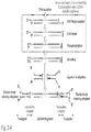
- Each piece of dsDNA can produce two clonally amplified clusters of reads, each cluster originating from one strand of the original dsDNA ( Fig.1 and 3 ).
- a more elaborate, singular value decomposition (SVD) analysis differentiates random events from potential systematic differences within the cluster, which are classified on the basis of correlations within the cluster as either arising from similar sequences within the genome (coincidences and/or polymorphisms) or from correlated sources of error, e.g. from chemical damage or PCR amplification.
- Decision thresholds in this analysis rely on the probabilistic error model that is also subject to improvement after the initial clustering is performed.
- Bayesian reasoning we explicitly model the events that can generate errors in multiple reads of the particular clonal cluster. In the normal use of Illumina sequencing, the clonal amplification reads are typically filtered out before aligning multiple reads to identify mutations or polymorphisms.
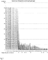
- a troublesome aspect of base-calling error in Illumina has the following properties: a) the effect is highly reproducible when encountering particular sequences ahead of the read position, which often have indications of a higher-order structure formation, e.g. DNA hairpins or quadruplexes; b) there is no decrease in fluorescence signal, so the elongation by the DNA polymerase has not terminated; c) all subsequent positions have increased read-out error, which is characteristic of phasing, where the extent by which polymerases are out of phase in the sequence position can only increase as the read progresses. This sequence-specific desynchronization generates a high level of errors ( Fig. 5 ).
- the problem can be recognized not only by incorrect base-calls, strongly biased towards the base preceding the position of the error in base-calling (53 out of 54 errors for the fragment presented in Fig. 5 ), but also, equally reliably, by a consistent, steep Q-value decrease along the read ( Fig. 5 ).
- Fig. 5 we calculated a cumulative Q-value for 104 reads of 130 bp length that aligned to positions 48308 to 48405 (SEQ ID NO:01) of the forward strand in the genome of Escherichia coli strain K-12, substrain MG1655. Only the 98 bp part common to all these reads is shown. Each read represents a separate polony in the Illumina flow-cell, so the fluorescence intensities of the reads were measured independently. The Figure shows a sudden decrease in the reads quality, with all the reads having lower Q-values at the site base errors without however same genomic site. There is a corresponding increase in base-calling errors, without, however, any decrease in the overall fluorescence intensity.
- Gathering the fluorescent intensities from multiple reads allows us to average the position-specific phasing delay estimate and to redo base-calling, using the averaged intensities and the averaged phasing delay estimate. Calculating averages are optimized by weighting the information from multiple reads, as each polony tends to have a somewhat different error level. Performing base-calling on averaged intensities follows a similar linear modeling of signal and an inversion of cross-talk equations as in the program TotalReCaller [ 74 ].
- Such patterns extend over thousands of polonies, so they can be modeled by local averaging procedures. Depending on the source of the problem, the procedure may involve the averaging of a multiplicative factor, of an additive factor or, in the case of focus variability, of a factor multiplying the uncertainty (error estimate).
- a somatic mutation is defined as a DNA sequence pattern that is not present in the zygotic DNA, from which the organism has developed. To establish that a candidate for a mutation was not present in the zygote an important factor is to differentiate between the polymorphisms present in the zygote and the somatic mutations generated later.
- the expected level of genomic polymorphisms is about 0.1 %.
- the data from the 1000 Genome Initiative indicate that there are still about 50,000 rare or private genomic polymorphisms existing in a particular individual that are absent from the database [ 73 ]. These rare polymorphism variants are normally present in only one copy, co-occurring with a reference allele present in the other chromosome. That means that if we observe a particular position in the genome N times, we have a chance of 2 -N of missing one allele by random selection from N possibilities. This criterion gives us a minimal genomic coverage of about 16 per a particular position in polymorphism control reads that is needed to accept a candidate for somatic mutation in the target reads.
- CNV Copy Number Variation
- Another aspect of the invention maps somatic mutations in different human tissues obtained from several (8-12) deceased persons- at least 8 samples per organ or tissue type (bladder, blood, bone marrow, brain, heart, liver, lung, lymph node, intestinal lining, pancreas, prostate, skin - both exposed to and shielded from sun, stomach, and thyroid).
- organ or tissue type bladedder, blood, bone marrow, brain, heart, liver, lung, lymph node, intestinal lining, pancreas, prostate, skin - both exposed to and shielded from sun, stomach, and thyroid.
- the invention enables a whole range applications including single cell studies, as we need only 10% of DNA in a single cell to be digested into the size range suitable for sequencing, then to be ligated to adapters and transferred to the PCR buffer.
- a study of somatic mutations selected by the affinity methods is performed by two-step PCR and the affinity selection by DNA hybridization in between the PCR steps.
- An example of the affinity selection approach is to capture all chromosomal translocations that involve the genomic neighborhood of the myc gene during the somatic evolution of cancer.
- Fig.1. The description of the preparation of the sequencing library in Twin-seq method in comparison to the standard Illumina library preparation. The fragmentation, the end-repair and the phosphorylation reactions will be replaced by the fragmentation using DFF40 nuclease.
- the ligated adapters will contain short multiplexing adapter, randomized at the ligation site. The arrows and circles represent the ends of the single-strand in the duplex to emphasize the polarity of the molecules.
- B The second important factor will be clonal-amplification introduced during selection of properly ligated fragments by PCR. Each dsDNA fragment with ligated Y adapters will create two distinct clonal clusters.
- Fig.2 The distribution and pattern of errors accumulated during the end-repair. The most probable chemistry and process that could generate them is presented inside the insert on the right.
- Fig. 3 The hierarchy of sequencing consensuses between clonal clusters. Each effect will have its own signature with associated expected magnitude of the effect.
- Fig. 5 We calculated a cumulative Q-value for 104 reads of 130 bp length that aligned to positions 48308 to 48405 of the forward strand in the genome of Escherichia coli strain K-12, substrain MG1655. Only the 98 bp part common to all these reads is shown. Each read represents a separate polony in the Illumina flow-cell, so the fluorescence intensities of the reads were measured independently. The Figure shows a sudden decrease in the reads quality, with all the reads having lower Q-values at the same genomic site. There is a corresponding increase in base-calling errors, without, however, any decrease in the overall fluorescence intensity.
- Another aspect of the invention maps somatic mutations in different human tissues obtained from several (8-12) deceased persons- at least 8 samples per organ or tissue type (bladder, blood, bone marrow, brain, heart, liver, lung, lymph node, intestinal lining, pancreas, prostate, skin - both exposed to and shielded from sun, stomach, and thyroid).
- organ or tissue type bladedder, blood, bone marrow, brain, heart, liver, lung, lymph node, intestinal lining, pancreas, prostate, skin - both exposed to and shielded from sun, stomach, and thyroid.
- Example 2 HHMI Grant: Twin-seq: a method for ultrahigh accuracy in next-generation sequencing and its applications to single-nucleotide and somatic mutations in mammals
- Twin-seq a new method applied to NGS that increases the accuracy of sequencing by more than three orders of magnitude.
- the method was developed by two collaborating investigators, Dominika Borek and Zbyszek Otwinowski, who undertook a comprehensive analysis of the chemical and instrumental effects leading to errors and then designed strategies that effectively deal with each important source of errors.
- a core aspect of Twin-seq is to obtain fully independent information from two strands of a single DNA fragment by preserving strand information during library construction. Since the reads from the two strands can be made independent in their errors, this method reduces the error rate by several orders of magnitude.
- Next-generation sequencing has enabled forward genetic studies recently 1 ; however, the identification of mutations in a priori unknown genomic positions, solely from NGS data, remains a challenge.
- the primary limitation is the occurrence of sequencing errors, which affects all downstream analyses and results in numerous false positives. Therefore, the use of NGS in forward genetic studies still relies on genetic mapping to narrow candidate regions or recessive mutations that can be discerned more easily. Point mutations, which make up 56% of human mutations, remain troublesome, especially heterozygous dominant mutations, and require exhaustive and expensive validation to discriminate mutant loci from false positives.
- Twin-seq a novel, highly efficient NGS sequencing approach, called Twin-seq, which unambiguously identifies even rare mutations in the background of experimental errors.
- a breakthrough aspect of our approach is a change in the experimental protocol that results in the generation of fully independent sequencing information for each of the two complementary strands of a particular dsDNA fragment in a sequencing library.
- Describing and modeling error sources directly let us separate problems introduced during sequencing library preparation from the effects generated later during sequencing by ligation, and from biological noise introduced by passenger mutations, unmapped SNPs, and various effects caused by genomic instabilities.
- Twin-seq allows for studying mammalian genomes with forward genetics methods in a very efficient way to obtain data for tens of genomes in the case of full genome sequencing and hundreds of genomes in the case of exome sequencing.
- Twin-Seq is a tool that can also be used to advance forward genetic studies in mammalian cell culture. Using forward genetics in a mammalian cell culture system has been technically challenging and is fundamentally limited by our ability to efficiently discover mutations. Unlike other model systems, in mammalian cell culture there are no mechanisms to positional clone or narrow a gene locus through recombination events. Highly accurate Twin-seq bridges mammalian cell culture with classical genetics and in so doing provides a tool for gene and pathway discovery across many biological disciplines. The base-calling at the level of PCR clonal clusters, which we use in Twin-seq, allows for determination of mutations even in copy amplified genes.
- Such mutations are of high interest, because they may cause the resistance to anticancer therapy, e.g. a single mutation in one of the 20 copies of gene coding for EGFR receptor in lung cancer cells, which makes these cells resistant to Erlotinib, a potent anticancer medication 11 .
- the Twin-seq method determines mutations directly by sequencing DNA fragments with a reliability level of a single error per 10 9 of DNA base pairs. To achieve such reliability, we need a much higher level of confidence than that generated by a single sequencing read. Therefore, we increase the confidence by combining information from two groups of sequencing reads that originated from complementary strands of a single DNA fragment.
- the design of the Illumina Y-adapters, in combination with opposite polarities of complementary DNA strands, results in a ligation product with built-in asymmetry. As a result, the polarity of a DNA strand serving as a template in PCR amplification can be unambiguously determined from a sequencing read.
- the overhang sequence used as a template, may contain a chemically altered base, e.g. 8-oxo-dG and the polymerase will insert with some frequency an incorrect nucleotide into the complementary strand.
- the damaged base may also induce the same mispairing later during PCR amplification. Consequently, the same chemical event causes a coupled sequencing error that appears in the same position, in two groups of reads corresponding to two complementary strands.
- these repair effects manifest themselves as a surplus of coupled errors in both strands, concentrated close to the ligation site of sequencing adapters. After removing all these correlated errors, we observed in wt MG1655 E. coli, a single coupled change in 31,000,000 sequenced positions, which was consistent with our expectation of ⁇ 0.7 mutations.
- Polony identification, fluorescence signal estimation, and the first round of base-calling are performed with standard Illumina software.
- a singular value decomposition (SVD) analysis differentiates random events from potential systematic differences within the cluster. These differences are classified on the basis of correlations within the cluster as either arising from similar sequences within the genome (coincidences and/or polymorphisms) or from correlated sources of error, e.g. from chemical damage or PCR amplification.
- SMD singular value decomposition
- To enable optimal statistical (Bayesian) reasoning we explicitly model the events that can generate errors in clonal clusters. Then we recalibrate the uncertainty estimates of the reads to include information about self-consistency between sequencing reads.
- the base-calling inefficiencies are at the core of the current problem with mutation calling, particularly when searching for mutations in a background arising from multiple homologous chromosomes typical for aneuploid cancer cells.
- a breakthrough aspect of the Twin-seq method is relying on full consistency of base-calls in a given position of the read within a clonal cluster. Without clonal amplification, we apply statistical criteria of how often a mutation is present in multiple reads overlapping at the same genomic position. In case of a polyploid genome, the signal for mutations appears with low frequency and it will be difficult to differentiate it from correlated base-calling errors.
- Performing base-calling on averaged intensities applies a combination of linear modeling of the signal, an inversion of cross-talk equations, and estimation of the base calling reliability by multi-component error modeling.
- Other improvements to base-calling involve a better model of the imaging artifacts generated during the scan of fluorescent images.
- the output from the mutation calling procedures will be analyzed by multi-level filtering to differentiate the mutations driving the phenotypes from passenger mutations that will be present in the samples.
- ENU mutagenesis provides a complementary approach to null alleles in providing a diversity of mutant alleles.
- Low resolution genetic mapping and Twin-seq whole-genome sequence at 20-30 x coverage reveals the identity of these new mutations and serves as a stringent test of Twin-seq technology for the discovery of point mutations in the mouse genome.
- An immortalized bronchial epithelial cell and each cancer cell line were screened for proliferation after treatment with one of 220,500 chemical compounds.
- 298 compounds inhibited the proliferation of at least one but not all the cancer cell lines.
- 83 chemicals representing multiple chemotypes inhibit proliferation by 50% at a concentration (IC 50 ) at least 100 fold lower than the other cell lines.
- the selective toxicity of these compounds is comparable to Erlotinib and Crizotnib, two clinically successful targeted therapies for the treatment of lung cancer 80,82 . Therefore, identifying the targets for and mechanism of action of these novel compounds has the potential to improve clinical outcomes. After confirming that differential toxicity is not a function of chemical metabolism, we identify these targets by combining forward genetics and Twin-Seq in lung cancer cell culture.
- Twin-Seq is required to discover a single base pair mutation in the context of a high background rate and allelic dilution. Once identified, target identification is confirmed by both genetic and biochemical assays.
- Twin-Seq and mammalian cell culture genetics provides a direct path to target identification of recently identified chemical compounds that selectively affect lung cancer cells.
- Fig. 2-1 The schematic view of the sequencing library preparation with the standard protocol of Illumina, and with our modifications.
- the asymmetric duplex formed after ligation of the adapters has two barcoding adapters (double-stranded) and four indexing adapters, which in our approach are partially randomized.
- the barcoding adapters will be used for multiplexing libraries, to monitor the quality and efficiency of dA tailing, ligation, and purification of libraries.
- the indexing adapters will be used in statistical analysis to identify the reads that coincidentally have the same length and the same barcoding adapters.
- Fig. 2-2 The schematic view of the mechanism that preserves information about the directionality and identity of DNA template strands during PCR amplification, which generates the sequencing library. We use this feature in statistical analysis following sequencing to analyze the type and level of errors in sequencing and to confirm the presence of mutations.
- Fig. 2-3 The Twin-seq principle in Illumina/Solexa pair-end sequencing.
- the sequencing was followed by data analysis to answer a variety of questions, including: (1) whether random drift is the only contributor to the observed mutational patterns; (2) whether there are any differences in the observed patterns in different mitochondrial isolates and if so, how they correlate with age and other biological factors; (3) what are the levels of heteroplasmy and how are they dependent on age and health status; (4) and how well do positions of lesions identified in mtDNA correlate with observed patterns of somatic mutations in mtDNA.
- Cardiovascular disease is the leading cause of death and disability in developed countries and its onset and outcomes correlate with age [ 2 , 9 ]. Multiple lines of evidence suggest that cardiovascular disease and aging are related to one another through decreased mitochondrial functioning [10-13].
- Mitochondria are multi-functional organelles that perform several inter-dependent functions: they are responsible for the production of ATP in the cell by oxidative phosphorylation; they participate in calcium homeostasis; they activate and regulate cellular death pathways and they generate reactive oxygen species that can cause oxidative damage to a variety of biomolecules in high concentrations, but also have diverse signaling functions in concentrations below damaging thresholds [ 7 , 14 , 15 ].
- Each mitochondrion contains multiple copies of its own, 16,569 bp genome, which is organized into a nucleoid [ 16 ]. The nucleoid has a complex structure that changes with age [ 17 , 18 ].
- Mitochondria are inherited maternally [ 19 ] and therefore the mtDNA lineages are clonal. Each cell contains hundreds to thousands of mitochondria [ 20 ], generated by proliferation, which is not synchronized with mitosis.
- Mitochondria are particularly important in the functioning of cardiac myocytes, in which thousands of spatially-organized mitochondria support continuous delivery of ATP to maintain heart muscle contraction.
- the myocardium contains two distinct types of mitochondria; subsarcolemmal (SSM), remaining in contact with the sarcolemma, and interfibrillar (IFM), located between the contractile filaments [ 13 , 21 ]. They differ in their cristae morphology, lamelliform for SSM and tubular for IFM mitochondria, and in multiple functional characteristics [ 5 , 22 , 23 ], such as their rate of oxidative phosphorylation [ 23, 24 ], association with specific proteins [ 25 , 26 ], and calcium retention capacity [ 27 ]. It was suggested that SSM mitochondria may serve a signaling function and IFM mitochondria may be responsible for protecting against damaging events [ 28 ].
- Deletions and point mutations in mtDNA are the cause of multiple mitochondrial disorders [ 29-31 ] with diverse phenotypic manifestations, including cardiovascular problems [ 29 ].
- the mutation rate of mitochondrial DNA is reported to be about two-orders of magnitude higher than that of nuclear DNA [32] .
- the explanation for this difference frequently invokes a combination of high concentrations of reactive oxygen species, which are produced in mitochondria during oxidative phosphorylation, with less robust mechanisms of DNA repair and mtDNA molecules being relatively exposed to chemical attack [ 33 ].
- heteroplasmy correlates with the severity of some mitochondrial diseases [ 37 ], and in some disorders it has to cross a threshold value for the disease to manifest [ 38-41 ].
- Improved methods for detecting mutations have shown that heteroplasmy is relatively common [ 42-44 ], its levels differing between tissue types [ 32 , 45 ], and that it correlates with increasing age [ 41 , 46 ].
- the mechanism that leads to reaching the threshold values of heteroplasmy is not fully understood.
- the generally accepted model of the formation of heteroplasmic mitochondrial populations invokes a combination of random genetic drift with relaxed replication [ 47 ], but the subject is still debated [ 48 ], with some data indicating increased homoplasmy in particular positions of mtDNA, suggesting selective pressure [ 49 ].
- mutated mitochondrial DNA undergoes strong purifying selection against nonsynonymous mutations in protein-coding genes [ 50 , 51 ], which shows that cells have cellular mechanisms to exert selective pressure.
- the single molecule PCR technique is applied to undigested or lightly digested mtDNA samples [ 63 ].
- the structure of mtDNA becomes increasingly complex with age, and this complexity may be related to mtDNA replication and/or to mtDNA repair processes [ 17 , 18 , 64 ].
- smPCR may bias the results by selective exclusion of some of the regions, which due to their structure might be inaccessible to PCR primers.
- Another method, the random mutation capture (RMC) assay has excellent sensitivity, detecting 1 mutation per 1,000,000,000 [ 65-67 ].
- the frequency of mutations is measured by quantifying the changes in the TagI restriction site.
- the point mutation in the TagI site generates a fragment of DNA that is resistant to digestion, which enables its detection and quantification by qPCR.
- the TagI site has only four base pairs and is present in mtDNA only 23 times. This severely restricts the applicability of the assay in the context of determining mutational spectra.
- Our method addresses all of those problems. It is sensitive at the level of RMC assays, resistant to the experimental errors introduced by PCR or by fixation of chemical lesions in DNA, covers the full mtDNA genome, and addresses topological concerns. Our methods preserve the information about the strand origin through the whole procedure of sequencing library generation. For example, we used in preliminary studies, strand information is preserved from fragmentation, which yields double-stranded DNA fragments, until the completion of the sequencing adapters' ligation.
- each read results from sequencing of a polony. Polonies are started by annealing single-stranded DNA to the specific oligonucleotides covering the surface of the flow-cell.
- the sequences that anneal to oligonucleotides on the flow-cell come from the sequencing adapters that were ligated to both ends of the dsDNA fragment prior to polony generation.
- the sequencing adapters are not entirely double-stranded; instead, they have a special Y-structure design, in which the double-stranded stem ligates to dsDNA while the arms remain single-stranded.
- the adapters' design in combination with the polarity of the DNA strands results in a ligation product that has built-in asymmetry. Therefore, regardless of what happens to the dsDNA fragment after ligation, it is possible to recover the information about the polarity of the original strand that lead to the particular sequencing read.
- PCR clonal amplification should be sufficiently high so as to: (1) increase the probability that the reads from both complementary strands are observed; (2) reach the desired reliability; and (3) improve the uncertainty estimates and the diagnosis of problems.
- Standard Illumina sample preparation that disrupts DNA by sonication or nebulization introduces uneven breaks on both DNA strands, which need to be repaired enzymatically. This is done using a T4 DNA polymerase.
- the polymerase encounters a chemically altered base, e.g. 8-Oxoguanine (8-Oxo-G) in the overhang, it can insert the wrong nucleotide into the opposite strand.
- a damaged base may induce the same type of mispairing later during PCR amplification. Consequently, one chemical event causes a coupled sequencing error that appears in both groups of reads.
- DFF40 double-strand-specific, apoptotic DNA endonuclease DFF40, which has been thoroughly characterized and is known to produce blunt ends with a 5' phosphate extension [ 77-79 ].
- DFF40 is activated by adding TEV protease and stopped by curcumin [78] .
- the dsDNA fragmentation is followed by extending the blunt ends with dA and ligating the sequencing adapters.
- Another improvement is introducing properly randomized adapters.
- Our analysis is based on analyzing the hierarchy of alignments between reads, where identifying the uniqueness of the read with high certainty is critical. The uniqueness is defined by a combination of factors; the presence of a specific multiplexing adapter, the length of the read, and the complementary strand having complementary properties - the same length and complementary adapters.
- Our randomization strategy was sufficient for the E. coli genome, which is 4.6 Mbp, and the level of randomization minimized to acceptable levels the probability of accidental coincidences, i.e.
- each base is preceded by a four to six base long, non-randomized multiplexing adapter, and this adapter is preceded by the standard Illumina adapter sequence.
- Illumina recently introduced the index adapter, which is placed in the single-stranded part of the Y adapter.
- the process is designed in such way that this 6 bp long sequence does not have to be defined to be properly sequenced. Therefore, to validate that we did not accidently count different reads twice, we fully randomize this sequence in our adapters. That way, if we have the cluster of reads with the same length of the sequence, the same multiplexing adapter at the ligation site, but with different sequences in the index site, we will know that we have two different sequences (coincidence). The complementary strands have differently randomized index sequences; however, their multiplexing parts will be the same. This built-in control for coincidences helped us to develop the assay and data analysis better.
- the spike-in controls are processed together with mtDNA, using the approach described above. It provides information about the patterns and specificities of the polymerases used in the process, about their error levels, and also about performance, sensitivity and other qualitative and quantitative parameters of our approach. This allows us to introduce probabilistic modeling of correlated errors in clonal cluster reads. This modeling is an improvement over existing methods [ 90 ] by considering chemically-modified DNA bases as intermediates in error generation in the PCR amplifications before the sequencing and during polony formation. Spike-in control together with mtDNA reads helps us to determine the minimal coverage of the mitochondrial genome required to achieve the desired sensitivity. This issue is debated [ 72 ], but the required coverage depends on the error levels.
- the assay has potential to be used sequentially: (1) first to prepare the library of longer dsDNA fragments with specific adapters, (2) then, treatment of the library with specific enzymes recognizing DNA lesions and generating the break, (3) and ligating another adapter to the newly created cut.
- This step is performed through a hierarchical analysis: first, a fast, rough clustering is performed, with criteria set so as not to miss a potential hit, at the cost of merging some clusters together.
- a more elaborate, singular value decomposition (SVD) analysis differentiates random events from potential systematic differences within the cluster, which are classified on the basis of correlations within the cluster as either arising from similar sequences within the genome (coincidences and/or polymorphisms) or from correlated sources of error, e.g. from chemical damage or PCR amplification.
- transversions identify mutational hotspots, and analyze insertion and deletion.
- Samples of human heart tissues can be obtained from the National Disease Research Interchange. The number of samples depends to some extent on the assay performance, and the number of Hi-seq 2000 lanes used to achieve this performance.
- Total mitochondrial isolation are prepared, and separately, fractions of SSM and IFM mitochondria (Palmer et al. [ 21 , 27] ).
- the three samples can be treated as separate libraries, but because they originate from a single donor, there is overlap between total mitochondrial isolation and either SSM or IFM isolations.
- Preparations of IFM mitochondria are contaminated with SSM mitochondria and that total mitochondria fraction contains predominantly SSM mitochondria [ 28 , 93 ]. Having data from all three samples allows us to statistically model the identified effects as a function of their presence and their levels in a particular population.
- the fractionation of SSM and IFM uses short enzymatic digestion to release the IFM fraction, which is tightly bound to the muscle fibers.
- An alternative procedure to prepare the SSM and IFM fractions can be used [ 94 ].
- Somatic mutations resulting from DNA damage or replication errors contribute to the central mechanisms of ageing and cancer development ( Loeb 2011) .
- our current understanding of the impact of somatic mutations on those processes is derived from indirect or fragmentary measurements, which has resulted in standing controversies and problems with mechanistic interpretations of the acquired data.
- methods to determine somatic mutations could not be applied on a genome-wide scale due to the cost and the error-level of sequencing.
- Studies of somatic mutations were limited to either special types of mutations (e.g. microsatellites, restriction sites) or mutations analyzed with artificial selection. It is not clear how biased the selection was or how the limited scope of those studies affected the conclusions.
- High-throughput sequencing methods developed in recent years have addressed the cost of obtaining data, but not the level of errors.
- the method is unique in terms of providing an unbiased scan for mutations and identifying even low levels of mutations.
- a strength of the method comes from an experimental setup that allows for statistical independence criteria validating the results.
- solid knowledge connecting DNA damage to the accumulation of mutations is achievable. This greatly influences the understanding of ageing, cancer processes, and possibly of other diseases where somatic mutations have been implicated.
- the method is ideally suited to address mutator phenotypes, how frequently they are present in different cancers and if they contribute to the development of metastasis - the mutator phenotype connection has already been established for colon and breast cancers (Loeb 2011 ).
- Somatic mutations have been studied for a long time by selecting them with a phenotypic marker, e.g. the HTRP gene.
- the selection introduces an unknown level of bias and requires cultivating a cell line, which severely restricts the range and type of studies.
- a recent approach has applied genome-wide mutation mapping to a clonally amplified group of cells, identifying the somatic mutations present in the cell that started the clonal line. Genomic DNA sequences obtained from a clonal colony with a particular phenotype are compared to the genomic sequences of cells in another part of the organism.
- the sequence variability in the studied cell population is lost in sequencing noise. Therefore, it is a method well-suited for identifying the mutations driving the phenotype, but not suited to be a general approach for studying the spectrum of somatic mutations.
- a central, novel aspect of the method is that, by generating clonal amplification after preparing a sequencing library, we can separate sequence information for both original strands of dsDNA. This requires a different protocol than that used by previous researchers.
- the clonally-amplified fragments are clustered together and a consensus sequence for a cluster is determined separately for the two original strands.
- the select, highly-reliable consensus sequences are mapped on a reference genome. Then, they are compared to each other and to the reference data. Depending on the pattern of differences between the clusters and the reference data, the difference are classified as a somatic mutation, polymorphism, DNA damage or another problem.
- Our method can reach the certainty level of 1 error per 10 10 sequenced base pairs with certain optimizations: (1) The level of errors induced by chemical damage in each DNA strand of the duplex DNA is sufficiently low, i.e. 10 -5 to 10 -6 per base; (2) Complementary errors (errors that generate an alternative, valid duplex DNA sequence) in two DNA strands of a dsDNA fragment are statistically independent, i.e. they occur in the complementary position only due to rare random coincidences; (3) Frequency of observing sequences from both strands of a particular DNA fragment is high enough. Moreover, we should not confuse them with coincidental observations of complementary sequences originating from different duplexes. Additionally, the relevance of results is higher if we have a uniform coverage of all regions of the genome, so we should minimize coverage biases.
- Fpg (known as formamidopyrimidine [fapy]-DNA glycosylase or 8-oxoguanine DNA glycosylase can be used to remove abasic sites and sites of oxidative damage, which are generally not processed by nucleases such as Mung Bean Nuclease (MBN).
- MBN Mung Bean Nuclease
- MBN or similar enzyme can be used to remove ssDNA extensions, recognize structures that miss one or more nucleotides on one strand of the duplex and then create dsDNA breaks at these points (Chaudhry and Weinfeld, NAR, 1995). The enzyme used at this point should not have polymerase activity, because that would generate complementary errors.
- T7 ligase that seals nicks but does not ligate fragments that have blunt ends.
- MBN Prior use of MBN should remove fragments with sticky ends, with which T7 ligase could produce undesired hybrids.
- DNA dilution may be additionally used to prevent the formation of hybrids.
- the need for ligase use is due to MNB not being very efficient in cleaving DNA with a simple nick.
- Other DNA repairing enzymes, without polymerase activity can be also used instead of or in addition to the ones just described. These procedures will produce blunt-ended dsDNA without nicks and abasic sites. Then a standard library-preparation procedure can be applied, for instance dA-tailing, followed by the ligation of sequencing adapters.
- Additional damaging events are catalyzed by thermal radicals generated during sonication and by various hydroxyl-based species generated during purification, storage and PCR amplification. All those reactions are catalyzed by the presence of specific ions, such as Fe 2+ or Cu + , leading to DNA breaks and base oxidation through Fenton and Fenton-like reactions.
- 0.1 mM EDTA as a chelator is, in our opinion, insufficient. Instead we use as additives: sodium iodide, to quench radicals generated in the procedure, sodium nitrate to quench hydrated electrons, deferoxamine to specifically chelate Fe 2+ ions, and neocuproine to specifically chelate Cu + cations without affecting the levels of other ions, such as for instance Mg 2+ , Ca 2+ and Zn 2+ , which are used in the enzymatic reactions involved in sequencing library preparation.
- the final concentration of these compounds typically ranges from 0.1 mM to 1 mM total for all components of the mix. They can be used either as a single additive or in combinations, also during PCR amplification. In Twin-seq, PCR amplifications are performed in standard conditions, i.e. high temperatures are used for 18-26 cycles of PCR.
- the standard protocols of generating samples for sequencing involve so-called blunt-ending, in which two enzymatic activities are used to remove single-stranded ends of double-stranded fragments.
- the first enzymatic activity fills in 5' overhangs, using 5'-3' polymerase activity; the second one removes 3' overhangs with 3'-5' exonuclease activity.
- the polymerase activity uses a single-strand overhang as a template. If this overhang contains a damaged base, e.g. oxidized guanidine, the synthesized second strand will contain a base complementary to damaged one. The presence of these two bases will generate complementary reads that look as if they contained a mutation. Because our method is focused on mapping rare mutations, including the somatic ones, even a rare occurrence of such errors is highly detrimental. In fact, we observed that this could be the biggest source of false mutation calls.
- DFF40 nuclease which is specific to dsDNA, for digesting DNA for sequencing.
- DFF40 creates double-stranded breaks ready for dA-tailing. This eliminates the need for blunt-ending reaction, which was the biggest source of correlated complementary sequencing errors in our initial experiments. This approach worked, and such errors were effectively eliminated.
- DFF40 nuclease Digestion with DFF40 nuclease is not completely random - DFF40 prefers some sequence contexts. Therefore, our initial adapters with limited randomization turned out to be insufficient for some genomic sequences. For particular genomic fragments, we observed sequencing readouts that had multiple different barcodes. Statistics implies that we were dealing with significant presence of coincidental, identical reads originating from different DNA fragments.
- Mutations may be also mimicked by random coincidence of two errors on the complementary strands. Therefore, we model the probability of coincidences with respect to each identified dsDNA Twin-seq cluster, which allows us to estimate false positive and false negative rates. It is well known, and we observed it in our data, that some sequences are much more susceptible to specific DNA modifications. Therefore, we count the observed frequency of single-stranded clonal errors in data as a function of their sequence environment. This creates a probabilistic model of false-positive background resulting from coincidental errors, i.e. the complementary change in DNA happening on both strands.
- the first approach deals with non-uniformity resulting from all fragmentation methods having their particular forms of sequence preferences that result in coverage bias when fragments are subjected to size selection.
- the bias is reduced by performing fragmentation as a series of incremental steps and pooling DNA fragments at each step. For instance, in the case of DFF40 digestion, the aliquots of digested DNA solution are pipetted out from the reaction to the stopping buffer at several time points and pooled.
- This approach generates a combination of underdigested, optimally digested, and overdigested fragments. These sub-pools have opposite sequence biases with respect to the coverage, so mixing them together improves uniformity.
- Another source of non-uniformity of coverage is due to specific secondary structures preventing the amplification, either by PCR or on sequencing flow cell.
- G-quadruplexes are known to cause such problems.
- 7-deazadGTP used as an additive or replacement for dGTP in PCR amplifications should greatly improve the amplification of sequences where such structures form a barrier.
- the oligos that were designed using Hamming (7,4) error codes can be annealed with the standard Illumina adapter before using Klenow (exo-) polymerase to create duplexes whose randomized part will be read out and will define the barcode for a particular DNA fragment being amplified.
- these barcodes contain indices corresponding to a particular experiment in which they are used.
- Chemical DNA synthesis proceeds one base at a time, starting from 3' end, with errors due to incomplete deprotection accumulating at the 5' end of the oligo.
- a compatible label such as biotin at 5' end of the standard primer and then, after annealing, select duplexes with biotin binding to streptavidin-coated beads.
- Biotin can be incorporated at 5' ends by two approaches: additional synthesis step after DNA is synthesized, or, alternatively, by adding biotinylated dT at the end. In the second approach, biotin will be present only in full-length oligos, so selecting by biotin presence will eliminate unwanted side-products of chemical synthesis.
- the randomized oligo is annealed with the standard sequencing adapter to create a substrate for the polymerase that will create a full Y-adapter, e.g. for the adapter 00001111:
- the product of ligating these adapters with DNA inserts is compatible with standard PCR primers and the rest of the sequencing process.
- the indexing/randomized part will be present at both ends of the insert. These random parts will contain the same chemical reaction identifier, but they will not be related with respect to randomization component. Each end will be sequenced at the start of its corresponding read. That means that total identified randomization will be the product of the number of possible base combinations at each end. Unlike in Illumina indexing, the identification of a chemical reaction tag will be done independently at each end, so even multiple readout or synthesis errors can be corrected. For example: Hamming (7,4) recognizes, but does not correct double errors; however, once recognized, even in the presence of a single error on the other end, the reaction will be properly recognized.
Claims (4)
- Procédé de séquençage d'ADN comprenanta) le séquençage indépendant d'un premier et d'un second brin d'un ADNdb afin d'obtenir les première et seconde séquences correspondantes ; etb) la combinaison des première et seconde séquences afin de générer une séquence consensus de l'ADNdb,où l'étape de séquençage (a) comprend :l'amplification de l'ADNdb afin d'obtenir des copies multiples du premier et du second brin ;le séquençage des copies afin d'obtenir des séquences de copie, où chacune des séquences de copie est facultativement une séquence composite générée à partir de lectures multiples de chaque copie ; etla combinaison des séquences de copie afin d'obtenir des séquences consensus du premier et du second brin qui sont les première et seconde séquences.
- Procédé selon la revendication 1, comprenant en outre les étapes consistant à :amplifier par PCR un fragment d'ADNdb possédant une séquence flanquée par des adaptateurs Y afin de produire un ADNdb amplifié asymétrique ;dénaturer l'ADNdb amplifié et attacher puis ponter l'ADNsb amplifié résultant à des emplacements distincts d'un support solide (« flow cell ») afin de produire des polonies ;lire les séquences des polonies ; etidentifier des séquences complémentaires de même taille, qui comportent les première et seconde séquences correspondant au premier et au second brin de l'ADNdb.
- Procédé selon la revendication 2, dans lequel les adaptateurs Y sont des adaptateurs d'indexation randomisés ou sont des adaptateurs différents produits par deux ligatures consécutives, les ligatures pouvant être séparées par des étapes de fragmentation ou d'amplification.
- Procédé selon l'une quelconque des revendications 1 à 3, comprenant en outre l'étape consistant à identifier une ou plusieurs correspondances entre les première et seconde séquences et une séquence d'ADN génomique
par une cartographie des première et seconde séquences sur une séquence d'ADN génomique ;
par une analyse métagénomique ; ou
par une relation entre une ou plusieurs mutations de la séquence d'ADN génomique et la première et/ou seconde séquence(s).
Applications Claiming Priority (2)
| Application Number | Priority Date | Filing Date | Title |
|---|---|---|---|
| US201261654069P | 2012-05-31 | 2012-05-31 | |
| PCT/US2013/042949 WO2013181170A1 (fr) | 2012-05-31 | 2013-05-28 | Procédé de séquençage précis d'adn |
Publications (3)
| Publication Number | Publication Date |
|---|---|
| EP2855707A1 EP2855707A1 (fr) | 2015-04-08 |
| EP2855707A4 EP2855707A4 (fr) | 2016-01-06 |
| EP2855707B1 true EP2855707B1 (fr) | 2017-07-12 |
Family
ID=49673858
Family Applications (1)
| Application Number | Title | Priority Date | Filing Date |
|---|---|---|---|
| EP13797483.8A Active EP2855707B1 (fr) | 2012-05-31 | 2013-05-28 | Procédé de séquençage précis d'adn |
Country Status (5)
| Country | Link |
|---|---|
| US (4) | US9970054B2 (fr) |
| EP (1) | EP2855707B1 (fr) |
| AU (2) | AU2013267609C1 (fr) |
| CA (1) | CA2872141C (fr) |
| WO (1) | WO2013181170A1 (fr) |
Families Citing this family (82)
| Publication number | Priority date | Publication date | Assignee | Title |
|---|---|---|---|---|
| US9260753B2 (en) | 2011-03-24 | 2016-02-16 | President And Fellows Of Harvard College | Single cell nucleic acid detection and analysis |
| SI3363901T1 (sl) | 2012-02-17 | 2021-04-30 | Fred Hutchinson Cancer Research Center | Sestavki in postopki za natančno identifikacijo mutacij |
| PT2828218T (pt) * | 2012-03-20 | 2020-11-11 | Univ Washington Through Its Center For Commercialization | Métodos para baixar a taxa de erro da sequenciação paralela massiva de adn utilizando sequenciação duplex de consensus |
| WO2013181170A1 (fr) | 2012-05-31 | 2013-12-05 | Board Of Regents, The University Of Texas System | Procédé de séquençage précis d'adn |
| DE202013012824U1 (de) | 2012-09-04 | 2020-03-10 | Guardant Health, Inc. | Systeme zum Erfassen von seltenen Mutationen und einer Kopienzahlvariation |
| US11913065B2 (en) | 2012-09-04 | 2024-02-27 | Guardent Health, Inc. | Systems and methods to detect rare mutations and copy number variation |
| US10876152B2 (en) | 2012-09-04 | 2020-12-29 | Guardant Health, Inc. | Systems and methods to detect rare mutations and copy number variation |
| US20160040229A1 (en) | 2013-08-16 | 2016-02-11 | Guardant Health, Inc. | Systems and methods to detect rare mutations and copy number variation |
| CA2889862C (fr) | 2012-11-05 | 2021-02-16 | Rubicon Genomics, Inc. | Marquage par code-barre d'acides nucleiques |
| WO2014093330A1 (fr) | 2012-12-10 | 2014-06-19 | Clearfork Bioscience, Inc. | Procédés pour analyse génomique ciblée |
| US11286519B2 (en) | 2013-12-11 | 2022-03-29 | Accuragen Holdings Limited | Methods and compositions for enrichment of amplification products |
| WO2015089333A1 (fr) | 2013-12-11 | 2015-06-18 | Accuragen, Inc. | Compositions et procédés permettant de détecter des variants de séquence rares |
| US11859246B2 (en) | 2013-12-11 | 2024-01-02 | Accuragen Holdings Limited | Methods and compositions for enrichment of amplification products |
| CN105849555B (zh) * | 2013-12-18 | 2019-05-14 | 加利福尼亚太平洋生物科学股份有限公司 | 用于错误校正的序列读数迭代聚类 |
| ES2822125T3 (es) | 2013-12-28 | 2021-04-29 | Guardant Health Inc | Métodos y sistemas para detectar variantes genéticas |
| KR102321956B1 (ko) * | 2014-01-31 | 2021-11-08 | 스위프트 바이오사이언시스 인코포레이티드 | Dna 기질을 처리하는 개선 방법 |
| WO2015121236A1 (fr) * | 2014-02-11 | 2015-08-20 | F. Hoffmann-La Roche Ag | Séquençage ciblé et filtrage uid |
| US20160053301A1 (en) * | 2014-08-22 | 2016-02-25 | Clearfork Bioscience, Inc. | Methods for quantitative genetic analysis of cell free dna |
| US11085084B2 (en) | 2014-09-12 | 2021-08-10 | The Board Of Trustees Of The Leland Stanford Junior University | Identification and use of circulating nucleic acids |
| US10000799B2 (en) | 2014-11-04 | 2018-06-19 | Boreal Genomics, Inc. | Methods of sequencing with linked fragments |
| SG11201705615UA (en) | 2015-01-12 | 2017-08-30 | 10X Genomics Inc | Processes and systems for preparing nucleic acid sequencing libraries and libraries prepared using same |
| EP3256607B1 (fr) * | 2015-02-13 | 2021-01-20 | Patel, Abhijit Ajit | Procédés de détection d'acides nucléiques hautement parallèle et précise |
| US10221448B2 (en) | 2015-03-06 | 2019-03-05 | Pillar Biosciences Inc. | Selective amplification of overlapping amplicons |
| US10733476B1 (en) * | 2015-04-20 | 2020-08-04 | Color Genomics, Inc. | Communication generation using sparse indicators and sensor data |
| US10844428B2 (en) | 2015-04-28 | 2020-11-24 | Illumina, Inc. | Error suppression in sequenced DNA fragments using redundant reads with unique molecular indices (UMIS) |
| JP6698708B2 (ja) | 2015-06-09 | 2020-05-27 | ライフ テクノロジーズ コーポレーション | 分子タグ付けのための方法、システム、組成物、キット、装置、及びコンピュータ可読媒体 |
| CN114807323A (zh) | 2015-10-09 | 2022-07-29 | 安可济控股有限公司 | 用于富集扩增产物的方法及组合物 |
| RU2018121254A (ru) | 2015-11-11 | 2019-12-16 | Резолюшн Байосайенс, Инк. | Высокоэффективное построение библиотек днк |
| US11371094B2 (en) | 2015-11-19 | 2022-06-28 | 10X Genomics, Inc. | Systems and methods for nucleic acid processing using degenerate nucleotides |
| EP4043584A1 (fr) | 2015-12-08 | 2022-08-17 | Twinstrand Biosciences, Inc. | Adaptateurs améliorés, procédés, et compositions pour le séquençage en double hélice |
| CN117174167A (zh) | 2015-12-17 | 2023-12-05 | 夸登特健康公司 | 通过分析无细胞dna确定肿瘤基因拷贝数的方法 |
| CN108474026A (zh) * | 2016-01-29 | 2018-08-31 | 豪夫迈·罗氏有限公司 | 用于核酸测序的新的衔接子和使用方法 |
| CA3019367A1 (fr) | 2016-03-28 | 2017-10-05 | Boreal Genomics, Inc. | Capture de cible duplex liee |
| US10961573B2 (en) | 2016-03-28 | 2021-03-30 | Boreal Genomics, Inc. | Linked duplex target capture |
| CN109511265B (zh) | 2016-05-16 | 2023-07-14 | 安可济控股有限公司 | 通过链鉴定改进测序的方法 |
| GB201609221D0 (en) * | 2016-05-25 | 2016-07-06 | Oxford Nanopore Tech Ltd | Method |
| US11306356B2 (en) | 2016-06-01 | 2022-04-19 | Roche Sequencing Solutions, Inc. | Immuno-PETE |
| US11708574B2 (en) | 2016-06-10 | 2023-07-25 | Myriad Women's Health, Inc. | Nucleic acid sequencing adapters and uses thereof |
| KR101915701B1 (ko) * | 2016-06-16 | 2018-11-07 | 한국한의학연구원 | 돌연변이 발생률의 측정 방법 |
| WO2017217694A2 (fr) * | 2016-06-16 | 2017-12-21 | 한국한의학연구원 | Procédé de mesure du taux de mutation |
| EP3831958B1 (fr) | 2016-06-30 | 2023-09-06 | Grail, LLC | Marquage différentiel d'arn pour la préparation d'une bibliothèque de séquençage d'adn/arn acellulaire |
| EP4180539A1 (fr) | 2016-07-12 | 2023-05-17 | Qiagen Sciences, LLC | Séquençage d'adn duplex à extrémité unique |
| CA3033749A1 (fr) | 2016-08-15 | 2018-02-22 | Accuragen Holdings Limited | Compositions et procedes permettant de detecter des variants de sequences rares |
| CN117286217A (zh) | 2016-08-25 | 2023-12-26 | 分析生物科学有限公司 | 用于检测dna样品中基因组拷贝变化的方法 |
| US11854666B2 (en) | 2016-09-29 | 2023-12-26 | Myriad Women's Health, Inc. | Noninvasive prenatal screening using dynamic iterative depth optimization |
| GB201620450D0 (en) | 2016-12-01 | 2017-01-18 | Oxford Nanopore Tech Ltd | Method |
| US11268137B2 (en) | 2016-12-09 | 2022-03-08 | Boreal Genomics, Inc. | Linked ligation |
| CN110741096B (zh) * | 2016-12-28 | 2023-07-14 | 奎斯特诊断投资有限责任公司 | 用于检测循环肿瘤dna的组合物和方法 |
| CA3050247A1 (fr) | 2017-01-18 | 2018-07-26 | Illumina, Inc. | Procedes et systemes de generation et de correction d'erreur d'ensembles d'indices moleculaires uniques ayant des longueurs moleculaires heterogenes |
| US10968447B2 (en) | 2017-01-31 | 2021-04-06 | Myriad Women's Health, Inc. | Methods and compositions for enrichment of target polynucleotides |
| WO2018144217A1 (fr) | 2017-01-31 | 2018-08-09 | Counsyl, Inc. | Méthodes et compositions pour enrichissement de polynucléotides cibles |
| US10689684B2 (en) | 2017-02-14 | 2020-06-23 | Microsoft Technology Licensing, Llc | Modifications to polynucleotides for sequencing |
| US10930370B2 (en) | 2017-03-03 | 2021-02-23 | Microsoft Technology Licensing, Llc | Polynucleotide sequencer tuned to artificial polynucleotides |
| WO2018175907A1 (fr) | 2017-03-24 | 2018-09-27 | Counsyl, Inc. | Appelant de variante de nombre de copies |
| US11584958B2 (en) | 2017-03-31 | 2023-02-21 | Grail, Llc | Library preparation and use thereof for sequencing based error correction and/or variant identification |
| WO2018204423A1 (fr) | 2017-05-01 | 2018-11-08 | Illumina, Inc. | Séquences index optimales pour séquençage multiplex massivement parallèle |
| GB201707140D0 (en) | 2017-05-04 | 2017-06-21 | Oxford Nanopore Tech Ltd | Method |
| AU2018266377A1 (en) | 2017-05-08 | 2019-11-14 | Illumina, Inc. | Universal short adapters for indexing of polynucleotide samples |
| GB2578038B (en) | 2017-06-16 | 2022-11-23 | Life Technologies Corp | Control nucleic acids, and compositions, kits, and uses thereof |
| WO2018235938A1 (fr) * | 2017-06-23 | 2018-12-27 | 国立研究開発法人理化学研究所 | Procédé pour séquençage et procédé pour analyse d'acides nucléiques |
| US11646100B2 (en) * | 2017-08-04 | 2023-05-09 | Billiontoone, Inc. | Target-associated molecules for characterization associated with biological targets |
| US11447818B2 (en) | 2017-09-15 | 2022-09-20 | Illumina, Inc. | Universal short adapters with variable length non-random unique molecular identifiers |
| EP3700672B1 (fr) | 2017-10-27 | 2022-12-28 | 10X Genomics, Inc. | Procédés de préparation et d'analyse d'échantillons |
| JP7091372B2 (ja) | 2017-11-06 | 2022-06-27 | イルミナ インコーポレイテッド | 核酸インデックス付け技術 |
| AU2018366213A1 (en) | 2017-11-08 | 2020-05-14 | Twinstrand Biosciences, Inc. | Reagents and adapters for nucleic acid sequencing and methods for making such reagents and adapters |
| WO2019108851A1 (fr) | 2017-11-30 | 2019-06-06 | 10X Genomics, Inc. | Systèmes et procédés de préparation et d'analyse d'acides nucléiques |
| JP7013490B2 (ja) * | 2017-11-30 | 2022-02-15 | イルミナ インコーポレイテッド | 配列バリアントコールのためのバリデーションの方法及びシステム |
| KR102239487B1 (ko) | 2018-01-08 | 2021-04-14 | 일루미나, 인코포레이티드 | 반도체-기반 검출을 사용한 고-처리율 서열분석 |
| WO2019136388A1 (fr) * | 2018-01-08 | 2019-07-11 | Illumina, Inc. | Systèmes et dispositifs de séquençage à haut débit avec détection basée sur un semi-conducteur |
| US20210355532A1 (en) * | 2018-02-13 | 2021-11-18 | Twinstrand Biosciences, Inc. | Methods and reagents for detecting and assessing genotoxicity |
| WO2019183640A1 (fr) | 2018-03-23 | 2019-09-26 | Board Of Regents, The University Of Texas System | Séquençage efficace d'adn double brin avec un taux d'erreurs extrêmement faible |
| US11203782B2 (en) | 2018-03-29 | 2021-12-21 | Accuragen Holdings Limited | Compositions and methods comprising asymmetric barcoding |
| EP4353836A3 (fr) * | 2018-04-27 | 2024-05-01 | Kao Corporation | Procédé de séquençage hautement précis |
| GB201809323D0 (en) | 2018-06-06 | 2018-07-25 | Oxford Nanopore Tech Ltd | Method |
| KR20210059694A (ko) | 2018-07-12 | 2021-05-25 | 트윈스트랜드 바이오사이언시스, 인코포레이티드 | 게놈 편집, 클론 팽창 및 연관된 분야를 규명하기 위한 방법 및 시약 |
| EP3830289A1 (fr) * | 2018-08-03 | 2021-06-09 | 10X Genomics, Inc. | Procédés et systèmes pour réduire au minimum un échange de codes à barres |
| US11459607B1 (en) | 2018-12-10 | 2022-10-04 | 10X Genomics, Inc. | Systems and methods for processing-nucleic acid molecules from a single cell using sequential co-partitioning and composite barcodes |
| US11473136B2 (en) | 2019-01-03 | 2022-10-18 | Ncan Genomics, Inc. | Linked target capture |
| EP4114978A2 (fr) | 2020-03-06 | 2023-01-11 | Singular Genomics Systems, Inc. | Séquençage de brin apparié lié |
| EP4208868A1 (fr) * | 2020-09-03 | 2023-07-12 | The Board of Regents of The University of Texas System | Évaluation et quantification de réparation imparfaite de rupture d'adndb pour diagnostic et traitement de cancer |
| WO2023230278A2 (fr) * | 2022-05-26 | 2023-11-30 | Element Biosciences, Inc. | Correction de phasage et préphasage d'appel de base dans un séquençage de nouvelle génération |
| WO2023230279A1 (fr) * | 2022-05-26 | 2023-11-30 | Element Biosciences, Inc. | Mesure de la qualité en matière d'identification des bases dans le cadre du séquençage nouvelle génération |
Citations (4)
| Publication number | Priority date | Publication date | Assignee | Title |
|---|---|---|---|---|
| WO2009120372A2 (fr) * | 2008-03-28 | 2009-10-01 | Pacific Biosciences Of California, Inc. | Compositions et procédés pour le séquençage d’acide nucléique |
| WO2012148477A1 (fr) | 2010-12-15 | 2012-11-01 | Cellular Research, Inc. | Comptage numérique de molécules individuelles par fixation stochastique d'étiquettes de marqueur |
| WO2013123442A1 (fr) | 2012-02-17 | 2013-08-22 | Fred Hutchinson Cancer Research Center | Compositions et procédés permettant d'identifier de manière précise des mutations |
| WO2013142389A1 (fr) | 2012-03-20 | 2013-09-26 | University Of Washington Through Its Center For Commercialization | Méthodes permettant de faire baisser le taux d'erreur observé lors d'un séquençage adn massivement parallèle en faisant appel à un séquençage consensus duplex |
Family Cites Families (10)
| Publication number | Priority date | Publication date | Assignee | Title |
|---|---|---|---|---|
| CN1123328A (zh) | 1994-11-17 | 1996-05-29 | 中国科学院上海生物化学研究所 | 高温、高传真脱氧核糖核酸(DNA)聚合酶(HiFi Bst) |
| US20030199007A1 (en) * | 1995-06-09 | 2003-10-23 | Sidney Pestka | Super proteins including interferons, interleukins, et al. |
| CA2185590A1 (fr) * | 1995-09-18 | 1997-03-19 | Kazunori Okano | Methode de sequencage d'adn et methode de preparation d'un echantillon d'adn |
| US6087099A (en) * | 1997-09-08 | 2000-07-11 | Myriad Genetics, Inc. | Method for sequencing both strands of a double stranded DNA in a single sequencing reaction |
| US20070172839A1 (en) * | 2006-01-24 | 2007-07-26 | Smith Douglas R | Asymmetrical adapters and methods of use thereof |
| US8921046B2 (en) * | 2008-09-19 | 2014-12-30 | Pacific Biosciences Of California, Inc. | Nucleic acid sequence analysis |
| GB0821981D0 (en) * | 2008-12-02 | 2009-01-07 | Genome Res Ltd | Method for use in polynucleotide sequencing |
| WO2010117804A2 (fr) * | 2009-03-30 | 2010-10-14 | The Regents Of The University Of California | Séquençage naturel de l'adn par synthèse |
| WO2011037990A1 (fr) * | 2009-09-22 | 2011-03-31 | President And Fellows Of Harvard College | Séquençage de paired-mates emmêlés |
| WO2013181170A1 (fr) | 2012-05-31 | 2013-12-05 | Board Of Regents, The University Of Texas System | Procédé de séquençage précis d'adn |
-
2013
- 2013-05-28 WO PCT/US2013/042949 patent/WO2013181170A1/fr active Application Filing
- 2013-05-28 CA CA2872141A patent/CA2872141C/fr active Active
- 2013-05-28 AU AU2013267609A patent/AU2013267609C1/en active Active
- 2013-05-28 EP EP13797483.8A patent/EP2855707B1/fr active Active
-
2014
- 2014-11-21 US US14/550,517 patent/US9970054B2/en active Active
-
2018
- 2018-05-14 US US15/978,208 patent/US10870882B2/en active Active
- 2018-09-26 AU AU2018236781A patent/AU2018236781B2/en active Active
-
2020
- 2020-11-18 US US16/950,923 patent/US11584964B2/en active Active
-
2022
- 2022-12-26 US US18/146,449 patent/US20230193378A1/en active Pending
Patent Citations (4)
| Publication number | Priority date | Publication date | Assignee | Title |
|---|---|---|---|---|
| WO2009120372A2 (fr) * | 2008-03-28 | 2009-10-01 | Pacific Biosciences Of California, Inc. | Compositions et procédés pour le séquençage d’acide nucléique |
| WO2012148477A1 (fr) | 2010-12-15 | 2012-11-01 | Cellular Research, Inc. | Comptage numérique de molécules individuelles par fixation stochastique d'étiquettes de marqueur |
| WO2013123442A1 (fr) | 2012-02-17 | 2013-08-22 | Fred Hutchinson Cancer Research Center | Compositions et procédés permettant d'identifier de manière précise des mutations |
| WO2013142389A1 (fr) | 2012-03-20 | 2013-09-26 | University Of Washington Through Its Center For Commercialization | Méthodes permettant de faire baisser le taux d'erreur observé lors d'un séquençage adn massivement parallèle en faisant appel à un séquençage consensus duplex |
Non-Patent Citations (1)
| Title |
|---|
| SHIROGUCHI ET AL.: "Digital RNA sequencing minimizes sequence 24.1.2012 dependent bias and amplification noise with optimized single- molecule barcodes", PNAS, vol. 109, no. 4, 24 January 2012 (2012-01-24), pages 1347 - 1352, XP055428301 |
Also Published As
| Publication number | Publication date |
|---|---|
| AU2013267609B2 (en) | 2018-06-28 |
| AU2018236781A1 (en) | 2018-10-18 |
| CA2872141C (fr) | 2016-01-19 |
| AU2018236781B2 (en) | 2022-02-03 |
| AU2013267609C1 (en) | 2019-01-03 |
| US20180282802A1 (en) | 2018-10-04 |
| WO2013181170A1 (fr) | 2013-12-05 |
| US20210207210A1 (en) | 2021-07-08 |
| CA2872141A1 (fr) | 2013-12-05 |
| US10870882B2 (en) | 2020-12-22 |
| US9970054B2 (en) | 2018-05-15 |
| US20150275289A1 (en) | 2015-10-01 |
| EP2855707A1 (fr) | 2015-04-08 |
| EP2855707A4 (fr) | 2016-01-06 |
| AU2013267609A1 (en) | 2014-12-18 |
| US11584964B2 (en) | 2023-02-21 |
| US20230193378A1 (en) | 2023-06-22 |
Similar Documents
| Publication | Publication Date | Title |
|---|---|---|
| US11584964B2 (en) | Method for accurate sequencing of DNA | |
| EP3601598B1 (fr) | Procédés d'enrichissement de séquences d'acide nucléique cibles comportant des applications dans le séquençage d'acide nucléique à correction d'erreur | |
| Marshall et al. | Performance evaluation of a mitogenome capture and Illumina sequencing protocol using non-probative, case-type skeletal samples: Implications for the use of a positive control in a next-generation sequencing procedure | |
| Orlando et al. | True single-molecule DNA sequencing of a pleistocene horse bone | |
| Mak et al. | Comparative performance of the BGISEQ-500 vs Illumina HiSeq2500 sequencing platforms for palaeogenomic sequencing | |
| McElhoe et al. | Development and assessment of an optimized next-generation DNA sequencing approach for the mtgenome using the Illumina MiSeq | |
| KR102393608B1 (ko) | 희귀 돌연변이 및 카피수 변이를 검출하기 위한 시스템 및 방법 | |
| EP3910069A1 (fr) | Procédés et composition de profilage d'adn | |
| CN110945144A (zh) | 利用无细胞dna的移植患者监测 | |
| CN111094593A (zh) | 使用供体特异性无细胞dna来评估移植对象中的病症 | |
| Peck et al. | Developmental validation of a Nextera XT mitogenome Illumina MiSeq sequencing method for high-quality samples | |
| Olivieri et al. | Characterization of nucleotide misincorporation patterns in the iceman's mitochondrial DNA | |
| Kieser et al. | Reverse complement PCR: a novel one-step PCR system for typing highly degraded DNA for human identification | |
| Benaglio et al. | Ultra high throughput sequencing in human DNA variation detection: a comparative study on the NDUFA3-PRPF31 region | |
| Austin et al. | PROCÉDÉ DE SEQUENÇAGE PRÉCIS D'ADN | |
| Zhu et al. | Topologically Dependent Abundance of Spontaneous DNA Damage in Single Human Cells | |
| Bueso et al. | Development of novel methodology for study of bacterial DNA from FFPE samples | |
| Inam et al. | Pooled and accurate quantification of Variants of Unknown Drug Resistance in the BCR-ABL fusion protein using error-corrected NGS | |
| Niederstätter et al. | Fluorescent duplex allele-specific PCR and amplicon melting for rapid homogeneous mtDNA haplogroup H screening and sensitive mixture detection | |
| Benovoy | Characterization of transcript isoform variations in human and chimpanzee | |
| Niederstätter et al. | Fluorescent Duplex Allele-Specific PCR and Amplicon Melting for Rapid Homogeneous mtDNA Haplogroup H |
Legal Events
| Date | Code | Title | Description |
|---|---|---|---|
| PUAI | Public reference made under article 153(3) epc to a published international application that has entered the european phase |
Free format text: ORIGINAL CODE: 0009012 |
|
| 17P | Request for examination filed |
Effective date: 20141201 |
|
| AK | Designated contracting states |
Kind code of ref document: A1 Designated state(s): AL AT BE BG CH CY CZ DE DK EE ES FI FR GB GR HR HU IE IS IT LI LT LU LV MC MK MT NL NO PL PT RO RS SE SI SK SM TR |
|
| AX | Request for extension of the european patent |
Extension state: BA ME |
|
| DAX | Request for extension of the european patent (deleted) | ||
| RA4 | Supplementary search report drawn up and despatched (corrected) |
Effective date: 20151207 |
|
| RIC1 | Information provided on ipc code assigned before grant |
Ipc: C12Q 1/68 20060101AFI20151201BHEP |
|
| 17Q | First examination report despatched |
Effective date: 20161010 |
|
| GRAP | Despatch of communication of intention to grant a patent |
Free format text: ORIGINAL CODE: EPIDOSNIGR1 |
|
| INTG | Intention to grant announced |
Effective date: 20170418 |
|
| GRAS | Grant fee paid |
Free format text: ORIGINAL CODE: EPIDOSNIGR3 |
|
| GRAA | (expected) grant |
Free format text: ORIGINAL CODE: 0009210 |
|
| AK | Designated contracting states |
Kind code of ref document: B1 Designated state(s): AL AT BE BG CH CY CZ DE DK EE ES FI FR GB GR HR HU IE IS IT LI LT LU LV MC MK MT NL NO PL PT RO RS SE SI SK SM TR |
|
| REG | Reference to a national code |
Ref country code: GB Ref legal event code: FG4D |
|
| REG | Reference to a national code |
Ref country code: CH Ref legal event code: EP |
|
| REG | Reference to a national code |
Ref country code: AT Ref legal event code: REF Ref document number: 908374 Country of ref document: AT Kind code of ref document: T Effective date: 20170715 |
|
| REG | Reference to a national code |
Ref country code: IE Ref legal event code: FG4D |
|
| REG | Reference to a national code |
Ref country code: DE Ref legal event code: R096 Ref document number: 602013023509 Country of ref document: DE |
|
| REG | Reference to a national code |
Ref country code: NL Ref legal event code: MP Effective date: 20170712 |
|
| REG | Reference to a national code |
Ref country code: LT Ref legal event code: MG4D |
|
| REG | Reference to a national code |
Ref country code: AT Ref legal event code: MK05 Ref document number: 908374 Country of ref document: AT Kind code of ref document: T Effective date: 20170712 |
|
| PG25 | Lapsed in a contracting state [announced via postgrant information from national office to epo] |
Ref country code: NL Free format text: LAPSE BECAUSE OF FAILURE TO SUBMIT A TRANSLATION OF THE DESCRIPTION OR TO PAY THE FEE WITHIN THE PRESCRIBED TIME-LIMIT Effective date: 20170712 Ref country code: FI Free format text: LAPSE BECAUSE OF FAILURE TO SUBMIT A TRANSLATION OF THE DESCRIPTION OR TO PAY THE FEE WITHIN THE PRESCRIBED TIME-LIMIT Effective date: 20170712 Ref country code: NO Free format text: LAPSE BECAUSE OF FAILURE TO SUBMIT A TRANSLATION OF THE DESCRIPTION OR TO PAY THE FEE WITHIN THE PRESCRIBED TIME-LIMIT Effective date: 20171012 Ref country code: HR Free format text: LAPSE BECAUSE OF FAILURE TO SUBMIT A TRANSLATION OF THE DESCRIPTION OR TO PAY THE FEE WITHIN THE PRESCRIBED TIME-LIMIT Effective date: 20170712 Ref country code: AT Free format text: LAPSE BECAUSE OF FAILURE TO SUBMIT A TRANSLATION OF THE DESCRIPTION OR TO PAY THE FEE WITHIN THE PRESCRIBED TIME-LIMIT Effective date: 20170712 Ref country code: SE Free format text: LAPSE BECAUSE OF FAILURE TO SUBMIT A TRANSLATION OF THE DESCRIPTION OR TO PAY THE FEE WITHIN THE PRESCRIBED TIME-LIMIT Effective date: 20170712 Ref country code: LT Free format text: LAPSE BECAUSE OF FAILURE TO SUBMIT A TRANSLATION OF THE DESCRIPTION OR TO PAY THE FEE WITHIN THE PRESCRIBED TIME-LIMIT Effective date: 20170712 |
|
| PG25 | Lapsed in a contracting state [announced via postgrant information from national office to epo] |
Ref country code: RS Free format text: LAPSE BECAUSE OF FAILURE TO SUBMIT A TRANSLATION OF THE DESCRIPTION OR TO PAY THE FEE WITHIN THE PRESCRIBED TIME-LIMIT Effective date: 20170712 Ref country code: ES Free format text: LAPSE BECAUSE OF FAILURE TO SUBMIT A TRANSLATION OF THE DESCRIPTION OR TO PAY THE FEE WITHIN THE PRESCRIBED TIME-LIMIT Effective date: 20170712 Ref country code: PL Free format text: LAPSE BECAUSE OF FAILURE TO SUBMIT A TRANSLATION OF THE DESCRIPTION OR TO PAY THE FEE WITHIN THE PRESCRIBED TIME-LIMIT Effective date: 20170712 Ref country code: BG Free format text: LAPSE BECAUSE OF FAILURE TO SUBMIT A TRANSLATION OF THE DESCRIPTION OR TO PAY THE FEE WITHIN THE PRESCRIBED TIME-LIMIT Effective date: 20171012 Ref country code: GR Free format text: LAPSE BECAUSE OF FAILURE TO SUBMIT A TRANSLATION OF THE DESCRIPTION OR TO PAY THE FEE WITHIN THE PRESCRIBED TIME-LIMIT Effective date: 20171013 Ref country code: LV Free format text: LAPSE BECAUSE OF FAILURE TO SUBMIT A TRANSLATION OF THE DESCRIPTION OR TO PAY THE FEE WITHIN THE PRESCRIBED TIME-LIMIT Effective date: 20170712 Ref country code: IS Free format text: LAPSE BECAUSE OF FAILURE TO SUBMIT A TRANSLATION OF THE DESCRIPTION OR TO PAY THE FEE WITHIN THE PRESCRIBED TIME-LIMIT Effective date: 20171112 |
|
| REG | Reference to a national code |
Ref country code: DE Ref legal event code: R026 Ref document number: 602013023509 Country of ref document: DE |
|
| PLBI | Opposition filed |
Free format text: ORIGINAL CODE: 0009260 |
|
| PLAX | Notice of opposition and request to file observation + time limit sent |
Free format text: ORIGINAL CODE: EPIDOSNOBS2 |
|
| PG25 | Lapsed in a contracting state [announced via postgrant information from national office to epo] |
Ref country code: CZ Free format text: LAPSE BECAUSE OF FAILURE TO SUBMIT A TRANSLATION OF THE DESCRIPTION OR TO PAY THE FEE WITHIN THE PRESCRIBED TIME-LIMIT Effective date: 20170712 Ref country code: RO Free format text: LAPSE BECAUSE OF FAILURE TO SUBMIT A TRANSLATION OF THE DESCRIPTION OR TO PAY THE FEE WITHIN THE PRESCRIBED TIME-LIMIT Effective date: 20170712 Ref country code: DK Free format text: LAPSE BECAUSE OF FAILURE TO SUBMIT A TRANSLATION OF THE DESCRIPTION OR TO PAY THE FEE WITHIN THE PRESCRIBED TIME-LIMIT Effective date: 20170712 |
|
| 26 | Opposition filed |
Opponent name: AMARYLLIS INNOVATION GMBH Effective date: 20180412 |
|
| REG | Reference to a national code |
Ref country code: FR Ref legal event code: PLFP Year of fee payment: 6 |
|
| PG25 | Lapsed in a contracting state [announced via postgrant information from national office to epo] |
Ref country code: EE Free format text: LAPSE BECAUSE OF FAILURE TO SUBMIT A TRANSLATION OF THE DESCRIPTION OR TO PAY THE FEE WITHIN THE PRESCRIBED TIME-LIMIT Effective date: 20170712 Ref country code: SM Free format text: LAPSE BECAUSE OF FAILURE TO SUBMIT A TRANSLATION OF THE DESCRIPTION OR TO PAY THE FEE WITHIN THE PRESCRIBED TIME-LIMIT Effective date: 20170712 Ref country code: IT Free format text: LAPSE BECAUSE OF FAILURE TO SUBMIT A TRANSLATION OF THE DESCRIPTION OR TO PAY THE FEE WITHIN THE PRESCRIBED TIME-LIMIT Effective date: 20170712 Ref country code: SK Free format text: LAPSE BECAUSE OF FAILURE TO SUBMIT A TRANSLATION OF THE DESCRIPTION OR TO PAY THE FEE WITHIN THE PRESCRIBED TIME-LIMIT Effective date: 20170712 |
|
| PLBP | Opposition withdrawn |
Free format text: ORIGINAL CODE: 0009264 |
|
| PG25 | Lapsed in a contracting state [announced via postgrant information from national office to epo] |
Ref country code: SI Free format text: LAPSE BECAUSE OF FAILURE TO SUBMIT A TRANSLATION OF THE DESCRIPTION OR TO PAY THE FEE WITHIN THE PRESCRIBED TIME-LIMIT Effective date: 20170712 |
|
| PLBB | Reply of patent proprietor to notice(s) of opposition received |
Free format text: ORIGINAL CODE: EPIDOSNOBS3 |
|
| PLBD | Termination of opposition procedure: decision despatched |
Free format text: ORIGINAL CODE: EPIDOSNOPC1 |
|
| REG | Reference to a national code |
Ref country code: DE Ref legal event code: R100 Ref document number: 602013023509 Country of ref document: DE |
|
| REG | Reference to a national code |
Ref country code: CH Ref legal event code: PL |
|
| PLBM | Termination of opposition procedure: date of legal effect published |
Free format text: ORIGINAL CODE: 0009276 |
|
| STAA | Information on the status of an ep patent application or granted ep patent |
Free format text: STATUS: OPPOSITION PROCEDURE CLOSED |
|
| REG | Reference to a national code |
Ref country code: BE Ref legal event code: MM Effective date: 20180531 |
|
| PG25 | Lapsed in a contracting state [announced via postgrant information from national office to epo] |
Ref country code: MC Free format text: LAPSE BECAUSE OF FAILURE TO SUBMIT A TRANSLATION OF THE DESCRIPTION OR TO PAY THE FEE WITHIN THE PRESCRIBED TIME-LIMIT Effective date: 20170712 |
|
| 27C | Opposition proceedings terminated |
Effective date: 20181005 |
|
| REG | Reference to a national code |
Ref country code: IE Ref legal event code: MM4A |
|
| PG25 | Lapsed in a contracting state [announced via postgrant information from national office to epo] |
Ref country code: LI Free format text: LAPSE BECAUSE OF NON-PAYMENT OF DUE FEES Effective date: 20180531 Ref country code: CH Free format text: LAPSE BECAUSE OF NON-PAYMENT OF DUE FEES Effective date: 20180531 |
|
| PG25 | Lapsed in a contracting state [announced via postgrant information from national office to epo] |
Ref country code: LU Free format text: LAPSE BECAUSE OF NON-PAYMENT OF DUE FEES Effective date: 20180528 |
|
| PG25 | Lapsed in a contracting state [announced via postgrant information from national office to epo] |
Ref country code: IE Free format text: LAPSE BECAUSE OF NON-PAYMENT OF DUE FEES Effective date: 20180528 |
|
| PG25 | Lapsed in a contracting state [announced via postgrant information from national office to epo] |
Ref country code: BE Free format text: LAPSE BECAUSE OF NON-PAYMENT OF DUE FEES Effective date: 20180531 |
|
| PG25 | Lapsed in a contracting state [announced via postgrant information from national office to epo] |
Ref country code: MT Free format text: LAPSE BECAUSE OF NON-PAYMENT OF DUE FEES Effective date: 20180528 |
|
| PG25 | Lapsed in a contracting state [announced via postgrant information from national office to epo] |
Ref country code: TR Free format text: LAPSE BECAUSE OF FAILURE TO SUBMIT A TRANSLATION OF THE DESCRIPTION OR TO PAY THE FEE WITHIN THE PRESCRIBED TIME-LIMIT Effective date: 20170712 |
|
| PG25 | Lapsed in a contracting state [announced via postgrant information from national office to epo] |
Ref country code: PT Free format text: LAPSE BECAUSE OF FAILURE TO SUBMIT A TRANSLATION OF THE DESCRIPTION OR TO PAY THE FEE WITHIN THE PRESCRIBED TIME-LIMIT Effective date: 20170712 |
|
| PG25 | Lapsed in a contracting state [announced via postgrant information from national office to epo] |
Ref country code: CY Free format text: LAPSE BECAUSE OF FAILURE TO SUBMIT A TRANSLATION OF THE DESCRIPTION OR TO PAY THE FEE WITHIN THE PRESCRIBED TIME-LIMIT Effective date: 20170712 Ref country code: HU Free format text: LAPSE BECAUSE OF FAILURE TO SUBMIT A TRANSLATION OF THE DESCRIPTION OR TO PAY THE FEE WITHIN THE PRESCRIBED TIME-LIMIT; INVALID AB INITIO Effective date: 20130528 Ref country code: MK Free format text: LAPSE BECAUSE OF NON-PAYMENT OF DUE FEES Effective date: 20170712 |
|
| PG25 | Lapsed in a contracting state [announced via postgrant information from national office to epo] |
Ref country code: AL Free format text: LAPSE BECAUSE OF FAILURE TO SUBMIT A TRANSLATION OF THE DESCRIPTION OR TO PAY THE FEE WITHIN THE PRESCRIBED TIME-LIMIT Effective date: 20170712 |
|
| P01 | Opt-out of the competence of the unified patent court (upc) registered |
Effective date: 20230508 |
|
| PGFP | Annual fee paid to national office [announced via postgrant information from national office to epo] |
Ref country code: FR Payment date: 20230523 Year of fee payment: 11 Ref country code: DE Payment date: 20230526 Year of fee payment: 11 |
|
| PGFP | Annual fee paid to national office [announced via postgrant information from national office to epo] |
Ref country code: GB Payment date: 20230530 Year of fee payment: 11 |