EP2854845B1 - Complexes multimères présentant une meilleure stabilité in vivo, une meilleure pharmacocinétique et une meilleure efficacité - Google Patents
Complexes multimères présentant une meilleure stabilité in vivo, une meilleure pharmacocinétique et une meilleure efficacité Download PDFInfo
- Publication number
- EP2854845B1 EP2854845B1 EP13797550.4A EP13797550A EP2854845B1 EP 2854845 B1 EP2854845 B1 EP 2854845B1 EP 13797550 A EP13797550 A EP 13797550A EP 2854845 B1 EP2854845 B1 EP 2854845B1
- Authority
- EP
- European Patent Office
- Prior art keywords
- seq
- cancer
- antibody
- antibodies
- igg
- Prior art date
- Legal status (The legal status is an assumption and is not a legal conclusion. Google has not performed a legal analysis and makes no representation as to the accuracy of the status listed.)
- Active
Links
- 238000001727 in vivo Methods 0.000 title description 16
- 230000001976 improved effect Effects 0.000 title description 7
- -1 CD1a Proteins 0.000 claims description 100
- 239000000427 antigen Substances 0.000 claims description 84
- 108091007433 antigens Proteins 0.000 claims description 84
- 102000036639 antigens Human genes 0.000 claims description 84
- 230000027455 binding Effects 0.000 claims description 73
- 206010028980 Neoplasm Diseases 0.000 claims description 69
- 108090000765 processed proteins & peptides Proteins 0.000 claims description 59
- 201000011510 cancer Diseases 0.000 claims description 42
- 102000008394 Immunoglobulin Fragments Human genes 0.000 claims description 38
- 108010021625 Immunoglobulin Fragments Proteins 0.000 claims description 37
- 108060008682 Tumor Necrosis Factor Proteins 0.000 claims description 36
- 108020001507 fusion proteins Proteins 0.000 claims description 34
- 102000037865 fusion proteins Human genes 0.000 claims description 34
- 239000000203 mixture Substances 0.000 claims description 34
- 101150044980 Akap1 gene Proteins 0.000 claims description 33
- 239000012636 effector Substances 0.000 claims description 33
- 208000037265 diseases, disorders, signs and symptoms Diseases 0.000 claims description 32
- 102000008130 Cyclic AMP-Dependent Protein Kinases Human genes 0.000 claims description 27
- 108010049894 Cyclic AMP-Dependent Protein Kinases Proteins 0.000 claims description 27
- 108010000499 Thromboplastin Proteins 0.000 claims description 27
- 102000000852 Tumor Necrosis Factor-alpha Human genes 0.000 claims description 27
- 102000004127 Cytokines Human genes 0.000 claims description 25
- 108090000695 Cytokines Proteins 0.000 claims description 25
- 201000010099 disease Diseases 0.000 claims description 24
- 239000012634 fragment Substances 0.000 claims description 24
- 102100022133 Complement C3 Human genes 0.000 claims description 22
- 102100031506 Complement C5 Human genes 0.000 claims description 22
- 102000004889 Interleukin-6 Human genes 0.000 claims description 21
- 108090001005 Interleukin-6 Proteins 0.000 claims description 21
- 150000001413 amino acids Chemical group 0.000 claims description 20
- 239000002158 endotoxin Substances 0.000 claims description 20
- 102000005962 receptors Human genes 0.000 claims description 19
- 108020003175 receptors Proteins 0.000 claims description 19
- 102100037792 Interleukin-6 receptor subunit alpha Human genes 0.000 claims description 18
- 102000004890 Interleukin-8 Human genes 0.000 claims description 18
- 108090001007 Interleukin-8 Proteins 0.000 claims description 18
- 230000000295 complement effect Effects 0.000 claims description 18
- 239000003795 chemical substances by application Substances 0.000 claims description 17
- 102100032937 CD40 ligand Human genes 0.000 claims description 16
- 102000003810 Interleukin-18 Human genes 0.000 claims description 16
- 108090000171 Interleukin-18 Proteins 0.000 claims description 16
- 102000003812 Interleukin-15 Human genes 0.000 claims description 15
- 108090000172 Interleukin-15 Proteins 0.000 claims description 15
- 229920006008 lipopolysaccharide Polymers 0.000 claims description 15
- 239000003053 toxin Substances 0.000 claims description 15
- 231100000765 toxin Toxicity 0.000 claims description 15
- 108700012359 toxins Proteins 0.000 claims description 15
- 102000014150 Interferons Human genes 0.000 claims description 14
- 108010050904 Interferons Proteins 0.000 claims description 14
- 102000000589 Interleukin-1 Human genes 0.000 claims description 14
- 108010002352 Interleukin-1 Proteins 0.000 claims description 14
- 108050003558 Interleukin-17 Proteins 0.000 claims description 14
- 102000013691 Interleukin-17 Human genes 0.000 claims description 14
- 229960004641 rituximab Drugs 0.000 claims description 14
- 208000023275 Autoimmune disease Diseases 0.000 claims description 13
- 102100024423 Carbonic anhydrase 9 Human genes 0.000 claims description 13
- 102000005789 Vascular Endothelial Growth Factors Human genes 0.000 claims description 13
- 108010019530 Vascular Endothelial Growth Factors Proteins 0.000 claims description 13
- 239000003102 growth factor Substances 0.000 claims description 13
- 108010071942 Colony-Stimulating Factors Proteins 0.000 claims description 12
- 102100030595 HLA class II histocompatibility antigen gamma chain Human genes 0.000 claims description 12
- 101001082627 Homo sapiens HLA class II histocompatibility antigen gamma chain Proteins 0.000 claims description 12
- 102100037877 Intercellular adhesion molecule 1 Human genes 0.000 claims description 12
- 108010065805 Interleukin-12 Proteins 0.000 claims description 12
- 102000013462 Interleukin-12 Human genes 0.000 claims description 12
- 208000015914 Non-Hodgkin lymphomas Diseases 0.000 claims description 12
- 108010083644 Ribonucleases Proteins 0.000 claims description 12
- 102000006382 Ribonucleases Human genes 0.000 claims description 12
- 102100037086 Bone marrow stromal antigen 2 Human genes 0.000 claims description 11
- 108090000394 Erythropoietin Proteins 0.000 claims description 11
- 102000003951 Erythropoietin Human genes 0.000 claims description 11
- 102100041003 Glutamate carboxypeptidase 2 Human genes 0.000 claims description 11
- 101000740785 Homo sapiens Bone marrow stromal antigen 2 Proteins 0.000 claims description 11
- 101000892862 Homo sapiens Glutamate carboxypeptidase 2 Proteins 0.000 claims description 11
- 101000599048 Homo sapiens Interleukin-6 receptor subunit alpha Proteins 0.000 claims description 11
- 102100037852 Insulin-like growth factor I Human genes 0.000 claims description 11
- 239000000539 dimer Substances 0.000 claims description 11
- 238000006471 dimerization reaction Methods 0.000 claims description 11
- 229940105423 erythropoietin Drugs 0.000 claims description 11
- 208000014018 liver neoplasm Diseases 0.000 claims description 11
- 229950003734 milatuzumab Drugs 0.000 claims description 11
- 238000003032 molecular docking Methods 0.000 claims description 11
- OXCMYAYHXIHQOA-UHFFFAOYSA-N potassium;[2-butyl-5-chloro-3-[[4-[2-(1,2,4-triaza-3-azanidacyclopenta-1,4-dien-5-yl)phenyl]phenyl]methyl]imidazol-4-yl]methanol Chemical compound [K+].CCCCC1=NC(Cl)=C(CO)N1CC1=CC=C(C=2C(=CC=CC=2)C2=N[N-]N=N2)C=C1 OXCMYAYHXIHQOA-UHFFFAOYSA-N 0.000 claims description 11
- MZOFCQQQCNRIBI-VMXHOPILSA-N (3s)-4-[[(2s)-1-[[(2s)-1-[[(1s)-1-carboxy-2-hydroxyethyl]amino]-4-methyl-1-oxopentan-2-yl]amino]-5-(diaminomethylideneamino)-1-oxopentan-2-yl]amino]-3-[[2-[[(2s)-2,6-diaminohexanoyl]amino]acetyl]amino]-4-oxobutanoic acid Chemical compound OC[C@@H](C(O)=O)NC(=O)[C@H](CC(C)C)NC(=O)[C@H](CCCN=C(N)N)NC(=O)[C@H](CC(O)=O)NC(=O)CNC(=O)[C@@H](N)CCCCN MZOFCQQQCNRIBI-VMXHOPILSA-N 0.000 claims description 10
- 102100022005 B-lymphocyte antigen CD20 Human genes 0.000 claims description 10
- 102100035793 CD83 antigen Human genes 0.000 claims description 10
- 102000006354 HLA-DR Antigens Human genes 0.000 claims description 10
- 108010058597 HLA-DR Antigens Proteins 0.000 claims description 10
- 101000897405 Homo sapiens B-lymphocyte antigen CD20 Proteins 0.000 claims description 10
- 101000946856 Homo sapiens CD83 antigen Proteins 0.000 claims description 10
- 101000599951 Homo sapiens Insulin-like growth factor I Proteins 0.000 claims description 10
- 108090000723 Insulin-Like Growth Factor I Proteins 0.000 claims description 10
- 108090000190 Thrombin Proteins 0.000 claims description 10
- 210000004899 c-terminal region Anatomy 0.000 claims description 10
- 208000015181 infectious disease Diseases 0.000 claims description 10
- NOESYZHRGYRDHS-UHFFFAOYSA-N insulin Chemical compound N1C(=O)C(NC(=O)C(CCC(N)=O)NC(=O)C(CCC(O)=O)NC(=O)C(C(C)C)NC(=O)C(NC(=O)CN)C(C)CC)CSSCC(C(NC(CO)C(=O)NC(CC(C)C)C(=O)NC(CC=2C=CC(O)=CC=2)C(=O)NC(CCC(N)=O)C(=O)NC(CC(C)C)C(=O)NC(CCC(O)=O)C(=O)NC(CC(N)=O)C(=O)NC(CC=2C=CC(O)=CC=2)C(=O)NC(CSSCC(NC(=O)C(C(C)C)NC(=O)C(CC(C)C)NC(=O)C(CC=2C=CC(O)=CC=2)NC(=O)C(CC(C)C)NC(=O)C(C)NC(=O)C(CCC(O)=O)NC(=O)C(C(C)C)NC(=O)C(CC(C)C)NC(=O)C(CC=2NC=NC=2)NC(=O)C(CO)NC(=O)CNC2=O)C(=O)NCC(=O)NC(CCC(O)=O)C(=O)NC(CCCNC(N)=N)C(=O)NCC(=O)NC(CC=3C=CC=CC=3)C(=O)NC(CC=3C=CC=CC=3)C(=O)NC(CC=3C=CC(O)=CC=3)C(=O)NC(C(C)O)C(=O)N3C(CCC3)C(=O)NC(CCCCN)C(=O)NC(C)C(O)=O)C(=O)NC(CC(N)=O)C(O)=O)=O)NC(=O)C(C(C)CC)NC(=O)C(CO)NC(=O)C(C(C)O)NC(=O)C1CSSCC2NC(=O)C(CC(C)C)NC(=O)C(NC(=O)C(CCC(N)=O)NC(=O)C(CC(N)=O)NC(=O)C(NC(=O)C(N)CC=1C=CC=CC=1)C(C)C)CC1=CN=CN1 NOESYZHRGYRDHS-UHFFFAOYSA-N 0.000 claims description 10
- 201000007270 liver cancer Diseases 0.000 claims description 10
- 101150047061 tag-72 gene Proteins 0.000 claims description 10
- 229960004072 thrombin Drugs 0.000 claims description 10
- 102100031585 ADP-ribosyl cyclase/cyclic ADP-ribose hydrolase 1 Human genes 0.000 claims description 9
- 102100021943 C-C motif chemokine 2 Human genes 0.000 claims description 9
- 102100025248 C-X-C motif chemokine 10 Human genes 0.000 claims description 9
- 101710098275 C-X-C motif chemokine 10 Proteins 0.000 claims description 9
- 102100024217 CAMPATH-1 antigen Human genes 0.000 claims description 9
- 101150013553 CD40 gene Proteins 0.000 claims description 9
- 108010065524 CD52 Antigen Proteins 0.000 claims description 9
- 102000007644 Colony-Stimulating Factors Human genes 0.000 claims description 9
- 102000012673 Follicle Stimulating Hormone Human genes 0.000 claims description 9
- 108010079345 Follicle Stimulating Hormone Proteins 0.000 claims description 9
- 206010018338 Glioma Diseases 0.000 claims description 9
- 208000017604 Hodgkin disease Diseases 0.000 claims description 9
- 101000777636 Homo sapiens ADP-ribosyl cyclase/cyclic ADP-ribose hydrolase 1 Proteins 0.000 claims description 9
- 101000868215 Homo sapiens CD40 ligand Proteins 0.000 claims description 9
- 101000946889 Homo sapiens Monocyte differentiation antigen CD14 Proteins 0.000 claims description 9
- 101000914484 Homo sapiens T-lymphocyte activation antigen CD80 Proteins 0.000 claims description 9
- 108010047761 Interferon-alpha Proteins 0.000 claims description 9
- 102000006992 Interferon-alpha Human genes 0.000 claims description 9
- 108010002350 Interleukin-2 Proteins 0.000 claims description 9
- 102000000588 Interleukin-2 Human genes 0.000 claims description 9
- 108010073521 Luteinizing Hormone Proteins 0.000 claims description 9
- 102000009151 Luteinizing Hormone Human genes 0.000 claims description 9
- 108090000542 Lymphotoxin-alpha Proteins 0.000 claims description 9
- 102000004083 Lymphotoxin-alpha Human genes 0.000 claims description 9
- 102100035877 Monocyte differentiation antigen CD14 Human genes 0.000 claims description 9
- 206010061902 Pancreatic neoplasm Diseases 0.000 claims description 9
- 206010035226 Plasma cell myeloma Diseases 0.000 claims description 9
- 206010039491 Sarcoma Diseases 0.000 claims description 9
- 102100027222 T-lymphocyte activation antigen CD80 Human genes 0.000 claims description 9
- 108010008125 Tenascin Proteins 0.000 claims description 9
- 108010041111 Thrombopoietin Proteins 0.000 claims description 9
- 102000036693 Thrombopoietin Human genes 0.000 claims description 9
- 102000011923 Thyrotropin Human genes 0.000 claims description 9
- 108010061174 Thyrotropin Proteins 0.000 claims description 9
- 102100040247 Tumor necrosis factor Human genes 0.000 claims description 9
- 102100040245 Tumor necrosis factor receptor superfamily member 5 Human genes 0.000 claims description 9
- 102100033177 Vascular endothelial growth factor receptor 2 Human genes 0.000 claims description 9
- 229940047120 colony stimulating factors Drugs 0.000 claims description 9
- 229940028334 follicle stimulating hormone Drugs 0.000 claims description 9
- 102000006495 integrins Human genes 0.000 claims description 9
- 108010044426 integrins Proteins 0.000 claims description 9
- 229940040129 luteinizing hormone Drugs 0.000 claims description 9
- 208000015486 malignant pancreatic neoplasm Diseases 0.000 claims description 9
- 201000002528 pancreatic cancer Diseases 0.000 claims description 9
- 208000008443 pancreatic carcinoma Diseases 0.000 claims description 9
- 102400000068 Angiostatin Human genes 0.000 claims description 8
- 108010079709 Angiostatins Proteins 0.000 claims description 8
- 102100038080 B-cell receptor CD22 Human genes 0.000 claims description 8
- 102100024222 B-lymphocyte antigen CD19 Human genes 0.000 claims description 8
- 102100032367 C-C motif chemokine 5 Human genes 0.000 claims description 8
- 102100039398 C-X-C motif chemokine 2 Human genes 0.000 claims description 8
- 108700012439 CA9 Proteins 0.000 claims description 8
- 102100022002 CD59 glycoprotein Human genes 0.000 claims description 8
- 108010055166 Chemokine CCL5 Proteins 0.000 claims description 8
- 102100025680 Complement decay-accelerating factor Human genes 0.000 claims description 8
- 102100032768 Complement receptor type 2 Human genes 0.000 claims description 8
- 102000004190 Enzymes Human genes 0.000 claims description 8
- 108090000790 Enzymes Proteins 0.000 claims description 8
- 108010073385 Fibrin Proteins 0.000 claims description 8
- 102000009123 Fibrin Human genes 0.000 claims description 8
- BWGVNKXGVNDBDI-UHFFFAOYSA-N Fibrin monomer Chemical compound CNC(=O)CNC(=O)CN BWGVNKXGVNDBDI-UHFFFAOYSA-N 0.000 claims description 8
- 102100037362 Fibronectin Human genes 0.000 claims description 8
- 108010067306 Fibronectins Proteins 0.000 claims description 8
- 208000032612 Glial tumor Diseases 0.000 claims description 8
- 102000003886 Glycoproteins Human genes 0.000 claims description 8
- 108090000288 Glycoproteins Proteins 0.000 claims description 8
- 208000010747 Hodgkins lymphoma Diseases 0.000 claims description 8
- 101000884305 Homo sapiens B-cell receptor CD22 Proteins 0.000 claims description 8
- 101000980825 Homo sapiens B-lymphocyte antigen CD19 Proteins 0.000 claims description 8
- 101000897400 Homo sapiens CD59 glycoprotein Proteins 0.000 claims description 8
- 101000856022 Homo sapiens Complement decay-accelerating factor Proteins 0.000 claims description 8
- 101000941929 Homo sapiens Complement receptor type 2 Proteins 0.000 claims description 8
- 101000935040 Homo sapiens Integrin beta-2 Proteins 0.000 claims description 8
- 101000599852 Homo sapiens Intercellular adhesion molecule 1 Proteins 0.000 claims description 8
- 101001057504 Homo sapiens Interferon-stimulated gene 20 kDa protein Proteins 0.000 claims description 8
- 101001055144 Homo sapiens Interleukin-2 receptor subunit alpha Proteins 0.000 claims description 8
- 101000853002 Homo sapiens Interleukin-25 Proteins 0.000 claims description 8
- 101000878605 Homo sapiens Low affinity immunoglobulin epsilon Fc receptor Proteins 0.000 claims description 8
- 101000961414 Homo sapiens Membrane cofactor protein Proteins 0.000 claims description 8
- 101001133056 Homo sapiens Mucin-1 Proteins 0.000 claims description 8
- 101001133081 Homo sapiens Mucin-2 Proteins 0.000 claims description 8
- 101000972284 Homo sapiens Mucin-3A Proteins 0.000 claims description 8
- 101000972286 Homo sapiens Mucin-4 Proteins 0.000 claims description 8
- 101000934338 Homo sapiens Myeloid cell surface antigen CD33 Proteins 0.000 claims description 8
- 101001128431 Homo sapiens Myeloid-derived growth factor Proteins 0.000 claims description 8
- 101000874179 Homo sapiens Syndecan-1 Proteins 0.000 claims description 8
- 101000851376 Homo sapiens Tumor necrosis factor receptor superfamily member 8 Proteins 0.000 claims description 8
- 108010000521 Human Growth Hormone Proteins 0.000 claims description 8
- 102000002265 Human Growth Hormone Human genes 0.000 claims description 8
- 239000000854 Human Growth Hormone Substances 0.000 claims description 8
- 102100025390 Integrin beta-2 Human genes 0.000 claims description 8
- 102100037850 Interferon gamma Human genes 0.000 claims description 8
- 108090000467 Interferon-beta Proteins 0.000 claims description 8
- 108010074328 Interferon-gamma Proteins 0.000 claims description 8
- 102100027268 Interferon-stimulated gene 20 kDa protein Human genes 0.000 claims description 8
- 102100038007 Low affinity immunoglobulin epsilon Fc receptor Human genes 0.000 claims description 8
- 102100039373 Membrane cofactor protein Human genes 0.000 claims description 8
- 102100034256 Mucin-1 Human genes 0.000 claims description 8
- 102100034263 Mucin-2 Human genes 0.000 claims description 8
- 102100022497 Mucin-3A Human genes 0.000 claims description 8
- 102100022693 Mucin-4 Human genes 0.000 claims description 8
- 102100025243 Myeloid cell surface antigen CD33 Human genes 0.000 claims description 8
- 108700020796 Oncogene Proteins 0.000 claims description 8
- 102000043276 Oncogene Human genes 0.000 claims description 8
- 102100035194 Placenta growth factor Human genes 0.000 claims description 8
- 208000006664 Precursor Cell Lymphoblastic Leukemia-Lymphoma Diseases 0.000 claims description 8
- 102100035721 Syndecan-1 Human genes 0.000 claims description 8
- 102100036857 Tumor necrosis factor receptor superfamily member 8 Human genes 0.000 claims description 8
- 102100027212 Tumor-associated calcium signal transducer 2 Human genes 0.000 claims description 8
- FZCSTZYAHCUGEM-UHFFFAOYSA-N aspergillomarasmine B Natural products OC(=O)CNC(C(O)=O)CNC(C(O)=O)CC(O)=O FZCSTZYAHCUGEM-UHFFFAOYSA-N 0.000 claims description 8
- 229940088598 enzyme Drugs 0.000 claims description 8
- 229950003499 fibrin Drugs 0.000 claims description 8
- 229940088597 hormone Drugs 0.000 claims description 8
- 239000005556 hormone Substances 0.000 claims description 8
- 229940079322 interferon Drugs 0.000 claims description 8
- 239000003446 ligand Substances 0.000 claims description 8
- 230000001105 regulatory effect Effects 0.000 claims description 8
- 102100032412 Basigin Human genes 0.000 claims description 7
- 101710155857 C-C motif chemokine 2 Proteins 0.000 claims description 7
- 102000017420 CD3 protein, epsilon/gamma/delta subunit Human genes 0.000 claims description 7
- 108050005493 CD3 protein, epsilon/gamma/delta subunit Proteins 0.000 claims description 7
- 108010029697 CD40 Ligand Proteins 0.000 claims description 7
- 102100025221 CD70 antigen Human genes 0.000 claims description 7
- 206010009944 Colon cancer Diseases 0.000 claims description 7
- 208000035473 Communicable disease Diseases 0.000 claims description 7
- 102100031940 Epithelial cell adhesion molecule Human genes 0.000 claims description 7
- 101000798441 Homo sapiens Basigin Proteins 0.000 claims description 7
- 101000934356 Homo sapiens CD70 antigen Proteins 0.000 claims description 7
- 101000920667 Homo sapiens Epithelial cell adhesion molecule Proteins 0.000 claims description 7
- 101000777628 Homo sapiens Leukocyte antigen CD37 Proteins 0.000 claims description 7
- 101000738771 Homo sapiens Receptor-type tyrosine-protein phosphatase C Proteins 0.000 claims description 7
- 101000716102 Homo sapiens T-cell surface glycoprotein CD4 Proteins 0.000 claims description 7
- 101000946843 Homo sapiens T-cell surface glycoprotein CD8 alpha chain Proteins 0.000 claims description 7
- 101000611023 Homo sapiens Tumor necrosis factor receptor superfamily member 6 Proteins 0.000 claims description 7
- 102100031586 Leukocyte antigen CD37 Human genes 0.000 claims description 7
- 108010057464 Prolactin Proteins 0.000 claims description 7
- 102000003946 Prolactin Human genes 0.000 claims description 7
- 102100037422 Receptor-type tyrosine-protein phosphatase C Human genes 0.000 claims description 7
- 208000005718 Stomach Neoplasms Diseases 0.000 claims description 7
- 102100036011 T-cell surface glycoprotein CD4 Human genes 0.000 claims description 7
- 102100034922 T-cell surface glycoprotein CD8 alpha chain Human genes 0.000 claims description 7
- 208000031981 Thrombocytopenic Idiopathic Purpura Diseases 0.000 claims description 7
- 108010009583 Transforming Growth Factors Proteins 0.000 claims description 7
- 102000009618 Transforming Growth Factors Human genes 0.000 claims description 7
- 102100040403 Tumor necrosis factor receptor superfamily member 6 Human genes 0.000 claims description 7
- 108091008605 VEGF receptors Proteins 0.000 claims description 7
- 238000004873 anchoring Methods 0.000 claims description 7
- 239000004037 angiogenesis inhibitor Substances 0.000 claims description 7
- 102000052116 epidermal growth factor receptor activity proteins Human genes 0.000 claims description 7
- 108700015053 epidermal growth factor receptor activity proteins Proteins 0.000 claims description 7
- 206010017758 gastric cancer Diseases 0.000 claims description 7
- YOHYSYJDKVYCJI-UHFFFAOYSA-N n-[3-[[6-[3-(trifluoromethyl)anilino]pyrimidin-4-yl]amino]phenyl]cyclopropanecarboxamide Chemical compound FC(F)(F)C1=CC=CC(NC=2N=CN=C(NC=3C=C(NC(=O)C4CC4)C=CC=3)C=2)=C1 YOHYSYJDKVYCJI-UHFFFAOYSA-N 0.000 claims description 7
- 229940097325 prolactin Drugs 0.000 claims description 7
- 108010061338 ranpirnase Proteins 0.000 claims description 7
- 206010039073 rheumatoid arthritis Diseases 0.000 claims description 7
- 201000011549 stomach cancer Diseases 0.000 claims description 7
- 208000024893 Acute lymphoblastic leukemia Diseases 0.000 claims description 6
- 208000014697 Acute lymphocytic leukaemia Diseases 0.000 claims description 6
- 208000031261 Acute myeloid leukaemia Diseases 0.000 claims description 6
- 102100035248 Alpha-(1,3)-fucosyltransferase 4 Human genes 0.000 claims description 6
- 206010006187 Breast cancer Diseases 0.000 claims description 6
- 208000026310 Breast neoplasm Diseases 0.000 claims description 6
- 102100032957 C5a anaphylatoxin chemotactic receptor 1 Human genes 0.000 claims description 6
- 101710098483 C5a anaphylatoxin chemotactic receptor 1 Proteins 0.000 claims description 6
- 201000009030 Carcinoma Diseases 0.000 claims description 6
- 208000024172 Cardiovascular disease Diseases 0.000 claims description 6
- 206010008342 Cervix carcinoma Diseases 0.000 claims description 6
- 102100031162 Collagen alpha-1(XVIII) chain Human genes 0.000 claims description 6
- 108010079505 Endostatins Proteins 0.000 claims description 6
- 208000007465 Giant cell arteritis Diseases 0.000 claims description 6
- 102100026122 High affinity immunoglobulin gamma Fc receptor I Human genes 0.000 claims description 6
- 101001022185 Homo sapiens Alpha-(1,3)-fucosyltransferase 4 Proteins 0.000 claims description 6
- 101000913074 Homo sapiens High affinity immunoglobulin gamma Fc receptor I Proteins 0.000 claims description 6
- 101000935043 Homo sapiens Integrin beta-1 Proteins 0.000 claims description 6
- 101000917858 Homo sapiens Low affinity immunoglobulin gamma Fc region receptor III-A Proteins 0.000 claims description 6
- 101000917839 Homo sapiens Low affinity immunoglobulin gamma Fc region receptor III-B Proteins 0.000 claims description 6
- 101000610551 Homo sapiens Prominin-1 Proteins 0.000 claims description 6
- 101000934341 Homo sapiens T-cell surface glycoprotein CD5 Proteins 0.000 claims description 6
- 206010021245 Idiopathic thrombocytopenic purpura Diseases 0.000 claims description 6
- 102100025304 Integrin beta-1 Human genes 0.000 claims description 6
- 208000008839 Kidney Neoplasms Diseases 0.000 claims description 6
- 206010058467 Lung neoplasm malignant Diseases 0.000 claims description 6
- 102000000440 Melanoma-associated antigen Human genes 0.000 claims description 6
- 108050008953 Melanoma-associated antigen Proteins 0.000 claims description 6
- 208000034578 Multiple myelomas Diseases 0.000 claims description 6
- 101100335081 Mus musculus Flt3 gene Proteins 0.000 claims description 6
- 208000033776 Myeloid Acute Leukemia Diseases 0.000 claims description 6
- 108010025020 Nerve Growth Factor Proteins 0.000 claims description 6
- 206010033128 Ovarian cancer Diseases 0.000 claims description 6
- 206010061535 Ovarian neoplasm Diseases 0.000 claims description 6
- 108090000445 Parathyroid hormone Proteins 0.000 claims description 6
- 102100040120 Prominin-1 Human genes 0.000 claims description 6
- 206010060862 Prostate cancer Diseases 0.000 claims description 6
- 208000000236 Prostatic Neoplasms Diseases 0.000 claims description 6
- 206010038389 Renal cancer Diseases 0.000 claims description 6
- 102100025244 T-cell surface glycoprotein CD5 Human genes 0.000 claims description 6
- 108010000449 TNF-Related Apoptosis-Inducing Ligand Receptors Proteins 0.000 claims description 6
- 102000002259 TNF-Related Apoptosis-Inducing Ligand Receptors Human genes 0.000 claims description 6
- 108060008245 Thrombospondin Proteins 0.000 claims description 6
- 102000002938 Thrombospondin Human genes 0.000 claims description 6
- 208000006105 Uterine Cervical Neoplasms Diseases 0.000 claims description 6
- SRHNADOZAAWYLV-XLMUYGLTSA-N alpha-L-Fucp-(1->2)-beta-D-Galp-(1->4)-[alpha-L-Fucp-(1->3)]-beta-D-GlcpNAc Chemical compound O[C@H]1[C@H](O)[C@H](O)[C@H](C)O[C@H]1O[C@H]1[C@H](O[C@H]2[C@@H]([C@@H](NC(C)=O)[C@H](O)O[C@@H]2CO)O[C@H]2[C@H]([C@H](O)[C@H](O)[C@H](C)O2)O)O[C@H](CO)[C@H](O)[C@@H]1O SRHNADOZAAWYLV-XLMUYGLTSA-N 0.000 claims description 6
- 201000003710 autoimmune thrombocytopenic purpura Diseases 0.000 claims description 6
- 201000010881 cervical cancer Diseases 0.000 claims description 6
- 208000032852 chronic lymphocytic leukemia Diseases 0.000 claims description 6
- 201000001981 dermatomyositis Diseases 0.000 claims description 6
- 206010012601 diabetes mellitus Diseases 0.000 claims description 6
- 102000006815 folate receptor Human genes 0.000 claims description 6
- 108020005243 folate receptor Proteins 0.000 claims description 6
- 229960000598 infliximab Drugs 0.000 claims description 6
- 201000010982 kidney cancer Diseases 0.000 claims description 6
- 201000001441 melanoma Diseases 0.000 claims description 6
- 208000030159 metabolic disease Diseases 0.000 claims description 6
- 238000012737 microarray-based gene expression Methods 0.000 claims description 6
- 238000012243 multiplex automated genomic engineering Methods 0.000 claims description 6
- 230000000861 pro-apoptotic effect Effects 0.000 claims description 6
- 201000000596 systemic lupus erythematosus Diseases 0.000 claims description 6
- 206010043207 temporal arteritis Diseases 0.000 claims description 6
- RTQWWZBSTRGEAV-PKHIMPSTSA-N 2-[[(2s)-2-[bis(carboxymethyl)amino]-3-[4-(methylcarbamoylamino)phenyl]propyl]-[2-[bis(carboxymethyl)amino]propyl]amino]acetic acid Chemical compound CNC(=O)NC1=CC=C(C[C@@H](CN(CC(C)N(CC(O)=O)CC(O)=O)CC(O)=O)N(CC(O)=O)CC(O)=O)C=C1 RTQWWZBSTRGEAV-PKHIMPSTSA-N 0.000 claims description 5
- 102100032912 CD44 antigen Human genes 0.000 claims description 5
- 108010053085 Complement Factor H Proteins 0.000 claims description 5
- 102000016550 Complement Factor H Human genes 0.000 claims description 5
- 206010014733 Endometrial cancer Diseases 0.000 claims description 5
- 206010014759 Endometrial neoplasm Diseases 0.000 claims description 5
- 102100038651 Four and a half LIM domains protein 1 Human genes 0.000 claims description 5
- 101710127220 Four and a half LIM domains protein 1 Proteins 0.000 claims description 5
- 102000006771 Gonadotropins Human genes 0.000 claims description 5
- 108010086677 Gonadotropins Proteins 0.000 claims description 5
- 208000021519 Hodgkin lymphoma Diseases 0.000 claims description 5
- 101000868273 Homo sapiens CD44 antigen Proteins 0.000 claims description 5
- 101001046683 Homo sapiens Integrin alpha-L Proteins 0.000 claims description 5
- 206010021143 Hypoxia Diseases 0.000 claims description 5
- 102000004877 Insulin Human genes 0.000 claims description 5
- 108090001061 Insulin Proteins 0.000 claims description 5
- 108090001117 Insulin-Like Growth Factor II Proteins 0.000 claims description 5
- 102100022339 Integrin alpha-L Human genes 0.000 claims description 5
- 102100026720 Interferon beta Human genes 0.000 claims description 5
- 102000004388 Interleukin-4 Human genes 0.000 claims description 5
- 108090000978 Interleukin-4 Proteins 0.000 claims description 5
- 108010002616 Interleukin-5 Proteins 0.000 claims description 5
- 102100039897 Interleukin-5 Human genes 0.000 claims description 5
- 108010057466 NF-kappa B Proteins 0.000 claims description 5
- 102000003945 NF-kappa B Human genes 0.000 claims description 5
- KUIFHYPNNRVEKZ-VIJRYAKMSA-N O-(N-acetyl-alpha-D-galactosaminyl)-L-threonine Chemical compound OC(=O)[C@@H](N)[C@@H](C)O[C@H]1O[C@H](CO)[C@H](O)[C@H](O)[C@H]1NC(C)=O KUIFHYPNNRVEKZ-VIJRYAKMSA-N 0.000 claims description 5
- 102000003982 Parathyroid hormone Human genes 0.000 claims description 5
- 108010003044 Placental Lactogen Proteins 0.000 claims description 5
- 239000000381 Placental Lactogen Substances 0.000 claims description 5
- 102000003743 Relaxin Human genes 0.000 claims description 5
- 108090000103 Relaxin Proteins 0.000 claims description 5
- 101710090322 Truncated surface protein Proteins 0.000 claims description 5
- 206010054094 Tumour necrosis Diseases 0.000 claims description 5
- 208000007097 Urinary Bladder Neoplasms Diseases 0.000 claims description 5
- 208000033559 Waldenström macroglobulinemia Diseases 0.000 claims description 5
- 229960002964 adalimumab Drugs 0.000 claims description 5
- 229960000548 alemtuzumab Drugs 0.000 claims description 5
- 229960004669 basiliximab Drugs 0.000 claims description 5
- 229960000397 bevacizumab Drugs 0.000 claims description 5
- 108091008324 binding proteins Proteins 0.000 claims description 5
- 229960003115 certolizumab pegol Drugs 0.000 claims description 5
- 229960005395 cetuximab Drugs 0.000 claims description 5
- 230000001684 chronic effect Effects 0.000 claims description 5
- 229960002806 daclizumab Drugs 0.000 claims description 5
- 208000016097 disease of metabolism Diseases 0.000 claims description 5
- 229960000284 efalizumab Drugs 0.000 claims description 5
- 229960000578 gemtuzumab Drugs 0.000 claims description 5
- 239000002622 gonadotropin Substances 0.000 claims description 5
- 230000007954 hypoxia Effects 0.000 claims description 5
- 229960001001 ibritumomab tiuxetan Drugs 0.000 claims description 5
- 230000001939 inductive effect Effects 0.000 claims description 5
- 229940125396 insulin Drugs 0.000 claims description 5
- 108040001304 interleukin-17 receptor activity proteins Proteins 0.000 claims description 5
- 102000053460 interleukin-17 receptor activity proteins Human genes 0.000 claims description 5
- 201000005202 lung cancer Diseases 0.000 claims description 5
- 208000020816 lung neoplasm Diseases 0.000 claims description 5
- 229960003816 muromonab-cd3 Drugs 0.000 claims description 5
- OHDXDNUPVVYWOV-UHFFFAOYSA-N n-methyl-1-(2-naphthalen-1-ylsulfanylphenyl)methanamine Chemical compound CNCC1=CC=CC=C1SC1=CC=CC2=CC=CC=C12 OHDXDNUPVVYWOV-UHFFFAOYSA-N 0.000 claims description 5
- 229960005027 natalizumab Drugs 0.000 claims description 5
- 229940053128 nerve growth factor Drugs 0.000 claims description 5
- 229960003347 obinutuzumab Drugs 0.000 claims description 5
- 229960000470 omalizumab Drugs 0.000 claims description 5
- 229960001972 panitumumab Drugs 0.000 claims description 5
- 239000000199 parathyroid hormone Substances 0.000 claims description 5
- 229960001319 parathyroid hormone Drugs 0.000 claims description 5
- 229960003989 tocilizumab Drugs 0.000 claims description 5
- 229960005267 tositumomab Drugs 0.000 claims description 5
- 229960000575 trastuzumab Drugs 0.000 claims description 5
- 201000005112 urinary bladder cancer Diseases 0.000 claims description 5
- FSPQCTGGIANIJZ-UHFFFAOYSA-N 2-[[(3,4-dimethoxyphenyl)-oxomethyl]amino]-4,5,6,7-tetrahydro-1-benzothiophene-3-carboxamide Chemical compound C1=C(OC)C(OC)=CC=C1C(=O)NC1=C(C(N)=O)C(CCCC2)=C2S1 FSPQCTGGIANIJZ-UHFFFAOYSA-N 0.000 claims description 4
- 108010066676 Abrin Proteins 0.000 claims description 4
- 108010059616 Activins Proteins 0.000 claims description 4
- 102000005606 Activins Human genes 0.000 claims description 4
- 206010002556 Ankylosing Spondylitis Diseases 0.000 claims description 4
- 108010005853 Anti-Mullerian Hormone Proteins 0.000 claims description 4
- 208000032791 BCR-ABL1 positive chronic myelogenous leukemia Diseases 0.000 claims description 4
- 101001069913 Bos taurus Growth-regulated protein homolog beta Proteins 0.000 claims description 4
- 102100021809 Chorionic somatomammotropin hormone 1 Human genes 0.000 claims description 4
- 206010008909 Chronic Hepatitis Diseases 0.000 claims description 4
- 208000010833 Chronic myeloid leukaemia Diseases 0.000 claims description 4
- 206010009900 Colitis ulcerative Diseases 0.000 claims description 4
- 108010008532 Deoxyribonuclease I Proteins 0.000 claims description 4
- 108010053187 Diphtheria Toxin Proteins 0.000 claims description 4
- 102000016607 Diphtheria Toxin Human genes 0.000 claims description 4
- 102000018233 Fibroblast Growth Factor Human genes 0.000 claims description 4
- 108050007372 Fibroblast Growth Factor Proteins 0.000 claims description 4
- 108700004714 Gelonium multiflorum GEL Proteins 0.000 claims description 4
- 206010019755 Hepatitis chronic active Diseases 0.000 claims description 4
- 108010004250 Inhibins Proteins 0.000 claims description 4
- 102000002746 Inhibins Human genes 0.000 claims description 4
- 108010041012 Integrin alpha4 Proteins 0.000 claims description 4
- 108010064593 Intercellular Adhesion Molecule-1 Proteins 0.000 claims description 4
- 102000003814 Interleukin-10 Human genes 0.000 claims description 4
- 108090000174 Interleukin-10 Proteins 0.000 claims description 4
- 108090000177 Interleukin-11 Proteins 0.000 claims description 4
- 102000003815 Interleukin-11 Human genes 0.000 claims description 4
- 102000003816 Interleukin-13 Human genes 0.000 claims description 4
- 108090000176 Interleukin-13 Proteins 0.000 claims description 4
- 101800003050 Interleukin-16 Proteins 0.000 claims description 4
- 102000049772 Interleukin-16 Human genes 0.000 claims description 4
- 102100030703 Interleukin-22 Human genes 0.000 claims description 4
- 108010002386 Interleukin-3 Proteins 0.000 claims description 4
- 108010002586 Interleukin-7 Proteins 0.000 claims description 4
- 102100021592 Interleukin-7 Human genes 0.000 claims description 4
- 108010002335 Interleukin-9 Proteins 0.000 claims description 4
- 102000000585 Interleukin-9 Human genes 0.000 claims description 4
- 102000016267 Leptin Human genes 0.000 claims description 4
- 108010092277 Leptin Proteins 0.000 claims description 4
- 102100032352 Leukemia inhibitory factor Human genes 0.000 claims description 4
- 108090000581 Leukemia inhibitory factor Proteins 0.000 claims description 4
- 208000033761 Myelogenous Chronic BCR-ABL Positive Leukemia Diseases 0.000 claims description 4
- 208000012902 Nervous system disease Diseases 0.000 claims description 4
- 208000031845 Pernicious anaemia Diseases 0.000 claims description 4
- 108010076181 Proinsulin Proteins 0.000 claims description 4
- 241000589516 Pseudomonas Species 0.000 claims description 4
- 101000762949 Pseudomonas aeruginosa (strain ATCC 15692 / DSM 22644 / CIP 104116 / JCM 14847 / LMG 12228 / 1C / PRS 101 / PAO1) Exotoxin A Proteins 0.000 claims description 4
- 201000004681 Psoriasis Diseases 0.000 claims description 4
- 102100020718 Receptor-type tyrosine-protein kinase FLT3 Human genes 0.000 claims description 4
- 101710151245 Receptor-type tyrosine-protein kinase FLT3 Proteins 0.000 claims description 4
- 108010039491 Ricin Proteins 0.000 claims description 4
- 206010039710 Scleroderma Diseases 0.000 claims description 4
- 208000001106 Takayasu Arteritis Diseases 0.000 claims description 4
- 208000024313 Testicular Neoplasms Diseases 0.000 claims description 4
- 108090001012 Transforming Growth Factor beta Proteins 0.000 claims description 4
- 102000004887 Transforming Growth Factor beta Human genes 0.000 claims description 4
- 101800004564 Transforming growth factor alpha Proteins 0.000 claims description 4
- 102400001320 Transforming growth factor alpha Human genes 0.000 claims description 4
- 101800001690 Transmembrane protein gp41 Proteins 0.000 claims description 4
- 201000006704 Ulcerative Colitis Diseases 0.000 claims description 4
- 108010073929 Vascular Endothelial Growth Factor A Proteins 0.000 claims description 4
- 239000000488 activin Substances 0.000 claims description 4
- 239000000868 anti-mullerian hormone Substances 0.000 claims description 4
- 108010006025 bovine growth hormone Proteins 0.000 claims description 4
- 208000029742 colonic neoplasm Diseases 0.000 claims description 4
- 229940126864 fibroblast growth factor Drugs 0.000 claims description 4
- 230000002440 hepatic effect Effects 0.000 claims description 4
- 210000000987 immune system Anatomy 0.000 claims description 4
- 239000000893 inhibin Substances 0.000 claims description 4
- ZPNFWUPYTFPOJU-LPYSRVMUSA-N iniprol Chemical compound C([C@H]1C(=O)NCC(=O)NCC(=O)N[C@H]2CSSC[C@H]3C(=O)N[C@@H](CCCCN)C(=O)N[C@@H](C)C(=O)N[C@@H](CCCNC(N)=N)C(=O)N[C@H](C(N[C@H](C(=O)N[C@@H](CCCNC(N)=N)C(=O)N[C@@H](CC=4C=CC(O)=CC=4)C(=O)N[C@@H](CC=4C=CC=CC=4)C(=O)N[C@@H](CC=4C=CC(O)=CC=4)C(=O)N[C@@H](CC(N)=O)C(=O)N[C@@H](C)C(=O)N[C@@H](CCCCN)C(=O)N[C@@H](C)C(=O)NCC(=O)N[C@@H](CC(C)C)C(=O)N[C@@H](CSSC[C@H](NC(=O)[C@H](CC(O)=O)NC(=O)[C@H](CCC(O)=O)NC(=O)[C@H](C)NC(=O)[C@H](CO)NC(=O)[C@H](CCCCN)NC(=O)[C@H](CC=4C=CC=CC=4)NC(=O)[C@H](CC(N)=O)NC(=O)[C@H](CC(N)=O)NC(=O)[C@H](CCCNC(N)=N)NC(=O)[C@H](CCCCN)NC(=O)[C@H](C)NC(=O)[C@H](CCCNC(N)=N)NC2=O)C(=O)N[C@@H](CCSC)C(=O)N[C@@H](CCCNC(N)=N)C(=O)N[C@@H]([C@@H](C)O)C(=O)N[C@@H](CSSC[C@H](NC(=O)[C@H](CC=2C=CC=CC=2)NC(=O)[C@H](CC(O)=O)NC(=O)[C@H]2N(CCC2)C(=O)[C@@H](N)CCCNC(N)=N)C(=O)N[C@@H](CC(C)C)C(=O)N[C@@H](CCC(O)=O)C(=O)N2[C@@H](CCC2)C(=O)N2[C@@H](CCC2)C(=O)N[C@@H](CC=2C=CC(O)=CC=2)C(=O)N[C@@H]([C@@H](C)O)C(=O)NCC(=O)N2[C@@H](CCC2)C(=O)N3)C(=O)NCC(=O)NCC(=O)N[C@@H](C)C(O)=O)C(=O)N[C@@H](CCC(N)=O)C(=O)N[C@H](C(=O)N[C@@H](CC=2C=CC=CC=2)C(=O)N[C@H](C(=O)N1)C(C)C)[C@@H](C)O)[C@@H](C)CC)=O)[C@@H](C)CC)C1=CC=C(O)C=C1 ZPNFWUPYTFPOJU-LPYSRVMUSA-N 0.000 claims description 4
- 108010074108 interleukin-21 Proteins 0.000 claims description 4
- 201000006417 multiple sclerosis Diseases 0.000 claims description 4
- 206010028417 myasthenia gravis Diseases 0.000 claims description 4
- 108700028325 pokeweed antiviral Proteins 0.000 claims description 4
- 108010087851 prorelaxin Proteins 0.000 claims description 4
- 150000003180 prostaglandins Chemical class 0.000 claims description 4
- 208000005069 pulmonary fibrosis Diseases 0.000 claims description 4
- 229950007649 ranpirnase Drugs 0.000 claims description 4
- 201000000306 sarcoidosis Diseases 0.000 claims description 4
- 201000003120 testicular cancer Diseases 0.000 claims description 4
- ZRKFYGHZFMAOKI-QMGMOQQFSA-N tgfbeta Chemical compound C([C@H](NC(=O)[C@H](C(C)C)NC(=O)CNC(=O)[C@H](CCC(O)=O)NC(=O)[C@H](CCCNC(N)=N)NC(=O)[C@H](CC(N)=O)NC(=O)[C@H](CC(C)C)NC(=O)[C@H]([C@@H](C)O)NC(=O)[C@H](CCC(O)=O)NC(=O)[C@H]([C@@H](C)O)NC(=O)[C@H](CC(C)C)NC(=O)CNC(=O)[C@H](C)NC(=O)[C@H](CO)NC(=O)[C@H](CCC(N)=O)NC(=O)[C@@H](NC(=O)[C@H](C)NC(=O)[C@H](C)NC(=O)[C@@H](NC(=O)[C@H](CC(C)C)NC(=O)[C@@H](N)CCSC)C(C)C)[C@@H](C)CC)C(=O)N[C@@H]([C@@H](C)O)C(=O)N[C@@H](C(C)C)C(=O)N[C@@H](CC=1C=CC=CC=1)C(=O)N[C@@H](C)C(=O)N1[C@@H](CCC1)C(=O)N[C@@H]([C@@H](C)O)C(=O)N[C@@H](CC(N)=O)C(=O)N[C@@H](CCC(O)=O)C(=O)N[C@@H](C)C(=O)N[C@@H](CC=1C=CC=CC=1)C(=O)N[C@@H](CCCNC(N)=N)C(=O)N[C@@H](C)C(=O)N[C@@H](CC(C)C)C(=O)N1[C@@H](CCC1)C(=O)N1[C@@H](CCC1)C(=O)N[C@@H](CCCNC(N)=N)C(=O)N[C@@H](CCC(O)=O)C(=O)N[C@@H](CCCNC(N)=N)C(=O)N[C@@H](CO)C(=O)N[C@@H](CCCNC(N)=N)C(=O)N[C@@H](CC(C)C)C(=O)N[C@@H](CC(C)C)C(O)=O)C1=CC=C(O)C=C1 ZRKFYGHZFMAOKI-QMGMOQQFSA-N 0.000 claims description 4
- 206010046766 uterine cancer Diseases 0.000 claims description 4
- 208000026872 Addison Disease Diseases 0.000 claims description 3
- 101710092462 Alpha-hemolysin Proteins 0.000 claims description 3
- 101710197219 Alpha-toxin Proteins 0.000 claims description 3
- 231100000699 Bacterial toxin Toxicity 0.000 claims description 3
- 208000008439 Biliary Liver Cirrhosis Diseases 0.000 claims description 3
- 208000033222 Biliary cirrhosis primary Diseases 0.000 claims description 3
- 206010005949 Bone cancer Diseases 0.000 claims description 3
- 208000018084 Bone neoplasm Diseases 0.000 claims description 3
- 208000011691 Burkitt lymphomas Diseases 0.000 claims description 3
- 206010008748 Chorea Diseases 0.000 claims description 3
- 206010015218 Erythema multiforme Diseases 0.000 claims description 3
- 208000022072 Gallbladder Neoplasms Diseases 0.000 claims description 3
- 206010017993 Gastrointestinal neoplasms Diseases 0.000 claims description 3
- 206010018372 Glomerulonephritis membranous Diseases 0.000 claims description 3
- 208000024869 Goodpasture syndrome Diseases 0.000 claims description 3
- 206010072579 Granulomatosis with polyangiitis Diseases 0.000 claims description 3
- 208000030836 Hashimoto thyroiditis Diseases 0.000 claims description 3
- 201000004331 Henoch-Schoenlein purpura Diseases 0.000 claims description 3
- 206010019617 Henoch-Schonlein purpura Diseases 0.000 claims description 3
- 206010020850 Hyperthyroidism Diseases 0.000 claims description 3
- 201000009794 Idiopathic Pulmonary Fibrosis Diseases 0.000 claims description 3
- 208000031814 IgA Vasculitis Diseases 0.000 claims description 3
- 208000010159 IgA glomerulonephritis Diseases 0.000 claims description 3
- 206010021263 IgA nephropathy Diseases 0.000 claims description 3
- 102000003996 Interferon-beta Human genes 0.000 claims description 3
- 102100020880 Kit ligand Human genes 0.000 claims description 3
- 208000005777 Lupus Nephritis Diseases 0.000 claims description 3
- 208000000112 Myalgia Diseases 0.000 claims description 3
- 206010034277 Pemphigoid Diseases 0.000 claims description 3
- 201000011152 Pemphigus Diseases 0.000 claims description 3
- 101710124951 Phospholipase C Proteins 0.000 claims description 3
- 231100000742 Plant toxin Toxicity 0.000 claims description 3
- 206010065159 Polychondritis Diseases 0.000 claims description 3
- 208000012654 Primary biliary cholangitis Diseases 0.000 claims description 3
- 102000007327 Protamines Human genes 0.000 claims description 3
- 108010007568 Protamines Proteins 0.000 claims description 3
- 108010084592 Saporins Proteins 0.000 claims description 3
- 208000021386 Sjogren Syndrome Diseases 0.000 claims description 3
- 208000000453 Skin Neoplasms Diseases 0.000 claims description 3
- 108010039445 Stem Cell Factor Proteins 0.000 claims description 3
- 208000027522 Sydenham chorea Diseases 0.000 claims description 3
- 208000000389 T-cell leukemia Diseases 0.000 claims description 3
- 208000028530 T-cell lymphoblastic leukemia/lymphoma Diseases 0.000 claims description 3
- 206010042971 T-cell lymphoma Diseases 0.000 claims description 3
- 208000027585 T-cell non-Hodgkin lymphoma Diseases 0.000 claims description 3
- 206010057644 Testis cancer Diseases 0.000 claims description 3
- 102100030859 Tissue factor Human genes 0.000 claims description 3
- 208000002495 Uterine Neoplasms Diseases 0.000 claims description 3
- 230000001154 acute effect Effects 0.000 claims description 3
- 239000002776 alpha toxin Substances 0.000 claims description 3
- 206010002026 amyotrophic lateral sclerosis Diseases 0.000 claims description 3
- 239000000688 bacterial toxin Substances 0.000 claims description 3
- 208000000594 bullous pemphigoid Diseases 0.000 claims description 3
- 208000025302 chronic primary adrenal insufficiency Diseases 0.000 claims description 3
- 230000004064 dysfunction Effects 0.000 claims description 3
- 201000010175 gallbladder cancer Diseases 0.000 claims description 3
- 201000009277 hairy cell leukemia Diseases 0.000 claims description 3
- 208000015446 immunoglobulin a vasculitis Diseases 0.000 claims description 3
- 229960001388 interferon-beta Drugs 0.000 claims description 3
- 201000008350 membranous glomerulonephritis Diseases 0.000 claims description 3
- 231100000855 membranous nephropathy Toxicity 0.000 claims description 3
- 208000002040 neurosyphilis Diseases 0.000 claims description 3
- 201000001976 pemphigus vulgaris Diseases 0.000 claims description 3
- 239000003123 plant toxin Substances 0.000 claims description 3
- 201000006292 polyarteritis nodosa Diseases 0.000 claims description 3
- 208000005987 polymyositis Diseases 0.000 claims description 3
- 229940048914 protamine Drugs 0.000 claims description 3
- 230000002685 pulmonary effect Effects 0.000 claims description 3
- 201000008158 rapidly progressive glomerulonephritis Diseases 0.000 claims description 3
- 201000003068 rheumatic fever Diseases 0.000 claims description 3
- 201000000849 skin cancer Diseases 0.000 claims description 3
- 208000011580 syndromic disease Diseases 0.000 claims description 3
- 208000002025 tabes dorsalis Diseases 0.000 claims description 3
- 208000005057 thyrotoxicosis Diseases 0.000 claims description 3
- 208000004736 B-Cell Leukemia Diseases 0.000 claims description 2
- 208000003950 B-cell lymphoma Diseases 0.000 claims description 2
- 206010015226 Erythema nodosum Diseases 0.000 claims description 2
- 102100026632 Mimecan Human genes 0.000 claims description 2
- 101800002327 Osteoinductive factor Proteins 0.000 claims description 2
- 108010082093 Placenta Growth Factor Proteins 0.000 claims description 2
- 206010043540 Thromboangiitis obliterans Diseases 0.000 claims description 2
- 208000024558 digestive system cancer Diseases 0.000 claims description 2
- 201000010231 gastrointestinal system cancer Diseases 0.000 claims description 2
- 201000008383 nephritis Diseases 0.000 claims description 2
- 201000005443 oral cavity cancer Diseases 0.000 claims description 2
- 229920001481 poly(stearyl methacrylate) Polymers 0.000 claims 6
- 102100023995 Beta-nerve growth factor Human genes 0.000 claims 2
- 102100020793 Interleukin-13 receptor subunit alpha-2 Human genes 0.000 claims 2
- 102100029185 Low affinity immunoglobulin gamma Fc region receptor III-B Human genes 0.000 claims 2
- 102100038126 Tenascin Human genes 0.000 claims 2
- 108040003607 interleukin-13 receptor activity proteins Proteins 0.000 claims 2
- 108040002039 interleukin-15 receptor activity proteins Proteins 0.000 claims 2
- 102000008616 interleukin-15 receptor activity proteins Human genes 0.000 claims 2
- 108040002014 interleukin-18 receptor activity proteins Proteins 0.000 claims 2
- 102000008625 interleukin-18 receptor activity proteins Human genes 0.000 claims 2
- 108040006852 interleukin-4 receptor activity proteins Proteins 0.000 claims 2
- 108040006858 interleukin-6 receptor activity proteins Proteins 0.000 claims 2
- 102000007260 Deoxyribonuclease I Human genes 0.000 claims 1
- 102100025947 Insulin-like growth factor II Human genes 0.000 claims 1
- 102000023732 binding proteins Human genes 0.000 claims 1
- 238000000034 method Methods 0.000 description 92
- 241000282414 Homo sapiens Species 0.000 description 67
- 210000004027 cell Anatomy 0.000 description 43
- 108090000623 proteins and genes Proteins 0.000 description 42
- 235000001014 amino acid Nutrition 0.000 description 38
- 239000003814 drug Substances 0.000 description 38
- 125000003275 alpha amino acid group Chemical group 0.000 description 33
- 102000004196 processed proteins & peptides Human genes 0.000 description 32
- 230000001225 therapeutic effect Effects 0.000 description 32
- 229940124597 therapeutic agent Drugs 0.000 description 31
- 238000006467 substitution reaction Methods 0.000 description 30
- NFGXHKASABOEEW-UHFFFAOYSA-N 1-methylethyl 11-methoxy-3,7,11-trimethyl-2,4-dodecadienoate Chemical group COC(C)(C)CCCC(C)CC=CC(C)=CC(=O)OC(C)C NFGXHKASABOEEW-UHFFFAOYSA-N 0.000 description 28
- 102000004169 proteins and genes Human genes 0.000 description 26
- 229950000815 veltuzumab Drugs 0.000 description 26
- 102000002262 Thromboplastin Human genes 0.000 description 25
- 206010058314 Dysplasia Diseases 0.000 description 24
- 235000018102 proteins Nutrition 0.000 description 24
- 239000000032 diagnostic agent Substances 0.000 description 22
- 239000004055 small Interfering RNA Substances 0.000 description 22
- 108020004459 Small interfering RNA Proteins 0.000 description 21
- 229940039227 diagnostic agent Drugs 0.000 description 21
- 210000004408 hybridoma Anatomy 0.000 description 21
- 239000002596 immunotoxin Substances 0.000 description 21
- 229940027941 immunoglobulin g Drugs 0.000 description 20
- 230000002637 immunotoxin Effects 0.000 description 20
- 229940051026 immunotoxin Drugs 0.000 description 20
- 231100000608 immunotoxin Toxicity 0.000 description 20
- 241000894007 species Species 0.000 description 20
- ROHFNLRQFUQHCH-YFKPBYRVSA-N L-leucine Chemical compound CC(C)C[C@H](N)C(O)=O ROHFNLRQFUQHCH-YFKPBYRVSA-N 0.000 description 18
- 230000000694 effects Effects 0.000 description 18
- DHMQDGOQFOQNFH-UHFFFAOYSA-N Glycine Chemical compound NCC(O)=O DHMQDGOQFOQNFH-UHFFFAOYSA-N 0.000 description 17
- 229950009760 epratuzumab Drugs 0.000 description 17
- 229940024606 amino acid Drugs 0.000 description 15
- WHUUTDBJXJRKMK-VKHMYHEASA-N L-glutamic acid Chemical compound OC(=O)[C@@H](N)CCC(O)=O WHUUTDBJXJRKMK-VKHMYHEASA-N 0.000 description 14
- 241000699670 Mus sp. Species 0.000 description 14
- KZSNJWFQEVHDMF-UHFFFAOYSA-N Valine Natural products CC(C)C(N)C(O)=O KZSNJWFQEVHDMF-UHFFFAOYSA-N 0.000 description 14
- 125000003295 alanine group Chemical group N[C@@H](C)C(=O)* 0.000 description 14
- 239000011159 matrix material Substances 0.000 description 14
- AGPKZVBTJJNPAG-WHFBIAKZSA-N L-isoleucine Chemical compound CC[C@H](C)[C@H](N)C(O)=O AGPKZVBTJJNPAG-WHFBIAKZSA-N 0.000 description 13
- 241001529936 Murinae Species 0.000 description 13
- 241000699666 Mus <mouse, genus> Species 0.000 description 13
- 238000004519 manufacturing process Methods 0.000 description 13
- 238000011282 treatment Methods 0.000 description 13
- MTCFGRXMJLQNBG-REOHCLBHSA-N (2S)-2-Amino-3-hydroxypropansäure Chemical compound OC[C@H](N)C(O)=O MTCFGRXMJLQNBG-REOHCLBHSA-N 0.000 description 12
- DCXYFEDJOCDNAF-REOHCLBHSA-N L-asparagine Chemical compound OC(=O)[C@@H](N)CC(N)=O DCXYFEDJOCDNAF-REOHCLBHSA-N 0.000 description 12
- CKLJMWTZIZZHCS-REOHCLBHSA-N L-aspartic acid Chemical compound OC(=O)[C@@H](N)CC(O)=O CKLJMWTZIZZHCS-REOHCLBHSA-N 0.000 description 12
- 210000004369 blood Anatomy 0.000 description 12
- 239000008280 blood Substances 0.000 description 12
- 238000000338 in vitro Methods 0.000 description 12
- 210000002966 serum Anatomy 0.000 description 12
- WHUUTDBJXJRKMK-UHFFFAOYSA-N Glutamic acid Natural products OC(=O)C(N)CCC(O)=O WHUUTDBJXJRKMK-UHFFFAOYSA-N 0.000 description 11
- KDXKERNSBIXSRK-YFKPBYRVSA-N L-lysine Chemical compound NCCCC[C@H](N)C(O)=O KDXKERNSBIXSRK-YFKPBYRVSA-N 0.000 description 11
- COLNVLDHVKWLRT-QMMMGPOBSA-N L-phenylalanine Chemical compound OC(=O)[C@@H](N)CC1=CC=CC=C1 COLNVLDHVKWLRT-QMMMGPOBSA-N 0.000 description 11
- KDXKERNSBIXSRK-UHFFFAOYSA-N Lysine Natural products NCCCCC(N)C(O)=O KDXKERNSBIXSRK-UHFFFAOYSA-N 0.000 description 11
- 230000006870 function Effects 0.000 description 11
- 238000002823 phage display Methods 0.000 description 11
- 210000001519 tissue Anatomy 0.000 description 11
- AOJJSUZBOXZQNB-TZSSRYMLSA-N Doxorubicin Chemical compound O([C@H]1C[C@@](O)(CC=2C(O)=C3C(=O)C=4C=CC=C(C=4C(=O)C3=C(O)C=21)OC)C(=O)CO)[C@H]1C[C@H](N)[C@H](O)[C@H](C)O1 AOJJSUZBOXZQNB-TZSSRYMLSA-N 0.000 description 10
- 102100039078 Interleukin-4 receptor subunit alpha Human genes 0.000 description 10
- ZDXPYRJPNDTMRX-VKHMYHEASA-N L-glutamine Chemical compound OC(=O)[C@@H](N)CCC(N)=O ZDXPYRJPNDTMRX-VKHMYHEASA-N 0.000 description 10
- OUYCCCASQSFEME-QMMMGPOBSA-N L-tyrosine Chemical compound OC(=O)[C@@H](N)CC1=CC=C(O)C=C1 OUYCCCASQSFEME-QMMMGPOBSA-N 0.000 description 10
- 206010040047 Sepsis Diseases 0.000 description 10
- 125000000539 amino acid group Chemical group 0.000 description 10
- 210000003719 b-lymphocyte Anatomy 0.000 description 10
- 239000013604 expression vector Substances 0.000 description 10
- 102000019034 Chemokines Human genes 0.000 description 9
- 108010012236 Chemokines Proteins 0.000 description 9
- 102100039620 Granulocyte-macrophage colony-stimulating factor Human genes 0.000 description 9
- AYFVYJQAPQTCCC-GBXIJSLDSA-N L-threonine Chemical compound C[C@@H](O)[C@H](N)C(O)=O AYFVYJQAPQTCCC-GBXIJSLDSA-N 0.000 description 9
- 230000014509 gene expression Effects 0.000 description 9
- 230000001965 increasing effect Effects 0.000 description 9
- 238000002347 injection Methods 0.000 description 9
- 239000007924 injection Substances 0.000 description 9
- 244000052769 pathogen Species 0.000 description 9
- 238000002560 therapeutic procedure Methods 0.000 description 9
- 102100025475 Carcinoembryonic antigen-related cell adhesion molecule 5 Human genes 0.000 description 8
- 102000055207 HMGB1 Human genes 0.000 description 8
- 108700010013 HMGB1 Proteins 0.000 description 8
- 102000015696 Interleukins Human genes 0.000 description 8
- 108010063738 Interleukins Proteins 0.000 description 8
- 241001465754 Metazoa Species 0.000 description 8
- 230000015572 biosynthetic process Effects 0.000 description 8
- 208000035475 disorder Diseases 0.000 description 8
- 230000028993 immune response Effects 0.000 description 8
- 230000005847 immunogenicity Effects 0.000 description 8
- 230000002757 inflammatory effect Effects 0.000 description 8
- 229910052751 metal Inorganic materials 0.000 description 8
- 239000002184 metal Substances 0.000 description 8
- 229920001184 polypeptide Polymers 0.000 description 8
- 230000000770 proinflammatory effect Effects 0.000 description 8
- 230000008685 targeting Effects 0.000 description 8
- 108010047041 Complementarity Determining Regions Proteins 0.000 description 7
- 238000002965 ELISA Methods 0.000 description 7
- 241000282412 Homo Species 0.000 description 7
- 108060003951 Immunoglobulin Proteins 0.000 description 7
- QNAYBMKLOCPYGJ-REOHCLBHSA-N L-alanine Chemical compound C[C@H](N)C(O)=O QNAYBMKLOCPYGJ-REOHCLBHSA-N 0.000 description 7
- ROHFNLRQFUQHCH-UHFFFAOYSA-N Leucine Natural products CC(C)CC(N)C(O)=O ROHFNLRQFUQHCH-UHFFFAOYSA-N 0.000 description 7
- 108091034117 Oligonucleotide Proteins 0.000 description 7
- 241000283973 Oryctolagus cuniculus Species 0.000 description 7
- 102000007000 Tenascin Human genes 0.000 description 7
- 235000004279 alanine Nutrition 0.000 description 7
- 150000001720 carbohydrates Chemical group 0.000 description 7
- 239000000562 conjugate Substances 0.000 description 7
- 239000010949 copper Substances 0.000 description 7
- 229940079593 drug Drugs 0.000 description 7
- 238000005516 engineering process Methods 0.000 description 7
- 206010073071 hepatocellular carcinoma Diseases 0.000 description 7
- 229940127121 immunoconjugate Drugs 0.000 description 7
- 102000018358 immunoglobulin Human genes 0.000 description 7
- 230000003993 interaction Effects 0.000 description 7
- 230000037361 pathway Effects 0.000 description 7
- 229960004528 vincristine Drugs 0.000 description 7
- OGWKCGZFUXNPDA-UHFFFAOYSA-N vincristine Natural products C1C(CC)(O)CC(CC2(C(=O)OC)C=3C(=CC4=C(C56C(C(C(OC(C)=O)C7(CC)C=CCN(C67)CC5)(O)C(=O)OC)N4C=O)C=3)OC)CN1CCC1=C2NC2=CC=CC=C12 OGWKCGZFUXNPDA-UHFFFAOYSA-N 0.000 description 7
- OGWKCGZFUXNPDA-XQKSVPLYSA-N vincristine Chemical compound C([N@]1C[C@@H](C[C@]2(C(=O)OC)C=3C(=CC4=C([C@]56[C@H]([C@@]([C@H](OC(C)=O)[C@]7(CC)C=CCN([C@H]67)CC5)(O)C(=O)OC)N4C=O)C=3)OC)C[C@@](C1)(O)CC)CC1=C2NC2=CC=CC=C12 OGWKCGZFUXNPDA-XQKSVPLYSA-N 0.000 description 7
- 102100023635 Alpha-fetoprotein Human genes 0.000 description 6
- 102100029470 Apolipoprotein E Human genes 0.000 description 6
- 239000004475 Arginine Substances 0.000 description 6
- 108010006654 Bleomycin Proteins 0.000 description 6
- CMSMOCZEIVJLDB-UHFFFAOYSA-N Cyclophosphamide Chemical compound ClCCN(CCCl)P1(=O)NCCCO1 CMSMOCZEIVJLDB-UHFFFAOYSA-N 0.000 description 6
- 239000004471 Glycine Substances 0.000 description 6
- 108010017080 Granulocyte Colony-Stimulating Factor Proteins 0.000 description 6
- 102000004269 Granulocyte Colony-Stimulating Factor Human genes 0.000 description 6
- 101000595923 Homo sapiens Placenta growth factor Proteins 0.000 description 6
- FFEARJCKVFRZRR-BYPYZUCNSA-N L-methionine Chemical compound CSCC[C@H](N)C(O)=O FFEARJCKVFRZRR-BYPYZUCNSA-N 0.000 description 6
- FBOZXECLQNJBKD-ZDUSSCGKSA-N L-methotrexate Chemical compound C=1N=C2N=C(N)N=C(N)C2=NC=1CN(C)C1=CC=C(C(=O)N[C@@H](CCC(O)=O)C(O)=O)C=C1 FBOZXECLQNJBKD-ZDUSSCGKSA-N 0.000 description 6
- 102100031789 Myeloid-derived growth factor Human genes 0.000 description 6
- 229930012538 Paclitaxel Natural products 0.000 description 6
- 108010029485 Protein Isoforms Proteins 0.000 description 6
- 102000001708 Protein Isoforms Human genes 0.000 description 6
- 241000700605 Viruses Species 0.000 description 6
- 238000012452 Xenomouse strains Methods 0.000 description 6
- ODKSFYDXXFIFQN-UHFFFAOYSA-N arginine Natural products OC(=O)C(N)CCCNC(N)=N ODKSFYDXXFIFQN-UHFFFAOYSA-N 0.000 description 6
- 229960001561 bleomycin Drugs 0.000 description 6
- OYVAGSVQBOHSSS-UAPAGMARSA-O bleomycin A2 Chemical compound N([C@H](C(=O)N[C@H](C)[C@@H](O)[C@H](C)C(=O)N[C@@H]([C@H](O)C)C(=O)NCCC=1SC=C(N=1)C=1SC=C(N=1)C(=O)NCCC[S+](C)C)[C@@H](O[C@H]1[C@H]([C@@H](O)[C@H](O)[C@H](CO)O1)O[C@@H]1[C@H]([C@@H](OC(N)=O)[C@H](O)[C@@H](CO)O1)O)C=1N=CNC=1)C(=O)C1=NC([C@H](CC(N)=O)NC[C@H](N)C(N)=O)=NC(N)=C1C OYVAGSVQBOHSSS-UAPAGMARSA-O 0.000 description 6
- 239000002738 chelating agent Substances 0.000 description 6
- 150000001875 compounds Chemical class 0.000 description 6
- 230000021615 conjugation Effects 0.000 description 6
- 229960004397 cyclophosphamide Drugs 0.000 description 6
- 238000010494 dissociation reaction Methods 0.000 description 6
- 230000005593 dissociations Effects 0.000 description 6
- 239000002955 immunomodulating agent Substances 0.000 description 6
- 229940121354 immunomodulator Drugs 0.000 description 6
- 229940047124 interferons Drugs 0.000 description 6
- 230000003211 malignant effect Effects 0.000 description 6
- GLVAUDGFNGKCSF-UHFFFAOYSA-N mercaptopurine Chemical compound S=C1NC=NC2=C1NC=N2 GLVAUDGFNGKCSF-UHFFFAOYSA-N 0.000 description 6
- 229930182817 methionine Natural products 0.000 description 6
- 229960000485 methotrexate Drugs 0.000 description 6
- 229960001592 paclitaxel Drugs 0.000 description 6
- 230000001717 pathogenic effect Effects 0.000 description 6
- 229920000642 polymer Polymers 0.000 description 6
- 239000002243 precursor Substances 0.000 description 6
- RCINICONZNJXQF-MZXODVADSA-N taxol Chemical compound O([C@@H]1[C@@]2(C[C@@H](C(C)=C(C2(C)C)[C@H](C([C@]2(C)[C@@H](O)C[C@H]3OC[C@]3([C@H]21)OC(C)=O)=O)OC(=O)C)OC(=O)[C@H](O)[C@@H](NC(=O)C=1C=CC=CC=1)C=1C=CC=CC=1)O)C(=O)C1=CC=CC=C1 RCINICONZNJXQF-MZXODVADSA-N 0.000 description 6
- 108091032973 (ribonucleotides)n+m Proteins 0.000 description 5
- UEJJHQNACJXSKW-UHFFFAOYSA-N 2-(2,6-dioxopiperidin-3-yl)-1H-isoindole-1,3(2H)-dione Chemical compound O=C1C2=CC=CC=C2C(=O)N1C1CCC(=O)NC1=O UEJJHQNACJXSKW-UHFFFAOYSA-N 0.000 description 5
- 102100022704 Amyloid-beta precursor protein Human genes 0.000 description 5
- 206010003571 Astrocytoma Diseases 0.000 description 5
- 102000015081 Blood Coagulation Factors Human genes 0.000 description 5
- 108010039209 Blood Coagulation Factors Proteins 0.000 description 5
- 108010022366 Carcinoembryonic Antigen Proteins 0.000 description 5
- 102000014914 Carrier Proteins Human genes 0.000 description 5
- 108010017213 Granulocyte-Macrophage Colony-Stimulating Factor Proteins 0.000 description 5
- 108010017511 Interleukin-13 Receptors Proteins 0.000 description 5
- 102000004556 Interleukin-15 Receptors Human genes 0.000 description 5
- 108010017535 Interleukin-15 Receptors Proteins 0.000 description 5
- 102000004557 Interleukin-18 Receptors Human genes 0.000 description 5
- 108010017537 Interleukin-18 Receptors Proteins 0.000 description 5
- 108010038486 Interleukin-4 Receptors Proteins 0.000 description 5
- 108010038501 Interleukin-6 Receptors Proteins 0.000 description 5
- XUJNEKJLAYXESH-REOHCLBHSA-N L-Cysteine Chemical compound SC[C@H](N)C(O)=O XUJNEKJLAYXESH-REOHCLBHSA-N 0.000 description 5
- KZSNJWFQEVHDMF-BYPYZUCNSA-N L-valine Chemical compound CC(C)[C@H](N)C(O)=O KZSNJWFQEVHDMF-BYPYZUCNSA-N 0.000 description 5
- 206010025323 Lymphomas Diseases 0.000 description 5
- 241000283984 Rodentia Species 0.000 description 5
- 108010003723 Single-Domain Antibodies Proteins 0.000 description 5
- 102000013275 Somatomedins Human genes 0.000 description 5
- 102100025237 T-cell surface antigen CD2 Human genes 0.000 description 5
- 210000001744 T-lymphocyte Anatomy 0.000 description 5
- 241001416177 Vicugna pacos Species 0.000 description 5
- 230000010056 antibody-dependent cellular cytotoxicity Effects 0.000 description 5
- 238000013459 approach Methods 0.000 description 5
- 239000003114 blood coagulation factor Substances 0.000 description 5
- 238000006243 chemical reaction Methods 0.000 description 5
- 230000004540 complement-dependent cytotoxicity Effects 0.000 description 5
- 210000004443 dendritic cell Anatomy 0.000 description 5
- 229960004679 doxorubicin Drugs 0.000 description 5
- 239000000975 dye Substances 0.000 description 5
- 102000034356 gene-regulatory proteins Human genes 0.000 description 5
- 108091006104 gene-regulatory proteins Proteins 0.000 description 5
- 238000001802 infusion Methods 0.000 description 5
- 229940047122 interleukins Drugs 0.000 description 5
- 150000002500 ions Chemical class 0.000 description 5
- 230000000670 limiting effect Effects 0.000 description 5
- 230000036210 malignancy Effects 0.000 description 5
- 125000001360 methionine group Chemical group N[C@@H](CCSC)C(=O)* 0.000 description 5
- 239000002245 particle Substances 0.000 description 5
- 230000003389 potentiating effect Effects 0.000 description 5
- IZTQOLKUZKXIRV-YRVFCXMDSA-N sincalide Chemical compound C([C@@H](C(=O)N[C@@H](CCSC)C(=O)NCC(=O)N[C@@H](CC=1C2=CC=CC=C2NC=1)C(=O)N[C@@H](CCSC)C(=O)N[C@@H](CC(O)=O)C(=O)N[C@@H](CC=1C=CC=CC=1)C(N)=O)NC(=O)[C@@H](N)CC(O)=O)C1=CC=C(OS(O)(=O)=O)C=C1 IZTQOLKUZKXIRV-YRVFCXMDSA-N 0.000 description 5
- 239000000126 substance Substances 0.000 description 5
- 125000003396 thiol group Chemical group [H]S* 0.000 description 5
- 239000004474 valine Substances 0.000 description 5
- VVIAGPKUTFNRDU-UHFFFAOYSA-N 6S-folinic acid Natural products C1NC=2NC(N)=NC(=O)C=2N(C=O)C1CNC1=CC=C(C(=O)NC(CCC(O)=O)C(O)=O)C=C1 VVIAGPKUTFNRDU-UHFFFAOYSA-N 0.000 description 4
- 102100024049 A-kinase anchor protein 13 Human genes 0.000 description 4
- 208000024827 Alzheimer disease Diseases 0.000 description 4
- 201000001320 Atherosclerosis Diseases 0.000 description 4
- 208000010839 B-cell chronic lymphocytic leukemia Diseases 0.000 description 4
- 241000894006 Bacteria Species 0.000 description 4
- 206010004593 Bile duct cancer Diseases 0.000 description 4
- 102100032528 C-type lectin domain family 11 member A Human genes 0.000 description 4
- 101710167766 C-type lectin domain family 11 member A Proteins 0.000 description 4
- 108010034753 Complement Membrane Attack Complex Proteins 0.000 description 4
- 208000009329 Graft vs Host Disease Diseases 0.000 description 4
- 101000771674 Homo sapiens Apolipoprotein E Proteins 0.000 description 4
- 102000004218 Insulin-Like Growth Factor I Human genes 0.000 description 4
- 102000048143 Insulin-Like Growth Factor II Human genes 0.000 description 4
- XEEYBQQBJWHFJM-UHFFFAOYSA-N Iron Chemical compound [Fe] XEEYBQQBJWHFJM-UHFFFAOYSA-N 0.000 description 4
- 102100029193 Low affinity immunoglobulin gamma Fc region receptor III-A Human genes 0.000 description 4
- 208000031422 Lymphocytic Chronic B-Cell Leukemia Diseases 0.000 description 4
- 239000004472 Lysine Substances 0.000 description 4
- 241000699660 Mus musculus Species 0.000 description 4
- 208000034038 Pathologic Neovascularization Diseases 0.000 description 4
- ONIBWKKTOPOVIA-UHFFFAOYSA-N Proline Natural products OC(=O)C1CCCN1 ONIBWKKTOPOVIA-UHFFFAOYSA-N 0.000 description 4
- 108010072866 Prostate-Specific Antigen Proteins 0.000 description 4
- 102100038358 Prostate-specific antigen Human genes 0.000 description 4
- 239000004365 Protease Substances 0.000 description 4
- MTCFGRXMJLQNBG-UHFFFAOYSA-N Serine Natural products OCC(N)C(O)=O MTCFGRXMJLQNBG-UHFFFAOYSA-N 0.000 description 4
- NKANXQFJJICGDU-QPLCGJKRSA-N Tamoxifen Chemical compound C=1C=CC=CC=1C(/CC)=C(C=1C=CC(OCCN(C)C)=CC=1)/C1=CC=CC=C1 NKANXQFJJICGDU-QPLCGJKRSA-N 0.000 description 4
- 102000002689 Toll-like receptor Human genes 0.000 description 4
- 108020000411 Toll-like receptor Proteins 0.000 description 4
- 206010067584 Type 1 diabetes mellitus Diseases 0.000 description 4
- RJURFGZVJUQBHK-UHFFFAOYSA-N actinomycin D Natural products CC1OC(=O)C(C(C)C)N(C)C(=O)CN(C)C(=O)C2CCCN2C(=O)C(C(C)C)NC(=O)C1NC(=O)C1=C(N)C(=O)C(C)=C2OC(C(C)=CC=C3C(=O)NC4C(=O)NC(C(N5CCCC5C(=O)N(C)CC(=O)N(C)C(C(C)C)C(=O)OC4C)=O)C(C)C)=C3N=C21 RJURFGZVJUQBHK-UHFFFAOYSA-N 0.000 description 4
- 238000004458 analytical method Methods 0.000 description 4
- 230000000890 antigenic effect Effects 0.000 description 4
- 230000006907 apoptotic process Effects 0.000 description 4
- 230000008901 benefit Effects 0.000 description 4
- 238000010367 cloning Methods 0.000 description 4
- 235000018417 cysteine Nutrition 0.000 description 4
- XUJNEKJLAYXESH-UHFFFAOYSA-N cysteine Natural products SCC(N)C(O)=O XUJNEKJLAYXESH-UHFFFAOYSA-N 0.000 description 4
- 238000011161 development Methods 0.000 description 4
- VSJKWCGYPAHWDS-UHFFFAOYSA-N dl-camptothecin Natural products C1=CC=C2C=C(CN3C4=CC5=C(C3=O)COC(=O)C5(O)CC)C4=NC2=C1 VSJKWCGYPAHWDS-UHFFFAOYSA-N 0.000 description 4
- 208000002169 ectodermal dysplasia Diseases 0.000 description 4
- ODKNJVUHOIMIIZ-RRKCRQDMSA-N floxuridine Chemical compound C1[C@H](O)[C@@H](CO)O[C@H]1N1C(=O)NC(=O)C(F)=C1 ODKNJVUHOIMIIZ-RRKCRQDMSA-N 0.000 description 4
- VVIAGPKUTFNRDU-ABLWVSNPSA-N folinic acid Chemical compound C1NC=2NC(N)=NC(=O)C=2N(C=O)C1CNC1=CC=C(C(=O)N[C@@H](CCC(O)=O)C(O)=O)C=C1 VVIAGPKUTFNRDU-ABLWVSNPSA-N 0.000 description 4
- 235000008191 folinic acid Nutrition 0.000 description 4
- 239000011672 folinic acid Substances 0.000 description 4
- 235000013922 glutamic acid Nutrition 0.000 description 4
- 239000004220 glutamic acid Substances 0.000 description 4
- 239000010931 gold Substances 0.000 description 4
- 208000024908 graft versus host disease Diseases 0.000 description 4
- 208000026278 immune system disease Diseases 0.000 description 4
- 230000002163 immunogen Effects 0.000 description 4
- 210000000244 kidney pelvis Anatomy 0.000 description 4
- 229960001691 leucovorin Drugs 0.000 description 4
- 235000018977 lysine Nutrition 0.000 description 4
- 229960001428 mercaptopurine Drugs 0.000 description 4
- 150000002739 metals Chemical class 0.000 description 4
- 238000010369 molecular cloning Methods 0.000 description 4
- 230000009826 neoplastic cell growth Effects 0.000 description 4
- 230000005298 paramagnetic effect Effects 0.000 description 4
- 244000045947 parasite Species 0.000 description 4
- 230000009963 pathologic angiogenesis Effects 0.000 description 4
- 239000000047 product Substances 0.000 description 4
- 230000002829 reductive effect Effects 0.000 description 4
- 238000012552 review Methods 0.000 description 4
- 206010041823 squamous cell carcinoma Diseases 0.000 description 4
- 229960003433 thalidomide Drugs 0.000 description 4
- 238000011830 transgenic mouse model Methods 0.000 description 4
- OUYCCCASQSFEME-UHFFFAOYSA-N tyrosine Natural products OC(=O)C(N)CC1=CC=C(O)C=C1 OUYCCCASQSFEME-UHFFFAOYSA-N 0.000 description 4
- FFTVPQUHLQBXQZ-KVUCHLLUSA-N (4s,4as,5ar,12ar)-4,7-bis(dimethylamino)-1,10,11,12a-tetrahydroxy-3,12-dioxo-4a,5,5a,6-tetrahydro-4h-tetracene-2-carboxamide Chemical compound C1C2=C(N(C)C)C=CC(O)=C2C(O)=C2[C@@H]1C[C@H]1[C@H](N(C)C)C(=O)C(C(N)=O)=C(O)[C@@]1(O)C2=O FFTVPQUHLQBXQZ-KVUCHLLUSA-N 0.000 description 3
- FDKXTQMXEQVLRF-ZHACJKMWSA-N (E)-dacarbazine Chemical compound CN(C)\N=N\c1[nH]cnc1C(N)=O FDKXTQMXEQVLRF-ZHACJKMWSA-N 0.000 description 3
- 102100025573 1-alkyl-2-acetylglycerophosphocholine esterase Human genes 0.000 description 3
- JHALWMSZGCVVEM-UHFFFAOYSA-N 2-[4,7-bis(carboxymethyl)-1,4,7-triazonan-1-yl]acetic acid Chemical compound OC(=O)CN1CCN(CC(O)=O)CCN(CC(O)=O)CC1 JHALWMSZGCVVEM-UHFFFAOYSA-N 0.000 description 3
- SGOOQMRIPALTEL-UHFFFAOYSA-N 4-hydroxy-N,1-dimethyl-2-oxo-N-phenyl-3-quinolinecarboxamide Chemical compound OC=1C2=CC=CC=C2N(C)C(=O)C=1C(=O)N(C)C1=CC=CC=C1 SGOOQMRIPALTEL-UHFFFAOYSA-N 0.000 description 3
- STQGQHZAVUOBTE-UHFFFAOYSA-N 7-Cyan-hept-2t-en-4,6-diinsaeure Natural products C1=2C(O)=C3C(=O)C=4C(OC)=CC=CC=4C(=O)C3=C(O)C=2CC(O)(C(C)=O)CC1OC1CC(N)C(O)C(C)O1 STQGQHZAVUOBTE-UHFFFAOYSA-N 0.000 description 3
- 208000030507 AIDS Diseases 0.000 description 3
- 241000024188 Andala Species 0.000 description 3
- 108010024976 Asparaginase Proteins 0.000 description 3
- DCXYFEDJOCDNAF-UHFFFAOYSA-N Asparagine Natural products OC(=O)C(N)CC(N)=O DCXYFEDJOCDNAF-UHFFFAOYSA-N 0.000 description 3
- 206010005003 Bladder cancer Diseases 0.000 description 3
- DLGOEMSEDOSKAD-UHFFFAOYSA-N Carmustine Chemical compound ClCCNC(=O)N(N=O)CCCl DLGOEMSEDOSKAD-UHFFFAOYSA-N 0.000 description 3
- VWFCHDSQECPREK-LURJTMIESA-N Cidofovir Chemical compound NC=1C=CN(C[C@@H](CO)OCP(O)(O)=O)C(=O)N=1 VWFCHDSQECPREK-LURJTMIESA-N 0.000 description 3
- 206010010452 Congenital ectodermal dysplasia Diseases 0.000 description 3
- 102100030012 Deoxyribonuclease-1 Human genes 0.000 description 3
- 208000006168 Ewing Sarcoma Diseases 0.000 description 3
- 241000233866 Fungi Species 0.000 description 3
- 101000833679 Homo sapiens A-kinase anchor protein 13 Proteins 0.000 description 3
- 101000823051 Homo sapiens Amyloid-beta precursor protein Proteins 0.000 description 3
- 101000914324 Homo sapiens Carcinoembryonic antigen-related cell adhesion molecule 5 Proteins 0.000 description 3
- 101000935587 Homo sapiens Flavin reductase (NADPH) Proteins 0.000 description 3
- 101000623901 Homo sapiens Mucin-16 Proteins 0.000 description 3
- 101001012157 Homo sapiens Receptor tyrosine-protein kinase erbB-2 Proteins 0.000 description 3
- 101000934346 Homo sapiens T-cell surface antigen CD2 Proteins 0.000 description 3
- 241000725303 Human immunodeficiency virus Species 0.000 description 3
- 206010061218 Inflammation Diseases 0.000 description 3
- 102000051628 Interleukin-1 receptor antagonist Human genes 0.000 description 3
- 102000000646 Interleukin-3 Human genes 0.000 description 3
- QIVBCDIJIAJPQS-VIFPVBQESA-N L-tryptophane Chemical compound C1=CC=C2C(C[C@H](N)C(O)=O)=CNC2=C1 QIVBCDIJIAJPQS-VIFPVBQESA-N 0.000 description 3
- 208000000172 Medulloblastoma Diseases 0.000 description 3
- 206010027476 Metastases Diseases 0.000 description 3
- 102100023123 Mucin-16 Human genes 0.000 description 3
- QPCDCPDFJACHGM-UHFFFAOYSA-N N,N-bis{2-[bis(carboxymethyl)amino]ethyl}glycine Chemical compound OC(=O)CN(CC(O)=O)CCN(CC(=O)O)CCN(CC(O)=O)CC(O)=O QPCDCPDFJACHGM-UHFFFAOYSA-N 0.000 description 3
- 125000000729 N-terminal amino-acid group Chemical group 0.000 description 3
- 208000034176 Neoplasms, Germ Cell and Embryonal Diseases 0.000 description 3
- 102000015336 Nerve Growth Factor Human genes 0.000 description 3
- 239000002202 Polyethylene glycol Substances 0.000 description 3
- 102100030086 Receptor tyrosine-protein kinase erbB-2 Human genes 0.000 description 3
- 108010008281 Recombinant Fusion Proteins Proteins 0.000 description 3
- 102000007056 Recombinant Fusion Proteins Human genes 0.000 description 3
- 241000219061 Rheum Species 0.000 description 3
- 238000011579 SCID mouse model Methods 0.000 description 3
- 208000021712 Soft tissue sarcoma Diseases 0.000 description 3
- WDLRUFUQRNWCPK-UHFFFAOYSA-N Tetraxetan Chemical compound OC(=O)CN1CCN(CC(O)=O)CCN(CC(O)=O)CCN(CC(O)=O)CC1 WDLRUFUQRNWCPK-UHFFFAOYSA-N 0.000 description 3
- AYFVYJQAPQTCCC-UHFFFAOYSA-N Threonine Natural products CC(O)C(N)C(O)=O AYFVYJQAPQTCCC-UHFFFAOYSA-N 0.000 description 3
- 239000004473 Threonine Substances 0.000 description 3
- 206010052779 Transplant rejections Diseases 0.000 description 3
- QIVBCDIJIAJPQS-UHFFFAOYSA-N Tryptophan Natural products C1=CC=C2C(CC(N)C(O)=O)=CNC2=C1 QIVBCDIJIAJPQS-UHFFFAOYSA-N 0.000 description 3
- 206010047115 Vasculitis Diseases 0.000 description 3
- 208000008383 Wilms tumor Diseases 0.000 description 3
- JLCPHMBAVCMARE-UHFFFAOYSA-N [3-[[3-[[3-[[3-[[3-[[3-[[3-[[3-[[3-[[3-[[3-[[5-(2-amino-6-oxo-1H-purin-9-yl)-3-[[3-[[3-[[3-[[3-[[3-[[5-(2-amino-6-oxo-1H-purin-9-yl)-3-[[5-(2-amino-6-oxo-1H-purin-9-yl)-3-hydroxyoxolan-2-yl]methoxy-hydroxyphosphoryl]oxyoxolan-2-yl]methoxy-hydroxyphosphoryl]oxy-5-(5-methyl-2,4-dioxopyrimidin-1-yl)oxolan-2-yl]methoxy-hydroxyphosphoryl]oxy-5-(6-aminopurin-9-yl)oxolan-2-yl]methoxy-hydroxyphosphoryl]oxy-5-(6-aminopurin-9-yl)oxolan-2-yl]methoxy-hydroxyphosphoryl]oxy-5-(6-aminopurin-9-yl)oxolan-2-yl]methoxy-hydroxyphosphoryl]oxy-5-(6-aminopurin-9-yl)oxolan-2-yl]methoxy-hydroxyphosphoryl]oxyoxolan-2-yl]methoxy-hydroxyphosphoryl]oxy-5-(5-methyl-2,4-dioxopyrimidin-1-yl)oxolan-2-yl]methoxy-hydroxyphosphoryl]oxy-5-(4-amino-2-oxopyrimidin-1-yl)oxolan-2-yl]methoxy-hydroxyphosphoryl]oxy-5-(5-methyl-2,4-dioxopyrimidin-1-yl)oxolan-2-yl]methoxy-hydroxyphosphoryl]oxy-5-(5-methyl-2,4-dioxopyrimidin-1-yl)oxolan-2-yl]methoxy-hydroxyphosphoryl]oxy-5-(6-aminopurin-9-yl)oxolan-2-yl]methoxy-hydroxyphosphoryl]oxy-5-(6-aminopurin-9-yl)oxolan-2-yl]methoxy-hydroxyphosphoryl]oxy-5-(4-amino-2-oxopyrimidin-1-yl)oxolan-2-yl]methoxy-hydroxyphosphoryl]oxy-5-(4-amino-2-oxopyrimidin-1-yl)oxolan-2-yl]methoxy-hydroxyphosphoryl]oxy-5-(4-amino-2-oxopyrimidin-1-yl)oxolan-2-yl]methoxy-hydroxyphosphoryl]oxy-5-(6-aminopurin-9-yl)oxolan-2-yl]methoxy-hydroxyphosphoryl]oxy-5-(4-amino-2-oxopyrimidin-1-yl)oxolan-2-yl]methyl [5-(6-aminopurin-9-yl)-2-(hydroxymethyl)oxolan-3-yl] hydrogen phosphate Polymers Cc1cn(C2CC(OP(O)(=O)OCC3OC(CC3OP(O)(=O)OCC3OC(CC3O)n3cnc4c3nc(N)[nH]c4=O)n3cnc4c3nc(N)[nH]c4=O)C(COP(O)(=O)OC3CC(OC3COP(O)(=O)OC3CC(OC3COP(O)(=O)OC3CC(OC3COP(O)(=O)OC3CC(OC3COP(O)(=O)OC3CC(OC3COP(O)(=O)OC3CC(OC3COP(O)(=O)OC3CC(OC3COP(O)(=O)OC3CC(OC3COP(O)(=O)OC3CC(OC3COP(O)(=O)OC3CC(OC3COP(O)(=O)OC3CC(OC3COP(O)(=O)OC3CC(OC3COP(O)(=O)OC3CC(OC3COP(O)(=O)OC3CC(OC3COP(O)(=O)OC3CC(OC3COP(O)(=O)OC3CC(OC3COP(O)(=O)OC3CC(OC3CO)n3cnc4c(N)ncnc34)n3ccc(N)nc3=O)n3cnc4c(N)ncnc34)n3ccc(N)nc3=O)n3ccc(N)nc3=O)n3ccc(N)nc3=O)n3cnc4c(N)ncnc34)n3cnc4c(N)ncnc34)n3cc(C)c(=O)[nH]c3=O)n3cc(C)c(=O)[nH]c3=O)n3ccc(N)nc3=O)n3cc(C)c(=O)[nH]c3=O)n3cnc4c3nc(N)[nH]c4=O)n3cnc4c(N)ncnc34)n3cnc4c(N)ncnc34)n3cnc4c(N)ncnc34)n3cnc4c(N)ncnc34)O2)c(=O)[nH]c1=O JLCPHMBAVCMARE-UHFFFAOYSA-N 0.000 description 3
- 230000004913 activation Effects 0.000 description 3
- 239000000074 antisense oligonucleotide Substances 0.000 description 3
- 238000012230 antisense oligonucleotides Methods 0.000 description 3
- 235000009582 asparagine Nutrition 0.000 description 3
- 229960001230 asparagine Drugs 0.000 description 3
- 229940009098 aspartate Drugs 0.000 description 3
- 238000003556 assay Methods 0.000 description 3
- 208000006673 asthma Diseases 0.000 description 3
- VSRXQHXAPYXROS-UHFFFAOYSA-N azanide;cyclobutane-1,1-dicarboxylic acid;platinum(2+) Chemical compound [NH2-].[NH2-].[Pt+2].OC(=O)C1(C(O)=O)CCC1 VSRXQHXAPYXROS-UHFFFAOYSA-N 0.000 description 3
- GXJABQQUPOEUTA-RDJZCZTQSA-N bortezomib Chemical compound C([C@@H](C(=O)N[C@@H](CC(C)C)B(O)O)NC(=O)C=1N=CC=NC=1)C1=CC=CC=C1 GXJABQQUPOEUTA-RDJZCZTQSA-N 0.000 description 3
- 229960005539 bryostatin 1 Drugs 0.000 description 3
- MJQUEDHRCUIRLF-TVIXENOKSA-N bryostatin 1 Chemical compound C([C@@H]1CC(/[C@@H]([C@@](C(C)(C)/C=C/2)(O)O1)OC(=O)/C=C/C=C/CCC)=C\C(=O)OC)[C@H]([C@@H](C)O)OC(=O)C[C@H](O)C[C@@H](O1)C[C@H](OC(C)=O)C(C)(C)[C@]1(O)C[C@@H]1C\C(=C\C(=O)OC)C[C@H]\2O1 MJQUEDHRCUIRLF-TVIXENOKSA-N 0.000 description 3
- 239000006227 byproduct Substances 0.000 description 3
- 229960004562 carboplatin Drugs 0.000 description 3
- 229960005243 carmustine Drugs 0.000 description 3
- 150000001768 cations Chemical class 0.000 description 3
- 239000013522 chelant Substances 0.000 description 3
- 230000000973 chemotherapeutic effect Effects 0.000 description 3
- 229960000724 cidofovir Drugs 0.000 description 3
- 230000024203 complement activation Effects 0.000 description 3
- 230000004154 complement system Effects 0.000 description 3
- 239000002299 complementary DNA Substances 0.000 description 3
- 238000010276 construction Methods 0.000 description 3
- 239000002872 contrast media Substances 0.000 description 3
- 230000000875 corresponding effect Effects 0.000 description 3
- 230000003247 decreasing effect Effects 0.000 description 3
- 230000029087 digestion Effects 0.000 description 3
- 239000002552 dosage form Substances 0.000 description 3
- VJJPUSNTGOMMGY-MRVIYFEKSA-N etoposide Chemical compound COC1=C(O)C(OC)=CC([C@@H]2C3=CC=4OCOC=4C=C3[C@@H](O[C@H]3[C@@H]([C@@H](O)[C@@H]4O[C@H](C)OC[C@H]4O3)O)[C@@H]3[C@@H]2C(OC3)=O)=C1 VJJPUSNTGOMMGY-MRVIYFEKSA-N 0.000 description 3
- 229930195712 glutamate Natural products 0.000 description 3
- ZDXPYRJPNDTMRX-UHFFFAOYSA-N glutamine Natural products OC(=O)C(N)CCC(N)=O ZDXPYRJPNDTMRX-UHFFFAOYSA-N 0.000 description 3
- 230000012010 growth Effects 0.000 description 3
- 230000003394 haemopoietic effect Effects 0.000 description 3
- 201000010536 head and neck cancer Diseases 0.000 description 3
- 208000014829 head and neck neoplasm Diseases 0.000 description 3
- 230000002267 hypothalamic effect Effects 0.000 description 3
- RAXXELZNTBOGNW-UHFFFAOYSA-N imidazole Natural products C1=CNC=N1 RAXXELZNTBOGNW-UHFFFAOYSA-N 0.000 description 3
- 229940127130 immunocytokine Drugs 0.000 description 3
- 230000006698 induction Effects 0.000 description 3
- 208000027866 inflammatory disease Diseases 0.000 description 3
- 230000004054 inflammatory process Effects 0.000 description 3
- 238000001990 intravenous administration Methods 0.000 description 3
- 229960000310 isoleucine Drugs 0.000 description 3
- AGPKZVBTJJNPAG-UHFFFAOYSA-N isoleucine Natural products CCC(C)C(N)C(O)=O AGPKZVBTJJNPAG-UHFFFAOYSA-N 0.000 description 3
- 208000032839 leukemia Diseases 0.000 description 3
- 238000012417 linear regression Methods 0.000 description 3
- 125000005647 linker group Chemical group 0.000 description 3
- 210000002540 macrophage Anatomy 0.000 description 3
- 238000002595 magnetic resonance imaging Methods 0.000 description 3
- 230000007246 mechanism Effects 0.000 description 3
- 208000023356 medullary thyroid gland carcinoma Diseases 0.000 description 3
- 229960004023 minocycline Drugs 0.000 description 3
- 201000000050 myeloid neoplasm Diseases 0.000 description 3
- 102000039446 nucleic acids Human genes 0.000 description 3
- 108020004707 nucleic acids Proteins 0.000 description 3
- 150000007523 nucleic acids Chemical class 0.000 description 3
- 201000008968 osteosarcoma Diseases 0.000 description 3
- 229960003330 pentetic acid Drugs 0.000 description 3
- 239000000546 pharmaceutical excipient Substances 0.000 description 3
- 239000000906 photoactive agent Substances 0.000 description 3
- 229920001223 polyethylene glycol Polymers 0.000 description 3
- 229960004618 prednisone Drugs 0.000 description 3
- XOFYZVNMUHMLCC-ZPOLXVRWSA-N prednisone Chemical compound O=C1C=C[C@]2(C)[C@H]3C(=O)C[C@](C)([C@@](CC4)(O)C(=O)CO)[C@@H]4[C@@H]3CCC2=C1 XOFYZVNMUHMLCC-ZPOLXVRWSA-N 0.000 description 3
- 238000002360 preparation method Methods 0.000 description 3
- CPTBDICYNRMXFX-UHFFFAOYSA-N procarbazine Chemical compound CNNCC1=CC=C(C(=O)NC(C)C)C=C1 CPTBDICYNRMXFX-UHFFFAOYSA-N 0.000 description 3
- 229960000624 procarbazine Drugs 0.000 description 3
- 238000000746 purification Methods 0.000 description 3
- 230000009467 reduction Effects 0.000 description 3
- 238000011160 research Methods 0.000 description 3
- 208000037803 restenosis Diseases 0.000 description 3
- 201000009410 rhabdomyosarcoma Diseases 0.000 description 3
- 229960003522 roquinimex Drugs 0.000 description 3
- 238000012216 screening Methods 0.000 description 3
- 230000003248 secreting effect Effects 0.000 description 3
- 208000000587 small cell lung carcinoma Diseases 0.000 description 3
- 210000003046 sporozoite Anatomy 0.000 description 3
- 208000037969 squamous neck cancer Diseases 0.000 description 3
- 238000010561 standard procedure Methods 0.000 description 3
- 238000003786 synthesis reaction Methods 0.000 description 3
- 230000009261 transgenic effect Effects 0.000 description 3
- 230000014616 translation Effects 0.000 description 3
- 210000004881 tumor cell Anatomy 0.000 description 3
- 239000013598 vector Substances 0.000 description 3
- PXOMSWXCVZBBIV-PQKSKRJKSA-N (2S,3S,4S,5R,6R)-6-[(2S,3R,4S,6R)-4-amino-2-methyl-6-[[(1S,3S)-3,5,12-trihydroxy-3-(2-hydroxyacetyl)-10-methoxy-6,11-dioxo-2,4-dihydro-1H-tetracen-1-yl]oxy]oxan-3-yl]oxy-3,4,5-trihydroxyoxane-2-carboxylic acid Chemical compound C[C@H]1[C@@H]([C@H](C[C@@H](O1)O[C@H]2C[C@@](CC3=C2C(=C4C(=C3O)C(=O)C5=C(C4=O)C(=CC=C5)OC)O)(C(=O)CO)O)N)O[C@H]6[C@@H]([C@H]([C@@H]([C@H](O6)C(=O)O)O)O)O PXOMSWXCVZBBIV-PQKSKRJKSA-N 0.000 description 2
- APOKYMYZOKIMLM-LUMVZWMBSA-N (2s,3s,4s,5r,6s)-3,4,5-trihydroxy-6-[4-[[(2s,3s,4s,6r)-3-hydroxy-2-methyl-6-[[(1s,3s)-3,5,12-trihydroxy-3-(2-hydroxyacetyl)-10-methoxy-6,11-dioxo-2,4-dihydro-1h-tetracen-1-yl]oxy]oxan-4-yl]carbamoyloxymethyl]-2-nitrophenoxy]oxane-2-carboxylic acid Chemical compound N([C@H]1C[C@@H](O[C@@H](C)[C@H]1O)O[C@H]1C[C@@](O)(CC=2C(O)=C3C(=O)C=4C=CC=C(C=4C(=O)C3=C(O)C=21)OC)C(=O)CO)C(=O)OCC(C=C1[N+]([O-])=O)=CC=C1O[C@@H]1O[C@H](C(O)=O)[C@@H](O)[C@H](O)[C@H]1O APOKYMYZOKIMLM-LUMVZWMBSA-N 0.000 description 2
- URCVASXWNJQAEH-HDWVWLDDSA-N (2s,3s,4s,5r,6s)-6-[4-[(5s,5ar,8ar,9r)-5-[[(2r,4ar,6r,7r,8r,8as)-7,8-dihydroxy-2-methyl-4,4a,6,7,8,8a-hexahydropyrano[3,2-d][1,3]dioxin-6-yl]oxy]-8-oxo-5a,6,8a,9-tetrahydro-5h-[2]benzofuro[5,6-f][1,3]benzodioxol-9-yl]-2,6-dimethoxyphenoxy]-3,4,5-trihydrox Chemical compound COC1=CC([C@@H]2C3=CC=4OCOC=4C=C3[C@@H](O[C@H]3[C@@H]([C@@H](O)[C@@H]4O[C@H](C)OC[C@H]4O3)O)[C@@H]3[C@@H]2C(OC3)=O)=CC(OC)=C1O[C@@H]1O[C@H](C(O)=O)[C@@H](O)[C@H](O)[C@H]1O URCVASXWNJQAEH-HDWVWLDDSA-N 0.000 description 2
- HMLGSIZOMSVISS-ONJSNURVSA-N (7r)-7-[[(2z)-2-(2-amino-1,3-thiazol-4-yl)-2-(2,2-dimethylpropanoyloxymethoxyimino)acetyl]amino]-3-ethenyl-8-oxo-5-thia-1-azabicyclo[4.2.0]oct-2-ene-2-carboxylic acid Chemical compound N([C@@H]1C(N2C(=C(C=C)CSC21)C(O)=O)=O)C(=O)\C(=N/OCOC(=O)C(C)(C)C)C1=CSC(N)=N1 HMLGSIZOMSVISS-ONJSNURVSA-N 0.000 description 2
- FPVKHBSQESCIEP-UHFFFAOYSA-N (8S)-3-(2-deoxy-beta-D-erythro-pentofuranosyl)-3,6,7,8-tetrahydroimidazo[4,5-d][1,3]diazepin-8-ol Natural products C1C(O)C(CO)OC1N1C(NC=NCC2O)=C2N=C1 FPVKHBSQESCIEP-UHFFFAOYSA-N 0.000 description 2
- VSNHCAURESNICA-NJFSPNSNSA-N 1-oxidanylurea Chemical compound N[14C](=O)NO VSNHCAURESNICA-NJFSPNSNSA-N 0.000 description 2
- HAWSQZCWOQZXHI-FQEVSTJZSA-N 10-Hydroxycamptothecin Chemical compound C1=C(O)C=C2C=C(CN3C4=CC5=C(C3=O)COC(=O)[C@]5(O)CC)C4=NC2=C1 HAWSQZCWOQZXHI-FQEVSTJZSA-N 0.000 description 2
- VILCJCGEZXAXTO-UHFFFAOYSA-N 2,2,2-tetramine Chemical compound NCCNCCNCCN VILCJCGEZXAXTO-UHFFFAOYSA-N 0.000 description 2
- CQOQDQWUFQDJMK-SSTWWWIQSA-N 2-methoxy-17beta-estradiol Chemical compound C([C@@H]12)C[C@]3(C)[C@@H](O)CC[C@H]3[C@@H]1CCC1=C2C=C(OC)C(O)=C1 CQOQDQWUFQDJMK-SSTWWWIQSA-N 0.000 description 2
- YIMDLWDNDGKDTJ-QLKYHASDSA-N 3'-deamino-3'-(3-cyanomorpholin-4-yl)doxorubicin Chemical compound N1([C@H]2C[C@@H](O[C@@H](C)[C@H]2O)O[C@H]2C[C@@](O)(CC=3C(O)=C4C(=O)C=5C=CC=C(C=5C(=O)C4=C(O)C=32)OC)C(=O)CO)CCOCC1C#N YIMDLWDNDGKDTJ-QLKYHASDSA-N 0.000 description 2
- OBKXEAXTFZPCHS-UHFFFAOYSA-N 4-phenylbutyric acid Chemical compound OC(=O)CCCC1=CC=CC=C1 OBKXEAXTFZPCHS-UHFFFAOYSA-N 0.000 description 2
- IDPUKCWIGUEADI-UHFFFAOYSA-N 5-[bis(2-chloroethyl)amino]uracil Chemical compound ClCCN(CCCl)C1=CNC(=O)NC1=O IDPUKCWIGUEADI-UHFFFAOYSA-N 0.000 description 2
- WYWHKKSPHMUBEB-UHFFFAOYSA-N 6-Mercaptoguanine Natural products N1C(N)=NC(=S)C2=C1N=CN2 WYWHKKSPHMUBEB-UHFFFAOYSA-N 0.000 description 2
- FJHBVJOVLFPMQE-QFIPXVFZSA-N 7-Ethyl-10-Hydroxy-Camptothecin Chemical compound C1=C(O)C=C2C(CC)=C(CN3C(C4=C([C@@](C(=O)OC4)(O)CC)C=C33)=O)C3=NC2=C1 FJHBVJOVLFPMQE-QFIPXVFZSA-N 0.000 description 2
- 206010001052 Acute respiratory distress syndrome Diseases 0.000 description 2
- 108010079054 Amyloid beta-Protein Precursor Proteins 0.000 description 2
- 206010061424 Anal cancer Diseases 0.000 description 2
- 102100034608 Angiopoietin-2 Human genes 0.000 description 2
- 108010048036 Angiopoietin-2 Proteins 0.000 description 2
- 108020000948 Antisense Oligonucleotides Proteins 0.000 description 2
- 101710095339 Apolipoprotein E Proteins 0.000 description 2
- 102100021569 Apoptosis regulator Bcl-2 Human genes 0.000 description 2
- 108010001478 Bacitracin Proteins 0.000 description 2
- 102100026596 Bcl-2-like protein 1 Human genes 0.000 description 2
- 208000003174 Brain Neoplasms Diseases 0.000 description 2
- COVZYZSDYWQREU-UHFFFAOYSA-N Busulfan Chemical compound CS(=O)(=O)OCCCCOS(C)(=O)=O COVZYZSDYWQREU-UHFFFAOYSA-N 0.000 description 2
- 102100031650 C-X-C chemokine receptor type 4 Human genes 0.000 description 2
- 108010062802 CD66 antigens Proteins 0.000 description 2
- HAWSQZCWOQZXHI-UHFFFAOYSA-N CPT-OH Natural products C1=C(O)C=C2C=C(CN3C4=CC5=C(C3=O)COC(=O)C5(O)CC)C4=NC2=C1 HAWSQZCWOQZXHI-UHFFFAOYSA-N 0.000 description 2
- FVLVBPDQNARYJU-XAHDHGMMSA-N C[C@H]1CCC(CC1)NC(=O)N(CCCl)N=O Chemical compound C[C@H]1CCC(CC1)NC(=O)N(CCCl)N=O FVLVBPDQNARYJU-XAHDHGMMSA-N 0.000 description 2
- KLWPJMFMVPTNCC-UHFFFAOYSA-N Camptothecin Natural products CCC1(O)C(=O)OCC2=C1C=C3C4Nc5ccccc5C=C4CN3C2=O KLWPJMFMVPTNCC-UHFFFAOYSA-N 0.000 description 2
- 241000282472 Canis lupus familiaris Species 0.000 description 2
- 102400000730 Canstatin Human genes 0.000 description 2
- 101800000626 Canstatin Proteins 0.000 description 2
- 102100024533 Carcinoembryonic antigen-related cell adhesion molecule 1 Human genes 0.000 description 2
- 206010007953 Central nervous system lymphoma Diseases 0.000 description 2
- JWBOIMRXGHLCPP-UHFFFAOYSA-N Chloditan Chemical compound C=1C=CC=C(Cl)C=1C(C(Cl)Cl)C1=CC=C(Cl)C=C1 JWBOIMRXGHLCPP-UHFFFAOYSA-N 0.000 description 2
- 101800001982 Cholecystokinin Proteins 0.000 description 2
- 108010062540 Chorionic Gonadotropin Proteins 0.000 description 2
- 102000011022 Chorionic Gonadotropin Human genes 0.000 description 2
- PTOAARAWEBMLNO-KVQBGUIXSA-N Cladribine Chemical compound C1=NC=2C(N)=NC(Cl)=NC=2N1[C@H]1C[C@H](O)[C@@H](CO)O1 PTOAARAWEBMLNO-KVQBGUIXSA-N 0.000 description 2
- 206010053567 Coagulopathies Diseases 0.000 description 2
- 108090000056 Complement factor B Proteins 0.000 description 2
- 102000003712 Complement factor B Human genes 0.000 description 2
- 239000004971 Cross linker Substances 0.000 description 2
- 102000011729 Cyclic AMP-Dependent Protein Kinase RIalpha Subunit Human genes 0.000 description 2
- 108010062023 Cyclic AMP-Dependent Protein Kinase RIalpha Subunit Proteins 0.000 description 2
- UHDGCWIWMRVCDJ-CCXZUQQUSA-N Cytarabine Chemical compound O=C1N=C(N)C=CN1[C@H]1[C@@H](O)[C@H](O)[C@@H](CO)O1 UHDGCWIWMRVCDJ-CCXZUQQUSA-N 0.000 description 2
- XUIIKFGFIJCVMT-GFCCVEGCSA-N D-thyroxine Chemical compound IC1=CC(C[C@@H](N)C(O)=O)=CC(I)=C1OC1=CC(I)=C(O)C(I)=C1 XUIIKFGFIJCVMT-GFCCVEGCSA-N 0.000 description 2
- 108020004414 DNA Proteins 0.000 description 2
- 108010092160 Dactinomycin Proteins 0.000 description 2
- KCXVZYZYPLLWCC-UHFFFAOYSA-N EDTA Chemical compound OC(=O)CN(CC(O)=O)CCN(CC(O)=O)CC(O)=O KCXVZYZYPLLWCC-UHFFFAOYSA-N 0.000 description 2
- 206010014967 Ependymoma Diseases 0.000 description 2
- 102400001368 Epidermal growth factor Human genes 0.000 description 2
- 101800003838 Epidermal growth factor Proteins 0.000 description 2
- ULGZDMOVFRHVEP-RWJQBGPGSA-N Erythromycin Chemical compound O([C@@H]1[C@@H](C)C(=O)O[C@@H]([C@@]([C@H](O)[C@@H](C)C(=O)[C@H](C)C[C@@](C)(O)[C@H](O[C@H]2[C@@H]([C@H](C[C@@H](C)O2)N(C)C)O)[C@H]1C)(C)O)CC)[C@H]1C[C@@](C)(OC)[C@@H](O)[C@H](C)O1 ULGZDMOVFRHVEP-RWJQBGPGSA-N 0.000 description 2
- 241000588724 Escherichia coli Species 0.000 description 2
- 208000000461 Esophageal Neoplasms Diseases 0.000 description 2
- 108010054265 Factor VIIa Proteins 0.000 description 2
- 241000282326 Felis catus Species 0.000 description 2
- GHASVSINZRGABV-UHFFFAOYSA-N Fluorouracil Chemical compound FC1=CNC(=O)NC1=O GHASVSINZRGABV-UHFFFAOYSA-N 0.000 description 2
- 229910052688 Gadolinium Inorganic materials 0.000 description 2
- 108010052343 Gastrins Proteins 0.000 description 2
- MCAHMSDENAOJFZ-UHFFFAOYSA-N Herbimycin A Natural products N1C(=O)C(C)=CC=CC(OC)C(OC(N)=O)C(C)=CC(C)C(OC)C(OC)CC(C)C(OC)C2=CC(=O)C=C1C2=O MCAHMSDENAOJFZ-UHFFFAOYSA-N 0.000 description 2
- NTYJJOPFIAHURM-UHFFFAOYSA-N Histamine Chemical compound NCCC1=CN=CN1 NTYJJOPFIAHURM-UHFFFAOYSA-N 0.000 description 2
- 101000897480 Homo sapiens C-C motif chemokine 2 Proteins 0.000 description 2
- 101000922348 Homo sapiens C-X-C chemokine receptor type 4 Proteins 0.000 description 2
- 101000889128 Homo sapiens C-X-C motif chemokine 2 Proteins 0.000 description 2
- 101100061856 Homo sapiens CXCL2 gene Proteins 0.000 description 2
- 101001046686 Homo sapiens Integrin alpha-M Proteins 0.000 description 2
- 101000610605 Homo sapiens Tumor necrosis factor receptor superfamily member 10A Proteins 0.000 description 2
- 101000610604 Homo sapiens Tumor necrosis factor receptor superfamily member 10B Proteins 0.000 description 2
- 101000614798 Homo sapiens cAMP-dependent protein kinase type II-alpha regulatory subunit Proteins 0.000 description 2
- XDXDZDZNSLXDNA-TZNDIEGXSA-N Idarubicin Chemical compound C1[C@H](N)[C@H](O)[C@H](C)O[C@H]1O[C@@H]1C2=C(O)C(C(=O)C3=CC=CC=C3C3=O)=C3C(O)=C2C[C@@](O)(C(C)=O)C1 XDXDZDZNSLXDNA-TZNDIEGXSA-N 0.000 description 2
- XDXDZDZNSLXDNA-UHFFFAOYSA-N Idarubicin Natural products C1C(N)C(O)C(C)OC1OC1C2=C(O)C(C(=O)C3=CC=CC=C3C3=O)=C3C(O)=C2CC(O)(C(C)=O)C1 XDXDZDZNSLXDNA-UHFFFAOYSA-N 0.000 description 2
- 102100026120 IgG receptor FcRn large subunit p51 Human genes 0.000 description 2
- 108010054477 Immunoglobulin Fab Fragments Proteins 0.000 description 2
- 102000001706 Immunoglobulin Fab Fragments Human genes 0.000 description 2
- 102100022338 Integrin alpha-M Human genes 0.000 description 2
- 102100022297 Integrin alpha-X Human genes 0.000 description 2
- 102000005755 Intercellular Signaling Peptides and Proteins Human genes 0.000 description 2
- 108010070716 Intercellular Signaling Peptides and Proteins Proteins 0.000 description 2
- 108010072621 Interleukin-1 Receptor-Associated Kinases Proteins 0.000 description 2
- 102000006940 Interleukin-1 Receptor-Associated Kinases Human genes 0.000 description 2
- 108700021006 Interleukin-1 receptor antagonist Proteins 0.000 description 2
- ODKSFYDXXFIFQN-BYPYZUCNSA-N L-arginine Chemical compound OC(=O)[C@@H](N)CCCN=C(N)N ODKSFYDXXFIFQN-BYPYZUCNSA-N 0.000 description 2
- 108010085895 Laminin Proteins 0.000 description 2
- 102000007547 Laminin Human genes 0.000 description 2
- GQYIWUVLTXOXAJ-UHFFFAOYSA-N Lomustine Chemical compound ClCCN(N=O)C(=O)NC1CCCCC1 GQYIWUVLTXOXAJ-UHFFFAOYSA-N 0.000 description 2
- 108010048043 Macrophage Migration-Inhibitory Factors Proteins 0.000 description 2
- 102100037791 Macrophage migration inhibitory factor Human genes 0.000 description 2
- 241000124008 Mammalia Species 0.000 description 2
- 208000009018 Medullary thyroid cancer Diseases 0.000 description 2
- 239000000637 Melanocyte-Stimulating Hormone Substances 0.000 description 2
- 108010007013 Melanocyte-Stimulating Hormones Proteins 0.000 description 2
- 206010027406 Mesothelioma Diseases 0.000 description 2
- 229930192392 Mitomycin Natural products 0.000 description 2
- 101000686934 Mus musculus Prolactin-7D1 Proteins 0.000 description 2
- NWIBSHFKIJFRCO-WUDYKRTCSA-N Mytomycin Chemical compound C1N2C(C(C(C)=C(N)C3=O)=O)=C3[C@@H](COC(N)=O)[C@@]2(OC)[C@@H]2[C@H]1N2 NWIBSHFKIJFRCO-WUDYKRTCSA-N 0.000 description 2
- ZDZOTLJHXYCWBA-VCVYQWHSSA-N N-debenzoyl-N-(tert-butoxycarbonyl)-10-deacetyltaxol Chemical compound O([C@H]1[C@H]2[C@@](C([C@H](O)C3=C(C)[C@@H](OC(=O)[C@H](O)[C@@H](NC(=O)OC(C)(C)C)C=4C=CC=CC=4)C[C@]1(O)C3(C)C)=O)(C)[C@@H](O)C[C@H]1OC[C@]12OC(=O)C)C(=O)C1=CC=CC=C1 ZDZOTLJHXYCWBA-VCVYQWHSSA-N 0.000 description 2
- 241001181114 Neta Species 0.000 description 2
- 206010029260 Neuroblastoma Diseases 0.000 description 2
- 108091028043 Nucleic acid sequence Proteins 0.000 description 2
- MSHZHSPISPJWHW-UHFFFAOYSA-N O-(chloroacetylcarbamoyl)fumagillol Chemical compound O1C(CC=C(C)C)C1(C)C1C(OC)C(OC(=O)NC(=O)CCl)CCC21CO2 MSHZHSPISPJWHW-UHFFFAOYSA-N 0.000 description 2
- MSHZHSPISPJWHW-PVDLLORBSA-N O-(chloroacetylcarbamoyl)fumagillol Chemical compound C([C@H]([C@H]([C@@H]1[C@]2(C)[C@H](O2)CC=C(C)C)OC)OC(=O)NC(=O)CCl)C[C@@]21CO2 MSHZHSPISPJWHW-PVDLLORBSA-N 0.000 description 2
- 206010030155 Oesophageal carcinoma Diseases 0.000 description 2
- 238000012408 PCR amplification Methods 0.000 description 2
- 108090000526 Papain Proteins 0.000 description 2
- BYPFEZZEUUWMEJ-UHFFFAOYSA-N Pentoxifylline Chemical compound O=C1N(CCCCC(=O)C)C(=O)N(C)C2=C1N(C)C=N2 BYPFEZZEUUWMEJ-UHFFFAOYSA-N 0.000 description 2
- 108090000284 Pepsin A Proteins 0.000 description 2
- 102000057297 Pepsin A Human genes 0.000 description 2
- 102000035195 Peptidases Human genes 0.000 description 2
- 108091005804 Peptidases Proteins 0.000 description 2
- 208000007641 Pinealoma Diseases 0.000 description 2
- 102000010752 Plasminogen Inactivators Human genes 0.000 description 2
- 108010077971 Plasminogen Inactivators Proteins 0.000 description 2
- 102000004211 Platelet factor 4 Human genes 0.000 description 2
- 108090000778 Platelet factor 4 Proteins 0.000 description 2
- 102100035703 Prostatic acid phosphatase Human genes 0.000 description 2
- 108010076504 Protein Sorting Signals Proteins 0.000 description 2
- 108010094028 Prothrombin Proteins 0.000 description 2
- 102100027378 Prothrombin Human genes 0.000 description 2
- 241000700159 Rattus Species 0.000 description 2
- 208000015634 Rectal Neoplasms Diseases 0.000 description 2
- 208000006265 Renal cell carcinoma Diseases 0.000 description 2
- 208000013616 Respiratory Distress Syndrome Diseases 0.000 description 2
- 208000004453 Retinal Dysplasia Diseases 0.000 description 2
- 201000000582 Retinoblastoma Diseases 0.000 description 2
- 108010005173 SERPIN-B5 Proteins 0.000 description 2
- 206010061934 Salivary gland cancer Diseases 0.000 description 2
- 206010040070 Septic Shock Diseases 0.000 description 2
- 102100030333 Serpin B5 Human genes 0.000 description 2
- 206010041067 Small cell lung cancer Diseases 0.000 description 2
- FAPWRFPIFSIZLT-UHFFFAOYSA-M Sodium chloride Chemical compound [Na+].[Cl-] FAPWRFPIFSIZLT-UHFFFAOYSA-M 0.000 description 2
- FOCVUCIESVLUNU-UHFFFAOYSA-N Thiotepa Chemical compound C1CN1P(N1CC1)(=S)N1CC1 FOCVUCIESVLUNU-UHFFFAOYSA-N 0.000 description 2
- 108010084754 Tuftsin Proteins 0.000 description 2
- 102100040113 Tumor necrosis factor receptor superfamily member 10A Human genes 0.000 description 2
- 102100040112 Tumor necrosis factor receptor superfamily member 10B Human genes 0.000 description 2
- 208000023915 Ureteral Neoplasms Diseases 0.000 description 2
- 206010046392 Ureteric cancer Diseases 0.000 description 2
- 108010053099 Vascular Endothelial Growth Factor Receptor-2 Proteins 0.000 description 2
- JXLYSJRDGCGARV-WWYNWVTFSA-N Vinblastine Natural products O=C(O[C@H]1[C@](O)(C(=O)OC)[C@@H]2N(C)c3c(cc(c(OC)c3)[C@]3(C(=O)OC)c4[nH]c5c(c4CCN4C[C@](O)(CC)C[C@H](C3)C4)cccc5)[C@@]32[C@H]2[C@@]1(CC)C=CCN2CC3)C JXLYSJRDGCGARV-WWYNWVTFSA-N 0.000 description 2
- 206010047741 Vulval cancer Diseases 0.000 description 2
- 208000004354 Vulvar Neoplasms Diseases 0.000 description 2
- 108010041979 accutin Proteins 0.000 description 2
- 239000002253 acid Substances 0.000 description 2
- 208000017733 acquired polycythemia vera Diseases 0.000 description 2
- RJURFGZVJUQBHK-IIXSONLDSA-N actinomycin D Chemical compound C[C@H]1OC(=O)[C@H](C(C)C)N(C)C(=O)CN(C)C(=O)[C@@H]2CCCN2C(=O)[C@@H](C(C)C)NC(=O)[C@H]1NC(=O)C1=C(N)C(=O)C(C)=C2OC(C(C)=CC=C3C(=O)N[C@@H]4C(=O)N[C@@H](C(N5CCC[C@H]5C(=O)N(C)CC(=O)N(C)[C@@H](C(C)C)C(=O)O[C@@H]4C)=O)C(C)C)=C3N=C21 RJURFGZVJUQBHK-IIXSONLDSA-N 0.000 description 2
- 208000009956 adenocarcinoma Diseases 0.000 description 2
- 201000000028 adult respiratory distress syndrome Diseases 0.000 description 2
- 208000026935 allergic disease Diseases 0.000 description 2
- 229960004238 anakinra Drugs 0.000 description 2
- 229960002932 anastrozole Drugs 0.000 description 2
- YBBLVLTVTVSKRW-UHFFFAOYSA-N anastrozole Chemical compound N#CC(C)(C)C1=CC(C(C)(C#N)C)=CC(CN2N=CN=C2)=C1 YBBLVLTVTVSKRW-UHFFFAOYSA-N 0.000 description 2
- 230000033115 angiogenesis Effects 0.000 description 2
- 230000003042 antagnostic effect Effects 0.000 description 2
- 239000005557 antagonist Substances 0.000 description 2
- 239000003242 anti bacterial agent Substances 0.000 description 2
- 230000003466 anti-cipated effect Effects 0.000 description 2
- 230000036436 anti-hiv Effects 0.000 description 2
- 229940088710 antibiotic agent Drugs 0.000 description 2
- 210000000612 antigen-presenting cell Anatomy 0.000 description 2
- 210000001106 artificial yeast chromosome Anatomy 0.000 description 2
- 235000003704 aspartic acid Nutrition 0.000 description 2
- QQOBRRFOVWGIMD-OJAKKHQRSA-N azaribine Chemical compound CC(=O)O[C@@H]1[C@H](OC(C)=O)[C@@H](COC(=O)C)O[C@H]1N1C(=O)NC(=O)C=N1 QQOBRRFOVWGIMD-OJAKKHQRSA-N 0.000 description 2
- 229950010054 azaribine Drugs 0.000 description 2
- MQTOSJVFKKJCRP-BICOPXKESA-N azithromycin Chemical compound O([C@@H]1[C@@H](C)C(=O)O[C@@H]([C@@]([C@H](O)[C@@H](C)N(C)C[C@H](C)C[C@@](C)(O)[C@H](O[C@H]2[C@@H]([C@H](C[C@@H](C)O2)N(C)C)O)[C@H]1C)(C)O)CC)[C@H]1C[C@@](C)(OC)[C@@H](O)[C@H](C)O1 MQTOSJVFKKJCRP-BICOPXKESA-N 0.000 description 2
- 229960003071 bacitracin Drugs 0.000 description 2
- 229930184125 bacitracin Natural products 0.000 description 2
- CLKOFPXJLQSYAH-ABRJDSQDSA-N bacitracin A Chemical compound C1SC([C@@H](N)[C@@H](C)CC)=N[C@@H]1C(=O)N[C@@H](CC(C)C)C(=O)N[C@H](CCC(O)=O)C(=O)N[C@@H]([C@@H](C)CC)C(=O)N[C@@H]1C(=O)N[C@H](CCCN)C(=O)N[C@@H]([C@@H](C)CC)C(=O)N[C@H](CC=2C=CC=CC=2)C(=O)N[C@@H](CC=2N=CNC=2)C(=O)N[C@H](CC(O)=O)C(=O)N[C@@H](CC(N)=O)C(=O)NCCCC1 CLKOFPXJLQSYAH-ABRJDSQDSA-N 0.000 description 2
- 108700000711 bcl-X Proteins 0.000 description 2
- YTKUWDBFDASYHO-UHFFFAOYSA-N bendamustine Chemical compound ClCCN(CCCl)C1=CC=C2N(C)C(CCCC(O)=O)=NC2=C1 YTKUWDBFDASYHO-UHFFFAOYSA-N 0.000 description 2
- 229960002707 bendamustine Drugs 0.000 description 2
- OQFSQFPPLPISGP-UHFFFAOYSA-N beta-carboxyaspartic acid Natural products OC(=O)C(N)C(C(O)=O)C(O)=O OQFSQFPPLPISGP-UHFFFAOYSA-N 0.000 description 2
- 230000003115 biocidal effect Effects 0.000 description 2
- 230000023555 blood coagulation Effects 0.000 description 2
- 229940019700 blood coagulation factors Drugs 0.000 description 2
- 210000000988 bone and bone Anatomy 0.000 description 2
- 210000001185 bone marrow Anatomy 0.000 description 2
- 229960001467 bortezomib Drugs 0.000 description 2
- 229960002092 busulfan Drugs 0.000 description 2
- VSJKWCGYPAHWDS-FQEVSTJZSA-N camptothecin Chemical compound C1=CC=C2C=C(CN3C4=CC5=C(C3=O)COC(=O)[C@]5(O)CC)C4=NC2=C1 VSJKWCGYPAHWDS-FQEVSTJZSA-N 0.000 description 2
- 229940127093 camptothecin Drugs 0.000 description 2
- 230000003197 catalytic effect Effects 0.000 description 2
- 229940047495 celebrex Drugs 0.000 description 2
- RZEKVGVHFLEQIL-UHFFFAOYSA-N celecoxib Chemical compound C1=CC(C)=CC=C1C1=CC(C(F)(F)F)=NN1C1=CC=C(S(N)(=O)=O)C=C1 RZEKVGVHFLEQIL-UHFFFAOYSA-N 0.000 description 2
- 230000001413 cellular effect Effects 0.000 description 2
- 229960004630 chlorambucil Drugs 0.000 description 2
- JCKYGMPEJWAADB-UHFFFAOYSA-N chlorambucil Chemical compound OC(=O)CCCC1=CC=C(N(CCCl)CCCl)C=C1 JCKYGMPEJWAADB-UHFFFAOYSA-N 0.000 description 2
- 229960004407 chorionic gonadotrophin Drugs 0.000 description 2
- 230000002759 chromosomal effect Effects 0.000 description 2
- DQLATGHUWYMOKM-UHFFFAOYSA-L cisplatin Chemical compound N[Pt](N)(Cl)Cl DQLATGHUWYMOKM-UHFFFAOYSA-L 0.000 description 2
- 229960004316 cisplatin Drugs 0.000 description 2
- 229960002436 cladribine Drugs 0.000 description 2
- 230000015271 coagulation Effects 0.000 description 2
- 238000005345 coagulation Methods 0.000 description 2
- 210000001072 colon Anatomy 0.000 description 2
- 238000007796 conventional method Methods 0.000 description 2
- 238000004132 cross linking Methods 0.000 description 2
- 125000000151 cysteine group Chemical group N[C@@H](CS)C(=O)* 0.000 description 2
- 229960000684 cytarabine Drugs 0.000 description 2
- 102000003675 cytokine receptors Human genes 0.000 description 2
- 108010057085 cytokine receptors Proteins 0.000 description 2
- 230000009089 cytolysis Effects 0.000 description 2
- 231100000433 cytotoxic Toxicity 0.000 description 2
- 229940127089 cytotoxic agent Drugs 0.000 description 2
- 239000002254 cytotoxic agent Substances 0.000 description 2
- 231100000599 cytotoxic agent Toxicity 0.000 description 2
- 230000001472 cytotoxic effect Effects 0.000 description 2
- 230000003013 cytotoxicity Effects 0.000 description 2
- 231100000135 cytotoxicity Toxicity 0.000 description 2
- 229960003901 dacarbazine Drugs 0.000 description 2
- 229960000640 dactinomycin Drugs 0.000 description 2
- 229960000975 daunorubicin Drugs 0.000 description 2
- STQGQHZAVUOBTE-VGBVRHCVSA-N daunorubicin Chemical compound O([C@H]1C[C@@](O)(CC=2C(O)=C3C(=O)C=4C=CC=C(C=4C(=O)C3=C(O)C=21)OC)C(C)=O)[C@H]1C[C@H](N)[C@H](O)[C@H](C)O1 STQGQHZAVUOBTE-VGBVRHCVSA-N 0.000 description 2
- CFCUWKMKBJTWLW-UHFFFAOYSA-N deoliosyl-3C-alpha-L-digitoxosyl-MTM Natural products CC=1C(O)=C2C(O)=C3C(=O)C(OC4OC(C)C(O)C(OC5OC(C)C(O)C(OC6OC(C)C(O)C(C)(O)C6)C5)C4)C(C(OC)C(=O)C(O)C(C)O)CC3=CC2=CC=1OC(OC(C)C1O)CC1OC1CC(O)C(O)C(C)O1 CFCUWKMKBJTWLW-UHFFFAOYSA-N 0.000 description 2
- 229960003957 dexamethasone Drugs 0.000 description 2
- UREBDLICKHMUKA-CXSFZGCWSA-N dexamethasone Chemical compound C1CC2=CC(=O)C=C[C@]2(C)[C@]2(F)[C@@H]1[C@@H]1C[C@@H](C)[C@@](C(=O)CO)(O)[C@@]1(C)C[C@@H]2O UREBDLICKHMUKA-CXSFZGCWSA-N 0.000 description 2
- 229960003668 docetaxel Drugs 0.000 description 2
- 208000036552 dowling-degos disease 3 Diseases 0.000 description 2
- 238000012377 drug delivery Methods 0.000 description 2
- 239000002961 echo contrast media Substances 0.000 description 2
- 201000004101 esophageal cancer Diseases 0.000 description 2
- FRPJXPJMRWBBIH-RBRWEJTLSA-N estramustine Chemical compound ClCCN(CCCl)C(=O)OC1=CC=C2[C@H]3CC[C@](C)([C@H](CC4)O)[C@@H]4[C@@H]3CCC2=C1 FRPJXPJMRWBBIH-RBRWEJTLSA-N 0.000 description 2
- 229960001842 estramustine Drugs 0.000 description 2
- 229960005420 etoposide Drugs 0.000 description 2
- 229960000752 etoposide phosphate Drugs 0.000 description 2
- LIQODXNTTZAGID-OCBXBXKTSA-N etoposide phosphate Chemical compound COC1=C(OP(O)(O)=O)C(OC)=CC([C@@H]2C3=CC=4OCOC=4C=C3[C@@H](O[C@H]3[C@@H]([C@@H](O)[C@@H]4O[C@H](C)OC[C@H]4O3)O)[C@@H]3[C@@H]2C(OC3)=O)=C1 LIQODXNTTZAGID-OCBXBXKTSA-N 0.000 description 2
- 230000001747 exhibiting effect Effects 0.000 description 2
- 238000002474 experimental method Methods 0.000 description 2
- 229940012414 factor viia Drugs 0.000 description 2
- 201000010103 fibrous dysplasia Diseases 0.000 description 2
- 229960000961 floxuridine Drugs 0.000 description 2
- 229960000390 fludarabine Drugs 0.000 description 2
- GIUYCYHIANZCFB-FJFJXFQQSA-N fludarabine phosphate Chemical compound C1=NC=2C(N)=NC(F)=NC=2N1[C@@H]1O[C@H](COP(O)(O)=O)[C@@H](O)[C@@H]1O GIUYCYHIANZCFB-FJFJXFQQSA-N 0.000 description 2
- 229960002949 fluorouracil Drugs 0.000 description 2
- 229960002074 flutamide Drugs 0.000 description 2
- MKXKFYHWDHIYRV-UHFFFAOYSA-N flutamide Chemical compound CC(C)C(=O)NC1=CC=C([N+]([O-])=O)C(C(F)(F)F)=C1 MKXKFYHWDHIYRV-UHFFFAOYSA-N 0.000 description 2
- UIWYJDYFSGRHKR-UHFFFAOYSA-N gadolinium atom Chemical compound [Gd] UIWYJDYFSGRHKR-UHFFFAOYSA-N 0.000 description 2
- 230000002496 gastric effect Effects 0.000 description 2
- 229960005277 gemcitabine Drugs 0.000 description 2
- SDUQYLNIPVEERB-QPPQHZFASA-N gemcitabine Chemical compound O=C1N=C(N)C=CN1[C@H]1C(F)(F)[C@H](O)[C@@H](CO)O1 SDUQYLNIPVEERB-QPPQHZFASA-N 0.000 description 2
- 230000002068 genetic effect Effects 0.000 description 2
- 229940045109 genistein Drugs 0.000 description 2
- TZBJGXHYKVUXJN-UHFFFAOYSA-N genistein Natural products C1=CC(O)=CC=C1C1=COC2=CC(O)=CC(O)=C2C1=O TZBJGXHYKVUXJN-UHFFFAOYSA-N 0.000 description 2
- 235000006539 genistein Nutrition 0.000 description 2
- ZCOLJUOHXJRHDI-CMWLGVBASA-N genistein 7-O-beta-D-glucoside Chemical compound O[C@@H]1[C@@H](O)[C@H](O)[C@@H](CO)O[C@H]1OC1=CC(O)=C2C(=O)C(C=3C=CC(O)=CC=3)=COC2=C1 ZCOLJUOHXJRHDI-CMWLGVBASA-N 0.000 description 2
- 208000005017 glioblastoma Diseases 0.000 description 2
- 239000000122 growth hormone Substances 0.000 description 2
- MCAHMSDENAOJFZ-BVXDHVRPSA-N herbimycin Chemical compound N1C(=O)\C(C)=C\C=C/[C@H](OC)[C@@H](OC(N)=O)\C(C)=C\[C@H](C)[C@@H](OC)[C@@H](OC)C[C@H](C)[C@@H](OC)C2=CC(=O)C=C1C2=O MCAHMSDENAOJFZ-BVXDHVRPSA-N 0.000 description 2
- 210000003630 histaminocyte Anatomy 0.000 description 2
- HNDVDQJCIGZPNO-UHFFFAOYSA-N histidine Natural products OC(=O)C(N)CC1=CN=CN1 HNDVDQJCIGZPNO-UHFFFAOYSA-N 0.000 description 2
- 102000051387 human PRKAR2A Human genes 0.000 description 2
- 229940084986 human chorionic gonadotropin Drugs 0.000 description 2
- 125000002887 hydroxy group Chemical group [H]O* 0.000 description 2
- 206010020718 hyperplasia Diseases 0.000 description 2
- 230000003463 hyperproliferative effect Effects 0.000 description 2
- 201000003535 hypohidrotic ectodermal dysplasia Diseases 0.000 description 2
- 208000035128 hypohidrotic/hair/tooth type autosomal recessive ectodermal dysplasia 10B Diseases 0.000 description 2
- 208000032771 hypohidrotic/hair/tooth type autosomal recessive ectodermal dysplasia 11B Diseases 0.000 description 2
- 229960000908 idarubicin Drugs 0.000 description 2
- 229960001101 ifosfamide Drugs 0.000 description 2
- HOMGKSMUEGBAAB-UHFFFAOYSA-N ifosfamide Chemical compound ClCCNP1(=O)OCCCN1CCCl HOMGKSMUEGBAAB-UHFFFAOYSA-N 0.000 description 2
- 230000036039 immunity Effects 0.000 description 2
- 230000003053 immunization Effects 0.000 description 2
- 238000002649 immunization Methods 0.000 description 2
- 230000002584 immunomodulator Effects 0.000 description 2
- 210000005007 innate immune system Anatomy 0.000 description 2
- 229960003130 interferon gamma Drugs 0.000 description 2
- 229940117681 interleukin-12 Drugs 0.000 description 2
- 230000003834 intracellular effect Effects 0.000 description 2
- 229960004768 irinotecan Drugs 0.000 description 2
- UWKQSNNFCGGAFS-XIFFEERXSA-N irinotecan Chemical compound C1=C2C(CC)=C3CN(C(C4=C([C@@](C(=O)OC4)(O)CC)C=4)=O)C=4C3=NC2=CC=C1OC(=O)N(CC1)CCC1N1CCCCC1 UWKQSNNFCGGAFS-XIFFEERXSA-N 0.000 description 2
- 229910052742 iron Inorganic materials 0.000 description 2
- 210000004153 islets of langerhan Anatomy 0.000 description 2
- 238000002955 isolation Methods 0.000 description 2
- 238000002372 labelling Methods 0.000 description 2
- 230000003902 lesion Effects 0.000 description 2
- 210000000265 leukocyte Anatomy 0.000 description 2
- 239000002502 liposome Substances 0.000 description 2
- 229960002247 lomustine Drugs 0.000 description 2
- 210000004072 lung Anatomy 0.000 description 2
- 208000003747 lymphoid leukemia Diseases 0.000 description 2
- 125000003588 lysine group Chemical group [H]N([H])C([H])([H])C([H])([H])C([H])([H])C([H])([H])C([H])(N([H])[H])C(*)=O 0.000 description 2
- 208000020984 malignant renal pelvis neoplasm Diseases 0.000 description 2
- 229950008959 marimastat Drugs 0.000 description 2
- OCSMOTCMPXTDND-OUAUKWLOSA-N marimastat Chemical compound CNC(=O)[C@H](C(C)(C)C)NC(=O)[C@H](CC(C)C)[C@H](O)C(=O)NO OCSMOTCMPXTDND-OUAUKWLOSA-N 0.000 description 2
- HAWPXGHAZFHHAD-UHFFFAOYSA-N mechlorethamine Chemical compound ClCCN(C)CCCl HAWPXGHAZFHHAD-UHFFFAOYSA-N 0.000 description 2
- 229960004961 mechlorethamine Drugs 0.000 description 2
- 230000001404 mediated effect Effects 0.000 description 2
- SGDBTWWWUNNDEQ-LBPRGKRZSA-N melphalan Chemical compound OC(=O)[C@@H](N)CC1=CC=C(N(CCCl)CCCl)C=C1 SGDBTWWWUNNDEQ-LBPRGKRZSA-N 0.000 description 2
- 229960001924 melphalan Drugs 0.000 description 2
- 239000003475 metalloproteinase inhibitor Substances 0.000 description 2
- 230000009401 metastasis Effects 0.000 description 2
- 230000001394 metastastic effect Effects 0.000 description 2
- 206010061289 metastatic neoplasm Diseases 0.000 description 2
- 208000037970 metastatic squamous neck cancer Diseases 0.000 description 2
- 125000002496 methyl group Chemical group [H]C([H])([H])* 0.000 description 2
- CFCUWKMKBJTWLW-BKHRDMLASA-N mithramycin Chemical compound O([C@@H]1C[C@@H](O[C@H](C)[C@H]1O)OC=1C=C2C=C3C[C@H]([C@@H](C(=O)C3=C(O)C2=C(O)C=1C)O[C@@H]1O[C@H](C)[C@@H](O)[C@H](O[C@@H]2O[C@H](C)[C@H](O)[C@H](O[C@@H]3O[C@H](C)[C@@H](O)[C@@](C)(O)C3)C2)C1)[C@H](OC)C(=O)[C@@H](O)[C@@H](C)O)[C@H]1C[C@@H](O)[C@H](O)[C@@H](C)O1 CFCUWKMKBJTWLW-BKHRDMLASA-N 0.000 description 2
- 229960004857 mitomycin Drugs 0.000 description 2
- 229960000350 mitotane Drugs 0.000 description 2
- 230000004048 modification Effects 0.000 description 2
- 238000012986 modification Methods 0.000 description 2
- 230000000921 morphogenic effect Effects 0.000 description 2
- 201000005962 mycosis fungoides Diseases 0.000 description 2
- 208000025113 myeloid leukemia Diseases 0.000 description 2
- 208000010125 myocardial infarction Diseases 0.000 description 2
- 208000031225 myocardial ischemia Diseases 0.000 description 2
- 230000004770 neurodegeneration Effects 0.000 description 2
- 208000015122 neurodegenerative disease Diseases 0.000 description 2
- 208000002154 non-small cell lung carcinoma Diseases 0.000 description 2
- 210000000056 organ Anatomy 0.000 description 2
- 201000002530 pancreatic endocrine carcinoma Diseases 0.000 description 2
- 229940055729 papain Drugs 0.000 description 2
- 235000019834 papain Nutrition 0.000 description 2
- 229960002340 pentostatin Drugs 0.000 description 2
- FPVKHBSQESCIEP-JQCXWYLXSA-N pentostatin Chemical compound C1[C@H](O)[C@@H](CO)O[C@H]1N1C(N=CNC[C@H]2O)=C2N=C1 FPVKHBSQESCIEP-JQCXWYLXSA-N 0.000 description 2
- 229960001476 pentoxifylline Drugs 0.000 description 2
- 229940111202 pepsin Drugs 0.000 description 2
- 210000001539 phagocyte Anatomy 0.000 description 2
- COLNVLDHVKWLRT-UHFFFAOYSA-N phenylalanine Natural products OC(=O)C(N)CC1=CC=CC=C1 COLNVLDHVKWLRT-UHFFFAOYSA-N 0.000 description 2
- 229950009215 phenylbutanoic acid Drugs 0.000 description 2
- 238000002428 photodynamic therapy Methods 0.000 description 2
- 238000001126 phototherapy Methods 0.000 description 2
- 239000002797 plasminogen activator inhibitor Substances 0.000 description 2
- 229960003171 plicamycin Drugs 0.000 description 2
- 229950008499 plitidepsin Drugs 0.000 description 2
- UUSZLLQJYRSZIS-LXNNNBEUSA-N plitidepsin Chemical compound CN([C@H](CC(C)C)C(=O)N[C@@H]1C(=O)N[C@@H]([C@H](CC(=O)O[C@H](C(=O)[C@H](C)C(=O)N[C@@H](CC(C)C)C(=O)N2CCC[C@H]2C(=O)N(C)[C@@H](CC=2C=CC(OC)=CC=2)C(=O)O[C@@H]1C)C(C)C)O)[C@@H](C)CC)C(=O)[C@@H]1CCCN1C(=O)C(C)=O UUSZLLQJYRSZIS-LXNNNBEUSA-N 0.000 description 2
- 108010049948 plitidepsin Proteins 0.000 description 2
- 208000037244 polycythemia vera Diseases 0.000 description 2
- 150000004032 porphyrins Chemical class 0.000 description 2
- 208000016800 primary central nervous system lymphoma Diseases 0.000 description 2
- 230000008569 process Effects 0.000 description 2
- 108010043671 prostatic acid phosphatase Proteins 0.000 description 2
- 230000002285 radioactive effect Effects 0.000 description 2
- 206010038038 rectal cancer Diseases 0.000 description 2
- 201000001275 rectum cancer Diseases 0.000 description 2
- 239000003488 releasing hormone Substances 0.000 description 2
- 201000007444 renal pelvis carcinoma Diseases 0.000 description 2
- 239000000523 sample Substances 0.000 description 2
- CXMXRPHRNRROMY-UHFFFAOYSA-N sebacic acid Chemical compound OC(=O)CCCCCCCCC(O)=O CXMXRPHRNRROMY-UHFFFAOYSA-N 0.000 description 2
- 229960003440 semustine Drugs 0.000 description 2
- 230000036303 septic shock Effects 0.000 description 2
- QZAYGJVTTNCVMB-UHFFFAOYSA-N serotonin Chemical compound C1=C(O)C=C2C(CCN)=CNC2=C1 QZAYGJVTTNCVMB-UHFFFAOYSA-N 0.000 description 2
- 239000011780 sodium chloride Substances 0.000 description 2
- 239000000243 solution Substances 0.000 description 2
- 239000002904 solvent Substances 0.000 description 2
- 208000017572 squamous cell neoplasm Diseases 0.000 description 2
- UCSJYZPVAKXKNQ-HZYVHMACSA-N streptomycin Chemical compound CN[C@H]1[C@H](O)[C@@H](O)[C@H](CO)O[C@H]1O[C@@H]1[C@](C=O)(O)[C@H](C)O[C@H]1O[C@@H]1[C@@H](NC(N)=N)[C@H](O)[C@@H](NC(N)=N)[C@H](O)[C@H]1O UCSJYZPVAKXKNQ-HZYVHMACSA-N 0.000 description 2
- 229960001052 streptozocin Drugs 0.000 description 2
- ZSJLQEPLLKMAKR-GKHCUFPYSA-N streptozocin Chemical compound O=NN(C)C(=O)N[C@H]1[C@@H](O)O[C@H](CO)[C@@H](O)[C@@H]1O ZSJLQEPLLKMAKR-GKHCUFPYSA-N 0.000 description 2
- 238000007920 subcutaneous administration Methods 0.000 description 2
- JLKIGFTWXXRPMT-UHFFFAOYSA-N sulphamethoxazole Chemical compound O1C(C)=CC(NS(=O)(=O)C=2C=CC(N)=CC=2)=N1 JLKIGFTWXXRPMT-UHFFFAOYSA-N 0.000 description 2
- 201000008205 supratentorial primitive neuroectodermal tumor Diseases 0.000 description 2
- 230000004083 survival effect Effects 0.000 description 2
- 229960001603 tamoxifen Drugs 0.000 description 2
- 229960001278 teniposide Drugs 0.000 description 2
- NRUKOCRGYNPUPR-QBPJDGROSA-N teniposide Chemical compound COC1=C(O)C(OC)=CC([C@@H]2C3=CC=4OCOC=4C=C3[C@@H](O[C@H]3[C@@H]([C@@H](O)[C@@H]4O[C@@H](OC[C@H]4O3)C=3SC=CC=3)O)[C@@H]3[C@@H]2C(OC3)=O)=C1 NRUKOCRGYNPUPR-QBPJDGROSA-N 0.000 description 2
- 229960001196 thiotepa Drugs 0.000 description 2
- 229940034208 thyroxine Drugs 0.000 description 2
- XUIIKFGFIJCVMT-UHFFFAOYSA-N thyroxine-binding globulin Natural products IC1=CC(CC([NH3+])C([O-])=O)=CC(I)=C1OC1=CC(I)=C(O)C(I)=C1 XUIIKFGFIJCVMT-UHFFFAOYSA-N 0.000 description 2
- 229960003087 tioguanine Drugs 0.000 description 2
- MNRILEROXIRVNJ-UHFFFAOYSA-N tioguanine Chemical compound N1C(N)=NC(=S)C2=NC=N[C]21 MNRILEROXIRVNJ-UHFFFAOYSA-N 0.000 description 2
- 229960000303 topotecan Drugs 0.000 description 2
- UCFGDBYHRUNTLO-QHCPKHFHSA-N topotecan Chemical compound C1=C(O)C(CN(C)C)=C2C=C(CN3C4=CC5=C(C3=O)COC(=O)[C@]5(O)CC)C4=NC2=C1 UCFGDBYHRUNTLO-QHCPKHFHSA-N 0.000 description 2
- 238000001890 transfection Methods 0.000 description 2
- 238000003151 transfection method Methods 0.000 description 2
- 239000013638 trimer Substances 0.000 description 2
- 208000029729 tumor suppressor gene on chromosome 11 Diseases 0.000 description 2
- 208000001072 type 2 diabetes mellitus Diseases 0.000 description 2
- 229960001055 uracil mustard Drugs 0.000 description 2
- 210000000626 ureter Anatomy 0.000 description 2
- 201000011294 ureter cancer Diseases 0.000 description 2
- VBEQCZHXXJYVRD-GACYYNSASA-N uroanthelone Chemical compound C([C@@H](C(=O)N[C@H](C(=O)N[C@@H](CS)C(=O)N[C@@H](CC(N)=O)C(=O)N[C@@H](CS)C(=O)N[C@H](C(=O)N[C@@H]([C@@H](C)CC)C(=O)NCC(=O)N[C@@H](CC=1C=CC(O)=CC=1)C(=O)N[C@@H](CO)C(=O)NCC(=O)N[C@@H](CC(O)=O)C(=O)N[C@@H](CCCNC(N)=N)C(=O)N[C@@H](CS)C(=O)N[C@@H](CCC(N)=O)C(=O)N[C@@H]([C@@H](C)O)C(=O)N[C@@H](CCCNC(N)=N)C(=O)N[C@@H](CC(O)=O)C(=O)N[C@@H](CC(C)C)C(=O)N[C@@H](CCCNC(N)=N)C(=O)N[C@@H](CC=1C2=CC=CC=C2NC=1)C(=O)N[C@@H](CC=1C2=CC=CC=C2NC=1)C(=O)N[C@@H](CCC(O)=O)C(=O)N[C@@H](CC(C)C)C(=O)N[C@@H](CCCNC(N)=N)C(O)=O)C(C)C)[C@@H](C)O)NC(=O)[C@H](CO)NC(=O)[C@H](CC(O)=O)NC(=O)[C@H](CC(C)C)NC(=O)[C@H](CO)NC(=O)[C@H](CCC(O)=O)NC(=O)[C@@H](NC(=O)[C@H](CC=1NC=NC=1)NC(=O)[C@H](CCSC)NC(=O)[C@H](CS)NC(=O)[C@@H](NC(=O)CNC(=O)CNC(=O)[C@H](CC(N)=O)NC(=O)[C@H](CC(C)C)NC(=O)[C@H](CS)NC(=O)[C@H](CC=1C=CC(O)=CC=1)NC(=O)CNC(=O)[C@H](CC(O)=O)NC(=O)[C@H](CC=1C=CC(O)=CC=1)NC(=O)[C@H](CO)NC(=O)[C@H](CO)NC(=O)[C@H]1N(CCC1)C(=O)[C@H](CS)NC(=O)CNC(=O)[C@H]1N(CCC1)C(=O)[C@H](CC=1C=CC(O)=CC=1)NC(=O)[C@H](CO)NC(=O)[C@@H](N)CC(N)=O)C(C)C)[C@@H](C)CC)C1=CC=C(O)C=C1 VBEQCZHXXJYVRD-GACYYNSASA-N 0.000 description 2
- 229960005486 vaccine Drugs 0.000 description 2
- MYPYJXKWCTUITO-UHFFFAOYSA-N vancomycin Natural products O1C(C(=C2)Cl)=CC=C2C(O)C(C(NC(C2=CC(O)=CC(O)=C2C=2C(O)=CC=C3C=2)C(O)=O)=O)NC(=O)C3NC(=O)C2NC(=O)C(CC(N)=O)NC(=O)C(NC(=O)C(CC(C)C)NC)C(O)C(C=C3Cl)=CC=C3OC3=CC2=CC1=C3OC1OC(CO)C(O)C(O)C1OC1CC(C)(N)C(O)C(C)O1 MYPYJXKWCTUITO-UHFFFAOYSA-N 0.000 description 2
- 229960003048 vinblastine Drugs 0.000 description 2
- JXLYSJRDGCGARV-XQKSVPLYSA-N vincaleukoblastine Chemical compound C([C@@H](C[C@]1(C(=O)OC)C=2C(=CC3=C([C@]45[C@H]([C@@]([C@H](OC(C)=O)[C@]6(CC)C=CCN([C@H]56)CC4)(O)C(=O)OC)N3C)C=2)OC)C[C@@](C2)(O)CC)N2CCC2=C1NC1=CC=CC=C21 JXLYSJRDGCGARV-XQKSVPLYSA-N 0.000 description 2
- 229960002066 vinorelbine Drugs 0.000 description 2
- GBABOYUKABKIAF-IELIFDKJSA-N vinorelbine Chemical compound C1N(CC=2C3=CC=CC=C3NC=22)CC(CC)=C[C@H]1C[C@]2(C(=O)OC)C1=CC([C@]23[C@H]([C@@]([C@H](OC(C)=O)[C@]4(CC)C=CCN([C@H]34)CC2)(O)C(=O)OC)N2C)=C2C=C1OC GBABOYUKABKIAF-IELIFDKJSA-N 0.000 description 2
- 210000000239 visual pathway Anatomy 0.000 description 2
- 230000004400 visual pathway Effects 0.000 description 2
- 201000005102 vulva cancer Diseases 0.000 description 2
- 230000003442 weekly effect Effects 0.000 description 2
- XMAYWYJOQHXEEK-OZXSUGGESA-N (2R,4S)-ketoconazole Chemical compound C1CN(C(=O)C)CCN1C(C=C1)=CC=C1OC[C@@H]1O[C@@](CN2C=NC=C2)(C=2C(=CC(Cl)=CC=2)Cl)OC1 XMAYWYJOQHXEEK-OZXSUGGESA-N 0.000 description 1
- CIDUJQMULVCIBT-MQDUPKMGSA-N (2r,3r,4r,5r)-2-[(1s,2s,3r,4s,6r)-4-amino-3-[[(2s,3r)-3-amino-6-(aminomethyl)-3,4-dihydro-2h-pyran-2-yl]oxy]-6-(ethylamino)-2-hydroxycyclohexyl]oxy-5-methyl-4-(methylamino)oxane-3,5-diol Chemical compound O([C@@H]1[C@@H](N)C[C@H]([C@@H]([C@H]1O)O[C@@H]1[C@@H]([C@@H](NC)[C@@](C)(O)CO1)O)NCC)[C@H]1OC(CN)=CC[C@H]1N CIDUJQMULVCIBT-MQDUPKMGSA-N 0.000 description 1
- LIFNDDBLJFPEAN-BPSSIEEOSA-N (2s)-4-amino-2-[[(2s)-2-[[2-[[2-[[(2s)-5-amino-2-[[(2s)-2-[[(2s)-6-amino-2-[[(2s)-2-[[(2s)-5-oxopyrrolidine-2-carbonyl]amino]propanoyl]amino]hexanoyl]amino]-3-hydroxypropanoyl]amino]-5-oxopentanoyl]amino]acetyl]amino]acetyl]amino]-3-hydroxypropanoyl]amino Chemical compound NC(=O)C[C@@H](C(O)=O)NC(=O)[C@H](CO)NC(=O)CNC(=O)CNC(=O)[C@H](CCC(N)=O)NC(=O)[C@H](CO)NC(=O)[C@H](CCCCN)NC(=O)[C@H](C)NC(=O)[C@@H]1CCC(=O)N1 LIFNDDBLJFPEAN-BPSSIEEOSA-N 0.000 description 1
- CUKWUWBLQQDQAC-VEQWQPCFSA-N (3s)-3-amino-4-[[(2s)-1-[[(2s)-1-[[(2s)-1-[[(2s,3s)-1-[[(2s)-1-[(2s)-2-[[(1s)-1-carboxyethyl]carbamoyl]pyrrolidin-1-yl]-3-(1h-imidazol-5-yl)-1-oxopropan-2-yl]amino]-3-methyl-1-oxopentan-2-yl]amino]-3-(4-hydroxyphenyl)-1-oxopropan-2-yl]amino]-3-methyl-1-ox Chemical compound C([C@@H](C(=O)N[C@@H]([C@@H](C)CC)C(=O)N[C@@H](CC=1NC=NC=1)C(=O)N1[C@@H](CCC1)C(=O)N[C@@H](C)C(O)=O)NC(=O)[C@@H](NC(=O)[C@H](CCCN=C(N)N)NC(=O)[C@@H](N)CC(O)=O)C(C)C)C1=CC=C(O)C=C1 CUKWUWBLQQDQAC-VEQWQPCFSA-N 0.000 description 1
- CLFHFIGNDKHDPG-MPJXNKHJSA-N (4s)-5-[[(2s)-1-[[(2s)-1-[[(2r)-6-amino-1-[[(2s)-1-(8-aminooctylamino)-1-oxo-3-phenylpropan-2-yl]amino]-1-oxohexan-2-yl]amino]-1-oxo-3-phenylpropan-2-yl]amino]-3-(1h-imidazol-5-yl)-1-oxopropan-2-yl]amino]-4-[[(2s)-2-amino-4-methylsulfonylbutanoyl]amino]-5 Chemical compound C([C@H](NC(=O)[C@H](CCC(O)=O)NC(=O)[C@@H](N)CCS(=O)(=O)C)C(=O)N[C@@H](CC=1C=CC=CC=1)C(=O)N[C@H](CCCCN)C(=O)N[C@@H](CC=1C=CC=CC=1)C(=O)NCCCCCCCCN)C1=CN=CN1 CLFHFIGNDKHDPG-MPJXNKHJSA-N 0.000 description 1
- SGKRLCUYIXIAHR-AKNGSSGZSA-N (4s,4ar,5s,5ar,6r,12ar)-4-(dimethylamino)-1,5,10,11,12a-pentahydroxy-6-methyl-3,12-dioxo-4a,5,5a,6-tetrahydro-4h-tetracene-2-carboxamide Chemical compound C1=CC=C2[C@H](C)[C@@H]([C@H](O)[C@@H]3[C@](C(O)=C(C(N)=O)C(=O)[C@H]3N(C)C)(O)C3=O)C3=C(O)C2=C1O SGKRLCUYIXIAHR-AKNGSSGZSA-N 0.000 description 1
- YJGVMLPVUAXIQN-LGWHJFRWSA-N (5s,5ar,8ar,9r)-5-hydroxy-9-(3,4,5-trimethoxyphenyl)-5a,6,8a,9-tetrahydro-5h-[2]benzofuro[5,6-f][1,3]benzodioxol-8-one Chemical compound COC1=C(OC)C(OC)=CC([C@@H]2C3=CC=4OCOC=4C=C3[C@@H](O)[C@@H]3[C@@H]2C(OC3)=O)=C1 YJGVMLPVUAXIQN-LGWHJFRWSA-N 0.000 description 1
- MMRINLZOZVAPDZ-LSGRDSQZSA-N (6r,7r)-7-[[(2z)-2-(2-amino-1,3-thiazol-4-yl)-2-methoxyiminoacetyl]amino]-3-[(1-methylpyrrolidin-1-ium-1-yl)methyl]-8-oxo-5-thia-1-azabicyclo[4.2.0]oct-2-ene-2-carboxylic acid;chloride Chemical compound Cl.S([C@@H]1[C@@H](C(N1C=1C([O-])=O)=O)NC(=O)\C(=N/OC)C=2N=C(N)SC=2)CC=1C[N+]1(C)CCCC1 MMRINLZOZVAPDZ-LSGRDSQZSA-N 0.000 description 1
- 102000040650 (ribonucleotides)n+m Human genes 0.000 description 1
- UBCHPRBFMUDMNC-UHFFFAOYSA-N 1-(1-adamantyl)ethanamine Chemical compound C1C(C2)CC3CC2CC1(C(N)C)C3 UBCHPRBFMUDMNC-UHFFFAOYSA-N 0.000 description 1
- OCAPBUJLXMYKEJ-UHFFFAOYSA-N 1-[biphenyl-4-yl(phenyl)methyl]imidazole Chemical compound C1=NC=CN1C(C=1C=CC(=CC=1)C=1C=CC=CC=1)C1=CC=CC=C1 OCAPBUJLXMYKEJ-UHFFFAOYSA-N 0.000 description 1
- LEZWWPYKPKIXLL-UHFFFAOYSA-N 1-{2-(4-chlorobenzyloxy)-2-(2,4-dichlorophenyl)ethyl}imidazole Chemical compound C1=CC(Cl)=CC=C1COC(C=1C(=CC(Cl)=CC=1)Cl)CN1C=NC=C1 LEZWWPYKPKIXLL-UHFFFAOYSA-N 0.000 description 1
- QXHHHPZILQDDPS-UHFFFAOYSA-N 1-{2-[(2-chloro-3-thienyl)methoxy]-2-(2,4-dichlorophenyl)ethyl}imidazole Chemical compound S1C=CC(COC(CN2C=NC=C2)C=2C(=CC(Cl)=CC=2)Cl)=C1Cl QXHHHPZILQDDPS-UHFFFAOYSA-N 0.000 description 1
- BFPYWIDHMRZLRN-UHFFFAOYSA-N 17alpha-ethynyl estradiol Natural products OC1=CC=C2C3CCC(C)(C(CC4)(O)C#C)C4C3CCC2=C1 BFPYWIDHMRZLRN-UHFFFAOYSA-N 0.000 description 1
- VHVPQPYKVGDNFY-DFMJLFEVSA-N 2-[(2r)-butan-2-yl]-4-[4-[4-[4-[[(2r,4s)-2-(2,4-dichlorophenyl)-2-(1,2,4-triazol-1-ylmethyl)-1,3-dioxolan-4-yl]methoxy]phenyl]piperazin-1-yl]phenyl]-1,2,4-triazol-3-one Chemical compound O=C1N([C@H](C)CC)N=CN1C1=CC=C(N2CCN(CC2)C=2C=CC(OC[C@@H]3O[C@](CN4N=CN=C4)(OC3)C=3C(=CC(Cl)=CC=3)Cl)=CC=2)C=C1 VHVPQPYKVGDNFY-DFMJLFEVSA-N 0.000 description 1
- FZDFGHZZPBUTGP-UHFFFAOYSA-N 2-[[2-[bis(carboxymethyl)amino]-3-(4-isothiocyanatophenyl)propyl]-[2-[bis(carboxymethyl)amino]propyl]amino]acetic acid Chemical compound OC(=O)CN(CC(O)=O)C(C)CN(CC(O)=O)CC(N(CC(O)=O)CC(O)=O)CC1=CC=C(N=C=S)C=C1 FZDFGHZZPBUTGP-UHFFFAOYSA-N 0.000 description 1
- MBRHNTMUYWQHMR-UHFFFAOYSA-N 2-aminoethanol;6-cyclohexyl-1-hydroxy-4-methylpyridin-2-one Chemical compound NCCO.ON1C(=O)C=C(C)C=C1C1CCCCC1 MBRHNTMUYWQHMR-UHFFFAOYSA-N 0.000 description 1
- WZRJTRPJURQBRM-UHFFFAOYSA-N 4-amino-n-(5-methyl-1,2-oxazol-3-yl)benzenesulfonamide;5-[(3,4,5-trimethoxyphenyl)methyl]pyrimidine-2,4-diamine Chemical compound O1C(C)=CC(NS(=O)(=O)C=2C=CC(N)=CC=2)=N1.COC1=C(OC)C(OC)=CC(CC=2C(=NC(N)=NC=2)N)=C1 WZRJTRPJURQBRM-UHFFFAOYSA-N 0.000 description 1
- HUDPLKWXRLNSPC-UHFFFAOYSA-N 4-aminophthalhydrazide Chemical compound O=C1NNC(=O)C=2C1=CC(N)=CC=2 HUDPLKWXRLNSPC-UHFFFAOYSA-N 0.000 description 1
- NMUSYJAQQFHJEW-KVTDHHQDSA-N 5-azacytidine Chemical compound O=C1N=C(N)N=CN1[C@H]1[C@H](O)[C@H](O)[C@@H](CO)O1 NMUSYJAQQFHJEW-KVTDHHQDSA-N 0.000 description 1
- LVRVABPNVHYXRT-BQWXUCBYSA-N 52906-92-0 Chemical compound C([C@H](N)C(=O)N[C@H](C(=O)N1CCC[C@H]1C(=O)N[C@@H]([C@@H](C)CC)C(=O)N[C@@H](CC=1C=CC=CC=1)C(=O)N[C@@H]([C@@H](C)O)C(=O)N[C@@H](CC=1C=CC(O)=CC=1)C(=O)NCC(=O)N[C@@H](CCC(O)=O)C(=O)N[C@@H](CC(C)C)C(=O)N[C@@H](CCC(N)=O)C(=O)N[C@@H](CCCNC(N)=N)C(=O)N[C@@H](CCSC)C(=O)N[C@@H](CCC(N)=O)C(=O)N[C@@H](CCC(O)=O)C(=O)N[C@@H](CCCCN)C(=O)N[C@@H](CCC(O)=O)C(=O)N[C@@H](CCCNC(N)=N)C(=O)N[C@@H](CC(N)=O)C(=O)N[C@@H](CCCCN)C(=O)NCC(=O)N[C@@H](CCC(N)=O)C(O)=O)C(C)C)C1=CC=CC=C1 LVRVABPNVHYXRT-BQWXUCBYSA-N 0.000 description 1
- HFDKKNHCYWNNNQ-YOGANYHLSA-N 75976-10-2 Chemical compound C([C@@H](C(=O)N[C@@H]([C@@H](C)CC)C(=O)N[C@@H](CC(N)=O)C(=O)N[C@@H](CCSC)C(=O)N[C@@H](CC(C)C)C(=O)N[C@@H]([C@@H](C)O)C(=O)N[C@@H](CCCNC(N)=N)C(=O)N1[C@@H](CCC1)C(=O)N[C@@H](CCCNC(N)=N)C(=O)N[C@@H](CC=1C=CC(O)=CC=1)C(N)=O)NC(=O)[C@H](CCCNC(N)=N)NC(=O)[C@H](CCCNC(N)=N)NC(=O)[C@H](CC(C)C)NC(=O)[C@H](CC(O)=O)NC(=O)[C@H](C)NC(=O)[C@H](C)NC(=O)[C@H](CC=1C=CC(O)=CC=1)NC(=O)[C@H](CCC(N)=O)NC(=O)[C@H](C)NC(=O)[C@H](CCSC)NC(=O)[C@H](CCC(N)=O)NC(=O)[C@H](CCC(O)=O)NC(=O)[C@H]1N(CCC1)C(=O)[C@@H](NC(=O)[C@H](C)NC(=O)[C@H](CC(N)=O)NC(=O)[C@H](CC(O)=O)NC(=O)CNC(=O)[C@H]1N(CCC1)C(=O)[C@H](CC=1C=CC(O)=CC=1)NC(=O)[C@@H](NC(=O)[C@H]1N(CCC1)C(=O)[C@H](CCC(O)=O)NC(=O)[C@H](CC(C)C)NC(=O)[C@H]1N(CCC1)C(=O)[C@H](C)N)C(C)C)[C@@H](C)O)C1=CC=C(O)C=C1 HFDKKNHCYWNNNQ-YOGANYHLSA-N 0.000 description 1
- 101710170217 A-kinase anchor protein 13 Proteins 0.000 description 1
- 102100031901 A-kinase anchor protein 2 Human genes 0.000 description 1
- 101710109888 A-kinase anchor protein 2 Proteins 0.000 description 1
- 102100040078 A-kinase anchor protein 5 Human genes 0.000 description 1
- 102100031906 A-kinase anchor protein 7 isoforms alpha and beta Human genes 0.000 description 1
- 208000002008 AIDS-Related Lymphoma Diseases 0.000 description 1
- 108010042708 Acetylmuramyl-Alanyl-Isoglutamine Proteins 0.000 description 1
- 102000007469 Actins Human genes 0.000 description 1
- 108010085238 Actins Proteins 0.000 description 1
- 206010000830 Acute leukaemia Diseases 0.000 description 1
- 239000000275 Adrenocorticotropic Hormone Substances 0.000 description 1
- 102100027211 Albumin Human genes 0.000 description 1
- 108010088751 Albumins Proteins 0.000 description 1
- 206010001935 American trypanosomiasis Diseases 0.000 description 1
- APKFDSVGJQXUKY-KKGHZKTASA-N Amphotericin-B Natural products O[C@H]1[C@@H](N)[C@H](O)[C@@H](C)O[C@H]1O[C@H]1C=CC=CC=CC=CC=CC=CC=C[C@H](C)[C@@H](O)[C@@H](C)[C@H](C)OC(=O)C[C@H](O)C[C@H](O)CC[C@@H](O)[C@H](O)C[C@H](O)C[C@](O)(C[C@H](O)[C@H]2C(O)=O)O[C@H]2C1 APKFDSVGJQXUKY-KKGHZKTASA-N 0.000 description 1
- 208000037259 Amyloid Plaque Diseases 0.000 description 1
- 108010089414 Anaphylatoxins Proteins 0.000 description 1
- 206010002388 Angina unstable Diseases 0.000 description 1
- 208000003120 Angiofibroma Diseases 0.000 description 1
- 201000003076 Angiosarcoma Diseases 0.000 description 1
- 102400000345 Angiotensin-2 Human genes 0.000 description 1
- 101800000733 Angiotensin-2 Proteins 0.000 description 1
- 102000015427 Angiotensins Human genes 0.000 description 1
- 108010064733 Angiotensins Proteins 0.000 description 1
- 208000001454 Anhidrotic Ectodermal Dysplasia 1 Diseases 0.000 description 1
- 235000002198 Annona diversifolia Nutrition 0.000 description 1
- WZPBZJONDBGPKJ-UHFFFAOYSA-N Antibiotic SQ 26917 Natural products O=C1N(S(O)(=O)=O)C(C)C1NC(=O)C(=NOC(C)(C)C(O)=O)C1=CSC(N)=N1 WZPBZJONDBGPKJ-UHFFFAOYSA-N 0.000 description 1
- 208000007860 Anus Neoplasms Diseases 0.000 description 1
- 208000008286 Aortic Arch Syndromes Diseases 0.000 description 1
- 101710095342 Apolipoprotein B Proteins 0.000 description 1
- 102100040202 Apolipoprotein B-100 Human genes 0.000 description 1
- 206010060971 Astrocytoma malignant Diseases 0.000 description 1
- 208000037260 Atherosclerotic Plaque Diseases 0.000 description 1
- 102000002723 Atrial Natriuretic Factor Human genes 0.000 description 1
- 101800001288 Atrial natriuretic factor Proteins 0.000 description 1
- 101710192393 Attachment protein G3P Proteins 0.000 description 1
- 108700020463 BRCA1 Proteins 0.000 description 1
- 102000036365 BRCA1 Human genes 0.000 description 1
- 101150072950 BRCA1 gene Proteins 0.000 description 1
- 241000193738 Bacillus anthracis Species 0.000 description 1
- 208000035143 Bacterial infection Diseases 0.000 description 1
- 102100021663 Baculoviral IAP repeat-containing protein 5 Human genes 0.000 description 1
- 206010004146 Basal cell carcinoma Diseases 0.000 description 1
- 101150017888 Bcl2 gene Proteins 0.000 description 1
- 101150008012 Bcl2l1 gene Proteins 0.000 description 1
- 241000228405 Blastomyces dermatitidis Species 0.000 description 1
- 201000004569 Blindness Diseases 0.000 description 1
- 241000120506 Bluetongue virus Species 0.000 description 1
- 241000589969 Borreliella burgdorferi Species 0.000 description 1
- 208000013165 Bowen disease Diseases 0.000 description 1
- 208000019337 Bowen disease of the skin Diseases 0.000 description 1
- 102400000967 Bradykinin Human genes 0.000 description 1
- 101800004538 Bradykinin Proteins 0.000 description 1
- 206010006143 Brain stem glioma Diseases 0.000 description 1
- 102000004219 Brain-derived neurotrophic factor Human genes 0.000 description 1
- 108090000715 Brain-derived neurotrophic factor Proteins 0.000 description 1
- 241000589567 Brucella abortus Species 0.000 description 1
- 102100036842 C-C motif chemokine 19 Human genes 0.000 description 1
- 102100036846 C-C motif chemokine 21 Human genes 0.000 description 1
- 102100036150 C-X-C motif chemokine 5 Human genes 0.000 description 1
- 125000001433 C-terminal amino-acid group Chemical group 0.000 description 1
- 102100028668 C-type lectin domain family 4 member C Human genes 0.000 description 1
- 102100021992 CD209 antigen Human genes 0.000 description 1
- 102100027207 CD27 antigen Human genes 0.000 description 1
- 210000004366 CD4-positive T-lymphocyte Anatomy 0.000 description 1
- 102100036008 CD48 antigen Human genes 0.000 description 1
- 108700020472 CDC20 Proteins 0.000 description 1
- 206010006895 Cachexia Diseases 0.000 description 1
- 101100381481 Caenorhabditis elegans baz-2 gene Proteins 0.000 description 1
- 108060001064 Calcitonin Proteins 0.000 description 1
- OYPRJOBELJOOCE-UHFFFAOYSA-N Calcium Chemical group [Ca] OYPRJOBELJOOCE-UHFFFAOYSA-N 0.000 description 1
- 241000282832 Camelidae Species 0.000 description 1
- 241000222122 Candida albicans Species 0.000 description 1
- 101710169873 Capsid protein G8P Proteins 0.000 description 1
- 102100024633 Carbonic anhydrase 2 Human genes 0.000 description 1
- 101710167917 Carbonic anhydrase 2 Proteins 0.000 description 1
- 206010007279 Carcinoid tumour of the gastrointestinal tract Diseases 0.000 description 1
- 108010078791 Carrier Proteins Proteins 0.000 description 1
- 102100026548 Caspase-8 Human genes 0.000 description 1
- 108090000538 Caspase-8 Proteins 0.000 description 1
- 108010020326 Caspofungin Proteins 0.000 description 1
- 102000016362 Catenins Human genes 0.000 description 1
- 108010067316 Catenins Proteins 0.000 description 1
- ZEOWTGPWHLSLOG-UHFFFAOYSA-N Cc1ccc(cc1-c1ccc2c(n[nH]c2c1)-c1cnn(c1)C1CC1)C(=O)Nc1cccc(c1)C(F)(F)F Chemical compound Cc1ccc(cc1-c1ccc2c(n[nH]c2c1)-c1cnn(c1)C1CC1)C(=O)Nc1cccc(c1)C(F)(F)F ZEOWTGPWHLSLOG-UHFFFAOYSA-N 0.000 description 1
- 101150023302 Cdc20 gene Proteins 0.000 description 1
- 102000016289 Cell Adhesion Molecules Human genes 0.000 description 1
- 108010067225 Cell Adhesion Molecules Proteins 0.000 description 1
- 102100038099 Cell division cycle protein 20 homolog Human genes 0.000 description 1
- 229930186147 Cephalosporin Natural products 0.000 description 1
- 241000282693 Cercopithecidae Species 0.000 description 1
- 206010008089 Cerebral artery occlusion Diseases 0.000 description 1
- 108010010737 Ceruletide Proteins 0.000 description 1
- 206010008263 Cervical dysplasia Diseases 0.000 description 1
- 208000024699 Chagas disease Diseases 0.000 description 1
- VEXZGXHMUGYJMC-UHFFFAOYSA-M Chloride anion Chemical compound [Cl-] VEXZGXHMUGYJMC-UHFFFAOYSA-M 0.000 description 1
- 102100025841 Cholecystokinin Human genes 0.000 description 1
- 102400000888 Cholecystokinin-8 Human genes 0.000 description 1
- 101800005151 Cholecystokinin-8 Proteins 0.000 description 1
- 206010008724 Chondroectodermal dysplasia Diseases 0.000 description 1
- 208000005243 Chondrosarcoma Diseases 0.000 description 1
- 201000009047 Chordoma Diseases 0.000 description 1
- 208000006332 Choriocarcinoma Diseases 0.000 description 1
- 108010005939 Ciliary Neurotrophic Factor Proteins 0.000 description 1
- 102100031614 Ciliary neurotrophic factor Human genes 0.000 description 1
- 108010003422 Circulating Thymic Factor Proteins 0.000 description 1
- HZZVJAQRINQKSD-UHFFFAOYSA-N Clavulanic acid Natural products OC(=O)C1C(=CCO)OC2CC(=O)N21 HZZVJAQRINQKSD-UHFFFAOYSA-N 0.000 description 1
- 201000000304 Cleidocranial dysplasia Diseases 0.000 description 1
- 201000007408 Clouston syndrome Diseases 0.000 description 1
- 102100022641 Coagulation factor IX Human genes 0.000 description 1
- 102100023804 Coagulation factor VII Human genes 0.000 description 1
- 241000223205 Coccidioides immitis Species 0.000 description 1
- 208000015943 Coeliac disease Diseases 0.000 description 1
- 108010078777 Colistin Proteins 0.000 description 1
- 208000001333 Colorectal Neoplasms Diseases 0.000 description 1
- 102000055157 Complement C1 Inhibitor Human genes 0.000 description 1
- 108700040183 Complement C1 Inhibitor Proteins 0.000 description 1
- 108010028780 Complement C3 Proteins 0.000 description 1
- 108010078546 Complement C5a Proteins 0.000 description 1
- 108010069112 Complement System Proteins Proteins 0.000 description 1
- 102000000989 Complement System Proteins Human genes 0.000 description 1
- 108020004635 Complementary DNA Proteins 0.000 description 1
- 108091035707 Consensus sequence Proteins 0.000 description 1
- RYGMFSIKBFXOCR-UHFFFAOYSA-N Copper Chemical compound [Cu] RYGMFSIKBFXOCR-UHFFFAOYSA-N 0.000 description 1
- 102400000739 Corticotropin Human genes 0.000 description 1
- 101800000414 Corticotropin Proteins 0.000 description 1
- 108010022152 Corticotropin-Releasing Hormone Proteins 0.000 description 1
- 239000000055 Corticotropin-Releasing Hormone Substances 0.000 description 1
- 102000012289 Corticotropin-Releasing Hormone Human genes 0.000 description 1
- 208000009798 Craniopharyngioma Diseases 0.000 description 1
- 208000011231 Crohn disease Diseases 0.000 description 1
- 201000007336 Cryptococcosis Diseases 0.000 description 1
- 241000221204 Cryptococcus neoformans Species 0.000 description 1
- JPVYNHNXODAKFH-UHFFFAOYSA-N Cu2+ Chemical compound [Cu+2] JPVYNHNXODAKFH-UHFFFAOYSA-N 0.000 description 1
- 241000701022 Cytomegalovirus Species 0.000 description 1
- 108010037897 DC-specific ICAM-3 grabbing nonintegrin Proteins 0.000 description 1
- 102000053602 DNA Human genes 0.000 description 1
- WEAHRLBPCANXCN-UHFFFAOYSA-N Daunomycin Natural products CCC1(O)CC(OC2CC(N)C(O)C(C)O2)c3cc4C(=O)c5c(OC)cccc5C(=O)c4c(O)c3C1 WEAHRLBPCANXCN-UHFFFAOYSA-N 0.000 description 1
- 108010000437 Deamino Arginine Vasopressin Proteins 0.000 description 1
- 206010051055 Deep vein thrombosis Diseases 0.000 description 1
- 241000725619 Dengue virus Species 0.000 description 1
- 208000005335 Dentin Dysplasia Diseases 0.000 description 1
- JXNRXNCCROJZFB-UHFFFAOYSA-N Di-Me ester-(2R, 3E)-Phytochromobilin Natural products NC(N)=NCCCC(C(O)=O)NC(=O)C(N)CC1=CC=C(O)C=C1 JXNRXNCCROJZFB-UHFFFAOYSA-N 0.000 description 1
- 206010012689 Diabetic retinopathy Diseases 0.000 description 1
- 208000002699 Digestive System Neoplasms Diseases 0.000 description 1
- 108010016626 Dipeptides Proteins 0.000 description 1
- 101710158332 Diuretic hormone Proteins 0.000 description 1
- 101710204261 Diuretic hormone class 2 Proteins 0.000 description 1
- 101100044298 Drosophila melanogaster fand gene Proteins 0.000 description 1
- 108010044191 Dynamin II Proteins 0.000 description 1
- 102100021238 Dynamin-2 Human genes 0.000 description 1
- 108010065372 Dynorphins Proteins 0.000 description 1
- 229910052692 Dysprosium Inorganic materials 0.000 description 1
- 208000006586 Ectromelia Diseases 0.000 description 1
- 108010051021 Eledoisin Proteins 0.000 description 1
- 201000002650 Ellis-van Creveld syndrome Diseases 0.000 description 1
- 201000009051 Embryonal Carcinoma Diseases 0.000 description 1
- 102000009025 Endorphins Human genes 0.000 description 1
- 108010049140 Endorphins Proteins 0.000 description 1
- 102000002045 Endothelin Human genes 0.000 description 1
- 108050009340 Endothelin Proteins 0.000 description 1
- 108010092674 Enkephalins Proteins 0.000 description 1
- 241000991587 Enterovirus C Species 0.000 description 1
- 102100023688 Eotaxin Human genes 0.000 description 1
- 101710139422 Eotaxin Proteins 0.000 description 1
- 241001480035 Epidermophyton Species 0.000 description 1
- 229910052691 Erbium Inorganic materials 0.000 description 1
- 206010015150 Erythema Diseases 0.000 description 1
- 208000031637 Erythroblastic Acute Leukemia Diseases 0.000 description 1
- 208000036566 Erythroleukaemia Diseases 0.000 description 1
- 102100038595 Estrogen receptor Human genes 0.000 description 1
- BFPYWIDHMRZLRN-SLHNCBLASA-N Ethinyl estradiol Chemical compound OC1=CC=C2[C@H]3CC[C@](C)([C@](CC4)(O)C#C)[C@@H]4[C@@H]3CCC2=C1 BFPYWIDHMRZLRN-SLHNCBLASA-N 0.000 description 1
- 208000017259 Extragonadal germ cell tumor Diseases 0.000 description 1
- 101150064015 FAS gene Proteins 0.000 description 1
- 101150021185 FGF gene Proteins 0.000 description 1
- 206010067141 Faciodigitogenital dysplasia Diseases 0.000 description 1
- 108010076282 Factor IX Proteins 0.000 description 1
- 108010023321 Factor VII Proteins 0.000 description 1
- 108010014173 Factor X Proteins 0.000 description 1
- CWYNVVGOOAEACU-UHFFFAOYSA-N Fe2+ Chemical compound [Fe+2] CWYNVVGOOAEACU-UHFFFAOYSA-N 0.000 description 1
- 241000714165 Feline leukemia virus Species 0.000 description 1
- 108010049003 Fibrinogen Proteins 0.000 description 1
- 102000008946 Fibrinogen Human genes 0.000 description 1
- 102100023593 Fibroblast growth factor receptor 1 Human genes 0.000 description 1
- 101710182386 Fibroblast growth factor receptor 1 Proteins 0.000 description 1
- 208000000571 Fibrocystic breast disease Diseases 0.000 description 1
- 201000008808 Fibrosarcoma Diseases 0.000 description 1
- 206010016654 Fibrosis Diseases 0.000 description 1
- 208000008961 Fibrous Dysplasia of Bone Diseases 0.000 description 1
- 241000724791 Filamentous phage Species 0.000 description 1
- 238000012413 Fluorescence activated cell sorting analysis Methods 0.000 description 1
- 206010073655 Freeman-Sheldon syndrome Diseases 0.000 description 1
- 206010017533 Fungal infection Diseases 0.000 description 1
- 108700012941 GNRH1 Proteins 0.000 description 1
- 101710113436 GTPase KRas Proteins 0.000 description 1
- GYHNNYVSQQEPJS-UHFFFAOYSA-N Gallium Chemical compound [Ga] GYHNNYVSQQEPJS-UHFFFAOYSA-N 0.000 description 1
- 108010004460 Gastric Inhibitory Polypeptide Proteins 0.000 description 1
- 102100039994 Gastric inhibitory polypeptide Human genes 0.000 description 1
- 102400000921 Gastrin Human genes 0.000 description 1
- 206010051635 Gastrointestinal tract adenoma Diseases 0.000 description 1
- 208000015872 Gaucher disease Diseases 0.000 description 1
- 229930182566 Gentamicin Natural products 0.000 description 1
- CEAZRRDELHUEMR-URQXQFDESA-N Gentamicin Chemical compound O1[C@H](C(C)NC)CC[C@@H](N)[C@H]1O[C@H]1[C@H](O)[C@@H](O[C@@H]2[C@@H]([C@@H](NC)[C@@](C)(O)CO2)O)[C@H](N)C[C@@H]1N CEAZRRDELHUEMR-URQXQFDESA-N 0.000 description 1
- 208000021309 Germ cell tumor Diseases 0.000 description 1
- 102000034615 Glial cell line-derived neurotrophic factor Human genes 0.000 description 1
- 108091010837 Glial cell line-derived neurotrophic factor Proteins 0.000 description 1
- 201000010915 Glioblastoma multiforme Diseases 0.000 description 1
- 108060003199 Glucagon Proteins 0.000 description 1
- 102400000321 Glucagon Human genes 0.000 description 1
- 102000006587 Glutathione peroxidase Human genes 0.000 description 1
- 108700016172 Glutathione peroxidases Proteins 0.000 description 1
- 201000003200 Goldenhar Syndrome Diseases 0.000 description 1
- 239000000579 Gonadotropin-Releasing Hormone Substances 0.000 description 1
- 108010026389 Gramicidin Proteins 0.000 description 1
- 206010018691 Granuloma Diseases 0.000 description 1
- 239000000095 Growth Hormone-Releasing Hormone Substances 0.000 description 1
- QXZGBUJJYSLZLT-UHFFFAOYSA-N H-Arg-Pro-Pro-Gly-Phe-Ser-Pro-Phe-Arg-OH Natural products NC(N)=NCCCC(N)C(=O)N1CCCC1C(=O)N1C(C(=O)NCC(=O)NC(CC=2C=CC=CC=2)C(=O)NC(CO)C(=O)N2C(CCC2)C(=O)NC(CC=2C=CC=CC=2)C(=O)NC(CCCN=C(N)N)C(O)=O)CCC1 QXZGBUJJYSLZLT-UHFFFAOYSA-N 0.000 description 1
- 108050008354 Heat shock factor protein 2 Proteins 0.000 description 1
- 102100034049 Heat shock factor protein 2 Human genes 0.000 description 1
- 208000001258 Hemangiosarcoma Diseases 0.000 description 1
- 208000002250 Hematologic Neoplasms Diseases 0.000 description 1
- 102100031573 Hematopoietic progenitor cell antigen CD34 Human genes 0.000 description 1
- 241000711549 Hepacivirus C Species 0.000 description 1
- 241000700721 Hepatitis B virus Species 0.000 description 1
- 102100022623 Hepatocyte growth factor receptor Human genes 0.000 description 1
- 241000238631 Hexapoda Species 0.000 description 1
- 208000031916 Hidrotic ectodermal dysplasia Diseases 0.000 description 1
- 241000545744 Hirudinea Species 0.000 description 1
- 241000228404 Histoplasma capsulatum Species 0.000 description 1
- 108010025076 Holoenzymes Proteins 0.000 description 1
- 206010050469 Holt-Oram syndrome Diseases 0.000 description 1
- 101000890614 Homo sapiens A-kinase anchor protein 5 Proteins 0.000 description 1
- 101000774725 Homo sapiens A-kinase anchor protein 7 isoform gamma Proteins 0.000 description 1
- 101000774727 Homo sapiens A-kinase anchor protein 7 isoforms alpha and beta Proteins 0.000 description 1
- 101000971171 Homo sapiens Apoptosis regulator Bcl-2 Proteins 0.000 description 1
- 101000713106 Homo sapiens C-C motif chemokine 19 Proteins 0.000 description 1
- 101000713085 Homo sapiens C-C motif chemokine 21 Proteins 0.000 description 1
- 101000947186 Homo sapiens C-X-C motif chemokine 5 Proteins 0.000 description 1
- 101000766907 Homo sapiens C-type lectin domain family 4 member C Proteins 0.000 description 1
- 101000914511 Homo sapiens CD27 antigen Proteins 0.000 description 1
- 101000716130 Homo sapiens CD48 antigen Proteins 0.000 description 1
- 101000900688 Homo sapiens Grancalcin Proteins 0.000 description 1
- 101000746373 Homo sapiens Granulocyte-macrophage colony-stimulating factor Proteins 0.000 description 1
- 101000777663 Homo sapiens Hematopoietic progenitor cell antigen CD34 Proteins 0.000 description 1
- 101000972946 Homo sapiens Hepatocyte growth factor receptor Proteins 0.000 description 1
- 101001044927 Homo sapiens Insulin-like growth factor-binding protein 3 Proteins 0.000 description 1
- 101001076407 Homo sapiens Interleukin-1 receptor antagonist protein Proteins 0.000 description 1
- 101001018034 Homo sapiens Lymphocyte antigen 75 Proteins 0.000 description 1
- 101001011906 Homo sapiens Matrix metalloproteinase-14 Proteins 0.000 description 1
- 101000972282 Homo sapiens Mucin-5AC Proteins 0.000 description 1
- 101000972276 Homo sapiens Mucin-5B Proteins 0.000 description 1
- 101001001487 Homo sapiens Phosphatidylinositol-glycan biosynthesis class F protein Proteins 0.000 description 1
- 101000692455 Homo sapiens Platelet-derived growth factor receptor beta Proteins 0.000 description 1
- 101001059454 Homo sapiens Serine/threonine-protein kinase MARK2 Proteins 0.000 description 1
- 101000884271 Homo sapiens Signal transducer CD24 Proteins 0.000 description 1
- 101000763314 Homo sapiens Thrombomodulin Proteins 0.000 description 1
- 101000635804 Homo sapiens Tissue factor Proteins 0.000 description 1
- 101000837456 Homo sapiens Transducin beta-like protein 3 Proteins 0.000 description 1
- 101000679921 Homo sapiens Tumor necrosis factor receptor superfamily member 21 Proteins 0.000 description 1
- 101000679903 Homo sapiens Tumor necrosis factor receptor superfamily member 25 Proteins 0.000 description 1
- 101000823316 Homo sapiens Tyrosine-protein kinase ABL1 Proteins 0.000 description 1
- 101000851007 Homo sapiens Vascular endothelial growth factor receptor 2 Proteins 0.000 description 1
- 102000003839 Human Proteins Human genes 0.000 description 1
- 108090000144 Human Proteins Proteins 0.000 description 1
- 241000701085 Human alphaherpesvirus 3 Species 0.000 description 1
- 241000701044 Human gammaherpesvirus 4 Species 0.000 description 1
- 241000701806 Human papillomavirus Species 0.000 description 1
- DOMWKUIIPQCAJU-LJHIYBGHSA-N Hydroxyprogesterone caproate Chemical compound C1CC2=CC(=O)CC[C@]2(C)[C@@H]2[C@@H]1[C@@H]1CC[C@@](C(C)=O)(OC(=O)CCCCC)[C@@]1(C)CC2 DOMWKUIIPQCAJU-LJHIYBGHSA-N 0.000 description 1
- 208000019758 Hypergammaglobulinemia Diseases 0.000 description 1
- 206010020751 Hypersensitivity Diseases 0.000 description 1
- 206010020880 Hypertrophy Diseases 0.000 description 1
- 206010021042 Hypopharyngeal cancer Diseases 0.000 description 1
- 206010056305 Hypopharyngeal neoplasm Diseases 0.000 description 1
- 108060006678 I-kappa-B kinase Proteins 0.000 description 1
- 101710123134 Ice-binding protein Proteins 0.000 description 1
- 101710082837 Ice-structuring protein Proteins 0.000 description 1
- 101710177940 IgG receptor FcRn large subunit p51 Proteins 0.000 description 1
- DGAQECJNVWCQMB-PUAWFVPOSA-M Ilexoside XXIX Chemical compound C[C@@H]1CC[C@@]2(CC[C@@]3(C(=CC[C@H]4[C@]3(CC[C@@H]5[C@@]4(CC[C@@H](C5(C)C)OS(=O)(=O)[O-])C)C)[C@@H]2[C@]1(C)O)C)C(=O)O[C@H]6[C@@H]([C@H]([C@@H]([C@H](O6)CO)O)O)O.[Na+] DGAQECJNVWCQMB-PUAWFVPOSA-M 0.000 description 1
- 208000028622 Immune thrombocytopenia Diseases 0.000 description 1
- 108700005091 Immunoglobulin Genes Proteins 0.000 description 1
- 108010067060 Immunoglobulin Variable Region Proteins 0.000 description 1
- 102000017727 Immunoglobulin Variable Region Human genes 0.000 description 1
- 102100022708 Insulin-like growth factor-binding protein 3 Human genes 0.000 description 1
- 102100040018 Interferon alpha-2 Human genes 0.000 description 1
- 108010079944 Interferon-alpha2b Proteins 0.000 description 1
- 229940119178 Interleukin 1 receptor antagonist Drugs 0.000 description 1
- 208000005016 Intestinal Neoplasms Diseases 0.000 description 1
- 208000032177 Intestinal Polyps Diseases 0.000 description 1
- 206010061252 Intraocular melanoma Diseases 0.000 description 1
- 108010044467 Isoenzymes Proteins 0.000 description 1
- UETNIIAIRMUTSM-UHFFFAOYSA-N Jacareubin Natural products CC1(C)OC2=CC3Oc4c(O)c(O)ccc4C(=O)C3C(=C2C=C1)O UETNIIAIRMUTSM-UHFFFAOYSA-N 0.000 description 1
- 208000012659 Joint disease Diseases 0.000 description 1
- 208000003456 Juvenile Arthritis Diseases 0.000 description 1
- 206010059176 Juvenile idiopathic arthritis Diseases 0.000 description 1
- 102000001399 Kallikrein Human genes 0.000 description 1
- 108060005987 Kallikrein Proteins 0.000 description 1
- 208000007766 Kaposi sarcoma Diseases 0.000 description 1
- 208000001126 Keratosis Diseases 0.000 description 1
- 150000008575 L-amino acids Chemical class 0.000 description 1
- ODKSFYDXXFIFQN-BYPYZUCNSA-P L-argininium(2+) Chemical compound NC(=[NH2+])NCCC[C@H]([NH3+])C(O)=O ODKSFYDXXFIFQN-BYPYZUCNSA-P 0.000 description 1
- LEVWYRKDKASIDU-IMJSIDKUSA-N L-cystine Chemical compound [O-]C(=O)[C@@H]([NH3+])CSSC[C@H]([NH3+])C([O-])=O LEVWYRKDKASIDU-IMJSIDKUSA-N 0.000 description 1
- 125000000510 L-tryptophano group Chemical group [H]C1=C([H])C([H])=C2N([H])C([H])=C(C([H])([H])[C@@]([H])(C(O[H])=O)N([H])[*])C2=C1[H] 0.000 description 1
- 108010007622 LDL Lipoproteins Proteins 0.000 description 1
- 102000007330 LDL Lipoproteins Human genes 0.000 description 1
- 241000282838 Lama Species 0.000 description 1
- 101710163560 Lamina-associated polypeptide 2, isoform alpha Proteins 0.000 description 1
- 101710189385 Lamina-associated polypeptide 2, isoforms beta/gamma Proteins 0.000 description 1
- 206010023825 Laryngeal cancer Diseases 0.000 description 1
- 241000589248 Legionella Species 0.000 description 1
- 208000007764 Legionnaires' Disease Diseases 0.000 description 1
- 208000018142 Leiomyosarcoma Diseases 0.000 description 1
- URLZCHNOLZSCCA-VABKMULXSA-N Leu-enkephalin Chemical class C([C@@H](C(=O)N[C@@H](CC(C)C)C(O)=O)NC(=O)CNC(=O)CNC(=O)[C@@H](N)CC=1C=CC(O)=CC=1)C1=CC=CC=C1 URLZCHNOLZSCCA-VABKMULXSA-N 0.000 description 1
- 206010024305 Leukaemia monocytic Diseases 0.000 description 1
- 206010024503 Limb reduction defect Diseases 0.000 description 1
- OJMMVQQUTAEWLP-UHFFFAOYSA-N Lincomycin Natural products CN1CC(CCC)CC1C(=O)NC(C(C)O)C1C(O)C(O)C(O)C(SC)O1 OJMMVQQUTAEWLP-UHFFFAOYSA-N 0.000 description 1
- 102000004895 Lipoproteins Human genes 0.000 description 1
- 108090001030 Lipoproteins Proteins 0.000 description 1
- 102100033486 Lymphocyte antigen 75 Human genes 0.000 description 1
- 241000712899 Lymphocytic choriomeningitis mammarenavirus Species 0.000 description 1
- 208000028018 Lymphocytic leukaemia Diseases 0.000 description 1
- 206010025312 Lymphoma AIDS related Diseases 0.000 description 1
- 208000030289 Lymphoproliferative disease Diseases 0.000 description 1
- 102100034069 MAP kinase-activated protein kinase 2 Human genes 0.000 description 1
- 108010041955 MAP-kinase-activated kinase 2 Proteins 0.000 description 1
- 102000043129 MHC class I family Human genes 0.000 description 1
- 108091054437 MHC class I family Proteins 0.000 description 1
- 102000043131 MHC class II family Human genes 0.000 description 1
- 108091054438 MHC class II family Proteins 0.000 description 1
- 241000829100 Macaca mulatta polyomavirus 1 Species 0.000 description 1
- 102100028123 Macrophage colony-stimulating factor 1 Human genes 0.000 description 1
- 101710127797 Macrophage colony-stimulating factor 1 Proteins 0.000 description 1
- 101710125418 Major capsid protein Proteins 0.000 description 1
- 101710156564 Major tail protein Gp23 Proteins 0.000 description 1
- 208000004059 Male Breast Neoplasms Diseases 0.000 description 1
- 208000006644 Malignant Fibrous Histiocytoma Diseases 0.000 description 1
- 208000030070 Malignant epithelial tumor of ovary Diseases 0.000 description 1
- 206010025557 Malignant fibrous histiocytoma of bone Diseases 0.000 description 1
- 208000032271 Malignant tumor of penis Diseases 0.000 description 1
- WAEMQWOKJMHJLA-UHFFFAOYSA-N Manganese(2+) Chemical compound [Mn+2] WAEMQWOKJMHJLA-UHFFFAOYSA-N 0.000 description 1
- 102100030216 Matrix metalloproteinase-14 Human genes 0.000 description 1
- 201000001853 McCune-Albright syndrome Diseases 0.000 description 1
- 241000712079 Measles morbillivirus Species 0.000 description 1
- 208000007054 Medullary Carcinoma Diseases 0.000 description 1
- 101710151321 Melanostatin Proteins 0.000 description 1
- 108010036176 Melitten Proteins 0.000 description 1
- 102000012750 Membrane Glycoproteins Human genes 0.000 description 1
- 108010090054 Membrane Glycoproteins Proteins 0.000 description 1
- 206010054949 Metaplasia Diseases 0.000 description 1
- BYBLEWFAAKGYCD-UHFFFAOYSA-N Miconazole Chemical compound ClC1=CC(Cl)=CC=C1COC(C=1C(=CC(Cl)=CC=1)Cl)CN1C=NC=C1 BYBLEWFAAKGYCD-UHFFFAOYSA-N 0.000 description 1
- 241001480037 Microsporum Species 0.000 description 1
- 102000009664 Microtubule-Associated Proteins Human genes 0.000 description 1
- 108010020004 Microtubule-Associated Proteins Proteins 0.000 description 1
- 102400001357 Motilin Human genes 0.000 description 1
- 101800002372 Motilin Proteins 0.000 description 1
- 241000713333 Mouse mammary tumor virus Species 0.000 description 1
- 102100022494 Mucin-5B Human genes 0.000 description 1
- 241000711386 Mumps virus Species 0.000 description 1
- 102000016943 Muramidase Human genes 0.000 description 1
- 108010014251 Muramidase Proteins 0.000 description 1
- 241000714177 Murine leukemia virus Species 0.000 description 1
- 241000711408 Murine respirovirus Species 0.000 description 1
- 241000186362 Mycobacterium leprae Species 0.000 description 1
- 241000187479 Mycobacterium tuberculosis Species 0.000 description 1
- 241000204031 Mycoplasma Species 0.000 description 1
- 208000031888 Mycoses Diseases 0.000 description 1
- 201000003793 Myelodysplastic syndrome Diseases 0.000 description 1
- 208000014767 Myeloproliferative disease Diseases 0.000 description 1
- 108010062010 N-Acetylmuramoyl-L-alanine Amidase Proteins 0.000 description 1
- 102100022219 NF-kappa-B essential modulator Human genes 0.000 description 1
- 208000001894 Nasopharyngeal Neoplasms Diseases 0.000 description 1
- 206010061306 Nasopharyngeal cancer Diseases 0.000 description 1
- 206010028851 Necrosis Diseases 0.000 description 1
- 241000588652 Neisseria gonorrhoeae Species 0.000 description 1
- 241000588650 Neisseria meningitidis Species 0.000 description 1
- 208000034827 Neointima Diseases 0.000 description 1
- 229930193140 Neomycin Natural products 0.000 description 1
- 206010029113 Neovascularisation Diseases 0.000 description 1
- 102000003729 Neprilysin Human genes 0.000 description 1
- 108090000028 Neprilysin Proteins 0.000 description 1
- 108010032605 Nerve Growth Factor Receptors Proteins 0.000 description 1
- 102000007072 Nerve Growth Factors Human genes 0.000 description 1
- 208000025966 Neurological disease Diseases 0.000 description 1
- 102400000064 Neuropeptide Y Human genes 0.000 description 1
- 101800001814 Neurotensin Proteins 0.000 description 1
- 102400001103 Neurotensin Human genes 0.000 description 1
- 108090000742 Neurotrophin 3 Proteins 0.000 description 1
- 102100029268 Neurotrophin-3 Human genes 0.000 description 1
- 102000003683 Neurotrophin-4 Human genes 0.000 description 1
- 108090000099 Neurotrophin-4 Proteins 0.000 description 1
- VEQPNABPJHWNSG-UHFFFAOYSA-N Nickel(2+) Chemical compound [Ni+2] VEQPNABPJHWNSG-UHFFFAOYSA-N 0.000 description 1
- 101710163270 Nuclease Proteins 0.000 description 1
- CANCCLAKQQHLNK-LSDHHAIUSA-N O-[[(1R,8S)-4-tricyclo[6.2.1.02,7]undeca-2(7),3,5-trienyl]] N-methyl-N-(3-methylphenyl)carbamothioate Chemical compound CN(C(=S)Oc1ccc2[C@H]3CC[C@H](C3)c2c1)c1cccc(C)c1 CANCCLAKQQHLNK-LSDHHAIUSA-N 0.000 description 1
- 206010051934 Oculoauriculovertebral dysplasia Diseases 0.000 description 1
- 208000008909 Oculodentodigital dysplasia Diseases 0.000 description 1
- 208000004910 Odontodysplasia Diseases 0.000 description 1
- 206010030113 Oedema Diseases 0.000 description 1
- 206010050171 Oesophageal dysplasia Diseases 0.000 description 1
- 201000010133 Oligodendroglioma Diseases 0.000 description 1
- 206010031096 Oropharyngeal cancer Diseases 0.000 description 1
- 206010057444 Oropharyngeal neoplasm Diseases 0.000 description 1
- 101710160107 Outer membrane protein A Proteins 0.000 description 1
- 208000007571 Ovarian Epithelial Carcinoma Diseases 0.000 description 1
- 206010061328 Ovarian epithelial cancer Diseases 0.000 description 1
- 206010033268 Ovarian low malignant potential tumour Diseases 0.000 description 1
- 102400000050 Oxytocin Human genes 0.000 description 1
- 101800000989 Oxytocin Proteins 0.000 description 1
- XNOPRXBHLZRZKH-UHFFFAOYSA-N Oxytocin Natural products N1C(=O)C(N)CSSCC(C(=O)N2C(CCC2)C(=O)NC(CC(C)C)C(=O)NCC(N)=O)NC(=O)C(CC(N)=O)NC(=O)C(CCC(N)=O)NC(=O)C(C(C)CC)NC(=O)C1CC1=CC=C(O)C=C1 XNOPRXBHLZRZKH-UHFFFAOYSA-N 0.000 description 1
- 101150030083 PE38 gene Proteins 0.000 description 1
- 238000012879 PET imaging Methods 0.000 description 1
- 101800005322 Pancreastatin Proteins 0.000 description 1
- 102400000203 Pancreastatin Human genes 0.000 description 1
- 102000018886 Pancreatic Polypeptide Human genes 0.000 description 1
- 208000016222 Pancreatic disease Diseases 0.000 description 1
- 208000002774 Paraproteinemias Diseases 0.000 description 1
- 208000000821 Parathyroid Neoplasms Diseases 0.000 description 1
- 102100036893 Parathyroid hormone Human genes 0.000 description 1
- UOZODPSAJZTQNH-UHFFFAOYSA-N Paromomycin II Natural products NC1C(O)C(O)C(CN)OC1OC1C(O)C(OC2C(C(N)CC(N)C2O)OC2C(C(O)C(O)C(CO)O2)N)OC1CO UOZODPSAJZTQNH-UHFFFAOYSA-N 0.000 description 1
- 229930182555 Penicillin Natural products 0.000 description 1
- JGSARLDLIJGVTE-MBNYWOFBSA-N Penicillin G Chemical compound N([C@H]1[C@H]2SC([C@@H](N2C1=O)C(O)=O)(C)C)C(=O)CC1=CC=CC=C1 JGSARLDLIJGVTE-MBNYWOFBSA-N 0.000 description 1
- 208000002471 Penile Neoplasms Diseases 0.000 description 1
- 206010034299 Penile cancer Diseases 0.000 description 1
- 108010079943 Pentagastrin Proteins 0.000 description 1
- 229940083963 Peptide antagonist Drugs 0.000 description 1
- 206010057249 Phagocytosis Diseases 0.000 description 1
- 102100029251 Phagocytosis-stimulating peptide Human genes 0.000 description 1
- 108010053210 Phycocyanin Proteins 0.000 description 1
- 108010004729 Phycoerythrin Proteins 0.000 description 1
- 208000007913 Pituitary Neoplasms Diseases 0.000 description 1
- 102000004576 Placental Lactogen Human genes 0.000 description 1
- 108010001014 Plasminogen Activators Proteins 0.000 description 1
- 102000001938 Plasminogen Activators Human genes 0.000 description 1
- 108010038512 Platelet-Derived Growth Factor Proteins 0.000 description 1
- 102000010780 Platelet-Derived Growth Factor Human genes 0.000 description 1
- 102100026547 Platelet-derived growth factor receptor beta Human genes 0.000 description 1
- 101100335198 Pneumocystis carinii fol1 gene Proteins 0.000 description 1
- 241000276498 Pollachius virens Species 0.000 description 1
- 229920001054 Poly(ethylene‐co‐vinyl acetate) Polymers 0.000 description 1
- 229920002732 Polyanhydride Polymers 0.000 description 1
- 108010039918 Polylysine Proteins 0.000 description 1
- 208000006994 Precancerous Conditions Diseases 0.000 description 1
- 102100033237 Pro-epidermal growth factor Human genes 0.000 description 1
- 101710098940 Pro-epidermal growth factor Proteins 0.000 description 1
- 102100024622 Proenkephalin-B Human genes 0.000 description 1
- 102100038567 Properdin Human genes 0.000 description 1
- 108010005642 Properdin Proteins 0.000 description 1
- 108090000315 Protein Kinase C Proteins 0.000 description 1
- 102000003923 Protein Kinase C Human genes 0.000 description 1
- 241000589517 Pseudomonas aeruginosa Species 0.000 description 1
- 102100033479 RAF proto-oncogene serine/threonine-protein kinase Human genes 0.000 description 1
- 101710141955 RAF proto-oncogene serine/threonine-protein kinase Proteins 0.000 description 1
- 238000012228 RNA interference-mediated gene silencing Methods 0.000 description 1
- 241000711798 Rabies lyssavirus Species 0.000 description 1
- 101100372762 Rattus norvegicus Flt1 gene Proteins 0.000 description 1
- 101710100969 Receptor tyrosine-protein kinase erbB-3 Proteins 0.000 description 1
- 102100029986 Receptor tyrosine-protein kinase erbB-3 Human genes 0.000 description 1
- 102100029981 Receptor tyrosine-protein kinase erbB-4 Human genes 0.000 description 1
- 101710100963 Receptor tyrosine-protein kinase erbB-4 Proteins 0.000 description 1
- 108020004511 Recombinant DNA Proteins 0.000 description 1
- 108090000783 Renin Proteins 0.000 description 1
- 102100028255 Renin Human genes 0.000 description 1
- 241000702263 Reovirus sp. Species 0.000 description 1
- 241000725643 Respiratory syncytial virus Species 0.000 description 1
- 241000710799 Rubella virus Species 0.000 description 1
- 240000004808 Saccharomyces cerevisiae Species 0.000 description 1
- 208000004337 Salivary Gland Neoplasms Diseases 0.000 description 1
- 239000002262 Schiff base Substances 0.000 description 1
- 150000004753 Schiff bases Chemical class 0.000 description 1
- 101100010298 Schizosaccharomyces pombe (strain 972 / ATCC 24843) pol2 gene Proteins 0.000 description 1
- 102100037505 Secretin Human genes 0.000 description 1
- 108010086019 Secretin Proteins 0.000 description 1
- 201000010208 Seminoma Diseases 0.000 description 1
- 108091081021 Sense strand Proteins 0.000 description 1
- 229920002684 Sepharose Polymers 0.000 description 1
- 102000012479 Serine Proteases Human genes 0.000 description 1
- 108010022999 Serine Proteases Proteins 0.000 description 1
- 102100028904 Serine/threonine-protein kinase MARK2 Human genes 0.000 description 1
- 208000009359 Sezary Syndrome Diseases 0.000 description 1
- 208000021388 Sezary disease Diseases 0.000 description 1
- 102100038081 Signal transducer CD24 Human genes 0.000 description 1
- 108010087230 Sincalide Proteins 0.000 description 1
- 241000710960 Sindbis virus Species 0.000 description 1
- 108020004682 Single-Stranded DNA Proteins 0.000 description 1
- 108091027967 Small hairpin RNA Proteins 0.000 description 1
- 102100032889 Sortilin Human genes 0.000 description 1
- 241001149963 Sporothrix schenckii Species 0.000 description 1
- 101000857870 Squalus acanthias Gonadoliberin Proteins 0.000 description 1
- 241000193985 Streptococcus agalactiae Species 0.000 description 1
- 241000193996 Streptococcus pyogenes Species 0.000 description 1
- OUUQCZGPVNCOIJ-UHFFFAOYSA-M Superoxide Chemical compound [O-][O] OUUQCZGPVNCOIJ-UHFFFAOYSA-M 0.000 description 1
- 102000019197 Superoxide Dismutase Human genes 0.000 description 1
- 108010021188 Superoxide Dismutase-1 Proteins 0.000 description 1
- 108010012715 Superoxide dismutase Proteins 0.000 description 1
- 102100038836 Superoxide dismutase [Cu-Zn] Human genes 0.000 description 1
- 108010002687 Survivin Proteins 0.000 description 1
- 101000983124 Sus scrofa Pancreatic prohormone precursor Proteins 0.000 description 1
- 208000004732 Systemic Vasculitis Diseases 0.000 description 1
- 230000006044 T cell activation Effects 0.000 description 1
- 230000024932 T cell mediated immunity Effects 0.000 description 1
- 208000031673 T-Cell Cutaneous Lymphoma Diseases 0.000 description 1
- 229940123237 Taxane Drugs 0.000 description 1
- 206010043189 Telangiectasia Diseases 0.000 description 1
- 229910052771 Terbium Inorganic materials 0.000 description 1
- PDMMFKSKQVNJMI-BLQWBTBKSA-N Testosterone propionate Chemical compound C1CC2=CC(=O)CC[C@]2(C)[C@@H]2[C@@H]1[C@@H]1CC[C@H](OC(=O)CC)[C@@]1(C)CC2 PDMMFKSKQVNJMI-BLQWBTBKSA-N 0.000 description 1
- 239000004098 Tetracycline Substances 0.000 description 1
- 108010012944 Tetragastrin Proteins 0.000 description 1
- WKDDRNSBRWANNC-UHFFFAOYSA-N Thienamycin Natural products C1C(SCCN)=C(C(O)=O)N2C(=O)C(C(O)C)C21 WKDDRNSBRWANNC-UHFFFAOYSA-N 0.000 description 1
- 102100036407 Thioredoxin Human genes 0.000 description 1
- 102100026966 Thrombomodulin Human genes 0.000 description 1
- 201000009365 Thymic carcinoma Diseases 0.000 description 1
- 101710185318 Thymic factor Proteins 0.000 description 1
- 102400000159 Thymopoietin Human genes 0.000 description 1
- 239000000898 Thymopoietin Substances 0.000 description 1
- 102000007501 Thymosin Human genes 0.000 description 1
- 108010046075 Thymosin Proteins 0.000 description 1
- 208000024770 Thyroid neoplasm Diseases 0.000 description 1
- AUYYCJSJGJYCDS-LBPRGKRZSA-N Thyrolar Chemical class IC1=CC(C[C@H](N)C(O)=O)=CC(I)=C1OC1=CC=C(O)C(I)=C1 AUYYCJSJGJYCDS-LBPRGKRZSA-N 0.000 description 1
- 102400000336 Thyrotropin-releasing hormone Human genes 0.000 description 1
- 101800004623 Thyrotropin-releasing hormone Proteins 0.000 description 1
- 102100024333 Toll-like receptor 2 Human genes 0.000 description 1
- 108010060888 Toll-like receptor 2 Proteins 0.000 description 1
- 241000223997 Toxoplasma gondii Species 0.000 description 1
- 201000005485 Toxoplasmosis Diseases 0.000 description 1
- 102100028683 Transducin beta-like protein 3 Human genes 0.000 description 1
- 206010044407 Transitional cell cancer of the renal pelvis and ureter Diseases 0.000 description 1
- 241000589884 Treponema pallidum Species 0.000 description 1
- 241000223238 Trichophyton Species 0.000 description 1
- 241000223109 Trypanosoma cruzi Species 0.000 description 1
- 108090000631 Trypsin Proteins 0.000 description 1
- 102000004142 Trypsin Human genes 0.000 description 1
- 102100033725 Tumor necrosis factor receptor superfamily member 16 Human genes 0.000 description 1
- 102100033732 Tumor necrosis factor receptor superfamily member 1A Human genes 0.000 description 1
- 101710187743 Tumor necrosis factor receptor superfamily member 1A Proteins 0.000 description 1
- 102100033733 Tumor necrosis factor receptor superfamily member 1B Human genes 0.000 description 1
- 101710187830 Tumor necrosis factor receptor superfamily member 1B Proteins 0.000 description 1
- 102100022205 Tumor necrosis factor receptor superfamily member 21 Human genes 0.000 description 1
- 102100022203 Tumor necrosis factor receptor superfamily member 25 Human genes 0.000 description 1
- 101710107540 Type-2 ice-structuring protein Proteins 0.000 description 1
- JXNRXNCCROJZFB-RYUDHWBXSA-N Tyr-Arg Chemical compound NC(=N)NCCC[C@@H](C(O)=O)NC(=O)[C@@H](N)CC1=CC=C(O)C=C1 JXNRXNCCROJZFB-RYUDHWBXSA-N 0.000 description 1
- 102100022596 Tyrosine-protein kinase ABL1 Human genes 0.000 description 1
- 208000015778 Undifferentiated pleomorphic sarcoma Diseases 0.000 description 1
- 208000007814 Unstable Angina Diseases 0.000 description 1
- 206010046431 Urethral cancer Diseases 0.000 description 1
- 206010046458 Urethral neoplasms Diseases 0.000 description 1
- 102000003990 Urokinase-type plasminogen activator Human genes 0.000 description 1
- 108090000435 Urokinase-type plasminogen activator Proteins 0.000 description 1
- 201000005969 Uveal melanoma Diseases 0.000 description 1
- 101150117115 V gene Proteins 0.000 description 1
- HDOVUKNUBWVHOX-QMMMGPOBSA-N Valacyclovir Chemical compound N1C(N)=NC(=O)C2=C1N(COCCOC(=O)[C@@H](N)C(C)C)C=N2 HDOVUKNUBWVHOX-QMMMGPOBSA-N 0.000 description 1
- 108010059993 Vancomycin Proteins 0.000 description 1
- 102000016549 Vascular Endothelial Growth Factor Receptor-2 Human genes 0.000 description 1
- 206010057469 Vascular stenosis Diseases 0.000 description 1
- 108010003205 Vasoactive Intestinal Peptide Proteins 0.000 description 1
- 102400000015 Vasoactive intestinal peptide Human genes 0.000 description 1
- 108010004977 Vasopressins Proteins 0.000 description 1
- 102000002852 Vasopressins Human genes 0.000 description 1
- 206010047249 Venous thrombosis Diseases 0.000 description 1
- 241000711975 Vesicular stomatitis virus Species 0.000 description 1
- 208000014070 Vestibular schwannoma Diseases 0.000 description 1
- 229940122803 Vinca alkaloid Drugs 0.000 description 1
- 208000036142 Viral infection Diseases 0.000 description 1
- 206010052428 Wound Diseases 0.000 description 1
- 208000027418 Wounds and injury Diseases 0.000 description 1
- 229910052769 Ytterbium Inorganic materials 0.000 description 1
- ZWBTYMGEBZUQTK-PVLSIAFMSA-N [(7S,9E,11S,12R,13S,14R,15R,16R,17S,18S,19E,21Z)-2,15,17,32-tetrahydroxy-11-methoxy-3,7,12,14,16,18,22-heptamethyl-1'-(2-methylpropyl)-6,23-dioxospiro[8,33-dioxa-24,27,29-triazapentacyclo[23.6.1.14,7.05,31.026,30]tritriaconta-1(32),2,4,9,19,21,24,26,30-nonaene-28,4'-piperidine]-13-yl] acetate Chemical compound CO[C@H]1\C=C\O[C@@]2(C)Oc3c(C2=O)c2c4NC5(CCN(CC(C)C)CC5)N=c4c(=NC(=O)\C(C)=C/C=C/[C@H](C)[C@H](O)[C@@H](C)[C@@H](O)[C@@H](C)[C@H](OC(C)=O)[C@@H]1C)c(O)c2c(O)c3C ZWBTYMGEBZUQTK-PVLSIAFMSA-N 0.000 description 1
- 229960000446 abciximab Drugs 0.000 description 1
- 230000002159 abnormal effect Effects 0.000 description 1
- 229960004150 aciclovir Drugs 0.000 description 1
- MKUXAQIIEYXACX-UHFFFAOYSA-N aciclovir Chemical compound N1C(N)=NC(=O)C2=C1N(COCCO)C=N2 MKUXAQIIEYXACX-UHFFFAOYSA-N 0.000 description 1
- 230000002378 acidificating effect Effects 0.000 description 1
- 208000004064 acoustic neuroma Diseases 0.000 description 1
- DZBUGLKDJFMEHC-UHFFFAOYSA-N acridine Chemical class C1=CC=CC2=CC3=CC=CC=C3N=C21 DZBUGLKDJFMEHC-UHFFFAOYSA-N 0.000 description 1
- 208000009621 actinic keratosis Diseases 0.000 description 1
- 229940125666 actinium-225 Drugs 0.000 description 1
- 230000009471 action Effects 0.000 description 1
- 239000004480 active ingredient Substances 0.000 description 1
- 208000021841 acute erythroid leukemia Diseases 0.000 description 1
- 230000004721 adaptive immunity Effects 0.000 description 1
- GFFGJBXGBJISGV-UHFFFAOYSA-N adenyl group Chemical group N1=CN=C2N=CNC2=C1N GFFGJBXGBJISGV-UHFFFAOYSA-N 0.000 description 1
- 239000002671 adjuvant Substances 0.000 description 1
- 208000020990 adrenal cortex carcinoma Diseases 0.000 description 1
- 208000007128 adrenocortical carcinoma Diseases 0.000 description 1
- 208000014619 adult acute lymphoblastic leukemia Diseases 0.000 description 1
- 201000011184 adult acute lymphocytic leukemia Diseases 0.000 description 1
- 208000011341 adult acute respiratory distress syndrome Diseases 0.000 description 1
- 238000001042 affinity chromatography Methods 0.000 description 1
- HXHWSAZORRCQMX-UHFFFAOYSA-N albendazole Chemical compound CCCSC1=CC=C2NC(NC(=O)OC)=NC2=C1 HXHWSAZORRCQMX-UHFFFAOYSA-N 0.000 description 1
- 229960002669 albendazole Drugs 0.000 description 1
- 229930013930 alkaloid Natural products 0.000 description 1
- 150000003797 alkaloid derivatives Chemical class 0.000 description 1
- 230000002152 alkylating effect Effects 0.000 description 1
- 230000007815 allergy Effects 0.000 description 1
- 108010004469 allophycocyanin Proteins 0.000 description 1
- 108010026331 alpha-Fetoproteins Proteins 0.000 description 1
- 229910052782 aluminium Inorganic materials 0.000 description 1
- XAGFODPZIPBFFR-UHFFFAOYSA-N aluminium Chemical compound [Al] XAGFODPZIPBFFR-UHFFFAOYSA-N 0.000 description 1
- DKNWSYNQZKUICI-UHFFFAOYSA-N amantadine Chemical compound C1C(C2)CC3CC2CC1(N)C3 DKNWSYNQZKUICI-UHFFFAOYSA-N 0.000 description 1
- 229960003805 amantadine Drugs 0.000 description 1
- 229960004821 amikacin Drugs 0.000 description 1
- LKCWBDHBTVXHDL-RMDFUYIESA-N amikacin Chemical compound O([C@@H]1[C@@H](N)C[C@H]([C@@H]([C@H]1O)O[C@@H]1[C@@H]([C@@H](N)[C@H](O)[C@@H](CO)O1)O)NC(=O)[C@@H](O)CCN)[C@H]1O[C@H](CN)[C@@H](O)[C@H](O)[C@H]1O LKCWBDHBTVXHDL-RMDFUYIESA-N 0.000 description 1
- 150000001412 amines Chemical group 0.000 description 1
- 125000003277 amino group Chemical group 0.000 description 1
- 229960003022 amoxicillin Drugs 0.000 description 1
- LSQZJLSUYDQPKJ-NJBDSQKTSA-N amoxicillin Chemical compound C1([C@@H](N)C(=O)N[C@H]2[C@H]3SC([C@@H](N3C2=O)C(O)=O)(C)C)=CC=C(O)C=C1 LSQZJLSUYDQPKJ-NJBDSQKTSA-N 0.000 description 1
- APKFDSVGJQXUKY-INPOYWNPSA-N amphotericin B Chemical compound O[C@H]1[C@@H](N)[C@H](O)[C@@H](C)O[C@H]1O[C@H]1/C=C/C=C/C=C/C=C/C=C/C=C/C=C/[C@H](C)[C@@H](O)[C@@H](C)[C@H](C)OC(=O)C[C@H](O)C[C@H](O)CC[C@@H](O)[C@H](O)C[C@H](O)C[C@](O)(C[C@H](O)[C@H]2C(O)=O)O[C@H]2C1 APKFDSVGJQXUKY-INPOYWNPSA-N 0.000 description 1
- 229960003942 amphotericin b Drugs 0.000 description 1
- 229960000723 ampicillin Drugs 0.000 description 1
- AVKUERGKIZMTKX-NJBDSQKTSA-N ampicillin Chemical compound C1([C@@H](N)C(=O)N[C@H]2[C@H]3SC([C@@H](N3C2=O)C(O)=O)(C)C)=CC=CC=C1 AVKUERGKIZMTKX-NJBDSQKTSA-N 0.000 description 1
- 230000003321 amplification Effects 0.000 description 1
- 206010002022 amyloidosis Diseases 0.000 description 1
- 201000007538 anal carcinoma Diseases 0.000 description 1
- 229950006323 angiotensin ii Drugs 0.000 description 1
- 238000010171 animal model Methods 0.000 description 1
- 238000000137 annealing Methods 0.000 description 1
- 229940045799 anthracyclines and related substance Drugs 0.000 description 1
- 230000001772 anti-angiogenic effect Effects 0.000 description 1
- 230000001032 anti-candidal effect Effects 0.000 description 1
- 230000000843 anti-fungal effect Effects 0.000 description 1
- 230000000340 anti-metabolite Effects 0.000 description 1
- 230000000845 anti-microbial effect Effects 0.000 description 1
- 230000002927 anti-mitotic effect Effects 0.000 description 1
- 230000002946 anti-pancreatic effect Effects 0.000 description 1
- 230000006023 anti-tumor response Effects 0.000 description 1
- 230000002137 anti-vascular effect Effects 0.000 description 1
- 230000000840 anti-viral effect Effects 0.000 description 1
- 239000000611 antibody drug conjugate Substances 0.000 description 1
- 238000009175 antibody therapy Methods 0.000 description 1
- 229940049595 antibody-drug conjugate Drugs 0.000 description 1
- 229940121375 antifungal agent Drugs 0.000 description 1
- 239000003429 antifungal agent Substances 0.000 description 1
- 230000030741 antigen processing and presentation Effects 0.000 description 1
- 229940100197 antimetabolite Drugs 0.000 description 1
- 239000002256 antimetabolite Substances 0.000 description 1
- 239000003443 antiviral agent Substances 0.000 description 1
- 201000011165 anus cancer Diseases 0.000 description 1
- 210000000709 aorta Anatomy 0.000 description 1
- 210000002376 aorta thoracic Anatomy 0.000 description 1
- 239000008135 aqueous vehicle Substances 0.000 description 1
- YZXBAPSDXZZRGB-DOFZRALJSA-N arachidonic acid Chemical class CCCCC\C=C/C\C=C/C\C=C/C\C=C/CCCC(O)=O YZXBAPSDXZZRGB-DOFZRALJSA-N 0.000 description 1
- 125000000637 arginyl group Chemical group N[C@@H](CCCNC(N)=N)C(=O)* 0.000 description 1
- 210000001367 artery Anatomy 0.000 description 1
- 206010003246 arthritis Diseases 0.000 description 1
- 125000003118 aryl group Chemical group 0.000 description 1
- 229960003272 asparaginase Drugs 0.000 description 1
- DCXYFEDJOCDNAF-UHFFFAOYSA-M asparaginate Chemical compound [O-]C(=O)C(N)CC(N)=O DCXYFEDJOCDNAF-UHFFFAOYSA-M 0.000 description 1
- 230000001746 atrial effect Effects 0.000 description 1
- 239000005667 attractant Substances 0.000 description 1
- 229960002756 azacitidine Drugs 0.000 description 1
- 229960004099 azithromycin Drugs 0.000 description 1
- 229960003644 aztreonam Drugs 0.000 description 1
- WZPBZJONDBGPKJ-VEHQQRBSSA-N aztreonam Chemical compound O=C1N(S([O-])(=O)=O)[C@@H](C)[C@@H]1NC(=O)C(=N/OC(C)(C)C(O)=O)\C1=CSC([NH3+])=N1 WZPBZJONDBGPKJ-VEHQQRBSSA-N 0.000 description 1
- 229940065181 bacillus anthracis Drugs 0.000 description 1
- 230000001580 bacterial effect Effects 0.000 description 1
- 208000022362 bacterial infectious disease Diseases 0.000 description 1
- 229940098166 bactrim Drugs 0.000 description 1
- 229950001863 bapineuzumab Drugs 0.000 description 1
- 150000001553 barium compounds Chemical class 0.000 description 1
- 229960002206 bifonazole Drugs 0.000 description 1
- 201000007180 bile duct carcinoma Diseases 0.000 description 1
- 208000026900 bile duct neoplasm Diseases 0.000 description 1
- 239000011230 binding agent Substances 0.000 description 1
- 238000005842 biochemical reaction Methods 0.000 description 1
- 229920000249 biocompatible polymer Polymers 0.000 description 1
- 238000007622 bioinformatic analysis Methods 0.000 description 1
- 229960000074 biopharmaceutical Drugs 0.000 description 1
- 108010027090 biotin-streptavidin complex Proteins 0.000 description 1
- 229910052797 bismuth Inorganic materials 0.000 description 1
- JCXGWMGPZLAOME-UHFFFAOYSA-N bismuth atom Chemical compound [Bi] JCXGWMGPZLAOME-UHFFFAOYSA-N 0.000 description 1
- 201000001531 bladder carcinoma Diseases 0.000 description 1
- 210000004204 blood vessel Anatomy 0.000 description 1
- 238000006664 bond formation reaction Methods 0.000 description 1
- 201000008873 bone osteosarcoma Diseases 0.000 description 1
- 208000012172 borderline epithelial tumor of ovary Diseases 0.000 description 1
- 150000001639 boron compounds Chemical class 0.000 description 1
- QXZGBUJJYSLZLT-FDISYFBBSA-N bradykinin Chemical compound NC(=N)NCCC[C@H](N)C(=O)N1CCC[C@H]1C(=O)N1[C@H](C(=O)NCC(=O)N[C@@H](CC=2C=CC=CC=2)C(=O)N[C@@H](CO)C(=O)N2[C@@H](CCC2)C(=O)N[C@@H](CC=2C=CC=CC=2)C(=O)N[C@@H](CCCNC(N)=N)C(O)=O)CCC1 QXZGBUJJYSLZLT-FDISYFBBSA-N 0.000 description 1
- 230000003925 brain function Effects 0.000 description 1
- DYODAJAEQDVYFX-UHFFFAOYSA-N brallobarbital Chemical compound BrC(=C)CC1(CC=C)C(=O)NC(=O)NC1=O DYODAJAEQDVYFX-UHFFFAOYSA-N 0.000 description 1
- 210000000481 breast Anatomy 0.000 description 1
- 208000011803 breast fibrocystic disease Diseases 0.000 description 1
- 208000003362 bronchogenic carcinoma Diseases 0.000 description 1
- 206010006475 bronchopulmonary dysplasia Diseases 0.000 description 1
- 229940056450 brucella abortus Drugs 0.000 description 1
- 229930190815 caerulein Natural products 0.000 description 1
- 229910052791 calcium Inorganic materials 0.000 description 1
- 239000011575 calcium Substances 0.000 description 1
- 229960003669 carbenicillin Drugs 0.000 description 1
- FPPNZSSZRUTDAP-UWFZAAFLSA-N carbenicillin Chemical compound N([C@H]1[C@H]2SC([C@@H](N2C1=O)C(O)=O)(C)C)C(=O)C(C(O)=O)C1=CC=CC=C1 FPPNZSSZRUTDAP-UWFZAAFLSA-N 0.000 description 1
- 125000000837 carbohydrate group Chemical group 0.000 description 1
- NSQLIUXCMFBZME-MPVJKSABSA-N carperitide Chemical class C([C@H]1C(=O)NCC(=O)NCC(=O)N[C@@H](CCCNC(N)=N)C(=O)N[C@@H](CCSC)C(=O)N[C@@H](CC(O)=O)C(=O)N[C@@H](CCCNC(N)=N)C(=O)N[C@H](C(NCC(=O)N[C@@H](C)C(=O)N[C@@H](CCC(N)=O)C(=O)N[C@@H](CO)C(=O)NCC(=O)N[C@@H](CC(C)C)C(=O)NCC(=O)N[C@@H](CSSC[C@@H](C(=O)N1)NC(=O)[C@H](CO)NC(=O)[C@H](CO)NC(=O)[C@H](CCCNC(N)=N)NC(=O)[C@H](CCCNC(N)=N)NC(=O)[C@H](CC(C)C)NC(=O)[C@@H](N)CO)C(=O)N[C@@H](CC(N)=O)C(=O)N[C@@H](CO)C(=O)N[C@@H](CC=1C=CC=CC=1)C(=O)N[C@@H](CCCNC(N)=N)C(=O)N[C@@H](CC=1C=CC(O)=CC=1)C(O)=O)=O)[C@@H](C)CC)C1=CC=CC=C1 NSQLIUXCMFBZME-MPVJKSABSA-N 0.000 description 1
- 239000000969 carrier Substances 0.000 description 1
- 229960003034 caspofungin Drugs 0.000 description 1
- JYIKNQVWKBUSNH-WVDDFWQHSA-N caspofungin Chemical compound C1([C@H](O)[C@@H](O)[C@H]2C(=O)N[C@H](C(=O)N3CC[C@H](O)[C@H]3C(=O)N[C@H](NCCN)[C@H](O)C[C@@H](C(N[C@H](C(=O)N3C[C@H](O)C[C@H]3C(=O)N2)[C@@H](C)O)=O)NC(=O)CCCCCCCC[C@@H](C)C[C@@H](C)CC)[C@H](O)CCN)=CC=C(O)C=C1 JYIKNQVWKBUSNH-WVDDFWQHSA-N 0.000 description 1
- 229960005361 cefaclor Drugs 0.000 description 1
- QYIYFLOTGYLRGG-GPCCPHFNSA-N cefaclor Chemical compound C1([C@H](C(=O)N[C@@H]2C(N3C(=C(Cl)CS[C@@H]32)C(O)=O)=O)N)=CC=CC=C1 QYIYFLOTGYLRGG-GPCCPHFNSA-N 0.000 description 1
- MLYYVTUWGNIJIB-BXKDBHETSA-N cefazolin Chemical compound S1C(C)=NN=C1SCC1=C(C(O)=O)N2C(=O)[C@@H](NC(=O)CN3N=NN=C3)[C@H]2SC1 MLYYVTUWGNIJIB-BXKDBHETSA-N 0.000 description 1
- 229960001139 cefazolin Drugs 0.000 description 1
- 229960002100 cefepime Drugs 0.000 description 1
- 229960004261 cefotaxime Drugs 0.000 description 1
- AZZMGZXNTDTSME-JUZDKLSSSA-M cefotaxime sodium Chemical compound [Na+].N([C@@H]1C(N2C(=C(COC(C)=O)CS[C@@H]21)C([O-])=O)=O)C(=O)\C(=N/OC)C1=CSC(N)=N1 AZZMGZXNTDTSME-JUZDKLSSSA-M 0.000 description 1
- 229960004755 ceftriaxone Drugs 0.000 description 1
- VAAUVRVFOQPIGI-SPQHTLEESA-N ceftriaxone Chemical compound S([C@@H]1[C@@H](C(N1C=1C(O)=O)=O)NC(=O)\C(=N/OC)C=2N=C(N)SC=2)CC=1CSC1=NC(=O)C(=O)NN1C VAAUVRVFOQPIGI-SPQHTLEESA-N 0.000 description 1
- 238000010609 cell counting kit-8 assay Methods 0.000 description 1
- 238000004113 cell culture Methods 0.000 description 1
- 230000005779 cell damage Effects 0.000 description 1
- 230000030833 cell death Effects 0.000 description 1
- 230000010261 cell growth Effects 0.000 description 1
- 208000037887 cell injury Diseases 0.000 description 1
- 230000006037 cell lysis Effects 0.000 description 1
- 210000000170 cell membrane Anatomy 0.000 description 1
- 210000003169 central nervous system Anatomy 0.000 description 1
- 229940124587 cephalosporin Drugs 0.000 description 1
- 150000001780 cephalosporins Chemical class 0.000 description 1
- 201000007335 cerebellar astrocytoma Diseases 0.000 description 1
- 208000030239 cerebral astrocytoma Diseases 0.000 description 1
- 230000002490 cerebral effect Effects 0.000 description 1
- 229960001706 ceruletide Drugs 0.000 description 1
- YRALAIOMGQZKOW-HYAOXDFASA-N ceruletide Chemical compound C([C@@H](C(=O)N[C@H](C(=O)NCC(=O)N[C@@H](CC=1C2=CC=CC=C2NC=1)C(=O)N[C@@H](CCSC)C(=O)N[C@@H](CC(O)=O)C(=O)N[C@@H](CC=1C=CC=CC=1)C(N)=O)[C@@H](C)O)NC(=O)[C@H](CC(O)=O)NC(=O)[C@H](CCC(N)=O)NC(=O)[C@H]1NC(=O)CC1)C1=CC=C(OS(O)(=O)=O)C=C1 YRALAIOMGQZKOW-HYAOXDFASA-N 0.000 description 1
- 208000026915 cervical aortic arch Diseases 0.000 description 1
- 230000008859 change Effects 0.000 description 1
- 238000012512 characterization method Methods 0.000 description 1
- 208000007287 cheilitis Diseases 0.000 description 1
- JQXXHWHPUNPDRT-BQVAUQFYSA-N chembl1523493 Chemical compound O([C@](C1=O)(C)O\C=C/[C@@H]([C@H]([C@@H](OC(C)=O)[C@H](C)[C@H](O)[C@H](C)[C@@H](O)[C@@H](C)/C=C\C=C(C)/C(=O)NC=2C(O)=C3C(O)=C4C)C)OC)C4=C1C3=C(O)C=2C=NN1CCN(C)CC1 JQXXHWHPUNPDRT-BQVAUQFYSA-N 0.000 description 1
- AOXOCDRNSPFDPE-UKEONUMOSA-N chembl413654 Chemical compound C([C@H](C(=O)NCC(=O)N[C@H](CC=1C2=CC=CC=C2NC=1)C(=O)N[C@H](CCSC)C(=O)N[C@H](CC(O)=O)C(=O)N[C@H](CC=1C=CC=CC=1)C(N)=O)NC(=O)[C@@H](C)NC(=O)[C@@H](CCC(O)=O)NC(=O)[C@@H](CCC(O)=O)NC(=O)[C@@H](CCC(O)=O)NC(=O)[C@H](CCC(O)=O)NC(=O)[C@H](CCC(O)=O)NC(=O)[C@H](CC(C)C)NC(=O)[C@H](CC=1C2=CC=CC=C2NC=1)NC(=O)[C@H]1N(CCC1)C(=O)CNC(=O)[C@@H](N)CCC(O)=O)C1=CC=C(O)C=C1 AOXOCDRNSPFDPE-UKEONUMOSA-N 0.000 description 1
- 238000010382 chemical cross-linking Methods 0.000 description 1
- 239000003153 chemical reaction reagent Substances 0.000 description 1
- 230000031902 chemoattractant activity Effects 0.000 description 1
- 230000003399 chemotactic effect Effects 0.000 description 1
- 239000005482 chemotactic factor Substances 0.000 description 1
- 239000002820 chemotaxin Substances 0.000 description 1
- 230000035605 chemotaxis Effects 0.000 description 1
- 208000018805 childhood acute lymphoblastic leukemia Diseases 0.000 description 1
- 201000011633 childhood acute lymphocytic leukemia Diseases 0.000 description 1
- 201000002687 childhood acute myeloid leukemia Diseases 0.000 description 1
- 201000004018 childhood brain stem glioma Diseases 0.000 description 1
- 201000004677 childhood cerebellar astrocytic neoplasm Diseases 0.000 description 1
- 201000008522 childhood cerebral astrocytoma Diseases 0.000 description 1
- 201000005793 childhood medulloblastoma Diseases 0.000 description 1
- 229960005091 chloramphenicol Drugs 0.000 description 1
- WIIZWVCIJKGZOK-RKDXNWHRSA-N chloramphenicol Chemical compound ClC(Cl)C(=O)N[C@H](CO)[C@H](O)C1=CC=C([N+]([O-])=O)C=C1 WIIZWVCIJKGZOK-RKDXNWHRSA-N 0.000 description 1
- 208000006990 cholangiocarcinoma Diseases 0.000 description 1
- 229940107137 cholecystokinin Drugs 0.000 description 1
- 208000001020 chondrodysplasia punctata Diseases 0.000 description 1
- BFGKITSFLPAWGI-UHFFFAOYSA-N chromium(3+) Chemical compound [Cr+3] BFGKITSFLPAWGI-UHFFFAOYSA-N 0.000 description 1
- 208000019069 chronic childhood arthritis Diseases 0.000 description 1
- 208000037976 chronic inflammation Diseases 0.000 description 1
- 230000006020 chronic inflammation Effects 0.000 description 1
- 208000024207 chronic leukemia Diseases 0.000 description 1
- 229940088516 cipro Drugs 0.000 description 1
- MYSWGUAQZAJSOK-UHFFFAOYSA-N ciprofloxacin Chemical compound C12=CC(N3CCNCC3)=C(F)C=C2C(=O)C(C(=O)O)=CN1C1CC1 MYSWGUAQZAJSOK-UHFFFAOYSA-N 0.000 description 1
- 229960002626 clarithromycin Drugs 0.000 description 1
- AGOYDEPGAOXOCK-KCBOHYOISA-N clarithromycin Chemical compound O([C@@H]1[C@@H](C)C(=O)O[C@@H]([C@@]([C@H](O)[C@@H](C)C(=O)[C@H](C)C[C@](C)([C@H](O[C@H]2[C@@H]([C@H](C[C@@H](C)O2)N(C)C)O)[C@H]1C)OC)(C)O)CC)[C@H]1C[C@@](C)(OC)[C@@H](O)[C@H](C)O1 AGOYDEPGAOXOCK-KCBOHYOISA-N 0.000 description 1
- 229960003324 clavulanic acid Drugs 0.000 description 1
- HZZVJAQRINQKSD-PBFISZAISA-N clavulanic acid Chemical compound OC(=O)[C@H]1C(=C/CO)/O[C@@H]2CC(=O)N21 HZZVJAQRINQKSD-PBFISZAISA-N 0.000 description 1
- 229960004022 clotrimazole Drugs 0.000 description 1
- VNFPBHJOKIVQEB-UHFFFAOYSA-N clotrimazole Chemical compound ClC1=CC=CC=C1C(N1C=NC=C1)(C=1C=CC=CC=1)C1=CC=CC=C1 VNFPBHJOKIVQEB-UHFFFAOYSA-N 0.000 description 1
- 230000035602 clotting Effects 0.000 description 1
- 229960003326 cloxacillin Drugs 0.000 description 1
- LQOLIRLGBULYKD-JKIFEVAISA-N cloxacillin Chemical compound N([C@@H]1C(N2[C@H](C(C)(C)S[C@@H]21)C(O)=O)=O)C(=O)C1=C(C)ON=C1C1=CC=CC=C1Cl LQOLIRLGBULYKD-JKIFEVAISA-N 0.000 description 1
- 229940047766 co-trimoxazole Drugs 0.000 description 1
- XLJKHNWPARRRJB-UHFFFAOYSA-N cobalt(2+) Chemical compound [Co+2] XLJKHNWPARRRJB-UHFFFAOYSA-N 0.000 description 1
- 230000019771 cognition Effects 0.000 description 1
- 229960003346 colistin Drugs 0.000 description 1
- 230000001268 conjugating effect Effects 0.000 description 1
- 239000000470 constituent Substances 0.000 description 1
- 229910052802 copper Inorganic materials 0.000 description 1
- IDLFZVILOHSSID-OVLDLUHVSA-N corticotropin Chemical compound C([C@@H](C(=O)N[C@@H](CO)C(=O)N[C@@H](CCSC)C(=O)N[C@@H](CCC(O)=O)C(=O)N[C@@H](CC=1NC=NC=1)C(=O)N[C@@H](CC=1C=CC=CC=1)C(=O)N[C@@H](CCCNC(N)=N)C(=O)N[C@@H](CC=1C2=CC=CC=C2NC=1)C(=O)NCC(=O)N[C@@H](CCCCN)C(=O)N1[C@@H](CCC1)C(=O)N[C@@H](C(C)C)C(=O)NCC(=O)N[C@@H](CCCCN)C(=O)N[C@@H](CCCCN)C(=O)N[C@@H](CCCNC(N)=N)C(=O)N[C@@H](CCCNC(N)=N)C(=O)N1[C@@H](CCC1)C(=O)N[C@@H](C(C)C)C(=O)N[C@@H](CCCCN)C(=O)N[C@@H](C(C)C)C(=O)N[C@@H](CC=1C=CC(O)=CC=1)C(=O)N1[C@@H](CCC1)C(=O)N[C@@H](CC(N)=O)C(=O)NCC(=O)N[C@@H](C)C(=O)N[C@@H](CCC(O)=O)C(=O)N[C@@H](CC(O)=O)C(=O)N[C@@H](CCC(O)=O)C(=O)N[C@@H](CO)C(=O)N[C@@H](C)C(=O)N[C@@H](CCC(O)=O)C(=O)N[C@@H](C)C(=O)N[C@@H](CC=1C=CC=CC=1)C(=O)N1[C@@H](CCC1)C(=O)N[C@@H](CC(C)C)C(=O)N[C@@H](CCC(O)=O)C(=O)N[C@@H](CC=1C=CC=CC=1)C(O)=O)NC(=O)[C@@H](N)CO)C1=CC=C(O)C=C1 IDLFZVILOHSSID-OVLDLUHVSA-N 0.000 description 1
- 229960000258 corticotropin Drugs 0.000 description 1
- 229940111134 coxibs Drugs 0.000 description 1
- 239000003431 cross linking reagent Substances 0.000 description 1
- 150000003983 crown ethers Chemical class 0.000 description 1
- 239000013078 crystal Substances 0.000 description 1
- 238000012258 culturing Methods 0.000 description 1
- 201000007241 cutaneous T cell lymphoma Diseases 0.000 description 1
- IMBXRZKCLVBLBH-OGYJWPHRSA-N cvp protocol Chemical compound ClCCN(CCCl)P1(=O)NCCCO1.O=C1C=C[C@]2(C)[C@H]3C(=O)C[C@](C)([C@@](CC4)(O)C(=O)CO)[C@@H]4[C@@H]3CCC2=C1.C([C@H](C[C@]1(C(=O)OC)C=2C(=C3C([C@]45[C@H]([C@@]([C@H](OC(C)=O)[C@]6(CC)C=CCN([C@H]56)CC4)(O)C(=O)OC)N3C=O)=CC=2)OC)C[C@@](C2)(O)CC)N2CCC2=C1NC1=CC=CC=C21 IMBXRZKCLVBLBH-OGYJWPHRSA-N 0.000 description 1
- 125000000113 cyclohexyl group Chemical group [H]C1([H])C([H])([H])C([H])([H])C([H])(*)C([H])([H])C1([H])[H] 0.000 description 1
- 239000003255 cyclooxygenase 2 inhibitor Substances 0.000 description 1
- 208000002445 cystadenocarcinoma Diseases 0.000 description 1
- 229960003067 cystine Drugs 0.000 description 1
- 210000004292 cytoskeleton Anatomy 0.000 description 1
- 230000006378 damage Effects 0.000 description 1
- 230000034994 death Effects 0.000 description 1
- 230000007423 decrease Effects 0.000 description 1
- 239000000412 dendrimer Substances 0.000 description 1
- 229920000736 dendritic polymer Polymers 0.000 description 1
- 210000003298 dental enamel Anatomy 0.000 description 1
- 230000001419 dependent effect Effects 0.000 description 1
- 238000013461 design Methods 0.000 description 1
- 229960004281 desmopressin Drugs 0.000 description 1
- NFLWUMRGJYTJIN-NXBWRCJVSA-N desmopressin Chemical compound C([C@H]1C(=O)N[C@H](C(N[C@@H](CC(N)=O)C(=O)N[C@@H](CSSCCC(=O)N[C@@H](CC=2C=CC(O)=CC=2)C(=O)N1)C(=O)N1[C@@H](CCC1)C(=O)N[C@@H](CCCNC(N)=N)C(=O)NCC(N)=O)=O)CCC(=O)N)C1=CC=CC=C1 NFLWUMRGJYTJIN-NXBWRCJVSA-N 0.000 description 1
- 238000001514 detection method Methods 0.000 description 1
- RGLYKWWBQGJZGM-ISLYRVAYSA-N diethylstilbestrol Chemical compound C=1C=C(O)C=CC=1C(/CC)=C(\CC)C1=CC=C(O)C=C1 RGLYKWWBQGJZGM-ISLYRVAYSA-N 0.000 description 1
- 229960000452 diethylstilbestrol Drugs 0.000 description 1
- 208000015799 differentiated thyroid carcinoma Diseases 0.000 description 1
- 230000004069 differentiation Effects 0.000 description 1
- 238000002050 diffraction method Methods 0.000 description 1
- 230000003292 diminished effect Effects 0.000 description 1
- LOKCTEFSRHRXRJ-UHFFFAOYSA-I dipotassium trisodium dihydrogen phosphate hydrogen phosphate dichloride Chemical compound P(=O)(O)(O)[O-].[K+].P(=O)(O)([O-])[O-].[Na+].[Na+].[Cl-].[K+].[Cl-].[Na+] LOKCTEFSRHRXRJ-UHFFFAOYSA-I 0.000 description 1
- 229940042396 direct acting antivirals thiosemicarbazones Drugs 0.000 description 1
- 231100000676 disease causative agent Toxicity 0.000 description 1
- BFMYDTVEBKDAKJ-UHFFFAOYSA-L disodium;(2',7'-dibromo-3',6'-dioxido-3-oxospiro[2-benzofuran-1,9'-xanthene]-4'-yl)mercury;hydrate Chemical compound O.[Na+].[Na+].O1C(=O)C2=CC=CC=C2C21C1=CC(Br)=C([O-])C([Hg])=C1OC1=C2C=C(Br)C([O-])=C1 BFMYDTVEBKDAKJ-UHFFFAOYSA-L 0.000 description 1
- 239000002270 dispersing agent Substances 0.000 description 1
- 208000009190 disseminated intravascular coagulation Diseases 0.000 description 1
- 229960003722 doxycycline Drugs 0.000 description 1
- KBQHZAAAGSGFKK-UHFFFAOYSA-N dysprosium atom Chemical compound [Dy] KBQHZAAAGSGFKK-UHFFFAOYSA-N 0.000 description 1
- 108010018544 ebiratide Proteins 0.000 description 1
- 229950003546 ebiratide Drugs 0.000 description 1
- 229960003913 econazole Drugs 0.000 description 1
- 208000031068 ectodermal dysplasia syndrome Diseases 0.000 description 1
- AYLPVIWBPZMVSH-FCKMLYJASA-N eledoisin Chemical compound C([C@@H](C(=O)N[C@@H]([C@@H](C)CC)C(=O)NCC(=O)N[C@@H](CC(C)C)C(=O)N[C@@H](CCSC)C(N)=O)NC(=O)[C@H](C)NC(=O)[C@H](CC(O)=O)NC(=O)[C@H](CCCCN)NC(=O)[C@H](CO)NC(=O)[C@H]1N(CCC1)C(=O)[C@H]1NC(=O)CC1)C1=CC=CC=C1 AYLPVIWBPZMVSH-FCKMLYJASA-N 0.000 description 1
- 229950011049 eledoisin Drugs 0.000 description 1
- 230000008030 elimination Effects 0.000 description 1
- 238000003379 elimination reaction Methods 0.000 description 1
- 210000001671 embryonic stem cell Anatomy 0.000 description 1
- 239000000839 emulsion Substances 0.000 description 1
- 210000002472 endoplasmic reticulum Anatomy 0.000 description 1
- 210000002889 endothelial cell Anatomy 0.000 description 1
- ZUBDGKVDJUIMQQ-UBFCDGJISA-N endothelin-1 Chemical compound C([C@@H](C(=O)N[C@@H](CC(C)C)C(=O)N[C@@H](CC(O)=O)C(=O)N[C@@H]([C@@H](C)CC)C(=O)N[C@@H]([C@@H](C)CC)C(=O)N[C@@H](CC=1C2=CC=CC=C2NC=1)C(O)=O)NC(=O)[C@H]1NC(=O)[C@H](CC=2C=CC=CC=2)NC(=O)[C@@H](CC=2C=CC(O)=CC=2)NC(=O)[C@H](C(C)C)NC(=O)[C@H]2CSSC[C@@H](C(N[C@H](CO)C(=O)N[C@@H](CO)C(=O)N[C@H](CC(C)C)C(=O)N[C@@H](CCSC)C(=O)N[C@H](CC(O)=O)C(=O)N[C@@H](CCCCN)C(=O)N[C@@H](CCC(O)=O)C(=O)N2)=O)NC(=O)[C@@H](CO)NC(=O)[C@H](N)CSSC1)C1=CNC=N1 ZUBDGKVDJUIMQQ-UBFCDGJISA-N 0.000 description 1
- 230000002708 enhancing effect Effects 0.000 description 1
- 230000002255 enzymatic effect Effects 0.000 description 1
- 238000006911 enzymatic reaction Methods 0.000 description 1
- 229940116977 epidermal growth factor Drugs 0.000 description 1
- YJGVMLPVUAXIQN-UHFFFAOYSA-N epipodophyllotoxin Natural products COC1=C(OC)C(OC)=CC(C2C3=CC=4OCOC=4C=C3C(O)C3C2C(OC3)=O)=C1 YJGVMLPVUAXIQN-UHFFFAOYSA-N 0.000 description 1
- 208000037828 epithelial carcinoma Diseases 0.000 description 1
- UYAHIZSMUZPPFV-UHFFFAOYSA-N erbium Chemical compound [Er] UYAHIZSMUZPPFV-UHFFFAOYSA-N 0.000 description 1
- 231100000321 erythema Toxicity 0.000 description 1
- 229960003276 erythromycin Drugs 0.000 description 1
- 108010038795 estrogen receptors Proteins 0.000 description 1
- 229960002568 ethinylestradiol Drugs 0.000 description 1
- 239000013613 expression plasmid Substances 0.000 description 1
- 201000008819 extrahepatic bile duct carcinoma Diseases 0.000 description 1
- 208000024519 eye neoplasm Diseases 0.000 description 1
- 210000000887 face Anatomy 0.000 description 1
- 229960004222 factor ix Drugs 0.000 description 1
- 229940012413 factor vii Drugs 0.000 description 1
- 208000030503 familial ossifying fibroma Diseases 0.000 description 1
- 201000007741 female breast cancer Diseases 0.000 description 1
- 201000002276 female breast carcinoma Diseases 0.000 description 1
- 229940012952 fibrinogen Drugs 0.000 description 1
- 208000008487 fibromuscular dysplasia Diseases 0.000 description 1
- 230000004761 fibrosis Effects 0.000 description 1
- 229940063190 flagyl Drugs 0.000 description 1
- 208000003341 florid cemento-osseous dysplasia Diseases 0.000 description 1
- 238000000684 flow cytometry Methods 0.000 description 1
- 229960004884 fluconazole Drugs 0.000 description 1
- RFHAOTPXVQNOHP-UHFFFAOYSA-N fluconazole Chemical compound C1=NC=NN1CC(C=1C(=CC(F)=CC=1)F)(O)CN1C=NC=N1 RFHAOTPXVQNOHP-UHFFFAOYSA-N 0.000 description 1
- 229960004413 flucytosine Drugs 0.000 description 1
- XRECTZIEBJDKEO-UHFFFAOYSA-N flucytosine Chemical compound NC1=NC(=O)NC=C1F XRECTZIEBJDKEO-UHFFFAOYSA-N 0.000 description 1
- 239000012530 fluid Substances 0.000 description 1
- ZFKJVJIDPQDDFY-UHFFFAOYSA-N fluorescamine Chemical compound C12=CC=CC=C2C(=O)OC1(C1=O)OC=C1C1=CC=CC=C1 ZFKJVJIDPQDDFY-UHFFFAOYSA-N 0.000 description 1
- MHMNJMPURVTYEJ-UHFFFAOYSA-N fluorescein-5-isothiocyanate Chemical compound O1C(=O)C2=CC(N=C=S)=CC=C2C21C1=CC=C(O)C=C1OC1=CC(O)=CC=C21 MHMNJMPURVTYEJ-UHFFFAOYSA-N 0.000 description 1
- 239000007850 fluorescent dye Substances 0.000 description 1
- 229960001751 fluoxymesterone Drugs 0.000 description 1
- YLRFCQOZQXIBAB-RBZZARIASA-N fluoxymesterone Chemical compound C1CC2=CC(=O)CC[C@]2(C)[C@]2(F)[C@@H]1[C@@H]1CC[C@](C)(O)[C@@]1(C)C[C@@H]2O YLRFCQOZQXIBAB-RBZZARIASA-N 0.000 description 1
- 238000009472 formulation Methods 0.000 description 1
- 125000000524 functional group Chemical group 0.000 description 1
- 229960001625 furazolidone Drugs 0.000 description 1
- PLHJDBGFXBMTGZ-WEVVVXLNSA-N furazolidone Chemical compound O1C([N+](=O)[O-])=CC=C1\C=N\N1C(=O)OCC1 PLHJDBGFXBMTGZ-WEVVVXLNSA-N 0.000 description 1
- 230000004927 fusion Effects 0.000 description 1
- 229910052733 gallium Inorganic materials 0.000 description 1
- 150000002259 gallium compounds Chemical class 0.000 description 1
- 210000000973 gametocyte Anatomy 0.000 description 1
- BTCSSZJGUNDROE-UHFFFAOYSA-N gamma-aminobutyric acid Chemical compound NCCCC(O)=O BTCSSZJGUNDROE-UHFFFAOYSA-N 0.000 description 1
- 229960002963 ganciclovir Drugs 0.000 description 1
- IRSCQMHQWWYFCW-UHFFFAOYSA-N ganciclovir Chemical compound O=C1NC(N)=NC2=C1N=CN2COC(CO)CO IRSCQMHQWWYFCW-UHFFFAOYSA-N 0.000 description 1
- 229950002508 gantenerumab Drugs 0.000 description 1
- 239000007789 gas Substances 0.000 description 1
- OKGNKPYIPKMGLR-ZPCKCTIPSA-N gastrins Chemical class C([C@@H](C(=O)N[C@@H](CC(C)C)C(=O)N[C@H](C(=O)N[C@@H](C)C(=O)N[C@@H](CC(O)=O)C(=O)N[C@@H](CC(C)C)C(=O)N[C@@H](C)C(=O)N[C@@H](CCCCN)C(=O)N[C@@H](CCCCN)C(=O)N[C@@H](CCC(N)=O)C(=O)NCC(=O)N1CCC[C@H]1C(=O)N[C@@H](CC=1C2=CC=CC=C2NC=1)C(=O)N[C@@H](CCSC)C(=O)N[C@@H](CCC(O)=O)C(=O)N[C@@H](CCC(O)=O)C(=O)N[C@@H](CCC(O)=O)C(=O)N[C@@H](CCC(O)=O)C(=O)N[C@@H](CCC(O)=O)C(=O)N[C@@H](C)C(=O)N[C@@H](CC=1C=CC(O)=CC=1)C(=O)NCC(=O)N[C@@H](CC=1C2=CC=CC=C2NC=1)C(=O)N[C@@H](CCSC)C(=O)N[C@@H](CC(O)=O)C(=O)N[C@@H](CC=1C=CC=CC=1)C(N)=O)C(C)C)NC(=O)[C@H]1N(CCC1)C(=O)[C@H]1N(CCC1)C(=O)CNC(=O)[C@H](CCC(N)=O)NC(=O)[C@H](CC(C)C)NC(=O)CNC(=O)[C@H](CC(C)C)NC(=O)[C@H]1N(CCC1)C(=O)[C@H]1NC(=O)CC1)C1=CN=CN1 OKGNKPYIPKMGLR-ZPCKCTIPSA-N 0.000 description 1
- 210000001035 gastrointestinal tract Anatomy 0.000 description 1
- 230000009368 gene silencing by RNA Effects 0.000 description 1
- 238000007429 general method Methods 0.000 description 1
- 201000007116 gestational trophoblastic neoplasm Diseases 0.000 description 1
- MASNOZXLGMXCHN-ZLPAWPGGSA-N glucagon Chemical compound C([C@@H](C(=O)N[C@H](C(=O)N[C@@H](CCC(N)=O)C(=O)N[C@@H](CC=1C2=CC=CC=C2NC=1)C(=O)N[C@@H](CC(C)C)C(=O)N[C@@H](CCSC)C(=O)N[C@@H](CC(N)=O)C(=O)N[C@@H]([C@@H](C)O)C(O)=O)C(C)C)NC(=O)[C@H](CC(O)=O)NC(=O)[C@H](CCC(N)=O)NC(=O)[C@H](C)NC(=O)[C@H](CCCNC(N)=N)NC(=O)[C@H](CCCNC(N)=N)NC(=O)[C@H](CO)NC(=O)[C@H](CC(O)=O)NC(=O)[C@H](CC(C)C)NC(=O)[C@H](CC=1C=CC(O)=CC=1)NC(=O)[C@H](CCCCN)NC(=O)[C@H](CO)NC(=O)[C@H](CC=1C=CC(O)=CC=1)NC(=O)[C@H](CC(O)=O)NC(=O)[C@H](CO)NC(=O)[C@@H](NC(=O)[C@H](CC=1C=CC=CC=1)NC(=O)[C@@H](NC(=O)CNC(=O)[C@H](CCC(N)=O)NC(=O)[C@H](CO)NC(=O)[C@@H](N)CC=1NC=NC=1)[C@@H](C)O)[C@@H](C)O)C1=CC=CC=C1 MASNOZXLGMXCHN-ZLPAWPGGSA-N 0.000 description 1
- 229960004666 glucagon Drugs 0.000 description 1
- 229930182480 glucuronide Natural products 0.000 description 1
- 125000000291 glutamic acid group Chemical group N[C@@H](CCC(O)=O)C(=O)* 0.000 description 1
- 150000004676 glycans Chemical class 0.000 description 1
- CBMIPXHVOVTTTL-UHFFFAOYSA-N gold(3+) Chemical compound [Au+3] CBMIPXHVOVTTTL-UHFFFAOYSA-N 0.000 description 1
- XLXSAKCOAKORKW-AQJXLSMYSA-N gonadorelin Chemical compound C([C@@H](C(=O)NCC(=O)N[C@@H](CC(C)C)C(=O)N[C@@H](CCCNC(N)=N)C(=O)N1[C@@H](CCC1)C(=O)NCC(N)=O)NC(=O)[C@H](CO)NC(=O)[C@H](CC=1C2=CC=CC=C2NC=1)NC(=O)[C@H](CC=1N=CNC=1)NC(=O)[C@H]1NC(=O)CC1)C1=CC=C(O)C=C1 XLXSAKCOAKORKW-AQJXLSMYSA-N 0.000 description 1
- 229960004905 gramicidin Drugs 0.000 description 1
- ZWCXYZRRTRDGQE-SORVKSEFSA-N gramicidina Chemical compound C1=CC=C2C(C[C@H](NC(=O)[C@@H](CC(C)C)NC(=O)[C@H](CC=3C4=CC=CC=C4NC=3)NC(=O)[C@@H](CC(C)C)NC(=O)[C@H](CC=3C4=CC=CC=C4NC=3)NC(=O)[C@@H](CC(C)C)NC(=O)[C@H](CC=3C4=CC=CC=C4NC=3)NC(=O)[C@H](C(C)C)NC(=O)[C@H](C(C)C)NC(=O)[C@@H](C(C)C)NC(=O)[C@H](C)NC(=O)[C@H](NC(=O)[C@H](C)NC(=O)CNC(=O)[C@@H](NC=O)C(C)C)CC(C)C)C(=O)NCCO)=CNC2=C1 ZWCXYZRRTRDGQE-SORVKSEFSA-N 0.000 description 1
- 238000005469 granulation Methods 0.000 description 1
- 230000003179 granulation Effects 0.000 description 1
- 230000009036 growth inhibition Effects 0.000 description 1
- 230000036541 health Effects 0.000 description 1
- 208000029427 heart-hand syndrome Diseases 0.000 description 1
- 208000025750 heavy chain disease Diseases 0.000 description 1
- 208000014845 hemimelia Diseases 0.000 description 1
- 230000002439 hemostatic effect Effects 0.000 description 1
- 231100000844 hepatocellular carcinoma Toxicity 0.000 description 1
- 231100000304 hepatotoxicity Toxicity 0.000 description 1
- 229960001340 histamine Drugs 0.000 description 1
- SCKNFLZJSOHWIV-UHFFFAOYSA-N holmium(3+) Chemical compound [Ho+3] SCKNFLZJSOHWIV-UHFFFAOYSA-N 0.000 description 1
- 239000003667 hormone antagonist Substances 0.000 description 1
- 102000050520 human GCA Human genes 0.000 description 1
- 230000028996 humoral immune response Effects 0.000 description 1
- 230000007062 hydrolysis Effects 0.000 description 1
- 238000006460 hydrolysis reaction Methods 0.000 description 1
- 230000005661 hydrophobic surface Effects 0.000 description 1
- 229950000801 hydroxyprogesterone caproate Drugs 0.000 description 1
- 201000006866 hypopharynx cancer Diseases 0.000 description 1
- 239000012216 imaging agent Substances 0.000 description 1
- 238000003384 imaging method Methods 0.000 description 1
- 150000002466 imines Chemical class 0.000 description 1
- 229960002182 imipenem Drugs 0.000 description 1
- ZSKVGTPCRGIANV-ZXFLCMHBSA-N imipenem Chemical compound C1C(SCC\N=C\N)=C(C(O)=O)N2C(=O)[C@H]([C@H](O)C)[C@H]21 ZSKVGTPCRGIANV-ZXFLCMHBSA-N 0.000 description 1
- 230000008938 immune dysregulation Effects 0.000 description 1
- 230000002480 immunoprotective effect Effects 0.000 description 1
- 238000009169 immunotherapy Methods 0.000 description 1
- 230000001771 impaired effect Effects 0.000 description 1
- 230000006872 improvement Effects 0.000 description 1
- 230000007574 infarction Effects 0.000 description 1
- 239000012678 infectious agent Substances 0.000 description 1
- 230000002458 infectious effect Effects 0.000 description 1
- 230000028709 inflammatory response Effects 0.000 description 1
- 239000003112 inhibitor Substances 0.000 description 1
- 230000002401 inhibitory effect Effects 0.000 description 1
- 230000005764 inhibitory process Effects 0.000 description 1
- 230000000977 initiatory effect Effects 0.000 description 1
- 230000000266 injurious effect Effects 0.000 description 1
- 208000014674 injury Diseases 0.000 description 1
- 230000015788 innate immune response Effects 0.000 description 1
- 239000003407 interleukin 1 receptor blocking agent Substances 0.000 description 1
- 201000004332 intermediate coronary syndrome Diseases 0.000 description 1
- 230000000968 intestinal effect Effects 0.000 description 1
- 230000006662 intracellular pathway Effects 0.000 description 1
- 208000020082 intraepithelial neoplasia Diseases 0.000 description 1
- 238000011835 investigation Methods 0.000 description 1
- 238000004255 ion exchange chromatography Methods 0.000 description 1
- 230000007794 irritation Effects 0.000 description 1
- 208000028867 ischemia Diseases 0.000 description 1
- 230000000302 ischemic effect Effects 0.000 description 1
- 229960003350 isoniazid Drugs 0.000 description 1
- QRXWMOHMRWLFEY-UHFFFAOYSA-N isoniazide Chemical compound NNC(=O)C1=CC=NC=C1 QRXWMOHMRWLFEY-UHFFFAOYSA-N 0.000 description 1
- 229960004130 itraconazole Drugs 0.000 description 1
- 238000005304 joining Methods 0.000 description 1
- 201000002215 juvenile rheumatoid arthritis Diseases 0.000 description 1
- 229960000318 kanamycin Drugs 0.000 description 1
- 229930027917 kanamycin Natural products 0.000 description 1
- SBUJHOSQTJFQJX-NOAMYHISSA-N kanamycin Chemical compound O[C@@H]1[C@@H](O)[C@H](O)[C@@H](CN)O[C@@H]1O[C@H]1[C@H](O)[C@@H](O[C@@H]2[C@@H]([C@@H](N)[C@H](O)[C@@H](CO)O2)O)[C@H](N)C[C@@H]1N SBUJHOSQTJFQJX-NOAMYHISSA-N 0.000 description 1
- 229930182823 kanamycin A Natural products 0.000 description 1
- 229960004125 ketoconazole Drugs 0.000 description 1
- 210000003734 kidney Anatomy 0.000 description 1
- 208000017169 kidney disease Diseases 0.000 description 1
- 108010053037 kyotorphin Proteins 0.000 description 1
- CZMAIROVPAYCMU-UHFFFAOYSA-N lanthanum(3+) Chemical compound [La+3] CZMAIROVPAYCMU-UHFFFAOYSA-N 0.000 description 1
- 206010023841 laryngeal neoplasm Diseases 0.000 description 1
- 208000002741 leukoplakia Diseases 0.000 description 1
- 229960005287 lincomycin Drugs 0.000 description 1
- OJMMVQQUTAEWLP-KIDUDLJLSA-N lincomycin Chemical compound CN1C[C@H](CCC)C[C@H]1C(=O)N[C@H]([C@@H](C)O)[C@@H]1[C@H](O)[C@H](O)[C@@H](O)[C@@H](SC)O1 OJMMVQQUTAEWLP-KIDUDLJLSA-N 0.000 description 1
- 229960003907 linezolid Drugs 0.000 description 1
- TYZROVQLWOKYKF-ZDUSSCGKSA-N linezolid Chemical compound O=C1O[C@@H](CNC(=O)C)CN1C(C=C1F)=CC=C1N1CCOCC1 TYZROVQLWOKYKF-ZDUSSCGKSA-N 0.000 description 1
- 208000012987 lip and oral cavity carcinoma Diseases 0.000 description 1
- 206010024627 liposarcoma Diseases 0.000 description 1
- 230000007056 liver toxicity Effects 0.000 description 1
- 238000011068 loading method Methods 0.000 description 1
- 230000004807 localization Effects 0.000 description 1
- HWYHZTIRURJOHG-UHFFFAOYSA-N luminol Chemical compound O=C1NNC(=O)C2=C1C(N)=CC=C2 HWYHZTIRURJOHG-UHFFFAOYSA-N 0.000 description 1
- 201000005249 lung adenocarcinoma Diseases 0.000 description 1
- 201000005296 lung carcinoma Diseases 0.000 description 1
- 208000037829 lymphangioendotheliosarcoma Diseases 0.000 description 1
- 208000012804 lymphangiosarcoma Diseases 0.000 description 1
- 210000004698 lymphocyte Anatomy 0.000 description 1
- 230000000329 lymphopenic effect Effects 0.000 description 1
- 239000004325 lysozyme Substances 0.000 description 1
- 229960000274 lysozyme Drugs 0.000 description 1
- 235000010335 lysozyme Nutrition 0.000 description 1
- 201000000564 macroglobulinemia Diseases 0.000 description 1
- 208000002780 macular degeneration Diseases 0.000 description 1
- 238000009115 maintenance therapy Methods 0.000 description 1
- 201000004792 malaria Diseases 0.000 description 1
- 201000003175 male breast cancer Diseases 0.000 description 1
- 208000010907 male breast carcinoma Diseases 0.000 description 1
- 208000030883 malignant astrocytoma Diseases 0.000 description 1
- 208000006178 malignant mesothelioma Diseases 0.000 description 1
- 208000026045 malignant tumor of parathyroid gland Diseases 0.000 description 1
- 239000011572 manganese Substances 0.000 description 1
- WPBNNNQJVZRUHP-UHFFFAOYSA-L manganese(2+);methyl n-[[2-(methoxycarbonylcarbamothioylamino)phenyl]carbamothioyl]carbamate;n-[2-(sulfidocarbothioylamino)ethyl]carbamodithioate Chemical compound [Mn+2].[S-]C(=S)NCCNC([S-])=S.COC(=O)NC(=S)NC1=CC=CC=C1NC(=S)NC(=O)OC WPBNNNQJVZRUHP-UHFFFAOYSA-L 0.000 description 1
- 229960004616 medroxyprogesterone Drugs 0.000 description 1
- FRQMUZJSZHZSGN-HBNHAYAOSA-N medroxyprogesterone Chemical compound C([C@@]12C)CC(=O)C=C1[C@@H](C)C[C@@H]1[C@@H]2CC[C@]2(C)[C@@](O)(C(C)=O)CC[C@H]21 FRQMUZJSZHZSGN-HBNHAYAOSA-N 0.000 description 1
- RQZAXGRLVPAYTJ-GQFGMJRRSA-N megestrol acetate Chemical compound C1=C(C)C2=CC(=O)CC[C@]2(C)[C@@H]2[C@@H]1[C@@H]1CC[C@@](C(C)=O)(OC(=O)C)[C@@]1(C)CC2 RQZAXGRLVPAYTJ-GQFGMJRRSA-N 0.000 description 1
- 229960004296 megestrol acetate Drugs 0.000 description 1
- VDXZNPDIRNWWCW-UHFFFAOYSA-N melitten Chemical compound NCC(=O)NC(C(C)CC)C(=O)NCC(=O)NC(C)C(=O)NC(C(C)C)C(=O)NC(CC(C)C)C(=O)NC(CCCCN)C(=O)NC(C(C)C)C(=O)NC(CC(C)C)C(=O)NC(C(C)O)C(=O)NC(C(C)O)C(=O)NCC(=O)NC(CC(C)C)C(=O)N1CCCC1C(=O)NC(C)C(=O)NC(CC(C)C)C(=O)NC(C(C)CC)C(=O)NC(CO)C(=O)NC(C(=O)NC(C(C)CC)C(=O)NC(CCCCN)C(=O)NC(CCCNC(N)=N)C(=O)NC(CCCCN)C(=O)NC(CCCNC(N)=N)C(=O)NC(CCC(N)=O)C(=O)NC(CCC(N)=O)C(N)=O)CC1=CNC2=CC=CC=C12 VDXZNPDIRNWWCW-UHFFFAOYSA-N 0.000 description 1
- 102000006240 membrane receptors Human genes 0.000 description 1
- 108020004084 membrane receptors Proteins 0.000 description 1
- 206010027191 meningioma Diseases 0.000 description 1
- 229960002260 meropenem Drugs 0.000 description 1
- DMJNNHOOLUXYBV-PQTSNVLCSA-N meropenem Chemical compound C=1([C@H](C)[C@@H]2[C@H](C(N2C=1C(O)=O)=O)[C@H](O)C)S[C@@H]1CN[C@H](C(=O)N(C)C)C1 DMJNNHOOLUXYBV-PQTSNVLCSA-N 0.000 description 1
- 210000003936 merozoite Anatomy 0.000 description 1
- 230000015689 metaplastic ossification Effects 0.000 description 1
- CWWARWOPSKGELM-SARDKLJWSA-N methyl (2s)-2-[[(2s)-2-[[2-[[(2s)-2-[[(2s)-2-[[(2s)-5-amino-2-[[(2s)-5-amino-2-[[(2s)-1-[(2s)-6-amino-2-[[(2s)-1-[(2s)-2-amino-5-(diaminomethylideneamino)pentanoyl]pyrrolidine-2-carbonyl]amino]hexanoyl]pyrrolidine-2-carbonyl]amino]-5-oxopentanoyl]amino]-5 Chemical compound C([C@@H](C(=O)NCC(=O)N[C@@H](CC(C)C)C(=O)N[C@@H](CCSC)C(=O)OC)NC(=O)[C@H](CC=1C=CC=CC=1)NC(=O)[C@H](CCC(N)=O)NC(=O)[C@H](CCC(N)=O)NC(=O)[C@H]1N(CCC1)C(=O)[C@H](CCCCN)NC(=O)[C@H]1N(CCC1)C(=O)[C@@H](N)CCCN=C(N)N)C1=CC=CC=C1 CWWARWOPSKGELM-SARDKLJWSA-N 0.000 description 1
- FPTPAIQTXYFGJC-UHFFFAOYSA-N metronidazole hydrochloride Chemical group Cl.CC1=NC=C([N+]([O-])=O)N1CCO FPTPAIQTXYFGJC-UHFFFAOYSA-N 0.000 description 1
- 229960002509 miconazole Drugs 0.000 description 1
- 230000000813 microbial effect Effects 0.000 description 1
- 244000005700 microbiome Species 0.000 description 1
- 230000005012 migration Effects 0.000 description 1
- 238000013508 migration Methods 0.000 description 1
- 210000003470 mitochondria Anatomy 0.000 description 1
- KKZJGLLVHKMTCM-UHFFFAOYSA-N mitoxantrone Chemical compound O=C1C2=C(O)C=CC(O)=C2C(=O)C2=C1C(NCCNCCO)=CC=C2NCCNCCO KKZJGLLVHKMTCM-UHFFFAOYSA-N 0.000 description 1
- 229960001156 mitoxantrone Drugs 0.000 description 1
- 238000001823 molecular biology technique Methods 0.000 description 1
- 210000001616 monocyte Anatomy 0.000 description 1
- 201000006894 monocytic leukemia Diseases 0.000 description 1
- 210000005087 mononuclear cell Anatomy 0.000 description 1
- 208000008084 monostotic fibrous dysplasia Diseases 0.000 description 1
- 238000010172 mouse model Methods 0.000 description 1
- BSOQXXWZTUDTEL-ZUYCGGNHSA-N muramyl dipeptide Chemical compound OC(=O)CC[C@H](C(N)=O)NC(=O)[C@H](C)NC(=O)[C@@H](C)O[C@H]1[C@H](O)[C@@H](CO)O[C@@H](O)[C@@H]1NC(C)=O BSOQXXWZTUDTEL-ZUYCGGNHSA-N 0.000 description 1
- 125000001446 muramyl group Chemical group N[C@@H](C=O)[C@@H](O[C@@H](C(=O)*)C)[C@H](O)[C@H](O)CO 0.000 description 1
- 210000000066 myeloid cell Anatomy 0.000 description 1
- 230000002107 myocardial effect Effects 0.000 description 1
- 208000001611 myxosarcoma Diseases 0.000 description 1
- JORAUNFTUVJTNG-BSTBCYLQSA-N n-[(2s)-4-amino-1-[[(2s,3r)-1-[[(2s)-4-amino-1-oxo-1-[[(3s,6s,9s,12s,15r,18s,21s)-6,9,18-tris(2-aminoethyl)-3-[(1r)-1-hydroxyethyl]-12,15-bis(2-methylpropyl)-2,5,8,11,14,17,20-heptaoxo-1,4,7,10,13,16,19-heptazacyclotricos-21-yl]amino]butan-2-yl]amino]-3-h Chemical compound CC(C)CCCCC(=O)N[C@@H](CCN)C(=O)N[C@H]([C@@H](C)O)CN[C@@H](CCN)C(=O)N[C@H]1CCNC(=O)[C@H]([C@@H](C)O)NC(=O)[C@H](CCN)NC(=O)[C@H](CCN)NC(=O)[C@H](CC(C)C)NC(=O)[C@@H](CC(C)C)NC(=O)[C@H](CCN)NC1=O.CCC(C)CCCCC(=O)N[C@@H](CCN)C(=O)N[C@H]([C@@H](C)O)CN[C@@H](CCN)C(=O)N[C@H]1CCNC(=O)[C@H]([C@@H](C)O)NC(=O)[C@H](CCN)NC(=O)[C@H](CCN)NC(=O)[C@H](CC(C)C)NC(=O)[C@@H](CC(C)C)NC(=O)[C@H](CCN)NC1=O JORAUNFTUVJTNG-BSTBCYLQSA-N 0.000 description 1
- JLTCWSBVQSZVLT-UHFFFAOYSA-N n-[6-amino-1-[(2-amino-2-oxoethyl)amino]-1-oxohexan-2-yl]-1-[19-amino-7-(2-amino-2-oxoethyl)-10-(3-amino-3-oxopropyl)-13-benzyl-16-[(4-hydroxyphenyl)methyl]-6,9,12,15,18-pentaoxo-1,2-dithia-5,8,11,14,17-pentazacycloicosane-4-carbonyl]pyrrolidine-2-carboxa Chemical compound NCCCCC(C(=O)NCC(N)=O)NC(=O)C1CCCN1C(=O)C1NC(=O)C(CC(N)=O)NC(=O)C(CCC(N)=O)NC(=O)C(CC=2C=CC=CC=2)NC(=O)C(CC=2C=CC(O)=CC=2)NC(=O)C(N)CSSC1.N1C(=O)C(CC=2C=CC(O)=CC=2)NC(=O)C(N)CSSCC(C(=O)N2C(CCC2)C(=O)NC(CCCN=C(N)N)C(=O)NCC(N)=O)NC(=O)C(CC(N)=O)NC(=O)C(CCC(N)=O)NC(=O)C1CC1=CC=CC=C1 JLTCWSBVQSZVLT-UHFFFAOYSA-N 0.000 description 1
- 229960004313 naftifine Drugs 0.000 description 1
- OZGNYLLQHRPOBR-DHZHZOJOSA-N naftifine Chemical compound C=1C=CC2=CC=CC=C2C=1CN(C)C\C=C\C1=CC=CC=C1 OZGNYLLQHRPOBR-DHZHZOJOSA-N 0.000 description 1
- 229960000210 nalidixic acid Drugs 0.000 description 1
- MHWLWQUZZRMNGJ-UHFFFAOYSA-N nalidixic acid Chemical compound C1=C(C)N=C2N(CC)C=C(C(O)=O)C(=O)C2=C1 MHWLWQUZZRMNGJ-UHFFFAOYSA-N 0.000 description 1
- 208000018795 nasal cavity and paranasal sinus carcinoma Diseases 0.000 description 1
- 229940086322 navelbine Drugs 0.000 description 1
- 230000017074 necrotic cell death Effects 0.000 description 1
- QEFYFXOXNSNQGX-UHFFFAOYSA-N neodymium atom Chemical compound [Nd] QEFYFXOXNSNQGX-UHFFFAOYSA-N 0.000 description 1
- 230000008692 neointimal formation Effects 0.000 description 1
- 229960004927 neomycin Drugs 0.000 description 1
- 108010068617 neonatal Fc receptor Proteins 0.000 description 1
- 208000025189 neoplasm of testis Diseases 0.000 description 1
- 230000001613 neoplastic effect Effects 0.000 description 1
- 201000008026 nephroblastoma Diseases 0.000 description 1
- 210000000885 nephron Anatomy 0.000 description 1
- 210000005036 nerve Anatomy 0.000 description 1
- 229960000808 netilmicin Drugs 0.000 description 1
- 201000011519 neuroendocrine tumor Diseases 0.000 description 1
- 201000001119 neuropathy Diseases 0.000 description 1
- 230000007823 neuropathy Effects 0.000 description 1
- PCJGZPGTCUMMOT-ISULXFBGSA-N neurotensin Chemical compound C([C@@H](C(=O)N[C@@H]([C@@H](C)CC)C(=O)N[C@@H](CC(C)C)C(O)=O)NC(=O)[C@H]1N(CCC1)C(=O)[C@H](CCCN=C(N)N)NC(=O)[C@H](CCCN=C(N)N)NC(=O)[C@H]1N(CCC1)C(=O)[C@H](CCCCN)NC(=O)[C@H](CC(N)=O)NC(=O)[C@H](CCC(O)=O)NC(=O)[C@H](CC=1C=CC(O)=CC=1)NC(=O)[C@H](CC(C)C)NC(=O)[C@H]1NC(=O)CC1)C1=CC=C(O)C=C1 PCJGZPGTCUMMOT-ISULXFBGSA-N 0.000 description 1
- 230000003448 neutrophilic effect Effects 0.000 description 1
- 229960000564 nitrofurantoin Drugs 0.000 description 1
- NXFQHRVNIOXGAQ-YCRREMRBSA-N nitrofurantoin Chemical compound O1C([N+](=O)[O-])=CC=C1\C=N\N1C(=O)NC(=O)C1 NXFQHRVNIOXGAQ-YCRREMRBSA-N 0.000 description 1
- OSTGTTZJOCZWJG-UHFFFAOYSA-N nitrosourea Chemical compound NC(=O)N=NO OSTGTTZJOCZWJG-UHFFFAOYSA-N 0.000 description 1
- 238000003199 nucleic acid amplification method Methods 0.000 description 1
- 238000007899 nucleic acid hybridization Methods 0.000 description 1
- 238000001668 nucleic acid synthesis Methods 0.000 description 1
- URPYMXQQVHTUDU-OFGSCBOVSA-N nucleopeptide y Chemical compound C([C@@H](C(=O)N[C@@H]([C@@H](C)CC)C(=O)N[C@@H](CC(N)=O)C(=O)N[C@@H](CC(C)C)C(=O)N[C@@H]([C@@H](C)CC)C(=O)N[C@@H]([C@@H](C)O)C(=O)N[C@@H](CCCNC(N)=N)C(=O)N[C@@H](CCC(N)=O)C(=O)N[C@@H](CCCNC(N)=N)C(=O)N[C@@H](CC=1C=CC(O)=CC=1)C(N)=O)NC(=O)[C@H](CC=1NC=NC=1)NC(=O)[C@H](CCCNC(N)=N)NC(=O)[C@H](CC(C)C)NC(=O)[C@H](C)NC(=O)[C@H](CO)NC(=O)[C@H](CC=1C=CC(O)=CC=1)NC(=O)[C@H](CC=1C=CC(O)=CC=1)NC(=O)[C@H](CCCNC(N)=N)NC(=O)[C@H](C)NC(=O)[C@H](CC(C)C)NC(=O)[C@H](CC(O)=O)NC(=O)[C@H](CCC(O)=O)NC(=O)[C@H](C)NC(=O)[C@H]1N(CCC1)C(=O)[C@H](C)NC(=O)[C@H](CC(O)=O)NC(=O)[C@H](CCC(O)=O)NC(=O)CNC(=O)[C@H]1N(CCC1)C(=O)[C@H](CC(N)=O)NC(=O)[C@H](CC(O)=O)NC(=O)[C@H]1N(CCC1)C(=O)[C@H](CCCCN)NC(=O)[C@H](CO)NC(=O)[C@H]1N(CCC1)C(=O)[C@@H](N)CC=1C=CC(O)=CC=1)C1=CC=C(O)C=C1 URPYMXQQVHTUDU-OFGSCBOVSA-N 0.000 description 1
- 210000004940 nucleus Anatomy 0.000 description 1
- 230000035764 nutrition Effects 0.000 description 1
- 235000016709 nutrition Nutrition 0.000 description 1
- 229960000988 nystatin Drugs 0.000 description 1
- VQOXZBDYSJBXMA-NQTDYLQESA-N nystatin A1 Chemical compound O[C@H]1[C@@H](N)[C@H](O)[C@@H](C)O[C@H]1O[C@H]1/C=C/C=C/C=C/C=C/CC/C=C/C=C/[C@H](C)[C@@H](O)[C@@H](C)[C@H](C)OC(=O)C[C@H](O)C[C@H](O)C[C@H](O)CC[C@@H](O)[C@H](O)C[C@](O)(C[C@H](O)[C@H]2C(O)=O)O[C@H]2C1 VQOXZBDYSJBXMA-NQTDYLQESA-N 0.000 description 1
- QIQXTHQIDYTFRH-UHFFFAOYSA-N octadecanoic acid Chemical class CCCCCCCCCCCCCCCCCC(O)=O QIQXTHQIDYTFRH-UHFFFAOYSA-N 0.000 description 1
- 201000008106 ocular cancer Diseases 0.000 description 1
- 201000002575 ocular melanoma Diseases 0.000 description 1
- 208000017920 oculo-auriculo-vertebral spectrum Diseases 0.000 description 1
- 230000014207 opsonization Effects 0.000 description 1
- 208000022982 optic pathway glioma Diseases 0.000 description 1
- 210000004789 organ system Anatomy 0.000 description 1
- 201000006958 oropharynx cancer Diseases 0.000 description 1
- 229960003752 oseltamivir Drugs 0.000 description 1
- VSZGPKBBMSAYNT-RRFJBIMHSA-N oseltamivir Chemical compound CCOC(=O)C1=C[C@@H](OC(CC)CC)[C@H](NC(C)=O)[C@@H](N)C1 VSZGPKBBMSAYNT-RRFJBIMHSA-N 0.000 description 1
- 230000002138 osteoinductive effect Effects 0.000 description 1
- 208000021284 ovarian germ cell tumor Diseases 0.000 description 1
- 229960001019 oxacillin Drugs 0.000 description 1
- UWYHMGVUTGAWSP-JKIFEVAISA-N oxacillin Chemical compound N([C@@H]1C(N2[C@H](C(C)(C)S[C@@H]21)C(O)=O)=O)C(=O)C1=C(C)ON=C1C1=CC=CC=C1 UWYHMGVUTGAWSP-JKIFEVAISA-N 0.000 description 1
- 108010071584 oxidized low density lipoprotein Proteins 0.000 description 1
- 239000001301 oxygen Substances 0.000 description 1
- 229910052760 oxygen Inorganic materials 0.000 description 1
- XNOPRXBHLZRZKH-DSZYJQQASA-N oxytocin Chemical compound C([C@H]1C(=O)N[C@H](C(N[C@@H](CCC(N)=O)C(=O)N[C@@H](CC(N)=O)C(=O)N[C@@H](CSSC[C@H](N)C(=O)N1)C(=O)N1[C@@H](CCC1)C(=O)N[C@@H](CC(C)C)C(=O)NCC(N)=O)=O)[C@@H](C)CC)C1=CC=C(O)C=C1 XNOPRXBHLZRZKH-DSZYJQQASA-N 0.000 description 1
- 229960001723 oxytocin Drugs 0.000 description 1
- LSQZJLSUYDQPKJ-UHFFFAOYSA-N p-Hydroxyampicillin Natural products O=C1N2C(C(O)=O)C(C)(C)SC2C1NC(=O)C(N)C1=CC=C(O)C=C1 LSQZJLSUYDQPKJ-UHFFFAOYSA-N 0.000 description 1
- RYZUEKXRBSXBRH-CTXORKPYSA-N pancreastatin Chemical compound C([C@H](NC(=O)[C@H](CCC(O)=O)NC(=O)[C@H](CCCNC(N)=N)NC(=O)[C@H](C)NC(=O)CNC(=O)[C@H](CCCCN)NC(=O)CNC(=O)[C@H](CCC(O)=O)NC(=O)[C@@H]1CCCN1C(=O)[C@@H]1CCCN1C(=O)[C@H](CCC(N)=O)NC(=O)[C@H](C)NC(=O)[C@H](CCC(O)=O)NC(=O)[C@H](CCC(O)=O)NC(=O)[C@H](C)NC(=O)CNC(=O)[C@H]([C@@H](C)O)NC(=O)[C@H](CCCCN)NC(=O)CNC(=O)[C@H](C)NC(=O)CNC(=O)[C@H](CC(O)=O)NC(=O)[C@@H](NC(=O)[C@H](C)NC(=O)[C@H]1N(CCC1)C(=O)[C@H](C)NC(=O)[C@H](CCC(N)=O)NC(=O)[C@H]1N(CCC1)C(=O)[C@H](CC=1C2=CC=CC=C2NC=1)NC(=O)CN)CCSC)C(=O)N[C@@H](CO)C(=O)N[C@@H](CCCNC(N)=N)C(=O)N[C@@H](CCC(N)=O)C(=O)N[C@@H](CCC(O)=O)C(=O)N[C@@H](CCC(O)=O)C(=O)N[C@@H](CCC(O)=O)C(=O)N[C@@H](CCC(O)=O)C(=O)N[C@@H](CCC(O)=O)C(=O)N[C@@H]([C@@H](C)O)C(=O)N[C@@H](C)C(=O)NCC(=O)N[C@@H](C)C(=O)N1[C@@H](CCC1)C(=O)N[C@@H](CCC(N)=O)C(=O)NCC(=O)N[C@@H](CC(C)C)C(=O)N[C@@H](CC=1C=CC=CC=1)C(=O)N[C@@H](CCCNC(N)=N)C(=O)NCC(N)=O)C1=CN=CN1 RYZUEKXRBSXBRH-CTXORKPYSA-N 0.000 description 1
- 208000004019 papillary adenocarcinoma Diseases 0.000 description 1
- 201000010198 papillary carcinoma Diseases 0.000 description 1
- 229960001914 paromomycin Drugs 0.000 description 1
- UOZODPSAJZTQNH-LSWIJEOBSA-N paromomycin Chemical compound N[C@@H]1[C@@H](O)[C@H](O)[C@H](CN)O[C@@H]1O[C@H]1[C@@H](O)[C@H](O[C@H]2[C@@H]([C@@H](N)C[C@@H](N)[C@@H]2O)O[C@@H]2[C@@H]([C@@H](O)[C@H](O)[C@@H](CO)O2)N)O[C@@H]1CO UOZODPSAJZTQNH-LSWIJEOBSA-N 0.000 description 1
- 230000007170 pathology Effects 0.000 description 1
- 229940049954 penicillin Drugs 0.000 description 1
- 208000030940 penile carcinoma Diseases 0.000 description 1
- 201000008174 penis carcinoma Diseases 0.000 description 1
- 229960000444 pentagastrin Drugs 0.000 description 1
- ANRIQLNBZQLTFV-DZUOILHNSA-N pentagastrin Chemical compound C([C@H](NC(=O)[C@H](CC(O)=O)NC(=O)[C@@H](NC(=O)[C@H](CC=1[C]2C=CC=CC2=NC=1)NC(=O)CCNC(=O)OC(C)(C)C)CCSC)C(N)=O)C1=CC=CC=C1 ANRIQLNBZQLTFV-DZUOILHNSA-N 0.000 description 1
- XDRYMKDFEDOLFX-UHFFFAOYSA-N pentamidine Chemical compound C1=CC(C(=N)N)=CC=C1OCCCCCOC1=CC=C(C(N)=N)C=C1 XDRYMKDFEDOLFX-UHFFFAOYSA-N 0.000 description 1
- 229960004448 pentamidine Drugs 0.000 description 1
- 238000013146 percutaneous coronary intervention Methods 0.000 description 1
- 208000033808 peripheral neuropathy Diseases 0.000 description 1
- 201000002628 peritoneum cancer Diseases 0.000 description 1
- 230000008782 phagocytosis Effects 0.000 description 1
- 239000008194 pharmaceutical composition Substances 0.000 description 1
- 239000000825 pharmaceutical preparation Substances 0.000 description 1
- RXNXLAHQOVLMIE-UHFFFAOYSA-N phenyl 10-methylacridin-10-ium-9-carboxylate Chemical compound C12=CC=CC=C2[N+](C)=C2C=CC=CC2=C1C(=O)OC1=CC=CC=C1 RXNXLAHQOVLMIE-UHFFFAOYSA-N 0.000 description 1
- 208000028591 pheochromocytoma Diseases 0.000 description 1
- 239000002953 phosphate buffered saline Substances 0.000 description 1
- 108010055837 phosphocarrier protein HPr Proteins 0.000 description 1
- ZWLUXSQADUDCSB-UHFFFAOYSA-N phthalaldehyde Chemical compound O=CC1=CC=CC=C1C=O ZWLUXSQADUDCSB-UHFFFAOYSA-N 0.000 description 1
- 230000004962 physiological condition Effects 0.000 description 1
- 230000001766 physiological effect Effects 0.000 description 1
- 230000035479 physiological effects, processes and functions Effects 0.000 description 1
- 208000024724 pineal body neoplasm Diseases 0.000 description 1
- 201000004123 pineal gland cancer Diseases 0.000 description 1
- 201000003113 pineoblastoma Diseases 0.000 description 1
- 230000001817 pituitary effect Effects 0.000 description 1
- 208000010916 pituitary tumor Diseases 0.000 description 1
- 208000010626 plasma cell neoplasm Diseases 0.000 description 1
- 229940127126 plasminogen activator Drugs 0.000 description 1
- 210000004180 plasmocyte Anatomy 0.000 description 1
- 229920001200 poly(ethylene-vinyl acetate) Polymers 0.000 description 1
- 229920000768 polyamine Polymers 0.000 description 1
- 229920000570 polyether Polymers 0.000 description 1
- 229920000656 polylysine Polymers 0.000 description 1
- XDJYMJULXQKGMM-UHFFFAOYSA-N polymyxin E1 Natural products CCC(C)CCCCC(=O)NC(CCN)C(=O)NC(C(C)O)C(=O)NC(CCN)C(=O)NC1CCNC(=O)C(C(C)O)NC(=O)C(CCN)NC(=O)C(CCN)NC(=O)C(CC(C)C)NC(=O)C(CC(C)C)NC(=O)C(CCN)NC1=O XDJYMJULXQKGMM-UHFFFAOYSA-N 0.000 description 1
- KNIWPHSUTGNZST-UHFFFAOYSA-N polymyxin E2 Natural products CC(C)CCCCC(=O)NC(CCN)C(=O)NC(C(C)O)C(=O)NC(CCN)C(=O)NC1CCNC(=O)C(C(C)O)NC(=O)C(CCN)NC(=O)C(CCN)NC(=O)C(CC(C)C)NC(=O)C(CC(C)C)NC(=O)C(CCN)NC1=O KNIWPHSUTGNZST-UHFFFAOYSA-N 0.000 description 1
- 208000001061 polyostotic fibrous dysplasia Diseases 0.000 description 1
- 229920001282 polysaccharide Polymers 0.000 description 1
- 239000005017 polysaccharide Substances 0.000 description 1
- 235000020004 porter Nutrition 0.000 description 1
- 238000012636 positron electron tomography Methods 0.000 description 1
- 239000000843 powder Substances 0.000 description 1
- 230000001855 preneoplastic effect Effects 0.000 description 1
- 239000003755 preservative agent Substances 0.000 description 1
- 230000002335 preservative effect Effects 0.000 description 1
- 230000002265 prevention Effects 0.000 description 1
- 208000025638 primary cutaneous T-cell non-Hodgkin lymphoma Diseases 0.000 description 1
- 238000012545 processing Methods 0.000 description 1
- 239000000651 prodrug Substances 0.000 description 1
- 229940002612 prodrug Drugs 0.000 description 1
- 210000002307 prostate Anatomy 0.000 description 1
- 235000019419 proteases Nutrition 0.000 description 1
- 238000000159 protein binding assay Methods 0.000 description 1
- 108020001580 protein domains Proteins 0.000 description 1
- 239000003528 protein farnesyltransferase inhibitor Substances 0.000 description 1
- 238000001814 protein method Methods 0.000 description 1
- 238000001243 protein synthesis Methods 0.000 description 1
- 230000002797 proteolythic effect Effects 0.000 description 1
- 230000006337 proteolytic cleavage Effects 0.000 description 1
- 229940039716 prothrombin Drugs 0.000 description 1
- 244000000040 protozoan parasite Species 0.000 description 1
- 208000002815 pulmonary hypertension Diseases 0.000 description 1
- 238000011002 quantification Methods 0.000 description 1
- 229960005562 radium-223 Drugs 0.000 description 1
- 229960004622 raloxifene Drugs 0.000 description 1
- GZUITABIAKMVPG-UHFFFAOYSA-N raloxifene Chemical compound C1=CC(O)=CC=C1C1=C(C(=O)C=2C=CC(OCCN3CCCCC3)=CC=2)C2=CC=C(O)C=C2S1 GZUITABIAKMVPG-UHFFFAOYSA-N 0.000 description 1
- 238000010188 recombinant method Methods 0.000 description 1
- 208000015347 renal cell adenocarcinoma Diseases 0.000 description 1
- 208000030859 renal pelvis/ureter urothelial carcinoma Diseases 0.000 description 1
- 230000004044 response Effects 0.000 description 1
- 108091008146 restriction endonucleases Proteins 0.000 description 1
- 238000003757 reverse transcription PCR Methods 0.000 description 1
- 230000002441 reversible effect Effects 0.000 description 1
- PYWVYCXTNDRMGF-UHFFFAOYSA-N rhodamine B Chemical compound [Cl-].C=12C=CC(=[N+](CC)CC)C=C2OC2=CC(N(CC)CC)=CC=C2C=1C1=CC=CC=C1C(O)=O PYWVYCXTNDRMGF-UHFFFAOYSA-N 0.000 description 1
- 229960000885 rifabutin Drugs 0.000 description 1
- 229960001225 rifampicin Drugs 0.000 description 1
- 229960000888 rimantadine Drugs 0.000 description 1
- 201000003804 salivary gland carcinoma Diseases 0.000 description 1
- DOSGOCSVHPUUIA-UHFFFAOYSA-N samarium(3+) Chemical compound [Sm+3] DOSGOCSVHPUUIA-UHFFFAOYSA-N 0.000 description 1
- 238000005070 sampling Methods 0.000 description 1
- 210000001563 schizont Anatomy 0.000 description 1
- 201000008407 sebaceous adenocarcinoma Diseases 0.000 description 1
- 150000003335 secondary amines Chemical class 0.000 description 1
- 229960002101 secretin Drugs 0.000 description 1
- OWMZNFCDEHGFEP-NFBCVYDUSA-N secretin human Chemical compound C([C@@H](C(=O)N[C@H](C(=O)N[C@@H](CO)C(=O)N[C@@H](CCC(O)=O)C(=O)N[C@@H](CC(C)C)C(=O)N[C@@H](CO)C(=O)N[C@@H](CCCNC(N)=N)C(=O)N[C@@H](CC(C)C)C(=O)N[C@@H](CCCNC(N)=N)C(=O)N[C@@H](CCC(O)=O)C(=O)NCC(=O)N[C@@H](C)C(=O)N[C@@H](CCCNC(N)=N)C(=O)N[C@@H](CC(C)C)C(=O)N[C@@H](CCC(N)=O)C(=O)N[C@@H](CCCNC(N)=N)C(=O)N[C@@H](CC(C)C)C(=O)N[C@@H](CC(C)C)C(=O)N[C@@H](CCC(N)=O)C(=O)NCC(=O)N[C@@H](CC(C)C)C(=O)N[C@@H](C(C)C)C(N)=O)[C@@H](C)O)NC(=O)[C@@H](NC(=O)CNC(=O)[C@H](CC(O)=O)NC(=O)[C@H](CO)NC(=O)[C@@H](N)CC=1NC=NC=1)[C@@H](C)O)C1=CC=CC=C1 OWMZNFCDEHGFEP-NFBCVYDUSA-N 0.000 description 1
- 208000013223 septicemia Diseases 0.000 description 1
- 208000002477 septooptic dysplasia Diseases 0.000 description 1
- 229940076279 serotonin Drugs 0.000 description 1
- 239000012679 serum free medium Substances 0.000 description 1
- 239000013605 shuttle vector Substances 0.000 description 1
- 230000019491 signal transduction Effects 0.000 description 1
- 230000007781 signaling event Effects 0.000 description 1
- 229960002959 sincalide Drugs 0.000 description 1
- 238000002741 site-directed mutagenesis Methods 0.000 description 1
- 238000012868 site-directed mutagenesis technique Methods 0.000 description 1
- 238000001542 size-exclusion chromatography Methods 0.000 description 1
- 210000002027 skeletal muscle Anatomy 0.000 description 1
- 210000003491 skin Anatomy 0.000 description 1
- 201000008261 skin carcinoma Diseases 0.000 description 1
- 208000017520 skin disease Diseases 0.000 description 1
- 201000002314 small intestine cancer Diseases 0.000 description 1
- 239000011734 sodium Substances 0.000 description 1
- 229910052708 sodium Inorganic materials 0.000 description 1
- TUPFOYXHAYOHIB-WZGOVNIISA-M sodium;(2s,5r,6r)-6-[[(2s)-2-[(4-ethyl-2,3-dioxopiperazine-1-carbonyl)amino]-2-phenylacetyl]amino]-3,3-dimethyl-7-oxo-4-thia-1-azabicyclo[3.2.0]heptane-2-carboxylate;(2s,3s,5r)-3-methyl-4,4,7-trioxo-3-(triazol-1-ylmethyl)-4$l^{6}-thia-1-azabicyclo[3.2.0]h Chemical compound [Na+].C([C@]1(C)S([C@H]2N(C(C2)=O)[C@H]1C(O)=O)(=O)=O)N1C=CN=N1.O=C1C(=O)N(CC)CCN1C(=O)N[C@@H](C=1C=CC=CC=1)C(=O)N[C@@H]1C(=O)N2[C@@H](C([O-])=O)C(C)(C)S[C@@H]21 TUPFOYXHAYOHIB-WZGOVNIISA-M 0.000 description 1
- 229950007874 solanezumab Drugs 0.000 description 1
- 239000007909 solid dosage form Substances 0.000 description 1
- 108010014657 sortilin Proteins 0.000 description 1
- 210000000952 spleen Anatomy 0.000 description 1
- 230000002269 spontaneous effect Effects 0.000 description 1
- 239000003381 stabilizer Substances 0.000 description 1
- 230000000087 stabilizing effect Effects 0.000 description 1
- 230000004936 stimulating effect Effects 0.000 description 1
- 229960005322 streptomycin Drugs 0.000 description 1
- 208000023516 stroke disease Diseases 0.000 description 1
- 239000000758 substrate Substances 0.000 description 1
- 229960005404 sulfamethoxazole Drugs 0.000 description 1
- 229960001940 sulfasalazine Drugs 0.000 description 1
- NCEXYHBECQHGNR-QZQOTICOSA-N sulfasalazine Chemical compound C1=C(O)C(C(=O)O)=CC(\N=N\C=2C=CC(=CC=2)S(=O)(=O)NC=2N=CC=CC=2)=C1 NCEXYHBECQHGNR-QZQOTICOSA-N 0.000 description 1
- NCEXYHBECQHGNR-UHFFFAOYSA-N sulfasalazine Natural products C1=C(O)C(C(=O)O)=CC(N=NC=2C=CC(=CC=2)S(=O)(=O)NC=2N=CC=CC=2)=C1 NCEXYHBECQHGNR-UHFFFAOYSA-N 0.000 description 1
- YRALAIOMGQZKOW-UHFFFAOYSA-N sulfated caerulein Natural products C=1C=CC=CC=1CC(C(N)=O)NC(=O)C(CC(O)=O)NC(=O)C(CCSC)NC(=O)C(CC=1C2=CC=CC=C2NC=1)NC(=O)CNC(=O)C(C(C)O)NC(=O)C(NC(=O)C(CC(O)=O)NC(=O)C(CCC(N)=O)NC(=O)C1NC(=O)CC1)CC1=CC=C(OS(O)(=O)=O)C=C1 YRALAIOMGQZKOW-UHFFFAOYSA-N 0.000 description 1
- 239000006228 supernatant Substances 0.000 description 1
- 239000000375 suspending agent Substances 0.000 description 1
- 239000000725 suspension Substances 0.000 description 1
- 201000010965 sweat gland carcinoma Diseases 0.000 description 1
- 208000024891 symptom Diseases 0.000 description 1
- 206010042863 synovial sarcoma Diseases 0.000 description 1
- 230000002194 synthesizing effect Effects 0.000 description 1
- 230000009885 systemic effect Effects 0.000 description 1
- 208000009056 telangiectasis Diseases 0.000 description 1
- GZCRRIHWUXGPOV-UHFFFAOYSA-N terbium atom Chemical compound [Tb] GZCRRIHWUXGPOV-UHFFFAOYSA-N 0.000 description 1
- 229960001712 testosterone propionate Drugs 0.000 description 1
- 229960002180 tetracycline Drugs 0.000 description 1
- 229930101283 tetracycline Natural products 0.000 description 1
- 235000019364 tetracycline Nutrition 0.000 description 1
- 150000003522 tetracyclines Chemical class 0.000 description 1
- RGYLYUZOGHTBRF-BIHRQFPBSA-N tetragastrin Chemical compound C([C@H](NC(=O)[C@H](CC(O)=O)NC(=O)[C@@H](NC(=O)[C@@H](N)CC=1C2=CC=CC=C2NC=1)CCSC)C(N)=O)C1=CC=CC=C1 RGYLYUZOGHTBRF-BIHRQFPBSA-N 0.000 description 1
- 150000003476 thallium compounds Chemical class 0.000 description 1
- 230000004797 therapeutic response Effects 0.000 description 1
- 108060008226 thioredoxin Proteins 0.000 description 1
- 229940094937 thioredoxin Drugs 0.000 description 1
- 210000000115 thoracic cavity Anatomy 0.000 description 1
- 201000005990 thymic dysplasia Diseases 0.000 description 1
- 230000002992 thymic effect Effects 0.000 description 1
- 208000008732 thymoma Diseases 0.000 description 1
- LCJVIYPJPCBWKS-NXPQJCNCSA-N thymosin Chemical compound SC[C@@H](N)C(=O)N[C@H](CO)C(=O)N[C@H](CC(O)=O)C(=O)N[C@@H](C)C(=O)N[C@@H](C)C(=O)N[C@H](C(C)C)C(=O)N[C@H](CC(O)=O)C(=O)N[C@H](C(C)C)C(=O)N[C@H](CO)C(=O)N[C@H](CO)C(=O)N[C@H](CCC(O)=O)C(=O)N[C@H]([C@@H](C)CC)C(=O)N[C@H]([C@H](C)O)C(=O)N[C@H](C(C)C)C(=O)N[C@H](CCCCN)C(=O)N[C@H](CC(O)=O)C(=O)N[C@H](CC(C)C)C(=O)N[C@H](CCCCN)C(=O)N[C@H](CCC(O)=O)C(=O)N[C@H](CCCCN)C(=O)N[C@H](CCCCN)C(=O)N[C@H](CCC(O)=O)C(=O)N[C@H](C(C)C)C(=O)N[C@H](C(C)C)C(=O)N[C@H](CCC(O)=O)C(=O)N[C@H](CCC(O)=O)C(=O)N[C@@H](C)C(=O)N[C@H](CCC(O)=O)C(O)=O LCJVIYPJPCBWKS-NXPQJCNCSA-N 0.000 description 1
- 229960003873 thymostimulin Drugs 0.000 description 1
- 230000002916 thymostimulin Effects 0.000 description 1
- 201000002510 thyroid cancer Diseases 0.000 description 1
- 229940036555 thyroid hormone Drugs 0.000 description 1
- 239000005495 thyroid hormone Substances 0.000 description 1
- 229960004214 tioconazole Drugs 0.000 description 1
- 229960000707 tobramycin Drugs 0.000 description 1
- NLVFBUXFDBBNBW-PBSUHMDJSA-N tobramycin Chemical compound N[C@@H]1C[C@H](O)[C@@H](CN)O[C@@H]1O[C@H]1[C@H](O)[C@@H](O[C@@H]2[C@@H]([C@@H](N)[C@H](O)[C@@H](CO)O2)O)[C@H](N)C[C@@H]1N NLVFBUXFDBBNBW-PBSUHMDJSA-N 0.000 description 1
- 229960003916 tolciclate Drugs 0.000 description 1
- 229960004880 tolnaftate Drugs 0.000 description 1
- FUSNMLFNXJSCDI-UHFFFAOYSA-N tolnaftate Chemical compound C=1C=C2C=CC=CC2=CC=1OC(=S)N(C)C1=CC=CC(C)=C1 FUSNMLFNXJSCDI-UHFFFAOYSA-N 0.000 description 1
- 230000001988 toxicity Effects 0.000 description 1
- 231100000419 toxicity Toxicity 0.000 description 1
- 238000012546 transfer Methods 0.000 description 1
- 238000002054 transplantation Methods 0.000 description 1
- 230000008733 trauma Effects 0.000 description 1
- 230000001960 triggered effect Effects 0.000 description 1
- 208000029387 trophoblastic neoplasm Diseases 0.000 description 1
- 239000012588 trypsin Substances 0.000 description 1
- 229960001322 trypsin Drugs 0.000 description 1
- 229940035670 tuftsin Drugs 0.000 description 1
- IESDGNYHXIOKRW-LEOABGAYSA-N tuftsin Chemical compound C[C@@H](O)[C@H](N)C(=O)N[C@@H](CCCCN)C(=O)N1CCC[C@H]1C(=O)N[C@H](CCCNC(N)=N)C(O)=O IESDGNYHXIOKRW-LEOABGAYSA-N 0.000 description 1
- 208000018417 undifferentiated high grade pleomorphic sarcoma of bone Diseases 0.000 description 1
- 241000701161 unidentified adenovirus Species 0.000 description 1
- 241001529453 unidentified herpesvirus Species 0.000 description 1
- 241000712461 unidentified influenza virus Species 0.000 description 1
- 241001515965 unidentified phage Species 0.000 description 1
- 241001430294 unidentified retrovirus Species 0.000 description 1
- 210000003932 urinary bladder Anatomy 0.000 description 1
- 208000010570 urinary bladder carcinoma Diseases 0.000 description 1
- 229960005356 urokinase Drugs 0.000 description 1
- 208000012991 uterine carcinoma Diseases 0.000 description 1
- 208000037965 uterine sarcoma Diseases 0.000 description 1
- 206010046885 vaginal cancer Diseases 0.000 description 1
- 208000013139 vaginal neoplasm Diseases 0.000 description 1
- 229940093257 valacyclovir Drugs 0.000 description 1
- 229910052720 vanadium Inorganic materials 0.000 description 1
- LEONUFNNVUYDNQ-UHFFFAOYSA-N vanadium atom Chemical compound [V] LEONUFNNVUYDNQ-UHFFFAOYSA-N 0.000 description 1
- 229960003165 vancomycin Drugs 0.000 description 1
- MYPYJXKWCTUITO-LYRMYLQWSA-N vancomycin Chemical compound O([C@@H]1[C@@H](O)[C@H](O)[C@@H](CO)O[C@H]1OC1=C2C=C3C=C1OC1=CC=C(C=C1Cl)[C@@H](O)[C@H](C(N[C@@H](CC(N)=O)C(=O)N[C@H]3C(=O)N[C@H]1C(=O)N[C@H](C(N[C@@H](C3=CC(O)=CC(O)=C3C=3C(O)=CC=C1C=3)C(O)=O)=O)[C@H](O)C1=CC=C(C(=C1)Cl)O2)=O)NC(=O)[C@@H](CC(C)C)NC)[C@H]1C[C@](C)(N)[C@H](O)[C@H](C)O1 MYPYJXKWCTUITO-LYRMYLQWSA-N 0.000 description 1
- 230000002792 vascular Effects 0.000 description 1
- 210000003556 vascular endothelial cell Anatomy 0.000 description 1
- 239000003981 vehicle Substances 0.000 description 1
- 229940099039 velcade Drugs 0.000 description 1
- CILBMBUYJCWATM-PYGJLNRPSA-N vinorelbine ditartrate Chemical compound OC(=O)[C@H](O)[C@@H](O)C(O)=O.OC(=O)[C@H](O)[C@@H](O)C(O)=O.C1N(CC=2C3=CC=CC=C3NC=22)CC(CC)=C[C@H]1C[C@]2(C(=O)OC)C1=CC([C@]23[C@H]([C@@]([C@H](OC(C)=O)[C@]4(CC)C=CCN([C@H]34)CC2)(O)C(=O)OC)N2C)=C2C=C1OC CILBMBUYJCWATM-PYGJLNRPSA-N 0.000 description 1
- 230000009385 viral infection Effects 0.000 description 1
- XLYOFNOQVPJJNP-UHFFFAOYSA-N water Substances O XLYOFNOQVPJJNP-UHFFFAOYSA-N 0.000 description 1
- NAWDYIZEMPQZHO-UHFFFAOYSA-N ytterbium Chemical compound [Yb] NAWDYIZEMPQZHO-UHFFFAOYSA-N 0.000 description 1
- 229910052727 yttrium Inorganic materials 0.000 description 1
- VWQVUPCCIRVNHF-UHFFFAOYSA-N yttrium atom Chemical compound [Y] VWQVUPCCIRVNHF-UHFFFAOYSA-N 0.000 description 1
- SFVVQRJOGUKCEG-OPQSFPLASA-N β-MSH Chemical compound C1C[C@@H](O)[C@H]2C(COC(=O)[C@@](O)([C@@H](C)O)C(C)C)=CCN21 SFVVQRJOGUKCEG-OPQSFPLASA-N 0.000 description 1
Images


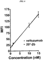
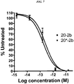

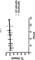
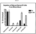


Classifications
-
- C—CHEMISTRY; METALLURGY
- C07—ORGANIC CHEMISTRY
- C07K—PEPTIDES
- C07K16/00—Immunoglobulins [IGs], e.g. monoclonal or polyclonal antibodies
- C07K16/18—Immunoglobulins [IGs], e.g. monoclonal or polyclonal antibodies against material from animals or humans
- C07K16/28—Immunoglobulins [IGs], e.g. monoclonal or polyclonal antibodies against material from animals or humans against receptors, cell surface antigens or cell surface determinants
- C07K16/30—Immunoglobulins [IGs], e.g. monoclonal or polyclonal antibodies against material from animals or humans against receptors, cell surface antigens or cell surface determinants from tumour cells
- C07K16/3076—Immunoglobulins [IGs], e.g. monoclonal or polyclonal antibodies against material from animals or humans against receptors, cell surface antigens or cell surface determinants from tumour cells against structure-related tumour-associated moieties
- C07K16/3092—Immunoglobulins [IGs], e.g. monoclonal or polyclonal antibodies against material from animals or humans against receptors, cell surface antigens or cell surface determinants from tumour cells against structure-related tumour-associated moieties against tumour-associated mucins
-
- A—HUMAN NECESSITIES
- A61—MEDICAL OR VETERINARY SCIENCE; HYGIENE
- A61K—PREPARATIONS FOR MEDICAL, DENTAL OR TOILETRY PURPOSES
- A61K45/00—Medicinal preparations containing active ingredients not provided for in groups A61K31/00 - A61K41/00
- A61K45/06—Mixtures of active ingredients without chemical characterisation, e.g. antiphlogistics and cardiaca
-
- A—HUMAN NECESSITIES
- A61—MEDICAL OR VETERINARY SCIENCE; HYGIENE
- A61K—PREPARATIONS FOR MEDICAL, DENTAL OR TOILETRY PURPOSES
- A61K47/00—Medicinal preparations characterised by the non-active ingredients used, e.g. carriers or inert additives; Targeting or modifying agents chemically bound to the active ingredient
- A61K47/50—Medicinal preparations characterised by the non-active ingredients used, e.g. carriers or inert additives; Targeting or modifying agents chemically bound to the active ingredient the non-active ingredient being chemically bound to the active ingredient, e.g. polymer-drug conjugates
- A61K47/51—Medicinal preparations characterised by the non-active ingredients used, e.g. carriers or inert additives; Targeting or modifying agents chemically bound to the active ingredient the non-active ingredient being chemically bound to the active ingredient, e.g. polymer-drug conjugates the non-active ingredient being a modifying agent
- A61K47/68—Medicinal preparations characterised by the non-active ingredients used, e.g. carriers or inert additives; Targeting or modifying agents chemically bound to the active ingredient the non-active ingredient being chemically bound to the active ingredient, e.g. polymer-drug conjugates the non-active ingredient being a modifying agent the modifying agent being an antibody, an immunoglobulin or a fragment thereof, e.g. an Fc-fragment
- A61K47/6801—Drug-antibody or immunoglobulin conjugates defined by the pharmacologically or therapeutically active agent
- A61K47/6803—Drugs conjugated to an antibody or immunoglobulin, e.g. cisplatin-antibody conjugates
- A61K47/6811—Drugs conjugated to an antibody or immunoglobulin, e.g. cisplatin-antibody conjugates the drug being a protein or peptide, e.g. transferrin or bleomycin
- A61K47/6813—Drugs conjugated to an antibody or immunoglobulin, e.g. cisplatin-antibody conjugates the drug being a protein or peptide, e.g. transferrin or bleomycin the drug being a peptidic cytokine, e.g. an interleukin or interferon
-
- A—HUMAN NECESSITIES
- A61—MEDICAL OR VETERINARY SCIENCE; HYGIENE
- A61K—PREPARATIONS FOR MEDICAL, DENTAL OR TOILETRY PURPOSES
- A61K47/00—Medicinal preparations characterised by the non-active ingredients used, e.g. carriers or inert additives; Targeting or modifying agents chemically bound to the active ingredient
- A61K47/50—Medicinal preparations characterised by the non-active ingredients used, e.g. carriers or inert additives; Targeting or modifying agents chemically bound to the active ingredient the non-active ingredient being chemically bound to the active ingredient, e.g. polymer-drug conjugates
- A61K47/51—Medicinal preparations characterised by the non-active ingredients used, e.g. carriers or inert additives; Targeting or modifying agents chemically bound to the active ingredient the non-active ingredient being chemically bound to the active ingredient, e.g. polymer-drug conjugates the non-active ingredient being a modifying agent
- A61K47/68—Medicinal preparations characterised by the non-active ingredients used, e.g. carriers or inert additives; Targeting or modifying agents chemically bound to the active ingredient the non-active ingredient being chemically bound to the active ingredient, e.g. polymer-drug conjugates the non-active ingredient being a modifying agent the modifying agent being an antibody, an immunoglobulin or a fragment thereof, e.g. an Fc-fragment
- A61K47/6801—Drug-antibody or immunoglobulin conjugates defined by the pharmacologically or therapeutically active agent
- A61K47/6803—Drugs conjugated to an antibody or immunoglobulin, e.g. cisplatin-antibody conjugates
- A61K47/6811—Drugs conjugated to an antibody or immunoglobulin, e.g. cisplatin-antibody conjugates the drug being a protein or peptide, e.g. transferrin or bleomycin
- A61K47/6815—Enzymes
-
- A—HUMAN NECESSITIES
- A61—MEDICAL OR VETERINARY SCIENCE; HYGIENE
- A61K—PREPARATIONS FOR MEDICAL, DENTAL OR TOILETRY PURPOSES
- A61K47/00—Medicinal preparations characterised by the non-active ingredients used, e.g. carriers or inert additives; Targeting or modifying agents chemically bound to the active ingredient
- A61K47/50—Medicinal preparations characterised by the non-active ingredients used, e.g. carriers or inert additives; Targeting or modifying agents chemically bound to the active ingredient the non-active ingredient being chemically bound to the active ingredient, e.g. polymer-drug conjugates
- A61K47/51—Medicinal preparations characterised by the non-active ingredients used, e.g. carriers or inert additives; Targeting or modifying agents chemically bound to the active ingredient the non-active ingredient being chemically bound to the active ingredient, e.g. polymer-drug conjugates the non-active ingredient being a modifying agent
- A61K47/68—Medicinal preparations characterised by the non-active ingredients used, e.g. carriers or inert additives; Targeting or modifying agents chemically bound to the active ingredient the non-active ingredient being chemically bound to the active ingredient, e.g. polymer-drug conjugates the non-active ingredient being a modifying agent the modifying agent being an antibody, an immunoglobulin or a fragment thereof, e.g. an Fc-fragment
- A61K47/6801—Drug-antibody or immunoglobulin conjugates defined by the pharmacologically or therapeutically active agent
- A61K47/6803—Drugs conjugated to an antibody or immunoglobulin, e.g. cisplatin-antibody conjugates
- A61K47/6811—Drugs conjugated to an antibody or immunoglobulin, e.g. cisplatin-antibody conjugates the drug being a protein or peptide, e.g. transferrin or bleomycin
- A61K47/6817—Toxins
- A61K47/6829—Bacterial toxins, e.g. diphteria toxins or Pseudomonas exotoxin A
-
- A—HUMAN NECESSITIES
- A61—MEDICAL OR VETERINARY SCIENCE; HYGIENE
- A61K—PREPARATIONS FOR MEDICAL, DENTAL OR TOILETRY PURPOSES
- A61K47/00—Medicinal preparations characterised by the non-active ingredients used, e.g. carriers or inert additives; Targeting or modifying agents chemically bound to the active ingredient
- A61K47/50—Medicinal preparations characterised by the non-active ingredients used, e.g. carriers or inert additives; Targeting or modifying agents chemically bound to the active ingredient the non-active ingredient being chemically bound to the active ingredient, e.g. polymer-drug conjugates
- A61K47/51—Medicinal preparations characterised by the non-active ingredients used, e.g. carriers or inert additives; Targeting or modifying agents chemically bound to the active ingredient the non-active ingredient being chemically bound to the active ingredient, e.g. polymer-drug conjugates the non-active ingredient being a modifying agent
- A61K47/68—Medicinal preparations characterised by the non-active ingredients used, e.g. carriers or inert additives; Targeting or modifying agents chemically bound to the active ingredient the non-active ingredient being chemically bound to the active ingredient, e.g. polymer-drug conjugates the non-active ingredient being a modifying agent the modifying agent being an antibody, an immunoglobulin or a fragment thereof, e.g. an Fc-fragment
- A61K47/6835—Medicinal preparations characterised by the non-active ingredients used, e.g. carriers or inert additives; Targeting or modifying agents chemically bound to the active ingredient the non-active ingredient being chemically bound to the active ingredient, e.g. polymer-drug conjugates the non-active ingredient being a modifying agent the modifying agent being an antibody, an immunoglobulin or a fragment thereof, e.g. an Fc-fragment the modifying agent being an antibody or an immunoglobulin bearing at least one antigen-binding site
- A61K47/6849—Medicinal preparations characterised by the non-active ingredients used, e.g. carriers or inert additives; Targeting or modifying agents chemically bound to the active ingredient the non-active ingredient being chemically bound to the active ingredient, e.g. polymer-drug conjugates the non-active ingredient being a modifying agent the modifying agent being an antibody, an immunoglobulin or a fragment thereof, e.g. an Fc-fragment the modifying agent being an antibody or an immunoglobulin bearing at least one antigen-binding site the antibody targeting a receptor, a cell surface antigen or a cell surface determinant
-
- A—HUMAN NECESSITIES
- A61—MEDICAL OR VETERINARY SCIENCE; HYGIENE
- A61P—SPECIFIC THERAPEUTIC ACTIVITY OF CHEMICAL COMPOUNDS OR MEDICINAL PREPARATIONS
- A61P25/00—Drugs for disorders of the nervous system
-
- A—HUMAN NECESSITIES
- A61—MEDICAL OR VETERINARY SCIENCE; HYGIENE
- A61P—SPECIFIC THERAPEUTIC ACTIVITY OF CHEMICAL COMPOUNDS OR MEDICINAL PREPARATIONS
- A61P31/00—Antiinfectives, i.e. antibiotics, antiseptics, chemotherapeutics
-
- A—HUMAN NECESSITIES
- A61—MEDICAL OR VETERINARY SCIENCE; HYGIENE
- A61P—SPECIFIC THERAPEUTIC ACTIVITY OF CHEMICAL COMPOUNDS OR MEDICINAL PREPARATIONS
- A61P35/00—Antineoplastic agents
-
- A—HUMAN NECESSITIES
- A61—MEDICAL OR VETERINARY SCIENCE; HYGIENE
- A61P—SPECIFIC THERAPEUTIC ACTIVITY OF CHEMICAL COMPOUNDS OR MEDICINAL PREPARATIONS
- A61P37/00—Drugs for immunological or allergic disorders
-
- A—HUMAN NECESSITIES
- A61—MEDICAL OR VETERINARY SCIENCE; HYGIENE
- A61P—SPECIFIC THERAPEUTIC ACTIVITY OF CHEMICAL COMPOUNDS OR MEDICINAL PREPARATIONS
- A61P9/00—Drugs for disorders of the cardiovascular system
-
- C—CHEMISTRY; METALLURGY
- C07—ORGANIC CHEMISTRY
- C07K—PEPTIDES
- C07K14/00—Peptides having more than 20 amino acids; Gastrins; Somatostatins; Melanotropins; Derivatives thereof
- C07K14/435—Peptides having more than 20 amino acids; Gastrins; Somatostatins; Melanotropins; Derivatives thereof from animals; from humans
- C07K14/52—Cytokines; Lymphokines; Interferons
- C07K14/555—Interferons [IFN]
- C07K14/56—IFN-alpha
-
- C—CHEMISTRY; METALLURGY
- C07—ORGANIC CHEMISTRY
- C07K—PEPTIDES
- C07K16/00—Immunoglobulins [IGs], e.g. monoclonal or polyclonal antibodies
- C07K16/18—Immunoglobulins [IGs], e.g. monoclonal or polyclonal antibodies against material from animals or humans
-
- C—CHEMISTRY; METALLURGY
- C07—ORGANIC CHEMISTRY
- C07K—PEPTIDES
- C07K16/00—Immunoglobulins [IGs], e.g. monoclonal or polyclonal antibodies
- C07K16/18—Immunoglobulins [IGs], e.g. monoclonal or polyclonal antibodies against material from animals or humans
- C07K16/28—Immunoglobulins [IGs], e.g. monoclonal or polyclonal antibodies against material from animals or humans against receptors, cell surface antigens or cell surface determinants
- C07K16/2803—Immunoglobulins [IGs], e.g. monoclonal or polyclonal antibodies against material from animals or humans against receptors, cell surface antigens or cell surface determinants against the immunoglobulin superfamily
- C07K16/2833—Immunoglobulins [IGs], e.g. monoclonal or polyclonal antibodies against material from animals or humans against receptors, cell surface antigens or cell surface determinants against the immunoglobulin superfamily against MHC-molecules, e.g. HLA-molecules
-
- C—CHEMISTRY; METALLURGY
- C07—ORGANIC CHEMISTRY
- C07K—PEPTIDES
- C07K16/00—Immunoglobulins [IGs], e.g. monoclonal or polyclonal antibodies
- C07K16/18—Immunoglobulins [IGs], e.g. monoclonal or polyclonal antibodies against material from animals or humans
- C07K16/28—Immunoglobulins [IGs], e.g. monoclonal or polyclonal antibodies against material from animals or humans against receptors, cell surface antigens or cell surface determinants
- C07K16/2851—Immunoglobulins [IGs], e.g. monoclonal or polyclonal antibodies against material from animals or humans against receptors, cell surface antigens or cell surface determinants against the lectin superfamily, e.g. CD23, CD72
-
- C—CHEMISTRY; METALLURGY
- C07—ORGANIC CHEMISTRY
- C07K—PEPTIDES
- C07K16/00—Immunoglobulins [IGs], e.g. monoclonal or polyclonal antibodies
- C07K16/18—Immunoglobulins [IGs], e.g. monoclonal or polyclonal antibodies against material from animals or humans
- C07K16/28—Immunoglobulins [IGs], e.g. monoclonal or polyclonal antibodies against material from animals or humans against receptors, cell surface antigens or cell surface determinants
- C07K16/2863—Immunoglobulins [IGs], e.g. monoclonal or polyclonal antibodies against material from animals or humans against receptors, cell surface antigens or cell surface determinants against receptors for growth factors, growth regulators
-
- C—CHEMISTRY; METALLURGY
- C07—ORGANIC CHEMISTRY
- C07K—PEPTIDES
- C07K16/00—Immunoglobulins [IGs], e.g. monoclonal or polyclonal antibodies
- C07K16/18—Immunoglobulins [IGs], e.g. monoclonal or polyclonal antibodies against material from animals or humans
- C07K16/28—Immunoglobulins [IGs], e.g. monoclonal or polyclonal antibodies against material from animals or humans against receptors, cell surface antigens or cell surface determinants
- C07K16/2887—Immunoglobulins [IGs], e.g. monoclonal or polyclonal antibodies against material from animals or humans against receptors, cell surface antigens or cell surface determinants against CD20
-
- C—CHEMISTRY; METALLURGY
- C07—ORGANIC CHEMISTRY
- C07K—PEPTIDES
- C07K16/00—Immunoglobulins [IGs], e.g. monoclonal or polyclonal antibodies
- C07K16/18—Immunoglobulins [IGs], e.g. monoclonal or polyclonal antibodies against material from animals or humans
- C07K16/28—Immunoglobulins [IGs], e.g. monoclonal or polyclonal antibodies against material from animals or humans against receptors, cell surface antigens or cell surface determinants
- C07K16/30—Immunoglobulins [IGs], e.g. monoclonal or polyclonal antibodies against material from animals or humans against receptors, cell surface antigens or cell surface determinants from tumour cells
- C07K16/3007—Carcino-embryonic Antigens
-
- C—CHEMISTRY; METALLURGY
- C07—ORGANIC CHEMISTRY
- C07K—PEPTIDES
- C07K16/00—Immunoglobulins [IGs], e.g. monoclonal or polyclonal antibodies
- C07K16/44—Immunoglobulins [IGs], e.g. monoclonal or polyclonal antibodies against material not provided for elsewhere, e.g. haptens, metals, DNA, RNA, amino acids
-
- C—CHEMISTRY; METALLURGY
- C12—BIOCHEMISTRY; BEER; SPIRITS; WINE; VINEGAR; MICROBIOLOGY; ENZYMOLOGY; MUTATION OR GENETIC ENGINEERING
- C12N—MICROORGANISMS OR ENZYMES; COMPOSITIONS THEREOF; PROPAGATING, PRESERVING, OR MAINTAINING MICROORGANISMS; MUTATION OR GENETIC ENGINEERING; CULTURE MEDIA
- C12N15/00—Mutation or genetic engineering; DNA or RNA concerning genetic engineering, vectors, e.g. plasmids, or their isolation, preparation or purification; Use of hosts therefor
- C12N15/09—Recombinant DNA-technology
- C12N15/11—DNA or RNA fragments; Modified forms thereof; Non-coding nucleic acids having a biological activity
- C12N15/111—General methods applicable to biologically active non-coding nucleic acids
-
- C—CHEMISTRY; METALLURGY
- C12—BIOCHEMISTRY; BEER; SPIRITS; WINE; VINEGAR; MICROBIOLOGY; ENZYMOLOGY; MUTATION OR GENETIC ENGINEERING
- C12N—MICROORGANISMS OR ENZYMES; COMPOSITIONS THEREOF; PROPAGATING, PRESERVING, OR MAINTAINING MICROORGANISMS; MUTATION OR GENETIC ENGINEERING; CULTURE MEDIA
- C12N15/00—Mutation or genetic engineering; DNA or RNA concerning genetic engineering, vectors, e.g. plasmids, or their isolation, preparation or purification; Use of hosts therefor
- C12N15/09—Recombinant DNA-technology
- C12N15/11—DNA or RNA fragments; Modified forms thereof; Non-coding nucleic acids having a biological activity
- C12N15/113—Non-coding nucleic acids modulating the expression of genes, e.g. antisense oligonucleotides; Antisense DNA or RNA; Triplex- forming oligonucleotides; Catalytic nucleic acids, e.g. ribozymes; Nucleic acids used in co-suppression or gene silencing
-
- C—CHEMISTRY; METALLURGY
- C12—BIOCHEMISTRY; BEER; SPIRITS; WINE; VINEGAR; MICROBIOLOGY; ENZYMOLOGY; MUTATION OR GENETIC ENGINEERING
- C12N—MICROORGANISMS OR ENZYMES; COMPOSITIONS THEREOF; PROPAGATING, PRESERVING, OR MAINTAINING MICROORGANISMS; MUTATION OR GENETIC ENGINEERING; CULTURE MEDIA
- C12N9/00—Enzymes; Proenzymes; Compositions thereof; Processes for preparing, activating, inhibiting, separating or purifying enzymes
- C12N9/10—Transferases (2.)
- C12N9/12—Transferases (2.) transferring phosphorus containing groups, e.g. kinases (2.7)
-
- C—CHEMISTRY; METALLURGY
- C12—BIOCHEMISTRY; BEER; SPIRITS; WINE; VINEGAR; MICROBIOLOGY; ENZYMOLOGY; MUTATION OR GENETIC ENGINEERING
- C12N—MICROORGANISMS OR ENZYMES; COMPOSITIONS THEREOF; PROPAGATING, PRESERVING, OR MAINTAINING MICROORGANISMS; MUTATION OR GENETIC ENGINEERING; CULTURE MEDIA
- C12N9/00—Enzymes; Proenzymes; Compositions thereof; Processes for preparing, activating, inhibiting, separating or purifying enzymes
- C12N9/14—Hydrolases (3)
- C12N9/16—Hydrolases (3) acting on ester bonds (3.1)
- C12N9/22—Ribonucleases RNAses, DNAses
-
- C—CHEMISTRY; METALLURGY
- C12—BIOCHEMISTRY; BEER; SPIRITS; WINE; VINEGAR; MICROBIOLOGY; ENZYMOLOGY; MUTATION OR GENETIC ENGINEERING
- C12Y—ENZYMES
- C12Y207/00—Transferases transferring phosphorus-containing groups (2.7)
- C12Y207/11—Protein-serine/threonine kinases (2.7.11)
- C12Y207/11011—Protein-serine/threonine kinases (2.7.11) cAMP-dependent protein kinase (2.7.11.11)
-
- C—CHEMISTRY; METALLURGY
- C12—BIOCHEMISTRY; BEER; SPIRITS; WINE; VINEGAR; MICROBIOLOGY; ENZYMOLOGY; MUTATION OR GENETIC ENGINEERING
- C12Y—ENZYMES
- C12Y301/00—Hydrolases acting on ester bonds (3.1)
- C12Y301/27—Endoribonucleases producing 3'-phosphomonoesters (3.1.27)
-
- A—HUMAN NECESSITIES
- A61—MEDICAL OR VETERINARY SCIENCE; HYGIENE
- A61K—PREPARATIONS FOR MEDICAL, DENTAL OR TOILETRY PURPOSES
- A61K39/00—Medicinal preparations containing antigens or antibodies
- A61K2039/505—Medicinal preparations containing antigens or antibodies comprising antibodies
-
- C—CHEMISTRY; METALLURGY
- C07—ORGANIC CHEMISTRY
- C07K—PEPTIDES
- C07K2317/00—Immunoglobulins specific features
- C07K2317/20—Immunoglobulins specific features characterized by taxonomic origin
- C07K2317/24—Immunoglobulins specific features characterized by taxonomic origin containing regions, domains or residues from different species, e.g. chimeric, humanized or veneered
-
- C—CHEMISTRY; METALLURGY
- C07—ORGANIC CHEMISTRY
- C07K—PEPTIDES
- C07K2317/00—Immunoglobulins specific features
- C07K2317/30—Immunoglobulins specific features characterized by aspects of specificity or valency
-
- C—CHEMISTRY; METALLURGY
- C07—ORGANIC CHEMISTRY
- C07K—PEPTIDES
- C07K2317/00—Immunoglobulins specific features
- C07K2317/30—Immunoglobulins specific features characterized by aspects of specificity or valency
- C07K2317/32—Immunoglobulins specific features characterized by aspects of specificity or valency specific for a neo-epitope on a complex, e.g. antibody-antigen or ligand-receptor
-
- C—CHEMISTRY; METALLURGY
- C07—ORGANIC CHEMISTRY
- C07K—PEPTIDES
- C07K2317/00—Immunoglobulins specific features
- C07K2317/50—Immunoglobulins specific features characterized by immunoglobulin fragments
- C07K2317/52—Constant or Fc region; Isotype
- C07K2317/526—CH3 domain
-
- C—CHEMISTRY; METALLURGY
- C07—ORGANIC CHEMISTRY
- C07K—PEPTIDES
- C07K2317/00—Immunoglobulins specific features
- C07K2317/50—Immunoglobulins specific features characterized by immunoglobulin fragments
- C07K2317/55—Fab or Fab'
-
- C—CHEMISTRY; METALLURGY
- C07—ORGANIC CHEMISTRY
- C07K—PEPTIDES
- C07K2317/00—Immunoglobulins specific features
- C07K2317/50—Immunoglobulins specific features characterized by immunoglobulin fragments
- C07K2317/56—Immunoglobulins specific features characterized by immunoglobulin fragments variable (Fv) region, i.e. VH and/or VL
- C07K2317/569—Single domain, e.g. dAb, sdAb, VHH, VNAR or nanobody®
-
- C—CHEMISTRY; METALLURGY
- C07—ORGANIC CHEMISTRY
- C07K—PEPTIDES
- C07K2317/00—Immunoglobulins specific features
- C07K2317/60—Immunoglobulins specific features characterized by non-natural combinations of immunoglobulin fragments
- C07K2317/62—Immunoglobulins specific features characterized by non-natural combinations of immunoglobulin fragments comprising only variable region components
- C07K2317/622—Single chain antibody (scFv)
-
- C—CHEMISTRY; METALLURGY
- C07—ORGANIC CHEMISTRY
- C07K—PEPTIDES
- C07K2317/00—Immunoglobulins specific features
- C07K2317/70—Immunoglobulins specific features characterized by effect upon binding to a cell or to an antigen
- C07K2317/73—Inducing cell death, e.g. apoptosis, necrosis or inhibition of cell proliferation
- C07K2317/732—Antibody-dependent cellular cytotoxicity [ADCC]
-
- C—CHEMISTRY; METALLURGY
- C07—ORGANIC CHEMISTRY
- C07K—PEPTIDES
- C07K2317/00—Immunoglobulins specific features
- C07K2317/70—Immunoglobulins specific features characterized by effect upon binding to a cell or to an antigen
- C07K2317/73—Inducing cell death, e.g. apoptosis, necrosis or inhibition of cell proliferation
- C07K2317/734—Complement-dependent cytotoxicity [CDC]
-
- C—CHEMISTRY; METALLURGY
- C07—ORGANIC CHEMISTRY
- C07K—PEPTIDES
- C07K2317/00—Immunoglobulins specific features
- C07K2317/90—Immunoglobulins specific features characterized by (pharmaco)kinetic aspects or by stability of the immunoglobulin
- C07K2317/94—Stability, e.g. half-life, pH, temperature or enzyme-resistance
-
- C—CHEMISTRY; METALLURGY
- C07—ORGANIC CHEMISTRY
- C07K—PEPTIDES
- C07K2319/00—Fusion polypeptide
-
- C—CHEMISTRY; METALLURGY
- C12—BIOCHEMISTRY; BEER; SPIRITS; WINE; VINEGAR; MICROBIOLOGY; ENZYMOLOGY; MUTATION OR GENETIC ENGINEERING
- C12N—MICROORGANISMS OR ENZYMES; COMPOSITIONS THEREOF; PROPAGATING, PRESERVING, OR MAINTAINING MICROORGANISMS; MUTATION OR GENETIC ENGINEERING; CULTURE MEDIA
- C12N2310/00—Structure or type of the nucleic acid
- C12N2310/10—Type of nucleic acid
- C12N2310/14—Type of nucleic acid interfering N.A.
-
- C—CHEMISTRY; METALLURGY
- C12—BIOCHEMISTRY; BEER; SPIRITS; WINE; VINEGAR; MICROBIOLOGY; ENZYMOLOGY; MUTATION OR GENETIC ENGINEERING
- C12N—MICROORGANISMS OR ENZYMES; COMPOSITIONS THEREOF; PROPAGATING, PRESERVING, OR MAINTAINING MICROORGANISMS; MUTATION OR GENETIC ENGINEERING; CULTURE MEDIA
- C12N2320/00—Applications; Uses
- C12N2320/30—Special therapeutic applications
- C12N2320/32—Special delivery means, e.g. tissue-specific
Definitions
- the present invention is related to a fusion protein comprising an antibody and an anchoring domain (AD) moiety; a multimeric complex comprising a first fusion protein and a second fusion protein, wherein the first fusion protein comprises a first antibody and an anchoring domain (AD) moiety and the second fusion protein comprises an effector moiety and a dimerization and docking domain (DDD) moiety; said multimeric complex for use in treating a disease; and a composition comprising said fusion protein or said multimeric complex.
- AD anchoring domain
- DDD dimerization and docking domain
- AD and/or DDD moieties may be attached to an effector by chemical cross-linking or other known means.
- the effectors may be selected from antibodies, antigen-binding antibody fragments, antigens, cytokines, chemokines, interleukins, interferons, growth factors, pro-apoptotic agents, anti-angiogenic agents, toxins, ligand-binding proteins, enzymes, therapeutic agents or polymers such as polyethylene glycol (PEG).
- At least one effector is an antibody or antigen-binding antibody fragment, with an AD moiety attached to the C-terminal end of each light chain of the antibody or antibody fragment. More preferably, at least one effector is an IgG antibody. In certain embodiments, all of the effectors may be antibodies or antibody fragments, providing bispecific or multispecific antigen-binding complexes.
- the subject multimeric complexes are of use for treating a wide variety of diseases and medical conditions, such as cancer, autoimmune disease, immune system dysfunction, graft-versus-host disease, organ transplant rejection, neurologic disease, metabolic disease, infectious disease or cardiovascular disease.
- a significant aspect of recent biomedical research is the development of increasingly sophisticated antibody-based biologics, such as bispecific antibodies, immunocytokines, antibody-toxin conjugates and antibody-drug conjugates.
- Development of more complex, and less natural, fusion proteins faces problems with yield, stability, immunogenicity and pharmacokinetics (Pk).
- immunoconjugates based on antibody fragments including single-chain Fv (scFv), Fab, or other Fc-lacking formats ( Kontermann, 2010, Curr Opin Mol Ther 12:176-83 ), are often difficult to produce with homogeneity and sufficient yield, lack Fc-effector functions, and inherently suffer from short circulating serum half-lives (T 1/2 ).
- immunoconjugates of IgG can be produced in high yields, with longer T 1/2 and in-vivo stability.
- intact monoclonal antibodies mAbs
- ADCC antibody-dependent cell-mediated cytotoxicity
- CDC complement-dependent cytotoxicity
- the enhanced Pk of IgG is attributed to two major factors. Its larger molecular size ( ⁇ 150 kDa) precludes renal clearance, which is responsible for the rapid elimination of smaller constructs ( ⁇ 60 kDa), such as scFv, and its dynamic binding to the neonatal Fc receptor (FcRn) ( Kue & Aveson, 2011, MAbs 3:422-30 ) extends T 1/2 .
- Multispecific or bispecific antibodies are useful in a number of biomedical applications.
- a bispecific antibody with binding sites for a tumor cell surface antigen and for a T-cell surface receptor can direct the lysis of specific tumor cells by T cells.
- Bispecific antibodies recognizing gliomas and the CD3 epitope on T cells have been successfully used in treating brain tumors in human patients ( Nitta, et al. Lancet. 1990; 355:368-371 ). Numerous methods to produce bispecific antibodies are known (see, e.g. U.S. Patent No. 7,405,320 ).
- Bispecific antibodies can be produced by the quadroma method, which involves the fusion of two different hybridomas, each producing a monoclonal antibody recognizing a different antigenic site ( Milstein and Cuello. Nature. 1983; 305:537-540 ).
- the fused hybridomas are capable of synthesizing two different heavy chains and two different light chains, which can associate randomly to give a heterogeneous population of 10 different antibody structures of which only one of them, amounting to 1/8 of the total antibody molecules, will be bispecific, and therefore must be further purified from the other forms.
- Fused hybridomas are often less stable cytogenetically than the parent hybridomas, making the generation of a production cell line more problematic.
- bispecific antibodies Another method for producing bispecific antibodies uses heterobifunctional crosslinkers to chemically tether two different monoclonal antibodies, so that the resulting hybrid conjugate will bind to two different targets ( Staerz, et al. Nature. 1985; 314:628-631 ; Perez, et al. Nature. 1985; 316:354-356 ).
- Bispecific antibodies generated by this approach are essentially heteroconjugates of two IgG molecules, which diffuse slowly into tissues and are rapidly removed from the circulation.
- Bispecific antibodies can also be produced by reduction of each of two parental monoclonal antibodies to the respective half molecules, which are then mixed and allowed to reoxidize to obtain the hybrid structure ( Staerz and Bevan. Proc Natl Acad Sci USA.
- Discrete V H and V L domains of antibodies produced by recombinant DNA technology may pair with each other to form a dimer (recombinant Fv fragment) with binding capability ( U.S. Pat. No. 4,642,334 ).
- a dimer recombinant Fv fragment
- binding capability U.S. Pat. No. 4,642,334
- Cognate V H and V L domains can be joined with a peptide linker of appropriate composition and length (usually consisting of more than 12 amino acid residues) to form a single-chain Fv (scFv) with binding activity.
- scFv single-chain Fv
- bsHexAbs Bispecific hexavalent antibodies based on veltuzumab (anti-CD20) and epratuzumab (anti-CD22) were constructed by combining a stabilized (Fab) 2 fused to a dimerization and docking domain (DDD) with an IgG containing an anchor domain (AD) appended at the C-terminus of each heavy chain (C H 3-AD2-IgG) ( Rossi et al., 2009, Blood 113, 6161-71 ).
- DDD dimerization and docking domain
- AD anchor domain
- Fc-bsHexAbs Compared to mixtures of their parental mAbs, these Fc-based bsHexAbs, referred to henceforth as "Fc-bsHexAbs", induced unique signaling events ( Gupta et al., 2010, Blood 116:3258-67 ), and exhibited potent cytotoxicity in vitro.
- the Fc-bsHexAbs were cleared from circulation of mice approximately twice as fast as the parental mAbs ( Rossi et al., 2009, Blood 113, 6161-71 ).
- the Fc-bsHexAbs are highly stable ex vivo, it is possible that some dissociation occurs in vivo, for example by intracellular processing. Further, the Fc-bsHexAbs lack CDC activity.
- Fc-based immunocytokines have also been assembled as DNLTM complexes, comprising two or four molecules of interferon-alpha 2b (IFN ⁇ 2b) fused to the C-terminal end of the C H 3-AD2-IgG Fc ( Rossi et al., 2009, Blood 114:3864-71 ; Rossi et al., 2010, Cancer Res 70:7600-09 ; Rossi et al., 2011, Blood 118:1877-84 ).
- the Fc-IgG-IFNa maintained high specific activity, approaching that of recombinant IFN ⁇ , and were remarkably potent in vitro and in vivo against non-Hodgkin lymphoma (NHL) xenografts.
- NHL non-Hodgkin lymphoma
- the T 1/2 of the Fc-IgG-IFN ⁇ in mice was longer than PEGylated IFN ⁇ , but half as long as the parental mAbs. Similar to the Fc-bsHexAbs, the Fc-IgG-IFN ⁇ dissociated in vivo over time and exhibited diminished CDC, but ADCC was enhanced.
- WO 2006/022225 A1 is related to methods and compositions for forming anti-cancer vaccine DNL complexes using dock-and-lock technology.
- WO 2006/032909 A2 is related to anchoring disruption molecules and related molecules which modulate the function of cAMP dependent protein kinase type II and related molecules which act as AKAP mimics and their use to produce pharmaceutical preparations to treat or prevent diseases which are typified by elevated or reduced PKA II activity such as in cardiovascular diseases and metabolic disorders.
- WO 2006/107786 A2 and US 2007/140966 A1 are related to methods and compositions for stably tethered structures for defined compositions with multiple functionalities and/or binding specificities.
- US 2009/0202487 A1 is related to methods and compositions of forming cytokine-antibody complexes using the dock-and-lock technology.
- US 2011/110854 A1 is related to compositions and methods of synthesis and use of 68 Ga, 18 F or 19 F labeled molecules in PET or MRI imaging.
- Rossi E A et al. ( Rossi E A et al., Bioconjugate Chemistry, vol. 23, no. 3, 21 March 2012 ) report on the dock-and-lock method which is combining recombinant engineering with site-specific covalent conjugation to generate multifunctional structures.
- a fusion protein comprising:
- the antibody binds to an antigen selected from the group consisting of AFP, ⁇ 4 integrin, B7, carbonic anhydrase IX, complement factors C1q, C1r, C1s, C2a, C2b, C3, C3a, C3b, C4, C4a, C4b, C5a, C5aR, C5b, C5, C6, C7, C8, C9n, CCCL19, CCCL21, CD1, CD1a, CD2, CD3, CD3R, CD4, CD5, CD8, CD11A, CD14, CD15, CD16, CD18, CD19, CD20, CD21, CD22, CD23, CD25, CD29, CD30, CD32b, CD33, CD37, CD38, CD40, CD40L, CD44, CD45, CD46, CD52, CD54, CD55, CD59, CD64, CD66a-e, CD67, CD70, CD74, CD79a, CD80, CD83, CD
- the antibody is selected from the group consisting of hR1 (anti-IGF-1R), hPAM4 (anti-mucin), KC4 (anti-mucin), hA20 (anti-CD20), hA19 (anti-CD19), hIMMU31 (anti-AFP), hLL1 (anti-CD74), hLL2 (anti-CD22), RFB4 (anti-CD22), hMu-9 (anti-CSAp), hL243 (anti-HLA-DR), hMN-14 (anti-CEACAM5), hMN-15 (anti-CEACAM6), hRS7 (anti-TROP-2), hMN-3 (anti-CEACAM6), CC49 (anti- TAG-72), J591 (anti-PSMA), D2/B (anti-PSMA), G250 (anti-carbonic anhydrase IX), infliximab (anti- TNF-a), certolizumab pegol (anti- TNF-
- one copy of the AD moiety binds to a dimer formed of two copies of a dimerization and docking domain (DDD) moiety, wherein the amino acid sequence of the DDD moiety is selected from the group consisting of residues 1-44 of RII ⁇ , 1-44 of RII ⁇ , 12-61 of RI ⁇ and 13-66 of RI ⁇ .
- DDD dimerization and docking domain
- the amino acid sequence of the DDD moiety is selected from the group consisting of SEQ ID NO:1, SEQ ID NO:2, SEQ ID NO:5, SEQ ID NO:6, SEQ ID NO:8, SEQ ID NO:9, SEQ ID NO:10, SEQ ID NO:11, SEQ ID NO:12, SEQ ID NO:13, SEQ ID NO:14, SEQ ID NO:15, SEQ ID NO:16, SEQ ID NO:17, SEQ ID NO:18, SEQ ID NO:19, SEQ ID NO:20, SEQ ID NO:22, SEQ ID NO:22, SEQ ID NO:23, SEQ ID NO:24, SEQ ID NO:25, SEQ ID NO:26, SEQ ID NO:27, SEQ ID NO:28, SEQ ID NO:29, SEQ ID NO:30, SEQ ID NO:31, SEQ ID NO:87 and SEQ ID NO:89.
- a multimeric complex comprising:
- the first and second antibodies bind to an antigen selected from the group consisting of AFP, ⁇ 4 integrin, B7, carbonic anhydrase IX, complement factors C1q, C1r, C1s, C2a, C2b, C3, C3a, C3b, C4, C4a, C4b, C5a, C5aR, C5b, C5, C6, C7, C8, C9n, CCCL19, CCCL21, CD1, CD1a, CD2, CD3, CD3R, CD4, CD5, CD8, CD11A, CD14, CD15, CD16, CD18, CD19, CD20, CD21, CD22, CD23, CD25, CD29, CD30, CD32b, CD33, CD37, CD38, CD40, CD40L, CD44, CD45, CD46, CD52, CD54, CD55, CD59, CD64, CD66a-e, CD67, CD70, CD74, CD79a, CD80, CD83
- the first and second antibodies are selected from the group consisting of hR1 (anti-IGF-1R), hPAM4 (anti-mucin), KC4 (anti-mucin), hA20 (anti-CD20), hA19 (anti-CD19), hIMMU31 (anti-AFP), hLL1 (anti-CD74), hLL2 (anti-CD22), RFB4 (anti-CD22), hMu-9 (anti-CSAp), hL243 (anti-HLA-DR), hMN-14 (anti-CEACAM5), hMN-15 (anti-CEACAM6), hRS7 (anti-TROP-2), hMN-3 (anti-CEACAM6), CC49 (anti- TAG-72), J591 (anti-PSMA), D2/B (anti-PSMA), G250 (anti-carbonic anhydrase IX), infliximab (anti- TNF- ⁇ ), certolizumab pegol (anti-
- the antigen-binding antibody fragment is selected from the group consisting of a F(ab') 2 , a F(ab) 2 , a Fab', a Fab, a Fv, a sFv, a scFv and a dAb.
- the amino acid sequence of the DDD moiety is selected from the group consisting of residues 1-44 of RII ⁇ , 1-44 of RII ⁇ , 12-61 of RI ⁇ and 13-66 of RI ⁇ .
- amino acid sequence of the AD moiety is selected from the group consisting of SEQ ID NO:3, SEQ ID NO:4, SEQ ID NO:7, SEQ ID NO:32, SEQ ID NO:33, SEQ ID NO:34, SEQ ID NO:35, SEQ ID NO:36, SEQ ID NO:37, SEQ ID NO:38, SEQ ID NO:39, SEQ ID NO:40, SEQ ID NO:41, SEQ ID NO:42, SEQ ID NO:43, SEQ ID NO:44, SEQ ID NO:45, SEQ ID NO:46, SEQ ID NO:47, SEQ ID NO:48, SEQ ID NO:49, SEQ ID NO:50, SEQ ID NO:51, SEQ ID NO:52, SEQ ID NO:53, SEQ ID NO:54, SEQ ID NO:55, SEQ ID NO:56, SEQ ID NO:57, SEQ ID NO:58, SEQ ID NO:59, SEQ ID NO:60, SEQ ID NO:61, SEQ ID NO:40, SEQ
- the toxin is selected from the group consisting of a bacterial toxin, a plant toxin, ricin, abrin, alpha toxin, saporin, ribonuclease (RNase), DNase I, Staphylococcal enterotoxin-A, pokeweed antiviral protein, gelonin, diphtheria toxin, Pseudomonas exotoxin, Pseudomonas endotoxin, ranpirnase (Rap) and Rap (N69Q).
- the cytokine is selected from the group consisting of human growth hormone, N-methionyl human growth hormone, bovine growth hormone, parathyroid hormone, thyroxin, insulin, proinsulin, relaxin, prorelaxin, follicle stimulating hormone (FSH), thyroid stimulating hormone (TSH), luteinizing hormone (LH), hepatic growth factor, prostaglandin, fibroblast growth factor, prolactin, placental lactogen, OB protein, tumor necrosis factor- ⁇ , tumor necrosis factor- ⁇ , mullerian-inhibiting substance, mouse gonadotropin-associated peptide, inhibin, activin, vascular endothelial growth factor, integrin, thrombopoietin (TPO), a nerve growth factor (NGF), NGF- ⁇ , platelet-growth factor, a transforming growth factors (TGF), TGF- ⁇ , TGF- ⁇ , insulin-like growth factor-I, insulin-like growth factor-II,
- the problem underlying the present invention is solved in a third aspect by the complex according to the second aspect, including any embodiment thereof, for use in treating a disease selected from the group consisting of cancer, an autoimmune disease, an immune system dysfunction, an infectious disease, a metabolic disease, a cardiovascular disease and a neurologic disease.
- a disease selected from the group consisting of cancer, an autoimmune disease, an immune system dysfunction, an infectious disease, a metabolic disease, a cardiovascular disease and a neurologic disease.
- the cancer is selected from the group consisting of non-Hodgkin's lymphoma, B cell lymphoma, B cell leukemia, T cell lymphoma, T cell leukemia, acute lymphoid leukemia, chronic lymphoid leukemia, Burkitt lymphoma, Hodgkin's lymphoma, hairy cell leukemia, acute myeloid leukemia, chronic myeloid leukemia, multiple myeloma, glioma, Waldenstrom's macroglobulinemia, carcinoma, melanoma, sarcoma, glioma, skin cancer, oral cavity cancer, gastrointestinal tract cancer, colon cancer, stomach cancer, pulmonary tract cancer, lung cancer, breast cancer, ovarian cancer, prostate cancer, uterine cancer, endometrial cancer, cervical cancer, urinary bladder cancer, pancreatic cancer, bone cancer, liver cancer, gall bladder cancer, kidney cancer, and testicular cancer.
- the autoimmune disease is selected from the group consisting of acute idiopathic thrombocytopenic purpura, chronic idiopathic thrombocytopenic purpura, dermatomyositis, Sydenham's chorea, myasthenia gravis, systemic lupus erythematosus, lupus nephritis, rheumatic fever, polyglandular syndromes, bullous pemphigoid, diabetes mellitus, Henoch-Schonlein purpura, post-streptococcal nephritis, erythema nodosum, Takayasu's arteritis, Addison's disease, rheumatoid arthritis, multiple sclerosis, sarcoidosis, ulcerative colitis, erythema multiforme, IgA nephropathy, polyarteritis nodosa, ankylosing spondylitis,
- composition comprising a fusion protein according to the first aspect, including any embodiment thereof, or a multimeric complex according to the second aspect, including any embodiment thereof.
- the present invention also concerns improved DNLTM complexes with longer T 1/2 , better pharmacokinetic properties and increased in vivo stability.
- the improved DNLTM complexes comprise IgG components in which AD moieties are fused to the C-terminal end of the antibody light chains.
- the subject complexes may be administered to a subject with a condition, for therapeutic and/or diagnostic purposes.
- a condition for therapeutic and/or diagnostic purposes.
- any condition that may be diagnosed and/or treated with a multifunctional, bivalent, trivalent, multispecific or bispecific complex may be treated with the subject compositions.
- Exemplary conditions include, but are not limited to, cancer, hyperplasia, neurodegenerative disease, Alzheimer's disease, cardiovascular disease, metabolic disease, vasculitis, viral infection, fungal infection, bacterial infection, diabetic retinopathy, macular degeneration, autoimmune disease, edema, pulmonary hypertension, sepsis, myocardial angiogenesis, plaque neovascularization, restenosis, neointima formation after vascular trauma, telangiectasia, hemophiliac joints, angiofibroma, fibrosis associated with chronic inflammation, lung fibrosis, deep venous thrombosis or wound granulation.
- the multimeric complex and the composition comprising the same may be of use to treat autoimmune disease, such as acute idiopathic thrombocytopenic purpura, chronic idiopathic thrombocytopenic purpura, dermatomyositis, Sydenham's chorea, myasthenia gravis, systemic lupus erythematosus, lupus nephritis, rheumatic fever, polyglandular syndromes, bullous pemphigoid, type 1 diabetes, type 2 diabetes, Henoch-Schonlein purpura, post-streptococcalnephritis, erythema nodosurn, Takayasu's arteritis, Addison's disease, rheumatoid arthritis, multiple sclerosis, sarcoidosis, ulcerative colitis, erythema multiforme, IgA nephropathy, polyarteritis nodosa,
- the complexes and compositions may be of use for therapeutic treatment of cancer. It is anticipated that any type of tumor and any type of tumor antigen may be targeted. Exemplary types of cancers that may be targeted include acute lymphoblastic leukemia, acute myelogenous leukemia, biliary cancer, breast cancer, cervical cancer, chronic lymphocytic leukemia, chronic myelogenous leukemia, colorectal cancer, endometrial cancer, esophageal, gastric, head and neck cancer, Hodgkin's lymphoma, lung cancer, medullary thyroid cancer, non-Hodgkin's lymphoma, multiple myeloma, renal cancer, ovarian cancer, pancreatic cancer, glioma, melanoma, liver cancer, prostate cancer, and urinary bladder cancer.
- Tumor-associated antigens that may be targeted include, but are not limited to, carbonic anhydrase IX, CCCL19, CCCL21, CSAp, CD1, CD1a, CD2, CD3, CD4, CD5, CD8, CD11A, CD14, CD15, CD16, CD18, CD19, IGF-1R, CD20, CD21, CD22, CD23, CD25, CD29, CD30, CD32b, CD33, CD37, CD38, CD40, CD40L, CD45, CD46, CD52, CD54, CD55, CD59, CD64, CD66a-e, CD67, CD70, CD74, CD79a, CD80, CD83, CD95, CD126, CD133, CD138, CD147, CD154, AFP, PSMA, CEACAM5, CEACAM-6, CSAp, B7, ED-B of fibronectin, Factor H, FHL-1, Flt-3, folate receptor, GROB, HMGB-1, hypoxia inducible factor (HIF
- Exemplary antibodies that may be utilized include, but are not limited to, hR1 (anti-IGF-1R, U.S. Patent Application Serial No. 12/722,645 , filed 3/12/10), hPAM4 (anti-mucin, U.S. Patent No. 7,282,567 ), hA20 (anti-CD20, U.S. Patent No. 7,251,164 ), hA19 (anti-CD19, U.S. Patent No. 7,109,304 ), hIMMU31 (anti-AFP, U.S. Patent No. 7,300,655 ), hLL1 (anti-CD74, U.S. Patent No. 7,312,318 ), hLL2 (anti-CD22, U.S. Patent No.
- the subject complexes may be of use to treat subjects infected with pathogenic organisms, such as bacteria, viruses or fungi.
- pathogenic organisms such as bacteria, viruses or fungi.
- fungi include Microsporum, Trichophyton, Epidermophyton, Sporothrix schenckii, Cryptococcus neoformans, Coccidioides immitis, Histoplasma capsulatum, Blastomyces dermatitidis or Candida albican.
- viruses include human immunodeficiency virus (HIV), herpes virus, cytomegalovirus, rabies virus, influenza virus, human papilloma virus, hepatitis B virus, hepatitis C virus, Sendai virus, feline leukemia virus, Reo virus, polio virus, human serum parvo-like virus, simian virus 40, respiratory syncytial virus, mouse mammary tumor virus, Varicella-Zoster virus, Dengue virus, rubella virus, measles virus, adenovirus, human T-cell leukemia viruses, Epstein-Barr virus, murine leukemia virus, mumps virus, vesicular stomatitis virus, Sindbis virus, lymphocytic choriomeningitis virus or blue tongue virus.
- HCV human immunodeficiency virus
- herpes virus cytomegalovirus
- rabies virus influenza virus
- human papilloma virus hepatitis B virus
- Exemplary bacteria include Bacillus anthracis, Streptococcus agalactiae, Legionella pneumophilia, Streptococcus pyogenes, Escherichia coli, Neisseria gonorrhoeae, Neisseria meningitidis, Pneumococcus spp., Hemophilis influenzae B, Treponema pallidum, Lyme disease spirochetes, Pseudomonas aeruginosa, Mycobacterium leprae, Brucella abortus, Mycobacterium tuberculosis or a Mycoplasma.
- Complexes of use for infection may comprise, for example, binding sites for one or more antigenic determinant on a pathogen, and may be conjugated or attached to a therapeutic agent for the pathogen, for example an anti-viral, antibiotic or anti-fungal agent.
- a stably tethered conjugate may comprise a first binding site for a pathogen antigen and a second binding site for a hapten or carrier that is attached to one or more therapeutic agents.
- Therapeutic agents of use against infectious organisms that may be conjugated to, incorporated into or targeted to bind to the subject complexes include, but are not limited to, acyclovir, albendazole, amantadine, amikacin, amoxicillin, amphotericin B, ampicillin, aztreonam, azithromycin, bacitracin, bactrim, BATRAFEN®, bifonazole, carbenicillin, caspofungin, cefaclor, cefazolin, cephalosporins, cefepime, ceftriaxone, cefotaxime, chloramphenicol, cidofovir, CIPRO®, clarithromycin, clavulanic acid, clotrimazole, cloxacillin, doxycycline, econazole, erythrocycline, erythromycin, flagyl, fluconazole, flucytosine, foscamet, furazolidone, ganciclovir, gent
- the complexes may comprise one or more toxins, such as a bacterial toxin, a plant toxin, ricin, abrin, a ribonuclease (RNase), DNase I, Staphylococcal enterotoxin-A, pokeweed antiviral protein, gelonin, diphtheria toxin, Pseudomonas exotoxin, Pseudomonas endotoxin, onconase, ranpirnase (Rap), Rap (N69Q) or PE38.
- toxins such as a bacterial toxin, a plant toxin, ricin, abrin, a ribonuclease (RNase), DNase I, Staphylococcal enterotoxin-A, pokeweed antiviral protein, gelonin, diphtheria toxin, Pseudomonas exotoxin, Pseudomonas endotoxin, oncona
- the complex may comprise an IgG antibody attached to two AD moieties and four copies of a toxin, such as ranpirnase, attached to DDD moieties.
- a toxin such as ranpirnase
- the subject complexes may also comprise an immunomodulator selected from the group consisting of a cytokine, a chemokine, a stem cell growth factor, a lymphotoxin, an hematopoietic factor, a colony stimulating factor (CSF), an interferon, erythropoietin, thrombopoietin, tumor necrosis factor- ⁇ (TNF), TNF- ⁇ , granulocyte-colony stimulating factor (G-CSF), granulocyte macrophage-colony stimulating factor (GM-CSF), interferon- ⁇ , interferon- ⁇ , interferon- ⁇ , interferon- ⁇ , interferon- ⁇ , stem cell growth factor designated "S1 factor", human growth hormone, N-methionyl human growth hormone, bovine growth hormone, parathyroid hormone, thyroxine, insulin, proinsulin, relaxin, prorelaxin, follicle stimulating hormone (FSH), thyroid stimulating hormone (TSH), luteinizing hormone (L
- the complex may comprise an antibody or antibody fragment attached to two AD moieties and four copies of an immunomodulator, such as a cytokine, each attached to a DDD moiety.
- an immunomodulator such as a cytokine
- Cytokine complexes are disclosed, for example, in U.S. Patent Nos. 7,906,118 and 8,034,3522 .
- Chemokines of use that may be incorporated in a subject complex include, but are not limited to, RANTES, MCAF, MIP1-alpha, MIP1-beta and IT-10.
- Anti-angiogenic agents of use that may be incorporated in a subject complex include, but are not limited to, angiostatin, baculostatin, canstatin, maspin, anti-VEGF antibodies or peptides, anti-placental growth factor antibodies or peptides, anti-Flk-1 antibodies, anti-Fit-1 antibodies or peptides, laminin peptides, fibronectin peptides, plasminogen activator inhibitors, tissue metalloproteinase inhibitors, interferons, interleukin 12, IP-10, Gro- ⁇ , thrombospondin, 2-methoxyoestradiol, proliferin-related protein, carboxiamidotriazole, CM101, Marimastat, pentosan polysulphate, angiopoietin 2, interferon-alpha, herbimycin A, PNU145156E, 16K prolactin fragment, Linomide, thalidomide, pentoxifylline, genistein,
- one or more therapeutic agents such as aplidin, azaribine, anastrozole, azacytidine, bleomycin, bortezomib, bryostatin-1, busulfan, calicheamycin, camptothecin, 10-hydroxycamptothecin, carmustine, celebrex, chlorambucil, cisplatin, irinotecan (CPT-11), SN-38, carboplatin, cladribine, cyclophosphamide, cytarabine, dacarbazine, docetaxel, dactinomycin, daunomycin glucuronide, daunorubicin, dexamethasone, diethylstilbestrol, doxorubicin, 2-pyrrolinodoxorubicine (2P-DOX), cyano-morpholino doxorubicin, doxorubicin glucuronide, epirubicin glucuronide, ethin
- aplidin
- the complex may comprise a first antibody or fragment thereof that binds to an antigen associated with a diseased cell, tissue or pathogen, while a second antibody or fragment thereof may bind to a hapten on a targetable construct.
- a targetable construct may be added to bind to the localized complex.
- a clearing agent may be administered to clear non-localized complexes from circulation before administration of the targetable construct.
- An exemplary targetable construct may have a structure of X-Phe-Lys(HSG)-D-Tyr-Lys(HSG)-Lys(Y)-NH 2 , where the compound includes a hard acid cation chelator at X or Y, and a soft acid cation chelator at remaining X or Y; and wherein the compound further comprises at least one diagnostic or therapeutic cation, and/or one or more chelated or chemically bound therapeutic agent, diagnostic agent, or enzyme.
- Such structures may comprise a first and/or second precursor with binding affinity for an antigen selected from the group consisting of CD2, CD3, CD8, CD10, CD21, CD23, CD24, CD25, CD30, CD33, CD37, CD38, CD40, CD48, CD52, CD55, CD59, CD70, CD74, CD80, CD86, CD138, CD147, HLA-DR, CEA, CSAp, CA-125, TAG-72, EFGR, HER2, HER3, HER4, IGF-1R, c-Met, PDGFR, MUC1, MUC2, MUC3, MUC4, TNFR1, TNFR2, NGFR, Fas (CD95), DR3, DR4, DR5, DR6, VEGF, PIGF, ED-B fibronectin, tenascin, PS
- a complex of use to induce apoptosis may comprise monoclonal antibodies, Fab fragments, chimeric, humanized or human antibodies or fragments.
- the complex may comprise combinations of anti-CD74 X anti-CD20, anti-CD74 X anti-CD22, anti-CD22 X anti-CD20, anti-CD20 X anti-HLA-DR, anti-CD 19 X anti-CD20, anti-CD20 X anti-CD80, anti-CD2 X anti-CD25, anti-CD8 X anti-CD25, and anti-CD2 X anti-CD147.
- the chimeric, humanized or human antibodies or antibody fragments may be derived from the variable domains of LL2 (anti-CD22), LL1 (anti-CD74) or A20 (anti-CD20).
- Various embodiments may concern complexes and their use for treating inflammatory and immune-dysregulatory diseases, infectious diseases, pathologic angiogenesis or cancer.
- the complexes bind to two different targets selected from the group consisting of (A) proinflammatory effectors of the innate immune system, (B) coagulation factors, (C) complement factors and complement regulatory proteins, and (D) targets specifically associated with an inflammatory or immune-dysregulatory disorder or with a pathologic angiogenesis or cancer, wherein the latter target is not (A), (B), or (C). At least one of the targets is (A), (B) or (C). Suitable combinations of targets are described in U.S. Provisional Application No. 60/634,076, filed Dec. 8, 2004 , entitled "Methods and Compositions for Immunotherapy and Detection of Inflammatory and Immune-Dysregulatory Disease, Infectious Disease, Pathologic Angiogenesis and Cancer".
- the proinflammatory effector of the innate immune system to which the binding molecules may bind may be a proinflammatory effector cytokine, a proinflammatory effector chemokine or a proinflammatory effector receptor.
- Suitable proinflammatory effector cytokines include MIF, HMGB-1 (high mobility group box protein 1), TNF-a, IL-1, IL-4, IL-5, IL-6, IL-8, IL-12, IL-15, and IL-18.
- Examples of proinflammatory effector chemokines include CCL19, CCL21, IL-8, MCP-1, RANTES, MIP-1A, MIP-1B, ENA-78, MCP-1, IP-10, GROB, and Eotaxin.
- Proinflammatory effector receptors include IL-4R (interleukin-4 receptor), IL-6R (interleukin-6 receptor), IL-13R (interleukin-13 receptor), IL-15R (interleukin-15 receptor) and IL-18R (interleukin-18 receptor).
- the complex also may react with at least one coagulation factor, particularly tissue factor (TF) or thrombin.
- the complex reacts with at least one complement factor or complement regulatory protein.
- the complement factor is selected from the group consisting of C3, C5, C3a, C3b, and C5a.
- the complement regulatory protein preferably is selected from the group consisting of CD46, CD55, CD59 and mCRP.
- any therapeutic protein or peptide known in the art may be attached to an AD or DDD sequence and used as an effector in the claimed methods and compositions.
- a large number of such therapeutic proteins or peptides are known, and are described for example, in U.S. Patent Application Publication No. 20060084794 , "Albumin fusion proteins," filed Nov. 2, 2005.
- Table 1 of 20060084794 which lists various known exemplary therapeutic proteins or peptides of use, including exemplary identifiers, patent reference numbers and preferred indications. Additional therapeutic proteins or peptides of use are disclosed, for example, in U.S. Pat. No.
- 6,309,633 may include but are not limited to adrenocorticotropic hormone, ebiratide, angiotensin, angiotensin II, asparaginase, atrial natriuretic peptides, atrial sodium diuretic peptides, bacitracin, beta-endorphins, blood coagulation factors VII, VIII and IX, blood thymic factor, bone morphogenic factor, bone morphogenic protein, bradykinin, caerulein, calcitonin gene related polypeptide, calcitonins, CCK-8, cell growth factors, EGF, TGF-alpha, TGF-beta, acidic FGF, basic FGF, chemokines, cholecystokinin, cholecystokinin-8, cholecystokinin-pancreozymin, colistin, colony-stimulating factors, GMCSF, MCSF, corticotropin-releasing factor, cytok
- the complexes may comprise an antibody or fragment thereof that binds to an antigen on an antigen-presenting cell (APC), such as a dendritic cell (DC) antigen, attached to a xenoantigen or a mutagenized or chemically modified antigen (see, e.g., U.S. Patent No. 7,901,680 , USSN 12/754,740 ).
- APC antigen-presenting cell
- DC dendritic cell
- Such complexes are of use to induce an immune response against the selected target antigen, for example a tumor-associated antigen.
- Exemplary DC antigens that may be targeted include, but are not limited to, CD209 (DC-SIGN), CD34, CD74, CD205, TLR 2 (toll-like receptor 2), TLR 4, TLR 7, TLR 9, BDCA-2, BDCA-3, BDCA-4, and HLA-DR.
- the DC targeting antibody or fragment thereof may be attached to a pathogen-associated antigen to induce an immune response against pathogens responsible for infectious disease.
- An exemplary embodiment is disclosed for pox-virus associated antigens in U.S. Patent Application Serial No. 12/915,515 .
- the DNLTM complex may comprise an antibody or antigen-binding antibody fragment attached to an oligonucleotide carrier moiety, such as a protamine, dendrimer or other polymer, of use to deliver interference RNA species such as siRNA.
- an oligonucleotide carrier moiety such as a protamine, dendrimer or other polymer
- RNA species such as siRNA.
- Exemplary complexes of use for such purposes are disclosed in U.S. Patent Application Serial No. 12/964,021 , filed 12/9/10 .
- a “therapeutic agent” is an atom, molecule, or compound that is useful in the treatment of a disease.
- therapeutic agents include antibodies, antibody fragments, peptides, drugs, toxins, enzymes, nucleases, hormones, immunomodulators, antisense oligonucleotides, small interfering RNA (siRNA), chelators, boron compounds, photoactive agents, dyes, and radioisotopes.
- a “diagnostic agent” is an atom, molecule, or compound that is useful in diagnosing a disease.
- useful diagnostic agents include, but are not limited to, radioisotopes, dyes (such as with the biotin-streptavidin complex), contrast agents, fluorescent compounds or molecules, and enhancing agents (e.g., paramagnetic ions) for magnetic resonance imaging (MRI).
- an “antibody” as used herein refers to a full-length (i.e., naturally occurring or formed by normal immunoglobulin gene fragment recombinatorial processes) immunoglobulin molecule (e.g., an IgG antibody) or an immunologically active (i.e., specifically binding) portion of an immunoglobulin molecule, like an antibody fragment.
- An “antibody” includes monoclonal, polyclonal, bispecific, multispecific, murine, chimeric, humanized and human antibodies.
- naked antibody is an antibody or antigen binding fragment thereof that is not attached to a therapeutic or diagnostic agent.
- the Fc portion of an intact naked antibody can provide effector functions, such as complement fixation and ADCC (see, e.g., Markrides, Pharmacol Rev 50:59-87, 1998 ).
- Other mechanisms by which naked antibodies induce cell death may include apoptosis. ( Vaswani and Hamilton, Ann Allergy Asthma Immunol 81: 105-119, 1998 .)
- antibody fragment is a portion of an intact antibody such as F(ab') 2 , F(ab) 2 , Fab', Fab, Fv, sFv, scFv, dAb and the like. Regardless of structure, an antibody fragment binds with the same antigen that is recognized by the full-length antibody.
- antibody fragments include isolated fragments consisting of the variable regions, such as the "Fv” fragments consisting of the variable regions of the heavy and light chains or recombinant single chain polypeptide molecules in which light and heavy variable regions are connected by a peptide linker ("scFv proteins").
- Single-chain antibodies often abbreviated as “scFv” consist of a polypeptide chain that comprises both a V H and a V L domain which interact to form an antigen- binding site.
- the V H and V L domains are usually linked by a peptide of 1 to 25 amino acid residues.
- Antibody fragments also include diabodies, triabodies and single domain antibodies (dAb).
- a “chimeric antibody” is a recombinant protein that contains the variable domains including the complementarity determining regions (CDRs) of an antibody derived from one species, preferably a rodent antibody, while the constant domains of the antibody molecule are derived from those of a human antibody.
- the constant domains of the chimeric antibody may be derived from that of other species, such as a cat or dog.
- a “humanized antibody” is a recombinant protein in which the CDRs from an antibody from one species; e.g., a rodent antibody, are transferred from the heavy and light variable chains of the rodent antibody into human heavy and light variable domains, including human framework region (FR) sequences.
- the constant domains of the antibody molecule are derived from those of a human antibody.
- a "human antibody” is an antibody obtained from transgenic mice that have been genetically engineered to produce specific human antibodies in response to antigenic challenge.
- elements of the human heavy and light chain locus are introduced into strains of mice derived from embryonic stem cell lines that contain targeted disruptions of the endogenous heavy chain and light chain loci.
- the transgenic mice can synthesize human antibodies specific for human antigens, and the mice can be used to produce human antibody-secreting hybridomas.
- Methods for obtaining human antibodies from transgenic mice are described by Green et al., Nature Genet. 7:13 (1994 ), Lonberg et al., Nature 368:856 (1994 ), and Taylor et al., Int. Immun. 6:579 (1994 ).
- a fully human antibody also can be constructed by genetic or chromosomal transfection methods, as well as phage display technology, all of which are known in the art. (See, e.g., McCafferty et al., Nature 348:552-553 (1990 ) for the production of human antibodies and fragments thereof in vitro, from immunoglobulin variable domain gene repertoires from unimmunized donors).
- antibody variable domain genes are cloned in-frame into either a major or minor coat protein gene of a filamentous bacteriophage, and displayed as functional antibody fragments on the surface of the phage particle.
- the filamentous particle contains a single-stranded DNA copy of the phage genome, selections based on the functional properties of the antibody also result in selection of the gene encoding the antibody exhibiting those properties. In this way, the phage mimics some of the properties of the B cell.
- Phage display can be performed in a variety of formats, for their review, see, e.g. Johnson and Chiswell, Current Opinion in Structural Biology 3:5564-571 (1993 ).
- Human antibodies may also be generated by in vitro activated B cells. (See, U.S. Pat. Nos. 5,567,610 and 5,229,275 ).
- antibody fusion protein is a recombinantly produced antigen-binding molecule in which an antibody or antibody fragment is linked to another protein or peptide, such as the same or different antibody or antibody fragment or a DDD or AD peptide.
- the fusion protein may comprise a single antibody component, a multivalent or multispecific combination of different antibody components or multiple copies of the same antibody component.
- the fusion protein may additionally comprise an antibody or an antibody fragment and a therapeutic agent. Examples of therapeutic agents suitable for such fusion proteins include immunomodulators and toxins.
- One preferred toxin comprises a ribonuclease (RNase), preferably a recombinant RNase.
- a “multispecific antibody” is an antibody that can bind simultaneously to at least two targets that are of different structure, e.g., two different antigens, two different epitopes on the same antigen, or a hapten and/or an antigen or epitope.
- a “multivalent antibody” is an antibody that can bind simultaneously to at least two targets that are of the same or different structure. Valency indicates how many binding arms or sites the antibody has to a single antigen or epitope; i.e., monovalent, bivalent, trivalent or multivalent. The multivalency of the antibody means that it can take advantage of multiple interactions in binding to an antigen, thus increasing the avidity of binding to the antigen.
- Specificity indicates how many antigens or epitopes an antibody is able to bind; i.e., monospecific, bispecific, trispecific, multispecific.
- a natural antibody e.g., an IgG
- Multispecific, multivalent antibodies are constructs that have more than one binding site of different specificity.
- bispecific antibody is an antibody that can bind simultaneously to two targets which are of different structure.
- Bispecific antibodies bsAb and bispecific antibody fragments (bsFab) may have at least one arm that specifically binds to, for example, a B cell, T cell, myeloid-, plasma-, and mast-cell antigen or epitope and at least one other arm that specifically binds to a targetable conjugate that bears a therapeutic or diagnostic agent.
- bsAb bispecific antibody fragments
- bsFab bispecific antibody fragments
- a variety of bispecific antibodies can be produced using molecular engineering.
- a "xenoantigen” is an antigen that is found in more than one species.
- the xenoantigen is from a species that is different from the subject.
- a xenoantigen that is of use in a human vaccine may be from a mouse, a rat, a rabbit or another non-human species.
- An antibody or immunotoxin preparation, or a composition described herein, is said to be administered in a "therapeutically effective amount" if the amount administered is physiologically significant.
- An agent is physiologically significant if its presence results in a detectable change in the physiology of a recipient subject.
- an antibody preparation is physiologically significant if its presence invokes an antitumor response or mitigates the signs and symptoms of an autoimmune disease state.
- a physiologically significant effect could also be the evocation of a humoral and/or cellular immune response in the recipient subject leading to growth inhibition or death of target cells.
- monoclonal antibodies can be obtained by injecting mice with a composition comprising an antigen, removing the spleen to obtain B-lymphocytes, fusing the B-lymphocytes with myeloma cells to produce hybridomas, cloning the hybridomas, selecting positive clones which produce antibodies to the antigen, culturing the clones that produce antibodies to the antigen, and isolating the antibodies from the hybridoma cultures.
- MAbs can be isolated and purified from hybridoma cultures by a variety of well-established techniques. Such isolation techniques include affinity chromatography with Protein-A Sepharose, size-exclusion chromatography, and ion-exchange chromatography. See, for example, Coligan at pages 2.7.1-2.7.12 and pages 2.9.1-2.9.3. Also, see Baines et al., "Purification of Immunoglobulin G (IgG)," in METHODS IN MOLECULAR BIOLOGY, VOL. 10, pages 79-104 (The Humana Press, Inc. 1992 ).
- the antibodies can be sequenced and subsequently prepared by recombinant techniques. Humanization and chimerization of murine antibodies and antibody fragments are well known to those skilled in the art. The use of antibody components derived from humanized, chimeric or human antibodies obviates potential problems associated with the immunogenicity of murine constant regions.
- a chimeric antibody is a recombinant protein in which the variable regions of a human antibody have been replaced by the variable regions of, for example, a mouse antibody, including the complementarity-determining regions (CDRs) of the mouse antibody.
- Chimeric antibodies exhibit decreased immunogenicity and increased stability when administered to a subject.
- CDRs complementarity-determining regions
- a chimeric or murine monoclonal antibody may be humanized by transferring the mouse CDRs from the heavy and light variable chains of the mouse immunoglobulin into the corresponding variable domains of a human antibody.
- the mouse framework regions (FR) in the chimeric monoclonal antibody are also replaced with human FR sequences.
- additional modification might be required in order to restore the original affinity of the murine antibody. This can be accomplished by the replacement of one or more human residues in the FR regions with their murine counterparts to obtain an antibody that possesses good binding affinity to its epitope.
- the phage display technique may be used to generate human antibodies (e.g., Dantas-Barbosa et al., 2005, Genet. Mol. Res. 4:126-40 ).
- Human antibodies may be generated from normal humans or from humans that exhibit a particular disease state, such as cancer (Dantas-Barbosa et al., 2005).
- the advantage to constructing human antibodies from a diseased individual is that the circulating antibody repertoire may be biased towards antibodies against disease-associated antigens.
- RNAs were converted to cDNAs and used to make Fab cDNA libraries using specific primers against the heavy and light chain immunoglobulin sequences ( Marks et al., 1991, J. Mol. Biol. 222:581-97 ).
- Phage display can be performed in a variety of formats, for their review, see e.g. Johnson and Chiswell, Current Opinion in Structural Biology 3:5564-571 (1993 ).
- Human antibodies may also be generated by in vitro activated B cells. See U.S. Patent Nos. 5,567,610 and 5,229,275 . The skilled artisan will realize that these techniques are exemplary and any known method for making and screening human antibodies or antibody fragments may be utilized.
- transgenic animals that have been genetically engineered to produce human antibodies may be used to generate antibodies against essentially any immunogenic target, using standard immunization protocols.
- Methods for obtaining human antibodies from transgenic mice are disclosed by Green et al., Nature Genet. 7:13 (1994 ), Lonberg et al., Nature 368:856 (1994 ), and Taylor et al., Int. Immun. 6:579 (1994 ).
- a non-limiting example of such a system is the XenoMouse® (e.g., Green et al., 1999, J. Immunol. Methods 231:11-23 ) from Abgenix (Fremont, CA).
- the mouse antibody genes have been inactivated and replaced by functional human antibody genes, while the remainder of the mouse immune system remains intact.
- the XenoMouse® was transformed with germline-configured YACs (yeast artificial chromosomes) that contained portions of the human IgH and Igkappa loci, including the majority of the variable region sequences, along accessory genes and regulatory sequences.
- the human variable region repertoire may be used to generate antibody producing B cells, which may be processed into hybridomas by known techniques.
- a XenoMouse® immunized with a target antigen will produce human antibodies by the normal immune response, which may be harvested and/or produced by standard techniques discussed above.
- a variety of strains of XenoMouse® are available, each of which is capable of producing a different class of antibody.
- Transgenically produced human antibodies have been shown to have therapeutic potential, while retaining the pharmacokinetic properties of normal human antibodies (Green et al., 1999).
- the skilled artisan will realize that the claimed compositions and methods are not limited to use of the XenoMouse® system but may utilize any transgenic animal that has been genetically engineered to produce human antibodies.
- the V genes of an antibody from a cell that expresses a murine antibody can be cloned by PCR amplification and sequenced.
- the cloned V L and V H genes can be expressed in cell culture as a chimeric Ab as described by Orlandi et al., (Proc. Natl. Acad. Sci., USA, 86: 3833 (1989 )).
- a humanized antibody can then be designed and constructed as described by Leung et al. (Mol. Immunol., 32: 1413 (1995 )).
- cDNA can be prepared from any known hybridoma line or transfected cell line producing a murine antibody by general molecular cloning techniques ( Sambrook et al., Molecular Cloning, A laboratory manual, 2nd Ed (1989 )).
- the V ⁇ sequence for the antibody may be amplified using the primers VK1BACK and VK1FOR (Orlandi et al ., 1989) or the extended primer set described by Leung et al. (BioTechniques, 15: 286 (1993 )).
- V H sequences can be amplified using the primer pair VH1BACK/VH1FOR (Orlandi et al ., 1989) or the primers annealing to the constant region of murine IgG described by Leung et al. (Hybridoma, 13:469 (1994 )).
- Humanized V genes can be constructed by a combination of long oligonucleotide template syntheses and PCR amplification as described by Leung et al. (Mol. Immunol., 32: 1413 (1995 )).
- PCR products for V ⁇ can be subcloned into a staging vector, such as a pBR327-based staging vector, VKpBR, that contains an Ig promoter, a signal peptide sequence and convenient restriction sites.
- PCR products for V H can be subcloned into a similar staging vector, such as the pBluescript-based VHpBS.
- Expression cassettes containing the V ⁇ and V H sequences together with the promoter and signal peptide sequences can be excised from VKpBR and VHpBS and ligated into appropriate expression vectors, such as pKh and pG1g, respectively ( Leung et al., Hybridoma, 13:469 (1994 )).
- the expression vectors can be co-transfected into an appropriate cell and supernatant fluids monitored for production of a chimeric, humanized or human antibody.
- the V ⁇ and V H expression cassettes can be excised and subcloned into a single expression vector, such as pdHL2, as described by Gillies et al. (J. Immunol. Methods 125:191 (1989 ) and also shown in Losman et al., Cancer, 80:2660 (1997 )).
- expression vectors may be transfected into host cells that have been pre-adapted for transfection, growth and expression in serum-free medium.
- Exemplary cell lines that may be used include the Sp/EEE, Sp/ESF and Sp/ESF-X cell lines (see, e.g., U.S. Patent Nos. 7,531,327 ; 7,537,930 and 7,608,425 ). These exemplary cell lines are based on the Sp2/0 myeloma cell line, transfected with a mutant Bcl-EEE gene, exposed to methotrexate to amplify transfected gene sequences and pre-adapted to serum-free cell line for protein expression.
- Antibody fragments which recognize specific epitopes can be generated by known techniques.
- Antibody fragments are antigen binding portions of an antibody, such as F(ab') 2 , Fab', F(ab) 2 , Fab, Fv, sFv and the like.
- F(ab') 2 fragments can be produced by pepsin digestion of the antibody molecule and Fab' fragments can be generated by reducing disulfide bridges of the F(ab') 2 fragments.
- Fab' expression libraries can be constructed ( Huse et al., 1989, Science, 246:1274-1281 ) to allow rapid and easy identification of monoclonal Fab' fragments with the desired specificity.
- F(ab) 2 fragments may be generated by papain digestion of an antibody.
- a single chain Fv molecule comprises a VL domain and a VH domain.
- the VL and VH domains associate to form a target binding site.
- These two domains are further covalently linked by a peptide linker (L).
- L peptide linker
- DABs single domain antibodies
- Single domain antibodies may be obtained, for example, from camels, alpacas or llamas by standard immunization techniques. (See, e.g., Muyldermans et al., TIBS 26:230-235, 2001 ; Yau et al., J Immunol Methods 281:161-75, 2003 ; Maass et al., J Immunol Methods 324:13-25, 2007 ).
- the VHH may have potent antigen-binding capacity and can interact with novel epitopes that are inacessible to conventional VH-VL pairs.
- Alpaca serum IgG contains about 50% camelid heavy chain only IgG antibodies (HCAbs) (Maass et al., 2007).
- Alpacas may be immunized with known antigens, such as TNF- ⁇ , and VHHs can be isolated that bind to and neutralize the target antigen (Maass et al., 2007).
- PCR primers that amplify virtually all alpaca VHH coding sequences have been identified and may be used to construct alpaca VHH phage display libraries, which can be used for antibody fragment isolation by standard biopanning techniques well known in the art (Maass et al., 2007).
- anti-pancreatic cancer VHH antibody fragments may be utilized in the claimed compositions and methods.
- An antibody fragment can be prepared by proteolytic hydrolysis of the full length antibody or by expression in E. coli or another host of the DNA coding for the fragment.
- An antibody fragment can be obtained by pepsin or papain digestion of full length antibodies by conventional methods. These methods are described, for example, by Goldenberg, U.S. Patent Nos. 4,036,945 and 4,331,647 and references contained therein. Also, see Nisonoff et al., Arch Biochem. Biophys. 89: 230 (1960 ); Porter, Biochem. J. 73: 119 (1959 ), Edelman et al., in METHODS IN ENZYMOLOGY VOL. 1, page 422 (Academic Press 1967 ), and Coligan at pages 2.8.1-2.8.10 and 2.10.-2.10.4.
- Immunogenicity of therapeutic antibodies is associated with increased risk of infusion reactions and decreased duration of therapeutic response ( Baert et al., 2003, N Engl J Med 348:602-08 ).
- the extent to which therapeutic antibodies induce an immune response in the host may be determined in part by the allotype of the antibody ( Stickler et al., 2011, Genes and Immunity 12:213-21 ).
- Antibody allotype is related to amino acid sequence variations at specific locations in the constant region sequences of the antibody.
- the allotypes of IgG antibodies containing a heavy chain ⁇ -type constant region are designated as Gm allotypes ( 1976, J Immunol 117:1056-59 ).
- G1m1 For the common IgG1 human antibodies, the most prevalent allotype is G1m1 ( Stickler et al., 2011, Genes and Immunity 12:213-21 ). However, the G1m3 allotype also occurs frequently in Caucasians (Stickler et al., 2011). It has been reported that G1m1 antibodies contain allotypic sequences that tend to induce an immune response when administered to non-G1m1 (nG1m1) recipients, such as G1m3 patients (Stickler et al., 2011). Non-Glml allotype antibodies are not as immunogenic when administered to G1m1 patients (Stickler et al., 2011).
- the human G1m1 allotype comprises the amino acids aspartic acid at Kabat position 356 and leucine at Kabat position 358 in the CH3 sequence of the heavy chain IgG1.
- the nG1m1 allotype comprises the amino acids glutamic acid at Kabat position 356 and methionine at Kabat position 358.
- Both G1m1 and nG1m1 allotypes comprise a glutamic acid residue at Kabat position 357 and the allotypes are sometimes referred to as DEL and EEM allotypes.
- a non-limiting example of the heavy chain constant region sequences for G1m1 and nG1m1 allotype antibodies is shown for the exemplary antibodies rituximab (SEQ ID NO:85) and veltuzumab (SEQ ID NO:86).
- veltuzumab and rituximab are, respectively, humanized and chimeric IgG1 antibodies against CD20, of use for therapy of a wide variety of hematological malignancies and/or autoimmune diseases.
- Table 1 compares the allotype sequences of rituximab vs. veltuzumab.
- rituximab (G1m17,1) is a DEL allotype IgG1, with an additional sequence variation at Kabat position 214 (heavy chain CH1) of lysine in rituximab vs. arginine in veltuzumab.
- veltuzumab is less immunogenic in subjects than rituximab (see, e.g., Morchhauser et al., 2009, J Clin Oncol 27:3346-53 ; Goldenberg et al., 2009, Blood 113:1062-70 ; Robak & Robak, 2011, BioDrugs 25:13-25 ), an effect that has been attributed to the difference between humanized and chimeric antibodies.
- the difference in allotypes between the EEM and DEL allotypes likely also accounts for the lower immunogenicity of veltuzumab. Table 1. Allotypes of Rituximab vs.
- Veltuzumab Heavy chain position and associated allotypes Complete allotype 214 (allotype) 356 / 358 (allotype) 431 (allotype) Rituximab G1m17,1 K 17 D / L 1 A - Veltuzumab G1m3 R 3 E / M - A -
- the allotype of the antibody In order to reduce the immunogenicity of therapeutic antibodies in individuals of nG1m1 genotype, it is desirable to select the allotype of the antibody to correspond to the G1m3 allotype, characterized by arginine at Kabat 214, and the nG1m1,2 null-allotype, characterized by glutamic acid at Kabat position 356, methionine at Kabat position 358 and alanine at Kabat position 431. Surprisingly, it was found that repeated subcutaneous administration of G1m3 antibodies over a long period of time did not result in a significant immune response.
- the human IgG4 heavy chain in common with the G1m3 allotype has arginine at Kabat 214, glutamic acid at Kabat 356, methionine at Kabat 359 and alanine at Kabat 431. Since immunogenicity appears to relate at least in part to the residues at those locations, use of the human IgG4 heavy chain constant region sequence for therapeutic antibodies is also a preferred embodiment. Combinations of G1m3 IgG1 antibodies with IgG4 antibodies may also be of use for therapeutic administration.
- the claimed methods and compositions may utilize any of a variety of antibodies known in the art.
- Antibodies of use may be commercially obtained from a number of known sources.
- a variety of antibody secreting hybridoma lines are available from the American Type Culture Collection (ATCC, Manassas, VA).
- ATCC American Type Culture Collection
- VA Manassas
- a large number of antibodies against various disease targets, including but not limited to tumor-associated antigens, have been deposited at the ATCC and/or have published variable region sequences and are available for use in the claimed methods and compositions. See, e.g., U.S. Patent Nos.
- antibody sequences or antibody-secreting hybridomas against almost any disease-associated antigen may be obtained by a simple search of the ATCC, NCBI and/or USPTO databases for antibodies against a selected disease-associated target of interest.
- the antigen binding domains of the cloned antibodies may be amplified, excised, ligated into an expression vector, transfected into an adapted host cell and used for protein production, using standard techniques well known in the art (see, e.g., U.S. Patent Nos. 7,531,327 ; 7,537,930 ; 7,608,425 and 7,785,880 ).
- antibodies that may be of use for therapy of cancer within the scope of the claimed methods and compositions include, but are not limited to, LL1 (anti-CD74), LL2 and RFB4 (anti-CD22), RS7 (anti-epithelial glycoprotein-1 (EGP-1)), PAM4 and KC4 (both anti-mucin), MN-14 (anti-carcinoembryonic antigen (CEA, also known as CD66e), Mu-9 (anti-colon-specific antigen-p), Immu 31 (an anti-alpha-fetoprotein), TAG-72 (e.g., CC49), Tn, J591 or HuJ591 (anti-PSMA (prostate-specific membrane antigen)), AB-PG1-XG1-026 (anti-PSMA dimer), D2/B (anti-PSMA), G250 (anti-carbonic anhydrase IX), hL243 (anti-HLA-DR), alemtuzumab (anti-CD52), bevacizumab (anti
- Such antibodies are known in the art (e.g., U.S. Patent Nos. 5,686,072 ; 5,874,540 ; 6,107,090 ; 6,183,744 ; 6,306,393 ; 6,653,104 ; 6,730.300 ; 6,899,864 ; 6,926,893 ; 6,962,702 ; 7,074,403 ; 7,230,084 ; 7,238,785 ; 7,238,786 ; 7,256,004 ; 7,282,567 ; 7,300,655 ; 7,312,318 ; 7,585,491 ; 7,612,180 ; 7,642,239 ; and U.S. Patent Application Publ. No.
- hPAM4 U.S. Patent No. 7,282,567
- hA20 U.S. Patent No. 7,251,164
- hA19 U.S. Patent No. 7,109,304
- hIMMU31 U.S. Patent No. 7,300,655
- hLL1 U.S. Patent No. 7,312,318
- hLL2 U.S. Patent No. 7,074,403
- hMu-9 U.S. Patent No. 7,387,773
- hL243 U.S. Patent No.
- Anti-TNF- ⁇ antibodies are known in the art and may be of use to treat immune diseases, such as autoimmune disease, immune dysfunction (e.g., graft-versus-host disease, organ transplant rejection) or diabetes.
- Known antibodies against TNF- ⁇ include the human antibody CDP571 ( Ofei et al., 2011, Diabetes 45:881-85 ); murine antibodies MTNFAI, M2TNFAI, M3TNFAI, M3TNFABI, M302B and M303 (Thermo Scientific, Rockford, IL); infliximab (Centocor, Malvern, PA); certolizumab pegol (UCB, Brussels, Belgium); and adalimumab (Abbott, Abbott Park, IL).
- anti-B-cell antibodies such as veltuzumab, epratuzumab, milatuzumab or hL243; tocilizumab (anti-IL-6 receptor); basiliximab (anti-CD25); daclizumab (anti-CD25); efalizumab (anti-CDlla); muromonab-CD3 (anti-CD3 receptor); anti-CD40L (UCB, Brussels, Belgium); natalizumab (anti- ⁇ 4 integrin) and omalizumab (anti-IgE).
- anti-B-cell antibodies such as veltuzumab, epratuzumab, milatuzumab or hL243; tocilizumab (anti-IL-6 receptor); basiliximab (anti-CD25); daclizumab (anti-CD25); efalizumab (anti-CDlla); muromonab-CD3 (anti-CD3 receptor); anti-CD40L (UCB,
- Type-1 and Type-2 diabetes may be treated using known antibodies against B-cell antigens, such as CD22 (epratuzumab), CD74 (milatuzumab), CD19 (hA19), CD20 (veltuzumab) or HLA-DR (hL243) ( see, e.g., Winer et al., 2011, Nature Med 17:610-18 ).
- Anti-CD3 antibodies also have been proposed for therapy of type 1 diabetes ( Cernea et al., 2010, Diabetes Metab Rev 26:602-05 ).
- Macrophage migration inhibitory factor is an important regulator of innate and adaptive immunity and apoptosis. It has been reported that CD74 is the endogenous receptor for MIF ( Leng et al., 2003, J Exp Med 197:1467-76 ).
- the therapeutic effect of antagonistic anti-CD74 antibodies on MIF-mediated intracellular pathways may be of use for treatment of a broad range of disease states, such as cancers of the bladder, prostate, breast, lung, colon and chronic lymphocytic leukemia (e.g., Meyer-Siegler et al., 2004, BMC Cancer 12:34 ; Shachar & Haran, 2011, Leuk Lymphoma 52:1446-54 ); autoimmune diseases such as rheumatoid arthritis and systemic lupus erythematosus ( Morand & Leech, 2005, Front Biosci 10:12-22 ; Shachar & Haran, 2011, Leuk Lymphoma 52:1446-54 ); kidney diseases such as renal allograft rejection ( Lan, 2008, Nephron Exp Nephrol.
- Milatuzumab (hLL1) is an exemplary anti-CD74 antibody of therapeutic use for treatment of MIF-mediated diseases.
- the complement system is a complex cascade involving proteolytic cleavage of serum glycoproteins often activated by cell receptors.
- the "complement cascade” is constitutive and non-specific but it must be activated in order to function. Complement activation results in a unidirectional sequence of enzymatic and biochemical reactions.
- a specific complement protein, C5 forms two highly active, inflammatory byproducts, C5a and C5b, which jointly activate white blood cells. This in turn evokes a number of other inflammatory byproducts, including injurious cytokines, inflammatory enzymes, and cell adhesion molecules. Together, these byproducts can lead to the destruction of tissue seen in many inflammatory diseases.
- This cascade ultimately results in induction of the inflammatory response, phagocyte chemotaxis and opsonization, and cell lysis.
- the complement system can be activated via two distinct pathways, the classical pathway and the alternate pathway. Most of the complement components are numbered (e.g., C1, C2, C3, etc.) but some are referred to as "Factors.” Some of the components must be enzymatically cleaved to activate their function; others simply combine to form complexes that are active. Active components of the classical pathway include C1q, C1r, C1s, C2a, C2b, C3a, C3b, C4a, and C4b. Active components of the alternate pathway include C3a, C3b, Factor B, Factor Ba, Factor Bb, Factor D, and Properdin. The last stage of each pathway is the same, and involves component assembly into a membrane attack complex. Active components of the membrane attack complex include C5a, C5b, C6, C7, C8, and C9n.
- C3a, C4a and C5a cause mast cells to release chemotactic factors such as histamine and serotonin, which attract phagocytes, antibodies and complement, etc. These form one group of preferred targets.
- Another group of preferred targets includes C3b, C4b and C5b, which enhance phagocytosis of foreign cells.
- Another preferred group of targets are the predecessor components for these two groups, i.e., C3, C4 and C5.
- C5b, C6, C7, C8 and C9 induce lysis of foreign cells (membrane attack complex) and form yet another preferred group of targets.
- Complement C5a is an anaphylatoxin. It mediates inflammation and is a chemotactic attractant for induction of neutrophilic release of antimicrobial proteases and oxygen radicals. Therefore, C5a and its predecessor C5 are particularly preferred targets. By targeting C5, not only is C5a affected, but also C5b, which initiates assembly of the membrane-attack complex. Thus, C5 is another preferred target. C3b, and its predecessor C3, also are preferred targets, as both the classical and alternate complement pathways depend upon C3b. Three proteins affect the levels of this factor, C1 inhibitor, protein H and Factor I, and these are also preferred targets according to the invention. Complement regulatory proteins, such as CD46, CD55, and CD59, may be targets to which the antibody complexes bind.
- Coagulation factors also are preferred targets, particularly tissue factor and thrombin.
- Tissue factor is also known also as tissue thromboplastin, CD142, coagulation factor III, or factor III.
- Tissue factor is an integral membrane receptor glycoprotein and a member of the cytokine receptor superfamily.
- the ligand binding extracellular domain of tissue factor consists of two structural modules with features that are consistent with the classification of tissue factor as a member of type-2 cytokine receptors.
- Tissue factor is involved in the blood coagulation protease cascade and initiates both the extrinsic and intrinsic blood coagulation cascades by forming high affinity complexes between the extracellular domain of tissue factor and the circulating blood coagulation factors, serine proteases factor VII or factor VIIa. These enzymatically active complexes then activate factor IX and factor X, leading to thrombin generation and clot formation.
- Tissue factor is expressed by various cell types, including monocytes, macrophages and vascular endothelial cells, and is induced by IL-1, TNF- ⁇ or bacterial lipopolysaccharides.
- Protein kinase C is involved in cytokine activation of endothelial cell tissue factor expression. Induction of tissue factor by endotoxin and cytokines is an important mechanism for initiation of disseminated intravascular coagulation seen in patients with Gram-negative sepsis.
- Tissue factor also appears to be involved in a variety of non-hemostatic functions including inflammation, cancer, brain function, immune response, and tumor-associated angiogenesis.
- antibody complexes that target tissue factor are useful not only in the treatment of coagulopathies, but also in the treatment of sepsis, cancer, pathologic angiogenesis, and other immune and inflammatory dysregulatory diseases according to the invention.
- a complex interaction between the coagulation pathway and the cytokine network is suggested by the ability of several cytokines to influence tissue factor expression in a variety of cells and by the effects of ligand binding to the receptor.
- Ligand binding (factor VIIa) has been reported to give an intracellular calcium signal, thus indicating that tissue factor is a true receptor.
- Thrombin is the activated form of coagulation factor II (prothrombin); it converts fibrinogen to fibrin.
- Thrombin is a potent chemotaxin for macrophages, and can alter their production of cytokines and arachidonic acid metabolites. It is of particular importance in the coagulopathies that accompany sepsis. Numerous studies have documented the activation of the coagulation system either in septic patients or following LPS administration in animal models. Despite more than thirty years of research, the mechanisms of LPS-induced liver toxicity remain poorly understood. It is now clear that they involve a complex and sequential series of interactions between cellular and humoral mediators.
- the antibody complexes bind to a MHC class I, MHC class II or accessory molecule, such as CD40, CD54, CD80 or CD86.
- the antibody complex also may bind to a T-cell activation cytokine, or to a cytokine mediator, such as NF- ⁇ B.
- one of the two different targets may be a cancer cell receptor or cancer-associated antigen, particularly one that is selected from the group consisting of B-cell lineage antigens (CD19, CD20, CD21, CD22, CD23, etc.), VEGF, VEGFR, EGFR, carcinoembryonic antigen (CEA), placental growth factor (P1GF), tenascin, HER-2/ neu , EGP-1, EGP-2, CD25, CD30, CD33, CD38, CD40, CD45, CD52, CD74, CD80, CD138, NCA66, CEACAM1, CEACAM6 (carcinoembryonic antigen-related cellular adhesion molecule 6), MUC1, MUC2, MUC3, MUC4, MUC16, IL-6, ⁇ -fetoprotein (AFP), A3, CA125, colon-specific antigen-p (CSAp), folate receptor, HLA-DR, human chorionic gonadotropin (HCG), Ia, EL-2
- Targets associated with sepsis and immune dysregulation and other immune disorders include MIF, IL-1, IL-6, IL-8, CD74, CD83, and C5aR.
- Antibodies and inhibitors against C5aR have been found to improve survival in rodents with sepsis ( Huber-Lang et al., FASEB J 2002; 16:1567-1574 ; Riedemann et al., J Clin Invest 2002; 110:101-108 ) and septic shock and adult respiratory distress syndrome in monkeys ( Hangen et al., J Surg Res 1989; 46:195-199 ; Stevens et al., J Clin Invest 1986; 77:1812-1816 ).
- one of the two different targets preferably is a target that is associated with infection, such as LPS/C5a.
- Other preferred targets include HMGB-1, tissue factor, CD14, VEGF, and IL-6, each of which is associated with septicemia or septic shock.
- Preferred antibody complexes are those that target two or more targets from HMGB-1, tissue factor and MIF, such as MIF/tissue factor, and HMGB-1/tissue factor.
- one of the different targets may be a target that is associated with graft versus host disease or transplant rejection, such as MIF ( Lo et al., Bone Marrow Transplant, 30(6):375-80 (2002 )).
- One of the different targets also may be one that associated with acute respiratory distress syndrome, such as IL-8 ( Bouros et al., PMC Pulm Med, 4(1):6 (2004 ), atherosclerosis or restenosis, such as MIF ( Chen et al., Arterioscler Thromb Vasc Biol, 24(4):709-14 (2004 ), asthma, such as IL-18 ( Hata et al., Int Immunol, Oct.
- a granulomatous disease such as TNF- ⁇ ( Ulbricht et al., Arthritis Rheum, 50(8):2717-8 (2004 )
- a neuropathy such as carbamylated EPO (erythropoietin) ( Leist et al., Science 305(5681):164-5 (2004 )
- cachexia such as IL-6 and TNF- ⁇ .
- Targets include C5a, LPS, IFN-gamma, B7; CD2, CD4, CD14, CD18, CD11a, CD11b, CD11c, CD14, CD18, CD27, CD29, CD38, CD40L, CD52, CD64, CD83, CD147, CD154.
- Activation of mononuclear cells by certain microbial antigens, including LPS can be inhibited to some extent by antibodies to CD18, CD11b, or CD11c, which thus implicate ⁇ 2 -integrins ( Cuzzola et al., J Immunol 2000; 164:5871-5876 ; Medvedev et al., J Immunol 1998; 160: 4535-4542 ).
- CD83 has been found to play a role in giant cell arteritis (GCA), which is a systemic vasculitis that affects medium- and large-size arteries, predominately the extracranial branches of the aortic arch and of the aorta itself, resulting in vascular stenosis and subsequent tissue ischemia, and the severe complications of blindness, stroke and aortic arch syndrome ( Weyand and Goronzy, N Engl J Med 2003; 349:160-169 ; Hunder and Valente, In: Inflammatory Diseases of Blood Vessels. G.S. Hoffman and C.M. Weyand, eds, Marcel Dekker, New York, 2002; 255-265 ).
- GAA giant cell arteritis
- CD154 a member of the TNF family, is expressed on the surface of CD4-positive T-lymphocytes, and it has been reported that a humanized monoclonal antibody to CD154 produced significant clinical benefit in patients with active systemic lupus erythematosus (SLE) ( Grammar et al., J Clin Invest 2003; 112:1506-1520 ). It also suggests that this antibody might be useful in other autoimmune diseases ( Kelsoe, J Clin Invest 2003; 112:1480-1482 ). Indeed, this antibody was also reported as effective in patients with refractory immune thrombocytopenic purpura ( Kuwana et al., Blood 2004; 103:1229-1236 ).
- IL-1Ra interleukin-1 receptor antagonist
- An improvement in treatment of these patients, which hitherto required concomitant treatment with methotrexate, is to combine anakinra with one or more of the anti-proinflammatory effector cytokines or anti-proinflammatory effector chemokines (as listed above).
- the pharmaceutical composition of the present invention may be used to treat a subject having a metabolic disease, such amyloidosis, or a neurodegenerative disease, such as Alzheimer's disease.
- a metabolic disease such as amyloidosis
- a neurodegenerative disease such as Alzheimer's disease.
- Bapineuzumab is in clinical trials for Alzheimer's disease therapy.
- Other antibodies proposed for therapy of Alzheimer's disease include Alz 50 (Ksiezak- Reding et al., 1987, J Biol Chem 263:7943-47 ), gantenerumab, and solanezumab.
- Infliximab an anti-TNF-a antibody, has been reported to reduce amyloid plaques and improve cognition.
- diseases that may be treated using the invention as defined in the claims include cardiovascular diseases, such as fibrin clots, atherosclerosis, myocardial ischemia and infarction.
- cardiovascular diseases such as fibrin clots, atherosclerosis, myocardial ischemia and infarction.
- Antibodies to fibrin e.g., scFv(59D8); T2G1s; MH1 are known and in clinical trials as imaging agents for disclosing said clots and pulmonary emboli, while anti-granulocyte antibodies, such as MN-3, MN-15, anti-NCA95, and anti-CD15 antibodies, can target myocardial infarcts and myocardial ischemia.
- fibrin e.g., scFv(59D8); T2G1s; MH1
- anti-granulocyte antibodies such as MN-3, MN-15, anti-NCA95, and anti-CD15 antibodies
- Anti-macrophage, anti-low-density lipoprotein (LDL), anti-MIF, and anti-CD74 (e.g., hLL1) antibodies can be used to target atherosclerotic plaques.
- Abciximab anti-glycoprotein IIb/IIIa
- Anti-CD3 antibodies have been reported to reduce development and progression of atherosclerosis ( Steffens et al., 2006, Circulation 114:1977-84 ). Antibodies against oxidized LDL induced a regression of established atherosclerosis in a mouse model ( Ginsberg, 2007, J Am Coll Cardiol 52:2319-21 ). Anti-ICAM-1 antibody was shown to reduce ischemic cell damage after cerebral artery occlusion in rats ( Zhang et al., 1994, Neurology 44:1747-51 ).
- OKT anti-T-cell monoclonal antibodies available from Ortho Pharmaceutical Company which bind to normal T-lymphocytes; the monoclonal antibodies produced by the hybridomas having the ATCC accession numbers HB44, HB55, HB12, HB78 and HB2; G7EII, W8E7, NKP15 and GO22 (Becton Dickinson); NEN9.4 (New England Nuclear); and FMC11 (Sera Labs).
- a description of antibodies against fibrin and platelet antigens is contained in Knight, Semin. Nucl. Med., 20:52-67 (1990 ).
- antibodies against infectious disease agents such as bacteria, viruses, mycoplasms or other pathogens.
- Many antibodies against such infectious agents are known in the art and any such known antibody may be used in the claimed methods and compositions.
- antibodies against the gp120 glycoprotein antigen of human immunodeficiency virus I (HIV-1) are known, and certain of such antibodies can have an immunoprotective role in humans. See, e.g., Rossi et al., Proc. Natl. Acad. Sci. USA. 86:8055-8058, 1990 .
- Known anti-HIV antibodies include the anti-envelope antibody described by Johansson et al. (AIDS.
- Antibodies against malaria parasites can be directed against the sporozoite, merozoite, schizont and gametocyte stages. Monoclonal antibodies have been generated against sporozoites (cirumsporozoite antigen), and have been shown to neutralize sporozoites in vitro and in rodents ( N. Yoshida et al., Science 207:71-73, 1980 ). Several groups have developed antibodies to T. gondii, the protozoan parasite involved in toxoplasmosis ( Kasper et al., J. Immunol. 129:1694-1699, 1982 ; Id., 30:2407-2412, 1983).
- Antibodies have been developed against schistosomular surface antigens and have been found to act against schistosomulae in vivo or in vitro ( Simpson et al., Parasitology, 83:163-177, 1981 ; Smith et al., Parasitology, 84:83-91, 1982 : Gryzch et al., J. Immunol., 129:2739-2743, 1982 ; Zodda et al., J. Immunol. 129:2326-2328, 1982 ; Dissous et al., J. immunol., 129:2232-2234, 1982 )
- Trypanosoma cruzi is the causative agent of Chagas' disease, and is transmitted by blood-sucking reduviid insects.
- An antibody has been generated that specifically inhibits the differentiation of one form of the parasite to another (epimastigote to trypomastigote stage) in vitro, and which reacts with a cell-surface glycoprotein; however, this antigen is absent from the mammalian (bloodstream) forms of the parasite ( Sher et al., Nature, 300:639-640, 1982 ).
- Anti-fungal antibodies are known in the art, such as anti-Sclerotinia antibody ( U.S. Patent 7,910,702 ); antiglucuronoxylomannan antibody ( Zhong and Priofski, 1998, Clin Diag Lab Immunol 5:58-64 ); anti-Candida antibodies (Matthews and Burnie, 2001, 2:472-76); and anti-glycosphingolipid antibodies ( Toledo et al., 2010, BMC Microbiol 10:47 ).
- Suitable antibodies have been developed against most of the microorganism (bacteria, viruses, protozoa, fungi, other parasites) responsible for the majority of infections in humans, and many have been used previously for in vitro diagnostic purposes. These antibodies, and newer antibodies that can be generated by conventional methods, are appropriate for use in the present invention.
- the antibodies or fragments thereof may be conjugated to one or more therapeutic or diagnostic agents.
- the therapeutic agents do not need to be the same but can be different, e.g. a drug and a radioisotope.
- 131 I can be incorporated into a tyrosine of an antibody or fusion protein and a drug attached to an epsilon amino group of a lysine residue.
- Therapeutic and diagnostic agents also can be attached, for example to reduced SH groups and/or to carbohydrate side chains. Many methods for making covalent or non-covalent conjugates of therapeutic or diagnostic agents with antibodies or fusion proteins are known in the art and any such known method may be utilized.
- a therapeutic or diagnostic agent can be attached at the hinge region of a reduced antibody component via disulfide bond formation.
- such agents can be attached using a heterobifunctional cross-linker, such as N -succinyl 3-(2-pyridyldithio)propionate (SPDP). Yu et al., Int. J. Cancer 56: 244 (1994 ). General techniques for such conjugation are well-known in the art.
- the therapeutic or diagnostic agent can be conjugated via a carbohydrate moiety in the Fc region of the antibody.
- the carbohydrate group can be used to increase the loading of the same agent that is bound to a thiol group, or the carbohydrate moiety can be used to bind a different therapeutic or diagnostic agent.
- the Fc region may be absent if the antibody used as the antibody component of the immunoconjugate is an antibody fragment.
- a carbohydrate moiety into the light chain variable region of a full length antibody or antibody fragment. See, for example, Leung et al., J. Immunol. 154: 5919 (1995 ); Hansen et al., U.S. Patent No. 5,443,953 (1995 ), Leung et al., U.S. patent No. 6,254,868 .
- the engineered carbohydrate moiety is used to attach the therapeutic or diagnostic agent.
- a chelating agent may be attached to an antibody, antibody fragment or fusion protein and used to chelate a therapeutic or diagnostic agent, such as a radionuclide.
- exemplary chelators include but are not limited to DTPA (such as Mx-DTPA), DOTA, TETA, NETA or NOTA.
- Methods of conjugation and use of chelating agents to attach metals or other ligands to proteins are well known in the art (see, e.g., U.S. Patent No. 7,563,433 ).
- radioactive metals or paramagnetic ions may be attached to proteins or peptides by reaction with a reagent having a long tail, to which may be attached a multiplicity of chelating groups for binding ions.
- a tail can be a polymer such as a polylysine, polysaccharide, or other derivatized or derivatizable chains having pendant groups to which can be bound chelating groups such as, e.g., ethylenediaminetetraacetic acid (EDTA), diethylenetriaminepentaacetic acid (DTPA), porphyrins, polyamines, crown ethers, bis-thiosemicarbazones, polyoximes, and like groups known to be useful for this purpose.
- EDTA ethylenediaminetetraacetic acid
- DTPA diethylenetriaminepentaacetic acid
- porphyrins polyamines, crown ethers, bis-thiosemicarbazones, polyoximes, and like groups known to be useful for
- Chelates may be directly linked to antibodies or peptides, for example as disclosed in U.S. Patent 4,824,659 .
- Particularly useful metal-chelate combinations include 2-benzyl-DTPA and its monomethyl and cyclohexyl analogs, used with diagnostic isotopes in the general energy range of 60 to 4,000 keV, such as 125 I, 131 I, 123 I, 124 I, 62 Cu, 64 Cu, 18 F, 111 In, 67 Ga, 68 Ga, 99m Tc, 94m Tc, 11 C, 13 N, 15 O, 76 Br, for radioimaging.
- the same chelates, when complexed with non-radioactive metals, such as manganese, iron and gadolinium are useful for MRI.
- Macrocyclic chelates such as NOTA, DOTA, and TETA are of use with a variety of metals and radiometals, most particularly with radionuclides of gallium, yttrium and copper, respectively. Such metal-chelate complexes can be made very stable by tailoring the ring size to the metal of interest.
- Other ring-type chelates such as macrocyclic polyethers, which are of interest for stably binding nuclides, such as 223 Ra for RAIT are encompassed.
- 18 F-labeling of use in PET scanning techniques for example by reaction of F-18 with a metal or other atom, such as aluminum.
- the 18 F-Al conjugate may be complexed with chelating groups, such as DOTA, NOTA or NETA that are attached directly to antibodies or used to label targetable constructs in pre-targeting methods.
- chelating groups such as DOTA, NOTA or NETA that are attached directly to antibodies or used to label targetable constructs in pre-targeting methods.
- Such F-18 labeling techniques are disclosed in U.S. Patent No. 7,563,433 .
- therapeutic agents may be administered by a pretargeting method, utilizing bispecific or multispecific antibody complexes.
- the bispecific or multispecific antibody comprises at least one binding arm that binds to an antigen exhibited by a targeted cell or tissue, while at least one other binding arm binds to a hapten on a targetable construct.
- the targetable construct comprises one or more haptens and one or more therapeutic and/or diagnostic agents.
- Pre-targeting is a multistep process originally developed to resolve the slow blood clearance of directly targeting antibodies, which contributes to undesirable toxicity to normal tissues such as bone marrow.
- a radionuclide or other diagnostic or therapeutic agent is attached to a small delivery molecule (targetable construct) that is cleared within minutes from the blood.
- Pre-targeting methods are disclosed, for example, in Goodwin et al., U.S. Pat. No. 4,863,713 ; Goodwin et al., J. Nucl. Med. 29:226, 1988 ; Hnatowich et al., J. Nucl. Med. 28:1294, 1987 ; Oehr et al., J. Nucl. Med. 29:728, 1988 ; Klibanov et al., J. Nucl. Med. 29:1951 , 1988; Sinitsyn et al., J. Nucl. Med. 30:66, 1989 ; Kalofonos et al., J. Nucl. Med.
- a pre-targeting method of treating or diagnosing a disease or disorder in a subject may be provided by: (1) administering to the subject an antibody complex comprising a bispecific antibody or antibody fragment; (2) optionally administering to the subject a clearing composition, and allowing the composition to clear the antibody from circulation; and (3) administering to the subject the targetable construct, containing one or more chelated or chemically bound therapeutic or diagnostic agents.
- a bivalent or multivalent antibody or an antibody complexed to one or more effectors is formed as a DOCK-AND-LOCKTM (DNLTM) complex (see, e.g., U.S. Patent Nos.
- the technique takes advantage of the specific and high-affinity binding interactions that occur between a dimerization and docking domain (DDD) sequence of the regulatory (R) subunits of cAMP-dependent protein kinase (PKA) and an anchor domain (AD) sequence derived from any of a variety of AKAP proteins ( Baillie et al., FEBS Letters. 2005; 579: 3264 . Wong and Scott, Nat. Rev. Mol. Cell Biol. 2004; 5: 959 ).
- DDD dimerization and docking domain
- R regulatory subunits of cAMP-dependent protein kinase
- AD anchor domain
- the DDD and AD peptides may be attached to any protein, peptide or other molecule. Because the DDD sequences spontaneously dimerize and bind to the AD sequence, the technique allows the formation of complexes between any selected molecules that may be attached to DDD or AD sequences.
- the standard DNLTM complex comprises a trimer with two DDD-linked molecules attached to one AD-linked molecule
- variations in complex structure allow the formation of dimers, trimers, tetramers, pentamers, hexamers and other multimers.
- the DNLTM complex may comprise two or more antibodies, antibody fragments or fusion proteins which bind to the same antigenic determinant or to two or more different antigens.
- the DNLTM complex may also comprise one or more other effectors, such as proteins, peptides, immunomodulators, cytokines, interleukins, interferons, binding proteins, peptide ligands, carrier proteins, toxins, ribonucleases such as onconase, inhibitory oligonucleotides such as siRNA, antigens or xenoantigens, polymers such as PEG, enzymes, therapeutic agents, hormones, cytotoxic agents, anti-angiogenic agents, pro-apoptotic agents or any other molecule or aggregate.
- effectors such as proteins, peptides, immunomodulators, cytokines, interleukins, interferons, binding proteins, peptide ligands, carrier proteins, toxins, ribonucleases such as onconase, inhibitory oligonucleotides such as siRNA, antigens or xenoantigens, polymers such as PEG, enzymes, therapeutic agents, hormones,
- PKA which plays a central role in one of the best studied signal transduction pathways triggered by the binding of the second messenger cAMP to the R subunits, was first isolated from rabbit skeletal muscle in 1968 ( Walsh et al., J. Biol. Chem. 1968;243:3763 ).
- the structure of the holoenzyme consists of two catalytic subunits held in an inactive form by the R subunits ( Taylor, J. Biol. Chem. 1989;264:8443 ).
- Isozymes of PKA are found with two types of R subunits (RI and RII), and each type has ⁇ and ⁇ isoforms ( Scott, Pharmacol. Ther. 1991;50:123 ).
- the four isoforms of PKA regulatory subunits are RI ⁇ , RI ⁇ , RII ⁇ and RII ⁇ .
- the R subunits have been isolated only as stable dimers and the dimerization domain has been shown to consist of the first 44 amino-terminal residues of RII ⁇ ( Newlon et al., Nat. Struct. Biol. 1999; 6:222 ).
- similar portions of the amino acid sequences of other regulatory subunits are involved in dimerization and docking, each located near the N-terminal end of the regulatory subunit.
- Binding of cAMP to the R subunits leads to the release of active catalytic subunits for a broad spectrum of serine/threonine kinase activities, which are oriented toward selected substrates through the compartmentalization of PKA via its docking with AKAPs ( Scott et al., J. Biol. Chem. 1990;265;21561 )
- AKAP microtubule-associated protein-2
- the amino acid sequences of the AD are quite varied among individual AKAPs, with the binding affinities reported for RII dimers ranging from 2 to 90 nM ( Alto et al., Proc. Natl. Acad. Sci. USA. 2003;100:4445 ). AKAPs will only bind to dimeric R subunits.
- human RII ⁇ the AD binds to a hydrophobic surface formed by the 23 amino-terminal residues ( Colledge and Scott, Trends Cell Biol. 1999; 6:216 ).
- the dimerization domain and AKAP binding domain of human RII ⁇ are both located within the same N-terminal 44 amino acid sequence ( Newlon et al., Nat. Struct. Biol. 1999;6:222 ; Newlon et al., EMBO J. 2001;20:1651 ), which is termed the DDD herein.
- Entity A is constructed by linking a DDD sequence to a precursor of A , resulting in a first component hereafter referred to as a. Because the DDD sequence would effect the spontaneous formation of a dimer, A would thus be composed of a2 .
- Entity B is constructed by linking an AD sequence to a precursor of B, resulting in a second component hereafter referred to as b.
- the dimeric motif of DDD contained in a 2 will create a docking site for binding to the AD sequence contained in b, thus facilitating a ready association of a 2 and b to form a binary, trimeric complex composed of a 2 b.
- This binding event is stabilized with a subsequent reaction to covalently secure the two entities via disulfide bridges, which occurs very efficiently based on the principle of effective local concentration because the initial binding interactions should bring the reactive thiol groups placed onto both the DDD and AD into proximity ( Chmura et al., Proc. Natl. Acad. Sci. USA.
- fusion proteins A variety of methods are known for making fusion proteins, including nucleic acid synthesis, hybridization and/or amplification to produce a synthetic double-stranded nucleic acid encoding a fusion protein of interest.
- double-stranded nucleic acids may be inserted into expression vectors for fusion protein production by standard molecular biology techniques (see, e.g. Sambrook et al., Molecular Cloning, A laboratory manual, 2nd Ed, 1989 ).
- the AD and/or DDD moiety may be attached to either the N-terminal or C-terminal end of an effector protein or peptide.
- site of attachment of an AD or DDD moiety to an effector moiety may vary, depending on the chemical nature of the effector moiety and the part(s) of the effector moiety involved in its physiological activity.
- Site-specific attachment of a variety of effector moieties may be performed using techniques known in the art, such as the use of bivalent cross-linking reagents and/or other chemical conjugation techniques.
- AD or DDD sequences may be utilized. Exemplary DDD and AD sequences are provided below.
- DDD1 and DDD2 are based on the DDD sequence of the human RII ⁇ isoform of protein kinase A.
- the DDD and AD moieties may be based on the DDD sequence of the human RI ⁇ form of protein kinase A and a corresponding AKAP sequence, as exemplified in DDD3, DDD3C and AD3 below.
- AD and/or DDD moieties may be utilized in construction of the DNLTM complexes.
- there are only four variants of human PKA DDD sequences corresponding to the DDD moieties of PKA RI ⁇ , RII ⁇ , RI ⁇ and RII ⁇ .
- the RII ⁇ DDD sequence is the basis of DDD1 and DDD2 disclosed above.
- the four human PKA DDD sequences are shown below.
- the DDD sequence represents residues 1-44 of RII ⁇ , 1-44 of RII ⁇ , 12-61 of RI ⁇ and 13-66 of RI ⁇ . (Note that the sequence of DDD1 is modified slightly from the human PKA RII ⁇ DDD moiety.)
- AD moiety binding may also be readily determined by standard binding assays, for example as disclosed in Alto et al. (2003, Proc Natl Acad Sci USA 100:4445-50 ).
- Alto et al. (2003, Proc Natl Acad Sci USA 100:4445-50 ) performed a bioinformatic analysis of the AD sequence of various AKAP proteins to design an RII selective AD sequence called AKAP-IS (SEQ ID NO:3), with a binding constant for DDD of 0.4 nM.
- the AKAP-IS sequence was designed as a peptide antagonist of AKAP binding to PKA. Residues in the AKAP-IS sequence where substitutions tended to decrease binding to DDD are underlined in SEQ ID NO:3 below.
- the SuperAKAP-IS sequence may be substituted for the AKAP-IS AD moiety sequence to prepare DNLTM constructs.
- Other alternative sequences that might be substituted for the AKAP-IS AD sequence are shown in SEQ ID NO:51-53. Substitutions relative to the AKAP-IS sequence are underlined. It is anticipated that, as with the AD2 sequence shown in SEQ ID NO:4, the AD moiety may also include the additional N-terminal residues cysteine and glycine and C-terminal residues glycine and cysteine.
- Figure 2 of Gold et al. disclosed additional DDD-binding sequences from a variety of AKAP proteins, shown below.
- AKAP Peptide sequences Peptide Sequence AKAPIS QIEYLAKQIVDNAIQQA (SEQ ID NO:3) AKAPIS-P QIEYLAKQIPDNAIQQA (SEQ ID NO:67) Ht31 KGADLIEEAASRIVDAVIEQVKAAG (SEQ ID NO:68) Ht31-P KGADLIEEAASRIPDAPIEQVKAAG (SEQ ID NO:69) AKAP7 ⁇ -wt-pep PEDAELVRLSKRLVENAVLKAVQQY (SEQ ID NO:70) AKAP7 ⁇ -L304T-pep PEDAELVRTSKRLVENAVLKAVQQY (SEQ ID NO:71) AKAP7 ⁇ -L308D-pep PEDAELVRLSKRDVENAVLKAVQQY (SEQ ID NO:72) AKAP7 ⁇ -P-pep PEDAELVRLSKRLPENAVLKAVQQY (SEQ ID NO:73
- Residues that were highly conserved among the AD domains of different AKAP proteins are indicated below by underlining with reference to the AKAP IS sequence (SEQ ID NO:3).
- the residues are the same as observed by Alto et al. (2003), with the addition of the C-terminal alanine residue. (See FIG. 4 of Hundsrucker et al. (2006).)
- the sequences of peptide antagonists with particularly high affinities for the RII DDD sequence were those of AKAP-IS, AKAP7 ⁇ -wt-pep, AKAP7 ⁇ -L304T-pep and AKAP7 ⁇ -L308D-pep.
- the disclosed methods and compositions may involve production and use of proteins or peptides with one or more substituted amino acid residues.
- the DDD and/or AD sequences used to make DNLTM constructs may be modified as discussed above.
- amino acid substitutions typically involve the replacement of an amino acid with another amino acid of relatively similar properties (i.e., conservative amino acid substitutions).
- conservative amino acid substitutions The properties of the various amino acids and effect of amino acid substitution on protein structure and function have been the subject of extensive study and knowledge in the art.
- the hydropathic index of amino acids may be considered ( Kyte & Doolittle, 1982, J. Mol. Biol., 157:105-132 ).
- the relative hydropathic character of the amino acid contributes to the secondary structure of the resultant protein, which in turn defines the interaction of the protein with other molecules.
- Each amino acid has been assigned a hydropathic index on the basis of its hydrophobicity and charge characteristics (Kyte & Doolittle, 1982), these are: isoleucine (+4.5); valine (+4.2); leucine (+3.8); phenylalanine (+2.8); cysteine/cystine (+2.5); methionine (+1.9); alanine (+1.8); glycine (-0.4); threonine (-0.7); serine (-0.8); tryptophan (-0.9); tyrosine (-1.3); proline (-1.6); histidine (-3.2); glutamate (-3.5); glutamine (-3.5); aspartate (-3.5); asparagine (-3.5); lysine (-3.9); and arginine (-4.5).
- the use of amino acids whose hydropathic indices are within ⁇ 2 is preferred, within ⁇ 1 are more preferred, and within ⁇ 0.5 are even more preferred.
- Amino acid substitution may also take into account the hydrophilicity of the amino acid residue (e.g., U.S. Pat. No. 4,554,101 ). Hydrophilicity values have been assigned to amino acid residues: arginine (+3.0); lysine (+3.0); aspartate (+3.0); glutamate (+3.0); serine (+0.3); asparagine (+0.2); glutamine (+0.2); glycine (0); threonine (-0.4); proline (-0.5 .+-.1); alanine (-0.5); histidine (-0.5); cysteine (-1.0); methionine (-1.3); valine (-1.5); leucine (-1.8); isoleucine (-1.8); tyrosine (-2.3); phenylalanine (-2.5); tryptophan (-3.4). Replacement of amino acids with others of similar hydrophilicity is preferred.
- amino acid side chain For example, it would generally not be preferred to replace an amino acid with a compact side chain, such as glycine or serine, with an amino acid with a bulky side chain, e.g., tryptophan or tyrosine.
- a compact side chain such as glycine or serine
- an amino acid with a bulky side chain e.g., tryptophan or tyrosine.
- the effect of various amino acid residues on protein secondary structure is also a consideration. Through empirical study, the effect of different amino acid residues on the tendency of protein domains to adopt an alpha-helical, beta-sheet or reverse turn secondary structure has been determined and is known in the art (see, e.g., Chou & Fasman, 1974, Biochemistry, 13:222-245 ; 1978, Ann. Rev. Biochem., 47: 251-276 ; 1979, Biophys. J., 26:367-384 ).
- amino acid substitutions include whether or not the residue is located in the interior of a protein or is solvent exposed.
- conservative substitutions would include: Asp and Asn; Ser and Thr; Ser and Ala; Thr and Ala; Ala and Gly; Ile and Val; Val and Leu; Leu and Ile; Leu and Met; Phe and Tyr; Tyr and Trp.
- conservative substitutions would include: Asp and Asn; Asp and Glu; Glu and Gln; Glu and Ala; Gly and Asn; Ala and Pro; Ala and Gly; Ala and Ser; Ala and Lys; Ser and Thr; Lys and Arg; Val and Leu; Leu and Ile; Ile and Val; Phe and Tyr.
- amino acid substitutions In determining amino acid substitutions, one may also consider the existence of intermolecular or intramolecular bonds, such as formation of ionic bonds (salt bridges) between positively charged residues (e.g., His, Arg, Lys) and negatively charged residues (e.g., Asp, Glu) or disulfide bonds between nearby cysteine residues.
- ionic bonds salt bridges
- positively charged residues e.g., His, Arg, Lys
- negatively charged residues e.g., Asp, Glu
- disulfide bonds between nearby cysteine residues.
- therapeutic agents such as cytotoxic agents, anti-angiogenic agents, pro-apoptotic agents, antibiotics, hormones, hormone antagonists, chemokines, drugs, prodrugs, toxins, enzymes or other agents may be used, either conjugated to the subject immunotoxins or separately administered before, simultaneously with, or after the immunotoxin.
- Drugs of use may possess a pharmaceutical property selected from the group consisting of antimitotic, antikinase, alkylating, antimetabolite, antibiotic, alkaloid, anti-angiogenic, pro-apoptotic agents and combinations thereof.
- Exemplary drugs of use may include 5-fluorouracil, aplidin, azaribine, anastrozole, anthracyclines, bendamustine, bleomycin, bortezomib, bryostatin-1, busulfan, calicheamycin, camptothecin, carboplatin, 10-hydroxycamptothecin, carmustine, celebrex, chlorambucil, cisplatin (CDDP), Cox-2 inhibitors, irinotecan (CPT-11), SN-38, carboplatin, cladribine, camptothecans, cyclophosphamide, cytarabine, dacarbazine, docetaxel, dactinomycin, daunorubicin, doxorubicin, 2-pyrrolinodoxorubicine (2P-DOX), cyano-morpholino doxorubicin, doxorubicin glucuronide, epirubicin glucuronide, estramus
- Toxins of use may include ricin, abrin, alpha toxin, saporin, ribonuclease (RNase), e.g., onconase, DNase I, Staphylococcal enterotoxin-A, pokeweed antiviral protein, gelonin, diphtheria toxin, Pseudomonas exotoxin, and Pseudomonas endotoxin.
- RNase ribonuclease
- Chemokines of use may include RANTES, MCAF, MIP1-alpha, MIP1-Beta and IP-10.
- anti-angiogenic agents such as angiostatin, baculostatin, canstatin, maspin, anti-VEGF antibodies, anti-PlGF peptides and antibodies, anti-vascular growth factor antibodies, anti-Flk-1 antibodies, anti-Flt-1 antibodies and peptides, anti-Kras antibodies, anti-cMET antibodies, anti-MIF (macrophage migration-inhibitory factor) antibodies, laminin peptides, fibronectin peptides, plasminogen activator inhibitors, tissue metalloproteinase inhibitors, interferons, interleukin-12, IP-10, Gro- ⁇ , thrombospondin, 2-methoxyoestradiol, proliferin-related protein, carboxiamidotriazole, CM101, Marimastat, pentosan polysulphate, angiopoietin-2, interferon-alpha, herbimycin A, PNU145156E, 16K prolactin fragment
- Immunomodulators of use may be selected from a cytokine, a stem cell growth factor, a lymphotoxin, a hematopoietic factor, a colony stimulating factor (CSF), an interferon (IFN), erythropoietin, thrombopoietin and a combination thereof.
- cytokine a stem cell growth factor
- lymphotoxin a lymphotoxin
- hematopoietic factor hematopoietic factor
- CSF colony stimulating factor
- IFN interferon
- erythropoietin erythropoietin
- thrombopoietin thrombopoietin
- lymphotoxins such as tumor necrosis factor (TNF), hematopoietic factors, such as interleukin (IL), colony stimulating factor, such as granulocyte-colony stimulating factor (G-CSF) or granulocyte macrophage-colony stimulating factor (GM-CSF), interferon, such as interferons-a, - ⁇ or - ⁇ , and stem cell growth factor, such as that designated "S1 factor”.
- TNF tumor necrosis factor
- IL interleukin
- colony stimulating factor such as granulocyte-colony stimulating factor (G-CSF) or granulocyte macrophage-colony stimulating factor (GM-CSF)
- interferon such as interferons-a, - ⁇ or - ⁇
- stem cell growth factor such as that designated "S1 factor”.
- cytokines include growth hormones such as human growth hormone, N-methionyl human growth hormone, and bovine growth hormone; parathyroid hormone; thyroxine; insulin; proinsulin; relaxin; prorelaxin; glycoprotein hormones such as follicle stimulating hormone (FSH), thyroid stimulating hormone (TSH), and luteinizing hormone (LH); hepatic growth factor; prostaglandin, fibroblast growth factor; prolactin; placental lactogen, OB protein; tumor necrosis factor- ⁇ and - ⁇ ; mullerian-inhibiting substance; mouse gonadotropin-associated peptide; inhibin; activin; vascular endothelial growth factor; integrin; thrombopoietin (TPO); nerve growth factors such as NGF- ⁇ ; platelet-growth factor; transforming growth factors (TGFs) such as TGF- ⁇ and TGF- ⁇ ; insulin-like growth factor-I and -II; erythropoie
- Radionuclides of use include, but are not limited to- 111 In , 177 Lu, 212 Bi, 213 Bi, 211 At, 62 Cu, 67 Cu, 90 Y, 125 I, 131 I, 32 P, 33 P, 47 Sc, 111 Ag, 67 Ga, 142 Pr, 153 Sm, 161 Tb, 166 Dy, 166 Ho, 186 Re, 188 Re, 189 Re, 212 Pb, 223 Ra, 225 Ac, 59 Fe, 75 Se, 77 As, 89 Sr, 99 Mo, 105 Ph, 109 Pd, 143 Pr, 149 Pm, 169 Er, 194 Ir, 198 Au, 199 Au, and 211 Pb.
- the therapeutic radionuclide preferably has a decay-energy in the range of 20 to 6,000 keV, preferably in the ranges 60 to 200 keV for an Auger emitter, 100-2,500 keV for a beta emitter, and 4,000-6,000 keV for an alpha emitter.
- Maximum decay energies of useful beta-particle-emitting nuclides are preferably 20-5,000 keV, more preferably 100-4,000 keV, and most preferably 500-2,500 keV. Also preferred are radionuclides that substantially decay with Auger-emitting particles.
- Radionuclides that substantially decay with generation of alpha-particles. Such radionuclides include, but are not limited to: Dy-152, At-211, Bi-212, Ra-223, Rn-219, Po-215, Bi-211, Ac-225, Fr-221, At-217, Bi-213 and Fm-255.
- Decay energies of useful alpha-particle-emitting radionuclides are preferably 2,000-10,000 keV, more preferably 3,000-8,000 keV, and most preferably 4,000-7,000 keV.
- Additional potential radioisotopes of use include 11 C, 13 N, 15 O, 75 Br, 198 Au, 224 Ac, 126 I, 133 I, 77 Br, 113m In, 95 Ru, 97 Ru, 103 Ru, 105 Ru, 107 H 203 H, 121m Te, 122m Te, 125m Te, 165 Tm, 167 Tm, 168 Tm, 197 Pt, 109 Pd, 105 Rh , 142 Pr, 143 Pr, 161 Tb, 166 Ho, 199 Au, 57 Co, 58 Co, 51 Cr, 59 Fe, 75 Se, 201 Tl, 225 Ac, 76 Br, 169 Yb, and the like.
- Some useful diagnostic nuclides may include 18 F, 52 Fe, 62 Cu, 64 Cu, 67 Cu, 67 Ga, 68 Ga, 86 Y, 89 Zr, 94 Tc, 94m Tc, 99m Tc, or 111 In.
- Therapeutic agents may include a photoactive agent or dye.
- Fluorescent compositions such as fluorochrome, and other chromogens, or dyes, such as porphyrins sensitive to visible light, have been used to detect and to treat lesions by directing the suitable light to the lesion. In therapy, this has been termed photoradiation, phototherapy, or photodynamic therapy. See Jori et al. (eds.), PHOTODYNAMIC THERAPY OF TUMORS AND OTHER DISEASES (Libreria Progetto 1985 ); van den Bergh, Chem. Britain (1986), 22:430 . Moreover, monoclonal antibodies have been coupled with photoactivated dyes for achieving phototherapy. See Mew et al., J. Immunol.
- oligonucleotides especially antisense oligonucleotides that preferably are directed against oncogenes and oncogene products, such as bcl-2 or p53.
- a preferred form of therapeutic oligonucleotide is siRNA.
- siRNA siRNA
- the skilled artisan will realize that any siRNA or interference RNA species may be attached to an antibody or fragment thereof for delivery to a targeted tissue. Many siRNA species against a wide variety of targets are known in the art, and any such known siRNA may be utilized in the claimed methods and compositions.
- siRNA species of potential use include those specific for IKK-gamma ( U.S. Patent 7,022,828 ); VEGF, Flt-1 and Flk-1/KDR ( U.S. Patent 7,148,342 ); Bcl2 and EGFR ( U.S. Patent 7,541,453 ); CDC20 ( U.S. Patent 7,550,572 ); transducin (beta)-like 3 ( U.S. Patent 7,576,196 ); KRAS ( U.S. Patent 7,576,197 ); carbonic anhydrase II ( U.S. Patent 7,579,457 ); complement component 3 ( U.S.
- Patent 7,582,746 interleukin-1 receptor-associated kinase 4 (IRAK4) ( U.S. Patent 7,592,443 ); survivin ( U.S. Patent 7,608,7070 ); superoxide dismutase 1 ( U.S. Patent 7,632,938 ); MET proto-oncogene ( U.S. Patent 7,632,939 ); amyloid beta precursor protein (APP) ( U.S. Patent 7,635,771 ); IGF-1R ( U.S. Patent 7,638,621 ); ICAM1 ( U.S. Patent 7,642,349 ); complement factor B ( U.S. Patent 7,696,344 ); p53 ( 7,781,575 ), and apolipoprotein B ( 7,795,421 ).
- IRAK4 interleukin-1 receptor-associated kinase 4
- IRAK4 interleukin-1 receptor-associated kinase 4
- survivin U.S. Patent 7,608,7070
- siRNA species are available from known commercial sources, such as Sigma-Aldrich (St Louis, MO), Invitrogen (Carlsbad, CA), Santa Cruz Biotechnology (Santa Cruz, CA), Ambion (Austin, TX), Dharmacon (Thermo Scientific, Lafayette, CO), Promega (Madison, WI), Mirus Bio (Madison, WI) and Qiagen (Valencia, CA), among many others.
- Other publicly available sources of siRNA species include the siRNAdb database at the Swedish Bioinformatics Centre, the MIT/ICBP siRNA Database, the RNAi Consortium shRNA Library at the Broad Institute, and the Probe database at NCBI. For example, there are 30,852 siRNA species in the NCBI Probe database. The skilled artisan will realize that for any gene of interest, either a siRNA species has already been designed, or one may readily be designed using publicly available software tools. Any such siRNA species may be delivered using the subject DNL complexes.
- siRNA species known in the art are listed in Table 6 . Although siRNA is delivered as a double-stranded molecule, for simplicity only the sense strand sequences are shown in Table 6. Table 6.
- Exemplary siRNA Sequences Target Sequence SEQ ID NO VEGF R2 AATGCGGCGGTGGTGACAGTA SEQ ID NO:90 VEGFR2 AAGCTCAGCACACAGAAAGAC SEQ ID NO:91 CXCR4 UAAAAUCUUCCUGCCCACCdTdT SEQ ID NO:92 CXCR4 GGAAGCUGUUGGCUGAAAAdTdT SEQ ID NO:93 PPARC1 AAGACCAGCCUCUUUGCCCAG SEQ ID NO:94 Dynamin 2 GGACCAGGCAGAAAACGAG SEQ ID NO:95 Catenin CUAUCAGGAUGACGCGG SEQ ID NO:96 E1A binding protein UGACACAGGCAGGCUUGACUU SEQ ID NO:97 Plasminogen activator GGTGAAGAAGGGCGTCCAA SEQ ID
- Table 6 represents a very small sampling of the total number of siRNA species known in the art, and that any such known siRNA may be utilized in the claimed methods and compositions.
- Diagnostic agents are preferably selected from the group consisting of a radionuclide, a radiological contrast agent, a paramagnetic ion, a metal, a fluorescent label, a chemiluminescent label, an ultrasound contrast agent and a photoactive agent. Such diagnostic agents are well known and any such known diagnostic agent may be used.
- Nonlimiting examples of diagnostic agents may include a radionuclide such as 110 In, 111 In, 177 Lu, 18 F, 52 Fe, 62 Cu, 64 Cu, 67 Cu, 67 Ga, 68 Ga, 86 Y, 90 Y, 89 Zr, 94m Tc, 94 Tc, 99m Tc, 120 I, 123 I, 124 I, 125 I, 131 I, 154-158 Gd, 32 P, 11 C, 13 N, 15 O, 186 Re, 188 Re, 51 Mn, 52m Mn, 55 Co, 72 As, 75 Br, 76 Br, 82m Rb, 83 Sr, or other gamma-, beta-, or positron-emitters.
- a radionuclide such as 110 In, 111 In, 177 Lu, 18 F, 52 Fe, 62 Cu, 64 Cu, 67 Cu, 67 Ga, 68 Ga, 86 Y, 90 Y, 89 Zr, 94m Tc, 94 Tc,
- Paramagnetic ions of use may include chromium (III), manganese (II), iron (III), iron (II), cobalt (II), nickel (II), copper (II), neodymium (III), samarium (III), ytterbium (III), gadolinium (III), vanadium (II), terbium (III), dysprosium (III), holmium (III) or erbium (III).
- Metal contrast agents may include lanthanum (III), gold (III), lead (II) or bismuth (III).
- Ultrasound contrast agents may comprise liposomes, such as gas filled liposomes.
- Radiopaque diagnostic agents may be selected from compounds, barium compounds, gallium compounds, and thallium compounds.
- fluorescent labels are known in the art, including but not limited to fluorescein isothiocyanate, rhodamine, phycoerytherin, phycocyanin, allophycocyanin, o-phthaldehyde and fluorescamine.
- Chemiluminescent labels of use may include luminol, isoluminol, an aromatic acridinium ester, an imidazole, an acridinium salt or an oxalate ester.
- a cancer in a subject such as a mammal, including humans, domestic or companion pets, such as dogs and cats, comprising administering to the subject a therapeutically effective amount of a cytotoxic immunoconjugate.
- immunological diseases which may be treated with the subject immunotoxins may include, for example, joint diseases such as ankylosing spondylitis, juvenile rheumatoid arthritis, rheumatoid arthritis; neurological disease such as multiple sclerosis and myasthenia gravis; pancreatic disease such as diabetes, especially juvenile onset diabetes; gastrointestinal tract disease such as chronic active hepatitis, celiac disease, ulcerative colitis, Crohn's disease, pernicious anemia; skin diseases such as psoriasis or scleroderma; allergic diseases such as asthma and in transplantation related conditions such as graft versus host disease and allograft rejection.
- joint diseases such as ankylosing spondylitis, juvenile rheumatoid arthritis, rheumatoid arthritis
- neurological disease such as multiple sclerosis and myasthenia gravis
- pancreatic disease such as diabetes, especially juvenile onset diabetes
- gastrointestinal tract disease such as chronic active hepatitis, celi
- cytotoxic immunoconjugates can be supplemented by administering concurrently or sequentially a therapeutically effective amount of another antibody that binds to or is reactive with another antigen on the surface of the target cell.
- Preferred additional MAbs comprise at least one humanized, chimeric or human MAb selected from the group consisting of a MAb reactive with CD4, CD5, CD8, CD14, CD15, CD16, CD19, IGF-1R, CD20, CD21, CD22, CD23, CD25, CD30, CD32b, CD33, CD37, CD38, CD40, CD40L, CD45, CD46, CD52, CD54, CD70, CD74, CD79a, CD80, CD95, CD126, CD133, CD138, CD154, CEACAM5, CEACAM6, B7, AFP, PSMA, EGP-1, EGP-2, carbonic anhydrase IX, PAM4 antigen, MUC1, MUC2, MUC3, MUC4, MUC5, Ia, MIF, HM1.24, H
- the immunotoxin therapy can be further supplemented with the administration, either concurrently or sequentially, of at least one therapeutic agent.
- "CVB” 1.5 g/m 2 cyclophosphamide, 200-400 mg/m 2 etoposide, and 150-200 mg/m 2 carmustine
- CVB is a regimen used to treat non-Hodgkin's lymphoma. Patti et al., Eur. J. Haematol. 51: 18 (1993 ).
- Other suitable combination chemotherapeutic regimens are well-known to those of skill in the art.
- first generation chemotherapeutic regimens for treatment of intermediate-grade non-Hodgkin's lymphoma include C-MOPP (cyclophosphamide, vincristine, procarbazine and prednisone) and CHOP (cyclophosphamide, doxorubicin, vincristine, and prednisone).
- a useful second generation chemotherapeutic regimen is m-BACOD (methotrexate, bleomycin, doxorubicin, cyclophosphamide, vincristine, dexamethasone and leucovorin), while a suitable third generation regimen is MACOP-B (methotrexate, doxorubicin, cyclophosphamide, vincristine, prednisone, bleomycin and leucovorin).
- Additional useful drugs include phenyl butyrate, bendamustine, and bryostatin-1.
- the subject immunotoxins can be formulated according to known methods to prepare pharmaceutically useful compositions, whereby the immunotoxin is combined in a mixture with a pharmaceutically suitable excipient.
- a pharmaceutically suitable excipient Sterile phosphate-buffered saline is one example of a pharmaceutically suitable excipient.
- Other suitable excipients are well-known to those in the art. See, for example, Ansel et al., PHARMACEUTICAL DOSAGE FORMS AND DRUG DELIVERY SYSTEMS, 5th Edition (Lea & Febiger 1990 ), and Gennaro (ed.), REMINGTON'S PHARMACEUTICAL SCIENCES, 18th Edition (Mack Publishing Company 1990 ), and revised editions thereof.
- the subject immunotoxins can be formulated for intravenous administration via, for example, bolus injection or continuous infusion.
- the immunotoxin is infused over a period of less than about 4 hours, and more preferably, over a period of less than about 3 hours.
- the first 25-50 mg could be infused within 30 minutes, preferably even 15 min, and the remainder infused over the next 2-3 hrs.
- Formulations for injection can be presented in unit dosage form, e.g., in ampoules or in multi-dose containers, with an added preservative.
- the compositions can take such forms as suspensions, solutions or emulsions in oily or aqueous vehicles, and can contain formulatory agents such as suspending, stabilizing and/or dispersing agents.
- the active ingredient can be in powder form for constitution with a suitable vehicle, e.g., sterile pyrogen-free water, before use.
- Control release preparations can be prepared through the use of polymers to complex or adsorb the immunotoxins.
- biocompatible polymers include matrices of poly(ethylene-co-vinyl acetate) and matrices of a polyanhydride copolymer of a stearic acid dimer and sebacic acid. Sherwood et al., Bio/Technology 10: 1446 (1992 ). The rate of release from such a matrix depends upon the molecular weight of the immunotoxin, the amount of immunotoxin within the matrix, and the size of dispersed particles. Saltzman et al., Biophys. J.
- the immunotoxin may also be administered to a mammal subcutaneously or even by other parenteral routes. Moreover, the administration may be by continuous infusion or by single or multiple boluses. Preferably, the immunotoxin is infused over a period of less than about 4 hours, and more preferably, over a period of less than about 3 hours.
- the dosage of an administered immunotoxin for humans will vary depending upon such factors as the patient's age, weight, height, sex, general medical condition and previous medical history. It may be desirable to provide the recipient with a dosage of immunotoxin that is in the range of from about 1 mg/kg to 25 mg/kg as a single intravenous infusion, although a lower or higher dosage also may be administered as circumstances dictate.
- the dosage may be repeated as needed, for example, once per week for 4-10 weeks, once per week for 8 weeks, or once per week for 4 weeks. It may also be given less frequently, such as every other week for several months, or monthly or quarterly for many months, as needed in a maintenance therapy.
- an immunotoxin may be administered as one dosage every 2 or 3 weeks, repeated for a total of at least 3 dosages.
- the construct may be administered twice per week for 4-6 weeks. If the dosage is lowered to approximately 200-300 mg/m 2 (340 mg per dosage for a 1.7-m patient, or 4.9 mg/kg for a 70 kg patient), it may be administered once or even twice weekly for 4 to 10 weeks.
- the dosage schedule may be decreased, namely every 2 or 3 weeks for 2-3 months. It has been determined, however, that even higher doses, such as 20 mg/kg once weekly or once every 2-3 weeks can be administered by slow i.v. infusion, for repeated dosing cycles.
- the dosing schedule can optionally be repeated at other intervals and dosage may be given through various parenteral routes, with appropriate adjustment of the dose and schedule.
- the immunotoxins are of use for therapy of cancer.
- cancers include, but are not limited to, carcinoma, lymphoma, glioblastoma, melanoma, sarcoma, and leukemia, myeloma, or lymphoid malignancies.
- squamous cell cancer e.g., epithelial squamous cell cancer
- Ewing sarcoma e.g., Ewing sarcoma
- Wilms tumor astrocytomas
- lung cancer including small-cell lung cancer, non-small cell lung cancer, adenocarcinoma of the lung and squamous carcinoma of the lung, cancer of the peritoneum, hepatocellular cancer, gastric or stomach cancer including gastrointestinal cancer, pancreatic cancer, glioblastoma multiforme, cervical cancer, ovarian cancer, liver cancer, bladder cancer, hepatoma, hepatocellular carcinoma, neuroendocrine tumors, medullary thyroid cancer, differentiated thyroid carcinoma, breast cancer, ovarian cancer, colon cancer, rectal cancer, endometrial cancer or uterine carcinoma, salivary gland carcinoma, kidney or renal cancer, prostate cancer, vulvar cancer, anal carcinoma, penile carcinoma, as well as head-and-neck cancer.
- cancer includes primary malignant cells or tumors (e.g., those whose cells have not migrated to sites in the subject's body other than the site of the original malignancy or tumor) and secondary malignant cells or tumors (e.g., those arising from metastasis, the migration of malignant cells or tumor cells to secondary sites that are different from the site of the original tumor).
- primary malignant cells or tumors e.g., those whose cells have not migrated to sites in the subject's body other than the site of the original malignancy or tumor
- secondary malignant cells or tumors e.g., those arising from metastasis, the migration of malignant cells or tumor cells to secondary sites that are different from the site of the original tumor.
- Cancers conducive to treatment methods of the present invention involves cells which express, over-express, or abnormally express IGF-1R.
- cancers or malignancies include, but are not limited to: Acute Childhood Lymphoblastic Leukemia, Acute Lymphoblastic Leukemia, Acute Lymphocytic Leukemia, Acute Myeloid Leukemia, Adrenocortical Carcinoma, Adult (Primary) Hepatocellular Cancer, Adult (Primary) Liver Cancer, Adult Acute Lymphocytic Leukemia, Adult Acute Myeloid Leukemia, Adult Hodgkin's Lymphoma, Adult Lymphocytic Leukemia, Adult Non-Hodgkin's Lymphoma, Adult Primary Liver Cancer, Adult Soft Tissue Sarcoma, AIDS-Related Lymphoma, AIDS-Related Malignancies, Anal Cancer, Astrocytoma, Bile Duct Cancer, Bladder Cancer, Bone Cancer, Brain Stem Glioma, Brain Tumors, Breast Cancer, Cancer of the Renal Pelvis and Ureter, Central Nervous System (Primary) Lymphoma,
- the invention as defined in the claims may be used to treat malignant or premalignant conditions and to prevent progression to a neoplastic or malignant state, including but not limited to those disorders described above.
- Such uses are indicated in conditions known or suspected of preceding progression to neoplasia or cancer, in particular, where non-neoplastic cell growth consisting of hyperplasia, metaplasia, or most particularly, dysplasia has occurred (for review of such abnormal growth conditions, see Robbins and Angell, Basic Pathology, 2d Ed., W. B. Saunders Co., Philadelphia, pp. 68-79 (1976 )).
- Dysplasia is frequently a forerunner of cancer, and is found mainly in the epithelia. It is the most disorderly form of non-neoplastic cell growth, involving a loss in individual cell uniformity and in the architectural orientation of cells. Dysplasia characteristically occurs where there exists chronic irritation or inflammation.
- Dysplastic disorders which can be treated include, but are not limited to, anhidrotic ectodermal dysplasia, anterofacial dysplasia, asphyxiating thoracic dysplasia, atriodigital dysplasia, bronchopulmonary dysplasia, cerebral dysplasia, cervical dysplasia, chondroectodermal dysplasia, cleidocranial dysplasia, congenital ectodermal dysplasia, craniodiaphysial dysplasia, craniocarpotarsal dysplasia, craniometaphysial dysplasia, dentin dysplasia, diaphysial dysplasia, ectodermal dysplasia, enamel dysplasia, encephalo-ophthalmic dysplasia, dysplasia epiphysialis hemimelia, dysplasia epiphysialis multiplex, dysplasia epiphysialis punctata, epi
- Additional pre-neoplastic disorders which can be treated include, but are not limited to, benign dysproliferative disorders (e.g., benign tumors, fibrocystic conditions, tissue hypertrophy, intestinal polyps or adenomas, and esophageal dysplasia), leukoplakia, keratoses, Bowen's disease, Farmer's Skin, solar cheilitis, and solar keratosis.
- benign dysproliferative disorders e.g., benign tumors, fibrocystic conditions, tissue hypertrophy, intestinal polyps or adenomas, and esophageal dysplasia
- leukoplakia keratoses
- Bowen's disease keratoses
- Farmer's Skin Farmer's Skin
- solar cheilitis solar cheilitis
- the invention is used to inhibit growth, progression, and/or metastasis of cancers, in particular those listed above.
- Additional hyperproliferative diseases, disorders, and/or conditions include, but are not limited to, progression, and/or metastases of malignancies and related disorders such as leukemia (including acute leukemias (e.g., acute lymphocytic leukemia, acute myelocytic leukemia (including myeloblastic, promyelocytic, myelomonocytic, monocytic, and erythroleukemia)) and chronic leukemias (e.g., chronic myelocytic (granulocytic) leukemia and chronic lymphocytic leukemia)), polycythemia vera, lymphomas (e.g., Hodgkin's disease and non-Hodgkin's disease), multiple myeloma, Waldenstrom's macroglobulinemia, heavy chain disease, and solid tumors including, but not limited to, sarcomas and carcinomas such as fibrosarcoma, myxosarcoma, liposarcoma
- DNA sequences comprising a nucleic acid encoding an antibody, antibody fragment, toxin or constituent fusion protein of an immunotoxin, such as a DNLTM construct.
- Fusion proteins may comprise an antibody or fragment or toxin attached to, for example, an AD or DDD moiety.
- expression vectors comprising the coding DNA sequences.
- the vectors may contain sequences encoding the light and heavy chain constant regions and the hinge region of a human immunoglobulin to which may be attached chimeric, humanized or human variable region sequences.
- the vectors may additionally contain promoters that express the encoded protein(s) in a selected host cell, enhancers and signal or leader sequences.
- Vectors that are particularly useful are pdHL2 or GS.
- the light and heavy chain constant regions and hinge region may be from a human EU myeloma immunoglobulin, where optionally at least one of the amino acid in the allotype positions is changed to that found in a different IgG1 allotype, and wherein optionally amino acid 253 of the heavy chain of EU based on the EU number system may be replaced with alanine.
- an IgG1 sequence may be converted to an IgG4 sequence.
- kits containing components suitable for treating or diagnosing diseased tissue in a patient may contain one or more immunotoxins as described herein.
- a device capable of delivering the kit components through some other route may be included.
- One type of device, for applications such as parenteral delivery, is a syringe that is used to inject the composition into the body of a subject. Inhalation devices may also be used.
- a therapeutic agent may be provided in the form of a prefilled syringe or autoinjection pen containing a sterile, liquid formulation or lyophilized preparation.
- the kit components may be packaged together or separated into two or more containers.
- the containers may be vials that contain sterile, lyophilized formulations of a composition that are suitable for reconstitution.
- a kit may also contain one or more buffers suitable for reconstitution and/or dilution of other reagents.
- Other containers that may be used include, but are not limited to, a pouch, tray, box, tube, or the like.
- Kit components may be packaged and maintained sterilely within the containers. Another component that can be included is instructions to a person using a kit for its use.
- Example 1 Production and Use of DNLTM Complexes Showing Improved Stability, Pharmacokinetics and Efficacy by Attaching AD Moieties to the C-Terminal End of the Antibody Light Chain
- Ck DNLTM complexes incorporating IgG molecules with an AD moiety fused to the C-terminal end of the kappa light chain (hereafter denoted as "Ck” complexes or fusion proteins), instead of the C-terminal end of the Fc (hereafter denoted as "C H ").
- Ck DNLTM complexes are also indicated by an asterisk (e.g., 20*-2b).
- Two exemplary Ck-derived prototypes an anti-CD22/CD20 bispecific hexavalent antibody, comprising epratuzumab (anti-CD22) and four Fabs of veltuzumab (anti-CD20), and a CD20-targeting immunocytokine, comprising veltuzumab and four molecules of interferon- ⁇ 2b, displayed enhanced Fc-effector functions in vitro, as well as improved pharmacokinetics, stability and anti-lymphoma activity in vivo, compared to their Fc-derived counterparts.
- the C k -IgG-IFN ⁇ designated 20*-2b, had a similar molecular size and composition to its Fc-IgG-IFN ⁇ counterpart, 20-2b, each comprising veltuzumab and 4 copies of IFN ⁇ 2b fused at the C-terminal ends of the light or heavy chains, respectively.
- the Ck-bsHexAb designated 22*-(20)-(20), and its Fc-bsHexAb homologue, 22-(20)-(20), each comprised epratuzumab and 4 veltuzumab Fabs, which were fused at the C-terminal ends of the light and heavy chains, respectively.
- the C k -IgG-IFN ⁇ and Ck-bsHexAb were more stable in vivo, cleared more slowly from the circulation and had improved Fc-effector function, significantly enhancing efficacy in vivo.
- Antibodies and cell culture - Immunomedics provided veltuzumab (anti-CD20 IgG1), epratuzumab (anti-CD22 IgG 1 ), a murine anti-IFN ⁇ mAb, hMN-14 (labetuzumab), a rat anti-idiotype mAb veltuzumab (WR2), and a rat anti-idiotype mAb to epratuzumab (WN).
- HRP-conjugated second antibodies were from Jackson Immunoresearch (Westgrove, PA). Heat-inactivated fetal bovine serum (FBS) was obtained from Hyclone (Logan, UT).
- DNLTM constructs Methods for production of Ck-based DNLTM constructs are described in further detail below.
- C H 3-AD2-IgG-veltuzumab For C H 3-AD2-IgG-veltuzumab, C H 3-AD2-IgG-epratuzumab, C H 1-DDD2-Fab-veltuzumab, and IFN ⁇ 2b-DDD2, generation of the mammalian expression vectors and production clones, and their use for the DNLTM conjugation of 20-2b and 22-(20)-(20), have been reported previously ( Rossi et al., 2008, Cancer Res. 68:8384-8392 ; Chang et al., 2009, Bioconjug. Chem.
- C k -AD2-IgG was generated by recombinant engineering, whereby the AD2 peptide was fused to the C-terminal end of the kappa light chain ( FIG. 1a ). Because the natural C-terminus of C K is a cysteine residue, which forms a disulfide bridge to C H 1, a 16-amino acid residue "hinge" linker (SEQ ID NO:122) was used to space the AD2 from the C K -V H 1 disulfide bridge.
- the goal of this approach was to obtain full binding and activities of all Fabs and effector groups, while maintaining a full Fc effector function.
- the ultimate goal was to maintain a Pk that approaches that of IgG and prevent the intracellular dissociation of the modules, which presumably occurs by proteolysis following uptake of the complex into the cell.
- the first C K -AD2-IgG module was constructed for veltuzumab (hA20), with additional C K -AD2-IgG modules produced subsequently for milatuzumab (hLL1), epratuzumab (hLL2) and hR1 (anti-IGF-1R). These modules have been used to generate hexavalent antibodies and immunocytokines, which were compared to constructs of similar composition that were made with the corresponding C H 3-AD2-IgG modules.
- the mammalian expression vectors for Ck-AD2-IgG-veltuzumab and Ck-AD2-IgG-epratuzumab were constructed using the pdHL2 vector, which was used previously for expression of the homologous C H 3-AD2-IgG modules.
- a 2208-bp nucleotide sequence (SEQ ID NO:130) was synthesized comprising the pdHL2 vector sequence ranging from the Bam HI restriction site within the V K /C K intron to the Xho I restriction site 3' of the C k intron, with the insertion of the coding sequence for the hinge linker (EFPKPSTPPGSSGGAP, SEQ ID NO:122) and AD2 in frame at the 3'end of the coding sequence for C K .
- This synthetic sequence was inserted into the IgG-pdHL2 expression vectors for veltuzumab and epratuzumab via Bam HI and Xho I restriction sites.
- Ck-AD2-IgG-veltuzumab and Ck-AD2-IgG-epratuzumab were produced by stably-transfected production clones in batch roller bottle culture, and purified from the supernatant fluid in a single step using MABSELECTTM (GE Healthcare) Protein A affinity chromatography.
- Ck-AD2-IgG-epratuzumab was conjugated with C H 1-DDD2-Fab-veltuzumab ( FIG. 1b ), a Fab-based module derived from veltuzumab, to generate the bsHexAb 22*-(20)-(20), where the 22* indicates the Ck-AD2 module of epratuzumab and each (20) symbolizes a stabilized dimer of veltuzumab Fab ( FIG. 1c ).
- C k -AD2-IgG-veltuzumab ( FIG. 1a ), was conjugated with IFN ⁇ 2b-DDD2, a module of IFN ⁇ 2b with a DDD2 peptide fused at its C-terminal end ( FIG. 1e ), to generate 20*-2b ( FIG. 1f ), which comprises veltuzumab with a dimeric IFN ⁇ 2b fused to each light chain.
- the properties of 20*-2b were compared with those of 20-2b ( FIG. 1g ), which is the homologous Fc-IgG-IFN ⁇ .
- Each of the bsHexAbs and IgG-IFN ⁇ were isolated from the reaction mixture by MABSELECTTM affinity chromatography.
- a 2208 basepair DNA sequence (SEQ ID NO:130) was synthesized, comprising the sequence of the pdHL2 expression vector from the Bam HI restriction site (within the V K /C K intron) to the Xho I restriction site (preceding the heavy chain expression cassette), with the insertion of the coding sequence for the hinge linker (SEQ ID NO:122) and AD2 (SEQ ID NO:4), in frame at the 3'end of the coding sequence for C K .
- This synthetic sequence was inserted into the Bam HI/XhoI restriction sites in the expression vector for veltuzumab (hA20-pdHL2) in a single cloning step, to generate C K -AD2-IgG-hA20-pdHL2 ( FIG. 11 ).
- the 2208 basepair fragment was inserted into the pGSHL expression vectors for epratuzumab, milatuzumab and hR1 using Bam HI/Xho I restriction sites ( FIG. 12 ).
- the synthetic nucleic acid sequence for conversion of IgG-pdHL2 to CK-AD2-IgG-pdHL2 vector is shown in SEQ ID NO:130. 5' Bam HI and 3' Xho I restriction sites are underlined. The coding sequence for the C K -hinge linker-AD2 peptide is shown in bold.
- C K -AD2-IgG-hA20-pdHL2 vector was linearized by digestion with Sal I restriction enzyme and transfected into SpESFX-10 myeloma cells by electroporation. Following electroporation, the cells were plated in 96-well tissue culture plates and transfectant clones were selected with 0.05 ⁇ M methotrexate (MTX). Clones were screened for protein expression by sandwich ELISA using wells coated with WR2 (hA20 anti-Id) and detection with peroxidase-conjugated goat anti-human Fab.
- MTX methotrexate
- the three C K -AD2-IgG-pGSHL expression vectors were transfected similarly to above, but plated in glutamine-free media for selection, instead of MTX. Clones were screened for protein expression by sandwich ELISA using wells coated with antibody-specific anti-Ids and detection with peroxidase-conjugated goat anti-human Fab.
- the highest producing clones were expanded and cultured in roller bottles for protein expression.
- the C K -AD2-IgG modules were purified using Protein A affinity chromatography.
- the productivity of the cell lines was similar to that of IgG or C H 3-AD2-IgG.
- Reducing SDS-PAGE resolved a protein band for the hA20 Kappa-AD2 polypeptide with a relative mobility consistent with its calculated molecular weight (26,951 Da) and larger than hA20 Kappa (23,204 Da) (not shown).
- the heavy chain polypeptides of C k -AD2-IgG-hA20 co-migrated with those of hA20 IgG.
- Bispecific hexavalent antibodies made by DNLTM with C K -AD2-IgG - Bispecific hexavalent antibodies were generated by combining Ck-AD2-IgG modules with C H 3-DDD2-Fab modules of a different specificity and performing DNLTM conjugation under mild redox conditions.
- Six bsHexAbs and one monospecific HexAb were produced and characterized, as exemplified by the construct named 20Ck-(74)-(74) (alternatively, 20*-(74)-(74)), where the first code (20Ck or 20*) indicates the Ck-AD2-IgG module and codes in parentheses indicate stabilized dimeric Fab-DDD2 modules.
- 20Ck-(74)-(74) (or 20*-(74)-(74))comprises veltuzumab (anti-CD20) fused with four anti-CD74 Fabs derived from milatuzumab.
- the component parts and valencies of the 7 HexAbs are given in Table 7.
- the reaction mixture was applied to a 5-ml MABSELECTTM (Protein A) chromatography column, which was washed with PBS prior to elution of the bsHexAb with 0.1M Citrate, pH 3.5.
- the 22*-(20)-(20) construct was dialyzed into 0.04M PBS, pH 7.4. A total of 56 mg of 22*-(20)-(20) was recovered, representing 96% yield.
- Size exclusion HPLC (SE-HPLC) resolved a single homogeneous protein peak with a retention time consistent with a protein of -368 kDa molecular weight (not shown).
- the SE-HPLC peak for the Ck-AD2-based bsHexAbs resolve with a slightly longer retention time compared to the corresponding C H 3-AD2-based bsHexAbs (not shown), which have a similar composition and molecular weight, indicating that the former have a smaller Stokes radius and are more compact molecules, compared to the latter.
- An immunocytokine comprising veltuzumab fused with four IFN ⁇ 2b groups was prepared using the DNLTM method by combining C k -AD2-IgG-hA20 with IFN ⁇ 2b-DDD2 ( FIG. 1f ).
- C K -AD2-IgG-hA20 54 mg was combined with 81.1 mg of IFN ⁇ -DDD2.
- EDTA (1 mM) and reduced glutathione (2 mM) were added and the solution was held for 5 hours at room temperature.
- Oxidized glutathione (4 mM) was added to the mixture, which was held overnight at room temperature.
- the 20*-2b was purified to near homogeneity using two sequential affinity chromatography steps.
- the reaction mixture was applied to a 4-ml MABSELECTTM (Protein A) column. Protein was eluted with 4 column volumes (16 ml) of 0.02% Polysorbate-80, 50 mM citrate, pH 3.5 directly into 16 ml of 0.02% P-80, 80 mM imidazole, 1 M NaCl, 100 mM Na 2 HPO 4 and the solution was adjusted to pH 7.3 with 50 mM Na 2 HPO 4 , 40mM imidazole, 500 mM NaCl.
- the adjusted eluent was applied to an 8-ml Ni-SEPHAROSE® 6 FF column equilibrated with 0.02% P-80, 40 mM imidazole, 0.5 M NaCl, 50 mM NaPO 4 , pH 7.5.
- a total of 85 mg of 20(Ck)-2b was eluted with 5 column volumes of 500 mM imidazole, 0.02% P-80, 50 mM NaCl, 20 mM NaH 2 PO 4 , pH 7.5.
- SE-HPLC resolved a major protein peak for 20*-2b with a retention time consistent with a protein of -250 kDa (not shown).
- the 20*-2b peak resolved with a longer retention time than that of 20-2b, which comprises the same components (veltuzumab and four IFN ⁇ 2b) and has a similar molecular weight, indicating that the former has a smaller Stokes radius and is more compact than the latter, similar to what was observed for the HexAbs.
- In vitro cytotoxicity - Daudi cells were plated at 10,000 cells/well in 96-well plates and incubated at 37°C for 3 days in the presence of increasing concentrations of 20*-2b or 20-2b. Viable cell densities were determined using the MTS-based CELLTITER 96® Cell Proliferation Assay (Promega, Madison, WI).
- FcRn binding measurements - FcRn binding was evaluated by surface plasmon resonance on a BIACORE® X instrument (GE Healthcare) following the methods of Wang et al. (2011, Drug Metab Dispos. 39:1469-1477 ). Soluble single-chain FcRn was generated following the methods of Feng et al. (2011, Protein Expr. Purif. 79:66-71 ). The extracellular domain of the human FcRn heavy chain was fused with ⁇ 2-microglobulin via a flexible peptide linker. The fusion protein was expressed using a modified pdHL2 vector in transfectant SpESFX-10 cells, and purified using Ni-Sepharose.
- Purified scFcRn was immobilized onto a CM5 biosensor chip using an amine coupling kit (GE Healthcare) to a density of -600 response units (RU).
- the test articles were diluted with pH 6.0 running buffer [50 mM NaPO 4 , 150 mM NaCl, and 0.05% (v/v) Surfactant 20] to 400, 200, 100, 50, and 25 nM and bound to the immobilized scFcRn for 3 min to reach equilibrium, followed by 2 min of dissociation with the flow rate at 30 ⁇ L/min.
- the sensorchip was regenerated with pH 7.5 running buffer between runs.
- FcRn binding affinity (K D ) at pH 6.0 the data from all five concentrations were used simultaneously to fit a two-state reaction model (BIAevaluation software; GE Healthcare). Goodness of fit was indicated by ⁇ 2 values.
- Pk analyses The pharmacokinetics (Pk) and in vivo stability were compared between 20*-2b and 20-2b following intravenous (i.v.) or subcutaneous (s.c) injection in mice. Groups of 18 Swiss-Webster mice were administered 1-mg doses of 20*-2b or 20-2b by either i.v. or s.c. injection. Using 3 mice per time point, animals were sacrificed and bled at 6, 16, 24, 48, 72 and 96 hours. Therefore, each serum sample represented an independent animal/time point.
- microtiter wells were adsorbed with WR2, a rat anti-Id for veltuzumab, at 5 ⁇ g/mL in 0.5 M Na 2 CO 3 , pH 9.5.
- serum dilutions in antibody buffer (0.1% gelatin, 0.05% proclin, 0.05% Tween-20, 0.1 M NaCl, 0.1 M NaPO 4 , pH 7.4) were incubated in the coated wells for 2 h.
- IgG-IFN ⁇ For measurement of intact IgG-IFN ⁇ , wells were probed with a mouse anti-IFNa mAb (5 ⁇ g/mL in antibody buffer) for 1 h, followed by detection with HRP-conjugated goat anti-mouse IgG-Fc. For measurement of total veltuzumab IgG, wells were probed with HRP-conjugated goat anti-human IgG-Fc for 1 h.
- microtiter wells were adsorbed with WN, a rat anti-idiotype for epratuzumab. Serum dilutions were incubated in the coated wells for 2 h.
- wells were probed with HRP-conjugated WR2 (1 ⁇ g/mL in antibody buffer) for 1 h.
- HRP-conjugated goat anti-human IgG-Fc for 1 h.
- ADCC Human blood specimens were collected from healthy donors. In-vitro ADCC and CDC activity were assayed as described previously ( Rossi et al., 2008, Cancer Res. 68, 8384-8392 ).
- Daudi cells were incubated for 4 h at 37°C with PBMCs, which were isolated from the blood of healthy donors, at a 50:1 effector:target ratio using test agents at 33 nM.
- mice Female 8-12-week old C.B.17 homozygous SCID mice (Taconic) were inoculated intravenously with 1.5 x 10 7 Daudi cells on day 0.
- SC injection For comparison of the bsHexAbs, treatment was administered by SC injection on days 1 and 5.
- IgG-IFN ⁇ treatments were administered as a single SC injection on day 7.
- Saline was used as a control treatment. Animals, monitored daily, were humanely euthanized when hind-limb paralysis developed or if they became otherwise moribund. Additionally, mice were euthanized if they lost more than 20% of initial body weight. Survival curves were analyzed using Kaplan-Meier plots, using the Prism (v4.03) software package (GraphPad Software, Inc., San Diego, CA). Some outliers determined by critical Z test were censored from analyses.
- mice the Pk parameters determined in rabbits were ⁇ 2-fold greater for 22*-(20)-(20), resulting in a 3.3-fold greater AUC, compared to 22-(20)-(20) ( FIG. 2c and d;
- Fc-IgG-IFN ⁇ constructs such as 20-2b
- 20-2b also were cleared from circulation faster than their parental mAb ( Rossi et al., 2009, Blood 114:3864-3871 ).
- the T 1/2 , C max , and MRT were each again about 2-fold higher for 20*-2b, resulting in a 2.8-fold greater AUC, compared to 20-2b ( Table 8 ).
- 20*-2b had a 2- and 2.8-fold longer T 1/2 and MRT, respectively, and a 2-fold greater AUC.
- the % intact IgG-IFN ⁇ was plotted versus time ( FIG. 3d ), and in vivo dissociation rates for 20-2b and 20*-2b were calculated by linear regression to 0.97%/h and 0.18%/h, respectively.
- a similar analysis was performed on serum samples following SC injection of the bsHexAbs in mice, with in vivo dissociation rates for 22-(20)-(20) and 22*-(20)-(20) calculated to 0.55%/h and 0.19%/h, respectively ( FIG. 8 ).
- both 22-(20)-(20) and 22*-(20)-(20) were completely stable in vivo following SC injections in rabbits ( FIG. 9 ). The reason for the difference in in vivo stabilities between mice and rabbits is not known.
- the Fc-based conjugates did not have reduced ADCC, but instead, 20-2b exhibited enhanced ADCC compared to veltuzumab ( Rossi et al., 2009, Blood 114:3864-3871 ).
- epratuzumab induced little or no ADCC in vitro, and not surprisingly, 22-(20)-(20) did not show a statistically significant improvement ( FIG. 4c ).
- the ADCC associated with 22*-(20)-(20) was not significantly different from veltuzumab, when PBMCs of a high-ADCC donor were used ( FIG. 4c ).
- the superiority of another different Ck construct over the Fc-parental construct was shown again in the disseminated Daudi model, where animals were administered two injections (days 1 and 5) of high (1 mg) or low (10 ⁇ g) doses of 22*-(20)-(20) or 22-(20)-(20) ( FIG. 5b ).
- the MST was >123 and 71 days with 100% and 10% survival for 22*-(20)-(20) and 22-(20-(20), respectively ( P ⁇ 0.0001).
- High doses of each bsHexAb improved survival significantly more ( P ⁇ 0.0001) than either epratuzumab alone or in combination with C H 1-DDD2-Fab-veltuzumab, which were given at a molar equivalent to the 1-mg dose of bsHexAb.
- both bsHexAbs were superior to high-dose epratuzumab ( P ⁇ 0.003), and 22*-(20)-(20), but not 22-(20)-(20), was superior to high-dose epratuzumab plus C H 1-DDD2-Fab-veltuzumab ( P ⁇ 0.0001).
- the various formats of antibody-based fusion proteins can largely be categorized into three groups, based on where additional moieties are fused to a whole IgG, an Fc, or an antigen-binding fragment such as Fab, scFv or diabody. Whereas Fc-fusion may increase T 1/2 , and fusion to antigen-binding fragments should impart targeting, only fusion to IgG could expect to achieve antibody targeting, full Fc effector function and markedly extended Pk. Because not all IgG-fusion designs are created equal, effector activities and Pk are known to vary widely among the different formats and even between particular constructs of the same design.
- DNLTM complexes are exceptional for producing immunoconjugates that retain full antigen-binding avidity of the targeting antibody and biological activity of the appending effector molecules (e.g., cytokines), and have potent efficacy both in vitro and in vivo ( Rossi et al., 2012, Bioconjug. Chem 23:309-323 ; Rossi et al., 2009, Blood 114:3864-3871 ; Rossi et al., 2009, Blood 113:6161-6171 ; Rossi et al., 2010, Cancer Res. 70:7600-7609 ; Rossi et al., 2011, Blood 118:1877-1884 ).
- the appending effector molecules e.g., cytokines
- the new module was used to assemble Ck-bsHexAbs and Ck-IgG-IFN ⁇ , which not only exhibited comparable in vitro properties to their Fc-based homologues, including antigen binding, IFN ⁇ specific activity and in vitro cytotoxicity, but also had superior Pk, in vivo stability and Fc effector activity, which together resulted in increased in vivo efficacy, compared to the already potent Fc-based counterparts.
- the superior Pk of the Ck-bsHexAbs and C k -IgG-IFN ⁇ is most likely attributed to their increased binding affinity to the FcRn, which was twice as strong at pH 6.0 for 22*-(20)-(20), compared to 22-(20)-(20).
- FcRn binding is mediated by portions of the C H 2 and C H 3 domains of IgG, with critical contact sites located near the C-terminal end of the Fc ( Huber et al., 1993, J Mol Biol 230:1077-1083 ; Raghavan et al., 1994, Immunity. 1:303-315 ).
- T 1/2 is not necessarily directly correlated with FcRn K D at pH 6.0 ( Dall'Acqua, 2002, J Immunol 169:5171-5180 ; Gurbaxani et al., 2006, Mol Immunol 43, 1462-1473 ). It has been suggested that the rate of dissociation at pH 7.4 is equally or perhaps more important in determining T 1/2 ( Wang, 2011, Drug Metab Dispos. 39:1469-1477 ).
- FcRn:IgG contacts are limited to the Fc domain, the antigen-binding domain can negatively impact FcRn binding, as evidenced by the fact that most therapeutic antibodies share a very similar Fc (IgG 1 ), yet vary widely in FcRn K D and T 1/2 ( Suzuki, 2010, J Immunol 184:1968-1976 ). Additional factors include endocytosis, ligand:antibody ratio, antibody structural stability, antibody pI, and methionine oxidation ( Kuo & Aveson, 2011, MAbs. 3:422-430 ).
- the FcRn K D and T 1/2 can be influenced by the nature and location of the fusion partner ( Suzuki et al., 2010, J Immunol 184:1968-1976 ; Lee et al., 2003, Clin Pharmacol Ther 73, 348-365 ).
- T 1/2 of each IgG-IFN ⁇ was shorter than the corresponding bsHexAb that was assembled using the same class of IgG-AD2 module.
- the T 1/2 of 20*-2b (37.9 h) was markedly shorter than that of 22*-(20)-(20) (106.5 h), suggesting that, independent of their location, the IFN ⁇ groups negatively impact FcRn binding, perhaps by lowering the pI of the adduct.
- the present Example identifies the C-terminal end of the light chain as the most advantageous location for fusion to IgG.
- An immunocytokine of single-chain IL-12 fused to the N-terminal end of the heavy chain of an anti-HER2 IgG 3 retained HER2 binding ( Peng et al., 1999, J Immunol 163:250-258 ) 17 .
- We applied a similar strategy using DNLTM by constructing an IgG-AD2 module having the AD2 peptide fused to the N-terminal end of veltuzumab heavy chain.
- bsHexAbs and IgG-IFN ⁇ made with this module did not bind CD20 on cells (data not shown).
- an immunocytokine of GM-CSF fused at the C-terminus of the heavy chain of an anti-HER2 IgG 3 exhibited markedly reduced T 1/2 (10 hours) compared to the parental mAb (110 hours) ( Dela Cruz et al., 2000, J Immunol 165:5112-5121 ).
- Fc-based bsAbs also suffer from diminished Pk.
- the C-terminal heavy chain fusion had the shortest T 1/2 . Fusion at the N- or C-terminus of the heavy chain resulted in substantially reduced or complete loss, respectively, of ADCC. Alternatively, fusion at the C-terminus of the light chain did not decrease ADCC.
- 22*-(20)-(20) showed moderate CDC, which was not detected for epratuzumab, and only modestly increased for 22-(20)-(20), suggesting that this effector function also can be bestowed to a CDC-lacking mAb by the addition of Fabs of a CDC-inducing mAb, and that, the activity is sensitive to the location of the fusion site. This was demonstrated clearly with the IgG-IFN ⁇ , where the Fc-IgG-IFN, 20-2b, did not have detectable CDC and 20*-2b induced potent activity, similar to veltuzumab.
- Fc-bsHexAbs and Fc-IgG-IFN ⁇ are quite stable in human or mouse sera and whole blood ( Rossi et al., 2009, Blood 114:3864-3871 ; Rossi et al., 2009, Blood 113:6161-6171 ), the Fc-fusions, in particular, were not completely stable in vivo.
- the Fc-based conjugates dissociated at a rate of 0.5 - 1.0%/h in mice, compared to ⁇ 0.2%/h for the Ck-based constructs. Because dissociation has never been observed ex vivo, we presume it occurs by an intracellular process. Interestingly, there was no evidence of in-vivo instability in rabbits, even after 5 days.
- the C-terminal lysine residue of the heavy chain is often cleaved proteolytically during antibody production.
- the common Fc-based fusion proteins, where additional groups are fused to the C-terminal lysine potentially can be cleaved in vivo by proteases, such as plasmin, which cleave after exposed lysine residues ( Gillies et al., 1992, Proc Natl Acad Sci U. S. A 89, 1428-1432 ).
- this study demonstrates the superior in vivo properties of bsAbs and immunocytokines made as DNLTM complexes with fusion at the C-terminal end of the light chain, suggesting that the C-terminus of the light chain is the preferred fusion location for most immunoconjugates with intended clinical use.
- SLE Systemic lupus erythematosus
- autoimmune disease may involve many organ systems, as an inflammatory multisystem rheumatic disorder, or as a collagen vascular disease.
- Corticosteroids remain the foundation for long-term management with most patients, even those in clinical remission, maintained using low doses.
- High-dose steroids particularly 0.5-1.0 g pulse i.v. methylprednisolone, are standard treatment for management of an acute flare, with immunosuppressants (azathioprine, cyclophosphamide, methotrexate, etc.) generally used in severe cases when other treatments are ineffective.
- immunosuppressants azathioprine, cyclophosphamide, methotrexate, etc.
- B cells Although the conventional view of B cells is as precursors of immunoglobulin-producing plasma cells, they may also play other roles in the pathogenesis of SLE, such as presenting autoantigens, promoting the breakdown of peripheral T-cell tolerance, and possibly by activating populations of T cells with low affinity toward autoantigens ( Looney, 2010, Drugs 70:529-540 ; Mok, 2010, Int J Rheum Dis 13:3-11 ; Goldenberg, 2006, Expert Rev Anticancer Ther 6:1341-1353 ; Thaunat et al., 2010, Blood 116:515-521 ). Because of the central role of B cells in the pathogenesis of autoimmunity, targeted anti-B-cell immunotherapies are expected to offer therapeutic value in for SLE. For example, Benlysta is a monoclonal antibody (mAb) that inhibits activation of B cells by blocking B-cell activating factor.
- mAb monoclonal antibody
- CD22 Another B-cell target is CD22, a 135-kD glycoprotein that is a B-lymphocyte-restricted member of the immunoglobulin superfamily, and a member of the sialoadhesin family of adhesion molecules that regulate B cell activation and interaction with T cells (e.g., Carnahan et al., 2007, Mol Immunol 44:1331-1341 ; Haas et al., 2006, J Immunol 177:3063-3073 ; Haas et al., 2010, J Immunol 184:4789-4800 ).
- CD22 has 7 extracellular domains and is rapidly internalized when cross-linked with its natural ligand, producing a potent costimulatory signal in primary B cells.
- CD22 is an attractive molecular target for therapy because of its restricted expression; it is not exposed on embryonic stem or pre-B cells nor is it normally shed from the surface of antigen-bearing cells.
- Epratuzumab is a humanized anti-CD22 antibody that is in advanced clinical trials. Clinical trials with epratuzumab have been undertaken for patients with non-Hodgkin's lymphoma, leukemias, Waldenström's macroglobulinemia, Sjögren's syndrome and SLE, and encompass an experience in more than 1000 patients.
- Treatment-related toxicity typically involved nausea, chills/rigors, fever and other transient mild or moderate infusion reactions occurring primarily during the first weekly infusion.
- Peripheral B-cell levels decreased following epratuzumab therapy, but otherwise no consistent changes were seen in RBC, ANC, platelets, immunoglobulins, or T-cell levels following treatment.
- Epratuzumab blood levels after the 4 th weekly infusion increased in a dose-dependent manner, and epratuzumab remained in circulation with a half-life of 23 days.
- 22-(20)-(20) comprises epratuzumab and 4 additional Fabs of veltuzumab, and thus binds CD22 bivalently and CD20 tetravalently.
- the other bsAb, 20-(22)-(22), comprising veltuzumab and 4 Fabs of epratuzumab binds CD20 bivalently and CD22 tetravalently.
- the two types of modules are C H 3-AD2-IgG and C H 1-DDD2-Fab. Each of these modules is produced as a fusion protein in myeloma cells and purified by protein A (C H 3-AD2-IgG) or KappaSelect (C H 1-DDD2-Fab) affinity chromatography.
- a HexAb can be either monospecific or bispecific.
- the C H 3-HexAbs comprise a pair of Fab-DDD2 dimers linked to a full IgG at the carboxyl termini of the two heavy chains, thus having six Fab-arms and a common Fc domain.
- the code of 20-(22)-(22) designates the bispecific HexAb comprising a divalent anti-CD20 IgG of veltuzumab and a pair of dimeric anti-CD22 Fab-arms of epratuzumab
- 22-(20)-(20) specifies the bispecific HexAb comprising a divalent anti-CD22 IgG of epratuzumab and a pair of dimeric anti-CD20 Fab-arms of veltuzumab.
- Ck-AD2-IgG which has the AD2 peptide fused to the carboxyl end of the kappa light chain, instead of at the end of the Fc.
- C H 1-DDD2-Fab results in a Ck-HexAb structure, having a different architecture, but similar composition (6 Fabs and an Fc), to the C H 3-HexAb, having the four additional Fabs linked at the end of the light chain.
- the Ck-HexAbs exhibit superior effector functions and have significantly improved pharmacokinetics (Pk), compared to the original C H 3-HexAbs.
- the Ck-HexAbs are particularly well suited for in vivo applications as they display favorable Pk, are stable in vivo, and may be less immunogenic as both DDD- and AD-peptides are derived from human proteins and the constitutive antibody components are humanized.
- each of the anti-CD22/CD20 potently induce direct cytotoxicity against various CD20/CD22-expressing lymphoma and leukemic cell lines in vitro without the need for a secondary antibody to effect hypercrosslinking, which is required for the parental mAbs.
- I n vivo studies confirmed the efficacy of the bsAbs to inhibit growth of Burkitt lymphoma xenografts in mice, thus indicating their larger size has not affected tumor targeting and tissue penetration.
- BILAG British Isles Lupus Activity Group
- Epratuzumab was well tolerated and had a median infusion time of 32 min.
- Blood B-cell levels decreased by an average of 35% at 6 weeks and remained decreased at 6 months post-therapy. No adverse safety signals were detected.
- B-cell levels decreased post-treatment by about 40%, which is much less than the experience with anti-CD20 mAbs.
- Post-treatment T-cell levels, immunoglobulins and other standard safety laboratory tests remained unchanged from baseline. No evidence of HAHA was found in these patients. No consistent changes in autoantibodies and other disease-related laboratory tests were seen.
- ALLEVIATE I and II (SL0003/SL0004; ClinicalTrials.gov registry: NCT00111306 and NCT00383214) that were intended to be 48-week, randomized, double-blind, placebo-controlled trials, followed by an open-label, long-term, safety study for patients in the USA (SL0006).
- the protocol included infusing patients with epratuzumab at 360 or 720 mg/m 2 (in addition to background therapy, which included corticosteroids and immunosuppressives) over four consecutive 12-week cycles: in the first cycle, four infusions were given at weeks 0, 1, 2 and 3; for the three subsequent retreatment cycles, two infusions were given at weeks 0 and 1.
- the primary efficacy end point was a BILAG responder analysis at week 12, since too few patients completed the originally intended 24 patient response variable evaluation. Responders had a reduction of BILAG A or B by one level, no new BILAG A or less than two new Bs, and no introduction of immunosuppression or increase in steroid doses during the treatment period. Initiated in 2005, the study was prematurely discontinued in 2006 due to drug supply interruption. At that point, only 90 patients had been studied long enough for analysis and the two groups were pooled.
- BILAG score was 13.2 and 43% had at least one BILAG A. In total, 63% were on immunosuppressive agents and 43% were on 25 mg or more of prednisone daily. A total of 91% received four infusions and 69% reached week 24.
- a BILAG response was achieved at week 12 in 44.1, 20 and 30.3% of the 360 mg/m 2 , 720 mg/m 2 and placebo groups, respectively, with responses seen within 6 weeks.
- Epratuzumab demonstrated significant steroid-sparing properties and correlated with improvements in health-related quality of life. The improvements were sustained in those who stayed in the open label follow-up. No significant intergroup differences were found in adverse events or serious adverse events. B-cell depletion was approximately 20-40% among treated patients.
- EMBLEM was a Phase IIb, 12-week, double-blind study of six different dosing regimens for patients with at least one BILAG A and/or two BILAG B's (ClinicalTrials.gov registry: NCT00624351).
- This study included 227 SLE patients with a mean total BILAG score of 15.2 and a mean SLE disease activity index of 14.8 who were on a mean 14 mg of prednisone daily, and the majority were also taking immunosuppressive agents. Study participants thus had more multisystem disease activity than has been seen in any other published lupus clinical trial.
- Veltuzumab has also been studied in patients with immune thrombocytopenia (ITP) (ClinicalTrials.gov registry: NCT00547066), and has been shown to be active in this disease, even when low doses have been administered (twice, on weeks 1 and 3) intravenously and subcutaneously (data not shown). Forty-one patients received 2 doses of veltuzumab 2 weeks apart. Veltuzumab was well-tolerated (limited Grade 1-2 transient reactions, except one Grade 3 infusion reaction), with no other safety issues.
- ITP immune thrombocytopenia
- 22-(20)-(20) with four Fabs from the slowly internalizing veltuzumab and two Fabs from the rapidly internalizing epratuzumab behaves similar to veltuzumab, showing a slow internalization rate.
- 20-(22)-(22) with four Fabs from the rapidly internalizing epratuzumab and two Fabs from the slowly internalizing veltuzumab exhibits rapid internalization, similar to epratuzumab.
- the two anti-CD20/CD22 bsAbs induced caspase-independent apoptosis more potently than veltuzumab or epratuzumab, either alone or in combination.
- veltuzumab which alone displays potent CDC
- neither bsAb is able to induce CDC.
- Both bsAbs exhibit ADCC, with 20-(22)-(22) more potent than 22-(20)-(20), presumably due to the higher density of CD20 than CD22 in normal B cells and NHL as well as the fact that veltuzumab mediates ADCC more efficiently than epratuzumab.
- the bsAbs inhibit lymphoma cells without immobilization (required for epratuzumab) or hypercrosslinking with a secondary antibody (required for veltuzumab).
- Such direct cytotoxicity is about 50-fold more potent in Daudi Burkitt lymphoma cells than the combination of both parental mAbs in the absence of immobilization or hypercrosslinking.
- 22-(20)-(20) is 8- to 10-fold more potent than 20-(22)-(22), which is in turn 8- to 10-fold more potent than the combination of both parental Abs.
- 22-(20)-(20) can be 100-fold more potent than the parental mAbs given in combination in vitro in the absence of other factors, such as effect cells.
- Both bsAbs induce extensive translocation of CD22 (as well as CD20) into lipid rafts. Both bsAbs induce strong homotypic adhesion in lymphoma cells, whereas under the same conditions the parental mAbs are ineffective, indicating that crosslinking CD20 and CD22 leads to homotypic adhesion, which may contribute to the enhanced in vitro cytotoxicity.
- 22*-(20)-(20) exhibited markedly superior Pk compared to 22-(20)-(20) (Example 1).
- 22*-(20)-(20) achieved a two-fold greater C max and three-fold longer circulating half-life, resulting in a three-fold greater area under the curve, compared to 22-(20)-(20).
- 22*-(20)-(20) was found to be highly stable in vivo over the entire 5-day Pk study. This was evident because two different ELISA formats, one of which detects any form of epratuzumab, and the other quantifying only intact 22*-(20)-(20), generated essentially overlapping Pk curves.
- the 22*-(20)-(20) DNLTM construct is selected for therapeutic use in SLE.
- 22*-(20)-(20) is derived from veltuzumab, the humanized anti-CD20 monoclonal antibody (mAb) and epratuzumab, the humanized anti-CD22 mAb, to form a covalent conjugate with four Fab fragments of veltuzumab attached to one IgG of epratuzumab (see Example 1). Both epratuzumab and veltuzumab have shown clinical activity in autoimmune disease and combination therapy with both mAbs will be more effective than either as monotherapies.
- a more potent therapy using two different mechanisms of action (B-cell depletion by anti-CD20 mAb and B-cell modulation by anti-CD22 mAb), is accomplished by using a bispecific antibody capable of targeting both CD20 and CD22 that is more convenient and economical than administering two different mAbs sequentially, which presently requires patients to be infused for many hours in each treatment session.
- Blood samples are collected from SLE patients.
- 3x10 7 PBLs obtained from individual SLE patients are injected intraperitoneally (i.p.) into an 8 -10 week old female SCID mouse.
- each animal represents an individual lupus patient.
- Approximately two-thirds of the mice have successful engraftment, with evidence of human antibody production in concentrations ⁇ 100 ⁇ g/mL within 2 weeks, with peak production within 4 weeks. Mice having evidence of engraftment, are used for treatment.
- mice are bled on days 24, 34, 44, and 54 and the sera are tested by ELISA or Protein-A HPLC for the presence of total hIgG, anti-dsDNA (measurement of lupus disease state) and anti-tetanus toxoid antibodies (to demonstrate functional human humoral immune system).
- Human anti-dsDNA antibodies in the recipient mouse sera are determined using maxisorb 96-well microtitre plates coated with poly L-lysine (5 mg/ml, Sigma, St. Louis, MO, USA), followed by coating with lambda phage dsDNA (5 mg/well, Boehringer Mannheim, Germany). Plates are blocked with 10% fetal calf serum (FCS) in PBS, and incubated with mouse sera (diluted 1 : 5-1 : 40) for 2 h. Bound anti-dsDNA is detected with a goat antihuman IgG antibody conjugated to horseradish peroxidase (Jackson).
- FCS fetal calf serum
- SLE-engrafted SCID mice are treated initially with 400 ⁇ g of 22*-(20)-(20) i.p. twice weekly for 2 weeks (starting on day 14). If titers return, a second cycle of treatment is initiated, continuing until study termination on day 60. If disease control at 400 ⁇ g is insufficient after 2 weeks of treatment, treatment in subsequent groups of animals given 400 ⁇ g is uninterrupted for 4 sequential weeks. If disease control is significantly improved after 2 weeks with 400 ⁇ g, a lower dose of 40 ⁇ g using the same twice weekly schedule for 2 weeks is followed by an observation period. Equal numbers of animals receive only the excipient (buffer) dosing so that a baseline for disease progression is established.
- Our goal is to establish a treatment protocol using a minimum dose that significantly decreases antibody production, proteinuria, and evidence of renal damage.
- the effects are determined of the parental mAbs of 22*-(20)-(20), epratuzumab, veltuzumab and a combination of epratuzumab and veltuzumab IgG, as well as veltuzumab-DDD2 (bivalent Fab), each given at equal molar amounts and following the same dosing schedule as the 22*-(20)-(20) test group.
- the primary comparator among treatment groups is the change in anti-dsDNA andibody serum titer following treatment. Based on the results of Maurermann et al. using this model system, the anti-dsDNA titer in control mice peaks at 30 - 40 days, before slowly declining. Successful therapy results in a much lower C max and more rapid decline in anti-dsDNA titer, to levels below those at the onset of therapy. The C max and the change in anti-dsDNA titer from day 14 to day 70 ( ⁇ C 70/14 ) are measured for each animal. At the end of the therapy study, animals are assessed for proteinuria and inflammatory glomerulonephritis as additional measurements of disease progression or control.
- the plasmid vector pdHL2 has been used to produce a number of antibodies and antibody-based constructs. See Gillies et al., J Immunol Methods (1989), 125:191-202 ; Losman et al., Cancer (Phila) (1997), 80:2660-6 .
- the di-cistronic mammalian expression vector directs the synthesis of the heavy and light chains of IgG.
- the vector sequences are mostly identical for many different IgG-pdHL2 constructs, with the only differences existing in the variable domain (V H and V L ) sequences. Using molecular biology tools known to those skilled in the art, these IgG expression vectors can be converted into Fab-DDD or Fab-AD expression vectors.
- Fab-DDD expression vectors To generate Fab-DDD expression vectors, the coding sequences for the hinge, CH2 and CH3 domains of the heavy chain were replaced with a sequence encoding the first 4 residues of the hinge, a 14 residue Gly-Ser linker and a DDD moiety, such as the first 44 residues of human RII ⁇ (referred to as DDD1, SEQ ID NO:1).
- Fab-AD expression vectors To generate Fab-AD expression vectors, the sequences for the hinge, CH2 and CH3 domains of IgG were replaced with a sequence encoding the first 4 residues of the hinge, a 15 residue Gly-Ser linker and an AD moiety, such as a 17 residue synthetic AD called AKAP- IS (referred to as AD1, SEQ ID NO:3), which was generated using bioinformatics and peptide array technology and shown to bind RII ⁇ dimers with a very high affinity (0.4 nM). See Alto, et al. Proc. Natl. Acad. Sci., U.S.A (2003), 100:4445-50 . Two shuttle vectors were designed to facilitate the conversion of IgG-pdHL2 vectors to either Fab-DDD1 or Fab-AD1 expression vectors, as described below.
- the CH1 domain was amplified by PCR using the pdHL2 plasmid vector as a template.
- the left PCR primer consisted of the upstream (5') end of the CH1 domain and a SacII restriction endonuclease site, which is 5' of the CH1 coding sequence.
- the right primer consisted of the sequence coding for the first 4 residues of the hinge (PKSC, SEQ ID NO:123) followed by four glycines and a serine, with the final two codons (GS) comprising a Bam HI restriction site.
- the 410 bp PCR amplimer was cloned into the PGEMT® PCR cloning vector (PROMEGA®, Inc.) and clones were screened for inserts in the T7 (5') orientation.
- a duplex oligonucleotide was synthesized to code for the amino acid sequence of DDD1 preceded by 11 residues of the linker peptide, with the first two codons comprising a BamHI restriction site. A stop codon and an EagI restriction site are appended to the 3'end.
- the encoded polypeptide sequence is shown below.
- oligonucleotides designated RIIA1-44 top and RIIA1-44 bottom, which overlap by 30 base pairs on their 3' ends, were synthesized and combined to comprise the central 154 base pairs of the 174 bp DDD1 sequence.
- the oligonucleotides were annealed and subjected to a primer extension reaction with Taq polymerase. Following primer extension, the duplex was amplified by PCR. The amplimer was cloned into PGEMT® and screened for inserts in the T7 (5') orientation.
- a duplex oligonucleotide was synthesized to code for the amino acid sequence of AD1 preceded by 11 residues of the linker peptide with the first two codons comprising a BamHI restriction site. A stop codon and an EagI restriction site are appended to the 3'end. The encoded polypeptide sequence is shown below.
- AKAP-IS Top and AKAP-IS Bottom Two complimentary overlapping oligonucleotides encoding the above peptide sequence, designated AKAP-IS Top and AKAP-IS Bottom, were synthesized and annealed. The duplex was amplified by PCR. The amplimer was cloned into the PGEMT® vector and screened for inserts in the T7 (5') orientation.
- a 190 bp fragment encoding the DDD1 sequence was excised from PGEMT® with BamHI and NotI restriction enzymes and then ligated into the same sites in CH1-PGEMT® to generate the shuttle vector CH1-DDD1-PGEMT®.
- a 110 bp fragment containing the AD1 sequence was excised from PGEMT® with BamHI and NotI and then ligated into the same sites in CH1-PGEMT® to generate the shuttle vector CH1-AD1-PGEMT®.
- CH1-DDD1 or CH1-AD1 can be incorporated into any IgG construct in the pdHL2 vector.
- the entire heavy chain constant domain is replaced with one of the above constructs by removing the SacII/EagI restriction fragment (CH1-CH3) from pdHL2 and replacing it with the SacII/EagI fragment of CH1-DDD1 or CH1-AD1, which is excised from the respective PGEMT® shuttle vector.
- C-DDD2-Fd-hMN-14-pdHL2 is an expression vector for production of C-DDD2-Fab-hMN-14, which possesses a dimerization and docking domain sequence of DDD2 (SEQ ID NO:2) appended to the carboxyl terminus of the Fd of hMN-14 via a 14 amino acid residue Gly/Ser peptide linker.
- the fusion protein secreted is composed of two identical copies of hMN-14 Fab held together by non-covalent interaction of the DDD2 domains.
- the expression vector was engineered as follows. Two overlapping, complimentary oligonucleotides, which comprise the coding sequence for part of the linker peptide and residues 1-13 of DDD2, were made synthetically. The oligonucleotides were annealed and phosphorylated with T4 PNK, resulting in overhangs on the 5' and 3' ends that are compatible for ligation with DNA digested with the restriction endonucleases BamHI and PstI, respectively.
- the duplex DNA was ligated with the shuttle vector CH1-DDD1-PGEMT®, which was prepared by digestion with BamHI and PstI, to generate the shuttle vector CH1-DDD2-PGEMT®.
- a 507 bp fragment was excised from CH1-DDD2-PGEMT® with SacII and EagI and ligated with the IgG expression vector hMN-14(I)-pdHL2, which was prepared by digestion with SacII and EagI.
- the final expression construct was designated C-DDD2-Fd-hMN-14-pdHL2. Similar techniques have been utilized to generated DDD2-fusion proteins of the Fab fragments of a number of different humanized antibodies.
- h679-Fab-AD2 was designed to pair to C-DDD2-Fab-hMN-14.
- h679-Fd-AD2-pdHL2 is an expression vector for the production of h679-Fab-AD2, which possesses an anchoring domain sequence of AD2 (SEQ ID NO:4) appended to the carboxyl terminal end of the CH1 domain via a 14 amino acid residue Gly/Ser peptide linker.
- AD2 has one cysteine residue preceding and another one following the anchor domain sequence of AD1.
- the expression vector was engineered as follows. Two overlapping, complimentary oligonucleotides (AD2 Top and AD2 Bottom), which comprise the coding sequence for AD2 and part of the linker sequence, were made synthetically. The oligonucleotides were annealed and phosphorylated with T4 PNK, resulting in overhangs on the 5' and 3' ends that are compatible for ligation with DNA digested with the restriction endonucleases BamHI and SpeI, respectively.
- duplex DNA was ligated into the shuttle vector CH1-AD1-PGEMT®, which was prepared by digestion with BamHI and SpeI, to generate the shuttle vector CH1-AD2-PGEMT®.
- a 429 base pair fragment containing CH1 and AD2 coding sequences was excised from the shuttle vector with SacII and EagI restriction enzymes and ligated into h679-pdHL2 vector that prepared by digestion with those same enzymes.
- the final expression vector is h679-Fd-AD2-pdHL2.
- a trimeric DNLTM construct designated TF2 was obtained by reacting C-DDD2-Fab-hMN-14 with h679-Fab-AD2.
- a pilot batch of TF2 was generated with >90% yield as follows. Protein L-purified C-DDD2-Fab-hMN-14 (200 mg) was mixed with h679-Fab-AD2 (60 mg) at a 1.4:1 molar ratio. The total protein concentration was 1.5 mg/ml in PBS containing 1 mM EDTA.
- Subsequent steps involved TCEP reduction, HIC chromatography, DMSO oxidation, and IMP 291 affinity chromatography. Before the addition of TCEP, SE-HPLC did not show any evidence of a 2 b formation.
- TF2 was purified to near homogeneity by IMP 291 affinity chromatography (not shown).
- IMP 291 is a synthetic peptide containing the HSG hapten to which the 679 Fab binds ( Rossi et al., 2005, Clin Cancer Res 11:7122s-29s ).
- SE-HPLC analysis of the IMP 291 unbound fraction demonstrated the removal of a 4 , a 2 and free kappa chains from the product (not shown).
- TF2 The functionality of TF2 was determined by BIACORE® assay.
- TF2, C-DDD1-hMN-14+h679-AD1 (used as a control sample of noncovalent a 2 b complex), or C-DDD2-hMN-14+h679-AD2 (used as a control sample of unreduced a 2 and b components) were diluted to 1 .mu.g/ml (total protein) and passed over a sensorchip immobilized with HSG.
- the response for TF2 was approximately two-fold that of the two control samples, indicating that only the h679-Fab-AD component in the control samples would bind to and remain on the sensorchip.
- TF10 DNLTM construct comprising two copies of a C-DDD2-Fab-hPAM4 and one copy of C-AD2-Fab-679.
- the TF10 bispecific ([hPAM4] 2 x h679) antibody was produced using the method disclosed for production of the (anti CEA) 2 x anti HSG bsAb TF2, as described above.
- the TF10 construct bears two humanized PAM4 Fabs and one humanized 679 Fab.
- the two fusion proteins (hPAM4-DDD2 and h679-AD2) were expressed independently in stably transfected myeloma cells.
- the tissue culture supernatant fluids were combined, resulting in a two-fold molar excess of hPAM4-DDD2.
- the reaction mixture was incubated at room temperature for 24 hours under mild reducing conditions using 1 mM reduced glutathione. Following reduction, the reaction was completed by mild oxidation using 2 mM oxidized glutathione.
- TF10 was isolated by affinity chromatography using IMP291-affigel resin, which binds with high specificity to the h679 Fab.
- the IgG and Fab fusion proteins shown in Table 10 were constructed and incorporated into DNLTM constructs.
- the fusion proteins retained the antigen-binding characteristics of the parent antibodies and the DNLTM constructs exhibited the antigen-binding activities of the incorporated antibodies or antibody fragments. Table 10.
- Cationic polymers such as polylysine, polyethylenimine, or polyamidoamine (PAMAM)-based dendrimers, form complexes with nucleic acids.
- PAMAM polyamidoamine
- One approach to improve selectivity and potency of a dendrimeric nanoparticle may be achieved by conjugation with an antibody that internalizes upon binding to target cells.
- E1-G5/2 We synthesized and characterized a novel immunoconjugate, designated E1-G5/2, which was made by the DNLTM method to comprise half of a generation 5 (G5) PAMAM dendrimer (G5/2) site-specifically linked to a stabilized dimer of Fab derived from hRS7, a humanized antibody that is rapidly internalized upon binding to the Trop-2 antigen expressed on various solid cancers.
- G5 generation 5
- G5/2 PAMAM dendrimer
- E1-G5/2 was prepared by combining two self-assembling modules, AD2-G5/2 and hRS7-Fab-DDD2, under mild redox conditions, followed by purification on a Protein L column.
- AD2-G5/2 we derivatized the AD2 peptide with a maleimide group to react with the single thiol generated from reducing a G5 PAMAM with a cystamine core and used reversed-phase HPLC to isolate AD2-G5/2.
- hRS7-Fab-DDD2 as a fusion protein in myeloma cells, as described in the Examples above.
- E1-G5/2 The molecular size, purity and composition of E1-G5/2 were analyzed by size-exclusion HPLC, SDS-PAGE, and Western blotting. The biological functions of E1-G5/2 were assessed by binding to an anti-idiotype antibody against hRS7, a gel retardation assay, and a DNase protection assay.
- E1-G5/2 was shown by size-exclusion HPLC to consist of a major peak (>90%) flanked by several minor peaks (not shown).
- the three constituents of E1-G5/2 (Fd-DDD2, the light chain, and AD2-G5/2) were detected by reducing SDS-PAGE and confirmed by Western blotting (not shown).
- Anti-idiotype binding analysis revealed E1-G5/2 contained a population of antibody-dendrimer conjugates of different size, all of which were capable of recognizing the anti-idiotype antibody, thus suggesting structural variability in the size of the purchased G5 dendrimer (not shown).
- the DNLTM technique can be used to build dendrimer-based nanoparticles that are targetable with antibodies.
- agents have improved properties as carriers of drugs, plasmids or siRNAs for applications in vitro and in vivo.
- anti-B-cell antibodies such as anti-CD20 and/or anti-CD22, may be utilized to deliver cytotoxic or cytostatic siRNA species to targeted B-cells for therapy of lymphoma, leukemia, autoimmune or other diseases and conditions.
- RNA interference has been shown to down-regulate the expression of various proteins such as HER2, VEGF, Raf-1, bcl-2, EGFR and numerous others in preclinical studies. Despite the potential of RNA i to silence specific genes, the full therapeutic potential of RNA i remains to be realized due to the lack of an effective delivery system to target cells in vivo.
- a DDD2-L-thP1 module comprising truncated human protamine (thP1, residues 8 to 29 of human protamine 1) was produced, in which the sequences of DDD2 and thP1 were fused respectively to the N- and C-terminal ends of a humanized antibody light chain (not shown).
- the sequence of the truncated hP1 (thP1) is shown below.
- E1-L-thP1 The purity and molecular integrity of E1-L-thP1 following Protein A purification were determined by size-exclusion HPLC and SDS-PAGE (not shown). In addition, the ability of E1-L-thP1 to bind plasmid DNA or siRNA was demonstrated by the gel shift assay (not shown). E1-L-thP1 was effective at binding short double-stranded oligonucleotides (not shown) and in protecting bound DNA from digestion by nucleases added to the sample or present in serum (not shown).
- E1-L-thP1 construct The ability of the E1-L-thP1 construct to internalize siRNAs into Trop-2-expressing cancer cells was confirmed by fluorescence microscopy using FITC-conjugated siRNA and the human Calu-3 lung cancer cell line (not shown).
- the DNLTM technique was employed to generate E1-L-thP1.
- the hRS7 IgG-AD module constructed as described in the Examples above, was expressed in myeloma cells and purified from the culture supernatant using Protein A affinity chromatography.
- the DDD2-L-thP1 module was expressed as a fusion protein in myeloma cells and was purified by Protein L affinity chromatography. Since the CH3-AD2-IgG module possesses two AD2 peptides and each can bind to a DDD2 dimer, with each DDD2 monomer attached to a protamine moiety, the resulting E1-L-thP1 conjugate comprises four protamine groups.
- E1-L-thp1 was formed in nearly quantitative yield from the constituent modules and was purified to near homogeneity (not shown) with Protein A.
- DDD2-L-thP1 was purified using Protein L affinity chromatography and assessed by size exclusion HPLC analysis and SDS-PAGE under reducing and nonreducing conditions (data not shown). A major peak was observed at 9.6 min (not shown). SDS-PAGE showed a major band between 30 and 40 kDa in reducing gel and a major band about 60 kDa (indicating a dimeric form of DDD2-L-thP1) in nonreducing gel (not shown). The results of Western blotting confirmed the presence of monomeric DDD2-L-tP1 and dimeric DDD2-L-tP1 on probing with anti-DDD antibodies (not shown).
- E1-L-thP1 To prepare the E1-L-thP1, hRS7-IgG-AD2 and DDD2-L-thP1 were combined in approximately equal amounts and reduced glutathione (final concentration 1 mM) was added. Following an overnight incubation at room temperature, oxidized glutathione was added (final concentration 2 mM) and the incubation continued for another 24 h.
- E1-L-thP1 was purified from the reaction mixture by Protein A column chromatography and eluted with 0.1 M sodium citrate buffer (pH 3.5). The product peak (not shown) was neutralized, concentrated, dialyzed with PBS, filtered, and stored in PBS containing 5% glycerol at 2 to 8°C. The composition of E1-L-thP1 was confirmed by reducing SDS-PAGE (not shown), which showed the presence of all three constituents (AD2-appended heavy chain, DDD2-L-htP1, and light chain).
- DDD2-L-thP1 and E1-L-thP1 were evaluated by gel shift assay.
- DDD2-L-thP1 retarded the mobility of 500 ng of a linear form of 3-kb DNA fragment in 1% agarose at a molar ratio of 6 or higher (not shown).
- E1-L-thP1 retarded the mobility of 250 ng of a linear 200-bp DNA duplex in 2% agarose at a molar ratio of 4 or higher (not shown), whereas no such effect was observed for hRS7-IgG-AD2 alone (not shown).
- the ability of E1-L-thP1 to protect bound DNA from degradation by exogenous DNase and serum nucleases was also demonstrated (not shown).
- E1-L-thP1 The ability of E1-L-thP1 to promote internalization of bound siRNA was examined in the Trop-2 expressing ME-180 cervical cell line (not shown). Internalization of the E1-L-thP1 complex was monitored using FITC conjugated goat anti-human antibodies. The cells alone showed no fluorescence (not shown). Addition of FITC-labeled siRNA alone resulted in minimal internalization of the siRNA (not shown). Internalization of E1-L-thP1 alone was observed in 60 minutes at 37°C (not shown). E1-L-thP1 was able to effectively promote internalization of bound FITC-conjugated siRNA (not shown).
- E1-L-thP1 (10 ⁇ g) was mixed with FITC-siRNA (300 nM) and allowed to form E1-L-thP1-siRNA complexes which were then added to Trop-2-expressing Calu-3 cells. After incubation for 4 h at 37°C the cells were checked for internalization of siRNA by fluorescence microscopy (not shown).
- E1-L-thP1 The ability of E1-L-thP1 to induce apoptosis by internalization of siRNA was examined.
- E1-L-thP1 (10 ⁇ g) was mixed with varying amounts of siRNA (AllStars Cell Death siRNA, Qiagen, Valencia, CA).
- the E1-L-thP1-siRNA complex was added to ME-180 cells. After 72 h of incubation, cells were trypsinized and annexin V staining was performed to evaluate apoptosis.
- Addition of increasing amounts of E1-L-thP1-siRNA produced a dose-dependent increase in apoptosis (not shown).
- the DNLTM technology provides a modular approach to efficiently tether multiple protamine molecules to the anti-Trop-2 hRS7 antibody resulting in the novel molecule E1-L-thP1.
- SDS-PAGE demonstrated the homogeneity and purity of E1-L-thP1.
- DNase protection and gel shift assays showed the DNA binding activity of E1-L-thP1.
- E1-L-thP1 internalized in the cells like the parental hRS7 antibody and was able to effectively internalize siRNA molecules into Trop-2-expressing cells, such as ME-180 and Calu-3.
- DNLTM technique is not limited to any specific antibody or siRNA species. Rather, the same methods and compositions demonstrated herein can be used to make targeted delivery complexes comprising any antibody (e.g., anti-CD20 or anti-CD22), any siRNA carrier and any siRNA species.
- a bivalent IgG in targeted delivery complexes would result in prolonged circulating half-life and higher binding avidity to target cells, resulting in increased uptake and improved efficacy.
- Rap-DDD Rap-DDD
- humanized IgG-AD modules which were produced in myeloma cells and targeted B-cell lymphomas and leukemias via binding to CD20 (hA20, veltuzumab), CD22 (hLL2, epratuzumab) or HLA-DR (hL243, IMMU-114), to generate 20-Rap, 22-Rap and C2-Rap, respectively.
- a dimer of Rap was covalently tethered to the C-terminus of each heavy chain of the respective IgG.
- a control construct, 14-Rap was made similarly, using labetuzumab (hMN-14), that binds to an antigen (CEACAM5) not expressed on B-cell lymphomas/leukemias.
- Rap-DDD2 The deduced amino acid sequence of secreted Rap-DDD2 is shown above (SEQ ID NO: 127). Rap, underlined; linker, italics; DDD2, bold; pQ , amino-terminal glutamine converted to pyroglutamate. Rap-DDD2 was produced in E. coli as inclusion bodies, which were purified by IMAC under denaturing conditions, refolded and then dialyzed into PBS before purification by Q-Sepharose anion exchange chromatography. SDS-PAGE under reducing conditions resolved a protein band with a Mr appropriate for Rap-DDD2 (18.6 kDa) (not shown). The final yield of purified Rap-DDD2 was 10 mg/L of culture.
- the DNLTM method was employed to rapidly generate a panel of IgG-Rap conjugates.
- the IgG-AD modules were expressed in myeloma cells and purified from the culture supernatant using Protein A affinity chromatography.
- the Rap-DDD2 module was produced and mixed with IgG-AD2 to form a DNLTM complex. Since the CH3-AD2-IgG modules possess two AD2 peptides and each can tether a Rap dimer, the resulting IgG-Rap DNLTM construct comprises four Rap groups and one IgG. IgG-Rap is formed nearly quantitatively from the constituent modules and purified to near homogeneity with Protein A.
- the CH3-AD2-IgG Prior to the DNLTM reaction, the CH3-AD2-IgG exists as both a monomer, and a disulfide-linked dimer (not shown). Under non-reducing conditions, the IgG-Rap resolves as a cluster of high molecular weight bands of the expected size between those for monomeric and dimeric CH3-AD2-IgG (not shown). Reducing conditions, which reduces the conjugates to their constituent polypeptides, shows the purity of the IgG-Rap and the consistency of the DNLTM method, as only bands representing heavy-chain-AD2 (HC-AD2), kappa light chain and Rap-DDD2 were visualized (not shown).
- HC-AD2 heavy-chain-AD2
- Rap-DDD2 Rap-DDD2
- Rap-DDD2 Reversed phase HPLC analysis of 22-Rap (not shown) resolved a single protein peak at 9.10 min eluting between the two peaks of CH3-AD2-IgG-hLL2, representing the monomeric (7.55 min) and the dimeric (8.00 min) forms.
- the Rap-DDD2 module was isolated as a mixture of dimer and tetramer (reduced to dimer during DNLTM), which were eluted at 9.30 and 9.55 min, respectively (not shown).
- LC/MS analysis of 22-Rap was accomplished by coupling reversed phase HPLC using a C8 column with ESI-TOF mass spectrometry (not shown).
- the spectrum of unmodified 22-Rap identifies two major species, having either two G0F (G0F/G0F) or one G0F plus one GIF (G0F/G1F) N-linked glycans, in addition to some minor glycoforms (not shown). Enzymatic deglycosylation resulted in a single deconvoluted mass consistent with the calculated mass of 22-Rap (not shown).
- the resulting spectrum following reduction with TCEP identified the heavy chain-AD2 polypeptide modified with an N-linked glycan of the G0F or G1F structure as well as additional minor forms (not shown).
- Each of the three subunit polypeptides comprising 22-Rap were identified in the deconvoluted spectrum of the reduced and deglycosylated sample (not shown).
- the results confirm that both the Rap-DDD2 and HC-AD2 polypeptides have an amino terminal glutamine that is converted to pyroglutamate (pQ); therefore, 22-Rap has 6 of its 8 constituent polypeptides modified by pQ.
- 20-Rap was the most potent IgG-Rap, suggesting that antigen density may be more important than internalization rate. Similar results were obtained for Daudi and Ramos, where 20-Rap (EC50 ⁇ 0.1 nM) was 3 - 6-fold more potent than 22-Rap (not shown).
- the rituximab-resistant mantle cell lymphoma line, Jeko-1 exhibits increased CD20 but decreased CD22, compared to Daudi and Ramos.
- 20-Rap exhibited very potent cytotoxicity (EC 50 ⁇ 20 pM) in Jeko-1, which was 25-fold more potent than 22-Rap (not shown).
- the DNLTM method provides a modular approach to efficiently tether multiple cytotoxins onto a targeting antibody, resulting in novel immunotoxins that are expected to show higher in vivo potency due to improved pharmacokinetics and targeting specificity.
- LC/MS, RP-HPLC and SDS-PAGE demonstrated the homogeneity and purity of IgG-Rap.
- Targeting Rap with a MAb to a cell surface antigen enhanced its tumor-specific cytotoxicity.
- Antigen density and internalization rate are both critical factors for the observed in vitro potency of IgG-Rap.
- In vitro results show that CD20-, CD22-, or HLA-DR-targeted IgG-Rap have potent biologic activity for therapy of B-cell lymphomas and leukemias.
- Example 8 Production and Use of a DNLTM Construct Comprising Two Different Antibody Moieties and a Cytokine
- trimeric DNLTM constructs may comprise three different effector moieties, for example two different antibody moieties and a cytokine moiety.
- 20-C2-2b which comprises two copies of IFN- ⁇ 2b and a stabilized F(ab) 2 of hL243 (humanized anti-HLA-DR; IMMU-114) site-specifically linked to veltuzumab (humanized anti-CD20).
- 20-C2-2b inhibited each of four lymphoma and eight myeloma cell lines, and was more effective than monospecific CD20-targeted MAb-IFN ⁇ or a mixture comprising the parental antibodies and IFN ⁇ in all but one (HLA-DR - /CD20 - ) myeloma line (not shown), suggesting that 20-C2-2b should be useful in the treatment of various hematopoietic disorders.
- the 20-C2-2b displayed greater cytotoxicity against KMS12-BM (CD20 + /HLA-DR + myeloma) than monospecific MAb-IFN ⁇ that targets only HLA-DR or CD20 (not shown), indicating that all three components in 20-C2-2b can contribute to toxicity.
- Our findings indicate that a given cell's responsiveness to MAb-IFN ⁇ depends on its sensitivity to IFN ⁇ and the specific antibodies, as well as the expression and density of the targeted antigens.
- 20-C2-2b has antibody-dependent cellular cytotoxicity (ADCC), but not CDC, and can target both CD20 and HLA-DR, it is useful for therapy of a broad range of hematopoietic disorders that express either or both antigens.
- ADCC antibody-dependent cellular cytotoxicity
- abbreviations used in the following discussion are: 20 (C H 3-AD2-IgG-v-mab, anti-CD20 IgG DNLTM module); C2 (C H 1-DDD2-Fab-hL243, anti-HLA-DR Fab 2 DNLTM module); 2b (dimeric IFN ⁇ 2B-DDD2 DNLTM module); 734 (anti-in-DTPA IgG DNLTM module used as non-targeting control).
- the following MAbs were provided by Immunomedics, Inc.: veltuzumab or v-mab (anti-CD20 IgG 1 ), hL243y4p (Immu-114, anti-HLA-DR IgG 4 ), a murine anti-IFNa MAb, and rat anti-idiotype MAbs to v-mab (WR2) and hL243 (WT).
- Monospecific MAb-IFN ⁇ (20-2b-2b, 734-2b-2b and C2-2b-2b) and the bispecific HexAb (20-C2-C2) were generated by combination of an IgG-AD2-module with DDD2-modules using the DNLTM method, as described in the preceding Examples.
- the 734-2b-2b which comprises tetrameric IFN ⁇ 2b and MAb h734 [anti-Indium-DTPA IgG 1 ], was used as a non-targeting control MAb-IFN ⁇ .
- the construction of the mammalian expression vector as well as the subsequent generation of the production clones and the purification of C H 3-AD2-IgG-v-mab are disclosed in the preceding Examples.
- the expressed recombinant fusion protein has the AD2 peptide linked to the carboxyl terminus of the C H 3 domain of v-mab via a 15 amino acid long flexible linker peptide.
- Co-expression of the heavy chain-AD2 and light chain polypeptides results in the formation of an IgG structure equipped with two AD2 peptides.
- the expression vector was transfected into Sp/ESF cells (an engineered cell line of Sp2/0) by electroporation.
- the pdHL2 vector contains the gene for dihydrofolate reductase, thus allowing clonal selection, as well as gene amplification with methotrexate (MTX).
- Stable clones were isolated from 96-well plates selected with media containing 0.2 ⁇ M MTX. Clones were screened for C H 3-AD2-IgG-vmab productivity via a sandwich ELISA. The module was produced in roller bottle culture with serum-free media.
- the DDD-module, IFN ⁇ 2b-DDD2 was generated as discussed above by recombinant fusion of the DDD2 peptide to the carboxyl terminus of human IFN ⁇ 2b via an 18 amino acid long flexible linker peptide. As is the case for all DDD-modules, the expressed fusion protein spontaneously forms a stable homodimer.
- the C H 1-DDD2-Fab-hL243 expression vector was generated from hL243-IgG-pdHL2 vector by excising the sequence for the C H 1-Hinge-C H 2-C H 3 domains with SacII and EagI restriction enzymes and replacing it with a 507 bp sequence encoding C H 1-DDD2, which was excised from the C-DDD2-hMN-14-pdHL2 expression vector with the same enzymes.
- C H 3-AD2-IgG-v-mab and IFN ⁇ 2b-DDD2 were purified from the culture broths by affinity chromatography using MABSELECTTM (GE Healthcare) and HIS-SELECT® HF Nickel Affinity Gel (Sigma), respectively, as described previously ( Rossi et al., Blood 2009, 114:3864-71 ).
- MABSELECTTM GE Healthcare
- HIS-SELECT® HF Nickel Affinity Gel Sigma
- DNLTM modules C H 3-AD2-IgG-v-mab, C H 1-DDD2-Fab-hL243, and IFN- ⁇ 2b-DDD2
- C H 3-AD2-IgG-v-mab, C H 1-DDD2-Fab-hL243, and IFN- ⁇ 2b-DDD2 were combined in equimolar quantities to generate the bsMAb-IFN ⁇ , 20-C2-2b.
- oxidized glutathione was added (2mM) to facilitate disulfide bond formation (locking).
- the 20-C2-2b was purified to near homogeneity using three sequential affinity chromatography steps.
- the DNLTM mixture was purified with Protein A (MABSELECTTM), which binds the C H 3-AD2-IgG-v-MAb group and eliminates un-reacted IFN ⁇ 2b-DDD2 or C H 1-DDD2-Fab-hL243.
- the Protein A-bound material was further purified by IMAC using HIS-SELECT® HF Nickel Affinity Gel, which binds specifically to the IFN ⁇ 2b-DDD2 moiety and eliminates any constructs lacking this group.
- the final process step, using an hL243-anti-idiotype affinity gel removed any molecules lacking C H 1-DDD2-Fab-hL243.
- affinity chromatography may be used to purify DNLTM complexes comprising any combination of effector moieties, so long as ligands for each of the three effector moieties can be obtained and attached to the column material.
- the selected DNLTM construct is the one that binds to each of three columns containing the ligand for each of the three effector moieties and can be eluted after washing to remove unbound complexes.
- Example 2 is representative of several similar preparations of 20-C2-2b.
- Equimolar amounts of C H 3-AD2-IgG-v-mab (15 mg), C H 1-DDD2-Fab-hL243 (12 mg), and IFN- ⁇ 2b-DDD2 (5 mg) were combined in 30-mL reaction volume and 1 mM reduced glutathione was added to the solution.
- 2 mM oxidized glutathione was added to the mixture, which was held at room temperature for an additional 6 h.
- the reaction mixture was applied to a 5-mL Protein A affinity column, which was washed to baseline with PBS and eluted with 0.1 M Glycine, pH 2.5.
- the eluate which contained -20 mg protein, was neutralized with 3 M Tris-HCl, pH 8.6 and dialyzed into HIS-SELECT® binding buffer (10 mM imidazole, 300 mM NaCl, 50 mM NaH 2 PO 4 , pH 8.0) prior to application to a 5-mL HIS-SELECT® IMAC column.
- HIS-SELECT® binding buffer 10 mM imidazole, 300 mM NaCl, 50 mM NaH 2 PO 4 , pH 8.0
- the column was washed to baseline with binding buffer and eluted with 250 mM imidazole, 150 mM NaCl, 50 mM NaH 2 PO 4 , pH 8.0.
- the IMAC eluate which contained ⁇ 11.5 mg of protein, was applied directly to a WP (anti-hL243) affinity column, which was washed to baseline with PBS and eluted with 0.1 M glycine, pH 2.5.
- the process resulted in 7 mg of highly purified 20-C2-2b. This was approximately 44% of the theoretical yield of 20-C2-2b, which is 50% of the total starting material (16 mg in this example) with 25% each of 20-2b-2b and 20-C2-C2 produced as side products.
- the bispecific MAb-IFN ⁇ was generated by combining the IgG-AD2 module, C H 3-AD2-IgG-v-mab, with two different dimeric DDD-modules, C H 1-DDD2-Fab-hL243 and IFN ⁇ 2b-DDD2. Due to the random association of either DDD-module with the two AD2 groups, two side-products, 20-C2-C2 and 20-2b-2b are expected to form, in addition to 20-C2-2b.
- Non-reducing SDS-PAGE resolved 20-C2-2b ( ⁇ 305 kDa) as a cluster of bands positioned between those of 20-C2-C2 ( ⁇ 365 kDa) and 20-2b-2b (255 kDa).
- Reducing SDS-PAGE resolved the five polypeptides (v-mab HC-AD2, hL243 Fd-DDD2, IFN ⁇ 2b-DDD2 and co-migrating v-mab and hL243 kappa light chains) comprising 20-C2-2b (not shown).
- IFN ⁇ 2b-DDD2 and hL243 Fd-DDD2 are absent in 20-C2-C2 and 20-2b-2b.
- MABSELECTTM binds to all three of the major species produced in the DNLTM reaction, but removes any excess IFN ⁇ 2b-DDD2 and C H 1-DDD2-Fab-hL243.
- the HIS-SELECT® unbound fraction contained mostly 20-C2-C2 (not shown).
- the unbound fraction from WT affinity chromatography comprised 20-2b-2b (not shown).
- Each of the samples was subjected to SE-HPLC and immunoreactivity analyses, which corroborated the results and conclusions of the SDS-PAGE analysis.
- LC/MS analysis of 20-C2-2b identified both the O-glycosylated and non-glycosylated species of IFN ⁇ 2b-DDD2 with mass accuracies of 15 ppm and 2 ppm, respectively (not shown).
- the observed mass of the O-glycosylated form indicates an O-linked glycan having the structure NeuGc-NeuGc-Gal-GalNAc, which was also predicted ( ⁇ 1 ppm) for 20-2b-2b (not shown).
- LC/MS identified both v-mab and hL243 kappa chains as well as hL243-Fd-DDD2 (not shown) as single, unmodified species, with observed masses matching the calculated ones ( ⁇ 35 ppm).
- v-mab HC-AD2 Two major glycoforms of v-mab HC-AD2 were identified as having masses of 53,714.73 (70%) and 53,877.33 (30%), indicating G0F and G1F N-glycans, respectively, which are typically associated with IgG (not shown). The analysis also confirmed that the amino terminus of the HC-AD2 is modified to pyroglutamate, as predicted for polypeptides having an amino terminal glutamine.
- Immunoreactivity assays demonstrated the homogeneity of 20-C2-2b with each molecule containing the three functional groups (not shown). Incubation of 20-C2-2b with an excess of antibodies to any of the three constituent modules resulted in quantitative formation of high molecular weight immune complexes and the disappearance of the 20-C2-2b peak (not shown).
- the HIS-SELECT® and WT affinity unbound fractions were not immunoreactive with WT and anti-IFNa, respectively (not shown).
- the MAb-IFN ⁇ showed similar binding avidity to their parental MAbs (not shown).
- the specific activities for various MAb-IFN ⁇ were measured using a cell-based reporter gene assay and compared to peginterferon alfa-2b (not shown).
- the specific activity of 20-C2-2b 2454 IU/pmol
- 734-2b-2b 3764 IU/pmol
- peginterferon alfa-2b P ⁇ 0.001
- the difference between 20-2b-2b and 734-2b-2b was not significant.
- the specific activity among all agents varies minimally when normalized to IU/pmol of total IFN ⁇ . Based on these data, the specific activity of each IFN ⁇ 2b group of the MAb-IFN ⁇ is approximately 30% of recombinant IFN ⁇ 2b (-4000 IU/pmol).
- the 20-C2-2b DNLTM construct depleted lymphoma cells more effectively than normal B cells and had no effect on T cells (not shown). However, it did efficiently eliminate monocytes (not shown). Where v-mab had no effect on monocytes, depletion was observed following treatment with hL243 ⁇ 4p and MAb-IFN ⁇ , with 20-2b-2b and 734-2b-2b exhibiting similar toxicity (not shown). Therefore, the predictably higher potency of 20-C2-2b is attributed to the combined actions of anti-HLA-DR and IFN ⁇ , which may be augmented by HLA-DR targeting.
- monocyte depletion may be a pharmacodynamic effect associated anti-HLA-DR as well as IFN ⁇ therapy; however, this side affect would likely be transient because the monocyte population should be repopulated from hematopoietic stem cells.
- the murine anti-gp120 antibody, P4/D10 is distinguished by its ability to induce antibody-dependent cellular cytotoxicity (ADCC) to eliminate infected T cells that express the antigenic gp120 epitope bound by P4/D10 ( Broliden et al., 1990, J Virol 64:936-40 ). Enhanced potency was also shown for doxorubicin-conjugated P4/D10 to neutralize and inhibit intercellular spread of HIV infection in vitro, as well as to protect against HIV-1/MuLV infection in vivo ( Johansson et al., 2006, AIDS 20:1911-15 ).
- ADCC antibody-dependent cellular cytotoxicity
- DNLTM complexes comprising P4/D10 IgG, or other anti-HIV antibodies or fragments thereof, along with one or more anti-HIV agents are prepared.
- the anti-HIV agent is the T20 HIV fusion inhibitor (enfuvirtide, FUZEON®) (Asboe, 2004, HIV Clin Trials 5:1-6).
- a novel class of anti-HIV agents that comprise multiple copies of enfuvirtide (T20) linked to a chimeric, human or humanized antibody with specificity for HIV-1, such as P4/D10 are prepared as DNLTM complexes.
- DNLTM complexes allow less frequent dosing than with unconjugated T20 to block entry of HIV-1 into T cells, neutralize free HIV-1 and eliminate HIV-infected cells.
- DNLTM construct The C-terminal end of each light chain of an IgG antibody is attached via a short linker to an AD2 moiety (SEQ ID NO:4) and expressed as a fusion protein as described in the Examples above.
- the T20 HIV fusion inhibitor is attached to a DDD2 moiety (SEQ ID NO:2) and also expressed as a fusion protein. Two copies of the DDD2 moiety spontaneously form a dimer that binds to the AD2 moiety, forming a DNLTM complex comprising one IgG antibody and four copies of T20.
- DDD2-T20 amino acid sequence is shown below in SEQ ID NO:128.
- the sequence of DDD2 is underlined. This is followed by a short linker and hinge region and a polyhistidine tag for affinity purification.
- the sequence of T20 at the C-terminal end is in bold.
- DDD2-T20 has been produced and used to make DNLTM complexes, as described below.
- P4/D10 is a murine antibody that may induce human anti-mouse antibodies (HAMA) when administered to human subjects. Chimeric or humanized forms of P4/D10 would be more suitable for human therapeutic use.
- a chimeric P4/D10 (cP4/D10) was constructed by substituting human antibody constant region sequences for the murine constant region sequences of P4/D10. cP4/D10 was prepared and its binding affinity for gp160 (comprising both gp120 and gp41) was compared to the murine P4/D10 and a control non-targeting T4-2 (anti-CD4) antibody.
- the affinity (not shown) and in vitro activity (not shown) of cP4/D10 were determined to be about two-fold lower than the parental P4/D10, which is acceptable and may be further improved by additional subcloning and humanization. Control non-targeting antibody did not bind to gp160.
- the primary target HIV patient population is individuals failing HAART therapy, where several doses of the DNLTM conjugates may effectively reduce the number of infected cells and circulating virions.
- a secondary patient population is individuals on effective HAART, with the goal to reach and delete the few persisting, virus-producing cells.
- the DDD2-T20 is combined with cP4/D10-AD2 to produce a DNLTM construct that is specific for HIV.
- the T20-DDD2 construct is more efficacious than unconjugated T20 and the *cP4/D10-T20 DNLTM construct is more than an order of magnitude more efficacious than unconjugated T20.
- the DNLTM construct also exhibits a significantly greater half-life in vivo, of about 24 hours compared to 2.8 hours for T20.
- Example 10 Generation of a DNL conjugate comprising four IFN- ⁇ 2b-DDD2 moieties linked to C H3 -AD2-IgG
- a DNL complex comprising four IFN- ⁇ 2b-DDD2 moieties linked to Ck-AD2-IgG is made as follows. Briefly, a select Ck-AD2-IgG is combined with approximately two mole-equivalents of IFN- ⁇ 2b-DDD2 and the mixture is reduced under mild conditions overnight at room temperature after adding 1 mM EDTA and 2 mM reduced glutathione (GSH). Oxidized glutathione is added to 2 mM and the mixture is held at room temperature for an additional 12-24 hours. The DNL conjugate is purified over a Protein A affinity column.
- a DNL conjugate designated *20-2b comprising four copies of IFN- ⁇ 2b anchored on Ck-AD2-IgG-hA20 (with specificity for CD20) is prepared.
- SE-HPLC analyses of *20-2b generated from mammalian (m) or E. coli (e)-produced IFN- ⁇ 2b-DDD2 each show a major peak having a retention time consistent with a covalent complex composed of an IgG and 4 IFN- ⁇ 2b groups. Similar SE-HPLC profiles are observed for the other three IFN-IgG conjugates.
- the in vitro IFN ⁇ biological activity of *20-2b is compared to that of commercial PEGylated IFN ⁇ 2 agents, PEGASYS® and PEG-INTRON®, using cell-based reporter, viral protection, and lymphoma proliferation assays. Specific activities are determined using a cell-based kit, which utilizes a transgenic human pro-monocyte cell line carrying a reporter gene fused to an interferon-stimulated response element.
- the specific activity of *20-2b is greater than both PEGASYS® and PEG-INTRON®. Having four IFN ⁇ 2b groups contributes to the enhanced potency of MAb-IFN ⁇ . When normalized to IFN ⁇ equivalents, the specific activity/IFN ⁇ is about 10-fold greater than PEGASYS® and only about 2-fold less than PEG-INTRON®.
- IFN ⁇ 2b can have a direct antiproliferative or cytotoxic effect on some tumor lines.
- the activity of *20-2b is measured in an in vitro proliferation assay with a Burkitt lymphoma cell line (Daudi) that is highly sensitive to IFN ⁇ .
- Daudi Burkitt lymphoma cell line
- Each of the IFN ⁇ 2 agents efficiently inhibits (>90%) Daudi in vitro with high potency.
- *20-2b is about 30-fold more potent than the non-targeting MAb-IFN ⁇ constructs.
- PK pharmacokinetic
- *20-2b The pharmacokinetic (PK) properties of *20-2b are evaluated in male Swiss-Webster mice and compared to those of PEGASYS®, PEG-INTRON and ⁇ 2b-413 (Pegylated IFN made by DNLTM). Concentrations of IFN- ⁇ in the serum samples at various times are determined by ELISA following the manufacturer's instructions. Briefly, the serum samples are diluted appropriately according to the human IFN- ⁇ standard provided in the kit. An antibody bound to the microtiter plate wells captures interferon. A second antibody is then used to reveal the bound interferon, which is quantified by anti-secondary antibody conjugated to horseradish peroxidase (HRP) following the addition of Tetramethyl benzidine (TMB). The plates are read at 450nm. The results of the PK analysis, which show significantly slower elimination and longer serum residence of *20-2b compared to the other agents.
- HRP horseradish peroxidase
- Example 11 Generation of DDD module based on CD20 Xenoantigen
- the cDNA sequence for murine CD20 is amplified by PCR, resulting in a sequence in which CD20 xenoantigen is fused at its C-terminus to a polypeptide consisting of SEQ ID NO:129.
- PCR amplification is accomplished using a full length murine CD20 cDNA clone as a template and selected PCR primers.
- the PCR amplimer is cloned into the PGEMT® vector (PROMEGA®).
- a DDD2-pdHL2 mammalian expression vector is prepared for ligation with CD20 by digestion with XbaI and Bam HIrestriction endonucleases.
- the CD20 amplimer is excised from PGEMT® with XbaI and Bam HI and ligated into the DDD2-pdHL2 vector to generate the expression vector CD20-DDD2-pdHL2.
- CD20-DDD2-pdHL2 is linearized by digestion with SalI enzyme and stably transfected into Sp/EEE myeloma cells by electroporation (see, e.g., U.S. Patent 7,537,930 , the Examples section).
- Two clones are found to have detectable levels of CD20 by ELISA.
- One of the two clones is adapted to growth in serum-free media without substantial decrease in productivity.
- the clone is subsequently amplified with increasing methotrexate (MTX) concentrations from 0.1 to 0.8 ⁇ M over five weeks. At this stage, it is sub-cloned by limiting dilution and the highest producing sub-clone is expanded.
- MTX methotrexate
- the clone is expanded to 34 roller bottles containing a total of 20 L of serum-free Hybridoma SFM with 0.8 ⁇ M MTX and allowed to reach terminal culture.
- the supernatant fluid is clarified by centrifugation and filtered (0.2 ⁇ M).
- the filtrate is diafiltered into IX Binding buffer (10 mM imidazole, 0.5 M NaCl, 50 mM NaH 2 PO 4 , pH 7.5) and concentrated to 310 mL in preparation for purification by immobilized metal affinity chromatography (IMAC).
- the concentrate is loaded onto a 30-mL Ni-NTA column, which is washed with 500 mL of 0.02% Tween 20 in IX binding buffer and then 290 mL of 30 mM imidazole, 0.02% Tween 20, 0.5 M NaCl, 50 mM NaH 2 PO 4 , pH 7.5.
- the product is eluted with 110 mL of 250 mM imidazole, 0.02% Tween 20, 150 mM NaCl, 50 mM NaH 2 PO 4 , pH 7.5.
- Approximately 6 mg of CD20-DDD2 is purified. The purity of CD20-DDD2 is assessed by SDS-PAGE under reducing conditions. CD20-DDD2 is the most heavily stained band and accounts for approximately 50% of the total protein.
- a C k -AD2-IgG-hLL1 (anti-CD74) moiety is produced as described in Example 1.
- a DDD2-mCD20 moiety is produced as described in Example 11.
- a DNL reaction is performed by the addition of reduced and lyophilized hLL1 IgG-AD2 to CD20-DDD2 in 250 mM imidazole, 0.02% Tween 20, 150 mM NaCl, 1 mM EDTA, 50 mM NaH 2 PO 4 , pH 7.5. After 6 h at room temperature in the dark, the reaction mixture is dialyzed against CM Loading Buffer (150 mM NaCl, 20 mM NaAc, pH 4.5) at 4°C in the dark.
- CM Loading Buffer 150 mM NaCl, 20 mM NaAc, pH 4.5
- the solution is loaded onto a 1-mL Hi-Trap CM-FF column (AMERSHAM®), which is pre-equilibrated with CM Loading buffer. After sample loading, the column is washed with CM loading buffer to baseline, followed by washing with 15 mL of 0.25 M NaCl, 20 mM NaAc, pH 4.5. The product is eluted with 12.5 mL of 0.5 M NaCl, 20 mM NaAc, pH 4.5.
- the DNL reaction results in the site-specific and covalent conjugation of hLL1 IgG with a dimer of mCD20. Both the IgG and CD20 moieties retain their respective physiological activities in the DNL construct by cell culture assay.
- the hLL1-mCD20 DNL construct is administered to subjects with multiple myeloma (MM) and found to induce an immune response against CD138 neg CD20 + putative MM stem cells.
- the immune response is effective to reduce or eliminate MM disease cells in the subjects.
Landscapes
- Health & Medical Sciences (AREA)
- Chemical & Material Sciences (AREA)
- Life Sciences & Earth Sciences (AREA)
- Engineering & Computer Science (AREA)
- Organic Chemistry (AREA)
- Bioinformatics & Cheminformatics (AREA)
- General Health & Medical Sciences (AREA)
- Immunology (AREA)
- Genetics & Genomics (AREA)
- Molecular Biology (AREA)
- Medicinal Chemistry (AREA)
- Biochemistry (AREA)
- Zoology (AREA)
- Wood Science & Technology (AREA)
- Biophysics (AREA)
- Pharmacology & Pharmacy (AREA)
- Veterinary Medicine (AREA)
- Public Health (AREA)
- Animal Behavior & Ethology (AREA)
- Biomedical Technology (AREA)
- Proteomics, Peptides & Aminoacids (AREA)
- General Engineering & Computer Science (AREA)
- Epidemiology (AREA)
- Biotechnology (AREA)
- Toxicology (AREA)
- Microbiology (AREA)
- Nuclear Medicine, Radiotherapy & Molecular Imaging (AREA)
- General Chemical & Material Sciences (AREA)
- Chemical Kinetics & Catalysis (AREA)
- Cell Biology (AREA)
- Plant Pathology (AREA)
- Physics & Mathematics (AREA)
- Oncology (AREA)
- Gastroenterology & Hepatology (AREA)
- Heart & Thoracic Surgery (AREA)
- Neurosurgery (AREA)
- Neurology (AREA)
- Cardiology (AREA)
- Communicable Diseases (AREA)
- Medicines That Contain Protein Lipid Enzymes And Other Medicines (AREA)
Claims (17)
- Une protéine de fusion comprenant :a) un anticorps; etb) une fraction AD (domaine d'ancrage) provenant d'une protéine AKAP fixée à l'extrémité C-terminale de chaque chaîne légère de l'anticorps.
- Protéine de fusion selon la revendication 1, dans laquelle l'anticorps se lie à un antigène choisi dans le groupe constitué des éléments AFP, intégrine α4, B7, anhydrase IX carbonique, facteurs du complément C1q, C1r, C1s, C2a, C2b, C3, C3a, C3b, C4, C4a, C4b, C5a, C5aR, C5b, C5, C6, C7, C8, C9n, CCCL19, CCCL21, CD1, CD1a, CD2, CD3, CD3R, CD4, CD5, CD8, CD11A, CD14, CD15, CD16, CD18, CD19, CD20, CD21, CD22, CD23, CD25, CD29, CD30, CD32b, CD33, CD37, CD38, CD40, CD40L, CD44, CD45, CD46, CD52, CD54, CD55, CD59, CD64, CD66a-e, CD67, CD70, CD74, CD79a, CD80, CD83, CD86, CD95, CD126, CD133, CD138, CD147, CD154, CEACAM-5, CEACAM-6, CSAp, ED-B de fibronectine, EGFR, EGP-1 (TROP-2), EGP-2, ErbB2, Facteur H, FHL-1, fibrine, Flt-3, récepteur de folate, glycoprotéine IIb/IIIa, gp41, gpl20, GRO-β, HLA-DR, HM1.24, HM1.24, HMGB-1, facteur inductible par l'hypoxie (HIF), Ia, ICAM-1, IFN-α, IFN-β, IFN-γ, IFN-λ, IgE, IGF-1R, IL-1, IL-IRa, IL-2, IL-4R, IL-6, IL-6R, IL-8, IL-13R, IL-15R, IL-15, IL-17, IL-17R, IL-18, IL-18R, IL-6, IL-8, IL-12, IL-15, IL-17, IL-18, IL-25, facteur de croissance analogue à l'insuline-1 (ILGF-1), IP-10, Le(y), lipopolysaccharide (LPS), MAGE, MCP-1, mCRP, MIF, MIP-1A, MIP-1B, MUC1, MUC2, MUC3, MUC4, MUC5ac, NCA-90, NCA-95, NF-κB, P1GF, PSMA, RANTES, T101, TAC, TAG-72, ténascine, antigènes de Thomson-Friedenreich, thrombine, facteur tissulaire, antigène Tn, TNF-α, récepteur TRAIL (R1 et R2), antigènes de nécrose tumorale, VEGF, VEGFR et d'un produit oncogène.
- Protéine de fusion selon la revendication 1, dans laquelle l'anticorps est choisi dans le groupe constitué des éléments hR1 (anti-IGF-1R), hPAM4 (anti-mucine), KC4 (anti-mucine), hA20 (anti-CD20), hA19 (anti-CD19), hIMMU31 (anti-AFP), hLL1 (anti-CD74), hLL2 (anti-CD22), RFB4 (anti-CD22), hMu-9 (anti-CSAp), hL243 (anti-HLA-DR), hMN-14 (anti-CEACAM5), hMN-15 (anti-CEACAM6), hRS7 (anti-TROP-2), hMN-3 (anti-CEACAM6), CC49 (anti-TAG-72), J591 (anti-PSMA), D2/B (anti-PSMA), G250 (anhydrase IX anti-carbonique), infliximab (anti-TNF-a), certolizumab pegol (anti-TNF-a), adalimumab (anti-TNF-a), alemtuzumab (anti-CD52), bévacizumab (anti-VEGF), cétuximab (anti-EGFR), gemtuzumab (anti-CD33), ibritumomab tiuxétan (anti-CD20), panitumumab (anti-EGFR), rituximab (anti-CD20), tositumomab (anti-CD20), GA101 (anti-CD20), trastuzumab (anti-ErbB2), tocilizumab (récepteur anti-IL-6), basiliximab (anti-CD25), daclizumab (anti-CD25), efalizumab (anti-CD11a), muromonab-CD3 (récepteur anti-CD3), natalizumab (anti-intégrine a4) et omalizumab (anti-IgE).
- Protéine de fusion selon la revendication 1, dans laquelle une copie de la fraction AD se lie à un dimère constitué de deux copies d'une fraction de domaine de dimérisation et d'amarrage (DDD), dans lequel la séquence d'acides aminés du fragment DDD est choisie dans le groupe constitué de résidus 1-44 de RIIα, 1-44 de RIIβ, 12-61 de RIa et 13-66 de RIβ.
- Protéine de fusion selon la revendication 4, dans laquelle la séquence d'acides aminés de la fraction DDD est choisie dans le groupe constitué de SEQ ID N° : 1, SEQ ID N° : 2, SEQ ID N° : 5, SEQ ID N° : 6, SEQ ID N° : 8, SEQ ID N° : 9, SEQ ID N° : 10, SEQ ID N° : I1, SEQ ID N° : 12, SEQ ID N° : 13, SEQ ID N° : 14, SEQ ID N° : 15, SEQ ID N° : 16, SEQ ID N° : 17, SEQ ID N° : 18, SEQ ID N° : 19, SEQ ID N° : 20, SEQ ID N° : 22, SEQ ID N° : 22, SEQ ID N° : 23, SEQ ID N° : 24, SEQ ID N° : 25, SEQ ID N° : 26, SEQ ID N° : 27, SEQ ID N° : 28, SEQ ID N° : 29, SEQ ID N° : 30, SEQ ID N° : 31, SEQ ID N° : 87 et SEQ ID N° : 89.
- Complexe multimère comprenant :a) une première protéine de fusion comprenant (i) un premier anticorps, et (ii) une fraction AD (domaine d'ancrage) provenant d'une protéine AKAP fixée à l'extrémité C-terminale de chaque chaîne légère de l'anticorps ; etb) une seconde protéine de fusion comprenant (iii) une fraction effectrice choisie dans le groupe constitué d'un second anticorps ou du fragment de liaison à l'antigène de celui-ci, d'une cytokine, d'un interféron, d'une toxine, d'un antigène, d'un xéno-antigène, d'un haptène, d'une protamine, d'une hormone, d'une enzyme, d'une protéine de liaison de ligand, d'un agent pro-apoptotique et d'un agent anti-angiogénique, et (iv) d'une fraction DDD (domaine de dimérisation et d'amarrage) de la sous-unité régulatrice de la protéine kinase A (PKA) RIα, RIβ, RIIα ou RIIβ ;dans lequel deux copies de la fraction DDD forment un dimère qui se lie à une copie de la fraction AD pour former le complexe.
- Complexe selon la revendication 6, dans lequel les premier et second anticorps se lient à un antigène choisi dans le groupe constitué des éléments AFP, intégrine α4, B7, anhydrase IX carbonique, facteurs du complément C1q, C1r, C1s, C2a, C2b, C3, C3a, C3b, C4, C4a, C4b, C5a, C5aR, C5b, C5, C6, C7, C8, C9n, CCCL19, CCCL21, CD1, CD1a, CD2, CD3, CD3R, CD4, CD5, CD8, CD11A, CD14, CD15, CD16, CD18, CD19, CD20, CD21, CD22, CD23, CD25, CD29, CD30, CD32b, CD33, CD37, CD38, CD40, CD40L, CD44, CD45, CD46, CD52, CD54, CD55, CD59, CD64, CD66a-e, CD67, CD70, CD74, CD79a, CD80, CD83, CD86, CD95, CD126, CD133, CD138, CD147, CD154, CEACAM-5, CEACAM-6, CSAp, ED-B de fibronectine, EGFR, EGP-1 (TROP-2), EGP-2, ErbB2, Facteur H, FHL-1, fibrine, Flt-3, récepteur de folate, glycoprotéine IIb/IIIa, gp41, gpl20, GRO-β, HLA-DR, HM1.24, HM1.24, HMGB-1, facteur inductible par l'hypoxie (HIF), Ia, ICAM-1, IFN-α, IFN-β, IFN-γ, IFN-λ, IgE, IGF-1R, IL-1, IL-IRa, IL-2, IL-4R, IL-6, IL-6R, IL-8, IL-13R, IL-15R, IL-15, IL-17, IL-17R, IL-18, IL-18R, IL-6, IL-8, IL-12, IL-15, IL-17, IL-18, IL-25, facteur de croissance analogue à l'insuline-1 (ILGF-1), IP-10, Le(y), lipopolysaccharide (LPS), MAGE, MCP-1, mCRP, MIF, MIP-1A, MIP-1B, MUC1, MUC2, MUC3, MUC4, MUC5ac, NCA-90, NCA-95, NF-κB, P1GF, PSMA, RANTES, T101, TAC, TAG-72, ténascine, antigènes de Thomson-Friedenreich, thrombine, facteur tissulaire, antigène Tn, TNF-α, récepteur TRAIL (R1 et R2), antigènes de nécrose tumorale, VEGF, VEGFR et d'un produit oncogène.
- Complexe selon la revendication 6, dans lequel les premier et second anticorps sont choisis dans le groupe constitué des éléments hR1 (anti-IGF-1R), hPAM4 (anti-mucine), KC4 (anti-mucine), hA20 (anti-CD20), hA19 (anti-CD19), hIMMU31 (anti-AFP), hLL1 (anti-CD74), hLL2 (anti-CD22), RFB4 (anti-CD22), hMu-9 (anti-CSAp), hL243 (anti-HLA-DR), hMN-14 (anti-CEACAM5), hMN-15 (anti-CEACAM6), hRS7 (anti-TROP-2), hMN-3 (anti-CEACAM6), CC49 (anti-TAG-72), J591 (anti-PSMA), D2/B (anti-PSMA), G250 (anhydrase IX anti-carbonique), infliximab (anti-TNF-a), certolizumab pegol (anti-TNF-a), adalimumab (anti-TNF-a), alemtuzumab (anti-CD52), bévacizumab (anti-VEGF), cétuximab (anti-EGFR), gemtuzumab (anti-CD33), ibritumomab tiuxétan (anti-CD20), panitumumab (anti-EGFR), rituximab (anti-CD20), tositumomab (anti-CD20), GA101 (anti-CD20), trastuzumab (anti-ErbB2), tocilizumab (récepteur anti-IL-6), basiliximab (anti-CD25), daclizumab (anti-CD25), efalizumab (anti-CD11a), muromonab-CD3 (récepteur anti-CD3), natalizumab (anti-intégrine a4) et omalizumab (anti-IgE).
- Complexe selon la revendication 6, dans lequel le fragment d'anticorps se liant à l'antigène est choisi dans le groupe constitué d'un F(ab')2, d'un F(ab)2, d'un Fab', d'un Fab, d'un Fv, d'un sFv, d'un scFv et d'un dAb.
- Complexe selon la revendication 6, dans lequel la séquence d'acides aminés de la fraction DDD est choisie dans le groupe constitué de résidus 1-44 de RIIα, 1-44 de RIIβ, 12-61 de RIa et 13-66 de RIβ.
- Complexe selon la revendication 6, dans lequel la séquence d'acides aminés de la fraction AD est choisie dans le groupe constitué de la SEQ ID N° : 3, SEQ ID N° : 4, SEQ ID N° : 7, SEQ ID N° : 32, SEQ ID N° : 33, SEQ ID N° : 34, SEQ ID N° : 35, SEQ ID N° : 36, SEQ ID N° : 37, SEQ ID N° : 38, SEQ ID N° : 39, SEQ ID N° : 40, SEQ ID N° : 41, SEQ ID N° : 42, SEQ ID N° : 43, SEQ ID N° : 44, SEQ ID N° : 45, SEQ ID N° : 46, SEQ ID N° : 47, SEQ ID N° : 48, SEQ ID N° : 49, SEQ ID N° : 50, SEQ ID N° : 51, SEQ ID N° : 52, SEQ ID N° : 53, SEQ ID N° : 54, SEQ ID N° : 55, SEQ ID N° : 56, SEQ ID N° : 57, SEQ ID N° : 58, SEQ ID N° : 59, SEQ ID N° : 60, SEQ ID N° : 61, SEQ ID N° : 62, SEQ ID N° : 63, SEQ ID N° : 64, SEQ ID N° : 65, SEQ ID N° : 66, SEQ ID N° : 67, SEQ ID N° : 68, SEQ ID N° : 69, SEQ ID N° : 70, SEQ ID N° : 71, SEQ ID N° : 72, SEQ ID N° : 73, SEQ ID N° : 74, SEQ ID N° : 75, SEQ ID N° : 76, SEQ ID N° : 77, SEQ ID N° : 78, SEQ ID N° : 79, SEQ ID N° : 80, SEQ ID N° : 81, SEQ ID N° : 82, SEQ ID N° : 83, SEQ ID N° : 44 et SEQ ID N° : 88.
- Complexe selon la revendication 6, dans lequel la toxine est choisie dans le groupe constitué d'une toxine bactérienne, d'une toxine végétale, de la ricine, de l'abrine, de la toxine alpha, de la saporine, de la ribonucléase (RNase), de la Dnase I, de l'entérotoxine-A staphylococcique, de la protéine antivirale de phytolaque, de la gélonine, de la toxine diphtérique, de l'exotoxine de Pseudomonas, de l'endotoxine de Pseudomonas, de la ranpirnase (Rap) et de la Rap (N69Q).
- Complexe selon la revendication 6, dans lequel la cytokine est choisie dans le groupe constitué d'une hormone de croissance humaine, d'une hormone de croissance humaine N-méthionyl, d'une hormone de croissance bovine, d'une hormone parathyroïdienne, de la thyroxine, de l'insuline, de la proinsuline, de la relaxine, de la prorelaxine, d'une hormone folliculostimulante (FSH), d'une thyréostimuline (TSH), d'une hormone lutéinisante (LH), d'un facteur de croissance hépatique, de la prostaglandine, d'un facteur de croissance des fibroblastes, de la prolactine, du lactogène placentaire, de la protéine OB, d'un facteur de nécrose tumorale-α, d'un facteur de nécrose tumorale-β, d'une substance inhibitrice müllérienne, d'un peptide associé à la gonadotrophine de souris, de l'inhibine, de l'activine, d'un facteur de croissance vasculaire endothélial, de l'intégrine, de la thrombopoïétine (TPO), d'un facteur de croissance nerveuse (NGF), de NGF-β, d'un facteur de croissance plaquettaire, d'un facteur de croissance transformant (TGF), du TGF-α, du TGF-β, d'un facteur de croissance analogue à l'insuline-I, d'un facteur de croissance analogue à l'insuline-II, de l'érythropoïétine (EPO), d'un facteur ostéoinductif, d'un interféron, d'interféron-α, d'un interféron-β, d'interféron-γ, d'interféron-λ, de facteurs de stimulation de colonies (CSF), du macrophage-CSF (M-CSF), du granulocyte-macrophage-CSF (GM-CSF), du granulocyte-CSF (G-CSF), de l'interleukine-1 (IL-1), IL-1α, IL-2, IL-3, IL-4, IL-5, IL-6, IL-7, IL-8, IL-9, IL-10, IL-11, IL-12, IL-13, IL-14, IL-15, IL-16, IL-17, IL-18 IL-21, LIF, d'un kit-ligand, FLT-3, de l'angiostatine, de la thrombospondine, de l'endostatine, d'un facteur de nécrose tumorale et de la LT (lymphotoxine).
- Complexe selon l'une quelconque des revendications 6 à 13 destiné à être utilisé dans le traitement d'une maladie choisie dans le groupe constitué d'un cancer, d'une maladie auto-immune, d'un dysfonctionnement du système immunitaire, d'une maladie infectieuse, d'une maladie métabolique, d'une maladie cardiovasculaire et d'une maladie neurologique.
- Complexe destiné à être utilisé selon la revendication 14, dans lequel le cancer est choisi parmi le groupe constitué d'un lymphome non hodgkinien, d'un lymphome à cellules B, d'une leucémie à cellules B, d'un lymphome à cellules T, d'une leucémie à cellules T, d'une leucémie lymphoïde aiguë, d'une leucémie lymphoïde chronique, d'un lymphome de Burkitt, d'un lymphome hodgkinien, d'une leucémie à tricholeucocytes, d'une leucémie myéloïde aiguë, d'une leucémie myéloïde chronique, d'un myélome multiple, d'un gliome, d'une macroglobulinémie de Waldenström, d'un carcinome, d'un mélanome, d'un sarcome, d'un gliome, d'un cancer de la peau, d'un cancer de la cavité buccale, d'un cancer du tractus gastro-intestinal, d'un cancer du côlon, d'un cancer de l'estomac, d'un cancer des voies pulmonaires, d'un cancer du poumon, d'un cancer du sein, d'un cancer de l'ovaire, d'un cancer de la prostate, d'un cancer de l'utérus, d'un cancer de l'endomètre, d'un cancer du col de l'utérus, d'un cancer de la vessie, d'un cancer du pancréas, d'un cancer des os, d'un cancer du foie, d'un cancer de la vésicule biliaire, d'un cancer du rein et d'un cancer des testicules.
- Complexe selon la revendication 14, dans lequel la maladie auto-immune est choisie parmi le groupe constitué du purpura thrombocytopénique idiopathique aigu, du purpura thrombocytopénique idiopathique chronique, de la dermatomyosite, de la chorée de Sydenham, de la myasthénie grave, du lupus érythémateux disséminé, de la néphrite lupique, de la fièvre rhumatismale, des syndromes polyglandulaires, de la pemphigoïde bulleuse, du diabète sucré, du purpura de Henoch-Schönlein, de la néphrite post-streptococcique, de l'érythème noueux, de l'artérite de Takayasu, de la maladie d'Addison, de la polyarthrite rhumatoïde, de la sclérose en plaques, de la sarcoïdose, de la colite ulcéreuse, de l'érythème polymorphe, de la néphropathie à IgA, de la polyartérite noueuse, de la spondylarthrite ankylosante, du syndrome de Goodpasture, thromboangéite oblitérante, du syndrome de Sjogren, de la cirrhose biliaire primitive, de la thyroïdite de Hashimoto, de la thyréotoxicose, de la sclérodermie, de l'hépatite active chronique, de la polymyosite/dermatomyosite, de la polychondrite, du pemphigus vulgaire, de la granulomatose de Wegener, de la néphropathie membraneuse, de la sclérose latérale amyotrophique, des tabes dorsalis, de l'artérite/polymyalgie à cellules géantes, de l'anémie pernicieuse, de la glomérulonéphrite à évolution rapide, du psoriasis et de l'alvéolite fibrosante.
- Composition comprenant une protéine de fusion selon l'une quelconque des revendications 1 à 5 ou complexe multimère selon l'une quelconque des revendications 6 à 13.
Applications Claiming Priority (7)
| Application Number | Priority Date | Filing Date | Title |
|---|---|---|---|
| US201261654310P | 2012-06-01 | 2012-06-01 | |
| US201261662086P | 2012-06-20 | 2012-06-20 | |
| US201261673553P | 2012-07-19 | 2012-07-19 | |
| US201261682531P | 2012-08-13 | 2012-08-13 | |
| US201261693042P | 2012-08-24 | 2012-08-24 | |
| US201261694072P | 2012-08-28 | 2012-08-28 | |
| PCT/US2013/042594 WO2013181087A2 (fr) | 2012-06-01 | 2013-05-24 | Complexes multimères présentant une meilleure stabilité in vivo, une meilleure pharmacocinétique et une meilleure efficacité |
Publications (3)
| Publication Number | Publication Date |
|---|---|
| EP2854845A2 EP2854845A2 (fr) | 2015-04-08 |
| EP2854845A4 EP2854845A4 (fr) | 2015-11-18 |
| EP2854845B1 true EP2854845B1 (fr) | 2018-03-28 |
Family
ID=49670522
Family Applications (1)
| Application Number | Title | Priority Date | Filing Date |
|---|---|---|---|
| EP13797550.4A Active EP2854845B1 (fr) | 2012-06-01 | 2013-05-24 | Complexes multimères présentant une meilleure stabilité in vivo, une meilleure pharmacocinétique et une meilleure efficacité |
Country Status (6)
| Country | Link |
|---|---|
| US (2) | US9446123B2 (fr) |
| EP (1) | EP2854845B1 (fr) |
| CN (1) | CN104363919A (fr) |
| CA (1) | CA2874541A1 (fr) |
| ES (1) | ES2672974T3 (fr) |
| WO (1) | WO2013181087A2 (fr) |
Cited By (1)
| Publication number | Priority date | Publication date | Assignee | Title |
|---|---|---|---|---|
| US11434291B2 (en) | 2019-05-14 | 2022-09-06 | Provention Bio, Inc. | Methods and compositions for preventing type 1 diabetes |
Families Citing this family (23)
| Publication number | Priority date | Publication date | Assignee | Title |
|---|---|---|---|---|
| US20170088635A1 (en) * | 2004-02-13 | 2017-03-30 | Ibc Pharmaceuticals, Inc. | Dock-and-Lock (DNL) Complexes for Therapeutic and Diagnostic Use |
| CA2833019A1 (fr) | 2011-04-22 | 2012-10-26 | Emergent Product Development Seattle, Llc | Proteines de liaison a un antigene membranaire specifique de la prostate et compositions et procedes associes |
| GB201403775D0 (en) | 2014-03-04 | 2014-04-16 | Kymab Ltd | Antibodies, uses & methods |
| CN104974259B (zh) * | 2014-04-01 | 2019-06-14 | 三生国健药业(上海)股份有限公司 | 抗vegf/pigf双特异性抗体、其制备方法及用途 |
| US11566082B2 (en) | 2014-11-17 | 2023-01-31 | Cytiva Bioprocess R&D Ab | Mutated immunoglobulin-binding polypeptides |
| CN107406496A (zh) * | 2015-03-10 | 2017-11-28 | 百时美施贵宝公司 | 可通过转谷氨酰胺酶缀合的抗体和由其制备的缀合物 |
| WO2017053469A2 (fr) | 2015-09-21 | 2017-03-30 | Aptevo Research And Development Llc | Polypeptides de liaison à cd3 |
| US10730908B2 (en) | 2016-05-11 | 2020-08-04 | Ge Healthcare Bioprocess R&D Ab | Separation method |
| US10703774B2 (en) | 2016-09-30 | 2020-07-07 | Ge Healthcare Bioprocess R&D Ab | Separation method |
| US10889615B2 (en) | 2016-05-11 | 2021-01-12 | Cytiva Bioprocess R&D Ab | Mutated immunoglobulin-binding polypeptides |
| CN109311949B (zh) | 2016-05-11 | 2022-09-16 | 思拓凡生物工艺研发有限公司 | 储存分离基质的方法 |
| EP3455241B1 (fr) | 2016-05-11 | 2022-02-23 | Cytiva BioProcess R&D AB | Procédé de nettoyage et/ou de désinfection d'une matrice de séparation |
| WO2017194597A1 (fr) | 2016-05-11 | 2017-11-16 | Ge Healthcare Bioprocess R&D Ab | Matrice de séparation |
| US10654887B2 (en) | 2016-05-11 | 2020-05-19 | Ge Healthcare Bio-Process R&D Ab | Separation matrix |
| US9567399B1 (en) | 2016-06-20 | 2017-02-14 | Kymab Limited | Antibodies and immunocytokines |
| EP3534947A1 (fr) | 2016-11-03 | 2019-09-11 | Kymab Limited | Anticorps, combinaisons comprenant des anticorps, biomarqueurs, utilisations et procédés |
| CN107857818A (zh) * | 2017-08-07 | 2018-03-30 | 上海科新生物技术股份有限公司 | 一种针对IL‑17和TNF‑α的双特异性融合蛋白 |
| JP2020536967A (ja) * | 2017-10-12 | 2020-12-17 | イミュノウェイク インコーポレイテッド | Vegfr−抗体軽鎖融合タンパク質 |
| CN110157801B (zh) * | 2018-02-12 | 2021-03-09 | 清华大学 | 一种组合标志物及其在制备胃癌发生风险预测试剂盒中的应用及其测定系统和方法 |
| CA3105454A1 (fr) * | 2018-07-03 | 2020-01-09 | Catalent Pharma Solutions, Llc | Molecules proteiques multifonctionnelles comprenant de la decorine et utilisation associee |
| EP4206226A1 (fr) * | 2020-08-31 | 2023-07-05 | Medytox Inc. | Anticorps hexamère anti-vegf et composition le comprenant |
| EP4295154A1 (fr) | 2021-02-18 | 2023-12-27 | F. Hoffmann-La Roche AG | Méthode de résolution d'interactions d'anticorps complexes multi-étapes |
| CN112553256B (zh) * | 2021-02-20 | 2021-06-22 | 北京百普赛斯生物科技股份有限公司 | Il-2受体复合物及其制备方法与应用 |
Family Cites Families (51)
| Publication number | Priority date | Publication date | Assignee | Title |
|---|---|---|---|---|
| GB1541435A (en) | 1975-02-04 | 1979-02-28 | Searle & Co | Immunological materials |
| US4624846A (en) | 1983-07-29 | 1986-11-25 | Immunomedics, Inc. | Method for enhancing target specificity of antibody localization and clearance of non-target diagnostic and therapeutic principles |
| NL8501219A (nl) | 1985-04-29 | 1986-11-17 | Stichting Vrienden Van De Stic | Immunologisch complex, de bereiding en toepassing daarvan. |
| US4699784A (en) | 1986-02-25 | 1987-10-13 | Center For Molecular Medicine & Immunology | Tumoricidal methotrexate-antibody conjugate |
| US5770198A (en) | 1988-05-18 | 1998-06-23 | The Research Foundation Of The State Of New York | Platelet-specific chimeric 7E3 immunoglobulin |
| US5364612A (en) | 1991-05-06 | 1994-11-15 | Immunomedics, Inc. | Detection of cardiovascular lesions |
| US20030198956A1 (en) | 2002-02-21 | 2003-10-23 | Lee Makowski | Staged assembly of nanostructures |
| US20040018587A1 (en) | 1994-10-13 | 2004-01-29 | Lee Makowski | Nanostructures containing antibody assembly units |
| US5871945A (en) | 1994-11-23 | 1999-02-16 | Icos Corporation | Modulators of anchoring protein function |
| US6261537B1 (en) | 1996-10-28 | 2001-07-17 | Nycomed Imaging As | Diagnostic/therapeutic agents having microbubbles coupled to one or more vectors |
| US6306393B1 (en) | 1997-03-24 | 2001-10-23 | Immunomedics, Inc. | Immunotherapy of B-cell malignancies using anti-CD22 antibodies |
| DK1049787T3 (da) * | 1998-01-23 | 2005-04-04 | Vlaams Interuniv Inst Biotech | Antistofderivater med flere anvendelsesmuligheder |
| US7550143B2 (en) | 2005-04-06 | 2009-06-23 | Ibc Pharmaceuticals, Inc. | Methods for generating stably linked complexes composed of homodimers, homotetramers or dimers of dimers and uses |
| US7534866B2 (en) | 2005-10-19 | 2009-05-19 | Ibc Pharmaceuticals, Inc. | Methods and compositions for generating bioactive assemblies of increased complexity and uses |
| US6756039B1 (en) | 1999-05-10 | 2004-06-29 | The Regents Of The University Of California | Self assembling proteins |
| US7666400B2 (en) | 2005-04-06 | 2010-02-23 | Ibc Pharmaceuticals, Inc. | PEGylation by the dock and lock (DNL) technique |
| US7527787B2 (en) | 2005-10-19 | 2009-05-05 | Ibc Pharmaceuticals, Inc. | Multivalent immunoglobulin-based bioactive assemblies |
| US7060506B2 (en) | 2000-01-31 | 2006-06-13 | Cyclacel, Ltd. | Compositions and methods for monitoring the modification of modification dependent binding partner polypeptides |
| US6524854B1 (en) | 2001-09-11 | 2003-02-25 | Isis Pharmaceuticals, Inc. | Antisense inhibition of PKA regulatory subunit RII alpha expression |
| WO2003093296A2 (fr) | 2002-05-03 | 2003-11-13 | Sequenom, Inc. | Muteines de proteines d'ancrage de kinase, leurs peptides, et procedes associes |
| US7906118B2 (en) | 2005-04-06 | 2011-03-15 | Ibc Pharmaceuticals, Inc. | Modular method to prepare tetrameric cytokines with improved pharmacokinetics by the dock-and-lock (DNL) technology |
| NZ541124A (en) | 2002-12-26 | 2008-08-29 | Mountain View Pharmaceuticals | Polymer conjugates of interferon-beta with enhanced biological potency |
| WO2004094613A2 (fr) | 2003-04-22 | 2004-11-04 | Ibc Pharmaceuticals | Complexe proteinique polyvalent |
| US8562988B2 (en) | 2005-10-19 | 2013-10-22 | Ibc Pharmaceuticals, Inc. | Strategies for improved cancer vaccines |
| US8034352B2 (en) | 2005-04-06 | 2011-10-11 | Ibc Pharmaceuticals, Inc. | Tetrameric cytokines with improved biological activity |
| US20110064754A1 (en) | 2005-03-03 | 2011-03-17 | Center For Molecular Medicine And Immunology | Immunoconjugates Comprising Poxvirus-Derived Peptides and Antibodies Against Antigen-Presenting Cells for Subunit-Based Poxvirus Vaccines |
| US8491914B2 (en) | 2004-02-13 | 2013-07-23 | Ibc Pharmaceuticals, Inc. | Dock-and-lock (DNL) complexes for delivery of interference RNA |
| US8883160B2 (en) | 2004-02-13 | 2014-11-11 | Ibc Pharmaceuticals, Inc. | Dock-and-lock (DNL) complexes for therapeutic and diagnostic use |
| US8003111B2 (en) | 2005-04-06 | 2011-08-23 | Ibc Pharmaceuticals, Inc. | Dimeric alpha interferon pegylated site-specifically shows enhanced and prolonged efficacy in vivo |
| US20110020273A1 (en) | 2005-04-06 | 2011-01-27 | Ibc Pharmaceuticals, Inc. | Bispecific Immunocytokine Dock-and-Lock (DNL) Complexes and Therapeutic Use Thereof |
| US8551480B2 (en) | 2004-02-13 | 2013-10-08 | Immunomedics, Inc. | Compositions and methods of use of immunotoxins comprising ranpirnase (Rap) show potent cytotoxic activity |
| US7608425B2 (en) | 2004-07-23 | 2009-10-27 | Immunomedics, Inc. | Methods for protein expression in mammalian cells in serum-free medium |
| GB0421356D0 (en) * | 2004-09-24 | 2004-10-27 | Univ Oslo | Inhibitors |
| AU2006218454B2 (en) | 2005-03-03 | 2011-11-17 | Immunomedics, Inc. | Humanized L243 antibodies |
| US8481041B2 (en) | 2005-04-06 | 2013-07-09 | Ibc Pharmaceuticals, Inc. | Dock-and-lock (DNL) constructs for human immunodeficiency virus (HIV) therapy |
| CA2604032C (fr) | 2005-04-06 | 2017-08-22 | Ibc Pharmaceuticals, Inc. | Methodes de generation de complexes lies stablement composes d'homodimeres, d'homotetrameres ou de dimeres de dimeres et utilisations associees |
| US8349332B2 (en) | 2005-04-06 | 2013-01-08 | Ibc Pharmaceuticals, Inc. | Multiple signaling pathways induced by hexavalent, monospecific and bispecific antibodies for enhanced toxicity to B-cell lymphomas and other diseases |
| US20120276100A1 (en) | 2005-04-06 | 2012-11-01 | Ibc Pharmaceuticals, Inc. | Compositions and Methods of Use of Immunotoxins Comprising Ranpirnase (Rap) Show Potent Cytotoxic Activity |
| AU2006232310B9 (en) * | 2005-04-06 | 2011-07-21 | Ibc Pharmaceuticals, Inc. | Improved stably tethered structures of defined compositions with multiple functions or binding specificities |
| US8475794B2 (en) | 2005-04-06 | 2013-07-02 | Ibc Pharmaceuticals, Inc. | Combination therapy with anti-CD74 antibodies provides enhanced toxicity to malignancies, Autoimmune disease and other diseases |
| US8158129B2 (en) | 2005-04-06 | 2012-04-17 | Ibc Pharmaceuticals, Inc. | Dimeric alpha interferon PEGylated site-specifically shows enhanced and prolonged efficacy in vivo |
| US9623115B2 (en) | 2005-04-06 | 2017-04-18 | Ibc Pharmaceuticals, Inc. | Dock-and-Lock (DNL) Complexes for Disease Therapy |
| US20100226884A1 (en) * | 2009-01-20 | 2010-09-09 | Immunomedics, Inc. | Novel Class of Monospecific and Bispecific Humanized Antibodies that Target the Insulin-like Growth Factor Type I Receptor (IGF-1R) |
| EP1937851A4 (fr) | 2005-10-19 | 2010-08-25 | Ibc Pharmaceuticals Inc | Procédés et compositions permettant de produire des ensembles bioactifs de complexité augmentée et utilisations |
| WO2007075270A2 (fr) | 2005-12-16 | 2007-07-05 | Ibc Pharmaceuticals, Inc. | Ensembles bio-actifs polyvalents a base d'immunoglobuline |
| CN101506358B (zh) | 2006-05-15 | 2013-07-17 | 免疫医学股份有限公司 | 使用偶合的抗体或抗体片段治疗人免疫缺陷病毒感染的方法和组合物 |
| JP5699362B2 (ja) * | 2008-04-10 | 2015-04-08 | アイビーシー ファーマスーティカルズ,インコーポレイテッド | 薬物動態が改善された四量体サイトカインをドック・アンド・ロック(dnl)技術により調製するためのモジュール法 |
| CA2734265C (fr) | 2008-08-20 | 2017-12-19 | Ibc Pharmaceuticals, Inc. | Vaccins d'accostage et de verrouillage « dock-and-lock » (dnl) pour une therapie contre le cancer |
| CN102481349B (zh) * | 2009-01-12 | 2014-10-15 | 诺华股份有限公司 | 抗革兰氏阳性细菌疫苗中的Cna_B结构域 |
| CA2782223A1 (fr) | 2009-12-04 | 2011-06-09 | Immunomedics, Inc. | Procedes et compositions de marquage f-18 ameliore de proteines, peptides et autres molecules |
| JP5851943B2 (ja) * | 2012-06-13 | 2016-02-03 | アルパイン株式会社 | 車載用表示装置 |
-
2013
- 2013-05-24 CN CN201380028790.8A patent/CN104363919A/zh active Pending
- 2013-05-24 ES ES13797550.4T patent/ES2672974T3/es active Active
- 2013-05-24 WO PCT/US2013/042594 patent/WO2013181087A2/fr active Application Filing
- 2013-05-24 EP EP13797550.4A patent/EP2854845B1/fr active Active
- 2013-05-24 CA CA2874541A patent/CA2874541A1/fr not_active Abandoned
- 2013-05-24 US US13/901,737 patent/US9446123B2/en active Active
-
2016
- 2016-08-09 US US15/232,039 patent/US20160340443A1/en not_active Abandoned
Non-Patent Citations (1)
| Title |
|---|
| None * |
Cited By (1)
| Publication number | Priority date | Publication date | Assignee | Title |
|---|---|---|---|---|
| US11434291B2 (en) | 2019-05-14 | 2022-09-06 | Provention Bio, Inc. | Methods and compositions for preventing type 1 diabetes |
Also Published As
| Publication number | Publication date |
|---|---|
| EP2854845A4 (fr) | 2015-11-18 |
| US20130323204A1 (en) | 2013-12-05 |
| US20160340443A1 (en) | 2016-11-24 |
| WO2013181087A3 (fr) | 2014-02-06 |
| US9446123B2 (en) | 2016-09-20 |
| CA2874541A1 (fr) | 2013-12-05 |
| EP2854845A2 (fr) | 2015-04-08 |
| WO2013181087A2 (fr) | 2013-12-05 |
| ES2672974T3 (es) | 2018-06-19 |
| CN104363919A (zh) | 2015-02-18 |
Similar Documents
| Publication | Publication Date | Title |
|---|---|---|
| AU2018236844B2 (en) | T-cell redirecting bispecific antibodies for treatment of disease | |
| EP2854845B1 (fr) | Complexes multimères présentant une meilleure stabilité in vivo, une meilleure pharmacocinétique et une meilleure efficacité | |
| US9359443B2 (en) | Combination therapy with anti-CD74 and anti-CD20 antibodies provides enhanced toxicity to B-cell diseases | |
| US7901680B2 (en) | Dock-and-lock (DNL) vaccines for cancer therapy | |
| AU2010286642B2 (en) | Bispecific immunocytokine dock-and-lock (DNL) complexes and therapeutic use thereof | |
| US20110020273A1 (en) | Bispecific Immunocytokine Dock-and-Lock (DNL) Complexes and Therapeutic Use Thereof | |
| US20120276100A1 (en) | Compositions and Methods of Use of Immunotoxins Comprising Ranpirnase (Rap) Show Potent Cytotoxic Activity | |
| US20120237442A1 (en) | Design and Construction of Novel Multivalent Antibodies | |
| WO2012162583A1 (fr) | Conception et construction de nouveaux anticorps multivalents | |
| EP2605788B1 (fr) | Polythérapie avec des anticorps anti-cd74 procurant une toxicité amplifiée pour les malignités, les maladies auto-immunes et d'autres maladies |
Legal Events
| Date | Code | Title | Description |
|---|---|---|---|
| PUAI | Public reference made under article 153(3) epc to a published international application that has entered the european phase |
Free format text: ORIGINAL CODE: 0009012 |
|
| 17P | Request for examination filed |
Effective date: 20141230 |
|
| AK | Designated contracting states |
Kind code of ref document: A2 Designated state(s): AL AT BE BG CH CY CZ DE DK EE ES FI FR GB GR HR HU IE IS IT LI LT LU LV MC MK MT NL NO PL PT RO RS SE SI SK SM TR |
|
| AX | Request for extension of the european patent |
Extension state: BA ME |
|
| DAX | Request for extension of the european patent (deleted) | ||
| A4 | Supplementary search report drawn up and despatched |
Effective date: 20151019 |
|
| RIC1 | Information provided on ipc code assigned before grant |
Ipc: A61K 39/00 20060101AFI20151013BHEP |
|
| 17Q | First examination report despatched |
Effective date: 20170210 |
|
| GRAP | Despatch of communication of intention to grant a patent |
Free format text: ORIGINAL CODE: EPIDOSNIGR1 |
|
| RIC1 | Information provided on ipc code assigned before grant |
Ipc: A61P 37/00 20060101ALI20170912BHEP Ipc: A61P 31/00 20060101ALI20170912BHEP Ipc: A61P 25/00 20060101ALI20170912BHEP Ipc: A61K 39/00 20060101AFI20170912BHEP Ipc: A61P 35/00 20060101ALI20170912BHEP Ipc: A61P 9/00 20060101ALI20170912BHEP |
|
| INTG | Intention to grant announced |
Effective date: 20171006 |
|
| GRAS | Grant fee paid |
Free format text: ORIGINAL CODE: EPIDOSNIGR3 |
|
| GRAA | (expected) grant |
Free format text: ORIGINAL CODE: 0009210 |
|
| AK | Designated contracting states |
Kind code of ref document: B1 Designated state(s): AL AT BE BG CH CY CZ DE DK EE ES FI FR GB GR HR HU IE IS IT LI LT LU LV MC MK MT NL NO PL PT RO RS SE SI SK SM TR |
|
| REG | Reference to a national code |
Ref country code: GB Ref legal event code: FG4D |
|
| REG | Reference to a national code |
Ref country code: CH Ref legal event code: EP |
|
| REG | Reference to a national code |
Ref country code: AT Ref legal event code: REF Ref document number: 982796 Country of ref document: AT Kind code of ref document: T Effective date: 20180415 |
|
| REG | Reference to a national code |
Ref country code: IE Ref legal event code: FG4D |
|
| REG | Reference to a national code |
Ref country code: DE Ref legal event code: R096 Ref document number: 602013035134 Country of ref document: DE |
|
| REG | Reference to a national code |
Ref country code: FR Ref legal event code: PLFP Year of fee payment: 6 |
|
| REG | Reference to a national code |
Ref country code: ES Ref legal event code: FG2A Ref document number: 2672974 Country of ref document: ES Kind code of ref document: T3 Effective date: 20180619 |
|
| PG25 | Lapsed in a contracting state [announced via postgrant information from national office to epo] |
Ref country code: HR Free format text: LAPSE BECAUSE OF FAILURE TO SUBMIT A TRANSLATION OF THE DESCRIPTION OR TO PAY THE FEE WITHIN THE PRESCRIBED TIME-LIMIT Effective date: 20180328 Ref country code: LT Free format text: LAPSE BECAUSE OF FAILURE TO SUBMIT A TRANSLATION OF THE DESCRIPTION OR TO PAY THE FEE WITHIN THE PRESCRIBED TIME-LIMIT Effective date: 20180328 Ref country code: FI Free format text: LAPSE BECAUSE OF FAILURE TO SUBMIT A TRANSLATION OF THE DESCRIPTION OR TO PAY THE FEE WITHIN THE PRESCRIBED TIME-LIMIT Effective date: 20180328 Ref country code: NO Free format text: LAPSE BECAUSE OF FAILURE TO SUBMIT A TRANSLATION OF THE DESCRIPTION OR TO PAY THE FEE WITHIN THE PRESCRIBED TIME-LIMIT Effective date: 20180628 |
|
| REG | Reference to a national code |
Ref country code: NL Ref legal event code: MP Effective date: 20180328 |
|
| REG | Reference to a national code |
Ref country code: LT Ref legal event code: MG4D |
|
| PG25 | Lapsed in a contracting state [announced via postgrant information from national office to epo] |
Ref country code: BG Free format text: LAPSE BECAUSE OF FAILURE TO SUBMIT A TRANSLATION OF THE DESCRIPTION OR TO PAY THE FEE WITHIN THE PRESCRIBED TIME-LIMIT Effective date: 20180628 Ref country code: GR Free format text: LAPSE BECAUSE OF FAILURE TO SUBMIT A TRANSLATION OF THE DESCRIPTION OR TO PAY THE FEE WITHIN THE PRESCRIBED TIME-LIMIT Effective date: 20180629 Ref country code: RS Free format text: LAPSE BECAUSE OF FAILURE TO SUBMIT A TRANSLATION OF THE DESCRIPTION OR TO PAY THE FEE WITHIN THE PRESCRIBED TIME-LIMIT Effective date: 20180328 Ref country code: SE Free format text: LAPSE BECAUSE OF FAILURE TO SUBMIT A TRANSLATION OF THE DESCRIPTION OR TO PAY THE FEE WITHIN THE PRESCRIBED TIME-LIMIT Effective date: 20180328 Ref country code: LV Free format text: LAPSE BECAUSE OF FAILURE TO SUBMIT A TRANSLATION OF THE DESCRIPTION OR TO PAY THE FEE WITHIN THE PRESCRIBED TIME-LIMIT Effective date: 20180328 |
|
| PG25 | Lapsed in a contracting state [announced via postgrant information from national office to epo] |
Ref country code: AL Free format text: LAPSE BECAUSE OF FAILURE TO SUBMIT A TRANSLATION OF THE DESCRIPTION OR TO PAY THE FEE WITHIN THE PRESCRIBED TIME-LIMIT Effective date: 20180328 Ref country code: RO Free format text: LAPSE BECAUSE OF FAILURE TO SUBMIT A TRANSLATION OF THE DESCRIPTION OR TO PAY THE FEE WITHIN THE PRESCRIBED TIME-LIMIT Effective date: 20180328 Ref country code: PL Free format text: LAPSE BECAUSE OF FAILURE TO SUBMIT A TRANSLATION OF THE DESCRIPTION OR TO PAY THE FEE WITHIN THE PRESCRIBED TIME-LIMIT Effective date: 20180328 Ref country code: EE Free format text: LAPSE BECAUSE OF FAILURE TO SUBMIT A TRANSLATION OF THE DESCRIPTION OR TO PAY THE FEE WITHIN THE PRESCRIBED TIME-LIMIT Effective date: 20180328 Ref country code: NL Free format text: LAPSE BECAUSE OF FAILURE TO SUBMIT A TRANSLATION OF THE DESCRIPTION OR TO PAY THE FEE WITHIN THE PRESCRIBED TIME-LIMIT Effective date: 20180328 |
|
| PG25 | Lapsed in a contracting state [announced via postgrant information from national office to epo] |
Ref country code: SM Free format text: LAPSE BECAUSE OF FAILURE TO SUBMIT A TRANSLATION OF THE DESCRIPTION OR TO PAY THE FEE WITHIN THE PRESCRIBED TIME-LIMIT Effective date: 20180328 Ref country code: CZ Free format text: LAPSE BECAUSE OF FAILURE TO SUBMIT A TRANSLATION OF THE DESCRIPTION OR TO PAY THE FEE WITHIN THE PRESCRIBED TIME-LIMIT Effective date: 20180328 Ref country code: SK Free format text: LAPSE BECAUSE OF FAILURE TO SUBMIT A TRANSLATION OF THE DESCRIPTION OR TO PAY THE FEE WITHIN THE PRESCRIBED TIME-LIMIT Effective date: 20180328 |
|
| REG | Reference to a national code |
Ref country code: CH Ref legal event code: PL |
|
| REG | Reference to a national code |
Ref country code: AT Ref legal event code: MK05 Ref document number: 982796 Country of ref document: AT Kind code of ref document: T Effective date: 20180328 |
|
| PG25 | Lapsed in a contracting state [announced via postgrant information from national office to epo] |
Ref country code: PT Free format text: LAPSE BECAUSE OF FAILURE TO SUBMIT A TRANSLATION OF THE DESCRIPTION OR TO PAY THE FEE WITHIN THE PRESCRIBED TIME-LIMIT Effective date: 20180730 |
|
| REG | Reference to a national code |
Ref country code: DE Ref legal event code: R097 Ref document number: 602013035134 Country of ref document: DE |
|
| REG | Reference to a national code |
Ref country code: BE Ref legal event code: MM Effective date: 20180531 |
|
| PG25 | Lapsed in a contracting state [announced via postgrant information from national office to epo] |
Ref country code: DK Free format text: LAPSE BECAUSE OF FAILURE TO SUBMIT A TRANSLATION OF THE DESCRIPTION OR TO PAY THE FEE WITHIN THE PRESCRIBED TIME-LIMIT Effective date: 20180328 Ref country code: MC Free format text: LAPSE BECAUSE OF FAILURE TO SUBMIT A TRANSLATION OF THE DESCRIPTION OR TO PAY THE FEE WITHIN THE PRESCRIBED TIME-LIMIT Effective date: 20180328 Ref country code: AT Free format text: LAPSE BECAUSE OF FAILURE TO SUBMIT A TRANSLATION OF THE DESCRIPTION OR TO PAY THE FEE WITHIN THE PRESCRIBED TIME-LIMIT Effective date: 20180328 |
|
| PLBE | No opposition filed within time limit |
Free format text: ORIGINAL CODE: 0009261 |
|
| STAA | Information on the status of an ep patent application or granted ep patent |
Free format text: STATUS: NO OPPOSITION FILED WITHIN TIME LIMIT |
|
| PG25 | Lapsed in a contracting state [announced via postgrant information from national office to epo] |
Ref country code: CH Free format text: LAPSE BECAUSE OF NON-PAYMENT OF DUE FEES Effective date: 20180531 Ref country code: LI Free format text: LAPSE BECAUSE OF NON-PAYMENT OF DUE FEES Effective date: 20180531 |
|
| 26N | No opposition filed |
Effective date: 20190103 |
|
| PG25 | Lapsed in a contracting state [announced via postgrant information from national office to epo] |
Ref country code: LU Free format text: LAPSE BECAUSE OF NON-PAYMENT OF DUE FEES Effective date: 20180524 |
|
| PG25 | Lapsed in a contracting state [announced via postgrant information from national office to epo] |
Ref country code: BE Free format text: LAPSE BECAUSE OF NON-PAYMENT OF DUE FEES Effective date: 20180531 Ref country code: SI Free format text: LAPSE BECAUSE OF FAILURE TO SUBMIT A TRANSLATION OF THE DESCRIPTION OR TO PAY THE FEE WITHIN THE PRESCRIBED TIME-LIMIT Effective date: 20180328 |
|
| PG25 | Lapsed in a contracting state [announced via postgrant information from national office to epo] |
Ref country code: MT Free format text: LAPSE BECAUSE OF NON-PAYMENT OF DUE FEES Effective date: 20180524 |
|
| PG25 | Lapsed in a contracting state [announced via postgrant information from national office to epo] |
Ref country code: TR Free format text: LAPSE BECAUSE OF FAILURE TO SUBMIT A TRANSLATION OF THE DESCRIPTION OR TO PAY THE FEE WITHIN THE PRESCRIBED TIME-LIMIT Effective date: 20180328 |
|
| PG25 | Lapsed in a contracting state [announced via postgrant information from national office to epo] |
Ref country code: HU Free format text: LAPSE BECAUSE OF FAILURE TO SUBMIT A TRANSLATION OF THE DESCRIPTION OR TO PAY THE FEE WITHIN THE PRESCRIBED TIME-LIMIT; INVALID AB INITIO Effective date: 20130524 |
|
| PG25 | Lapsed in a contracting state [announced via postgrant information from national office to epo] |
Ref country code: MK Free format text: LAPSE BECAUSE OF NON-PAYMENT OF DUE FEES Effective date: 20180328 Ref country code: CY Free format text: LAPSE BECAUSE OF FAILURE TO SUBMIT A TRANSLATION OF THE DESCRIPTION OR TO PAY THE FEE WITHIN THE PRESCRIBED TIME-LIMIT Effective date: 20180328 |
|
| PG25 | Lapsed in a contracting state [announced via postgrant information from national office to epo] |
Ref country code: IS Free format text: LAPSE BECAUSE OF FAILURE TO SUBMIT A TRANSLATION OF THE DESCRIPTION OR TO PAY THE FEE WITHIN THE PRESCRIBED TIME-LIMIT Effective date: 20180728 |
|
| REG | Reference to a national code |
Ref country code: FR Ref legal event code: PLFP Year of fee payment: 11 |
|
| PGFP | Annual fee paid to national office [announced via postgrant information from national office to epo] |
Ref country code: GB Payment date: 20230330 Year of fee payment: 11 |
|
| PGFP | Annual fee paid to national office [announced via postgrant information from national office to epo] |
Ref country code: IT Payment date: 20230412 Year of fee payment: 11 Ref country code: FR Payment date: 20230411 Year of fee payment: 11 Ref country code: ES Payment date: 20230602 Year of fee payment: 11 Ref country code: DE Payment date: 20230331 Year of fee payment: 11 |
|
| PGFP | Annual fee paid to national office [announced via postgrant information from national office to epo] |
Ref country code: IE Payment date: 20240312 Year of fee payment: 12 |