EP2851086A1 - Serpine: Verfahren zur therapeutischen ß-Zellen-Regeneration und Funktion - Google Patents
Serpine: Verfahren zur therapeutischen ß-Zellen-Regeneration und Funktion Download PDFInfo
- Publication number
- EP2851086A1 EP2851086A1 EP13306284.4A EP13306284A EP2851086A1 EP 2851086 A1 EP2851086 A1 EP 2851086A1 EP 13306284 A EP13306284 A EP 13306284A EP 2851086 A1 EP2851086 A1 EP 2851086A1
- Authority
- EP
- European Patent Office
- Prior art keywords
- cells
- cell
- analog
- lirko
- serpin
- Prior art date
- Legal status (The legal status is an assumption and is not a legal conclusion. Google has not performed a legal analysis and makes no representation as to the accuracy of the status listed.)
- Withdrawn
Links
Images


Classifications
-
- A—HUMAN NECESSITIES
- A61—MEDICAL OR VETERINARY SCIENCE; HYGIENE
- A61K—PREPARATIONS FOR MEDICAL, DENTAL OR TOILETRY PURPOSES
- A61K38/00—Medicinal preparations containing peptides
- A61K38/16—Peptides having more than 20 amino acids; Gastrins; Somatostatins; Melanotropins; Derivatives thereof
- A61K38/55—Protease inhibitors
- A61K38/57—Protease inhibitors from animals; from humans
-
- A—HUMAN NECESSITIES
- A61—MEDICAL OR VETERINARY SCIENCE; HYGIENE
- A61P—SPECIFIC THERAPEUTIC ACTIVITY OF CHEMICAL COMPOUNDS OR MEDICINAL PREPARATIONS
- A61P3/00—Drugs for disorders of the metabolism
- A61P3/08—Drugs for disorders of the metabolism for glucose homeostasis
- A61P3/10—Drugs for disorders of the metabolism for glucose homeostasis for hyperglycaemia, e.g. antidiabetics
Definitions
- the present invention relates to compositions and methods of use for improving ⁇ cell function and promoting pancreatic ⁇ cell proliferation in vitro, in vivo and ex vivo.
- the active agents of the pending invention comprise Serpin family peptides (e.g., SerpinB1), functional and structural analogs of Serpin family peptides and nucleic acids encoding Serpin family peptides, as well as active fragments thereof.
- Diabetes has reached epidemic proportions in both developed and developing countries, and the cost of treating individuals with complications resulting from uncontrolled hyperglycemia is a major economic burden in the world.
- a promising but still unrealized goal of efforts to improve diabetes therapy is the identification of novel factors that promote pancreatic ⁇ cell ( ⁇ cell) regeneration, with the long-term goal of increasing functional ⁇ cell mass in patients with either type 1 or type 2 diabetes.
- Reduced functional ⁇ cell mass is a central feature in both forms of the disease and in diabetes associated with obesity ( Muoio, D.M., and Newgard, C.B. (2008). Mechanisms of disease: molecular and metabolic mechanisms of insulin resistance and beta-cell failure in type 2 diabetes. Nat. Rev. Mol. Cell Biol. 9, 193-205 ). While autoimmune destruction of ⁇ cells is the major cause of ⁇ cell loss in type 1 diabetes, a failure of ⁇ cells to compensate for ambient insulin resistance leads to uncontrolled hyperglycemia in type 2 diabetes.
- compositions and methods effective in promoting ⁇ cell proliferation especially in subjects that are in need of improved ⁇ cell function.
- Beta-cell replication is the primary mechanism subserving the postnatal expansion of beta-cell mass in humans. Diabetes 57, 1584-1594 ; Teta, M., et al., (2007). Growth and regeneration of adult beta cells does not involve specialized progenitors. Dev.
- gut-derived incretins such as glucagon-like peptide-1 (GLP-1 glucose-dependent insulin-tropic polypeptide (GIP) ( Renner, S., et al., (2010). Glucose intolerance and reduced proliferation of pancreatic beta-cells in transgenic pigs with impaired glucose-dependent insulinotropic polypeptide function. Diabetes 59, 1228-1238 ). Glucose intolerance and reduced proliferation of pancreatic beta-cells in transgenic pigs with impaired glucose-dependent insulinotropic polypeptide function.
- GLP-1 glucose-dependent insulin-tropic polypeptide GLP-1 glucose-dependent insulin-tropic polypeptide
- Diabetes 59, 1228-1238 Saxena, R., et al.; GIANT consortium; MAGIC investigators. (2010). Genetic variation in GIPR influences the glucose and insulin responses to an oral glucose challenge. Nat. Genet. 42, 142-148 ), adipocyte-derived adipokines including leptin ( Morioka, T., et al., (2007). Disruption of leptin receptor expression in the pancreas directly affects beta cell growth and function in mice. J. Clin. Invest. 117, 2860-2868 ) and adiponectin ( Holland, W.L., et al., (2011).
- Receptor-mediated activation of ceramidase activity initiates the pleiotropic actions of adiponectin. Nat. Med. 17, 55-63 ), muscle-derived myokines such as IL-6 ( Ellingsgaard, H., et al., (2008). Interleukin-6 regulates pancreatic alpha-cell mass expansion. Proc. Natl. Acad. Sci. USA 105, 13163-13168 ; Suzuki, T., et al., (2011). Interleukin-6 enhances glucose-stimulated insulin secretion from pancreatic beta-cells: potential involvement of the PLC-IP3-dependent pathway.
- Beta cell mass regulation in the rat pancreas through glucocorticoids and thyroid hormones Pancreas 39, 1167-1172 ; Verga Falzacappa, C., et al., (2010).
- the thyroid hormone T3 improves function and survival of rat pancreatic islets during in vitro culture. Islets 2, 96-103 ), platelet-derived growth factor (PDGF) ( Chen, H., et al., (2011). PDGF signaling controls age-dependent proliferation in pancreatic b-cells. Nature 478, 349-355 ), serotonin ( Kim, H., et al., (2010). Serotonin regulates pancreatic beta cell mass during pregnancy. Nat. Med.
- PDGF platelet-derived growth factor
- liver-specific insulin receptor knockout (LIRKO) mouse a unique model that exhibits dramatic islet hyperplasia.
- LIRKO liver-specific insulin receptor knockout
- hepatocyte-derived factor(s) stimulates mouse and human ⁇ cell proliferation in ex vivo assays, independent of ambient glucose and insulin levels. These data implicate the liver as a critical source of ⁇ cell growth factor(s) in insulin-resistant states.
- Serine peptidase inhibitor B1 is a 42 kD protein known to regulate the activity of the neutrophil proteases, elastase, cathrpsin G, proteinase-3, chymase, chymotrypsin and kallikrein-3. Thus, the role of SerpinB1 is presumably assigned to cellular proteolysis.
- SerpinB1 was identified as a top candidate ⁇ -cell growth factor by Affymetrix analysis and Proteomics screening using serum, LCM and HCM samples from the LIRKO mouse. This finding is supported by data from our recent study pointing to a serum factor as a potential pro-proliferative candidate.
- Sivelestat a SerpinB1 functional analog; i.e., a pharmacological mimicker of SerpinB1 activity
- Serpin B1 Serpin B1
- SerpinB1 mRNA level was demonstrated to be commonly increased in regenerating pancreas of mice administrated with Exendin-4 or subjected to partial pancreatectomy. These data suggested SerpinB1 activity correlated with ⁇ -cell proliferation induced by Exendin-4 and after partial removal of pancreas (see, De León, et al., Identification of transcriptional targets during pancreatic growth after partial pancreatectomy and exendin-4 treatment. Physiol Genomics. 2006, 24:133-143 ). The importance of SerpinB1 in ⁇ -cell biology was also described using a chip on chip approach.
- the present invention relates to a Serpin peptide or active fragment, or an analog thereof for use as medicament.
- said Serpin peptide is SerpinB1.
- said Serpin analog is a known functional analog.
- said Serpin analog is a known structural analog.
- the subject has diabetes.
- the subject is at risk of developing diabetes.
- a Serpin peptide or active fragment, or an analog thereof for use in a method of improving the ⁇ cell function in a subject.
- said Serpin peptide is SerpinB1.
- said Serpin analog is a known functional analog.
- said Serpin analog is a known structural analog.
- the subject has diabetes.
- the subject is at risk of developing diabetes.
- a Serpin peptide or active fragment, or an analog thereof for use in a method of promoting pancreatic ⁇ cell proliferation in a subject.
- said Serpin peptide is SerpinB1.
- said population of pancreatic ⁇ cells are in vivo.
- said population of pancreatic ⁇ cells are in vitro.
- said Serpin analog is a known functional analog.
- said Serpin analog is a known structural analog.
- pancreatic ⁇ cell proliferation in vivo is indicated by detecting increased glycemic control in the subject.
- the present invention relates to an expression construct encoding a Serpin peptide or active fragment, or analog thereof for use in a method of improving the ⁇ cell function in a subject.
- said encoded Serpin peptide is SerpinB1.
- said encoded Serpin peptide analog is a known functional analog.
- said encoded Serpin peptide analog is a known structural analog.
- the subject has diabetes.
- the subject is at risk of developing diabetes.
- Yet another aspect of the present invention relates to the before mentioned use, wherein the subject has diabetes.
- Yet another aspect of the present invention relates to the before mentioned use, wherein the subject is at risk of developing diabetes.
- Another aspect of the present invention relates to the use of a Serpin peptide or active fragment, or an analog thereof for manufacturing of a medicament for promoting pancreatic ⁇ cell proliferation in a subject.
- Serpin peptide is SerpinB1.
- said population of pancreatic ⁇ cells are in vivo.
- pancreatic ⁇ cells are in vitro.
- Serpin analog is a known functional analog.
- Serpin analog is a known structural analog.
- pancreatic ⁇ cell proliferation in vivo is indicated by detecting increased glycemic control in the subject.
- Another aspect of the present invention relates to the use of an expression construct encoding a Serpin peptide or active fragment, or analog thereof for manufacturing of a medicament for improving the ⁇ cell function in a subject.
- Serpins are a superfamily of ⁇ 45 kDa (kD) proteins with a highly conserved tertiary structure. Serpins regulate important intracellular and extracellular proteolytic events, including apoptosis, complement activation, fibrinolysis and blood coagulation.
- Serpins known to those of ordinary skill in the art is provided in: Benarafa, et al., Characterization of Four Murine Homologs of the Human ov-serpin Monocyte Neurophil Elastase Inhibitor MNE1 (SERPINB1), 2002, J. Biol Chem., 277(44):42028-42033 .
- Other Serpins and serpin analogs are also know to those of ordinary skill in the art and reference to them can be found at, for example, www.ncbi.nlm.nih.gov/pubmed/ and other suitable databases.
- Serpin family denotes a family of serine proteinase inhibitors which are similar in amino acid sequence and mechanism of inhibition, but may differ in their specificity toward proteolytic enzymes.
- This family includes, for example, alpha 1-antitrypsin (A1-Pi), angiotensinogen, ovalbumin, antiplasmin, alpha 1-antichymotrypsin, thyroxine-binding protein, complement 1 inactivators, antithrombin III, heparin cofactor II, plasminogen inactivators, gene Y protein, placental plasminogen activator inhibitor, and barley Z protein.
- Some members of the Serpin family may be substrates rather than inhibitors of serine endopeptidases, and some serpins occur in plants where their function is not known. See, for example, US Patent Publication No. 20120195859 and references therein.
- cellular proliferation and “cell proliferation” refer to an increase in the number of cells as a result of cell growth and cell division.
- Cell or cellular proliferation may include the inducement of cell division by resting cells or senescent cells and may include the increase in the rate of cell division of cells already undergoing cell division.
- compositions which include compounds identified by a method described herein as active ingredients. Also included are the pharmaceutical compositions themselves.
- compositions typically include a pharmaceutically acceptable carrier.
- pharmaceutically acceptable carrier includes compositions and carriers comprising one or more of, for example, saline, solvents, dispersion media, coatings, antibacterial and antifungal agents, isotonic and absorption delaying agents, buffers and the like, compatible with pharmaceutical administration. Supplementary active compounds can also be incorporated into the compositions.
- Suitable pharmaceutical compositions and carriers are also defined herein to include compositions and carriers suitable for in vitro use, e.g., for diagnostic use, research use and ex vivo manipulation of cells and tissues.
- compositions are typically formulated to be compatible with its intended route of administration.
- routes of administration include parenteral, e.g., intravenous, intradermal, subcutaneous, oral (e.g., inhalation), transdermal (topical), transmucosal, and rectal administration.
- solutions or suspensions used for parenteral, intradermal, or subcutaneous application can include the following components: a sterile diluent such as water for injection, saline solution, fixed oils, polyethylene glycols, glycerine, propylene glycol or other synthetic solvents; antibacterial agents such as benzyl alcohol or methyl parabens; antioxidants such as ascorbic acid or sodium bisulfate; chelating agents such as ethylenediaminetetraacetic acid (EDTA); buffers such as acetates, citrates or phosphates and agents for the adjustment of tonicity such as sodium chloride or dextrose. pH can be adjusted with acids or bases, such as hydrochloric acid or sodium hydroxide.
- the parenteral preparation can be enclosed in
- compositions suitable for injectable use can include sterile aqueous solutions or dispersions and sterile powders for the extemporaneous preparation of sterile injectable solutions or dispersion.
- suitable carriers include physiological saline, bacteriostatic water, Cremophor ELTM (BASF, Parsippany, N.J.) or phosphate buffered saline (PBS).
- the composition must be sterile or capable of being sterilized and should be fluid to the extent that easy syringability exists. It should be stable under the conditions of manufacture and storage and must be preserved against the contaminating action of microorganisms such as bacteria and fungi.
- the carrier can be a solvent or dispersion medium containing, for example, water, ethanol, polyol (for example, glycerol, propylene glycol, and liquid polyetheylene glycol, and the like), and suitable mixtures thereof.
- the proper fluidity can be maintained, for example, by the use of a coating such as lecithin, by the maintenance of the required particle size in the case of dispersion and by the use of surfactants.
- Prevention of the action of microorganisms can be achieved by various antibacterial and antifungal agents, for example, parabens, chlorobutanol, phenol, ascorbic acid, thimerosal, and the like.
- isotonic agents for example, sugars, polyalcohols such as mannitol, sorbitol, sodium chloride, in the composition.
- Prolonged absorption of the injectable compositions can be brought about by including in the composition an agent that delays absorption, for example, aluminum monostearate and gelatin.
- Sterile injectable solutions can be prepared by incorporating the active compound in the required amount in an appropriate solvent with one or a combination of ingredients enumerated above, as required, followed by filtered sterilization.
- dispersions are prepared by incorporating the active compound into a sterile vehicle, which contains a basic dispersion medium and the required other ingredients from those enumerated above.
- the preferred methods of preparation are vacuum drying and freeze-drying, which yield a powder of the active ingredient plus any additional desired ingredient from a previously sterile-filtered solution thereof.
- Oral compositions generally include an inert diluent or an edible carrier.
- the active compound can be incorporated with excipients and used in the form of tablets, troches, or capsules, e.g., gelatin capsules.
- Oral compositions can also be prepared using a fluid carrier for use as a mouthwash.
- Pharmaceutically compatible binding agents, and/or adjuvant materials can be included as part of the composition.
- the tablets, pills, capsules, troches and the like can contain any of the following ingredients, or compounds of a similar nature: a binder such as microcrystalline cellulose, gum tragacanth or gelatin; an excipient such as starch or lactose, a disintegrating agent such as alginic acid, PRIMOGEL® (sodium starch glycollate), or corn starch; a lubricant such as magnesium stearate or Sterotes; a glidant such as colloidal silicon dioxide; a sweetening agent such as sucrose or saccharin; or a flavoring agent such as peppermint, methyl salicylate, or orange flavoring.
- a binder such as microcrystalline cellulose, gum tragacanth or gelatin
- an excipient such as starch or lactose, a disintegrating agent such as alginic acid, PRIMOGEL® (sodium starch glycollate), or corn starch
- a lubricant such as magnesium stearate or Sterote
- the compounds can be delivered in the form of an aerosol spray from a pressured container or dispenser that contains a suitable propellant, e.g., a gas such as carbon dioxide, or a nebulizer.
- a suitable propellant e.g., a gas such as carbon dioxide, or a nebulizer.
- Systemic administration of a therapeutic compound as described herein can also be by transmucosal or transdermal means.
- penetrants appropriate to the barrier to be permeated are used in the formulation.
- penetrants are generally known in the art, and include, for example, for transmucosal administration, detergents, bile salts, and fusidic acid derivatives.
- Transmucosal administration can be accomplished through the use of nasal sprays or suppositories.
- the active compounds are formulated into ointments, salves, gels, or creams as generally known in the art, or into adhesive pads, as is generally known in the art.
- compositions can also be prepared in the form of suppositories (e.g., with conventional suppository bases such as cocoa butter and other glycerides) or retention enemas for rectal delivery.
- suppositories e.g., with conventional suppository bases such as cocoa butter and other glycerides
- retention enemas for rectal delivery.
- nucleic acids i.e., a nucleic acid encoding one or more of the Serpins or a Serpin analog of the present invention
- nucleic acid agents such as a DNA vaccine.
- methods include gene guns, bio injectors, and skin patches as well as needle-free methods such as the micro-particle DNA vaccine technology disclosed in U.S. Pat. No. 6,194,389 , and the mammalian transdermal needle-free vaccination with powder-form vaccine as disclosed in U.S. Pat. No. 6,168,587 .
- intranasal delivery is possible, as described in, inter alia, Hamajima et al., Clin. Immunol.
- Liposomes e.g., as described in U.S. Pat. No. 6,472,375
- microencapsulation can also be used.
- Biodegradable targetable microparticle delivery systems can also be used (e.g., as described in U.S. Pat. No. 6,471,996 ).
- the therapeutic compounds are prepared with carriers that will protect the therapeutic compounds against rapid elimination from the body, such as a controlled release formulation, including implants and microencapsulated delivery systems.
- a controlled release formulation including implants and microencapsulated delivery systems.
- Biodegradable, biocompatible polymers can be used, such as ethylene vinyl acetate, polyanhydrides, polyglycolic acid, collagen, polyorthoesters, and polylactic acid.
- Such formulations can be prepared using standard techniques as are known to one of ordinary skill in the art.
- the materials can also be obtained commercially from Alza Corporation and Nova Pharmaceuticals, Inc.
- Liposomal suspensions (including liposomes targeted to infected cells with monoclonal antibodies to viral antigens) can also be used as pharmaceutically acceptable carriers. These can be prepared according to methods known to those skilled in the art, for example, as described in U.S. Pat. No. 4,522,811 .
- compositions can be included in a kit, container, pack or dispenser together with instructions for administration.
- the methods described herein include methods for the treatment of disorders associated with impaired glucose tolerance, e.g., for the improvement of glycemic control and insulin sensitivity by promoting ⁇ cell proliferation.
- the disorder is type 1 or type 2 diabetes.
- the methods include administering a therapeutically effective amount of therapeutic compound as described herein, to a subject who is in need of, or who has been determined to be in need of, such treatment.
- to "treat” means to ameliorate at least one symptom of the disorder associated with impaired glucose tolerance. Often, impaired glucose tolerance results in hyperglycemia; thus, a treatment can result in a return or approach to normoglycemia/normal insulin sensitivity.
- to "prevent diabetes,” “prevent type 1 diabetes” or “prevent type 2 DM” means to reduce the likelihood that a subject will develop diabetes, type 1 diabetes or type 2 DM, respectively.
- a preventive treatment is not required to be 100% effective, but can instead result in a delay in the onset of T1 D, T2DM, or a reduction in symptoms, e.g., an improvement in glucose tolerance.
- Dosage, toxicity and therapeutic efficacy of the compounds can be determined, e.g., by standard pharmaceutical procedures in cell cultures or experimental animals, e.g., for determining the LD50 (the dose lethal to 50% of the population) and the ED50 (the dose therapeutically effective in 50% of the population).
- the dose ratio between toxic and therapeutic effects is the therapeutic index and it can be expressed as the ratio LD50/ED50.
- Compounds that exhibit high therapeutic indices are preferred. While compounds that exhibit toxic side effects may be used, care should be taken to design a delivery system that targets such compounds to the site of affected tissue in order to minimize potential damage to uninfected cells and, thereby, reduce side effects.
- the data obtained from the cell culture assays and animal studies can be used in formulating a range of dosage for use in humans.
- the dosage of such compounds lies preferably within a range of circulating concentrations that include the ED50 with little or no toxicity.
- the dosage may vary within this range depending upon the dosage form employed and the route of administration utilized.
- the therapeutically effective dose can be estimated initially from cell culture assays.
- a dose may be formulated in animal models to achieve a circulating plasma concentration range that includes the IC 50 (i.e., the concentration of the test compound that achieves a half-maximal inhibition of symptoms) as determined in cell culture.
- IC 50 i.e., the concentration of the test compound that achieves a half-maximal inhibition of symptoms
- levels in plasma may be measured, for example, by high performance liquid chromatography.
- an “effective amount,” “therapeutic amount” or “sufficient amount” is an amount sufficient to effect beneficial or desired results.
- a therapeutic amount is one that achieves the desired therapeutic effect.
- This amount can be the same or different from a prophylactically effective amount, which is an amount necessary to prevent onset of disease or disease symptoms.
- An effective amount can be administered in one or more administrations, applications or dosages.
- a therapeutically effective amount of a composition depends on the composition selected.
- the compositions can be administered one from one or more times per day to one or more times per week; including once every other day. The skilled artisan will appreciate that certain factors may influence the dosage and timing required to effectively treat a subject, including but not limited to the severity of the disease or disorder, previous treatments, the general health and/or age of the subject, and other diseases present.
- treatment of a subject with a therapeutically effective amount of the compositions described herein can include a single treatment or a series of treatments.
- compositions can be included in a container, pack, or dispenser together with instructions for administration.
- the pharmaceutical composition is injected into a tissue, e.g., pancreatic tissue or liver tissue.
- nucleic acid molecules encoding the peptides described herein can be inserted into vectors and used as expression vectors and as gene therapy vectors.
- Other Serpin sequences are known to those of skill in the art and can be found, for example, at www.ncbi.nlm.nih.gov/pubmed/ and similar databases.
- the construction of suitable, functional expression constructs and expression vectors is known to one of ordinary skill in the art.
- expression of the Serpin peptide is directed towards the open reading frame of the sequences given in Figures 13 and 14 (see, for example, GenBank sequence nos. NM_030666.3 and NM_025429.2).
- constructs expressing Serpin fragments can be transfected into primary and cultured cell lines suitable for responding to Serpin activity or in vivo model systems, as are known to those of ordinary skill in the art, some of which are exemplified below.
- One of ordinary skill in the art knowledgeable of protein secondary, tertiary and quaternary structures and protein function, will be able to identify protein fragments suitable for testing.
- Gene therapy vectors can be delivered to a subject by, for example, intravenous injection, local administration (see U.S. Pat. No. 5,328,470 ) or by stereotactic injection (see, e.g., Chen et al., PNAS 91:3054-3057 (1994 )).
- the pharmaceutical preparation of the gene therapy vector can include the gene therapy vector in an acceptable diluent, or can include a slow release matrix in which the gene delivery vehicle is imbedded.
- the pharmaceutical preparation can include one or more cells which produce the gene delivery system.
- antisense nucleic acids short interfering RNA (siRNA), interfering RNA (RNAi) and microRNA (miRNA) can be used to regulate expression of target Serpin genes and associated regulatory peptides.
- siRNA short interfering RNA
- miRNA microRNA
- compositions disclosed herein can include agents that detect or bind (e.g., that detect or bind specifically) to a biomarker described herein (e.g., one or more Serpins and members of the Serpin family, as described herein).
- agents can include, but are not limited to, for example, antibodies, antibody fragments, peptides and known small molecule agents.
- the compositions can be in the form of a kit.
- kits can include one or more agents that can detect or bind (e.g., that detect or bind specifically) to one or more biomarkers described herein and instructions for use.
- nucleic acids described herein e.g., an antisense nucleic acid described herein, or a Serpin (e.g., SerpinB1) polypeptide encoding nucleic acid
- a Serpin e.g., SerpinB1
- the invention features expression vectors for in vivo transfection and expression of e.g., a Serpin polypeptide (e.g., SerpinB1) or an active fragment thereof or a functional or structural analog thereof, described herein.
- Expression constructs of such components may be administered in any biologically effective carrier, e.g., any formulation or composition capable of effectively delivering the component gene to cells in vivo, as are known to one of ordinary skill in the art.
- Approaches include insertion of the subject gene in viral vectors including recombinant retroviruses, adenovirus, adeno-associated virus and herpes simplex virus-1, or recombinant bacterial or eukaryotic plasmids.
- Viral vectors transfect cells directly; plasmid DNA can be delivered with the help of, for example, cationic liposomes (e.g., LIPOFECTINTM) or derivatized (e.g., antibody conjugated), polylysine conjugates, gramicidin S, artificial viral envelopes or other such intracellular carriers, as well as direct injection of the gene construct or CaPO 4 precipitation carried out in vivo, as is known to one of ordinary skill in the art.
- cationic liposomes e.g., LIPOFECTINTM
- derivatized e.g., antibody conjugated
- polylysine conjugates e.g., gramicidin S
- artificial viral envelopes e.g., artificial viral envelopes or other such intracellular carriers
- nucleic acid into a cell
- a viral vector containing nucleic acid e.g., a cDNA
- Infection of cells with a viral vector has the advantage that a large proportion of the targeted cells can receive the nucleic acid.
- molecules encoded within the viral vector e.g., by a cDNA contained in the viral vector, are expressed efficiently in cells which have taken up viral vector nucleic acid.
- Retrovirus vectors and adeno-associated virus vectors can be used as a recombinant gene delivery system for the transfer of exogenous genes in vivo, particularly into humans. These vectors provide efficient delivery of genes into cells, and the transferred nucleic acids are stably integrated into the chromosomal DNA of the host.
- the development of specialized cell lines (termed "packaging cells") which produce only replication-defective retroviruses has increased the utility of retroviruses for gene therapy, and defective retroviruses are characterized for use in gene transfer for gene therapy purposes (for a review see, e.g., Miller, Blood 76:271-78 (1990 )).
- a replication defective retrovirus can be packaged into virions which can be used to infect a target cell through the use of a helper virus by standard techniques. Protocols for producing recombinant retroviruses and for infecting cells in vitro or in vivo with such viruses can be found in Current Protocols in Molecular Biology, Ausubel, et al., (eds.) Greene Publishing Associates, (1989 ), Sections 9.10-9.14, and other standard laboratory manuals. Non-limiting examples of suitable retroviruses include pLJ, pZIP, pWE and pEM which are known to those of ordinary skill in the art.
- Retroviruses have been used to introduce a variety of genes into many different cell types, including epithelial cells, in vitro and/or in vivo (see, for example, Eglitis, et al., Science 230:1395-1398 (1985 ); Danos and Mulligan, Proc. Natl. Acad. Sci. USA 85:6460-6464 (1988 ); Wilson, et al., Proc. Natl. Acad. Sci. USA 85:3014-3018 (1988 ); Armentano, et al., Proc. Natl. Acad. Sci.
- Another viral gene delivery system useful in the present invention utilizes adenovirus-derived vectors.
- the genome of an adenovirus can be manipulated such that it encodes and expresses a gene product of interest but is inactivated in terms of its ability to replicate in a normal lytic viral life cycle. See, for example, Berkner, et al., BioTechniques 6:616 (1988 ); Rosenfeld, et al., Science 252:431-434 (1991 ); and Rosenfeld, et al., Cell 68:143-155 (1992 ).
- Suitable adenoviral vectors derived from the adenovirus strain Ad type 5 d1324 or other strains of adenovirus are known to those of ordinary skill in the art.
- Recombinant adenoviruses can be advantageous in certain circumstances in that they are not capable of infecting non-dividing cells and can be used to infect a wide variety of cell types, including epithelial cells (Rosenfeld, et al. (1992), supra).
- the virus particle is relatively stable and amenable to purification and concentration and, as above, can be modified so as to affect the spectrum of infectivity.
- introduced adenoviral DNA (and foreign DNA contained therein) is not integrated into the genome of a host cell but remains episomal, thereby avoiding potential problems that can occur as a result of insertional mutagenesis in situ where introduced DNA becomes integrated into the host genome (e.g., retroviral DNA).
- the carrying capacity of the adenoviral genome for foreign DNA is large (up to 8 kilobases) relative to other gene delivery vectors (Berkner, et al. (1998), supra; Haj-Ahmand and Graham , J. Virol. 57:267 (1986 )).
- Adeno-associated virus is a naturally occurring defective virus that requires another virus, such as an adenovirus or a herpes virus, as a helper virus for efficient replication and a productive life cycle.
- AAV adeno-associated virus
- Adeno-associated virus is a naturally occurring defective virus that requires another virus, such as an adenovirus or a herpes virus, as a helper virus for efficient replication and a productive life cycle.
- Vectors containing as little as 300 base pairs of AAV can be packaged and can integrate. Space for exogenous DNA is limited to about 4.5 kb.
- An AAV vector such as that described in Tratschin, et al., Mol. Cell. Biol. 5:3251-3260 (1985 ) can be used to introduce DNA into cells.
- a variety of nucleic acids have been introduced into different cell types using AAV vectors (see for example Hermonat, et al., Proc. Natl. Acad. Sci.
- non-viral methods can also be employed to cause expression of an nucleic acid agent described herein (e.g., a Serpin (e.g., SerpinB1), an active fragment thereof or a functional or structural analog thereof polypeptide encoding nucleic acid) in the tissue of a subject.
- an nucleic acid agent described herein e.g., a Serpin (e.g., SerpinB1), an active fragment thereof or a functional or structural analog thereof polypeptide encoding nucleic acid
- Most nonviral methods of gene transfer rely on normal mechanisms used by mammalian cells for the uptake and intracellular transport of macromolecules.
- non-viral gene delivery systems of the present invention rely on endocytic pathways for the uptake of the subject gene by the targeted cell. Exemplary gene delivery systems of this type include liposomal derived systems, poly-lysine conjugates, and artificial viral envelopes.
- plasmid injection systems such as are described in Meuli, et al., J. Invest. Dermatol. 116 (1):131-135 (2001 ); Cohen, et al., Gene Ther 7 (22):1896-905 (2000 ); or Tam, et al., Gene Ther. 7 (21):1867-74 (2000 ).
- a gene encoding a Serpin peptide described herein can be entrapped in liposomes bearing positive charges on their surface (e.g., lipofectins) and (optionally) which are tagged with antibodies against cell surface antigens of the target tissue ( Mizuno, et al., No Shinkei Geka 20:547-551 (1992 ); PCT publication WO91/06309 ; Japanese patent application 1047381 ; and European patent publication EP-A-43075 ).
- the gene delivery systems for the therapeutic gene can be introduced into a patient by any of a number of methods, each of which is familiar in the art.
- a pharmaceutical preparation of the gene delivery system can be introduced systemically, e.g., by intravenous injection.
- Specific transduction of the protein in the target cells occurs predominantly from specificity of transfection provided by the gene delivery vehicle, cell-type or tissue-type expression due to the transcriptional regulatory sequences controlling expression of the receptor gene, or a combination thereof.
- initial delivery of the recombinant gene is more limited with introduction into the animal being quite localized.
- the gene delivery vehicle can be introduced by catheter (see, U.S. Pat. No. 5,328,470 ) or by stereotactic injection (e.g., Chen, et al., PNAS 91: 3054-3057 (1994 )).
- the pharmaceutical preparation of the gene therapy construct can consist essentially of the gene delivery system in an acceptable diluent, or can comprise a slow release matrix in which the gene delivery vehicle is imbedded.
- the pharmaceutical preparation can comprise one or more cells which produce the gene delivery system.
- An agent described herein suitable for, for example, improving pancreatic ⁇ cell function or increase pancreatic ⁇ cell proliferation e.g., a Serpin (e.g., SerpinB1), or an active fragment thereof or a functional or structural analog thereof, can also be increased in a subject by introducing into a cell, e.g., a pancreatic ⁇ cell, a nucleotide sequence that encodes a Serpin (e.g., SerpinB1), or an active fragment thereof or a functional or structural analog thereof.
- the nucleotide sequence can include a promoter sequence, e.g., a promoter sequence from a Serpin gene or from another gene; an enhancer sequence, e.g., 5' untranslated region (UTR), e.g., a 5' UTR, a 3' UTR; a polyadenylation site; an insulator sequence; or another sequence that modulates the expression of a Serpin (e.g., SerpinB1), or an active fragment thereof or a functional or structural analog thereof.
- UTR 5' untranslated region
- a SerpinB1 e.g., SerpinB1
- the cell can then be introduced into the subject by methods know to one of ordinary skill in the art.
- Primary and secondary cells to be genetically engineered can be obtained from a variety of tissues and include cell types which can be maintained and propagated in culture.
- primary and secondary cells include adipose cells, fibroblasts, keratinocytes, epithelial cells (e.g., mammary epithelial cells, intestinal epithelial cells), endothelial cells, glial cells, neural cells, formed elements of the blood (e.g., lymphocytes, bone marrow cells), muscle cells (myoblasts) and precursors of these somatic cell types.
- Primary cells are preferably obtained from the individual to whom the genetically engineered primary or secondary cells are administered. However, primary cells may be obtained for a donor (other than the recipient).
- the preferred cell for the compositions and methods of the present invention is a pancreatic ⁇ cell(s) or a liver cell(s).
- primary cell includes cells present in a suspension of cells isolated from a vertebrate tissue source (prior to their being plated, i.e., attached to a tissue culture substrate such as a dish or flask), cells present in an explant derived from tissue, both of the previous types of cells plated for the first time, and cell suspensions derived from these plated cells.
- tissue culture substrate such as a dish or flask
- secondary cell or “cell strain” refers to cells at all subsequent steps in culturing. Secondary cells are cell strains which consist of secondary cells which have been passaged one or more times.
- Primary or secondary cells of vertebrate, particularly mammalian, origin can be transfected with an exogenous nucleic acid sequence which includes a nucleic acid sequence encoding a signal peptide, and/or a heterologous nucleic acid sequence, e.g., encoding a Serpin (e.g., SerpinB1), or an active fragment thereof or a functional or structural analog thereof, and produce the encoded product stably and reproducibly in vitro and in vivo, over extended periods of time (i.e., hours, days, weeks or longer).
- a heterologous amino acid can also be a regulatory sequence, e.g., a promoter, which causes expression, e.g., inducible expression or upregulation, of an endogenous sequence.
- An exogenous nucleic acid sequence can be introduced into a primary or secondary cell by homologous recombination as described, for example, in U.S. Pat. No. 5,641,670 .
- the transfected primary or secondary cells may also include DNA encoding a selectable marker which confers a selectable phenotype upon them, facilitating their identification and isolation.
- Vertebrate tissue can be obtained by standard methods such a punch biopsy or other surgical methods of obtaining a tissue source of the primary cell type of interest. For example, punch biopsy is used to obtain skin as a source of fibroblasts or keratinocytes. A mixture of primary cells is obtained from the tissue, using known methods, such as enzymatic digestion or explanting. If enzymatic digestion is used, enzymes such as collagenase, hyaluronidase, dispase, pronase, trypsin, elastase and chymotrypsin can be used.
- enzymes such as collagenase, hyaluronidase, dispase, pronase, trypsin, elastase and chymotrypsin can be used.
- the resulting primary cell mixture can be transfected directly or it can be cultured first, removed from the culture plate and resuspended before transfection is carried out.
- Primary cells or secondary cells are combined with exogenous nucleic acid sequence to, e.g., stably integrate into their genomes, and treated in order to accomplish transfection.
- the term "transfection” includes a variety of techniques for introducing an exogenous nucleic acid into a cell including calcium phosphate or calcium chloride precipitation, microinjection, DEAE-dextrin-mediated transfection, lipofection or electroporation, all of which are routine in the art.
- Transfected primary or secondary cells undergo sufficient number doubling to produce either a clonal cell strain or a heterogeneous cell strain of sufficient size to provide the therapeutic protein to an individual in effective amounts.
- the number of required cells in a transfected clonal heterogeneous cell strain is variable and depends on a variety of factors, including but not limited to, the use of the transfected cells, the functional level of the exogenous DNA in the transfected cells, the site of implantation of the transfected cells (for example, the number of cells that can be used is limited by the anatomical site of implantation), and the age, surface area, and clinical condition of the patient.
- the transfected cells e.g., cells produced as described herein, can be introduced into an individual to whom the product is to be delivered.
- Various routes of administration and various sites e.g., renal sub capsular, subcutaneous, central nervous system (including intrathecal), intravascular, intrahepatic, intrasplanchnic, intraperitoneal (including intraomental), intramuscularly implantation
- Preferred sites for introduction are the pancreas or the liver.
- an individual who suffers from disease related to impaired pancreatic ⁇ cell function is a candidate for implantation of cells producing an agent described herein, e.g., a Serpin (e.g., SerpinB1), or an active fragment thereof or a functional or structural analog or mimic thereof as described herein or known to those of ordinary skill in the art.
- an agent described herein e.g., a Serpin (e.g., SerpinB1), or an active fragment thereof or a functional or structural analog or mimic thereof as described herein or known to those of ordinary skill in the art.
- An immunosuppressive agent e.g., drug, or antibody
- Dosage ranges for immunosuppressive drugs are known in the art. See, e.g., Freed, et al., N. Engl. J. Med. 327:1549 (1992 ); Spencer, et al., N. Engl. J. Med. 327:1541 (1992 ); Widner, et al., N. Engl. J. Med. 327:1556 (1992 )).
- Dosage values may vary according to factors such as the disease state, age, sex, and weight of the individual.
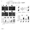
- TUNEL staining did not reveal significant differences in the number of apoptotic cells between groups.
- We also observed no difference in a cell proliferation (LIRKO 0.24% ⁇ 0.09% versus control 0.29% ⁇ 0.1 % BrdU+ a cells; n 6) ( Figures 1A-1F ), or in the proliferation of cells in multiple non- ⁇ cell tissues, including visceral adipose, subcutaneous adipose, muscle, kidney, liver, or spleen.
- Table 2 Histological characteristics of 12-month-old Control and LIRKO mice Control LIRKO Pancreas mild pancreatitis very mild pancreatitis no steatosis + focal dysplasia+ hyperplastic Liver severe steatosis nodules Skeletal muscle normal normal severe lymphocyte Visceral adipose infiltration mild lymphocyte infiltration Subcutaneous adipose normal normal Spleen normal normal Kidney normal normal Lung normal normal normal
- each recipient mouse (control or LIRKO) was transplanted with two islet grafts, one derived from control and the other derived from LIRKO donors, under the left and right kidney, respectively ( Figure 2G ).
- islet grafts were harvested, sectioned, and analyzed for ⁇ cell BrdU incorporation.
- control islets grafted into control animals exhibited minimal ⁇ cell proliferation (0.017% ⁇ 0.017% BrdU+ ⁇ cells).
- Table 3 Islet-donor characteristics Donor Gender Ethnicity/Race Age (years) BMI Diabetic donor status Experiment 1 Male White 55 20.1 No stimulation with serum 2 Male White 23 25.6 No stimulation with serum 3 Female White 18 26.4 No stimulation with serum 4 Male Hispanic/Latino 25 29.3 No stimulation with serum 5 Male Hispanic/Latino 50 26.5 No stimulation with serum 6 Unkown Unkown 64 30 No stimulation with serum 7 Female African american 41 42 No stimulation with serum 8 Male White 54 19.5 No stimulation with serum 9 Male African american 20 31.3 No stimulation with serum 10 Male Unknown 53 31 T2D stimulation with serum 11 Female White 38 37.8 T2D on metformin stimulation with serum 12 Unknown Unknown Unknown 65 31 No stimulation with LECM 13 Unknown Unknown 54 34 T2D stimulation with LECM 14 Male White 52 50 T2D stimulation with HCM
- hepatocyte nuclear factor-4a initiates cell cycle entry, but is not sufficient to promote ⁇ cell expansion in human islets. Mol. Endocrinol. 26, 1590-1602 ).
- LIRKO serum was also effective in promoting proliferation of islet ⁇ cells from patients with type 2 diabetes ( Figures 4E-4G ).
- the ⁇ cell mitogen(s) present in the circulation of LIRKO mice shows conserved activity toward mouse and human islets, including islets from patients with type 2 diabetes.
- Glucose and insulin have been reported to promote ⁇ cell growth ( Assmann, A., et al., (2009). Growth factor control of pancreatic islet regeneration and function. Pediatr.
- Diabetes 10, 14-32 Assmann, A., et al., (2009b). Glucose effects on beta-cell growth and survival require activation of insulin receptors and insulin receptor substrate 2. Mol. Cell. Biol. 29, 3219-3228 ; Bonner-Weir, S., et al., (1989). Compensatory growth of pancreatic beta-cells in adult rats after short-term glucose infusion. Diabetes 38, 49-53 ) and are potential candidates in the LIRKO model, which manifests glucose intolerance and hyperinsulinemia ( Michael, M.D., et al., (2000). Loss of insulin signaling in hepatocytes leads to severe insulin resistance and progressive hepatic dysfunction. Mol. Cell 6, 87-97 ).
- Hepatocyte-Derived Factors Stimulate Mouse and Human Islet ⁇ Cell Replication In Vitro.
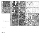
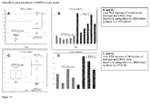
- Adipose tissue selective insulin receptor knockout protects against obesity and obesity-related glucose intolerance. Dev. Cell 3, 25-38 ), or brain ( Brüning, J.C., et al., (2000). Role of brain insulin receptor in control of body weight and reproduction. Science 289, 2122-2125 ) prompted us to hypothesize that the liver serves as a source of ⁇ cell growth factor(s) in response to metabolic insults such as insulin resistance. To test this hypothesis, we collected conditioned media from liver explant cultures (LECM) from either 3-or 12-month old LIRKO or control animals and evaluated their effects on ⁇ cell proliferation in mouse islets ( Figure 5A ).
- LCM liver explant cultures
- Ki67- positive ⁇ cells were significantly elevated in islets cultured in LECM from either 3- or 12- month-old LIRKO mice, compared to cells cultured in LECM derived from age-matched controls ( Figure 5B ).
- mouse islets cultured in control LECM derived from 3- and 12- month-old animals displayed similar levels of proliferation, the levels were 2-fold higher in cultures containing 12-month-old LIRKO-LECM compared to 3-month-old control LECM ( Figure 5C ).
- This age-dependent effect of LIRKO-LECM is consistent with the age-dependent increase in ⁇ cell proliferation in LIRKO mice ( Okada, T., et al., (2007). Insulin receptors in beta-cells are critical for islet compensatory growth response to insulin resistance. Proc.
- ⁇ cells in islets obtained from healthy human controls and patients with type 2 diabetes (for donor characteristics, see Table 3) cultured in LECM derived from LIRKO animals exhibited increased proliferation compared to islets from the same donors cultured in control LECM ( Figures 5D and 5E ).
- the liver contains multiple cell types, including hepatocytes, Kupffer cells, and endothelial cells ( Si-Tayeb, K., et al., (2010). Organogenesis and development of the liver. Dev. Cell 18, 175-189 ).
- LIRKO serum was a product of hepatocytes or nonhepatic cells.
- HCM primary hepatocytes
- the number of TUNEL+ ⁇ cells was similar in both conditions ( Figures 5F and 5G ).
- LIRKO HCM proliferative effect of LIRKO HCM was also evident when human islets obtained from a patient with type 2 diabetes (for donor characteristics, see Table 3) were exposed to LIRKO HCM compared to control HCM ( Figures 5H and 5I ).
- insulin-resistant hepatocytes produce a ⁇ cell growth-promoting factor(s) that enhances proliferation of mouse and human ⁇ cells.
- numerous signaling pathways impacting ⁇ cell growth have been documented ( Kulkarni, R.N., et al., (2012). Human ⁇ -cell proliferation and intracellular signaling: driving in the dark without a road map. Diabetes 61, 2205-2213 ), specific blood-borne molecules that trigger ⁇ cell replication directly in response to insulin resistance have, to our knowledge, not been reported.
- Pancreatic ⁇ -cell dysfunction underlies the development of both type1 and type 2 diabetes. Although the natural history of both forms diabetes is different, reduced functional ⁇ -cell mass is a common hallmark in both diseases. Regenerative approaches represent an attractive strategy to increase the number of functional ⁇ -cells. In this context, we recently reported ( EI Ouaamari, et al, Cell Reports, 2013, 3:1-10 ) the existence possible of liver-derived systemic factors capable of stimulating ⁇ -cell proliferation in Liver Insulin Receptor Knockout mouse (LIRKO), a unique model of islet hyperplasia and increased ⁇ -cell mass caused by insulin resistance.
- LIRKO Liver Insulin Receptor Knockout mouse
- SerpinB1 was identified as the being a consistently up-regulated hepatocyte-derived systemic ⁇ -cell trophic factor.
- Nanozeolite LTL nanoparticles were obtained from NanoScape AG, Germany. Adsorption of proteins on the surface of Nanozeolite LTL was carried out for 90 min at 4°C by incubation of proteins from hepatocyte conditioned media (0.1 mg/ml) and nanoparticles (0.1 mg/ml) in suspension in PBS. After centrifugal separation at 16000g during 20 min, proteins bound to nanoparticles are washed twice in 0.1M ammonium carbonate buffer, ph 8.0.
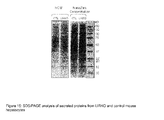
- Protein samples were resolved by SDS-PAGE on NuPAGE® Novex® 4-12% Bis-Tris gels using the NuPAGE® MES SDS Running Buffer according to the manufacturer's instructions (Invitrogen, Grand Island, NY) stained using the SilverQuestTM silver staining kit from Invitrogen.
- the proteins captured on nanozeolites were reduced in the presence of 10 mM dithiothreitol, 0.05% AALS (Anionic Acid Labile Surfactants from Protea Biosciences) in 50 mM ammonium carbonate buffer, pH 8.0 at 56°C for 30 min and then alkylated by adding 20 mM iodoacetamide for 30 min at room temperature in the dark.
- AALS Ammonium carbonate buffer
- bound proteins were digested with LysC (1/50 w/w), 4hrs at 37°c and then with trypsin (1/50 w/w) for 18 hr at 37°C.
- LC-MS Liquid Chromatograph Mass Spectrometer experiments were performed on NanoAcquity UPLC (Waters, Milford, MA) connected to a hybrid LTQ (Linear Trap Quadropole) Orbitrap VelosTM mass spectrometer (Thermo Fisher Scientific, Waltham, MA) equipped with a nanoelectrospray source. Protein digests were loaded onto a nanoAcquity UPLC Trap column (Symmetry C18, 5 ⁇ m, 180 ⁇ m x 20 mm, Waters) and washed with 0.2% formic acid at 20 ⁇ L/min for 5 min.
- Peptides were then eluted on a C18 reverse-phase nanoAcquity column (BEH130 C18, 1.7 ⁇ m, 75 ⁇ m x 250 mm, Waters) with a linear gradient of 7-30% solvent B (H 2 O/CH 3 CN/HCOOH, 10:90:0.2, by vol.) for 120 min, 30-90% solvent B for 20 min, and 90% solvent B for 5 min, at a flow rate of 250 nL/min.
- solvent B H 2 O/CH 3 CN/HCOOH, 10:90:0.2, by vol.
- the mass spectrometer was operated in the data-dependent mode to automatically switch between MS and MS/MS acquisition.
- Survey full scan MS spectra (from m/z 300-1700) were acquired in the Orbitrap with a resolution of 60,000 at m/z: 400.
- the AGC automated gain control
- the most intense ions (up to 20) were then isolated for fragmentation in the LTQ linear ion trap using a normalized collision energy of 28% at the default activation q of 0.25 with an AGC settings of 2 ⁇ 10 4 and a maximum injection time of 200 ms.
- the dynamic exclusion time window was set to 150 s. Samples were injected in triplicate.
- LC-MS/MS data acquired using the Xcalibur software (version 2.07, Thermo-Fisher Scientific), were processed using a Visual Basic program software developed using XRawfile libraries (distributed by Thermo-Fisher Scientific). Similar programs are known to and can be developed by one of ordinary skill in the art. Three different files were generated by this program: the first one corresponds to a MS/MS peak list (MGF file) which will be used for database searching. This MGF file contains the exact parent mass and the retention time (RT) associated with each LTQ-MS/MS spectrum.
- MGF file MS/MS peak list
- the exact parent mass is the 12 C isotope ion mass of the most intense isotopic pattern detected on the high resolution Orbitrap MS parallel scan and included in the LTQ-MS/MS selection window.
- the RT is issued from the LTQ-MS/MS scan.
- the second file is a MS/MS log file which reports, for each acquired MS/MS, the scan number, the 12 C isotope exact mass, the RT and the parent filter (LTQ selection window).
- the third file corresponds to the conversion of the high resolution MS raw data file into a "csv" format file which will be used for quantitative analysis.
- DIFFTAL Algorithm Overview DIFFTAL is a set of software tools developed in Sanofi under MatLab environment (www.mathworks.com) for label-free differential analysis of complex proteomic mixtures dedicated to data recorded with high resolution MSMS instruments.
- DIFFTAL runs in 6 main steps. These steps consist of the following: (1) Feature detection, (2) MS matching, (3) MS/MS annotations, (4) MS/MS matching, (5) Peptide quantification report and (6) Protein relative quantifications.
- Step 1 Feature detection.
- Each LC/MS file is treated independently for feature detection.
- the signal apparition is detected scan by scan by analyzing the evolution of the average signal of 3 consecutive scans.
- Feature detection is achieved using the peptide isotopic patterns calculated with "Averagine" algorithm.
- a matrix of the features detected in the 3D space m/z (mass/charge), RT (retention time) and intensity
- This matrix contains links to retrieve the corresponding processed signals, which are stored in a temporary data bank.
- Step 2 MS matching. All LC-MS data are matched together using a progressive alignment procedure. First, the most intense detected features are matched in agreement with m/z and RT precision windows defined by the user. Then, all peptides are used to compute a specific RT alignment model. A definitive RT window is calculated according to the dispersion observed between real and calculated RTs. Finally, every remaining unmatched m/z is checked by going back to the processed signal stored during the feature detection step. This last point allows a very confident determination of the unmatched feature class.
- Step 3 MS/MS annotations. This step corresponds to the data bank search previously reported in the "Database Searching" paragraph.
- Step 4 MS/MS matching.
- MSMS Spectrum reports exported from Scaffold are matched with the matrix of detected features using the corresponding acquisition MS/MS log files (see LC-MS/MS data processing). This matching requires starting and ending time points of each feature. Indeed, the RT feature is the time at the maximum intensity of the observed MS signal, whereas the MS/MS spectrum is recorded at any time during the peptide elution. In case of ambiguity, the comparison between the exact isotopic profile calculated from the MS/MS sequence and the detected signal at the feature RT is used for sorting.
- Another routine has been also introduced in the software that quantifies only the MS/MS identified peptides according to the following scheme: the time profiles of the 2 major isotopes of each identified peptide are computed in a small time window where the MS/MS spectrum was recorded. Only the co-eluted signals of these 2 isotopes are analyzed to determine the peptide RT. The 3 scans averaged signal centered at this time is then compared with the full theoretical peptide isotopic pattern. This additional quantification is compared to the first one to generate a final result report. The convergence of these two quantification routines is used to improve the quantification confidence and identification coverage.
- Step 5 Peptide Quantification report: Peptide quantification is calculated from the statistical analysis of the previous matrix. Statistical analyses were realized with DanteR program, an R based software written by Tom Taverner (Thomas.Taverner@pnl.gov and Ashoka Polpitiya for the U.S. Department of Energy (PNNL, Richland, WA, USA: on the World Wide Web (www): omics.pnl.gov/software). The median intensity value of the detected feature population is used to normalize the 3 replicate injections of the same sample. Only peptides detected at least 2 times (over replicates) are kept and an average intensity value per sample is calculated for each peptide. A threshold value representing the minimum detectable signal level is used instead of quantification for non detected peptide.
- a protein which is identified, for example, in the treated sample but not detected in the control sample is represented with a minimum positive fold change which is the result of the treated signal divided by the minimum detectable signal.
- Step 6 Protein quantification: Finally, peptides arising from the same protein are grouped to evaluate the peptide fold change dispersion. Protein-level inferences are performed utilizing all of the available peptide abundances and a likelihood ratio test to compute p-values (Karpievitch, et al., 2009a). Significant up or down protein expression changes are sorted and plotted by p-value from hypothesis testing through the sample types and the replicate analyses.
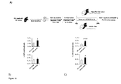
- Hepatocytes from control and LIRKO mice were cultured in serum free medium and supernatants collected after 18hrs.
- the resulting peptides were identified using high-performance liquid chromatography tandem mass spectrometry LC-MS/MS analysis. Proteins were identified by searching MS and MS/MS data of peptides against the UniProtKB/Swiss-Prot protein knowledgebase using the MASCOT search engine and then quantified by a label free quantitative LC-MS analysis using in-house DIFFTAL software algorithm. Relative quantification of each protein was obtained by averaging the intensity ratios of the three most intense derived peptides (or two derived peptides if only two unique peptides were identified) as described in the experimental procedure.
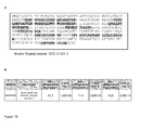
- Mouse SerpinB1 was identified by LC-MSMS by 12 unique tryptic peptides given a protein coverage of 37% and in the 3 independent experiments, SerpinB1 was shown to be up-regulated in LIRKO hepatocyte supernatants with the respective ratio of 17.5, 11.6 and 18.4 and p-values smaller than 0.01 ( Figure 16 A & B ).
- oligonucleotides arrays for gene expression monitoring has been described in U.S. Pat. No. 6,177,248 .
- the used micro arrays (GeneChips) from Affymetrix, Santa Clara, CA. USA contain deoxynucleotide sequences that represent approximately 39,000 mouse transcripts and variants from >34,000 well characterized mouse genes (Mouse Genome 430 2.0 GeneChip). Each transcript and variant is represented by 11 different oligonucleotide probes with 25 basepairs in length.
- Sequences used in the design of the array were selected from GenBank, dbEST, and RefSeq. The sequence clusters were created from the UniGene database (Build 107, June 2002) and then refined by analysis and comparison with the publicly available draft assembly of the mouse genome from the Whitehead Institute Center for Genome Research (MGSC, April 2002).
- RNA nano assay Agilent, Santa Clara, CA.
- First and second strand cDNA synthesis were performed with 10 ⁇ g of each total RNA using SuperScript SSII RT polymerase system (Invitrogen) and a T7(dT)24 primer linking the T7 RNA polymerase promoter and oligo(deoxythymidine)24.
- Double strand cDNA was phenol-chloroform extracted followed by ethanol precipitation and resuspended in 12 ⁇ l RNAse-free water.
- Biotin-UPT and -CTP labelled cRNA was transcribed in vitro using Enzo BioArray High Yield RNA Transcript Labelling Kit (Enzo Diagnostics, NY, NY) and purified by RNeasy cleanup and ethanol precipitation.
- RNA nano assay (Agilent 2100 BioAnalyzer). 15 ⁇ g cRNA samples were fragmented at 94 degree Celsius for 35 min in 40 mM Tris/acetate pH 8.1, 100 mM KOAc and 30 mM MgOAc, added to hybridisation buffer and hybridised to Affymetrix GeneChip for 16-18 hours at 45 degree Celsius and 60 rpm in a rotating hybridization oven (Hybridization Oven 640, Affymetrix).
- Micro arrays were washed in a fluidics station (GeneChip Fluidics Station 450, Affymetrix) and double-stained with streptavidin-phycoerythrin conjugate (Molecular Probes, Life Technologies, Grand Island, NY), anti-streptavidin antibody and again streptavidin-phycoerythrin conjugate to enhance signal intensity according to the methodologies described by Affymetrix.
- the micro arrays were scanned with the GeneChip Scanner 3000 7G (Affymetrix), which is controlled by Affymetrix software GeneChip Operating System (GCOS) v1.4. Quality control of each chip was performed according the Affymetrix quality criteria, including mean average difference, raw intensity and 3'/5' ratio of housekeeping genes beta-actin and GAPDH.
- RNAs were extracted using RNeasy Mini Kit (QIAGEN, Valencia, CA). One ⁇ g RNA was used for a reverse transcription step using high-capacity cDNA Archive Kit (Applied Biosystems). cDNA was analyzed by ABI 7900HT system (Applied Biosystems). TBP was used as an internal control.
- Bioinformatics analysis of the Affymetrix raw data has been performed in the Array Studio software package from OmicSoft Corp. Cary, NC, USA.
- Affymetrix cel files have been first processed with Robust Multi-array Average (RMA) as normalization method and the data have been then log2 transformed.
- RMA Robust Multi-array Average
- For detection of expressed genes all Affymetrix probe sets with intensity signals of ⁇ 6 in at least 25% of the samples each of the LIRKO and wildtype group have been filtered out.
- Principal component analysis (PCA) has been applied to all samples as a quality control measure.
- PCA Principal component analysis
- SerpinB1 is regulated at the post-transcriptional level in HFD model, a feature observed with other proteins (He, et al., 2009; Vannay, et al., 2004). Together, these data strongly implicate SerpinB1 as a potential marker of insulin resistance.
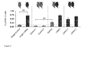
- SerpinB1 is a ⁇ -cell growth factor
- mice freshly isolated primary mouse islets in presence of various doses of recombinant human SerpinB1 or ovalbumin (SerpinB14) and evaluated ⁇ -cell proliferation by Ki67 immunofluorescent staining (known to those of ordinary skill in the art).
- Ki67 immunofluorescent staining known to those of ordinary skill in the art.
- mouse islets cultured in ovalbumin (1 ⁇ g/ml) displayed normal low ⁇ -cell proliferation
- isolated islets cultured with recombinant SerpinB1 exhibited a dose-dependent increase in Ki67+ insulin+ cells; the data reached statistical significance when islets were cultured at a concentration of 1 ⁇ g/ml of SerpinB1 compared to controls (islets cultured at a similar concentration of ovalbumin).
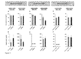
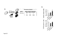
- islets derived from 5-6 week old C57BL/6J male mice were cultured for 24 hours in media containing serum (dilution 1:10) or LECM (dilution 1:10) derived from animals fed either chow diet (CD) or high fat diet (HFD) or from ob/ob animals and respective controls, followed by evaluation of ⁇ -cell proliferation by Ki67 immunostaining and fluorescence microscopy ( Figure 19A ).
- a major substrate of SerpinB1 is neutrophil elastase.
- Sivelestat is the International Nonproprietary Name (INN) as given by the World Health Organization (WHO); the chemical name is: N - ⁇ 2-[( ⁇ 4-[(2,2-dimethylpropanoyl)oxy]phenyl ⁇ sulfonyl)amino]benzoyl ⁇ glycine), to stimulate islet ⁇ -cell proliferation in vitro.
- ⁇ -cell proliferation will be assessed in in vitro assays.
- AAV Associated-adenoviruses
- mice over-expressing the AAV-SeprinB1 in the liver will be transplanted with human islets to create a "humanized mouse model". Mice will be monitored for body weight, blood glucose for 2, 4 and 16 weeks. At the end of the experiment, islet grafts and pancreases will be harvested and analyzed for proliferation and survival of endocrine cells. This model will directly indicate whether altering the expression of SerpinB1 in the liver promotes human ⁇ -cell proliferation in vivo - with important implications for human therapy.
- mice were housed in pathogen-free facilities and maintained on a 12 hr light/dark cycle in the Animal Care Facility at Joslin Diabetes Center, Boston, and the Foster Biomedical Research Laboratory, Brandeis University, Waltham, MA. All studies conducted and protocols used were approved by the Institutional Animal Care and Use Committee of the Joslin Diabetes Center and Brandeis University and were in accordance with NIH guidelines. LIRKO mice were generated by crossing Albumin- Cre to IR flox/flox on a mixed genetic background and were backcrossed for more than 15 generations on the C57/BI6 background.
- Islets were isolated from 9-month-old mice using the intraductal collagenase technique (Kulkarni, R.N., et al. , 1999); EI Ouaamari, et al., 2013, Cell Rep., 3(2):401-410 ). Altered function of insulin receptor substrate-1-deficient mouse islets and cultured beta-cell lines. J. Clin. Invest. 104, R69-R75 ). Islets were handpicked, concentrated in a pellet, and kept on ice until transplantation ( Flier, S.N., et al., (2001). Evidence for a circulating islet cell growth factor in insulin-resistant states. Proc. Natl. Acad. Sci. USA 98, 7475-7480 ).
- mice After intraperitoneal injection (15 ml/g body weight) of a 1:1 (w/v) mixture of 2,2,2-tribromoethanol and tert-amyl alcohol and diluted 1:50 with PBS (pH 7.4).
- the capsules of the kidneys were incised, and the islets were implanted near the upper pole of each kidney in 5-month-old male mice.
- the capsules were cauterized, and the mice were allowed to recover on a heating pad.
- ELISA-based assays were used to measure growth factors and hormones, including IGF-1 (catalog #MG100; R&D Systems), HGF (catalog #ab100686; Abcam), EGF (catalog #IB39411; IBL-America, Minneapolis, MN), PDGFAA (catalog #DAA00B; R&D Systems), PDGFBB (catalog #MBB00; R&D Systems, Minneapolis, MN), VEGF (Millipore, Billerica, MA), FGF21 (catalog #EZRMFGF21-26K; Millipore), Gastrin (catalog #E91224mu; USCN Life Science, Hubei 430056, PRC), Adiponectin (catalog #EZMADP-60K; Millipore), Ostepontin (catalog #MOST00; R&D Systems), and Osteocalcin (catalog #EIA4010; International).
- IGF-1 catalog #MG100; R&D Systems
- Pancreatic islets were isolated from 5-week-old male mice by liberase and thermolysin digestion (Roche), handpicked, and cultured for 16 hr in RPMI 1640 with 7 mM glucose and 10% FBS (v/v).
- RPMI 1640 with 0.1% BSA (v/v) containing 3 mM glucose for 3 hr and thereafter treated with RPMI 1640 with 5.5 mM glucose supplemented every 12 hr with 10% (v/v) serum obtained from 3- or 12-month-old LIRKO and control mice.
- islets Twenty-four to 48 hr later, islets were handpicked, fixed with 4% paraformaldehyde, embedded in agarose/paraffin, and sectioned for immunohistochemistry studies. To evaluate ⁇ cell replication, sections were analyzed by fluorescent microscopy subsequent to Ki67, TUNEL, and insulin immunostaining.
- Liver explant-conditioned medium (LECM) preparation was adapted from Nicoleau, et al. (2009, Endogenous hepatocyte growth factor is a niche signal for subventricular zone neural stem cell amplification and self-renewal. Stem Cells 27, 408-419 ).
- Mice were anesthetized with Avertin (240 mg/kg intraperitoneally), and 100 mg liver explants were dissected from LIRKO or control mice. Explants were washed twice in cold PBS, incubated in PBS at 37°C for 30 min, and then cultured in serum-free Dulbecco's modified Eagle's medium (DMEM) containing 5.5 mM glucose.
- DMEM Dulbecco's modified Eagle's medium
- Islets were initially starved for 3 hr in DMEM containing 3 mM glucose and 0.1 % BSA and thereafter stimulated for 24 hr with DMEM/5.5 mM glucose media containing 10% LECM. Islet ⁇ cell proliferation and apoptosis were analyzed by fluorescent microscopy after Ki67, TUNEL, and insulin immunostaining.
- Hepatocytes were isolated from 6-month-old LIRKO and control mice by collagenase digestion via portal vein perfusion ( Sun, R., et al., (2005). IL-6 modulates hepatocyte proliferation via induction of HGF/p21cip1: regulation by SOCS3. Biochem. Biophys. Res. Commun. 338, 1943-1949 ). Mice were anesthetized with Avertin (240 mg/kg intraperitoneally), and the portal vein was cannulated with JELCO 22G x 1 inch catheter (Smiths Medical, Dublin, OH).
- the liver was perfused with EGTA solution (5.4 mmol/l KCI, 0.44 mmol/l KH 2 PO 4 , 140 mmol/l NaCl, 0.34 mmol/l Na 2 HPO 4 , and 0.5 mmol/l EGTA [pH 7.4]) and digested with DMEM containing 0.075% type I collagenase.
- Hepatocytes were washed twice in Hepatocyte Wash Medium (Invitrogen, Grand Island, NY).
- the isolated mouse hepatocytes were seeded in collagen-coated 6-well plates (BD BioCoat, San Jose, CA) at a density of 106 cells/well in DMEM containing 25 mM glucose and 10% FBS (v/v).
- hepatocytes were cultured for 24 hr in serum-free DMEM containing 5.5 mM glucose. HCM was collected, centrifuged, and kept at ⁇ 80°C. Islets were initially starved for 3 hr in DMEM containing 3 mM glucose and 0.1 % BSA and thereafter incubated for 24 hr in DMEM/5.5mMglucose media containing 50% HCM. Islet ⁇ cell proliferation and apoptosis were analyzed by fluorescent microscopy after Ki67, TUNEL, and insulin immunostaining.
- Pancreases and islets were analyzed by immunostaining using anti-Ki67 (BD), anti-insulin (Abcam, Cambridge, MA), or anti-glucagon (Sigma-Aldrich, St. Louis, MO) antibodies. Quantification of replicating ⁇ - and ⁇ -cells was performed as described previously (EI Ouaamari, et al., 2013).
- mice were injected with BrdU intraperitoneally (100 mg/kg body weight) 5 hr prior to animal sacrifice for immunostaining of the pancreas.
- BrdU injections in the in vivo serum administration experiments were performed on three occasions as denoted in Figure 2A .
- mice were anesthetized, and osmotic pumps (ALZET) containing 100 ⁇ l of either vehicle or Sivelestat (Santa Cruz) were implanted subcutaneously. Sivelestat was dissolved in 50% DMSO and was administered at a dose of 150 or 300 ⁇ g/kg/day. Control mice were infused with DMSO 50% alone. GW311616A was administered into mice by daily oral gavage for two weeks. Mice received either GW311616A (2 mg/kg body weight) or vehicle.
- AZET osmotic pumps
- Proteins extracted from hepatocyte-conditioned media were pre-enriched on nanoparticles (NanoScape AG, Germany). Samples were resolved by SDS-PAGE NuPAGE 4-12% Bis-Tris gels and stained using SilverQuest (Invitrogen). The proteins captured on nanozeolites were reduced and alkylated and then digested with LysC (1/50 w/w) for 4 hours at 37 °C and then with trypsin (1/50 w/w) for 18 hours at 37 °C. Enzymatic digests were subjected to liquid chromatography mass spectrometry (LC-MS) analysis.
- LC-MS liquid chromatography mass spectrometry
- LC-MS experiments were performed on NanoAcquity UPLC (Waters, Milford, MA) connected to a hybrid LTQ (Linear Trap Quadropole) Orbitrap Velos mass spectrometer (Thermo Fisher Scientific, Waltham, MA) equipped with a nanoelectrospray source.
- NanoAcquity UPLC Waters, Milford, MA
- hybrid LTQ Linear Trap Quadropole
- Orbitrap Velos mass spectrometer Thermo Fisher Scientific, Waltham, MA
- Protein digests were loaded onto a nanoAcuity UPLC Trap column (Symmetry C18, 5 ⁇ m, 180 ⁇ m x 20 mm, Waters) and peptides were eluted on a C18 reverse-phase nanoAcquity column (BEH130 C18, 1.7 ⁇ m, 75 ⁇ m x 250 mm, Waters) with a linear gradient of 7-30 % solvent B (H2O/CH3CN/HCOOH, 10:90:0.2, by vol) for 120 min, 30-90% Solvent B for 20 min, and 90% solvent B for 5 min, at a flow rate of 250 nL/min. samples were injected in triplicate.
- solvent B H2O/CH3CN/HCOOH
- LC-MS data were acquired using the Xcalibur software (version 2.07, Thermo-Fisher Scientific) and were processed using a Visual Basic program developed using XRawfile libraries (distributed by Thermo-Fisher Scientific). Data base searches were performed using MASCOT server (version 2.1, matrix Science; www.matrixscience.com/) using the Swiss-Prot database.
- livers 150 mg were lysed in RIPA buffer and total protein concentration was determined using BCA assay (Pierce, Rockford, IL). Samples were resuspended in Laemmli buffer, boiled and resolved by SDS-polyacrylamide gel electrophoresis. Proteins were transferred onto nitrocellulose membranes, blocked in blocking buffer (PBS containing 5% Bovine Serum Albumin and 0.1 % tween 20) and incubated with primary antibodies SerpinB1 (Santa cruz, sc-34305; Santa Cruz Biotechnology, Santa Cruz, CA) or Actin (Santa cruz, sc-1615) for overnight or one hour, respectively. Secondary rabbit anti-goat (Santa cruz, sc-2922) were used thereafter. Quantification of protein amounts were estimated by ImageJ software (National Institutes of Health, Bethesda, Maryland).
Priority Applications (5)
| Application Number | Priority Date | Filing Date | Title |
|---|---|---|---|
| EP13306284.4A EP2851086A1 (de) | 2013-09-20 | 2013-09-20 | Serpine: Verfahren zur therapeutischen ß-Zellen-Regeneration und Funktion |
| EP14706025.5A EP2958585A1 (de) | 2013-02-22 | 2014-02-21 | Serpine: verfahren zur therapeutischen beta-zellen-regeneration und -funktion |
| US14/769,637 US20160002316A1 (en) | 2013-02-22 | 2014-02-21 | Serpins: methods of therapeutic beta-cell regeneration and function |
| PCT/EP2014/053419 WO2014128257A1 (en) | 2013-02-22 | 2014-02-21 | Serpins: methods of therapeutic beta-cell regeneration and function |
| TW103106093A TW201519902A (zh) | 2013-02-22 | 2014-02-24 | 絲胺酸蛋白酶抑制蛋白:治療β細胞再生之方法及功能 |
Applications Claiming Priority (1)
| Application Number | Priority Date | Filing Date | Title |
|---|---|---|---|
| EP13306284.4A EP2851086A1 (de) | 2013-09-20 | 2013-09-20 | Serpine: Verfahren zur therapeutischen ß-Zellen-Regeneration und Funktion |
Publications (1)
| Publication Number | Publication Date |
|---|---|
| EP2851086A1 true EP2851086A1 (de) | 2015-03-25 |
Family
ID=49293565
Family Applications (1)
| Application Number | Title | Priority Date | Filing Date |
|---|---|---|---|
| EP13306284.4A Withdrawn EP2851086A1 (de) | 2013-02-22 | 2013-09-20 | Serpine: Verfahren zur therapeutischen ß-Zellen-Regeneration und Funktion |
Country Status (1)
| Country | Link |
|---|---|
| EP (1) | EP2851086A1 (de) |
Citations (21)
| Publication number | Priority date | Publication date | Assignee | Title |
|---|---|---|---|---|
| EP0043075A2 (de) | 1980-06-25 | 1982-01-06 | Studiengesellschaft Kohle mbH | Verfahren zur Erhöhung der Inkorporation und der Expression von genetischem Material in die Kerne von intakten Zellen mit Hilfe von Liposomen |
| US4522811A (en) | 1982-07-08 | 1985-06-11 | Syntex (U.S.A.) Inc. | Serial injection of muramyldipeptides and liposomes enhances the anti-infective activity of muramyldipeptides |
| JPS6447381A (en) | 1987-08-19 | 1989-02-21 | Vitamin Kenkyusho Kk | Preparation of liposome containing sealed gene |
| WO1989002468A1 (en) | 1987-09-11 | 1989-03-23 | Whitehead Institute For Biomedical Research | Transduced fibroblasts and uses therefor |
| WO1989005345A1 (en) | 1987-12-11 | 1989-06-15 | Whitehead Institute For Biomedical Research | Genetic modification of endothelial cells |
| WO1989007136A2 (en) | 1988-02-05 | 1989-08-10 | Whitehead Institute For Biomedical Research | Modified hepatocytes and uses therefor |
| US4868116A (en) | 1985-07-05 | 1989-09-19 | Whitehead Institute For Biomedical Research | Introduction and expression of foreign genetic material in epithelial cells |
| US4980286A (en) | 1985-07-05 | 1990-12-25 | Whitehead Institute For Biomedical Research | In vivo introduction and expression of foreign genetic material in epithelial cells |
| WO1991006309A1 (en) | 1989-11-03 | 1991-05-16 | Vanderbilt University | Method of in vivo delivery of functioning foreign genes |
| WO1992007573A1 (en) | 1990-10-31 | 1992-05-14 | Somatix Therapy Corporation | Genetic modification of endothelial cells |
| US5328470A (en) | 1989-03-31 | 1994-07-12 | The Regents Of The University Of Michigan | Treatment of diseases by site-specific instillation of cells or site-specific transformation of cells and kits therefor |
| US5641670A (en) | 1991-11-05 | 1997-06-24 | Transkaryotic Therapies, Inc. | Protein production and protein delivery |
| US6168587B1 (en) | 1993-04-08 | 2001-01-02 | Powderject Research Limited | Needleless syringe using supersonic gas flow for particle delivery |
| US6177248B1 (en) | 1999-02-24 | 2001-01-23 | Affymetrix, Inc. | Downstream genes of tumor suppressor WT1 |
| US6194389B1 (en) | 1989-11-16 | 2001-02-27 | Duke University | Particle-mediated bombardment of DNA sequences into tissue to induce an immune response |
| US6468798B1 (en) | 1991-12-17 | 2002-10-22 | The Regents Of The University Of California | Expression of cloned genes in the lung by aerosol and liposome-based delivery |
| US6471996B1 (en) | 1996-12-20 | 2002-10-29 | Aventis Pasteur Limited | Biodegradable targetable microparticle delivery system |
| US6472375B1 (en) | 1998-04-16 | 2002-10-29 | John Wayne Cancer Institute | DNA vaccine and methods for its use |
| WO2007006858A2 (en) * | 2005-07-12 | 2007-01-18 | Oy Jurilab Ltd | Method for treatment of cardiovascular and metabolic diseases and detecting the risk of the same |
| US20100184658A1 (en) * | 2006-09-01 | 2010-07-22 | Cohava Gelber | Compositions and methods for diagnosis and treatment of type 1 diabetes |
| US20120195859A1 (en) | 2010-01-14 | 2012-08-02 | Nathalie Vergnolle | Recombinant probiotic bacteria for the prevention and treatment of inflammatory bowel disease (ibd) and irritable bowel syndrome (ibs) |
-
2013
- 2013-09-20 EP EP13306284.4A patent/EP2851086A1/de not_active Withdrawn
Patent Citations (21)
| Publication number | Priority date | Publication date | Assignee | Title |
|---|---|---|---|---|
| EP0043075A2 (de) | 1980-06-25 | 1982-01-06 | Studiengesellschaft Kohle mbH | Verfahren zur Erhöhung der Inkorporation und der Expression von genetischem Material in die Kerne von intakten Zellen mit Hilfe von Liposomen |
| US4522811A (en) | 1982-07-08 | 1985-06-11 | Syntex (U.S.A.) Inc. | Serial injection of muramyldipeptides and liposomes enhances the anti-infective activity of muramyldipeptides |
| US4868116A (en) | 1985-07-05 | 1989-09-19 | Whitehead Institute For Biomedical Research | Introduction and expression of foreign genetic material in epithelial cells |
| US4980286A (en) | 1985-07-05 | 1990-12-25 | Whitehead Institute For Biomedical Research | In vivo introduction and expression of foreign genetic material in epithelial cells |
| JPS6447381A (en) | 1987-08-19 | 1989-02-21 | Vitamin Kenkyusho Kk | Preparation of liposome containing sealed gene |
| WO1989002468A1 (en) | 1987-09-11 | 1989-03-23 | Whitehead Institute For Biomedical Research | Transduced fibroblasts and uses therefor |
| WO1989005345A1 (en) | 1987-12-11 | 1989-06-15 | Whitehead Institute For Biomedical Research | Genetic modification of endothelial cells |
| WO1989007136A2 (en) | 1988-02-05 | 1989-08-10 | Whitehead Institute For Biomedical Research | Modified hepatocytes and uses therefor |
| US5328470A (en) | 1989-03-31 | 1994-07-12 | The Regents Of The University Of Michigan | Treatment of diseases by site-specific instillation of cells or site-specific transformation of cells and kits therefor |
| WO1991006309A1 (en) | 1989-11-03 | 1991-05-16 | Vanderbilt University | Method of in vivo delivery of functioning foreign genes |
| US6194389B1 (en) | 1989-11-16 | 2001-02-27 | Duke University | Particle-mediated bombardment of DNA sequences into tissue to induce an immune response |
| WO1992007573A1 (en) | 1990-10-31 | 1992-05-14 | Somatix Therapy Corporation | Genetic modification of endothelial cells |
| US5641670A (en) | 1991-11-05 | 1997-06-24 | Transkaryotic Therapies, Inc. | Protein production and protein delivery |
| US6468798B1 (en) | 1991-12-17 | 2002-10-22 | The Regents Of The University Of California | Expression of cloned genes in the lung by aerosol and liposome-based delivery |
| US6168587B1 (en) | 1993-04-08 | 2001-01-02 | Powderject Research Limited | Needleless syringe using supersonic gas flow for particle delivery |
| US6471996B1 (en) | 1996-12-20 | 2002-10-29 | Aventis Pasteur Limited | Biodegradable targetable microparticle delivery system |
| US6472375B1 (en) | 1998-04-16 | 2002-10-29 | John Wayne Cancer Institute | DNA vaccine and methods for its use |
| US6177248B1 (en) | 1999-02-24 | 2001-01-23 | Affymetrix, Inc. | Downstream genes of tumor suppressor WT1 |
| WO2007006858A2 (en) * | 2005-07-12 | 2007-01-18 | Oy Jurilab Ltd | Method for treatment of cardiovascular and metabolic diseases and detecting the risk of the same |
| US20100184658A1 (en) * | 2006-09-01 | 2010-07-22 | Cohava Gelber | Compositions and methods for diagnosis and treatment of type 1 diabetes |
| US20120195859A1 (en) | 2010-01-14 | 2012-08-02 | Nathalie Vergnolle | Recombinant probiotic bacteria for the prevention and treatment of inflammatory bowel disease (ibd) and irritable bowel syndrome (ibs) |
Non-Patent Citations (113)
| Title |
|---|
| "Current Protocols in Molecular Biology", 1989, GREENE PUBLISHING ASSOCIATES |
| ARMENTANO ET AL., PROC. NATL. ACAD. SCI. USA, vol. 87, 1990, pages 6141 - 6145 |
| ASSMANN, A ET AL.: "Growth factor control of pancreatic islet regeneration and function", PEDIATR. DIABETES, vol. 10, 2009, pages 14 - 32 |
| ASSMANN, A. ET AL.: "Glucose effects on beta-cell growth and survival require activation of insulin receptors and insulin receptor substrate 2", MOL. CELL. BIOL., vol. 29, 2009, pages 3219 - 3228 |
| BENARAFA CHARAF ET AL: "SerpinB1 protects the mature neutrophil reserve in the bone marrow.", July 2011, JOURNAL OF LEUKOCYTE BIOLOGY JUL 2011, VOL. 90, NR. 1, PAGE(S) 21 - 29, ISSN: 1938-3673, XP002718863 * |
| BENARAFA ET AL.: "Characterization of Four Murine Homologs of the Human ov-serpin Monocyte Neurophil Elastase Inhibitor MNE1 (SERPINBI", J. BIOL CHEM., vol. 277, no. 44, 2002, pages 42028 - 42033 |
| BENARAFA, C.; COOLEY, J.; ZENG, W.; BIRD, P.I.; REMOLD-O'DONNELL, E.: "Characterization of four murine homologs of the human ov-serpin monocyte neutrophil elastase inhibitor MNEI (SERPINB1", J BIOL CHEM, vol. 277, 2002, pages 42028 - 42033 |
| BENARAFA, C.; REMOLD-O'DONNELL, E.: "The ovalbumin serpins revisited: perspective from the chicken genome of clade B serpin evolution in vertebrates", PROCEEDINGS OF THE NATIONAL ACADEMY OF SCIENCES OF THE UNITED STATES OF AMERICA, vol. 102, 2005, pages 11367 - 11372 |
| BERKNER ET AL., BIOTECHNIQUES, vol. 6, 1988, pages 616 |
| BLOHER, M. ET AL.: "Adipose tissue selective insulin receptor knockout protects against obesity and obesity-related glucose intolerance", DEV. CELL, vol. 3, 2002, pages 25 - 38 |
| BLÜHER MATTHIAS: "Vaspin in obesity and diabetes: pathophysiological and clinical significance.", April 2012, ENDOCRINE APR 2012, VOL. 41, NR. 2, PAGE(S) 176 - 182, ISSN: 1559-0100, XP002718864 * |
| BONNER-WEIR, S. ET AL.: "Compensatory growth of pancreatic beta-cells in adult rats after short-term glucose infusion", DIABETES, vol. 38, 1989, pages 49 - 53 |
| BRÜNING, J.C. ET AL.: "A muscle-specific insulin receptor knockout exhibits features of the metabolic syndrome of NIDDM without altering glucose tolerance", MOL. CELL, vol. 2, 1998, pages 559 - 569 |
| BRÜNING, J.C. ET AL.: "Role of brain insulin receptor in control of body weight and reproduction", SCIENCE, vol. 289, 2000, pages 2122 - 2125 |
| BUNSTER, E.; MEYER, R.K.: "An improved method of parabiosis", ANAT. REC., vol. 57, 1933, pages 339 - 43 |
| BUTLER, A.E.; JANSON, J.; BONNER-WEIR, S.; RITZEL, R.; RIZZA, R.A.; BUTLER, P.C.: "Beta-cell deficit and increased beta-cell apoptosis in humans with type 2 diabetes", DIABETES, vol. 52, 2003, pages 102 - 110 |
| CAO J. ET AL.: "Nanozeolite-driven approach for enrichment of secretory proteins in human hepatocellular carcinoma cells", PROTEOMICS, vol. 9, no. 21, 2009, pages 4881 - 8 |
| CHEN ET AL., PNAS, vol. 91, 1994, pages 3054 - 3057 |
| CHEN, H. ET AL.: "PDGF signaling controls age- dependent proliferation in pancreatic b-cells", NATURE, vol. 478, 2011, pages 349 - 355 |
| CHOI SUNG HEE ET AL: "Clinical implications of adipocytokines and newly emerging metabolic factors with relation to insulin resistance and cardiovascular health.", FRONTIERS IN ENDOCRINOLOGY, vol. 4, 97, 21 August 2013 (2013-08-21), FRONTIERS RESEARCH FOUNDATION, CH, pages 1 - 7, XP002718862, ISSN: 1664-2392, DOI: 10.3389/fendo.2013.00097 * |
| CHOWDHURY ET AL., SCIENCE, vol. 254, 1991, pages 1802 - 1805 |
| COHEN ET AL., GENE THER, vol. 7, no. 22, 2000, pages 1896 - 905 |
| COOLEY, J.; TAKAYAMA, T.K.; SHAPIRO, S.D.; SCHECHTER, N.M.; REMOLD-O'DONNELL, E.: "The serpin MNEI inhibits elastase-like and chymotrypsin-like serine proteases through efficient reactions at two active sites", BIOCHEMISTRY, 2001, pages 15762 - 15770 |
| DAI ET AL., PROC. NATL. ACAD. SCI. USA, vol. 89, 1992, pages 10892 - 10895 |
| D'AMOUR, K.A.; BANG, A.G.; ELIAZER, S.; KELLY, O.G.; AGULNICK, A.D.; SMART, N.G.; MOORMAN, M.A.; KROON, E.; CARPENTER, M.K.; BAETG: "Production of pancreatic hormone-expressing endocrine cells from human embryonic stem cells", NAT BIOTECHNOL, vol. 24, 2006, pages 1392 - 1401 |
| DANOS; MULLIGAN, PROC. NATL. ACAD. SCI. USA, vol. 85, 1988, pages 6460 - 6464 |
| DE LEON ET AL.: "Identification of transcriptional targets during pancreatic growth after partial pancreatectomy and exendin-4 treatment", PHYSIOL GENOMICS, vol. 24, 2006, pages 133 - 143 |
| DIABETES, vol. 59, pages 1228 - 1238 |
| DOR, Y. ET AL.: "Adult pancreatic beta- cells are formed by self-duplication rather than stem-cell differentiation", NATURE, vol. 429, 2004, pages 41 - 46 |
| EGGAN ET AL.: "Ovulated oocytes in adult mice derive from non-circulating germ cells", NATURE, vol. 441, 2006, pages 1109 - 1114 |
| EGLITIS ET AL., SCIENCE, vol. 230, 1985, pages 1395 - 1398 |
| EI OUAAMARI ET AL., CELL REP., vol. 3, no. 2, 2013, pages 401 - 410 |
| EI OUAAMARI ET AL., CELL REPORTS, vol. 3, 2013, pages 1 - 10 |
| EI OUAAMARI, A.; KAWAMORI, D.; DIRICE, E.; LIEW, C.W.; SHADRACH, J.L.; HU, J.; KATSUTA, H.; HOLLISTER-LOCK, J.; QIAN, W.J.; WAGERS: "Liver-derived systemic factors drive beta cell hyperplasia in insulin-resistant states", CELL REP, vol. 3, 2013, pages 401 - 410 |
| EL OUAAMARI A ET AL: "Liver-derived systemic factors drive [beta] cell hyperplasia in insulin-resistant states", CELL REPORTS, ELSEVIER INC, US, vol. 3, no. 2, 21 February 2013 (2013-02-21), pages 401 - 410, XP002707329, ISSN: 2211-1247, [retrieved on 20130131], DOI: 10.1016/J.CELREP.2013.01.007 * |
| ELLINGSGAARD, H. ET AL.: "Interleukin-6 regulates pancreatic alpha-cell mass expansion", PROC. NATL. ACAD. SCI. USA, vol. 105, 2008, pages 13163 - 13168 |
| FERRON, M. ET AL.: "Osteocalcin differentially regulates beta cell and adipocyte gene expression and affects the development of metabolic diseases in wild- type mice", PROC. NATL. ACAD. SCI. USA, vol. 105, 2008, pages 5266 - 5270 |
| FERRY ET AL., PROC. NATL. ACAD. SCI. USA, vol. 88, 1991, pages 8377 - 8381 |
| FLIER, S.N. ET AL.: "Evidence for a circulating islet cell growth factor in insulin-resistant states", PROC. NATL. ACAD. SCI. USA, vol. 98, 2001, pages 7475 - 7480 |
| FLIER, S.N.; KULKARNI, R.N.; KAHN, C.R.: "Evidence for a circulating islet cell growth factor in insulin-resistant states", PROCEEDINGS OF THE NATIONAL ACADEMY OF SCIENCES OF THE UNITED STATES OF AMERICA, vol. 98, 2001, pages 7475 - 7480 |
| FLOTTE ET AL., AM. J. RESPIR. CELL. MOL. BIOL., vol. 7, 1992, pages 349 - 356 |
| FLOTTE ET AL., J. BIOL. CHEM., vol. 268, 1993, pages 3781 - 3790 |
| FREED ET AL., N. ENGL. J. MED., vol. 327, 1992, pages 1549 |
| GONG D ET AL: "Critical role of serpinB1 in regulating inflammatory responses in pulmonary influenza infection", THE JOURNAL OF INFECTIOUS DISEASES UNITED STATES 15 DEC 2010, INFECTIOUS DISEASES SOCIETY OF AMERICA, US, vol. 204, no. 4, 15 August 2011 (2011-08-15), pages 592 - 600, XP002707330, ISSN: 1537-6613, DOI: 10.1093/INFDIS/JIR352 * |
| HAJ-AHMAND; GRAHAM, J. VIROL., vol. 57, 1986, pages 267 |
| HAMAJIMA ET AL., CLIN. IMMUNOL. IMMUNOPATHOL., vol. 88, no. 2, 1998, pages 205 - 10 |
| HE, L.M.; SARTORI, D.J.; TETA, M.; OPARE-ADDO, L.M.; RANKIN, M.M.; LONG, S.Y.; DIEHL, J.A; KUSHNER, J.A.: "Cyclin D2 protein stability is regulated in pancreatic beta-cells", MOL ENDOCRINOL, vol. 23, 2009, pages 1865 - 1875 |
| HENQUIN, J.C.; RAHIER, J.: "Pancreatic alpha cell mass in European subjects with type 2 diabetes", DIABETOLOGIA, vol. 54, 2011, pages 1720 - 1725 |
| HERMONAT ET AL., PROC. NATL. ACAD. SCI. USA, vol. 81, 1984, pages 6466 - 6470 |
| HOLLAND, W.L. ET AL.: "Receptor-mediated activation of ceramidase activity initiates the pleiotropic actions of adiponectin", NAT. MED., vol. 17, 2011, pages 55 - 63 |
| HUBER ET AL., PROC. NATL. ACAD. SCI. USA, vol. 88, 1991, pages 8039 - 8043 |
| HUNTINGTON, J.A.: "Serpin structure, function and dysfunction", J THROMB HAEMOST, vol. 9, no. 1, 2011, pages 26 - 34 |
| HWU ET AL., J. IMMUNOL., vol. 150, 1993, pages 4104 - 4115 |
| IMAI, J. ET AL.: "Regulation of pancreatic beta cell mass by neuronal signals from the liver", SCIENCE, vol. 322, 2008, pages 1250 - 1254 |
| J. CLIN. INVEST., vol. 104, pages R69 - R75 |
| JORNS, A. ET AL.: "Beta cell mass regulation in the rat pancreas through glucocorticoids and thyroid hormones", PANCREAS, vol. 39, 2010, pages 1167 - 1172 |
| KAWABATA, K.; SUZUKI, M.; SUGITANI, M.; IMAKI, K.; TODA, M.; MIYAMOTO, T.: "ONO- 5046, a novel inhibitor of human neutrophil elastase", BIOCHEMICAL AND BIOPHYSICAL RESEARCH COMMUNICATIONS, vol. 177, 1991, pages 814 - 820 |
| KAY ET AL., HUMAN GENE THERAPY, vol. 3, 1992, pages 641 - 647 |
| KIM, H. ET AL.: "Serotonin regulates pancreatic beta cell mass during pregnancy", NAT. MED., vol. 16, 2010, pages 804 - 808 |
| KROON, E.; MARTINSON, L.A.; KADOYA, K.; BANG, A.G.; KELLY, O.G.; ELIAZER, S.; YOUNG, H.; RICHARDSON, M.; SMART, N.G.; CUNNINGHAM,: "Pancreatic endoderm derived from human embryonic stem cells generates glucose-responsive insulin-secreting cells in vivo", NAT BIOTECHNOL, vol. 26, 2008, pages 443 - 452 |
| KULKARNI, R.N. ET AL.: "Human ?-cell proliferation and intracellular signaling: driving in the dark without a road map", DIABETES, vol. 61, 2012, pages 2205 - 2213 |
| KULKARNI, R.N. ET AL.: "Impact of genetic background on development of hyperinsulinemia and diabetes in insulin receptor/insulin receptor substrate-1 double heterozygous mice", DIABETES, vol. 52, 2003, pages 1528 - 1534 |
| MACDONALD, S.J.; DOWLE, M.D.; HARRISON, L.A.; SHAH, P.; JOHNSON, M.R.; INGLIS, G.G.; CLARKE, G.D.; SMITH, R.A.; HUMPHREYS, D.; MOL: "The discovery of a potent, intracellular, orally bioavailable, long duration inhibitor of human neutrophil elastase-- GW311616A a development candidate", BIOORGANIC & MEDICINAL CHEMISTRY LETTERS, vol. 11, 2001, pages 895 - 898 |
| MCLAUGHLIN ET AL., J. VIROL., vol. 62, 1989, pages 1963 - 1973 |
| MEIER, J.J. ET AL.: "Beta-cell replication is the primary mechanism subserving the postnatal expansion of beta-cell mass in humans", DIABETES, vol. 57, 2008, pages 1584 - 1594 |
| MEULI ET AL., J. INVEST. DERMATOL., vol. 116, no. 1, 2001, pages 131 - 135 |
| MICHAEL, M.D. ET AL.: "Loss of insulin signaling in hepatocytes leads to severe insulin resistance and progressive hepatic dysfunction", MOL. CELL, vol. 6, 2000, pages 87 - 97 |
| MILLER, BLOOD, vol. 76, 1990, pages 271 - 78 |
| MIZUNO ET AL., NO SHINKEI GEKA, vol. 20, 1992, pages 547 - 551 |
| MOL. ENDOCRINOL., vol. 26, pages 1590 - 1602 |
| MORI, M.A. ET AL.: "A systems biology approach identifies inflammatory abnormalities between mouse strains prior to development of metabolic disease", DIABETES, vol. 59, 2010, pages 2960 - 2971 |
| MORIOKA, T. ET AL.: "Disruption of leptin receptor expression in the pancreas directly affects beta cell growth and function in mice", J. CLIN. INVEST., vol. 117, 2007, pages 2860 - 2868 |
| MUOIO, D.M.; NEWGARD, C.B.: "Mechanisms of disease: molecular and metabolic mechanisms of insulin resistance and beta-cell failure in type 2 diabetes", NAT REV MOL CELL BIOL, vol. 9, 2008, pages 193 - 205 |
| MUOIO, D.M.; NEWGARD, C.B.: "Mechanisms of disease: molecular and metabolic mechanisms of insulin resistance and beta-cell failure in type 2 diabetes", NAT. REV. MOL. CELL BIOL., vol. 9, 2008, pages 193 - 205 |
| MUZYCZKA ET AL., CURR. TOPICS IN MICRO. AND IMMUNOL, vol. 158, 1992, pages 97 - 129 |
| NICOLEAU ET AL.: "Endogenous hepatocyte growth factor is a niche signal for subventricular zone neural stem cell amplification and self-renewal", STEM CELLS, vol. 27, 2009, pages 408 - 419 |
| OGILVIE, R.F.: "The islands of langerhans in 19 cases of obesity", J. PATHOL. BACTERIOL., vol. 37, 1933, pages 473 - 481 |
| OKADA ET AL.: "Insulin receptors in beta-cells are critical for islet compensatory growth response to insulin resistance", PROC. NATL. ACAD. SCI. USA, vol. 104, 2007, pages 8977 - 8982 |
| OKADA, T. ET AL.: "Insulin receptors in beta-cells are critical for islet compensatory growth response to insulin resistance", PROC. NATL. ACAD. SCI. USA, vol. 104, 2007, pages 8977 - 8982 |
| OKADA, T.; LIEW, C.W.; HU, J.; HINAULT, C.; MICHAEL, M.D.; KRTZFELDT, J.; YIN, C.; HOLZENBERGER, M.; STOFFEL, M.; KULKARNI, R.N.: "Insulin receptors in beta-cells are critical for islet compensatory growth response to insulin resistance", PROCEEDINGS OF THE NATIONAL ACADEMY OF SCIENCES OF THE UNITED STATES OF AMERICA, vol. 104, 2007, pages 8977 - 8982 |
| OLFAT G SHAKER ET AL: "Vaspin gene in rat adipose tissue: relation to obesity-induced insulin resistance", MOLECULAR AND CELLULAR BIOCHEMISTRY, KLUWER ACADEMIC PUBLISHERS, BO, vol. 373, no. 1 - 2, 8 November 2012 (2012-11-08), pages 229 - 239, XP035154058, ISSN: 1573-4919, DOI: 10.1007/S11010-012-1494-5 * |
| PIETRAMAGGIORI, G. ET AL.: "Improved cutaneous healing in diabetic mice exposed to healthy peripheral circulation", J. INVEST. DERMATOL., vol. 129, 2009, pages 2265 - 2274 |
| QIAN, W.J.; KALETA, D.T.; PETRITIS, B.O.; JIANG, H.; LIU, T.; ZHANG, X.; MOTTAZ, H.M.; VARNUM, S.M.; CAMP, D.G., 2ND; HUANG, L. ET: "Enhanced detection of low abundance human plasma proteins using a tandem IgY12-SuperMix immunoaffinity separation strategy", MOLECULAR & CELLULAR PROTEOMICS : MCP, vol. 7, 2008, pages 1963 - 1973 |
| REMOLD-O'DONNELL, E.; CHIN, J.; ALBERTS, M.: "Sequence and molecular characterization of human monocyte/neutrophil elastase inhibitor", PROCEEDINGS OF THE NATIONAL ACADEMY OF SCIENCES OF THE UNITED STATES OF AMERICA, vol. 89, 1992, pages 5635 - 5639 |
| RENNER, S. ET AL.: "Glucose intolerance and reduced proliferation of pancreatic beta-cells in transgenic pigs with impaired glucose-dependent insulinotropic polypeptide function", DIABETES, vol. 59, 2010, pages 1228 - 1238 |
| ROSENFELD ET AL., CELL, vol. 68, 1992, pages 143 - 155 |
| ROSENFELD ET AL., SCIENCE, vol. 252, 1991, pages 431 - 434 |
| SACHDEVA ET AL.: "Pdx1 (MODY4) regulates pancreatic beta cell susceptibility to ER stress", PNAS, vol. 106, no. 45, 2009, pages 19090 - 19095 |
| SAMULSKI ET AL., J. VIROL., vol. 63, 1989, pages 3822 - 3828 |
| SASWATA ET AL., NATURE MEDICINE, 2012 |
| SAXENA, R. ET AL.: "GIANT consortium; MAGIC investigators. (2010). Genetic variation in GIPR influences the glucose and insulin responses to an oral glucose challenge", NAT. GENET., vol. 42, pages 142 - 148 |
| SENNELS, L.; SALEK, M.; LOMAS, L.; BOSCHETTI, E.; RIGHETTI, P.G.; RAPPSILBER, J.: "Proteomic analysis of human blood serum using peptide library beads", JOURNAL OF PROTEOME RESEARCH, vol. 6, 2007, pages 4055 - 4062 |
| SI-TAYEB, K. ET AL.: "Organogenesis and development of the liver", DEV. CELL, vol. 18, 2010, pages 175 - 189 |
| SPENCER ET AL., N. ENGL. J. MED., vol. 327, 1992, pages 1541 |
| SUN, R. ET AL.: "IL-6 modulates hepatocyte proliferation via induction of HGF/p21cip1: regulation by SOCS3", BIOCHEM. BIOPHYS. RES. COMMUN., vol. 338, 2005, pages 1943 - 1949 |
| SUZUKI, T. ET AL.: "Interleukin-6 enhances glucose-stimulated insulin secretion from pancreatic beta-cells: potential involvement of the PLC-IP3-dependent pathway", DIABETES, vol. 60, 2011, pages 537 - 547 |
| TALUKDAR, S.; OH DA, Y.; BANDYOPADHYAY, G.; LI, D.; XU, J.; MCNELIS, J.; LU, M.; LI, P.; YAN, Q; ZHU, Y. ET AL.: "Neutrophils mediate insulin resistance in mice fed a high-fat diet through secreted elastase", NATURE MEDICINE, vol. 18, 2012, pages 1407 - 1412 |
| TAM ET AL., GENE THER., vol. 7, no. 21, 2000, pages 1867 - 74 |
| TETA, M. ET AL.: "Growth and regeneration of adult beta cells does not involve specialized progenitors", DEV. CELL, vol. 12, 2007, pages 817 - 826 |
| TRATSCHIN ET AL., J. VIROL., vol. 51, 1984, pages 611 - 619 |
| TRATSCHIN ET AL., MOL. CELL. BIOL., vol. 4, 1985, pages 2072 - 2081 |
| TRATSCHIN ET AL., MOL. CELL. BIOL., vol. 5, 1985, pages 3251 - 3260 |
| VAN ASSCHE, F.A. ET AL.: "A morphological study of the endocrine pancreas in human pregnancy", BR. J. OBSTET. GYNAECOL., vol. 85, 1978, pages 818 - 820 |
| VAN BEUSECHEM ET AL., PROC. NATL. ACAD. SCI. USA, vol. 89, 1992, pages 7640 - 7644 |
| VANNAY, A.; FEKETE, A.; ADORI, C; TOTH, T.; LOSONCZY, G.; LASZLO, L.; VASARHELYI, B.; TULASSAY, T.; SZABO, A.: "Divergence of renal vascular endothelial growth factor mRNA expression and protein level in post-ischaemic rat kidneys", EXPERIMENTAL PHYSIOLOGY, vol. 89, 2004, pages 435 - 444 |
| VERGA FALZACAPPA, C ET AL.: "The thyroid hormone T3 improves function and survival of rat pancreatic islets during in vitro culture", ISLETS, vol. 2, 2010, pages 96 - 103 |
| WANG, C. ET AL.: "Cytokines in the Progression of Pancreatic b-Cell Dysfunction", INT. J. ENDOCRINOL., 2010, pages 515136 |
| WENTE, W. ET AL.: "Fibroblast growth factor-21 improves pancreatic beta-cell function and survival by activation of extracellular signal-regulated kinase 1/2 and Akt signaling pathways", DIABETES, vol. 55, 2006, pages 2470 - 2478 |
| WIDNER ET AL., N. ENGL. J. MED., vol. 327, 1992, pages 1556 |
| WILSON ET AL., PROC. NATL. ACAD. SCI. USA, vol. 85, 1988, pages 3014 - 3018 |
| WONDISFORD ET AL., MOL. ENDOCRINOL., vol. 2, 1988, pages 32 - 39 |
| YI, P.; PARK, J.S.; MELTON, D.A.: "Betatrophin: A Hormone that Controls Pancreatic beta Cell Proliferation", CELL, 2013 |
| ZARET, K.S.: "Genetic programming of liver and pancreas progenitors: lessons for stem-cell differentiation", NAT. REV. GENET., vol. 9, 2008, pages 329 - 340 |
Similar Documents
| Publication | Publication Date | Title |
|---|---|---|
| Lo et al. | Adipsin is an adipokine that improves β cell function in diabetes | |
| Yi et al. | Retracted: betatrophin: a hormone that controls pancreatic β cell proliferation | |
| US7700555B2 (en) | Methods of treating diabetes | |
| EP2565275B1 (de) | Verfahren zur Behandlung vaskulärer Komplikationen mittels modulatoren von TRX und TRXNIP | |
| EP2605013A1 (de) | Stimulierung der Proliferation pankreatischer Beta-Zellen | |
| JP2008104466A (ja) | 糖尿病の処置 | |
| US11186827B1 (en) | Genetically modified stem cells | |
| US20170157110A1 (en) | Methods for inducing insulin production and uses thereof | |
| CN108601810B (zh) | 用于修复心脏组织的心外膜衍生的旁分泌因子 | |
| Román et al. | INGAP-PP effects on β-cell mass and function are related to its positive effect on islet angiogenesis and VEGFA production | |
| US20160120945A1 (en) | Thymic Regeneration | |
| US20160002316A1 (en) | Serpins: methods of therapeutic beta-cell regeneration and function | |
| EP2851086A1 (de) | Serpine: Verfahren zur therapeutischen ß-Zellen-Regeneration und Funktion | |
| US9763998B2 (en) | Serpins: methods of therapeutic β-cell regeneration and function | |
| CN115381949A (zh) | 靶向抑制色素上皮衍生因子在促进肝脏再生及改善肝损伤中的应用 | |
| KR101780597B1 (ko) | TIF1γ의 발현 또는 활성 증강제를 유효성분으로 포함하는 간 섬유화 또는 간경화 예방 또는 치료용 조성물 | |
| US20090305987A1 (en) | Method For Inducing Beta Cell Neogenesis From Epithelial Cells | |
| WO2006019824A2 (en) | Methods of treating diabetes | |
| Lo | The Role of Serpin B13 Antibody in Regeneration of Pancreatic β-Cells | |
| WO2016149187A1 (en) | Methods to accelerate wound healing in diabetic subjects | |
| JP2021153595A (ja) | 抗インスリン抵抗性物質スクリーニング方法 | |
| US20080102032A1 (en) | Methods of Treating Diabetes Using Inhibitors of ARNT2 | |
| WO2023010049A1 (en) | Therapeutic uses of isthmin protein | |
| KR20200045910A (ko) | 섬유화 질환을 예방 또는 치료하는 조성물 | |
| WO2011134063A1 (en) | Ccn3 and uses thereof against metabolic syndrome- associated disorders |
Legal Events
| Date | Code | Title | Description |
|---|---|---|---|
| PUAI | Public reference made under article 153(3) epc to a published international application that has entered the european phase |
Free format text: ORIGINAL CODE: 0009012 |
|
| 17P | Request for examination filed |
Effective date: 20130920 |
|
| AK | Designated contracting states |
Kind code of ref document: A1 Designated state(s): AL AT BE BG CH CY CZ DE DK EE ES FI FR GB GR HR HU IE IS IT LI LT LU LV MC MK MT NL NO PL PT RO RS SE SI SK SM TR |
|
| AX | Request for extension of the european patent |
Extension state: BA ME |
|
| RIN1 | Information on inventor provided before grant (corrected) |
Inventor name: EL OUAAMARI, DR. ABDELFATTAH Inventor name: GUILLEMOT, JEAN CLAUDE Inventor name: LOYAUX, DENIS Inventor name: MARGERIE, DR. DANIEL Inventor name: KULKARNI, PROF. DR. ROHIT Inventor name: LOHMANN, DR. MATTHIAS |
|
| STAA | Information on the status of an ep patent application or granted ep patent |
Free format text: STATUS: THE APPLICATION IS DEEMED TO BE WITHDRAWN |
|
| 18D | Application deemed to be withdrawn |
Effective date: 20150925 |