EP2819695B1 - ANTI-EPITHELIAL CELL ADHESION MOLECULE (EpCAM) ANTIBODIES AND METHODS OF USE THEREOF - Google Patents
ANTI-EPITHELIAL CELL ADHESION MOLECULE (EpCAM) ANTIBODIES AND METHODS OF USE THEREOF Download PDFInfo
- Publication number
- EP2819695B1 EP2819695B1 EP13754991.1A EP13754991A EP2819695B1 EP 2819695 B1 EP2819695 B1 EP 2819695B1 EP 13754991 A EP13754991 A EP 13754991A EP 2819695 B1 EP2819695 B1 EP 2819695B1
- Authority
- EP
- European Patent Office
- Prior art keywords
- epcam
- seq
- cells
- antibody
- binding fragment
- Prior art date
- Legal status (The legal status is an assumption and is not a legal conclusion. Google has not performed a legal analysis and makes no representation as to the accuracy of the status listed.)
- Active
Links
Images


Classifications
-
- C—CHEMISTRY; METALLURGY
- C07—ORGANIC CHEMISTRY
- C07K—PEPTIDES
- C07K16/00—Immunoglobulins [IGs], e.g. monoclonal or polyclonal antibodies
- C07K16/18—Immunoglobulins [IGs], e.g. monoclonal or polyclonal antibodies against material from animals or humans
-
- A—HUMAN NECESSITIES
- A61—MEDICAL OR VETERINARY SCIENCE; HYGIENE
- A61P—SPECIFIC THERAPEUTIC ACTIVITY OF CHEMICAL COMPOUNDS OR MEDICINAL PREPARATIONS
- A61P35/00—Antineoplastic agents
-
- A—HUMAN NECESSITIES
- A61—MEDICAL OR VETERINARY SCIENCE; HYGIENE
- A61P—SPECIFIC THERAPEUTIC ACTIVITY OF CHEMICAL COMPOUNDS OR MEDICINAL PREPARATIONS
- A61P43/00—Drugs for specific purposes, not provided for in groups A61P1/00-A61P41/00
-
- C—CHEMISTRY; METALLURGY
- C07—ORGANIC CHEMISTRY
- C07K—PEPTIDES
- C07K16/00—Immunoglobulins [IGs], e.g. monoclonal or polyclonal antibodies
- C07K16/18—Immunoglobulins [IGs], e.g. monoclonal or polyclonal antibodies against material from animals or humans
- C07K16/28—Immunoglobulins [IGs], e.g. monoclonal or polyclonal antibodies against material from animals or humans against receptors, cell surface antigens or cell surface determinants
- C07K16/30—Immunoglobulins [IGs], e.g. monoclonal or polyclonal antibodies against material from animals or humans against receptors, cell surface antigens or cell surface determinants from tumour cells
-
- G—PHYSICS
- G01—MEASURING; TESTING
- G01N—INVESTIGATING OR ANALYSING MATERIALS BY DETERMINING THEIR CHEMICAL OR PHYSICAL PROPERTIES
- G01N33/00—Investigating or analysing materials by specific methods not covered by groups G01N1/00 - G01N31/00
- G01N33/48—Biological material, e.g. blood, urine; Haemocytometers
- G01N33/50—Chemical analysis of biological material, e.g. blood, urine; Testing involving biospecific ligand binding methods; Immunological testing
- G01N33/53—Immunoassay; Biospecific binding assay; Materials therefor
- G01N33/574—Immunoassay; Biospecific binding assay; Materials therefor for cancer
-
- A—HUMAN NECESSITIES
- A61—MEDICAL OR VETERINARY SCIENCE; HYGIENE
- A61K—PREPARATIONS FOR MEDICAL, DENTAL OR TOILETRY PURPOSES
- A61K39/00—Medicinal preparations containing antigens or antibodies
- A61K2039/505—Medicinal preparations containing antigens or antibodies comprising antibodies
-
- C—CHEMISTRY; METALLURGY
- C07—ORGANIC CHEMISTRY
- C07K—PEPTIDES
- C07K2317/00—Immunoglobulins specific features
- C07K2317/20—Immunoglobulins specific features characterized by taxonomic origin
- C07K2317/24—Immunoglobulins specific features characterized by taxonomic origin containing regions, domains or residues from different species, e.g. chimeric, humanized or veneered
-
- C—CHEMISTRY; METALLURGY
- C07—ORGANIC CHEMISTRY
- C07K—PEPTIDES
- C07K2317/00—Immunoglobulins specific features
- C07K2317/30—Immunoglobulins specific features characterized by aspects of specificity or valency
- C07K2317/34—Identification of a linear epitope shorter than 20 amino acid residues or of a conformational epitope defined by amino acid residues
-
- C—CHEMISTRY; METALLURGY
- C07—ORGANIC CHEMISTRY
- C07K—PEPTIDES
- C07K2317/00—Immunoglobulins specific features
- C07K2317/50—Immunoglobulins specific features characterized by immunoglobulin fragments
- C07K2317/56—Immunoglobulins specific features characterized by immunoglobulin fragments variable (Fv) region, i.e. VH and/or VL
- C07K2317/565—Complementarity determining region [CDR]
-
- C—CHEMISTRY; METALLURGY
- C07—ORGANIC CHEMISTRY
- C07K—PEPTIDES
- C07K2317/00—Immunoglobulins specific features
- C07K2317/70—Immunoglobulins specific features characterized by effect upon binding to a cell or to an antigen
- C07K2317/73—Inducing cell death, e.g. apoptosis, necrosis or inhibition of cell proliferation
-
- C—CHEMISTRY; METALLURGY
- C07—ORGANIC CHEMISTRY
- C07K—PEPTIDES
- C07K2317/00—Immunoglobulins specific features
- C07K2317/70—Immunoglobulins specific features characterized by effect upon binding to a cell or to an antigen
- C07K2317/76—Antagonist effect on antigen, e.g. neutralization or inhibition of binding
-
- C—CHEMISTRY; METALLURGY
- C07—ORGANIC CHEMISTRY
- C07K—PEPTIDES
- C07K2317/00—Immunoglobulins specific features
- C07K2317/90—Immunoglobulins specific features characterized by (pharmaco)kinetic aspects or by stability of the immunoglobulin
- C07K2317/92—Affinity (KD), association rate (Ka), dissociation rate (Kd) or EC50 value
Definitions
- the present invention relates generally to anti-cancer agent, and more specifically to antibodies for treatment, diagnosis and imaging of cancer.
- HNSCC Head and neck squamous cell carcinoma
- OSCC Oral squamous cell carcinoma
- WO 2004/106917 A2 discloses a method for the selection of epitopes for immunotherapy, petpides obtained by said method, the use of said peptides as vaccines and diagnostics and an immune serum obtained by said method.
- WO 98/46645 A2 discloses a method for the production of an anti-human antigen receptor that is low or not immunogenic in humans comprising the steps of selecting a combination of functionally rearranged VH and VL immunoglobulin chains wherein at least said VH chain is derived from essentially unprimed mature human B-lymphocytes or from essentially anergic human B cells and said VL chain is derived from a naturally occurring human B cell repertoire, said chains being expressed from a recombinant vector and using an in vitro display system for binding to a human antigen.
- the invention relates to an isolated monoclonal antibody or an antigen-binding fragment thereof, which comprises:
- the binding fragment comprises an Fv fragment of the antibody.
- the binding fragment may comprise an Fab fragment of the antibody.
- the antibody is a humanized monoclonal antibody.
- the heavy chain variable region comprises the amino acid sequence of SEQ ID NO: 24, and the light chain variable region comprises the amino acid sequence of SEQ ID NO: 25.
- said cancer cell is at least one selected from the group consisting of oral cancer cells, nasopharyngeal cancer cells, colorectal cancer cells, and ovarian cancer cells.
- said antibody or antigen-binding fragment is labeled by a detectable compound or an enzyme, or is encapsulated within a liposome.
- the heavy chain variable region comprises the amino acid sequence of SEQ ID NO: 2 and the light chain variable region comprises the amino acid sequence of SEQ ID NO: 3.
- the invention relates to a composition
- a composition comprising: (a) an isolated antibody or antigen-binding fragment as aforementioned; (b) an anti-cancer agent; and (c) a pharmaceutically acceptable carrier.
- the invention relates to a composition
- a composition comprising a monoclonal antibody or an antigen-binding fragment thereof and a pharmaceutically acceptable carrier for use in inhibiting growth of cancer cells and/or tumor-initiating cells in a subject in need thereof, wherein said cancer cells and/or tumor-initiating cells express EpCAM.
- the invention relates to a monoclonal antibody or a binding fragment thereof for use in detecting and/or diagnosing cancer cells that expresses EpCAM in vitro.
- mAb monoclonal antibodies
- NNM Normal nasal mucosal
- FACS flow cytometric analysis
- ELISA Enzyme-linked immunosorbent assay
- EMT epithelial-mesenchymal transition
- ChIP Chromatin Immunoprecipitation
- GAPDH glyceraldehyde-3-phosphate dehydrogenase
- HUVEC Human Umbilical Vein Endothelial Cells
- IHC immunohistochemistry
- CDR complementarity-determining region.
- preparation shall generally mean something prepared, manufactured, a substance especially prepared.
- the term "antibody” means an immunoglobulin (Ig) molecule or a fragment of an immunoglobulin molecule having the ability to specifically bind to a particular antigen.
- the Ig monomer is a "Y"-shaped molecule that consists of four polypeptide chains; two identical heavy chains and two identical light chains connected by disulfide bonds. The arms of the Y, for example, contain the site that bind antigen and, therefore, recognize specific foreign objects. This region of the antibody is called the Fab (fragment, antigen binding) region.
- antibody means not only full-length antibody molecules but also fragments of antibody molecules retaining antigen binding ability. Such fragments are also well known in the art and are regularly employed both in vitro and in vivo.
- antibody means not only full-length immunoglobulin molecules but also antigen binding active fragments such as the well-known active fragments F(ab') 2 , Fab, Fv, and Fd.
- the fragment antigen-binding is a region on an antibody that binds to antigens. It is composed of one constant and one variable domain of each of the heavy and the light chain. The two variable domains bind the epitope on their specific antigens.
- Fc and Fab fragments can be generated in the laboratory.
- the enzyme papain can be used to cleave an immunoglobulin monomer into two Fab fragments and an Fc fragment.
- the enzyme pepsin cleaves below hinge region, so a F(ab') 2 fragment and a pFc' fragment is formed.
- the enzyme IdeS (Immunoglobulin degrading enzyme from Streptococcus pyoenes , trade name FabRICATORTM) cleaves IgG in a sequence specific manner at neutral pH.
- the F(ab') 2 fragment can be split into two Fab' fragments by mild reduction.
- variable domain of an antibody is referred to as the Fv region and is the most important region for binding to antigens.
- an “Fv fragment” is an active antibody fragment (Fv) composed of the variable portions of heavy and light chains.
- the "Fv fragment” consists of the heavy chain variable domain (VH) and the light chain variable domain (VL) held together by strong noncovalent interaction.
- VH heavy chain variable domain
- VL light chain variable domain
- variable regions of the heavy and light chains can be fused together to form a single-chain variable fragment (scFv), which is only half the size of the Fab fragment, yet retains the original specificity of the parent immunoglobulin.
- scFv single-chain variable fragment
- Humanized forms of non-human (e.g., murine) antibodies are chimeric immunoglobulins, immunoglobulin chains or fragments thereof (such as Fv, Fab, Fab', F(ab') 2 or other antigen-binding subsequences of antibodies) which contain minimal sequence derived from non-human immunoglobulin.
- Humanized antibodies include human immunoglobulins (recipient antibody) in which residues from a complementary determining region (CDR) of the recipient are replaced by residues from a CDR of a non-human species (donor antibody) such as mouse, rat or rabbit having the desired specificity, affinity and capacity.
- CDR complementary determining region
- donor antibody such as mouse, rat or rabbit having the desired specificity, affinity and capacity.
- Fv framework residues of the human immunoglobulin are replaced by corresponding non-human residues.
- Humanized antibodies may also comprise residues which are found neither in the recipient antibody nor in the imported CDR or framework sequences.
- the humanized antibody will comprise substantially all of at least one, and typically two, variable domains, in which all or substantially all of the CDR regions correspond to those of a non-human immunoglobulin and all or substantially all of the FR regions are those of a human immunoglobulin consensus sequence.
- the humanized antibody optimally also will comprise at least a portion of an immunoglobulin constant region (Fc), typically that of a human immunoglobulin [ Jones et al., Nature, 321:522-525 (1986 ); Riechmann et al., Nature, 332:323-329 (1988 ); and Presta. Curr. Op. Struct. Biol., 2:593-596 (1992 )].
- Fc immunoglobulin constant region
- a humanized antibody has one or more amino acid residues introduced into it from a source which is non-human. These non-human amino acid residues are often referred to as "import" residues, which are typically taken from an "import” variable domain.
- Humanization can be essentially performed following the method of Winter and co-workers [ Jones et al., Nature, 321:522-525 (1986 ); Riechmann et al., Nature, 332:323-327 (1988 ); Verhoeyen et al., Science, 239:1534-1536 (1988 )], by substituting rodent CDRs or CDR sequences for the corresponding sequences of a human antibody.
- humanized antibodies are chimeric antibodies ( U.S. Pat. No. 4,816,567 ), wherein substantially less than an intact human variable domain has been substituted by the corresponding sequence from a non-human species.
- humanized antibodies are typically human antibodies in which some CDR residues and possibly some FR residues are substituted by residues from analogous sites in rodent antibodies.
- Human antibodies can also be produced using various techniques known in the art, including phage display libraries. The techniques of Cole et al. and Boerner et al. are also available for the preparation of human monoclonal antibodies ( Cole et al., Monoclonal Antibodies and Cancer Therapy, Alan R. Liss, p. 77(1985 ) and Boerner et al., J. Immunol., 147(1):86-95 (1991 )]. Similarly, human antibodies can be made by introducing of human immunoglobulin loci into transgenic animals, e.g., mice in which the endogenous immunoglobulin genes have been partially or completely inactivated.
- the antibody may be labeled and may be immobilized on a solid support.
- label when used herein refers to a detectable compound or composition which is conjugated directly or indirectly to the antibody so as to generate a "labeled" antibody.
- the label may be detectable by itself (e.g. radioisotope labels or fluorescent labels) or, in the case of an enzymatic label, may catalyze chemical alteration of a substrate compound or composition which is detectable.
- NPC normal nasal mucosal epithelia
- Human umbilical vein endothelial cells were purchased (Lonza, Walkersville, MD) and were grown in EBM-2 medium (Lonza, Walkersville, MD).
- Human oral cancer cell line SAS was obtained from the Japanese Collection of Research Bioresources (Tokyo, Japan). The cells were cultivated in 5% CO 2 at 37 °C in Dulbecco modified Eagles' medium (DMEM) supplemented with 10% FBS.
- Lung adenocarcinoma cell line (CL1-5) Chu et al., (1997) "Selection of invasive and metastatic subpopulations from a human lung adenocarcinoma cell line" Am J Respir Cell Mol Biol. 17, 353-60 ) provided by Dr.
- Pan-Chyr Yang was cultured in RPMI medium supplemented with 10% FBS.
- Other cell lines were purchased from ATCC and were cultured in Dulbecco's Modified Eagle's medium (DMEM) supplemented with 5% or 10% fetal bovine serum (FBS) at 37 °C in a humidified incubator containing 5% CO 2 . These cells were cultured by ATCC's protocols and had been passaged for fewer than 6 months after resuscitation. FaDu (pharynx carcinoma).
- DMEM Dulbecco's Modified Eagle's medium
- FBS fetal bovine serum
- mice Female BALB/cJ mice were immunized intraperitoneally with SAS four times at 3-week intervals.
- splenocytes were harvested from the immunized mouse spleen and fused with NSI/1-Ag4-1 myeloma cells by 50% polyethylene glycol (GIBCO, CA, USA).
- Fused cells were suspended in DMEM supplemented with hypoxanthine-aminopterin-thymidine (HAT) (SIGMATM, St. Louis, MO) and hybridoma cloning factor (ICN, Aurora, Ohio) and then plated in 96-well plates.
- HAT hypoxanthine-aminopterin-thymidine
- ICN hybridoma cloning factor
- hybridomas which were positive for SAS but negative for NNM, were then subcloned by limited dilution and preserved in liquid nitrogen. Ascites were produced in pristane-primed BALB/cJ mice and mAbs purified with protein G Sepharose 4G gel (GE Healthcare Biosciences, Pittsburgh, PA).
- ELISA 96-well plates (Corning Costar, St. Louis, MO) were seeded with SAS (oral carcinoma), NPC, HCT116 (colon cancer), SKOV3 (ovarian cancer cell line), NNM and HUVEC cells. The plates fixed with 2% paraformaldehyde and blocked with 1% bovine serum albumin. OCAb9-1 was added to the plates and incubated for 1 h. The plates were then washed with PBS containing 0.1% (w/v) TWEEN® 20 (PBST 0.1 ) and incubated with horseradish peroxidase-conjugated anti-mouse IgG (Jackson ImmunoResearch Laboratories) for another 1 h. After washing, the plates were incubated with substrate solution o -phenylenediamine dihydrochloride (SIGMATM). The reaction was stopped by adding 3 N HCl, and the plates were read using a microplate reader at 490 nm.
- SAS oral carcinoma
- NPC colon cancer
- Phycoerythrin-conjugated goat anti-mouse IgG was used as a secondary antibody (dilution 1:250; Jackson ImmunoResearch Laboratories (West Grove, PA) for 30 min at 4 °C. After the final wash, the cells were re-suspended with 1% FBS in PBS and analyzed by flow cytometry (BD, San Jose, CA).
- SAS cells were lysed with lysis buffer (50 mM Tris-HCl, pH 7.4, 150 mM NaCl, 1% NP-40) supplemented with a protease inhibitor cocktail tablet (Roche. Indianapolis, IN). The supernatant was applied to protein G sepharose (GE Healthcare Biosciences, Pittsburgh, PA) coupled with OCAb9-1. After washing, the proteins bound to OCAb9-1 were eluted with elution buffer (0.2 M Glycine, pH 2.5, 150 mM NaCl, and 1% NP-40), and the eluates were neutralized with 1 M Tris-HCl, pH 9.1 (Liu et al., 2011). The eluates were separated by SDS-PAGE.
- the band of interest was cut from the gel, reduced with 50 mM dithioerythreitol (DTE) in 25 mM ammonium bicarbonate (ABC) at pH 8.5 for 1 hr at 37 °C, and alkylated with 100 mM iodoacetamide (IAA) in ABC for 1 hr at RT. After washing with 50% acetonitrile in ABC, the gel was soaked in 100% acetonitrile and incubated with 0.02 ⁇ g trypsin for 16 hrs at 37 °C.
- DTE dithioerythreitol
- ABS ammonium bicarbonate
- IAA iodoacetamide
- the digested peptides were extracted with 50% acetonitrile in 5% TFA and were concentrated using a Concentrator (Eppendorf, Hamburg, Germany), The sample was analyzed by LC-MS/MS sequencing in the Core Facility for Proteomics and Structural Biology Research at Academia Sinica.
- Apoptosis Assays Cells were separately seeded and treated with 0-20 ⁇ g/ml mAbs for 6 h. Apoptotic cells were detected by Annexin V-FITC and PI and analyzed by a flow cytometer (BD immmunocytometry systems, San Jose, CA). Early apoptosis was measured with the Annexin V-FITC Apoptosis Detection kit II (BD Pharmingen, La Jolla, CA). Apoptotic nuclei were detected with propidium iodide (PI) staining.
- Annexin V-FITC Apoptosis Detection kit II BD Pharmingen, La Jolla, CA.
- Apoptotic nuclei were detected with propidium iodide (PI) staining.
- SCID mice bearing SAS-derived oral cancer xenografts were intravenously injected in the tail vein with EpAb2-6 or equivalent volumes of PBS. Treatments were administered through tail vein injection, 10 mg/kg twice a week, for 4 consecutive weeks, with a total dose of 80 mg/kg. Tumors were measured by calipers twice weekly, and mice were observed routinely for weight loss as a symptom of drug toxicity. The tumor volumes were calculated as length ⁇ (width) 2 ⁇ 0.52.
- SCID mice bearing HCT116-derived colonl cancer xenografts were divided into four groups based on the different treatment regimens (EpAb2-6, IFL, EpAb2-6 plus IFL, and PBS control).
- mice were administrated with the EpAb2-6 monotherapy at a dose of 20 mg/kg through tail-vein by intravenously (i.v.) injection twice a week for 4 weeks (Weekly ⁇ 4).
- IFL For the groups receiving IFL only, IFL (5-FU of 25mg/kg + leucovorin of 10 mg/kg + irinotecan of 10 mg/kg) were also administrated by intravenously (i.v.) injection twice a week for 4 weekly (Weekly ⁇ 4).
- EpAb2-6 was administered 24 h before IFL; both EpAb2-6 and IFL were given at the same dosage cycle as the other two groups.
- the procedures for EpAb2-6 combination IFL were modified from a previous report ( Azrak RG et al. (2004) "Therapeutic synergy between irinotecan and 5-fluorouracil against human tumor xenografts" Clin Cancer Res. 10, 1121-9 ; Kim et al. (2010) "Dendritic cell vaccine in addition to FOLFIRI regimen improve antitumor effects through the inhibition of immunosuppressive cells in murine colorectal cancer model" Vaccine 28, 7787-7796 ),
- PCR products were cloned using the TA kit (Promega, Madison, WI), and the V H and V L sequences were determined by DNA sequencing.
- Software Vector NTI (InforMax) was used for sequences analysis. From these sequences, the framework regions (FR) and complementarity-determining regions (CDR) were analyzed through comparison with those found in the Kabat database and with alignment to ImMunoGeneTics database ( Lefranc et al. (2009) "IMGT, the international ImMunoGeneTics information system" Nucleic Acids Res. 37, D1006-12 ).
- Humanized EpAb2-6 V H consisted of the modified FR1 to FR4 from the accession DI164282 gene, and the CDR1 to CDR3 of the EpAb2-6 V H , respectively.
- the humanized EpAb2-6 V L CDRs consisted of the modified FRs from the accession GM882764 gene and the CDRs of the EpAb2-6 V L .
- the resulting V H was cloned into modified expression vector pcDNA3.1 (INVITROGEN®) with a signal peptide and human IgG1 constant region.
- the V L was cloned into modified expression vector pSecTag (INVITROGEN®).
- V H and V L plasmids were cotransfected into CHO-K1 cells and selected by G 418 and puromycin for 2-3 weeks. Transformed cells were limit diluted in 96-well plates. After two weeks, stable clones produced humanized antibody in the McCoy's 5A medium (SIGMA-ALDRICH®) and were identified by ELISA. Humanized antibodies were produced by CELLine AD 1000 (INTEGRA Biosciences, Switzerland), according to manufacturer's recommendations.
- Phage Display Biopanning The phage display biopanning procedures were performed according to previous reports (Wu, et al., 2003; Liu, et al., 2011). Briefly, an ELISA plate was coated with mAbs at 100 ⁇ g/ml. Samples of 100 ⁇ g/ml mAb were then added to wells and incubated at 4 °C for 6 h. After washing and blocking, the phage-displayed peptide library (New England BioLabs, Inc.) was diluted to 4 ⁇ 10 10 pfu of phage and incubated for 50 mins at RT.
- phage was eluted with 100 ml of 0.2 M glycine/HCl (pH 2.2) and neutralized with 15 ml of 1 M Tris/HCl (pH 9.1).
- the eluted phage was amplified in ER2738 (New England Biolabs, Inc. MA, USA) for subsequent rounds of selection.
- the phage was titrated on LB/IPTG/X-Gal plates.
- the biopanning protocol for the second and third rounds was identical to the first round except for the addition of 2 ⁇ 10 11 pfu of amplified phage for biopanning.
- EpCAM Mutants Identification of EpAb2-6 Epitopes by EpCAM Mutants.
- pcDNATM3.1/V5-His Various EpCAM mutants were generated by site-directed mutagenesis derived from pcDNATM3.1/V5-His as a template. PCR was performed using pfu ultra DNA polymerase (MERCK) and all mutant constructs were confirmed by sequencing.
- MERCK pfu ultra DNA polymerase
- Cells were extracted with RIPA buffer, supplemented with a protease inhibitor mixture tablet and spun at 20,000 g for 30 min at 4 °C.
- the wild-type and mutated recombinant protein were stained by incubating with 1 ⁇ g/ml primary antibody (EpAb2-6 or EpAb3-5), followed by HRP-conjugated secondary antibodies (Jackson Immuno Research Labs, West Grove, PA).
- the signals were developed using enhanced chemiluminescence reagents (ECL) (Thermo Scientific, Rockford, IL).
- RNA Extraction and Quantitative Real-time RT-PCR Total RNAs were prepared from the cell lines using ULTRASPEC RNA isolation reagent (Biotecx Laboratories, Houston, TX). cDNAs were reverse-transcribed using Super-Script III RNaseH-reverse transcriptase (INVITROGEN®, Carlsbad, CA) according to the manufacturer's instructions. The forward and reverse primers used for cloning, Quantitative RT-PCR are listed in Table 1. Quantitative RT-PCR was performed by using the LightCycler480 System (Roche Applied Science). The gene expression level of each sample was normalized to the expression level of GAPDH in the same sample. The reactions were performed in triplicate, and S.D. values were calculated.
- E-cadherin F GGAACTATGAAAAGTGGGCTTG (SEQ ID NO: 36)
- Vimentin F GTTTCCCCTAAACCGCTAGG (SEQ ID NO: 38)
- Snail F CTTCGGCTCCAGGAGAGTC (SEQ ID NO: 40)
- TTCCCACTGTCCTCATCTGAC SEQ ID NO: 41
- Slug F TGGTTGCTTCAAGGACACAT (SEQ ID NO: 42)
- GAPDH F CTTCACCACCATGGAGGAGGC (SEQ ID NO: 44)
- 18s rRNA F GCAATTATTCCCCATGAACG
- Spheroid Assay For spheroid formaiion, culture cells were disassociated into single cells. 5 ⁇ 10 3 cells were seeded at an ultra-low 6-well plate (CORNINGTM for 6 days and were maintained in DMEM/F12 supplemented with B27 (INVITROGEN®), EGF (10 ng/ml) and FGF (25 ng/ml) twice a week. The spheres were counted under a microscope.
- Plasmid Constructions Full-length human EpCAM was cloned into pcDNA3.1 vector tagged with v5 and 6xHis.
- the pEpEX 291 (composed of the extracellular and transmembrane domain of EpCAM) and pEpICD plasmids were sub-constructed from pcDNA3.1-EpCAM.
- Luciferase reporter activities were constructed by inserting the PCR fragments of c - MYC (-1224/+47 related to transcriptional start site), OCT4 (-2616/+1), and NANOG (-1590/+250) into pGL4.1 plasmid (Promega).
- Lentivirus encoding small hairpin RNA of EpCAM (pLKO-shEpCAM) and the control plasmid pLKO-AS1 were obtained from RNAi core facility (Academia Sinica).
- HEK293T packaging cells were co-transfected with packaging plasmid (pCMV- ⁇ R8.91), envelope (pMDG), and hairpin pLKO-RNAi vectors by PolyJET transfection kit (SignaGen Laboratories). After 48 h post-transfection, virus-containing supernatant were collected, mixed with fresh medium containing polybrene (8 ⁇ g/ml), and incubated with target cells for a further 48 h infection. The transduced cells were selected with puromycin (4 ⁇ g/ml) for 4 day.
- Luciferase Reporter Assay Cells were seeded in a plate and co-transfected with pcDNA3.1, EpCAM, EpICD, or EpEX-expressing vectors (400 ng) and pGL4-Oct4-Luc, Nanog-Luc, Sox2-Luc, or c-Myc-Luc-expressing plasmid (100 ng) by PolyJET for 24 h. Promoter activities were measured by the Dul-Glo Luciferase kit (Promega), and the transfected efficiency was normalized by co-transfection with pRL-TK (20 ng) as an internal control.
- Colony Formation and Invasion Assay For colony formation assay, cells were seeded at a density of 5 ⁇ 10 3 in 6-well plate for 10 day, followed by fixing and staining with crystal violet. For invasion assay, cells (1 ⁇ 10 5 ) were seeded at a transwell insert (8- ⁇ m polycarbonate Nucleopore filters, Corning) coated with Matrigel (BD Biosciences) at room temperature for 30 min to form a genuine reconstituted basement membrane. After 24 h incubation, cells were fixed with methanol for 10 min, and non-invaded cells were then removed by cotton swap. The invaded cells were observed by staining with DAPI, imaged under an inverted fluorescent microscopy (Zeiss), and quantified by ImageJ software.
- DAPI inverted fluorescent microscopy
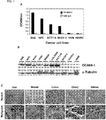
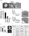
- the SAS cell lysates were prepared and purified by OCAb9-1-conjugated immunoaffinity chromatography. Silver stain and Western blotting demonstrated that OCAb9-1 recognized a target protein with a molecular weight of 39 kDa ( FIG. 2A ). Protein identity was analyzed by LC-MS/MS, we found the target protein of OCAb9-1 to be a human EpCAM ( FIG. 2B ). The specificity of OCAb9-1 to EpCAM was confirmed by conducting immunoprecipitation and Western blotting in parallel with commercial anti-EpCAM antibody 1144-1 (Santa Cruz Biotech; Epitomics respectively) ( FIG. 2C ). Western blotting ( FIG. 2E ) and FASC ( FIG. 2F ) with OCAb9-1 showed a dramatic decrease in signal after the knockdown of EpCAM by shRNA ( FIG. 2D ), confirming that OCAb9-1 specifically recognized EpCAM ( FIG. 2 ).
- OCAb9-1 cannot induce cancer cell apoptosis.
- SAS cancer cell lines
- NPC039, HCT116 and SKOV3 normal cell lines
- FACS Western blot and FACS analysis
- CDRs complementarity-determining regions
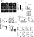
- EpCAM shRNA was knocked down by EpCAM shRNA in SAS cells.
- the growth rate ( FIG. 4A ), colony formation ( FIG. 4B ), migration ( FIG. 4C ) and invasion ability ( FIG. 4D ) were significantly reduced when EpCAM was knocked down.
- EpCAM knockdown affected cancer cell growth and induced cancer cell apoptosis in vitro
- EpAb2-6 could be used to directly inhibit tumor growth in vivo .
- Oral cancer xenografts were established and treated with either EpAb2-6 or control PBS. The volume of the tumors treated with EpAb2-6 became smaller than those of the two controls.
- the median overall survival of tumor-bearing mice after treatment with EpAb2-6 and PBS was 71 and 48 days, respectively ( FIG. 4F ).
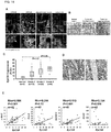
- Treatment of EpAb2-6 suppressed tumorsphere formation effectively ( FIG 5C ) and induced hypodiploid DNA content in both tumor and tumorsphere cells ( FIG 5A and 5B ).
- HCT116 3 ⁇ 10 6 cells
- HCT116 xenografts ⁇ 50 mm 3
- IFL 5-FU of 25 mg/kg + leucovorin of 10 mg/kg + irinotecan of 10 mg/kg
- the tumors in mice with treatment using combination of EpAb2-6 and IFL were found to be smaller than that in mice with treatment using IFL alone (*, P ⁇ 0.05) ( FIG. 6A ).
- the tumor size of the IFL group gradually increased to 1.6-fold that of the EpAb2-6 + IFL by day 25.
- the EpAb2-6 + IFL and IFL groups did not have significant changes in body weight during treatment period ( FIG. 6B ).
- the final average tumor weight in mice treated with IFL was 0.23 g, compared to 0.146 g in mice treated with EpAb2-6 + IFL, and 0.952 g in mice injected with PBS buffer ( FIG. 6C and D ).
- EpAb2-6 possessed the high affinity and potent activity for induction of cancer cell apoptosis, which suggested its potential as a therapeutic antibody.
- V H and V L segment of the EpAb2-6 mAbs from hybridoma cell lines ( FIG. 17 ).
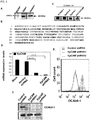
- the CDRs of EpAb2-6 were grafted onto human IgG1 backbone to create humanized EpAb2-6 (hEpAb2-6) ( FIG. 7A ).
- the hEpAb2-6 was expressed in CHO-K1 cells and purified from culture supernatants.
- the hEpAb2-6 that maintained the specificity of murine EpAb2-6 recognized both SAS and HCT116 cancer cells but not CCD-1112Sk normal cells.
- Cellular ELISA and Western blotting further demonstrated highly binding activities of hEpAb2-6 ( FIGs. 7B-D ).
- the affinity of EpAb2-6 and hEpAb2-6 for EpCAM was analyzed by surface plasmon resonance and was determined as 03491 nM and 0.6773 nM, respectively ( FIG. 18 ).
- FIGs. 7E-F in vitro studies using SAS and HCT116 cell lines found that hEpAb2-6 induced cancer cell apoptosis.
- the results reveal that humanized EpAb2-6 possesses high binding affinity to EpCAM, which suggested its potential applications in cancer therapy as therapeutic antibody, or tumor-targeted drug delivery, and imaging.
- Peptide sequences were aligned by using MacDNASIS software to analyze epitopes and binding motifs of EpAb2-6 antibody.
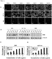
- the cDNA encoding the sequence covering the first EGF-like repeat (aa 27-59) of human EpCAM (EGF-I) or second EGF-like repeat (aa 66-135) of human EpCAM (EGF-II) was amplified by PCR. Overlapping PCR and PCR-based site-directed mutagenesis were used to introduce mutation shown in FIGs. 8A-B into the wild-type of EGF-I or EGF-II domain. A Western bloting was used for testing the reactivity of EpAb2-6 or EpAb3-5 antibodies toward variant EpCAM mutants.
- FIG. 8B shows the binding of each EpAb2-6 antibody to individual EpCAM mutant.
- the amino acids mutations in positions Y95 or D96 on EGF-II domain cause markedly reduction in the relative binding activity, hence Y95 and D96 are considered "essential" residues for the antibody binding.
- FIG. 8 shows the sequences VGAQNTVIC (SEQ ID NO: 62) in the EGF-like domain I and KPEGALQNNDGLYDPDCDE (SEQ ID NO: 63) in the EGF-like domain II of EpCAM.
- Tumor-initiating cells are endowed with the ability to be attached independently.
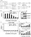
- HCT116 colon cancer cells in two kinds of anchorage-independent cultures, tumorsphere formation and anoikis-resistant selection ( FIG. 9A ).
- expression of EpCAM mRNA was elevated in both anoikis-resistance (4-folds) and spheroid (12-folds) cells, compared to that in adherent culture ( FIG. 9B left).
- the enrichment of cell surface EpCAM was found to increase by four folds in spheroid-formation ( FIG. 9C ) and two folds in anoikis-resistance cells (data not shown), when compared to that in adherent cells via flow cytometric analysis.
- EMT Epithelial-mesenchymal transition
- FIG. 11A Immunofluorescence analyses illustrated the changes occurred in EMT markers (epithelial markers E-cadherin and cytokeratin 18 in up-regulations) and mesenchymal marker (vimentin in down-regulation) after knockdown of EpCAM ( FIG. 11A ).
- Real-time PCR data showed that an increase in E-cadherin with a concomitant decrease in vimentin in both mRNA and protein levels were detected in EpCAM knockdown cells, compared to that in vector alone cells ( FIG. 11B ).
- FIGs. 11B and 11C Other EMT-regulatory transcriptional factors, such as snail and slug, were simultaneously reduced in both EpCAM knockdown cells and EpCAM-low expressed cells.
- Assessment of the effect of EpCAM on tumorigenic potential revealed that suppression of EpCAM resulted in the reduction of invasive and colony formation abilities in vitro ( FIGs. 11D and 11E ).
- suppression of EpCAM also inhibited xenograft tumor growth in vivo ( FIG. 11F ).
- RNA samples from primary tumor extraction demonstrated that expressions of EpCAM, reprogramming genes (c-Myc, Oct4, Nanog, and Sox2) and mesenchymal markers (vimentin and snail) were significantly diminished in EpCAM knockdown tumor cells, compared to that in vector alone ( FIG. 11G ).
- EpCAM contains an extracellular domain (EpEX), a transmembrane domain, and an intracellular domain (EpICD).
- EpEx is composed of two epidermal growth factor-like domains and a cysteine-poor region, while EpICD is composed of a short 26-amino acid.
- EpICD is composed of a short 26-amino acid.
- soluble EpICD was expressed increasingly in spheroid-derived tumor section than that in adherent tumor section.
- Western blot analysis confirmed a dominant band with molecular weight in 40 kDa (EpCAM-v5), and a minor band lower than 10 kDa (EpICD-v5) in 193T/EpCAM-v5 cells; however, the expression of soluble EpICD-v5 (10 kDa) was reduced in the treatment of DAPT ( FIG. 12B ).
- Treatment with DAPT suppressed the expressions of vimentin, snail, and slug ( FIG. 12C ), which was accompanied by decreasing tumor invasiveness and sphere-forming capacity ( FIGs. 12D and 12E ).
- chromatin immunoprecipitation assay indicating an occupancy of EpICD on c-MYC (proximal upstream region instead of exon 1), OCT4 (distal upstream region), NANOG (upstream region), and SOX2 (downstream region) promoters were detected in HCT116 cells ( FIGs. 12I-J ), whereas these phenomena were not found in normal nasal mucosa cells (NNM), which express rare level of EpCAM (data not shown).
- Extracellular Domain of EpCAM serves as an Activator for EpCAM's Signaling
- EpICD truncated vector (EpEX 291 -v5), which contained the extracellular and transmembrane domains of EpCAM, ( FIG. 13A ) to discern the importance of EpICD in controlling reprogramming gene expression.
- transfecting HCT116 cells with EpEX 291 -v5 also induced reporter activities of c-MYC, OCT4, NANOG, and SOX2 .
- Similar results were observed when treated with soluble EpEX (sEpEX) ( FIG. 13B ), suggesting that shedding or releasing of extracellular domain of EpCAM might also play a part in coordinating EpCAM's signaling.
- EpCAM/EpICD Expression in Human Colon Cancer Correlates with Reprogramming Factors
- EpCAM upregulates both reprogramming factors ( c-Myc, Oct4, Nanog, and Sox2 ) and EMT expressions.
- Proteolysis of EpCAM into EpEX and EpICD played an important role in mediating EpCAM's signaling.
- Nuclear translocation of EpICD regulates reprogramming gene expression while extracellular releasing of EpEX triggers further activation of EpICD.
- Suppression of EpCAM or blocking of EpICD cleavage decreases invasiveness, growth, and self-renewal ability both in vitro and in vivo .
- the results indicate that overexpression of EpCAM helps promote tumor-initiation and tumor-progression.
- the EpCAM mAbs can be used for cancer diagnosis and prognosis, and cancer-targeted therapy and imaging.
- EpCAM epithelial specific antigen
- ESA epithelial specific antigen
- EpICD is regulated by proteolysis by TNF- ⁇ converting enzyme (TACE) and ⁇ -secretase (presenilin 2; PS2), followed by collaboration with FHL2 and Tcf/Lefl.
- TACE TNF- ⁇ converting enzyme
- PS2 ⁇ -secretase
- EpICD EpICD-induced cleavage and nuclear translocation of EpICD, which was accompanied by the suppression of reprogramming factors and EMT gene expressions, thereby inhibiting tumor self-renewal and invasiveness.
- EpICD soluble EpICD in either cytoplasm or nucleus was not expressed homogenously in all tumor cells, suggesting that the cleavage of EpICD might be a dynamical process.
- EpICD we also found that releasing of EpEX may trigger EpCAM's signaling event. The release of EpEX in supernatants was increased by serum concentration, and the addition of either DAPT or TAPI blocked the liberation of EpEX and EpICD.
- EpCAM's activation may initialize EpCAM's signaling and that its release may further trigger EpCAM's activation.
- the invention relates to novel EpCAM mAbs with extreme binding affinity against EpCAM, which displayed effective tumor inhibitory activity and thus could provide promising therapeutic meaning.
- EpCAM especially EpICD
- releasing of EpEX may trigger EpCAM's signaling event via autocrine or paracrine effect. Therefore, development and application of inhibitors or antibodies for EpCAM and/or EpEX in either treating or detecting are helpful to eradicate tumor and TICs as well as for tumor targeting imaging.
Landscapes
- Health & Medical Sciences (AREA)
- Chemical & Material Sciences (AREA)
- Life Sciences & Earth Sciences (AREA)
- Immunology (AREA)
- Organic Chemistry (AREA)
- Molecular Biology (AREA)
- Medicinal Chemistry (AREA)
- General Health & Medical Sciences (AREA)
- Engineering & Computer Science (AREA)
- Biochemistry (AREA)
- Urology & Nephrology (AREA)
- Hematology (AREA)
- Biomedical Technology (AREA)
- Biophysics (AREA)
- Genetics & Genomics (AREA)
- Proteomics, Peptides & Aminoacids (AREA)
- Cell Biology (AREA)
- Analytical Chemistry (AREA)
- Physics & Mathematics (AREA)
- Biotechnology (AREA)
- Hospice & Palliative Care (AREA)
- Pathology (AREA)
- Microbiology (AREA)
- General Physics & Mathematics (AREA)
- Oncology (AREA)
- Food Science & Technology (AREA)
- Pharmacology & Pharmacy (AREA)
- Animal Behavior & Ethology (AREA)
- Public Health (AREA)
- Veterinary Medicine (AREA)
- Nuclear Medicine, Radiotherapy & Molecular Imaging (AREA)
- General Chemical & Material Sciences (AREA)
- Chemical Kinetics & Catalysis (AREA)
- Bioinformatics & Cheminformatics (AREA)
- Peptides Or Proteins (AREA)
- Medicines Containing Antibodies Or Antigens For Use As Internal Diagnostic Agents (AREA)
- Preparation Of Compounds By Using Micro-Organisms (AREA)
- Medicinal Preparation (AREA)
Applications Claiming Priority (2)
| Application Number | Priority Date | Filing Date | Title |
|---|---|---|---|
| US201261606220P | 2012-03-02 | 2012-03-02 | |
| PCT/US2013/028667 WO2013131001A1 (en) | 2012-03-02 | 2013-03-01 | ANTI-EPITHELIAL CELL ADHESION MOLECULE (EpCAM) ANTIBODIES AND METHODS OF USE THEREOF |
Publications (3)
| Publication Number | Publication Date |
|---|---|
| EP2819695A1 EP2819695A1 (en) | 2015-01-07 |
| EP2819695A4 EP2819695A4 (en) | 2016-01-20 |
| EP2819695B1 true EP2819695B1 (en) | 2018-06-27 |
Family
ID=49083343
Family Applications (1)
| Application Number | Title | Priority Date | Filing Date |
|---|---|---|---|
| EP13754991.1A Active EP2819695B1 (en) | 2012-03-02 | 2013-03-01 | ANTI-EPITHELIAL CELL ADHESION MOLECULE (EpCAM) ANTIBODIES AND METHODS OF USE THEREOF |
Country Status (6)
| Country | Link |
|---|---|
| US (1) | US9187558B2 (enExample) |
| EP (1) | EP2819695B1 (enExample) |
| JP (1) | JP6163502B2 (enExample) |
| CN (1) | CN104168916B (enExample) |
| TW (1) | TWI485161B (enExample) |
| WO (1) | WO2013131001A1 (enExample) |
Families Citing this family (21)
| Publication number | Priority date | Publication date | Assignee | Title |
|---|---|---|---|---|
| WO2013003624A2 (en) | 2011-06-29 | 2013-01-03 | Academia Sinica | The capture, purification and release of biological substance using a surface coating |
| EP3126814B1 (en) | 2014-04-01 | 2019-06-12 | Academia Sinica | Methods and systems for cancer diagnosis and prognosis |
| WO2017023704A1 (en) * | 2015-07-31 | 2017-02-09 | Sutro Biopharma, Inc. | ANTI-EpCAM ANTIBODIES, COMPOSITIONS COMPRISING ANTI-EpCAM ANTIBODIES AND METHODS OF MAKING AND USING ANTI-EpCAM ANTIBODIES |
| AU2016358296B2 (en) * | 2015-11-19 | 2020-05-21 | Revitope Limited | Functional antibody fragment complementation for a two-components system for redirected killing of unwanted cells |
| US10107726B2 (en) | 2016-03-16 | 2018-10-23 | Cellmax, Ltd. | Collection of suspended cells using a transferable membrane |
| KR20190038567A (ko) | 2016-07-26 | 2019-04-08 | 테사 테라퓨틱스 피티이. 엘티디. | 키메라 항원 수용체 |
| JP6433951B2 (ja) * | 2016-08-09 | 2018-12-05 | 東芝デジタルソリューションズ株式会社 | ネットワーク監視装置およびプログラム |
| US10787499B2 (en) | 2017-02-13 | 2020-09-29 | Regents Of The University Of Minnesota | EpCAM targeted polypeptides, conjugates thereof, and methods of use thereof |
| WO2020018964A1 (en) | 2018-07-20 | 2020-01-23 | Fred Hutchinson Cancer Research Center | Compositions and methods for controlled expression of antigen-specific receptors |
| WO2020051141A1 (en) * | 2018-09-04 | 2020-03-12 | Academia Sinica | A medium composition and method for culturing mesenchymal stem cells |
| MA55305A (fr) * | 2019-03-11 | 2022-01-19 | Janssen Biotech Inc | Anticorps bispécifiques anti-v béta 17/anti-cd123 |
| AU2020292304B2 (en) * | 2019-06-11 | 2023-03-30 | Bioatla, Inc. | Conditionally active anti-EpCam antibodies, antibody fragments, their immunoconjugates and uses thereof |
| BR112022009259A2 (pt) * | 2019-11-14 | 2022-08-02 | Academia Sinica | Métodos para tratar, inibir ou eliminar um câncer em um indivíduo |
| CN110950959B (zh) * | 2020-02-25 | 2020-07-03 | 和铂医药(上海)有限责任公司 | 靶向EpCAM的抗体及其制备和应用 |
| CN112094350B (zh) * | 2020-06-01 | 2024-01-05 | 普众发现医药科技(上海)有限公司 | 可应用于肿瘤细胞捕获的小鼠抗细胞表面糖蛋白cd326的单克隆抗体 |
| EP4190808A4 (en) * | 2020-08-19 | 2024-07-31 | Suzhou Immunofoco Biotechnology Co., Ltd. | Humanized antibody, chimeric antigen receptor, nucleic acid, vector, cell and use |
| CA3205465A1 (en) * | 2020-12-18 | 2022-06-23 | Bioardis, Llc | Epcam binding molecules and uses thereof |
| TWI754504B (zh) * | 2020-12-25 | 2022-02-01 | 龍華科技大學 | 棉花棒棒體之膠體檢測系統及其檢測方法 |
| KR20240026954A (ko) * | 2021-06-25 | 2024-02-29 | 아카데미아 시니카 | 상피 세포 부착 분자(epcam) 억제제 및 wnt 억제제와의 조합된 암 요법 |
| KR20240026985A (ko) * | 2021-06-25 | 2024-02-29 | 아카데미아 시니카 | 상피 세포 부착 분자(epcam) 억제제 및 간세포 성장 인자 수용체(hgfr) 억제제와의 조합된 암 요법 |
| JP2023069350A (ja) * | 2021-11-05 | 2023-05-18 | シスメックス株式会社 | 抗体の製造方法及び抗体 |
Family Cites Families (15)
| Publication number | Priority date | Publication date | Assignee | Title |
|---|---|---|---|---|
| BRPI9809391B8 (pt) * | 1997-04-14 | 2021-05-25 | Amgen Res Munich Gmbh | processo para a produção de um receptor de antígeno anti-humano, anticorpo humano e composição farmacêutica |
| EP1242823B1 (en) * | 1999-12-27 | 2007-07-04 | Crucell Holland B.V. | Selecting library members capable of binding to epitopes |
| DK1383785T3 (da) * | 2001-05-03 | 2011-05-23 | Merck Patent Gmbh | Rekombinant tumorspecifikt antistof og anvendelse deraf |
| US20070122406A1 (en) * | 2005-07-08 | 2007-05-31 | Xencor, Inc. | Optimized proteins that target Ep-CAM |
| US20100311954A1 (en) * | 2002-03-01 | 2010-12-09 | Xencor, Inc. | Optimized Proteins that Target Ep-CAM |
| US7696320B2 (en) * | 2004-08-24 | 2010-04-13 | Domantis Limited | Ligands that have binding specificity for VEGF and/or EGFR and methods of use therefor |
| EP1629275A2 (de) * | 2003-06-02 | 2006-03-01 | Igeneon Krebs-Immuntherapie Forschungs- und Entwicklungs-AG | Verfahren zur selektion von epitopen zur immuntherapie |
| US20050180979A1 (en) * | 2004-02-13 | 2005-08-18 | Micromet Ag | Anti-EpCAM immunoglobulins |
| US7521195B1 (en) * | 2005-07-21 | 2009-04-21 | Celera Corporation | Lung disease targets and uses thereof |
| EP2142570B1 (en) * | 2007-04-04 | 2011-06-15 | SIGMA-TAU Industrie Farmaceutiche Riunite S.p.A. | Anti-epcam antibody and uses thereof |
| JP2011193728A (ja) * | 2008-07-17 | 2011-10-06 | Murata Mfg Co Ltd | EpCAMに結合能を有するペプチド |
| GB0909904D0 (en) * | 2009-06-09 | 2009-07-22 | Affitech As | Product |
| EP2480687B1 (en) * | 2009-09-21 | 2015-01-28 | Paul Walfish | Methods and compositions for the diagnosis and treatment of thyroid cancer |
| WO2011079283A1 (en) | 2009-12-23 | 2011-06-30 | Bioalliance C.V. | Anti-epcam antibodies that induce apoptosis of cancer cells and methods using same |
| CA2798202A1 (en) * | 2010-05-04 | 2011-11-10 | Paul Walfish | Method for the diagnosis of epithelial cancers by the detection of ep-icd polypeptide |
-
2013
- 2013-03-01 EP EP13754991.1A patent/EP2819695B1/en active Active
- 2013-03-01 WO PCT/US2013/028667 patent/WO2013131001A1/en not_active Ceased
- 2013-03-01 TW TW102107344A patent/TWI485161B/zh active
- 2013-03-01 CN CN201380012187.0A patent/CN104168916B/zh active Active
- 2013-03-01 US US14/381,362 patent/US9187558B2/en active Active
- 2013-03-01 JP JP2014560096A patent/JP6163502B2/ja active Active
Non-Patent Citations (1)
| Title |
|---|
| None * |
Also Published As
| Publication number | Publication date |
|---|---|
| US9187558B2 (en) | 2015-11-17 |
| EP2819695A1 (en) | 2015-01-07 |
| CN104168916B (zh) | 2017-07-04 |
| JP2015517982A (ja) | 2015-06-25 |
| EP2819695A4 (en) | 2016-01-20 |
| TWI485161B (zh) | 2015-05-21 |
| TW201350507A (zh) | 2013-12-16 |
| CN104168916A (zh) | 2014-11-26 |
| US20150017230A1 (en) | 2015-01-15 |
| JP6163502B2 (ja) | 2017-07-12 |
| WO2013131001A1 (en) | 2013-09-06 |
Similar Documents
| Publication | Publication Date | Title |
|---|---|---|
| EP2819695B1 (en) | ANTI-EPITHELIAL CELL ADHESION MOLECULE (EpCAM) ANTIBODIES AND METHODS OF USE THEREOF | |
| AU2012341450B2 (en) | Anti-human TROP-2 antibody having antitumor activity in vivo | |
| US9670287B2 (en) | Anti-human TROP-2 antibody having anti-tumor activity in vivo | |
| EP2699603B1 (en) | Human monoclonal antibodies specific for glypican-3 and use thereof | |
| JP5788384B2 (ja) | 形質転換増殖因子アルファに結合し、Ras遺伝子変異癌に対して増殖抑制活性を有する抗体 | |
| JP6280040B2 (ja) | invivoで抗腫瘍活性を有する抗ヒトDlk−1抗体 | |
| AU2016249991B2 (en) | Anti-VEGFR2 human antibody for anti-angiogenic and targeted cancer therapy | |
| EP2322610B1 (en) | Anti-human clcp1 antibody and use thereof | |
| HK1219965B (en) | Human monoclonal antibodies specific for glypican-3 and use thereof | |
| HK1228931B (en) | Human monoclonal antibodies specific for glypican-3 and use thereof | |
| HK1198042A (en) | Anti-human trop-2 antibody exhibiting antitumor activity in vivo | |
| HK1198042B (en) | Anti-human trop-2 antibody exhibiting antitumor activity in vivo |
Legal Events
| Date | Code | Title | Description |
|---|---|---|---|
| PUAI | Public reference made under article 153(3) epc to a published international application that has entered the european phase |
Free format text: ORIGINAL CODE: 0009012 |
|
| 17P | Request for examination filed |
Effective date: 20140828 |
|
| AK | Designated contracting states |
Kind code of ref document: A1 Designated state(s): AL AT BE BG CH CY CZ DE DK EE ES FI FR GB GR HR HU IE IS IT LI LT LU LV MC MK MT NL NO PL PT RO RS SE SI SK SM TR |
|
| AX | Request for extension of the european patent |
Extension state: BA ME |
|
| DAX | Request for extension of the european patent (deleted) | ||
| RIC1 | Information provided on ipc code assigned before grant |
Ipc: C07K 16/30 20060101AFI20150831BHEP Ipc: C07K 16/18 20060101ALI20150831BHEP Ipc: G01N 33/574 20060101ALI20150831BHEP Ipc: A61K 39/395 20060101ALI20150831BHEP Ipc: A61K 39/00 20060101ALI20150831BHEP |
|
| REG | Reference to a national code |
Ref country code: DE Ref legal event code: R079 Ref document number: 602013039432 Country of ref document: DE Free format text: PREVIOUS MAIN CLASS: A61K0039000000 Ipc: C07K0016300000 |
|
| RA4 | Supplementary search report drawn up and despatched (corrected) |
Effective date: 20151222 |
|
| RIC1 | Information provided on ipc code assigned before grant |
Ipc: C07K 16/18 20060101ALI20151216BHEP Ipc: A61K 39/395 20060101ALI20151216BHEP Ipc: G01N 33/574 20060101ALI20151216BHEP Ipc: C07K 16/30 20060101AFI20151216BHEP Ipc: A61K 39/00 20060101ALI20151216BHEP |
|
| STAA | Information on the status of an ep patent application or granted ep patent |
Free format text: STATUS: EXAMINATION IS IN PROGRESS |
|
| 17Q | First examination report despatched |
Effective date: 20161111 |
|
| GRAP | Despatch of communication of intention to grant a patent |
Free format text: ORIGINAL CODE: EPIDOSNIGR1 |
|
| STAA | Information on the status of an ep patent application or granted ep patent |
Free format text: STATUS: GRANT OF PATENT IS INTENDED |
|
| INTG | Intention to grant announced |
Effective date: 20180130 |
|
| GRAS | Grant fee paid |
Free format text: ORIGINAL CODE: EPIDOSNIGR3 |
|
| GRAA | (expected) grant |
Free format text: ORIGINAL CODE: 0009210 |
|
| STAA | Information on the status of an ep patent application or granted ep patent |
Free format text: STATUS: THE PATENT HAS BEEN GRANTED |
|
| AK | Designated contracting states |
Kind code of ref document: B1 Designated state(s): AL AT BE BG CH CY CZ DE DK EE ES FI FR GB GR HR HU IE IS IT LI LT LU LV MC MK MT NL NO PL PT RO RS SE SI SK SM TR |
|
| REG | Reference to a national code |
Ref country code: GB Ref legal event code: FG4D |
|
| REG | Reference to a national code |
Ref country code: AT Ref legal event code: REF Ref document number: 1012252 Country of ref document: AT Kind code of ref document: T Effective date: 20180715 |
|
| REG | Reference to a national code |
Ref country code: IE Ref legal event code: FG4D |
|
| REG | Reference to a national code |
Ref country code: DE Ref legal event code: R096 Ref document number: 602013039432 Country of ref document: DE |
|
| REG | Reference to a national code |
Ref country code: SE Ref legal event code: TRGR |
|
| PG25 | Lapsed in a contracting state [announced via postgrant information from national office to epo] |
Ref country code: FI Free format text: LAPSE BECAUSE OF FAILURE TO SUBMIT A TRANSLATION OF THE DESCRIPTION OR TO PAY THE FEE WITHIN THE PRESCRIBED TIME-LIMIT Effective date: 20180627 Ref country code: NO Free format text: LAPSE BECAUSE OF FAILURE TO SUBMIT A TRANSLATION OF THE DESCRIPTION OR TO PAY THE FEE WITHIN THE PRESCRIBED TIME-LIMIT Effective date: 20180927 Ref country code: LT Free format text: LAPSE BECAUSE OF FAILURE TO SUBMIT A TRANSLATION OF THE DESCRIPTION OR TO PAY THE FEE WITHIN THE PRESCRIBED TIME-LIMIT Effective date: 20180627 Ref country code: BG Free format text: LAPSE BECAUSE OF FAILURE TO SUBMIT A TRANSLATION OF THE DESCRIPTION OR TO PAY THE FEE WITHIN THE PRESCRIBED TIME-LIMIT Effective date: 20180927 |
|
| REG | Reference to a national code |
Ref country code: NL Ref legal event code: MP Effective date: 20180627 |
|
| REG | Reference to a national code |
Ref country code: LT Ref legal event code: MG4D |
|
| PG25 | Lapsed in a contracting state [announced via postgrant information from national office to epo] |
Ref country code: LV Free format text: LAPSE BECAUSE OF FAILURE TO SUBMIT A TRANSLATION OF THE DESCRIPTION OR TO PAY THE FEE WITHIN THE PRESCRIBED TIME-LIMIT Effective date: 20180627 Ref country code: RS Free format text: LAPSE BECAUSE OF FAILURE TO SUBMIT A TRANSLATION OF THE DESCRIPTION OR TO PAY THE FEE WITHIN THE PRESCRIBED TIME-LIMIT Effective date: 20180627 Ref country code: GR Free format text: LAPSE BECAUSE OF FAILURE TO SUBMIT A TRANSLATION OF THE DESCRIPTION OR TO PAY THE FEE WITHIN THE PRESCRIBED TIME-LIMIT Effective date: 20180928 Ref country code: HR Free format text: LAPSE BECAUSE OF FAILURE TO SUBMIT A TRANSLATION OF THE DESCRIPTION OR TO PAY THE FEE WITHIN THE PRESCRIBED TIME-LIMIT Effective date: 20180627 |
|
| PG25 | Lapsed in a contracting state [announced via postgrant information from national office to epo] |
Ref country code: NL Free format text: LAPSE BECAUSE OF FAILURE TO SUBMIT A TRANSLATION OF THE DESCRIPTION OR TO PAY THE FEE WITHIN THE PRESCRIBED TIME-LIMIT Effective date: 20180627 |
|
| PG25 | Lapsed in a contracting state [announced via postgrant information from national office to epo] |
Ref country code: EE Free format text: LAPSE BECAUSE OF FAILURE TO SUBMIT A TRANSLATION OF THE DESCRIPTION OR TO PAY THE FEE WITHIN THE PRESCRIBED TIME-LIMIT Effective date: 20180627 Ref country code: IS Free format text: LAPSE BECAUSE OF FAILURE TO SUBMIT A TRANSLATION OF THE DESCRIPTION OR TO PAY THE FEE WITHIN THE PRESCRIBED TIME-LIMIT Effective date: 20181027 Ref country code: PL Free format text: LAPSE BECAUSE OF FAILURE TO SUBMIT A TRANSLATION OF THE DESCRIPTION OR TO PAY THE FEE WITHIN THE PRESCRIBED TIME-LIMIT Effective date: 20180627 Ref country code: SK Free format text: LAPSE BECAUSE OF FAILURE TO SUBMIT A TRANSLATION OF THE DESCRIPTION OR TO PAY THE FEE WITHIN THE PRESCRIBED TIME-LIMIT Effective date: 20180627 Ref country code: CZ Free format text: LAPSE BECAUSE OF FAILURE TO SUBMIT A TRANSLATION OF THE DESCRIPTION OR TO PAY THE FEE WITHIN THE PRESCRIBED TIME-LIMIT Effective date: 20180627 Ref country code: RO Free format text: LAPSE BECAUSE OF FAILURE TO SUBMIT A TRANSLATION OF THE DESCRIPTION OR TO PAY THE FEE WITHIN THE PRESCRIBED TIME-LIMIT Effective date: 20180627 |
|
| PG25 | Lapsed in a contracting state [announced via postgrant information from national office to epo] |
Ref country code: ES Free format text: LAPSE BECAUSE OF FAILURE TO SUBMIT A TRANSLATION OF THE DESCRIPTION OR TO PAY THE FEE WITHIN THE PRESCRIBED TIME-LIMIT Effective date: 20180627 Ref country code: SM Free format text: LAPSE BECAUSE OF FAILURE TO SUBMIT A TRANSLATION OF THE DESCRIPTION OR TO PAY THE FEE WITHIN THE PRESCRIBED TIME-LIMIT Effective date: 20180627 Ref country code: IT Free format text: LAPSE BECAUSE OF FAILURE TO SUBMIT A TRANSLATION OF THE DESCRIPTION OR TO PAY THE FEE WITHIN THE PRESCRIBED TIME-LIMIT Effective date: 20180627 |
|
| REG | Reference to a national code |
Ref country code: DE Ref legal event code: R097 Ref document number: 602013039432 Country of ref document: DE |
|
| PLBE | No opposition filed within time limit |
Free format text: ORIGINAL CODE: 0009261 |
|
| STAA | Information on the status of an ep patent application or granted ep patent |
Free format text: STATUS: NO OPPOSITION FILED WITHIN TIME LIMIT |
|
| PG25 | Lapsed in a contracting state [announced via postgrant information from national office to epo] |
Ref country code: DK Free format text: LAPSE BECAUSE OF FAILURE TO SUBMIT A TRANSLATION OF THE DESCRIPTION OR TO PAY THE FEE WITHIN THE PRESCRIBED TIME-LIMIT Effective date: 20180627 |
|
| 26N | No opposition filed |
Effective date: 20190328 |
|
| PG25 | Lapsed in a contracting state [announced via postgrant information from national office to epo] |
Ref country code: SI Free format text: LAPSE BECAUSE OF FAILURE TO SUBMIT A TRANSLATION OF THE DESCRIPTION OR TO PAY THE FEE WITHIN THE PRESCRIBED TIME-LIMIT Effective date: 20180627 |
|
| PG25 | Lapsed in a contracting state [announced via postgrant information from national office to epo] |
Ref country code: MC Free format text: LAPSE BECAUSE OF FAILURE TO SUBMIT A TRANSLATION OF THE DESCRIPTION OR TO PAY THE FEE WITHIN THE PRESCRIBED TIME-LIMIT Effective date: 20180627 |
|
| PG25 | Lapsed in a contracting state [announced via postgrant information from national office to epo] |
Ref country code: LU Free format text: LAPSE BECAUSE OF NON-PAYMENT OF DUE FEES Effective date: 20190301 Ref country code: AL Free format text: LAPSE BECAUSE OF FAILURE TO SUBMIT A TRANSLATION OF THE DESCRIPTION OR TO PAY THE FEE WITHIN THE PRESCRIBED TIME-LIMIT Effective date: 20180627 |
|
| REG | Reference to a national code |
Ref country code: BE Ref legal event code: MM Effective date: 20190331 |
|
| PG25 | Lapsed in a contracting state [announced via postgrant information from national office to epo] |
Ref country code: IE Free format text: LAPSE BECAUSE OF NON-PAYMENT OF DUE FEES Effective date: 20190301 |
|
| REG | Reference to a national code |
Ref country code: CH Ref legal event code: PCAR Free format text: NEW ADDRESS: ROUTE DU COUTSET 18, 1485 NUVILLY (CH) |
|
| PG25 | Lapsed in a contracting state [announced via postgrant information from national office to epo] |
Ref country code: BE Free format text: LAPSE BECAUSE OF NON-PAYMENT OF DUE FEES Effective date: 20190331 |
|
| PG25 | Lapsed in a contracting state [announced via postgrant information from national office to epo] |
Ref country code: TR Free format text: LAPSE BECAUSE OF FAILURE TO SUBMIT A TRANSLATION OF THE DESCRIPTION OR TO PAY THE FEE WITHIN THE PRESCRIBED TIME-LIMIT Effective date: 20180627 |
|
| PG25 | Lapsed in a contracting state [announced via postgrant information from national office to epo] |
Ref country code: MT Free format text: LAPSE BECAUSE OF NON-PAYMENT OF DUE FEES Effective date: 20190301 Ref country code: PT Free format text: LAPSE BECAUSE OF FAILURE TO SUBMIT A TRANSLATION OF THE DESCRIPTION OR TO PAY THE FEE WITHIN THE PRESCRIBED TIME-LIMIT Effective date: 20181029 |
|
| REG | Reference to a national code |
Ref country code: AT Ref legal event code: UEP Ref document number: 1012252 Country of ref document: AT Kind code of ref document: T Effective date: 20180627 |
|
| REG | Reference to a national code |
Ref country code: DE Ref legal event code: R082 Ref document number: 602013039432 Country of ref document: DE Representative=s name: VENNER SHIPLEY GERMANY LLP, DE Ref country code: DE Ref legal event code: R082 Ref document number: 602013039432 Country of ref document: DE Representative=s name: VENNER SHIPLEY LLP, DE |
|
| PG25 | Lapsed in a contracting state [announced via postgrant information from national office to epo] |
Ref country code: CY Free format text: LAPSE BECAUSE OF FAILURE TO SUBMIT A TRANSLATION OF THE DESCRIPTION OR TO PAY THE FEE WITHIN THE PRESCRIBED TIME-LIMIT Effective date: 20180627 |
|
| PG25 | Lapsed in a contracting state [announced via postgrant information from national office to epo] |
Ref country code: HU Free format text: LAPSE BECAUSE OF FAILURE TO SUBMIT A TRANSLATION OF THE DESCRIPTION OR TO PAY THE FEE WITHIN THE PRESCRIBED TIME-LIMIT; INVALID AB INITIO Effective date: 20130301 |
|
| PG25 | Lapsed in a contracting state [announced via postgrant information from national office to epo] |
Ref country code: MK Free format text: LAPSE BECAUSE OF FAILURE TO SUBMIT A TRANSLATION OF THE DESCRIPTION OR TO PAY THE FEE WITHIN THE PRESCRIBED TIME-LIMIT Effective date: 20180627 |
|
| PGFP | Annual fee paid to national office [announced via postgrant information from national office to epo] |
Ref country code: SE Payment date: 20250312 Year of fee payment: 13 |
|
| PGFP | Annual fee paid to national office [announced via postgrant information from national office to epo] |
Ref country code: DE Payment date: 20250327 Year of fee payment: 13 |
|
| PGFP | Annual fee paid to national office [announced via postgrant information from national office to epo] |
Ref country code: AT Payment date: 20250219 Year of fee payment: 13 |
|
| PGFP | Annual fee paid to national office [announced via postgrant information from national office to epo] |
Ref country code: FR Payment date: 20250325 Year of fee payment: 13 |
|
| PGFP | Annual fee paid to national office [announced via postgrant information from national office to epo] |
Ref country code: GB Payment date: 20250327 Year of fee payment: 13 |
|
| PGFP | Annual fee paid to national office [announced via postgrant information from national office to epo] |
Ref country code: CH Payment date: 20250401 Year of fee payment: 13 |