EP2818478B1 - Humanized IL-6 and IL-6 receptor - Google Patents
Humanized IL-6 and IL-6 receptor Download PDFInfo
- Publication number
- EP2818478B1 EP2818478B1 EP14177863.9A EP14177863A EP2818478B1 EP 2818478 B1 EP2818478 B1 EP 2818478B1 EP 14177863 A EP14177863 A EP 14177863A EP 2818478 B1 EP2818478 B1 EP 2818478B1
- Authority
- EP
- European Patent Office
- Prior art keywords
- human
- murine
- gene
- mouse
- humanized
- Prior art date
- Legal status (The legal status is an assumption and is not a legal conclusion. Google has not performed a legal analysis and makes no representation as to the accuracy of the status listed.)
- Active
Links
- 108090001005 Interleukin-6 Proteins 0.000 title claims description 139
- 102000004889 Interleukin-6 Human genes 0.000 title claims description 126
- 108010038501 Interleukin-6 Receptors Proteins 0.000 title description 43
- 102000010781 Interleukin-6 Receptors Human genes 0.000 title description 41
- 101001076408 Homo sapiens Interleukin-6 Proteins 0.000 claims description 127
- 102000052611 human IL6 Human genes 0.000 claims description 122
- 108090000623 proteins and genes Proteins 0.000 claims description 98
- 241001529936 Murinae Species 0.000 claims description 87
- 230000001105 regulatory effect Effects 0.000 claims description 32
- 210000001185 bone marrow Anatomy 0.000 claims description 27
- 101100286713 Homo sapiens IL6 gene Proteins 0.000 claims description 24
- 101000599048 Homo sapiens Interleukin-6 receptor subunit alpha Proteins 0.000 claims description 21
- 108700024394 Exon Proteins 0.000 claims description 17
- 206010035485 plasmacytosis Diseases 0.000 claims description 17
- 210000000952 spleen Anatomy 0.000 claims description 17
- 210000003593 megakaryocyte Anatomy 0.000 claims description 16
- 210000004436 artificial bacterial chromosome Anatomy 0.000 claims description 11
- 210000001165 lymph node Anatomy 0.000 claims description 11
- 210000004180 plasmocyte Anatomy 0.000 claims description 10
- 230000002159 abnormal effect Effects 0.000 claims description 9
- 238000000034 method Methods 0.000 claims description 9
- 206010018364 Glomerulonephritis Diseases 0.000 claims description 8
- 239000012634 fragment Substances 0.000 claims description 8
- 206010041660 Splenomegaly Diseases 0.000 claims description 7
- 208000001647 Renal Insufficiency Diseases 0.000 claims description 6
- 206010061989 glomerulosclerosis Diseases 0.000 claims description 6
- 230000003834 intracellular effect Effects 0.000 claims description 6
- 201000006370 kidney failure Diseases 0.000 claims description 6
- 208000019758 Hypergammaglobulinemia Diseases 0.000 claims description 5
- 241000699666 Mus <mouse, genus> Species 0.000 description 176
- 241000699670 Mus sp. Species 0.000 description 136
- 229940100601 interleukin-6 Drugs 0.000 description 107
- 210000004027 cell Anatomy 0.000 description 69
- 210000003719 b-lymphocyte Anatomy 0.000 description 49
- 108020004414 DNA Proteins 0.000 description 34
- 101001076414 Mus musculus Interleukin-6 Proteins 0.000 description 29
- 238000011830 transgenic mouse model Methods 0.000 description 26
- 241000699660 Mus musculus Species 0.000 description 25
- 206010048998 Acute phase reaction Diseases 0.000 description 23
- 230000004658 acute-phase response Effects 0.000 description 23
- 238000012413 Fluorescence activated cell sorting analysis Methods 0.000 description 21
- 101000599053 Mus musculus Interleukin-6 receptor subunit alpha Proteins 0.000 description 20
- 239000002773 nucleotide Substances 0.000 description 20
- 125000003729 nucleotide group Chemical group 0.000 description 20
- 230000014509 gene expression Effects 0.000 description 17
- 239000000523 sample Substances 0.000 description 16
- 210000002966 serum Anatomy 0.000 description 16
- 102000004169 proteins and genes Human genes 0.000 description 15
- 230000003393 splenic effect Effects 0.000 description 14
- 238000010186 staining Methods 0.000 description 14
- 230000009261 transgenic effect Effects 0.000 description 14
- 238000011144 upstream manufacturing Methods 0.000 description 13
- 241001465754 Metazoa Species 0.000 description 12
- 101100286719 Mus musculus Il6 gene Proteins 0.000 description 12
- 210000001744 T-lymphocyte Anatomy 0.000 description 12
- 238000003556 assay Methods 0.000 description 12
- 101150047862 hil-6 gene Proteins 0.000 description 11
- 210000004185 liver Anatomy 0.000 description 11
- 108700028369 Alleles Proteins 0.000 description 10
- 150000001413 amino acids Chemical class 0.000 description 10
- 210000004369 blood Anatomy 0.000 description 10
- 239000008280 blood Substances 0.000 description 10
- 230000001086 cytosolic effect Effects 0.000 description 10
- 239000002299 complementary DNA Substances 0.000 description 9
- 230000007170 pathology Effects 0.000 description 9
- 102000005962 receptors Human genes 0.000 description 9
- 108020003175 receptors Proteins 0.000 description 9
- 238000011529 RT qPCR Methods 0.000 description 8
- 108700019146 Transgenes Proteins 0.000 description 8
- 239000003623 enhancer Substances 0.000 description 8
- 230000001404 mediated effect Effects 0.000 description 8
- 210000000822 natural killer cell Anatomy 0.000 description 8
- 238000003753 real-time PCR Methods 0.000 description 8
- 230000008685 targeting Effects 0.000 description 8
- 238000011740 C57BL/6 mouse Methods 0.000 description 7
- 102100040019 Interferon alpha-1/13 Human genes 0.000 description 7
- 210000004556 brain Anatomy 0.000 description 7
- 238000011577 humanized mouse model Methods 0.000 description 7
- 210000003734 kidney Anatomy 0.000 description 7
- 239000000203 mixture Substances 0.000 description 7
- 230000007342 reactive astrogliosis Effects 0.000 description 7
- 108010042685 trinitrophenyl keyhole limpet hemocyanin Proteins 0.000 description 7
- 210000004885 white matter Anatomy 0.000 description 7
- 101000884271 Homo sapiens Signal transducer CD24 Proteins 0.000 description 6
- 102100027584 Protein c-Fos Human genes 0.000 description 6
- 241000700159 Rattus Species 0.000 description 6
- 102100038081 Signal transducer CD24 Human genes 0.000 description 6
- 241000779819 Syncarpia glomulifera Species 0.000 description 6
- 230000004913 activation Effects 0.000 description 6
- 239000005557 antagonist Substances 0.000 description 6
- 230000002025 microglial effect Effects 0.000 description 6
- 239000001739 pinus spp. Substances 0.000 description 6
- 230000002441 reversible effect Effects 0.000 description 6
- 229940036248 turpentine Drugs 0.000 description 6
- NHBKXEKEPDILRR-UHFFFAOYSA-N 2,3-bis(butanoylsulfanyl)propyl butanoate Chemical compound CCCC(=O)OCC(SC(=O)CCC)CSC(=O)CCC NHBKXEKEPDILRR-UHFFFAOYSA-N 0.000 description 5
- 108010062271 Acute-Phase Proteins Proteins 0.000 description 5
- 102000011767 Acute-Phase Proteins Human genes 0.000 description 5
- 206010025323 Lymphomas Diseases 0.000 description 5
- 206010028289 Muscle atrophy Diseases 0.000 description 5
- 238000004458 analytical method Methods 0.000 description 5
- 230000011712 cell development Effects 0.000 description 5
- 230000001419 dependent effect Effects 0.000 description 5
- 238000011161 development Methods 0.000 description 5
- 230000018109 developmental process Effects 0.000 description 5
- 239000003814 drug Substances 0.000 description 5
- 238000005516 engineering process Methods 0.000 description 5
- 238000001943 fluorescence-activated cell sorting Methods 0.000 description 5
- 238000012239 gene modification Methods 0.000 description 5
- 230000005017 genetic modification Effects 0.000 description 5
- 235000013617 genetically modified food Nutrition 0.000 description 5
- 210000001161 mammalian embryo Anatomy 0.000 description 5
- 201000000585 muscular atrophy Diseases 0.000 description 5
- 108091032973 (ribonucleotides)n+m Proteins 0.000 description 4
- 101001046686 Homo sapiens Integrin alpha-M Proteins 0.000 description 4
- 101000608935 Homo sapiens Leukosialin Proteins 0.000 description 4
- 102100022338 Integrin alpha-M Human genes 0.000 description 4
- 102100039564 Leukosialin Human genes 0.000 description 4
- 108010018242 Transcription Factor AP-1 Proteins 0.000 description 4
- 239000002771 cell marker Substances 0.000 description 4
- 238000010367 cloning Methods 0.000 description 4
- 229940079593 drug Drugs 0.000 description 4
- 210000003494 hepatocyte Anatomy 0.000 description 4
- 239000003550 marker Substances 0.000 description 4
- 210000003519 mature b lymphocyte Anatomy 0.000 description 4
- 210000003205 muscle Anatomy 0.000 description 4
- 210000000066 myeloid cell Anatomy 0.000 description 4
- 210000002569 neuron Anatomy 0.000 description 4
- 230000002018 overexpression Effects 0.000 description 4
- 238000002360 preparation method Methods 0.000 description 4
- 230000035755 proliferation Effects 0.000 description 4
- 210000001519 tissue Anatomy 0.000 description 4
- 102000040650 (ribonucleotides)n+m Human genes 0.000 description 3
- 102000006734 Beta-Globulins Human genes 0.000 description 3
- 108010087504 Beta-Globulins Proteins 0.000 description 3
- 101100338243 Caenorhabditis elegans hil-6 gene Proteins 0.000 description 3
- 102000004225 Cathepsin B Human genes 0.000 description 3
- 108090000712 Cathepsin B Proteins 0.000 description 3
- 108020004705 Codon Proteins 0.000 description 3
- 102100021519 Hemoglobin subunit beta Human genes 0.000 description 3
- 108091005904 Hemoglobin subunit beta Proteins 0.000 description 3
- 241000282412 Homo Species 0.000 description 3
- 206010066453 Mesangioproliferative glomerulonephritis Diseases 0.000 description 3
- 101100452395 Mus musculus Il6ra gene Proteins 0.000 description 3
- 206010035226 Plasma cell myeloma Diseases 0.000 description 3
- 108010076504 Protein Sorting Signals Proteins 0.000 description 3
- 102000004022 Protein-Tyrosine Kinases Human genes 0.000 description 3
- 108090000412 Protein-Tyrosine Kinases Proteins 0.000 description 3
- 108700028909 Serum Amyloid A Proteins 0.000 description 3
- 102000054727 Serum Amyloid A Human genes 0.000 description 3
- 239000003098 androgen Substances 0.000 description 3
- 230000033228 biological regulation Effects 0.000 description 3
- 230000024245 cell differentiation Effects 0.000 description 3
- 230000004663 cell proliferation Effects 0.000 description 3
- 102000003675 cytokine receptors Human genes 0.000 description 3
- 108010057085 cytokine receptors Proteins 0.000 description 3
- 210000001151 cytotoxic T lymphocyte Anatomy 0.000 description 3
- 235000013601 eggs Nutrition 0.000 description 3
- 210000002950 fibroblast Anatomy 0.000 description 3
- 230000006870 function Effects 0.000 description 3
- 210000004602 germ cell Anatomy 0.000 description 3
- 239000003862 glucocorticoid Substances 0.000 description 3
- 210000003958 hematopoietic stem cell Anatomy 0.000 description 3
- 238000013394 immunophenotyping Methods 0.000 description 3
- 239000000411 inducer Substances 0.000 description 3
- 210000004072 lung Anatomy 0.000 description 3
- 210000002540 macrophage Anatomy 0.000 description 3
- 239000012528 membrane Substances 0.000 description 3
- 210000004379 membrane Anatomy 0.000 description 3
- 210000003584 mesangial cell Anatomy 0.000 description 3
- 230000007171 neuropathology Effects 0.000 description 3
- 230000002829 reductive effect Effects 0.000 description 3
- 210000004989 spleen cell Anatomy 0.000 description 3
- 239000000126 substance Substances 0.000 description 3
- 210000001541 thymus gland Anatomy 0.000 description 3
- 108020005345 3' Untranslated Regions Proteins 0.000 description 2
- 108020005029 5' Flanking Region Proteins 0.000 description 2
- 108010074051 C-Reactive Protein Proteins 0.000 description 2
- 102100032752 C-reactive protein Human genes 0.000 description 2
- 102000005572 Cathepsin A Human genes 0.000 description 2
- 108010059081 Cathepsin A Proteins 0.000 description 2
- 102000005600 Cathepsins Human genes 0.000 description 2
- 108010084457 Cathepsins Proteins 0.000 description 2
- 102000004127 Cytokines Human genes 0.000 description 2
- 108090000695 Cytokines Proteins 0.000 description 2
- 102000053171 Glial Fibrillary Acidic Human genes 0.000 description 2
- 101710193519 Glial fibrillary acidic protein Proteins 0.000 description 2
- 102000003839 Human Proteins Human genes 0.000 description 2
- 108090000144 Human Proteins Proteins 0.000 description 2
- 108060003951 Immunoglobulin Proteins 0.000 description 2
- 102000015696 Interleukins Human genes 0.000 description 2
- 108010063738 Interleukins Proteins 0.000 description 2
- 102000043129 MHC class I family Human genes 0.000 description 2
- 108091054437 MHC class I family Proteins 0.000 description 2
- 101000969137 Mus musculus Metallothionein-1 Proteins 0.000 description 2
- 230000004988 N-glycosylation Effects 0.000 description 2
- 206010028980 Neoplasm Diseases 0.000 description 2
- 206010051081 Nodular regenerative hyperplasia Diseases 0.000 description 2
- 230000004989 O-glycosylation Effects 0.000 description 2
- 101150012195 PREB gene Proteins 0.000 description 2
- 108090000472 Phosphoenolpyruvate carboxykinase (ATP) Proteins 0.000 description 2
- 108091000080 Phosphotransferase Proteins 0.000 description 2
- 208000007452 Plasmacytoma Diseases 0.000 description 2
- 206010060862 Prostate cancer Diseases 0.000 description 2
- 208000000236 Prostatic Neoplasms Diseases 0.000 description 2
- 108010071563 Proto-Oncogene Proteins c-fos Proteins 0.000 description 2
- 108091027981 Response element Proteins 0.000 description 2
- 230000006044 T cell activation Effects 0.000 description 2
- 108090000848 Ubiquitin Proteins 0.000 description 2
- 102000044159 Ubiquitin Human genes 0.000 description 2
- 206010047295 Ventricular hypertrophy Diseases 0.000 description 2
- 210000001056 activated astrocyte Anatomy 0.000 description 2
- 210000001130 astrocyte Anatomy 0.000 description 2
- 238000001574 biopsy Methods 0.000 description 2
- 230000015572 biosynthetic process Effects 0.000 description 2
- 230000037396 body weight Effects 0.000 description 2
- 210000005013 brain tissue Anatomy 0.000 description 2
- 210000003169 central nervous system Anatomy 0.000 description 2
- 230000002490 cerebral effect Effects 0.000 description 2
- 238000006243 chemical reaction Methods 0.000 description 2
- 210000000349 chromosome Anatomy 0.000 description 2
- 238000003776 cleavage reaction Methods 0.000 description 2
- 238000012790 confirmation Methods 0.000 description 2
- YPHMISFOHDHNIV-FSZOTQKASA-N cycloheximide Chemical compound C1[C@@H](C)C[C@H](C)C(=O)[C@@H]1[C@H](O)CC1CC(=O)NC(=O)C1 YPHMISFOHDHNIV-FSZOTQKASA-N 0.000 description 2
- 238000012217 deletion Methods 0.000 description 2
- 230000037430 deletion Effects 0.000 description 2
- 230000004069 differentiation Effects 0.000 description 2
- 238000006471 dimerization reaction Methods 0.000 description 2
- 230000001747 exhibiting effect Effects 0.000 description 2
- 238000010195 expression analysis Methods 0.000 description 2
- 238000001506 fluorescence spectroscopy Methods 0.000 description 2
- 108010074605 gamma-Globulins Proteins 0.000 description 2
- 238000010353 genetic engineering Methods 0.000 description 2
- 238000003205 genotyping method Methods 0.000 description 2
- 210000005046 glial fibrillary acidic protein Anatomy 0.000 description 2
- 210000003714 granulocyte Anatomy 0.000 description 2
- 210000002443 helper t lymphocyte Anatomy 0.000 description 2
- 230000011132 hemopoiesis Effects 0.000 description 2
- 206010020718 hyperplasia Diseases 0.000 description 2
- 210000003297 immature b lymphocyte Anatomy 0.000 description 2
- 102000018358 immunoglobulin Human genes 0.000 description 2
- 238000001727 in vivo Methods 0.000 description 2
- 230000001939 inductive effect Effects 0.000 description 2
- 230000008595 infiltration Effects 0.000 description 2
- 238000001764 infiltration Methods 0.000 description 2
- 238000003780 insertion Methods 0.000 description 2
- 230000037431 insertion Effects 0.000 description 2
- 239000003446 ligand Substances 0.000 description 2
- 210000004698 lymphocyte Anatomy 0.000 description 2
- 210000005210 lymphoid organ Anatomy 0.000 description 2
- 201000008265 mesangial proliferative glomerulonephritis Diseases 0.000 description 2
- 108020004999 messenger RNA Proteins 0.000 description 2
- 238000000520 microinjection Methods 0.000 description 2
- 230000004048 modification Effects 0.000 description 2
- 238000012986 modification Methods 0.000 description 2
- 201000000050 myeloid neoplasm Diseases 0.000 description 2
- 230000003287 optical effect Effects 0.000 description 2
- 230000008520 organization Effects 0.000 description 2
- 102000020233 phosphotransferase Human genes 0.000 description 2
- 238000003752 polymerase chain reaction Methods 0.000 description 2
- 210000001948 pro-b lymphocyte Anatomy 0.000 description 2
- 230000008439 repair process Effects 0.000 description 2
- 230000004044 response Effects 0.000 description 2
- 230000000284 resting effect Effects 0.000 description 2
- 230000007017 scission Effects 0.000 description 2
- 230000019491 signal transduction Effects 0.000 description 2
- 210000002027 skeletal muscle Anatomy 0.000 description 2
- 210000000130 stem cell Anatomy 0.000 description 2
- 230000002463 transducing effect Effects 0.000 description 2
- 230000001052 transient effect Effects 0.000 description 2
- 238000011714 129 mouse Methods 0.000 description 1
- NJYVEMPWNAYQQN-UHFFFAOYSA-N 5-carboxyfluorescein Chemical compound C12=CC=C(O)C=C2OC2=CC(O)=CC=C2C21OC(=O)C1=CC(C(=O)O)=CC=C21 NJYVEMPWNAYQQN-UHFFFAOYSA-N 0.000 description 1
- 208000003200 Adenoma Diseases 0.000 description 1
- 108010088751 Albumins Proteins 0.000 description 1
- 102000009027 Albumins Human genes 0.000 description 1
- 208000037259 Amyloid Plaque Diseases 0.000 description 1
- 102100038080 B-cell receptor CD22 Human genes 0.000 description 1
- 230000003844 B-cell-activation Effects 0.000 description 1
- 102100024222 B-lymphocyte antigen CD19 Human genes 0.000 description 1
- 102100022005 B-lymphocyte antigen CD20 Human genes 0.000 description 1
- 238000011725 BALB/c mouse Methods 0.000 description 1
- 208000026310 Breast neoplasm Diseases 0.000 description 1
- 238000011746 C57BL/6J (JAX™ mouse strain) Methods 0.000 description 1
- 210000003967 CLP Anatomy 0.000 description 1
- 101100454807 Caenorhabditis elegans lgg-1 gene Proteins 0.000 description 1
- 206010007572 Cardiac hypertrophy Diseases 0.000 description 1
- 206010061005 Cardiac myxoma Diseases 0.000 description 1
- 208000005024 Castleman disease Diseases 0.000 description 1
- 102000004172 Cathepsin L Human genes 0.000 description 1
- 108090000624 Cathepsin L Proteins 0.000 description 1
- 102000016989 Ciliary Neurotrophic Factor Receptor Human genes 0.000 description 1
- 108010000063 Ciliary Neurotrophic Factor Receptor Proteins 0.000 description 1
- 108020004635 Complementary DNA Proteins 0.000 description 1
- 108010006197 Cytokine Receptor gp130 Proteins 0.000 description 1
- 102000005754 Cytokine Receptor gp130 Human genes 0.000 description 1
- 241000588724 Escherichia coli Species 0.000 description 1
- 102100027286 Fanconi anemia group C protein Human genes 0.000 description 1
- 101000834253 Gallus gallus Actin, cytoplasmic 1 Proteins 0.000 description 1
- 108700039691 Genetic Promoter Regions Proteins 0.000 description 1
- 208000034951 Genetic Translocation Diseases 0.000 description 1
- 206010019842 Hepatomegaly Diseases 0.000 description 1
- 101000884305 Homo sapiens B-cell receptor CD22 Proteins 0.000 description 1
- 101000980825 Homo sapiens B-lymphocyte antigen CD19 Proteins 0.000 description 1
- 101000897405 Homo sapiens B-lymphocyte antigen CD20 Proteins 0.000 description 1
- 101000914680 Homo sapiens Fanconi anemia group C protein Proteins 0.000 description 1
- 101100452394 Homo sapiens IL6R gene Proteins 0.000 description 1
- 102000003996 Interferon-beta Human genes 0.000 description 1
- 108090000467 Interferon-beta Proteins 0.000 description 1
- 108010050904 Interferons Proteins 0.000 description 1
- 102000014150 Interferons Human genes 0.000 description 1
- 102000000589 Interleukin-1 Human genes 0.000 description 1
- 108010002352 Interleukin-1 Proteins 0.000 description 1
- 108010002350 Interleukin-2 Proteins 0.000 description 1
- 108020004509 Intracisternal A-Particle Genes Proteins 0.000 description 1
- 108090000143 Mouse Proteins Proteins 0.000 description 1
- 208000034578 Multiple myelomas Diseases 0.000 description 1
- 101100351020 Mus musculus Pax5 gene Proteins 0.000 description 1
- 206010073599 Myeloma cast nephropathy Diseases 0.000 description 1
- 208000012902 Nervous system disease Diseases 0.000 description 1
- 102100037504 Paired box protein Pax-5 Human genes 0.000 description 1
- 101710149067 Paired box protein Pax-5 Proteins 0.000 description 1
- 102000012288 Phosphopyruvate Hydratase Human genes 0.000 description 1
- 108010022181 Phosphopyruvate Hydratase Proteins 0.000 description 1
- 201000004681 Psoriasis Diseases 0.000 description 1
- 206010070834 Sensitisation Diseases 0.000 description 1
- 108091023040 Transcription factor Proteins 0.000 description 1
- 102000040945 Transcription factor Human genes 0.000 description 1
- 102000000887 Transcription factor STAT Human genes 0.000 description 1
- 108050007918 Transcription factor STAT Proteins 0.000 description 1
- 101100351021 Xenopus laevis pax5 gene Proteins 0.000 description 1
- 108091027569 Z-DNA Proteins 0.000 description 1
- 230000001594 aberrant effect Effects 0.000 description 1
- 230000003213 activating effect Effects 0.000 description 1
- 230000001154 acute effect Effects 0.000 description 1
- 210000000577 adipose tissue Anatomy 0.000 description 1
- 230000001668 ameliorated effect Effects 0.000 description 1
- 229940030486 androgens Drugs 0.000 description 1
- 210000004102 animal cell Anatomy 0.000 description 1
- 230000037037 animal physiology Effects 0.000 description 1
- 230000003305 autocrine Effects 0.000 description 1
- 102000012740 beta Adrenergic Receptors Human genes 0.000 description 1
- 108010079452 beta Adrenergic Receptors Proteins 0.000 description 1
- 210000000601 blood cell Anatomy 0.000 description 1
- 238000010804 cDNA synthesis Methods 0.000 description 1
- 201000011510 cancer Diseases 0.000 description 1
- 230000020411 cell activation Effects 0.000 description 1
- 230000003915 cell function Effects 0.000 description 1
- 230000010261 cell growth Effects 0.000 description 1
- 230000011748 cell maturation Effects 0.000 description 1
- 230000001413 cellular effect Effects 0.000 description 1
- 238000012512 characterization method Methods 0.000 description 1
- 230000009918 complex formation Effects 0.000 description 1
- 230000001010 compromised effect Effects 0.000 description 1
- 230000006378 damage Effects 0.000 description 1
- 230000007547 defect Effects 0.000 description 1
- 230000005860 defense response to virus Effects 0.000 description 1
- 230000003831 deregulation Effects 0.000 description 1
- 230000001627 detrimental effect Effects 0.000 description 1
- 238000010586 diagram Methods 0.000 description 1
- 239000000539 dimer Substances 0.000 description 1
- 208000037265 diseases, disorders, signs and symptoms Diseases 0.000 description 1
- 208000035475 disorder Diseases 0.000 description 1
- 231100000673 dose–response relationship Toxicity 0.000 description 1
- 239000000975 dye Substances 0.000 description 1
- -1 e.g. Proteins 0.000 description 1
- 210000004667 early pro-b cell Anatomy 0.000 description 1
- 230000000694 effects Effects 0.000 description 1
- 210000001671 embryonic stem cell Anatomy 0.000 description 1
- 210000002889 endothelial cell Anatomy 0.000 description 1
- 238000002474 experimental method Methods 0.000 description 1
- 238000000684 flow cytometry Methods 0.000 description 1
- 239000007850 fluorescent dye Substances 0.000 description 1
- 238000002825 functional assay Methods 0.000 description 1
- 230000001744 histochemical effect Effects 0.000 description 1
- 210000005260 human cell Anatomy 0.000 description 1
- 229940116886 human interleukin-6 Drugs 0.000 description 1
- 230000003053 immunization Effects 0.000 description 1
- 238000002649 immunization Methods 0.000 description 1
- 238000000338 in vitro Methods 0.000 description 1
- 239000003112 inhibitor Substances 0.000 description 1
- 238000007689 inspection Methods 0.000 description 1
- 230000010354 integration Effects 0.000 description 1
- 229940079322 interferon Drugs 0.000 description 1
- 229960001388 interferon-beta Drugs 0.000 description 1
- 102000002467 interleukin receptors Human genes 0.000 description 1
- 108010093036 interleukin receptors Proteins 0.000 description 1
- 230000000968 intestinal effect Effects 0.000 description 1
- 210000000936 intestine Anatomy 0.000 description 1
- 210000000014 large pre-b cell Anatomy 0.000 description 1
- 210000002202 late pro-b cell Anatomy 0.000 description 1
- 230000007774 longterm Effects 0.000 description 1
- 210000003738 lymphoid progenitor cell Anatomy 0.000 description 1
- 230000007246 mechanism Effects 0.000 description 1
- 230000020763 muscle atrophy Effects 0.000 description 1
- 230000009826 neoplastic cell growth Effects 0.000 description 1
- 230000000926 neurological effect Effects 0.000 description 1
- 230000003961 neuronal insult Effects 0.000 description 1
- 108091027963 non-coding RNA Proteins 0.000 description 1
- 102000042567 non-coding RNA Human genes 0.000 description 1
- 210000000056 organ Anatomy 0.000 description 1
- 210000002997 osteoclast Anatomy 0.000 description 1
- 230000003076 paracrine Effects 0.000 description 1
- 230000000803 paradoxical effect Effects 0.000 description 1
- 210000004738 parenchymal cell Anatomy 0.000 description 1
- 230000001717 pathogenic effect Effects 0.000 description 1
- 230000026731 phosphorylation Effects 0.000 description 1
- 238000006366 phosphorylation reaction Methods 0.000 description 1
- 230000035479 physiological effects, processes and functions Effects 0.000 description 1
- 230000001817 pituitary effect Effects 0.000 description 1
- 230000004983 pleiotropic effect Effects 0.000 description 1
- 230000036178 pleiotropy Effects 0.000 description 1
- 230000017363 positive regulation of growth Effects 0.000 description 1
- 230000003389 potentiating effect Effects 0.000 description 1
- 230000008569 process Effects 0.000 description 1
- 230000000750 progressive effect Effects 0.000 description 1
- 208000023958 prostate neoplasm Diseases 0.000 description 1
- 238000002731 protein assay Methods 0.000 description 1
- 230000002797 proteolythic effect Effects 0.000 description 1
- 238000000746 purification Methods 0.000 description 1
- 230000009467 reduction Effects 0.000 description 1
- 230000000717 retained effect Effects 0.000 description 1
- 238000012552 review Methods 0.000 description 1
- 206010039073 rheumatoid arthritis Diseases 0.000 description 1
- 230000008313 sensitization Effects 0.000 description 1
- 230000011664 signaling Effects 0.000 description 1
- 210000000345 small pre-b cell Anatomy 0.000 description 1
- 241000894007 species Species 0.000 description 1
- 230000004936 stimulating effect Effects 0.000 description 1
- 239000000758 substrate Substances 0.000 description 1
- 230000002459 sustained effect Effects 0.000 description 1
- 238000003786 synthesis reaction Methods 0.000 description 1
- 230000036962 time dependent Effects 0.000 description 1
- 238000013518 transcription Methods 0.000 description 1
- 230000035897 transcription Effects 0.000 description 1
- 230000002103 transcriptional effect Effects 0.000 description 1
- 230000009466 transformation Effects 0.000 description 1
- 238000013519 translation Methods 0.000 description 1
- 230000004614 tumor growth Effects 0.000 description 1
Images


Classifications
-
- A—HUMAN NECESSITIES
- A01—AGRICULTURE; FORESTRY; ANIMAL HUSBANDRY; HUNTING; TRAPPING; FISHING
- A01K—ANIMAL HUSBANDRY; CARE OF BIRDS, FISHES, INSECTS; FISHING; REARING OR BREEDING ANIMALS, NOT OTHERWISE PROVIDED FOR; NEW BREEDS OF ANIMALS
- A01K67/00—Rearing or breeding animals, not otherwise provided for; New breeds of animals
- A01K67/027—New breeds of vertebrates
- A01K67/0275—Genetically modified vertebrates, e.g. transgenic
- A01K67/0278—Humanized animals, e.g. knockin
-
- A—HUMAN NECESSITIES
- A01—AGRICULTURE; FORESTRY; ANIMAL HUSBANDRY; HUNTING; TRAPPING; FISHING
- A01K—ANIMAL HUSBANDRY; CARE OF BIRDS, FISHES, INSECTS; FISHING; REARING OR BREEDING ANIMALS, NOT OTHERWISE PROVIDED FOR; NEW BREEDS OF ANIMALS
- A01K67/00—Rearing or breeding animals, not otherwise provided for; New breeds of animals
- A01K67/027—New breeds of vertebrates
- A01K67/0275—Genetically modified vertebrates, e.g. transgenic
-
- C—CHEMISTRY; METALLURGY
- C07—ORGANIC CHEMISTRY
- C07K—PEPTIDES
- C07K14/00—Peptides having more than 20 amino acids; Gastrins; Somatostatins; Melanotropins; Derivatives thereof
- C07K14/435—Peptides having more than 20 amino acids; Gastrins; Somatostatins; Melanotropins; Derivatives thereof from animals; from humans
- C07K14/52—Cytokines; Lymphokines; Interferons
- C07K14/54—Interleukins [IL]
- C07K14/5412—IL-6
-
- C—CHEMISTRY; METALLURGY
- C07—ORGANIC CHEMISTRY
- C07K—PEPTIDES
- C07K14/00—Peptides having more than 20 amino acids; Gastrins; Somatostatins; Melanotropins; Derivatives thereof
- C07K14/435—Peptides having more than 20 amino acids; Gastrins; Somatostatins; Melanotropins; Derivatives thereof from animals; from humans
- C07K14/705—Receptors; Cell surface antigens; Cell surface determinants
- C07K14/715—Receptors; Cell surface antigens; Cell surface determinants for cytokines; for lymphokines; for interferons
- C07K14/7155—Receptors; Cell surface antigens; Cell surface determinants for cytokines; for lymphokines; for interferons for interleukins [IL]
-
- C—CHEMISTRY; METALLURGY
- C12—BIOCHEMISTRY; BEER; SPIRITS; WINE; VINEGAR; MICROBIOLOGY; ENZYMOLOGY; MUTATION OR GENETIC ENGINEERING
- C12N—MICROORGANISMS OR ENZYMES; COMPOSITIONS THEREOF; PROPAGATING, PRESERVING, OR MAINTAINING MICROORGANISMS; MUTATION OR GENETIC ENGINEERING; CULTURE MEDIA
- C12N15/00—Mutation or genetic engineering; DNA or RNA concerning genetic engineering, vectors, e.g. plasmids, or their isolation, preparation or purification; Use of hosts therefor
- C12N15/09—Recombinant DNA-technology
- C12N15/63—Introduction of foreign genetic material using vectors; Vectors; Use of hosts therefor; Regulation of expression
-
- C—CHEMISTRY; METALLURGY
- C12—BIOCHEMISTRY; BEER; SPIRITS; WINE; VINEGAR; MICROBIOLOGY; ENZYMOLOGY; MUTATION OR GENETIC ENGINEERING
- C12N—MICROORGANISMS OR ENZYMES; COMPOSITIONS THEREOF; PROPAGATING, PRESERVING, OR MAINTAINING MICROORGANISMS; MUTATION OR GENETIC ENGINEERING; CULTURE MEDIA
- C12N15/00—Mutation or genetic engineering; DNA or RNA concerning genetic engineering, vectors, e.g. plasmids, or their isolation, preparation or purification; Use of hosts therefor
- C12N15/09—Recombinant DNA-technology
- C12N15/87—Introduction of foreign genetic material using processes not otherwise provided for, e.g. co-transformation
- C12N15/90—Stable introduction of foreign DNA into chromosome
- C12N15/902—Stable introduction of foreign DNA into chromosome using homologous recombination
- C12N15/907—Stable introduction of foreign DNA into chromosome using homologous recombination in mammalian cells
-
- C—CHEMISTRY; METALLURGY
- C12—BIOCHEMISTRY; BEER; SPIRITS; WINE; VINEGAR; MICROBIOLOGY; ENZYMOLOGY; MUTATION OR GENETIC ENGINEERING
- C12N—MICROORGANISMS OR ENZYMES; COMPOSITIONS THEREOF; PROPAGATING, PRESERVING, OR MAINTAINING MICROORGANISMS; MUTATION OR GENETIC ENGINEERING; CULTURE MEDIA
- C12N5/00—Undifferentiated human, animal or plant cells, e.g. cell lines; Tissues; Cultivation or maintenance thereof; Culture media therefor
- C12N5/06—Animal cells or tissues; Human cells or tissues
- C12N5/0602—Vertebrate cells
- C12N5/0603—Embryonic cells ; Embryoid bodies
- C12N5/0606—Pluripotent embryonic cells, e.g. embryonic stem cells [ES]
-
- C—CHEMISTRY; METALLURGY
- C12—BIOCHEMISTRY; BEER; SPIRITS; WINE; VINEGAR; MICROBIOLOGY; ENZYMOLOGY; MUTATION OR GENETIC ENGINEERING
- C12N—MICROORGANISMS OR ENZYMES; COMPOSITIONS THEREOF; PROPAGATING, PRESERVING, OR MAINTAINING MICROORGANISMS; MUTATION OR GENETIC ENGINEERING; CULTURE MEDIA
- C12N5/00—Undifferentiated human, animal or plant cells, e.g. cell lines; Tissues; Cultivation or maintenance thereof; Culture media therefor
- C12N5/10—Cells modified by introduction of foreign genetic material
- C12N5/12—Fused cells, e.g. hybridomas
- C12N5/16—Animal cells
-
- A—HUMAN NECESSITIES
- A01—AGRICULTURE; FORESTRY; ANIMAL HUSBANDRY; HUNTING; TRAPPING; FISHING
- A01K—ANIMAL HUSBANDRY; CARE OF BIRDS, FISHES, INSECTS; FISHING; REARING OR BREEDING ANIMALS, NOT OTHERWISE PROVIDED FOR; NEW BREEDS OF ANIMALS
- A01K2207/00—Modified animals
- A01K2207/15—Humanized animals
-
- A—HUMAN NECESSITIES
- A01—AGRICULTURE; FORESTRY; ANIMAL HUSBANDRY; HUNTING; TRAPPING; FISHING
- A01K—ANIMAL HUSBANDRY; CARE OF BIRDS, FISHES, INSECTS; FISHING; REARING OR BREEDING ANIMALS, NOT OTHERWISE PROVIDED FOR; NEW BREEDS OF ANIMALS
- A01K2217/00—Genetically modified animals
- A01K2217/05—Animals comprising random inserted nucleic acids (transgenic)
- A01K2217/052—Animals comprising random inserted nucleic acids (transgenic) inducing gain of function
-
- A—HUMAN NECESSITIES
- A01—AGRICULTURE; FORESTRY; ANIMAL HUSBANDRY; HUNTING; TRAPPING; FISHING
- A01K—ANIMAL HUSBANDRY; CARE OF BIRDS, FISHES, INSECTS; FISHING; REARING OR BREEDING ANIMALS, NOT OTHERWISE PROVIDED FOR; NEW BREEDS OF ANIMALS
- A01K2217/00—Genetically modified animals
- A01K2217/07—Animals genetically altered by homologous recombination
- A01K2217/072—Animals genetically altered by homologous recombination maintaining or altering function, i.e. knock in
-
- A—HUMAN NECESSITIES
- A01—AGRICULTURE; FORESTRY; ANIMAL HUSBANDRY; HUNTING; TRAPPING; FISHING
- A01K—ANIMAL HUSBANDRY; CARE OF BIRDS, FISHES, INSECTS; FISHING; REARING OR BREEDING ANIMALS, NOT OTHERWISE PROVIDED FOR; NEW BREEDS OF ANIMALS
- A01K2217/00—Genetically modified animals
- A01K2217/15—Animals comprising multiple alterations of the genome, by transgenesis or homologous recombination, e.g. obtained by cross-breeding
-
- A—HUMAN NECESSITIES
- A01—AGRICULTURE; FORESTRY; ANIMAL HUSBANDRY; HUNTING; TRAPPING; FISHING
- A01K—ANIMAL HUSBANDRY; CARE OF BIRDS, FISHES, INSECTS; FISHING; REARING OR BREEDING ANIMALS, NOT OTHERWISE PROVIDED FOR; NEW BREEDS OF ANIMALS
- A01K2227/00—Animals characterised by species
- A01K2227/10—Mammal
- A01K2227/105—Murine
-
- A—HUMAN NECESSITIES
- A01—AGRICULTURE; FORESTRY; ANIMAL HUSBANDRY; HUNTING; TRAPPING; FISHING
- A01K—ANIMAL HUSBANDRY; CARE OF BIRDS, FISHES, INSECTS; FISHING; REARING OR BREEDING ANIMALS, NOT OTHERWISE PROVIDED FOR; NEW BREEDS OF ANIMALS
- A01K2267/00—Animals characterised by purpose
- A01K2267/02—Animal zootechnically ameliorated
-
- A—HUMAN NECESSITIES
- A01—AGRICULTURE; FORESTRY; ANIMAL HUSBANDRY; HUNTING; TRAPPING; FISHING
- A01K—ANIMAL HUSBANDRY; CARE OF BIRDS, FISHES, INSECTS; FISHING; REARING OR BREEDING ANIMALS, NOT OTHERWISE PROVIDED FOR; NEW BREEDS OF ANIMALS
- A01K2267/00—Animals characterised by purpose
- A01K2267/03—Animal model, e.g. for test or diseases
-
- C—CHEMISTRY; METALLURGY
- C07—ORGANIC CHEMISTRY
- C07K—PEPTIDES
- C07K2319/00—Fusion polypeptide
Definitions
- the invention relates to the field of genetically modified murine animals.
- mice transgenic for a human IL-6 gene are known in the art.
- random insertion of a human IL-6 transgene into the mouse genome results in poorly regulated expression of the human IL-6 protein, which manifests itself in a variety of pathologies in such transgenic mice, including, but not limited to, plasmacytosis and glomerulonephritis. As a result, these mice have limited usefulness.
- mice and rats the express human or humanized IL-6 and/or human or humanized IL-6 receptor.
- humanized mice that do not exhibit one or more pathologies exhibited by transgenic hIL-6 mice.
- the invention provides a genetically modified murine animal comprising a replacement at an endogenous murine IL-6 locus of a murine gene encoding IL-6 with a human gene encoding human IL-6, wherein the human gene encoding human IL-6 is under control of endogenous murine regulatory elements at the endogenous murine IL-6 locus.
- genetically modified murine animals are provided that express a human IL-6 gene under the control of endogenous murine promoter and/or endogenous murine regulatory elements, from an endogenous murine IL-6 locus.
- the human gene encoding human IL-6 comprises exons 1 through 5 of the human IL-6 gene found in the CTD-2369M23 bacterial artificial chromosome.
- the murine animal expresses a humanized IL-6R ⁇ wherein the endogenous murine IL-6R ⁇ gene has been replaced with a human sequence comprising a sequence that encodes an ectodomain of a human IL-6R ⁇ .
- the murine animal does not exhibit a feature selected from plasmocytosis, glomerulosclerosis, glomerulonephritis, kidney failure, hypergammaglobulinemia, elevated megakaryocytes in spleen, elevated megakaryocytes in bone marrow, splenomegaly, lymph node enlargement, compacted abnormal plasma cells, and a combination thereof.
- the humanized IL-6R ⁇ may comprise murine transmembrane and intracellular domains.
- the invention provides a genetically modified murine animal, comprising a humanization of an endogenous murine IL-6R ⁇ gene, wherein the humanization comprises a replacement of a murine IL-6R ⁇ ectodomain-encoding sequence with a human IL-6R ⁇ ectodomain-encoding sequence at the endogenous murine IL-6R ⁇ locus, and wherein the humanized IL-6R ⁇ gene is under control of endogenous murine regulatory elements.
- the genetically modified murine animal further comprises a humanized IL-6 gene comprising a replacement at an endogenous murine IL-6 locus of a murine gene encoding IL-6 with a human gene encoding human IL-6.
- the invention provides a method for making a humanized murine animal, comprising replacing a murine gene sequence encoding murine IL-6 with a human gene encoding human IL-6 so that the human IL-6 gene is under control of endogenous murine regulatory elements.
- the invention provides a method for making a humanized murine animal, comprising replacing all murine exons encoding ectodomain sequences of murine IL-6R ⁇ with a human genomic fragment encoding human IL-6R ⁇ ectodomain to form a humanized IL-6R ⁇ gene, wherein the humanized IL-6R ⁇ gene is under control of endogenous murine regulatory elements.
- the invention provides a genetically modified murine animal comprising a humanized IL-6R ⁇ gene comprising a replacement of a murine ectodomain-encoding sequence with a human ectodomain sequence, wherein the humanized IL-6R ⁇ gene comprises murine transmembrane and intracellular domain sequences, wherein the murine animal further comprises a gene encoding a human IL-6, and wherein the genes encoding human IL-6 and humanized IL-6R ⁇ are under control of endogenous murine regulatory elements.
- Described herein are genetically modified murine animals are provided that express a human IL-6 receptor gene (or a gene encoding a human ectodomain and mouse transmembrane and intracellular domains) under the control of endogenous murine promoter and/or endogenous murine regulatory elements, from an endogenous murine IL-6 receptor locus.
- a genetically modified animal e.g., a murine animal, e.g., a mouse or rat
- a pathology selected from plasmacytosis, glomerulonephritis, glomerulosclerosis, mesangio-proliferative glomerulonephritis, intestinal lymphoma, kidney lymphoma, splenomegaly, lymph node enlargement, liver enlargement, megakaryocytes in bone marrow, compacted abnormal plasma cells, infiltration of plasma cells into lung or liver or kidney, mesangial cell proliferation in kidney, cerebral overexpression of IL-6, ramified microglial cells in white matter, reactive astrocytosis in brain, kidney failure, elevated megakaryocytes in spleen, muscle wasting (e.g., gastrocnemius muscle wasting), elevated muscle cathepsins B and B+L (e.g.,
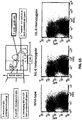
- non-human animals comprising a normal B cell population.
- the normal B cell population may be approximately the same in number and immunophenotype as a wild-type animal, e.g., a wild-type mouse.
- non-human murine animal e.g., a mouse or rat
- hIL-6 human IL-6
- the murine animal may express hIL-6 in serum at a level of about 50 to about no more than 200 pg/mL, about 75-125 pg/mL, or at about 100 pg/mL.
- non-human animal that expresses hIL-6 and/or hIL-6R, wherein the non-human animal expresses hIL-6 and/or hIL-6R from an endogenous non-human IL-6 locus and/or an endogenous non-human hIL-6R locus.
- a genetically modified mouse that expresses hIL-6 from an endogenous mouse IL-6 locus, wherein the endogenous mouse IL-6 gene has been replaced with a hIL-6 gene.
- a mouse comprising a cell that expresses an IL-6 receptor (IL-6R) that comprises a human ectodomain on the surface of the cell.
- the cell may be a lymphocyte, for example a B cell.
- the human IL-6 gene may comprise exons 1 through 5 of the human IL-6 gene of human BAC CTD-2369M23.
- the mouse serum may exhibit a serum concentration of human IL-6 of about 25 to about 300 pg/mL, 50 to about 250 pg/mL, 75 to about 200 pg/mL, or 100 to about 150 pg/mL.
- the level of human IL-6 in the serum of the mouse may be about 100 pg/mL.
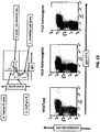
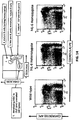
- the level of a pan B cell-specific marker in bone marrow of the mouse may be about the same as that of a wild-type mouse.
- the level of a pan B cell-specific marker in spleen may be about the same as that of a wild-type mouse.
- the pan B cell-specific marker may be selected from B220, CD19, CD20, CD22, CD79a, CD79b, L26, and Pax-5 (BSAP).
- the mouse may comprise a spleen that is about the same weight (per body weight) as a wild-type mouse.
- the lymph nodes of the mouse may be about the same weight (per body weight) as a wild-type mouse.
- the plasma cells of the mouse may not exhibit plasmocytosis characteristic of mice that overexpress human IL-6.
- the mouse may not exhibit glomerulonephritis.
- the mouse may exhibit a mesangial cell level comparable to a wild-type mouse.
- mouse that expresses hIL6 from an endogenous mouse IL-6 locus, wherein the endogenous mouse IL-6 gene has been replaced with a hIL-6 gene, wherein the mouse does not exhibit a feature selected from a morphologically detectable neuropathology, a reactive astrocytosis, and a combination thereof.
- the mouse may comprises a brain that is morphologically indistinct from a wild-type mouse brain.
- the mouse may comprise brain tissue that exhibits a level of reactive astrocytosis that is no higher than that of a wild-type mouse.
- the mouse may not express human IL-6 in neurons.
- the mouse may comprise activated astrocyte levels that are comparable to activated astrocyte levels in a wild-type mouse.
- the mouse may comprise ramified microglial cells in its white matter, wherein the ramified microglial cells are present in an amount equivalent to an amount of ramified microglial cells in a wild-type mouse.
- the mouse may not exhibit a reactive atrocytosis.
- the white matter of the mouse may be morphologically indistinct from the white matter of a wild-type mouse.
- the white matter of the mouse may be histologically indistinct from a wild-type mouse white matter with respect to histochemical staining of reactive astrocytes.
- the mouse may comprise a brain that is morphologically indistinct from a wild-type mouse brain.
- the mouse may comprise brain tissue that exhibits a level of reactive astrocytosis that is no higher than that of a wild-type mouse.
- Also described herein is a genetically modified mouse that expresses hIL6 from an endogenous mouse IL-6 locus, wherein the endogenous mouse IL-6 gene has been replaced with a hIL-6 gene, wherein the mouse does not exhibit a feature selected from a life span shortened by about 50% or more, kidney failure, hypergammaglobulinemia, elevated megakaryocytes in spleen, elevated megakaryocytes in bone marrow, plasmacytosis of spleen, plasmacytosis of thymus, plasmacytosis of lymph nodes, glomerulonephritis, glomerulosclerosis, and a combination thereof.
- the mice may have a life span that exceeds 20 weeks, for example having a life span that exceeds 30 weeks, 40 weeks, or 50 weeks.
- the mice may exhibit a life span about equal to that of a wild-type mouse of the same strain.
- the mice may exhibit a level of megakaryocytes in spleen that is no more than about the splenic megakaryocyte level of a wild-type mouse.
- the mice may comprise lymphoid organs that are essentially devoid of abnormal and compactly arranged plasmacytoid cells.
- mice may exhibit gamma globulin serum levels equivalent to gamma globulin serum levels in wild-type mice.
- the levels of ⁇ 1- and ⁇ -globulin in serum of the mice may be equivalent to ⁇ 1- and ⁇ -globulin serum levels of wild-type mice of the same strain.
- Also described herein is a genetically modified mouse that expresses human IL-6 from an endogenous mouse IL-6 locus, wherein the endogenous mouse IL-6 gene has been replaced with a hIL-6 gene, wherein the mouse does not exhibit a feature selected from muscle wasting, an elevated cathepsin B level as compared with a wild-type mouse of the same strain, an elevated cathepsin A+B level as compared with a wild-type mouse of the same strain, an increased liver weight as compared with a wild-type mouse of the same strain, and a combination thereof.
- the weight of the liver of the mouse may be about 800-900 mg at 12 weeks.
- the mouse may exhibit a cathepsin B level throughout its life span that is no more than about the level observed in a wild-type mouse.
- the mouse may exhibit a cathepsin A+B level throughout its life span that is no more than about the level observed in a wild-type mouse.
- the mouse may, as an adult, exhibit a gastrocnemus muscle weight that is within about 10% of the weight of a wild-type mouse of the same strain.
- the mouse as an adult, may exhibit a gastrocnemus muscle weight that is about the same as that of a wild-type mouse.
- a mouse that comprises a nucleotide sequence encoding a human IL-6 protein, wherein the nucleotide sequence encoding the human IL-6 protein replaces in whole or in part an endogenous nucleotide sequence encoding and endogenous mouse IL-6 protein.
- mouse that comprises a replacement at an endogenous mouse IL-6 receptor locus of mouse IL-6R ⁇ ectodomain with an ectodomain sequence of a human IL-6R ⁇ to form a chimeric human/mouse IL-6R ⁇ gene.
- the chimeric IL-6R ⁇ gene may be under the control of a mouse promoter and/or mouse regulatory elements at the endogenous mouse IL-6R ⁇ locus.
- mouse IL-6R ⁇ ectodomain-encoding sequence may be replaced with about 45.5 kb of human IL-6R ectodomain-encoding sequence.
- the human IL-6R ectodomain-encoding sequence may encompass the first (ATG) codon in exon 1 through exon 8.
- the mouse IL-6R ⁇ sequence that is replaced may include a contiguous sequence that encompasses exons 1 through 8, for example, exons 1 through 8 and a portion of intron 8 may be deleted.
- Also described herein is a genetically modified mouse, comprising a replacement at an endogenous mouse IL-6 locus of a mouse gene encoding IL-6 with a human gene encoding human IL-6, wherein the human gene encoding human IL-6 is under control of endogenous mouse regulatory elements at the endogenous mouse IL-6 locus.
- the human gene encoding human IL-6 may be a human IL-6 gene of BAC ID CTD-2369M23.
- the mouse may express a mouse IL-6R ⁇ .
- the mouse may express a human IL-6R ⁇ .
- the humanized IL-6R ⁇ may comprise a human ectodomain.
- the humanized IL-6R ⁇ may comprise a mouse transmembrane domain and a mouse cytoplasmic domain.
- the mouse may express a humanized IL-6R ⁇ that comprises a humanization of ectodomain but not transmembrane and/or cytosolic domain.
- the mouse may not exhibit a feature selected from plasmocytosis, glomerulosclerosis, glomerulonephritis, kidney failure, hypergammaglobulinemia, elevated megakaryocytes in spleen, elevated megakaryocytes in bone marrow, splenomegaly, lymph node enlargement, compacted abnormal plasma cells, and a combination thereof.
- a genetically modified mouse comprising a humanization of an endogenous mouse IL-6R ⁇ gene, wherein the humanization comprises a replacement of mouse IL-6R ⁇ ectodomain-encoding sequence with human IL-6R ⁇ ectodomain-encoding sequence at the endogenous mouse IL-6R ⁇ locus.
- a contiguous mouse sequence comprising mouse exons 1 through 8 may be replaced with a contiguous genomic fragment of human IL-6R ⁇ sequence encoding a human IL-6R ⁇ ectodomain.
- the contiguous genomic fragment of human IL-6R ⁇ sequence encoding the ectodomain may be from BAC CTD-2192J23.
- the mouse may further comprise a humanized IL-6 gene.
- the mouse may comprise a replacement at an endogenous mouse IL-6 locus of a mouse IL-6 gene with a human IL-6 gene.
- the humanized IL-6 gene may be under control of endogenous mouse regulatory elements.
- Also described herein is a method for making a humanized mouse, comprising replacing a mouse gene sequence encoding mouse IL-6 with a human gene encoding human IL-6.
- the replacement may be at an endogenous mouse IL-6 locus, and the human gene encoding human IL-6 may be operably linked to endogenous mouse regulatory sequences.
- Also described herein is a method for making a humanized mouse, comprising replacing mouse exons encoding ectodomain sequences of mouse IL-6R ⁇ with a human genomic fragment encoding ectodomain sequences of human IL-6R ⁇ to form a humanized IL-6R ⁇ gene.
- the replacement may be at an endogenous mouse IL-6R ⁇ locus, and the humanized IL-6R ⁇ gene may be operably linked to endogenous mouse regulatory sequences.
- a genetically modified mouse comprising a humanized IL-6R ⁇ gene comprising a replacement of mouse ectodomain-encoding sequence with human ectodomain sequence, wherein the humanized IL-6R ⁇ gene comprises a mouse transmembrane sequence and a mouse cytoplasmic sequence; wherein the mouse further comprises a gene encoding a human IL-6, wherein the gene encoding a human IL-6 is under control of endogenous mouse IL-6 regulatory elements.
- the mouse may be incapable of expressing a fully mouse IL-6R ⁇ and incapable of expressing a mouse IL-6.
- the genetically modified mice described herein may comprise the genetic modifications in their germline.
- tissue, cell, or membrane fragment from a mouse as described herein.
- the tissue or cell may be from a mouse that expresses a human IL-6 protein, but that does not express a mouse IL-6 protein.
- the tissue or cell may be from a mouse that expresses a humanized IL-6R ⁇ protein, but not a mouse IL-6R ⁇ protein.
- the humanized IL-6R ⁇ protein may comprise a human ectodomain and a mouse transmembrane domain and a mouse cytosolic domain.
- the tissue or cell may be from a mouse that expresses a human IL-6, a humanized IL-6R ⁇ , and that does not express a mouse IL-6 and does not express an IL-6R ⁇ that comprises a mouse ectodomain.
- IL-6R ⁇ human ectodomain and mouse transmembrane and mouse cytoplasmic domain
- human IL-6 human ectodomain and mouse transmembrane and mouse cytoplasmic domain
- mouse embryo comprising a genetic modification as described herein.
- mouse host embryo that comprises a donor cell that comprises a genetic modification as described herein.
- a pluripotent or totipotent non-human animal cell comprising a genetic modification as described herein.
- the cell may be a murine cell.
- the cell may be an ES cell.
- mouse egg comprising an ectopic mouse chromosome, wherein the ectopic mouse chromosome comprises a genetic modification as described herein.
- the mouse, embryo, egg, or cell that is genetically modified to comprise a human IL-6 gene or human or humanized IL-6R ⁇ gene is of a mouse that is of a C57BL strain selected from C57BL/A, C57BL/An, C57BL/GrFa, C57BL/KaLwN, C57BL/6, C57BL/6J, C57BL/6ByJ, C57BL/6NJ, C57BL/10, C57BL/10ScSn, C57BL/10Cr, and C57BL/Ola.
- the mouse is a 129 strain selected from the group consisting of a strain that is 129P1, 129P2, 129P3, 129X1, 129S1 (e.g., 129S1/SV, 129S1/Svlm), 129S2, 129S4, 129S5, 129S9/SvEvH, 129S6 (129/SvEvTac), 129S7, 129S8, 129T1, 129T2 (see, e.g., Festing et al.
- the genetically modified mouse may be a mix of an aforementioned 129 strain and an aforementioned C57BL/6 strain.
- the mouse may be a mix of aforementioned 129 strains, or a mix of aforementioned BL/6 strains.
- the 129 strain of the mix may be a 129S6 (129/SvEvTac) strain.
- the mouse may be a BALB strain, e.g., BALB/c strain.
- the mouse may be a mix of a BALB strain and another aforementioned strain.
- the mouse may be a Swiss or Swiss Webster mouse.
- the IL-6 receptor was long characterized as a receptor for a B cell stimulatory factor (BSF-2, or B cell Stimulatory Factor 2; also, BCDF, or B Cell Differentiation Factor) responsible for inducing B cells to synthesize immunoglobulin ( Yamasaki et al. (1988) Cloning and Expression of the Human Interleukin-6(BSF-2/IFN ⁇ 2) Receptor, Science 241:825-828 ).
- BSF-2 B cell stimulatory factor
- BCDF B Cell Stimulatory Factor 2
- B Cell Differentiation Factor B Cell Differentiation Factor
- interferon- ⁇ 2 was first described as interferon- ⁇ 2 as the result of its discovery during a search for a virally-induced protein termed interferon- ⁇ , by treating human fibroblasts with dsRNA poly(I)poly(C) to induce an anti-viral response
- interferon- ⁇ a virally-induced protein termed interferon- ⁇
- the human cDNA encodes a 468 amino acid protein having a 19-mer signal sequence and a cytoplasmic domain of about 82 amino acids that lacks a tyrosine kinase domain (see, Id. ) .
- the N-terminal (ectodomain) of the protein has an Ig superfamily domain of about 90 amino acids, a 250-amino acid domain between the Ig superfamily domain and the membrane, a transmembrane span of about 28 amino acids (see, Id. ) .
- the ectodomain of the receptor binds its ligand IL-6, which triggers association with gp130 in the membrane and it is this complex that conducts signal transduction; the cytoplasmic domain reportedly does not transduce signal ( Taga et al. (1989) Interleukin-6 Triggers the Association of Its Receptor with a Possible Signal Transducer, gp130, Cell 58:573-581 ). Indeed, a soluble form of IL-6R lacking a cytoplasmic domain can associate with IL-6 and bind gp130 on the surface of a cell and effectively transduce signal ( Id. ) .
- the homology of hIL-6R and mIL-6R at the protein level is only about 54%; the transmembrane domain has a homology of about 79%, whereas the cytoplasmic domain has a homology of about 54% (Sugito et al. (1990)).
- the natural ligand for the IL-6R was first isolated from cultures of HTLV-1-transformed T cells (see, Hirano et al. (1985) Purification to homogeneity and characterization of human B cell differentiation factor (BCDF or BSFp-2), Proc. Natl. Acad. Sci. USA 82:5490-5494 ).
- a human cDNA for the IL-6 gene was cloned at least twice, once as BSF-2 (see, Hirano et al.
- Human IL-6 is a 184-amino acid protein that exhibits only about 42% homology with mouse IL-6, although the genomic organization of the human and mouse genes are basically the same, and the promoter regions of the human and mouse genes share a 400-bp stretch that is highly conserved (see, Tanabe et al. (1988) Genomic Structure of the Murine IL-6 Gene: High Degree Conservation of Potential Regulatory Sequences between Mouse and Human, J. Immunol. 141(11):3875-3881 ).
- the human IL-6 gene is about 5 kb ( Yasukawa et al. (1987) Structure and expression of human B cell stimulatory factor-2 (BSC-2/IL-6) gene, EMBO J. 6(10):2939-2945 ), whereas the mouse IL-6 gene is about 7 kb ( Tanabe et al. (1988) Genomic Structure of the Murine IL-6 Gene: High Degree Conservation of Potential Regulatory Sequences between Mouse and Human, J. Immunol. 141(11):3875-3881 ).
- the mouse and human IL-6 genes reportedly share highly conserved 5'-flanking sequence important to regulation.
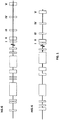
- a schematic diagram of the human and mouse IL-6 genomic loci is shown in FIG. 1 (not to scale).
- Exons I, II, III, IV, and V are indicated by closed boxes to the right in the figure. Selected putative regulatory regions are indicated by open boxes to the left in the figure.
- the putative regulatory regions for humans are, from left to right, a glucocorticoid element from -557 to -552; an IFN enhancer core sequence from -472 to -468; a glucocorticoid element from -466 to -461; an AT-rich region from -395 to - 334, a consensus AP-1 binding site from -383 to -277; an IFN enhancer core sequence from -253 to -248; a GGAAA-containing motif from -205 to -192; a c-fos SRE homology sequence from -169 to -82 containing an IFN enhancer core sequence, a cAMP-response element, a GGAAA motif, a CCAAT box, and a GC-rich region; and AP
- the putative regulatory regions for mouse are, from left to right, a GC rich region from - 553 to -536, a glucocorticoid element from -521 to -516 and from -500 to -495; a Z-DNA stretch from -447 to -396; an AP-1 binding site overlapping an IFN enhancer core sequence from -277 to -288, a GGAAA motif overlapping an IFN enhancer core sequence from -210 to -195; a c-fos SRE homology region from -171 to -82 containing a cAMP response element, a GGAAA motif overlapping an IFN enhancer core sequence, and a GC-rich region; and, an AP-1 binding site from -61 to -55.
- Mouse codons I-V have lengths 19, 185, 114, 150, and 165, respectively.
- Mouse intron lengths are: I-II, 162 bp; II-III, 1253 bp; III-IV, 2981 bp; IV-V, 1281 bp.
- Human codons I-V have lengths 19, 191, 114, 147, and 165.
- Human intron lengths are I-II , 154; II-III, 1047; III-IV, 706; IV-V, 1737.
- Genomic organization data are from Tanabe et al. (1988), and Yasukawa et al. (1987) Structure and expression of human B cell stimulatory factor-2 (BSF-2/IL-6) gene, EMBO J. 9(10):2939-2945 .
- mice and human IL-6 genes appear to be similarly regulated based on the similarity of their 5'-flanking sequence.
- IL-6 in humans mediates the acute phase response, hematopoiesis, B cell differentiation, T cell activation, growth and/or differentiation and/or activation of a variety of cell types (e.g., hepatocytes, fibroblasts, endothelial cells, neurons, pituitary cells, lymphomas, myelomas, breast carcinomas, NK cells, macrophages, osteoclasts, etc .) (reviewed in, e . g ., Heinrich et al. (1990), Kishimoto et al. (1989), and Keller et al. (1996); Sugita et al. (1990) Functional Murine Interleukin Receptor with Intracisternal A Particle Gene Product at its Cytoplasmic Domain, J. Exp. Med. 171:2001-2009 ).
- hepatocytes e.g ., Heinrich et al. (1990), Kishimoto et al. (1989), and Keller et al. (1996)
- mice transgenic for human IL-6 exhibit a panoply of substantial and debilitating pathologies, reflecting a significant pleiotropy of the IL-6 gene.
- Transgenic mice comprising a 6.6-kb fragment containing the human IL-6 gene and a ⁇ enhancer (E ⁇ ) produce high concentrations of hIL-6 and extremely high lgG1 levels (120- to 400-fold over wild-type mice), reflecting an IL-6 deregulation that is accompanied by plasmacytosis, mesangio-proliferative glomerulonephritis, and high bone marrow megakaryocyte levels ( Suematsu et al. (1989) IgG1 plasmacytosis in interleukin 6 transgenic mice, Proc.
- IL-6 and/or IL-6R Aberrant regulation of IL-6 and/or IL-6R is associated with myelomas, plastocytomas, rheumatoid arthritis, Castleman's disease, mesangial proliferative glomerulonephritis, cardiac myxoma, plams cell neoplasias, psoriasis, and other disorders (see, Kishimoto, T. (1989) The Biology of Interleukin-6, Blood 74(1):1-10 ; Sugita et al. (1990); also, Hirano et al.
- IL-6 is also implicated in sustaining levels of intra-prostatic androgens during androgen deprivation treatment of prostate cancer patients by a paracrine and/or autocrine mechanism, potentially providing castration-resistant prostate tumor growth ( Chun et al. (2009) Interleukin-6 Regulates Androgen Synthesis in Prostate Cancer Cells, Clin. Cancer Res. 15:4815-4822 ).
- the human protein is encoded as a 212 amino acid protein, in mature form a 184 amino acid protein following cleavage of a 28 amino acid signal sequence. It contains two N-glycosylation and two O-glycosylation sites, and human IL-6 is phosphorylated in some cells.
- the mouse protein is encoded as a 211 amino acid protein, in mature form a 187 amino acid protein following cleavage of a 23 amino acid signal sequence. O-glycosylation sites are present, but not N-glycosylation sites. (See reviews on IL-6, e.g., Heinrich et al. (1990) Interleukin-6 and the acute phase response, Biochem. J. 265:621-636 .)
- IL-6 function is pleiotropic.
- the IL-6 receptor is found on activated B cells but reportedly not on resting B cells.
- IL-6R is found on resting T cells and can reportedly promote T cell differentiation, activation, and proliferation, including the differentiation of T cells into cytotoxic T lymphocytes in the presence of IL-2.
- IL-6 induces the acute phase response.
- acute phase proteins such as, e.g., C-reactive protein (CRP) and serum amyloid A (SAA) in a dose-dependent and time-dependent manner (reviewed in Heinrich et al. (1990) Interleukin-6 and the acute phase response, Biochem. J. 265:621-636 ).
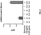
- Non-human animals e.g., mice or rats, comprising humanized IL-6 and IL-6R genes are therefore useful systems for measuring the acute phase response mediated by human IL-6.
- Such animals are also useful for determining whether a substance induces an IL-6-mediated acute phase response, by exposing a humanized IL-6/IL-6R animal as described herein to the substance, and measuring a level of one or more acute phase response proteins (or RNAs).
- the humanized animal is exposed to the substance in the presence of an antagonist of a human IL-6R, and a level of one or more acute phase response proteins (or RNAs) is measured, wherein a reduction in a level of an acute phase response protein (or RNA) in the presence of the human IL-6R antagonist indicates a human IL-6R-mediated acute phase response.
- Human IL-6 can bind both human IL-6R and mouse IL-6R; mouse IL-6 binds mouse IL-6R but not human IL-6R (no binding of mIL-6 to hIL-6R detectable, whereas hIL-6 can compete with mIL-6 for binding mIL-6R; Coulie et al. (1989) High-and low-affinity receptors for murine interleukin 6. Distinct distribution on B and T cells, Eur. J. Immunol. 19:2107-211 ); see also, e . g ., Peters et al.
- an acute phase response inducer is not expected to induce detectable levels of acute phase proteins that would indicate an acute phase response.
- a humanized mouse as described herein, comprising a humanized IL-6 gene and an IL-6R gene comprising a humanized ectodomain sequence will respond to an acute phase response inducer and exhibit acute phase response proteins in serum.
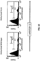
- Mice wild-type for IL-6/IL-6R tested for acute phase proteins in the presence or absence of the acute phase inducer turpentine showed a turpentine-dependent increase in acute phase proteins.
- a double humanization of IL-6 and IL-6R recapitulates the wild-type IL-6-mediated acute phase response with respect to serum acute phase proteins.
- mice that express a human IL-6 and/or a humanized IL-6 receptor from endogenous mouse loci, wherein the endogenous mouse IL-6 gene and/or the endogenous mouse IL-6 receptor gene have been replaced with a human IL-6 gene and/or a human sequence comprising a sequence that encodes an ectodomain of a human IL-6 receptor.
- the genetically modified mice express the human IL-6 and/or humanized IL-6 receptor from humanized endogenous loci that are under control of mouse promoters and/or mouse regulatory elements.
- the replacement(s) at the endogenous mouse loci provide non-human animals that express human IL-6 and a humanized IL-6 receptor in a manner that does not result in the panoply of substantial pathologies observed in IL-6 transgenic mice known in the art.
- mice that express human IL-6 are known in the art. However, they generally suffer from significant pathologies that severely limit their usefulness. Humanized mice as described herein express a human IL-6 and/or humanized IL-6 receptor under the control of endogenous mouse regulatory elements at endogenous mouse IL-6 and IL-6R ⁇ loci. These mice, in contrast, exhibit expression patterns with respect to these genes that are different from transgenic mice known in the art.
- Non-human genes in a non-human animal with homologous or orthologous human genes or human sequences, at the endogenous non-human locus and under control of endogenous promoters and/or regulatory elements can result in a non-human animal with qualities and characteristics that may be substantially different from a typical knockout-plus-transgene animal.
- an endogenous locus is removed or damaged and a fully human transgene is inserted into the animal's genome and presumably integrates at random into the genome.
- the location of the integrated transgene is unknown; expression of the human protein is measured by transcription of the human gene and/or protein assay and/or functional assay.
- Fertilized mouse eggs injected with a construct having the MHC class I promoter H2 and a ⁇ -globin intron driving expression of a 695-bp mouse IL-6 gene reportedly produce mice that constitutively express mouse IL-6 at relatively high levels (as compared with wild-type mice) (see, Woodrofe et al. (1992) Long-Term Consequences of Interleukin-6 Overexpression in Transgenic Mice, DNA and Cell Biology 11 (8):587-592 ). But these mice are prone to develop lymphomas associated with the intestines, lymph nodes, and kidney, as well as splenic amyloid deposits.
- mice as described herein that comprise a replacement of the mouse IL-6 gene with a human IL-6 gene at the mouse IL-6 locus are not prone to develop these lymphomas, and the mice exhibit apparently normal B cell populations.
- mice C57BL/6) transgenic for hIL-6 due to a random insertion of a 6.6-kb (BamHI-Pvu II fragment) length of human DNA containing the hIL-6 gene coupled with an IgM enhancer have been reported (see, Suematsu et al. (1989) IgG1 plasmocytosis in interleukin 6 transgenic mice, Proc. Natl. Acad. Sci. USA 86:7547-7551 ).
- the mice express hIL-6 at between 800 pg/mL and 20,000 pg/mL in serum, where wild-type mice typically express only about 100 pg/mL IL-6.
- mice exhibit an increase in serum Ig (120 to 400-fold over wild-type mice) and a decrease in albumin as they age.
- the mice suffer from a massive plasmacytosis, exhibit splenomegaly and lymph node enlargement, as well as exhibiting plasma cells and increased megakaryocytes in bone marrow. Upon inspection, what appear to be enlarged lymph nodes are instead massed of compacted abnormal plasma cells.
- Both spleen and thymus exhibit massive proliferation of plasma cells, which also infiltrate portions of the lung, liver, and kidney. Kidney in these mice also exhibits IL-6-stimulated mesangial cell proliferation typical of mesangio-proliferative glomerulonephritis.
- mice (BALB/c) transgenic for a trimmed hIL-6 cDNA driven by a mouse H-2L d promoter randomly inserted into the genome display severe plasmacytosis (see, Suematsu et al. (1992) Generation of plasmacytomas with the chromosomal translocation t(12;15) in interleukin 6 transgenic mice, Proc. Natl. Acad. Sci. USA 89:232-235 ).
- C57BL/6 mice that overexpress hIL-6 do not develop transplantable plasmacytomas (they do exhibit plasmacytosis)
- transgenic BL/6 mice back-crossed into BALB/c mice reportedly do.
- Random transgenesis of a hIL-6 cDNA driven by a glial fibrillary acidic protein (GFAP) gene promoter reportedly results in hIL-6 overexpression in the mouse central nervous system, which also leads to significant pathologies (see, Campbell et al. (1993) Neurologic disease induced in transgenic mice by cerebral overexpression of interleukin 6, Proc. Natl. Acad. Sci. USA 90:10061-10065 ). These mice exhibit extensive neuropathology and reactive astrocytosis resulting from IL-6 expression in the CNS due to loss of control as the result of random integration of an IL-6 transgene at an apparently CNS-permissive transcriptional locus.
- GFAP glial fibrillary acidic protein
- hIL-6 cDNA linked to a ⁇ -globin 3'-UTR and driven by a neuron-specific enolase promoter microinjected into fertilized mouse eggs produced mice with a normal lifespan and without apparent neurological defects that expressed hIL-6 in neurons but not elsewhere (see, Fattor et al. (1994) IL-6 Expression in Neurons of Transgenic Mice causess Reactive Astrocytosis and Increase in Ramified Microglial Cells But No Neuronal Damage, Eur. J.
- mice exhibited high levels (20- to 30-fold higher than wild-type) of activated and enlarged astrocytes with increased processes throughout the brain, as well as a 10- to 15-fold increase in ramified microglial cells in white matter.
- brain expression of IL-6 reportedly leads to conditions that range from reactive astrocytosis to frank and profound neuropathology.
- Microinjection into fertilized eggs of an F1 cross of C57BL/6x"DBAII" mice of a 639-bp hIL-6 cDNA linked to a ⁇ -globin 3'-UTR and a mouse MT-1 promoter reportedly produced a transgenic mouse in which the hIL-6 gene was randomly integrated produced a weakened and diseased mouse that dies young of kidney failure (see Fattori et al. (1994) Blood, Development of Progressive Kidney Damage and Myeloma Kidney in Interleukin-6 Transgenic Mice, Blood 63(9):2570-2579 ).
- Transgenic mice expired at 12-20 weeks and exhibited elevated levels of ⁇ 1 and ⁇ -globulins in plasma, hypergammaglobulinemia, elevated megakaryocytes in spleen (3-fold higher than wild-type) and bone marrow, plasmacytosis of lymphoid organs (spleen, thymus, and lymph nodes) characterized by abnormal and compactly arranged plasmocytoid cells, and glomerulonephritis leading to glomerulosclerosis similar to multiple myeloma.
- mice Microinjection into fertilized eggs of a C57BL/6J mouse of a H-2L d -driven hIL-6 cDNA caused IL-6-dependent muscle wasting in mice, characterized in part by a significantly lower gastrocnemius muscle weight in transgenic mice as compared to weight-matched controls, a difference that was ameliorated by treatment with an IL-6 antagonist (see, Tsujinaka et al. (1996) Interleukin 6 Receptor Antibody Inhibits Muscle Atrophy and Modulates Proteolytic Systems in Interleukin 6 Transgenic Mice, J. Clin. Invest. 97(1):244-249 ). At 12 weeks these mice displayed serum hIL-6 levels of more than 600,000 pg/mL.
- transgenic mice also had livers that weighed about 1,242 mg, as compared to control livers that were about 862 mg.
- Transgenic mice treated with IL-6 antagonist had livers that weighed about 888 mg.
- Muscle cathepsins B and B+L were significantly higher (20-fold and 6.2-fold) in transgenic mice than in controls, a phenomenon that was eliminated in transgenic mice treated with an IL-6 antagonist.
- cathepsin B and L mRNAs were estimated to be about 277% and 257%, respectively, as compared with wild-type mice; the difference was significantly reduced with IL-6 antagonist treatment.
- mice comprising a hIL-6 minigene driven by a mouse MHC class I H-2Ld promoter and a hIL-6R minigene driven by a chicken ⁇ -actin promoter, and a gp130 gene, exhibited pathologies typical of hIL-6 transgenic mice (e . g ., hepergammaglobulinemia, splenomegaly, mesangial proliferative glomerulonephritis, lung lymphoid infiltration) as well as ventricular hypertrophy ( Hirota et al.
- gp130 a signal-transducing receptor component for interleukin 6-related cytokines
- the ventricular hypertrophy is believed to be mediated by a continuous activation of gp130 ( Id. ) .
- the role of IL-6 is reportedly to help strengthen the cytokine receptor complex and induce dimerization of gp130, which is the signal transducing component responsible for transducing the IL-6 signal ( Paonessa et al. (1995) Two distinct and independent sites on IL-6 trigger gp130 dimer formation and signalling, EMBO J.
- the activated complex is believed to be a hexamer composed of two IL-6, each IL-6 bound to one IL-6R ⁇ and two gp130 (each IL-6 contains two independent gp130-binding sites) exhibiting a 2:2:2 stoichiometry, wherein the dimerization of gp130 causes activation of JAK-Tyk tyrosine kinases, phosphorylation of gp130 and STAT family transcription factors and other intracellular substrates ( Id. ; Stahl, N.
- mice transgenic for human slL-6R driven by a rat PEP carboxykinase promoter and human IL-6 driven by a mouse metallothionein-1 promoter are reportedly markedly smaller that mice transgenic for human IL-6 alone or human sIL-6R alone ( Peters et al. (1997) Extramedullary Expansion of Hematopoietic Progenitor Cells in Interleukin(IL-)-6-slL-6R Double Transgenic Mice, J. Exp. Med. 185(4):755-766 ), reflected in reduced body fat and reduced weight (20-25 g vs. 40 g).
- Double transgenic mice reportedly also exhibit spleen (5-fold) and liver (2-fold) enlargement as compared with reportedly normal organ weights for single transgenic mice, apparently due to extramedullary proliferation of hematopoeitic cells of spleena and liver but not bone marrow, as well as elevated megakaryocytes in spleen and plasmacellular infiltrates in all parenchymal organs ( Id. ) .
- Double transgenics also exhibit livers with an increase of about 200- to about 300-fold in granulocytes, macrophages, progenitor cells, and B cells as compared with single transgenics; in contrast, IL-6 single transgenic mice exhibited lesser increases in macrophages (15-fold) and B cells (45-fold) ( Id. ) .
- the extraordinary findings are presumably due to stimulation of growth and differentiation of hematopoietic progenitor cells by activating gp130 signal transduction ( Id. ) .
- double transgenic mice exhibit a hepatocellular hyperplasia that is reportedly identical to human nodular regenerative hyperplasia with sustained hepatocyte proliferation that strongly suggests that IL-6 is responsible for both hepatocyte proliferation and pathogenic hepatocellular transformation ( Maione et al. (1998) Coexrpession of IL-6 and soluble IL-6R causes nodular regenerative hyperplasia and adenomas of the liver, EMBO J. 17(19):5588-5597 ).
- hepatocellular hyperplasia is reportedly not observed in single transgenic hIL-6 mice and hIL-6 can bind mIL-6R, the finding may appear paradoxical until it is considered that the double transgenic may result in higher levels of hIL-6 complexed to soluble IL-6R (here, soluble hIL-6R), which complex is a more potent inhibitor that IL-6 alone ( Id. ) .
- mice that are transgenic for human IL-6 In contrast to mice that are transgenic for human IL-6, humanized IL-6 mice that comprise a replacement at an endogenous mouse IL-6 locus, which retain mouse regulatory elements but comprise a humanization of IL-6-encoding sequence, do not exhibit the severe pathologies of prior art mice. Genetically modified mice that were heterozygous or homozygous for hIL-6 were grossly normal.
- mice with a humanized IL-6 gene (MAID 760) as described in the Examples were immunophenotyped and found to have normal B cell numbers in FACS analyses (lymphocyte-gated) of spleen B cells using a pan B cell marker (CD445R(B220)) ( FIG.4 ).
- FACS analyses lymphocyte-gated
- spleen B cells using a pan B cell marker (CD445R(B220))
- FIG.4 pan B cell marker
- wild-type mice exhibited 63% B cells
- hIL-6 heterozygote mice exhibited 63% B cells
- mice homozygous for hIL-6 at the endogenous mouse locus exhibited 63% B cells.
- B cell numbers for homozygous hIL-6 mice immunized with TNP-KLH were also normal (65% for wild-type, and 61% for hIL-6 homozygotes).
- Splenic T cells were also about the same as wild-type ( FIG.5 ). Percentages of splenic T cells for Thelper/Tcytoxic were, for wild-type 20%/40% (ratio of 1.4:1); for hIL-6 heterozygotes 23%/14% (ratio of 1.6:1); for hIL-6 homozygotes 21%/15% (ratio of 1.4:1) (markers were CD8a-APC; CD4-FITC).
- mice immunized with TNP-KLH exhibited similar splenic T cell numbers to wild-type mice, i.e., Thelper/Tcytotoxic were 22%/20% (ratio of 1.1:1) as compared with 21%/19% for wild-type (also a ratio of 1.1:1).
- Humanized IL-6 mice also exhibited about normal levels of splenic NK cells on FACS analysis (CD11b and DX5) ( FIG. 7 ).
- hIL-6 heterozygotes exhibited 2.2% NK cells, and hIL-6 homozygotes exhibited 1.8% NK cells, whereas wild-type mice exhibited 2.4% NK cells.
- homozygotes exhibited 1.6% splenic NK cells, whereas wild-type mice exhibited 2.1% splenic NK cells.
- Humanized IL-6 mice also exhibited normal blood B and T cell numbers in FACS analysis ( FIG. 8 and FIG. 9 ).
- FACs with a pan B cell marker CD445R(B220) revealed that homozygous hIL-6 mice exhibited 52% B cell as compared with wild-type 53%; heterozygotes exhibited 38% (an average of two different stainings of 29% and 47%).
- Homozygous hIL-6 mice immunized with TNP-KLH gave similar B cell numbers (43%, as compared with 45% for wild-type mice).
- Humanized IL-6 mice exhibited normal blood T cell numbers in FACS analysis as measured by CD8a and CD4 staining. Heterozygous hIL-6 mice exhibited Thelper/Tcytotoxic numbers of 39%/26% (ratio of 1.5:1); homozygous hIL-6 mice exhibited Th/Tc numbers of 24%/20% (ratio of 1.2:1), whereas wild-type mice exhibited Th/Tc numbers of 26%/20% (ratio of 1.3:1). Homozygous hIL-6 mice immunized with TNP-KLH had Th/Tc numbers of 29%/21% (ratio of 1.4:1), whereas wild-type immunized mice had Th/Tc numbers of 28%/23% (1.2:1).
- Humanized IL-6 mice also exhibited myeloid cell numbers in blood that were similar to wild-type mice as measured by FACS analysis of na ⁇ ve and immunized mouse blood stained with Ly6G/C(Gr1) and CD11 b, as well as CD11 b and DX5 ( FIG. 10 , FIG. 11 , and FIG 12 ).
- Heterozygous hIL-6 mice exhibited %Gr+ cells of 10.8%, homozygotes 6.9%, whereas wild-type mice exhibited 9.7%.
- Immunized hIL-6 homozygotes exhibited M1(Ly6G/C(Gr) of 10 1 -10 4 )/ M2(Ly6G/C(Gr) staining of about 10 2 -10 3 ) numbers of 43%/34%, whereas wild-type mice had numbers of 45%/38%.
- FACS plots of CD11 b (vertical axis) vs. Ly6G/C (horizontal axis) for immunized homozygous hIL-6 mice showed cell percentage in quadrants (upper left/upper right/lower right) of 16%/8%/3%, which were identical to immunized wild-type quadrant numbers.
- Humanized IL-6 mice exhibited an isotype response that was essentially the same as observed in wild-type mice. Early and final IgG1, IgG2a, IgG2b, IgG3, IgA, IgE, and IgM levels were about the same as observed in wild-type mice. In one experiment, final IgM was slightly higher in humanized mice; final IgG3 was also elevated in humanized mice.
- B cell development in naive hIL-6 mice was essentially indistinguishable from development in wild-type mice based on FACS analysis of bone marrow IgM/CD24/B220 staining ( FIG. 13 ).
- Immunophenotyping of immune mice revealed that marker populations for various cell types in the B cell development progression were essentially normal in hIL-6 mice. Progression of cells from hematopoietic stem cells, common lymphoid progenitors, ProB cells, PreB cells, and immature and mature B cells is normal in hIL-6 mice ( FIG. 14 and FIG. 15 )
- the 4.8-kb human IL-6 gene containing exons 1 through 5 of the human IL-6 gene replaced 6.8 kb of the murine IL-6 gene locus.
- a targeting construct for replacing the mouse with the human IL-6 gene in a single targeting step was constructed using VELOCIGENE® genetic engineering technology (see, Valenzuela et al. (2003) High-throughput engineering of the mouse genome coupled with high-resolution expression analysis, Nature Biotech, 21(6):652-659 ).
- Mouse and human IL-6 DNA were obtained from bacterial artificial chromosome (BAC) RPCI-23 clone 368C3, and from BAC CTD clone 2369M23, respectively.
- BAC bacterial artificial chromosome
- a NotI linearized targeting construct generated by gap repair cloning containing mouse IL-6 upstream and downstream homology arms flanking a 4.8 kb human IL-6 sequence extending from ATG in exon 1 to exon 5 with 16 nucleotides of 3' downstream sequence (genomic coordinates: NCBlh37.1: ch7:22,766,882 to 22,771,637) and a floxed neo selection cassette, was electroporated into F1 H4 mouse embryonic stem (ES) cells (C57BL/6 x 129 F1 hybrid). Correctly targeted ES cells (MAID 790) were further electroporated with a transient Cre-expressing vector to remove the drug selection cassette.
- ES mouse embryonic stem
- Targeted ES cell clones without drug cassette were introduced into an 8-cell stage mouse embryo by the VELOCIMOUSE® method (see, US Pat. No. 7,294,754 , 7,576,259 , 7,659,442 , and Poueymirou et al. (2007) F0 generation mice that are essentially fully derived from the donor gene-targeted ES cells allowing immediate phenotypic analyses Nature Biotech. 25(1):91-99 ).
- VELOCIMICE® F0 mice fully derived from the donor ES cell bearing the humanized IL-6 gene were identified by genotyping for loss of mouse allele and gain of human allele using a modification of allele assay (see, Valenzuela et al. (2003)).
- LONA loss-of-native-allele
- the qPCR assays comprised the following primer-probe sets (written 5' to 3'): upstream forward primer, TTGCCGGTTT TCCCTTTTCT C (SEQ ID NO:1); upstream reverse primer, AGGGAAGGCC GTGGTTGTC (SEQ ID NO:2); upstream probe, FAM-CCAGCATCAG TCCCAAGAAG GCAACT-BHQ (SEQ ID NO:3); downstream forward primer, TCAGAGTGTG GGCGAACAAA G (SEQ ID NO:4); downstream reverse primer, GTGGCAAAAG CAGCCTTAGC (SEQ ID NO:5); downstream probe, FAM-TCATTCCAGG CCCTTCTTAT TGCATCTG-BHQ (SEQ ID NO:6); in which FAM refers to the 5-carboxyfluorescein fluorescent probe and BHQ refers to the fluorescence quencher of the black hole quencher type (Biosearch Technologies).
- DNA purified from ES cell clones that that have taken up the targeting vector and incorporated in their genomes was combined with TaqManTM Gene Expression Master Mix (Life Technologies) according to the manufacturer's suggestions in a 384-well PCR plate (MicroAmpTM Optical 384-Well Reaction Plate, Life Technologies) and cycled in an Applied Biosystems Prism 7900HT, which collects fluorescence data during the course of the PCRs and determines a threshold cycle (Ct), the fractional PCR cycle at which the accumulated fluorescence reaches a pre-set threshold.
- Ct threshold cycle
- the same LONA assay was used to assay DNA purified from tail biopsies for mice derived from the targeted ES cells to determine their 116 genotypes and confirm that the humanized 116 allele had transmitted through the germline.
- Two pups heterozygous for the replacement are bred to generate a mouse that is homozygous for the replacement of the endogenous mouse IL-6 gene by the human IL-6 gene. Pups that are homozygous for the replacement are used for phenotyping.
- the upstream junction of the murine locus and the sequence containing the hIL-6 gene is designed to be within 5'-AATTAGAGAG TTGACTCCTA ATAAATATGA GACTGGGGAT GTCTGTAGCT CATTCTGCTC TGGAGCCCAC CAAGAACGAT AGTCAATTCC AGAAACCGCT ATGAACTCCT TCTCCACAAG TAAGTGCAGG AAATCCTTAG CCCTGGAACT GCCAGCGGCG GTCGAGCCCT GTGTGAGGGA GGGGTGTGTG GCCCAGG (SEQ ID NO:10), wherein the final mouse nucleotide prior to the first nucleotide of the human gene is the "T" in CCGCT, and the first nucleotide of the human sequence is the first "A" in ATGAA.
- the downstream junction of the sequence containing the hIL-6 gene and the murine locus is designed to be within 5'-TTTTAAAGAA ATATTTATAT TGTATTTATA TAATGTATAA ATGGTTTTTA TACCAATAAA TGGCATTTTA AAAAATTCAG CAACTTTGAG TGTGTCACGC TCCCGGGCTC GATAACTATA ACGGTCCTAA GGTAGCGACT CGAGATAACT T-3' (SEQ ID NO:11), wherein the final nucleotide of the human sequence is with the final "G” in TCACG and the first nucleotide of the mouse sequence is the first "C" in CTCCC; the downstream junction region also contained a IoxP site at the 3' end (the beginning of which is shown) for removal of a floxed ubiquitin promoter-driven neo cassette.
- the junction of the neo cassette with the mouse IL-6 locus is designed to be within 5'-TATACGAAGT TATCCTAGGT TGGAGCTCCT AAGTTACATC CAAACATCCT CCCCCAAATC AATAATTAAG CACTTTTTAT GACATGTAAA GTTAAATAAG AAGTGAAAGC TGCAGATGGT GAGTGAGA (SEQ ID NO:12), where the final "C” of AGCTC is the final nucleotide of the neo cassette; the first nucleotide of the mouse genome following the cassette is the initial "C" of CTAAG.
- mice homozygous for the hIL-6 gene replacement were analyzed for B cells (DC445R(B220). Lymphocyte-gated fractions from spleen cell preparations of naive and immunized (TNP-KLH) hIL-6 mice were stained and immunophenotyped using flow cytometry. FACS analysis showed that the percentage of B cells of the spleen cell preparation as measured by CD45R(B220)-FITC staining were about the same (63% of cells) for preparations from naive wild-type, hIL-6 heterozygotes, and hIL-6 homozygotes.
- B cells accounted for about 65% of total cells of the spleen cell preparation in wild-type mice, and about 61% of total cells in hIL-6 homozygotes.
- Spleens of hIL-6 mice both naive and immunized contain a population of B cells that is about the same size as the splenic B cell population in wild-type mice.
- Bone marrow of wild-type, hIL-6 heterozygotes, and hIL-6 homozygotes was stained with B cell markers (CD45R(B220)-APC, CD24(HSA)-PE, or CD43 conjugated to a dye and/or IgM (IgM-FITC).
- B cell development in bone marrow of normal mice will be reflected in surface markers as cells progress from stem cells to early pro-B cells to late pro-B cells, to large pre-B cells to small pre-B cells to immature B cells and finally, to mature B cells.
- Common lymphocyte progenitor pro-B cells will express CD45R, and in later stages will express IgM as immature and later as mature B cells.
- CD45R-stained and anti-IgM-stained B cells should reveal a pattern characteristic of B cell development.
- Bone marrow of hIL-6 heterozygotes and homozygotes displayed a pattern of CD45R(B220)-APC and anti-IgM-FITC staining that was essentially indistinguishable from wild-type bone marrow, showing populations of B cells that stained positive for CD45R(B220) and IgM, or CD45R(B220) alone.
- B cell sub-populations in bone marrow of hIL-6 mice revealed by FACS staining were similar to those in wild-type mice (Table 1; see also, FIG. 13 ). Table 1.
- the 45 kb human IL-6R ⁇ gene containing exons 1 through 8 of the human IL-6R ⁇ gene replaced 35.4 kb of the murine IL-6R ⁇ gene locus.
- Mouse exons 9 and 10 were retained; only exons 1-8 were humanized.
- 35,384 nt of mouse sequence was replaced by 45,047 nt of human sequence.
- a targeting construct for replacing the mouse with the human IL-6R ⁇ gene in a single targeting step was constructed using VELOCIGENE® genetic engineering technology (see, Valenzuela et al. (2003) High-throughput engineering of the mouse genome coupled with high-resolution expression analysis, Nature Biotech, 21(6):652-659 ).
- Mouse and human IL-6R ⁇ DNA were obtained from bacterial artificial chromosome (BAC) RPCI-23 clone 125J8, and from BAC CTD clone 2192J23, respectively.
- BAC bacterial artificial chromosome
- a NotI linearized targeting construct generated by gap repair cloning containing mouse IL-6R ⁇ upstream and downstream homology arms flanking a 45 kb human IL-6R ⁇ sequence extending from ATG in exon 1 to exon 8 with 69 nucleotides of 3' downstream sequence and a floxed neo selection cassette, was electroporated into F1 H4 mouse embryonic stem (ES) cells (C57BL/6 x 129 F1 hybrid). Correctly targeted ES cells (MAID 794) were further electroporated with a transient Cre-expressing vector to remove the drug selection cassette.
- ES mouse embryonic stem
- Targeted ES cell clones without drug cassette were introduced into an 8-cell stage mouse embryo by the VELOCIMOUSE® method (see, US Pat. No. 7,294,754 , 7,576,259 , 7,659,442 , and Poueymirou et al. (2007) F0 generation mice that are essentially fully derived from the donor gene-targeted ES cells allowing immediate phenotypic analyses Nature Biotech. 25(1):91-99 ).
- VELOCIMICE® F0 mice fully derived from the donor ES cell bearing the humanized IL-6R ⁇ gene were identified by genotyping for loss of mouse allele and gain of human allele using a modification of allele assay (see, Valenzuela et al. (2003)).
- LONA loss-of-native-allele
- the qPCR assays comprised the following primer-probe sets (written 5' to 3'): upstream forward primer, GCCCTAGCAT GCAGAATGC (SEQ ID NO:13); upstream reverse primer, AAGAGGTCCC ACATCCTTTG C (SEQ ID NO:14); upstream probe, CCCACATCCA TCCCAATCCT GTGAG (SEQ ID NO:15); downstream forward primer, GAGCTTGCCC CCAGAAAGG (SEQ ID NO:16); downstream reverse primer, CGGCCACATC TCTGGAAGAC (SEQ ID NO:17); downstream probe, CATGCACTGC CCCAAGTCTG GTTTCAGT (SEQ ID NO:18).
- DNA purified from ES cell clones that that have taken up the targeting vector and incorporated in their genomes was combined with TaqManTM Gene Expression Master Mix (Life Technologies) according to the manufacturer's suggestions in a 384-well PCR plate (MicroAmpTM Optical 384-Well Reaction Plate, Life Technologies) and cycled in an Applied Biosystems Prism 7900HT, which collects fluorescence data during the course of the PCRs and determines a threshold cycle (Ct), the fractional PCR cycle at which the accumulated fluorescence reaches a pre-set threshold.
- Ct threshold cycle
- the same LONA assay was used to assay DNA purified from tail biopsies for mice derived from the targeted ES cells to determine their IL-6R ⁇ genotypes and confirm that the humanized IL-6R ⁇ allele had transmitted through the germline. Pups heterozygous for the replacement are bred to generate a mouse that is homozygous for the replacement of the endogenous mouse IL-6R ⁇ gene by the human IL-6R ⁇ (ectodomain) gene. Pups that are homozygous for the replacement are used for phenotyping.
- the upstream junction of the murine locus and the sequence containing the hIL-6R ⁇ gene is designed to be within 5'-CGAGGGCGAC TGCTCTCGCT GCCCCAGTCT GCCGGCCGCC CGGCCCCGGC TGCGGAGCCG CTCTGCCGCC CGCCGTCCCG CGTAGAAGGA AGCATGCTGG CCGTCGGCTG CGCGCTGCTG GCTGCCCTGC TGGCCGCGCC GGGAGCGGCG CTGGCCCCAA GGCGCTGCCC TGCGCAGGGT AAGGGCTTCG G (SEQ ID NO:22), wherein the final mouse nucleotide prior to the first nucleotide of the human gene is the "C" in GAAGC, and the first nucleotide of the human sequence is the first "A" in ATGCT.
- the downstream junction of the sequence containing the hIL-6 gene and the murine locus is designed to be within 5'-CAAGATTATT GGAGTCTGAA ATGGAATACC TGTTGAGGGA AATCTTTATT TTGGGAGCCC TTGATTTCAA TGCTTTTGAT TCCCTATCCC TGCAAGACCC GGGCTCGATA ACTATAACGG TCCTAAGGTA GCGACTCGAG ATAACTTC-3' (SEO ID NO:23), wherein the final nucleotide of the human sequence is with the final "A" in CAAGA and the first nucleotide of the mouse sequence is the first "C" in CCCGG; the downstream junction region also contained a IoxP site at the 3' end for removal of a floxed ubiquitin promoter-driven neo cassette.
- the first nucleotide of the loxp site is the first "A" in ATAAC.
- the junction of the neo cassette with the mouse IL-6R ⁇ locus is designed to be within 5'-TATACGAAGT TATCCTAGGT TGGAGCTCTA CTCCATATGC TCACTTGCCG TTGTTTGCTA CGATACGGTG AGGCCCGTGC GAAGAGTGGC ACAGATCAGG AGGCTTATGT GGTCAGTCCA CAGTATGGC (SEQ ID NO:24), where the final "C” of AGCTC is the final nucleotide of the neo cassette; the first nucleotide of the mouse genome following the cassette is the initial "T" of TACTC.
Description
- The invention relates to the field of genetically modified murine animals.
- Mice transgenic for a human IL-6 gene are known in the art. However, random insertion of a human IL-6 transgene into the mouse genome results in poorly regulated expression of the human IL-6 protein, which manifests itself in a variety of pathologies in such transgenic mice, including, but not limited to, plasmacytosis and glomerulonephritis. As a result, these mice have limited usefulness.
- There is a need for non-human animals, e.g., mice and rats, the express human or humanized IL-6 and/or human or humanized IL-6 receptor. There is a need for such humanized mice that do not exhibit one or more pathologies exhibited by transgenic hIL-6 mice.
- In one aspect, the invention provides a genetically modified murine animal comprising a replacement at an endogenous murine IL-6 locus of a murine gene encoding IL-6 with a human gene encoding human IL-6, wherein the human gene encoding human IL-6 is under control of endogenous murine regulatory elements at the endogenous murine IL-6 locus. In one embodiment, genetically modified murine animals are provided that express a human IL-6 gene under the control of endogenous murine promoter and/or endogenous murine regulatory elements, from an endogenous murine IL-6 locus.
- In one embodiment, the human gene encoding human IL-6 comprises
exons 1 through 5 of the human IL-6 gene found in the CTD-2369M23 bacterial artificial chromosome. - In another embodiment, the murine animal expresses a humanized IL-6Rα wherein the endogenous murine IL-6Rα gene has been replaced with a human sequence comprising a sequence that encodes an ectodomain of a human IL-6Rα.
- In a further embodiment, the murine animal does not exhibit a feature selected from plasmocytosis, glomerulosclerosis, glomerulonephritis, kidney failure, hypergammaglobulinemia, elevated megakaryocytes in spleen, elevated megakaryocytes in bone marrow, splenomegaly, lymph node enlargement, compacted abnormal plasma cells, and a combination thereof. In such embodiments, the humanized IL-6Rα may comprise murine transmembrane and intracellular domains.
- In a second aspect, the invention provides a genetically modified murine animal, comprising a humanization of an endogenous murine IL-6Rα gene, wherein the humanization comprises a replacement of a murine IL-6Rα ectodomain-encoding sequence with a human IL-6Rα ectodomain-encoding sequence at the endogenous murine IL-6Rα locus, and wherein the humanized IL-6Rα gene is under control of endogenous murine regulatory elements.
- In one embodiment of the second aspect of the invention, the genetically modified murine animal further comprises a humanized IL-6 gene comprising a replacement at an endogenous murine IL-6 locus of a murine gene encoding IL-6 with a human gene encoding human IL-6.
- In a third aspect, the invention provides a method for making a humanized murine animal, comprising replacing a murine gene sequence encoding murine IL-6 with a human gene encoding human IL-6 so that the human IL-6 gene is under control of endogenous murine regulatory elements.
- In a fourth aspect, the invention provides a method for making a humanized murine animal, comprising replacing all murine exons encoding ectodomain sequences of murine IL-6Rα with a human genomic fragment encoding human IL-6Rα ectodomain to form a humanized IL-6Rα gene, wherein the humanized IL-6Rα gene is under control of endogenous murine regulatory elements.
- In a fifth aspect, the invention provides a genetically modified murine animal comprising a humanized IL-6Rα gene comprising a replacement of a murine ectodomain-encoding sequence with a human ectodomain sequence, wherein the humanized IL-6Rα gene comprises murine transmembrane and intracellular domain sequences, wherein the murine animal further comprises a gene encoding a human IL-6, and wherein the genes encoding human IL-6 and humanized IL-6Rα are under control of endogenous murine regulatory elements.
- Described herein are genetically modified murine animals are provided that express a human IL-6 receptor gene (or a gene encoding a human ectodomain and mouse transmembrane and intracellular domains) under the control of endogenous murine promoter and/or endogenous murine regulatory elements, from an endogenous murine IL-6 receptor locus.
- Also described herein is a genetically modified animal (e.g., a murine animal, e.g., a mouse or rat) that expresses a human IL-6 protein, wherein the non-human animal does not exhibit a pathology selected from plasmacytosis, glomerulonephritis, glomerulosclerosis, mesangio-proliferative glomerulonephritis, intestinal lymphoma, kidney lymphoma, splenomegaly, lymph node enlargement, liver enlargement, megakaryocytes in bone marrow, compacted abnormal plasma cells, infiltration of plasma cells into lung or liver or kidney, mesangial cell proliferation in kidney, cerebral overexpression of IL-6, ramified microglial cells in white matter, reactive astrocytosis in brain, kidney failure, elevated megakaryocytes in spleen, muscle wasting (e.g., gastrocnemius muscle wasting), elevated muscle cathepsins B and B+L (e.g., around 20-fold and 6-fold), and a combination thereof.
- Also described herein are non-human animals comprising a normal B cell population. The normal B cell population may be approximately the same in number and immunophenotype as a wild-type animal, e.g., a wild-type mouse.
- Also described herein is a non-human murine animal (e.g., a mouse or rat) which expresses human IL-6 (hIL-6) in serum at a level below about 800 pg/mL, below about 700, 600, 500, 400, 300, or 200 pg/mL. The murine animal may express hIL-6 in serum at a level of about 50 to about no more than 200 pg/mL, about 75-125 pg/mL, or at about 100 pg/mL.
- Also described herein is a non-human animal that expresses hIL-6 and/or hIL-6R, wherein the non-human animal expresses hIL-6 and/or hIL-6R from an endogenous non-human IL-6 locus and/or an endogenous non-human hIL-6R locus. Also described herein is a genetically modified mouse that expresses hIL-6 from an endogenous mouse IL-6 locus, wherein the endogenous mouse IL-6 gene has been replaced with a hIL-6 gene.
- Also described herein is a mouse comprising a cell that expresses an IL-6 receptor (IL-6R) that comprises a human ectodomain on the surface of the cell. The cell may be a lymphocyte, for example a B cell.
- Also described herein are genetically modified murine animals in which about 6.8 kb at the endogenous mouse IL-6 locus, including
exons 1 through 5 and a 3' untranslated sequence, is deleted and replaced with about 4.8 kb of human IL-6 genesequence comprising exons 1 through 5 of the human IL-6 gene. The human IL-6 gene may compriseexons 1 through 5 of the human IL-6 gene of human BAC CTD-2369M23. - Also described herein is a genetically modified mouse that expresses IL-6 from a human IL-6 gene, wherein the mouse expresses human IL-6 in its serum. The mouse serum may exhibit a serum concentration of human IL-6 of about 25 to about 300 pg/mL, 50 to about 250 pg/mL, 75 to about 200 pg/mL, or 100 to about 150 pg/mL. The level of human IL-6 in the serum of the mouse may be about 100 pg/mL. The level of a pan B cell-specific marker in bone marrow of the mouse may be about the same as that of a wild-type mouse. The level of a pan B cell-specific marker in spleen may be about the same as that of a wild-type mouse. The pan B cell-specific marker may be selected from B220, CD19, CD20, CD22, CD79a, CD79b, L26, and Pax-5 (BSAP).
- Also described herein is a genetically modified mouse that expresses hIL6, wherein the mouse does not exhibit a feature selected from plasmacytosis, splenomegaly, lymph node enlargement, compacted abnormal plasma cells, and a combination thereof. The mouse may comprise a spleen that is about the same weight (per body weight) as a wild-type mouse. The lymph nodes of the mouse may be about the same weight (per body weight) as a wild-type mouse. The plasma cells of the mouse may not exhibit plasmocytosis characteristic of mice that overexpress human IL-6. The mouse may not exhibit glomerulonephritis. The mouse may exhibit a mesangial cell level comparable to a wild-type mouse.
- Also described herein is a genetically modified mouse that expresses hIL6 from an endogenous mouse IL-6 locus, wherein the endogenous mouse IL-6 gene has been replaced with a hIL-6 gene, wherein the mouse does not exhibit a feature selected from a morphologically detectable neuropathology, a reactive astrocytosis, and a combination thereof. The mouse may comprises a brain that is morphologically indistinct from a wild-type mouse brain. The mouse may comprise brain tissue that exhibits a level of reactive astrocytosis that is no higher than that of a wild-type mouse. The mouse may not express human IL-6 in neurons. The mouse may comprise activated astrocyte levels that are comparable to activated astrocyte levels in a wild-type mouse.
- The mouse may comprise ramified microglial cells in its white matter, wherein the ramified microglial cells are present in an amount equivalent to an amount of ramified microglial cells in a wild-type mouse.
- The mouse may not exhibit a reactive atrocytosis. The white matter of the mouse may be morphologically indistinct from the white matter of a wild-type mouse. For example, the white matter of the mouse may be histologically indistinct from a wild-type mouse white matter with respect to histochemical staining of reactive astrocytes.
- The mouse may comprise a brain that is morphologically indistinct from a wild-type mouse brain. The mouse may comprise brain tissue that exhibits a level of reactive astrocytosis that is no higher than that of a wild-type mouse.
- Also described herein is a genetically modified mouse that expresses hIL6 from an endogenous mouse IL-6 locus, wherein the endogenous mouse IL-6 gene has been replaced with a hIL-6 gene, wherein the mouse does not exhibit a feature selected from a life span shortened by about 50% or more, kidney failure, hypergammaglobulinemia, elevated megakaryocytes in spleen, elevated megakaryocytes in bone marrow, plasmacytosis of spleen, plasmacytosis of thymus, plasmacytosis of lymph nodes, glomerulonephritis, glomerulosclerosis, and a combination thereof.
- The mice may have a life span that exceeds 20 weeks, for example having a life span that exceeds 30 weeks, 40 weeks, or 50 weeks. The mice may exhibit a life span about equal to that of a wild-type mouse of the same strain.
- The mice may exhibit a level of megakaryocytes in spleen that is no more than about the splenic megakaryocyte level of a wild-type mouse. The mice may comprise lymphoid organs that are essentially devoid of abnormal and compactly arranged plasmacytoid cells.
- The mice may exhibit gamma globulin serum levels equivalent to gamma globulin serum levels in wild-type mice. The levels of α1- and β-globulin in serum of the mice may be equivalent to α1- and β-globulin serum levels of wild-type mice of the same strain.
- Also described herein is a genetically modified mouse that expresses human IL-6 from an endogenous mouse IL-6 locus, wherein the endogenous mouse IL-6 gene has been replaced with a hIL-6 gene, wherein the mouse does not exhibit a feature selected from muscle wasting, an elevated cathepsin B level as compared with a wild-type mouse of the same strain, an elevated cathepsin A+B level as compared with a wild-type mouse of the same strain, an increased liver weight as compared with a wild-type mouse of the same strain, and a combination thereof.
- The weight of the liver of the mouse may be about 800-900 mg at 12 weeks.
- The mouse may exhibit a cathepsin B level throughout its life span that is no more than about the level observed in a wild-type mouse. The mouse may exhibit a cathepsin A+B level throughout its life span that is no more than about the level observed in a wild-type mouse.
- The mouse may, as an adult, exhibit a gastrocnemus muscle weight that is within about 10% of the weight of a wild-type mouse of the same strain. The mouse, as an adult, may exhibit a gastrocnemus muscle weight that is about the same as that of a wild-type mouse.
- Also described herein is a mouse that comprises a nucleotide sequence encoding a human IL-6 protein, wherein the nucleotide sequence encoding the human IL-6 protein replaces in whole or in part an endogenous nucleotide sequence encoding and endogenous mouse IL-6 protein.
- Also described herein is a mouse that comprises a replacement at an endogenous mouse IL-6 receptor locus of mouse IL-6Rα ectodomain with an ectodomain sequence of a human IL-6Rα to form a chimeric human/mouse IL-6Rα gene.
- The chimeric IL-6Rα gene may be under the control of a mouse promoter and/or mouse regulatory elements at the endogenous mouse IL-6Rα locus.
- About 35.4 kb of mouse IL-6Rα ectodomain-encoding sequence may be replaced with about 45.5 kb of human IL-6R ectodomain-encoding sequence. The human IL-6R ectodomain-encoding sequence may encompass the first (ATG) codon in
exon 1 through exon 8. - The mouse IL-6Rα sequence that is replaced may include a contiguous sequence that encompasses
exons 1 through 8, for example,exons 1 through 8 and a portion of intron 8 may be deleted. - Also described herein is a genetically modified mouse, comprising a replacement at an endogenous mouse IL-6 locus of a mouse gene encoding IL-6 with a human gene encoding human IL-6, wherein the human gene encoding human IL-6 is under control of endogenous mouse regulatory elements at the endogenous mouse IL-6 locus.
- The human gene encoding human IL-6 may be a human IL-6 gene of BAC ID CTD-2369M23.
- The mouse may express a mouse IL-6Rα. The mouse may express a human IL-6Rα. The humanized IL-6Rα may comprise a human ectodomain. The humanized IL-6Rα may comprise a mouse transmembrane domain and a mouse cytoplasmic domain. The mouse may express a humanized IL-6Rα that comprises a humanization of ectodomain but not transmembrane and/or cytosolic domain.
- The mouse may not exhibit a feature selected from plasmocytosis, glomerulosclerosis, glomerulonephritis, kidney failure, hypergammaglobulinemia, elevated megakaryocytes in spleen, elevated megakaryocytes in bone marrow, splenomegaly, lymph node enlargement, compacted abnormal plasma cells, and a combination thereof.
- Also described herein is a genetically modified mouse, comprising a humanization of an endogenous mouse IL-6Rα gene, wherein the humanization comprises a replacement of mouse IL-6Rα ectodomain-encoding sequence with human IL-6Rα ectodomain-encoding sequence at the endogenous mouse IL-6Rα locus.
- A contiguous mouse sequence comprising
mouse exons 1 through 8 may be replaced with a contiguous genomic fragment of human IL-6Rα sequence encoding a human IL-6Rα ectodomain. The contiguous genomic fragment of human IL-6Rα sequence encoding the ectodomain may be from BAC CTD-2192J23. - The mouse may further comprise a humanized IL-6 gene. The mouse may comprise a replacement at an endogenous mouse IL-6 locus of a mouse IL-6 gene with a human IL-6 gene. The humanized IL-6 gene may be under control of endogenous mouse regulatory elements.
- Also described herein is a method for making a humanized mouse, comprising replacing a mouse gene sequence encoding mouse IL-6 with a human gene encoding human IL-6.
- The replacement may be at an endogenous mouse IL-6 locus, and the human gene encoding human IL-6 may be operably linked to endogenous mouse regulatory sequences.
- Also described herein is a method for making a humanized mouse, comprising replacing mouse exons encoding ectodomain sequences of mouse IL-6Rα with a human genomic fragment encoding ectodomain sequences of human IL-6Rα to form a humanized IL-6Rα gene.
- The replacement may be at an endogenous mouse IL-6Rα locus, and the humanized IL-6Rα gene may be operably linked to endogenous mouse regulatory sequences.
- Also described herein is a genetically modified mouse, comprising a humanized IL-6Rα gene comprising a replacement of mouse ectodomain-encoding sequence with human ectodomain sequence, wherein the humanized IL-6Rα gene comprises a mouse transmembrane sequence and a mouse cytoplasmic sequence; wherein the mouse further comprises a gene encoding a human IL-6, wherein the gene encoding a human IL-6 is under control of endogenous mouse IL-6 regulatory elements.
- The mouse may be incapable of expressing a fully mouse IL-6Rα and incapable of expressing a mouse IL-6.
- The genetically modified mice described herein may comprise the genetic modifications in their germline.
- Also described herein is a tissue, cell, or membrane fragment from a mouse as described herein.
- The tissue or cell may be from a mouse that expresses a human IL-6 protein, but that does not express a mouse IL-6 protein. The tissue or cell may be from a mouse that expresses a humanized IL-6Rα protein, but not a mouse IL-6Rα protein. The humanized IL-6Rα protein may comprise a human ectodomain and a mouse transmembrane domain and a mouse cytosolic domain. The tissue or cell may be from a mouse that expresses a human IL-6, a humanized IL-6Rα, and that does not express a mouse IL-6 and does not express an IL-6Rα that comprises a mouse ectodomain.
- Also described herein is an ex vivo complex of a mouse cell bearing a humanized IL-6Rα (human ectodomain and mouse transmembrane and mouse cytoplasmic domain) and a human IL-6.
- Also described herein is a mouse embryo comprising a genetic modification as described herein.
- Also described herein is a mouse host embryo that comprises a donor cell that comprises a genetic modification as described herein.
- Also described herein is a pluripotent or totipotent non-human animal cell comprising a genetic modification as described herein. The cell may be a murine cell. The cell may be an ES cell.
- Also described herein is a mouse egg, wherein the mouse egg comprises an ectopic mouse chromosome, wherein the ectopic mouse chromosome comprises a genetic modification as described herein.
- The mouse, embryo, egg, or cell that is genetically modified to comprise a human IL-6 gene or human or humanized IL-6Rα gene is of a mouse that is of a C57BL strain selected from C57BL/A, C57BL/An, C57BL/GrFa, C57BL/KaLwN, C57BL/6, C57BL/6J, C57BL/6ByJ, C57BL/6NJ, C57BL/10, C57BL/10ScSn, C57BL/10Cr, and C57BL/Ola. In another embodiment, the mouse is a 129 strain selected from the group consisting of a strain that is 129P1, 129P2, 129P3, 129X1, 129S1 (e.g., 129S1/SV, 129S1/Svlm), 129S2, 129S4, 129S5, 129S9/SvEvH, 129S6 (129/SvEvTac), 129S7, 129S8, 129T1, 129T2 (see, e.g., Festing et al. (1999) Revised nomenclature for strain 129 mice, Mammalian Genome 10:836, see also, Auerbach et al (2000) Establishment and Chimera Analysis of 129/SvEv- and C57BL/6-Derived Mouse Embryonic Stem Cell Lines). The genetically modified mouse may be a mix of an aforementioned 129 strain and an aforementioned C57BL/6 strain. The mouse may be a mix of aforementioned 129 strains, or a mix of aforementioned BL/6 strains. The 129 strain of the mix may be a 129S6 (129/SvEvTac) strain. The mouse may be a BALB strain, e.g., BALB/c strain. The mouse may be a mix of a BALB strain and another aforementioned strain. The mouse may be a Swiss or Swiss Webster mouse.
- Each of the aspects and embodiments described herein are capable of being used together, unless excluded either explicitly or clearly from the context of the embodiment or aspect.
-
-
FIG. 1 provides an illustration, not to scale, of the human (top) and mouse (bottom) IL-6 genomic loci. Exons I, II, III, IV, and V (in both human and mouse) are indicated by closed boxes to the right in the figure. Selected putative regulatory regions are indicated by open boxes to the left in the figure. -
FIG. 2 shows acute phase response (mSAA level) in the presence or absence of turpentine in wild-type mice, humanized ectodomain IL-6R mice, and mice with humanized IL-6 and IL-6R genes. -
FIG. 3 shows turpentine-dependent acute phase response (SAA) in wild-type mice the absence or presence of anti-mouse IL-6R antibody (left); and turpentine-dependent acute phase response in humanized IL-6/IL-6R mice in the absence or present of anti-human IL-6R antibody (right). -
FIG. 4 shows FACS analysis for splenic B cells of wild-type and humanized IL-6 mice; pan B cell marker. -
FIG. 5 shows FACS analysis for splenic T cells of wild-type an humanized IL-6 mice; T helper cells and cytotoxic T cells. -
FIG. 6 shows FACS analysis for splenic cells of wild-type and humanized IL-6 mice; Ly6G/C(Gr1). -
FIG. 7 shows FACS analysis for splenic cells of wild-type and humanized IL-6 mice; NK cells and granulocytes (Ly6Ghi+/CD11bhi+). -
FIG. 8 shows FACS analysis for blood B cells of wild-type and humanized IL-6 mice; pan B cell marker. -
FIG. 9 shows FACS analysis for blood T cells of wild-type and humanized IL-6 mice; T helper cells and cytotoxic T cells. -
FIG. 10 shows FACS analysis for blood myeloid cells of wild-type and humanized IL-6 mice; Gr1+ cells. -
FIG. 11 shows FACS analysis for blood myeloid cells of wild-type and humanized IL-6 mice; CD11b vs. Ly6G/C(Gr1). -
FIG. 12 shows FACS analysis for blood myeloid cells of wild-type and humanized IL-6 mice; DX5 vs CD11b cells. -
FIG. 13 shows FACS analysis of bone marrow IgM/CD24/B220 for wild-type and humanized IL-6 mice. Top: normal progression in bone marrow. Bottom: FACS analysis for wild-type, hIL-6 heterozygotes, and hIL-6 homozygotes (IgM staining). -
FIG. 14 shows FACS analysis of bone marrow lgM/CD24/B220 for wild-type and humanized IL-6 mice. Top: normal progression in bone marrow. Bottom: FACS analysis for wild-type, hIL-6 heterozygotes, and hIL-6 homozygotes (CD24 staining). -
FIG. 15 shows FACS analysis of bone marrow CD43 and B220 for wild-type and humanized IL-6 mice. Top: normal progression in bone marrow. Bottom: FACS analysis for wild-type, hIL-6 heterozygotes, and hIL-6 homozygotes (CD43 staining). - The IL-6 receptor (IL-6R) was long characterized as a receptor for a B cell stimulatory factor (BSF-2, or B
cell Stimulatory Factor 2; also, BCDF, or B Cell Differentiation Factor) responsible for inducing B cells to synthesize immunoglobulin (Yamasaki et al. (1988) Cloning and Expression of the Human Interleukin-6(BSF-2/IFNβ 2) Receptor, Science 241:825-828). IL-6 was first described as interferon-β2 as the result of its discovery during a search for a virally-induced protein termed interferon-β, by treating human fibroblasts with dsRNA poly(I)poly(C) to induce an anti-viral response (Weissenbach et al. (1980) Two interferon mRNAs in human fibroblasts: In vitro translation and Escherichia coli cloning studies, Proc. Natl Acad. Sci. USA 77(12):7152-7156; Keller et al. (1996) Molecular and Cellular Biology of Interleukin-6 and Its Receptor, Frontiers in Bioscience 1:d340-357). - The human cDNA encodes a 468 amino acid protein having a 19-mer signal sequence and a cytoplasmic domain of about 82 amino acids that lacks a tyrosine kinase domain (see, Id.). The N-terminal (ectodomain) of the protein has an Ig superfamily domain of about 90 amino acids, a 250-amino acid domain between the Ig superfamily domain and the membrane, a transmembrane span of about 28 amino acids (see, Id.). The ectodomain of the receptor binds its ligand IL-6, which triggers association with gp130 in the membrane and it is this complex that conducts signal transduction; the cytoplasmic domain reportedly does not transduce signal (Taga et al. (1989) Interleukin-6 Triggers the Association of Its Receptor with a Possible Signal Transducer, gp130, Cell 58:573-581). Indeed, a soluble form of IL-6R lacking a cytoplasmic domain can associate with IL-6 and bind gp130 on the surface of a cell and effectively transduce signal (Id.).
- The homology of hIL-6R and mIL-6R at the protein level is only about 54%; the transmembrane domain has a homology of about 79%, whereas the cytoplasmic domain has a homology of about 54% (Sugito et al. (1990)).
- The natural ligand for the IL-6R, IL-6, was first isolated from cultures of HTLV-1-transformed T cells (see, Hirano et al. (1985) Purification to homogeneity and characterization of human B cell differentiation factor (BCDF or BSFp-2), Proc. Natl. Acad. Sci. USA 82:5490-5494). A human cDNA for the IL-6 gene was cloned at least twice, once as BSF-2 (see, Hirano et al. (1086) Complementary DNA fro a novel human interleukin (BSF-2) that induces B lymphocytes to produce immunoglobulin, Nature 324:73-76) and once as IFNβ 2 (see, Zilberstein et al. (1986) Structure and expression of cDNA and genes for human interferon-beta-2, a distinct species inducible by growth-stimulatory cytokines, EMBO 5:2529-2537), although it has since been demonstrated that recombinant human IL-6 exhibits no detectable IFN activity.
- Human IL-6 is a 184-amino acid protein that exhibits only about 42% homology with mouse IL-6, although the genomic organization of the human and mouse genes are basically the same, and the promoter regions of the human and mouse genes share a 400-bp stretch that is highly conserved (see, Tanabe et al. (1988) Genomic Structure of the Murine IL-6 Gene: High Degree Conservation of Potential Regulatory Sequences between Mouse and Human, J. Immunol. 141(11):3875-3881).
- The human IL-6 gene is about 5 kb (Yasukawa et al. (1987) Structure and expression of human B cell stimulatory factor-2 (BSC-2/IL-6) gene, EMBO J. 6(10):2939-2945), whereas the mouse IL-6 gene is about 7 kb (Tanabe et al. (1988) Genomic Structure of the Murine IL-6 Gene: High Degree Conservation of Potential Regulatory Sequences between Mouse and Human, J. Immunol. 141(11):3875-3881). The mouse and human IL-6 genes reportedly share highly conserved 5'-flanking sequence important to regulation. A schematic diagram of the human and mouse IL-6 genomic loci is shown in
FIG. 1 (not to scale). Exons I, II, III, IV, and V (in both human and mouse) are indicated by closed boxes to the right in the figure. Selected putative regulatory regions are indicated by open boxes to the left in the figure. The putative regulatory regions for humans are, from left to right, a glucocorticoid element from -557 to -552; an IFN enhancer core sequence from -472 to -468; a glucocorticoid element from -466 to -461; an AT-rich region from -395 to - 334, a consensus AP-1 binding site from -383 to -277; an IFN enhancer core sequence from -253 to -248; a GGAAA-containing motif from -205 to -192; a c-fos SRE homology sequence from -169 to -82 containing an IFN enhancer core sequence, a cAMP-response element, a GGAAA motif, a CCAAT box, and a GC-rich region; and AP-1 binding site from -61 to -55; and a CCAAT box from -34 to -30. The putative regulatory regions for mouse are, from left to right, a GC rich region from - 553 to -536, a glucocorticoid element from -521 to -516 and from -500 to -495; a Z-DNA stretch from -447 to -396; an AP-1 binding site overlapping an IFN enhancer core sequence from -277 to -288, a GGAAA motif overlapping an IFN enhancer core sequence from -210 to -195; a c-fos SRE homology region from -171 to -82 containing a cAMP response element, a GGAAA motif overlapping an IFN enhancer core sequence, and a GC-rich region; and, an AP-1 binding site from -61 to -55. Mouse codons I-V have lengths 19, 185, 114, 150, and 165, respectively. Mouse intron lengths are: I-II, 162 bp; II-III, 1253 bp; III-IV, 2981 bp; IV-V, 1281 bp. Human codons I-V have lengths 19, 191, 114, 147, and 165. Human intron lengths are I-II , 154; II-III, 1047; III-IV, 706; IV-V, 1737. Genomic organization data are from Tanabe et al. (1988), and Yasukawa et al. (1987) Structure and expression of human B cell stimulatory factor-2 (BSF-2/IL-6) gene, EMBO J. 9(10):2939-2945. - It might be reasonable to presume that the mouse and human IL-6 genes appear to be similarly regulated based on the similarity of their 5'-flanking sequence. A variety cell types exhibit enhanced IL-6 expression in response to IL-1, TNF, PDGF, IFNβ, serum, poly(I)poly(C), and cycloheximide (see, Tanabe et al. (1988). IL-6 in humans mediates the acute phase response, hematopoiesis, B cell differentiation, T cell activation, growth and/or differentiation and/or activation of a variety of cell types (e.g., hepatocytes, fibroblasts, endothelial cells, neurons, pituitary cells, lymphomas, myelomas, breast carcinomas, NK cells, macrophages, osteoclasts, etc.) (reviewed in, e.g., Heinrich et al. (1990), Kishimoto et al. (1989), and Keller et al. (1996); Sugita et al. (1990) Functional Murine Interleukin Receptor with Intracisternal A Particle Gene Product at its Cytoplasmic Domain, J. Exp. Med. 171:2001-2009).
- In practice, however, mice transgenic for human IL-6 exhibit a panoply of substantial and debilitating pathologies, reflecting a significant pleiotropy of the IL-6 gene. Transgenic mice comprising a 6.6-kb fragment containing the human IL-6 gene and a µ enhancer (Eµ) produce high concentrations of hIL-6 and extremely high lgG1 levels (120- to 400-fold over wild-type mice), reflecting an IL-6 deregulation that is accompanied by plasmacytosis, mesangio-proliferative glomerulonephritis, and high bone marrow megakaryocyte levels (Suematsu et al. (1989) IgG1 plasmacytosis in ). Aberrant regulation of IL-6 and/or IL-6R is associated with myelomas, plastocytomas, rheumatoid arthritis, Castleman's disease, mesangial proliferative glomerulonephritis, cardiac myxoma, plams cell neoplasias, psoriasis, and other disorders (see, Kishimoto, T. (1989) The Biology of Interleukin-6, Blood 74(1):1-10; Sugita et al. (1990); also, Hirano et al. (1990) Biological and clinical aspects of )). IL-6 is also implicated in sustaining levels of intra-prostatic androgens during androgen deprivation treatment of prostate cancer patients by a paracrine and/or autocrine mechanism, potentially providing castration-resistant prostate tumor growth (Chun et al. (2009) Interleukin-6 Regulates Androgen Synthesis in Prostate Cancer Cells, Clin. Cancer Res. 15:4815-4822).
- The human protein is encoded as a 212 amino acid protein, in mature form a 184 amino acid protein following cleavage of a 28 amino acid signal sequence. It contains two N-glycosylation and two O-glycosylation sites, and human IL-6 is phosphorylated in some cells. The mouse protein is encoded as a 211 amino acid protein, in mature form a 187 amino acid protein following cleavage of a 23 amino acid signal sequence. O-glycosylation sites are present, but not N-glycosylation sites. (See reviews on IL-6, e.g., Heinrich et al. (1990) Interleukin-6 and the acute phase response, Biochem. J. 265:621-636.)
- IL-6 function is pleiotropic. The IL-6 receptor is found on activated B cells but reportedly not on resting B cells. In contrast, IL-6R is found on resting T cells and can reportedly promote T cell differentiation, activation, and proliferation, including the differentiation of T cells into cytotoxic T lymphocytes in the presence of IL-2.
- In humans, IL-6 induces the acute phase response. Early studies with human hepatocytes established that IL-6 induces acute phase proteins such as, e.g., C-reactive protein (CRP) and serum amyloid A (SAA) in a dose-dependent and time-dependent manner (reviewed in Heinrich et al. (1990) Interleukin-6 and the acute phase response, Biochem. J. 265:621-636). Non-human animals, e.g., mice or rats, comprising humanized IL-6 and IL-6R genes are therefore useful systems for measuring the acute phase response mediated by human IL-6. Such animals are also useful for determining whether a substance induces an IL-6-mediated acute phase response, by exposing a humanized IL-6/IL-6R animal as described herein to the substance, and measuring a level of one or more acute phase response proteins (or RNAs). In one embodiment, the humanized animal is exposed to the substance in the presence of an antagonist of a human IL-6R, and a level of one or more acute phase response proteins (or RNAs) is measured, wherein a reduction in a level of an acute phase response protein (or RNA) in the presence of the human IL-6R antagonist indicates a human IL-6R-mediated acute phase response.
- Human IL-6 can bind both human IL-6R and mouse IL-6R; mouse IL-6 binds mouse IL-6R but not human IL-6R (no binding of mIL-6 to hIL-6R detectable, whereas hIL-6 can compete with mIL-6 for binding mIL-6R; Coulie et al. (1989) High-and low-affinity receptors for ); see also, e.g., Peters et al. (1996) The Function of the Soluble Interleukin 6 (IL-6) Receptor In Vivo: Sensitization of Human Soluble IL-6 Receptor Transgenic Mice Towards IL-6 and Prolongation of the Plasma Half-life of IL-6, J. Exp. Med. 183:1399-1406). Thus, human cells that bear hIL-6R in a mouse (e.g., in a xenogenic transplant) cannot rely on endogenous mIL-6 to carry out IL-6-mediated functions, including but not limited to the role of IL-6 blood cell or lymphocyte development (e.g., hematopoiesis, B cell activation, T cell activation, etc.).
- In a mixed in vivo system comprising a wild-type mouse IL-6 gene and a human IL-6R gene (but no mouse IL-6R gene), an acute phase response inducer is not expected to induce detectable levels of acute phase proteins that would indicate an acute phase response. However, a humanized mouse as described herein, comprising a humanized IL-6 gene and an IL-6R gene comprising a humanized ectodomain sequence will respond to an acute phase response inducer and exhibit acute phase response proteins in serum. Mice wild-type for IL-6/IL-6R tested for acute phase proteins in the presence or absence of the acute phase inducer turpentine showed a turpentine-dependent increase in acute phase proteins. Mice with humanized IL-6 gene, but not IL-6R, showed no acute phase response in the presence of turpentine. But mice bearing both a human IL-6 gene and an IL-6R gene with a humanized ectodomain exhibited a strong acute phase response (
FIG. 2 ). The IL-6-mediated acute phase response was IL-6 dependent in both wild-type mice (FIG. 3 , top) and in humanized IL-6/IL-6R ectodomain mice (FIG. 3 , bottom), as evidenced by the ability of the appropriate anti-IL-6R antibody to abrogate the acute phase response at a sufficiently high antibody dose. Thus, a double humanization of IL-6 and IL-6R recapitulates the wild-type IL-6-mediated acute phase response with respect to serum acute phase proteins. - Genetically modified mice are provided that express a human IL-6 and/or a humanized IL-6 receptor from endogenous mouse loci, wherein the endogenous mouse IL-6 gene and/or the endogenous mouse IL-6 receptor gene have been replaced with a human IL-6 gene and/or a human sequence comprising a sequence that encodes an ectodomain of a human IL-6 receptor. The genetically modified mice express the human IL-6 and/or humanized IL-6 receptor from humanized endogenous loci that are under control of mouse promoters and/or mouse regulatory elements. The replacement(s) at the endogenous mouse loci provide non-human animals that express human IL-6 and a humanized IL-6 receptor in a manner that does not result in the panoply of substantial pathologies observed in IL-6 transgenic mice known in the art.
- Transgenic mice that express human IL-6 are known in the art. However, they generally suffer from significant pathologies that severely limit their usefulness. Humanized mice as described herein express a human IL-6 and/or humanized IL-6 receptor under the control of endogenous mouse regulatory elements at endogenous mouse IL-6 and IL-6Rα loci. These mice, in contrast, exhibit expression patterns with respect to these genes that are different from transgenic mice known in the art.
- Replacement of non-human genes in a non-human animal with homologous or orthologous human genes or human sequences, at the endogenous non-human locus and under control of endogenous promoters and/or regulatory elements, can result in a non-human animal with qualities and characteristics that may be substantially different from a typical knockout-plus-transgene animal. In the typical knockout-plus-transgene animal, an endogenous locus is removed or damaged and a fully human transgene is inserted into the animal's genome and presumably integrates at random into the genome. Typically, the location of the integrated transgene is unknown; expression of the human protein is measured by transcription of the human gene and/or protein assay and/or functional assay. Inclusion in the human transgene of upstream and/or downstream human sequences are apparently presumed to be sufficient to provide suitable support for expression and/or regulation of the transgene no matter where in the animal's genome the transgene winds up. But in many cases the transgene with human regulatory elements expresses in a manner that is unphysiological or otherwise unsatisfactory, and can be actually detrimental to the animal. In contrast, the inventors demonstrate that a replacement with human sequence at an endogenous locus under control of endogenous regulatory elements provides a physiologically appropriate expression pattern and level that results in a useful humanized animal whose physiology with respect to the replaced gene are meaningful and appropriate and context of the humanized animal's physiology.
- Fertilized mouse eggs injected with a construct having the MHC class I promoter H2 and a β-globin intron driving expression of a 695-bp mouse IL-6 gene reportedly produce mice that constitutively express mouse IL-6 at relatively high levels (as compared with wild-type mice) (see, Woodrofe et al. (1992) Long-Term Consequences of Interleukin-6 Overexpression in Transgenic Mice, DNA and Cell Biology 11 (8):587-592). But these mice are prone to develop lymphomas associated with the intestines, lymph nodes, and kidney, as well as splenic amyloid deposits. They also exhibit abnormal B cell maturation (see, Woodrofe et al., Id.), so that studies of B cell function are compromised. In contrast, mice as described herein that comprise a replacement of the mouse IL-6 gene with a human IL-6 gene at the mouse IL-6 locus are not prone to develop these lymphomas, and the mice exhibit apparently normal B cell populations.
- Mice (C57BL/6) transgenic for hIL-6 due to a random insertion of a 6.6-kb (BamHI-Pvu II fragment) length of human DNA containing the hIL-6 gene coupled with an IgM enhancer have been reported (see, Suematsu et al. (1989) IgG1 plasmocytosis in ). The mice express hIL-6 at between 800 pg/mL and 20,000 pg/mL in serum, where wild-type mice typically express only about 100 pg/mL IL-6. The mice exhibit an increase in serum Ig (120 to 400-fold over wild-type mice) and a decrease in albumin as they age. The mice suffer from a massive plasmacytosis, exhibit splenomegaly and lymph node enlargement, as well as exhibiting plasma cells and increased megakaryocytes in bone marrow. Upon inspection, what appear to be enlarged lymph nodes are instead massed of compacted abnormal plasma cells. Both spleen and thymus exhibit massive proliferation of plasma cells, which also infiltrate portions of the lung, liver, and kidney. Kidney in these mice also exhibits IL-6-stimulated mesangial cell proliferation typical of mesangio-proliferative glomerulonephritis. Similarly, mice (BALB/c) transgenic for a trimmed hIL-6 cDNA driven by a mouse H-2Ld promoter randomly inserted into the genome display severe plasmacytosis (see, Suematsu et al. (1992) Generation of plasmacytomas with the chromosomal translocation t(12;15) in ). Although C57BL/6 mice that overexpress hIL-6 do not develop transplantable plasmacytomas (they do exhibit plasmacytosis), transgenic BL/6 mice back-crossed into BALB/c mice reportedly do.
- Random transgenesis of a hIL-6 cDNA driven by a glial fibrillary acidic protein (GFAP) gene promoter reportedly results in hIL-6 overexpression in the mouse central nervous system, which also leads to significant pathologies (see, Campbell et al. (1993) Neurologic disease induced in transgenic mice by cerebral overexpression of ). These mice exhibit extensive neuropathology and reactive astrocytosis resulting from IL-6 expression in the CNS due to loss of control as the result of random integration of an IL-6 transgene at an apparently CNS-permissive transcriptional locus. Although expression of hIL-6 cDNA linked to a β-globin 3'-UTR and driven by a neuron-specific enolase promoter microinjected into fertilized mouse eggs (F1 C57BL/6 x BALB/c) produced mice with a normal lifespan and without apparent neurological defects that expressed hIL-6 in neurons but not elsewhere (see, Fattor et al. (1994) IL-6 Expression in Neurons of Transgenic Mice Causes Reactive Astrocytosis and Increase in Ramified Microglial Cells But No Neuronal Damage, Eur. J. Neuroscience 7:2441-2449), the mice exhibited high levels (20- to 30-fold higher than wild-type) of activated and enlarged astrocytes with increased processes throughout the brain, as well as a 10- to 15-fold increase in ramified microglial cells in white matter. Thus, brain expression of IL-6 reportedly leads to conditions that range from reactive astrocytosis to frank and profound neuropathology.
- Microinjection into fertilized eggs of an F1 cross of C57BL/6x"DBAII" mice of a 639-bp hIL-6 cDNA linked to a β-globin 3'-UTR and a mouse MT-1 promoter reportedly produced a transgenic mouse in which the hIL-6 gene was randomly integrated produced a weakened and diseased mouse that dies young of kidney failure (see Fattori et al. (1994) Blood, Development of Progressive Kidney Damage and Myeloma Kidney in Interleukin-6 Transgenic Mice, Blood 63(9):2570-2579). Transgenic mice expired at 12-20 weeks and exhibited elevated levels of α1 and β-globulins in plasma, hypergammaglobulinemia, elevated megakaryocytes in spleen (3-fold higher than wild-type) and bone marrow, plasmacytosis of lymphoid organs (spleen, thymus, and lymph nodes) characterized by abnormal and compactly arranged plasmocytoid cells, and glomerulonephritis leading to glomerulosclerosis similar to multiple myeloma.
- Microinjection into fertilized eggs of a C57BL/6J mouse of a H-2Ld-driven hIL-6 cDNA caused IL-6-dependent muscle wasting in mice, characterized in part by a significantly lower gastrocnemius muscle weight in transgenic mice as compared to weight-matched controls, a difference that was ameliorated by treatment with an IL-6 antagonist (see, Tsujinaka et al. (1996) ). At 12 weeks these mice displayed serum hIL-6 levels of more than 600,000 pg/mL. The transgenic mice also had livers that weighed about 1,242 mg, as compared to control livers that were about 862 mg. Transgenic mice treated with IL-6 antagonist had livers that weighed about 888 mg. Muscle cathepsins B and B+L were significantly higher (20-fold and 6.2-fold) in transgenic mice than in controls, a phenomenon that was eliminated in transgenic mice treated with an IL-6 antagonist. cathepsin B and L mRNAs were estimated to be about 277% and 257%, respectively, as compared with wild-type mice; the difference was significantly reduced with IL-6 antagonist treatment.
- Mice comprising a hIL-6 minigene driven by a mouse MHC class I H-2Ld promoter and a hIL-6R minigene driven by a chicken β-actin promoter, and a gp130 gene, exhibited pathologies typical of hIL-6 transgenic mice (e.g., hepergammaglobulinemia, splenomegaly, mesangial proliferative glomerulonephritis, lung lymphoid infiltration) as well as ventricular hypertrophy (Hirota et al. (1995) Continuous activation of gp130, a signal-transducing receptor component for interleukin 6-related cytokines, causes myocardial hypertrophy in mice, Proc. Natl Acad. Sci. USA 92:4862-4866). The ventricular hypertrophy is believed to be mediated by a continuous activation of gp130 (Id.). The role of IL-6 is reportedly to help strengthen the cytokine receptor complex and induce dimerization of gp130, which is the signal transducing component responsible for transducing the IL-6 signal (Paonessa et al. (1995) Two distinct and independent sites on IL-6 trigger gp130 dimer formation and signalling, EMBO J. 14(9):1942-1951). The activated complex is believed to be a hexamer composed of two IL-6, each IL-6 bound to one IL-6Rα and two gp130 (each IL-6 contains two independent gp130-binding sites) exhibiting a 2:2:2 stoichiometry, wherein the dimerization of gp130 causes activation of JAK-Tyk tyrosine kinases, phosphorylation of gp130 and STAT family transcription factors and other intracellular substrates (Id.; Stahl, N. (1994) Association and Activation of Jak-Tyk Kinases by CNTF-LIF-OSM-IL-6 β Receptor Components, Science 263:92-95), consistent with a general model of cytokine receptor complex formation (see, Stahl, N. and Yancopoulos, G. (1993) The Alphas, Betas, and Kinases of Cytokine Receptor Complexes, Cell 74:587-590; Davis et al. (1993) LIFRβ and gp130 as Heterodimerizing Signal Transducers of the Tripartite CNTF Receptor, Science 260:1805-1808; Murakami et al. (1993) IL-6-Induced Homodimerization of gp130 and Associated Activation of a Tyrosine Kinase, Science 2601808-1810).
- Mice transgenic for human slL-6R driven by a rat PEP carboxykinase promoter and human IL-6 driven by a mouse metallothionein-1 promoter are reportedly markedly smaller that mice transgenic for human IL-6 alone or human sIL-6R alone (Peters et al. (1997) Extramedullary Expansion of Hematopoietic Progenitor Cells in Interleukin(IL-)-6-slL-6R Double Transgenic Mice, J. Exp. Med. 185(4):755-766), reflected in reduced body fat and reduced weight (20-25 g vs. 40 g). Double transgenic mice reportedly also exhibit spleen (5-fold) and liver (2-fold) enlargement as compared with reportedly normal organ weights for single transgenic mice, apparently due to extramedullary proliferation of hematopoeitic cells of spleena and liver but not bone marrow, as well as elevated megakaryocytes in spleen and plasmacellular infiltrates in all parenchymal organs (Id.). Double transgenics also exhibit livers with an increase of about 200- to about 300-fold in granulocytes, macrophages, progenitor cells, and B cells as compared with single transgenics; in contrast, IL-6 single transgenic mice exhibited lesser increases in macrophages (15-fold) and B cells (45-fold) (Id.). The extraordinary findings are presumably due to stimulation of growth and differentiation of hematopoietic progenitor cells by activating gp130 signal transduction (Id.).
- Further, double transgenic (mouse metallothionine promoter-driven hIL-6/rat PEP carboxykinase promoter-driven hIL-6R) mice exhibit a hepatocellular hyperplasia that is reportedly identical to human nodular regenerative hyperplasia with sustained hepatocyte proliferation that strongly suggests that IL-6 is responsible for both hepatocyte proliferation and pathogenic hepatocellular transformation (Maione et al. (1998) Coexrpession of IL-6 and soluble IL-6R causes nodular regenerative hyperplasia and adenomas of the liver, EMBO J. 17(19):5588-5597). Because hepatocellular hyperplasia is reportedly not observed in single transgenic hIL-6 mice and hIL-6 can bind mIL-6R, the finding may appear paradoxical until it is considered that the double transgenic may result in higher levels of hIL-6 complexed to soluble IL-6R (here, soluble hIL-6R), which complex is a more potent inhibitor that IL-6 alone (Id.).
- In contrast to mice that are transgenic for human IL-6, humanized IL-6 mice that comprise a replacement at an endogenous mouse IL-6 locus, which retain mouse regulatory elements but comprise a humanization of IL-6-encoding sequence, do not exhibit the severe pathologies of prior art mice. Genetically modified mice that were heterozygous or homozygous for hIL-6 were grossly normal.
- Mice with a humanized IL-6 gene (MAID 760) as described in the Examples were immunophenotyped and found to have normal B cell numbers in FACS analyses (lymphocyte-gated) of spleen B cells using a pan B cell marker (CD445R(B220)) (
FIG.4 ). For spleen, wild-type mice exhibited 63% B cells; hIL-6 heterozygote mice exhibited 63% B cells; and mice homozygous for hIL-6 at the endogenous mouse locus exhibited 63% B cells. B cell numbers for homozygous hIL-6 mice immunized with TNP-KLH were also normal (65% for wild-type, and 61% for hIL-6 homozygotes). - Splenic T cells were also about the same as wild-type (
FIG.5 ). Percentages of splenic T cells for Thelper/Tcytoxic were, for wild-type 20%/40% (ratio of 1.4:1); for hIL-6 heterozygotes 23%/14% (ratio of 1.6:1); for hIL-6 homozygotes 21%/15% (ratio of 1.4:1) (markers were CD8a-APC; CD4-FITC). Homozygous hIL-6 mice immunized with TNP-KLH exhibited similar splenic T cell numbers to wild-type mice, i.e., Thelper/Tcytotoxic were 22%/20% (ratio of 1.1:1) as compared with 21%/19% for wild-type (also a ratio of 1.1:1). - Humanized IL-6 mice also exhibited about normal levels of splenic NK cells on FACS analysis (CD11b and DX5) (
FIG. 7 ). hIL-6 heterozygotes exhibited 2.2% NK cells, and hIL-6 homozygotes exhibited 1.8% NK cells, whereas wild-type mice exhibited 2.4% NK cells. Following immunization with TNP-KLH, homozygotes exhibited 1.6% splenic NK cells, whereas wild-type mice exhibited 2.1% splenic NK cells. - Humanized IL-6 mice also exhibited normal levels of splenic Ly6G/C(Gr1) cells (
FIG. 6 ). hIL-6 heterozygotes exhibited 7.0% GR1+ cells (1.3% Gr1hi); homozygotes exhibited 6.8% Gr1+ + cells (0.9% Gr1hi), whereas wild-type mice exhibited 8.0% Gr1+ cells (1.8%Gr1hi). Immunized IL-6 homozygotes (immunized with TNP-KLH) exhibited 11% Gr1+ cells (4.0% Gr1hi), whereas wild-type mice exhibited 10% Gr1+ cells (3.0% Gr1hi). - Humanized IL-6 mice also exhibited normal blood B and T cell numbers in FACS analysis (
FIG. 8 andFIG. 9 ). FACs with a pan B cell marker (CD445R(B220)) revealed that homozygous hIL-6 mice exhibited 52% B cell as compared with wild-type 53%; heterozygotes exhibited 38% (an average of two different stainings of 29% and 47%). Homozygous hIL-6 mice immunized with TNP-KLH gave similar B cell numbers (43%, as compared with 45% for wild-type mice). - Humanized IL-6 mice exhibited normal blood T cell numbers in FACS analysis as measured by CD8a and CD4 staining. Heterozygous hIL-6 mice exhibited Thelper/Tcytotoxic numbers of 39%/26% (ratio of 1.5:1); homozygous hIL-6 mice exhibited Th/Tc numbers of 24%/20% (ratio of 1.2:1), whereas wild-type mice exhibited Th/Tc numbers of 26%/20% (ratio of 1.3:1). Homozygous hIL-6 mice immunized with TNP-KLH had Th/Tc numbers of 29%/21% (ratio of 1.4:1), whereas wild-type immunized mice had Th/Tc numbers of 28%/23% (1.2:1).
- Humanized IL-6 mice also exhibited myeloid cell numbers in blood that were similar to wild-type mice as measured by FACS analysis of naïve and immunized mouse blood stained with Ly6G/C(Gr1) and CD11 b, as well as CD11 b and DX5 (
FIG. 10 ,FIG. 11 , andFIG 12 ). Heterozygous hIL-6 mice exhibited %Gr+ cells of 10.8%, homozygotes 6.9%, whereas wild-type mice exhibited 9.7%. Immunized hIL-6 homozygotes exhibited M1(Ly6G/C(Gr) of 101-104)/ M2(Ly6G/C(Gr) staining of about 102-103) numbers of 43%/34%, whereas wild-type mice had numbers of 45%/38%. FACS plots of CD11 b (vertical axis) vs. Ly6G/C (horizontal axis) for immunized homozygous hIL-6 mice showed cell percentage in quadrants (upper left/upper right/lower right) of 16%/8%/3%, which were identical to immunized wild-type quadrant numbers. - Homozygous TNP-KLH-immunized humanized IL-6 mice exhibited CD11 b vs. DX5(NK) staining FACS plots that were similar to immunized wild-type mice. Quadrant analysis blood FACS plots (CD11b vertical axis, DX5(NK) horizontal axis) revealed upper left/upper right/lower right numbers of 9.5%/17%/10% for hIL-6 homozygotes and 6.5%/17.3%/14% for wild-type mice.
- Humanized IL-6 mice exhibited an isotype response that was essentially the same as observed in wild-type mice. Early and final IgG1, IgG2a, IgG2b, IgG3, IgA, IgE, and IgM levels were about the same as observed in wild-type mice. In one experiment, final IgM was slightly higher in humanized mice; final IgG3 was also elevated in humanized mice.
- B cell development in naive hIL-6 mice was essentially indistinguishable from development in wild-type mice based on FACS analysis of bone marrow IgM/CD24/B220 staining (
FIG. 13 ). Immunophenotyping of immune mice revealed that marker populations for various cell types in the B cell development progression were essentially normal in hIL-6 mice. Progression of cells from hematopoietic stem cells, common lymphoid progenitors, ProB cells, PreB cells, and immature and mature B cells is normal in hIL-6 mice (FIG. 14 andFIG. 15 ) - The 4.8-kb human IL-6
gene containing exons 1 through 5 of the human IL-6 gene replaced 6.8 kb of the murine IL-6 gene locus. - A targeting construct for replacing the mouse with the human IL-6 gene in a single targeting step was constructed using VELOCIGENE® genetic engineering technology (see, Valenzuela et al. (2003) High-throughput engineering of the mouse genome coupled with high-resolution expression analysis, Nature Biotech, 21(6):652-659). Mouse and human IL-6 DNA were obtained from bacterial artificial chromosome (BAC) RPCI-23 clone 368C3, and from BAC CTD clone 2369M23, respectively. Briefly, a NotI linearized targeting construct generated by gap repair cloning containing mouse IL-6 upstream and downstream homology arms flanking a 4.8 kb human IL-6 sequence extending from ATG in
exon 1 toexon 5 with 16 nucleotides of 3' downstream sequence (genomic coordinates: NCBlh37.1: ch7:22,766,882 to 22,771,637) and a floxed neo selection cassette, was electroporated into F1 H4 mouse embryonic stem (ES) cells (C57BL/6 x 129 F1 hybrid). Correctly targeted ES cells (MAID 790) were further electroporated with a transient Cre-expressing vector to remove the drug selection cassette. Targeted ES cell clones without drug cassette (MAID 1428) were introduced into an 8-cell stage mouse embryo by the VELOCIMOUSE® method (see,US Pat. No. 7,294,754 ,7,576,259 ,7,659,442 , and Poueymirou et al. (2007) F0 generation mice that are essentially fully derived from the donor gene-targeted ES cells allowing immediate phenotypic analyses Nature Biotech. 25(1):91-99). VELOCIMICE® (F0 mice fully derived from the donor ES cell) bearing the humanized IL-6 gene were identified by genotyping for loss of mouse allele and gain of human allele using a modification of allele assay (see, Valenzuela et al. (2003)). - Correctly targeted ES cell clones were identified by a loss-of-native-allele (LONA) assay (Valenzuela et al. 2003) in which the number of copies of the native, unmodified 116 gene were determined by two TaqMan™ quantitative polymerase chain reactions (qPCRs) specific for sequences in the mouse 116 gene that were targeted for deletion. The qPCR assays comprised the following primer-probe sets (written 5' to 3'): upstream forward primer, TTGCCGGTTT TCCCTTTTCT C (SEQ ID NO:1); upstream reverse primer, AGGGAAGGCC GTGGTTGTC (SEQ ID NO:2); upstream probe, FAM-CCAGCATCAG TCCCAAGAAG GCAACT-BHQ (SEQ ID NO:3); downstream forward primer, TCAGAGTGTG GGCGAACAAA G (SEQ ID NO:4); downstream reverse primer, GTGGCAAAAG CAGCCTTAGC (SEQ ID NO:5); downstream probe, FAM-TCATTCCAGG CCCTTCTTAT TGCATCTG-BHQ (SEQ ID NO:6); in which FAM refers to the 5-carboxyfluorescein fluorescent probe and BHQ refers to the fluorescence quencher of the black hole quencher type (Biosearch Technologies). DNA purified from ES cell clones that that have taken up the targeting vector and incorporated in their genomes was combined with TaqMan™ Gene Expression Master Mix (Life Technologies) according to the manufacturer's suggestions in a 384-well PCR plate (MicroAmp™ Optical 384-Well Reaction Plate, Life Technologies) and cycled in an Applied Biosystems Prism 7900HT, which collects fluorescence data during the course of the PCRs and determines a threshold cycle (Ct), the fractional PCR cycle at which the accumulated fluorescence reaches a pre-set threshold. The upstream and downstream //6-specific qPCRs and two qPCRs for non-targeted reference genes were run for each DNA sample. The differences in the Ct values (ΔCt) between each //6-specific qPCR and each reference gene qPCR were calculated, and then the difference between each ΔCt and the median ΔCt for all samples assayed was calculated to obtain ΔΔCT values for each sample. The copy number of the II6 gene in each sample was calculated from the following formula: copy number = 2 • 2-ΔΔCt. A correctly targeted clone, having lost one of its native copies, will has an 116 gene copy number equal to one. Confirmation that the human IL6 gene sequence replaced the deleted mouse 116 gene sequence in the humanized allele was confirmed by a TaqMan™ qPCR assay that comprises the following primer-probe sets (written 5' to 3'): the human forward primer, CCCCACTCCACTGGAATTTG (SEQ ID NO:7); the human reverse primer, GTTCAACCACAGCCAGGAAAG (SEQ ID NO:8); and the human probe, FAM-AGCTACAACTCATTGGCATCCTGGCAA-BHQ (SEQ ID NO:9).
- The same LONA assay was used to assay DNA purified from tail biopsies for mice derived from the targeted ES cells to determine their 116 genotypes and confirm that the humanized 116 allele had transmitted through the germline. Two pups heterozygous for the replacement are bred to generate a mouse that is homozygous for the replacement of the endogenous mouse IL-6 gene by the human IL-6 gene. Pups that are homozygous for the replacement are used for phenotyping.
- The upstream junction of the murine locus and the sequence containing the hIL-6 gene is designed to be within 5'-AATTAGAGAG TTGACTCCTA ATAAATATGA GACTGGGGAT GTCTGTAGCT CATTCTGCTC TGGAGCCCAC CAAGAACGAT AGTCAATTCC AGAAACCGCT ATGAACTCCT TCTCCACAAG TAAGTGCAGG AAATCCTTAG CCCTGGAACT GCCAGCGGCG GTCGAGCCCT GTGTGAGGGA GGGGTGTGTG GCCCAGG (SEQ ID NO:10), wherein the final mouse nucleotide prior to the first nucleotide of the human gene is the "T" in CCGCT, and the first nucleotide of the human sequence is the first "A" in ATGAA. The downstream junction of the sequence containing the hIL-6 gene and the murine locus is designed to be within 5'-TTTTAAAGAA ATATTTATAT TGTATTTATA TAATGTATAA ATGGTTTTTA TACCAATAAA TGGCATTTTA AAAAATTCAG CAACTTTGAG TGTGTCACGC TCCCGGGCTC GATAACTATA ACGGTCCTAA GGTAGCGACT CGAGATAACT T-3' (SEQ ID NO:11), wherein the final nucleotide of the human sequence is with the final "G" in TCACG and the first nucleotide of the mouse sequence is the first "C" in CTCCC; the downstream junction region also contained a IoxP site at the 3' end (the beginning of which is shown) for removal of a floxed ubiquitin promoter-driven neo cassette. The junction of the neo cassette with the mouse IL-6 locus is designed to be within 5'-TATACGAAGT TATCCTAGGT TGGAGCTCCT AAGTTACATC CAAACATCCT CCCCCAAATC AATAATTAAG CACTTTTTAT GACATGTAAA GTTAAATAAG AAGTGAAAGC TGCAGATGGT GAGTGAGA (SEQ ID NO:12), where the final "C" of AGCTC is the final nucleotide of the neo cassette; the first nucleotide of the mouse genome following the cassette is the initial "C" of CTAAG.
- Mice homozygous for the hIL-6 gene replacement were analyzed for B cells (DC445R(B220). Lymphocyte-gated fractions from spleen cell preparations of naive and immunized (TNP-KLH) hIL-6 mice were stained and immunophenotyped using flow cytometry. FACS analysis showed that the percentage of B cells of the spleen cell preparation as measured by CD45R(B220)-FITC staining were about the same (63% of cells) for preparations from naive wild-type, hIL-6 heterozygotes, and hIL-6 homozygotes. For immunized mice, B cells accounted for about 65% of total cells of the spleen cell preparation in wild-type mice, and about 61% of total cells in hIL-6 homozygotes. Spleens of hIL-6 mice (both naive and immunized) contain a population of B cells that is about the same size as the splenic B cell population in wild-type mice.
- Bone marrow of wild-type, hIL-6 heterozygotes, and hIL-6 homozygotes was stained with B cell markers (CD45R(B220)-APC, CD24(HSA)-PE, or CD43 conjugated to a dye and/or IgM (IgM-FITC). B cell development in bone marrow of normal mice will be reflected in surface markers as cells progress from stem cells to early pro-B cells to late pro-B cells, to large pre-B cells to small pre-B cells to immature B cells and finally, to mature B cells. Common lymphocyte progenitor pro-B cells will express CD45R, and in later stages will express IgM as immature and later as mature B cells. Thus, CD45R-stained and anti-IgM-stained B cells should reveal a pattern characteristic of B cell development. Bone marrow of hIL-6 heterozygotes and homozygotes displayed a pattern of CD45R(B220)-APC and anti-IgM-FITC staining that was essentially indistinguishable from wild-type bone marrow, showing populations of B cells that stained positive for CD45R(B220) and IgM, or CD45R(B220) alone. B cell sub-populations in bone marrow of hIL-6 mice revealed by FACS staining were similar to those in wild-type mice (Table 1; see also,
FIG. 13 ).Table 1. B Cells in Bone Marrow of Naive Mice Wild-type Mouse (%) hIL-6 Mouse Heterozygote (%) Homozygote (%) CLP-ProB 40 29 32 PreB-ImmatureB 12.3 19.3 15.3 Mature B 6.4 6.5 6.7 - Staining for CD24 (see
FIG. 14 ) revealed the (normal) pattern shown in Table 2, indicating normal development in bone marrow.Table 2. B Cells in Bone Marrow of Naive Mice Wild-type Mouse (%) hIL-6 Mouse Heterozygote (%) Homozygote (%) Developing HSC-CLP 46.6 46 43 Mature CLP/early ProB 10.2 9.0 10.1 Late ProB, PreB, Immature B 7.2 11.6 10.7 Mature B 14.1 14.9 17 - Staining for CD43 (see
FIG. 15 ) revealed the (normal) pattern shown in Table 3, indicating normal development in bone marrow.Table 3. B Cells in Bone Marrow of Naive Mice Wild-type Mouse (%) hIL-6 Mouse Heterozygote (%) Homozygote (%) PreBII-Immature B cells 28.4 21.4 21.2 Mature B cells 8.1 11.5 8.0 ProB-PreBI 3.4 4.3 4.7 - Thus, immunophenotyping of naïve hIL-6 mice revealed that B cell development in such mice is essentially normal.
- The 45 kb human IL-6Rα
gene containing exons 1 through 8 of the human IL-6Rα gene replaced 35.4 kb of the murine IL-6Rα gene locus.Mouse exons 9 and 10 were retained; only exons 1-8 were humanized. In total, 35,384 nt of mouse sequence was replaced by 45,047 nt of human sequence. - A targeting construct for replacing the mouse with the human IL-6Rα gene in a single targeting step was constructed using VELOCIGENE® genetic engineering technology (see, Valenzuela et al. (2003) High-throughput engineering of the mouse genome coupled with high-resolution expression analysis, Nature Biotech, 21(6):652-659). Mouse and human IL-6Rα DNA were obtained from bacterial artificial chromosome (BAC) RPCI-23 clone 125J8, and from BAC CTD clone 2192J23, respectively. Briefly, a NotI linearized targeting construct generated by gap repair cloning containing mouse IL-6Rα upstream and downstream homology arms flanking a 45 kb human IL-6Rα sequence extending from ATG in
exon 1 to exon 8 with 69 nucleotides of 3' downstream sequence and a floxed neo selection cassette, was electroporated into F1 H4 mouse embryonic stem (ES) cells (C57BL/6 x 129 F1 hybrid). Correctly targeted ES cells (MAID 794) were further electroporated with a transient Cre-expressing vector to remove the drug selection cassette. Targeted ES cell clones without drug cassette (MAID 1442) were introduced into an 8-cell stage mouse embryo by the VELOCIMOUSE® method (see,US Pat. No. 7,294,754 ,7,576,259 ,7,659,442 , and Poueymirou et al. (2007) F0 generation mice that are essentially fully derived from the donor gene-targeted ES cells allowing immediate phenotypic analyses Nature Biotech. 25(1):91-99). VELOCIMICE® (F0 mice fully derived from the donor ES cell) bearing the humanized IL-6Rα gene were identified by genotyping for loss of mouse allele and gain of human allele using a modification of allele assay (see, Valenzuela et al. (2003)). - Correctly targeted ES cell clones were identified by a loss-of-native-allele (LONA) assay (Valenzuela et al. 2003) in which the number of copies of the native, unmodified II6 gene were determined by two TaqMan™ quantitative polymerase chain reactions (qPCRs) specific for sequences in the mouse II6 gene that were targeted for deletion. The qPCR assays comprised the following primer-probe sets (written 5' to 3'): upstream forward primer, GCCCTAGCAT GCAGAATGC (SEQ ID NO:13); upstream reverse primer, AAGAGGTCCC ACATCCTTTG C (SEQ ID NO:14); upstream probe, CCCACATCCA TCCCAATCCT GTGAG (SEQ ID NO:15); downstream forward primer, GAGCTTGCCC CCAGAAAGG (SEQ ID NO:16); downstream reverse primer, CGGCCACATC TCTGGAAGAC (SEQ ID NO:17); downstream probe, CATGCACTGC CCCAAGTCTG GTTTCAGT (SEQ ID NO:18). DNA purified from ES cell clones that that have taken up the targeting vector and incorporated in their genomes was combined with TaqMan™ Gene Expression Master Mix (Life Technologies) according to the manufacturer's suggestions in a 384-well PCR plate (MicroAmp™ Optical 384-Well Reaction Plate, Life Technologies) and cycled in an Applied Biosystems Prism 7900HT, which collects fluorescence data during the course of the PCRs and determines a threshold cycle (Ct), the fractional PCR cycle at which the accumulated fluorescence reaches a pre-set threshold. The upstream and downstream IL-6Rα-specific qPCRs and two qPCRs for non-targeted reference genes were run for each DNA sample. The differences in the Ct values (ΔCt) between each IL-6Rα-specific qPCR and each reference gene qPCR were calculated, and then the difference between each ΔCt and the median ΔCt for all samples assayed was calculated to obtain ΔΔCt values for each sample. The copy number of the II6 gene in each sample was calculated from the following formula: copy number = 2 • 2-ΔΔt. A correctly targeted clone, having lost one of its native copies, will have an IL-6Rα gene copy number equal to one. Confirmation that the human IL-6Rα gene sequence replaced the deleted mouse IL-6Rα gene sequence in the humanized allele was confirmed by a TaqMan™ qPCR assay that comprises the following primer-probe sets (written 5' to 3'): the human forward primer, GGAGAGGGCA GAGGCACTTA C (SEQ ID NO:19); the human reverse primer, GGCCAGAGCC CAAGAAAAG (SEQ ID NO:20); and the human probe, CCCGTTGACT GTAATCTGCC CCTGG (SEQ ID NO:21).
- The same LONA assay was used to assay DNA purified from tail biopsies for mice derived from the targeted ES cells to determine their IL-6Rα genotypes and confirm that the humanized IL-6Rα allele had transmitted through the germline. Pups heterozygous for the replacement are bred to generate a mouse that is homozygous for the replacement of the endogenous mouse IL-6Rα gene by the human IL-6Rα (ectodomain) gene. Pups that are homozygous for the replacement are used for phenotyping.
- The upstream junction of the murine locus and the sequence containing the hIL-6Rα gene is designed to be within 5'-CGAGGGCGAC TGCTCTCGCT GCCCCAGTCT GCCGGCCGCC CGGCCCCGGC TGCGGAGCCG CTCTGCCGCC CGCCGTCCCG CGTAGAAGGA AGCATGCTGG CCGTCGGCTG CGCGCTGCTG GCTGCCCTGC TGGCCGCGCC GGGAGCGGCG CTGGCCCCAA GGCGCTGCCC TGCGCAGGGT AAGGGCTTCG G (SEQ ID NO:22), wherein the final mouse nucleotide prior to the first nucleotide of the human gene is the "C" in GAAGC, and the first nucleotide of the human sequence is the first "A" in ATGCT. The downstream junction of the sequence containing the hIL-6 gene and the murine locus is designed to be within 5'-CAAGATTATT GGAGTCTGAA ATGGAATACC TGTTGAGGGA AATCTTTATT TTGGGAGCCC TTGATTTCAA TGCTTTTGAT TCCCTATCCC TGCAAGACCC GGGCTCGATA ACTATAACGG TCCTAAGGTA GCGACTCGAG ATAACTTC-3' (SEO ID NO:23), wherein the final nucleotide of the human sequence is with the final "A" in CAAGA and the first nucleotide of the mouse sequence is the first "C" in CCCGG; the downstream junction region also contained a IoxP site at the 3' end for removal of a floxed ubiquitin promoter-driven neo cassette. The first nucleotide of the loxp site is the first "A" in ATAAC. The junction of the neo cassette with the mouse IL-6Rα locus is designed to be within 5'-TATACGAAGT TATCCTAGGT TGGAGCTCTA CTCCATATGC TCACTTGCCG TTGTTTGCTA CGATACGGTG AGGCCCGTGC GAAGAGTGGC ACAGATCAGG AGGCTTATGT GGTCAGTCCA CAGTATGGC (SEQ ID NO:24), where the final "C" of AGCTC is the final nucleotide of the neo cassette; the first nucleotide of the mouse genome following the cassette is the initial "T" of TACTC.
-
- <110> Regeneron Pharmaceuticals, Inc.
- <120> HUMANIZED IL-6 AND IL-6 RECEPTOR
- <130> E1596.002(F).EPWD1
- <140> To be assigned
<141> Filed herwith - <150>
61/552,900
<151> 2011-10-28 - <150>
61/556,579
<151> 2011-11-07 - <160> 24
- <170> FastSEQ for Windows Version 4.0
- <210> 1
<211> 21
<212> DNA
<213> Artificial Sequence - <220>
<223> Synthetic - <400> 1
ttgccggttt tcccttttct c 21 - <210> 2
<211> 19
<212> DNA
<213> Artificial Sequence - <220>
<223> Synthetic - <400> 2
agggaaggcc gtggttgtc 19 - <210> 3
<211> 26
<212> DNA
<213> Artificial Sequence - <220>
<223> Synthetic - <400> 3
ccagcatcag tcccaagaag gcaact 26 - <210> 4
<211> 21
<212> DNA
<213> Artificial Sequence - <220>
<223> Synthetic - <400> 4
tcagagtgtg ggcgaacaaa g 21 - <210> 5
<211> 20
<212> DNA
<213> Artificial Sequence - <220>
<223> Synthetic - <400> 5
gtggcaaaag cagccttagc 20 - <210> 6
<211> 28
<212> DNA
<213> Artificial Sequence - <220>
<223> Synthetic - <400> 6
tcattccagg cccttcttat tgcatctg 28 - <210> 7
<211> 20
<212> DNA
<213> Artificial Sequence - <220>
<223> Synthetic - <400> 7
ccccactcca ctggaatttg 20 - <210> 8
<211> 21
<212> DNA
<213> Artificial Sequence - <220>
<223> Synthetic - <400> 8
gttcaaccac agccaggaaa g 21 - <210> 9
<211> 27
<212> DNA
<213> Artificial Sequence - <220>
<223> Synthetic - <400> 9
agctacaact cattggcatc ctggcaa 27 - <210> 10
<211> 197
<212> DNA
<213> Artificial Sequence - <220>
<223> Synthetic - <400> 10

- <210> 11
<211> 151
<212> DNA
<213> Artificial Sequence - <220>
<223> Synthetic - <400> 11

- <210> 12
<211> 128
<212> DNA
<213> Artificial Sequence - <220>
<223> Synthetic - <400> 12

- <210> 13
<211> 19
<212> DNA
<213> Artificial Sequence - <220>
<223> Synthetic - <400> 13
gccctagcat gcagaatgc 19 - <210> 14
<211> 21
<212> DNA
<213> Artificial Sequence - <220>
<223> Synthetic - <400> 14
aagaggtccc acatcctttg c 21 - <210> 15
<211> 25
<212> DNA
<213> Artificial Sequence - <220>
<223> Synthetic - <400> 15
cccacatcca tcccaatcct gtgag 25 - <210> 16
<211> 19
<212> DNA
<213> Artificial Sequence - <220>
<223> Synthetic - <400> 16
gagcttgccc ccagaaagg 19 - <210> 17
<211> 20
<212> DNA
<213> Artificial Sequence - <220>
<223> Synthetic - <400> 17
cggccacatc tctggaagac 20 - <210> 18
<211> 28
<212> DNA
<213> Artificial Sequence - <220>
<223> Synthetic - <400> 18
catgcactgc cccaagtctg gtttcagt 28 - <210> 19
<211> 21
<212> DNA
<213> Artificial Sequence - <220>
<223> Synthetic - <400> 19
ggagagggca gaggcactta c 21 - <210> 20
<211> 19
<212> DNA
<213> Artificial Sequence - <220>
<223> Synthetic - <400> 20
ggccagagcc caagaaaag 19 - <210> 21
<211> 25
<212> DNA
<213> Artificial Sequence - <220>
<223> Synthetic - <400> 21
cccgttgact gtaatctgcc cctgg 25 - <210> 22
<211> 191
<212> DNA
<213> Artificial Sequence - <220>
<223> Synthetic - <400> 22

- <210> 23
<211> 148
<212> DNA
<213> Artificial Sequence - <220>
<223> Synthetic - <400> 23

- <210> 24
<211> 129
<212> DNA
<213> Artificial Sequence - <220>
<223> Synthetic - <400> 24

Claims (10)
- A genetically modified murine animal comprising a replacement at an endogenous murine IL-6 locus of a murine gene encoding IL-6 with a human gene encoding human IL-6, wherein the human gene encoding human IL-6 is under control of endogenous murine regulatory elements at the endogenous murine IL-6 locus.
- The genetically modified murine animal of claim 1, wherein the human gene encoding human IL-6 comprises exons 1 through 5 of the human IL-6 gene found in the CTD-2369M23 bacterial artificial chromosome.
- The genetically modified murine animal of claim 1, wherein the murine animal expresses a humanized IL-6Rα wherein the endogenous murine IL-6Rα gene has been replaced with a human sequence comprising a sequence that encodes an ectodomain of a human IL-6Rα.
- The genetically modified murine animal of claim 1, wherein the murine animal does not exhibit a feature selected from plasmocytosis, glomerulosclerosis, glomerulonephritis, kidney failure, hypergammaglobulinemia, elevated megakaryocytes in spleen, elevated megakaryocytes in bone marrow, splenomegaly, lymph node enlargement, compacted abnormal plasma cells, and a combination thereof.
- The genetically modified murine animal of claim 3, wherein the humanized IL-6Rα comprises murine transmembrane and intracellular domains.
- A genetically modified murine animal, comprising a humanization of an endogenous murine IL-6Rα gene, wherein the humanization comprises a replacement of murine IL-6Rα ectodomain-encoding sequence with human IL-6Rα ectodomain-encoding sequence at the endogenous murine IL-6Rα locus, and wherein the humanized IL-6Rα gene is under control of endogenous murine regulatory elements.
- The genetically modified murine animal of claim 6, further comprising a humanized IL-6 gene comprising a replacement at an endogenous murine IL-6 locus of a murine gene encoding IL-6 with a human gene encoding human IL-6.
- A method for making a humanized murine animal, comprising replacing a murine gene sequence encoding murine IL-6 with a human gene encoding human IL-6 so that the human IL-6 gene is under control of endogenous murine regulatory elements.
- A method for making a humanized murine animal, comprising replacing all murine exons encoding ectodomain sequences of murine IL-6Rα with a human genomic fragment encoding human IL-6Rα ectodomain to form a humanized IL-6Rα gene, wherein the humanized IL-6Rα gene is under control of endogenous murine regulatory elements.
- A genetically modified murine animal comprising a humanized IL-6Rα gene comprising a replacement of a murine ectodomain-encoding sequence with a human ectodomain sequence, wherein the humanized IL-6Rα gene comprises murine transmembrane and intracellular domain sequences, wherein the murine animal further comprises a gene encoding a human IL-6, and wherein the genes encoding human IL-6 and humanized IL-6Rα are under control of endogenous murine regulatory elements.
Priority Applications (2)
| Application Number | Priority Date | Filing Date | Title |
|---|---|---|---|
| SI201230941A SI2818478T1 (en) | 2011-10-28 | 2012-10-29 | Humanized IL-6 and IL-6 receptor |
| HRP20170656TT HRP20170656T1 (en) | 2011-10-28 | 2017-04-28 | Humanized il-6 and il-6 receptor |
Applications Claiming Priority (3)
| Application Number | Priority Date | Filing Date | Title |
|---|---|---|---|
| US201161552900P | 2011-10-28 | 2011-10-28 | |
| US201161556579P | 2011-11-07 | 2011-11-07 | |
| EP12795905.4A EP2663575B1 (en) | 2011-10-28 | 2012-10-29 | Humanized il-6 and il-6 receptor |
Related Parent Applications (2)
| Application Number | Title | Priority Date | Filing Date |
|---|---|---|---|
| EP12795905.4A Division EP2663575B1 (en) | 2011-10-28 | 2012-10-29 | Humanized il-6 and il-6 receptor |
| EP12795905.4A Division-Into EP2663575B1 (en) | 2011-10-28 | 2012-10-29 | Humanized il-6 and il-6 receptor |
Publications (2)
| Publication Number | Publication Date |
|---|---|
| EP2818478A1 EP2818478A1 (en) | 2014-12-31 |
| EP2818478B1 true EP2818478B1 (en) | 2017-02-01 |
Family
ID=47295138
Family Applications (2)
| Application Number | Title | Priority Date | Filing Date |
|---|---|---|---|
| EP14177863.9A Active EP2818478B1 (en) | 2011-10-28 | 2012-10-29 | Humanized IL-6 and IL-6 receptor |
| EP12795905.4A Active EP2663575B1 (en) | 2011-10-28 | 2012-10-29 | Humanized il-6 and il-6 receptor |
Family Applications After (1)
| Application Number | Title | Priority Date | Filing Date |
|---|---|---|---|
| EP12795905.4A Active EP2663575B1 (en) | 2011-10-28 | 2012-10-29 | Humanized il-6 and il-6 receptor |
Country Status (28)
| Country | Link |
|---|---|
| US (9) | US8878001B2 (en) |
| EP (2) | EP2818478B1 (en) |
| JP (2) | JP6120451B2 (en) |
| KR (2) | KR102084927B1 (en) |
| CN (2) | CN108866101A (en) |
| AU (1) | AU2015207889B2 (en) |
| BR (1) | BR112014008529A2 (en) |
| CA (1) | CA2853731C (en) |
| CY (2) | CY1116614T1 (en) |
| DK (2) | DK2818478T3 (en) |
| ES (2) | ES2525368T3 (en) |
| HK (2) | HK1190408A1 (en) |
| HR (2) | HRP20141192T1 (en) |
| HU (1) | HUE033400T2 (en) |
| IL (4) | IL231896B (en) |
| IN (1) | IN2014CN03920A (en) |
| LT (1) | LT2818478T (en) |
| MX (1) | MX358390B (en) |
| MY (2) | MY172726A (en) |
| PL (2) | PL2663575T3 (en) |
| PT (2) | PT2818478T (en) |
| RS (2) | RS55949B1 (en) |
| RU (2) | RU2751240C2 (en) |
| SG (3) | SG10201600965YA (en) |
| SI (2) | SI2818478T1 (en) |
| SM (1) | SMT201400187B (en) |
| WO (1) | WO2013063556A1 (en) |
| ZA (1) | ZA201402635B (en) |
Families Citing this family (70)
| Publication number | Priority date | Publication date | Assignee | Title |
|---|---|---|---|---|
| EP4049535A1 (en) | 2009-10-06 | 2022-08-31 | Regeneron Pharmaceuticals, Inc. | Genetically modified mice and engraftment |
| KR20230145500A (en) | 2011-02-15 | 2023-10-17 | 리제너론 파마슈티칼스 인코포레이티드 | Humanized m-csf mice |
| PT2818478T (en) | 2011-10-28 | 2017-05-12 | Regeneron Pharma | Humanized il-6 and il-6 receptor |
| US20130261016A1 (en) * | 2012-03-28 | 2013-10-03 | Meso Scale Technologies, Llc | Diagnostic methods for inflammatory disorders |
| US8962913B2 (en) | 2012-06-18 | 2015-02-24 | Regeneron Pharmaceuticals, Inc. | Humanized IL-7 rodents |
| EP4193834A1 (en) | 2012-09-07 | 2023-06-14 | Yale University | Genetically modified non-human animals and methods of use thereof |
| SG11201501924UA (en) * | 2012-09-13 | 2015-05-28 | Chugai Pharmaceutical Co Ltd | Gene knock-in non-human animal |
| NZ631614A (en) | 2012-11-05 | 2016-10-28 | Regeneron Pharma | Genetically modified non-human animals and methods of use thereof |
| LT2958937T (en) | 2013-02-22 | 2018-11-26 | Regeneron Pharmaceuticals, Inc. | Mice expressing humanized major histocompatibility complex |
| GB2513884B (en) | 2013-05-08 | 2015-06-17 | Univ Bristol | Method and apparatus for producing an acoustic field |
| FI3434101T3 (en) * | 2013-09-23 | 2023-10-02 | Regeneron Pharma | Non-human animals having a humanized signal-regulatory protein gene |
| RS58465B1 (en) * | 2013-10-15 | 2019-04-30 | Regeneron Pharma | Humanized il-15 animals |
| ES2700972T3 (en) | 2013-11-19 | 2019-02-20 | Regeneron Pharma | Non-human animals that have a humanized B cell activating factor gene |
| CA2929774C (en) | 2013-11-19 | 2020-09-15 | Regeneron Pharmaceuticals, Inc. | Non-human animal cells having a humanized proliferation-inducing ligand gene |
| US9612658B2 (en) | 2014-01-07 | 2017-04-04 | Ultrahaptics Ip Ltd | Method and apparatus for providing tactile sensations |
| AU2015315834B2 (en) | 2014-01-31 | 2019-12-12 | Boehringer Ingelheim International Gmbh | Novel anti-BAFF antibodies |
| PL3129400T3 (en) | 2014-04-08 | 2020-09-07 | Regeneron Pharmaceuticals, Inc. | Non-human animals having humanized fc-gamma receptors |
| CA3225091A1 (en) | 2014-05-05 | 2015-11-12 | Regeneron Pharmaceuticals, Inc. | Humanized c5 and c3 animals |
| NO2785538T3 (en) * | 2014-05-07 | 2018-08-04 | ||
| KR102627332B1 (en) | 2014-05-19 | 2024-01-26 | 리제너론 파마슈티칼스 인코포레이티드 | Genetically modified non-human animals expressing human epo |
| US9497945B2 (en) | 2014-05-30 | 2016-11-22 | Regeneron Pharmaceuticals, Inc. | Humanized dipeptidyl peptidase IV (DPP4) animals |
| KR102482295B1 (en) * | 2014-06-19 | 2022-12-30 | 리제너론 파마슈티칼스 인코포레이티드 | Non-human animals having a humanized programmed cell death 1 gene |
| GB2530036A (en) | 2014-09-09 | 2016-03-16 | Ultrahaptics Ltd | Method and apparatus for modulating haptic feedback |
| KR20170120090A (en) | 2014-11-24 | 2017-10-30 | 리제너론 파마슈티칼스 인코포레이티드 | Non-human animals expressing humanized cd3 complex |
| BR112017010490A2 (en) * | 2014-12-05 | 2018-04-03 | Regeneron Pharma | rodent, cd47 polypeptide, isolated rodent cell or tissue, rodent entrepreneurial stem cell, rodent embryo, and methods of producing a rodent, grafting human cells into a rodent, evaluating efficacy therapy of a drug directed to human cells and to evaluate the pharmacokinetic properties, to evaluate the specific and unspecific toxicity of a drug directed to human cd47. |
| CA2976319C (en) | 2015-02-20 | 2023-06-27 | Ultrahaptics Ip Limited | Algorithm improvements in a haptic system |
| US9841819B2 (en) | 2015-02-20 | 2017-12-12 | Ultrahaptics Ip Ltd | Perceptions in a haptic system |
| CN107666828B (en) | 2015-04-06 | 2021-04-09 | 瑞泽恩制药公司 | Humanized T cell-mediated immune response in non-human animals |
| RU2020124128A (en) | 2015-04-13 | 2020-09-22 | Ридженерон Фармасьютикалз, Инк. | SIRPA-IL15 HUMANIZED MICE WITH NOKIN AND METHODS OF THEIR USE |
| US10818162B2 (en) | 2015-07-16 | 2020-10-27 | Ultrahaptics Ip Ltd | Calibration techniques in haptic systems |
| SG11201803409UA (en) | 2015-11-20 | 2018-05-30 | Regeneron Pharma | Non-human animals having a humanized lymphocyte-activation gene 3 |
| DK3782639T3 (en) | 2015-12-08 | 2022-08-29 | Regeneron Pharma | Compositions and methods for internalizing enzymes |
| US11189140B2 (en) | 2016-01-05 | 2021-11-30 | Ultrahaptics Ip Ltd | Calibration and detection techniques in haptic systems |
| AU2017213627B2 (en) | 2016-02-04 | 2022-05-19 | Regeneron Pharmaceuticals, Inc. | Non-human animals having an engineered ANGPTL8 gene |
| CN109068621B (en) | 2016-02-29 | 2021-07-20 | 再生元制药公司 | Rodent with humanized TMPRSS gene |
| SI3462853T1 (en) | 2016-06-03 | 2023-05-31 | Regeneron Pharmaceuticals, Inc. | Rodents expressing exogenous terminal deoxynucleotidyltransferase |
| US10531212B2 (en) | 2016-06-17 | 2020-01-07 | Ultrahaptics Ip Ltd. | Acoustic transducers in haptic systems |
| US10268275B2 (en) | 2016-08-03 | 2019-04-23 | Ultrahaptics Ip Ltd | Three-dimensional perceptions in haptic systems |
| US10755538B2 (en) | 2016-08-09 | 2020-08-25 | Ultrahaptics ilP LTD | Metamaterials and acoustic lenses in haptic systems |
| CN107815467B (en) | 2016-08-31 | 2021-03-16 | 百奥赛图(北京)医药科技股份有限公司 | Preparation method and application of humanized gene modified animal model |
| US10943578B2 (en) | 2016-12-13 | 2021-03-09 | Ultrahaptics Ip Ltd | Driving techniques for phased-array systems |
| US10497358B2 (en) | 2016-12-23 | 2019-12-03 | Ultrahaptics Ip Ltd | Transducer driver |
| WO2018177441A1 (en) | 2017-03-31 | 2018-10-04 | Beijing Biocytogen Co., Ltd | GENETICALLY MODIFIED NON-HUMAN ANIMAL WITH HUMAN OR CHIMERIC SIRPα |
| CN108588126B (en) | 2017-03-31 | 2020-04-10 | 北京百奥赛图基因生物技术有限公司 | Preparation method and application of humanized modified animal model of CD47 gene |
| US11531395B2 (en) | 2017-11-26 | 2022-12-20 | Ultrahaptics Ip Ltd | Haptic effects from focused acoustic fields |
| SG11202002456WA (en) | 2017-11-30 | 2020-04-29 | Regeneron Pharma | Non-human animals comprising a humanized trkb locus |
| US11704983B2 (en) | 2017-12-22 | 2023-07-18 | Ultrahaptics Ip Ltd | Minimizing unwanted responses in haptic systems |
| WO2019122912A1 (en) | 2017-12-22 | 2019-06-27 | Ultrahaptics Limited | Tracking in haptic systems |
| EP3772926A1 (en) | 2018-03-26 | 2021-02-17 | Regeneron Pharmaceuticals, Inc. | Humanized rodents for testing therapeutic agents |
| WO2019211616A1 (en) | 2018-05-02 | 2019-11-07 | Ultrahaptics Limited | Blocking plate structure for improved acoustic transmission efficiency |
| KR20210031868A (en) * | 2018-07-16 | 2021-03-23 | 리제너론 파마슈티칼스 인코포레이티드 | Non-human animal model of DITRA disease and uses thereof |
| US11098951B2 (en) | 2018-09-09 | 2021-08-24 | Ultrahaptics Ip Ltd | Ultrasonic-assisted liquid manipulation |
| US11378997B2 (en) | 2018-10-12 | 2022-07-05 | Ultrahaptics Ip Ltd | Variable phase and frequency pulse-width modulation technique |
| US11550395B2 (en) | 2019-01-04 | 2023-01-10 | Ultrahaptics Ip Ltd | Mid-air haptic textures |
| CN114107385A (en) * | 2019-01-17 | 2022-03-01 | 百奥赛图(北京)医药科技股份有限公司 | Humanized transgenic animal |
| WO2020206139A1 (en) | 2019-04-04 | 2020-10-08 | Regeneron Pharmaceuticals, Inc. | Non-human animals comprising a humanized coagulation factor 12 locus |
| US11842517B2 (en) | 2019-04-12 | 2023-12-12 | Ultrahaptics Ip Ltd | Using iterative 3D-model fitting for domain adaptation of a hand-pose-estimation neural network |
| CN113853433A (en) * | 2019-05-27 | 2021-12-28 | 转基因股份有限公司 | Exon humanized mouse |
| CN113874510A (en) | 2019-06-04 | 2021-12-31 | 瑞泽恩制药公司 | Non-human animals including humanized TTR loci with beta glide mutations and methods of use |
| TW202112229A (en) | 2019-06-07 | 2021-04-01 | 美商雷傑納榮製藥公司 | Non-human animals comprising a humanized albumin locus |
| JP2022551944A (en) | 2019-10-13 | 2022-12-14 | ウルトラリープ リミテッド | Dynamic capping with virtual microphones |
| US11374586B2 (en) | 2019-10-13 | 2022-06-28 | Ultraleap Limited | Reducing harmonic distortion by dithering |
| US11169610B2 (en) | 2019-11-08 | 2021-11-09 | Ultraleap Limited | Tracking techniques in haptic systems |
| CN111304246B (en) * | 2019-12-17 | 2021-05-04 | 百奥赛图江苏基因生物技术有限公司 | Humanized cytokine animal model, preparation method and application |
| US11715453B2 (en) | 2019-12-25 | 2023-08-01 | Ultraleap Limited | Acoustic transducer structures |
| KR20220133248A (en) * | 2020-01-28 | 2022-10-04 | 리제너론 파마슈티칼스 인코포레이티드 | Non-Human Animals Comprising Humanized PNPLA3 Locus and Methods of Use |
| CN116075221A (en) | 2020-04-21 | 2023-05-05 | 瑞泽恩制药公司 | Non-human animals with humanized CXCL13 genes |
| US11816267B2 (en) | 2020-06-23 | 2023-11-14 | Ultraleap Limited | Features of airborne ultrasonic fields |
| WO2022058738A1 (en) | 2020-09-17 | 2022-03-24 | Ultraleap Limited | Ultrahapticons |
| WO2024073679A1 (en) | 2022-09-29 | 2024-04-04 | Regeneron Pharmaceuticals, Inc. | Correction of hepatosteatosis in humanized liver animals through restoration of il6/il6r/gp130 signaling in human hepatocytes |
Family Cites Families (17)
| Publication number | Priority date | Publication date | Assignee | Title |
|---|---|---|---|---|
| EP0786007A1 (en) * | 1994-10-14 | 1997-07-30 | Basf Aktiengesellschaft | Transgenic nonhuman animal having functionally disrupted interleukin-1-beta converting enzyme gene |
| CN101829325A (en) | 1994-10-21 | 2010-09-15 | 岸本忠三 | The purposes of the antibody of IL-6 receptor in pharmaceutical compositions |
| US20030082721A1 (en) * | 2001-01-17 | 2003-05-01 | Tai-Jay Chang | Transgenic animals expressing androgen receptor complex-associated protein |
| CN1560081A (en) * | 2004-02-17 | 2005-01-05 | 大连帝恩生物工程有限公司 | Preparing human source monoclone antibody by mouse capable of producing human IgGl weight chain-k light chain and application thereof |
| ES2667169T3 (en) | 2004-10-19 | 2018-05-09 | Regeneron Pharmaceuticals, Inc. | Method to generate a homozygous non-human animal for genetic modification |
| US7759541B2 (en) * | 2004-12-13 | 2010-07-20 | Iti Life Sciences | Transgenic animals for assessing drug metabolism and toxicity |
| GB2434578A (en) | 2006-01-26 | 2007-08-01 | Univ Basel | Transgenic animals |
| PL2041177T3 (en) | 2006-06-02 | 2012-09-28 | Regeneron Pharma | High affinity antibodies to human il-6 receptor |
| PE20081216A1 (en) * | 2006-09-01 | 2008-09-04 | Therapeutic Human Polyclonals Inc | ENHANCED EXPRESSION OF HUMAN OR HUMANIZED IMMUNOGLOBULIN IN NON-HUMAN TRANSGENIC ANIMALS |
| FR2942218A1 (en) | 2009-02-13 | 2010-08-20 | Eurocave Sa | APPARATUS FOR SERVING GLASS OF A LIQUID, IN PARTICULAR WINE |
| EP4049535A1 (en) | 2009-10-06 | 2022-08-31 | Regeneron Pharmaceuticals, Inc. | Genetically modified mice and engraftment |
| HUE038684T2 (en) * | 2009-12-21 | 2018-11-28 | Regeneron Pharma | Humanized fc gamma r mice |
| KR20230145500A (en) | 2011-02-15 | 2023-10-17 | 리제너론 파마슈티칼스 인코포레이티드 | Humanized m-csf mice |
| RU2722829C9 (en) * | 2011-09-30 | 2020-09-22 | Чугаи Сейяку Кабусики Кайся | Antigen-binding molecule inducing an immune response to a target antigen |
| KR101921126B1 (en) | 2011-10-28 | 2018-11-23 | 리제너론 파아마슈티컬스, 인크. | Genetically modified major histocompatibility complex mice |
| PL3272214T3 (en) | 2011-10-28 | 2020-06-29 | Regeneron Pharmaceuticals, Inc. | Genetically modified mice expressing chimeric major histocompatibility complex (mhc) ii molecules |
| PT2818478T (en) | 2011-10-28 | 2017-05-12 | Regeneron Pharma | Humanized il-6 and il-6 receptor |
-
2012
- 2012-10-29 PT PT141778639T patent/PT2818478T/en unknown
- 2012-10-29 MY MYPI2014000768A patent/MY172726A/en unknown
- 2012-10-29 EP EP14177863.9A patent/EP2818478B1/en active Active
- 2012-10-29 KR KR1020147014316A patent/KR102084927B1/en active Application Filing
- 2012-10-29 MY MYPI2017703646A patent/MY186903A/en unknown
- 2012-10-29 DK DK14177863.9T patent/DK2818478T3/en active
- 2012-10-29 RU RU2017129763A patent/RU2751240C2/en active
- 2012-10-29 BR BR112014008529A patent/BR112014008529A2/en not_active Application Discontinuation
- 2012-10-29 RU RU2014121324A patent/RU2634417C2/en active
- 2012-10-29 ES ES12795905.4T patent/ES2525368T3/en active Active
- 2012-10-29 SG SG10201600965YA patent/SG10201600965YA/en unknown
- 2012-10-29 SG SG11201400945UA patent/SG11201400945UA/en unknown
- 2012-10-29 PL PL12795905T patent/PL2663575T3/en unknown
- 2012-10-29 LT LTEP14177863.9T patent/LT2818478T/en unknown
- 2012-10-29 CA CA2853731A patent/CA2853731C/en active Active
- 2012-10-29 SI SI201230941A patent/SI2818478T1/en unknown
- 2012-10-29 CN CN201810620759.8A patent/CN108866101A/en active Pending
- 2012-10-29 DK DK12795905.4T patent/DK2663575T3/en active
- 2012-10-29 WO PCT/US2012/062379 patent/WO2013063556A1/en active Application Filing
- 2012-10-29 KR KR1020207005686A patent/KR102234632B1/en active IP Right Grant
- 2012-10-29 SG SG10202100485XA patent/SG10202100485XA/en unknown
- 2012-10-29 JP JP2014539105A patent/JP6120451B2/en active Active
- 2012-10-29 CN CN201280065013.6A patent/CN104039821A/en active Pending
- 2012-10-29 ES ES14177863.9T patent/ES2624605T3/en active Active
- 2012-10-29 IN IN3920CHN2014 patent/IN2014CN03920A/en unknown
- 2012-10-29 SI SI201230111T patent/SI2663575T1/en unknown
- 2012-10-29 PL PL14177863T patent/PL2818478T3/en unknown
- 2012-10-29 RS RS20170430A patent/RS55949B1/en unknown
- 2012-10-29 RS RS20140677A patent/RS53683B1/en unknown
- 2012-10-29 PT PT127959054T patent/PT2663575E/en unknown
- 2012-10-29 MX MX2014004900A patent/MX358390B/en active IP Right Grant
- 2012-10-29 US US13/662,880 patent/US8878001B2/en active Active
- 2012-10-29 EP EP12795905.4A patent/EP2663575B1/en active Active
- 2012-10-29 HU HUE14177863A patent/HUE033400T2/en unknown
-
2014
- 2014-04-03 HK HK14103209.5A patent/HK1190408A1/en unknown
- 2014-04-03 IL IL231896A patent/IL231896B/en active IP Right Grant
- 2014-04-10 ZA ZA2014/02635A patent/ZA201402635B/en unknown
- 2014-09-19 US US14/490,877 patent/US9078418B2/en active Active
- 2014-09-19 US US14/490,875 patent/US9125386B2/en active Active
- 2014-12-10 HR HRP20141192AT patent/HRP20141192T1/en unknown
- 2014-12-11 CY CY20141101035T patent/CY1116614T1/en unknown
- 2014-12-15 SM SM201400187T patent/SMT201400187B/en unknown
-
2015
- 2015-06-09 HK HK15105472.9A patent/HK1205135A1/en unknown
- 2015-06-10 US US14/735,710 patent/US9392777B2/en active Active
- 2015-07-30 AU AU2015207889A patent/AU2015207889B2/en active Active
-
2016
- 2016-01-18 IL IL243665A patent/IL243665B/en active IP Right Grant
- 2016-01-18 IL IL243666A patent/IL243666B/en active IP Right Grant
- 2016-06-09 US US15/177,582 patent/US9622460B2/en active Active
- 2016-11-14 US US15/350,739 patent/US10004211B2/en active Active
- 2016-11-18 JP JP2016224713A patent/JP2017035115A/en active Pending
-
2017
- 2017-04-28 HR HRP20170656TT patent/HRP20170656T1/en unknown
- 2017-04-28 CY CY20171100474T patent/CY1118952T1/en unknown
-
2018
- 2018-06-06 US US16/001,074 patent/US10433528B2/en active Active
-
2019
- 2019-01-28 IL IL264508A patent/IL264508B/en active IP Right Grant
- 2019-07-03 US US16/502,197 patent/US11102962B2/en active Active
-
2021
- 2021-07-26 US US17/385,039 patent/US20220053742A1/en active Pending
Non-Patent Citations (1)
| Title |
|---|
| None * |
Also Published As
Similar Documents
| Publication | Publication Date | Title |
|---|---|---|
| US11102962B2 (en) | Humanized IL-6 and IL-6 receptor | |
| AU2012327206B2 (en) | Humanized IL-6 and IL-6 receptor | |
| NZ709432B2 (en) | Humanized il-6 and il-6 receptor | |
| NZ623145B2 (en) | Humanized il-6 and il-6 receptor |
Legal Events
| Date | Code | Title | Description |
|---|---|---|---|
| PUAI | Public reference made under article 153(3) epc to a published international application that has entered the european phase |
Free format text: ORIGINAL CODE: 0009012 |
|
| 17P | Request for examination filed |
Effective date: 20140721 |
|
| AC | Divisional application: reference to earlier application |
Ref document number: 2663575 Country of ref document: EP Kind code of ref document: P |
|
| AK | Designated contracting states |
Kind code of ref document: A1 Designated state(s): AL AT BE BG CH CY CZ DE DK EE ES FI FR GB GR HR HU IE IS IT LI LT LU LV MC MK MT NL NO PL PT RO RS SE SI SK SM TR |
|
| AX | Request for extension of the european patent |
Extension state: BA ME |
|
| R17P | Request for examination filed (corrected) |
Effective date: 20150630 |
|
| RBV | Designated contracting states (corrected) |
Designated state(s): AL AT BE BG CH CY CZ DE DK EE ES FI FR GB GR HR HU IE IS IT LI LT LU LV MC MK MT NL NO PL PT RO RS SE SI SK SM TR |
|
| REG | Reference to a national code |
Ref country code: HK Ref legal event code: DE Ref document number: 1205135 Country of ref document: HK |
|
| 17Q | First examination report despatched |
Effective date: 20160401 |
|
| REG | Reference to a national code |
Ref country code: DE Ref legal event code: R079 Ref document number: 602012028506 Country of ref document: DE Free format text: PREVIOUS MAIN CLASS: C07K0014540000 Ipc: C12N0015900000 |
|
| RIC1 | Information provided on ipc code assigned before grant |
Ipc: C12N 15/90 20060101AFI20160620BHEP |
|
| GRAP | Despatch of communication of intention to grant a patent |
Free format text: ORIGINAL CODE: EPIDOSNIGR1 |
|
| INTG | Intention to grant announced |
Effective date: 20160805 |
|
| STAA | Information on the status of an ep patent application or granted ep patent |
Free format text: STATUS: GRANT OF PATENT IS INTENDED |
|
| GRAS | Grant fee paid |
Free format text: ORIGINAL CODE: EPIDOSNIGR3 |
|
| GRAA | (expected) grant |
Free format text: ORIGINAL CODE: 0009210 |
|
| STAA | Information on the status of an ep patent application or granted ep patent |
Free format text: STATUS: THE PATENT HAS BEEN GRANTED |
|
| AC | Divisional application: reference to earlier application |
Ref document number: 2663575 Country of ref document: EP Kind code of ref document: P |
|
| AK | Designated contracting states |
Kind code of ref document: B1 Designated state(s): AL AT BE BG CH CY CZ DE DK EE ES FI FR GB GR HR HU IE IS IT LI LT LU LV MC MK MT NL NO PL PT RO RS SE SI SK SM TR |
|
| REG | Reference to a national code |
Ref country code: GB Ref legal event code: FG4D |
|
| REG | Reference to a national code |
Ref country code: AT Ref legal event code: REF Ref document number: 865632 Country of ref document: AT Kind code of ref document: T Effective date: 20170215 Ref country code: CH Ref legal event code: EP |
|
| REG | Reference to a national code |
Ref country code: IE Ref legal event code: FG4D |
|
| REG | Reference to a national code |
Ref country code: DE Ref legal event code: R096 Ref document number: 602012028506 Country of ref document: DE |
|
| REG | Reference to a national code |
Ref country code: RO Ref legal event code: EPE |
|
| REG | Reference to a national code |
Ref country code: HR Ref legal event code: TUEP Ref document number: P20170656 Country of ref document: HR Ref country code: CH Ref legal event code: NV Representative=s name: VENI GMBH, CH |
|
| REG | Reference to a national code |
Ref country code: NL Ref legal event code: FP |
|
| REG | Reference to a national code |
Ref country code: PT Ref legal event code: SC4A Ref document number: 2818478 Country of ref document: PT Date of ref document: 20170512 Kind code of ref document: T Free format text: AVAILABILITY OF NATIONAL TRANSLATION Effective date: 20170428 |
|
| REG | Reference to a national code |
Ref country code: DK Ref legal event code: T3 Effective date: 20170516 |
|
| REG | Reference to a national code |
Ref country code: SE Ref legal event code: TRGR |
|
| REG | Reference to a national code |
Ref country code: NO Ref legal event code: T2 Effective date: 20170201 |
|
| REG | Reference to a national code |
Ref country code: HR Ref legal event code: T1PR Ref document number: P20170656 Country of ref document: HR |
|
| REG | Reference to a national code |
Ref country code: EE Ref legal event code: FG4A Ref document number: E013799 Country of ref document: EE Effective date: 20170428 Ref country code: ES Ref legal event code: FG2A Ref document number: 2624605 Country of ref document: ES Kind code of ref document: T3 Effective date: 20170717 |
|
| REG | Reference to a national code |
Ref country code: FR Ref legal event code: PLFP Year of fee payment: 6 |
|
| REG | Reference to a national code |
Ref country code: SK Ref legal event code: T3 Ref document number: E 24103 Country of ref document: SK |
|
| REG | Reference to a national code |
Ref country code: GR Ref legal event code: EP Ref document number: 20170401223 Country of ref document: GR Effective date: 20171023 |
|
| REG | Reference to a national code |
Ref country code: DE Ref legal event code: R097 Ref document number: 602012028506 Country of ref document: DE |
|
| PLBE | No opposition filed within time limit |
Free format text: ORIGINAL CODE: 0009261 |
|
| STAA | Information on the status of an ep patent application or granted ep patent |
Free format text: STATUS: NO OPPOSITION FILED WITHIN TIME LIMIT |
|
| REG | Reference to a national code |
Ref country code: HU Ref legal event code: AG4A Ref document number: E033400 Country of ref document: HU |
|
| 26N | No opposition filed |
Effective date: 20171103 |
|
| REG | Reference to a national code |
Ref country code: HK Ref legal event code: GR Ref document number: 1205135 Country of ref document: HK |
|
| REG | Reference to a national code |
Ref country code: HU Ref legal event code: HC9C |
|
| REG | Reference to a national code |
Ref country code: FR Ref legal event code: PLFP Year of fee payment: 7 |
|
| REG | Reference to a national code |
Ref country code: HR Ref legal event code: ODRP Ref document number: P20170656 Country of ref document: HR Payment date: 20190918 Year of fee payment: 8 |
|
| REG | Reference to a national code |
Ref country code: AT Ref legal event code: UEP Ref document number: 865632 Country of ref document: AT Kind code of ref document: T Effective date: 20170201 |
|
| REG | Reference to a national code |
Ref country code: HR Ref legal event code: ODRP Ref document number: P20170656 Country of ref document: HR Payment date: 20200922 Year of fee payment: 9 |
|
| REG | Reference to a national code |
Ref country code: HR Ref legal event code: ODRP Ref document number: P20170656 Country of ref document: HR Payment date: 20211028 Year of fee payment: 10 |
|
| REG | Reference to a national code |
Ref country code: HR Ref legal event code: ODRP Ref document number: P20170656 Country of ref document: HR Payment date: 20221027 Year of fee payment: 11 |
|
| PGFP | Annual fee paid to national office [announced via postgrant information from national office to epo] |
Ref country code: MK Payment date: 20220922 Year of fee payment: 11 |
|
| PGFP | Annual fee paid to national office [announced via postgrant information from national office to epo] |
Ref country code: AL Payment date: 20221025 Year of fee payment: 11 |
|
| PGFP | Annual fee paid to national office [announced via postgrant information from national office to epo] |
Ref country code: SM Payment date: 20230920 Year of fee payment: 12 Ref country code: RO Payment date: 20230922 Year of fee payment: 12 Ref country code: NO Payment date: 20230921 Year of fee payment: 12 Ref country code: NL Payment date: 20230922 Year of fee payment: 12 Ref country code: MC Payment date: 20230922 Year of fee payment: 12 Ref country code: LU Payment date: 20230920 Year of fee payment: 12 Ref country code: IT Payment date: 20230920 Year of fee payment: 12 Ref country code: IE Payment date: 20230921 Year of fee payment: 12 Ref country code: GB Payment date: 20230920 Year of fee payment: 12 Ref country code: FI Payment date: 20230921 Year of fee payment: 12 Ref country code: EE Payment date: 20230921 Year of fee payment: 12 Ref country code: CZ Payment date: 20230926 Year of fee payment: 12 |
|
| REG | Reference to a national code |
Ref country code: HR Ref legal event code: ODRP Ref document number: P20170656 Country of ref document: HR Payment date: 20230928 Year of fee payment: 12 |
|
| PGFP | Annual fee paid to national office [announced via postgrant information from national office to epo] |
Ref country code: SK Payment date: 20230925 Year of fee payment: 12 Ref country code: SE Payment date: 20230922 Year of fee payment: 12 Ref country code: PT Payment date: 20230922 Year of fee payment: 12 Ref country code: PL Payment date: 20230922 Year of fee payment: 12 Ref country code: HR Payment date: 20230928 Year of fee payment: 12 Ref country code: GR Payment date: 20230921 Year of fee payment: 12 Ref country code: FR Payment date: 20230920 Year of fee payment: 12 Ref country code: DK Payment date: 20230920 Year of fee payment: 12 Ref country code: BE Payment date: 20230920 Year of fee payment: 12 |
|
| PGFP | Annual fee paid to national office [announced via postgrant information from national office to epo] |
Ref country code: LV Payment date: 20230920 Year of fee payment: 12 Ref country code: LT Payment date: 20230920 Year of fee payment: 12 |
|
| PGFP | Annual fee paid to national office [announced via postgrant information from national office to epo] |
Ref country code: ES Payment date: 20231102 Year of fee payment: 12 |
|
| PGFP | Annual fee paid to national office [announced via postgrant information from national office to epo] |
Ref country code: IS Payment date: 20231006 Year of fee payment: 12 |
|
| PGFP | Annual fee paid to national office [announced via postgrant information from national office to epo] |
Ref country code: TR Payment date: 20231002 Year of fee payment: 12 Ref country code: SI Payment date: 20231006 Year of fee payment: 12 Ref country code: RS Payment date: 20231026 Year of fee payment: 12 Ref country code: MT Payment date: 20231013 Year of fee payment: 12 Ref country code: HU Payment date: 20231004 Year of fee payment: 12 Ref country code: DE Payment date: 20230920 Year of fee payment: 12 Ref country code: CY Payment date: 20230921 Year of fee payment: 12 Ref country code: CH Payment date: 20231102 Year of fee payment: 12 Ref country code: BG Payment date: 20230929 Year of fee payment: 12 Ref country code: AT Payment date: 20230921 Year of fee payment: 12 |
|
| PGFP | Annual fee paid to national office [announced via postgrant information from national office to epo] |
Ref country code: MK Payment date: 20230925 Year of fee payment: 12 |