EP2801368B1 - Selectively targeted antimicrobial peptides and the use thereof - Google Patents
Selectively targeted antimicrobial peptides and the use thereof Download PDFInfo
- Publication number
- EP2801368B1 EP2801368B1 EP14173519.1A EP14173519A EP2801368B1 EP 2801368 B1 EP2801368 B1 EP 2801368B1 EP 14173519 A EP14173519 A EP 14173519A EP 2801368 B1 EP2801368 B1 EP 2801368B1
- Authority
- EP
- European Patent Office
- Prior art keywords
- peptide
- seq
- antimicrobial
- stamp
- g10khc
- Prior art date
- Legal status (The legal status is an assumption and is not a legal conclusion. Google has not performed a legal analysis and makes no representation as to the accuracy of the status listed.)
- Not-in-force
Links
Images


Classifications
-
- A—HUMAN NECESSITIES
- A61—MEDICAL OR VETERINARY SCIENCE; HYGIENE
- A61K—PREPARATIONS FOR MEDICAL, DENTAL OR TOILETRY PURPOSES
- A61K38/00—Medicinal preparations containing peptides
- A61K38/04—Peptides having up to 20 amino acids in a fully defined sequence; Derivatives thereof
- A61K38/10—Peptides having 12 to 20 amino acids
-
- A—HUMAN NECESSITIES
- A01—AGRICULTURE; FORESTRY; ANIMAL HUSBANDRY; HUNTING; TRAPPING; FISHING
- A01N—PRESERVATION OF BODIES OF HUMANS OR ANIMALS OR PLANTS OR PARTS THEREOF; BIOCIDES, e.g. AS DISINFECTANTS, AS PESTICIDES OR AS HERBICIDES; PEST REPELLANTS OR ATTRACTANTS; PLANT GROWTH REGULATORS
- A01N37/00—Biocides, pest repellants or attractants, or plant growth regulators containing organic compounds containing a carbon atom having three bonds to hetero atoms with at the most two bonds to halogen, e.g. carboxylic acids
- A01N37/18—Biocides, pest repellants or attractants, or plant growth regulators containing organic compounds containing a carbon atom having three bonds to hetero atoms with at the most two bonds to halogen, e.g. carboxylic acids containing the group —CO—N<, e.g. carboxylic acid amides or imides; Thio analogues thereof
-
- A—HUMAN NECESSITIES
- A01—AGRICULTURE; FORESTRY; ANIMAL HUSBANDRY; HUNTING; TRAPPING; FISHING
- A01N—PRESERVATION OF BODIES OF HUMANS OR ANIMALS OR PLANTS OR PARTS THEREOF; BIOCIDES, e.g. AS DISINFECTANTS, AS PESTICIDES OR AS HERBICIDES; PEST REPELLANTS OR ATTRACTANTS; PLANT GROWTH REGULATORS
- A01N37/00—Biocides, pest repellants or attractants, or plant growth regulators containing organic compounds containing a carbon atom having three bonds to hetero atoms with at the most two bonds to halogen, e.g. carboxylic acids
- A01N37/44—Biocides, pest repellants or attractants, or plant growth regulators containing organic compounds containing a carbon atom having three bonds to hetero atoms with at the most two bonds to halogen, e.g. carboxylic acids containing at least one carboxylic group or a thio analogue, or a derivative thereof, and a nitrogen atom attached to the same carbon skeleton by a single or double bond, this nitrogen atom not being a member of a derivative or of a thio analogue of a carboxylic group, e.g. amino-carboxylic acids
- A01N37/46—N-acyl derivatives
-
- A—HUMAN NECESSITIES
- A61—MEDICAL OR VETERINARY SCIENCE; HYGIENE
- A61K—PREPARATIONS FOR MEDICAL, DENTAL OR TOILETRY PURPOSES
- A61K38/00—Medicinal preparations containing peptides
- A61K38/16—Peptides having more than 20 amino acids; Gastrins; Somatostatins; Melanotropins; Derivatives thereof
- A61K38/17—Peptides having more than 20 amino acids; Gastrins; Somatostatins; Melanotropins; Derivatives thereof from animals; from humans
- A61K38/1703—Peptides having more than 20 amino acids; Gastrins; Somatostatins; Melanotropins; Derivatives thereof from animals; from humans from vertebrates
- A61K38/1709—Peptides having more than 20 amino acids; Gastrins; Somatostatins; Melanotropins; Derivatives thereof from animals; from humans from vertebrates from mammals
-
- A—HUMAN NECESSITIES
- A61—MEDICAL OR VETERINARY SCIENCE; HYGIENE
- A61K—PREPARATIONS FOR MEDICAL, DENTAL OR TOILETRY PURPOSES
- A61K47/00—Medicinal preparations characterised by the non-active ingredients used, e.g. carriers or inert additives; Targeting or modifying agents chemically bound to the active ingredient
- A61K47/50—Medicinal preparations characterised by the non-active ingredients used, e.g. carriers or inert additives; Targeting or modifying agents chemically bound to the active ingredient the non-active ingredient being chemically bound to the active ingredient, e.g. polymer-drug conjugates
- A61K47/51—Medicinal preparations characterised by the non-active ingredients used, e.g. carriers or inert additives; Targeting or modifying agents chemically bound to the active ingredient the non-active ingredient being chemically bound to the active ingredient, e.g. polymer-drug conjugates the non-active ingredient being a modifying agent
- A61K47/62—Medicinal preparations characterised by the non-active ingredients used, e.g. carriers or inert additives; Targeting or modifying agents chemically bound to the active ingredient the non-active ingredient being chemically bound to the active ingredient, e.g. polymer-drug conjugates the non-active ingredient being a modifying agent the modifying agent being a protein, peptide or polyamino acid
- A61K47/65—Peptidic linkers, binders or spacers, e.g. peptidic enzyme-labile linkers
-
- A—HUMAN NECESSITIES
- A61—MEDICAL OR VETERINARY SCIENCE; HYGIENE
- A61P—SPECIFIC THERAPEUTIC ACTIVITY OF CHEMICAL COMPOUNDS OR MEDICINAL PREPARATIONS
- A61P31/00—Antiinfectives, i.e. antibiotics, antiseptics, chemotherapeutics
- A61P31/04—Antibacterial agents
-
- A—HUMAN NECESSITIES
- A61—MEDICAL OR VETERINARY SCIENCE; HYGIENE
- A61P—SPECIFIC THERAPEUTIC ACTIVITY OF CHEMICAL COMPOUNDS OR MEDICINAL PREPARATIONS
- A61P43/00—Drugs for specific purposes, not provided for in groups A61P1/00-A61P41/00
-
- C—CHEMISTRY; METALLURGY
- C07—ORGANIC CHEMISTRY
- C07K—PEPTIDES
- C07K14/00—Peptides having more than 20 amino acids; Gastrins; Somatostatins; Melanotropins; Derivatives thereof
- C07K14/001—Peptides having more than 20 amino acids; Gastrins; Somatostatins; Melanotropins; Derivatives thereof by chemical synthesis
-
- C—CHEMISTRY; METALLURGY
- C07—ORGANIC CHEMISTRY
- C07K—PEPTIDES
- C07K14/00—Peptides having more than 20 amino acids; Gastrins; Somatostatins; Melanotropins; Derivatives thereof
- C07K14/195—Peptides having more than 20 amino acids; Gastrins; Somatostatins; Melanotropins; Derivatives thereof from bacteria
- C07K14/315—Peptides having more than 20 amino acids; Gastrins; Somatostatins; Melanotropins; Derivatives thereof from bacteria from Streptococcus (G), e.g. Enterococci
-
- C—CHEMISTRY; METALLURGY
- C07—ORGANIC CHEMISTRY
- C07K—PEPTIDES
- C07K7/00—Peptides having 5 to 20 amino acids in a fully defined sequence; Derivatives thereof
- C07K7/04—Linear peptides containing only normal peptide links
- C07K7/06—Linear peptides containing only normal peptide links having 5 to 11 amino acids
-
- C—CHEMISTRY; METALLURGY
- C07—ORGANIC CHEMISTRY
- C07K—PEPTIDES
- C07K7/00—Peptides having 5 to 20 amino acids in a fully defined sequence; Derivatives thereof
- C07K7/04—Linear peptides containing only normal peptide links
- C07K7/08—Linear peptides containing only normal peptide links having 12 to 20 amino acids
-
- A—HUMAN NECESSITIES
- A61—MEDICAL OR VETERINARY SCIENCE; HYGIENE
- A61K—PREPARATIONS FOR MEDICAL, DENTAL OR TOILETRY PURPOSES
- A61K38/00—Medicinal preparations containing peptides
-
- C—CHEMISTRY; METALLURGY
- C07—ORGANIC CHEMISTRY
- C07K—PEPTIDES
- C07K2319/00—Fusion polypeptide
- C07K2319/01—Fusion polypeptide containing a localisation/targetting motif
Definitions
- the present invention relates to the field of antimicrobial compositions and treatment.
- the indigenous microflora found at human mucosal surfaces are critical for acquiring nutrients and providing protective colonization against pathogenic microorganisms.
- the result is often microbial infections at the mucosal surface, many of which affect populations worldwide.
- the lack of a robust immune response at mucosal surfaces has limited the prescribing clinician to conventional antibiotics or antimicrobials for treatment of mucosal infections.
- most small molecule antibiotics have broad spectrum of activity, killing benign and pathogenic organisms indiscriminately. This effect often leads to severe antibiotic associated infections due to the vacated niche available for pathogen colonization.
- Clostridium difficile, Candida albicans and Staphylococcus aureus are examples of classical opportunistic pathogens that take advantage of increased niche size after antibiotic treatment.
- G1OKHc a specifically targeted antimicrobial peptide (STAMP)
- STAMP a specifically targeted antimicrobial peptide
- the present invention provides a composition comprising a targeting peptide that binds Streptococcus mutans attached to an antimicrobial peptide, wherein the amino acid sequence of said targeting peptide is of a fragment of the competence stimulating peptide (CSP) that comprises the amino acid sequence TFFRLFNR (SEQ ID NO:5).
- CSP competence stimulating peptide
- the present invention provides the use of a peptide according the disclosed inventions to selectively kill Streptococcus mutans in vitro.
- the present invention provides the composition of the present invention for use in a method of selectively killing Streptoccous mutans in a host, optionally wherein said S. mutans is an oral soft tissue or on teeth.
- One aspect of the present disclosure relates to a selectively/specifically targeted antimicrobial peptide (STAMP) which comprises a targeting peptide and an antimicrobial peptide.
- the STAMP further comprises a linker peptide.
- the targeting peptide is selected from the group consisting of C16 or CSP C16 (TFFRLFNRSFTQALGK, SEQ ID NO. 2), M8 or CSP M8 (TFFRLFNR, SEQ ID NO 5), and peptide 1903 (NIFEYFLE, SEQ ID NO 10).
- the linker peptide is selected from the group consisting of GGG (SEQ ID NO 17), AAA (SEQ ID NO 18), SAT (SEQ ID NO 19), ASA (SEQ ID NO 20), SGG (SEQ ID NO 21), PYP (SEQ ID NO 22), SGS (SEQ ID NO 23), GGS (SEQ ID NO 24), SPS(SEQ ID NO 25), PSGSP(SEQ ID NO 26), PSPSP(SEQ ID NO 27), GGSGGS (SEQ ID NO 28) or a combination (a multimer) of any two (dimer), three (trimer), four (tetramer), five (pentamer) or more than five thereof.
- the antimicrobial peptide is selected from the group consisting of G2 (a derivative of novispirin G10, KNLRRIIRKGIHIIKKY* as shown in SEQ ID NO 3) (* denotes C-terminal amidation), S6L3-33 having an amino acid sequence of FKKFWKWFRRF (SEQ ID NO 7) and BD2.21 having an amino acid sequence of KLFKFLRKHLL (SEQ ID NO 11).
- the STAMP comprises a targeting peptide and an antimicrobial peptide, wherein the targeting peptide is covalently linked to the antimicrobial peptide via a peptide bond, wherein the targeting peptide is selected from the group consisting of C16 (SEQ ID NO 2), M8 (SEQ ID NO 5), and 1903 (SEQ ID NO10); and wherein the antimicrobial peptide is selected from the group consisting of G2 (SEQ ID NO 3), S6L3-33 (SEQ ID NO 7) and BD2.21 (SEQ ID NO 11).
- the STAMP comprises a targeting peptide which is covalently linked to a linker peptide via a peptide bond and an antimicrobial peptide which is covalently linked to the linker peptide via a peptide bond
- the targeting peptide is selected from the group consisting of C16 (SEQ ID NO 2), M8 (SEQ ID NO 5), and 1903 (SEQ ID NO10); wherein the antimicrobial peptide is selected from the group consisting of G2 (SEQ ID NO 3), S6L3-33 (SEQ ID NO 7) and BD2.21 (SEQ ID NO 11); and wherein the peptide linker is selected from the group consisting of GGG (SEQ ID NO 17), AAA (SEQ ID NO 18), SAT (SEQ ID NO 19), ASA (SEQ ID NO 20), SGG (SEQ ID NO 21), PYP (SEQ ID NO 22), SGS (SEQ ID NO 23), GGS (SEQ ID NO 24), SPS(SEQ ID NO 25
- the STAMP is selected from the group consisting of C16G2 (SEQ ID NO. 4); C16-33 (SEQ ID NO. 8); C16-BD2.21 (SEQ ID NO. 14); M8G2 (SEQ ID NO. 15); M8-33 (SEQ ID NO. 9); M8-BD2.21 (SEQ ID NO.6); 1903-G2 (SEQ ID NO. 12);1903-33 (SEQ ID NO. 16); and 1903-BD2.21 (SEQ ID NO. 13).
- amino acids in the STAMP are D-amino acid enantiomer.
- STAMP composition comprising a STAMP and an antibiotic, wherein the STAMP composition shows a synergistic antimicrobial effect in killing or reducing the growth of a target microbial organism.
- the STAMP is G10KHc (SEQ ID NO 36).
- the antibiotic is tobramycin.
- the STAMP composition comprises G10KHc (SEQ ID NO 36) and tobramycin.
- STAMP composition comprising a STAMP and an agent which can enhance, maintain, or facilitate the function or activity of the STAMP.
- the agent is a protease inhibitor or rhDNase.
- the STAMP composition comprises G10KHc (SEQ ID NO 36) and a protease inhibitor and/or rhDNase.
- Another aspect of the present disclosure is a diagnostic agent comprising a targeting peptide and a detectable agent.
- the targeting peptide is conjugated to the detectable agent.
- composition of the present invention e.g., the STAMP or the STAMP composition
- a composition of the present invention in selectively killing, inhibiting or reducing the growth of a target microbial organism in a subject or on a biofilm or treating a disease associated with a target microbial organism.
- One aspect of the present invention relates to selectively/specifically targeted antimicrobial peptides (STAMPs) and the use thereof.
- STAMPs selectively/specifically targeted antimicrobial peptides
- selectively/specifically targeted antimicrobial peptide refers to a chimeric polypeptide which comprises a targeting peptide and an antimicrobial peptide, wherein the targeting peptide is covalently linked or conjugated (e.g., via a peptide bond) to the antimicrobial peptide either at the C-terminal or N-terminal of the targeting peptide.
- one STAMP may comprise one of the following two structures: 1) a targeting peptide with its C-terminal covalently linked to the N-terminal of an antimicrobial peptide [Amino terminus-targeting peptide - peptide bond-antimicrobial peptide-carboxyl terminus], and 2) an antimicrobial peptide with its C-terminal covalently linked to the N-terminal of a targeting peptide [Amino terminus-antimicrobial peptide-peptide bond-targeting peptide-carboxyl terminus] .
- the STAMP further comprises a peptide linker by which the targeting peptide is covalently linked or conjugated to the antimicrobial peptide.
- a STAMP may comprise one of the following two structures: 1) a targeting peptide with its C-terminal covalently linked to the N-terminal of a linker peptide and an antimicrobial peptide with its N-terminal covalently linked to the C-terminal of the linker peptide (Amino terminus-targeting peptide -peptide bond-linker peptide-anti microbial peptide-peptide bond-carboxyl terminus) and 2) a targeting peptide with its N-terminal covalently linked to the C-terminal of a linker peptide and an antimicrobial peptide with its C-terminal covalently linked to the N-terminal of the linker peptide (Amino terminus-antimicrobial peptide -peptide bond-linker peptide-peptid
- a targeting peptide can be any suitable peptide which recognizes or binds to a target (e.g., a target cell, a target tissue, a target microbial organism).
- a targeting peptide specifically interacts with or specifically recognizes a target, through, for example, the cell surface appendages such as flagella and pili, and surface exposed proteins, lipids and polysaccharides of a target.
- a targeting peptide specifically recognizes or interacts with only one or a few target(s) while minimally recognizing or interacting with non-target(s).
- a targeting peptide can be a peptide capable of specifically binding to a microorganism, e.g., a target microbial organism.
- the targeting peptide provided by the present disclosure can be identified via screening peptide libraries.
- a phage display peptide library can be screened against a target microbial organism or a desired antigen or epitope thereof.
- phage display peptide libraries e.g., Ph.D 7, Ph.D. 12, Ph.D C7C libraries from New England Biolabs
- the Ph.D.-C7C library displays 7-mer peptides with disulfide linkages, while the Ph.D.-7 and Ph.D.-12 libraries contain completely randomized 7-mer and 12-mer residues, respectively.
- the M13 filamentous phage used for the procedure carried the random insert as an N-terminal fusion to the minor coat protein pill.
- 10 10 pfu/ml of phage library was incubated with 10 9 microbial organisms (e.g., bacterial cells) for which targeting peptides are desired. After centrifugation, unbound phage was removed by aspiration. The pellet, which contained the target microbial organisms with the bound phage, was washed several times using buffers containing mild detergent to remove loosely bound phage particles, and the tightly bound phage particles were eluted. This process is termed panning.
- the eluted phage was amplified by infecting E. coli F + strains. After 3-4 rounds of panning and amplification, a phage pool was obtained, which contained clones with high binding affinity for the bacteria that it was panned against. Ten to twenty phage clones from this pool were randomly picked for DNA sequencing, from which the amino acid sequence of the peptide insert was determined. By aligning the amino acid sequence of multiple clones from the same phage pool, a consensus sequence for the binding/targeting peptide was constructed. This consensus sequence represents one of the binding/targeting peptides specific for the particular microbial organism.
- the peptide was chemically synthesized and conjugated to a dye (e.g., FITC, a green fluorescence dye).
- a dye e.g., FITC, a green fluorescence dye.
- the labeled peptide was incubated with the microbial organism and analyzed by fluorescent microscopy for target microbial species-specific binding. This methodology ensured that peptides selected from phage display exhibit the same binding specificity as a free peptide independent of the M13 phage particle.
- the targeting peptide of the present disclosure can also be a peptide obtained based on rational design. For example, one can design a targeting peptide based on the biochemical and biophysical characteristics of amino acids and the surfaces of microorganisms. In general, positively charged peptides are likely to bind negatively charged components on the cell surface and vice versa. Similarly, hydrophobic peptides may bind to hydrophobic pockets on the cell surface based on hydrophobic interactions while the secondary or tertiary structure of a peptide may fit into certain structures on the surface of a microorganism.
- a peptide identified through a screening or design method can be used as a targeting peptide for specifically recognizing a target microbial organism.
- Examples of such targeting peptide are disclosed in U.S. Pat. Appl. No. 10/706, 391 ( U.S. Pub. No. 20040137482 ), which include, for example, 1) targeting peptides capable of specifically binding to or recognizing Pseudomonas, especially P.aeruginosa (e.g., targeting peptides 12:1, 12:2, 12:3, 12:4, 12:5, 12:6, 12:7, 12:8; 12:9, and 12:10); 2) targeting peptides capable of specifically binding to Staphylococcus, especially S.
- Pseudomonas especially P.aeruginosa
- P.aeruginosa e.g., targeting peptides 12:1, 12:2, 12:3, 12:4, 12:5, 12:6, 12:7, 12:8; 12:9, and 12:
- aureus e.g., targeting peptides SA5:1, SA5:3, SA5:4, SA5:5, SA5:6, SA5:7, SA5:8, SA5:9, SA5:10, SA2:2, SA2:4, SA2:5, SA2:6, SA2:7, SA2:8, SA2:9, SA2:10, and SA2:11); and 3) targeting peptides capable of specifically binding to E. coli (e.g., targeting peptides DH5.1, DH5.2, DH5.3, DH5.4, DH5.5, DH5.6, DH5.7, DH5.8, and DH5.9).
- SA5:1, SA5:3, SA5:4, SA5:5, SA5:6, SA5:7, SA5:8, SA5:9, SA5:10, SA2:2, SA2:4, SA2:5, SA2:6, SA2:7, SA2:8, SA2:9, SA2:10, and SA2:11 e.g., targeting peptides DH
- targeting peptides specifically binding to or recognizing Pseudomonas include, for example, cat-1 (or KH) domain, KKHRKHRKHRKH (SEQ ID NO 31).
- Targeting peptides specifically binding to or recognizing Streptococci include, for example, bacterial pheromones such as CSP (SGSLSTFFRLFNRSFTQALGK, SEQ ID NO 1), CSP 1 (EMRLSKFFRDFILQRKK, SEQ ID NO 29) and CSP2 (EMRISRIILDFLFLRKK, SEQ ID NO 30) and fragments thereof.
- CSP SGSLSTFFRLFNRSFTQALGK
- CSP 1 ERLSKFFRDFILQRKK
- CSP2 EMRISRIILDFLFLRKK
- pneumoniae include, for example, CSP1 and CSP2.
- Targeting peptides specifically binding to or recognizing S. mutans include, for example, CSP, C16 or CSP C16 (TFFRLFNRSFTQALGK, SEQ ID NO 2), M8 or CSP M8 (TFFRLFNR, SEQ ID NO 5), and peptide 1903 (NIFEYFLE, SEQ ID NO 10).
- the targeting peptides provided by the present invention can be naturally or non-naturally occurring peptides.
- the targeting peptides provided by the present invention can be recombinantly made, chemically synthesized, or naturally existing.
- the targeting peptide contains an amino acid sequence that naturally exists (e.g., CSP, CSP 1 and CSP2).
- the targeting peptide contains an amino acid sequence that constitutes an internal part of a naturally occurring polypeptide (e.g., C16 and M8 in the present invention).
- the targeting peptide contains an amino acid sequence encoded by a sequence naturally existing in a genome and such amino acid sequence is not adjacent to any amino acid sequence naturally adjacent to it, e.g., such amino acid sequence is adjacent to a heterologous sequence in the targeting peptide.
- the targeting peptide provided by the present disclosure can also include a peptide having an amino acid sequence that is derived or modified from a targeting amino acid sequence specifically illustrated in the present invention, provided that the derived or modified sequence still maintains or has an enhanced specificity with respect to its target microbial organism.
- the targeting amino acid sequence can be structurally modified via deletion, mutation, addition of amino acids or other structural entities, or any other structural changes as long as these changes do not alter or adversely affect the binding ability of the targeting amino acid sequence to its target microbial organism.
- the targeting peptide according to the present disclosure specifically interacts with or binds to the target organism (e.g., through the external surface of the organism) via different molecular interactions such as ionic interaction, Vander Waals forces, ligand-receptor interaction, or hydrophobic interaction.
- the targeting peptide of the present disclosure can also be a peptide ligand, receptor, or fragment thereof that specifically recognizes a target microbial organism.
- the targeting peptide of the present disclosure can be a glucan binding protein of Streptococcus mutans that can specifically bind insoluble glucans on the surface of S. mutans.
- the targeting peptides can be a bacterial pheromone or a fragment thereof.
- the targeting peptide according to the present disclosure comprises about 4 to about 40 amino acids, from about 5 to about 30, or from about 6 to about 20.
- the targeting peptide has a length of 5, 6, 7, 8, 9, 10, 11, 12, 13, 14, 16, 17, 18, 19, 20, 21, 22, 23, 24, 25, 26, 27, 28, 29, or 30 amino acids.
- the targeting peptides includes peptides which specifically bind a target cell or tissue (e.g., a plant call, an animal cell, or fungal organism).
- a target cell or tissue e.g., a plant call, an animal cell, or fungal organism.
- the examples of these targeting peptides include Chitinase, Lectins, and targeting fragments thereof.
- the targeting peptide according to the present invention can be produced by any suitable method known to one skilled in the art by itself or in combination with a linker peptide and an antimicrobial peptide.
- the targeting peptides can be chemically synthesized via a synthesizer or recombinantly made using an expression system, e.g., a bacterial, yeast, or eukaryotic cell expression system.
- the targeting peptide can be made by L-amino acid enantiomers or D-amino acid enantiomers. It is observed that the targeting peptide consisting of D-enantiomers increases the stability without compromising the activity of the targeting peptide.
- the linker peptide according to the present invention is a peptide that can be used to connect a targeting peptide to an antimicrobial peptide without interfering or reducing the activity of the targeting peptide or the antimicrobial peptide.
- the peptide linker is from about 2 to 20 amino acids, from 2 to 12, or from 3 to 12 amino acids.
- the peptide linkers include, for example, GGG (SEQ ID NO 17), AAA (SEQ ID NO 18), SAT (SEQ ID NO 19), ASA (SEQ ID NO 20), SGG (SEQ ID NO 21), PYP (SEQ ID NO 22), SGS (SEQ ID NO 23), GGS (SEQ ID NO 24), SPS(SEQ ID NO 25), PSGSP(SEQ ID NO 26), PSPSP(SEQ ID NO 27), or a combination (a multimer) of any two (dimer), three (trimer), four (tetramer), five (pentamer) or more than five of the listed peptide linkers.
- the linker peptide is GGG (SEQ ID NO 17).
- the linker peptide is a dimmer of GGS (SEQ ID NO 24), which is GGSGGS (SEQ ID NO 28).
- the linker peptide according to the present invention can be produced by any suitable method known to one skilled in the art by itself or in combination with a targeting peptide and an antimicrobial peptide.
- the linker peptides can be chemically synthesized via a synthesizer or recombinantly made using an expression system, e.g., a bacterial, yeast, or eukaryotic cell expression system.
- the linker peptide can be made by L-amino acid enantiomers or D- amino acid enantiomers. It is observed that peptides consisting of D-enantiomers increase the stability without comprising the activity of the peptides.
- the antimicrobial peptide according to the present disclosure is a peptide capable of killing a microbial organism or inhibiting its growth.
- the antimicrobial activities of the antimicrobial peptides of the present disclosure include, without limitation, antibacterial, antiviral, or antifungal activities.
- Antimicrobial peptides include various classes of peptides, e.g., peptides originally isolated from plants as well as animals. In animals, antimicrobial peptides are usually expressed by various cells including neutrophils and epithelial cells. In mammals including human, antimicrobial peptides are usually found on the surface of the tongue, trachea, and upper intestine. Naturally occurring antimicrobial peptides are generally amphipathic molecules that contain fewer than 100 amino acids. Many of these peptides generally have a net positive charge (i.e., cationic) and most form helical structures.
- the antimicrobial peptide according to the present disclosure comprises about 2 to about 100 amino acids, from about 5 to about 50, or from about 7 to about 20.
- the targeting peptide has a length of 5, 6, 7, 8, 9, 10, 11, 12, 13, 14, 16, 17, 18, 19, 20, 21, 22, 23, 24, 25, 26, 27, 28, 29, or 30 amino acids.
- the antimicrobial peptide has the antimicrobial activity with a minimum inhibitory concentration (MIC) of no more than about 40 ⁇ M, no more than about 30 ⁇ M, no more than 20 ⁇ M, or no more than 10 ⁇ M.
- MIC minimum inhibitory concentration
- the antimicrobial peptides include those listed in Table 7 (SEQ ID Nos 34-35 and 54-97).
- the antimicrobial peptide contains one or more antimicrobial peptides including, without limitation, alexomycin, andropin, apidaecin, bacteriocin, ⁇ -pleated sheet bacteriocin, bactenecin, buforin, cathelicidin, ⁇ -helical clavanin, cecropin, dodecapeptide, defensin, ( ⁇ -defensin, ⁇ -defensin, gaegurin, histatin, indolicidin, magainin, melittin, nisin, novispirin G10, protegrin, ranalexin, tachyplesin, and derivatives thereof.
- tachyplesins are known to have antifungal and antibacterial activities.
- Andropin, apidaecin, bactencin, clavanin, dodecappeptide, defensin, and indolicidin are antimicrobial peptides having antibacterial activities.
- Buforin, nisin and cecropin peptides have been demonstrated to have antimicrobial effects on Escherichia, coli, Shigella disenteriae, Salmonella typhimurium, Streptococcus pneumoniae, Staphylococcus aureus, and Pseudomonas aeroginosa.
- Magainin and ranalexin peptides have been demonstrated to have antimicrobial effects on the same organisms, and in addition have such effects on Candida albicans, Cryptococcus neoformans, Candida krusei, and Helicobacter pylori. Magainin has also been demonstrated to have antimicrobial effects on herpes simplex virus. Alexomycin peptides have been demonstrated to have antimicrobial effects on Campylobacter jejuni, Moraxella catarrhalis and Haemophilus inflluenzae while defensin and ( ⁇ -pleated sheet defensin peptides have been shown to have antimicrobial effects on Streptococcus pneumoneae.
- Histatin peptides and the derivatives thereof are another class of antimicrobial peptides, which have antifungal and antibacterial activities against a variety of organisms including Streptococcus mutans ( MacKay, B. J. et al., Infect. Immun. 44:695-701 (1984 ); Xu, et al., J. Dent. Res. 69:239 (1990 )).
- the antimicrobial peptide of the present disclosure contains one or more antimicrobial peptides from a class of histatin peptides and the derivatives thereof.
- the antimicrobial peptide of the present invention contains one or more derivatives of histatin including, without limitation, histatin 5 having an amino acid sequence of DSHAKRHHGY KRKFHEKHHS HRGY (SEQ ID NO 32) or dhvar-1 having an amino acid sequence of KRLFKELKFS LRKY (SEQ ID NO 33).
- the antimicrobial peptide of the present disclosure contains one or more antimicrobial peptides from a class of protegrins and the derivatives thereof.
- the antimicrobial peptide of the present invention contains protegrin PG-1 having an amino acid sequence of RGGRLCYCRRRFCVCVGR (SEQ ID NO 3334).
- the protegrin peptides have been shown to have antimicrobial effects on Streptococcus mutans, Neisseria gonorrhoeae, Chlamydia trachomatis and Haempohilus influenzae.
- Protegrin peptides are described in U.S. Pat. Nos. 5,693,486 , 5,708,145 , 5,804,558 , 5,994,306 , and 6,159,936 .
- the antimicrobial peptide of the present disclosure contains one or more antimicrobial peptides from a class of novispirin and the derivatives thereof for treating cariogenic organisms, e.g., Streptococcus mutans or Pseudomonas aeroginosa.
- the antimicrobial peptide of the present invention includes novispirin G10 having an amino acid sequence KNLRRIIRKGIHIIKKYG (SEQ ID NO 35) and G2 (a derivative of novispirin G10) which has one C-terminal amino acid deletion, and one internal deletion from G10 and an amidated C-terminus having the amino acid sequence of KNLRIIRKGIHIIKKY* (SEQ ID NO 3) (* denotes C-terminal amidation).
- the antimicrobial peptide of the present disclosure contains peptides rationally designed and tested to possess the antimicrobial activity against a microbial organism (e.g., Streptococcus mutans).
- the examples of these peptides include, without any limitation, S6L3-33 having an amino acid sequence of FKKFWKWFRRF (SEQ ID NO 7) and BD2.21 having an amino acid sequence of KLFKFLRKHLL (SEQ ID NO 11).
- the antimicrobial peptide according to the present disclosure can be produced by any suitable method known to one skilled in the art by itself or in combination with a targeting peptide and a linker peptide.
- the antimicrobial peptides can be chemically synthesized via a synthesizer or recombinantly made using an expression system, e.g., a bacterial, yeast, or eukaryotic cell expression system.
- an expression system e.g., a bacterial, yeast, or eukaryotic cell expression system.
- the antimicrobial peptide can be made by L-amino acid enantiomers or D-amino acid enantiomers.
- the STAMP comprises a targeting peptide and an antimicrobial peptide, wherein the targeting peptide is covalently linked to the antimicrobial peptide via a peptide bond, wherein the targeting peptide is selected from the group consisting of C16 (SEQ ID NO 2), M8 (SEQ ID NO 5), and 1903 (SEQ ID NO 10); and wherein the antimicrobial peptide is selected from the group consisting of G2 (SEQ ID NO 3), S6L3-33 (SEQ ID NO 7) and BD2.21 (SEQ ID NO 11).
- the STAMP comprises a targeting peptide which is covalently linked to a linker peptide via a peptide bond and an antimicrobial peptide which is covalently linked to the linker peptide via a peptide bond
- the targeting peptide is selected from the group consisting of C16 (SEQ ID NO 2), M8 (SEQ ID NO 5), and 1903 (SEQ ID NO 10); wherein the antimicrobial peptide is selected from the group consisting of G2 (SEQ ID NO 3), S6L3-33 (SEQ ID NO 7) and BD2.21 (SEQ ID NO 11).
- the peptide linker is selected from the group consisting of GGG (SEQ ID NO 17), AAA (SEQ ID NO 18), SAT (SEQ ID NO 19), ASA (SEQ ID NO 20), SGG (SEQ ID NO 21), PYP (SEQ ID NO 22), SGS (SEQ ID NO 23), GGS (SEQ ID NO 24), SPS(SEQ ID NO 25), PSGSP(SEQ ID NO 26), PSPSP(SEQ ID NO 27), and GGSGGS (SEQ ID NO 28).
- STAMPs include but are not limited to the STAMPS listed in Table 1: C16G2 (SEQ ID NO 4); C16-33 (SEQ ID NO 8); C16-BD2.21 (SEQ ID NO 14); M8G2 (SEQ ID NO 6); M8-33 (SEQ ID NO 9); M8-BD2.21 (SEQ ID NO 15); 1903-G2 (SEQ ID NO 12);1903-33 (SEQ ID NO 16); and 1903-BD2.21 (SEQ ID NO 13).
- compositions comprising a plurality of STAMPS, wherein the composition comprises a first STAMP and a second STAMP and the first STAMP is different from the second STAMP.
- the first STAMP comprises a first targeting peptide covalently linked to a first antimicrobial peptide via a peptide bond.
- the second STAMP comprises a second targeting peptide covalently linked to a second antimicrobial peptide via a peptide bond.
- the difference between the first STAMP and the second STAMP is such that at least one corresponding moiety of the two STAMPs is different from each other.
- the first targeting peptide is different from the second targeting peptide or the first antimicrobial peptide is different from the second antimicrobial peptide.
- the first targeting peptide is different from the second targeting peptide and the first antimicrobial peptide is different from the second antimicrobial peptide.
- the first STAMP comprises a first targeting peptide which is covalently linked to a first linker peptide via a peptide bond and a first antimicrobial peptide which is covalently linked to the first linker peptide via a peptide bond.
- the second STAMP comprises a second targeting peptide which is covalently linked to a second linker peptide via a peptide bond and a second antimicrobial peptide which is covalently linked to the second linker peptide via a peptide bond.
- the difference between the first STAMP and the second STAMP is such that at least one corresponding moiety of the two STAMPs is different from each other.
- the first targeting peptide is different from the second targeting peptide (the first linker peptide is the same as the second linker peptide and the first antimicrobial peptide is the same as the second antimicrobial peptide); or the first linker peptide is different from the second peptide linker; or the first antimicrobial peptide is different from the second antimicrobial peptide.
- the first targeting peptide is the same as the second targeting peptide (the first linker peptide is different from the second linker peptide and the first antimicrobial peptide is different from the second antimicrobial peptide); or the first peptide linker is the same as the second peptide linker; or the first antimicrobial peptide is the same the second antimicrobial peptide.
- the first targeting peptide is different from the second targeting peptide, the first linker peptide is different from the second peptide linker; and the first antimicrobial peptide is different from the second antimicrobial peptide.
- the STAMP of the present disclosure can be made by any suitable means known to one skilled in the art.
- a nucleotide sequence encoding the STAMP can be synthesized through a DNA synthesizer or a nucleotide sequence encoding a targeting peptide can be ligated to a nucleotide sequence encoding an antimicrobial peptide moiety, either directly or via a nucleotide sequence encoding a peptide linker.
- the nucleotide can be expressed in an appropriate expression system, e.g., a commercially available bacterial, yeast, or eukaryotic cell expression system.
- the STAMP can be made by L-amino acid enantiomers or D-amino acid enantiomers. It is observed that the STAMP consisting of D-enantiomers increases the stability without comprising the activity of the STAMP.
- STAMP composition comprising a STAMP and an antibiotic.
- a synergistic antimicrobial effect has been unexpectedly observed when a STAMP is co-administered with an antibiotic in killing or reducing the growth of a target microbial organism.
- Antibiotics suitable for co-administration with the STAMP include substances, produced synthetically or naturally, which can inhibit the growth of or kill microbial organisms.
- antibiotics include, without any limitation, ⁇ -lactam antibiotics (e.g., ampicillin, aziocillin, aztreonam, carbenicillin, cefoperazone, ceftriaxone, cephaloridine, cephalothin, cloxacillin, moxalactam, penicillin, piperacillin, and ticarcillin), amoxicillin, bacitracin, chloramphenicol, clindamycin, capreomycin, colistimethate, ciprofloxacin, doxycycline, erythromycin, fusidic acid, fosfomycin, fusidate sodium, gramicidin, gentamycin, , lincomycin, minocycline, macrolides, monobactams, nalidixic acid, novobiocin, ofloxcin, rifamycins, tetracyclines, vancomycin, tobramycin, and trimethoprim.
- the STAMP composition comprises a G10KHc STAMP (SEQ ID NO 36) and tobramycin and exhibits a synergistic enhancement of antimicrobial activity to P. aeruginosa in both biofilms and planktonic cultures.
- a STAMP composition comprising a STAMP and an agent which can enhance, maintain, or facilitate the function or activity of the STAMP.
- the chemical is a protease inhibitor.
- the STAMP composition is exposed to a protease-present environment where the presence of the protease may reduce the antimicrobial activity of the STAMP via, for example, enzymatic degradation.
- the combination of a protease inhibitor and a STAMP stabilizes the STAMP from the protease degradation and thus enhance the activity of the STAMP.
- the protease-present environment includes, for example, body fluid (e.g., urine, blood, serum, salvia, sputum, mucosal fluid).
- the protease includes, for example, neutrophil elastase, proteinase-3, cycteine protease, metalloprotease, serine-protease, or other proteases derived from bacteria and/or hosts.
- the protease inhibitor includes, for example, BMP, EDTA, PMFS, benzamidine, and/or recombinant ⁇ -1 antitrypsin (rAAT).
- the agent is human DNase.
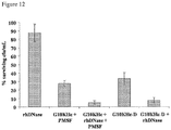
- One example of the STAMP composition is the combination of a STAMP (G10KHc (SEQ ID NO 36) and a DNase. The composition was used to reduce P.areruginosa in sputum and exhibited enhanced antimicrobial activity of G10KHc, as the DNase reduced sputum viscosity and enhanced the STAMP diffusion.
- compositions comprising a STAMP and a suitable pharmaceutical carrier.
- pharmaceutically acceptable carrier means a pharmaceutically-acceptable material, composition or vehicle, such as a liquid or solid filler, diluent, excipient, solvent or encapsulating material, involved in carrying or transporting a STAMP from one location, body fluid, tissue, organ, or portion of the body, to another location, body fluid, tissue, organ, or portion of the body.
- Each carrier must be “pharmaceutically acceptable” in the sense of being compatible with the other ingredients, e.g., a STAMP, of the formulation and suitable for use in contact with the tissue or organ of humans and animals without excessive toxicity, irritation, allergic response, immunogenicity, or other problems or complications, commensurate with a reasonable benefit/risk ratio.
- a STAMP pharmaceutically acceptable
- materials which can serve as pharmaceutically-acceptable carriers include: (1) sugars, such as lactose, glucose and sucrose; (2) starches, such as corn starch and potato starch; (3) cellulose, and its derivatives, such as sodium carboxymethyl cellulose, ethyl cellulose and cellulose acetate; (4) powdered tragacanth; (5) malt; (6) gelatin; (7) talc; (8) excipients, such as cocoa butter and suppository waxes; (9) oils, such as peanut oil, cottonseed oil, safflower oil, sesame oil, olive oil, corn oil and soybean oil; (10) glycols, such as propylene glycol; (1) polyols, such as glycerin, sorbitol, mannitol and polyethylene glycol; (12) esters, such as ethyl oleate and ethyl laurate; (13) agar; (14) buffering 19 agents, such as magnesium hydroxide and aluminum hydrox
- diagnostic agent comprising a targeting peptide and a detectable agent.
- the diagnostic agent of this disclosure can be useful in diagnostic assays, e.g., for detecting the presence or amount of a target or target microbial organism in a place where the target organism is susceptible to exist (e.g., tissues, organs, body fluid, sputum, surface of a body or organ, mucosal surface, implant, biofilm, or serum, device, air, fluid, cell culture, surface of an industry article or a device), or for detecting the onset, development, or remission of a condition (e.g., an infection or a disease) associated with the target microorganism.
- a condition e.g., an infection or a disease
- the targeting peptide typically will be labeled with or conjugated to a detectable agent.
- detectable agents are available which can be generally grouped into the following categories:
- the detectable agent is not necessarily conjugated to the targeting peptide but is capable of recognizing the presence of the targeting peptide and the agent can be detected.
- the detectable agent is an antibody that specifically binds to the targeting peptide. The antibody can then be detected or quantified through various methods known in the art (See Harlow & Lane, Antibodies- A Laboratory Manual (1988 )).
- the diagnostic agent of the present disclosure can be provided in a kit, i.e., a packaged combination of reagents in predetermined amounts with instructions for performing the diagnostic assay.
- the kit will include substrates and cofactors required by the enzyme (e.g., a substrate precursor which provides the detectable chromophore or fluorophore).
- substrates and cofactors required by the enzyme e.g., a substrate precursor which provides the detectable chromophore or fluorophore
- other additives may be included such as stabilizers, buffers (e.g., a block buffer or lysis buffer) and the like.
- the relative amounts of the various reagents may be varied widely to provide for concentrations in solution of the reagents which substantially optimize the sensitivity of the assay.
- the reagents may be provided as dry powders, usually lyophilized, including excipients which on dissolution will provide a reagent solution having the appropriate concentration.
- compositions e.g., the STAMPs or the STAMP compositions
- the compositions can be used to kill, inhibit or reduce the growth of a target microbial organism to which the targeting peptide specifically binds.
- the compositions of the present disclosure provide antimicrobial effect to a target microbial organism and can be used to treat a disease or infection associated with the target microbial organism.
- An antimicrobial effect includes inhibiting the growth or killing of the target microbial organisms, or interfering with any biological functions of the target microbial organisms.
- the compositions of the present aspect can be used to treat a disease or infection at any place in a host, e.g., at any tissue including surfaces of any tissue or implant.
- the compositions are used to specifically kill or inhibit planktonic target microbial organisms in body fluid (e.g., blood, sputum).
- body fluid e.g., blood, sputum
- the compositions of the present disclosure are used to treat a disease or infection on a mucosal surface or a surface containing a biofilm.
- biofilm refers to an accumulation of microbial organisms that produce an extracellular polysaccharide and proteinaceous material that act as a natural glue to immobilize or embed the organisms. Biofilms may form on solid biological or non-biological surfaces. A biofilm consisting essentially of non-harmful, non-pathogenic, commensal microbial organisms is essential for maintaining a healthy and normal microbial flora to prevent the invasion and establishment of other pathogenic microbial organisms, e.g., yeast infection.
- biofilm may lead to biofilm-associated microbial infection.
- pathogenic microbial organisms e.g., cariogenic organisms like Streptococcus mutans
- biofilm-associated microbial infections include infections of oral soft tissues, teeth and dental implants; middle ear; gastrointestinal tract; urogenital tract; airway/lung tissue; eye; urinary tract prostheses; peritoneal membrane and peritoneal dialysis catheters, indwelling catheters for hemodialysis and for chronic administration of chemotherapeutic agents (Hickman catheters); cardiac implants such as pacemakers, prosthetic heart valves, ventricular assist devices, and synthetic vascular grafts and stents; prostheses, internal fixation devices, and percutaneous sutures; and tracheal and ventilator tubing.
- Biomedical systems such as blood oxygenators, tracheal lavage, dental water units, and dialyzers are also susceptible to bacterial contamination and biofilm formation.
- the composition of present disclosure can be used to disturb the balance of pathogen-containing biofilm (e.g., a biofilm overpopulated by pathogenic microbial organisms) such that undesirable, pathogenic microbial organisms (target microbial organisms) are selectively killed, inhibited or reduced and the desirable, non-pathogenic microbial populations (non-target microbial organisms) are minimally impacted.
- pathogen-containing biofilm e.g., a biofilm overpopulated by pathogenic microbial organisms
- target microbial organisms undesirable, pathogenic microbial organisms
- non-pathogenic microbial populations non-target microbial organisms
- the composition can be used in many places in an animal or human body which have mucosal surfaces colonized by multiple species microbial biofilms. Examples of these places include mouth, vagina, gastrointestinal (GI) tract, esophageal tract, respiratory tract, implants. For example, in human mouth there usually exist many different microbes including yeasts and bacteria.
- composition of the present invention targets specifically to, for example, cariogenic organisms (e.g. Streptococcus mutans) and will have minimum effect on non-targeted microbial organisms, and thus will not have an undesirable effect on non-targeted microbial organisms.
- cariogenic organisms e.g. Streptococcus mutans
- composition of the present disclosure can also be used to inhibit target microbial organisms or apply to various biofilm surfaces outside of a human body, e.g., industrial articles or applications.
- composition of the present disclosure can be administered to food processing equipments or food itself to prevent infections related to food consumption, e.g., Salmonella in a poultry processing facility.
- the target microbial organism of the present disclosure can be any bacteria, rickettsia, fungi, yeasts, protozoa, or parasites.
- the target microbial organism is a cariogenic organism, e.g., Streptococcus mutans.
- the target microbial organisms of the present disclosure include, without limitation, Escherichia coli, Candida, Salmonella, Staphylococcus, and Pseudomonas, especially Campylobacter jejuni, Candida albicans, Candida krusei, Chlamydia trachomatis, Clostridium difficile, Cryptococcus neoformans, Haempohilus influenzae, Helicobacter pylor, Moraxella catarrhalis, Neisseria gonorrhoeae, Pseudomonas aeroginosa, Salmonella typhimurium, Shigella disenteriae, Staphylococcus aureus, and Streptococcus pneumoniae.
- S. mutans infection is commonly found in mouth and causes dental caries.
- Porphyromonas gingivalis, various Actinomyces species, spirochetes, and black-pigmented bacteroides are commonly associated with infections of gingival and surrounding connective tissues, which cause periodontal diseases.
- Streptococcus pneumoniae, Haemophilius influenza, or Moraxella cararrhalis infections are commonly found in acute otitis media (AOM) and otitis media effusion (OME) as complications of upper respiratory infections in young children.
- AOM acute otitis media
- OME otitis media effusion
- H. pylori Helicobacter pylori bacteria are found in the gastric mucous layer or adherent to the epithelial lining of the stomach, and cause more than 90% of duodenal ulcers and up to 80% of gastric ulcers.
- Other GI tract infections include, without limitation, Campylobacter bacterial infection, primarily Campylobacter jejuni associated with diarrhea, cholera caused by Vibrio cholerae serogroups, salmonellosis caused by bacteria salmonella such as S. typhimurium and S.
- enteritidis shigellosis caused by bacteria Shigella, e.g., Shigella dysenteriae and traveler's diarrhea caused by enterotoxigenic Escherichia coli (ETEC).
- ETEC enterotoxigenic Escherichia coli
- Clostridium difficile infection is also commonly found in the gastrointestinal tract or esophageal tract.
- Pseudomonas organisms have been associated with common-source nosocomial outbreaks; in addition, they have been incriminated in bacteremia, endocarditis, and osteomyelitis in narcotic addicts. Infections with Pseudomonas organisms can also occur in the ear, lung, skin, or urinary tract of patients, often after the primary pathogen has been eradicated by antibiotics. Serious infections are almost invariably associated with damage to local tissue or with diminished host resistance. Patients compromised by cystic fibrosis and those with neutropenia appear at particular risk to severe infection with P. aeruginosa.
- Premature infants; children with congenital anomalies and patients with leukemia; patients with burns; and geriatric patients with debilitating diseases are likely to develop Pseudomonas infections.
- the organism is prevalent in urine receptacles and on catheters, and on the hands of hospital staff.
- staphylococci of which Staphylococcus aureus is the most important human pathogen, are hardy, gram-positive bacteria that colonize the skin of most human beings. If the skin or mucous membranes are disrupted by surgery or trauma, staphylococci may gain access to and proliferate in the underlying tissues, giving rise to a typically localized, superficial abscess. Although these cutaneous infections are most commonly harmless, the multiplying organisms may invade the lymphatics and the blood, leading to the potentially serious complications of staphylococcal bacteremia.
- S. aureus produces toxins that cause skin rashes or that mediate multisystem dysfunction, as in toxic shock syndrome.
- Coagulase-negative staphylococci particularly S. epidermidis, are important nosocomial pathogens, with a particular predilection for infecting vascular catheters and prosthetic devices.
- S. saprophyticus is a common cause of urinary tract infection.
- Yeast or Candida infections typically occur either orally (Oropharyngeal Candida or OPC) or vaginally (Vulvovaginal Candida or VVC).
- Candidiasis is caused by a shift in the local environment that allows Candida strains (most commonly Candida albicans) already present on skin and on mucosal surfaces such as mouth and vagina to multiply unchecked.
- Gonorrhea, chlamydia, syphilis, and trichomoniasis are infections in the reproductive tract, which cause sexually transmitted diseases, e.g., pelvic inflammatory disease.
- the STAMP or the STAMP composition can be administered to a subject by any administration route known in the art, including without limitation, oral, enteral, buccal, nasal, topical, rectal, vaginal, aerosol, transmucosal, epidermal, transdermal, ophthalmic, pulmonary, and/or parenteral administration.
- a parenteral administration refers to an administration route that typically relates to injection which includes but is not limited to intravenous, intramuscular, intraarterial, intrathecal, intracapsular, intraorbital, intra cardiac, intradermal, intraperitoneal, transtracheal, subcutaneous, subcuticular, intraarticular, subcapsular, subarachnoid, intraspinal, and/or intrasternal injection and/or infusion.
- the STAMP or the STAMP composition can be given to a subject in the form of formulations or preparations suitable for each administration route.
- the formulations useful in the methods of the present disclosure include one or more STAMPs, one or more pharmaceutically acceptable carriers therefor, and optionally other therapeutic ingredients.
- the formulations may conveniently be presented in unit dosage form and may be prepared by any methods well known in the art of pharmacy.
- the amount of active ingredient which can be combined with a carrier material to produce a single dosage form will vary depending upon the subject being treated and the particular mode of administration.
- the amount of a STAMP which can be combined with a carrier material to produce a pharmaceutically effective dose will generally be that amount of a STAMP which produces a therapeutic effect. Generally, out of one hundred percent, this amount will range from about 1 percent to about ninety-nine percent of the STAMP, preferably from about 5 percent to about 70 percent.
- Methods of preparing these formulations or compositions include the step of bringing into association a STAMP with one or more pharmaceutically acceptable carriers and, optionally, one or more accessory ingredients.
- the formulations are prepared by uniformly and intimately bringing into association a STAMP with liquid carriers, or finely divided solid carriers, or both, and then, if necessary, shaping the product.
- Formulations suitable for oral administration may be in the form of capsules, cachets, pills, tablets, lozenges (using a flavored basis, usually sucrose and acacia or tragacanth), powders, granules, or as a solution or a suspension in an aqueous or nonaqueous liquid, or as an oil-in-water or water-in-oil liquid emulsion, or as an elixir or syrup, or as pastilles (using an inert base, such as gelatin and glycerin, or sucrose and acacia) and/or as mouth washes and the like, each containing a predetermined amount of a STAMP as an active ingredient.
- compositions of the present disclosure are used to treat or prevent cariogenic organism infections, e.g., S. mutans infection associated with dental caries and are prepared as additives to food or any products having direct contact to an oral environment, especially an oral environment susceptible to dental caries.
- cariogenic organism infections e.g., S. mutans infection associated with dental caries
- compositions of the present disclosure can be formulated into a baby formula, mouthwash, lozenges, gel, varnish, toothpaste, toothpicks, tooth brushes, or other tooth cleansing devices, localized delivery devices such as sustained release polymers or microcapsules, oral irrigation solutions of any kind whether mechanically delivered or as oral rinses, pacifiers, and any food including, without limitation, chewing gums, candies, drinks, breads, cookies, and milk.
- the STAMP is mixed with one or more pharmaceutically-acceptable carriers, such as sodium citrate or dicalcium phosphate, and/or any of the following: (1) fillers or extenders, such as starches, lactose, sucrose, glucose, mannitol, and/or silicic acid; (2) binders, such as, for example, carboxymethylcellulose, alginates, gelatin, polyvinyl pyrrolidone, sucrose and/or acacia; (3) humectants, such as glycerol; (4) disintegrating agents, such as agar-agar, calcium carbonate, potato or tapioca starch, alginic acid, certain silicates, and sodium carbonate, (5) solution retarding agents, such as paraffin, (6) absorption accelerators, such as quaternary ammonium compounds; (7) wetting agents, such as, for
- compositions may also comprise buffering agents.
- Solid compositions of a similar type may also be employed as fillers in soft and hard-filled gelatin capsules using such excipients as lactose or milk sugars, as well as high molecular weight polyethylene glycols and the like.
- a tablet may be made by compression or molding, optionally with one or more accessory ingredients.
- Compressed tablets may be prepared using binder (for example, gelatin or hydroxypropylmethyl cellulose), lubricant, inert diluent, preservative, disintegrant (for example, sodium starch glycolate or cross-linked sodium carboxymethyl cellulose), surface-active or dispersing agent.
- Molded tablets may be made by molding in a suitable machine a mixture of the powdered peptide or peptidomimetic moistened with an inert liquid diluent.
- Tablets, and other solid dosage forms may optionally be scored or prepared with coatings and shells, such as enteric coatings and other coatings well known in the pharmaceutical-formulating art. They may also be formulated so as to provide slow or controlled release of a STAMP therein using, for example, hydroxypropylmethyl cellulose in varying proportions to provide the desired release profile, other polymer matrices, liposomes and/or microspheres. They may be sterilized by, for example, filtration through a bacteria-retaining filter, or by incorporating sterilizing agents in the form of sterile solid compositions which can be dissolved in sterile water, or some other sterile injectable medium immediately before use.
- compositions may also optionally contain pacifying agents and may be of a composition that they release the STAMP(s) only, or preferentially, in a certain portion of the gastrointestinal tract, optionally, in a delayed manner.
- pacifying agents include polymeric substances and waxes.
- the STAMP can also be in micro-encapsulated form, if appropriate, with one or more of the above-described excipients.
- Liquid dosage forms, Tor oral administration include pharmaceutically acceptable emulsions, microemulsions, solutions, suspensions, syrups and elixirs.
- the liquid dosage forms may contain inert diluents commonly used in the art, such as, for example, water or other solvents, solubilizing agents and emulsifiers, such as ethyl alcohol, isopropyl alcohol, ethyl carbonate, ethyl acetate, benzyl alcohol, benzyl benzoate, propylene glycol, 1,3-butylene glycol, oils (in particular, cottonseed, groundnut, corn, germ, olive, castor and sesame oils), glycerol, tetrahydrofuryl alcohol, polyethylene glycols and fatty acid esters of sorbitan, and mixtures thereof.
- the oral compositions can also include adjuvants such as wetting agents, emulsifying and suspend
- Suspensions in addition to the STAMP, may contain suspending agents as, for example, ethoxylated isostearyl alcohols, polyoxyethylene sorbitol and sorbitan esters, microcrystalline cellulose, aluminum metahydroxide, bentonite, agar-agar and tragacanth, and mixtures thereof.
- suspending agents as, for example, ethoxylated isostearyl alcohols, polyoxyethylene sorbitol and sorbitan esters, microcrystalline cellulose, aluminum metahydroxide, bentonite, agar-agar and tragacanth, and mixtures thereof.
- Formulations for rectal or vaginal administration may be presented as a suppository, which may be prepared by mixing one or more STAMPs with one or more suitable nonirritating excipients or earners comprising, for example, cocoa butter, polyethylene glycol, a suppository wax or a salicylate, and which is solid at room temperature, but liquid at body temperature and, therefore, will melt in the rectum or vaginal cavity and release the active agent.
- Formulations which are suitable for vaginal administration also include pessaries, tampons, creams, gels, pastes, foams or spray formulations containing such carriers as are known in the art to be appropriate.
- Formulations for the topical or transdermal or epidermal administration of a STAMP composition include powders, sprays, ointments, pastes, creams, lotions, gels, solutions, patches and inhalants.
- the active component may be mixed under sterile conditions with a pharmaceutically acceptable carrier, and with any preservatives, buffers, or propellants which may be required.
- the ointments, pastes, creams and gels may contain, in addition to the STAMP composition, excipients, such as animal and vegetable fats, oils, waxes, paraffins, starch, tragacanth, cellulose derivatives, polyethylene glycols, silicones, bentonites, silicic acid, talc and zinc oxide, or mixtures thereof.
- Powders and sprays can contain, in addition to the STAMP composition, excipients such as lactose, talc, silicic acid, aluminum hydroxide, calcium silicates and polyamide powder, or mixtures of these substances.
- Sprays can additionally contain customary propellants, such as chlorofluorohydrocarbons and volatile unsubstituted hydrocarbons, such as butane and propane.
- the STAMP composition can be alternatively administered by aerosol. This is accomplished by preparing an aqueous aerosol, liposomal preparation or solid particles containing the STAMPs.
- a nonaqueous (e. g., fluorocarbon propellant) suspension could be used.
- Sonic nebulizers can also be used.
- An aqueous aerosol is made by formulating an aqueous solution or suspension of the agent together with conventional pharmaceutically acceptable carriers and stabilizers.
- the carriers and stabilizers vary with the requirements of the particular compound, but typically include nonionic surfactants (Tweens, Pluronics, or polyethylene glycol), innocuous proteins like serum albumin, sorbitan esters, oleic acid, lecithin, amino acids such as glycine, buffers, salts, sugars or sugar alcohols. Aerosols generally are prepared from isotonic solutions.
- Transdermal patches can also be used to deliver STAMP compositions to an infection site.
- Such formulations can be made by dissolving or dispersing the agent in the proper medium.
- Absorption enhancers can also be used to increase the flux of the peptidomimetic across the skin. The rate of such flux can be controlled by either providing a rate controlling membrane or dispersing the peptidomimetic in a polymer matrix or gel.
- Ophthalmic formulations are also contemplated as being within the scope of this invention.
- Formulations suitable for parenteral administration comprise a STAMP in combination with one or more pharmaceutically-acceptable sterile isotonic aqueous or nonaqueous solutions, dispersions, suspensions or emulsions, or sterile powders which may be reconstituted into sterile injectable solutions or dispersions just prior to use, which may contain antioxidants, buffers, bacterostats, solutes which render the formulation isotonic with the blood of the intended recipient or suspending or thickening agents.
- aqueous and nonaqueous carriers examples include water, ethanol, polyols (e. g., such as glycerol, propylene glycol, polyethylene glycol, and the like), and suitable mixtures thereof, vegetable oils, such as olive oil, and injectable organic esters, such as ethyl oleate.
- polyols e. g., such as glycerol, propylene glycol, polyethylene glycol, and the like
- vegetable oils such as olive oil
- injectable organic esters such as ethyl oleate.
- Proper fluidity can be maintained, for example, by the use of coating materials, such as lecithin, by the maintenance of the required particle size in the case of dispersions, and by the use of surfactants.
- Formulations suitable for parenteral administration may also contain adjuvants such as preservatives, wetting agents, emulsifying agents and dispersing agents. Prevention of the action of microorganisms may be ensured by the inclusion of various antibacterial and antifungal agents, for example, paraben, chlorobutanol, phenol sorbic acid, and the like. It may also be desirable to include isotonic agents, such as sugars, sodium chloride, and the like into the compositions. In addition, prolonged absorption of the injectable pharmaceutical form may be brought about by the inclusion of agents which delay absorption such as aluminum monostearate and gelatin.
- Injectable depot forms are made by forming microencapsule matrices of a STAMP or in biodegradable polymers such as polylactide-polyglycolide. Depending on the ratio of the STAMP to polymer, and the nature of the particular polymer employed, the rate of drug release can be controlled. Examples of other biodegradable polymers include poly (orthoesters) and poly (anhydrides). Depot injectable formulations are also prepared by entrapping the STAMP in liposomes or microemulsions which are compatible with body tissue.
- a STAMP composition is delivered to a disease or infection site in a therapeutically effective dose.
- the precise amount of the pharmaceutically effective dose of a STAMP that will yield the most effective results in terms of efficacy of treatment in a given patient will depend upon, for example, the activity, the particular nature, pharmacokinetics, pharmacodynamics, and bioavailability of a particular STAMP, physiological condition of the subject (including race, age, sex, weight, diet, disease type and stage, general physical condition, responsiveness to a given dosage and type of medication), the nature of pharmaceutically acceptable carriers in a formulation, the route and frequency of administration being used, and the severity or propensity of a disease caused by pathogenic target microbial organisms, to name a few.
- Example 1 Targeted killing of Streptococcus mutans by a list of antimicrobial peptides
- C16-4 peptides with Arg to Asn (a positive to negative change in charge)
- C16-3 Phe to Gly substitutions (for a general decrease in hydrophobicity)
- C16-3 peptides representing a 4-residue Ala scan of the C16 sequence
- Binding assays were performed as described previously (15), and the results summarized in Table 2.
- CSP C16 and any peptides containing Thr6 through Arg13 (TFFRLFNR, SEQ ID NO 5) of CSP were detected as bound to S. mutans UA159 or comD cells while any interruption to this region via deletion, substitution or Ala scanning reduced the detected fluorescent binding compared to CSP C16 .
- Some peptides such as C16-3, -11, -16 and -17, which contained only Thr6-Phe11 and Phe7-Phe11, showed binding but at a weaker intensity than CSP C16 or any other peptides with the complete Thr6-Argl3 region. Additionally, we observed that Arg to Asn or Phe to Gly substitutions were deleterious to cell binding, suggesting that these residues within TFFRLFNR (SEQ ID NO 5, called M8 or CSP M8 ) are required for binding to S. mutans. CSP M8 exhibited little or no binding to the other oral streptococci listed in Table 3, indicating that CSP M8 may also be capable of specifically binding to S. mutans surfaces.
- the peptides listed in Table that showed positive binding to S. mutans can be used as targeting peptides against S. mutans.
- These peptides include C16 (SEQ ID NO 1), C16-3 (SEQ ID NO 39), C16-4 (SEQ ID NO 40), C16-5 (SEQ ID NO 41), C16-6 (SEQ ID NO 42), C16-11 (SEQ ID NO 47), C16-12 (SEQ ID NO 5), C16-16 (SEQ ID NO 51), C16-17 (SEQ ID NO 52), and C16-18 (SEQ ID NO 53).
- Table 2 - Binding of CSP-fragment peptides to S. mutans Reported relative binding represents results from both UA159 and comD.
- Targeting peptides specific to S. mutans 1903 (SEQ ID NO 10) and antimicrobial peptides S6L3-33 (SEQ ID NO 7) and BD2.21 (SEQ IN NO 13) were developed in the inventors' laboratory (See Example 4).
- Targeting peptides were conjugated to antimicrobial peptides via a linker GGG (SEQ ID NO 17) to yield the STAMPS C16-33 (SEQ ID NO 8), M8-33(SEQ ID NO 9), 1903-BD2.21(SEQ ID NO. 13), and C16-BD2.21(SEQ ID NO 14), all of which were tested in the similar manner as C16G2 and M8G2.
- Peptide molecular mass was determined by matrix-assisted laser desorption/ionization (MALDI) mass spectrometry.
- Peptides C16G2, G2, and M8G2 were synthesized with amidated C-termini using Fmoc-Tyr(tBu)-Rink Amide MBHA resin (Anaspec). All other peptides were synthesized with the appropriately-substituted Wang resins.
- streptococci from an overnight culture were washed with phosphate buffered saline (1 ⁇ PBS), diluted 1:2 into 1 ⁇ PBS, and exposed to peptide (16 ⁇ M) for 5 min at 25 °C. After incubation with peptide, unbound agent was removed from the bacteria by three cycles of centrifugation (5 min, 16,000 ⁇ g) and resuspension in 1 ⁇ PBS. Labeling of oral streptococci was evaluated using brightfield and fluorescence microscopy (Nikon E400) at a 40 ⁇ magnification.
- the digital images utilized for the semi-quantitative binding assessment were acquired with the factory-supplied software (SPOT, Diagnostics). 1.3 Determination of antimicrobial activity.
- the general antimicrobial activity of peptides against planktonic bacteria was determined by a MIC assay in TH broth (all oral streptococci) (Qi et al., 2005).
- S. mutans, S. gordonii Challis (DL1), and S. sanguinis NY101 strains were grown in Todd Hewitt (TH, Fisher) broth medium at 37 °C under anaerobic conditions (80% N 2 , 10% CO 2 , and 10% H 2 ).
- mutans strains UA159 (Ajdic et al., 2002), ATCC 25175, and T8 (Rogers, 1975), are wild-type clinical isolates, while comD is a knockout mutant that was constructed previously from the wild-type UA140 background (Qi et al., 2005).
- Luciferase expressing S. mutans strain JM11 was constructed from UA140 as described (Merritt et al., 2005). Exponentially growing bacterial cells were diluted to ⁇ 1 ⁇ 10 5 cfu/mL in TH and placed into 96-well plates (Fisher). Peptides were then serially diluted and added to the bacteria.
- MIC was determined by identifying the concentration of peptide that completely inhibited bacterial growth after ⁇ 24 h incubation.
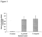
- S. mutans UA159, S. gordonii, or S. sanguinis were grown to log phase and diluted to ⁇ 1 ⁇ 10 5 cfu/mL in growth medium. Under aerobic conditions, 25 ⁇ M G2 or C16G2 was added to the cell suspension and incubated at 25 °C.
- saliva was collected and pooled from 5 adult volunteers in the laboratory, diluted 1:4 in TH broth and centrifuged 2,000 ⁇ g for 10 min. The supernatant was then filter sterilized (0.2 ⁇ m filter, Nunc) and stored at 4 °C. A portion of pooled saliva was also diluted 1:2 in 1 ⁇ PBS and processed as before.
- overnight cultures of JM11 and other oral streptococci were normalized to OD 600 1.0 and ⁇ 3 ⁇ 10 6 cfu/mL of each species was added to 10 mL of the TH-diluted saliva. Sucrose, mannose, and glucose (1% w/v each) were then added and the solution mixed.
- RLU relative light unit
- C16G2 has enhanced antimicrobial activity and specificity against planktonic S. mutans cells.
- minimum inhibitory concentration (MIC) tests were performed against a panel of bacterial species including various strains of S. mutans and closely related oral streptococci (Gilmore et al., 1987). As shown in Table 3, The MIC values of C16G2 ranged from 3-5 ⁇ M for all S. mutans strains tested, a 4-5 fold increase in antimicrobial activity over the parental antimicrobial peptide G2 (12-20 ⁇ M).
- biofilm-associated cells are 100-1000 fold more resistant to antibiotics (Donlan et al., 2002).
- biofilm-associated S. mutans, S. gordonii, or S. sanguinis were treated with 25 ⁇ M C16G2, G2, CSP, CSP C16 , or 1 ⁇ PBS, for 1 min, washed, and their re-growth was monitored over time.
- S. gordonii or S. sanguinis biofilms exposed to any of the peptides tested grew similarly to untreated biofilms after peptide addition and removal ( Fig. 2A-B ).
- S. gordonii or S. sanguinis biofilms exposed to any of the peptides tested grew similarly to untreated biofilms after peptide addition and removal ( Fig. 2A-B ).
- S. gordonii or S. sanguinis biofilms exposed to any of the peptides tested grew similarly to untreated biofilms after peptide addition and removal ( Fig. 2A-
- C16G2 can function as an anti-S. mutans STAMP in a biofilm environment with only a short period of exposure (1 min), a time-frame which is relevant for clinical treatments in the oral cavity (Axelsson & Lindhe, 1987).
- C16G2 can selectively eliminate S. mutans from a mixed species biofilm. In addition to growing as biofilm in vivo, S. mutans are also constantly bathed in saliva as they adhere to the tooth surface. To examine whether C16G2 could selectively kill S.
- S. mutans under these conditions, 2 species of non-cariogenic oral streptococci (S. gordonii and S. sanguinis), were mixed with S. mutans JM11, a strain harboring a transcriptionabfusion between luciferase (luc) and the promoter for the constitutively active gene lactate dehydrogenase (ldh), which has the same susceptibility to C16G2 as the wild type UA159. JM11 has been previously utilized to measure the fitness of S. mutans populations, and decreasing relative light unit (RLU) production was shown to strongly correlate with reduced cell viability (Merritt et al., 2005).
- RLU relative light unit
- the mixed-species biofilms were formed with saliva, and then peptides (25 ⁇ M) suspended in saliva were added for 5 min and removed, and the post-treatment growth of the biofilm was further monitored.
- the number of viable S. mutans cells within the biofilm was quantified in parallel by luciferase expression. It was found that C16G2 was able to dramatically reduce the S. mutans population within the mixture (reflected in the low luciferase activity) after 5 min exposure, compared to CSP C16 and G2 ( Fig. 3 ). Interestingly, even after 120 min post treatment, the total number of S. mutans within the mixture remained low ( Fig. 3 ).
- C16G2 Enhanced antimicrobial activity of C16G2 is related to targeted ComD-independent binding of CSP C16 to S. mutans.
- CSP C16 and C16G2 were fluorescently labeled and tested their ability to bind S. mutans and other streptococci. Consistent with observed killing activity, it was found that CSP C16 and C16G2 could specifically bind to S.
- CSP M8 would be sufficient to function as an alternative targeting domain for an anti-S. mutans STAMP.
- CSP M8 and G2 were synthesized together to form the STAMP M8G2 (Table 1).
- M8G2 displayed similar MICs against S. mutans and other oral streptococci when compared to C16G2.
- single-species biofilm inhibition assays showed that M8G2, like C16G2, was capable of inhibiting the recovery of S. mutans biofilms ( Fig. 4A ), but not those of S. sanguinis ( Fig. 4B ), after 1 min exposure.
- both targeting peptides were conjugated to S6L3-33, a model wide-spectrum antimicrobial peptide, in a similar arrangement as C16G2 and M8G2, to yield the STAMPs C16-33 and M8-33 (Table 1).
- Table 4 a 2-3 fold difference in MIC between S6L3-33 and the derived STAMPs was observed against S. mutans and the other oral streptococci tested.
- the S. mutans-selective activity of the STAMPs was readily apparent: both C16-33 and M8-33 were capable of retarding S. mutans biofilm growth after a short exposure ( Fig.
- STAMPs which exhibited specificity for S. mutans and not other oral streptococci were synthesized and evaluated.
- the STAMPs were designed for S. mutans-selective activity by incorporating portions of a natural pheromone produced by these cariogenic bacteria (CSP) as the targeting domain within the linear STAMP peptide.
- CSP cariogenic bacteria
- bacterial STAMP resistance (should it evolve) (Perron et al, 2006) could be easily overcome by switching to alternative, functionally analogous STAMP components, as was done with G2 and S6L3-33 in this study.
- peptide pheromones are widely utilized by pathogenic bacteria especially Gram-positive organisms, and therefore represent a large and growing pool from which future targeting peptides could be selected for STAMP construction.
- a STAMP with a pathogen-selective (e.g., S. mutans-selective) killing ability is an ideal solution which selectively kills or reduces the pathogen (e.g., S. mutans) in the flora and allows the normal flora to outgrow affected S. mutans populations.
- pathogen-selective e.g., S. mutans-selective
- Such an "antibiotic-probiotic" therapeutic will help prevent dental caries progression and the high health care costs associated with this disease (Anderson & Shi, 2006).
- Example 2 Enhancement of antimicrobial activity against Pseudomonas aeruginosa by co-administration of G10KHc and tobramycin.
- Pseudomonas aeruginosa is a common opportunistic human pathogen that is associated with life-threatening acute infections and chronic airway colonization during cystic fibrosis.
- novispirin G10 a wide-spectrum antimicrobial peptide was converted into a selectively-targeted antimicrobial peptide (STAMP), G10KHc.
- STAMP selectively-targeted antimicrobial peptide
- G10Hc STAMP has the following sequence and components:
- P. aeruginosa is a persistent and recurrent opportunistic pathogen responsible for life-threatening recurrent infections during CF. Frequent isolation of antibiotic-resistant P. aeruginosa suggests that it is critical that new therapies be developed to inhibit and treat P. aeruginosa colonization of airway mucosal surfaces before currently-prescribed treatment options are no longer effective.
- Aerosolized tobramycin has been approved for the control of P. aeruginosa infections in CF patients, and not unexpectedly, tobramycin and aminoglycoside-resistant strains of P. aeruginosa and other organisms have been isolated from CF sputum. This fact, combined with the relatively high rate of unpleasant post-treatment dyspnea, bronchospasm, and increased cough, suggests that tobramycin may be best utilized in a smaller dosage size in combination with another agent. It is concluded that G10KHc can be a candidate for co-administration due to its engineered Pseudomonas selectivity, and potent antimicrobial effects against P. aeruginosa biofilms and multi-drug and aminoglycoside-resistant strains.
- Example 3 Enhanced stability and activity of the G10KHc STAMP by using D-amino acid enantiomer to synthesize the STAMP and/or chemical antagonists (e.g., rhDNase).
- D-amino acid enantiomer to synthesize the STAMP and/or chemical antagonists (e.g., rhDNase).
- protease inhibitors In samples examining the effect of protease inhibitors on peptide activity, pooled sputum samples were pre-treated for 30 min with 1 mM protease inhibitor, either phenylmethylsulphonylfluoride (PMSF), beta-mercaptoethanol (BME), or ethylenediaminetetraacetic acid (EDTA), (all acquired from Sigma-Aldrich), followed by addition of 25 ⁇ M G10KHc and ATCC 15692 ( ⁇ 5x10 6 cfu/mL).
- PMSF phenylmethylsulphonylfluoride
- BME beta-mercaptoethanol
- EDTA ethylenediaminetetraacetic acid
- Bacteria surviving peptide treatment were rescued by dilution (1:50) in growth media at 4 h and kept on ice before appropriate dilution and plating on LB agar supplemented with ampicillin (25 ⁇ g/mL). After overnight incubation at 37 °C, colonies were counted and the surviving cfu/mL quantitated. 100 cfu/mL was considered as the countable limit for all plating procedures. Endogenous organisms already present in the pooled sputum were observed, but were a minority of the population compared to the exogenously added cells (less than 1%, data not shown). As a result endogenous and exogenous cfu/mL were not differentiated for these cultures.
- G10KHc The general antimicrobial effect of G10KHc in sputum samples from CF patients was evaluated by a killing kinetics assay. As shown in Figure 11A, at 4 h post-peptide addition G10KHc was not active against exogenously added P. aeruginosa when mixed with pooled sputum samples. This finding was in contrast to G10KHc activity in growth medium, were the G10KHc STAMP was found to reduce the recoverable cfu/mL over 90% after 30 min of exposure, and eliminate all P. aeruginosa cfu/mL by 2 h of treatment (See Experiment 2).
- G10KHc STAMP stability in sputum was examined over time. Stability of peptides in sputum was monitored by HPLC. Briefly, pooled sputum samples were diluted 1:10 in PBS and centrifuged repeatedly to remove insoluble materials. G10KHc or G10KHc-D (100 ⁇ M) was then added to 100 ⁇ L diluted pooled sputum (with or without 1 mM PMSF pretreatment for 1 h) and mixed at room temperature.
- HPLC profiles were obtained using the manufacturers protocol (Unicorn, Amersham), and differentially colored and overlayed using Photoshop 7.0 (Adobe) for construction of Figure 11B .
- the targeting peptide1903 was obtained by scanning the genomic sequence of S. mutans UA140.
- the predicted open reading frames (ORFs) of the publicly-available genome were examined and those ORFs that encoded for proteins under 50 amino acids were noted and re-examined after scanning the entire genome.
- a number of peptides predicted to be encoded by these ORFs were selected, synthesized with fluorescent labels, tested for binding to S. mutans biofilms.
- Peptide 1903 showed the binding activity to S. mutans and then was used to synthesize 1903 based STAMPs.
- BD2.21 was rationally-designed as part of the "Beta-deletion 2" antimicrobial peptide library.
- a common alpha helical residue arrangement, HHCCHHCHHH(n) was replaced using mostly positive (C) and hydrophobic (H) residues at the positions indicated. There is some variability in the pattern of residues we used, and some non-hydrophobic and uncharged residues were incorporated. The replacement was limited to 3-5 cationic amino acids per peptide, and 4-7 hydrophobic residues (9 to 12 total).
- the antimicrobial affects of modified peptides were tested and BD2.21 showed an MIC of 5.5 uM against planktonic S mutans.
- BD2.21 was then used to synthesize BD2.21 based STAMP such as 1903-BD2.21 having an amino acid sequence of NIFEYFLE-GGG-KLFKFLRKHLL as shown in SEQ ID NO. 13 and C16-BD2.21 having an amino acid sequence of TFFRLFNRSFTQALGK-GGG-KLFKFLRKHLL as shown in SEQ ID NO 14.
- S. mutans biofilms were seeded with 10 ⁇ 5 cells/well and grown overnight with 1 % sucrose (TH medium) in 48-well plates (final volume 400 ⁇ L). After incubation, the supernatant was removed from the biofilms and replaced with 200 ⁇ L PBS with 50 ⁇ M STAMP. PBS alone was used for the negative control (100% survival) with ethanol as the control for complete killing of the biofilms (0% survival). Biofilms were treated with peptide for 20 min, then washed 1x with PBS. To measure biofilm survival, 20 ⁇ L CellTiterBlue diluted into 160 ⁇ L TH medium was added per well.
- biofilms were washed 2x in PBS and then physically disrupted with a sterile pipette tip in 100 ⁇ L PBS per well. Cell suspensions were then serially diluted 10-fold to 10 ⁇ -6. Diluted suspensions were then plated on TH medium and TH supplemented with spectinomycin, 800 ⁇ g/mL. The total amount of biofilm killing (all streptococci) was determined by counting colonies from TH-only plates. Controls: 100% of untreated corresponds to the number of cfu/mL recorded from untreated biofilms, 0% survival was obtained from ethanol sterilized samples. S. mutans killing was determined from quantitating colonies on TH-spectinomycin plates, and combined total cfu/mL, was utilized to calculate the ratio of S. mutans:total population. 1:1 ratio indicates no selectivity.
- C16-BD2.21 has no impact on the total cfu/mL population, suggesting that non-S. mutans streptococci are not affected by the STAMP to a significant degree (See Figure 14(A) ). This is confirmed by the observed ratio of surviving S. mutans to total streptococci, which is .075 (See Figure 14 (B) ). 1903-BD2.21 also had a selective ratio (well under 1, see Figure 14 (B) ), though had some impact on other oral streptococci (See Figure 14 (A) ). Table 7.
Description
- This application claims priority to
U.S. Provisional Application Serial No. 60/842,871, filed on September 6, 2006 - The present invention relates to the field of antimicrobial compositions and treatment.
- The indigenous microflora found at human mucosal surfaces are critical for acquiring nutrients and providing protective colonization against pathogenic microorganisms. When the normal flora are disrupted by any number of factors, the result is often microbial infections at the mucosal surface, many of which affect populations worldwide. The lack of a robust immune response at mucosal surfaces has limited the prescribing clinician to conventional antibiotics or antimicrobials for treatment of mucosal infections. Unfortunately for the normal flora, most small molecule antibiotics have broad spectrum of activity, killing benign and pathogenic organisms indiscriminately. This effect often leads to severe antibiotic associated infections due to the vacated niche available for pathogen colonization. Clostridium difficile, Candida albicans and Staphylococcus aureus are examples of classical opportunistic pathogens that take advantage of increased niche size after antibiotic treatment. The problems resulting from wide-spectrum antibiotic use, combined with the emergence of drug-resistant strains, highlight the fundamental need for new "targeted" antibiotic therapies to combat mucosal pathogens with a minimal impact on normal microflora.
- Previous efforts toward achieving target-specific antimicrobial therapy consisted of conjugating antibiotics to monoclonal antibodies or constructing large fusion proteins with bactericidal and bacterial recognition domains (Qiu et al., 2005). Neither method has yet to result in functional, effective therapeutics due to the low efficiency of chemical conjugation, instability of large proteins, or high cost of production.
- Although G1OKHc, a specifically targeted antimicrobial peptide (STAMP), has been developed and demonstrated increased killing potency, selectivity and kinetics against targeted bacteria (Eckert et al., 2006), there is a need to develop novel STAMPs that are capable of specifically or selectively killing or inhibiting the growth of undesirable target microorganisms.
- In a first aspect, the present invention provides a composition comprising a targeting peptide that binds Streptococcus mutans attached to an antimicrobial peptide, wherein the amino acid sequence of said targeting peptide is of a fragment of the competence stimulating peptide (CSP) that comprises the amino acid sequence TFFRLFNR (SEQ ID NO:5).
In a further aspect, the present invention provides the use of a peptide according the disclosed inventions to selectively kill Streptococcus mutans in vitro.
In a further aspect, the present invention provides the composition of the present invention for use in a method of selectively killing Streptoccous mutans in a host, optionally wherein said S. mutans is an oral soft tissue or on teeth.
One aspect of the present disclosure relates to a selectively/specifically targeted antimicrobial peptide (STAMP) which comprises a targeting peptide and an antimicrobial peptide. The STAMP further comprises a linker peptide. - In one aspect, the targeting peptide is selected from the group consisting of C16 or CSPC16 (TFFRLFNRSFTQALGK, SEQ ID NO. 2), M8 or CSPM8 (TFFRLFNR, SEQ ID NO 5), and peptide 1903 (NIFEYFLE, SEQ ID NO 10).
- In another aspect, the linker peptide is selected from the group consisting of GGG (SEQ ID NO 17), AAA (SEQ ID NO 18), SAT (SEQ ID NO 19), ASA (SEQ ID NO 20), SGG (SEQ ID NO 21), PYP (SEQ ID NO 22), SGS (SEQ ID NO 23), GGS (SEQ ID NO 24), SPS(SEQ ID NO 25), PSGSP(SEQ ID NO 26), PSPSP(SEQ ID NO 27), GGSGGS (SEQ ID NO 28) or a combination (a multimer) of any two (dimer), three (trimer), four (tetramer), five (pentamer) or more than five thereof.
- In another aspect, the antimicrobial peptide is selected from the group consisting of G2 (a derivative of novispirin G10, KNLRRIIRKGIHIIKKY* as shown in SEQ ID NO 3) (* denotes C-terminal amidation), S6L3-33 having an amino acid sequence of FKKFWKWFRRF (SEQ ID NO 7) and BD2.21 having an amino acid sequence of KLFKFLRKHLL (SEQ ID NO 11).
- In another aspect, the STAMP comprises a targeting peptide and an antimicrobial peptide, wherein the targeting peptide is covalently linked to the antimicrobial peptide via a peptide bond, wherein the targeting peptide is selected from the group consisting of C16 (SEQ ID NO 2), M8 (SEQ ID NO 5), and 1903 (SEQ ID NO10); and wherein the antimicrobial peptide is selected from the group consisting of G2 (SEQ ID NO 3), S6L3-33 (SEQ ID NO 7) and BD2.21 (SEQ ID NO 11).
- In another aspect, the STAMP comprises a targeting peptide which is covalently linked to a linker peptide via a peptide bond and an antimicrobial peptide which is covalently linked to the linker peptide via a peptide bond, wherein the targeting peptide is selected from the group consisting of C16 (SEQ ID NO 2), M8 (SEQ ID NO 5), and 1903 (SEQ ID NO10); wherein the antimicrobial peptide is selected from the group consisting of G2 (SEQ ID NO 3), S6L3-33 (SEQ ID NO 7) and BD2.21 (SEQ ID NO 11); and wherein the peptide linker is selected from the group consisting of GGG (SEQ ID NO 17), AAA (SEQ ID NO 18), SAT (SEQ ID NO 19), ASA (SEQ ID NO 20), SGG (SEQ ID NO 21), PYP (SEQ ID NO 22), SGS (SEQ ID NO 23), GGS (SEQ ID NO 24), SPS(SEQ ID NO 25), PSGSP(SEQ ID NO 26), PSPSP(SEQ ID NO 27), and GGSGGS (SEQ ID NO 28).
- In another aspect, the STAMP is selected from the group consisting of C16G2 (SEQ ID NO. 4); C16-33 (SEQ ID NO. 8); C16-BD2.21 (SEQ ID NO. 14); M8G2 (SEQ ID NO. 15); M8-33 (SEQ ID NO. 9); M8-BD2.21 (SEQ ID NO.6); 1903-G2 (SEQ ID NO. 12);1903-33 (SEQ ID NO. 16); and 1903-BD2.21 (SEQ ID NO. 13).
- In another aspect, the amino acids in the STAMP are D-amino acid enantiomer.
- Another aspect of the present disclosure relates to a STAMP composition comprising a STAMP and an antibiotic, wherein the STAMP composition shows a synergistic antimicrobial effect in killing or reducing the growth of a target microbial organism. In one embodiment, the STAMP is G10KHc (SEQ ID NO 36). In another embodiment, the antibiotic is tobramycin. In a preferred aspect, the STAMP composition comprises G10KHc (SEQ ID NO 36) and tobramycin.
- Another aspect of the present disclosure relates to a STAMP composition comprising a STAMP and an agent which can enhance, maintain, or facilitate the function or activity of the STAMP. In one embodiment, the agent is a protease inhibitor or rhDNase. In a preferred aspect, the STAMP composition comprises G10KHc (SEQ ID NO 36) and a protease inhibitor and/or rhDNase.
- Another aspect of the present disclosure is a diagnostic agent comprising a targeting peptide and a detectable agent. In one aspect, the targeting peptide is conjugated to the detectable agent.
- Another aspect of the present disclosure relates to the use of a composition of the present invention (e.g., the STAMP or the STAMP composition) in selectively killing, inhibiting or reducing the growth of a target microbial organism in a subject or on a biofilm or treating a disease associated with a target microbial organism.
-
-
Figure 1 : Selective killing activity of C16G2 against S. mutans. S. mutans, S. sanguinis, and S. gordonii planktonic cells were exposed to 25 µM of the STAMP C16G2, or its untargeted parent antimicrobial peptide G2, for 1 min. Surviving cfu/mL were detected and compared. Data represent averages from at least 3 independent experiments. -
Figure 2 : Inhibitory activity of G2 and C16G2 against single-species biofilms. S. gordonii (A), S. sanguinis (B) and S. mutans (C) monoculture biofilms were grown and then exposed for 1 min to 25 µM STAMP or STAMP component (as indicated in the figure), washed, and regrown with fresh medium. Biofilm recovery was monitored over time by OD600. Data represent averages from 3 independent experiments. -
Figure 3 : C16G2 activity against S. mutans within a multi-species biofilm. Mixed cultures of S. mutans, S. sanguinis, and S. gordonii were allowed to form a biofilm in saliva and were then exposed to 25 µM C16G2, CSPC16, or G2. After washing, the biofilms were allowed to recover in fresh medium/saliva. The regrowth of the biofilm over time was monitored by measuring absorbance at OD600, while the health of the S. mutans within the biofilm was measured by luciferase activity (RLU production). The data were plotted as RLU/OD600 and represent averages of at least 3 independent experiments. -
Figure 4 : Activity of M8G2 against oral bacteria in biofilms. S. mutans (A) or S. sanguinis (B) single-species biofilms were mock-treated or exposed to 25 µM M8G2 (specified in the figure). After removal of the STAMP and the addition of fresh medium, biofilm recovery was monitored over time by monitoring absorbance at OD600. The data represent the average of 3 independent experiments. -
Figure 5 : Biofilm inhibitory activity of S6L3-33 and S6L3-33-containing STAMPs. Single-species biofilms of S. mutans (A) or S. sanguinis (B) were treated with M8-33, C16-33 or S6L3-33 alone (specified in the figure) for 1 min. After agent removal and stringent washing, the regrowth of the biofilms was tracked over 4 h by measuring absorbance at OD600 after the addition of fresh medium. The data represent an average value obtained from at least 3 independent assays. -
Figure 6 : HPLC and MALDI spectra for G10KHc. The quality of purified G10KHc was assessed by HPLC (a) and MALDI mass spectrometry (b). By monitoring UV 215, a single peak was detected during HPLC (at 10.06 mL) that had the correct mass for G10KHc (4267.44). -
Figure 7 : Antimicrobial kinetics of G10KHc, G10, and tobramycin. P. aeruginosa strain ATCC 15692 was either mock treated or challenged with the STAMP G10KHc, untargeted G10, or tobramycin (10 µM) and the surviving cfu/mL quantitated after 1 min, 5 min, 30 min and 2h. The assay was conducted in 30% mouse serum and represents the average of at least three independent experiments. -
Figure 8 : Time-kill assay against high-density planktonic P. aeruginosa. Cultures (1x10⁸ cfu/mL) were exposed to 5 µM G10KHc or G10 with and without equal molar tobramycin co-treatment, as well as administered tobramycin alone. After 24 h, surviving cfu/mL were determined by plating. Data points represent the averages of three independent experiments. -
Figure 9 : Enhanced antimicrobial activity of G10KHc and tobramycin against biofilm P. aeruginosa. Biofilms were grown on disk reactors and challenged with 100 µg/mL of agent as indicated. After 4 and 24 h, surviving bacteria were harvested and plated for quantitation from at least 3 independent experiments.
*indicates that the number of cfu/mL from G10KHc/tobramycin treated cultures was too small to appear on the log scale. -
Figure 10 : Dye uptake mediated by sub-inhibitory concentrations of G10KHc. (a) P. aeruginosa were treated with medium (left column) or 2 µM G10KHc (right column) for 5 min followed by PI dye addition. Bright-field (upper panel) and fluorescence (lower panel) images of the same field were collected and evaluated for intracellular dye accumulation (red fluorescence), (b) Surviving cfu/mL from untreated (dye only) and G10KHc-treated cultures were quantitated after visualization and plated as 5-fold serial dilution. -
Figure 11 : Activity and stability of G10KHc and G10KHc-D in sputum. (A) Exogenously added P. aeruginosa were challenged with 25 µM G10KHc (with or without PMSF), 25 µM G10KHc-D, or left untreated (specified in the figure), and the surviving cfu/mL rescued and quantitated 4 h after agent addition. Rescued cfu/mL were expressed as the average of three independent experiments with standard deviations. (B) G10KHc (with and without PMSF, specified in the figure) was added to spuhim for specific durations and peptide stability (milli-absorbance units, mAU) was monitored by HPLC. The increasing mobile phase linear gradient is shown in black. (**) Intact G10KHc identified by MALDI mass spectrometry at retention volume 10.29 mL. (*) Fractions collected for antimicrobial analysis (Table 1). -
Figure 12 : Effect of rhDNase on G10KHc and G10KHc-D activity in concentrated sputum. Minimally diluted pooled sputum samples with exogenously added P. aeruginosa were treated with 25 µM G10KHc (with PMSF) or 50 µM G10KHc-D for 1 h after pretreatment with or without rhDNase. Rescued cells were quantitated and expressed as the percentage of input cfu/mL. The data represent the average of at least 3 independent experiments with standard deviation. -
Figure 13 : Killing of single-species Streptococcus mutans mature biofilms with C16-BD2.21 and 1903-BD2.21. The figure indicates that C16-BD2.21 and 1903-BD2.21 can kill 33% and 15% of the viable S. mutans within the mature biofilm (grown 18-24 h) respectively, after the biofilm was treated by the peptides for only 20 min. -
Figure 14 : Impact of C16-BD2.21 and 1903-BD2.21 on multi-species biofilm of oral Streptococci. (A) shows that C16-BD2.21 has no impact on the total cfu/mL population and 1903-BD2.21 reduced total population by about 30%. (B) shows that ratio of surviving S. mutans to total Streptococci was 0.075 under C16- BD2.21 treatment and the ratio was about 0.2 under 1903-BD2.21 treatment. - One aspect of the present invention relates to selectively/specifically targeted antimicrobial peptides (STAMPs) and the use thereof.
- The term "selectively/specifically targeted antimicrobial peptide" or "STAMP" refers to a chimeric polypeptide which comprises a targeting peptide and an antimicrobial peptide, wherein the targeting peptide is covalently linked or conjugated (e.g., via a peptide bond) to the antimicrobial peptide either at the C-terminal or N-terminal of the targeting peptide. For example, one STAMP may comprise one of the following two structures: 1) a targeting peptide with its C-terminal covalently linked to the N-terminal of an antimicrobial peptide [Amino terminus-targeting peptide - peptide bond-antimicrobial peptide-carboxyl terminus], and 2) an antimicrobial peptide with its C-terminal covalently linked to the N-terminal of a targeting peptide [Amino terminus-antimicrobial peptide-peptide bond-targeting peptide-carboxyl terminus] .
- In one embodiment of the present invention, the STAMP further comprises a peptide linker by which the targeting peptide is covalently linked or conjugated to the antimicrobial peptide. In this case, a STAMP may comprise one of the following two structures: 1) a targeting peptide with its C-terminal covalently linked to the N-terminal of a linker peptide and an antimicrobial peptide with its N-terminal covalently linked to the C-terminal of the linker peptide (Amino terminus-targeting peptide -peptide bond-linker peptide-anti microbial peptide-peptide bond-carboxyl terminus) and 2) a targeting peptide with its N-terminal covalently linked to the C-terminal of a linker peptide and an antimicrobial peptide with its C-terminal covalently linked to the N-terminal of the linker peptide (Amino terminus-antimicrobial peptide -peptide bond-linker peptide-peptide bond-targeting peptide-carboxyl terminus).
- According to the present invention, a targeting peptide can be any suitable peptide which recognizes or binds to a target (e.g., a target cell, a target tissue, a target microbial organism). Particularly, a targeting peptide specifically interacts with or specifically recognizes a target, through, for example, the cell surface appendages such as flagella and pili, and surface exposed proteins, lipids and polysaccharides of a target. In one embodiment, a targeting peptide specifically recognizes or interacts with only one or a few target(s) while minimally recognizing or interacting with non-target(s). In another embodiment, a targeting peptide can be a peptide capable of specifically binding to a microorganism, e.g., a target microbial organism.
- In one aspect, the targeting peptide provided by the present disclosure can be identified via screening peptide libraries. For example, a phage display peptide library can be screened against a target microbial organism or a desired antigen or epitope thereof. In particular, phage display peptide libraries (e.g.,
Ph.D 7, Ph.D. 12, Ph.D C7C libraries from New England Biolabs) that contain >109 unique random- peptide-sequence-containing phage clones. The Ph.D.-C7C library displays 7-mer peptides with disulfide linkages, while the Ph.D.-7 and Ph.D.-12 libraries contain completely randomized 7-mer and 12-mer residues, respectively. The M13 filamentous phage used for the procedure carried the random insert as an N-terminal fusion to the minor coat protein pill. In screening a targeting peptide that specifically recognizes a target or target microbial organism, 1010 pfu/ml of phage library was incubated with 109 microbial organisms (e.g., bacterial cells) for which targeting peptides are desired. After centrifugation, unbound phage was removed by aspiration. The pellet, which contained the target microbial organisms with the bound phage, was washed several times using buffers containing mild detergent to remove loosely bound phage particles, and the tightly bound phage particles were eluted. This process is termed panning. The eluted phage was amplified by infecting E. coli F+ strains. After 3-4 rounds of panning and amplification, a phage pool was obtained, which contained clones with high binding affinity for the bacteria that it was panned against. Ten to twenty phage clones from this pool were randomly picked for DNA sequencing, from which the amino acid sequence of the peptide insert was determined. By aligning the amino acid sequence of multiple clones from the same phage pool, a consensus sequence for the binding/targeting peptide was constructed. This consensus sequence represents one of the binding/targeting peptides specific for the particular microbial organism. To confirm the binding specificity of the consensus peptide, the peptide was chemically synthesized and conjugated to a dye (e.g., FITC, a green fluorescence dye). The labeled peptide was incubated with the microbial organism and analyzed by fluorescent microscopy for target microbial species-specific binding. This methodology ensured that peptides selected from phage display exhibit the same binding specificity as a free peptide independent of the M13 phage particle. - The targeting peptide of the present disclosure can also be a peptide obtained based on rational design. For example, one can design a targeting peptide based on the biochemical and biophysical characteristics of amino acids and the surfaces of microorganisms. In general, positively charged peptides are likely to bind negatively charged components on the cell surface and vice versa. Similarly, hydrophobic peptides may bind to hydrophobic pockets on the cell surface based on hydrophobic interactions while the secondary or tertiary structure of a peptide may fit into certain structures on the surface of a microorganism.
- A peptide identified through a screening or design method can be used as a targeting peptide for specifically recognizing a target microbial organism. Examples of such targeting peptide are disclosed in
U.S. Pat. Appl. No. 10/706, 391 (U.S. Pub. No. 20040137482 ), which include, for example, 1) targeting peptides capable of specifically binding to or recognizing Pseudomonas, especially P.aeruginosa (e.g., targeting peptides 12:1, 12:2, 12:3, 12:4, 12:5, 12:6, 12:7, 12:8; 12:9, and 12:10); 2) targeting peptides capable of specifically binding to Staphylococcus, especially S. aureus (e.g., targeting peptides SA5:1, SA5:3, SA5:4, SA5:5, SA5:6, SA5:7, SA5:8, SA5:9, SA5:10, SA2:2, SA2:4, SA2:5, SA2:6, SA2:7, SA2:8, SA2:9, SA2:10, and SA2:11); and 3) targeting peptides capable of specifically binding to E. coli (e.g., targeting peptides DH5.1, DH5.2, DH5.3, DH5.4, DH5.5, DH5.6, DH5.7, DH5.8, and DH5.9). - In one aspect of the present disclosure, targeting peptides specifically binding to or recognizing Pseudomonas, especially P.aeruginosa, include, for example, cat-1 (or KH) domain, KKHRKHRKHRKH (SEQ ID NO 31). Targeting peptides specifically binding to or recognizing Streptococci include, for example, bacterial pheromones such as CSP (SGSLSTFFRLFNRSFTQALGK, SEQ ID NO 1), CSP 1 (EMRLSKFFRDFILQRKK, SEQ ID NO 29) and CSP2 (EMRISRIILDFLFLRKK, SEQ ID NO 30) and fragments thereof. Further, targeting peptides specifically binding to or recognizing S. pneumoniae include, for example, CSP1 and CSP2. Targeting peptides specifically binding to or recognizing S. mutans include, for example, CSP, C16 or CSPC16 (TFFRLFNRSFTQALGK, SEQ ID NO 2), M8 or CSPM8 (TFFRLFNR, SEQ ID NO 5), and peptide 1903 (NIFEYFLE, SEQ ID NO 10).
- The targeting peptides provided by the present invention can be naturally or non-naturally occurring peptides. For example, the targeting peptides provided by the present invention can be recombinantly made, chemically synthesized, or naturally existing. In one embodiment, the targeting peptide contains an amino acid sequence that naturally exists (e.g., CSP,
CSP 1 and CSP2). In another embodiment, the targeting peptide contains an amino acid sequence that constitutes an internal part of a naturally occurring polypeptide (e.g., C16 and M8 in the present invention). In another embodiment, the targeting peptide contains an amino acid sequence encoded by a sequence naturally existing in a genome and such amino acid sequence is not adjacent to any amino acid sequence naturally adjacent to it, e.g., such amino acid sequence is adjacent to a heterologous sequence in the targeting peptide. - The targeting peptide provided by the present disclosure can also include a peptide having an amino acid sequence that is derived or modified from a targeting amino acid sequence specifically illustrated in the present invention, provided that the derived or modified sequence still maintains or has an enhanced specificity with respect to its target microbial organism. For example, the targeting amino acid sequence can be structurally modified via deletion, mutation, addition of amino acids or other structural entities, or any other structural changes as long as these changes do not alter or adversely affect the binding ability of the targeting amino acid sequence to its target microbial organism.
- The targeting peptide according to the present disclosure specifically interacts with or binds to the target organism (e.g., through the external surface of the organism) via different molecular interactions such as ionic interaction, Vander Waals forces, ligand-receptor interaction, or hydrophobic interaction. For example, the targeting peptide of the present disclosure can also be a peptide ligand, receptor, or fragment thereof that specifically recognizes a target microbial organism. In one example, the targeting peptide of the present disclosure can be a glucan binding protein of Streptococcus mutans that can specifically bind insoluble glucans on the surface of S. mutans. For another example, the targeting peptides can be a bacterial pheromone or a fragment thereof.
- The targeting peptide according to the present disclosure comprises about 4 to about 40 amino acids, from about 5 to about 30, or from about 6 to about 20. In one preferred embodiment, the targeting peptide has a length of 5, 6, 7, 8, 9, 10, 11, 12, 13, 14, 16, 17, 18, 19, 20, 21, 22, 23, 24, 25, 26, 27, 28, 29, or 30 amino acids.
- It is contemplated that the targeting peptides includes peptides which specifically bind a target cell or tissue (e.g., a plant call, an animal cell, or fungal organism). The examples of these targeting peptides include Chitinase, Lectins, and targeting fragments thereof.
- The targeting peptide according to the present invention can be produced by any suitable method known to one skilled in the art by itself or in combination with a linker peptide and an antimicrobial peptide. For example, the targeting peptides can be chemically synthesized via a synthesizer or recombinantly made using an expression system, e.g., a bacterial, yeast, or eukaryotic cell expression system. In the chemical synthesis, the targeting peptide can be made by L-amino acid enantiomers or D-amino acid enantiomers. It is observed that the targeting peptide consisting of D-enantiomers increases the stability without compromising the activity of the targeting peptide.
- The linker peptide according to the present invention is a peptide that can be used to connect a targeting peptide to an antimicrobial peptide without interfering or reducing the activity of the targeting peptide or the antimicrobial peptide. The peptide linker is from about 2 to 20 amino acids, from 2 to 12, or from 3 to 12 amino acids. Examples of the peptide linkers include, for example, GGG (SEQ ID NO 17), AAA (SEQ ID NO 18), SAT (SEQ ID NO 19), ASA (SEQ ID NO 20), SGG (SEQ ID NO 21), PYP (SEQ ID NO 22), SGS (SEQ ID NO 23), GGS (SEQ ID NO 24), SPS(SEQ ID NO 25), PSGSP(SEQ ID NO 26), PSPSP(SEQ ID NO 27), or a combination (a multimer) of any two (dimer), three (trimer), four (tetramer), five (pentamer) or more than five of the listed peptide linkers. In one embodiment, the linker peptide is GGG (SEQ ID NO 17). In another embodiment, the linker peptide is a dimmer of GGS (SEQ ID NO 24), which is GGSGGS (SEQ ID NO 28).
- The linker peptide according to the present invention can be produced by any suitable method known to one skilled in the art by itself or in combination with a targeting peptide and an antimicrobial peptide. For example, the linker peptides can be chemically synthesized via a synthesizer or recombinantly made using an expression system, e.g., a bacterial, yeast, or eukaryotic cell expression system. In the chemical synthesis, the linker peptide can be made by L-amino acid enantiomers or D- amino acid enantiomers. It is observed that peptides consisting of D-enantiomers increase the stability without comprising the activity of the peptides.
- The antimicrobial peptide according to the present disclosure is a peptide capable of killing a microbial organism or inhibiting its growth. The antimicrobial activities of the antimicrobial peptides of the present disclosure include, without limitation, antibacterial, antiviral, or antifungal activities. Antimicrobial peptides include various classes of peptides, e.g., peptides originally isolated from plants as well as animals. In animals, antimicrobial peptides are usually expressed by various cells including neutrophils and epithelial cells. In mammals including human, antimicrobial peptides are usually found on the surface of the tongue, trachea, and upper intestine. Naturally occurring antimicrobial peptides are generally amphipathic molecules that contain fewer than 100 amino acids. Many of these peptides generally have a net positive charge (i.e., cationic) and most form helical structures.
- In one aspect, the antimicrobial peptide according to the present disclosure comprises about 2 to about 100 amino acids, from about 5 to about 50, or from about 7 to about 20. In one preferred aspect, the targeting peptide has a length of 5, 6, 7, 8, 9, 10, 11, 12, 13, 14, 16, 17, 18, 19, 20, 21, 22, 23, 24, 25, 26, 27, 28, 29, or 30 amino acids.
- In another aspect, the antimicrobial peptide has the antimicrobial activity with a minimum inhibitory concentration (MIC) of no more than about 40 µM, no more than about 30 µM, no more than 20 µM, or no more than 10 µM.
- In another aspect, the antimicrobial peptides include those listed in Table 7 (SEQ ID Nos 34-35 and 54-97). In another embodiment, the antimicrobial peptide contains one or more antimicrobial peptides including, without limitation, alexomycin, andropin, apidaecin, bacteriocin, β-pleated sheet bacteriocin, bactenecin, buforin, cathelicidin, α-helical clavanin, cecropin, dodecapeptide, defensin, (β-defensin, α-defensin, gaegurin, histatin, indolicidin, magainin, melittin, nisin, novispirin G10, protegrin, ranalexin, tachyplesin, and derivatives thereof.
- Among these known antimicrobial peptides, tachyplesins are known to have antifungal and antibacterial activities. Andropin, apidaecin, bactencin, clavanin, dodecappeptide, defensin, and indolicidin are antimicrobial peptides having antibacterial activities. Buforin, nisin and cecropin peptides have been demonstrated to have antimicrobial effects on Escherichia, coli, Shigella disenteriae, Salmonella typhimurium, Streptococcus pneumoniae, Staphylococcus aureus, and Pseudomonas aeroginosa. Magainin and ranalexin peptides have been demonstrated to have antimicrobial effects on the same organisms, and in addition have such effects on Candida albicans, Cryptococcus neoformans, Candida krusei, and Helicobacter pylori. Magainin has also been demonstrated to have antimicrobial effects on herpes simplex virus. Alexomycin peptides have been demonstrated to have antimicrobial effects on Campylobacter jejuni, Moraxella catarrhalis and Haemophilus inflluenzae while defensin and (β-pleated sheet defensin peptides have been shown to have antimicrobial effects on Streptococcus pneumoneae. Histatin peptides and the derivatives thereof are another class of antimicrobial peptides, which have antifungal and antibacterial activities against a variety of organisms including Streptococcus mutans (MacKay, B. J. et al., Infect. Immun. 44:695-701 (1984); Xu, et al., J. Dent. Res. 69:239 (1990)).
- In one aspect, the antimicrobial peptide of the present disclosure contains one or more antimicrobial peptides from a class of histatin peptides and the derivatives thereof. For example, the antimicrobial peptide of the present invention contains one or more derivatives of histatin including, without limitation,
histatin 5 having an amino acid sequence of DSHAKRHHGY KRKFHEKHHS HRGY (SEQ ID NO 32) or dhvar-1 having an amino acid sequence of KRLFKELKFS LRKY (SEQ ID NO 33). - In another aspect, the antimicrobial peptide of the present disclosure contains one or more antimicrobial peptides from a class of protegrins and the derivatives thereof. For example, the antimicrobial peptide of the present invention contains protegrin PG-1 having an amino acid sequence of RGGRLCYCRRRFCVCVGR (SEQ ID NO 3334). The protegrin peptides have been shown to have antimicrobial effects on Streptococcus mutans, Neisseria gonorrhoeae, Chlamydia trachomatis and Haempohilus influenzae. Protegrin peptides are described in
U.S. Pat. Nos. 5,693,486 ,5,708,145 ,5,804,558 ,5,994,306 , and6,159,936 . - In yet another aspect, the antimicrobial peptide of the present disclosure contains one or more antimicrobial peptides from a class of novispirin and the derivatives thereof for treating cariogenic organisms, e.g., Streptococcus mutans or Pseudomonas aeroginosa. For example, the antimicrobial peptide of the present invention includes novispirin G10 having an amino acid sequence KNLRRIIRKGIHIIKKYG (SEQ ID NO 35) and G2 (a derivative of novispirin G10) which has one C-terminal amino acid deletion, and one internal deletion from G10 and an amidated C-terminus having the amino acid sequence of KNLRIIRKGIHIIKKY* (SEQ ID NO 3) (* denotes C-terminal amidation).
- In yet another aspect, the antimicrobial peptide of the present disclosure contains peptides rationally designed and tested to possess the antimicrobial activity against a microbial organism (e.g., Streptococcus mutans). The examples of these peptides include, without any limitation, S6L3-33 having an amino acid sequence of FKKFWKWFRRF (SEQ ID NO 7) and BD2.21 having an amino acid sequence of KLFKFLRKHLL (SEQ ID NO 11).
- The antimicrobial peptide according to the present disclosure can be produced by any suitable method known to one skilled in the art by itself or in combination with a targeting peptide and a linker peptide. For example, the antimicrobial peptides can be chemically synthesized via a synthesizer or recombinantly made using an expression system, e.g., a bacterial, yeast, or eukaryotic cell expression system. In the chemical synthesis, the antimicrobial peptide can be made by L-amino acid enantiomers or D-amino acid enantiomers.
- In yet another aspect, the STAMP comprises a targeting peptide and an antimicrobial peptide, wherein the targeting peptide is covalently linked to the antimicrobial peptide via a peptide bond, wherein the targeting peptide is selected from the group consisting of C16 (SEQ ID NO 2), M8 (SEQ ID NO 5), and 1903 (SEQ ID NO 10); and wherein the antimicrobial peptide is selected from the group consisting of G2 (SEQ ID NO 3), S6L3-33 (SEQ ID NO 7) and BD2.21 (SEQ ID NO 11).
- In yet another aspect, the STAMP comprises a targeting peptide which is covalently linked to a linker peptide via a peptide bond and an antimicrobial peptide which is covalently linked to the linker peptide via a peptide bond, wherein the targeting peptide is selected from the group consisting of C16 (SEQ ID NO 2), M8 (SEQ ID NO 5), and 1903 (SEQ ID NO 10); wherein the antimicrobial peptide is selected from the group consisting of G2 (SEQ ID NO 3), S6L3-33 (SEQ ID NO 7) and BD2.21 (SEQ ID NO 11). In yet another aspect, the peptide linker is selected from the group consisting of GGG (SEQ ID NO 17), AAA (SEQ ID NO 18), SAT (SEQ ID NO 19), ASA (SEQ ID NO 20), SGG (SEQ ID NO 21), PYP (SEQ ID NO 22), SGS (SEQ ID NO 23), GGS (SEQ ID NO 24), SPS(SEQ ID NO 25), PSGSP(SEQ ID NO 26), PSPSP(SEQ ID NO 27), and GGSGGS (SEQ ID NO 28). Examples of such STAMPs include but are not limited to the STAMPS listed in Table 1: C16G2 (SEQ ID NO 4); C16-33 (SEQ ID NO 8); C16-BD2.21 (SEQ ID NO 14); M8G2 (SEQ ID NO 6); M8-33 (SEQ ID NO 9); M8-BD2.21 (SEQ ID NO 15); 1903-G2 (SEQ ID NO 12);1903-33 (SEQ ID NO 16); and 1903-BD2.21 (SEQ ID NO 13).
- Another aspect of the present disclosure relates to a composition comprising a plurality of STAMPS, wherein the composition comprises a first STAMP and a second STAMP and the first STAMP is different from the second STAMP. In one aspect, the first STAMP comprises a first targeting peptide covalently linked to a first antimicrobial peptide via a peptide bond. The second STAMP comprises a second targeting peptide covalently linked to a second antimicrobial peptide via a peptide bond. The difference between the first STAMP and the second STAMP is such that at least one corresponding moiety of the two STAMPs is different from each other. For example, in one aspect, the first targeting peptide is different from the second targeting peptide or the first antimicrobial peptide is different from the second antimicrobial peptide. In another aspectt, the first targeting peptide is different from the second targeting peptide and the first antimicrobial peptide is different from the second antimicrobial peptide.
- In another aspect, the first STAMP comprises a first targeting peptide which is covalently linked to a first linker peptide via a peptide bond and a first antimicrobial peptide which is covalently linked to the first linker peptide via a peptide bond. The second STAMP comprises a second targeting peptide which is covalently linked to a second linker peptide via a peptide bond and a second antimicrobial peptide which is covalently linked to the second linker peptide via a peptide bond. The difference between the first STAMP and the second STAMP is such that at least one corresponding moiety of the two STAMPs is different from each other. For example, in one aspect, the first targeting peptide is different from the second targeting peptide (the first linker peptide is the same as the second linker peptide and the first antimicrobial peptide is the same as the second antimicrobial peptide); or the first linker peptide is different from the second peptide linker; or the first antimicrobial peptide is different from the second antimicrobial peptide. In another aspect, the first targeting peptide is the same as the second targeting peptide (the first linker peptide is different from the second linker peptide and the first antimicrobial peptide is different from the second antimicrobial peptide); or the first peptide linker is the same as the second peptide linker; or the first antimicrobial peptide is the same the second antimicrobial peptide. In another aspect, the first targeting peptide is different from the second targeting peptide, the first linker peptide is different from the second peptide linker; and the first antimicrobial peptide is different from the second antimicrobial peptide.
- The STAMP of the present disclosure can be made by any suitable means known to one skilled in the art. In one disclosure, a nucleotide sequence encoding the STAMP can be synthesized through a DNA synthesizer or a nucleotide sequence encoding a targeting peptide can be ligated to a nucleotide sequence encoding an antimicrobial peptide moiety, either directly or via a nucleotide sequence encoding a peptide linker. The nucleotide can be expressed in an appropriate expression system, e.g., a commercially available bacterial, yeast, or eukaryotic cell expression system. In the chemical synthesis, the STAMP can be made by L-amino acid enantiomers or D-amino acid enantiomers. It is observed that the STAMP consisting of D-enantiomers increases the stability without comprising the activity of the STAMP.
- Another aspect of the present disclosure relates to a STAMP composition comprising a STAMP and an antibiotic. A synergistic antimicrobial effect has been unexpectedly observed when a STAMP is co-administered with an antibiotic in killing or reducing the growth of a target microbial organism. Antibiotics suitable for co-administration with the STAMP include substances, produced synthetically or naturally, which can inhibit the growth of or kill microbial organisms. Such antibiotics include, without any limitation, β-lactam antibiotics (e.g., ampicillin, aziocillin, aztreonam, carbenicillin, cefoperazone, ceftriaxone, cephaloridine, cephalothin, cloxacillin, moxalactam, penicillin, piperacillin, and ticarcillin), amoxicillin, bacitracin, chloramphenicol, clindamycin, capreomycin, colistimethate, ciprofloxacin, doxycycline, erythromycin, fusidic acid, fosfomycin, fusidate sodium, gramicidin, gentamycin, , lincomycin, minocycline, macrolides, monobactams, nalidixic acid, novobiocin, ofloxcin, rifamycins, tetracyclines, vancomycin, tobramycin, and trimethoprim. In one example, the STAMP composition comprises a G10KHc STAMP (SEQ ID NO 36) and tobramycin and exhibits a synergistic enhancement of antimicrobial activity to P. aeruginosa in both biofilms and planktonic cultures.
- Another aspect of the present disclosure relates to a STAMP composition comprising a STAMP and an agent which can enhance, maintain, or facilitate the function or activity of the STAMP. In one aspect, the chemical is a protease inhibitor. The STAMP composition is exposed to a protease-present environment where the presence of the protease may reduce the antimicrobial activity of the STAMP via, for example, enzymatic degradation. The combination of a protease inhibitor and a STAMP stabilizes the STAMP from the protease degradation and thus enhance the activity of the STAMP. The protease-present environment includes, for example, body fluid (e.g., urine, blood, serum, salvia, sputum, mucosal fluid). The protease includes, for example, neutrophil elastase, proteinase-3, cycteine protease, metalloprotease, serine-protease, or other proteases derived from bacteria and/or hosts. The protease inhibitor includes, for example, BMP, EDTA, PMFS, benzamidine, and/or recombinant α-1 antitrypsin (rAAT).
- In yet another aspect, the agent is human DNase. One example of the STAMP composition is the combination of a STAMP (G10KHc (SEQ ID NO 36) and a DNase. The composition was used to reduce P.areruginosa in sputum and exhibited enhanced antimicrobial activity of G10KHc, as the DNase reduced sputum viscosity and enhanced the STAMP diffusion.
- Another aspect of the present disclosure relates to a pharmaceutical composition comprising a STAMP and a suitable pharmaceutical carrier. The term "pharmaceutically acceptable carrier" as used herein means a pharmaceutically-acceptable material, composition or vehicle, such as a liquid or solid filler, diluent, excipient, solvent or encapsulating material, involved in carrying or transporting a STAMP from one location, body fluid, tissue, organ, or portion of the body, to another location, body fluid, tissue, organ, or portion of the body. Each carrier must be "pharmaceutically acceptable" in the sense of being compatible with the other ingredients, e.g., a STAMP, of the formulation and suitable for use in contact with the tissue or organ of humans and animals without excessive toxicity, irritation, allergic response, immunogenicity, or other problems or complications, commensurate with a reasonable benefit/risk ratio. Some examples of materials which can serve as pharmaceutically-acceptable carriers include: (1) sugars, such as lactose, glucose and sucrose; (2) starches, such as corn starch and potato starch; (3) cellulose, and its derivatives, such as sodium carboxymethyl cellulose, ethyl cellulose and cellulose acetate; (4) powdered tragacanth; (5) malt; (6) gelatin; (7) talc; (8) excipients, such as cocoa butter and suppository waxes; (9) oils, such as peanut oil, cottonseed oil, safflower oil, sesame oil, olive oil, corn oil and soybean oil; (10) glycols, such as propylene glycol; (1) polyols, such as glycerin, sorbitol, mannitol and polyethylene glycol; (12) esters, such as ethyl oleate and ethyl laurate; (13) agar; (14) buffering 19 agents, such as magnesium hydroxide and aluminum hydroxide; (15) alginic acid; (16) pyrogen-free water; (17) isotonic saline; (18) Ringer's solution; (19) ethyl alcohol; (20) phosphate buffer solutions; and (21) other non-toxic compatible substances employed in pharmaceutical formulations.
- Another aspect of the present disclosure is a diagnostic agent comprising a targeting peptide and a detectable agent. The diagnostic agent of this disclosure can be useful in diagnostic assays, e.g., for detecting the presence or amount of a target or target microbial organism in a place where the target organism is susceptible to exist (e.g., tissues, organs, body fluid, sputum, surface of a body or organ, mucosal surface, implant, biofilm, or serum, device, air, fluid, cell culture, surface of an industry article or a device), or for detecting the onset, development, or remission of a condition (e.g., an infection or a disease) associated with the target microorganism.
- In one aspect, the targeting peptide typically will be labeled with or conjugated to a detectable agent. Numerous detectable agents are available which can be generally grouped into the following categories:
- (a) Radioisotopes, such as 35S, 14C, 13C, 15N, 125I, 3H, and 131I. The peptide can be labeled with the radioisotope using the techniques known in the art and radioactivity can be measured using scintillation counting; in addition, the peptide can be spin labeled for electron paramagnetic resonance for carbon and nitrogen labeling.
- (b) Fluorescent agents such as BODIPY, BODIPY analogs, rare earth chelates (europium chelates), fluorescein and its derivatives, FITC, 5,6 carboxyfluorescein, rhodamine and its derivatives, dansyl, Lissamine, phycoerythrin, green fluorescent protein, yellow fluorescent protein, red fluorescent protein and Texas Red. Fluorescence can be quantified using a fluorometer.
- (c) Various enzyme-substrate agents, such luciferases (e.g., firefly luciferase and bacterial luciferase), luciferin, 2,3-dihydrophthalazinediones, malate dehydrogenase, urease, peroxidase such as horseradish peroxidase (HRPO), alkaline phosphatase, (β-galactosidase, glucoamylase, lysozyme, saccharide oxidases (e.g., glucose oxidase, galactose oxidase, and glucose-6-phosphate dehydrogenase), heterocyclic oxidases (such as uricase and xanthine oxidase), lactoperoxidase, microperoxidase, and the like. Examples of enzyme-substrate combinations include, for example: (i) Horseradish peroxidase (HRPO) with hydrogen peroxidase as a substrate, wherein the hydrogen peroxidase oxidizes a dye precursor (e.g., orthophenylene diamine (OPD) or 3,3',5,5'-tetramethyl benzidine hydrochloride (TMB)); (ii) alkaline phosphatase (AP) with para-Nitrophenyl phosphate as chromogenic substrate; and (iii) β-D-galactosidase (β-D-Gal) with a chromogenic substrate (e.g., p-nitrophenyl- β-D-galactosidase) or fluorogenic substrate 4-methylumbelliferyl- β-D-galactosidase.
- In another aspect, the detectable agent is not necessarily conjugated to the targeting peptide but is capable of recognizing the presence of the targeting peptide and the agent can be detected. For example, the detectable agent is an antibody that specifically binds to the targeting peptide. The antibody can then be detected or quantified through various methods known in the art (See Harlow & Lane, Antibodies- A Laboratory Manual (1988)).
- In another aspect, the diagnostic agent of the present disclosure can be provided in a kit, i.e., a packaged combination of reagents in predetermined amounts with instructions for performing the diagnostic assay. Where the targeting peptide is labeled with an enzyme, the kit will include substrates and cofactors required by the enzyme (e.g., a substrate precursor which provides the detectable chromophore or fluorophore). In addition, other additives may be included such as stabilizers, buffers (e.g., a block buffer or lysis buffer) and the like. The relative amounts of the various reagents may be varied widely to provide for concentrations in solution of the reagents which substantially optimize the sensitivity of the assay. Particularly, the reagents may be provided as dry powders, usually lyophilized, including excipients which on dissolution will provide a reagent solution having the appropriate concentration.
- According to another aspect of the present disclosure, the compositions (e.g., the STAMPs or the STAMP compositions) of the present disclosure can be used to kill, inhibit or reduce the growth of a target microbial organism to which the targeting peptide specifically binds.
- In one aspect, the compositions of the present disclosure provide antimicrobial effect to a target microbial organism and can be used to treat a disease or infection associated with the target microbial organism. An antimicrobial effect includes inhibiting the growth or killing of the target microbial organisms, or interfering with any biological functions of the target microbial organisms. In general, the compositions of the present aspect can be used to treat a disease or infection at any place in a host, e.g., at any tissue including surfaces of any tissue or implant. In one aspect, the compositions are used to specifically kill or inhibit planktonic target microbial organisms in body fluid (e.g., blood, sputum). In one aspect, the compositions of the present disclosure are used to treat a disease or infection on a mucosal surface or a surface containing a biofilm.
- The term "biofilm" refers to an accumulation of microbial organisms that produce an extracellular polysaccharide and proteinaceous material that act as a natural glue to immobilize or embed the organisms. Biofilms may form on solid biological or non-biological surfaces. A biofilm consisting essentially of non-harmful, non-pathogenic, commensal microbial organisms is essential for maintaining a healthy and normal microbial flora to prevent the invasion and establishment of other pathogenic microbial organisms, e.g., yeast infection. However, if the microorganism population in a biofilm is disturbed by overpopulation of pathogenic microbial organisms (e.g., cariogenic organisms like Streptococcus mutans), the resulting biofilm may lead to biofilm-associated microbial infection. Examples of biofilm-associated microbial infections include infections of oral soft tissues, teeth and dental implants; middle ear; gastrointestinal tract; urogenital tract; airway/lung tissue; eye; urinary tract prostheses; peritoneal membrane and peritoneal dialysis catheters, indwelling catheters for hemodialysis and for chronic administration of chemotherapeutic agents (Hickman catheters); cardiac implants such as pacemakers, prosthetic heart valves, ventricular assist devices, and synthetic vascular grafts and stents; prostheses, internal fixation devices, and percutaneous sutures; and tracheal and ventilator tubing. Both indwelling and subcutaneous biomedical implants or devices are potential sites for microbial or boilfilm-based infections and represent important targets for the control of infection, inflammation, and the immune response. Biomedical systems such as blood oxygenators, tracheal lavage, dental water units, and dialyzers are also susceptible to bacterial contamination and biofilm formation.
- In yet another aspect, the composition of present disclosure can be used to disturb the balance of pathogen-containing biofilm (e.g., a biofilm overpopulated by pathogenic microbial organisms) such that undesirable, pathogenic microbial organisms (target microbial organisms) are selectively killed, inhibited or reduced and the desirable, non-pathogenic microbial populations (non-target microbial organisms) are minimally impacted. The composition can be used in many places in an animal or human body which have mucosal surfaces colonized by multiple species microbial biofilms. Examples of these places include mouth, vagina, gastrointestinal (GI) tract, esophageal tract, respiratory tract, implants. For example, in human mouth there usually exist many different microbes including yeasts and bacteria. Most of the bacteria are non-harmful commensal bacteria. Administering the composition of the present invention targets specifically to, for example, cariogenic organisms (e.g. Streptococcus mutans) and will have minimum effect on non-targeted microbial organisms, and thus will not have an undesirable effect on non-targeted microbial organisms.
- The composition of the present disclosure can also be used to inhibit target microbial organisms or apply to various biofilm surfaces outside of a human body, e.g., industrial articles or applications. For example, in food processing industry the composition of the present disclosure can be administered to food processing equipments or food itself to prevent infections related to food consumption, e.g., Salmonella in a poultry processing facility.
- The target microbial organism of the present disclosure can be any bacteria, rickettsia, fungi, yeasts, protozoa, or parasites. In one embodiment, the target microbial organism is a cariogenic organism, e.g., Streptococcus mutans. In another aspect, the target microbial organisms of the present disclosure include, without limitation, Escherichia coli, Candida, Salmonella, Staphylococcus, and Pseudomonas, especially Campylobacter jejuni, Candida albicans, Candida krusei, Chlamydia trachomatis, Clostridium difficile, Cryptococcus neoformans, Haempohilus influenzae, Helicobacter pylor, Moraxella catarrhalis, Neisseria gonorrhoeae, Pseudomonas aeroginosa, Salmonella typhimurium, Shigella disenteriae, Staphylococcus aureus, and Streptococcus pneumoniae.
- For example, S. mutans infection is commonly found in mouth and causes dental caries. Porphyromonas gingivalis, various Actinomyces species, spirochetes, and black-pigmented bacteroides are commonly associated with infections of gingival and surrounding connective tissues, which cause periodontal diseases. Streptococcus pneumoniae, Haemophilius influenza, or Moraxella cararrhalis infections are commonly found in acute otitis media (AOM) and otitis media effusion (OME) as complications of upper respiratory infections in young children.
- Helicobacter pylori (H. pylori) bacteria are found in the gastric mucous layer or adherent to the epithelial lining of the stomach, and cause more than 90% of duodenal ulcers and up to 80% of gastric ulcers. Other GI tract infections include, without limitation, Campylobacter bacterial infection, primarily Campylobacter jejuni associated with diarrhea, cholera caused by Vibrio cholerae serogroups, salmonellosis caused by bacteria salmonella such as S. typhimurium and S. enteritidis, shigellosis caused by bacteria Shigella, e.g., Shigella dysenteriae and traveler's diarrhea caused by enterotoxigenic Escherichia coli (ETEC). Clostridium difficile infection is also commonly found in the gastrointestinal tract or esophageal tract.
- Pseudomonas organisms have been associated with common-source nosocomial outbreaks; in addition, they have been incriminated in bacteremia, endocarditis, and osteomyelitis in narcotic addicts. Infections with Pseudomonas organisms can also occur in the ear, lung, skin, or urinary tract of patients, often after the primary pathogen has been eradicated by antibiotics. Serious infections are almost invariably associated with damage to local tissue or with diminished host resistance. Patients compromised by cystic fibrosis and those with neutropenia appear at particular risk to severe infection with P. aeruginosa. Premature infants; children with congenital anomalies and patients with leukemia; patients with burns; and geriatric patients with debilitating diseases are likely to develop Pseudomonas infections. The organism is prevalent in urine receptacles and on catheters, and on the hands of hospital staff.
- The staphylococci, of which Staphylococcus aureus is the most important human pathogen, are hardy, gram-positive bacteria that colonize the skin of most human beings. If the skin or mucous membranes are disrupted by surgery or trauma, staphylococci may gain access to and proliferate in the underlying tissues, giving rise to a typically localized, superficial abscess. Although these cutaneous infections are most commonly harmless, the multiplying organisms may invade the lymphatics and the blood, leading to the potentially serious complications of staphylococcal bacteremia.
- These complications include septic shock and serious metastatic infections, including endocarditis, arthritis, osteomyelitis, pneumonia, and abscesses in virtually any organ. Certain strains of S. aureus produce toxins that cause skin rashes or that mediate multisystem dysfunction, as in toxic shock syndrome. Coagulase-negative staphylococci, particularly S. epidermidis, are important nosocomial pathogens, with a particular predilection for infecting vascular catheters and prosthetic devices. S. saprophyticus is a common cause of urinary tract infection.
- Yeast or Candida infections (Candidiasis) typically occur either orally (Oropharyngeal Candida or OPC) or vaginally (Vulvovaginal Candida or VVC). Candidiasis is caused by a shift in the local environment that allows Candida strains (most commonly Candida albicans) already present on skin and on mucosal surfaces such as mouth and vagina to multiply unchecked. Gonorrhea, chlamydia, syphilis, and trichomoniasis are infections in the reproductive tract, which cause sexually transmitted diseases, e.g., pelvic inflammatory disease.
- Administration of the compositions according to the present disclosure. The STAMP or the STAMP composition can be administered to a subject by any administration route known in the art, including without limitation, oral, enteral, buccal, nasal, topical, rectal, vaginal, aerosol, transmucosal, epidermal, transdermal, ophthalmic, pulmonary, and/or parenteral administration. A parenteral administration refers to an administration route that typically relates to injection which includes but is not limited to intravenous, intramuscular, intraarterial, intrathecal, intracapsular, intraorbital, intra cardiac, intradermal, intraperitoneal, transtracheal, subcutaneous, subcuticular, intraarticular, subcapsular, subarachnoid, intraspinal, and/or intrasternal injection and/or infusion.
- The STAMP or the STAMP composition can be given to a subject in the form of formulations or preparations suitable for each administration route. The formulations useful in the methods of the present disclosure include one or more STAMPs, one or more pharmaceutically acceptable carriers therefor, and optionally other therapeutic ingredients. The formulations may conveniently be presented in unit dosage form and may be prepared by any methods well known in the art of pharmacy. The amount of active ingredient which can be combined with a carrier material to produce a single dosage form will vary depending upon the subject being treated and the particular mode of administration. The amount of a STAMP which can be combined with a carrier material to produce a pharmaceutically effective dose will generally be that amount of a STAMP which produces a therapeutic effect. Generally, out of one hundred percent, this amount will range from about 1 percent to about ninety-nine percent of the STAMP, preferably from about 5 percent to about 70 percent.
- Methods of preparing these formulations or compositions include the step of bringing into association a STAMP with one or more pharmaceutically acceptable carriers and, optionally, one or more accessory ingredients. In general, the formulations are prepared by uniformly and intimately bringing into association a STAMP with liquid carriers, or finely divided solid carriers, or both, and then, if necessary, shaping the product.
- Formulations suitable for oral administration may be in the form of capsules, cachets, pills, tablets, lozenges (using a flavored basis, usually sucrose and acacia or tragacanth), powders, granules, or as a solution or a suspension in an aqueous or nonaqueous liquid, or as an oil-in-water or water-in-oil liquid emulsion, or as an elixir or syrup, or as pastilles (using an inert base, such as gelatin and glycerin, or sucrose and acacia) and/or as mouth washes and the like, each containing a predetermined amount of a STAMP as an active ingredient. A compound may also be administered as a bolus, electuary, or paste. For example, in one aspect, the compositions of the present disclosure are used to treat or prevent cariogenic organism infections, e.g., S. mutans infection associated with dental caries and are prepared as additives to food or any products having direct contact to an oral environment, especially an oral environment susceptible to dental caries. To treat or prevent dental caries one or more compositions of the present disclosure can be formulated into a baby formula, mouthwash, lozenges, gel, varnish, toothpaste, toothpicks, tooth brushes, or other tooth cleansing devices, localized delivery devices such as sustained release polymers or microcapsules, oral irrigation solutions of any kind whether mechanically delivered or as oral rinses, pacifiers, and any food including, without limitation, chewing gums, candies, drinks, breads, cookies, and milk.
- In solid dosage forms for oral administration (e. g., capsules, tablets, pills, dragees, powders, granules and the like), the STAMP is mixed with one or more pharmaceutically-acceptable carriers, such as sodium citrate or dicalcium phosphate, and/or any of the following: (1) fillers or extenders, such as starches, lactose, sucrose, glucose, mannitol, and/or silicic acid; (2) binders, such as, for example, carboxymethylcellulose, alginates, gelatin, polyvinyl pyrrolidone, sucrose and/or acacia; (3) humectants, such as glycerol; (4) disintegrating agents, such as agar-agar, calcium carbonate, potato or tapioca starch, alginic acid, certain silicates, and sodium carbonate, (5) solution retarding agents, such as paraffin, (6) absorption accelerators, such as quaternary ammonium compounds; (7) wetting agents, such as, for example, acetyl alcohol and glycerol monostearate; (8) absorbents, such as kaolin and bentonite clay; (9) lubricants, such a talc, calcium stearate, magnesium stearate, solid polyethylene glycols, sodium lauryl sulfate, and mixtures thereof; and (10) coloring agents. In the case of capsules, tablets and pills, the pharmaceutical compositions may also comprise buffering agents. Solid compositions of a similar type may also be employed as fillers in soft and hard-filled gelatin capsules using such excipients as lactose or milk sugars, as well as high molecular weight polyethylene glycols and the like.
- A tablet may be made by compression or molding, optionally with one or more accessory ingredients. Compressed tablets may be prepared using binder (for example, gelatin or hydroxypropylmethyl cellulose), lubricant, inert diluent, preservative, disintegrant (for example, sodium starch glycolate or cross-linked sodium carboxymethyl cellulose), surface-active or dispersing agent. Molded tablets may be made by molding in a suitable machine a mixture of the powdered peptide or peptidomimetic moistened with an inert liquid diluent. Tablets, and other solid dosage forms, such as dragees, capsules, pills and granules, may optionally be scored or prepared with coatings and shells, such as enteric coatings and other coatings well known in the pharmaceutical-formulating art. They may also be formulated so as to provide slow or controlled release of a STAMP therein using, for example, hydroxypropylmethyl cellulose in varying proportions to provide the desired release profile, other polymer matrices, liposomes and/or microspheres. They may be sterilized by, for example, filtration through a bacteria-retaining filter, or by incorporating sterilizing agents in the form of sterile solid compositions which can be dissolved in sterile water, or some other sterile injectable medium immediately before use. These compositions may also optionally contain pacifying agents and may be of a composition that they release the STAMP(s) only, or preferentially, in a certain portion of the gastrointestinal tract, optionally, in a delayed manner. Examples of embedding compositions which can be used include polymeric substances and waxes. The STAMP can also be in micro-encapsulated form, if appropriate, with one or more of the above-described excipients.
- Liquid dosage forms, Tor oral administration include pharmaceutically acceptable emulsions, microemulsions, solutions, suspensions, syrups and elixirs. In addition to the STAMP, the liquid dosage forms may contain inert diluents commonly used in the art, such as, for example, water or other solvents, solubilizing agents and emulsifiers, such as ethyl alcohol, isopropyl alcohol, ethyl carbonate, ethyl acetate, benzyl alcohol, benzyl benzoate, propylene glycol, 1,3-butylene glycol, oils (in particular, cottonseed, groundnut, corn, germ, olive, castor and sesame oils), glycerol, tetrahydrofuryl alcohol, polyethylene glycols and fatty acid esters of sorbitan, and mixtures thereof. Besides inert diluents, the oral compositions can also include adjuvants such as wetting agents, emulsifying and suspending agents, sweetening, flavoring, coloring, perfuming and preservative agents.
- Suspensions, in addition to the STAMP, may contain suspending agents as, for example, ethoxylated isostearyl alcohols, polyoxyethylene sorbitol and sorbitan esters, microcrystalline cellulose, aluminum metahydroxide, bentonite, agar-agar and tragacanth, and mixtures thereof.
- Formulations for rectal or vaginal administration may be presented as a suppository, which may be prepared by mixing one or more STAMPs with one or more suitable nonirritating excipients or earners comprising, for example, cocoa butter, polyethylene glycol, a suppository wax or a salicylate, and which is solid at room temperature, but liquid at body temperature and, therefore, will melt in the rectum or vaginal cavity and release the active agent. Formulations which are suitable for vaginal administration also include pessaries, tampons, creams, gels, pastes, foams or spray formulations containing such carriers as are known in the art to be appropriate.
- Formulations for the topical or transdermal or epidermal administration of a STAMP composition include powders, sprays, ointments, pastes, creams, lotions, gels, solutions, patches and inhalants. The active component may be mixed under sterile conditions with a pharmaceutically acceptable carrier, and with any preservatives, buffers, or propellants which may be required. The ointments, pastes, creams and gels may contain, in addition to the STAMP composition, excipients, such as animal and vegetable fats, oils, waxes, paraffins, starch, tragacanth, cellulose derivatives, polyethylene glycols, silicones, bentonites, silicic acid, talc and zinc oxide, or mixtures thereof. Powders and sprays can contain, in addition to the STAMP composition, excipients such as lactose, talc, silicic acid, aluminum hydroxide, calcium silicates and polyamide powder, or mixtures of these substances. Sprays can additionally contain customary propellants, such as chlorofluorohydrocarbons and volatile unsubstituted hydrocarbons, such as butane and propane.
- The STAMP composition can be alternatively administered by aerosol. This is accomplished by preparing an aqueous aerosol, liposomal preparation or solid particles containing the STAMPs. A nonaqueous (e. g., fluorocarbon propellant) suspension could be used. Sonic nebulizers can also be used. An aqueous aerosol is made by formulating an aqueous solution or suspension of the agent together with conventional pharmaceutically acceptable carriers and stabilizers. The carriers and stabilizers vary with the requirements of the particular compound, but typically include nonionic surfactants (Tweens, Pluronics, or polyethylene glycol), innocuous proteins like serum albumin, sorbitan esters, oleic acid, lecithin, amino acids such as glycine, buffers, salts, sugars or sugar alcohols. Aerosols generally are prepared from isotonic solutions.
- Transdermal patches can also be used to deliver STAMP compositions to an infection site. Such formulations can be made by dissolving or dispersing the agent in the proper medium. Absorption enhancers can also be used to increase the flux of the peptidomimetic across the skin. The rate of such flux can be controlled by either providing a rate controlling membrane or dispersing the peptidomimetic in a polymer matrix or gel.
- Ophthalmic formulations, eye ointments, powders, solutions and the like, are also contemplated as being within the scope of this invention.
- Formulations suitable for parenteral administration comprise a STAMP in combination with one or more pharmaceutically-acceptable sterile isotonic aqueous or nonaqueous solutions, dispersions, suspensions or emulsions, or sterile powders which may be reconstituted into sterile injectable solutions or dispersions just prior to use, which may contain antioxidants, buffers, bacterostats, solutes which render the formulation isotonic with the blood of the intended recipient or suspending or thickening agents.
- Examples of suitable aqueous and nonaqueous carriers which may be employed in the formulations suitable for parenteral administration include water, ethanol, polyols (e. g., such as glycerol, propylene glycol, polyethylene glycol, and the like), and suitable mixtures thereof, vegetable oils, such as olive oil, and injectable organic esters, such as ethyl oleate. Proper fluidity can be maintained, for example, by the use of coating materials, such as lecithin, by the maintenance of the required particle size in the case of dispersions, and by the use of surfactants.
- Formulations suitable for parenteral administration may also contain adjuvants such as preservatives, wetting agents, emulsifying agents and dispersing agents. Prevention of the action of microorganisms may be ensured by the inclusion of various antibacterial and antifungal agents, for example, paraben, chlorobutanol, phenol sorbic acid, and the like. It may also be desirable to include isotonic agents, such as sugars, sodium chloride, and the like into the compositions. In addition, prolonged absorption of the injectable pharmaceutical form may be brought about by the inclusion of agents which delay absorption such as aluminum monostearate and gelatin.
- Injectable depot forms are made by forming microencapsule matrices of a STAMP or in biodegradable polymers such as polylactide-polyglycolide. Depending on the ratio of the STAMP to polymer, and the nature of the particular polymer employed, the rate of drug release can be controlled. Examples of other biodegradable polymers include poly (orthoesters) and poly (anhydrides). Depot injectable formulations are also prepared by entrapping the STAMP in liposomes or microemulsions which are compatible with body tissue.
- In a preferred aspect of the disclosure, a STAMP composition is delivered to a disease or infection site in a therapeutically effective dose. As is known in the art of pharmacology, the precise amount of the pharmaceutically effective dose of a STAMP that will yield the most effective results in terms of efficacy of treatment in a given patient will depend upon, for example, the activity, the particular nature, pharmacokinetics, pharmacodynamics, and bioavailability of a particular STAMP, physiological condition of the subject (including race, age, sex, weight, diet, disease type and stage, general physical condition, responsiveness to a given dosage and type of medication), the nature of pharmaceutically acceptable carriers in a formulation, the route and frequency of administration being used, and the severity or propensity of a disease caused by pathogenic target microbial organisms, to name a few. However, the above guidelines can be used as the basis for fine-tuning the treatment, e. g., determining the optimum dose of administration, which will require no more than routine experimentation consisting of monitoring the subject and adjusting the dosage. Remington: The Science and Practice of Pharmacy (Gennaro ed. 20.sup.th edition, Williams & Wilkins PA, USA) (2000).
-
- 1.1 Design and construction of STAMPs used in the example and STAMP components (e.g., selectively targeting domains/peptides, antimicrobial peptides, and linker peptides). STAMPs and their components designed and synthesized in the examples are listed in Table 1. An initial STAMP was constructed by synthesizing full length S. mutans-specific competence stimulating peptide (CSP, 21 amino acids,
SEQ ID NO 1, pheromone produced by S. mutans) with the antimicrobial peptide G2 (SEQ ID NO3) (16 amino acids, derived from the wide spectrum antimicrobial peptide novispirin G10 (SEQ ID NO 35)(Eckert et al., 2006) at either the C-terminus or the N-terminus. Biological testing of these STAMPs did not reveal any antimicrobial activity. The C-terminal 16 amino acids of CSP, called CSPC16 (SEQ ID NO 2), which in previous studies was shown to still have pheromone activity (Qi et al., 2005)), was used as a substitute for CSP. Peptides containing CSPC16 at either the N or C-terminus of G2, with different linker regions of flexible amino acids in between, were then synthesized and screened for their antimicrobial activities (data not shown). From among the potential STAMPs, C16G2 (SEQ ID NO 4) which consisted of (from N to C terminus) CSPC16, a short linker peptide (GGG) and G2 (Table 1), was selected for further study due to its improved minimum inhibitory concentration (MIC), greatly enhanced killing kinetics and selectivity against S. mutans (when compared to G2 alone), as disclosed in detail below. - To determine whether there was a region within the CSPC16 sequence that was responsible for S. mutans-specific binding, we synthesized a series of fluorescently labeled CSPC16 fragments, and analyzed their ability to bind to S. mutans. The following strategies were utilized in dissecting the CSPC16 sequence (Table 2): First, a series of fragments were constructed by generating deletions of 3 or 4 amino acids, from the N to C terminus, across the CSPC16 sequence (C16-1 to C16-5). Peptides lacking larger portions of the C or N termini of CSPC16 were also synthesized (C16-6 to C16-12). Additionally, peptides with Arg to Asn (a positive to negative change in charge) (C16-4) or Phe to Gly substitutions (for a general decrease in hydrophobicity) (C16-3), as well as peptides representing a 4-residue Ala scan of the C16 sequence were constructed (C16-15 to C16-18). Binding assays were performed as described previously (15), and the results summarized in Table 2. CSPC16 and any peptides containing Thr6 through Arg13 (TFFRLFNR, SEQ ID NO 5) of CSP were detected as bound to S. mutans UA159 or comD cells while any interruption to this region via deletion, substitution or Ala scanning reduced the detected fluorescent binding compared to CSPC16. Some peptides, such as C16-3, -11, -16 and -17, which contained only Thr6-Phe11 and Phe7-Phe11, showed binding but at a weaker intensity than CSPC16 or any other peptides with the complete Thr6-Argl3 region. Additionally, we observed that Arg to Asn or Phe to Gly substitutions were deleterious to cell binding, suggesting that these residues within TFFRLFNR (
SEQ ID NO 5, called M8 or CSPM8) are required for binding to S. mutans. CSPM8 exhibited little or no binding to the other oral streptococci listed in Table 3, indicating that CSPM8 may also be capable of specifically binding to S. mutans surfaces. In general, the peptides listed in Table that showed positive binding to S. mutans can be used as targeting peptides against S. mutans. These peptides include C16 (SEQ ID NO 1), C16-3 (SEQ ID NO 39), C16-4 (SEQ ID NO 40), C16-5 (SEQ ID NO 41), C16-6 (SEQ ID NO 42), C16-11 (SEQ ID NO 47), C16-12 (SEQ ID NO 5), C16-16 (SEQ ID NO 51), C16-17 (SEQ ID NO 52), and C16-18 (SEQ ID NO 53).Table 2 - Binding of CSP-fragment peptides to S. mutans. Reported relative binding represents results from both UA159 and comD. Peptide Amino acid sequence Relative S. mutans binding C16 (SEQ ID NO 1) T F F R L F N R S F T Q A L G K +++ 3 to 4 amino acid internal deletions C16-1 (SEQ ID NO 37) - - - R L F N R S F T Q A L G K - C16-2 (SEQ ID NO 38) T F F - - - N R S F T Q A L G K - C16-3 (SEQ ID NO 39) T F F R L F - - - - T Q A L G K ++ C16-4 (SEQ ID NO 40) T F F R L F N R S - - - A L G K +++ C16-5 (SEQ ID NO 41) T F F R L F N R S F T Q - - - K +++ Terminal deletions C16-6 (SEQ ID NO 42) T F F R L F N R S - - - - - - - +++ C16-7 (SEQ ID NO 43) - - - R L F N R S F T Q A - - - - C16-8 (SEQ ID NO 44) - - - - - - - R S F T Q A L G K - C16-9 (SEQ ID NO 45) T F F - - - - - - - - - - - - - - C16-10 (SEQ ID NO 46) T F F R - - - - - - - - - - - - - C16-11 (SEQ ID NO 47) T F F R L - - - - - - - - - - - + C16-12 (SEQ ID NO 5) (CSPM8) T F F R L F N R - - - - - - - - +++ Substitutions C16-13 (SEQ ID NO 48) T G G R L G N R S G T Q A L G K - C16-14 (SEQ ID NO 49) T F F N L F N N S F T Q A L G K - Alanine-scanning C16-15 (SEQ ID NO 50) A A A A L F N R S F T Q A L G K - C16-16 (SEQ ID NO 51) T F F R A A A A S F T Q A L G K + C16-17 (SEQ ID NO 52) T F F R L F N R A A A A A L G K ++ C16-18 (SEQ ID NO 53) T F F R L F N R S F T Q A A A A +++ - Targeting peptides specific to S. mutans 1903 (SEQ ID NO 10) and antimicrobial peptides S6L3-33 (SEQ ID NO 7) and BD2.21 (SEQ IN NO 13) were developed in the inventors' laboratory (See Example 4). Targeting peptides were conjugated to antimicrobial peptides via a linker GGG (SEQ ID NO 17) to yield the STAMPS C16-33 (SEQ ID NO 8), M8-33(SEQ ID NO 9), 1903-BD2.21(SEQ ID NO. 13), and C16-BD2.21(SEQ ID NO 14), all of which were tested in the similar manner as C16G2 and M8G2.
- All peptides listed in Tables 1 and 3 were synthesized using double-coupling cycles by standard 9-fluorenylmethyloxycarbonyl (Fmoc) solid-phase synthesis methods (431A Peptide Synthesizer, Applied Biosciences or Apex396, Advanced Chemtech) as described previously (Eckert et al., 2006). Completed peptides were cleaved from the resin with 95% trifluoroacetic acid (TFA) with appropriate scavengers and purified by reverse-phase high performance liquid chromatography (RP-HPLC) (ACTA Purifier, Amersham) to 90-95%. Peptide molecular mass was determined by matrix-assisted laser desorption/ionization (MALDI) mass spectrometry. Peptides C16G2, G2, and M8G2 were synthesized with amidated C-termini using Fmoc-Tyr(tBu)-Rink Amide MBHA resin (Anaspec). All other peptides were synthesized with the appropriately-substituted Wang resins.
1.2 Fluorescent labeling of peptides and fluorescence microscopy. Aliquots of CSPC16 (SEQ ID NO 2), CSP-fragment peptides (Table 2), and C16G2 (SEQ ID NO 4) were labeled with carboxyfluorescein (Sigma) as described previously (Eckert et al., 2006). After peptide cleavage but prior to the bacterial labeling assay, fluorescence intensity per µM peptide was checked fluorimetrically (λex=488nm, λem=520nm VersaFluor, BioRad) and found to be relatively similar (data not shown). To evaluate the level of peptide binding to bacteria, streptococci from an overnight culture (OD600 of 0.7-1.0) were washed with phosphate buffered saline (1×PBS), diluted 1:2 into 1×PBS, and exposed to peptide (16 µM) for 5 min at 25 °C. After incubation with peptide, unbound agent was removed from the bacteria by three cycles of centrifugation (5 min, 16,000 × g) and resuspension in 1×PBS. Labeling of oral streptococci was evaluated using brightfield and fluorescence microscopy (Nikon E400) at a 40× magnification. The digital images utilized for the semi-quantitative binding assessment were acquired with the factory-supplied software (SPOT, Diagnostics).
1.3 Determination of antimicrobial activity. The general antimicrobial activity of peptides against planktonic bacteria was determined by a MIC assay in TH broth (all oral streptococci) (Qi et al., 2005).
S. mutans, S. gordonii Challis (DL1), and S. sanguinis NY101 strains were grown in Todd Hewitt (TH, Fisher) broth medium at 37 °C under anaerobic conditions (80% N2, 10% CO2, and 10% H2). S. mutans strains UA159 (Ajdic et al., 2002), ATCC 25175, and T8 (Rogers, 1975), are wild-type clinical isolates, while comD is a knockout mutant that was constructed previously from the wild-type UA140 background (Qi et al., 2005). Luciferase expressing S. mutans strain JM11 was constructed from UA140 as described (Merritt et al., 2005). Exponentially growing bacterial cells were diluted to ∼1×10⁵ cfu/mL in TH and placed into 96-well plates (Fisher). Peptides were then serially diluted and added to the bacteria. MIC was determined by identifying the concentration of peptide that completely inhibited bacterial growth after ∼24 h incubation.
1.4 Determination of bactericidal kinetics. To determine the short term killing rate and selectivity of C16G2 and G2 we performed time-kill experiments, essentially as described previously (Eckert et al.,2006). S. mutans UA159, S. gordonii, or S. sanguinis were grown to log phase and diluted to ∼1×10⁵ cfu/mL in growth medium. Under aerobic conditions, 25 µM G2 or C16G2 was added to the cell suspension and incubated at 25 °C. At 1 min, 10 µL of cell suspension was removed, rescued by dilution into growth medium (1:50) and kept on ice. For plating, 20-500 µL of rescued cells were spread on growth medium agar plates and colonies were counted after overnight incubation at 37 °C under anaerobic conditions. We considered 60 cfu/mL as the detection limit for this assay. Values of surviving cfu/mL were expressed as the ratio of survivors from C16G2-treated cultures to cfu/mL from samples exposed to G2.
1.5 Examination of antimicrobial activity against single-species biofilms. To initiate biofilm formation, ∼1×10⁷ bacteria per well (from overnight cultures) were seeded in TH medium (100 µL) to a 96-well flat-bottom plate. For all streptococci except S. mutans, the medium was supplemented with 0.5% (w/v) mannose and glucose. S. mutans UA159 biofilms were grown with 0.5% (w/v) sucrose. Plates were then centrifuged briefly to pellet the cells, and bacteria were incubated for 3-4 hours at 37 °C for biofilm formation. After incubation, the supernatant was carefully removed and biofilms were treated with 25 µM peptide in 1×PBS or 1×PBS alone for 1 min. The peptide solution was then removed and 100 µL TH was added to further dilute any remaining peptide. To minimize biofilm loss, cells were briefly centrifuged after TH addition, after which the supernatants were removed and fresh medium plus appropriate sugars were returned. Cells were then incubated anaerobically at 37 °C and biofilm growth was monitored over time by measuring absorbance at OD600 with a microplate spectrophotometer (Benchmark Plus, BioRad).
1.6 Evaluation of antimicrobial activity against bacterial biofilm in saliva. For these experiments, we employed methods similar to those previously described (Bleher et al., 2003). A day prior to the assay, saliva was collected and pooled from 5 adult volunteers in the laboratory, diluted 1:4 in TH broth and centrifuged 2,000×g for 10 min. The supernatant was then filter sterilized (0.2 µm filter, Nunc) and stored at 4 °C. A portion of pooled saliva was also diluted 1:2 in 1×PBS and processed as before. On the day of the assay, overnight cultures of JM11 and other oral streptococci were normalized to OD600 1.0 and ∼3 × 10⁶ cfu/mL of each species was added to 10 mL of the TH-diluted saliva. Sucrose, mannose, and glucose (1% w/v each) were then added and the solution mixed. Aliquots (500 µL) of the saliva and bacteria mixture were then placed into 1.5 mL Eppendorf tubes (Fisher). After a brief centrifugation (4,000×g, 2 min), the tubes were incubated for 3-4 hours at 37 °C to form multi-species biofilms. The supernatants were then removed and the spent media replaced with 100 µL PBS-diluted saliva (1:2) plus 25 µM (freshly added) peptide. After biofilms were exposed to the agent for 5 minutes, the PBS-saliva was removed, cells briefly centrifuged, and 500 µL fresh TH-saliva with sugars was returned. At each time point, total biofilm growth was measured by reading absorbance at OD600, and the health of S. mutans within the population examined by relative luciferase expression (relative light unit, (RLU) production), as described previously (Merritt et al., 2005). Briefly, biofilms were resuspended by vortex and aspiration and 100 µL of each sample transferred to a new Eppendorf tube with 25µL 1 mM D-luciferin (Sigma) solution suspended in 0.1 M citrate buffer, pH 6.0. For the 2 h timepoint, biofilms were stimulated after resuspension by the addition of 1% sucrose 30 min prior to recording luciferase activity. RLU production was measured using aTD 20/20 luminometer (Turner Biosystems) and reported values were obtained from the average of 3 independent samples. The data were plotted as the RLU/OD600 over time.
1.7 C16G2 has enhanced antimicrobial activity and specificity against planktonic S. mutans cells. To evaluate the antimicrobial activity and general specificity of C16G2, minimum inhibitory concentration (MIC) tests were performed against a panel of bacterial species including various strains of S. mutans and closely related oral streptococci (Gilmore et al., 1987). As shown in Table 3, The MIC values of C16G2 ranged from 3-5 □M for all S. mutans strains tested, a 4-5 fold increase in antimicrobial activity over the parental antimicrobial peptide G2 (12-20 □M). In comparison, we observed little difference in susceptibility between G2 and C16G2 (2 fold or less) against S. gordonii and S. sanguinis.
G10KHc (SEQ ID No 36) did not show much improvement in MIC after 24 h incubation, but displayed greatly enhanced killing kinetics and specificity against the targeted bacteria during short time exposure (when compared to the untargeted parental antimicrobial peptide (Eckert et al., 2006). Therefore, comparative experiments were performed to examine the killing ability C16G2 and G2 against its targeted and untargeted bacteria after a short time exposure. As shown inFigure 1 , with 1 min exposure, C16G2 was over 20-fold more active against its targeted bacterium S. mutans, in comparison to G2, while it exhibited a similar level of activity as G2 against other oral streptococci tested. These findings provided the first indications that the addition of the CSPC16 targeting domain to G2 had resulted in an antimicrobial with selective activity against S. mutans, and not other closely related oral streptococci.Table 3 - MIC of G2-containing STAMPs and STAMP components against bacteria. MICs represent averages of at least 3 independent experiments with standard deviations. MIC (µM) of: Strains CSP CSPC16 G2 C16G2 CSPM8 M8G2 S. mutans UA159 50.8±9.3 >60 12.1±4.5 3.0±1.6 >60 3.25±1.9 25175 >60 >60 14.8±2.0 3.8±0.3 >60 3.5±0.5 T8 >60 >60 14.2±1.5 3.7±0.2 >60 nt comD >60 >60 15.3±4.2 5.1±2.4 >60 4.0±2.0 Non-mutans streptococci S. gordonii >60 >60 41.3±14.0 23.5±7.8 >60 20±5.0 S. sanguinis >60 >60 33.6±7.5 19.1±4.0 >60 15±2.5
1.8 C16G2 is also active against biofilm cells. S. mutans predominantly exist in a biofilm growth state in vivo. It is known in the art that biofilm-associated cells are 100-1000 fold more resistant to antibiotics (Donlan et al., 2002). To test whether C16G2 still has activity against S. mutans biofilms in vitro, biofilm-associated S. mutans, S. gordonii, or S. sanguinis, were treated with 25 □M C16G2, G2, CSP, CSPC16, or 1×PBS, for 1 min, washed, and their re-growth was monitored over time. As shown inFigure 2 , S. gordonii or S. sanguinis biofilms exposed to any of the peptides tested grew similarly to untreated biofilms after peptide addition and removal (Fig. 2A-B ). In contrast, S. mutans strains UA159 (Fig. 2C ) as well as T8 and 25175 (data not shown) were severely inhibited by treatment with C16G2, but were unaffected by treatment with the other peptides. These results indicate that C16G2 can function as an anti-S. mutans STAMP in a biofilm environment with only a short period of exposure (1 min), a time-frame which is relevant for clinical treatments in the oral cavity (Axelsson & Lindhe, 1987).
1.9 C16G2 can selectively eliminate S. mutans from a mixed species biofilm. In addition to growing as biofilm in vivo, S. mutans are also constantly bathed in saliva as they adhere to the tooth surface. To examine whether C16G2 could selectively kill S. mutans under these conditions, 2 species of non-cariogenic oral streptococci (S. gordonii and S. sanguinis), were mixed with S. mutans JM11, a strain harboring a transcriptionabfusion between luciferase (luc) and the promoter for the constitutively active gene lactate dehydrogenase (ldh), which has the same susceptibility to C16G2 as the wild type UA159. JM11 has been previously utilized to measure the fitness of S. mutans populations, and decreasing relative light unit (RLU) production was shown to strongly correlate with reduced cell viability (Merritt et al., 2005). The mixed-species biofilms were formed with saliva, and then peptides (25 µM) suspended in saliva were added for 5 min and removed, and the post-treatment growth of the biofilm was further monitored. The number of viable S. mutans cells within the biofilm was quantified in parallel by luciferase expression. It was found that C16G2 was able to dramatically reduce the S. mutans population within the mixture (reflected in the low luciferase activity) after 5 min exposure, compared to CSPC16 and G2 (Fig. 3 ). Interestingly, even after 120 min post treatment, the total number of S. mutans within the mixture remained low (Fig. 3 ). Taken together, these results indicate that a short exposure of C16G2 is capable of selectively inhibiting the growth of S. mutans within a multi-species biofilm and in the presence of saliva for a minimum of 2 h without harming bystander bacteria or affecting the overall health of the biofilm.
1.10 Enhanced antimicrobial activity of C16G2 is related to targeted ComD-independent binding of CSPC16 to S. mutans. To further explore the mechanism of C16G2 enhanced activity against S. mutans, CSPC16 and C16G2 were fluorescently labeled and tested their ability to bind S. mutans and other streptococci. Consistent with observed killing activity, it was found that CSPC16 and C16G2 could specifically bind to S. mutans with a very short time exposure (1-2 min), but not to other oral streptococci (data not shown). Previous genetic studies suggested that CSP may interact with ComD to activate DNA competence in S. mutans (Li et al, 2001). It was unexpected to find that a similar MIC was observed for UA159 and the comD strain (Table 3). Consistent with this observation, it was also found that fluorescent labeled CSPC16 and C16G2 bound to UA159 and the comD mutant in a similar manner, indicating that the specific binding ability of CSP to S. mutans is independent of ComD.
1.11 M8G2 has similar anti-S. mutans activity as C16G2. Based on the data above, it was hypothesized that CSPM8 would be sufficient to function as an alternative targeting domain for an anti-S. mutans STAMP. To test this hypothesis, CSPM8 and G2 were synthesized together to form the STAMP M8G2 (Table 1). As shown in Table 3, M8G2 displayed similar MICs against S. mutans and other oral streptococci when compared to C16G2. Furthermore, single-species biofilm inhibition assays showed that M8G2, like C16G2, was capable of inhibiting the recovery of S. mutans biofilms (Fig. 4A ), but not those of S. sanguinis (Fig. 4B ), after 1 min exposure. Since the CSPM8 domain is much smaller than CSPC16 and consequently easier to chemically synthesize, these results provide a basis for a future design of shorter anti-S. mutans STAMPs based on CSPM8.
1.12 CSPC16/CSPM8-guided STAMPs are functional with an alternative killing domain. Since the targeting and antimicrobial components of a STAMP are functionally independent, despite being synthesized as one peptide (Eckert et al, 2006), it was reasoned that a combination of CSPC16 or CSPM8 with a different general antimicrobial peptide could also result in increased killing activity and selectivity towards S. mutans when compared with the untargeted killing peptide alone. Therefore, both targeting peptides were conjugated to S6L3-33, a model wide-spectrum antimicrobial peptide, in a similar arrangement as C16G2 and M8G2, to yield the STAMPs C16-33 and M8-33 (Table 1). As shown in Table 4, a 2-3 fold difference in MIC between S6L3-33 and the derived STAMPs was observed against S. mutans and the other oral streptococci tested. However, when single-species biofilm studies were conducted (shown inFig. 5 ), the S. mutans-selective activity of the STAMPs was readily apparent: both C16-33 and M8-33 were capable of retarding S. mutans biofilm growth after a short exposure (Fig. 5A ), while cultures of S. sanguinis were not affected by STAMP administration (Fig. 5B ). These results indicate a clear enhancement of STAMP activity selective for S. mutans biofilms.Table 4 - MIC of STAMPs constructed with the S6L3-33 antimicrobial region. MICs represent averages of at least 3 independent experiments with standard deviations. MIC (µM) Peptide UA159 comD S. sanguinis S. gordonii S6L3-33 7.0±3.0 6.5±2.5 40±7.5 20±5.0 C16-33 2.5±2.1 2.2±0.5 13.3±5.8 14.6±5.0 M8-33 2.5+2.0 2.5±2.0 20±2.0 10±2.5 - In general, a series of STAMPs which exhibited specificity for S. mutans and not other oral streptococci were synthesized and evaluated. The STAMPs were designed for S. mutans-selective activity by incorporating portions of a natural pheromone produced by these cariogenic bacteria (CSP) as the targeting domain within the linear STAMP peptide. By exclusively utilizing short (<3 kD) linear peptides for the targeting and antimicrobial regions, we were able to rapidly synthesize and isolate the complete STAMP molecule in once piece via solid-phase chemical methods, a distinct advantage over the recombinant expression and difficult purification routes necessary to construct the large (>70 kD) protein-based targeted antimicrobials that have been described (Qiu et al. 2005). Additionally, the flexibility provided by synthetic routes enabled us to easily increase STAMP diversity by switching between different combinations of targeting domains (CSPM8 and CSPC16) and killing domains (G2 and S6L3-33) when constructing STAMPs against S. mutans, a task that would otherwise require tedious cloning procedures.
- As shown in
Figure 5 , CSPC16 and CSPM8 were able to be conjugated to an alternative antimicrobial peptide (S6L3-33) without the loss of S. mutans selective killing ability. This finding further validates the notion that the STAMP targeting and antimicrobial domains function independently, and are capable of being linked in different combinations without the loss of activity. This suggests that future STAMP construction will be an unlimited "tunable" process whereby a myriad of combinations of antimicrobial, linker and targeting domains can be synthesized in order to select a STAMP with the best specific activity. Furthermore, bacterial STAMP resistance (should it evolve) (Perron et al, 2006) could be easily overcome by switching to alternative, functionally analogous STAMP components, as was done with G2 and S6L3-33 in this study. Additionally, peptide pheromones are widely utilized by pathogenic bacteria especially Gram-positive organisms, and therefore represent a large and growing pool from which future targeting peptides could be selected for STAMP construction. - C16G2, M8G2, C16-33 and M8-33 displayed robust specific activity against targeted S. mutans bacteria in planktonic cultures and in biofilms with both single and multi-species, suggesting that we were able to construct a set of functional STAMPs that can discriminate between S. mutans and other non-cariogenic oral streptococci. This selective activity, combined the low cytotoxicity of these peptides (Eckert, et al, unpublished data) indicates that they are useful for anti-caries therapeutic development. Currently, treatments for S. mutans infection include abstinence from dietary sugars, mechanical removal of the dental plaque, and general biocide mouthwashes. While all are temporarily effective to varied degrees, the unavoidable loss of normal flora that occurs with mechanical removal or general antibiotic treatment allows S. mutans to re-establish a niche in the oral cavity without difficulty (Caufield et al., 2000). Therefore, a STAMP with a pathogen-selective (e.g., S. mutans-selective) killing ability is an ideal solution which selectively kills or reduces the pathogen (e.g., S. mutans) in the flora and allows the normal flora to outgrow affected S. mutans populations. Such an "antibiotic-probiotic" therapeutic will help prevent dental caries progression and the high health care costs associated with this disease (Anderson & Shi, 2006).
-
2.1 Pseudomonas aeruginosa is a common opportunistic human pathogen that is associated with life-threatening acute infections and chronic airway colonization during cystic fibrosis. In the US Patent Application Publication NO.20040137482 , novispirin G10, a wide-spectrum antimicrobial peptide was converted into a selectively-targeted antimicrobial peptide (STAMP), G10KHc. Compared to novispirin G10, the G10KHc STAMP had an enhanced killing ability against Pseudomonas mendocina. In this experiment, we explored the antimicrobial activity of G10KHc against P. aeruginosa and a synergistic enhancement in killing activity when the G10KHc STAMP was co-administered with tobramycin.
2.2 The G10KHc STAMP and its components. G10Hc STAMP has the following sequence and components: - G10KHc [targeting peptide-linker peptide-antimicrobial peptide, the linker is underlined] :
- KKHRKHRKHRKHGGSGGSKNLRRIIRKGIHIIKKYG (SEQ ID NO 36)
- G10 (Novispirin) antimicrobial peptide: KNLRRIIRKGIHIIKKYG (SEQ ID NO 35)
- Cat-1(also called KH) targeting peptide: KKHRKHRKHRKH (SEQ ID NO 31)
- Linker peptide: GGSGGS (SEQ ID NO 28).
- P. aeruginosa is a persistent and recurrent opportunistic pathogen responsible for life-threatening recurrent infections during CF. Frequent isolation of antibiotic-resistant P. aeruginosa suggests that it is critical that new therapies be developed to inhibit and treat P. aeruginosa colonization of airway mucosal surfaces before currently-prescribed treatment options are no longer effective.
- This experiment shows that G10KHc is markedly improved in comparison to its wide-spectrum parent peptide G10, and is similar to that of tobramycin. Additionally, G10KHc is effective against high density planktonic cultures and P. aeruginosa biofilms in vitro (
Fig. 3-4 ). When compared to tobramycin, G10KHc was nearly 10-fold more effective per µM at reducing biofilm viability (100 µg/mL tobramycin = 213 µM, 100 µg/mL G10KHc = 23.5 µM). Against high-density planktonic cells, however, 5 µM (2.34 µg/mL) tobramycin alone was markedly more bactericidal than either 5 µM G10 or G10KHc after 24 hours (1-2 log10 improvement). The difference in tobramycin activity may be linked to the anaerobic environment found at the interior of P. aeruginosa biofilms, which inhibits robust aminoglycoside cellular uptake. - The highest level of anti-P. aeruginosa activity observed in planktonic or biofilm cultures occurred when both agents were applied together. Co-administered tobramycin and G10KHc resulted in a marked enhancement of killing activity: nearly 10,000-fold more bacteria were eliminated by co-treatment than by either agent alone in planktonic and biofilm cultures. Though additive and synergistic anti-P. aeruginosa activity has been described between an antimicrobial peptide and tobramycin (Saiman et al., 2001), as well as tobramycin plus numerous other conventional small-molecule antibiotic (Bonacorsi et al., 1999), the current experiment represents the first reported example of biofilm-associated P. aeruginosa being synergistically or additively eliminated by an aminoglycoside/peptide combination.
- Aerosolized tobramycin has been approved for the control of P. aeruginosa infections in CF patients, and not unexpectedly, tobramycin and aminoglycoside-resistant strains of P. aeruginosa and other organisms have been isolated from CF sputum. This fact, combined with the relatively high rate of unpleasant post-treatment dyspnea, bronchospasm, and increased cough, suggests that tobramycin may be best utilized in a smaller dosage size in combination with another agent. It is concluded that G10KHc can be a candidate for co-administration due to its engineered Pseudomonas selectivity, and potent antimicrobial effects against P. aeruginosa biofilms and multi-drug and aminoglycoside-resistant strains.
-
- 3.1 Material preparation and methods. G10KHc (KKHRKHRKHRKH-GGSGGS-KNLRRIIRKGIHIIKKYG, SEQ ID NO 36, [targeting domain-linker-antimicrobial domain]), and the D-enantiomer G10KHc-D were synthesized by Fast-Fmoc (9-fluorenylmethoxycarbonyl) methodology on a 431A Peptide Synthesizer (Applied Biosciences), as described previously (Eckert et al., 2006), using D-amino acids for G10KHc-D purchased from Anaspec (San Jose, CA).
Eight expectorated sputum samples from different patients were obtained from patients with CF at Children's Hospital Los Angeles (Los Angeles, CA, USA) during routine clinical practice and stored at -80 °C within 1 h of collection. Sputum sample collection for this study was approved by the institutional review board at Children's Hospital Los Angeles (CCI #05-00040). All personal identifiers, including age, gender and prognosis, were unknown to our laboratory. - 3.2 Activity and stability of G10KHc and G10KHc-D in sputum. Determination of peptide antimicrobial effects or activity in sputum was investigated in a similar manner to previous reports (Sajjan et al., 2001). To assay the activity of G10KHc and G10KHc-D against exogenous P. aeruginosa in sputum, collected sputum samples were diluted 1:10 in 10 mM PBS (PBS) and pooled (referred to as pooled sputum). In 100 µL pooled sputum, ATCC 15692 was added to a final concentration of ∼5x10⁶ cfu/mL prior to 25 µM peptide addition.
- In samples examining the effect of protease inhibitors on peptide activity, pooled sputum samples were pre-treated for 30 min with 1 mM protease inhibitor, either phenylmethylsulphonylfluoride (PMSF), beta-mercaptoethanol (BME), or ethylenediaminetetraacetic acid (EDTA), (all acquired from Sigma-Aldrich), followed by addition of 25 µM G10KHc and ATCC 15692 (∼5x10⁶ cfu/mL).
- Bacteria surviving peptide treatment were rescued by dilution (1:50) in growth media at 4 h and kept on ice before appropriate dilution and plating on LB agar supplemented with ampicillin (25 µg/mL). After overnight incubation at 37 °C, colonies were counted and the surviving cfu/mL quantitated. 100 cfu/mL was considered as the countable limit for all plating procedures. Endogenous organisms already present in the pooled sputum were observed, but were a minority of the population compared to the exogenously added cells (less than 1%, data not shown). As a result endogenous and exogenous cfu/mL were not differentiated for these cultures.
- The general antimicrobial effect of G10KHc in sputum samples from CF patients was evaluated by a killing kinetics assay. As shown in
Figure 11A, at 4 h post-peptide addition G10KHc was not active against exogenously added P. aeruginosa when mixed with pooled sputum samples. This finding was in contrast to G10KHc activity in growth medium, were the G10KHc STAMP was found to reduce the recoverable cfu/mL over 90% after 30 min of exposure, and eliminate all P. aeruginosa cfu/mL by 2 h of treatment (See Experiment 2). - Considering it likely that the loss of G10KHc activity was due to degradation, the G10KHc STAMP stability in sputum was examined over time. Stability of peptides in sputum was monitored by HPLC. Briefly, pooled sputum samples were diluted 1:10 in PBS and centrifuged repeatedly to remove insoluble materials. G10KHc or G10KHc-D (100 µM) was then added to 100 µL diluted pooled sputum (with or without 1 mM PMSF pretreatment for 1 h) and mixed at room temperature. At the indicated timepoints, 20 µL 10% HCl was added to stop peptide degradation and the sample was filtered twice (0.2 µm nylon, Nunc) prior to injection to the column (
Source 15 RPC, Amersham). Water/acetonitrile with 0.1% trifluorocetic acid was used as the mobile phase and samples were eluted with a linear gradient of increasing acetonitrile composition (from 10% to -35%) at a flow rate of 0.25 mL/min, 11.5 mL of mobile phase per run. Intact G10KHc and degradation products were monitored by UV (215 nm) and fractions collected where indicated. Collected fractions from successive runs were pooled and lyophilized overnight prior to evaluation of antimicrobial activity by the MIC assay described above. HPLC profiles were obtained using the manufacturers protocol (Unicorn, Amersham), and differentially colored and overlayed using Photoshop 7.0 (Adobe) for construction ofFigure 11B . - As shown in
Figure 11B , the signature peak (retention volume 10.29) for G10KHc was almost entirely degraded after 30 min of exposure to sputum. Several of the resultant fractions were collected and possible degradation products were identified by mass spectrometry, none of which showed an MIC below 100 µM against several clinical P. aeruginosa isolates (Table 6).Table 6 - MIC (µM) of G10KHc, G10KHc-D and sputum-digested products MIC (µM) a Synthesized ATCC ATCC 9027 ATCC 27853 Peptides: 15692 G10KHc b6 5.5 6 G10KHc- D 15 15 10 HPLC collected fractions c : 10.29 (G10KHc) 6 6 6 8.50 >100 >100 >100 7.49 >100 >100 >100 4.04 >100 >100 >100 aMIC range of 3 independent experiments
bdata previously reported included for comparison (6)
cFactions shown inFigure1B - It is known in the art that the increased levels of serine proteases present in CF sputum. Thus, it was contemplated that we hypothesized that this class of proteases were responsible for the rapid G10KHc degradation observed, and that protease inhibitors could stabilize G10KHc in sputum and restore antimicrobial function. To examine this possibility, P. aeruginosa and G10KHc were added to pooled sputum samples pre-treated with a variety of protease inhibitors, and the surviving bacteria were quantitated. Samples treated with BME (a cysteine protease inhibitor) or EDTA (metalloprotease inhibitor) were ineffective in rescuing G10KHc activity or stability (data not shown). As shown in
Figure 11A , however, P. aeruginosa were effectively killed (a reduction of over 3 log10 in surviving cfu/mL) by the G10KHc STAMP in samples pre-treated with the general serine protease inhibitor PMSF. The inhibitor alone had only a small affect on P. aeruginosa viability. Accordingly, the G10KHc signal from PMSF-treated sputum remained high through 4 h when examined by HPLC (Figure 11B ). Overall these data indicate that G10KHc is active against P. aeruginosa in sputum when protected from serine protease degradation. - 3.3 D-enantiomer G10KHc STAMP and its activity. Because of the chiral requirement of most serine proteases (Milton et al., 1992), an all D-amino acid enantiomer of G10KHc, G10KHc-D, was synthesized as an alternative means to circumvent protease activity without the use of inhibitors. G10KHc-D was synthesized by standard solid phase methods as mentioned and confirmed by mass spectrometry.
As shown inFigure 11A , G10KHc-D reduced the level of recovered P. aeruginosa 3-4 log10 (compared to untreated samples) after 4 h of peptide exposure, indicating that G10KHc-D has a level of activity in sputum similar to that of L-G10KHc when stabilized. However, the enantiomer was less affective against P. aeruginosa in growth medium after 24 h (as evaluated by MIC, Table 6), suggesting that G10KHc-D and L-G10KHc do not have completely identical activities. - 3.4 Effect of rhDNase on STAMP activity in sputum. Recombinant human DNase, which is commonly used during treatment of CF to reduce sputum viscosity and promote airway clearing (Fuchs et al., 1994), was added to pooled, concentrated sputum samples to determine if G10KHc/PMSF and G10KHc-D activity could be improved during co-treatment under these conditions. To determine the effect of rhDNase (Genentech, San Francisco, CA) on STAMP killing ability, individual sputum samples were diluted 1:2 in 100 µg/mL rhDNase, briefly vortexed, and incubated at room temperature for 10 min. Treated samples were then pooled, followed by 1 mM PMSF addition, where appropriate, and incubation for 1 h. 25 µM G10KHc or G10KHc-D was then added with ∼5x10⁶ cfu/mL ATCC 15692 and incubated 4 h. Survivors were then rescued and quantitated by plating as described above.
As shown inFigure 12 , we observed a clear enhancement of antimicrobial activity when rhDNase was utilized in conjunction with G10KHc/PMSF or G10KHc-D (fewer than 5% of untreated cfu/mL remaining), when compared to samples not treated with rhDNase (∼30% of untreated cfu/mL recovered). P. aeruginosa were not affected by rhDNase treatment alone. These results suggest that the killing effects of G10KHc/PMSF or G10KHc-D in sputum can be further enhanced by co-treatment with a sputum mucolytic agent, which may reduce sputum viscosity and enhance peptide diffusion. - 3.5 Conclusions. In general, the activity of G10KHc can be extended in expectorated sputum when protected from proteolytic cleavage, either by constructing D-version peptides and/or by co-administering a protease inhibitor and/or in combination with rhDNase. In particular, it was found that robust G10KHc STAMP activity could be maintained in expectorated sputum if serine protease-dependant digestion associated with this fluid was inhibited, either by chemical antagonists or by the construction of a D-amino acid enantiomer of G10KHc. Further it was revealed that STAMP activity in sputum can be further enhanced when samples were treated with a combination of peptide and rhDNase. The results illustrates the importance of exploring a combination therapy to treat CF, especially if protease-sensitive peptide-based agents, such as G10KHc, are used as alternatives to, or in conjunction with, conventional small-molecule antibiotics.
- The targeting peptide1903 was obtained by scanning the genomic sequence of S. mutans UA140. The predicted open reading frames (ORFs) of the publicly-available genome were examined and those ORFs that encoded for proteins under 50 amino acids were noted and re-examined after scanning the entire genome. A number of peptides predicted to be encoded by these ORFs were selected, synthesized with fluorescent labels, tested for binding to S. mutans biofilms.
Peptide 1903 showed the binding activity to S. mutans and then was used to synthesize 1903 based STAMPs. - BD2.21 was rationally-designed as part of the "Beta-
deletion 2" antimicrobial peptide library. A common alpha helical residue arrangement, HHCCHHCHHH(n), was replaced using mostly positive (C) and hydrophobic (H) residues at the positions indicated. There is some variability in the pattern of residues we used, and some non-hydrophobic and uncharged residues were incorporated. The replacement was limited to 3-5 cationic amino acids per peptide, and 4-7 hydrophobic residues (9 to 12 total). The antimicrobial affects of modified peptides were tested and BD2.21 showed an MIC of 5.5 uM against planktonic S mutans. BD2.21 was then used to synthesize BD2.21 based STAMP such as 1903-BD2.21 having an amino acid sequence of NIFEYFLE-GGG-KLFKFLRKHLL as shown in SEQ ID NO. 13 and C16-BD2.21 having an amino acid sequence of TFFRLFNRSFTQALGK-GGG-KLFKFLRKHLL as shown inSEQ ID NO 14. - Killing of single-species Streptococcus mutans mature biofilms. S. mutans biofilms were seeded with 10^5 cells/well and grown overnight with 1 % sucrose (TH medium) in 48-well plates (
final volume 400 µL). After incubation, the supernatant was removed from the biofilms and replaced with 200 µL PBS with 50 µM STAMP. PBS alone was used for the negative control (100% survival) with ethanol as the control for complete killing of the biofilms (0% survival). Biofilms were treated with peptide for 20 min, then washed 1x with PBS. To measure biofilm survival, 20 µL CellTiterBlue diluted into 160µL TH medium was added per well. After 3-5 min, the supernatants were removed to a 96 well plate and the absorbance read at 570 nm. High absorbance at 570 indicated more substrate reduction by viable cells remaining in the biofilm. As shown inFigure 13 , the results indicate that C16-BD2.21 and 1903-BD2.21 can kill 66% and 85% of the viable S. mutans within the biofilm, respectively, after a treatment time of only 20 min. - Selectivity of STAMPs against multi-species biofilms of oral streptococci. To measure selectivity of STAMPs, mixed biofilms were seeded to 48-well plates. Streptococcus mitis, S. sobrinus, S. gordonii, and S. sanguinis were mixed with S. mutans strain JM11 (spectinomycin resistant) at a 1:1:1:1:10 ratio, total of 10^5 cells/well. Biofilms were grown overnight in TH medium with 1% sucrose, 1% dextrose, and 1% mannose, for 18-24 h. Mature biofilms were treated with PBS plus STAMP as described for the single-species biofilm assay, with the same controls. After treatment, biofilms were washed 2x in PBS and then physically disrupted with a sterile pipette tip in 100 µL PBS per well. Cell suspensions were then serially diluted 10-fold to 10^-6. Diluted suspensions were then plated on TH medium and TH supplemented with spectinomycin, 800 µg/mL. The total amount of biofilm killing (all streptococci) was determined by counting colonies from TH-only plates. Controls: 100% of untreated corresponds to the number of cfu/mL recorded from untreated biofilms, 0% survival was obtained from ethanol sterilized samples. S. mutans killing was determined from quantitating colonies on TH-spectinomycin plates, and combined total cfu/mL, was utilized to calculate the ratio of S. mutans:total population. 1:1 ratio indicates no selectivity.
- As shown in
Figure 14 , C16-BD2.21 has no impact on the total cfu/mL population, suggesting that non-S. mutans streptococci are not affected by the STAMP to a significant degree (SeeFigure 14(A) ). This is confirmed by the observed ratio of surviving S. mutans to total streptococci, which is .075 (SeeFigure 14 (B) ). 1903-BD2.21 also had a selective ratio (well under 1, seeFigure 14 (B) ), though had some impact on other oral streptococci (SeeFigure 14 (A) ).Table 7. Antimicrobial Peptides Andropin (SEQ ID NO 54) VFIDILDKMENAIHKAAQAGIGIAKPIEKMILPK Apidaecin(SEQ ID NO 55) GNRPVYIPPPRPPHPRL Bacteriocin leucocin A (SEQ ID NO 56) KYYGNGVHCTKSGCSVNWGEAFSAGVHRLANGGNGFW bactenecin (SEQ ID NO 57) RLCRIVVIRVCR Buforin II (SEQ ID NO 58) TRSSRAGLQFPVGRVHRLLRK Cathelicidin (human LL-37) (SEQ ID NO 59) LLGDFFRKSKEKIGKEFKRIVQRIKDFLRNLVPRTES Clavanin A (SEQ ID NO 60) VFQFLGKIIHHVGNFVHGFSHVF Cecropin (SEQ ID NO 61) RWKIFKKIEKVGQNIRDGIVKAGPAVAVVGQAATI Cyclic Dodecapeptide (SEQ ID NO 62) RICRIIFLRVCR β-defensin I (human) (SEQ ID NO 63) DFASCHTNGGICLPNRCPGHMIQIGICFRPRVKCCRSW α-defensin (HNP-1) (SEQ ID NO 64) ACYCRIPACIAGERRYGTCIYQGRLWAFCC Gaegurin (SEQ ID NO 65) SLFSLIKAGAKFLGKNLLKQGACYAACKASKQC Histatin (SEQ ID NO 66) DSHEERHHGRHGHHKYGRKFHEKHHSHRGYRSNYLYDN Indolicidin (SEQ ID NO 67) ILPWKWPWWPWRR Magainin II (SEQ ID NO 68) GIGKFLHSAKKFGKAFVGEIMNS Melittin B (SEQ ID NO 69) GIGAVLKVLTTGLPALISWIKRKRQQ Nisin A (SEQ ID NO 70) ITSISLCTPGCKTGALMGCNMKTATCHCSIHVSK novispirin G10 (SEQ ID NO 35) KNLRRIIRKGIHIIKKYG Protegrin (SEQ ID NO34) RGGRLCYCRRRFCVCVGR Ranalexin (SEQ ID NO 71) LGGLIKIVPAMICAVTKKC Tachyplesin (SEQ ID NO 72) KWCFRVCYRGICYRRCR Maximin H5 (amphibians) (SEQ ID NO 73) ILGPVLGLVSDTLDDVLGIL Surfactant Extract 1 (SEQ ID NO 74) DDDDDD DCD-1 (SEQ ID NO 75) SSLLEKGLDGAKKAVGGLGKLGKDAVEDLESVGKGAVHDVKDVLDSV SSL-25 (SEQ ID NO 76) SSLLEKGLDGAKKAVGGLGKLGKDA SSL-23 (SEQ ID NO 77) SSLLEKGLDGAKKAVGGLGKLGK DermaseptinDS5 (SEQ ID NO 78) GLWSKIKTAGKSVAKAAAKAAVKAVTNAV Moricin (insect) (SEQ ID NO 79) AKIPIKAIKTVGKAVGKGLRAINIASTANDVFNFLKPKKRKA Bombinin (frog) (SEQ ID NO 80) GIGALSAKGALKGLAKGLAEHFAN Pleurocidin (white flounder) (SEQ ID NO 81) GWGSFFKKAAHVGKHVGKAALHTYL SMAP29 (sheep) (SEQ ID NO 81) RGLRRLGRKIAHGVKKYGPTVLRIIRIAG PMAP-23 (pig) (SEQ ID NO 83) RIIDLLWRVRRPQKPKFVTVWVR VCP-5h (wasp) (SEQ ID NO 84) FLPIIGKLLSGLL-NH2 Abaecin (honeybee) (SEQ ID NO 85) YVPLPNVPQPGRRPFPTFPGQGPFNPKIKWPQGY Drosocin (fruitfly) (SEQ ID NO 86) GKPRPYSPRPTSHPRPIRV Pyrrohocoricin (sap-sucker bug) (SEQ ID NO 87) VDKGSYLPRPTPPRPIYNRN L15K7 (SEQ ID NO 88) KLLKLLLKLLKLLLKLLLKLLK KLApep (SEQ ID NO 89) KLALKLALKAWKAALKLA-NH2 D2A21 (SEQ ID NO 90) FAKKFAKKFKKFAKKFAKFAFAF Modelin-1 (SEQ ID NO 91) KLWKKWAKKWLKLWKAW LARL (SEQ ID NO 92) Ac-LARLLARLLARL-Ac YLK-P (SEQ ID NO 93) YKLLKLLLPKLKGLLFKL-NH2 KSL2 (SEQ ID NO 94) KKVVFKFKFK-NH2 CAM135 (SEQ ID NO 95) GWRLIKKILRVFKGL-NH2 PGAa (SEQ ID NO 96) GILSKLGKALKKAAKHAAKA-NH2 PGYa (SEQ ID NO 97) GLLRRLRDFLKKIGEKFKKIGY-NH2 - References in the disclosures are listed below.
- Ajdic, D., W. M. McShan, R. E. McLaughlin, G. Savic, J. Chang, M. B. Carson, C. Primeaux, R. Tian, S. Kenton, H. Jia, S. Lin, Y. Qian, S. Li, H. Zhu, L. Najar, H. Lai, J. White, B. A. Roe, and J. J. Lerretti. 2002. Genome sequence of Streptococcus mutans UA159, a cariogenic dental pathogen. Proc. Natl. Acad. Sci. USA 99:14434-9.
- Anderson, M. H., and W. Shi. 2006. A probiotic approach to caries management. Pediatic Dentistry In Press.
- Axelsson, P., and J. Lindhe. 1987. Efficacy of mouthrinses in inhibiting dental plaque and gingivitis in man. J. Clin. Periodontol. 14:205-12.
- Blehert, D. S., R. J. Palmer, Jr., J. B. Xavier, J. S. Almeida, and P. E. Kolenbrander. 2003. .
- Bonacorsi, S., L. Litoussi, S. Lhopital, and E. Bingen. 1999. Comparative in vitro activities of meropenem, imipenem, temocillin, piperacillin, and ceftazidime in combination with tobramycin, rifampin, or ciprofloxacin against Burldiolderia cepacia isolates from patients with cystic fibrosis. Antimicrob. Agents Chemother. 43:213-217.
- Caufield, P. W., A. P. Dasanayake, Y. Li, Y. Pan, J. Hsu, and J. M. Hardin. 2000. Natural history of Streptococcus sanguinis in the oral cavity of infants: evidence for a discrete window of infectivity. Infect. Immun. 68:4018-23.
- Donlan, R. M., and J. W. Costerton. 2002. Biofilms: survival mechanisms of clinically relevant microorganisms. Clin. Microbiol. Rev. 15:167-93.
- Eckert, R., L. Qi, D. K. Yarbrough, J. He, M. H. Anderson, W. Shi. 2006. Adding selectivity to antimicrobial peptides: rational design of a multidomain peptide against Pseudomonas spp. Antimicrob. Agents. Chemother. 50:1480-1488.
- Fuchs, H. J., D. S. Borowitz, D. H. Christiansen, E. M. Moms, M. L. Nash, B. W. Ramsey, B.J. Rosenstein, A. L. Smith, and M. E. Wohl. 1994. Effect of aerosolized recombinant human DNase on exacerbations of respiratory symptoms and on pulmonary function in patients with cystic fibrosis. The Pulmozyme Study Group. N Engl J Med 331:637-42.
- Gilmour, M. N., T. S. Whittam, M. Kilian, and R. K. Selander. 1987. Genetic relationships among the oral streptococci. J. Bacteriol. 169:5247-57.
- Li, Y. H., P. C. Lau, J. H. Lee, R. P. Ellen, and D. G. Cvitkovitch. 2001. Natural genetic transformation of Streptococcus mutans growing in biofilms. J. Bacteriol. 183:897-908.
- Merritt, J., J. Kreth, L. Qi, R. Sullivan, and W. Shi. 2005. Non-dismptive, real-time analyses of the metabolic status and viability of Streptococcus mutans cells in response to antimicrobial treatments. J. Microbiol. Methods 61:161-70. Milton, R. C., S. C. Milton, and S. B. Kent. 1992. Total chemical synthesis of a D-enzyme: the enantiomers of HIV-1 protease show reciprocal chiral substrate specificity [corrected]. Science 256:1445-8.
- Perron, G. G., M. Zasloff, and G. Bell. 2006. Experimental evolution of resistance to an antimicrobial peptide. Proc. Biol. Sci. 273:251-6.
- Qi, F., J. Kreth, C. M. Levesque, O. Kay, R. W. Mair, W. Shi, D. G. Cvitkovitch, and S. D. Goodman. 2005. Peptide pheromone induced cell death of Streptococcus mutans. FEMS Microbiol. Lett. 251:321-6.
- Qiu, X. Q., J. Zhang, H. Wang, G. Y. Wu. 2005. A novel engineered peptide, a narrow-spectrum antibiotic, is effective against vancomycin-resistant Enterococcus faecalis. Antimicrob. Agents Chemother. 49:1184-1189.
- Rogers, A. H. 1975. Bacteriocin types of Streptococcus mutans in human mouths. Arch. Oral Biol. 20:853-858.
- Saiman, L,, S. Tabibi, T. D. Stainer, P. San Gabriel, P. L. Winokur, H. P. Jia, P. B. McCray, Jr., and B. F. Tack. 2001. Cathelicidin peptides inhibit multiply antibiotic-resistant from patients with cystic fibrosis. Antimicrob. Agents Chemother. 45:2838-44.
- Sajjan, U. S., L. T. Tran, N. Sole, C. Rovaldi, A. Akiyama, P. M. Friden, J. F. Forstner, and D. M. Rothstein. 2001. P-113D, an antimicrobial peptide active against Pseudomonas aeruginosa, retains activity in the presence of sputum from cystic fibrosis patients. Antimicrob Agents Chemother 45:3437-44.
-
- <110> THE REGENTS OF THE UNIVERSITY OF CALIFORNIA
- <120> SELECTIVELY TARGETED ANTIMICROBIAL PEPTIDES AND THE USES THEREOF
- <130> N.107429
- <140>
EP 07841999.1
<141> 2007-09-06 - <150>
US 60/842,871
<151> 2006-09-06 - <160> 97
- <170> FastSEQ for Windows Version 4.0
- <210> 1
<211> 21
<212> PRT
<213> Artificial Sequence - <220>
<223> bacterial pheromone competence stimulating peptide (CSP) targeting peptide - <400> 1

- <210> 2
<211> 16
<212> PRT
<213> Artificial Sequence - <220>
<223> C-terminal 16 amino acids of bacterial pheromone competence stimulating peptide (CSP), targeting peptide C16 (CSP-16) - <400> 2

- <210> 3
<211> 16
<212> PRT
<213> Artificial Sequence - <220>
<223> antimicrobial peptide G2, derivative of novispirin G10 - <220>
<221> MOD_RES
<222> (16)...(16)
<223> tyrosinamide - <400> 3

- <210> 4
<211> 35
<212> PRT
<213> Artificial Sequence - <220>
<223> selectively/specifically targeted antimicrobial peptide (STAMP) chimeric polypeptide C16G2 - <220>
<221> MOD_RES
<222> (35)...(35)
<223> tyrosinamide - <400> 4

- <210> 5
<211> 8
<212> PRT
<213> Artificial Sequence - <220>
<223> targeting peptide M8 (CSP-M8), competence stimulating peptide (CSP) fragment peptide C16-12 with C-terminal deletion - <400> 5

- <210> 6
<211> 27
<212> PRT
<213> Artificial Sequence - <220>
<223> selectively/specifically targeted antimicrobial peptide (STAMP) chimeric polypeptide M8G2 - <220>
<221> MOD_RES
<222> (27)...(27)
<223> tyrosinamide - <400> 6

- <210> 7
<211> 11
<212> PRT
<213> Artificial Sequence - <220>
<223> antimicrobial peptide S6L3-33 - <400> 7

- <210> 8
<211> 31
<212> PRT
<213> Artificial Sequence - <220>
<223> selectively/specifically targeted antimicrobial peptide (STAMP) chimeric polypeptide C16-33 - <400> 8

- <210> 9
<211> 23
<212> PRT
<213> Artificial Sequence - <220>
<223> selectively/specifically targeted antimicrobial peptide (STAMP) chimeric polypeptide M8-33 - <400> 9

- <210> 10
<211> 8
<212> PRT
<213> Artificial Sequence - <220>
<223> targetingpeptide 1903 - <400> 10

- <210> 11
<211> 11
<212> PRT
<213> Artificial Sequence - <220>
<223> antimicrobial peptide BD2.21 - <400> 11

- <210> 12
<211> 27
<212> PRT
<213> Artificial Sequence - <220>
<223> selectively/specifically targeted antimicrobial peptide (STAMP) chimeric polypeptide 1903-G2 - <400> 12

- <210> 13
<211> 22
<212> PRT
<213> Artificial Sequence - <220>
<223> selectively/specifically targeted antimicrobial peptide (STAMP) chimeric polypeptide 1903-BD2.21 - <400> 13

- <210> 14
<211> 30
<212> PRT
<213> Artificial Sequence - <220>
<223> selectively/specifically targeted antimicrobial peptide (STAMP) chimeric polypeptide C16-BD2.21 - <400> 14

- <210> 15
<211> 22
<212> PRT
<213> Artificial Sequence - <220>
<223> selectively/specifically targeted antimicrobial peptide (STAMP) chimeric polypeptide M8-BD2.21 - <400> 15

- <210> 16
<211> 22
<212> PRT
<213> Artificial Sequence - <220>
<223> selectively/specifically targeted antimicrobial peptide (STAMP) chimeric polypeptide 1903-33 - <400> 16

- <210> 17
<211> 3
<212> PRT
<213> Artificial Sequence - <220>
<223> linker peptide - <400> 17

- <210> 18
<211> 3
<212> PRT
<213> Artificial Sequence - <220>
<223> linker peptide - <400> 18

- <210> 19
<211> 3
<212> PRT
<213> Artificial Sequence - <220>
<223> linker peptide - <400> 19

- <210> 20
<211> 3
<212> PRT
<213> Artificial Sequence - <220>
<223> linker peptide - <400> 20

- <210> 21
<211> 3
<212> PRT
<213> Artificial Sequence - <220>
<223> linker peptide - <400> 21

- <210> 22
<211> 3
<212> PRT
<213> Artificial Sequence - <220>
<223> linker peptide - <400> 22

- <210> 23
<211> 3
<212> PRT
<213> Artificial Sequence - <220>
<223> linker peptide - <400> 23

- <210> 24
<211> 3
<212> PRT
<213> Artificial Sequence - <220>
<223> linker peptide - <400> 24

- <210> 25
<211> 3
<212> PRT
<213> Artificial Sequence - <220>
<223> linker peptide - <400> 25

- <210> 26
<211> 5
<212> PRT
<213> Artificial Sequence - <220>
<223> linker peptide - <400> 26

- <210> 27
<211> 5
<212> PRT
<213> Artificial Sequence - <220>
<223> linker peptide - <400> 27

- <210> 28
<211> 6
<212> PRT
<213> Artificial Sequence - <220>
<223> linker peptide dimer - <400> 28

- <210> 29
<211> 17
<212> PRT
<213> Artificial Sequence - <220>
<223> bacterial pheromone competence stimulating peptide (CSP) targeting peptide CSP1 - <400> 29

- <210> 30
<211> 17
<212> PRT
<213> Artificial Sequence - <220>
<223> bacterial pheromone competence stimulating peptide (CSP) targeting peptide CSP2 - <400> 30

- <210> 31
<211> 12
<212> PRT
<213> Artificial Sequence - <220>
<223> targeting peptide Cat-1 domain, KH domain - <400> 31

- <210> 32
<211> 24
<212> PRT
<213> Artificial Sequence - <220>
<223>antimicrobial peptide histatin 5 - <400> 32

- <210> 33
<211> 14
<212> PRT
<213> Artificial Sequence - <220>
<223> antimicrobial peptide dhvar-1 - <400> 33

- <210> 34
<211> 18
<212> PRT
<213> Artificial Sequence - <220>
<223> antimicrobial peptide protegrin PG-1 - <400> 34

- <210> 35
<211> 18
<212> PRT
<213> Artificial Sequence - <220>
<223> antimicrobial peptide novispirin G10 - <400> 35

- <210> 36
<211> 36
<212> PRT
<213> Artificial Sequence - <220>
<223> selectively/specifically targeted antimicrobial peptide (STAMP) chimeric polypeptide G10KHc, targeting peptide-linker peptide-antimicrobial peptide G10KHc - <400> 36

- <210> 37
<211> 13
<212> PRT
<213> Artificial Sequence - <220>
<223> competence stimulating peptide (CSP) fragment peptide C16-1 with N-terminal deletion - <400> 37

- <210> 38
<211> 13
<212> PRT
<213> Artificial Sequence - <220>
<223> competence stimulating peptide (CSP) fragment peptide C16-2 with internal deletion - <400> 38

- <210> 39
<211> 12
<212> PRT
<213> Artificial Sequence - <220>
<223> competence stimulating peptide (CSP) fragment peptide C16-3 with internal deletion - <400> 39

- <210> 40
<211> 13
<212> PRT
<213> Artificial Sequence - <220>
<223> competence stimulating peptide (CSP) fragment peptide C16-4 with internal deletion - <400> 40

- <210> 41
<211> 13
<212> PRT
<213> Artificial Sequence - <220>
<223> competence stimulating peptide (CSP) fragment peptide C16-5 with internal deletion - <400> 41

- <210> 42
<211> 9
<212> PRT
<213> Artificial Sequence - <220>
<223> competence stimulating peptide (CSP) fragment peptide C16-6 with C-terminal deletion - <400> 42

- <210> 43
<211> 10
<212> PRT
<213> Artificial Sequence - <220>
<223> competence stimulating peptide (CSP) fragment peptide C16-7 with N-terminal and C-terminal deletions - <400> 43

- <210> 44
<211> 9
<212> PRT
<213> Artificial Sequence - <220>
<223> competence stimulating peptide (CSP) fragment peptide C16-8 with N-terminal deletion - <400> 44

- <210> 45
<211> 3
<212> PRT
<213> Artificial Sequence - <220>
<223> competence stimulating peptide (CSP) fragment peptide C16-9 with C-terminal deletion - <400> 45

- <210> 46
<211> 4
<212> PRT
<213> Artificial Sequence - <220>
<223> competence stimulating peptide (CSP) fragment peptide C16-10 with C-terminal deletion - <400> 46

- <210> 47
<211> 5
<212> PRT
<213> Artificial Sequence - <220>
<223> competence stimulating peptide (CSP) fragment peptide C16-11 with C-terminal deletion - <400> 47

- <210> 48
<211> 16
<212> PRT
<213> Artificial Sequence - <220>
<223> competence stimulating peptide (CSP) fragment peptide C16-13 with Phe to Gly substitutions - <400> 48

- <210> 49
<211> 16
<212> PRT
<213> Artificial Sequence - <220>
<223> competence stimulating peptide (CSP) fragment peptide C16-14 with Arg to Asn substitutions - <400> 49

- <210> 50
<211> 16
<212> PRT
<213> Artificial Sequence - <220>
<223> competence stimulating peptide (CSP) fragment peptide C16-15 with 4-residue Ala scan - <400> 50

- <210> 51
<211> 16
<212> PRT
<213> Artificial Sequence - <220>
<223> competence stimulating peptide (CSP) fragment peptide C16-16 with 4-residue Ala scan - <400> 51

- <210> 52
<211> 16
<212> PRT
<213> Artificial Sequence - <220>
<223> competence stimulating peptide (CSP) fragment peptide C16-17 with 4-residue Ala scan - <400> 52

- <210> 53
<211> 16
<212> PRT
<213> Artificial Sequence - <220>
<223> competence stimulating peptide (CSP) fragment peptide C16-18 with 4-residue Ala scan - <400> 53

- <210> 54
<211> 34
<212> PRT
<213> Artificial Sequence - <220>
<223> antimicrobial peptide andropin - <400> 54

- <210> 55
<211> 17
<212> PRT
<213> Artificial Sequence - <220>
<223> antimicrobial peptide apidaecin - <400> 55

- <210> 56
<211> 37
<212> PRT
<213> Artificial Sequence - <220>
<223> antimicrobial peptide bacteriocin leucocin A - <400> 56

- <210> 57
<211> 12
<212> PRT
<213> Artificial Sequence - <220>
<223> antimicrobial peptide bactenecin - <400> 57

- <210> 58
<211> 21
<212> PRT
<213> Artificial Sequence - <220>
<223> antimicrobial peptide buforin II - <400> 58

- <210> 59
<211> 37
<212> PRT
<213> Artificial Sequence - <220>
<223> antimicrobial peptide cathelicidin (human LL-37) - <400> 59

- <210> 60
<211> 23
<212> PRT
<213> Artificial Sequence - <220>
<223> antimicrobial peptide clavanin A - <400> 60

- <210> 61
<211> 35
<212> PRT
<213> Artificial Sequence - <220>
<223> antimicrobial peptide cecropin - <400> 61

- <210> 62
<211> 12
<212> PRT
<213> Artificial Sequence - <220>
<223> antimicrobial peptide cyclic dodecapeptide - <400> 62

- <210> 63
<211> 38
<212> PRT
<213> Artificial Sequence - <220>
<223> human antimicrobial peptide beta-defensin I - <400> 63

- <210> 64
<211> 30
<212> PRT
<213> Artificial Sequence - <220>
<223> antimicrobial peptide alpha-defensin (HNP-1) - <400> 64

- <210> 65
<211> 33
<212> PRT
<213> Artificial Sequence - <220>
<223> antimicrobial peptide gaegurin - <400> 65

- <210> 66
<211> 38
<212> PRT
<213> Artificial Sequence - <220>
<223> antimicrobial peptide histatin - <400> 66

- <210> 67
<211> 13
<212> PRT
<213> Artificial Sequence - <220>
<223> antimicrobial peptide indolicidin - <400> 67

- <210> 68
<211> 23
<212> PRT
<213> Artificial Sequence - <220>
<223> antimicrobial peptide magainin II - <400> 68

- <210> 69
<211> 26
<212> PRT
<213> Artificial Sequence - <220>
<223> antimicrobial peptide melittin B - <400> 69

- <210> 70
<211> 34
<212> PRT
<213> Artificial Sequence - <220>
<223> antimicrobial peptide nisin A - <400> 70

- <210> 71
<211> 19
<212> PRT
<213> Artificial Sequence - <220>
<223> antimicrobial peptide ranalexin - <400> 71


- <210> 72
<211> 17
<212> PRT
<213> Artificial Sequence - <220>
<223> antimicrobial peptide tachyplesin - <400> 72

- <210> 73
<211> 20
<212> PRT
<213> Artificial Sequence - <220>
<223> amphibian antimicrobial peptide maximin H5 - <400> 73

- <210> 74
<211> 6
<212> PRT
<213> Artificial Sequence - <220>
<223> antimicrobialpeptide surfactant extract 1 - <400> 74

- <210> 75
<211> 47
<212> PRT
<213> Artificial Sequence - <220>
<223> antimicrobial peptide DCD-1 - <400> 75

- <210> 76
<211> 25
<212> PRT
<213> Artificial Sequence - <220>
<223> antimicrobial peptide SSL-25 - <400> 76

- <210> 77
<211> 23
<212> PRT
<213> Artificial Sequence - <220>
<223> antimicrobial peptide SSL-23 - <400> 77

- <210> 78
<211> 29
<212> PRT
<213> Artificial Sequence - <220>
<223> antimicrobial peptide dermaseptin DS5 - <400> 78

- <210> 79
<211> 42
<212> PRT
<213> Artificial Sequence - <220>
<223> insect antimicrobial peptide moricin - <400> 79

- <210> 80
<211> 24
<212> PRT
<213> Artificial Sequence - <220>
<223> frog antimicrobial peptide bombinin - <400> 80

- <210> 81
<211> 25
<212> PRT
<213> Artificial Sequence - <220>
<223> white flounder antimicrobial peptide pleurocidin - <400> 81

- <210> 82
<211> 29
<212> PRT
<213> Artificial Sequence - <220>
<223> sheep antimicrobial peptide SMAP29 - <400> 82

- <210> 83
<211> 23
<212> PRT
<213> Artificial Sequence - <220>
<223> pig antimicrobial peptide PMAP-23 - <400> 83

- <210> 84
<211> 13
<212> PRT
<213> Artificial Sequence - <220>
<223> wasp antimicrobial peptide VCP-5h - <220>
<221> MOD_RES
<222> (13)...(13)
<223> leucinamide - <400> 84

- <210> 85
<211> 34
<212> PRT
<213> Artificial Sequence - <220>
<223> honeybee antimicrobial peptide abaecin - <400> 85

- <210> 86
<211> 19
<212> PRT
<213> Artificial Sequence - <220>
<223> fruitfly antimicrobial peptide drosocin - <400> 86

- <210> 87
<211> 20
<212> PRT
<213> Artificial Sequence - <220>
<223> sap-sucker bug antimicrobial peptide pyrrohocoricin - <400> 87

- <210> 88
<211> 22
<212> PRT
<213> Artificial Sequence - <220>
<223> antimicrobial peptide L-15K-7 - <400> 88

- <210> 89
<211> 18
<212> PRT
<213> Artificial Sequence - <220>
<223> antimicrobial peptide KLApep - <220>
<221> MOD_RES
<222> (18)...(18)
<223> alaninamide - <400> 89

- <210> 90
<211> 23
<212> PRT
<213> Artificial Sequence - <220>
<223> antimicrobial peptide D-2A-21 - <400> 90

- <210> 91
<211> 17
<212> PRT
<213> Artificial Sequence - <220>
<223> antimicrobial peptide modelin-1 - <400> 91

- <210> 92
<211> 12
<212> PRT
<213> Artificial Sequence - <220>
<223> antimicrobial peptide LARL - <220>
<221> MOD_RES
<222> (1)...(1)
<223> acetyl-leucine - <220>
<221> MOD_RES
<222> (12)...(12)
<223> acetyl-leucine - <400> 92

- <210> 93
<211> 18
<212> PRT
<213> Artificial Sequence - <220>
<223> antimicrobial peptide YLK-P - <220>
<221> MOD_RES
<222> (18)...(18)
<223> leucinamide - <400> 93

- <210> 94
<211> 10
<212> PRT
<213> Artificial Sequence - <220>
<223> antimicrobial peptide KSL2 - <220>
<221> MOD_RES
<222> (10)...(10)
<223> lysinamide - <400> 94

- <210> 95
<211> 15
<212> PRT
<213> Artificial Sequence - <220>
<223> antimicrobial peptide CAM135 - <220>
<221> MOD_RES
<222> (15)...(15)
<223> leucinamide - <400> 95

- <210> 96
<211> 20
<212> PRT
<213> Artificial Sequence - <220>
<223> antimicrobial peptide PGAa - <220>
<221> MOD_RES
<222> (20)...(20)
<223> alaninamide - <400> 96

- <210> 97
<211> 22
<212> PRT
<213> Artificial Sequence - <220>
<223> antimicrobial peptide PGYa - <220>
<221> MOD_RES
<222> (22)...(22)
<223> tyrosinamide - <400> 97

| Peptide | Properties | Amino-acid sequence | SEQ ID No. | |
| | Pheromone | SGSLSTFFRLFNRSFTQALGK | 1 | |
| C16 (CSPC16) | Targeting | | 2 | |
| G2 | Antimicrobial | KNLRIIRKGIHIIKKY* | 3 | |
| C16G2 | STAMP | TFFRLFNRSFTQALGKGGGKNLRIIRKGIHIIKKY* | 4 | |
| M8 or CSPM8 | Targeting | TFFRLFNR | 5 | |
| M8G2 | STAMP | TFFRLFNRGGGKNLRIIRKGIHIIKKY* | 6 | |
| S6L3-33 | | FKKFWKWFRRF | 7 | |
| C16-33 | | TRRRLFNRSFTQALGKSGGGFKKFWKWFRRF | 8 | |
| M8-33 | | TFFRLFNRSGGGFKKFWKWFRRF | 9 | |
| 1903 | | NIFEYFLE | 10 | |
| BD2.21 | | KLFKFLRKHLL | 11 | |
| 1903-G2 | STAMP | NIFEYFLEGGGKNLRIIRKGIHIIKKY | 12 | |
| 1903-BD2.21 | STAMP | NIFEYFLEGGGKLFKFLRKHLL | 13 | |
| C16-BD2.21 | | TFFRLFNRSFTQALGKGGGKLFKFLRKHLL | 14 | |
| M8-BD2.21 | | TFFRLFNRGGGKLFKFLRKHLL | 15 | |
| 1903-33 | STAMP | NIFEYFLEGGGFKKFWKWFRRF | 17 |
| *Denotes peptide C-terminal amidation |
Solid-phase peptide synthesis of G10 (KNLRRIIRKGIHIIKKYG, SEQ ID NO 35) and G10KHc (KKHRKHRKHRKH-GGSGGS-KNLRRIIRKGIHIIKKYG, SEQ ID NO 36) was carried out using the Fast-Fmoc (9-fluorenylmethoxycarbonyl) methodology on a 431A Peptide Synthesizer (Applied Biosciences). Completed peptides were cleaved from the resin using 95% TFA with the appropriate scavengers. Peptide mass was confirmed by matrix-assisted laser desorption/ionization (MALDI) mass spectroscopy (Voyager System 4291, Applied Biosystems) and crude peptides purified by reverse-phase high-pressure liquid chromatography (HPLC, ACTA Purifier, Amersham) while monitoring UV 215. The mobile phase during HPLC consisted of water/acetonitrile (with 0.1% trifluorocetic acid) at a flow rate of 0.5 mL/min (
2.3 Antimicrobial Activities. The general antimicrobial activities of G10KHc, G10, and tobramycin against clinical isolates of P. aeruginosa were evaluated by minimum inhibitory concentration (MIC) assay as previously described (Eckert et al., 2006) and shown in Table 5. MICs are reported in µM, though for familiarity, 1 µM tobramycin = 0.468 µg/mL. P. aeruginosa were grown to log phase and adjusted to ∼1x10⁵ cfu/mL in Mueller-Hinton (MH) broth and added to 96-well plates. Two-fold serial dilutions of peptide were then added to bacteria and the plates incubated for 18-24 hours at 37 °C. MIC was determined as the concentration of peptide present in the last clear well (no growth). As expected, G10KHc was significantly more active against the P. aeruginosa clinical isolates when compared with G10 alone (Student's t test, p = 0.001): the MICs for G10KHc ranged from 0.5 to 29 µM (mean 6.22 µM), compared with the MICs for G10, which ranged from 10 to 60 µM (mean 23.4 µM). Since the KH domain (or the Cat-1 peptide) itself does not have any antimicrobial activity, the increased anti-P. aeruginosa activity of G10KHc is likely due to the targeted binding ability of KH to Pseudomonas spp, as previously reported (Eckert et al., 2006). In contrast to tobramycin, G10KHc was also effective against aminoglycoside and multiple-antibiotic resistant P. aeruginosa isolated from CF patients (AGR10, MR15). Additionally, as mucoid P. aeruginosa are often associated with reduced susceptibility to antimicrobial agents, we were encouraged to find that G10KHc was active against one such strain, PDO300. Overall, G10KHc was not as active as tobramycin against the sensitive isolates examined (typically 1-2 dilution steps less effective).
Table 5 - MICs of tobramycin, G10, and G10KHc against P. aeruginosa lab and clinical isolates. The average MIC from at least 3 independent experiments is shown. The KH targeting domain alone does not have any antimicrobial activity (data not shown). For reference, 1 µM tobramycin = 0.468 µg/mL.
| MIC (µM) | |||
| Strain | G10KHc | novispirin | tobramycin |
| PAO1 | |||
| 6 | 23 | 2.5 | |
| PA14 | 5.5 | 10 | 0.7 |
| PAK | 5.5 | 13 | 2.12 |
| PDO300* | 6 | 45 | nt |
| ATCC 15692 | 6 | 16 | 3.05 |
| ATCC 27583 | 6 | 16 | 1.75 |
| ATCC 10145 | 4.5 | 14.5 | 1.75 |
| ATCC 9027 | 5.5 | 15 | 2.12 |
| AGR10 | 1.1 | 18 | 55 |
| MR15 | 0.5 | 14 | 55 |
| S40 | 29 | 60 | 0.4 |
| S60 | 3.13 | 30 | 0.4 |
| S100 | 1.1 | 30 | 3.5 |
| *mucoid phenotype, nt: not tested |
Time-Kill (killing kinetics) experiments were performed essentially as described previously (Eckert et al., 2006). Briefly, P. aeruginosa were grown to log phase and diluted to ∼1x10⁵ cfu/mL (moderate density planktonic cultures) in LB with 30% mouse serum (MP Biomedicals) prior to the addition of 10 µM tobramycin, G10 or G10KHc to the cell suspensions. At each time point, 10 µL of the culture was removed and P. aeruginosa cells rescued by dilution in 500 µL LB and kept on ice until plating. Surviving cfu/mL were quantitated after plating on LB agar and incubating overnight at 37 °C under aerobic conditions.
As shown in
2.4 Synergistic killing effect of G10KHc and tobramycin. For evaluation of enhanced activity between G10KHc and tobramycin against high density planktonic cultures, ATCC 15692 were grown overnight were adjusted to ∼1x10⁸ cfu/mL in ddH2O (pH 7.4) and exposed to 5 µM tobramycin, 5 µM G10KHc or a combination of both agents (a combination of 5 µM tobramycin (2.34 µg/mL) and 5 µM G10KHc or G10). 10 µL of the treated cultures was rescued by dilution after 24 h and the surviving cfu/mL plated on LB and counted after growth on LB agar.
As shown in
2.5 Synergistic killing effect of G10KHc and tobramycin on biofilm. A synergistic killing effect between tobramycin and G10KHc was also observed against biofilm-associated P. aeruginosa. In this experiment, a rotating-disk biofilm reactor system was used for generating quantitative data on biofilm susceptibility to tobramycin, G10 and G10KHc. The system consisted of a reactor vessel containing 250 mL of diluted trypticase-soy broth (TSB) (1:100) medium. Reactors were inoculated with overnight cultures (1%, v/v). After static overnight growth in TSB, a flow of fresh medium was initiated (dilution rate, 0.7 h-1). After 24 h in a flow of medium, the polycarbonate chips with attached biofilm bacteria were aseptically removed from the spinning disk and washed three times in ddH2O (pH 7.4) and incubated in 1 mL ddH2O. G10 (100 µg/mL), G10KHc (100 µg/mL), tobramycin (100 µg/mL), or a combination of the two was added as indicated. The chips were then incubated for 4 or 24 h in 24-well tissue culture plates (Falcon no. 353047; Becton Dickinson Labware, Franklin Lakes, NJ). To estimate the number of viable P. aeruginosa remaining, the disks were placed in 1 mL PBS and the cells were dispersed using a tissue homogenizer (Brinkmann Instruments, Westbury, NY) and the total cfu per chip was determined by serial dilution and plating on LB agar.
As shown in
2.6 G10KHc mediated membrane permeability. The results from
In order to confirm that G10KHc-membrane disruption could mediate cellular accumulation of a small molecule, overnight cultures of P. aeruginosa were diluted 1:50 in LB and grown to log phase (3-4 h, ∼1x10⁵ cfu/mL) prior to mock treatment or treatment with 2 µM G10KHc. After 5 min, membrane-compromised cells were stained with propidium iodide (PI) (LIVE/DEAD Baclight Viable Stain, Invitrogen) in the presence or absence of sub-lethal G10KHc concentrations (2 µM) in accordance with the manufacturer's protocol. PI is a small molecule dye that binds double stranded DNA and fluoresces red upon excitation and was used as a surrogate for tobramycin as the internalization of an aminoglycoside is not easily assayable. The dye cannot cross an intact cytoplasmic membrane and is commonly used for cell viability analysis. Dye intercalation into DNA (red stain), was detected by fluorescence microscopy (Nikon E400) at a 40× magnification. Brightfield and red fluorescence images were collected using the factory default settings (SPOT, Diagnostics). To determine bactericidal activity after peptide treatment and PI staining, samples prepared in parallel to visualized cultures were plated on LB agar after 1:5 serial dilutions. Images of surviving cfu/mL were taken with a GelDoc (BioRad) using QuantityOne software.
It was expected that G10KHc-induced membrane disruption would lead to an increase in nucleic acid staining when compared to PI alone. As shown in
2.7 Conclusion. In general, In this study, we explored the antimicrobial activity of G10KHc against P. aeruginosa. G10KHc was found to be highly active (equal to tobramycin) against P. aeruginosa clinical isolates. Most interestingly, we observed a synergistic-like enhancement in killing activity when biofilms and planktonic cultures of P. aeruginosa were co-treated with G10KHc and tobramycin. The data indicate that the mechanism of enhanced activity may involve increased tobramycin uptake due to G10KHc-mediated cell membrane disruption. These results suggest that G10KHc may be useful against P. aeruginosa during acute and chronic infection states, especially when co-administered with tobramycin.
Claims (8)
- A composition comprising a targeting peptide that binds Streptococcus mutans attached to an antimicrobial peptide, wherein the amino acid sequence of said targeting peptide is of a fragment of the competence stimulating peptide (CSP) that comprises the amino acid sequence TFFRLFNR (SEQ ID NO:5).
- The composition of claim 1, wherein said fragment comprises the amino acid sequence TFFRLFNRSFTQALGK (SEQ ID NO:2).
- The composition of claim 1 or 2, wherein the amino acid sequence of said antimicrobial peptide comprises the sequence KNLRIIRKGIHIIKKY* (SEQ ID NO:3) and wherein * denotes C-terminal amidation.
- The composition of any one of claims 1-3, wherein the targeting peptide is attached to the antimicrobial peptide by a linker peptide.
- Use of a composition of any one of claims 1-4 to selectively kill Streptococcus mutans in vitro.
- The use of claim 5, wherein the S. mutans is present in a biofilm.
- The composition of any one of claims 1-4 for use in a method of selectively killing Streptococcus mutans in a host.
- The composition for use of claim 7, wherein said S. mutans is in the oral cavity.
Applications Claiming Priority (3)
| Application Number | Priority Date | Filing Date | Title |
|---|---|---|---|
| US84287106P | 2006-09-06 | 2006-09-06 | |
| PCT/US2007/077795 WO2008030988A2 (en) | 2006-09-06 | 2007-09-06 | Selectively targeted antimicrobial peptides and the use thereof |
| EP07841999.1A EP2061488B1 (en) | 2006-09-06 | 2007-09-06 | Selectively targeted antimicrobial peptides and the use thereof |
Related Parent Applications (2)
| Application Number | Title | Priority Date | Filing Date |
|---|---|---|---|
| EP07841999.1A Division EP2061488B1 (en) | 2006-09-06 | 2007-09-06 | Selectively targeted antimicrobial peptides and the use thereof |
| EP07841999.1A Division-Into EP2061488B1 (en) | 2006-09-06 | 2007-09-06 | Selectively targeted antimicrobial peptides and the use thereof |
Publications (2)
| Publication Number | Publication Date |
|---|---|
| EP2801368A1 EP2801368A1 (en) | 2014-11-12 |
| EP2801368B1 true EP2801368B1 (en) | 2018-08-29 |
Family
ID=39158065
Family Applications (2)
| Application Number | Title | Priority Date | Filing Date |
|---|---|---|---|
| EP14173519.1A Not-in-force EP2801368B1 (en) | 2006-09-06 | 2007-09-06 | Selectively targeted antimicrobial peptides and the use thereof |
| EP07841999.1A Active EP2061488B1 (en) | 2006-09-06 | 2007-09-06 | Selectively targeted antimicrobial peptides and the use thereof |
Family Applications After (1)
| Application Number | Title | Priority Date | Filing Date |
|---|---|---|---|
| EP07841999.1A Active EP2061488B1 (en) | 2006-09-06 | 2007-09-06 | Selectively targeted antimicrobial peptides and the use thereof |
Country Status (9)
| Country | Link |
|---|---|
| US (4) | US7846895B2 (en) |
| EP (2) | EP2801368B1 (en) |
| JP (1) | JP5775260B2 (en) |
| CN (1) | CN101511382B (en) |
| AU (1) | AU2007292221B2 (en) |
| CA (2) | CA2957056A1 (en) |
| ES (1) | ES2490815T3 (en) |
| IL (1) | IL197413A (en) |
| WO (1) | WO2008030988A2 (en) |
Families Citing this family (57)
| Publication number | Priority date | Publication date | Assignee | Title |
|---|---|---|---|---|
| US8329865B2 (en) * | 2001-06-13 | 2012-12-11 | Eberhard-Karls-Univresitaet Tuebingen Universitaetsklinikum | Antimicrobially active peptides |
| US20100190735A1 (en) * | 2006-03-28 | 2010-07-29 | Myrex Pharmaceuticals Inc. | Mouthwash and Method of Using Same for the Treatment of Mucositis or Stomatitis |
| JP5775260B2 (en) | 2006-09-06 | 2015-09-09 | シー3 ジアン インコーポレイテッド | Selectively targeted antimicrobial peptides and uses thereof |
| JP2010516688A (en) * | 2007-01-16 | 2010-05-20 | ザ リージェンツ オブ ザ ユニバーシティ オブ カリフォルニア | Antibacterial peptide |
| EP2018864A1 (en) | 2007-07-23 | 2009-01-28 | Biomet Deutschland GmbH | Pharmaceutical composition, substrate comprising a pharmaceutical composition, and use of a pharmaceutical composition |
| WO2010080819A1 (en) * | 2009-01-06 | 2010-07-15 | C3 Jian, Inc. | Targeted antimicrobial moieties |
| US20100272736A1 (en) * | 2009-02-04 | 2010-10-28 | Kalobios Pharmaceuticals, Inc. | Combination antibiotic and antibody therapy for the treatment of pseudomonas aeruginosa infection |
| US8389679B2 (en) * | 2009-02-05 | 2013-03-05 | The Regents Of The University Of California | Targeted antimicrobial moieties |
| US20100202983A1 (en) * | 2009-02-09 | 2010-08-12 | Jernberg Gary R | Selectively targeted antimicrobials for the treatment of periodontal disease |
| US9848760B2 (en) * | 2009-06-29 | 2017-12-26 | Gearbox, Llc | Devices for continual monitoring and introduction of gastrointestinal microbes |
| US20120213764A1 (en) * | 2009-09-25 | 2012-08-23 | B.R.A.I.N. Biotechnology Research And Information Network Ag | Novel method for the production of a antimicrobial peptide |
| MX340520B (en) | 2010-04-27 | 2016-07-12 | Lysando Ag * | Method of reducing biofilms. |
| US20120003661A1 (en) * | 2010-07-05 | 2012-01-05 | C3 Jian, Inc. | Methods and devices for the selective detection of microorganisms |
| CA2807552A1 (en) | 2010-08-06 | 2012-02-09 | Moderna Therapeutics, Inc. | Engineered nucleic acids and methods of use thereof |
| EP4108671A1 (en) | 2010-10-01 | 2022-12-28 | ModernaTX, Inc. | Modified nucleosides, nucleotides, and nucleic acids, and uses thereof |
| DE102010051226A1 (en) * | 2010-11-12 | 2012-05-31 | Dental Care Innovation Gmbh | Rinse-off tray with abrasive components |
| KR101286733B1 (en) * | 2010-12-06 | 2013-07-16 | 재단법인 지능형 바이오 시스템 설계 및 합성 연구단 | Multimeric antimicrobial peptide expressed on cell surface |
| AU2012236099A1 (en) | 2011-03-31 | 2013-10-03 | Moderna Therapeutics, Inc. | Delivery and formulation of engineered nucleic acids |
| US10683362B2 (en) | 2011-05-06 | 2020-06-16 | The United States Of America, As Represented By The Secretary, Department Of Health And Human Services | Recombinant immunotoxin targeting mesothelin |
| ITMI20110818A1 (en) * | 2011-05-11 | 2012-11-12 | Sofar Spa | TOPICAL PHARMACEUTICAL COMPOSITION FOR USE IN THE TREATMENT AND / OR IN THE PREVENTION OF INFECTIONS OF SKIN LESIONS |
| US9464124B2 (en) | 2011-09-12 | 2016-10-11 | Moderna Therapeutics, Inc. | Engineered nucleic acids and methods of use thereof |
| EP2755693A4 (en) * | 2011-09-12 | 2015-05-20 | Moderna Therapeutics Inc | Engineered nucleic acids and methods of use thereof |
| CN103974724B (en) | 2011-10-03 | 2019-08-30 | 现代泰克斯公司 | Nucleosides, nucleotide and nucleic acid of modification and application thereof |
| PT2782598T (en) * | 2011-11-23 | 2020-09-02 | In3Bio Ltd Codan Services Ltd | Recombinant proteins and their therapeutic uses |
| CA2859387A1 (en) | 2011-12-16 | 2013-06-20 | Moderna Therapeutics, Inc. | Modified nucleoside, nucleotide, and nucleic acid compositions |
| WO2013142372A1 (en) * | 2012-03-20 | 2013-09-26 | Kci Licensing, Inc. | Targeted enzymatic degradation of quorum-sensing peptides |
| US9572897B2 (en) | 2012-04-02 | 2017-02-21 | Modernatx, Inc. | Modified polynucleotides for the production of cytoplasmic and cytoskeletal proteins |
| US9283287B2 (en) | 2012-04-02 | 2016-03-15 | Moderna Therapeutics, Inc. | Modified polynucleotides for the production of nuclear proteins |
| US9878056B2 (en) | 2012-04-02 | 2018-01-30 | Modernatx, Inc. | Modified polynucleotides for the production of cosmetic proteins and peptides |
| AU2013243951A1 (en) | 2012-04-02 | 2014-10-30 | Moderna Therapeutics, Inc. | Modified polynucleotides for the production of secreted proteins |
| US20130310327A1 (en) | 2012-05-18 | 2013-11-21 | Rapid Pathogen Screening, Inc. | Histatin for Corneal Wound Healing and Ocular Surface Disease |
| US8614194B1 (en) * | 2012-07-25 | 2013-12-24 | Kaohsiung Medical University | Anionic cell penetrating peptide and its use for intracellular delivery |
| CN102827288B (en) * | 2012-08-30 | 2015-07-29 | 兰州大学 | A kind of acid active CSP target antibacterial peptide and Synthesis and applications thereof |
| JP6144355B2 (en) | 2012-11-26 | 2017-06-07 | モデルナティエックス インコーポレイテッドModernaTX,Inc. | Chemically modified mRNA |
| US8980864B2 (en) | 2013-03-15 | 2015-03-17 | Moderna Therapeutics, Inc. | Compositions and methods of altering cholesterol levels |
| WO2014144004A1 (en) | 2013-03-15 | 2014-09-18 | The Board Of Trustees Of The University Of Arkansas | Peptides with antifungal activity and methods of using the peptides |
| EA201690675A1 (en) | 2013-10-03 | 2016-08-31 | Модерна Терапьютикс, Инк. | POLYNUCLEOTES ENCODING THE RECEPTOR OF LOW DENSITY LIPOPROTEINS |
| CN103698515B (en) * | 2013-12-24 | 2015-09-16 | 广西医科大学 | A kind of biomembranous method of destruction aspergillus fumigatus |
| WO2015138494A1 (en) | 2014-03-10 | 2015-09-17 | Georges Belfort | Anti-microbial peptides and method for designing novel anti-microbial peptides |
| WO2016007551A1 (en) * | 2014-07-11 | 2016-01-14 | C3 Jian, Inc. | Targeting peptides that bind s. mutans, constructs comprising such peptides and uses thereof |
| WO2016061561A1 (en) | 2014-10-16 | 2016-04-21 | Natureza, Inc. | Formulations having anti-inflammatory activity and antimicrobial activity against gram-positive bacteria |
| US20160207969A1 (en) * | 2014-12-29 | 2016-07-21 | C3 Jian, Inc. | Clostridium difficile targeting moieties and constructs comprising said moieties |
| CN108367042B (en) * | 2015-06-05 | 2022-03-01 | 加州大学洛杉矶分校医疗中心洛杉矶生物医学研究所 | Antimicrobial fusion peptides |
| WO2017091734A2 (en) * | 2015-11-25 | 2017-06-01 | Indiana University Research And Technology Corporation | Bacteriocidal peptides and uses thereof |
| CN106008718A (en) * | 2016-03-31 | 2016-10-12 | 西北民族大学 | Recombinant human-derived antimicrobial peptide C16LL-37 and application method thereof in streptococcus mutans bioactivity role |
| CN105767419A (en) * | 2016-04-06 | 2016-07-20 | 俞祖勋 | Production method of caries-prevention candies |
| KR102575843B1 (en) | 2017-02-02 | 2023-09-08 | 워어터 피이크, 인코포레이티드 | Tablets containing abrasives for cleaning teeth |
| EP3360891B1 (en) * | 2017-02-14 | 2022-03-30 | Karlsruher Institut für Technologie | Cell penetrating peptides of human origin |
| US11633341B2 (en) | 2017-04-14 | 2023-04-25 | C3 Jian, Llc | Dental varnishes that release specifically targeted antimicrobial peptides and/or fluoride |
| WO2018191533A1 (en) | 2017-04-14 | 2018-10-18 | C3 Jian Llc | Dental strips for the delivery of specifically targeted antimicrobial peptides |
| EP3664794A4 (en) | 2017-08-11 | 2021-01-06 | Natureza, Inc. | Lauric acid derivatives displaying inhibitory activity against gram-positive and/or gram-negative organisms |
| EP3784786A1 (en) | 2018-04-24 | 2021-03-03 | Laboratory Corporation of America Holdings | Indicator bacteriophage for selecting and monitoring for efficacy of therapeutics and methods for using same |
| EP3927363A1 (en) | 2019-02-19 | 2021-12-29 | IRTA, Institut de Recerca I Transferència Agroalimentàries | Recombinant antimicrobial multidomain polypeptide, methods of producing and uses thereof |
| US20210052679A1 (en) * | 2019-08-19 | 2021-02-25 | Baylor University | Probiotic delivery of guided antimicrobial peptides |
| CN112794892B (en) * | 2019-11-13 | 2022-09-09 | 中国科学院动物研究所 | Antifungal peptide mutant and preparation method and application thereof |
| CN111909243B (en) * | 2020-08-04 | 2022-01-18 | 立康荣健(北京)生物医药科技有限公司 | Antibacterial peptide, antibacterial and itching-relieving pharmaceutical composition and application |
| KR102302983B1 (en) * | 2021-06-16 | 2021-09-17 | 주식회사 쓰리빅스 | Novel antimicrobial peptide and uses thereof |
Family Cites Families (106)
| Publication number | Priority date | Publication date | Assignee | Title |
|---|---|---|---|---|
| JPS5221571B2 (en) | 1972-12-18 | 1977-06-11 | ||
| US4324782A (en) | 1974-01-28 | 1982-04-13 | Beck Lee R | Dental caries inhibiting product of immunized cow's milk having antibodies specific to killed Streptococcus mutans cells |
| JPS5221571A (en) | 1975-08-11 | 1977-02-18 | Kanichi Tsuji | Force transformation device |
| US4150116A (en) | 1978-02-21 | 1979-04-17 | Forsyth Dental Infirmary For Children | Immunization against dental caries with glucosyltransferase antigens |
| ZA794413B (en) | 1978-09-01 | 1980-08-27 | Us Government | Protection against dental caries |
| US4250262A (en) | 1979-12-14 | 1981-02-10 | Forsyth Dental Infirmary For Children | Method of preparing a purified glucosyltransferase |
| EP0068660B1 (en) | 1981-06-19 | 1985-04-17 | Secretary of State for Social Services in Her Britannic Majesty's Gov. of the U.K. of Great Britain and Northern Ireland | Protection against dental caries |
| US4671958A (en) | 1982-03-09 | 1987-06-09 | Cytogen Corporation | Antibody conjugates for the delivery of compounds to target sites |
| JPS6028937A (en) | 1983-07-25 | 1985-02-14 | Kitasato Inst:The | Noncariogenic antibody and composition |
| US4693888A (en) | 1983-08-11 | 1987-09-15 | Lion Corporation | Caries-preventive composition |
| JPS6038329A (en) | 1983-08-11 | 1985-02-27 | Lion Corp | Preventive for dental caries |
| JPH0641424B2 (en) | 1984-11-06 | 1994-06-01 | ライオン株式会社 | Caries preventive agent |
| US4750116A (en) * | 1985-10-11 | 1988-06-07 | International Business Machines Corporation | Hardware resource management |
| US5057313A (en) | 1986-02-25 | 1991-10-15 | The Center For Molecular Medicine And Immunology | Diagnostic and therapeutic antibody conjugates |
| US5352446A (en) | 1986-03-25 | 1994-10-04 | Council Of Governors Of The United Medical And Dental Schools Of Guy's And St. Thomas's Hospitals | Method of treating dental caries with monoclonal antibodies against the antigen I and antigen I/II of streptococcus mutans |
| US5518721A (en) | 1987-02-27 | 1996-05-21 | Council Of Governors Of The United Medical And Dental Schools Of Guy's And St. Thomas Hospital | Antibodies against Streptococcus |
| US5281524A (en) | 1988-01-22 | 1994-01-25 | Kanebo, Ltd. | Cell-associated glucosyltransferase from Streptococcus mutans serotype, c, e or f |
| JP2641228B2 (en) | 1988-01-22 | 1997-08-13 | 鐘紡株式会社 | Antibody, caries preventive agent containing the same as an active ingredient, and methods for producing the same |
| US5851527A (en) | 1988-04-18 | 1998-12-22 | Immunomedics, Inc. | Method for antibody targeting of therapeutic agents |
| JPH02177899A (en) | 1988-12-28 | 1990-07-10 | Nagase Sangyo Kk | Igd monoclonal antibody specifically reactive with streptococcus mutans |
| NO902352L (en) | 1989-05-29 | 1990-11-30 | Lion Corp | PROCEDURE FOR THE PREPARATION OF A VACCINE AGAINST DENTAL KARIES, AND VACCINE MIXTURES FOR USE AS NOSE KILLS |
| US5332567A (en) | 1989-08-24 | 1994-07-26 | Immunomedics | Detection and treatment of infections with immunoconjugates |
| US5314995A (en) | 1990-01-22 | 1994-05-24 | Oncogen | Therapeutic interleukin-2-antibody based fusion proteins |
| GB9014932D0 (en) | 1990-07-05 | 1990-08-22 | Celltech Ltd | Recombinant dna product and method |
| US6197299B1 (en) | 1990-07-20 | 2001-03-06 | Pharmacia & Upjohn Ab | Antibody conjugates |
| US5981726A (en) | 1990-10-12 | 1999-11-09 | The United States Of America As Represented By The Department Of Health And Human Services | Chimeric and mutationally stabilized tumor-specific B1, B3 and B5 antibody fragments; immunotoxic fusion proteins; and uses thereof |
| DE69233254T2 (en) | 1991-06-14 | 2004-09-16 | Genentech, Inc., South San Francisco | Humanized Heregulin antibody |
| US5646119A (en) | 1991-11-01 | 1997-07-08 | Periodontix, Inc. | D-amino acid histatin-based peptides as anti-fungal and anti-bacterial agents |
| JPH05227916A (en) | 1992-01-11 | 1993-09-07 | Kanebo Ltd | Dental caries preventing food |
| ES2102007T3 (en) | 1992-01-23 | 1997-07-16 | Merck Patent Gmbh | MONOMER AND DIMER ANTIBODY FRAGMENT FUSION PROTEINS. |
| EP1266662A3 (en) | 1992-05-01 | 2003-05-28 | Forsyth Dental Infirmary For Children | Synthetic peptide vaccines for dental caries |
| US5286456A (en) * | 1992-09-24 | 1994-02-15 | Mobil Oil Corporation | Containment of an aerosolable liquid jet |
| US5726293A (en) | 1992-10-02 | 1998-03-10 | The General Hospital Corporation | Affinity purification methods involving imidazole elution |
| JPH06122633A (en) | 1992-10-09 | 1994-05-06 | Lion Corp | Antibody having immunoactivity on streptococcus mutans and dental caries preventive |
| AU5332894A (en) | 1992-10-27 | 1994-05-24 | Carl-Heinz Kapitz | Animal-feeding apparatus |
| DE69232604T2 (en) | 1992-11-04 | 2002-11-07 | City Of Hope Duarte | ANTIBODY CONSTRUCTS |
| US6159936A (en) | 1993-07-20 | 2000-12-12 | The Regents Of The University Of California | Compositions and methods for treating and preventing microbial and viral infections |
| US5693486A (en) | 1993-07-20 | 1997-12-02 | Intrabiotics | DNA sequences encoding protegrins and protegrin analogs and their use in recombinant methods of producing protegrins |
| US5804558A (en) | 1993-07-20 | 1998-09-08 | University Of California | Protegrins |
| US5708145A (en) | 1993-07-20 | 1998-01-13 | University Of California | Immunglobulins reactive with protegrins |
| JP3076195B2 (en) * | 1994-04-27 | 2000-08-14 | 日本電気株式会社 | Nonvolatile semiconductor memory device |
| DE69528445D1 (en) | 1994-12-12 | 2002-11-07 | Unilever Nv | Anti-microbial agent |
| US6046037A (en) | 1994-12-30 | 2000-04-04 | Hiatt; Andrew C. | Method for producing immunoglobulins containing protection proteins in plants and their use |
| US5994306A (en) | 1995-11-22 | 1999-11-30 | Intrabiotics Pharmaceuticals, Inc. | Fine-tuned protegrins |
| FR2742924B1 (en) * | 1995-12-22 | 1998-03-20 | Jorge Luis Regolini | METHOD FOR THE SELECTIVE DEPOSITION OF A REFRACTORY METAL SILICIDE ON SILICON AND METALLIC SILICON WAFER BY THIS PROCESS |
| DE69711044T2 (en) | 1996-04-16 | 2002-11-07 | Novozymes As | DEVICE FOR REMOVING THE DENTAL PLAQUE |
| US5891422A (en) | 1996-10-10 | 1999-04-06 | Warner-Lambert Company | Antimicrobial composition containing a C3 -C6 alcohol |
| US6183744B1 (en) | 1997-03-24 | 2001-02-06 | Immunomedics, Inc. | Immunotherapy of B-cell malignancies using anti-CD22 antibodies |
| US6455031B1 (en) | 1997-06-18 | 2002-09-24 | David G Davies | Methods and compositions for controlling biofilm development |
| DE19733307A1 (en) * | 1997-08-01 | 1999-02-04 | Clariant Gmbh | Quinoxaline monoazo acetarylide pigment |
| US5875798A (en) | 1997-09-23 | 1999-03-02 | Advanced Medical Instruments, Inc. | Therapeutic toothpick for treating oral and systemic diseases |
| US5874068A (en) | 1997-12-08 | 1999-02-23 | Warner-Lambert Company | Stabilized antiplaque and antigingivitis oral compositions containing N.sup.α -alkyl-L-arginine alkyl ester salts |
| JP2000198724A (en) | 1998-01-28 | 2000-07-18 | Pola Chem Ind Inc | Skin-beautifying preparation for external use for skin |
| US6303575B1 (en) | 1998-05-12 | 2001-10-16 | The Regents Of The University Of California | Indolicidin analogs and methods of using same |
| US6673900B2 (en) | 1999-11-04 | 2004-01-06 | University College London | Polypeptide hormone-phosphatonin |
| JP4731687B2 (en) | 1998-08-21 | 2011-07-27 | ザ リージェンツ オブ ザ ユニヴァーシティ オブ カリフォルニア | Monoclonal antibodies specific for Streptococcus mutans and uses thereof |
| US6231857B1 (en) | 1998-09-28 | 2001-05-15 | The Regents Of The University Of California | Antibodies to S. mutans and uses thereof |
| US20040052814A1 (en) | 1998-09-28 | 2004-03-18 | Wenyuan Shi | Fusion proteins for targeted delivery of antimicrobial peptides |
| US6309835B1 (en) | 1999-05-27 | 2001-10-30 | Koninkiijke Philips Electronics N.V. | Methods for quantitating the efficacy of oral care products |
| US7071293B1 (en) | 1999-08-18 | 2006-07-04 | The University Of Iowa Research Foundation | Alpha helical peptides with broad spectrum antimicrobial activity that are insensitive to salt |
| US20030143234A1 (en) | 1999-08-20 | 2003-07-31 | Wenyuan Shi | Anti-microbial targeting chimeric pharmaceutical |
| US7569542B2 (en) | 1999-08-20 | 2009-08-04 | The Regents Of The University Of California | Anti-microbial targeting chimeric pharmaceutical |
| US20020068066A1 (en) | 1999-08-20 | 2002-06-06 | Wenyuan Shi | Method for the treatment and prevention of dental caries |
| US6860100B1 (en) * | 2000-03-17 | 2005-03-01 | Ford Global Technologies, Llc | Degradation detection method for an engine having a NOx sensor |
| US6923962B2 (en) | 2000-04-10 | 2005-08-02 | Dennis Cvitkovitch | Signal peptides, nucleic acid molecules and methods for treatment of caries |
| JP2003532698A (en) | 2000-05-10 | 2003-11-05 | プリンストン ユニバーシティ | Compounds and methods for regulating bacterial growth and pathogenesis |
| US6492328B2 (en) | 2000-06-28 | 2002-12-10 | The University Of Iowa Research Foundation | Novispirins: antimicrobial peptides |
| US6314987B1 (en) | 2000-06-30 | 2001-11-13 | Fmc Corporation | Aseptic product discharge valve and method |
| US6346267B1 (en) | 2000-07-07 | 2002-02-12 | Wakunaga Of America Co., Ltd. | Composition and method for treatment of symptoms associated with insufficient estrogen production |
| US7137008B1 (en) * | 2000-07-25 | 2006-11-14 | Laurence Hamid | Flexible method of user authentication |
| WO2002013857A2 (en) | 2000-08-17 | 2002-02-21 | Intercell Biomedizinische Forschungs- Und Entwicklungs Ag | A vaccine which comprises at least one antigen and a cathelididin derived antimicrobial peptide or a derivative thereof |
| WO2002015931A1 (en) | 2000-08-24 | 2002-02-28 | Washington Dental Service | Immunologic method for the prevention of dental caries |
| US7754676B2 (en) | 2000-09-15 | 2010-07-13 | The United States Of America As Represented By The Department Of Health And Human Services | Defensin-antigen fusion proteins |
| US6790465B2 (en) | 2000-12-01 | 2004-09-14 | Snore-Fix, Inc. | Composition and method for treating snoring |
| US6346288B1 (en) * | 2001-01-03 | 2002-02-12 | Salad Cosmo U.S.A. Corp. | Method and apparatus for severing root of bean sprout |
| US7556807B2 (en) | 2001-02-20 | 2009-07-07 | Kane Biotech, Inc. | Signal peptides, nucleic acid molecules and methods for treatment of caries |
| US7597895B2 (en) | 2001-02-20 | 2009-10-06 | The Governing Council Of The University Of Toronto | Signal peptides, nucleic acid molecules and methods for treatment of caries |
| AUPR575401A0 (en) | 2001-06-18 | 2001-07-12 | Unisearch Limited | Method of causing sloughing |
| IL145767A (en) | 2001-10-04 | 2006-10-31 | Israel State | Microbicidal formulation comprising an essential oil or its derivatives |
| US6818231B2 (en) | 2001-12-12 | 2004-11-16 | Brian Alexis | Treatment of vulvovaginitis with spirostanol enriched extract from Tribulus terrestris |
| US20040023254A1 (en) | 2002-01-08 | 2004-02-05 | Fuhrmann Jeffry J. | Method to assess quorum sensing potential of microbial communities |
| AU2003220161A1 (en) | 2002-03-12 | 2003-09-29 | Nitto Denko Corporation | Vector for transfection of eukaryotic cells |
| DE10218221A1 (en) * | 2002-04-22 | 2003-10-30 | Sipra Patent Beteiligung | Circular knitting machine and suitable protective device |
| US7491691B2 (en) | 2002-05-03 | 2009-02-17 | Sindrey Dennis R | Connective tissue stimulating peptides |
| US7193811B2 (en) * | 2002-05-07 | 2007-03-20 | Seagate Technology Llc | Fluid dynamic bearing with non-linear damping |
| US20030228379A1 (en) | 2002-05-28 | 2003-12-11 | Wenyuan Shi | Herbs and herbal combinations useful for the treatment of microbial infections |
| US7427408B2 (en) | 2002-06-06 | 2008-09-23 | The Regents Of The University Of California | Quorum sensing and biofilm formation |
| US7087228B2 (en) * | 2002-07-03 | 2006-08-08 | University Of Southern California | Preventing tooth decay and infective endocarditis using natural oligopeptides |
| JP3615218B2 (en) | 2002-09-26 | 2005-02-02 | 株式会社マンダム | Antiseptic disinfectant and cosmetics, pharmaceuticals and foods containing the antiseptic disinfectant |
| CN1415301A (en) | 2002-11-12 | 2003-05-07 | 蔡海德 | Compound recipe formula containing kurarinone prostaglandin E1 and aspirin, its preparation method and application |
| US20050014932A1 (en) | 2003-05-15 | 2005-01-20 | Iogenetics, Llc | Targeted biocides |
| US7875598B2 (en) | 2004-03-04 | 2011-01-25 | The Regents Of The University Of California | Compositions useful for the treatment of microbial infections |
| US20050245452A1 (en) | 2004-03-29 | 2005-11-03 | Novozymes A/S | Pharmaceutical use of novispirins |
| US7148404B2 (en) | 2004-05-04 | 2006-12-12 | Novozymes A/S | Antimicrobial polypeptides |
| JP4932731B2 (en) | 2004-12-06 | 2012-05-16 | ケイン バイオテック インコーポレイテッド | Signal peptide, nucleic acid molecule and therapeutic method |
| WO2006076742A2 (en) | 2005-01-16 | 2006-07-20 | Georgia Tech Research Corporation | Methods and compositions for increasing membrane permeability |
| US7704991B2 (en) | 2005-04-04 | 2010-04-27 | Neurosearch A/S | Substituted diazabicyclo derivatives and their use as monoamine neurotransmitter re-uptake inhibitors |
| CA2622915A1 (en) | 2005-09-28 | 2007-04-05 | The Regents Of The University Of California | Calcium binding peptides |
| WO2007053945A1 (en) | 2005-11-09 | 2007-05-18 | Kane Biotech Inc. | Oral anti-microbial composition comprising competence-stimulating peptide |
| US9127045B2 (en) | 2005-11-17 | 2015-09-08 | University Of Southern California | Method of inhibiting bacterial growth and biofilm formation with natural quorum sensing peptides |
| JP5775260B2 (en) * | 2006-09-06 | 2015-09-09 | シー3 ジアン インコーポレイテッド | Selectively targeted antimicrobial peptides and uses thereof |
| JP2010516688A (en) | 2007-01-16 | 2010-05-20 | ザ リージェンツ オブ ザ ユニバーシティ オブ カリフォルニア | Antibacterial peptide |
| SE530993C2 (en) | 2007-03-21 | 2008-11-11 | Fidens Holding Ab | Insulating layer for use in thermal insulation, an insulation and a method of making one |
| WO2008151434A1 (en) | 2007-06-12 | 2008-12-18 | The University Of British Columbia | Small cationic antimicrobial peptides |
| GB2454241B (en) * | 2007-11-02 | 2009-12-23 | Microsaic Systems Ltd | A mounting arrangement |
| CN102053731B (en) | 2009-10-30 | 2015-03-11 | 深圳富泰宏精密工业有限公司 | Stylus |
-
2007
- 2007-09-06 JP JP2009527572A patent/JP5775260B2/en not_active Expired - Fee Related
- 2007-09-06 CA CA2957056A patent/CA2957056A1/en not_active Abandoned
- 2007-09-06 EP EP14173519.1A patent/EP2801368B1/en not_active Not-in-force
- 2007-09-06 CA CA2661634A patent/CA2661634C/en active Active
- 2007-09-06 CN CN2007800328531A patent/CN101511382B/en not_active Expired - Fee Related
- 2007-09-06 US US11/851,372 patent/US7846895B2/en not_active Expired - Fee Related
- 2007-09-06 AU AU2007292221A patent/AU2007292221B2/en not_active Ceased
- 2007-09-06 EP EP07841999.1A patent/EP2061488B1/en active Active
- 2007-09-06 ES ES07841999.1T patent/ES2490815T3/en active Active
- 2007-09-06 WO PCT/US2007/077795 patent/WO2008030988A2/en active Application Filing
-
2009
- 2009-03-05 IL IL197413A patent/IL197413A/en active IP Right Grant
-
2010
- 2010-11-02 US US12/938,278 patent/US8680058B2/en active Active
-
2014
- 2014-02-11 US US14/178,061 patent/US9351490B2/en active Active
-
2016
- 2016-05-24 US US15/163,334 patent/US10111926B2/en not_active Expired - Fee Related
Also Published As
| Publication number | Publication date |
|---|---|
| US10111926B2 (en) | 2018-10-30 |
| US7846895B2 (en) | 2010-12-07 |
| WO2008030988A2 (en) | 2008-03-13 |
| US9351490B2 (en) | 2016-05-31 |
| IL197413A (en) | 2015-09-24 |
| CA2661634C (en) | 2017-03-28 |
| AU2007292221A1 (en) | 2008-03-13 |
| US20080170991A1 (en) | 2008-07-17 |
| US20110207657A1 (en) | 2011-08-25 |
| WO2008030988A3 (en) | 2008-11-27 |
| JP5775260B2 (en) | 2015-09-09 |
| EP2061488A2 (en) | 2009-05-27 |
| US20140155317A1 (en) | 2014-06-05 |
| EP2801368A1 (en) | 2014-11-12 |
| US8680058B2 (en) | 2014-03-25 |
| CN101511382A (en) | 2009-08-19 |
| ES2490815T3 (en) | 2014-09-04 |
| CA2661634A1 (en) | 2008-03-13 |
| US20160331804A1 (en) | 2016-11-17 |
| CA2957056A1 (en) | 2008-03-13 |
| CN101511382B (en) | 2013-11-13 |
| IL197413A0 (en) | 2011-08-01 |
| AU2007292221B2 (en) | 2013-08-29 |
| EP2061488A4 (en) | 2010-04-28 |
| JP2010502735A (en) | 2010-01-28 |
| EP2061488B1 (en) | 2014-07-30 |
Similar Documents
| Publication | Publication Date | Title |
|---|---|---|
| US10111926B2 (en) | Selectively targeted antimicrobial peptides and the use thereof | |
| US9072793B2 (en) | Antibacterial and antifungal peptides | |
| Upert et al. | Emerging peptide antibiotics with therapeutic potential | |
| EP2518080B1 (en) | Antibiotic peptides | |
| Pepperney et al. | Antibacterial peptides: opportunities for the prevention and treatment of dental caries | |
| Sharma et al. | Short antimicrobial peptides | |
| PT750506E (en) | ANTIMICROBIAL COMPOUNDS OF LONG SPECTRUM AND METHODS FOR THEIR USE | |
| JP2001520631A (en) | Antifungal peptide | |
| EP2750689B1 (en) | Modified apidaecin derivatives as antibiotic peptides | |
| EP2548883A2 (en) | Peptide compounds that can be used as antibacterial agents | |
| AU2016228293B2 (en) | Selectively targeted antimicrobial peptides and the use thereof | |
| AU2013204065B2 (en) | Selectively targeted antimicrobial peptides and the use thereof | |
| AU763679B2 (en) | Crosslink-stabilized indolicidin analogs | |
| JPH09507669A (en) | Amphipathic ion channel-forming peptide with N-terminal modification | |
| US20190375791A1 (en) | New d-configured cateslytin peptide | |
| JP2005532784A (en) | New antibacterial voricin peptide |
Legal Events
| Date | Code | Title | Description |
|---|---|---|---|
| PUAI | Public reference made under article 153(3) epc to a published international application that has entered the european phase |
Free format text: ORIGINAL CODE: 0009012 |
|
| 17P | Request for examination filed |
Effective date: 20140623 |
|
| AC | Divisional application: reference to earlier application |
Ref document number: 2061488 Country of ref document: EP Kind code of ref document: P |
|
| AK | Designated contracting states |
Kind code of ref document: A1 Designated state(s): AT BE BG CH CY CZ DE DK EE ES FI FR GB GR HU IE IS IT LI LT LU LV MC MT NL PL PT RO SE SI SK TR |
|
| AX | Request for extension of the european patent |
Extension state: AL BA HR MK RS |
|
| RIN1 | Information on inventor provided before grant (corrected) |
Inventor name: HE, JIAN Inventor name: QI, FENGXIA Inventor name: MCHARDY, IAN H Inventor name: ECKERT, RANDAL H. Inventor name: YARBROUGH, DANIEL K. Inventor name: ANDERSON, MAXWELL H Inventor name: SHI, WENYUAN |
|
| R17P | Request for examination filed (corrected) |
Effective date: 20150512 |
|
| RBV | Designated contracting states (corrected) |
Designated state(s): AT BE BG CH CY CZ DE DK EE ES FI FR GB GR HU IE IS IT LI LT LU LV MC MT NL PL PT RO SE SI SK TR |
|
| 17Q | First examination report despatched |
Effective date: 20151016 |
|
| GRAP | Despatch of communication of intention to grant a patent |
Free format text: ORIGINAL CODE: EPIDOSNIGR1 |
|
| STAA | Information on the status of an ep patent application or granted ep patent |
Free format text: STATUS: GRANT OF PATENT IS INTENDED |
|
| INTG | Intention to grant announced |
Effective date: 20180320 |
|
| RIN1 | Information on inventor provided before grant (corrected) |
Inventor name: HE, JIAN Inventor name: MCHARDY, IAN H Inventor name: ANDERSON, MAXWELL H Inventor name: ECKERT, RANDAL H. Inventor name: QI, FENGXIA Inventor name: YARBROUGH, DANIEL K. Inventor name: SHI, WENYUAN |
|
| GRAS | Grant fee paid |
Free format text: ORIGINAL CODE: EPIDOSNIGR3 |
|
| GRAA | (expected) grant |
Free format text: ORIGINAL CODE: 0009210 |
|
| STAA | Information on the status of an ep patent application or granted ep patent |
Free format text: STATUS: THE PATENT HAS BEEN GRANTED |
|
| AC | Divisional application: reference to earlier application |
Ref document number: 2061488 Country of ref document: EP Kind code of ref document: P |
|
| AK | Designated contracting states |
Kind code of ref document: B1 Designated state(s): AT BE BG CH CY CZ DE DK EE ES FI FR GB GR HU IE IS IT LI LT LU LV MC MT NL PL PT RO SE SI SK TR |
|
| REG | Reference to a national code |
Ref country code: GB Ref legal event code: FG4D |
|
| REG | Reference to a national code |
Ref country code: CH Ref legal event code: EP |
|
| REG | Reference to a national code |
Ref country code: AT Ref legal event code: REF Ref document number: 1034346 Country of ref document: AT Kind code of ref document: T Effective date: 20180915 |
|
| REG | Reference to a national code |
Ref country code: IE Ref legal event code: FG4D |
|
| REG | Reference to a national code |
Ref country code: FR Ref legal event code: PLFP Year of fee payment: 12 |
|
| REG | Reference to a national code |
Ref country code: DE Ref legal event code: R096 Ref document number: 602007055987 Country of ref document: DE |
|
| REG | Reference to a national code |
Ref country code: DE Ref legal event code: R081 Ref document number: 602007055987 Country of ref document: DE Owner name: C3 JIAN, LLC, MARINA DEL REY, US Free format text: FORMER OWNERS: C3 JIAN, INC., INGLEWOOD, CALIF., US; THE REGENTS OF THE UNIVERSITY OF CALIFORNIA, OAKLAND, CALIF., US Ref country code: DE Ref legal event code: R081 Ref document number: 602007055987 Country of ref document: DE Owner name: THE REGENTS OF THE UNIVERSITY OF CALIFORNIA, O, US Free format text: FORMER OWNERS: C3 JIAN, INC., INGLEWOOD, CALIF., US; THE REGENTS OF THE UNIVERSITY OF CALIFORNIA, OAKLAND, CALIF., US |
|
| RAP2 | Party data changed (patent owner data changed or rights of a patent transferred) |
Owner name: THE REGENTS OF THE UNIVERSITY OF CALIFORNIA Owner name: C3 JIAN, LLC |
|
| REG | Reference to a national code |
Ref country code: CH Ref legal event code: NV Representative=s name: VENI GMBH, CH |
|
| REG | Reference to a national code |
Ref country code: NL Ref legal event code: MP Effective date: 20180829 |
|
| REG | Reference to a national code |
Ref country code: LT Ref legal event code: MG4D |
|
| PG25 | Lapsed in a contracting state [announced via postgrant information from national office to epo] |
Ref country code: NL Free format text: LAPSE BECAUSE OF FAILURE TO SUBMIT A TRANSLATION OF THE DESCRIPTION OR TO PAY THE FEE WITHIN THE PRESCRIBED TIME-LIMIT Effective date: 20180829 Ref country code: BG Free format text: LAPSE BECAUSE OF FAILURE TO SUBMIT A TRANSLATION OF THE DESCRIPTION OR TO PAY THE FEE WITHIN THE PRESCRIBED TIME-LIMIT Effective date: 20181129 Ref country code: LT Free format text: LAPSE BECAUSE OF FAILURE TO SUBMIT A TRANSLATION OF THE DESCRIPTION OR TO PAY THE FEE WITHIN THE PRESCRIBED TIME-LIMIT Effective date: 20180829 Ref country code: SE Free format text: LAPSE BECAUSE OF FAILURE TO SUBMIT A TRANSLATION OF THE DESCRIPTION OR TO PAY THE FEE WITHIN THE PRESCRIBED TIME-LIMIT Effective date: 20180829 Ref country code: IS Free format text: LAPSE BECAUSE OF FAILURE TO SUBMIT A TRANSLATION OF THE DESCRIPTION OR TO PAY THE FEE WITHIN THE PRESCRIBED TIME-LIMIT Effective date: 20181229 Ref country code: GR Free format text: LAPSE BECAUSE OF FAILURE TO SUBMIT A TRANSLATION OF THE DESCRIPTION OR TO PAY THE FEE WITHIN THE PRESCRIBED TIME-LIMIT Effective date: 20181130 Ref country code: FI Free format text: LAPSE BECAUSE OF FAILURE TO SUBMIT A TRANSLATION OF THE DESCRIPTION OR TO PAY THE FEE WITHIN THE PRESCRIBED TIME-LIMIT Effective date: 20180829 |
|
| REG | Reference to a national code |
Ref country code: AT Ref legal event code: MK05 Ref document number: 1034346 Country of ref document: AT Kind code of ref document: T Effective date: 20180829 |
|
| PG25 | Lapsed in a contracting state [announced via postgrant information from national office to epo] |
Ref country code: LV Free format text: LAPSE BECAUSE OF FAILURE TO SUBMIT A TRANSLATION OF THE DESCRIPTION OR TO PAY THE FEE WITHIN THE PRESCRIBED TIME-LIMIT Effective date: 20180829 |
|
| PG25 | Lapsed in a contracting state [announced via postgrant information from national office to epo] |
Ref country code: AT Free format text: LAPSE BECAUSE OF FAILURE TO SUBMIT A TRANSLATION OF THE DESCRIPTION OR TO PAY THE FEE WITHIN THE PRESCRIBED TIME-LIMIT Effective date: 20180829 Ref country code: IT Free format text: LAPSE BECAUSE OF FAILURE TO SUBMIT A TRANSLATION OF THE DESCRIPTION OR TO PAY THE FEE WITHIN THE PRESCRIBED TIME-LIMIT Effective date: 20180829 Ref country code: EE Free format text: LAPSE BECAUSE OF FAILURE TO SUBMIT A TRANSLATION OF THE DESCRIPTION OR TO PAY THE FEE WITHIN THE PRESCRIBED TIME-LIMIT Effective date: 20180829 Ref country code: PL Free format text: LAPSE BECAUSE OF FAILURE TO SUBMIT A TRANSLATION OF THE DESCRIPTION OR TO PAY THE FEE WITHIN THE PRESCRIBED TIME-LIMIT Effective date: 20180829 Ref country code: ES Free format text: LAPSE BECAUSE OF FAILURE TO SUBMIT A TRANSLATION OF THE DESCRIPTION OR TO PAY THE FEE WITHIN THE PRESCRIBED TIME-LIMIT Effective date: 20180829 Ref country code: CZ Free format text: LAPSE BECAUSE OF FAILURE TO SUBMIT A TRANSLATION OF THE DESCRIPTION OR TO PAY THE FEE WITHIN THE PRESCRIBED TIME-LIMIT Effective date: 20180829 Ref country code: RO Free format text: LAPSE BECAUSE OF FAILURE TO SUBMIT A TRANSLATION OF THE DESCRIPTION OR TO PAY THE FEE WITHIN THE PRESCRIBED TIME-LIMIT Effective date: 20180829 |
|
| PG25 | Lapsed in a contracting state [announced via postgrant information from national office to epo] |
Ref country code: SK Free format text: LAPSE BECAUSE OF FAILURE TO SUBMIT A TRANSLATION OF THE DESCRIPTION OR TO PAY THE FEE WITHIN THE PRESCRIBED TIME-LIMIT Effective date: 20180829 Ref country code: DK Free format text: LAPSE BECAUSE OF FAILURE TO SUBMIT A TRANSLATION OF THE DESCRIPTION OR TO PAY THE FEE WITHIN THE PRESCRIBED TIME-LIMIT Effective date: 20180829 |
|
| REG | Reference to a national code |
Ref country code: DE Ref legal event code: R097 Ref document number: 602007055987 Country of ref document: DE |
|
| REG | Reference to a national code |
Ref country code: BE Ref legal event code: MM Effective date: 20180930 |
|
| REG | Reference to a national code |
Ref country code: IE Ref legal event code: MM4A |
|
| PG25 | Lapsed in a contracting state [announced via postgrant information from national office to epo] |
Ref country code: MC Free format text: LAPSE BECAUSE OF FAILURE TO SUBMIT A TRANSLATION OF THE DESCRIPTION OR TO PAY THE FEE WITHIN THE PRESCRIBED TIME-LIMIT Effective date: 20180829 Ref country code: LU Free format text: LAPSE BECAUSE OF NON-PAYMENT OF DUE FEES Effective date: 20180906 |
|
| PLBE | No opposition filed within time limit |
Free format text: ORIGINAL CODE: 0009261 |
|
| STAA | Information on the status of an ep patent application or granted ep patent |
Free format text: STATUS: NO OPPOSITION FILED WITHIN TIME LIMIT |
|
| PG25 | Lapsed in a contracting state [announced via postgrant information from national office to epo] |
Ref country code: IE Free format text: LAPSE BECAUSE OF NON-PAYMENT OF DUE FEES Effective date: 20180906 |
|
| 26N | No opposition filed |
Effective date: 20190531 |
|
| PG25 | Lapsed in a contracting state [announced via postgrant information from national office to epo] |
Ref country code: SI Free format text: LAPSE BECAUSE OF FAILURE TO SUBMIT A TRANSLATION OF THE DESCRIPTION OR TO PAY THE FEE WITHIN THE PRESCRIBED TIME-LIMIT Effective date: 20180829 Ref country code: BE Free format text: LAPSE BECAUSE OF NON-PAYMENT OF DUE FEES Effective date: 20180930 |
|
| PG25 | Lapsed in a contracting state [announced via postgrant information from national office to epo] |
Ref country code: MT Free format text: LAPSE BECAUSE OF NON-PAYMENT OF DUE FEES Effective date: 20180906 |
|
| PG25 | Lapsed in a contracting state [announced via postgrant information from national office to epo] |
Ref country code: TR Free format text: LAPSE BECAUSE OF FAILURE TO SUBMIT A TRANSLATION OF THE DESCRIPTION OR TO PAY THE FEE WITHIN THE PRESCRIBED TIME-LIMIT Effective date: 20180829 |
|
| PG25 | Lapsed in a contracting state [announced via postgrant information from national office to epo] |
Ref country code: HU Free format text: LAPSE BECAUSE OF FAILURE TO SUBMIT A TRANSLATION OF THE DESCRIPTION OR TO PAY THE FEE WITHIN THE PRESCRIBED TIME-LIMIT; INVALID AB INITIO Effective date: 20070906 Ref country code: PT Free format text: LAPSE BECAUSE OF FAILURE TO SUBMIT A TRANSLATION OF THE DESCRIPTION OR TO PAY THE FEE WITHIN THE PRESCRIBED TIME-LIMIT Effective date: 20180829 |
|
| PG25 | Lapsed in a contracting state [announced via postgrant information from national office to epo] |
Ref country code: CY Free format text: LAPSE BECAUSE OF FAILURE TO SUBMIT A TRANSLATION OF THE DESCRIPTION OR TO PAY THE FEE WITHIN THE PRESCRIBED TIME-LIMIT Effective date: 20180829 |
|
| PGFP | Annual fee paid to national office [announced via postgrant information from national office to epo] |
Ref country code: FR Payment date: 20210927 Year of fee payment: 15 |
|
| PGFP | Annual fee paid to national office [announced via postgrant information from national office to epo] |
Ref country code: DE Payment date: 20210929 Year of fee payment: 15 Ref country code: GB Payment date: 20210927 Year of fee payment: 15 |
|
| PGFP | Annual fee paid to national office [announced via postgrant information from national office to epo] |
Ref country code: CH Payment date: 20211004 Year of fee payment: 15 |
|
| REG | Reference to a national code |
Ref country code: DE Ref legal event code: R119 Ref document number: 602007055987 Country of ref document: DE |
|
| REG | Reference to a national code |
Ref country code: CH Ref legal event code: PL |
|
| GBPC | Gb: european patent ceased through non-payment of renewal fee |
Effective date: 20220906 |
|
| PG25 | Lapsed in a contracting state [announced via postgrant information from national office to epo] |
Ref country code: LI Free format text: LAPSE BECAUSE OF NON-PAYMENT OF DUE FEES Effective date: 20220930 Ref country code: FR Free format text: LAPSE BECAUSE OF NON-PAYMENT OF DUE FEES Effective date: 20220930 Ref country code: DE Free format text: LAPSE BECAUSE OF NON-PAYMENT OF DUE FEES Effective date: 20230401 Ref country code: CH Free format text: LAPSE BECAUSE OF NON-PAYMENT OF DUE FEES Effective date: 20220930 |
|
| PG25 | Lapsed in a contracting state [announced via postgrant information from national office to epo] |
Ref country code: GB Free format text: LAPSE BECAUSE OF NON-PAYMENT OF DUE FEES Effective date: 20220906 |