EP2766485B1 - Assemblage d'acides nucléiques, vecteur, procédés et trousse de ceux-ci - Google Patents
Assemblage d'acides nucléiques, vecteur, procédés et trousse de ceux-ci Download PDFInfo
- Publication number
- EP2766485B1 EP2766485B1 EP12840407.6A EP12840407A EP2766485B1 EP 2766485 B1 EP2766485 B1 EP 2766485B1 EP 12840407 A EP12840407 A EP 12840407A EP 2766485 B1 EP2766485 B1 EP 2766485B1
- Authority
- EP
- European Patent Office
- Prior art keywords
- nucleic acid
- assembly
- aim
- naa
- motif
- Prior art date
- Legal status (The legal status is an assumption and is not a legal conclusion. Google has not performed a legal analysis and makes no representation as to the accuracy of the status listed.)
- Active
Links
Images


Classifications
-
- C—CHEMISTRY; METALLURGY
- C12—BIOCHEMISTRY; BEER; SPIRITS; WINE; VINEGAR; MICROBIOLOGY; ENZYMOLOGY; MUTATION OR GENETIC ENGINEERING
- C12N—MICROORGANISMS OR ENZYMES; COMPOSITIONS THEREOF; PROPAGATING, PRESERVING, OR MAINTAINING MICROORGANISMS; MUTATION OR GENETIC ENGINEERING; CULTURE MEDIA
- C12N15/00—Mutation or genetic engineering; DNA or RNA concerning genetic engineering, vectors, e.g. plasmids, or their isolation, preparation or purification; Use of hosts therefor
- C12N15/09—Recombinant DNA-technology
- C12N15/63—Introduction of foreign genetic material using vectors; Vectors; Use of hosts therefor; Regulation of expression
- C12N15/79—Vectors or expression systems specially adapted for eukaryotic hosts
- C12N15/85—Vectors or expression systems specially adapted for eukaryotic hosts for animal cells
-
- C—CHEMISTRY; METALLURGY
- C07—ORGANIC CHEMISTRY
- C07K—PEPTIDES
- C07K16/00—Immunoglobulins [IGs], e.g. monoclonal or polyclonal antibodies
- C07K16/44—Immunoglobulins [IGs], e.g. monoclonal or polyclonal antibodies against material not provided for elsewhere, e.g. haptens, metals, DNA, RNA, amino acids
-
- C—CHEMISTRY; METALLURGY
- C12—BIOCHEMISTRY; BEER; SPIRITS; WINE; VINEGAR; MICROBIOLOGY; ENZYMOLOGY; MUTATION OR GENETIC ENGINEERING
- C12N—MICROORGANISMS OR ENZYMES; COMPOSITIONS THEREOF; PROPAGATING, PRESERVING, OR MAINTAINING MICROORGANISMS; MUTATION OR GENETIC ENGINEERING; CULTURE MEDIA
- C12N15/00—Mutation or genetic engineering; DNA or RNA concerning genetic engineering, vectors, e.g. plasmids, or their isolation, preparation or purification; Use of hosts therefor
- C12N15/09—Recombinant DNA-technology
- C12N15/11—DNA or RNA fragments; Modified forms thereof; Non-coding nucleic acids having a biological activity
- C12N15/113—Non-coding nucleic acids modulating the expression of genes, e.g. antisense oligonucleotides; Antisense DNA or RNA; Triplex- forming oligonucleotides; Catalytic nucleic acids, e.g. ribozymes; Nucleic acids used in co-suppression or gene silencing
-
- C—CHEMISTRY; METALLURGY
- C07—ORGANIC CHEMISTRY
- C07K—PEPTIDES
- C07K2317/00—Immunoglobulins specific features
- C07K2317/30—Immunoglobulins specific features characterized by aspects of specificity or valency
- C07K2317/34—Identification of a linear epitope shorter than 20 amino acid residues or of a conformational epitope defined by amino acid residues
-
- C—CHEMISTRY; METALLURGY
- C07—ORGANIC CHEMISTRY
- C07K—PEPTIDES
- C07K2317/00—Immunoglobulins specific features
- C07K2317/50—Immunoglobulins specific features characterized by immunoglobulin fragments
- C07K2317/56—Immunoglobulins specific features characterized by immunoglobulin fragments variable (Fv) region, i.e. VH and/or VL
- C07K2317/565—Complementarity determining region [CDR]
-
- C—CHEMISTRY; METALLURGY
- C07—ORGANIC CHEMISTRY
- C07K—PEPTIDES
- C07K2317/00—Immunoglobulins specific features
- C07K2317/60—Immunoglobulins specific features characterized by non-natural combinations of immunoglobulin fragments
- C07K2317/62—Immunoglobulins specific features characterized by non-natural combinations of immunoglobulin fragments comprising only variable region components
- C07K2317/622—Single chain antibody (scFv)
-
- C—CHEMISTRY; METALLURGY
- C07—ORGANIC CHEMISTRY
- C07K—PEPTIDES
- C07K2317/00—Immunoglobulins specific features
- C07K2317/80—Immunoglobulins specific features remaining in the (producing) cell, i.e. intracellular antibodies or intrabodies
- C07K2317/82—Immunoglobulins specific features remaining in the (producing) cell, i.e. intracellular antibodies or intrabodies functional in the cytoplasm, the inner aspect of the cell membrane, the nucleus or the mitochondria
-
- C—CHEMISTRY; METALLURGY
- C07—ORGANIC CHEMISTRY
- C07K—PEPTIDES
- C07K2317/00—Immunoglobulins specific features
- C07K2317/90—Immunoglobulins specific features characterized by (pharmaco)kinetic aspects or by stability of the immunoglobulin
- C07K2317/92—Affinity (KD), association rate (Ka), dissociation rate (Kd) or EC50 value
-
- C—CHEMISTRY; METALLURGY
- C07—ORGANIC CHEMISTRY
- C07K—PEPTIDES
- C07K2319/00—Fusion polypeptide
-
- C—CHEMISTRY; METALLURGY
- C12—BIOCHEMISTRY; BEER; SPIRITS; WINE; VINEGAR; MICROBIOLOGY; ENZYMOLOGY; MUTATION OR GENETIC ENGINEERING
- C12N—MICROORGANISMS OR ENZYMES; COMPOSITIONS THEREOF; PROPAGATING, PRESERVING, OR MAINTAINING MICROORGANISMS; MUTATION OR GENETIC ENGINEERING; CULTURE MEDIA
- C12N2810/00—Vectors comprising a targeting moiety
- C12N2810/50—Vectors comprising as targeting moiety peptide derived from defined protein
- C12N2810/80—Vectors comprising as targeting moiety peptide derived from defined protein from vertebrates
- C12N2810/85—Vectors comprising as targeting moiety peptide derived from defined protein from vertebrates mammalian
- C12N2810/859—Vectors comprising as targeting moiety peptide derived from defined protein from vertebrates mammalian from immunoglobulins
Definitions
- the present disclosure relates to a nucleic acid assembly (NAA), comprising sensor domain and handle domain; an assembly interfaceable motif (AIM) and an AIM-nucleic acid assembly complex. It also relates to a vector comprising intracellular targeting motif (ITM) sequence and assembly interfaceable motif sequence and a cell comprising the vector. Further, the instant disclosure also provides a method to obtain the nucleic acid assembly; method of intracellular targeting and kit thereof.
- NAA nucleic acid assembly
- AIM assembly interfaceable motif
- ITM intracellular targeting motif
- ITM intracellular targeting motif
- Chemical messengers are small, diffusible molecules within living organisms and include second messengers such as ions, hormones, neurotransmitters, cyclic nucleotides etc that play a central role in development and cell function.
- second messengers such as ions, hormones, neurotransmitters, cyclic nucleotides etc that play a central role in development and cell function.
- Methods that are generalized to capture maps of chemical messengers within cells are invaluable in order to understand the manifold functions of second messengers. Methods to obtain chemical maps specific to a given messenger mostly use genetically encodable fluorescent sensors.
- a second messenger that plays a crucial role in metabolism, neuronal activity, cell-cycle control and growth is pH.
- Different second messengers are functionally coupled and act in concert to stringently maintain sub-cellular proton concentrations since pH regulates the activity of key enzymes and ion channels.
- Subcellular organelles are bounded by membranes where the intra-organellar pH is stringently regulated.
- the lumenal pH of various cellular organelles has been mapped primarily using pH-sensitive fluorophore functionalised ligands that bind specifically to receptors that are resident in the relevant organelles.
- the receptor shuttles between the plasma membrane and its relevant organelles the derivatised ligand is ferried by the receptor along its retrograde endocytic pathway.
- Organelle pH may also be measured by expressing pH sensitive fluorescent proteins fused to peptide sequences that function as organelle localization signals. However these cannot give temporal information on pH changes of a receptor containing compartment while it undergoes maturation.
- the burgeoning field of DNA nanotechnology has yielded a number of powerful synthetic molecular devices for small molecule sensing in vitro. Yet remarkably, this chemical diversity in sensing has not yet been exploited in cellulo or in vivo.
- One of the rare examples of DNA-based molecular devices that show quantitative preservation of its sensing functionality both in cellulo and in vivo is the I-switch. This is a DNA assembly that undergoes a conformational change triggered by acidic pH. Acidic pH causes the formation of a non-Watson-Crick based DNA motif called the I-tetraplex, or i-motif that is then transduced into a large scale conformational change of the overall DNA assembly.
- the I-switch has therefore been used in living systems as a pH sensor to map spatial and temporal pH changes associated with the maturation of endosomes, by conjugating it with endocytic ligands.
- DNA devices are not amenable to report on the chemical environments of the vast majority of proteins.
- DNA has been used to build nanomechanical devices with potential in cellulo and in vivo applications.
- their in cellulo applications in different biological pathways are limited due to current device response times as well as limitations associated with their delivery to precise intracellular locations.
- nucleic acid assembly comprising sensor domain and handle domain, optionally along with sensor molecule
- the nucleic acid assembly is selected from group comprising SEQ ID Nos. 1 to 21 or any combinations thereof
- an assembly interfaceable motif (AIM) comprising intracellular targeting motif (ITM) conjugated with an artificial receptor, wherein the artificial receptor is selected from group comprising single chain variable fragment (scFv), transcription factor, Zn-fingered protein, leucine zipper, DNA binding immunoglobulin, DNA binding protein or any combinations thereof
- an assembly interfaceable motif (AIM) - nucleic acid assembly (NAA) complex wherein the nucleic acid assembly as mentioned above is conjugated with the assembly interfaceable motif (AIM) as mentioned above through the artificial receptor
- a vector comprising assembly interfaceable motif (AIM) mentioned above, optionally along with intracellular targeting motif (ITM)
- a cell comprising vector expressing assembly interfaceable motif (AIM) as mentioned above, optionally along with intracellular targeting motif (ITM
- said method comprising acts of - a) designing complementary strands coding for sensor domain and handle domain, b) positioning and annealing of the complementary strands in solution to obtain the nucleic acid assembly, and c) optionally adding the sensor molecule to the Nucleic acid assembly; a method of obtaining nucleic acid assembly [NAA] - assembly interfaceable motif [AIM] complex as mentioned above, said method comprising acts of - a) obtaining nucleic acid assembly by method as mentioned above, b) obtaining a vector comprising assembly interfaceable motif (AIM), c) transfecting a cell with the vector to express the assembly interfaceable motif (AIM) and obtain the AIM on the transfected cell, and d) incubating the nucleic acid assembly with the cell comprising the AIM to obtain the nucleic acid assembly [NAA] - assembly interfaceable motif [AIM] complex; a method of intracellular targeting of Nucleic acid assembly- Assembly interfaceable motif (AIM-NAA)
- the invention provides an assembly interfaceable motif (AIM) - nucleic acid assembly (NAA) complex, wherein the nucleic acid assembly (NAA) comprises a pH sensor domain and a handle domain, optionally along with a sensor molecule; wherein the nucleic acid assembly is a duplex selected from SEQ ID Nos. 1 and 4, SEQ ID Nos. 2 and 4, SEQ ID Nos. 3 and 4, SEQ ID Nos. 5 and 6, SEQ ID Nos. 7 and 8, SEQ ID Nos. 9 and 12, SEQ ID Nos. 10 and 12, and SEQ ID Nos. 11 and 12; wherein said sequences of the nucleic acid assembly are labelled with a fluorophore, and wherein the handle domain is set forth in SEQ ID No.
- AIM assembly interfaceable motif
- NAA nucleic acid assembly
- assembly interfaceable motif comprises an intracellular targeting motif (ITM) conjugated with an artificial receptor selected from the group comprising single chain variable fragment (scFv), transcription factor, Zn-fingered protein, leucine zipper, DNA binding immunoglobulin, DNA binding protein or any combinations thereof; and wherein the artificial receptor binds specifically to the handle domain of the nucleic acid assembly (NAA); and wherein the nucleic acid assembly (NAA) is conjugated with the assembly interfaceable motif (AIM) through the artificial receptor.
- ITM intracellular targeting motif conjugated with an artificial receptor selected from the group comprising single chain variable fragment (scFv), transcription factor, Zn-fingered protein, leucine zipper, DNA binding immunoglobulin, DNA binding protein or any combinations thereof; and wherein the artificial receptor binds specifically to the handle domain of the nucleic acid assembly (NAA); and wherein the nucleic acid assembly (NAA) is conjugated with the assembly interfaceable motif (AIM) through the artificial receptor.
- scFv single chain variable fragment
- the invention further provides a method of obtaining a nucleic acid assembly (NAA) - assembly interfaceable motif (AIM) complex as defined above, said method comprising steps of:
- kits for obtaining nucleic acid assembly (NAA) - assembly interfaceable motif (AIM) complex or for intracellular targeting of the nucleic acid assembly (NAA) - assembly interfaceable motif (AIM) complex comprising a nucleic acid assembly as defined above, and an AIM as defined above or a nucleic acid assembly (NAA) - assembly interfaceable motif (AIM) complex as defined above, optionally along with an instruction manual.
- the present disclosure relates to a nucleic acid assembly (NAA) comprising sensor domain and handle domain, optionally along with sensor molecule; wherein the nucleic acid assembly is selected from group comprising SEQ ID Nos. 1 to 21 or any combinations thereof.
- NAA nucleic acid assembly
- the present disclosure also relates to an assembly interfaceable motif (AIM) comprising intracellular targeting motif (ITM) conjugated with an artificial receptor; wherein the artificial receptor is selected from group comprising single chain variable fragment (scFv), transcription factor, Zn-fingered protein, leucine zipper, DNA binding immunoglobulin, DNA binding protein or any combinations thereof.
- AIM assembly interfaceable motif
- ITM intracellular targeting motif conjugated with an artificial receptor
- the artificial receptor is selected from group comprising single chain variable fragment (scFv), transcription factor, Zn-fingered protein, leucine zipper, DNA binding immunoglobulin, DNA binding protein or any combinations thereof.
- the present disclosure also relates to an assembly interfaceable motif (AIM) - nucleic acid assembly (NAA) complex, wherein the nucleic acid assembly as mentioned above is conjugated with the assembly interfaceable motif (AIM) as mentioned above through the artificial receptor.
- AIM assembly interfaceable motif
- NAA nucleic acid assembly
- the present disclosure also relates to a vector comprising assembly interfaceable motif (AIM) as mentioned above, optionally along with intracellular targeting motif (ITM).
- AIM assembly interfaceable motif
- ITM intracellular targeting motif
- the present disclosure also relates to a cell comprising vector expressing assembly interfaceable motif (AIM) as mentioned above, optionally along with intracellular targeting motif (ITM).
- AIM assembly interfaceable motif
- ITM intracellular targeting motif
- the present disclosure also relates to a method of arriving at a cell comprising a vector as mentioned above, said method comprising acts of:
- the present disclosure also relates to a method for obtaining nucleic acid assembly comprising sensor domain and handle domain, optionally along with sensor molecule, wherein the nucleic acid assembly is selected from group comprising SEQ ID Nos. 1 to 21 or any combinations thereof, said method comprising acts of:
- the present disclosure also relates to a method of obtaining nucleic acid assembly [NAA] - assembly interfaceable motif [AIM] complex as mentioned above, said method comprising acts of:
- the present disclosure also relates to a method of intracellular targeting of Nucleic acid assembly - Assembly interfaceable motif (AIM-NAA) complex as mentioned above, said method comprising acts of:
- the sensor molecule of the NAA is selected from group comprising physiological sensor, small molecule sensor, organic molecules, proteins, nucleic acids, metabolites, drugs and their derivatives, amino acids, nucleotides and its derivatives, biological cofactors, antibiotics, vitamins, proteins, small peptides, toxins, lipids, growth factors, hormones and enzymes or any combinations thereof.
- the AIM is selected from group comprising nucleic acid binding protein, recombinant antibody, transcription factor, Zn-finger protein, leucine zipper, peptide, proteins that posses natural receptor, trafficking protein, toxins, virus, viral coat protein, cell penetrating peptide, signal sequence, intracellular targeting sequence, small organic molecule, endocytic ligand, enzyme, aptamer against trafficking protein or any combinations thereof.
- the ITM is selected from group comprising endocytosable plasma membrane protein, protein that possesses a natural receptor, trafficking protein, toxins, virus, viral coat protein, cell penetrating peptide, signal sequence, intracellular targeting sequence, small organic molecule and endocytic ligand or any combinations thereof; and the artificial receptor is selected from group comprising single chain variable fragment (scFv), transcription factor, Zn-fingered protein, leucine zipper, DNA binding immunoglobulin , DNA binding protein or any combinations thereof.
- scFv single chain variable fragment
- the ITM is Furin and binds to nucleic acid assembly (NAA) by way of hydrogen bonding interactions.
- the vector is selected from group comprising plasmids, viruses, or viral vectors, cosmids, phagemids and artificial chromosomes.
- the cell is eukaryotic cell and is selected from group comprising HeLa cell, TRVb - 1 and IA2.2 cell.
- the incubating is carried out at temperature ranging from about 4°C to about 37°C.
- the re - incubating is carried out at temperature ranging from about 20° C to about 37°C; and the intracellular targeting is to endosome .
- the present disclosure also relates to a kit for obtaining nucleic acid assembly [NAA] - assembly interfaceable motif [AIM] complex, said kit comprising components selected from group comprising nucleic acid assembly as mentioned above, AIM as mentioned above, vector as mentioned above, cell as mentioned above and instruction manual or any combinations thereof.
- the present disclosure also relates to a method of assembling a kit as mentioned above, said method comprising act of combining components selected from group comprising nucleic acid assembly as mentioned above, AIM as mentioned above, vector as mentioned above, cell as mentioned above and instruction manual or any combinations thereof.
- the present disclosure also relates to a kit for intracellular targeting of nucleic acid assembly [NAA] - assembly interfaceable motif [AIM] complex, said kit comprising components selected from group comprising nucleic acid assembly as mentioned above, AIM as mentioned above, nucleic acid assembly [NAA] - assembly interfaceable motif [AIM] complex as mentioned above, vector as mentioned above, cell as mentioned above and instruction manual or any combinations thereof.
- the present disclosure also relates to a method of assembling a kit as mentioned above, said method comprising act of combining components selected from group comprising nucleic acid assembly as mentioned above, AIM as mentioned above, nucleic acid assembly [NAA] - assembly interfaceable motif [AIM] complex as mentioned above, vector as mentioned above, cell as mentioned above and instruction manual or any combinations thereof.
- the signal sequence is localization signal sequence selected from a group comprising organellar localization signal sequence and cytoplasmic localization signal sequence.
- a localization signal sequence is a peptide sequence that allows a protein to localize at desired organelles or cell surface.
- the present disclosure uses signal sequence of Furin. This sequence allows targeting of scFv-Furin into the endoplasmic reticulum for folding, followed by delivery to Golgi complex from where it directed to plasma membrane for expression.
- NAA nucleic acid assembly
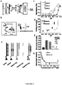
- a targeting strategy enabled by an assembly interfaceable motif (AIM) attachable to any portion of the NAA is described in Figure 1a and 1b .
- AIM is interfaced with locations on NAA that are similar but not restricted to a tag sequence located within the handle domain.
- AIM is interfaced to NAA using two modes (i) covalent attachment and (ii) non covalent recognition.
- NAA and AIM are classified as any functional NAA including but not limited to DNA polyhedra, DNA sensors, RNA nanostructures, DNA plasmids being illustrative examples.
- the Intracellular Targeting Motif attaches covalently and directly to the NAA.
- ITM can bind by itself to the NAA through hydrogen bonding or it can bind through the AIM which comprises of the ITM [for ex.: Furin] and the Artificial receptor [for ex.: antibody such as scFv].
- Figure 1b presents a schematic description of ligand free delivery method, wherein a Nucleic Acid Assembly (NAA) is recognized and delivered inside living cells by Assembly Interfaceable Motif (AIM) containing an Intracellular Targeting Motif (ITM).
- NAA Nucleic Acid Assembly
- AIM Assembly Interfaceable Motif
- ITM Intracellular Targeting Motif
- the ITM or the intracellular targeting motif consists of Cytoplasmic domain of Furin.
- the NAA is similar but not restricted to assemblies made from natural nucleobases, natural modified bases (such as 6-keto purine, xanthine, 5-methylcytosine or 2-aminopurine) or unnatural modified bases (such as thioguanine or 8-oxoguanine, deazapurine or azapurine), or analogs of bases such as universal bases (such as nebularin, nitroindole or nitropyrrole derivatives), synthetic derivatives of nucleobases (such as but not restricted to bromo, fluoro substituted), nucleic acid analogs similar but not restricted to PNA, LNA, morpholino, methyl phosphonate, phosphorothipate, 2'-O-modified oligos etc.
- natural modified bases such as 6-keto purine, xanthine, 5-methylcytosine or 2-aminopurine
- unnatural modified bases such as thioguanine or 8-oxoguanine, deazapurine or aza
- the sensor molecule is selected from group comprising physiological sensor, small molecule sensor, organic molecules, proteins, nucleic acids, metabolites, drugs and their derivatives, amino acids, nucleotides and its derivatives, biological cofactors, antibiotics, vitamins, proteins, small peptides, toxins, lipids, growth factors, hormones and enzymes.
- the handle domain comprises of nucleotide sequences.
- the AIM is selected from group comprising nucleic acid-binding proteins similar to but not restricted to nucleic acid binding antibodies, recombinant antibodies, recombinant antibodies conjugated to a signal sequence, transcription factors, Zn-finger proteins, leucine zippers and peptides.
- AIM is extended to any molecules similar to but not restricted to proteins that posses a natural receptor, a protein that traffics between intracellular locations via the plasma membrane, toxins, viruses and viral coat proteins, cell penetrating peptides, signal sequences, intracellular targeting sequences, small organic molecules, endocytic ligands, enzymes and aptamers against a trafficking protein.
- the AIM comprises Furin conjugated to scFv.
- the ITM is selected from a group comprising a protein similar to but not restricted to any plasma membrane protein that is endocytosable, any proteins that posses a natural receptor, a protein that traffics between intracellular locations via the plasma membrane, toxins, viruses and viral coat proteins, cell penetrating peptides, signal sequences, intracellular targeting sequences, small organic molecules, endocytic ligands and trafficking proteins.
- the vector comprises signal sequence, assembly interfaceable motif (AIM), intracellular targeting motif (ITM) and the vector is optimized for the host cell.
- the cell is selected from group comprising any eukaryotic cells in culture or primary cells, most precisely plant cells, mammalian cells, fly cells and yeast cells.
- NAA Nucleic Acid Assembly
- a molecular design of DNA pH sensor with response times that are nearly 20 fold faster is presented.
- tuning of their pH sensitive regimes and creation of a family of DNA sensors spanning pH ranges from pH about 4.0 to about 7.6 is also conducted.
- This sensor design of the NAA also incorporates a 'handle' domain.
- a recombinant antibody is engineered (example: scFv) that binds sequence specifically to the handle domain and acts as an artificial receptor for the whole family of DNA pH sensors or the NAAs.
- the scFv conjugated Furin or the AIM binds to the 'handle' domain of the NAA and ferries the DNA pH sensor or the NAA along the Furin endocytic pathway.
- the membrane protein or the ITM can bind individually to the handle domain of the NAA as well.
- the DNA nanodevice or the AIM-NAA conjugate retains its functionality in cellulo and provides spatiotemporal pH maps of retrogradely trafficking the Furin or the ITM inside living cells.
- This molecular technology is useful to specifically localize DNA nanoarchitectures within different organelles of a living cell.
- scFv conjugated protein or AIMs are used to site-specifically attach diverse proteins to DNA nanostructures for bioanalytical applications selected from group comprising DNA & RNA aptamer delivery for sensing small molecule dynamics, drug delivery using DNA cages, delivery of antisense oligonucleotides for therapeutics and delivery of plasmid DNA for analysis of gene expressions.
- a molecular design is outlined, with at least 20 fold faster response times when compared with the conventional I-switch.
- the instant disclosure also elaborates on the engineering of an artificial DNA binding protein [example scFv] that binds specifically to the 'handle' domain of the sensor that is fused to the intracellular protein of choice [example Furin]
- scFv artificial DNA binding protein
- Furin intracellular protein of choice
- This enables pH mapping of compartment maturation of the desired protein in a ligand-free manner and thus positions DNA-based sensors to investigate a significantly larger number of currently inaccessible protein environments.
- the present disclosure also demonstrates proof of concept by pH mapping in different cell types by the protein Furin, whose trafficking is well-studied and yet whose pH maps have remained inaccessible thus far.
- the sensor domain of the Nucleic Acid Assembly consists of oligonucleotides described in Table 1 and annealing of the oligonucleotides will provide different sensors.
- the handle domain is inbuilt in sensor domain so annealing will provide a scaffold which consists of sensor domain and handle domain and is called Nucleic Acid Assembly.
- NAA is pulsed or incubated with cells expressing scFv-Furin
- scFv recognizes NAA and the whole complex can be called AIM-NAA complex.
- AIM Assembly Interfaceable Motif
- ITM Intracellular Targeting Motif
- I n when they carry no fluorescent labels.
- I 3 is formed from a 1: 1 mixture of I 3 and I 3'
- I 4 from a 1: 1 mixture of I 4 and I 4'
- I 7 from a 1: 1 mixture of I 7 and I 4 '
- I C3-C4 from a 1: 1 mixture of I C3-C4 and I 4'
- I c-myc from a 1: 1 mixture of I c-myc and I c- myc' .
- Fluorescently labeled Nucleic Acid assemblies indicate the respective fluorophore in the subscript, where e.g., I 3 A488/A647 is formed from I 3 A488 and I 4 A647 , I 4 A488/A647 from I 4 A488 and I 4 A647 , I 4 A488 from I 4 A488 and I 4' .
- the 35 bp dsDNA epitope is formed from ssDNA and ssDNA', R 1 : ssDNA and Region 1, R 2 : ssDNA and Region 2, R M : ssDNA and Region M.
- I comp I comp and I comp' and I non comp : I non comp and I non comp'
- sequences that are used as Nucleic acid assemblies are represented by SEQ ID Nos. 1- 21.
- DNA pH sensor which is a part of Nucleic acid assembly incorporates a 35 bp long duplex leading into a mismatched duplex at one end as shown in Figure 1a .
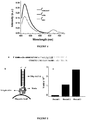
- the CD spectra of I 4 in phosphate buffer at about pH 8.3 showed a positive band centered at about 278 nm and a negative band centered at about 248 nm characteristic of a B-DNA duplex ( Fig. 3a ). However at about pH 4.0, the same assembly showed a CD where the positive and negative bands shifted to about 285 nm and about 251 nm respectively ( Fig. 3a ).
- the difference spectrum of the DNA assembly I 4 at about pH 4 and about pH 8.3 gives a trace that shows a positive band centered at about 288 nm and a negative band centered at about 262 nm which is consistent with the CD signature characteristic of DNA 4 i-motifs.
- Figure 4 relates to steady state fluorescence measurements on Nucleic Acid Assembly. Fluorescence spectra of differently labeled Nucleic Acid Assemblies demonstrate i-motif folding. About 50 nM of labeled switch is diluted in about 20 mM phosphate buffer of about pH 5.0 containing about 100 mM KCl, incubated for about 30 min before acquiring spectra. Samples are excited at about 495 nm and fluorescence spectra is recorded from about 505 nm to about 725 nm.
- Tunable nature of the Nucleic Acid Assemblies is further demonstrated by FRET.
- a solution of the doubly labeled assembly (I n A488/A647 ) in about 100 mM KCl at different pH values is excited at about 495 nm and the ratio of emission at about 520 and about 669 nm is plotted as a function of pH.
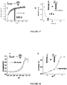
- I 3 A488/A647 yields a sigmoidal curve with a pH sensitive regime of about pH 6 to about 6.5; 1 4 A488/A647 shows a pH sensitivity from pH about 6.2 to about 7 and I 7 A488/A647 shows a pH sensitive regime of about 7-7.5 ( Fig. 1c ).
- oligonucleotides as captured in TABLE I above are dissolved in Milli-Q water to make about 200 ⁇ M stock which is aliquoted and kept at around -20°C. Depending on the purity of fluorescently labeled oligonucleotides, they are subjected to ethanol precipitation prior to use to remove any contaminating fluorophores.
- I n and I n' are mixed in equimolar ratios in about 20 mM potassium phosphate buffer of desired pH containing about 100 mM KCl.
- the resultant solution is heated to 90°C for about 5 minutes, cooled to the room temperature at 5°C/15 min and equilibrated at 4°C overnight. Prior to experiment, the solution is diluted in appropriate buffer containing about 100 mM KCl unless mentioned.
- an artificial DNA binding protein is engineered that is grafted onto any intracellular protein of choice.
- This artificial DNA binding protein possesses the following desirable characteristics: (i) it binds dsDNA specifically (ii) with a high affinity (iii) is relatively small in size and (iv) does not perturb the cellular transcription program upon its expression.
- Single chain variable fragments are the minimal binding domains of immunoglobulin composed of a single polypeptide with the V fragment heavy chain (V H ) and light chain (V L ) attached sequentially by a flexible glycine-serine linker with a C-terminal His-tag and Myc-tag. They possess all the above characteristics and further are used to develop recombinant antibodies against many classes of molecules including DNA using phage display. Therefore, phage display of recombinant antibodies against the 35 base pair dsDNA handle engineered into the pH sensor assemblies to obtain high-affinity, sequence-specific protein binders to the handle domain is conducted.
- the DNA duplex or the NAA is formed by annealing the 35 bp DNA (Table 1) functionalized with biotin at the 5' end of 35bp dsDNA that enables its immobilization on Streptavidin-coated magnetic beads that is then presented as the epitope to the scFv [Artificial Receptor] libraries ( Fig. 5a,b ).
- scFv libraries (Tomlinson I and J, Geneservice, UK) that contain 10 8 different scFv fragments cloned in an ampicillin resistant phagemid vector are used for screening against dsDNA epitope (i.e. 35 base pair long Nucleic Acid Assembly handle, Fig. 5a ). These libraries allow expression of a recombinant antibody fragment with a Myc and His tag as a protein fusion with a truncated integral phage coat protein P3 on the surface of a phage particle.
- Phage particles are presorbed against Streptavidin coated magnetic particles (Dynabeads, Invitrogen) in order to remove anti-Streptavidin scFvs followed by incubation with Streptavidin coated magnetic particles immobilized with dsDNA epitope or NAA ( Fig. 5b ). After each round of incubation, bound phage particles are collected by magnetic separation and then used to infect bacteria for amplification. Selected, amplified phage particles are isolated from TG-1 by superinfection with helper phage and used as a library for subsequent rounds of selection.
- Fig. 5c Randomly picked clones from the selected population are arrayed in a 96 well plate, and corresponding soluble scFvs are expressed and screened for dsDNA binders or NAA binders. Nearly 70% of screened clones display DNA binding properties coupled with negligible affinity for Streptavidin that also forms a part of the epitope during phage display but is also selected against using a pre-clearing step.
- Sequence specific scFvs are identified by dividing the 35bp dsDNA or NAA into three regions.
- One region is composed of the 13 base pairs of the antigen from 3' end (known as Region 1 (R 1 ), Fig. 6a , red), the second region corresponded to base pairs 16 to 28 from 3' end (Region 2 (R 2 ), Fig. 6a , green) and third region is an overlap of regions 1 and 2 (Region M (R M ), Fig. 6a , blue).
- each of the selected clones is similarly assayed by ELISA to a set of DNA bound scaffolds that are surface immobilized. These correspond to a single stranded DNA sequence comprising only the biotinylated strand of the 35 bp dsDNA ( Fig. 1e , ssDNA), the 35 bp dsDNA duplex ( Fig. 1e , dsDNA), and three shorter duplex regions R 1 , R 2 and R M corresponding to various sections on the 35 bp dsDNA duplex ( Fig. 1e ).
- R 1 corresponds to a 13 bp region at the 3' terminus of the biotinylated ssDNA oligonucleotide
- R 2 corresponds to a 13 bp region abutting R 1
- R M corresponds to a 13 bp region overlapping the 5' end of R 1 and the 3' of R 2 (shown in italics in Fig. 5a ).
- the DNA binding scFvs that show binding to the 35 bp dsDNA epitope or NAA but not the ssDNA epitope, are chosen for a further screen against the three epitopes R 1 , R 2 and R M .
- the binding efficiencies of the scFvs against R 1 , R 2 and R M indicate their sequence specificity as well as narrow down the size of their respective epitopes. It is observed that 21% of the clones are specific for R 1 (Fig. If, Fig. 6b(i) ), whereas 3% of clones bound R 2 ( Fig. 6c ).
- clones C1, D1, E1, G1 and H1 are distinct from each other binding to R 1 and no other region.
- the scFvs corresponding to these clones recognize only the first 8 base pairs of the 35bp dsDNA i.e., the sequence d(AT) 4 . 3 clones are specific for R 2 (One representative clone is shown in Fig. 6c ). Few clones that bind all three regions, showing no sequence specificity are also shown, which are not included for further analysis ( Fig. 6d ).
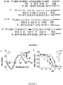
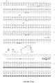
- Phage display libraries Tomlinson I and J incorporate diversity in 18 residues in the complementarity determining regions (CDR), specifically H50, H52, H52a, H53, H55, H56, H58, H95, H96, H97, H98, L50, L53, L91, L92, L93, L94 and L96 (where H corresponds to the heavy chain and L corresponds to the light chain).
- CDR complementarity determining regions
- Sequencing also reveals the presence of lysine, arginine, asparagine which are known for their DNA binding properties. Most of the scFvs have around 5-8 positively charged residues in their CDRs, reminiscent of other DNA binding antibodies. The most notable feature revealed by sequencing is the presence of three nearly conserved residues among 21 region specific clones, lysine at H53 ( ⁇ 81% clones), serine at H95 (76%) and arginine at position L92 ( ⁇ 85%) ( Fig. 7 ).
- the clone C1 is chosen for further studies due to its high representation in the pool that reflects its high affinity and/or production efficiency ( FIG. 8 ).
- This scFv contains the bacterial pelB signal sequence at the N-terminus that secretes scFv into the periplasm where the pelB signal sequence is removed and scFv secreted into the medium.
- protein When protein is expressed in either 2xTY or M9 minimal media and subjected to a purification involving Ni-NTA or HisPur Cobalt Resin, it shows a single band near 27 kDa.
- maximum abundance is seen in fractions 2-4 ( Fig. 8 ).
- These fractions are pooled and the concentration of scFv is measured by Bradford assay. A typical preparation yields about 0.5-2 mg/300 mL protein.
- Figure 8 shows scFv production and purification.
- Clone C1 is expressed in M9 media supplemented with about 0.1% casamino acids and about 0.2% glycerol.
- Affinity of scFv C1 with the 35 base pair DNA duplex or NAA ( Fig. 5a ) is studied by ELISA in three formats that (i) use varying dsDNA concentration, (ii) use varying scFv concentration, (iii) use unlabeled dsDNA epitope as a competitor.
- a competition binding assay is typically performed using a fixed amount of immobilized dsDNA or NAA (25 pmoles) followed by the addition of a fixed concentration of scFv (200 nM) in the presence of an increasing concentrations of unlabeled dsDNA or NAA (I comp , about 10 nM to about 1 ⁇ M) in solution ( Fig. 1g ).
- This assay yields an estimate of the binding affinity of scFv and dsDNA or NAA in solution.
- One well, with no added competitor serves as a normalization factor for the 100% binding event.
- dsDNA epitope or NAA is prepared by annealing about 50 ⁇ M ssDNA and ssDNA' in 1 ⁇ PBS supplemented with about 100 mM KCl.
- Nonspecific binders in particular anti-Streptavidin scFvs
- scFvs displayed on the surface of phage with Streptavidin-coated magnetic beads alone. Rest of the phage display screen is adapted from the Tomlinson protocol provided by the library manufacturer and carried out as described in prior art. This protocol provides a method to screen a dsDNA specific scFv.
- EXAMPLE 5 ELISA FOR SCREENING POSITIVE CLONES
- Approximately 96 colonies from the final round of selection are screened by ELISA against dsDNA or NAA. Individual colonies from last round of selection are grown into a 96 well plate till OD 600 nm reaches approximately 0.9 and induced with about 1 mM IPTG. Cultures are grown at about 30°C overnight ( ⁇ 16 hours) for the expression of soluble scFvs and soup containing scFvs is transferred onto Streptavidin-conjugated 96-well plate containing immobilized dsDNA or NAA to carry out standard ELISA assays.
- scFv soup containing 1 in 1500 dilutions of anti-cMyc antibody (clone 9E10, Millipore)/anti His-tag antibody (clone His-1, Sigma,) is added to each well and incubated for about 1.5 hours with gentle shaking.
- Goat anti-mouse secondary antibody conjugated to HRP (1 in 1000 dilution, Invitrogen) is used to detect anti-cMyc antibody bound to plate through scFv which binds to ds DNA immobilized on the plate.
- scFvs are subjected to another round of ELISA assay against various DNA epitopes.
- the epitopes used are ssDNA (5'-biotinylated oligo only), dsDNA (duplex DNA used as antigen) and various parts of this dsDNA (e.g. Region 1 as R 1 , region 2 as R 2 and region middle as R M ).
- ELISA assay is carried out as mentioned earlier. This assay identifies and differentiates the dsDNA binding scFvs from other non interacting scFvs.
- EXAMPLE 6 SEQUENCING OF dsDNA or NAA SPECIFIC CLONES
- clones showing binding to dsDNA or NAA are chosen for sequencing. Individual clones are grown overnight and subjected to a plasmid DNA isolation using Nucleospin Plasmid (Macharey-Nagel, Germany) miniprep Kit. Sequencing is performed using a standard dideoxy sequencing method using a specific primer pHen Seq (5'- CTATGCGGCCCCATTCA-3'). This protocol provides sequence of the scFvs which is used further to generate AIM.
- the affinity and specificity of scFv is analyzed by ELISA after purifying the scFv using protocol described in prior art.
- Streptavidin-coated 96-well plate is incubated with dsDNA epitope used for screening in 5 ⁇ SSCT (75 mM Sodium citrate, 750 mM NaCl +0.05% Tween-20) for about 1.5 hours for immobilization of dsDNA or NAA onto the Streptavidin-coated 96-well plate. This is followed by incubation with a mixture of serial dilutions of purified scFv and anti myc-tag antibody (1 in 1000 dilution) for 1.5 hours at room temperature.
- 5 ⁇ SSCT 75 mM Sodium citrate, 750 mM NaCl +0.05% Tween-20
- dsDNA immobilized onto Streptavidin-coated 96-well plate is incubated with the mixture of competing nucleic acid, purified scFv (250-300 nM) and anti myc-tag antibody (1 in 1000 dilution) for about 1.5 hours at room temperature.
- the wells are then washed briefly in 1 litre PBST (1 ⁇ PBS+0.1% Tween-20) bath before the addition of the secondary antibody conjugated to HRP.
- Bound scFvs are detected by addition of TMB/H 2 O 2 and OD at 450 nm is recorded and normalized with respect to the well where no competitor is added.
- EXAMPLE 8 CLONING OF scFV WITH FURIN CYTOPLASMIC DOMAIN
- Plasmid expressing chimeric forms of Furin is obtained from Dr Michael Marks. This plasmid contains lumenal and trans-membrane domains of human Interleukin receptor beta chain (TAC) with cytoplasmic tail of Furin.
- TAC human Interleukin receptor beta chain
- EGFP Enhanced Green Fluorescent Protein
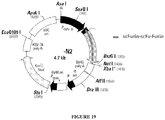
- EGFP-N2 backbone is removed by digestion of NheI and NotI ( Figure 21 ) and cytoplasmic tail of Furin and Lumenal TAC domain is introduced by cutting parent plasmid by the same enzymes and re-ligating it to the N2 backbone.
- Lumenal domain of TAC is cloned between EcoR1 and BglII site and same restriction enzymes are used to remove this domain.
- scFv fused with signal sequence of Furin is PCR amplified using two primers that contain EcoR1 and BamH1 restriction sites to yield ssFurin-scFv chimera.
- This PCR product is gel purified and digested using EcoR1 and BamH1 and ligated to EcoR1 and BglII digested N2 backbone containing cytoplasmic domain of Furin. Colonies are screened for positive clones that contain ssFurin-scFv-Furin. Sequences of the positive clones are finally confirmed by sequencing.
- Figure 19 describes vector based on N2 backbone expressing a Chimeric protein consists of signal sequence of Furin (ssFurin) - scFv and cytoplasmic domain of Furin.
- HeLa cells are cultured in Dulbecco's Modified Eagle's medium/F-12 (1:1) (Invitrogen Corporation, USA) containing about 10% heat inactivated Fetal Bovine Serum (FBS) (Invitrogen Corporation, USA), about 100 ⁇ g/mL streptomycin and about 100 U/mL penicillin (Invitrogen Corporation, USA).
- Dulbecco's Modified Eagle's medium/F-12 (1:1) containing about 10% heat inactivated Fetal Bovine Serum (FBS) (Invitrogen Corporation, USA), about 100 ⁇ g/mL streptomycin and about 100 U/mL penicillin (Invitrogen Corporation, USA).
- TRVb-1 cells are a CHO cell line which lacks endogenous transferrin receptors but stably expresses the human Transferrin receptor are cultured in Ham's-F12 media (HF-12, Himedia, India) containing about 10% heat inactivated FBS, about 100 ⁇ g/mL streptomycin and about 100 U/mL penicillin with about 200 ⁇ g/mL G418 (Sigma) to ensure maintenance of Transferrin receptor.
- Ham's-F12 media HF-12, Himedia, India
- cells are plated at >50% density onto coverslip bottomed 35 mm dish and about 24 hours post plating, cells are washed and incubated for about 30 min in Opti-Mem at about 37°C.
- About 1 ⁇ g of DNA is diluted in about 100 ⁇ L Opti-Mem in a tube and in a separate tube, about 3 ⁇ L of LipofectamineTM 2000 was diluted to 100 ⁇ L in same Opti-Mem. After 5min incubation, both solutions are mixed and incubated for about 30 min at Room Temperature.
- About 200 ng (200 ⁇ L) of transfection mixture is introduced to the cells and incubated for about 3 hours at 37°C. Transfection mixture is removed after about 3 hours, complete media is added and incubated for about 20-24hours. Cells are imaged 24 hours after transfection.
- This protocol expresses the AIM at the plasma membrane of any given cell type.
- EXAMPLE 10 EXPRESSION OF scFV-FURIN CHIMERA IN HeLa CELLS
- scFv acts as an artificial receptor that recognizes the Nucleic acid assembly and traffic the latter specifically inside cells
- a scFv fusion of Furin is created, which is a protein that traffics from the plasma membrane via a retrograde endocytic pathway.
- Furin is an endoprotease that is expressed predominantly in the TGN and shuttles between plasma membrane and TGN via this retrograde endocytic pathway. Its cytoplasmic C-terminus is responsible for its correct retrograde trafficking from the plasma membrane. Therefore an N-terminal fusion of the scFv with Furin (scFv-Furin) is made, so that the scFv is presented towards the extracellular milieu when Furin is present at the plasma membrane.
- This chosen topology is compatible with redox conditions required for proper scFv folding and function; as antibodies are secreted naturally by vertebrates and during phage display by E. coli in oxidizing conditions. a priori any attenuation of scFv binding to the DNA handle is not expected.
- the scFv acts as an artificial receptor for the Nucleic acid assembly that is ferried by retrogradely trafficking scFv-Furin when the fusion is expressed in cellulo.
- cells are transfected with scFv-Furin and about 24 hours post transfection, the cells are lysed, total protein is isolated, resolved on SDS-PAGE and probed with anti-His tag antibody.
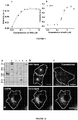
- scFv-Furin shows two closely spaced bands near the expected size ( ⁇ 43-45 kDa) with negligible cross reactivity towards both positive and negative controls ( Fig. 10a ).
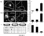
- scFv-Furin In order to study its localization within the cell, immunofluorescence studies are carried out on cells expressing scFv-Furin. Since the scFv incorporates a His tag and endogenous Furin is predominantly localized in the TGN, co-localization studies with an anti-His tag antibody and a TGN marker such as anti-TGN 46 are performed. HeLa cells expressing scFv-Furin show a staining pattern consistent with scFv resident in tubular compartments present in the perinuclear region as well as in proximal small vesicles whereas untransfected controls show only non-specific background staining ( Fig. 10b,c ).
- scFv-Furin expressing cells When scFv-Furin expressing cells are co-stained with anti-TGN46 antibody and anti-His Tag antibody, they show perfect co-localization ( Fig. 10d ), indicating that the expression and localization of scFv-Furin is not disrupted due to presence of the scFv domain at the N-terminus of Furin.
- EXAMPLE 11 scFV ACTS AS AN ARTIFICAL RECEPTOR FOR NUCLEIC ACID ASSEMBLY
- scFv domain is capable of recognizing and endocytosing Nucleic acid assembly from the extracellular milieu
- scFv-Furin expressing HeLa cells are incubated with about 500 nM I 4 A488/A647 in the external medium for about 1 hour, washed, chased for about 10 min to about 3 hours and imaged ( Fig. 11a ).
- EXAMPLE 12 NUCLEIC ACID ASSEMBLY MARKS RETROGRADELY TRAFFICKING scFV-FURIN
- Furin is predominantly present in the Trans Golgi Network (TGN), of which a small population shuttles between the plasma membrane and TGN via the sorting endosome and late endosome.
- TGN Trans Golgi Network
- a small population shuttles between the plasma membrane and TGN via the sorting endosome and late endosome.
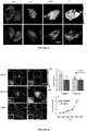
- an antibody uptake assay is performed. HeLa cells are co-transfected with scFv-Furin and EGFP-Furin ( Fig. 12b ). 24 hours post transfection, these cells are pulsed with fluorescently labeled anti-GFP antibody and I A488/A647 , chased for 3hours and imaged. It is seen that anti-GFP antibody and I 4 A488/A647 are co-localized in live as well as fixed cells confirming that I 4 A488/A647 uptake is Furin mediated ( Fig. 12b,c ).
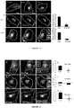
- the sorting endosomes in the scFv-Furin expressing HeLa cells are labeled with a 10 min pulse containing a cocktail of Alexa 568 labeled Transferrin (Tfn 568 ) and I 4 A488/A647 and imaged. It is observed that Tfn 568 and I 4 A488/A647 show significant co-localization indicating that after endocytosis, most of the Nucleic acid assembly is resident in sorting endosome ( Fig. 13 a,d). However when chased for about 2 hours, this co-localization is markedly reduced indicating that the Nucleic acid assembly has trafficked forward from the sorting endosome ( Fig. 13b,e ).
- scFv expressing HeLa cells are labeled with a mixture of I 4 A488/A647 and TMR-Dextran. It is known that the TMR-Dextran marks the late endosome at the 2 hour time point and that it co-localizes with an antibody against the N-terminal domain of a Furin chimera that is resident at the late endosome at 2 hours.
- scFv-Furin expressing TRVb-1 cells and HeLa cells are labeled with I 4 A488/A647 and TMR dextran for about 1.5 hours, washed, chased for about 3hours and imaged.
- I 4 A488/A647 and TMR dextran show significant co-localization ( Fig. 12d, e and Fig. 13c,f respectively) indicating that I 4 A488/A647 has trafficked from sorting endosome into the late endosome, characteristic of Furin mediated retrograde trafficking.
- scFv expressing HeLa cells are labeled with about 500 nM I 4 A488/A647 for about 15-30 minutes at 37°C in complete media. Cells are washed and briefly fixed for about 2 min in ice using about 2% paraformaldehyde. Cells are pH clamped using clamping buffer containing about 25-40 ⁇ M nigericin. Cells are imaged in a widefield microscope after exciting at 488 nm and imaging at 520 nm (D) and 669 nm (A). D and A images are aligned and a binary image is created by dividing D images by A images. This binary D/A image is further pseudocolored in ImageJ to provide a spatial pH map of cells clamped at indicated pH values. Due to high FRET, at low pH, cells show uniformly lower D/A which concomitantly increases to higher D/A value as value of clamping buffer pH increases ( Fig. 14 ).
- the corresponding image in live cells shows the characteristic heterogeneity in pH at chase times ( Fig. 15e, f ).
- the fold change in D/A values for two different pH sensors I 3 A488/A647 and I 4 A488/A647 in cellulo indicates that sensor performance and integrity are uncompromised ( Fig. 15g ).
- I 4 A488/A647 labeled endosomes are clamped at the indicated pH values and D/A as a function of pH is plotted to yield the pH calibration curve.
- the in vitro and the in cellulo pH calibration curves show good correspondence ( Fig. 15h ) indicating that the performance of the DNA pH sensor is not affected post-recognition by its artificial receptor.
- EXAMPLE 14 NUCLEIC ACID ASSEMBLY REVEALS pH GRADIENT INSIDE CELLS
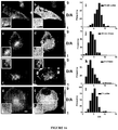
- scFv-Furin expressing HeLa cells are pulsed with I 4 A488/A647 for about 10 min which is shown to label sorting endosomes, washed and imaged.
- the pH map at this time point shows a uniform distribution pH across all endosomes which leads to a mean pH spread of 6.4 ⁇ 0.27 characteristic of sorting endosomes ( Fig. 16a,b,i ).
- a complementary method to label cells that provides a spatial pH map of the set of all retrogradely trafficking scFv-Furin compartments across cell is used.
- scFv-Furin expressing HeLa cells are labeled for about 2 hours to achieve steady state labeling where all scFv-Furin containing endosomes are expected to be labeled.
- Figure 16f represents the pH map of labeled endosomes which shows a clear spatial correlate of endosomes with similar acidities, indicating the ability of the Nucleic Acid Assembly to differentiate between sorting and late endosomes on the Furin pathway and also their relative spatial intracellular locations.
- CD signal at 292 nm at different pH is recorded and normalised to 0-1 by taking CD signal at closed state as Y max and CD at open state as Y min and using the formula given below in Origin 8.
- Y Y ⁇ Y min Y max ⁇ Y min
- About 5 ⁇ M stock of fluorescently labelled Nucleic Acid Assembly sample is prepared in about 20 mM phosphate buffer pH 5.5, supplemented with about 100 mM KCl (I n A488 , I 4 A647 are used to prepare samples). Solutions of Nucleic Acid Assembly at different pH are made by diluting about 1 ⁇ L of about 5 ⁇ M stock samples into 99 ⁇ L of 1 ⁇ clamping buffer of desired pH. All samples are vortexed and equilibrated for about 30 min at room temperature.
- the experiments are performed in a widefield microscope.
- the cover-slips containing about 50 ⁇ L samples of different pH are excited at 488 nm in a widefield microscope and emission images are acquired using 520 nm (Donor, D channel) and 669 nm (Acceptor, A channel).
- An in vitro pH calibration curve is obtained by plotting the ratio of donor intensity (D) at 520 nm by acceptor intensity (A) at 669 nm as a function of pH. Mean of D/A from two independent experiments and their SD are plotted for each pH value. This protocol provides the platform to generate a range of pH sensor with varying transition pH.
- EXAMPLE 17 KINETICS OF FOLDING OF NUCLEIC ACID ASSEMBLY
- HeLa cells (10 6 ) are transfected using ssFurin-scFv-Furin and 24 hours post transfection, cells are collected and washed twice with PBS. Untransfected cells and EGFP-Furin transfected cells are used as negative control and positive control for transfection respectively.
- Whole cell lysates are prepared by addition of about 100 ⁇ L RIPA buffer followed by incubation for about 1.5 hours at 4°C. An aliquot of each lysate is subjected to SDS-PAGE and transferred to nitrocellulose using standard protocol. The nitrocellulose membrane is incubated with a 1:1000 dilution of anti-His-tag antibody (clone His-1, Sigma). Binding of primary antibody is detected by the ECL method (Pierce).
- scFv-Furin expressing HeLa cells are fixed with about 4% paraformaldehyde for about 10 minutes on ice. To detect intracellular antigens they are permeabilized with about 0.1% Saponin (Sigma) in M1 buffer and stained with rabbit anti-TGN46 antibody (Abcam) and mouse anti His-tag antibodies (Sigma) followed by goat anti rabbit-Cy3 conjugated (Abcam) and goat anti mouse-FITC conjugated secondary antibodies (EMD bioscience) for 1 hour respectively. This experiment confirms full length expression of AIM at the TGN.
- scFv-Furin expressing mammalian cells like HeLa/TRVb-1 cells are washed with M1 buffer three times prior to labeling. Cells are incubated with endocytic tracers for indicated times in labeling medium (HF-12 or DMEM, 10% serum, antibiotics).
- labeling medium HF-12 or DMEM, 10% serum, antibiotics.
- I 3/4 A488/A647 is diluted in labeling media to final concentration of about 500 nM and incubated for different times at 37°C.
- cells are incubated with I 4 A488/A647 and about 4 mg/mL TMR dextran in labeling media at 37°C for 1.5 h.
- Sorting endosomes are labeled by 100 ⁇ g/mL Alexa-568 labeled, human-holo Transferrin after incubating HeLa cells at 37°C for about 10 mins. After incubation, excess endocytic tracers are washed off using M1 and chased for indicated times at 37°C in complete media.
- cells are co-transfected with scFv-Furin and EGFP-Furin and 24 hours after transfection, cells are labeled with Alexa 594 conjugated rabbit anti-GFP antibody (Invitrogen, 1 in 50 dilution) and I 3/4 A488/A647 in labeling media for 1 hour, washed with M1 followed by a long chase for about 3 hours at 37°C. Cells are washed three times with M1 and fixed with about 4% PFA in M1 for about 10 min at room temperature.
- scFv-Furin expressing HeLa cells are labeled with about 500 nM I 4 A488/A647 in HBSS buffer for 1hour at room temperature in presence of (1) No exogenously added DNA, (2) in presence of 25 ⁇ M random sequence DNA (I non comp ), (3) in presence of 25 ⁇ M I comp and chased for 3 hours in complete media.
- Cells are washed and imaged in a widefield microscope and perinuclear fluorescence is quantified and presented as percentage intensity internalized after normalizing the data with (1).
- cells are labeled with (1), (2) and (3) in complete media at 37°C for about 30 min which provides similar results.
- EXAMPLE 20 MEASUREMENT OF pH IN SORTING ENDOSOMES AND LATE ENDOSOMES
- scFv-Furin expressing HeLa cells are labelled with about 500 nM I 3/4 A488/A647 in complete media for indicated times at 37°C. Cells are incubated on ice and briefly fixed by addition of about 2% PFA for 1 min in M1. Intracellular pH gradient is abolished by addition of about 25-40 ⁇ M nigericin in different pH clamping buffer ranging from 5.0 to 7.5 for about 40 min. The cells are kept in this medium until imaging, and the fluorescence ratio of donor (D, 520 nm) image to acceptor (A, 669 nm) image at different equilibrated pHs are calculated in individual endosomes after exciting at 488 nm.
- the mean from the distribution of D to A ratio of individual endosome are obtained at different pH values and plotted to obtain a calibration curve.
- the pH of sorting endosomes and late endosomes are estimated after labeling respective compartments with I 4 A488/A647 .
- D to A ratios is used to estimate the pH value from to the calibration curve.
- the in cellulo pH calibration curve of the Nucleic Acid Assembly post complexation with and post trafficking by scFv-Furin shows remarkable correspondence with the in vitro protein-free Nucleic Acid Assembly.
- the fold change in D/A ratio which is a measure of sensor performance, shows an in cellulo fold change of 4.1 ⁇ 0.7 and 4.0 ⁇ 1.7 for I 4 A488/647 and I 3 A488/647 similar to their in vitro values (4.8 ⁇ 0.8 and 6.5 ⁇ 1.02 respectively). This suggests that recognition of the duplex 'handle' region of the sensor by the scFv domain in cellulo does not alter the pH sensing characteristics of the DNA sensor. This is consistent with the modular nature of the nucleic acid scaffold of the DNA pH sensor where protein binding at a remote site is independent of the functionality due to a conformational change at a distal site.
- Cross talk and bleed-through are measured with donor only and acceptor only samples and found to be negligible for Alexa 488-647 pair. Auto-fluorescence is measured on unlabeled cells. All the images are then background subtracted taking mean intensity of the cytoplasm and Donor and acceptor images are co-localized and endosomes showing co localization are analysed using Image J. Total intensity as well as mean intensity in each endosome is measured in donor and acceptor channels and a ratio of donor to acceptor intensities (D/A) of each endosomes is obtained.
- D/A donor to acceptor intensities
- the present design of the Nucleic Acid Assembly uses an i-motif forming C-rich sequence that exists as a mismatched duplex at basic pH, folds into an intramolecular i-motif at acidic pH and possesses distinct advantages over the prototype device. Notably, the kinetics of folding, which proves to be rate determining for the response times of the prototype sensor, is dramatically improved. Thus the response times of the new DNA sensors reach as fast as ⁇ 15 sec, predisposing them to investigating a greater breadth of cellular applications related to pH maps that correlate with the functional status of the cell.
- An additional feature of this design is the presence of a 35 base pair duplex that plays a dual role.
- One of these is to keep the functional pH responsive C-rich region and its partially complementary strand together even in acidic pH environments.
- the other is to act as a recognizable epitope for an artificial receptor that is generalizable to the complete range of pH sensors described in this study.
- This recognizable epitope or 'handle' domain of the DNA pH sensor gains importance in the context of a general strategy that enables small molecule sensing to yield intra-compartmental spatiotemporal chemical maps within living cells.
- the Nucleic Acid Assembly alone binds to scavenger receptors and maps spatiotemporal pH changes in endosomes along this pathway. Its use is expanded to more endocytic pathways by functionalizing the Nucleic Acid Assembly with endocytic ligands and mapping pH changes along pathways of the corresponding receptors to those ligands.
- a ligand free method to specifically traffic a DNA pH sensor is generalizable to the largest number of trafficking pathways. This is achieved by fusing a sequence specific DNA binding protein or adapter such as the scFv onto the cellular membrane protein of interest. Using phage display, a collection of recombinant antibodies including one that binds this handle domain at d(AT) 4 with a K D ⁇ 50-80 nM is produced.
- sequence of hypervariable regions of the recombinant antibodies of the present disclosure yields key indicators of molecular interactions between the DNA epitope and its sequence-specific antibody. Due to its monomeric nature and small size, this scFv acts as an artificial receptor for all DNA pH sensors that incorporate the handle domain. By expressing this scFv as a fusion protein with the membrane protein of interest, it is used as an artificial receptor to recruit a DNA pH sensor enabling one to understand pH correlates of any receptor/protein whose trafficking in endosomal maturation is unknown.
- this method is also extendable to the delivery and trafficking of any DNA nanostructure inside cells that incorporate the minimal dsDNA tag i.e. d(AT) 4 . It is noteworthy that a single phage display screen yields a collection of binders of various binding specificities and affinities from which optimal solutions for various applications are picked.
- the receptor design strategy is thus not only applicable to any DNA tag - and other chemical entities - but also highly efficient in simultaneously generating a versatile array of DNA device binders/receptors.
- the scFv is fused to the cytoplasmic tail of Furin whose retrograde endocytosis is characterized in various cell lines.
- the scFv-Furin chimera is expressed well in HeLa cells and recapitulates the maturation of endogenous Furin.
- Furin is a well known protease
- pH correlates of retrogradely transported Furin in endocytic compartments has not been mapped. These are followed by live cell pH measurements on scFv-Furin expressing HeLa cells and labeling the resulting compartments with 1 4 A488/647 . Retrogradely trafficking Furin enters the sorting endosome at 5 min which later matures into late endosome over 1 hour, where there is a concomitant acidification of luminal pH.
- the Nucleic Acid Assembly efficiently captures this maturation from sorting endosomes to late endosomes when following multiple compartments labeled at a given time point, and imaged as a function of time.
- a spatial correlate of compartmental pH for this pathway is obtained.
- Such a pH map reveals the existence of spatial gradient of compartments with different acidities with a cluster of high pH compartments surrounded by compartments of progressively decreasing pH in retrogradely transported scFvFurin.
- NAA Nucleic Acid Assembly
- figures 17(a) and (b); and figures 18(a) and (b) give a graphical representation of the comparative analysis between the Nucleic acid assembly of the present disclosure and the conventionally known I-Switch.
Claims (11)
- Complexe motif interfaçable d'assemblage (AIM)-assemblage d'acides nucléiques (NAA), l'assemblage d'acides nucléiques (NAA) comprenant un domaine capteur de pH et un domaine en poignée de main, éventuellement accompagnés d'une molécule de capteur ;
dans lequel l'assemblage d'acides nucléiques est un duplex choisi parmi SEQ ID N° 1 et 4, SEQ ID N° 2 et 4, SEQ ID N° 3 et 4, SEQ ID N° 5 et 6, SEQ ID N° 7 et 8, SEQ ID N° 9 et 12, SEQ ID N° 10 et12, et SEQ ID N° 11 et12 ; dans lequel lesdites séquences de l'assemblage d'acides nucléiques sont marquées avec un fluorophore, et dans lequel le domaine en poignée de main est indiqué SEQ ID N° 14 et sa séquence complémentaire ;
dans lequel le motif interfaçable d'assemblage (AIM) comprend un motif de ciblage intracellulaire (ITM) conjugué à un récepteur artificiel choisi dans le groupe comprenant un fragment variable de chaîne simple (scFv), un facteur de transcription, une protéine à doigt de zinc, une glissière à leucine, une immunoglobuline de liaison à l'ADN, une protéine de liaison à l'ADN ou toute combinaison de ceux-ci ; et dans lequel le récepteur artificiel se lie spécifiquement au domaine en poignée de main de l'assemblage d'acides nucléiques (NAA) ; et
dans l'assemblage d'acides nucléiques (NAA) est conjugué au motif interfaçable d'assemblage (AIM) par le biais du récepteur artificiel. - Procédé d'obtention d'un complexe assemblage d'acides nucléiques (NAA)-motif interfaçable d'assemblage (AIM) de la revendication 1, ledit procédé comprenant les étapes consistant à :a) obtenir un assemblage d'acides nucléiques tel que défini dans la revendication 1 ;b) obtenir un vecteur codant pour un motif interfaçable d'assemblage (AIM) tel que défini dans la revendication 1 ;c) transfecter une cellule avec le vecteur pour exprimer le motif interfaçable d'assemblage (AIM) et obtenir l'AIM dans la cellule transfectée ; etd) incuber l'assemblage d'acides nucléiques avec la cellule comprenant l'AIM pour obtenir le complexe assemblage d'acides nucléiques (NAA)-motif interfaçable d'assemblage (AIM).
- Procédé selon la revendication 2, comprenant en outre la ré-incubation de la cellule ayant le complexe, pour ainsi permettre une absorption cellulaire et un ciblage intracellulaire du complexe assemblage d'acides nucléiques-motif interfaçable d'assemblage (AIM-NAA).
- Complexe AIM-NAA selon la revendication 1 ou procédé selon la revendication 2 ou la revendication 3, dans lequel la molécule capteur du NAA est choisie dans le groupe comprenant un capteur physiologique, un capteur de petites molécules, des molécules organiques, des protéines, des acides nucléiques, des métabolites, des médicaments et leurs dérivés, des acides aminés, des nucléotides et leurs dérivés, des cofacteurs biologiques, des antibiotiques, des vitamines, des protéines, des petits peptides, des toxines, des lipides, des facteurs de croissance, des hormones et des enzymes ou toute combinaison de ceux-ci.
- Complexe AIM-NAA selon la revendication 1, dans lequel l'ITM est choisi dans le groupe comprenant une protéine de membrane plasmatique endocytosable, une protéine qui possède un récepteur naturel, une protéine de trafic, des toxines, une protéine du manteau viral, un peptide de pénétration cellulaire, une séquence signal, une séquence de ciblage intracellulaire, une petite molécule organique, un ligand endocytique, une enzyme, un aptamère dirigé contre une protéine de trafic ou toute combinaison de ceux-ci.
- Complexe AIM-NAA selon la revendication 1, dans lequel l'ITM est la furine conjuguée à un fragment variable de chaîne simple (scFv), qui se lie au domaine en poignée de main de l'assemblage d'acides nucléiques (NAA).
- Procédé selon la revendication 2 ou la revendication 3, dans lequel le vecteur est choisi dans le groupe comprenant des plasmides, des virus, des vecteurs viraux, des cosmides, des phagémides et des chromosomes artificiels.
- Procédé selon la revendication 2 ou la revendication 3, dans lequel la cellule est une cellule eucaryote et est choisie dans le groupe comprenant une cellule HeLa, TRVb-1 et une cellule IA2.2.
- Procédé selon la revendication 2 ou la revendication 3, dans lequel l'incubation est réalisée à une température dans la plage d'environ 4 °C à environ 37 °C.
- Procédé selon la revendication 3, dans lequel la réincubation est réalisée à une température dans la plage d'environ 20 °C à environ 37 °C ; et dans lequel le ciblage intracellulaire vise un endosome.
- Kit d'obtention d'un complexe assemblage d'acides nucléiques (NAA)-motif interfaçable d'assemblage (AIM) ou de ciblage intracellulaire du complexe assemblage d'acides nucléiques (NAA)-motif interfaçable d'assemblage (AIM), ledit kit comprenant un assemblage d'acides nucléiques tel que défini dans la revendication 1, et un AIM tel que défini dans la revendication 1 ou un complexe assemblage d'acides nucléiques (NAA)-motif interfaçable d'assemblage (AIM) tel que revendiqué dans la revendication 1, éventuellement accompagné d'un manuel d'utilisation.
Applications Claiming Priority (2)
| Application Number | Priority Date | Filing Date | Title |
|---|---|---|---|
| IN3252CH2011 | 2011-10-12 | ||
| PCT/IB2012/055515 WO2013054286A1 (fr) | 2011-10-12 | 2012-10-11 | Assemblage d'acides nucléiques, vecteur, procédés et trousse de ceux-ci |
Publications (3)
| Publication Number | Publication Date |
|---|---|
| EP2766485A1 EP2766485A1 (fr) | 2014-08-20 |
| EP2766485A4 EP2766485A4 (fr) | 2015-04-15 |
| EP2766485B1 true EP2766485B1 (fr) | 2020-07-08 |
Family
ID=48081450
Family Applications (1)
| Application Number | Title | Priority Date | Filing Date |
|---|---|---|---|
| EP12840407.6A Active EP2766485B1 (fr) | 2011-10-12 | 2012-10-11 | Assemblage d'acides nucléiques, vecteur, procédés et trousse de ceux-ci |
Country Status (3)
| Country | Link |
|---|---|
| US (1) | US9404123B2 (fr) |
| EP (1) | EP2766485B1 (fr) |
| WO (1) | WO2013054286A1 (fr) |
Families Citing this family (4)
| Publication number | Priority date | Publication date | Assignee | Title |
|---|---|---|---|---|
| EP2830991A1 (fr) | 2012-03-26 | 2015-02-04 | President and Fellows of Harvard College | Nanostructures d'acide nucléique enrobées de lipides de forme définie |
| CN105705143A (zh) * | 2013-11-08 | 2016-06-22 | 达娜-法勃肿瘤研究所公司 | 用于体内试剂递送的核酸纳米结构 |
| WO2015159122A1 (fr) * | 2014-04-15 | 2015-10-22 | National Centre For Biological Sciences | Capteur à base d'acide nucléique et procédés associés |
| WO2016187284A1 (fr) * | 2015-05-19 | 2016-11-24 | The University Of Chicago | Procédés et compositions pour déterminer le ph |
Family Cites Families (3)
| Publication number | Priority date | Publication date | Assignee | Title |
|---|---|---|---|---|
| CN100584945C (zh) | 2002-01-18 | 2010-01-27 | 先正达合作有限公司 | 调节基因表达的核被膜和核纤层结合嵌合体 |
| US9250252B2 (en) * | 2009-05-29 | 2016-02-02 | National Centre For Biological Sciences | Intracellular pH sensor using nucleic acid assemblies |
| US8153437B2 (en) | 2010-03-10 | 2012-04-10 | National Center For Biological Sciences | DNA-based molecular switches and uses thereof |
-
2012
- 2012-10-11 WO PCT/IB2012/055515 patent/WO2013054286A1/fr active Application Filing
- 2012-10-11 US US14/351,400 patent/US9404123B2/en active Active
- 2012-10-11 EP EP12840407.6A patent/EP2766485B1/fr active Active
Non-Patent Citations (1)
| Title |
|---|
| None * |
Also Published As
| Publication number | Publication date |
|---|---|
| EP2766485A1 (fr) | 2014-08-20 |
| US9404123B2 (en) | 2016-08-02 |
| EP2766485A4 (fr) | 2015-04-15 |
| WO2013054286A1 (fr) | 2013-04-18 |
| US20140335568A1 (en) | 2014-11-13 |
Similar Documents
| Publication | Publication Date | Title |
|---|---|---|
| US9629801B2 (en) | Blood-brain barrier targeting antibodies | |
| Stefan et al. | DARPins recognizing the tumor-associated antigen EpCAM selected by phage and ribosome display and engineered for multivalency | |
| Modi et al. | Recombinant antibody mediated delivery of organelle-specific DNA pH sensors along endocytic pathways | |
| Li et al. | A membrane microdomain-associated protein, Arabidopsis Flot1, is involved in a clathrin-independent endocytic pathway and is required for seedling development | |
| EP2766485B1 (fr) | Assemblage d'acides nucléiques, vecteur, procédés et trousse de ceux-ci | |
| Tomlinson et al. | [28] Methods for generating multivalent and bispecific antibody fragments | |
| EP3173099B1 (fr) | Procédé d'inhibition de la protéine ras activée dans une cellule utilisant un anticorps ayant la capacité de pénétrer dans le cytoplasme et se présentant sous la forme d'une immunoglobuline complète, et son utilisation | |
| US11155641B2 (en) | Cytosol-penetrating antibody and use thereof | |
| AU2014385799A1 (en) | Insulin-like growth factor 1 receptor -specific antibodies and uses thereof | |
| EP3481870A1 (fr) | Anticorps humanisés franchissant la barrière hématoencéphalique et leurs utilisations | |
| Kesarwani et al. | Genetically encoded live-cell sensor for tyrosinated microtubules | |
| US20130108690A1 (en) | Anti-apoptotic protein antibodies | |
| CA2681170C (fr) | Procedes de production d'anticorps scfv actifs et bibliotheques de ceux-ci | |
| Dingus et al. | A general approach for stabilizing nanobodies for intracellular expression | |
| US20170101669A1 (en) | Nucleic Acid Based Sensor and Methods Thereof | |
| KR102091195B1 (ko) | 세포질 침투 항체 및 이의 용도 | |
| KR20180129514A (ko) | 세포질 침투 항체 및 이의 용도 | |
| Baker et al. | In vivo proximity biotin ligation identifies the interactome of Egalitarian, a Dynein cargo adaptor | |
| EP1482309A1 (fr) | Moyens pour la détection de la conformation de protéines et leurs applications | |
| CN111655865B (zh) | 使用基于杂交链式反应的方法的多重免疫信号放大 | |
| Tao | Investigating Protein-lipid-membrane Interactions in Plant Cells using Bimolecular Fluorescence Complementation | |
| KR20180116204A (ko) | 항체에 엔도좀 탈출능을 부여하는 엔도좀 탈출 구조 모티프 및 이의 활용 | |
| Tillotson | Lead optimization of antibodies against membrane proteins: Targeting the transferrin receptor | |
| NZ749020B2 (en) | Cytosol-penetrating antibody and use thereof | |
| Tos | Team: LMU-TUM Munich/Localization |
Legal Events
| Date | Code | Title | Description |
|---|---|---|---|
| PUAI | Public reference made under article 153(3) epc to a published international application that has entered the european phase |
Free format text: ORIGINAL CODE: 0009012 |
|
| 17P | Request for examination filed |
Effective date: 20140424 |
|
| AK | Designated contracting states |
Kind code of ref document: A1 Designated state(s): AL AT BE BG CH CY CZ DE DK EE ES FI FR GB GR HR HU IE IS IT LI LT LU LV MC MK MT NL NO PL PT RO RS SE SI SK SM TR |
|
| DAX | Request for extension of the european patent (deleted) | ||
| RA4 | Supplementary search report drawn up and despatched (corrected) |
Effective date: 20150318 |
|
| RIC1 | Information provided on ipc code assigned before grant |
Ipc: C12N 15/62 20060101AFI20150312BHEP Ipc: C12Q 1/68 20060101ALI20150312BHEP |
|
| STAA | Information on the status of an ep patent application or granted ep patent |
Free format text: STATUS: EXAMINATION IS IN PROGRESS |
|
| 17Q | First examination report despatched |
Effective date: 20161110 |
|
| GRAP | Despatch of communication of intention to grant a patent |
Free format text: ORIGINAL CODE: EPIDOSNIGR1 |
|
| STAA | Information on the status of an ep patent application or granted ep patent |
Free format text: STATUS: GRANT OF PATENT IS INTENDED |
|
| INTG | Intention to grant announced |
Effective date: 20200109 |
|
| GRAS | Grant fee paid |
Free format text: ORIGINAL CODE: EPIDOSNIGR3 |
|
| GRAA | (expected) grant |
Free format text: ORIGINAL CODE: 0009210 |
|
| STAA | Information on the status of an ep patent application or granted ep patent |
Free format text: STATUS: THE PATENT HAS BEEN GRANTED |
|
| AK | Designated contracting states |
Kind code of ref document: B1 Designated state(s): AL AT BE BG CH CY CZ DE DK EE ES FI FR GB GR HR HU IE IS IT LI LT LU LV MC MK MT NL NO PL PT RO RS SE SI SK SM TR |
|
| REG | Reference to a national code |
Ref country code: GB Ref legal event code: FG4D |
|
| REG | Reference to a national code |
Ref country code: CH Ref legal event code: EP Ref country code: AT Ref legal event code: REF Ref document number: 1288480 Country of ref document: AT Kind code of ref document: T Effective date: 20200715 |
|
| REG | Reference to a national code |
Ref country code: DE Ref legal event code: R096 Ref document number: 602012071180 Country of ref document: DE |
|
| REG | Reference to a national code |
Ref country code: IE Ref legal event code: FG4D |
|
| REG | Reference to a national code |
Ref country code: CH Ref legal event code: NV Representative=s name: COSMOVICI INTELLECTUAL PROPERTY SARL, CH |
|
| REG | Reference to a national code |
Ref country code: LT Ref legal event code: MG4D |
|
| REG | Reference to a national code |
Ref country code: AT Ref legal event code: MK05 Ref document number: 1288480 Country of ref document: AT Kind code of ref document: T Effective date: 20200708 |
|
| REG | Reference to a national code |
Ref country code: NL Ref legal event code: MP Effective date: 20200708 |
|
| PG25 | Lapsed in a contracting state [announced via postgrant information from national office to epo] |
Ref country code: FI Free format text: LAPSE BECAUSE OF FAILURE TO SUBMIT A TRANSLATION OF THE DESCRIPTION OR TO PAY THE FEE WITHIN THE PRESCRIBED TIME-LIMIT Effective date: 20200708 Ref country code: HR Free format text: LAPSE BECAUSE OF FAILURE TO SUBMIT A TRANSLATION OF THE DESCRIPTION OR TO PAY THE FEE WITHIN THE PRESCRIBED TIME-LIMIT Effective date: 20200708 Ref country code: LT Free format text: LAPSE BECAUSE OF FAILURE TO SUBMIT A TRANSLATION OF THE DESCRIPTION OR TO PAY THE FEE WITHIN THE PRESCRIBED TIME-LIMIT Effective date: 20200708 Ref country code: ES Free format text: LAPSE BECAUSE OF FAILURE TO SUBMIT A TRANSLATION OF THE DESCRIPTION OR TO PAY THE FEE WITHIN THE PRESCRIBED TIME-LIMIT Effective date: 20200708 Ref country code: NO Free format text: LAPSE BECAUSE OF FAILURE TO SUBMIT A TRANSLATION OF THE DESCRIPTION OR TO PAY THE FEE WITHIN THE PRESCRIBED TIME-LIMIT Effective date: 20201008 Ref country code: GR Free format text: LAPSE BECAUSE OF FAILURE TO SUBMIT A TRANSLATION OF THE DESCRIPTION OR TO PAY THE FEE WITHIN THE PRESCRIBED TIME-LIMIT Effective date: 20201009 Ref country code: SE Free format text: LAPSE BECAUSE OF FAILURE TO SUBMIT A TRANSLATION OF THE DESCRIPTION OR TO PAY THE FEE WITHIN THE PRESCRIBED TIME-LIMIT Effective date: 20200708 Ref country code: BG Free format text: LAPSE BECAUSE OF FAILURE TO SUBMIT A TRANSLATION OF THE DESCRIPTION OR TO PAY THE FEE WITHIN THE PRESCRIBED TIME-LIMIT Effective date: 20201008 Ref country code: PT Free format text: LAPSE BECAUSE OF FAILURE TO SUBMIT A TRANSLATION OF THE DESCRIPTION OR TO PAY THE FEE WITHIN THE PRESCRIBED TIME-LIMIT Effective date: 20201109 Ref country code: AT Free format text: LAPSE BECAUSE OF FAILURE TO SUBMIT A TRANSLATION OF THE DESCRIPTION OR TO PAY THE FEE WITHIN THE PRESCRIBED TIME-LIMIT Effective date: 20200708 |
|
| PG25 | Lapsed in a contracting state [announced via postgrant information from national office to epo] |
Ref country code: PL Free format text: LAPSE BECAUSE OF FAILURE TO SUBMIT A TRANSLATION OF THE DESCRIPTION OR TO PAY THE FEE WITHIN THE PRESCRIBED TIME-LIMIT Effective date: 20200708 Ref country code: RS Free format text: LAPSE BECAUSE OF FAILURE TO SUBMIT A TRANSLATION OF THE DESCRIPTION OR TO PAY THE FEE WITHIN THE PRESCRIBED TIME-LIMIT Effective date: 20200708 Ref country code: LV Free format text: LAPSE BECAUSE OF FAILURE TO SUBMIT A TRANSLATION OF THE DESCRIPTION OR TO PAY THE FEE WITHIN THE PRESCRIBED TIME-LIMIT Effective date: 20200708 Ref country code: IS Free format text: LAPSE BECAUSE OF FAILURE TO SUBMIT A TRANSLATION OF THE DESCRIPTION OR TO PAY THE FEE WITHIN THE PRESCRIBED TIME-LIMIT Effective date: 20201108 |
|
| PG25 | Lapsed in a contracting state [announced via postgrant information from national office to epo] |
Ref country code: NL Free format text: LAPSE BECAUSE OF FAILURE TO SUBMIT A TRANSLATION OF THE DESCRIPTION OR TO PAY THE FEE WITHIN THE PRESCRIBED TIME-LIMIT Effective date: 20200708 |
|
| REG | Reference to a national code |
Ref country code: DE Ref legal event code: R097 Ref document number: 602012071180 Country of ref document: DE |
|
| PG25 | Lapsed in a contracting state [announced via postgrant information from national office to epo] |
Ref country code: IT Free format text: LAPSE BECAUSE OF FAILURE TO SUBMIT A TRANSLATION OF THE DESCRIPTION OR TO PAY THE FEE WITHIN THE PRESCRIBED TIME-LIMIT Effective date: 20200708 Ref country code: EE Free format text: LAPSE BECAUSE OF FAILURE TO SUBMIT A TRANSLATION OF THE DESCRIPTION OR TO PAY THE FEE WITHIN THE PRESCRIBED TIME-LIMIT Effective date: 20200708 Ref country code: DK Free format text: LAPSE BECAUSE OF FAILURE TO SUBMIT A TRANSLATION OF THE DESCRIPTION OR TO PAY THE FEE WITHIN THE PRESCRIBED TIME-LIMIT Effective date: 20200708 Ref country code: CZ Free format text: LAPSE BECAUSE OF FAILURE TO SUBMIT A TRANSLATION OF THE DESCRIPTION OR TO PAY THE FEE WITHIN THE PRESCRIBED TIME-LIMIT Effective date: 20200708 Ref country code: SM Free format text: LAPSE BECAUSE OF FAILURE TO SUBMIT A TRANSLATION OF THE DESCRIPTION OR TO PAY THE FEE WITHIN THE PRESCRIBED TIME-LIMIT Effective date: 20200708 Ref country code: RO Free format text: LAPSE BECAUSE OF FAILURE TO SUBMIT A TRANSLATION OF THE DESCRIPTION OR TO PAY THE FEE WITHIN THE PRESCRIBED TIME-LIMIT Effective date: 20200708 |
|
| PLBE | No opposition filed within time limit |
Free format text: ORIGINAL CODE: 0009261 |
|
| STAA | Information on the status of an ep patent application or granted ep patent |
Free format text: STATUS: NO OPPOSITION FILED WITHIN TIME LIMIT |
|
| PG25 | Lapsed in a contracting state [announced via postgrant information from national office to epo] |
Ref country code: AL Free format text: LAPSE BECAUSE OF FAILURE TO SUBMIT A TRANSLATION OF THE DESCRIPTION OR TO PAY THE FEE WITHIN THE PRESCRIBED TIME-LIMIT Effective date: 20200708 |
|
| 26N | No opposition filed |
Effective date: 20210409 |
|
| PG25 | Lapsed in a contracting state [announced via postgrant information from national office to epo] |
Ref country code: MC Free format text: LAPSE BECAUSE OF FAILURE TO SUBMIT A TRANSLATION OF THE DESCRIPTION OR TO PAY THE FEE WITHIN THE PRESCRIBED TIME-LIMIT Effective date: 20200708 Ref country code: LU Free format text: LAPSE BECAUSE OF NON-PAYMENT OF DUE FEES Effective date: 20201011 Ref country code: SK Free format text: LAPSE BECAUSE OF FAILURE TO SUBMIT A TRANSLATION OF THE DESCRIPTION OR TO PAY THE FEE WITHIN THE PRESCRIBED TIME-LIMIT Effective date: 20200708 |
|
| REG | Reference to a national code |
Ref country code: BE Ref legal event code: MM Effective date: 20201031 |
|
| PG25 | Lapsed in a contracting state [announced via postgrant information from national office to epo] |
Ref country code: BE Free format text: LAPSE BECAUSE OF NON-PAYMENT OF DUE FEES Effective date: 20201031 Ref country code: SI Free format text: LAPSE BECAUSE OF FAILURE TO SUBMIT A TRANSLATION OF THE DESCRIPTION OR TO PAY THE FEE WITHIN THE PRESCRIBED TIME-LIMIT Effective date: 20200708 |
|
| PG25 | Lapsed in a contracting state [announced via postgrant information from national office to epo] |
Ref country code: IE Free format text: LAPSE BECAUSE OF NON-PAYMENT OF DUE FEES Effective date: 20201011 |
|
| PGFP | Annual fee paid to national office [announced via postgrant information from national office to epo] |
Ref country code: GB Payment date: 20211104 Year of fee payment: 10 Ref country code: DE Payment date: 20211102 Year of fee payment: 10 |
|
| PGFP | Annual fee paid to national office [announced via postgrant information from national office to epo] |
Ref country code: FR Payment date: 20211029 Year of fee payment: 10 Ref country code: CH Payment date: 20211116 Year of fee payment: 10 |
|
| PG25 | Lapsed in a contracting state [announced via postgrant information from national office to epo] |
Ref country code: TR Free format text: LAPSE BECAUSE OF FAILURE TO SUBMIT A TRANSLATION OF THE DESCRIPTION OR TO PAY THE FEE WITHIN THE PRESCRIBED TIME-LIMIT Effective date: 20200708 Ref country code: MT Free format text: LAPSE BECAUSE OF FAILURE TO SUBMIT A TRANSLATION OF THE DESCRIPTION OR TO PAY THE FEE WITHIN THE PRESCRIBED TIME-LIMIT Effective date: 20200708 Ref country code: CY Free format text: LAPSE BECAUSE OF FAILURE TO SUBMIT A TRANSLATION OF THE DESCRIPTION OR TO PAY THE FEE WITHIN THE PRESCRIBED TIME-LIMIT Effective date: 20200708 |
|
| PG25 | Lapsed in a contracting state [announced via postgrant information from national office to epo] |
Ref country code: MK Free format text: LAPSE BECAUSE OF FAILURE TO SUBMIT A TRANSLATION OF THE DESCRIPTION OR TO PAY THE FEE WITHIN THE PRESCRIBED TIME-LIMIT Effective date: 20200708 |
|
| REG | Reference to a national code |
Ref country code: DE Ref legal event code: R119 Ref document number: 602012071180 Country of ref document: DE |
|
| REG | Reference to a national code |
Ref country code: CH Ref legal event code: PL |
|
| GBPC | Gb: european patent ceased through non-payment of renewal fee |
Effective date: 20221011 |
|
| PG25 | Lapsed in a contracting state [announced via postgrant information from national office to epo] |
Ref country code: LI Free format text: LAPSE BECAUSE OF NON-PAYMENT OF DUE FEES Effective date: 20221031 Ref country code: FR Free format text: LAPSE BECAUSE OF NON-PAYMENT OF DUE FEES Effective date: 20221031 Ref country code: DE Free format text: LAPSE BECAUSE OF NON-PAYMENT OF DUE FEES Effective date: 20230503 Ref country code: CH Free format text: LAPSE BECAUSE OF NON-PAYMENT OF DUE FEES Effective date: 20221031 |
|
| PG25 | Lapsed in a contracting state [announced via postgrant information from national office to epo] |
Ref country code: GB Free format text: LAPSE BECAUSE OF NON-PAYMENT OF DUE FEES Effective date: 20221011 |