EP2745111B1 - System and method for the detection of precancer or cancer cells in a biological sample - Google Patents
System and method for the detection of precancer or cancer cells in a biological sample Download PDFInfo
- Publication number
- EP2745111B1 EP2745111B1 EP12787088.9A EP12787088A EP2745111B1 EP 2745111 B1 EP2745111 B1 EP 2745111B1 EP 12787088 A EP12787088 A EP 12787088A EP 2745111 B1 EP2745111 B1 EP 2745111B1
- Authority
- EP
- European Patent Office
- Prior art keywords
- sample
- cells
- cell
- aforementioned
- biological sample
- Prior art date
- Legal status (The legal status is an assumption and is not a legal conclusion. Google has not performed a legal analysis and makes no representation as to the accuracy of the status listed.)
- Not-in-force
Links
Images


Classifications
-
- G—PHYSICS
- G06—COMPUTING OR CALCULATING; COUNTING
- G06V—IMAGE OR VIDEO RECOGNITION OR UNDERSTANDING
- G06V20/00—Scenes; Scene-specific elements
- G06V20/60—Type of objects
- G06V20/69—Microscopic objects, e.g. biological cells or cellular parts
- G06V20/695—Preprocessing, e.g. image segmentation
-
- C—CHEMISTRY; METALLURGY
- C12—BIOCHEMISTRY; BEER; SPIRITS; WINE; VINEGAR; MICROBIOLOGY; ENZYMOLOGY; MUTATION OR GENETIC ENGINEERING
- C12Q—MEASURING OR TESTING PROCESSES INVOLVING ENZYMES, NUCLEIC ACIDS OR MICROORGANISMS; COMPOSITIONS OR TEST PAPERS THEREFOR; PROCESSES OF PREPARING SUCH COMPOSITIONS; CONDITION-RESPONSIVE CONTROL IN MICROBIOLOGICAL OR ENZYMOLOGICAL PROCESSES
- C12Q1/00—Measuring or testing processes involving enzymes, nucleic acids or microorganisms; Compositions therefor; Processes of preparing such compositions
- C12Q1/70—Measuring or testing processes involving enzymes, nucleic acids or microorganisms; Compositions therefor; Processes of preparing such compositions involving virus or bacteriophage
-
- G—PHYSICS
- G06—COMPUTING OR CALCULATING; COUNTING
- G06T—IMAGE DATA PROCESSING OR GENERATION, IN GENERAL
- G06T7/00—Image analysis
- G06T7/0002—Inspection of images, e.g. flaw detection
- G06T7/0012—Biomedical image inspection
-
- G—PHYSICS
- G06—COMPUTING OR CALCULATING; COUNTING
- G06T—IMAGE DATA PROCESSING OR GENERATION, IN GENERAL
- G06T7/00—Image analysis
- G06T7/0002—Inspection of images, e.g. flaw detection
- G06T7/0012—Biomedical image inspection
- G06T7/0014—Biomedical image inspection using an image reference approach
-
- G—PHYSICS
- G06—COMPUTING OR CALCULATING; COUNTING
- G06V—IMAGE OR VIDEO RECOGNITION OR UNDERSTANDING
- G06V20/00—Scenes; Scene-specific elements
- G06V20/60—Type of objects
- G06V20/69—Microscopic objects, e.g. biological cells or cellular parts
- G06V20/698—Matching; Classification
-
- G—PHYSICS
- G06—COMPUTING OR CALCULATING; COUNTING
- G06T—IMAGE DATA PROCESSING OR GENERATION, IN GENERAL
- G06T2207/00—Indexing scheme for image analysis or image enhancement
- G06T2207/30—Subject of image; Context of image processing
- G06T2207/30004—Biomedical image processing
- G06T2207/30024—Cell structures in vitro; Tissue sections in vitro
Definitions
- the invention relates to the field microscopic detection of precancer or cancer in a biological sample comprising cells.
- Cancer arising from cervix is the number one cancer in women in many industrialized countries as well as emerging countries. About 30% of cancers in women are due to cervical cancer with more than 100,000 new cases diagnosed every year, e.g., in India.
- the estimated compounded annual growth rate (CAGR) for cervical cancer cases is 2.56% and at this growth rate approximately 175,000 new cases of cervical cancer will be detected in the year 2012.
- One of the recommended tools for screening of cervical cancer is to detect cytological precursors of cancer in Papanicolaou tests (also called Pap-smear, Pap-test, cervical smear, or smear test), which is a screening test used in gynecology to detect premalignant and malignant processes in the cervical canal especially in the transformation zone.
- Papanicolaou tests also called Pap-smear, Pap-test, cervical smear, or smear test
- a speculum is used to gather cells from the outer opening of the cervix of the uterus and the endocervix.
- the cells are examined under a microscope to look for abnormalities.
- the test aims to detect potentially pre-cancerous changes, which are, among others, caused by sexually transmitted human papilloma viruses.
- the test remains an effective, widely used method for early detection of pre-cancer and cervical cancer.
- the test may also detect infections and abnormalities in the endocervix and endometrium.
- Each Pap smear slide has more than 10,000 cells of different morphological features. Depending on the stage of the cancer it is not unlikely that only a small fraction of cells (e.g., ⁇ 1%) in the sample is abnormal. This abnormality is detected by changes in the morphological features of the cell such as nuclear features, nuclear membrane, nuclear cytoplasmic ratio etc. Thus, careful observation of each cell feature is required to prevent false negative impression. This is a challenging task, considering the limited number of qualified pathologist in the field, the tremendous economical pressure under which they work, the limited time budget they have for each patient and the huge workload they are exposed to.
- Bozzo J. et al. (European Journal of Clinical Investigtion 26 (1996) 747-754 ) teaches estimating an aggregation rate of red blood cells through digital analysis of light microscopy images, wherein randomized images of aggregated red blood cells were taken with a CCD camera connected to a microscope and saved by means of a digital image recorder for further processing and analysis.
- a specifically developed computer program was used to calculate a rate representative of red blood cell aggregation considering the number of red blood cells composing each rouleau, and the number of contacts that each rouleau had with others.
- the program calculated the mean number of red blood cells per rouleau, and the mean number of rouleaux in contact with each rouleau for calculating the rate of red blood cell aggregation.
- Munn, L.L. et al. present the development and testing of an assay of lymphocyte adhesion based on time-resolved morphological measurements of intercellular aggregation.
- Digital images of the aggregating cell population were acquired and analyzed to obtain the size distribution and the shape of aggregates. By following the temporal evolution of the size distribution of aggregates, the rates of aggregation events can be accurately quantified and compared.
- a method for the detection of precancer or cancer in a biological sample comprising cells comprises at least the following steps, or is capable to carry out, and/or comprises means capable to carry out, at least the following steps, respectively:
- cell aggregates refers to a group of at least two cells being in physical contact with one another and forming a two- or three-dimensional cluster.
- the determination of cell aggregates is negative means that no cell aggregates could be found in the actual field of view.
- the term “the determination of cell aggregates is affirmative” as used herein, means that cell aggregates have been found in the actual field of view, suggesting that the actual field of view could comprise abnormal cells.
- the process according to the invention automatically scans the sample for fields of view which are likely to comprise images of abnormal cells - which is usually the case for cells which appear in clusters, or aggregates.
- the approach thus carries out a preselection in which only those fields of view are passed over to further analysis which comprise images of cell aggregates, while those fields of view which do not comprise images of cell aggregates are discarded, because it is unlikely that they comprise images of abnormal cells. This again saves time and reduces computational efforts.
- this methodological approach comprises a feedback loop, which significantly reduces the computational time for complex image processing algorithms as well as it simplifies the tedious task of examining each of the cells found in the sample, namely by reducing the evaluation time.
- the approach does not analyse the acquired image as a whole, but selects subunits of said image, which are called "field of view” herein, for image processing.
- An overview of the described method is given in Fig. 1 .
- the cells which have the highest risk to become cancerous are ectocervical cells, while endocervical cells have a smaller risk to become cancerous (in which case an adenocarcinoma is formed).
- Ectocervical cells are also called squamous cells, while endocervical cells are also called columnar cells.
- classifying the sample as likely to comprise abnormal cells is equivalent to "suspecting a sample for abnormality", as shown in Fig. 2 .
- classifying the sample as likely to comprise normal cells is equivalent to "suspecting a sample for normal endocervical cells", as shown in Fig. 8 , or, although clinically imprecise, "suspecting a sample for endocervical cells", as shown in Fig. 2 .
- abnormal cells relates to cells which are in a process of becoming cancerous, or malignant, or are cancerous, or malignant, already.
- the variability of said morphological feature can be used as a further distinguishing feature, because in abnormal cell aggregates the nucleus size varies widely, whereas in normal cell aggregates the nucleus sizes remain uniform.
- This approach thus serves to identify abnormal cells, particularly in those regions of interest which have earlier been identified, by cell aggregation analysis, as suspicious. If the variation said morphological feature in the field of view exceeds, statistically, a given threshold, the sample can be classified as "likely to comprise abnormal cells".
- the cell nuclei sizes in the field of view can for example be determined by calculating the respective image area, e.g., by counting the number of pixels in the respective region.
- the variation of nuclei sizes in the field of view can for example be expressed as standard deviation of these areas, or by determining the variation in major axis or minor axis of the ellipse encircling these regions, or ratio of these values. While no fixed threshold value exists for the variation of sizes of the nuclei, thresholds can be determined a priori using ground truth data which could also vary on account other factors such as magnification, resolution of the image, etc.
- shape relates to the two-dimensional shape of a cell nucleus image, in the field of view.
- a cell nucleus shape is considered to have a high degree of regularity in case the shape of its image is circular, or close to circular.
- the criterion to determine the cellular shape is by employing properties like form factor, perimeter, major axis, minor axis cell membrane signature.
- the sample can as well be classified as likely to comprise abnormal cells.
- the ratio of cytoplasm size, or area and nucleus size, or area, in a given cell, is another indicator which can be used in the context of the present invention. While normal cells have large cytoplasm and small nucleus, abnormal cells tend to have large nuclei and small cytoplasm.
- the cellular nucleus harbours the most significant changes in precancerous and cancerous cells.
- identifying the nucleus automatically can be a useful approach to detect abnormal cells in cervical smears.
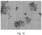
- the segmentation of nucleus is a challenging task due to the varied morphological appearance with clumps and artefacts (see Fig. 15 ).
- the Pap smear images are blurry and highly affected by unwanted noises, e.g., blood, air artefacts (bubbles), or vagina discharge.
- Papsmear images of that kind may lead to an increase in false alarms whene analyzed with a segmentation algorithm.
- Existing techniques are unable to tackle all these problems. Hence a fast and accurate nucleus segmentation technique is necessary to solve this issue.
- the cell nucleus is therefore detected by an optical techniqe encompassing multilevel thresholding.
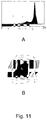
- Image histograms are usually the basis for thresholding.
- a histogram is unimodal if there is one hump, bimodal if there are two humps and multimodal if there are many humps.
- Histograms of Pap-smear images are multimodal in nature (see, e.g., Fig. 11A ).
- multilevel thresholding has been introduced to locate initial seeds for nucleus segmentation. This is the first and foremost common step for all the above four methods.
- said optical technique encompassing multilevel thresholding is at least one selected from the group consisting of
- IGMT an information gain-based local region refinement is introduced after multiple thresholding to segment out nucleolus region.
- IEMT color edge analysis is introduced along with information gain to improve the accuracy of segmentation.
- region growing based on global multiple threshold levels may lead to chance of under/over segmentation of nuclei.
- IRMT may be used to refine the region based on local multilevel thresholding, although it may be unable to solve the boundary leak problem. Hence, local boundary adjustment is necessary to solve this problem. IGTMT may be used to fine-tune the boundary region of Pap-smear nuclei. The different approaches wuill be discussed in detail in the follwing:
- IGMT and IEMT methods are capable of finding the nucleus region in Papsmear images.
- region growing for a ROI based on multiple threshold levels may lead to chance of under/over segmentation of nuclei (see, e.g., Fig. 11(B) ).
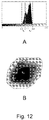
- IRMT Recursive multilevel-based thresholding is introduced in IRMT to reduce the possibility of over/under segmentation of nuclei in IGMT and IEMT methods (see Fig. 7(A)).
- IRMT consists of two major steps: (i) global seed selection, followed by (ii) local region refinement.
- IRMT Global seed selection of Pap-smear images as carried out in the IRMT method is identical to IGMT and IEMT. In case of a local region refinement, the IRMT method provides two major steps:
- the proposed IRMT method may sometimes fails to solve the boundary leak problem.
- the basic idea behind the IGTMT approach is to utilize the graph cut theory for local boundary refinement (see Fig. 14 (B) ).
- IGTMT global multilevel thresholding is the first step in the IGTMT method, to carry out seed initialization of the probable region of the nucleus. This is followed by the selection of upper and lower bound of threshold for individual region which is similar to the IRMT method. Thereafter, to increase the accuracy of segmentation scheme, IGTMT introduces the min cut/max flow based graph theory approach.
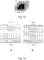
- the IGTMT method uses the similarity measure based on gray level difference of neighbourhood pixels in R ub region. This affinity measures are used as weights in graph where image pixels are represents as nodes with pre-calculated single source and sink (see Fig. 13 ).
- Min cut finds a partitioning of graph nodes which minimizes the sum of weights of cut edges. This introduces the granularity concept helping to achieve the exact boundary of the nucleus.
- the method further comprises the step of determining at least one feature selected from the group of
- jazziness shall mean the variation of distance between cell boundary points from centre of the cell or a fixed reference point preferably inside the cell. High jazziness in texture can be considered as an indication of abnormality.
- texture shall mean the spatial arrangements of colors or intensities in a nucleus or cell region. High variations in texture can be considered as an indication of abnormality.
- fractal dimension of the nucleus relates to a statistical quantity that gives an indication of how completely a fractal appears to fill space of a given cell, as one zooms down to finer and finer scales.
- morphological feature algorithm/technique for determination aggregation of cells determining inter-nuclei distance cell nucleus size, or area calculating the respective image area, e.g., by counting the number of pixels in the respective region inter-nuclei distance distance between the centre of any two nucleus regularity of shape of a cell, and/or a cell nucleus employing properties like form factor, perimeter, major axis, minor axis cell membrane signature.
- ratio of cytoplasm to nucleus in terms of size, or ratio of areas of cytoplam and nucleus size, or area, in a given cell see cell nucleus size
- Brightness/ intensity variation within a cell and/or a cell nucleus variance of intensity in the cell nucleus jazziness of a cellular membrane the variation of distance between cell boudry points from centre of the cell or a some fixed reference point prferably inside the cell Texture the spatial arrangements of colors or intensities in a nucleus or cell region fractal dimension of the nucleus to measure the deviation from the uniformity Hausdorff dimension, box counting dimension, Renyi dimension
- the method according to the invention further comprises at least one step selected from the group consisting of:
- the method according to the invention further comprises the step of recommending further investigation by at least one step selected from the group consisting of
- Colposcopy is a medical diagnostic procedure to examine an illuminated, magnified view of the cervix and the tissues of the vagina and vulva. Primarily in order to detect premalignant lesions and malignant lesions which may result in cancer. Colposcopy is done using a colposcope, which provides an enlarged view of the areas, allowing the colposcopist to visually distinguish normal from abnormal appearing tissue and take directed biopsies for further pathological examination. The main goal of colposcopy is to prevent cervical cancer by detecting precancerous lesions early and treating them.
- HPV DNA test detects cervical infection with human papilloma virus (HPV), which is one of the most important infectious causes of cervical cancer. 84% of new cervical cancers were in the developing world, compared with about 50% of all new cancers. HPV DNA test kits are today commercially available. Such test may be carried out during a routine smear test, as described above (in which case part of the smear sample is taken for the HPV DNA test, while another part is taken for the method according to the invention, or with a newly taken sample with comparable properties, and can be used to improve, confirm or falsify the diagnostic significance of the method according to the invention.
- Biomarker tests have been developed to investigate whether or not a patient suspected to be predisposed for cervical cancer, or a patient who is suspected for having cervical cancer, or in which cervical cancer has already been diagnosed, has, in its genome or proteome, an abnormality which coincides with increased or decreased likelihood of getting a given cancer, or which coincides with increased or decreased responsiveness towards a given therapy.
- abnormality is, for example, a mutation in a given gene, an abnormality in an epigenomic feature, like DNA methylation, or an abnormality with respect to expression of a given gene.
- the image acquisition is carried out by means of a scanner.
- a two dimensional imaging device can be used. In both cases the imaging device is preferably a CCD (linear or two dimensional) or a CMOS (linear or two dimensional).
- the image acquisition is carried out by means of an optical magnification device.
- Said optical magnification device is, for example, a microscope.
- the method according to the invention further comprises, prior to step a), a step in which an image of the sample is acquired at lower magnification, as is the case in step a).
- a step in which an image of the sample is acquired at lower magnification as is the case in step a).
- an overview image is made first.
- the low magnification slide overview is processed by the algorithm to identify the regions suspicious for abnormality and those suspected region are further scanned with higher magnification. This will provide an advantage of quick scanning of the slides.
- steps b) and following are carried out while the digital image acquisition and/or the digital image processing is still in process.
- this approach allows to forgo an image archive of the raw images.
- step d) comprises at least the steps of
- the latter can be done by determining the inter-nuclei distance of at least two cells.
- segmentation of cell nuclei refers to the process of partitioning a digital image comprising the image of at least one cell into multiple segments (sets of pixels in order to identify the cell nuclei. More precisely, image segmentation is the process of assigning a label to every pixel in an image such that pixels with the same label share certain visual characteristics.
- the result of image segmentation is a set of segments that collectively cover the entire image, or a set of contours extracted from the image (see edge detection).
- Each of the pixels in a region are similar with respect to some characteristic or computed property, such as color, intensity, or texture. Adjacent regions are significantly different with respect to the same characteristic(s).
- the resulting contours after image segmentation can be used to create 3D reconstructions with the help of interpolation algorithms like marching cubes.
- centroid of a cell relates to the geometric center, or barycenter, of a cell's two-dimensional image, as determined with digital image processing methods, e.g., by image moments.
- the biological sample comprising cells is a cervical sample.
- the method can also be used with samples from other body tissues which have to be checked for abnormalities, like breast samples, prostate samples, liver samples, lung samples and so forth.
- the method further comprises at least one step selected from the group consisting of:
- the method according to the invention further comprises at least one step of counting a given cell type, or updating an existing count thereof.
- the biological sample comprising cells comprises at least one sample selected from the group consisting of
- a smear sample is for example similar or identical to those samples used in the Papanicolaou tests (also called Pap smear, Pap test, cervical smear, or smear test).
- a tissue slice is for example, sliced by a microtome.
- a liquid sample can preferably consist of a suspension of cells, e.g., obtained by a smear.
- FNAC fine needle aspiration cytology
- abrasive cytology samples and/or exfoliated samples.
- the term slide is used.
- a sample is indeed placed on a slide to make it available for investigation, e.g. a tissue slice, or a smear.
- other devices can also be used to carry a sample, e.g. a small cuvette in case the sample is a liquid sample or a cartridge in case the sample is a brush sample.
- slice as used in the flow charts is thus by no means construed to limiting the scope of the present invention.
- the biological sample comprising cells is stained, preferably prior to step a) of image acquisition.
- Dyes which are preferably used comprise Pap-stain, ultra fast Pap-statin, Romanowsky-type stain, Haris Haematoxylin stain, fluorescent stains like Achrodyn Orange, and H & E stain.
- optical and/or digital image enhancement approaches are used.
- Optical image enhancement is preferably carried out prior to step a) of image acquisition.
- Preferred methods comprise dark field microscopy, phase contrast, differential interference contrast (DIC) and/or reflected interference contrast (RIC).
- Digital contrast enhancement is preferably carried out after step a) of image acquisition.
- Preferred methods comprise bright field microscopy, for example a typical transmission microscope.
- steps b) and following are carried out while the data related to the acquired image, or parts thereof, is still in a volatile memory.
- volatile memory is used interchangeably with the term temporary memory, and shall be understood in such way that the data related to the acquired image are not yet stored on the hard disk or on a flash storage.
- a preferred form of such volatile memory is a random access memory (RAM) used by the image processor, or by the computer's CPU.
- the system is capable to carry out, and/or comprises means capable to carry out, at least the following steps:
- said system is further capable to carry out, and/or comprises further means capable to carry out said the steps of:
- steps b) and following are carried out while the data related to the acquired image, or parts thereof, is still in a volatile memory.
- volatile memory is used interchangeably with the term temporary memory, and shall be understood in such way that the data related to the acquired image are not yet stored on the hard disk or on a flash storage.
- a preferred form of such volatile memory is a random access memory (RAM) used by the image processor, or by the computer's CPU.
- a system comprising digital image acquisition and digital image processing units, wherein the system is configured to perform the methods described above, is provided..
- a device for the detection of precancer or cancer in a biological sample comprising cells comprises at least the following items:
- Said output means is preferably a display, or a touch screen, while said input means is preferably an array of keys, or buttons, or a touchscreen.
- the method according to the invention is not restricted to the use in such device. It can also be used "stand-alone" for the detection of locations/regions in a slide or set of images being suspected to contain images of abnormal cells.
- the device according to the invention further comprises at least one optical magnification unit.
- the device according to the invention further comprises at least one interface for connecting the device with other equipment.
- Such interface is, preferably, a GSM interface, a 3G interface, a USB interface, a Bluetooth interface, a Firewire interface and/or a WiFi interface, hypertext terminal, etc.
- the device according to the invention further comprises at least one sample collector and/or at least one cartridge in which the sample is transferred, said cartridge being disposed for placement in the sample receiving unit.
- the system are device discussed above is preferably in the form of a point of are device (POC).
- POC point of are device
- it is provided as a handheld or desktop unit. Even more preferably, it is battery driven and/or portable.
- Figs. 1 and 2 show flow charts which have been described in the text already.
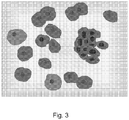
- Fig. 3 shows an artificially created example of a Pap smear image to explain the concept of the present invention.
- the cytoplasm parts of the cells stick together, and the nuclei of the cells are very close to one another (small inter-nuclei distance).
- Inter-nuclei distance mapping is thus used in the algorithmic identification process to identify abnormal cells, particularly in those regions of interest which have earlier been identified, by cell aggregation analysis, as suspicious.
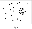
- Fig. 4 shows the result of the segmentation of the cells and the corresponding nuclei. Based on the subsequent algorithms, regions of interest can be determined which are highly suspicious to comprise abnormal cells.
- Fig. 5 shows the results of such process clustering. Two different clusters have been identified, wherein cluster No 2 - marked by a rectangle - is likely to comprise abnormal cells, due to (i) extensive cell clustering, (ii) high variation of nuclei sizes, and (iii) small inter-nuclei distances.
- Fig. 6 shows a system and work flow according to the present invention.
- the sample is collected by means of a sample collector 61, which may adopt the shape of a brush, similar to a Q-tip, and is transferred to a cartridge 62 which is disposed for placement in the sample receiving unit 63 of a device 64 according to the invention.
- the device is shown in form of a portable point of care device.
- the sample can be stained in between.
- the sample is then scanned by the optical magnification unit and the image acquisition device unit present in the device.
- an input means 65 which is embodied, herein, as an array of keys
- the device acquires an image from the sample in the cartridge and passes it to the algorithm embedded into the system.
- the method steps according to the invention are accomplished, and results of the analysis are shown on output means 66, which is embodied, herein, as a display screen.
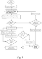
- Figs. 7 and 8 show flow diagrams comprising a more elaborated approach to identify regions of interest which may be likely to comprise abnormal cells.
- the main focus is to identify the cellular regions and find cell aggregates. Once the latter are identified the algorithm passes the respective field of view to further analysis, as explained in Fig. 8 .
- the system initiation is done. Here, essentially, the system resets and arrives at a reference position, clears all the buffers, etc.
- a cytology sample is loaded, in most cases as a slide (see above), and the initiation for cell count of all type of cells, such as normal/abnormal squamous cells and their types, and normal/abnormal endocervical cells, is initiated, and the respective result is stored.
- a scan strategy is chosen. The scan strategy involves the selection of step size of the movement of the sample in x and y direction and the focus depth (z direction) relative to the imaging devices (i.e., either the sample or the imaging device is moved), and the selection of magnification, contrast, etc. Further, the system starts reading the sample/slide, and the images are acquired.
- the quality parameter of the slide/images are estimated.
- the quality parameters are checked for their adequacy (by comparing with a priori data). If the quality is not adequate, the system goes in the loop to check the scope for up-gradation in Box 13. If there is scope to upgrade the scan strategy (i.e, if the scan parameters are within the defined range of the system), then the system provides an on-the-fly feedback to change the scan strategy (Box 12). If there is no scope for upgrading the scan parameters, a report is generated (Box 14) and the systems stops or goes for the next slide (Box 15).
- the actual field of view comprises only normal squamous cell, and their number is counted.
- the cells which are not comprised in clusters are assumed to be normal, given the fact that significant abnormalities are rather found in clustered cells than in isolated cells. If in Box 10 clusters are found, they are suspected for abnormality and the system passes the image of the actual field of view, or other data related to the said clusters, for detailed analysis to link 1 in Fig. 8 . If in the respective field of view isolated cells as well as clusters of cells have been determined, the image of the actual field of view, or other data related to the said clusters, are passed for further processing in Fig. 8 , while isolated cells are just counted.
- Fig. 8 After detailed analysis in Fig. 8 (see below) the control is returned back to Fig. 7 at link 2.
- the system checks if there is any other field of view left to be scanned in Box 11. If yes, the system scans the next field of view and repeats the above described process. If all the fields of view are completed the system generates a report on the actual sample (estimation of abnormality, its severity, type of carcinoma, number of cells etc.) and stops, or moves to the next sample in Box 16.
- Images of a field of view comprising clusters, or other data related to the said clusters, are then passed on to the algorithm in Fig. 8 .
- the morphological features in each cell of a given cluster are extracted to measure the variability of these features (Box 2 and Box 3). If the measurements are not in an abnormal range (Box 4), then the cell cluster is suspected to comprise normal endocervical cells (Box 11), hence a confirmatory test is done (Box 12). If the assumption is true then the count of normal endocervical cells is done in Box 14. If not, the sample is considered to comprise squamous cells, and a count for squamous cells is done in Box 13.
Landscapes
- Engineering & Computer Science (AREA)
- Health & Medical Sciences (AREA)
- Life Sciences & Earth Sciences (AREA)
- General Health & Medical Sciences (AREA)
- Physics & Mathematics (AREA)
- Theoretical Computer Science (AREA)
- General Physics & Mathematics (AREA)
- Chemical & Material Sciences (AREA)
- Molecular Biology (AREA)
- Organic Chemistry (AREA)
- Biomedical Technology (AREA)
- Multimedia (AREA)
- Immunology (AREA)
- Proteomics, Peptides & Aminoacids (AREA)
- Zoology (AREA)
- Wood Science & Technology (AREA)
- Computer Vision & Pattern Recognition (AREA)
- Quality & Reliability (AREA)
- Radiology & Medical Imaging (AREA)
- Nuclear Medicine, Radiotherapy & Molecular Imaging (AREA)
- Medical Informatics (AREA)
- Biophysics (AREA)
- Microbiology (AREA)
- Biotechnology (AREA)
- Biochemistry (AREA)
- Bioinformatics & Cheminformatics (AREA)
- General Engineering & Computer Science (AREA)
- Genetics & Genomics (AREA)
- Analytical Chemistry (AREA)
- Virology (AREA)
- Investigating Or Analysing Biological Materials (AREA)
- Measuring Or Testing Involving Enzymes Or Micro-Organisms (AREA)
Applications Claiming Priority (2)
| Application Number | Priority Date | Filing Date | Title |
|---|---|---|---|
| US201161534031P | 2011-09-13 | 2011-09-13 | |
| PCT/IB2012/054708 WO2013038331A1 (en) | 2011-09-13 | 2012-09-11 | System and method for the detection of abnormalities in a biological sample |
Publications (2)
| Publication Number | Publication Date |
|---|---|
| EP2745111A1 EP2745111A1 (en) | 2014-06-25 |
| EP2745111B1 true EP2745111B1 (en) | 2016-06-29 |
Family
ID=47178777
Family Applications (1)
| Application Number | Title | Priority Date | Filing Date |
|---|---|---|---|
| EP12787088.9A Not-in-force EP2745111B1 (en) | 2011-09-13 | 2012-09-11 | System and method for the detection of precancer or cancer cells in a biological sample |
Country Status (6)
| Country | Link |
|---|---|
| US (1) | US9567651B2 (enExample) |
| EP (1) | EP2745111B1 (enExample) |
| CN (1) | CN103907023B (enExample) |
| IN (1) | IN2014CN01734A (enExample) |
| MX (1) | MX346624B (enExample) |
| WO (1) | WO2013038331A1 (enExample) |
Families Citing this family (32)
| Publication number | Priority date | Publication date | Assignee | Title |
|---|---|---|---|---|
| JP6143365B2 (ja) * | 2014-03-05 | 2017-06-07 | 富士フイルム株式会社 | 細胞画像評価装置および方法並びにプログラム |
| GB2530252B (en) * | 2014-09-10 | 2020-04-01 | Smiths Heimann Sas | Determination of a degree of homogeneity in images |
| CA2965564C (en) * | 2014-11-10 | 2024-01-02 | Ventana Medical Systems, Inc. | Classifying nuclei in histology images |
| US20180253590A1 (en) * | 2015-03-20 | 2018-09-06 | Inspirata, Inc. | Systems, methods, and apparatuses for digital histopathological imaging for prescreened detection of cancer and other abnormalities |
| CN106290169A (zh) * | 2015-05-29 | 2017-01-04 | 深圳市搜罗盟网络科技有限公司 | 一种癌症检测设备 |
| WO2017109860A1 (ja) * | 2015-12-22 | 2017-06-29 | 株式会社ニコン | 画像処理装置 |
| EP3396375B1 (en) * | 2015-12-24 | 2021-08-04 | Konica Minolta, Inc. | Image processing device and program |
| TWI594207B (zh) * | 2016-04-26 | 2017-08-01 | 財團法人金屬工業研究發展中心 | 細胞核影像輪廓擷取裝置及其方法 |
| RU2647675C1 (ru) * | 2016-10-26 | 2018-03-16 | Федеральное государственное казенное военное образовательное учреждение высшего образования "Военный учебно-научный центр Военно-воздушных сил "Военно-воздушная академия имени профессора Н.Е. Жуковского и Ю.А. Гагарина" (г. Воронеж) Министерства обороны Российской Федерации | Способ измерения морфологической мультифрактальной сигнатуры |
| CN106479880B (zh) * | 2016-11-01 | 2019-06-04 | 上海市同仁医院 | 一种人乳头瘤病毒结果判读装置 |
| JP6886306B2 (ja) * | 2017-02-02 | 2021-06-16 | オリンパス株式会社 | 位相分布算出方法、評価方法、画像処理装置、画像処理システム、プログラム |
| CN108693342A (zh) * | 2017-12-13 | 2018-10-23 | 青岛汉朗智能医疗科技有限公司 | 宫颈癌、子宫癌的检测方法及系统 |
| CN110135221A (zh) * | 2018-02-08 | 2019-08-16 | 佛山市水木联合医疗器械研究院 | 一种利用标准细胞库图像识别样本细胞的方法 |
| FR3079617B1 (fr) * | 2018-03-29 | 2023-12-22 | Office National Detude Et De Rech Aerospatiales Onera | Methode de detection de cellules presentant au moins une anomalie dans un echantillon cytologique |
| US11024062B2 (en) | 2018-06-11 | 2021-06-01 | Shanghai United Imaging Healthcare Co., Ltd. | Systems and methods for evaluating image quality |
| JP7164978B2 (ja) * | 2018-06-29 | 2022-11-02 | キヤノン株式会社 | 粒子測定方法、粒子測定装置及び核酸濃度測定システム |
| KR102357952B1 (ko) * | 2018-07-06 | 2022-02-08 | 엔지반트 테라퓨틱스 게엠베하 | 정량적 조직형태 분석을 통한 조직 효능 결정 |
| CN108961249A (zh) * | 2018-07-19 | 2018-12-07 | 厦门理工学院 | 一种子宫颈癌细胞再识别诊断方法 |
| EP3611695A1 (en) * | 2018-08-15 | 2020-02-19 | Koninklijke Philips N.V. | Generating annotation data of tissue images |
| AU2019403134A1 (en) | 2018-12-18 | 2021-06-17 | Pathware Inc. | Computational microscopy based-system and method for automated imaging and analysis of pathology specimens |
| WO2020205200A1 (en) * | 2019-03-29 | 2020-10-08 | Becton, Dickinson And Company | Parameters for use in particle discrimination |
| CN110210578B (zh) * | 2019-06-19 | 2021-11-16 | 四川智动木牛智能科技有限公司 | 基于图论的宫颈癌组织病理学显微图像聚类系统 |
| CN110400281B (zh) * | 2019-08-05 | 2021-06-01 | 山东志盈医学科技有限公司 | 一种数字切片扫描仪中图像增强方法 |
| CN110853022B (zh) * | 2019-11-14 | 2020-11-06 | 腾讯科技(深圳)有限公司 | 病理切片图像的处理方法、装置、系统及存储介质 |
| CN111275191B (zh) * | 2020-02-26 | 2023-11-28 | 上海商汤智能科技有限公司 | 检测细胞的方法及装置、电子设备和存储介质 |
| CN112233060B (zh) * | 2020-09-04 | 2024-03-29 | 广州金域医学检验中心有限公司 | 数字病理图像异常样本的筛选方法、装置、设备及介质 |
| CN114428076A (zh) * | 2020-09-29 | 2022-05-03 | 中国石油化工股份有限公司 | 聚合物改性沥青的储存稳定性判定方法和质量控制方法 |
| CN112378727A (zh) * | 2020-12-14 | 2021-02-19 | 湖南莱博赛医用机器人有限公司 | 基于机器视觉的dna倍体定量分析装置及其应用方法 |
| WO2024063671A1 (ru) * | 2022-09-21 | 2024-03-28 | Ооо "Хоспитекс Диагностикс" | Система дая цитологического анализа патологии шейки матки |
| CN117787864B (zh) * | 2023-12-27 | 2024-10-15 | 石家庄博瑞迪生物技术有限公司 | 一种动物组织采样管溯源入库管理方法 |
| CN119741248B (zh) * | 2024-09-20 | 2025-11-18 | 四川大学 | 一种类器官细胞聚集实时监测及动态分析方法 |
| CN120125582B (zh) * | 2025-05-13 | 2026-04-10 | 大熊(天津)医疗科技有限公司 | 一种医学细胞检测方法 |
Family Cites Families (10)
| Publication number | Priority date | Publication date | Assignee | Title |
|---|---|---|---|---|
| US5740270A (en) | 1988-04-08 | 1998-04-14 | Neuromedical Systems, Inc. | Automated cytological specimen classification system and method |
| US5257182B1 (en) | 1991-01-29 | 1996-05-07 | Neuromedical Systems Inc | Morphological classification system and method |
| US6026174A (en) | 1992-10-14 | 2000-02-15 | Accumed International, Inc. | System and method for automatically detecting malignant cells and cells having malignancy-associated changes |
| WO1996009604A1 (en) | 1994-09-20 | 1996-03-28 | Neopath, Inc. | Apparatus for automated identification of cell groupings on a biological specimen |
| AU3629295A (en) | 1994-09-20 | 1996-04-09 | Neopath, Inc. | Apparatus for automated identification of thick cell groupings on a biological specimen |
| WO1996009600A1 (en) | 1994-09-20 | 1996-03-28 | Neopath, Inc. | Apparatus for identification and integration of multiple cell patterns |
| AUPP278698A0 (en) | 1998-04-03 | 1998-04-30 | University Of Queensland, The | Method of cell nuclei segmentation |
| US6379907B1 (en) * | 1999-08-05 | 2002-04-30 | The Regents Of The University Of California | Diagnostic method using expression of MN/CA9 protein in AGUS Pap smears |
| US7274809B2 (en) * | 2002-08-29 | 2007-09-25 | Perceptronix Medical, Inc. And British Columbia Cancer Agency | Computerized methods and systems related to the detection of malignancy-associated changes (MAC) to detect cancer |
| CN101059505A (zh) * | 2007-05-28 | 2007-10-24 | 宋士明 | 一种活细胞技术检查宫颈癌细胞的方法 |
-
2012
- 2012-09-11 IN IN1734CHN2014 patent/IN2014CN01734A/en unknown
- 2012-09-11 CN CN201280044762.0A patent/CN103907023B/zh not_active Expired - Fee Related
- 2012-09-11 US US14/343,427 patent/US9567651B2/en not_active Expired - Fee Related
- 2012-09-11 WO PCT/IB2012/054708 patent/WO2013038331A1/en not_active Ceased
- 2012-09-11 EP EP12787088.9A patent/EP2745111B1/en not_active Not-in-force
- 2012-09-11 MX MX2014002843A patent/MX346624B/es active IP Right Grant
Also Published As
| Publication number | Publication date |
|---|---|
| MX346624B (es) | 2017-03-27 |
| WO2013038331A1 (en) | 2013-03-21 |
| CN103907023B (zh) | 2016-10-05 |
| EP2745111A1 (en) | 2014-06-25 |
| IN2014CN01734A (enExample) | 2015-05-29 |
| CN103907023A (zh) | 2014-07-02 |
| US20140227682A1 (en) | 2014-08-14 |
| US9567651B2 (en) | 2017-02-14 |
| MX2014002843A (es) | 2014-07-09 |
Similar Documents
| Publication | Publication Date | Title |
|---|---|---|
| EP2745111B1 (en) | System and method for the detection of precancer or cancer cells in a biological sample | |
| Belsare et al. | Histopathological image analysis using image processing techniques: An overview | |
| Demir et al. | Automated cancer diagnosis based on histopathological images: a systematic survey | |
| TWI379248B (en) | Methods and systems for processing biological specimens utilizing multiple wavelengths | |
| US8891850B2 (en) | System and methods for digital evaluation of cellblock preparations | |
| US20190042826A1 (en) | Automatic nuclei segmentation in histopathology images | |
| CN101167101A (zh) | 自动图像分析 | |
| WO2012041333A1 (en) | Automated imaging, detection and grading of objects in cytological samples | |
| US7865000B2 (en) | Medical image processing apparatus for analyzing properties of living body based on image data thereof | |
| Shirazi et al. | Automated pathology image analysis | |
| CN113723441B (zh) | 一种唇腺病理智能分析系统及方法 | |
| CN115018762A (zh) | 基于显微高光谱数据库模型的恶性肿瘤识别方法及系统 | |
| Ko et al. | A computer-aided grading system of breast carcinoma: scoring of tubule formation | |
| Schilling et al. | Towards rapid cervical cancer diagnosis: automated detection and classification of pathologic cells in phase-contrast images | |
| Sobrevilla et al. | Fuzzy-based analysis of microscopic color cervical pap smear images: nuclei detection | |
| Forsberg et al. | Evaluating cell nuclei segmentation for use on whole-slide images in lung cytology | |
| Li et al. | Detection of epithelial versus mesenchymal regions in 2D images of tumor biopsies using shearlets | |
| WO2024186756A2 (en) | System and method for biomarker detection | |
| Thiripurasundari et al. | Pap screening for cervical carcinoma–evolving trends | |
| Wang et al. | Automated segmentation and analysis of fluorescent in situ hybridization (FISH) signals in interphase nuclei of pap-smear specimens | |
| CN117995384A (zh) | 基于膀胱非尿路上皮癌的亚型的人工智能辅助诊疗系统 | |
| Das et al. | A Novel Analysis of Clinical Data and Image Processing Algorithms in Detection of Cervical Cancer | |
| Joseph et al. | Automated Identification of Breast Cancer from Low Resolution Microscopic Videos | |
| Joel et al. | Cervical Cancer Detection System Analysis by Segmentation Methods | |
| Friedrich et al. | Detection of immunocytological markers in photomicroscopic images |
Legal Events
| Date | Code | Title | Description |
|---|---|---|---|
| PUAI | Public reference made under article 153(3) epc to a published international application that has entered the european phase |
Free format text: ORIGINAL CODE: 0009012 |
|
| 17P | Request for examination filed |
Effective date: 20140317 |
|
| AK | Designated contracting states |
Kind code of ref document: A1 Designated state(s): AL AT BE BG CH CY CZ DE DK EE ES FI FR GB GR HR HU IE IS IT LI LT LU LV MC MK MT NL NO PL PT RO RS SE SI SK SM TR |
|
| DAX | Request for extension of the european patent (deleted) | ||
| 17Q | First examination report despatched |
Effective date: 20141211 |
|
| GRAP | Despatch of communication of intention to grant a patent |
Free format text: ORIGINAL CODE: EPIDOSNIGR1 |
|
| INTG | Intention to grant announced |
Effective date: 20160126 |
|
| GRAS | Grant fee paid |
Free format text: ORIGINAL CODE: EPIDOSNIGR3 |
|
| GRAA | (expected) grant |
Free format text: ORIGINAL CODE: 0009210 |
|
| AK | Designated contracting states |
Kind code of ref document: B1 Designated state(s): AL AT BE BG CH CY CZ DE DK EE ES FI FR GB GR HR HU IE IS IT LI LT LU LV MC MK MT NL NO PL PT RO RS SE SI SK SM TR |
|
| REG | Reference to a national code |
Ref country code: GB Ref legal event code: FG4D |
|
| REG | Reference to a national code |
Ref country code: CH Ref legal event code: EP |
|
| REG | Reference to a national code |
Ref country code: AT Ref legal event code: REF Ref document number: 809504 Country of ref document: AT Kind code of ref document: T Effective date: 20160715 |
|
| REG | Reference to a national code |
Ref country code: IE Ref legal event code: FG4D |
|
| REG | Reference to a national code |
Ref country code: DE Ref legal event code: R096 Ref document number: 602012020031 Country of ref document: DE |
|
| REG | Reference to a national code |
Ref country code: FR Ref legal event code: PLFP Year of fee payment: 5 |
|
| REG | Reference to a national code |
Ref country code: LT Ref legal event code: MG4D |
|
| PG25 | Lapsed in a contracting state [announced via postgrant information from national office to epo] |
Ref country code: LT Free format text: LAPSE BECAUSE OF FAILURE TO SUBMIT A TRANSLATION OF THE DESCRIPTION OR TO PAY THE FEE WITHIN THE PRESCRIBED TIME-LIMIT Effective date: 20160629 Ref country code: FI Free format text: LAPSE BECAUSE OF FAILURE TO SUBMIT A TRANSLATION OF THE DESCRIPTION OR TO PAY THE FEE WITHIN THE PRESCRIBED TIME-LIMIT Effective date: 20160629 Ref country code: NO Free format text: LAPSE BECAUSE OF FAILURE TO SUBMIT A TRANSLATION OF THE DESCRIPTION OR TO PAY THE FEE WITHIN THE PRESCRIBED TIME-LIMIT Effective date: 20160929 |
|
| REG | Reference to a national code |
Ref country code: NL Ref legal event code: MP Effective date: 20160629 |
|
| PG25 | Lapsed in a contracting state [announced via postgrant information from national office to epo] |
Ref country code: NL Free format text: LAPSE BECAUSE OF FAILURE TO SUBMIT A TRANSLATION OF THE DESCRIPTION OR TO PAY THE FEE WITHIN THE PRESCRIBED TIME-LIMIT Effective date: 20160629 Ref country code: RS Free format text: LAPSE BECAUSE OF FAILURE TO SUBMIT A TRANSLATION OF THE DESCRIPTION OR TO PAY THE FEE WITHIN THE PRESCRIBED TIME-LIMIT Effective date: 20160629 Ref country code: HR Free format text: LAPSE BECAUSE OF FAILURE TO SUBMIT A TRANSLATION OF THE DESCRIPTION OR TO PAY THE FEE WITHIN THE PRESCRIBED TIME-LIMIT Effective date: 20160629 Ref country code: LV Free format text: LAPSE BECAUSE OF FAILURE TO SUBMIT A TRANSLATION OF THE DESCRIPTION OR TO PAY THE FEE WITHIN THE PRESCRIBED TIME-LIMIT Effective date: 20160629 Ref country code: GR Free format text: LAPSE BECAUSE OF FAILURE TO SUBMIT A TRANSLATION OF THE DESCRIPTION OR TO PAY THE FEE WITHIN THE PRESCRIBED TIME-LIMIT Effective date: 20160930 Ref country code: SE Free format text: LAPSE BECAUSE OF FAILURE TO SUBMIT A TRANSLATION OF THE DESCRIPTION OR TO PAY THE FEE WITHIN THE PRESCRIBED TIME-LIMIT Effective date: 20160629 |
|
| REG | Reference to a national code |
Ref country code: AT Ref legal event code: MK05 Ref document number: 809504 Country of ref document: AT Kind code of ref document: T Effective date: 20160629 |
|
| PG25 | Lapsed in a contracting state [announced via postgrant information from national office to epo] |
Ref country code: IT Free format text: LAPSE BECAUSE OF FAILURE TO SUBMIT A TRANSLATION OF THE DESCRIPTION OR TO PAY THE FEE WITHIN THE PRESCRIBED TIME-LIMIT Effective date: 20160629 Ref country code: IS Free format text: LAPSE BECAUSE OF FAILURE TO SUBMIT A TRANSLATION OF THE DESCRIPTION OR TO PAY THE FEE WITHIN THE PRESCRIBED TIME-LIMIT Effective date: 20161029 Ref country code: CZ Free format text: LAPSE BECAUSE OF FAILURE TO SUBMIT A TRANSLATION OF THE DESCRIPTION OR TO PAY THE FEE WITHIN THE PRESCRIBED TIME-LIMIT Effective date: 20160629 Ref country code: RO Free format text: LAPSE BECAUSE OF FAILURE TO SUBMIT A TRANSLATION OF THE DESCRIPTION OR TO PAY THE FEE WITHIN THE PRESCRIBED TIME-LIMIT Effective date: 20160629 Ref country code: EE Free format text: LAPSE BECAUSE OF FAILURE TO SUBMIT A TRANSLATION OF THE DESCRIPTION OR TO PAY THE FEE WITHIN THE PRESCRIBED TIME-LIMIT Effective date: 20160629 Ref country code: SK Free format text: LAPSE BECAUSE OF FAILURE TO SUBMIT A TRANSLATION OF THE DESCRIPTION OR TO PAY THE FEE WITHIN THE PRESCRIBED TIME-LIMIT Effective date: 20160629 |
|
| PG25 | Lapsed in a contracting state [announced via postgrant information from national office to epo] |
Ref country code: SM Free format text: LAPSE BECAUSE OF FAILURE TO SUBMIT A TRANSLATION OF THE DESCRIPTION OR TO PAY THE FEE WITHIN THE PRESCRIBED TIME-LIMIT Effective date: 20160629 Ref country code: ES Free format text: LAPSE BECAUSE OF FAILURE TO SUBMIT A TRANSLATION OF THE DESCRIPTION OR TO PAY THE FEE WITHIN THE PRESCRIBED TIME-LIMIT Effective date: 20160629 Ref country code: PT Free format text: LAPSE BECAUSE OF FAILURE TO SUBMIT A TRANSLATION OF THE DESCRIPTION OR TO PAY THE FEE WITHIN THE PRESCRIBED TIME-LIMIT Effective date: 20161031 Ref country code: BE Free format text: LAPSE BECAUSE OF NON-PAYMENT OF DUE FEES Effective date: 20160629 Ref country code: AT Free format text: LAPSE BECAUSE OF FAILURE TO SUBMIT A TRANSLATION OF THE DESCRIPTION OR TO PAY THE FEE WITHIN THE PRESCRIBED TIME-LIMIT Effective date: 20160629 Ref country code: PL Free format text: LAPSE BECAUSE OF FAILURE TO SUBMIT A TRANSLATION OF THE DESCRIPTION OR TO PAY THE FEE WITHIN THE PRESCRIBED TIME-LIMIT Effective date: 20160629 |
|
| REG | Reference to a national code |
Ref country code: DE Ref legal event code: R097 Ref document number: 602012020031 Country of ref document: DE |
|
| PG25 | Lapsed in a contracting state [announced via postgrant information from national office to epo] |
Ref country code: MC Free format text: LAPSE BECAUSE OF FAILURE TO SUBMIT A TRANSLATION OF THE DESCRIPTION OR TO PAY THE FEE WITHIN THE PRESCRIBED TIME-LIMIT Effective date: 20160629 |
|
| REG | Reference to a national code |
Ref country code: CH Ref legal event code: PL |
|
| PLBE | No opposition filed within time limit |
Free format text: ORIGINAL CODE: 0009261 |
|
| STAA | Information on the status of an ep patent application or granted ep patent |
Free format text: STATUS: NO OPPOSITION FILED WITHIN TIME LIMIT |
|
| PG25 | Lapsed in a contracting state [announced via postgrant information from national office to epo] |
Ref country code: DK Free format text: LAPSE BECAUSE OF FAILURE TO SUBMIT A TRANSLATION OF THE DESCRIPTION OR TO PAY THE FEE WITHIN THE PRESCRIBED TIME-LIMIT Effective date: 20160629 |
|
| 26N | No opposition filed |
Effective date: 20170330 |
|
| REG | Reference to a national code |
Ref country code: IE Ref legal event code: MM4A |
|
| PG25 | Lapsed in a contracting state [announced via postgrant information from national office to epo] |
Ref country code: CH Free format text: LAPSE BECAUSE OF NON-PAYMENT OF DUE FEES Effective date: 20160930 Ref country code: IE Free format text: LAPSE BECAUSE OF NON-PAYMENT OF DUE FEES Effective date: 20160911 Ref country code: LI Free format text: LAPSE BECAUSE OF NON-PAYMENT OF DUE FEES Effective date: 20160930 |
|
| PG25 | Lapsed in a contracting state [announced via postgrant information from national office to epo] |
Ref country code: BG Free format text: LAPSE BECAUSE OF FAILURE TO SUBMIT A TRANSLATION OF THE DESCRIPTION OR TO PAY THE FEE WITHIN THE PRESCRIBED TIME-LIMIT Effective date: 20160929 Ref country code: SI Free format text: LAPSE BECAUSE OF FAILURE TO SUBMIT A TRANSLATION OF THE DESCRIPTION OR TO PAY THE FEE WITHIN THE PRESCRIBED TIME-LIMIT Effective date: 20160629 Ref country code: LU Free format text: LAPSE BECAUSE OF NON-PAYMENT OF DUE FEES Effective date: 20160911 |
|
| REG | Reference to a national code |
Ref country code: FR Ref legal event code: PLFP Year of fee payment: 6 |
|
| PG25 | Lapsed in a contracting state [announced via postgrant information from national office to epo] |
Ref country code: HU Free format text: LAPSE BECAUSE OF FAILURE TO SUBMIT A TRANSLATION OF THE DESCRIPTION OR TO PAY THE FEE WITHIN THE PRESCRIBED TIME-LIMIT; INVALID AB INITIO Effective date: 20120911 Ref country code: CY Free format text: LAPSE BECAUSE OF FAILURE TO SUBMIT A TRANSLATION OF THE DESCRIPTION OR TO PAY THE FEE WITHIN THE PRESCRIBED TIME-LIMIT Effective date: 20160629 |
|
| PG25 | Lapsed in a contracting state [announced via postgrant information from national office to epo] |
Ref country code: MT Free format text: LAPSE BECAUSE OF NON-PAYMENT OF DUE FEES Effective date: 20160930 Ref country code: MK Free format text: LAPSE BECAUSE OF FAILURE TO SUBMIT A TRANSLATION OF THE DESCRIPTION OR TO PAY THE FEE WITHIN THE PRESCRIBED TIME-LIMIT Effective date: 20160629 |
|
| REG | Reference to a national code |
Ref country code: FR Ref legal event code: PLFP Year of fee payment: 7 |
|
| PG25 | Lapsed in a contracting state [announced via postgrant information from national office to epo] |
Ref country code: AL Free format text: LAPSE BECAUSE OF FAILURE TO SUBMIT A TRANSLATION OF THE DESCRIPTION OR TO PAY THE FEE WITHIN THE PRESCRIBED TIME-LIMIT Effective date: 20160629 Ref country code: TR Free format text: LAPSE BECAUSE OF FAILURE TO SUBMIT A TRANSLATION OF THE DESCRIPTION OR TO PAY THE FEE WITHIN THE PRESCRIBED TIME-LIMIT Effective date: 20160629 |
|
| PGFP | Annual fee paid to national office [announced via postgrant information from national office to epo] |
Ref country code: FR Payment date: 20190926 Year of fee payment: 8 |
|
| PGFP | Annual fee paid to national office [announced via postgrant information from national office to epo] |
Ref country code: GB Payment date: 20190927 Year of fee payment: 8 |
|
| GBPC | Gb: european patent ceased through non-payment of renewal fee |
Effective date: 20200911 |
|
| PG25 | Lapsed in a contracting state [announced via postgrant information from national office to epo] |
Ref country code: FR Free format text: LAPSE BECAUSE OF NON-PAYMENT OF DUE FEES Effective date: 20200930 |
|
| PG25 | Lapsed in a contracting state [announced via postgrant information from national office to epo] |
Ref country code: GB Free format text: LAPSE BECAUSE OF NON-PAYMENT OF DUE FEES Effective date: 20200911 |
|
| PGFP | Annual fee paid to national office [announced via postgrant information from national office to epo] |
Ref country code: DE Payment date: 20210929 Year of fee payment: 10 |
|
| REG | Reference to a national code |
Ref country code: DE Ref legal event code: R119 Ref document number: 602012020031 Country of ref document: DE |
|
| PG25 | Lapsed in a contracting state [announced via postgrant information from national office to epo] |
Ref country code: DE Free format text: LAPSE BECAUSE OF NON-PAYMENT OF DUE FEES Effective date: 20230401 |