EP2722818B1 - Method for digitizing dento-maxillofacial objects - Google Patents
Method for digitizing dento-maxillofacial objects Download PDFInfo
- Publication number
- EP2722818B1 EP2722818B1 EP14000057.1A EP14000057A EP2722818B1 EP 2722818 B1 EP2722818 B1 EP 2722818B1 EP 14000057 A EP14000057 A EP 14000057A EP 2722818 B1 EP2722818 B1 EP 2722818B1
- Authority
- EP
- European Patent Office
- Prior art keywords
- dento
- maxillofacial
- calibration object
- image data
- segmentation
- Prior art date
- Legal status (The legal status is an assumption and is not a legal conclusion. Google has not performed a legal analysis and makes no representation as to the accuracy of the status listed.)
- Active
Links
Images


Classifications
-
- G—PHYSICS
- G06—COMPUTING OR CALCULATING; COUNTING
- G06T—IMAGE DATA PROCESSING OR GENERATION, IN GENERAL
- G06T7/00—Image analysis
- G06T7/10—Segmentation; Edge detection
- G06T7/155—Segmentation; Edge detection involving morphological operators
-
- A—HUMAN NECESSITIES
- A61—MEDICAL OR VETERINARY SCIENCE; HYGIENE
- A61C—DENTISTRY; APPARATUS OR METHODS FOR ORAL OR DENTAL HYGIENE
- A61C9/00—Impression cups, i.e. impression trays; Impression methods
- A61C9/004—Means or methods for taking digitized impressions
- A61C9/0046—Data acquisition means or methods
-
- G—PHYSICS
- G06—COMPUTING OR CALCULATING; COUNTING
- G06T—IMAGE DATA PROCESSING OR GENERATION, IN GENERAL
- G06T7/00—Image analysis
- G06T7/10—Segmentation; Edge detection
- G06T7/11—Region-based segmentation
-
- G—PHYSICS
- G06—COMPUTING OR CALCULATING; COUNTING
- G06T—IMAGE DATA PROCESSING OR GENERATION, IN GENERAL
- G06T7/00—Image analysis
- G06T7/10—Segmentation; Edge detection
- G06T7/136—Segmentation; Edge detection involving thresholding
-
- G—PHYSICS
- G06—COMPUTING OR CALCULATING; COUNTING
- G06T—IMAGE DATA PROCESSING OR GENERATION, IN GENERAL
- G06T7/00—Image analysis
- G06T7/10—Segmentation; Edge detection
- G06T7/194—Segmentation; Edge detection involving foreground-background segmentation
-
- G—PHYSICS
- G06—COMPUTING OR CALCULATING; COUNTING
- G06T—IMAGE DATA PROCESSING OR GENERATION, IN GENERAL
- G06T2207/00—Indexing scheme for image analysis or image enhancement
- G06T2207/10—Image acquisition modality
- G06T2207/10072—Tomographic images
- G06T2207/10081—Computed x-ray tomography [CT]
-
- G—PHYSICS
- G06—COMPUTING OR CALCULATING; COUNTING
- G06T—IMAGE DATA PROCESSING OR GENERATION, IN GENERAL
- G06T2207/00—Indexing scheme for image analysis or image enhancement
- G06T2207/20—Special algorithmic details
- G06T2207/20036—Morphological image processing
- G06T2207/20041—Distance transform
-
- G—PHYSICS
- G06—COMPUTING OR CALCULATING; COUNTING
- G06T—IMAGE DATA PROCESSING OR GENERATION, IN GENERAL
- G06T2207/00—Indexing scheme for image analysis or image enhancement
- G06T2207/30—Subject of image; Context of image processing
- G06T2207/30004—Biomedical image processing
- G06T2207/30036—Dental; Teeth
Definitions
- the present invention relates to a method for capturing the shape of a dento-maxillofacial object out of volumetric image data of that object. Further, the present invention relates to a method for determining a parameter for use in digitizing the dento-maxillofacial object.
- Dento-maxillofacial treatments are related to the dentition, the skull and the facial soft tissues.
- the scope of the treatments goes from handling teeth - such as aligning, restoring crowns, extracting, restoring including root and crown - over bone related treatments - such as maxillofacial surgery involving surgical remodelling or restoring of the skull and dentition, encompassing surgical interventions of repair, in particular, of a mis-positioning of the jaws with respect to one another, called orthognathic surgery, temporomandibular joint (TMJ) treatments - over facial soft tissue treatments - such as tissue sculpting, lifting, .... Important in these treatments is creating a good occlusion and smile line.
- 'occlusion' is meant the manner in which the teeth from the upper and lower arches come together when the mouth is closed.
- dento-maxillofacial treatments are complex and have a big impact on the patient's facial outlook, accurate treatment planning is required.
- Computer aided dento-maxillofacial planning systems are becoming available which digitize the traditional manual treatment planning process. In order to be able to optimize the treatment plan, it is often necessary to incorporate in these systems a digitized version of dento-maxillofacial objects, such as dental impressions, dental stone models or removable prostheses etc. Consequently, a need exists to enable accurate digitization of dento-maxillofacial objects.
- Dento-maxillofacial objects are characterized by a highly irregular shape showing various undercuts and small details. This characteristic makes digitizing the shape a challenging task.
- An alternative method to digitize the shape of the dento-maxillofacial material is using volumetric imaging techniques, such as destructive scanning or tomographic imaging.
- Tomographic imaging includes all image modalities that generate tomographic images. These tomographic images can be arranged in a 3D image volume.
- CT scanning An example of such tomographic imaging is CT scanning.
- X-rays are employed to digitize the shape of the dento-maxillofacial material. This is typically done in an industrialized environment based on industrial CT scanners or micro-CT scanners.
- this approach needs a significant investment, and creates a logistic hassle. For example, a dental impression deforms when it dries. Therefore, it is advisable to digitize the impression as soon as possible and to carefully control the environment in which it is stored.
- Patent publication EP 1808129 describes a reference device positioned in the mouth which can be used for calibration of distortion at the time of CT imaging.
- US20090212225 discloses a method for positive emission tomography (PET) target image segmentation.
- the method comprises capturing and digitizing image data of a selected target, determining an initial concentration ratio based on an initial source background ratio and an initial volume estimate of the selected target employing a concentration ratio table, determining a desired threshold from the initial concentration ratio and the initial volume estimate employing a threshold table, and determining a final volume estimate of the selected target based on the determined desired threshold.
- the concentration ratio table and threshold table are determined in a sphere phantom study investigating and quantifying the relationship between optimal threshold, target volume and target-background concentration ratio.
- Tomographic imaging creates a volumetric image dataset, or even several ones, out of which the surface of the dento-maxillofacial object needs to be segmented. Given the large variety of tomographic imaging equipment, an easy and highly automated method is required in order to allow convenient, accurate digitization of the shape of dento-maxillofacial objects.
- the present invention aims to provide a method for generating a digital model of the shape of a dento-maxillofacial object out of a volumetric image data set, whereby the drawbacks and limitations of the prior art are overcome.
- the present invention relates to a method for capturing the shape of a dento-maxillofacial object out of volumetric image data of said dento-maxillofacial object.
- the method comprises the steps of: a) performing a segmentation of said volumetric image data with at least one calculated segmentation parameter indicative of the distinction between said dento-maxillofacial object and its background, and b) capturing the shape of said dento-maxillofacial object from said segmented volumetric image data. More particularly, the present invention also relates to a method for determining (i.e.
- calculating) at least one segmentation parameter of volumetric image data of a dento-maxillofacial object comprising the steps of: i) obtaining volumetric image data of a calibration object with the same imaging protocol as used for obtaining said volumetric image data of said dento-maxillofacial object, and ii) determining said at least one segmentation parameter by means of the shape of said calibration object and said volumetric image data of said calibration object.
- the at least one segmentation parameter is determined by the steps of: a) aligning image data sets of the calibration object and of the volumetric image data of the calibration object, b) deriving a measure for comparing the aligned data sets, and c) deriving the at least one segmentation parameter based on a selection criterion on said measure.
- said method comprises the step of computing an accuracy measure of the segmentation obtained by applying the at least one segmentation parameter.
- step a) is performed by voxel-based registration or by a point based alignment method.
- the selection criterion is based on a histogram that is built by measuring the image values in the volumetric image data of the calibration object at the surface of the aligned calibration object.
- the volumetric image data is obtained by a tomographic imaging technique comprising CT scanning.
- the calibration object has material properties substantially equal to those of the dento-maxillofacial object for a specific imaging technique.
- the calibration object has shape characteristics substantially equal to the shape of the dento-maxillofacial object.
- the calibration object has dimensions substantially equal to the dimensions of the dento-maxillofacial object.
- the present invention is related to a method for digitizing a dento-maxillofacial object comprising of the steps of: a) taking a calibration object designed with material properties suitable for a tomographic imaging technique; and optionally substantially equal to the dento-maxillofacial object in both shape and dimensions; b) scanning the calibration object with a tomographic imaging device; c) deriving at least one segmentation parameter; d) scanning the dento-maxillofacial object with the same imaging device and settings as used for the calibration object in step b; and e) applying a segmentation on the scanned dento-maxillofacial object with the at least one segmentation parameter obtained from step c.
- said segmentation of the method of the present invention is thresholding.
- the present invention is related to a program, executable on a programmable device containing instructions, which when executed, perform the method as in any of the methods as described above.
- the present invention is related to a kit comprising a calibration object and a data carrier containing the program as described above.
- said kit further comprises a carrier object for positioning the calibration object in an imaging device, said carrier object imaging significantly different than the calibration object.
- the invention further discloses a method for designing a calibration object.
- a major advantage of the method of the present invention is to correctly, robustly and reliably digitize a material with the equipment readily available to clinicians or dentists.
- the method guarantees that a detailed and accurate surface is automatically generated given the resolution of the volumetric image volume acquired by the tomographic imaging method.
- volume scan means data obtained by a volume imaging technique such as tomographic imaging or destructive scanning. Synonyms used throughout the text are “volumetric image data” or “volumetric image dataset”.
- impression materials impression materials. Impressions are made of anatomical parts such as teeth, face, ears.
- plaster casts plaster casts. Plaster models of various anatomical models are typically produced from impressions.
- prostheses or especially designed materials, such as radiographic guides and wax-ups, need to be digitized.
- volumetric imaging technique such as destructive imaging or tomographic imaging
- surface scanning techniques can be applied.
- a typical tomographic scanning technique uses X-rays.
- scanning with a CT scanner can be used for digitizing the patient's anatomy.
- the CT scanner can be a medical CT scanner, a cone-beam CT scanner (CBCT) or micro CT scanner ( ⁇ CT).
- CBCT cone-beam CT scanner
- ⁇ CT micro CT scanner
- the dento-maxillofacial object reflecting a shape of the body can be positioned on a carrier material that images very different. When the material properties of these two materials are different, the object reflecting a shape of the body can be clearly seen. When the material is scanned, it shows as if it is floating. For imaging using X-rays, very radio-lucent carrier material is good, such as a sponge. However, for the segmentation of the exact shape out of this volumetric scan, given the broad range of equipment present in the medical and dental field, a new step is required, which suits the medical or dental working environment. For this purpose the present invention provides a calibration and segmentation procedure.
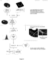
- Figure 1 represents a workflow of a method for digitizing an object according to the invention.
- a tomographic scanner (2) is calibrated by performing a scan (4) of a calibration object (3). From this scan one or more segmentation parameters (6) are automatically computed (5).
- Said calibration object (3) is specifically designed (10) for the object to be digitized (1) with the calibrated tomographic scanner (7).
- a calibrated segmentation (8) is performed on the scanned material to provide an accurate surface model (9) of said material.
- a calibration object (3) is designed.
- the material for the calibration object has similar material properties for the tomographic imaging method as the target material that needs to be digitized.
- the exact shape information (10), which can be similar to the shape of the real material that needs to be digitized, is known by design.
- the calibration object is scanned in the same way and with the same scanner as the target material is scanned (4).
- the parameters that generate the exact shape for a specific segmentation approach (6) are determined (5).
- the binary decision point where the exact shape of the scanned object is located is determined.
- an accuracy measure of the resulting segmentation can be computed (12).
- the actual material is scanned with the same scan protocol as the calibration scan (7).
- the segmentation algorithm is applied (8) with the determined parameters (6). In this way the exact shape of the material is obtained (9).
- the calibration scan can easily be redone with a regular frequency in time, or when changes or updates to the CT-scanning equipment, or to the materials used, occur. This method is fast and can be handled by the clinicians and their team themselves.
- segmentation of a surface out of a volumetric image volume is performed by thresholding.
- a threshold value defines the transition between the material and background, and hence the surface of the material.
- Figure 2 illustrates an algorithm for automatically computing the optimal threshold value or segmentation parameters (5).
- the algorithm requires two input data sets: the calibration object design (10) and the image volume(s) of the calibration object (11).
- the algorithm consists of three major steps: aligning the two input data sets (13-14), building a histogram (15-16) and finally deriving the optimal threshold value (17-20), i.e. the value of the segmentation parameter.
- Aligning is defined as searching a transformation so that the transformed object and the image volume share the same 3D space, and thus coincide. To obtain this alignment different procedures can be used.
- a possible approach is as follows. First, an image volume based on the calibration object design (10) is computed. Next this image volume is aligned with the image volume of the calibration object obtained through tomographic imaging (11). The outcome of this algorithm is a transformation which is then applied to the calibration object design (10) to obtain an aligned calibration object design (14). The aligned calibration object design (14) coincides with the image volume of the calibration object (11) in the same 3D space.
- the alignment can be done by voxel-based registration based on maximization of mutual information ( IEEE Transactions on Medical Imaging, 16(2):187-198, April 1997 ).
- a point based alignment method IEEE Transactions on Pattern Analysis and Machine Intelligence, 9(5), September 1987 . This point based alignment method first extracts well definable points or features on the calibration object design (10) and in the image volume of the calibration object (11). Next the method searches the transformation which aligns the corresponding 3D points of both data sets.
- the algorithm measures the image values in the image volume of the calibration object (11) at the surface of the aligned calibration object design (14). All measured image values are stored and a histogram of the stored image values (15) is built. To improve the stability of the algorithm the measure area can be extended towards a small region around the surface of the aligned calibration object design (14). In this way noise in the alignment algorithm or in the scanned data can be partially eliminated.
- the optimal threshold value (19) is derived (17) by using a selection criterion (18) in combination with the generated image values histogram (16).
- Possible selection criteria (18) are: mean image value, most frequent image value, maximum image value, etc. Different selection criteria may result in slightly different threshold values and the optimal selection criterion is dependent on the final application.
- a measure of the to-be-expected overall accuracy (20) of the segmentation can be obtained.
- a surface representation is generated out of the scanned image volume of the calibration object (11) using a marching cubes algorithm ( Proc. of SIGGRAPH, pp.163-169, 1987 ) and the derived optimal threshold value.
- This distance map or any statistical derived measure from this distance map represents the to-be-expected accuracy of the overall digitization procedure for the material to be digitized given the tomographic imaging method and equipment with the according imaging protocol.
- An alternative method for automatically computing the optimal threshold value comprises the steps of aligning the scanned calibration object and the virtual calibration object design, generating for any threshold value a distance map between the reconstructed surface of the scanned object and the virtual surface of the object design and deriving the optimal threshold value based on the calculated distance maps.
- the material to be digitized (1) is an acrylic dental prosthesis
- some specific guidelines can be considered when designing the calibration object (10).
- the volume of the designed object is preferably more or less equal to the volume of a typical dental prosthesis.
- the surface of the object contains sufficient detailed 3D information, i.e. shape variation, so that the accuracy of the algorithm can be guaranteed.
- the properties of the material used for the calibration object should be similar or equal to those of the material to be digitized for the specific tomographic imaging technique.
- the calibration object (10) can be designed as follows.
- the calibration object consists of a typical dental surface virtually mounted on a cylinder with a small height.
- the designed object is produced in polycarbonate which has similar radio-opacity characteristics as the acrylic materials used for producing dental prostheses ( Figure 3 ).
- An example of such polycarbonate is TECANATTM.
- Example 2 Design of calibration object (10) for a dental impression
- the calibration object In case the material to be digitized is a dental impression and the tomographic imaging method is CT scanning, some specific guidelines can be considered when designing the calibration object (10). First, it should be noted that many dental impression materials exist. All these materials have different radio-opacity characteristics. Therefore the designed calibration object should be usable for any of these dental impression materials. Second, it is preferred that the volume of the calibration object is more or less equal to the volume of a typical dental impression. Finally the calibration object preferably includes sufficient detailed 3D information, i.e. shape variation, so that the accuracy of the algorithm can be guaranteed. To meet these guidelines a calibration object can be produced and a special calibration procedure can be elaborated.
- the designed object (10) consists of two parts: a top part and a container part.
- the top part is a cubic shaped block with at the lower side a structure which resembles the upper dentition.
- the container part consists of two cavities.
- the size of the first cavity (C1 in Fig.4b ) is slightly larger than the top part.
- the second cavity (C2 in Fig.4b ) is slightly smaller than the top part. Due to the different sizes of the two cavities the top part can be placed on top of the container part in a well known position.
- the lower cavity C1 is filled with the impression material (see Fig.4b ).
- the top part is placed on the container and the two parts are pushed into tight contact with each other. After a few minutes when the impression material has hardened, the top part can be removed. The remaining part, i.e. the container part with the impression material, defines the final calibration object which will be scanned to obtain the image volume of the calibration object (11).
- the dentition surface at the lower side of the top part serves as the calibration object design (10).
Landscapes
- Engineering & Computer Science (AREA)
- Computer Vision & Pattern Recognition (AREA)
- Physics & Mathematics (AREA)
- General Physics & Mathematics (AREA)
- Theoretical Computer Science (AREA)
- Health & Medical Sciences (AREA)
- Oral & Maxillofacial Surgery (AREA)
- Dentistry (AREA)
- Epidemiology (AREA)
- Life Sciences & Earth Sciences (AREA)
- Animal Behavior & Ethology (AREA)
- General Health & Medical Sciences (AREA)
- Public Health (AREA)
- Veterinary Medicine (AREA)
- Apparatus For Radiation Diagnosis (AREA)
- Image Processing (AREA)
Priority Applications (2)
| Application Number | Priority Date | Filing Date | Title |
|---|---|---|---|
| EP14000057.1A EP2722818B1 (en) | 2009-09-04 | 2009-09-04 | Method for digitizing dento-maxillofacial objects |
| DK14000057.1T DK2722818T3 (da) | 2009-09-04 | 2009-09-04 | Fremgangsmåde til digitalisering af dento-maxillofaciale objekter |
Applications Claiming Priority (2)
| Application Number | Priority Date | Filing Date | Title |
|---|---|---|---|
| EP09169487.7A EP2306400B1 (en) | 2009-09-04 | 2009-09-04 | Method for digitizing dento-maxillofacial objects |
| EP14000057.1A EP2722818B1 (en) | 2009-09-04 | 2009-09-04 | Method for digitizing dento-maxillofacial objects |
Related Parent Applications (2)
| Application Number | Title | Priority Date | Filing Date |
|---|---|---|---|
| EP09169487.7A Division EP2306400B1 (en) | 2009-09-04 | 2009-09-04 | Method for digitizing dento-maxillofacial objects |
| EP09169487.7A Division-Into EP2306400B1 (en) | 2009-09-04 | 2009-09-04 | Method for digitizing dento-maxillofacial objects |
Publications (2)
| Publication Number | Publication Date |
|---|---|
| EP2722818A1 EP2722818A1 (en) | 2014-04-23 |
| EP2722818B1 true EP2722818B1 (en) | 2019-11-06 |
Family
ID=41666574
Family Applications (2)
| Application Number | Title | Priority Date | Filing Date |
|---|---|---|---|
| EP09169487.7A Active EP2306400B1 (en) | 2009-09-04 | 2009-09-04 | Method for digitizing dento-maxillofacial objects |
| EP14000057.1A Active EP2722818B1 (en) | 2009-09-04 | 2009-09-04 | Method for digitizing dento-maxillofacial objects |
Family Applications Before (1)
| Application Number | Title | Priority Date | Filing Date |
|---|---|---|---|
| EP09169487.7A Active EP2306400B1 (en) | 2009-09-04 | 2009-09-04 | Method for digitizing dento-maxillofacial objects |
Country Status (11)
| Country | Link |
|---|---|
| US (1) | US8824764B2 (enExample) |
| EP (2) | EP2306400B1 (enExample) |
| JP (1) | JP5696146B2 (enExample) |
| KR (1) | KR101710022B1 (enExample) |
| CN (1) | CN102576465B (enExample) |
| AU (1) | AU2010291554B2 (enExample) |
| BR (1) | BR112012004927B8 (enExample) |
| DK (1) | DK2722818T3 (enExample) |
| ES (1) | ES2536523T3 (enExample) |
| WO (1) | WO2011026609A1 (enExample) |
| ZA (1) | ZA201201072B (enExample) |
Families Citing this family (13)
| Publication number | Priority date | Publication date | Assignee | Title |
|---|---|---|---|---|
| KR20130132038A (ko) * | 2012-05-25 | 2013-12-04 | 이화여자대학교 산학협력단 | 맞춤형 치아 교정 시스템 및 그의 치아 교정 방법 |
| FI125322B (en) * | 2012-06-11 | 2015-08-31 | Planmeca Oy | Tooth Surface Models |
| GB201216230D0 (en) | 2012-09-12 | 2012-10-24 | Nobel Biocare Services Ag | An improved surgical template |
| GB201216214D0 (en) | 2012-09-12 | 2012-10-24 | Nobel Biocare Services Ag | A digital splint |
| GB201216224D0 (en) | 2012-09-12 | 2012-10-24 | Nobel Biocare Services Ag | An improved virtual splint |
| KR20180016406A (ko) * | 2015-06-09 | 2018-02-14 | 쳉싱 쉬 | 악안면 수술 영상 교정 설계 시스템 및 방법 |
| JP6707991B2 (ja) * | 2016-05-30 | 2020-06-10 | 富士通株式会社 | 歯軸推定プログラム、歯軸推定装置及びその方法、並びに歯形データ生成プログラム、歯形データ生成装置及びその方法 |
| CN107684463B (zh) * | 2016-08-03 | 2020-06-16 | 佛山市诺威科技有限公司 | 一种全冠桥连接体数字化生成方法 |
| US10368814B2 (en) * | 2016-12-30 | 2019-08-06 | Carestream Dental Technology Topco Limited | Method for cephalometric analysis |
| GB201708520D0 (en) | 2017-05-27 | 2017-07-12 | Dawood Andrew | A method for reducing artefact in intra oral scans |
| ES2807263T3 (es) * | 2017-06-30 | 2021-02-22 | Promaton Holding Bv | Clasificación y modelado 3D de estructuras dentomaxilofaciales 3D usando métodos de aprendizaje profundo |
| DK3666225T3 (da) | 2018-12-11 | 2022-09-12 | Sirona Dental Systems Gmbh | Fremgangsmåde til frembringelse af en grafisk gengivelse af en tandtilstand |
| JP7374193B2 (ja) | 2018-12-20 | 2023-11-06 | メディシム ナームロゼ ベンノートチャップ | 表面メッシュの自動トリミング |
Citations (1)
| Publication number | Priority date | Publication date | Assignee | Title |
|---|---|---|---|---|
| US20090212225A1 (en) * | 2008-01-24 | 2009-08-27 | Yiran Zheng | Methods for Positive Emission Tomography (PET) Target Image Segmentation |
Family Cites Families (23)
| Publication number | Priority date | Publication date | Assignee | Title |
|---|---|---|---|---|
| US4663720A (en) | 1984-02-21 | 1987-05-05 | Francois Duret | Method of and apparatus for making a prosthesis, especially a dental prosthesis |
| CN1068670A (zh) * | 1991-07-11 | 1993-02-03 | 中国人民解放军第四军医大学口腔医学院 | 用定位x线片重建活体颅骨立体形态方法 |
| US5360446A (en) | 1992-12-18 | 1994-11-01 | Zimmer, Inc. | Interactive prosthesis design system for implantable prosthesis |
| NL9301308A (nl) | 1993-07-26 | 1995-02-16 | Willem Frederick Van Nifterick | Werkwijze voor het vastzetten van een tandprothese op implantaten in het kaakbeen van een patiënt en middel te gebruiken daarbij. |
| SE502035C2 (sv) * | 1993-12-06 | 1995-07-24 | Nobelpharma Ab | Metod och och anordning för framtagning av information för framställning av artifiella stödorgan eller ersättningsdelar till människokroppen |
| BE1008372A3 (nl) | 1994-04-19 | 1996-04-02 | Materialise Nv | Werkwijze voor het vervaardigen van een geperfektioneerd medisch model uitgaande van digitale beeldinformatie van een lichaamsdeel. |
| SE503073C2 (sv) | 1994-07-04 | 1996-03-18 | Nobelpharma Ab | Metod för framställning av långsträckt stöddel i ersättningsuppbyggnad samt sådan stöddel tillverkad med metoden |
| US7331786B2 (en) | 1996-02-27 | 2008-02-19 | Technique D'usinage Sinlab Inc. | Manufacturing a dental implant drill guide and a dental implant superstructure |
| US5725376A (en) | 1996-02-27 | 1998-03-10 | Poirier; Michel | Methods for manufacturing a dental implant drill guide and a dental implant superstructure |
| US6409504B1 (en) | 1997-06-20 | 2002-06-25 | Align Technology, Inc. | Manipulating a digital dentition model to form models of individual dentition components |
| WO2000019929A1 (en) | 1998-10-08 | 2000-04-13 | Align Technology, Inc. | Computer automated development of an orthodontic treatment plan and appliance |
| DE19952962B4 (de) * | 1999-11-03 | 2004-07-01 | Sirona Dental Systems Gmbh | Verfahren zur Herstellung einer Bohrhilfe für ein Zahnimplantat |
| US7084868B2 (en) * | 2000-04-26 | 2006-08-01 | University Of Louisville Research Foundation, Inc. | System and method for 3-D digital reconstruction of an oral cavity from a sequence of 2-D images |
| EP1449489A4 (en) * | 2001-10-31 | 2009-03-11 | Imagnosis Inc | MEDICAL SIMULATION APPARATUS AND THREE-DIMENSIONAL IMAGE DISPLAY CONTROL METHOD INCLUDED IN THIS MEDICAL SIMULATION APPARATUS |
| JP4149189B2 (ja) * | 2002-04-04 | 2008-09-10 | 株式会社日立メディコ | X線ct装置 |
| US7004754B2 (en) * | 2003-07-23 | 2006-02-28 | Orametrix, Inc. | Automatic crown and gingiva detection from three-dimensional virtual model of teeth |
| JP2005040239A (ja) * | 2003-07-24 | 2005-02-17 | Univ Nihon | 画像処理方法、画像処理プログラム及びコンピュータ読取可能な記録媒体 |
| EP1624411A3 (en) * | 2004-08-06 | 2006-08-09 | Gendex Corporation | Soft tissue filtering in medical images |
| DK1808129T3 (en) * | 2004-09-24 | 2017-08-28 | Icat Corp | Device for detecting cross-sectional information |
| GB0507204D0 (en) | 2005-04-08 | 2005-05-18 | Leuven K U Res & Dev | Maxillofacial and plastic surgery |
| CN101292270B (zh) * | 2005-10-20 | 2012-12-05 | 皇家飞利浦电子股份有限公司 | 用于检查所关注对象的检查设备和方法 |
| US7844429B2 (en) * | 2006-07-19 | 2010-11-30 | Align Technology, Inc. | System and method for three-dimensional complete tooth modeling |
| EP1982652A1 (en) | 2007-04-20 | 2008-10-22 | Medicim NV | Method for deriving shape information |
-
2009
- 2009-09-04 ES ES09169487.7T patent/ES2536523T3/es active Active
- 2009-09-04 EP EP09169487.7A patent/EP2306400B1/en active Active
- 2009-09-04 DK DK14000057.1T patent/DK2722818T3/da active
- 2009-09-04 EP EP14000057.1A patent/EP2722818B1/en active Active
-
2010
- 2010-09-01 JP JP2012527229A patent/JP5696146B2/ja active Active
- 2010-09-01 BR BR112012004927A patent/BR112012004927B8/pt not_active IP Right Cessation
- 2010-09-01 CN CN201080038917.0A patent/CN102576465B/zh active Active
- 2010-09-01 AU AU2010291554A patent/AU2010291554B2/en not_active Ceased
- 2010-09-01 KR KR1020127008524A patent/KR101710022B1/ko active Active
- 2010-09-01 US US13/394,269 patent/US8824764B2/en active Active
- 2010-09-01 WO PCT/EP2010/005355 patent/WO2011026609A1/en not_active Ceased
-
2012
- 2012-02-14 ZA ZA2012/01072A patent/ZA201201072B/en unknown
Patent Citations (1)
| Publication number | Priority date | Publication date | Assignee | Title |
|---|---|---|---|---|
| US20090212225A1 (en) * | 2008-01-24 | 2009-08-27 | Yiran Zheng | Methods for Positive Emission Tomography (PET) Target Image Segmentation |
Also Published As
| Publication number | Publication date |
|---|---|
| EP2306400A1 (en) | 2011-04-06 |
| US8824764B2 (en) | 2014-09-02 |
| ES2536523T3 (es) | 2015-05-26 |
| JP2013503662A (ja) | 2013-02-04 |
| BR112012004927B8 (pt) | 2021-07-27 |
| DK2722818T3 (da) | 2020-01-06 |
| KR101710022B1 (ko) | 2017-03-08 |
| BR112012004927A2 (pt) | 2016-04-05 |
| AU2010291554B2 (en) | 2016-03-24 |
| BR112012004927B1 (pt) | 2020-10-13 |
| US20120201443A1 (en) | 2012-08-09 |
| AU2010291554A1 (en) | 2012-03-29 |
| ZA201201072B (en) | 2013-05-29 |
| EP2306400B1 (en) | 2015-02-11 |
| CN102576465B (zh) | 2015-02-11 |
| EP2722818A1 (en) | 2014-04-23 |
| KR20120065376A (ko) | 2012-06-20 |
| JP5696146B2 (ja) | 2015-04-08 |
| WO2011026609A1 (en) | 2011-03-10 |
| CN102576465A (zh) | 2012-07-11 |
Similar Documents
| Publication | Publication Date | Title |
|---|---|---|
| EP2722818B1 (en) | Method for digitizing dento-maxillofacial objects | |
| US12272067B2 (en) | Apparatuses and methods for three-dimensional dental segmentation using dental image data | |
| ES2627810T3 (es) | Método y sistema de análisis para el análisis geométrico de datos de exploración de estructuras orales | |
| EP2134290B1 (en) | Computer-assisted creation of a custom tooth set-up using facial analysis | |
| CN107595413B (zh) | 牙齿预备引导件 | |
| RU2605515C2 (ru) | Способ трехмерного проектирования лечения корневого канала зуба, устройство трехмерного проектирования лечения корневого канала зуба и компьютер | |
| Tilotta et al. | Construction and analysis of a head CT-scan database for craniofacial reconstruction | |
| US9036899B2 (en) | Method for quantifying local bone changes | |
| EP1486900A1 (en) | Method and system for manufacturing a surgical guide | |
| EP3108850A2 (en) | Generating a design for a dental restorative product from dental images | |
| CN106572831A (zh) | 口内扫描期间关注区域的识别 | |
| JP2018530372A (ja) | 歯科保存修復治療に用いるための歯の融通性のあるアーチモデルを作成する方法 | |
| Liao et al. | Anisotropic finite element modeling for patient-specific mandible | |
| Gupta | Challenges for computer aided diagnostics using X-ray and tomographic reconstruction images in craniofacial applications | |
| Chen et al. | Quantification of tooth displacement from cone-beam computed tomography images | |
| US20250143852A1 (en) | Method, device, storage medium, and medical system for generating a restoration dental model | |
| Barone et al. | Geometrical modeling of complete dental shapes by using panoramic X-ray, digital mouth data and anatomical templates | |
| Grzegorzek et al. | A multi-stage approach for 3D teeth segmentation from dentition surfaces | |
| CN115315229B (zh) | 牙科器具的数字上蜡 | |
| US12288291B2 (en) | Method and system for generation of a model for use in a virtual extraction procedure of a targeted extraction object in a patient | |
| Mahmood et al. | Three-dimensional cephalometric analysis of virtual dentoskeletal model | |
| El-Bialy | Towards a complete computer dental treatment system | |
| Yang et al. | Snake-based interactive tooth segmentation for 3D mandibular meshes | |
| Barone et al. | 3D reconstruction of individual tooth shapes by integrating dental cad templates and patient-specific anatomy | |
| Cronauer | A Finite Element Analysis of Maxillary Centers of Resistance |
Legal Events
| Date | Code | Title | Description |
|---|---|---|---|
| PUAI | Public reference made under article 153(3) epc to a published international application that has entered the european phase |
Free format text: ORIGINAL CODE: 0009012 |
|
| AC | Divisional application: reference to earlier application |
Ref document number: 2306400 Country of ref document: EP Kind code of ref document: P |
|
| AK | Designated contracting states |
Kind code of ref document: A1 Designated state(s): AT BE BG CH CY CZ DE DK EE ES FI FR GB GR HR HU IE IS IT LI LT LU LV MC MK MT NL NO PL PT RO SE SI SK SM TR |
|
| RIN1 | Information on inventor provided before grant (corrected) |
Inventor name: MOLLEMANS, WOUTER Inventor name: SCHUTYSER, FILIP Inventor name: WOUTERS, VEERLE |
|
| 17P | Request for examination filed |
Effective date: 20141023 |
|
| RBV | Designated contracting states (corrected) |
Designated state(s): AT BE BG CH CY CZ DE DK EE ES FI FR GB GR HR HU IE IS IT LI LT LU LV MC MK MT NL NO PL PT RO SE SI SK SM TR |
|
| STAA | Information on the status of an ep patent application or granted ep patent |
Free format text: STATUS: EXAMINATION IS IN PROGRESS |
|
| 17Q | First examination report despatched |
Effective date: 20180809 |
|
| REG | Reference to a national code |
Ref country code: DE Ref legal event code: R079 Ref document number: 602009060398 Country of ref document: DE Free format text: PREVIOUS MAIN CLASS: G06T0007200000 Ipc: G06T0007194000 |
|
| GRAP | Despatch of communication of intention to grant a patent |
Free format text: ORIGINAL CODE: EPIDOSNIGR1 |
|
| STAA | Information on the status of an ep patent application or granted ep patent |
Free format text: STATUS: GRANT OF PATENT IS INTENDED |
|
| RIC1 | Information provided on ipc code assigned before grant |
Ipc: A61C 9/00 20060101ALI20190409BHEP Ipc: G06T 7/136 20170101ALI20190409BHEP Ipc: G06T 7/155 20170101ALI20190409BHEP Ipc: G06T 7/194 20170101AFI20190409BHEP Ipc: G06T 7/11 20170101ALI20190409BHEP |
|
| INTG | Intention to grant announced |
Effective date: 20190506 |
|
| RIN1 | Information on inventor provided before grant (corrected) |
Inventor name: WOUTERS, VEERLE Inventor name: MOLLEMANS, WOUTER Inventor name: SCHUTYSER, FILIP |
|
| GRAS | Grant fee paid |
Free format text: ORIGINAL CODE: EPIDOSNIGR3 |
|
| GRAA | (expected) grant |
Free format text: ORIGINAL CODE: 0009210 |
|
| STAA | Information on the status of an ep patent application or granted ep patent |
Free format text: STATUS: THE PATENT HAS BEEN GRANTED |
|
| AC | Divisional application: reference to earlier application |
Ref document number: 2306400 Country of ref document: EP Kind code of ref document: P |
|
| AK | Designated contracting states |
Kind code of ref document: B1 Designated state(s): AT BE BG CH CY CZ DE DK EE ES FI FR GB GR HR HU IE IS IT LI LT LU LV MC MK MT NL NO PL PT RO SE SI SK SM TR |
|
| REG | Reference to a national code |
Ref country code: GB Ref legal event code: FG4D |
|
| REG | Reference to a national code |
Ref country code: CH Ref legal event code: EP Ref country code: AT Ref legal event code: REF Ref document number: 1199810 Country of ref document: AT Kind code of ref document: T Effective date: 20191115 |
|
| REG | Reference to a national code |
Ref country code: IE Ref legal event code: FG4D |
|
| REG | Reference to a national code |
Ref country code: DE Ref legal event code: R096 Ref document number: 602009060398 Country of ref document: DE |
|
| REG | Reference to a national code |
Ref country code: DK Ref legal event code: T3 Effective date: 20191218 |
|
| REG | Reference to a national code |
Ref country code: NL Ref legal event code: MP Effective date: 20191106 |
|
| REG | Reference to a national code |
Ref country code: LT Ref legal event code: MG4D |
|
| PG25 | Lapsed in a contracting state [announced via postgrant information from national office to epo] |
Ref country code: ES Free format text: LAPSE BECAUSE OF FAILURE TO SUBMIT A TRANSLATION OF THE DESCRIPTION OR TO PAY THE FEE WITHIN THE PRESCRIBED TIME-LIMIT Effective date: 20191106 Ref country code: PT Free format text: LAPSE BECAUSE OF FAILURE TO SUBMIT A TRANSLATION OF THE DESCRIPTION OR TO PAY THE FEE WITHIN THE PRESCRIBED TIME-LIMIT Effective date: 20200306 Ref country code: GR Free format text: LAPSE BECAUSE OF FAILURE TO SUBMIT A TRANSLATION OF THE DESCRIPTION OR TO PAY THE FEE WITHIN THE PRESCRIBED TIME-LIMIT Effective date: 20200207 Ref country code: NO Free format text: LAPSE BECAUSE OF FAILURE TO SUBMIT A TRANSLATION OF THE DESCRIPTION OR TO PAY THE FEE WITHIN THE PRESCRIBED TIME-LIMIT Effective date: 20200206 Ref country code: PL Free format text: LAPSE BECAUSE OF FAILURE TO SUBMIT A TRANSLATION OF THE DESCRIPTION OR TO PAY THE FEE WITHIN THE PRESCRIBED TIME-LIMIT Effective date: 20191106 Ref country code: LT Free format text: LAPSE BECAUSE OF FAILURE TO SUBMIT A TRANSLATION OF THE DESCRIPTION OR TO PAY THE FEE WITHIN THE PRESCRIBED TIME-LIMIT Effective date: 20191106 Ref country code: SE Free format text: LAPSE BECAUSE OF FAILURE TO SUBMIT A TRANSLATION OF THE DESCRIPTION OR TO PAY THE FEE WITHIN THE PRESCRIBED TIME-LIMIT Effective date: 20191106 Ref country code: LV Free format text: LAPSE BECAUSE OF FAILURE TO SUBMIT A TRANSLATION OF THE DESCRIPTION OR TO PAY THE FEE WITHIN THE PRESCRIBED TIME-LIMIT Effective date: 20191106 Ref country code: NL Free format text: LAPSE BECAUSE OF FAILURE TO SUBMIT A TRANSLATION OF THE DESCRIPTION OR TO PAY THE FEE WITHIN THE PRESCRIBED TIME-LIMIT Effective date: 20191106 Ref country code: FI Free format text: LAPSE BECAUSE OF FAILURE TO SUBMIT A TRANSLATION OF THE DESCRIPTION OR TO PAY THE FEE WITHIN THE PRESCRIBED TIME-LIMIT Effective date: 20191106 Ref country code: BG Free format text: LAPSE BECAUSE OF FAILURE TO SUBMIT A TRANSLATION OF THE DESCRIPTION OR TO PAY THE FEE WITHIN THE PRESCRIBED TIME-LIMIT Effective date: 20200206 |
|
| PG25 | Lapsed in a contracting state [announced via postgrant information from national office to epo] |
Ref country code: HR Free format text: LAPSE BECAUSE OF FAILURE TO SUBMIT A TRANSLATION OF THE DESCRIPTION OR TO PAY THE FEE WITHIN THE PRESCRIBED TIME-LIMIT Effective date: 20191106 Ref country code: IS Free format text: LAPSE BECAUSE OF FAILURE TO SUBMIT A TRANSLATION OF THE DESCRIPTION OR TO PAY THE FEE WITHIN THE PRESCRIBED TIME-LIMIT Effective date: 20200306 |
|
| PG25 | Lapsed in a contracting state [announced via postgrant information from national office to epo] |
Ref country code: EE Free format text: LAPSE BECAUSE OF FAILURE TO SUBMIT A TRANSLATION OF THE DESCRIPTION OR TO PAY THE FEE WITHIN THE PRESCRIBED TIME-LIMIT Effective date: 20191106 Ref country code: RO Free format text: LAPSE BECAUSE OF FAILURE TO SUBMIT A TRANSLATION OF THE DESCRIPTION OR TO PAY THE FEE WITHIN THE PRESCRIBED TIME-LIMIT Effective date: 20191106 Ref country code: CZ Free format text: LAPSE BECAUSE OF FAILURE TO SUBMIT A TRANSLATION OF THE DESCRIPTION OR TO PAY THE FEE WITHIN THE PRESCRIBED TIME-LIMIT Effective date: 20191106 |
|
| REG | Reference to a national code |
Ref country code: DE Ref legal event code: R097 Ref document number: 602009060398 Country of ref document: DE |
|
| REG | Reference to a national code |
Ref country code: AT Ref legal event code: MK05 Ref document number: 1199810 Country of ref document: AT Kind code of ref document: T Effective date: 20191106 |
|
| PG25 | Lapsed in a contracting state [announced via postgrant information from national office to epo] |
Ref country code: SM Free format text: LAPSE BECAUSE OF FAILURE TO SUBMIT A TRANSLATION OF THE DESCRIPTION OR TO PAY THE FEE WITHIN THE PRESCRIBED TIME-LIMIT Effective date: 20191106 Ref country code: SK Free format text: LAPSE BECAUSE OF FAILURE TO SUBMIT A TRANSLATION OF THE DESCRIPTION OR TO PAY THE FEE WITHIN THE PRESCRIBED TIME-LIMIT Effective date: 20191106 |
|
| PLBE | No opposition filed within time limit |
Free format text: ORIGINAL CODE: 0009261 |
|
| STAA | Information on the status of an ep patent application or granted ep patent |
Free format text: STATUS: NO OPPOSITION FILED WITHIN TIME LIMIT |
|
| 26N | No opposition filed |
Effective date: 20200807 |
|
| PG25 | Lapsed in a contracting state [announced via postgrant information from national office to epo] |
Ref country code: SI Free format text: LAPSE BECAUSE OF FAILURE TO SUBMIT A TRANSLATION OF THE DESCRIPTION OR TO PAY THE FEE WITHIN THE PRESCRIBED TIME-LIMIT Effective date: 20191106 Ref country code: AT Free format text: LAPSE BECAUSE OF FAILURE TO SUBMIT A TRANSLATION OF THE DESCRIPTION OR TO PAY THE FEE WITHIN THE PRESCRIBED TIME-LIMIT Effective date: 20191106 |
|
| PG25 | Lapsed in a contracting state [announced via postgrant information from national office to epo] |
Ref country code: MC Free format text: LAPSE BECAUSE OF FAILURE TO SUBMIT A TRANSLATION OF THE DESCRIPTION OR TO PAY THE FEE WITHIN THE PRESCRIBED TIME-LIMIT Effective date: 20191106 |
|
| PG25 | Lapsed in a contracting state [announced via postgrant information from national office to epo] |
Ref country code: LU Free format text: LAPSE BECAUSE OF NON-PAYMENT OF DUE FEES Effective date: 20200904 |
|
| PG25 | Lapsed in a contracting state [announced via postgrant information from national office to epo] |
Ref country code: IE Free format text: LAPSE BECAUSE OF NON-PAYMENT OF DUE FEES Effective date: 20200904 |
|
| PG25 | Lapsed in a contracting state [announced via postgrant information from national office to epo] |
Ref country code: TR Free format text: LAPSE BECAUSE OF FAILURE TO SUBMIT A TRANSLATION OF THE DESCRIPTION OR TO PAY THE FEE WITHIN THE PRESCRIBED TIME-LIMIT Effective date: 20191106 Ref country code: MT Free format text: LAPSE BECAUSE OF FAILURE TO SUBMIT A TRANSLATION OF THE DESCRIPTION OR TO PAY THE FEE WITHIN THE PRESCRIBED TIME-LIMIT Effective date: 20191106 Ref country code: CY Free format text: LAPSE BECAUSE OF FAILURE TO SUBMIT A TRANSLATION OF THE DESCRIPTION OR TO PAY THE FEE WITHIN THE PRESCRIBED TIME-LIMIT Effective date: 20191106 |
|
| PG25 | Lapsed in a contracting state [announced via postgrant information from national office to epo] |
Ref country code: MK Free format text: LAPSE BECAUSE OF FAILURE TO SUBMIT A TRANSLATION OF THE DESCRIPTION OR TO PAY THE FEE WITHIN THE PRESCRIBED TIME-LIMIT Effective date: 20191106 |
|
| PGFP | Annual fee paid to national office [announced via postgrant information from national office to epo] |
Ref country code: DK Payment date: 20220909 Year of fee payment: 14 |
|
| PGFP | Annual fee paid to national office [announced via postgrant information from national office to epo] |
Ref country code: BE Payment date: 20220823 Year of fee payment: 14 |
|
| PGFP | Annual fee paid to national office [announced via postgrant information from national office to epo] |
Ref country code: CH Payment date: 20221001 Year of fee payment: 14 |
|
| P01 | Opt-out of the competence of the unified patent court (upc) registered |
Effective date: 20230515 |
|
| PGFP | Annual fee paid to national office [announced via postgrant information from national office to epo] |
Ref country code: IT Payment date: 20230810 Year of fee payment: 15 |
|
| REG | Reference to a national code |
Ref country code: DK Ref legal event code: EBP Effective date: 20230930 |
|
| REG | Reference to a national code |
Ref country code: CH Ref legal event code: PL |
|
| REG | Reference to a national code |
Ref country code: BE Ref legal event code: MM Effective date: 20230930 |
|
| PG25 | Lapsed in a contracting state [announced via postgrant information from national office to epo] |
Ref country code: CH Free format text: LAPSE BECAUSE OF NON-PAYMENT OF DUE FEES Effective date: 20230930 |
|
| PG25 | Lapsed in a contracting state [announced via postgrant information from national office to epo] |
Ref country code: CH Free format text: LAPSE BECAUSE OF NON-PAYMENT OF DUE FEES Effective date: 20230930 |
|
| PG25 | Lapsed in a contracting state [announced via postgrant information from national office to epo] |
Ref country code: BE Free format text: LAPSE BECAUSE OF NON-PAYMENT OF DUE FEES Effective date: 20230930 |
|
| PG25 | Lapsed in a contracting state [announced via postgrant information from national office to epo] |
Ref country code: DK Free format text: LAPSE BECAUSE OF NON-PAYMENT OF DUE FEES Effective date: 20230930 |
|
| PG25 | Lapsed in a contracting state [announced via postgrant information from national office to epo] |
Ref country code: DK Free format text: LAPSE BECAUSE OF NON-PAYMENT OF DUE FEES Effective date: 20230930 |
|
| PG25 | Lapsed in a contracting state [announced via postgrant information from national office to epo] |
Ref country code: IT Free format text: LAPSE BECAUSE OF NON-PAYMENT OF DUE FEES Effective date: 20240904 |
|
| PGFP | Annual fee paid to national office [announced via postgrant information from national office to epo] |
Ref country code: DE Payment date: 20250702 Year of fee payment: 17 |
|
| PGFP | Annual fee paid to national office [announced via postgrant information from national office to epo] |
Ref country code: GB Payment date: 20250703 Year of fee payment: 17 |
|
| PGFP | Annual fee paid to national office [announced via postgrant information from national office to epo] |
Ref country code: FR Payment date: 20250703 Year of fee payment: 17 |