EP2694115B1 - Sonden für optische bildgebung - Google Patents
Sonden für optische bildgebung Download PDFInfo
- Publication number
- EP2694115B1 EP2694115B1 EP12722474.9A EP12722474A EP2694115B1 EP 2694115 B1 EP2694115 B1 EP 2694115B1 EP 12722474 A EP12722474 A EP 12722474A EP 2694115 B1 EP2694115 B1 EP 2694115B1
- Authority
- EP
- European Patent Office
- Prior art keywords
- dye construct
- dye
- fluorescence
- construct
- activated
- Prior art date
- Legal status (The legal status is an assumption and is not a legal conclusion. Google has not performed a legal analysis and makes no representation as to the accuracy of the status listed.)
- Active
Links
Images







Classifications
-
- A—HUMAN NECESSITIES
- A61—MEDICAL OR VETERINARY SCIENCE; HYGIENE
- A61K—PREPARATIONS FOR MEDICAL, DENTAL OR TOILETRY PURPOSES
- A61K49/00—Preparations for testing in vivo
- A61K49/001—Preparation for luminescence or biological staining
- A61K49/0013—Luminescence
- A61K49/0017—Fluorescence in vivo
- A61K49/0019—Fluorescence in vivo characterised by the fluorescent group, e.g. oligomeric, polymeric or dendritic molecules
- A61K49/0021—Fluorescence in vivo characterised by the fluorescent group, e.g. oligomeric, polymeric or dendritic molecules the fluorescent group being a small organic molecule
- A61K49/0041—Xanthene dyes, used in vivo, e.g. administered to a mice, e.g. rhodamines, rose Bengal
- A61K49/0043—Fluorescein, used in vivo
-
- A—HUMAN NECESSITIES
- A61—MEDICAL OR VETERINARY SCIENCE; HYGIENE
- A61K—PREPARATIONS FOR MEDICAL, DENTAL OR TOILETRY PURPOSES
- A61K49/00—Preparations for testing in vivo
- A61K49/001—Preparation for luminescence or biological staining
- A61K49/0013—Luminescence
- A61K49/0017—Fluorescence in vivo
- A61K49/0019—Fluorescence in vivo characterised by the fluorescent group, e.g. oligomeric, polymeric or dendritic molecules
- A61K49/0021—Fluorescence in vivo characterised by the fluorescent group, e.g. oligomeric, polymeric or dendritic molecules the fluorescent group being a small organic molecule
- A61K49/0041—Xanthene dyes, used in vivo, e.g. administered to a mice, e.g. rhodamines, rose Bengal
-
- A—HUMAN NECESSITIES
- A61—MEDICAL OR VETERINARY SCIENCE; HYGIENE
- A61K—PREPARATIONS FOR MEDICAL, DENTAL OR TOILETRY PURPOSES
- A61K49/00—Preparations for testing in vivo
- A61K49/001—Preparation for luminescence or biological staining
- A61K49/0013—Luminescence
- A61K49/0017—Fluorescence in vivo
- A61K49/005—Fluorescence in vivo characterised by the carrier molecule carrying the fluorescent agent
- A61K49/0054—Macromolecular compounds, i.e. oligomers, polymers, dendrimers
-
- A—HUMAN NECESSITIES
- A61—MEDICAL OR VETERINARY SCIENCE; HYGIENE
- A61K—PREPARATIONS FOR MEDICAL, DENTAL OR TOILETRY PURPOSES
- A61K49/00—Preparations for testing in vivo
- A61K49/001—Preparation for luminescence or biological staining
- A61K49/0013—Luminescence
- A61K49/0017—Fluorescence in vivo
- A61K49/005—Fluorescence in vivo characterised by the carrier molecule carrying the fluorescent agent
- A61K49/0056—Peptides, proteins, polyamino acids
-
- A—HUMAN NECESSITIES
- A61—MEDICAL OR VETERINARY SCIENCE; HYGIENE
- A61K—PREPARATIONS FOR MEDICAL, DENTAL OR TOILETRY PURPOSES
- A61K49/00—Preparations for testing in vivo
- A61K49/001—Preparation for luminescence or biological staining
- A61K49/006—Biological staining of tissues in vivo, e.g. methylene blue or toluidine blue O administered in the buccal area to detect epithelial cancer cells, dyes used for delineating tissues during surgery
-
- G—PHYSICS
- G01—MEASURING; TESTING
- G01N—INVESTIGATING OR ANALYSING MATERIALS BY DETERMINING THEIR CHEMICAL OR PHYSICAL PROPERTIES
- G01N21/00—Investigating or analysing materials by the use of optical means, i.e. using sub-millimetre waves, infrared, visible or ultraviolet light
- G01N21/62—Systems in which the material investigated is excited whereby it emits light or causes a change in wavelength of the incident light
- G01N21/63—Systems in which the material investigated is excited whereby it emits light or causes a change in wavelength of the incident light optically excited
- G01N21/64—Fluorescence; Phosphorescence
- G01N21/6428—Measuring fluorescence of fluorescent products of reactions or of fluorochrome labelled reactive substances, e.g. measuring quenching effects, using measuring "optrodes"
-
- G—PHYSICS
- G01—MEASURING; TESTING
- G01N—INVESTIGATING OR ANALYSING MATERIALS BY DETERMINING THEIR CHEMICAL OR PHYSICAL PROPERTIES
- G01N21/00—Investigating or analysing materials by the use of optical means, i.e. using sub-millimetre waves, infrared, visible or ultraviolet light
- G01N21/62—Systems in which the material investigated is excited whereby it emits light or causes a change in wavelength of the incident light
- G01N21/63—Systems in which the material investigated is excited whereby it emits light or causes a change in wavelength of the incident light optically excited
- G01N21/64—Fluorescence; Phosphorescence
- G01N21/645—Specially adapted constructive features of fluorimeters
- G01N21/6456—Spatial resolved fluorescence measurements; Imaging
- G01N21/6458—Fluorescence microscopy
-
- G—PHYSICS
- G01—MEASURING; TESTING
- G01N—INVESTIGATING OR ANALYSING MATERIALS BY DETERMINING THEIR CHEMICAL OR PHYSICAL PROPERTIES
- G01N21/00—Investigating or analysing materials by the use of optical means, i.e. using sub-millimetre waves, infrared, visible or ultraviolet light
- G01N21/62—Systems in which the material investigated is excited whereby it emits light or causes a change in wavelength of the incident light
- G01N21/63—Systems in which the material investigated is excited whereby it emits light or causes a change in wavelength of the incident light optically excited
- G01N21/64—Fluorescence; Phosphorescence
- G01N21/6428—Measuring fluorescence of fluorescent products of reactions or of fluorochrome labelled reactive substances, e.g. measuring quenching effects, using measuring "optrodes"
- G01N2021/6432—Quenching
-
- G—PHYSICS
- G01—MEASURING; TESTING
- G01N—INVESTIGATING OR ANALYSING MATERIALS BY DETERMINING THEIR CHEMICAL OR PHYSICAL PROPERTIES
- G01N21/00—Investigating or analysing materials by the use of optical means, i.e. using sub-millimetre waves, infrared, visible or ultraviolet light
- G01N21/62—Systems in which the material investigated is excited whereby it emits light or causes a change in wavelength of the incident light
- G01N21/63—Systems in which the material investigated is excited whereby it emits light or causes a change in wavelength of the incident light optically excited
- G01N21/64—Fluorescence; Phosphorescence
- G01N21/6428—Measuring fluorescence of fluorescent products of reactions or of fluorochrome labelled reactive substances, e.g. measuring quenching effects, using measuring "optrodes"
- G01N2021/6439—Measuring fluorescence of fluorescent products of reactions or of fluorochrome labelled reactive substances, e.g. measuring quenching effects, using measuring "optrodes" with indicators, stains, dyes, tags, labels, marks
-
- G—PHYSICS
- G01—MEASURING; TESTING
- G01N—INVESTIGATING OR ANALYSING MATERIALS BY DETERMINING THEIR CHEMICAL OR PHYSICAL PROPERTIES
- G01N21/00—Investigating or analysing materials by the use of optical means, i.e. using sub-millimetre waves, infrared, visible or ultraviolet light
- G01N21/62—Systems in which the material investigated is excited whereby it emits light or causes a change in wavelength of the incident light
- G01N21/63—Systems in which the material investigated is excited whereby it emits light or causes a change in wavelength of the incident light optically excited
- G01N21/64—Fluorescence; Phosphorescence
- G01N21/6428—Measuring fluorescence of fluorescent products of reactions or of fluorochrome labelled reactive substances, e.g. measuring quenching effects, using measuring "optrodes"
- G01N2021/6439—Measuring fluorescence of fluorescent products of reactions or of fluorochrome labelled reactive substances, e.g. measuring quenching effects, using measuring "optrodes" with indicators, stains, dyes, tags, labels, marks
- G01N2021/6441—Measuring fluorescence of fluorescent products of reactions or of fluorochrome labelled reactive substances, e.g. measuring quenching effects, using measuring "optrodes" with indicators, stains, dyes, tags, labels, marks with two or more labels
Definitions
- the present invention relates to methods of imaging activated neutrophil cells in vivo using a dendrimer-dye molecule or polybranched-dye molecule which is internalised by the cells and thus permits subsequent visualisation by confocal fluorescence endomicroscopy.
- the cells are activated neutrophils within the lung of a subject.
- Neutrophil predominant lung inflammation is a major cause of morbidity and mortality 11 . Yet despite decades of investigation, accurate stratification of patients with neutrophil predominant lung injury on intensive care has been hindered by the lack of bedside point of care diagnostics that can reliably and rapidly distinguish acute neutrophilic inflammation 2 . The ability to perform bedside diagnostics has the potential to accurately stratify such patients for further neutrophil specific interventions. Excessive neutrophil activity degrades matrix and cellular receptors, activates profibrogenic mediators and contributes to epithelial and endothelial cell damage 3,4,5 . The involvement of neutrophils in several diseases such as acute lung injury, ischemia-reperfusion injury 6,7 , cystic fibrosis 8 and chronic obstructive pulmonary disease 9 , makes them important targets for modulation.
- PET imaging 10 Although offering tiny sensitivity, is cumbersome, expensive and is difficult to implement as a bedside molecular imaging modality.
- confocal endoscopy such as probe based confocal laser endoscopy has revolutionised the ability to directly visualise the alveolar space in both preclinical and clinical arenas.
- this modality has only been used to image autofluorescent structures within the alveolar space or using non-specific fluorescent dyes 11,12 .
- the optical detection of activated neutrophilic activity is feasible with imaging enzymatic activity in whole animals 13 .
- These approaches require substrate specificity with internally quenched molecular beacons. Often the dequenching may take hours and the substrate may be cleaved by non neutrophil proteases.
- Dendrimers are a class of macromolecules possessing a well-defined structure and molecular composition 1415 .
- branches are created by the stepwise attachment of monomer units in repeating unit layers, termed generations, which creates branches built upon a central core. These branches terminate in a specific chemical functional group that can be used for further dendrimer growth or modification, or attachment of specific compounds as required.
- WO2003014743 describes the use of dendrimers and polybranched molecules to enhance signals in in vitro fluorescent assay systems 16 .
- WO2003014743 comprise cleavage sites which when treated with an appropriate chemical or enzyme lead to cleavage of selective bonds within the molecules and a subsequent change in the fluorescent properties of the molecule, most notably an increase in fluorescence.
- WO2003014743 only shows the results of in vitro data and there is no suggestion or teaching of how one might use the molecules in an in vivo setting, or indeed if this would in fact be possible.
- the dye construct is a poly-branched molecule with surface groups linked to a fluorescent dye, as further described herein.
- Detectable increase in fluorescence is understood to relate to fluorescence which can be detected by confocal microendoscopy techniques. If the dye constructs initially display a low, but detectable level of fluorescence, then a detectable increase can be observed following internalisation by an activated neutrophil cell or cells. Initially the dye constructs for use in the present invention are internally quenched. That is, the dye constructs do not fluoresce or fluoresce poorly due to the fluorescent groups or groups being quenched. However, following internalisation by the cells, dequenching occurs and an increase in fluorescence can be detected.
- fluorescence detection is understood to be related to fluorescence intensity, fluorescence lifetime and polarisation may also be detected.
- the "low or little" amount of fluorescence” is practically not detectable using confocal microendoscopy techniques, or is sufficiently low to permit a clear identification of the "increase” in fluorescence.
- a suitable increase is understood to be an increase by a factor of 1.2 or more.
- the present inventors have observed that combining confocal endoscopic visualisation techniques with the localised administration of a dendrimer dye molecule or poly-branched molecule linked fluorescent dye, it is possible to observe in vivo, by way of fluorescence, specific activated neutrophil cells which internalise a dye construct of the present invention or poly-branched molecule linked fluorescent dye, with a distinct increase in fluorescence. Without wishing to be bound by theory, it is thought that the dye contructs/polybranched-dye molecules of the present invention are internalised or taken up by activated neutrophil cells before internal cellular mechanisms act upon the constructs causing an increase in fluorescence.
- the present inventors have observed that through the use of the dye construct/poly-branched molecules described herein, that such molecules are capable of being internalised, by activated neutrophil cells within seconds or a few minutes.
- the detection of fluorescence should be carried out within a few minutes of administering the dye construct - typically within seconds to minutes, such as 1-30 minutes, typically 1-10 minutes.
- the confocal endoscope or microendoscope enables real- time in vivo human and animal imaging.
- the instrument couples a custom built fluorescence slit-scan confocal microscope to a fibre-optic catheter. Further teaching may be found in Thiberville et al 17 and WO2008020130 , to which the skilled reader is directed and the contents of which are hereby incorporated by way of reference.
- Activated neutrophils which are characterised by degranulation and protease release may be found at sites of inflammation and may therefore be detected using confocal endoscopy techniques at a variety of locations within the body of a subject such as within in the lung, within the gastrointestinal tract, within the reproductive tract or any other endoscopically accessible orifice.
- activated neutrophils are detected in the lung of a subject.
- the subject may be a subject already hospitalised, such as a patient in intensive care, where early detection of such activated neutrophils would be desirable.
- the dye construct or poly-branched molecule linked to fluorescent dye as described herein comprise one or more cleavage sites which are cleavable by appropriate chemical or enzyme means.
- Preferred molecules comprise three or more, typically six or more branches, such that a significant increase in fluorescence may be observed following dequenching of the fluorescent moieties.
- a peptide and/or polyethylene glycol (PEG) portion is intended to improve the solubility of the dendrimer/branched molecules.
- the peptide sequence comprises an enzyme cleavage recognition sequence. or may be random in the sense of not including a recognisable enzyme or chemical cleavage recognition sequence.
- the peptide sequence is not thought to be cleaved by an enzyme present in the cell to be detected.
- the increase in fluorescence observed following internalisation of the molecules of the present invention comprising random peptide sequences is not thought to be due to cleavage of the peptide moiety and release of previously quenched fluorescent moieties, in contrast to previous teachings.
- the peptide sequence is cleaved by an enzyme which may be present outside of the cell and this may result in a low amount of fluorescence being observed.
- a far greater observable increase in fluorescence is observed upon internalisation of the molecules/probes.
- a detectable increase is observed (i.e. greater than a factor of 1.2 as compared to any fluorescence which is observed outside of the cell) when the construct in internalised. This may in fact be an advantage, as it may allow cells to be generally identified by way of a level of fluorescence being observed outside any cells, but desired cells can more easily be identified once the constructs are internalised and an increase in fluorescence observed.
- Dye construct molecules of the present invention include one of the following structures: or wherein:
- the molecules of the present invention may comprise a peptide linkage, represented in the above structures as (AA)n, where AA means any amino acid and n may be zero or is a positive integer from 1-30 such as 1-20, or 1-15.
- AA means any amino acid and n may be zero or is a positive integer from 1-30 such as 1-20, or 1-15.
- Such peptide sequences may therefore be random sequences, or conform to known sequences contained within peptides or proteins. Sequences which are recognised by the enzyme neutrophil elastase include A-A-P-V, A-A-A-P-V-K, E-E-I-Nle-R-R.
- Gnf nonproteinogenic 4-guanidine-I-phenylalanine
- D-C-V-D for Caspase
- the molecules of the present invention are initially quenched, that is they display little or no fluorescence in terms of fluorescence which may be detected from the fluorescent moiety following appropriate excitation. However, following internalisation of the molecules by an activated neutrophil cell or cells to be detected, a de-quenching of the molecules occurs and an increase in fluorescent signal, following excitation using light of a suitable wavelength, can be detected 18 .
- the present inventors have observed that certain molecules of the present invention which do not have recognisably cleavable peptide sequences are nevertheless internalised by activated neutrophils and a de-quenching i.e. increase in fluorescence can be observed. Without wishing to be bound by theory, it is thought that the probes may be internalised into acidified vacuolar structures that directly effect internal quenching efficiency.
- the molecules of the present invention do not necessarily have to possess recognisable enzyme cleavable peptide sequences in order to visualise activated neutrophils.
- the present inventors are able through confocal endoscopy to visualise cells in situ in vivo.
- in vivo is to be understood to relate to cells within the living body and hence is to be distinguished from visualising cells obtained from tissue samples which have been extracted or excised from the body.
- the present methods may be conducted on or within any organ into which an endomicroscopic catheter may be inserted. This may be, for example, the gut including the large and small intestine; arteries and veins; the respiratory system including the lungs, the brain such as via an intracranial catheter; and the reproductive system including the womb and fallopian tubes.
- the methods of the present invention may be carried out whilst visualising cells in the lung, such as in the alveolar space.
- the present inventors have observed that through the use of the dye constructs/poly-branched molecules described herein, that such molecules are capable of being internalised, by activated neutrophils.
- Such internalisation by activated neutrophils occurs very rapidly, within a few seconds or minutes and as such activated neutrophils may be visualised within 1-30 minutes, typically 1-10 minutes of the molecules of the present invention being administered to the subject at the site of investigation, such as within the lung.
- the methods of detection as described herein should typically be conducted within a short period of time, following local administration of the initially quenched molecules, typically within a few minutes of administration, so that only cells, such as activated neutrophils, which internalise or take up the molecules of the present invention rapidly, are detected.
- microdosed amounts typically less than 100 ⁇ g
- 50 ⁇ g or even 10 ⁇ g or less of the dye construct which has been administered This is particularly advantageous in terms of certain possible regulatory issues concerning the use of larger quantities and toxicity concerns when administering any exogenous molecule - although the molecules of the present invention may not in fact be significantly toxic in any case. It is in fact particularly surprising that such low microdose amounts of molecule when administered are capable of eliciting a signal, which is detectable using microendoscopy techniques.
- the present invention enables use of a dendrimer dye molecule or poly-branched molecule linked fluorescent dye of the present invention in an amount of less than 100 ⁇ g, 50 ⁇ g, or 10 ⁇ g or less, for visualising cells in vivo using confocal endoscopy.
- the dye construct may be provided by a catheter or other suitable administration device comprising a microdose (i.e. less than 100 ⁇ g, 50 ⁇ g, or 10 ⁇ g or less) amount of a dye construct or poly-branched molecule linked fluorescent dye of the present invention, for administration to a subject, such that an activated neutrophil cell or cells is capable of being visualised by confocal endoscopy.
- a microdose i.e. less than 100 ⁇ g, 50 ⁇ g, or 10 ⁇ g or less
- an activated neutrophil cell or cells is capable of being visualised by confocal endoscopy.
- One potential advantage of the present invention is that it may be carried out on subjects who are being given respiratory support in terms of being administered oxygen or air and who as such may have a face mask covering their mouth and/or nose or who are being intubated. Even in such a situation, it is possible to insert a confocal microendoscopy catheter into the lungs through the nasal passage or via the endotracheal tube. In this manner the present invention can truly be carried out at the bedside, without necessarily having to move the subject unduly. Moreover, for such subjects, the ability to detect any activated neutrophils, which are a key marker of an inflammatory response, is of paramount importance and as such the present invention may find particular use in being conducted on such ill patients where moving them to another location may be undesirable and/or problematic.
- RP-HPLC Semi-preparative RP-HPLC was performed on an HP1100 system equipped with a Phenomenex Prodigy C18 reverse-phase column (250 x 10 mm, 5 ⁇ m) with a flow rate of 2.5 ml/min and eluting with 0.1% HCOOH in H 2 O (A) and 0.1% HCOOH in CH 3 CN (B), with a gradient of 5 to 95% B over 18 min and an initial isocratic period of 5 min.
- Electrospray ionization mass spectrometry (ESI-MS) analyses were carried out on an Agilent Technologies LC/MSD Series 1100 quadrupole mass spectrometer (QMS) in an ESI mode.
- MALDI spectra were acquired on a Voyager-DE TM STR MALDI-TOF MS (Applied Biosystems) with a matrix solution of sinapinic acid (10 mg/ml) in 50% MeCN in water with 0.1% TFA.
- the monomer 6 was synthesised in six steps 19 as shown in Scheme 1.
- the 1,4 addition of the hydroxy groups of 1,1,1- tris (hydroxymethyl)amino-methane onto acrylonitrile, followed by amino protection (Boc), and reduction of the nitrile groups with borane-THF complex gave 3.
- the isocyanate 6 was prepared following the procedure of Knölker 20 .
- the crude product (3) (4.5 g, 11.4 mmol) was dissolved in methanol (75 ml) and DIPEA (2.4 ml, 13.68 mmol) was added.
- DIPEA 2.4 ml, 13.68 mmol
- the protected amine (4) (2.3 g, 2.7 mmol) was dissolved in 20% TFA in dichloromethane (40 ml) and the resulting mixture was stirred for 2 h. The solvent was removed in vacuo and the residue was dissolved in dichloromethane (150 ml) and washed with saturated aqueous NaHCO 3 solution (75 ml) and water (75 ml). The organic layer was dried with Na 2 SO 4 and the solvents removed in vacuo.
- Monomer (12) used in the preparation of 6-branched dendrimer was synthesised in 5 steps using ⁇ -resorcylic acid as a starting material (Scheme 2). Esterification of (7) in methanol provides methyl benzoate (8), which then undergoes alkylation with 2-(Boc-amino)ethyl bromide in the presence of potassium carbonate in DMF to yield (9). The latter was then subjected to saponification of the methyl ester by using NaOH/MeOH/dioxane mixture, followed by removal of the Boc protective group using HCl to give amine as the hydrochloride salt (11). Subsequently, the amine salt was selectively protected with FmocOSu to give the branching monomer (12), which was utilised in the Fmoc-based solid support synthesis.
- Hydrochloride salt (11) (717 mg, 2.2 mmol) was dissolved in acetone:water (1:1, 100 ml) containing sodium carbonate (950 mg, 9 mmol). To this solution was added Fmoc-Osu (1.57 g, 4.6 mmol) in acetone (25 ml) dropwise at room temperature. The solution was stirred at room temperature for 18 h. The reaction mixture was concentrated and the residue dissolved in water and extracted with ether (2 ⁇ 50 ml). The aqueous layer was cooled in an ice bath and acidified with 2M HCl to pH3. The white solid (12) obtained was filtered, washed with water and dried under vacuum (1.15 g, 77%); m.p.
- Resin 13 was synthesized using a 4-[(2,4-dimethoxyphenyl)-(Fmoc-amino)methyl]phenoxyacetic acid (Rink amide linker) attached to aminomethyl PS resin (1.6 mmol/g, 1% DVB, 100-200 mesh).
- Rink amide linker 4-[(2,4-dimethoxyphenyl)-(Fmoc-amino)methyl]phenoxyacetic acid (Rink amide linker) attached to aminomethyl PS resin (1.6 mmol/g, 1% DVB, 100-200 mesh).
- Fmoc-Rink-amide linker 2.6 g, 4.8 mmol
- HOBt 0.7 g, 4.8 mmol
- DIC 0.7 ml, 4.8 mmol
- Human peripheral blood leukocytes from healthy volunteers were prepared as previously described 25 . Briefly, citrated blood was centrifuged at room temperature for 20 min at 350 g, and platelet-rich plasma was removed. Autologous serum was prepared by recalcification of platelet-rich plasma by addition of CaCl 2 to a final concentration of 2 mM. Leukocytes were separated from erythrocytes by dextran sedimentation using 0.6% dextran T500 (Pharmacia, Milton Keynes, UK), and the leukocyte-rich upper layer was then fractionated using isotonic Percoll (Pharmacia). Neutrophils and mononuclear leukocytes (PBMC) were harvested from the 68%/81% and 55/68% interfaces, respectively. In some experiments, neutrophils were labelled with DiD (Invitrogen, molecular probes) (2.5 ⁇ M) in D-PBS (w/o Ca/Mg) for 20 min at room temperature.
- DiD Invitrogen, molecular probes
- PBMC-derived macrophages were generated from mononuclear leukocytes as follows ( Rossi AG, McCutcheon JC, Roy N, Chilvers ER, Haslett C, Dransfield I. Regulation of macrophage phagocytosis of apoptotic cells by cAMP. J Immunol. 1998 Apr 1;160(7):3562-8 .Regulation of macrophage phagocytosis of apoptotic cells by cAMP). Mononuclear leukocytes were allowed to adhere to poly-d-lysine coated coverslips for 1 hour before washing to remove non-adherent cells. Adherent cells were cultured for 7-10 days in vitro (DIV) in IMDM containing 10% autologous serum.
- DIV vitro
- A549 cells (ATCC CCL-185) were maintained at 80-90 % confluence in culture medium which consisted of Dulbecco's modified Eagle's medium (4.5 g.l -1 glucose) supplemented with 10 % (v/v) heat inactivated fetal calf serum (FCS), L-glutamine (2mM), streptomycin (50 ⁇ g.ml -1 ) and penicillin (50 units.ml -1 ) in a thermostatted (37 °C) and humidified atmosphere of 5 % CO 2 / 95 % air. Suspensions of exponentially growing cells (2 ⁇ 10 6 cells), detached following trypsin/EDTA exposure were then seeded onto coverslips or 6 well microplates and grown to confluence over 24-48 h.
- Bronchoalveolar lavage was obtained from patients with Idiopathic Pulmonary Fibrosis (IPF). Written informed consent was obtained from all subjects. The study was approved by the Lothian Research Ethics Committee. BAL was performed as part of ongoing clinical research studies. 200 ml of saline was instilled into the right middle lobe and lavaged in 20 ml aliquots. Samples were kept on ice and 100 ⁇ l aliquots pipetted onto coverslips followed by immediate staining as detailed and live confocal imaging in a POC mini (perfusion open and closed) (PeCon GmbH) cultivation chamber.
- POC mini perfusion open and closed
- a laser-scanning confocal imaging system (LSM510; Carl Zeiss, Jena, Germany), incorporating an upright Axioskop FS2 microscope (63 ⁇ objective) was used for image acquisition and processing. Exposure to 488 nm light was limited to 1-6% of the maximum laser power in order to minimize photobleaching and toxicity. In all cases, images were obtained without Kalman averaging and typically with a pixel dwell time of 3.2 ⁇ s with a pinhole diameter corresponding to 1 Airy unit. In multiple-labelling experiments pinhole diameters were adjusted to give optical Z-sections of equivalent depths, corresponding to 1 Airy unit for the longest excitation wavelength. All live time-lapse imaging of was performed in IMDM.
- Neutrophils and mononuclear cells (6 ⁇ 10 5 cells total), or PBMC-derived macrophages, were seeded onto glass coverslips pre-coated with 10 ⁇ g/ml fibronectin (Sigma). Cells were allowed to adhere and 10 ⁇ M (final concentration) IQR1.2 added to wells prior to transfer to POC mini and placement in environmental chamber. Live imaging commenced at 30 s intervals. Baseline images were acquired for 10-15 min prior to addition of A21387 (10 ⁇ M, Sigma) and live time-lapse confocal images acquired for further 20 min. Where sivelestat or dynasore was included, cells were pretreated for 10 minutes prior to imaging.
- A549 cells grown to confluence (>80%) in 6 well plates on coverslips were used and cultured as described above. Coverslips were transferred to POC mini and freshly isolated neutrophils added (0.6-1.2 ⁇ 10 6 ). The coculture was allowed to settle for 30 min prior to addition of PAF (platelet activating factor) (10 nM, Sigma) for 5 min then fMLP (formyl-met-leu-phe)(10nM, Sigma) (1.8mM calcium). For these experiments IQR was present throughout at 1 ⁇ M. Z-stack images for 3D reconstruction were captured every 15 min overnight for 15 h.
- LPS lipopolysaccharide
- IQR4 (0.5uM) was incubated with of human neutrophil elastase (HNE; 11.3ug/ml) in reaction buffer (50 mm Hepes buffer, pH 7.4, 0.75m NaCl, 0.05% Igepal CA-630 (v/v)) with or without of sivelestat (50uM).
- HNE human neutrophil elastase
- reaction buffer 50 mm Hepes buffer, pH 7.4, 0.75m NaCl, 0.05% Igepal CA-630 (v/v)
- sivelestat 50uM
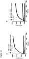
- the timecourse of fluorescence dequenching was followed for 30min with a fluorescence microplate reader (excitation 480/20, emission 528/25). A fluorescence increase is observed only in presence of HNE, and this is inhibited by the presence of sivelestat.
- IQR4 (0.5uM) is incubated with freeze-thawed neutrophil lysate (10 ⁇ 10 6 cells/ml) in IMDM with or without sivelestat (50uM). The timecourse of fluorescence dequenching was followed for 30min with a fluorescence microplate reader (excitation 480/20, emission 528/25).
- Human neutrophils were isolated as described above and suspended in PBS with or without activating agents.
- a fibreoptic confocal system (488 Cellvono) was used to acquire images in eppendorfs of cells both in the presence of free FAM and IQRs and after washing cells of free FAM and IQR in media. Imaging was performed for 60 s and representative still frames converted to bmp format.
- alveoscopy was performed by passing a alveoflex (fibreoptic bundle) down the working channel and live imaging commenced upon distal alveoscopy. Images were captured at 12 frames per second and representative frames converted to jpeg format.
- Fmoc-Lys(Dde)-OH is prepared in 3 steps.
- Fmoc-Lys(Dde)-OH Fmoc-Lys-OH.HCI (10.2 g, 25.2 mmol) was dissolved in H 2 O, N, N-diisopropylethylamine (DIPEA 1.1 eq, 4.8 mL, 27.7 mmol) was added and the resulting solid was collected by filtration and dried in a vacuum oven overnight.
- DIPEA N, N-diisopropylethylamine
- Fmoc-Lys(Dde)-OH was isolated by flash column chromatography (elute with 10% acetic acid / ethyl acetate) and crystallised from ethyl acetate/hexane as an off white solid (7.5g, 70%).
- Fmoc-Rink Amide-PS resin was prepared using a 4-[(2,4-dimethoxyphenyl)-(Fmoc-amino)methyl]phenoxyacetic acid (Rink amide linker) attached to aminomethyl PS resin (1.23 mmol/g, 1% DVB, 100-200 mesh).
- Rink amide linker 4-[(2,4-dimethoxyphenyl)-(Fmoc-amino)methyl]phenoxyacetic acid (Rink amide linker) attached to aminomethyl PS resin (1.23 mmol/g, 1% DVB, 100-200 mesh).
- Fmoc-Rink-amide linker 3eq
- Oxyma (3eq) was added and the mixture was stirred for 10min.
- DIC (3eq) was then added and the resulting mixture was stirred for further 5 min.
- the solution was added to aminomethyl polystyrene resin (1eq) and shaken for 2 hours.
- the resulting resin was washed
- Solid phase couplings A solution of the appropriate Fmoc-amino acid or or Methyl red (MR) or 5(6)-carboxyfluorescein (FAM) (10eq) and Oxyma (10eq) in DMF (0.1M) was stirred for 10min. DIC (10eq) was then added and the resulting solution was stirred for further 5min. The solution was then added to the resin (1eq), pre-swollen in DCM (10mL), and the reaction mixture was shaken for 6 hours. The solution was drained and the resin washed DMF (3 ⁇ 20mL), DCM (3 ⁇ 20mL) and MeOH (3 ⁇ 20mL). The coupling reactions were monitored by the Kaiser and chloranil tests.
- the probe was then released from the resin using a cocktail of TFA/TIS/DCM (90/5/5) for 3 h. To the filtrate, cold ether was added to precipitate the product.
- Fluid phase fluorophores permit detection of cell specific uptake/internalisation in activated neutrophils using a bench top confocal
- IQR specifically dequenches in neutrophils compared to other highly pinocytic/endocytic cells such as human macrophages
- Dendrimeric IQRs but not FRET IQRs with enhanced solubility permit cell specific detection
- these optical reporters For the ultimate delivery of these optical reporters to the human lung, they would require enhanced solubility compared to the probes described above.
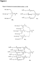
- the enhanced solubility of these IQRs was achieved by incorporating a small series of peptides within the dendrimer backbone (IQR1.2 and IQR2.2) or pegylating the probes (IQR1.3 and IQR2.3) ( Figure 4 ).
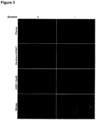
- these were initially evaluated in vitro for selectivity against other cells commonly obtained from the inflammatory milieu; monocytes, macrophages and lymphocytes and epithelial cells. Again, we confirmed the cellular specificity of the probes using live confocal imaging ( Figure 5 ). This was extended to freshly isolated cells from the BAL of patients with IPF.
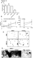
- IQR fluorescent amplification in neutrophils is dependent on the combination of endocytosis and degranulation.
- the solubilsed dendrimeric IQRs exhibit no in vitro or in vivo toxicity
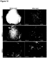
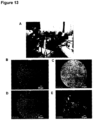
- FIG. 15 A further experiment ( Figure 15 ) was performed to image recruited activated human neutrophils.
- an ex vivo ovine lung was ventilated and perfused with human blood.
- a subsegment of the lung received lipopolysaccharide to induce the recruitment of human neutrophils.
- 10 ⁇ g ⁇ of IQR1.2 was delivered into a control segment and the LPS instilled segment. Only the LPS segment showed signal enhancement imaged using probe based confocal microendoscopy.
- This experiment conclusively demsonstarted that recruited activated neutrophils can be detected by IQR.
- the method and timing of probe delivery are crucial to this approach. Only minute quantities are required ( ⁇ 100 ⁇ g) and the timing of detection is within minutes. To image differential neutrophil pinocytosis, it is imperative that imaging is conducted immediately as delaying imaging may lead to pinocytic uptake by other cells within a few hours. The directed delivery of minute quantities foregoes any toxicity issues and direct instillation into the distal lung is ideally suited to confocal microendoscopy to permit molecular resolution. Proof of concept in vivo is demonstrated.
- directly visualisable reporters of neutrophil activity provide a potential diagnostic tool.
- neutrophil dominant conditions such as acute lung injury.
- Developing bedside methodologies could aid in stratifying those patients who have chest x-ray infiltrates due to non inflammatory causes versus those with either sterile or infective lung injury.
- Confocal laser microendoscopy coupled with the delivery of small concentrations of molecular probes permits the visualisation deep within the lung and provides the detection platform for the delivery and imaging of such probes in humans.
- a dendrimeric IQR incorporating a FRET-based elastase reporter was generated and shown to be dequenched by recombinant HNE and neutrophil lysate, an effect that was blocked by the specific elastase inhibitor sivelestat ( Figure 16 ). Furthermore, in live confocal imaging experiments this FRET-dendrimer IQR construct reported elastase activity on neutrophils, since the fluorescent cell-associated signal could be blocked by sivelestat ( Figure 17 ).
Landscapes
- Health & Medical Sciences (AREA)
- Life Sciences & Earth Sciences (AREA)
- General Health & Medical Sciences (AREA)
- Engineering & Computer Science (AREA)
- Biomedical Technology (AREA)
- Epidemiology (AREA)
- Animal Behavior & Ethology (AREA)
- Public Health (AREA)
- Veterinary Medicine (AREA)
- Physics & Mathematics (AREA)
- Nuclear Medicine, Radiotherapy & Molecular Imaging (AREA)
- Immunology (AREA)
- Chemical & Material Sciences (AREA)
- Analytical Chemistry (AREA)
- Biochemistry (AREA)
- General Physics & Mathematics (AREA)
- Pathology (AREA)
- Proteomics, Peptides & Aminoacids (AREA)
- Optics & Photonics (AREA)
- Biodiversity & Conservation Biology (AREA)
- Oncology (AREA)
- Chemical Kinetics & Catalysis (AREA)
- Medicines Containing Antibodies Or Antigens For Use As Internal Diagnostic Agents (AREA)
- Investigating Or Analysing Biological Materials (AREA)
- Peptides Or Proteins (AREA)
- Measuring Or Testing Involving Enzymes Or Micro-Organisms (AREA)
- Endoscopes (AREA)
- Investigating, Analyzing Materials By Fluorescence Or Luminescence (AREA)
Claims (12)
- Farbstoffkonstrukt zur Verwendung in einem Verfahren zum Diagnostizieren von Entzündungen durch Bildgeben der aktivierten Neutrophil-Zellen in vivo durch konfokale Endoskopie, wobei das Farbstoffkonstrukt eine der Strukturen (I) bis (V) aufweist:


 AA jede Aminosäure ist und n zwischen 0 und 30 liegt; (AA)n von (II) eine Peptidsequenz ist, die von dem Enzym Neutrophil-Elastase erkannt wird;Q* ein dunkler Quencher ist;R1 ist:
 *F Carboxyfluorescein (FAM) oder Rhodamin ist,wobei das Farbstoffkonstrukt vor der Internalisierung der aktivierten Neutrophilen im Wesentlichen keine feststellbare oder nur eine niedrige Menge an Fluoreszenz anzeigt, aber nach der Internalisierung der aktivierten Neutrophilen einen Anstieg der Fluoreszenz um einen Faktor von 1,2 oder mehr im Vergleich zu jeder Fluoreszenz anzeigt, die außerhalb der aktivierten Neutrophilen durch konfokale Endoskopie beobachtet wird.
 - Farbstoffkonstrukt zur Verwendung in einem Verfahren nach Anspruch 1, wobei das polyverzweigte Molekül eine Peptidsequenz umfasst, wobei die Peptidsequenz (i) eine oder mehrere Spaltungsstellen umfasst, die durch eine angemessene Chemikalie oder ein Enzym spaltbar sind, oder (ii) eine zufällige Sequenz umfasst, die keine erkennbare Sequenz umfasst, die durch eine Chemikalie oder ein Enzym spaltbar ist.
- Farbstoffkonstrukt zur Verwendung in einem Verfahren nach Anspruch 2, wobei das polyverzweigte Molekül eine Peptidsequenz umfasst, die von dem Enzym Neutrophil-Elastase erkannt wird.
- Farbstoffkonstrukt zur Verwendung in einem Verfahren nach Anspruch 3, wobei die Peptidsequenz ausgewählt ist aus A-A-P-V, A-A-A-P-V-K und E-E-I-Nle-R-R.
- Farbstoffkonstrukt zur Verwendung in einem Verfahren nach einem der vorstehenden Ansprüche, wobei Q* unabhängig ausgewählt ist aus DABCYL, Methylrot, BHQ1, BHQ2 und BHQ3.
- Farbstoffkonstrukt zur Verwendung in einem Verfahren nach einem der vorstehenden Ansprüche, wobei das Farbstoffkonstrukt eine der folgenden Strukturen aufweist:
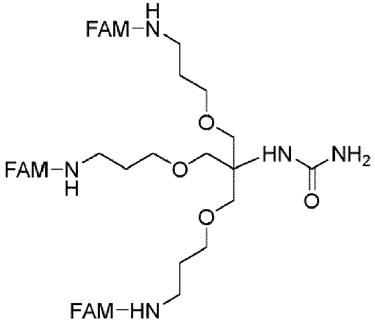


 FAM 5(6)-Carboxyfluorescein ist;TERA 5(6)-Tetraethylrhodamin ist;AA jede Aminosäure ist;n zwischen 0 und 30 liegt; undR1 ist:
- Farbstoffkonstrukt zur Verwendung in einem Verfahren nach einem der vorstehenden Ansprüche, wobei das Bildgeben durch konfokale Endoskopie im Darm, einschließend Dick- und Dünndarm; Arterien und Venen; im Atmungssystem, einschließend Lunge, im Gehirn, z. B. über einen intrakraniellen Katheter; oder im Fortpflanzungssystem, einschließend Gebärmutter und Eileiter, durchgeführt werden soll.
- Farbstoffkonstrukt zur Verwendung in einem Verfahren nach einem der vorstehenden Ansprüche, wobei das Verfahren das lokale Verabreichen des Farbstoffkonstrukts an ein abzubildendes Gewebebereich umfasst.
- Farbstoffkonstrukt zur Verwendung in einem Verfahren nach einem der vorstehenden Ansprüche, wobei die Erkennung der aktivierten Neutrophilen, die das Farbstoffkonstrukt internalisiert haben, innerhalb von 1-30 Minuten nach Verabreichen des Farbstoffkonstrukts an ein Subjekt durchgeführt wird.
- Farbstoffkonstrukt zur Verwendung in einem Verfahren nach einem der vorstehenden Ansprüche, wobei die Menge des Konstrukts, die an ein Subjekt verabreicht werden soll, weniger als 100 µg ist.
- Verfahren zum Bildgeben der aktivierten Neutrophil-Zellen in viva durch konfokale Endoskopie, das Verfahren umfassend das Beobachten der aktivierten Neutrophilen eines Subjekts, die mit einem Farbstoffkonstrukt verabreicht wurden und die das Farbstoffkonstrukt internalisiert haben, wobei das Farbstoffkonstrukt vor der Internalisierung der aktivierten Neutrophilen im Wesentlichen keine feststellbare oder nur eine niedrige Menge an Fluoreszenz anzeigt, aber nach der Internalisierung der aktivierten Neutrophilen einen Anstieg der Fluoreszenz um einen Faktor von 1,2 oder mehr im Vergleich zu jeder Fluoreszenz anzeigt, die außerhalb der aktivierten Neutrophilen durch ein konfokales Endomikroskop beobachtet wird; wobei das Farbstoffkonstrukt eine der Strukturen (I) bis (V) aufweist:


 AA jede Aminosäure ist und n zwischen 0 und 30 liegt;(AA)n von (II) eine Peptidsequenz ist, die von dem Enzym Neutrophil-Elastase erkannt wird;Q* ein dunkler Quencher ist;R1 ist:
 *F Carboxyfluorescein (FAM) oder Rhodamin ist.
 - Verfahren nach Anspruch 11, wobei das Farbstoffkonstrukt wie in einem der Ansprüche 2 bis 6 beschrieben ist.
Applications Claiming Priority (2)
| Application Number | Priority Date | Filing Date | Title |
|---|---|---|---|
| GBGB1106004.3A GB201106004D0 (en) | 2011-04-08 | 2011-04-08 | Optical imaging probes |
| PCT/GB2012/000314 WO2012136958A2 (en) | 2011-04-08 | 2012-04-05 | Optical imaging probes |
Publications (3)
| Publication Number | Publication Date |
|---|---|
| EP2694115A2 EP2694115A2 (de) | 2014-02-12 |
| EP2694115B1 true EP2694115B1 (de) | 2024-10-23 |
| EP2694115C0 EP2694115C0 (de) | 2024-10-23 |
Family
ID=44122836
Family Applications (1)
| Application Number | Title | Priority Date | Filing Date |
|---|---|---|---|
| EP12722474.9A Active EP2694115B1 (de) | 2011-04-08 | 2012-04-05 | Sonden für optische bildgebung |
Country Status (8)
| Country | Link |
|---|---|
| US (2) | US9549997B2 (de) |
| EP (1) | EP2694115B1 (de) |
| JP (3) | JP2014511855A (de) |
| AU (2) | AU2012238478B8 (de) |
| CA (1) | CA2831205C (de) |
| ES (1) | ES2996587T3 (de) |
| GB (2) | GB201106004D0 (de) |
| WO (1) | WO2012136958A2 (de) |
Families Citing this family (9)
| Publication number | Priority date | Publication date | Assignee | Title |
|---|---|---|---|---|
| GB201106004D0 (en) * | 2011-04-08 | 2011-05-25 | Univ Edinburgh | Optical imaging probes |
| GB201420221D0 (en) * | 2014-11-13 | 2014-12-31 | Univ Edinburgh | Molecular probes and methods using the same |
| GB201420223D0 (en) | 2014-11-13 | 2014-12-31 | Univ Edinburgh | Molecular probes and methods using the same |
| GB201420222D0 (en) * | 2014-11-13 | 2014-12-31 | Univ Edinburgh | Molecular probes and methods using the same |
| GB201504775D0 (en) * | 2015-03-20 | 2015-05-06 | Univ Edinburgh | Optical probe for thrombin |
| GB201504778D0 (en) * | 2015-03-20 | 2015-05-06 | Univ Edinburgh | Optical probes for matrix metalloproteinases |
| CN110305042A (zh) * | 2019-07-01 | 2019-10-08 | 吉尔生化(上海)有限公司 | Nα-芴甲氧羰基-Nγ-(4,4-二甲基-2,6-二氧代环己亚基)乙基-丁酸制法 |
| DE102019210421B4 (de) * | 2019-07-15 | 2023-03-09 | Leica Microsystems Cms Gmbh | Verfahren und Vorrichtung zur Messung eines Lichtsignalparameters und nichtflüchtiges Speichermedium |
| EP4092466A1 (de) * | 2021-05-21 | 2022-11-23 | Leica Microsystems CMS GmbH | Verfahren zur ermittlung eines betriebswertes eines beleuchtungsparameters zur untersuchung einer fluoreszenten probe, verfahren zur untersuchung einer fluoreszenten probe, mikroskopsystem und computerprogramm |
Citations (1)
| Publication number | Priority date | Publication date | Assignee | Title |
|---|---|---|---|---|
| WO2011011384A2 (en) * | 2009-07-20 | 2011-01-27 | The Regents Of The University Of Michigan | Synthesis of dendrimer conjugates |
Family Cites Families (9)
| Publication number | Priority date | Publication date | Assignee | Title |
|---|---|---|---|---|
| US5527524A (en) * | 1986-08-18 | 1996-06-18 | The Dow Chemical Company | Dense star polymer conjugates |
| GB0119001D0 (en) * | 2001-08-03 | 2001-09-26 | Amersham Pharm Biotech Uk Ltd | Use of dendrimers and poly-branched molecules to enhance signal in fluorescent assay systems |
| JP2005537234A (ja) | 2002-05-31 | 2005-12-08 | トランスモルキュラー インコーポレイテッド | クロロトキシンとの併用化学療法 |
| US20100189657A1 (en) | 2006-03-20 | 2010-07-29 | The General Hospital Corporation | Intramolecularly quenched fluorochrome conjugates and methods of use |
| FR2904927B1 (fr) | 2006-08-17 | 2018-05-18 | Mauna Kea Technologies | Utilisation d'un systeme d'imagerie par fluorescence confocale fibre in vivo in situ, systeme et procede d'imagerie par fluorescence confocale fibres in vivo in situ |
| WO2008078190A2 (en) * | 2006-12-21 | 2008-07-03 | Universite De Geneve | Compounds for fluorescence imaging |
| GB0803477D0 (en) * | 2008-02-26 | 2008-04-02 | Ge Healthcare As | Therapy selection method |
| EP2100621A1 (de) * | 2008-03-10 | 2009-09-16 | mivenion GmbH | Polyether-Polyol-Dendron-Konjugate mit Effektormolekülen für biologisches Targeting |
| GB201106004D0 (en) * | 2011-04-08 | 2011-05-25 | Univ Edinburgh | Optical imaging probes |
-
2011
- 2011-04-08 GB GBGB1106004.3A patent/GB201106004D0/en not_active Ceased
-
2012
- 2012-04-05 WO PCT/GB2012/000314 patent/WO2012136958A2/en not_active Ceased
- 2012-04-05 GB GB1317127.7A patent/GB2503170B/en active Active
- 2012-04-05 ES ES12722474T patent/ES2996587T3/es active Active
- 2012-04-05 US US14/110,616 patent/US9549997B2/en active Active
- 2012-04-05 CA CA2831205A patent/CA2831205C/en active Active
- 2012-04-05 AU AU2012238478A patent/AU2012238478B8/en active Active
- 2012-04-05 JP JP2014503205A patent/JP2014511855A/ja active Pending
- 2012-04-05 EP EP12722474.9A patent/EP2694115B1/de active Active
-
2017
- 2017-01-23 US US15/413,139 patent/US10434191B2/en active Active
- 2017-02-03 JP JP2017018646A patent/JP2017128572A/ja active Pending
- 2017-09-29 AU AU2017236010A patent/AU2017236010B2/en active Active
-
2018
- 2018-09-21 JP JP2018177692A patent/JP2019014727A/ja active Pending
Patent Citations (1)
| Publication number | Priority date | Publication date | Assignee | Title |
|---|---|---|---|---|
| WO2011011384A2 (en) * | 2009-07-20 | 2011-01-27 | The Regents Of The University Of Michigan | Synthesis of dendrimer conjugates |
Non-Patent Citations (5)
| Title |
|---|
| DATABASE MEDLINE [online] US NATIONAL LIBRARY OF MEDICINE (NLM), BETHESDA, MD, US; 7 October 2003 (2003-10-07), TERNON M ET AL: "Assay amplification-multiple valent fluorophores.", Database accession no. NLM14587702 * |
| DATABASE MEDLINE [online] US NATIONAL LIBRARY OF MEDICINE (NLM), BETHESDA, MD, US; November 2000 (2000-11-01), ZHANG Z Y ET AL: "High-generation polycationic dendrimers are unusually effective at disrupting anionic vesicles: membrane bending model.", Database accession no. NLM11087328 * |
| KOLHE PARAG ET AL: "Hyperbranched polymer-drug conjugates with high drug payload for enhanced cellular delivery", ENVIRONMENTAL SCIENCE AND POLLUTION RESEARCH, SPRINGER BERLIN HEIDELBERG, BERLIN/HEIDELBERG, vol. 21, no. 12, 1 December 2004 (2004-12-01), pages 2185 - 2195, XP002472253, ISSN: 0724-8741, DOI: 10.1007/S11095-004-7670-X * |
| TERNON M ET AL: "Assay amplification-multiple valent fluorophores.", CHEMICAL COMMUNICATIONS (CAMBRIDGE, ENGLAND) 07 OCT 2003, no. 19, 7 October 2003 (2003-10-07), pages 2402 - 2403, ISSN: 1359-7345 * |
| ZHANG Z Y ET AL: "High-generation polycationic dendrimers are unusually effective at disrupting anionic vesicles: membrane bending model.", BIOCONJUGATE CHEMISTRY, vol. 11, no. 6, November 2000 (2000-11-01), pages 805 - 814, XP002684578, ISSN: 1043-1802, DOI: 10.1021/bc000018z * |
Also Published As
| Publication number | Publication date |
|---|---|
| AU2012238478B8 (en) | 2017-07-27 |
| WO2012136958A8 (en) | 2013-05-16 |
| US10434191B2 (en) | 2019-10-08 |
| CA2831205A1 (en) | 2012-10-11 |
| EP2694115C0 (de) | 2024-10-23 |
| ES2996587T3 (en) | 2025-02-12 |
| AU2017236010B2 (en) | 2019-04-04 |
| AU2012238478A8 (en) | 2017-07-27 |
| AU2012238478B2 (en) | 2017-06-29 |
| US20140134110A1 (en) | 2014-05-15 |
| AU2012238478A1 (en) | 2013-10-24 |
| JP2017128572A (ja) | 2017-07-27 |
| EP2694115A2 (de) | 2014-02-12 |
| WO2012136958A3 (en) | 2012-12-20 |
| GB201106004D0 (en) | 2011-05-25 |
| GB201317127D0 (en) | 2013-11-06 |
| GB2503170A (en) | 2013-12-18 |
| GB2503170B (en) | 2019-02-06 |
| JP2014511855A (ja) | 2014-05-19 |
| CA2831205C (en) | 2020-03-10 |
| US9549997B2 (en) | 2017-01-24 |
| WO2012136958A2 (en) | 2012-10-11 |
| JP2019014727A (ja) | 2019-01-31 |
| US20170196995A1 (en) | 2017-07-13 |
| AU2017236010A1 (en) | 2017-10-26 |
Similar Documents
| Publication | Publication Date | Title |
|---|---|---|
| AU2017236010B2 (en) | Optical imaging probes | |
| JP7689753B2 (ja) | 活性に基づくプローブの化合物、組成物、および使用方法 | |
| CN102603695B (zh) | 一种氨基酸-荧光团类化合物及其应用 | |
| Craven et al. | Super-silent FRET sensor enables live cell imaging and flow cytometric stratification of intracellular serine protease activity in neutrophils | |
| JP2020515507A (ja) | 活性ベースのプローブ化合物、組成物、および使用方法 | |
| CN113979912A (zh) | 两种前列腺特异性膜抗原靶向荧光探针及其制备方法与应用 | |
| CN106459126B (zh) | 新型葡萄糖衍生物、及使用该衍生物的细胞成像方法及显像剂 | |
| US20180231548A1 (en) | Molecular probes for detecting gram-negative bacteria in vitro and in vivo | |
| JP7149614B2 (ja) | in vivo画像化のためのプロテアーゼ活性化コントラスト剤 | |
| WO2016151297A1 (en) | Optical probe for thrombin | |
| US20260061080A1 (en) | Fluorescent compound based on new indocyanine green ir820, and preparation and use thereof | |
| US8859223B1 (en) | Compositions and methods for imaging beta-secretase activity in living cells and organisms | |
| WO2021002771A1 (en) | Conjugate of fluorescent dye for the visualization of psma expressing cells | |
| WO2016133913A1 (en) | Fluorescently labeled molecules containing modified tryptophan | |
| FR2910897A1 (fr) | Nouveaux composes pro-fluorescents. |
Legal Events
| Date | Code | Title | Description |
|---|---|---|---|
| PUAI | Public reference made under article 153(3) epc to a published international application that has entered the european phase |
Free format text: ORIGINAL CODE: 0009012 |
|
| 17P | Request for examination filed |
Effective date: 20130926 |
|
| AK | Designated contracting states |
Kind code of ref document: A2 Designated state(s): AL AT BE BG CH CY CZ DE DK EE ES FI FR GB GR HR HU IE IS IT LI LT LU LV MC MK MT NL NO PL PT RO RS SE SI SK SM TR |
|
| RIN1 | Information on inventor provided before grant (corrected) |
Inventor name: BRADLEY, MARK Inventor name: DEBUNNE, MANUELLE Inventor name: AVLONITIS, NIKOLAOS Inventor name: DHALIWAL, KEV Inventor name: HASLETT, CHRIS Inventor name: MCDONALD, NEIL Inventor name: WALTON, TASHFEEN |
|
| DAX | Request for extension of the european patent (deleted) | ||
| STAA | Information on the status of an ep patent application or granted ep patent |
Free format text: STATUS: EXAMINATION IS IN PROGRESS |
|
| 17Q | First examination report despatched |
Effective date: 20180416 |
|
| GRAP | Despatch of communication of intention to grant a patent |
Free format text: ORIGINAL CODE: EPIDOSNIGR1 |
|
| STAA | Information on the status of an ep patent application or granted ep patent |
Free format text: STATUS: GRANT OF PATENT IS INTENDED |
|
| INTG | Intention to grant announced |
Effective date: 20240514 |
|
| GRAS | Grant fee paid |
Free format text: ORIGINAL CODE: EPIDOSNIGR3 |
|
| GRAA | (expected) grant |
Free format text: ORIGINAL CODE: 0009210 |
|
| STAA | Information on the status of an ep patent application or granted ep patent |
Free format text: STATUS: THE PATENT HAS BEEN GRANTED |
|
| AK | Designated contracting states |
Kind code of ref document: B1 Designated state(s): AL AT BE BG CH CY CZ DE DK EE ES FI FR GB GR HR HU IE IS IT LI LT LU LV MC MK MT NL NO PL PT RO RS SE SI SK SM TR |
|
| REG | Reference to a national code |
Ref country code: GB Ref legal event code: FG4D |
|
| REG | Reference to a national code |
Ref country code: CH Ref legal event code: EP |
|
| REG | Reference to a national code |
Ref country code: DE Ref legal event code: R096 Ref document number: 602012081152 Country of ref document: DE |
|
| REG | Reference to a national code |
Ref country code: IE Ref legal event code: FG4D |
|
| U01 | Request for unitary effect filed |
Effective date: 20241120 |
|
| U07 | Unitary effect registered |
Designated state(s): AT BE BG DE DK EE FI FR IT LT LU LV MT NL PT RO SE SI Effective date: 20241127 |
|
| REG | Reference to a national code |
Ref country code: ES Ref legal event code: FG2A Ref document number: 2996587 Country of ref document: ES Kind code of ref document: T3 Effective date: 20250212 |
|
| PG25 | Lapsed in a contracting state [announced via postgrant information from national office to epo] |
Ref country code: HR Free format text: LAPSE BECAUSE OF FAILURE TO SUBMIT A TRANSLATION OF THE DESCRIPTION OR TO PAY THE FEE WITHIN THE PRESCRIBED TIME-LIMIT Effective date: 20241023 Ref country code: IS Free format text: LAPSE BECAUSE OF FAILURE TO SUBMIT A TRANSLATION OF THE DESCRIPTION OR TO PAY THE FEE WITHIN THE PRESCRIBED TIME-LIMIT Effective date: 20250223 |
|
| PG25 | Lapsed in a contracting state [announced via postgrant information from national office to epo] |
Ref country code: NO Free format text: LAPSE BECAUSE OF FAILURE TO SUBMIT A TRANSLATION OF THE DESCRIPTION OR TO PAY THE FEE WITHIN THE PRESCRIBED TIME-LIMIT Effective date: 20250123 |
|
| PG25 | Lapsed in a contracting state [announced via postgrant information from national office to epo] |
Ref country code: GR Free format text: LAPSE BECAUSE OF FAILURE TO SUBMIT A TRANSLATION OF THE DESCRIPTION OR TO PAY THE FEE WITHIN THE PRESCRIBED TIME-LIMIT Effective date: 20250124 |
|
| PG25 | Lapsed in a contracting state [announced via postgrant information from national office to epo] |
Ref country code: PL Free format text: LAPSE BECAUSE OF FAILURE TO SUBMIT A TRANSLATION OF THE DESCRIPTION OR TO PAY THE FEE WITHIN THE PRESCRIBED TIME-LIMIT Effective date: 20241023 |
|
| PG25 | Lapsed in a contracting state [announced via postgrant information from national office to epo] |
Ref country code: RS Free format text: LAPSE BECAUSE OF FAILURE TO SUBMIT A TRANSLATION OF THE DESCRIPTION OR TO PAY THE FEE WITHIN THE PRESCRIBED TIME-LIMIT Effective date: 20250123 |
|
| U20 | Renewal fee for the european patent with unitary effect paid |
Year of fee payment: 14 Effective date: 20250411 |
|
| PG25 | Lapsed in a contracting state [announced via postgrant information from national office to epo] |
Ref country code: SM Free format text: LAPSE BECAUSE OF FAILURE TO SUBMIT A TRANSLATION OF THE DESCRIPTION OR TO PAY THE FEE WITHIN THE PRESCRIBED TIME-LIMIT Effective date: 20241023 |
|
| PGFP | Annual fee paid to national office [announced via postgrant information from national office to epo] |
Ref country code: GB Payment date: 20250411 Year of fee payment: 14 Ref country code: ES Payment date: 20250507 Year of fee payment: 14 |
|
| PG25 | Lapsed in a contracting state [announced via postgrant information from national office to epo] |
Ref country code: SK Free format text: LAPSE BECAUSE OF FAILURE TO SUBMIT A TRANSLATION OF THE DESCRIPTION OR TO PAY THE FEE WITHIN THE PRESCRIBED TIME-LIMIT Effective date: 20241023 |
|
| PG25 | Lapsed in a contracting state [announced via postgrant information from national office to epo] |
Ref country code: CZ Free format text: LAPSE BECAUSE OF FAILURE TO SUBMIT A TRANSLATION OF THE DESCRIPTION OR TO PAY THE FEE WITHIN THE PRESCRIBED TIME-LIMIT Effective date: 20241023 |
|
| PLBE | No opposition filed within time limit |
Free format text: ORIGINAL CODE: 0009261 |
|
| STAA | Information on the status of an ep patent application or granted ep patent |
Free format text: STATUS: NO OPPOSITION FILED WITHIN TIME LIMIT |
|
| 26N | No opposition filed |
Effective date: 20250724 |
|
| REG | Reference to a national code |
Ref country code: CH Ref legal event code: H13 Free format text: ST27 STATUS EVENT CODE: U-0-0-H10-H13 (AS PROVIDED BY THE NATIONAL OFFICE) Effective date: 20251125 |
|
| PG25 | Lapsed in a contracting state [announced via postgrant information from national office to epo] |
Ref country code: MC Free format text: LAPSE BECAUSE OF FAILURE TO SUBMIT A TRANSLATION OF THE DESCRIPTION OR TO PAY THE FEE WITHIN THE PRESCRIBED TIME-LIMIT Effective date: 20241023 |
|
| PG25 | Lapsed in a contracting state [announced via postgrant information from national office to epo] |
Ref country code: CH Free format text: LAPSE BECAUSE OF NON-PAYMENT OF DUE FEES Effective date: 20250430 |
|
| PG25 | Lapsed in a contracting state [announced via postgrant information from national office to epo] |
Ref country code: IE Free format text: LAPSE BECAUSE OF NON-PAYMENT OF DUE FEES Effective date: 20250405 |