EP2678692B1 - Solid support and method of enhancing the recovery of biological material therefrom - Google Patents
Solid support and method of enhancing the recovery of biological material therefrom Download PDFInfo
- Publication number
- EP2678692B1 EP2678692B1 EP12705857.6A EP12705857A EP2678692B1 EP 2678692 B1 EP2678692 B1 EP 2678692B1 EP 12705857 A EP12705857 A EP 12705857A EP 2678692 B1 EP2678692 B1 EP 2678692B1
- Authority
- EP
- European Patent Office
- Prior art keywords
- paper
- solid support
- biological material
- group
- recovery
- Prior art date
- Legal status (The legal status is an assumption and is not a legal conclusion. Google has not performed a legal analysis and makes no representation as to the accuracy of the status listed.)
- Active
Links
- 239000007787 solid Substances 0.000 title claims description 28
- 238000011084 recovery Methods 0.000 title claims description 26
- 239000012620 biological material Substances 0.000 title claims description 25
- 238000000034 method Methods 0.000 title claims description 19
- 230000002708 enhancing effect Effects 0.000 title description 2
- 239000000126 substance Substances 0.000 claims description 23
- 239000003814 drug Substances 0.000 claims description 20
- 229940079593 drug Drugs 0.000 claims description 19
- 229960000074 biopharmaceutical Drugs 0.000 claims description 12
- 102000009027 Albumins Human genes 0.000 claims description 10
- 108010088751 Albumins Proteins 0.000 claims description 10
- NYEZZYQZRQDLEH-UHFFFAOYSA-N 2-ethyl-4,5-dihydro-1,3-oxazole Chemical compound CCC1=NCCO1 NYEZZYQZRQDLEH-UHFFFAOYSA-N 0.000 claims description 9
- 229920000036 polyvinylpyrrolidone Polymers 0.000 claims description 8
- 239000001267 polyvinylpyrrolidone Substances 0.000 claims description 8
- 235000013855 polyvinylpyrrolidone Nutrition 0.000 claims description 8
- PNEYBMLMFCGWSK-UHFFFAOYSA-N Alumina Chemical compound [O-2].[O-2].[O-2].[Al+3].[Al+3] PNEYBMLMFCGWSK-UHFFFAOYSA-N 0.000 claims description 6
- 239000005018 casein Substances 0.000 claims description 6
- BECPQYXYKAMYBN-UHFFFAOYSA-N casein, tech. Chemical compound NCCCCC(C(O)=O)N=C(O)C(CC(O)=O)N=C(O)C(CCC(O)=N)N=C(O)C(CC(C)C)N=C(O)C(CCC(O)=O)N=C(O)C(CC(O)=O)N=C(O)C(CCC(O)=O)N=C(O)C(C(C)O)N=C(O)C(CCC(O)=N)N=C(O)C(CCC(O)=N)N=C(O)C(CCC(O)=N)N=C(O)C(CCC(O)=O)N=C(O)C(CCC(O)=O)N=C(O)C(COP(O)(O)=O)N=C(O)C(CCC(O)=N)N=C(O)C(N)CC1=CC=CC=C1 BECPQYXYKAMYBN-UHFFFAOYSA-N 0.000 claims description 6
- 235000021240 caseins Nutrition 0.000 claims description 6
- 239000012528 membrane Substances 0.000 claims description 6
- 230000001413 cellular effect Effects 0.000 claims description 5
- 229920002678 cellulose Polymers 0.000 claims description 5
- 239000001913 cellulose Substances 0.000 claims description 5
- 210000002381 plasma Anatomy 0.000 claims description 5
- -1 polypropylene Polymers 0.000 claims description 4
- 239000004810 polytetrafluoroethylene Substances 0.000 claims description 4
- 229920001343 polytetrafluoroethylene Polymers 0.000 claims description 4
- 229920001410 Microfiber Polymers 0.000 claims description 2
- 239000000020 Nitrocellulose Substances 0.000 claims description 2
- 239000004677 Nylon Substances 0.000 claims description 2
- 229920012266 Poly(ether sulfone) PES Polymers 0.000 claims description 2
- 239000004952 Polyamide Substances 0.000 claims description 2
- 239000004743 Polypropylene Substances 0.000 claims description 2
- FJWGYAHXMCUOOM-QHOUIDNNSA-N [(2s,3r,4s,5r,6r)-2-[(2r,3r,4s,5r,6s)-4,5-dinitrooxy-2-(nitrooxymethyl)-6-[(2r,3r,4s,5r,6s)-4,5,6-trinitrooxy-2-(nitrooxymethyl)oxan-3-yl]oxyoxan-3-yl]oxy-3,5-dinitrooxy-6-(nitrooxymethyl)oxan-4-yl] nitrate Chemical compound O([C@@H]1O[C@@H]([C@H]([C@H](O[N+]([O-])=O)[C@H]1O[N+]([O-])=O)O[C@H]1[C@@H]([C@@H](O[N+]([O-])=O)[C@H](O[N+]([O-])=O)[C@@H](CO[N+]([O-])=O)O1)O[N+]([O-])=O)CO[N+](=O)[O-])[C@@H]1[C@@H](CO[N+]([O-])=O)O[C@@H](O[N+]([O-])=O)[C@H](O[N+]([O-])=O)[C@H]1O[N+]([O-])=O FJWGYAHXMCUOOM-QHOUIDNNSA-N 0.000 claims description 2
- 210000000601 blood cell Anatomy 0.000 claims description 2
- 229920002301 cellulose acetate Polymers 0.000 claims description 2
- 238000001035 drying Methods 0.000 claims description 2
- 239000011521 glass Substances 0.000 claims description 2
- 239000003658 microfiber Substances 0.000 claims description 2
- 229920001220 nitrocellulos Polymers 0.000 claims description 2
- 229920001778 nylon Polymers 0.000 claims description 2
- 229920002647 polyamide Polymers 0.000 claims description 2
- 239000004417 polycarbonate Substances 0.000 claims description 2
- 229920000515 polycarbonate Polymers 0.000 claims description 2
- 229920000728 polyester Polymers 0.000 claims description 2
- 229920001155 polypropylene Polymers 0.000 claims description 2
- 210000003296 saliva Anatomy 0.000 claims description 2
- 210000001519 tissue Anatomy 0.000 claims description 2
- 210000002700 urine Anatomy 0.000 claims description 2
- 239000000123 paper Substances 0.000 description 53
- 108010002350 Interleukin-2 Proteins 0.000 description 27
- 102000000588 Interleukin-2 Human genes 0.000 description 27
- 210000004369 blood Anatomy 0.000 description 24
- 239000008280 blood Substances 0.000 description 24
- 238000004458 analytical method Methods 0.000 description 12
- 241001465754 Metazoa Species 0.000 description 10
- 235000018102 proteins Nutrition 0.000 description 10
- 108090000623 proteins and genes Proteins 0.000 description 10
- 102000004169 proteins and genes Human genes 0.000 description 10
- 238000003860 storage Methods 0.000 description 8
- XLYOFNOQVPJJNP-UHFFFAOYSA-N water Substances O XLYOFNOQVPJJNP-UHFFFAOYSA-N 0.000 description 8
- 201000003883 Cystic fibrosis Diseases 0.000 description 5
- 150000001875 compounds Chemical class 0.000 description 5
- 238000001514 detection method Methods 0.000 description 5
- 108020004707 nucleic acids Proteins 0.000 description 5
- 102000039446 nucleic acids Human genes 0.000 description 5
- 150000007523 nucleic acids Chemical class 0.000 description 5
- 238000012216 screening Methods 0.000 description 5
- 229940040591 biotech drug Drugs 0.000 description 4
- 238000002474 experimental method Methods 0.000 description 4
- 239000000523 sample Substances 0.000 description 4
- 238000012360 testing method Methods 0.000 description 4
- 108020004414 DNA Proteins 0.000 description 3
- 102000053602 DNA Human genes 0.000 description 3
- 238000002965 ELISA Methods 0.000 description 3
- 229920001202 Inulin Polymers 0.000 description 3
- 238000003556 assay Methods 0.000 description 3
- 210000004027 cell Anatomy 0.000 description 3
- 238000003018 immunoassay Methods 0.000 description 3
- JYJIGFIDKWBXDU-MNNPPOADSA-N inulin Chemical compound O[C@H]1[C@H](O)[C@@H](CO)O[C@@]1(CO)OC[C@]1(OC[C@]2(OC[C@]3(OC[C@]4(OC[C@]5(OC[C@]6(OC[C@]7(OC[C@]8(OC[C@]9(OC[C@]%10(OC[C@]%11(OC[C@]%12(OC[C@]%13(OC[C@]%14(OC[C@]%15(OC[C@]%16(OC[C@]%17(OC[C@]%18(OC[C@]%19(OC[C@]%20(OC[C@]%21(OC[C@]%22(OC[C@]%23(OC[C@]%24(OC[C@]%25(OC[C@]%26(OC[C@]%27(OC[C@]%28(OC[C@]%29(OC[C@]%30(OC[C@]%31(OC[C@]%32(OC[C@]%33(OC[C@]%34(OC[C@]%35(OC[C@]%36(O[C@@H]%37[C@@H]([C@@H](O)[C@H](O)[C@@H](CO)O%37)O)[C@H]([C@H](O)[C@@H](CO)O%36)O)[C@H]([C@H](O)[C@@H](CO)O%35)O)[C@H]([C@H](O)[C@@H](CO)O%34)O)[C@H]([C@H](O)[C@@H](CO)O%33)O)[C@H]([C@H](O)[C@@H](CO)O%32)O)[C@H]([C@H](O)[C@@H](CO)O%31)O)[C@H]([C@H](O)[C@@H](CO)O%30)O)[C@H]([C@H](O)[C@@H](CO)O%29)O)[C@H]([C@H](O)[C@@H](CO)O%28)O)[C@H]([C@H](O)[C@@H](CO)O%27)O)[C@H]([C@H](O)[C@@H](CO)O%26)O)[C@H]([C@H](O)[C@@H](CO)O%25)O)[C@H]([C@H](O)[C@@H](CO)O%24)O)[C@H]([C@H](O)[C@@H](CO)O%23)O)[C@H]([C@H](O)[C@@H](CO)O%22)O)[C@H]([C@H](O)[C@@H](CO)O%21)O)[C@H]([C@H](O)[C@@H](CO)O%20)O)[C@H]([C@H](O)[C@@H](CO)O%19)O)[C@H]([C@H](O)[C@@H](CO)O%18)O)[C@H]([C@H](O)[C@@H](CO)O%17)O)[C@H]([C@H](O)[C@@H](CO)O%16)O)[C@H]([C@H](O)[C@@H](CO)O%15)O)[C@H]([C@H](O)[C@@H](CO)O%14)O)[C@H]([C@H](O)[C@@H](CO)O%13)O)[C@H]([C@H](O)[C@@H](CO)O%12)O)[C@H]([C@H](O)[C@@H](CO)O%11)O)[C@H]([C@H](O)[C@@H](CO)O%10)O)[C@H]([C@H](O)[C@@H](CO)O9)O)[C@H]([C@H](O)[C@@H](CO)O8)O)[C@H]([C@H](O)[C@@H](CO)O7)O)[C@H]([C@H](O)[C@@H](CO)O6)O)[C@H]([C@H](O)[C@@H](CO)O5)O)[C@H]([C@H](O)[C@@H](CO)O4)O)[C@H]([C@H](O)[C@@H](CO)O3)O)[C@H]([C@H](O)[C@@H](CO)O2)O)[C@@H](O)[C@H](O)[C@@H](CO)O1 JYJIGFIDKWBXDU-MNNPPOADSA-N 0.000 description 3
- 229940029339 inulin Drugs 0.000 description 3
- 229940113116 polyethylene glycol 1000 Drugs 0.000 description 3
- 229940113115 polyethylene glycol 200 Drugs 0.000 description 3
- 238000012545 processing Methods 0.000 description 3
- 229920001059 synthetic polymer Polymers 0.000 description 3
- 229920002554 vinyl polymer Polymers 0.000 description 3
- 102000004127 Cytokines Human genes 0.000 description 2
- 108090000695 Cytokines Proteins 0.000 description 2
- LYCAIKOWRPUZTN-UHFFFAOYSA-N Ethylene glycol Chemical compound OCCO LYCAIKOWRPUZTN-UHFFFAOYSA-N 0.000 description 2
- 229920000604 Polyethylene Glycol 200 Polymers 0.000 description 2
- 102000001708 Protein Isoforms Human genes 0.000 description 2
- 108010029485 Protein Isoforms Proteins 0.000 description 2
- 102000007056 Recombinant Fusion Proteins Human genes 0.000 description 2
- 108010008281 Recombinant Fusion Proteins Proteins 0.000 description 2
- 239000012491 analyte Substances 0.000 description 2
- 239000000074 antisense oligonucleotide Substances 0.000 description 2
- 238000012230 antisense oligonucleotides Methods 0.000 description 2
- 239000012131 assay buffer Substances 0.000 description 2
- 230000008901 benefit Effects 0.000 description 2
- 238000010241 blood sampling Methods 0.000 description 2
- 238000011161 development Methods 0.000 description 2
- 238000007876 drug discovery Methods 0.000 description 2
- 238000001415 gene therapy Methods 0.000 description 2
- 238000011065 in-situ storage Methods 0.000 description 2
- 229920002523 polyethylene Glycol 1000 Polymers 0.000 description 2
- 108090000765 processed proteins & peptides Proteins 0.000 description 2
- 231100000041 toxicology testing Toxicity 0.000 description 2
- 108020000948 Antisense Oligonucleotides Proteins 0.000 description 1
- 241000894006 Bacteria Species 0.000 description 1
- 241000283690 Bos taurus Species 0.000 description 1
- OYPRJOBELJOOCE-UHFFFAOYSA-N Calcium Chemical compound [Ca] OYPRJOBELJOOCE-UHFFFAOYSA-N 0.000 description 1
- 229920000742 Cotton Polymers 0.000 description 1
- 241000196324 Embryophyta Species 0.000 description 1
- 101001002657 Homo sapiens Interleukin-2 Proteins 0.000 description 1
- 206010061598 Immunodeficiency Diseases 0.000 description 1
- 208000029462 Immunodeficiency disease Diseases 0.000 description 1
- COLNVLDHVKWLRT-QMMMGPOBSA-N L-phenylalanine Chemical compound OC(=O)[C@@H](N)CC1=CC=CC=C1 COLNVLDHVKWLRT-QMMMGPOBSA-N 0.000 description 1
- FYYHWMGAXLPEAU-UHFFFAOYSA-N Magnesium Chemical compound [Mg] FYYHWMGAXLPEAU-UHFFFAOYSA-N 0.000 description 1
- 108091034117 Oligonucleotide Proteins 0.000 description 1
- 241000283973 Oryctolagus cuniculus Species 0.000 description 1
- 201000011252 Phenylketonuria Diseases 0.000 description 1
- 108020004511 Recombinant DNA Proteins 0.000 description 1
- 102000004142 Trypsin Human genes 0.000 description 1
- 108090000631 Trypsin Proteins 0.000 description 1
- 241000700605 Viruses Species 0.000 description 1
- 239000002250 absorbent Substances 0.000 description 1
- 230000002745 absorbent Effects 0.000 description 1
- 230000036436 anti-hiv Effects 0.000 description 1
- 230000000845 anti-microbial effect Effects 0.000 description 1
- 238000013459 approach Methods 0.000 description 1
- 238000004638 bioanalytical method Methods 0.000 description 1
- 239000013060 biological fluid Substances 0.000 description 1
- 239000012472 biological sample Substances 0.000 description 1
- 239000000090 biomarker Substances 0.000 description 1
- 230000000740 bleeding effect Effects 0.000 description 1
- 210000001124 body fluid Anatomy 0.000 description 1
- 229910052791 calcium Inorganic materials 0.000 description 1
- 239000011575 calcium Substances 0.000 description 1
- 239000003153 chemical reaction reagent Substances 0.000 description 1
- 239000003795 chemical substances by application Substances 0.000 description 1
- 238000002512 chemotherapy Methods 0.000 description 1
- 238000004587 chromatography analysis Methods 0.000 description 1
- 239000011248 coating agent Substances 0.000 description 1
- 238000000576 coating method Methods 0.000 description 1
- 239000002131 composite material Substances 0.000 description 1
- 238000004925 denaturation Methods 0.000 description 1
- 230000036425 denaturation Effects 0.000 description 1
- 239000002274 desiccant Substances 0.000 description 1
- 238000003745 diagnosis Methods 0.000 description 1
- 229940000406 drug candidate Drugs 0.000 description 1
- 238000009509 drug development Methods 0.000 description 1
- 238000012912 drug discovery process Methods 0.000 description 1
- 230000036267 drug metabolism Effects 0.000 description 1
- 238000007877 drug screening Methods 0.000 description 1
- 230000000694 effects Effects 0.000 description 1
- 238000005516 engineering process Methods 0.000 description 1
- 238000011156 evaluation Methods 0.000 description 1
- 238000000605 extraction Methods 0.000 description 1
- 239000003365 glass fiber Substances 0.000 description 1
- 150000004676 glycans Chemical class 0.000 description 1
- 150000003278 haem Chemical class 0.000 description 1
- 230000007813 immunodeficiency Effects 0.000 description 1
- 238000000338 in vitro Methods 0.000 description 1
- 230000002779 inactivation Effects 0.000 description 1
- 208000015978 inherited metabolic disease Diseases 0.000 description 1
- 230000000366 juvenile effect Effects 0.000 description 1
- 150000002611 lead compounds Chemical class 0.000 description 1
- 239000007788 liquid Substances 0.000 description 1
- 230000007774 longterm Effects 0.000 description 1
- 229910052749 magnesium Inorganic materials 0.000 description 1
- 239000011777 magnesium Substances 0.000 description 1
- 238000004519 manufacturing process Methods 0.000 description 1
- 239000000463 material Substances 0.000 description 1
- 208000030159 metabolic disease Diseases 0.000 description 1
- 239000002207 metabolite Substances 0.000 description 1
- 244000005700 microbiome Species 0.000 description 1
- 235000013336 milk Nutrition 0.000 description 1
- 239000008267 milk Substances 0.000 description 1
- 210000004080 milk Anatomy 0.000 description 1
- 239000000203 mixture Substances 0.000 description 1
- 229940126619 mouse monoclonal antibody Drugs 0.000 description 1
- 229930014626 natural product Natural products 0.000 description 1
- 235000016709 nutrition Nutrition 0.000 description 1
- 230000035764 nutrition Effects 0.000 description 1
- 230000003287 optical effect Effects 0.000 description 1
- 210000003463 organelle Anatomy 0.000 description 1
- 230000002093 peripheral effect Effects 0.000 description 1
- COLNVLDHVKWLRT-UHFFFAOYSA-N phenylalanine Natural products OC(=O)C(N)CC1=CC=CC=C1 COLNVLDHVKWLRT-UHFFFAOYSA-N 0.000 description 1
- 229920003023 plastic Polymers 0.000 description 1
- 239000004033 plastic Substances 0.000 description 1
- 229920001282 polysaccharide Polymers 0.000 description 1
- 239000005017 polysaccharide Substances 0.000 description 1
- 229930010796 primary metabolite Natural products 0.000 description 1
- 102000004196 processed proteins & peptides Human genes 0.000 description 1
- 230000002035 prolonged effect Effects 0.000 description 1
- 230000001105 regulatory effect Effects 0.000 description 1
- 238000011160 research Methods 0.000 description 1
- 229930000044 secondary metabolite Natural products 0.000 description 1
- 230000000405 serological effect Effects 0.000 description 1
- 239000000725 suspension Substances 0.000 description 1
- 229940124597 therapeutic agent Drugs 0.000 description 1
- 231100000607 toxicokinetics Toxicity 0.000 description 1
- 239000012588 trypsin Substances 0.000 description 1
- 238000010200 validation analysis Methods 0.000 description 1
Images


Classifications
-
- G—PHYSICS
- G01—MEASURING; TESTING
- G01N—INVESTIGATING OR ANALYSING MATERIALS BY DETERMINING THEIR CHEMICAL OR PHYSICAL PROPERTIES
- G01N1/00—Sampling; Preparing specimens for investigation
- G01N1/28—Preparing specimens for investigation including physical details of (bio-)chemical methods covered elsewhere, e.g. G01N33/50, C12Q
- G01N1/36—Embedding or analogous mounting of samples
-
- G—PHYSICS
- G01—MEASURING; TESTING
- G01N—INVESTIGATING OR ANALYSING MATERIALS BY DETERMINING THEIR CHEMICAL OR PHYSICAL PROPERTIES
- G01N33/00—Investigating or analysing materials by specific methods not covered by groups G01N1/00 - G01N31/00
- G01N33/48—Biological material, e.g. blood, urine; Haemocytometers
- G01N33/50—Chemical analysis of biological material, e.g. blood, urine; Testing involving biospecific ligand binding methods; Immunological testing
- G01N33/53—Immunoassay; Biospecific binding assay; Materials therefor
- G01N33/543—Immunoassay; Biospecific binding assay; Materials therefor with an insoluble carrier for immobilising immunochemicals
- G01N33/54393—Improving reaction conditions or stability, e.g. by coating or irradiation of surface, by reduction of non-specific binding, by promotion of specific binding
-
- G—PHYSICS
- G01—MEASURING; TESTING
- G01N—INVESTIGATING OR ANALYSING MATERIALS BY DETERMINING THEIR CHEMICAL OR PHYSICAL PROPERTIES
- G01N33/00—Investigating or analysing materials by specific methods not covered by groups G01N1/00 - G01N31/00
- G01N33/48—Biological material, e.g. blood, urine; Haemocytometers
- G01N33/50—Chemical analysis of biological material, e.g. blood, urine; Testing involving biospecific ligand binding methods; Immunological testing
- G01N33/53—Immunoassay; Biospecific binding assay; Materials therefor
- G01N33/543—Immunoassay; Biospecific binding assay; Materials therefor with an insoluble carrier for immobilising immunochemicals
- G01N33/544—Immunoassay; Biospecific binding assay; Materials therefor with an insoluble carrier for immobilising immunochemicals the carrier being organic
-
- G—PHYSICS
- G01—MEASURING; TESTING
- G01N—INVESTIGATING OR ANALYSING MATERIALS BY DETERMINING THEIR CHEMICAL OR PHYSICAL PROPERTIES
- G01N2800/00—Detection or diagnosis of diseases
- G01N2800/04—Endocrine or metabolic disorders
-
- G—PHYSICS
- G01—MEASURING; TESTING
- G01N—INVESTIGATING OR ANALYSING MATERIALS BY DETERMINING THEIR CHEMICAL OR PHYSICAL PROPERTIES
- G01N2800/00—Detection or diagnosis of diseases
- G01N2800/38—Pediatrics
- G01N2800/385—Congenital anomalies
Definitions
- the present invention relates to solid supports and is particularly concerned with solid supports which can be used in the storage, recovery and further processing of biological materials such as biopharmaceutical drugs.
- DBS dried blood spot
- DBS specimens are collected by spotting whole blood onto a solid support, such as a membrane, glass fiber or paper, either from venous blood or directly from a finger or heel prick, making this method particularly suitable for the shipment of specimens from peripheral clinics to central laboratories. Furthermore, DBS packed in zip-lock plastic bags with desiccant can be stored and shipped at ambient temperature, thus avoiding the need for i) cold chain storage and ii) fast specialized transportation. DBS collected by applying a drop of blood onto an absorbent material such as Whatman 903 Neonatal STD paper are not subject to the IATA Dangerous Goods Regulations (Addendum II, Mar 2005).
- Additional solid paper supports that are used for collecting, transportation and storing DBS and other bodily fluids for newborn and neonatal screening purposes include -
- DBS consumable costs for DBS are less than US$1 per test, and transport costs are markedly reduced compared with plasma, which requires a liquid format and specialized transportation conditions ( Johannessen, A., et al., 2009; J Antimicrobial Chemotherapy, 64, 1126-1129 ).
- the actual assay costs remain unchanged, and the extraction of analytes from DBS involves some extra hands-on time at a centralised laboratory, the use of DBS and specifically solid paper supports is increasingly used in the storage and /or analysis of biological materials such as nucleic acids, proteins etc.
- DBS have also been utilised during the drug discovery process in which candidate low molecular weight drug compounds have been introduced into test animals and concentration levels in the blood monitored.
- biotechnologically-derived recombinant proteins, peptides and antibody-based drugs, as well as antisense oligonucleotides and DNA for gene therapy have developed into mainstream therapeutic agents and now constitute a substantial portion of the compounds under clinical development.
- These agents are commonly termed “biotech-drugs” or “biopharmaceutical drugs” to differentiate them from low molecular weight drug compounds.
- DMPK Drug Metabolism and Pharmacokinetic (DMPK) analysis of Biotech-drugs and low molecular weight drug compounds is important as DMPK analysis is vital to drug discovery as it provides insight into how drug candidates may be absorbed, metabolised and excreted by the body. Analyses are routinely performed at the drug discovery stage and involve dosing animals with the compound of interest, and measuring the drug (or metabolite) concentration in biological fluids as a function of time. This generates valuable information such as drug clearance, bioavailability etc, but demands a significant amount of time and resource ( Beaudette, P., et al., 2004; J. of Chromatography B 809, 153-158 ).
- the small blood volume needed for DBS enables serial blood sampling from one animal rather than composite bleeds from several animals which significantly improves the quality of DMPK and toxicokinetic data and assessments.
- the ethical benefits of the reduced blood volume (typically 15 -20 ⁇ l per spot) needed for DBS with regard to the "3Rs" (reduction, refinement, and replacement) are obvious in preclinical drug development.
- the numbers of test animals can be significantly reduced.
- non-terminal blood sampling is possible in juvenile toxicity studies which are increasingly required by authorities as part of the safety evaluation of drugs for paediatric use. Another advantage for regulatory animal toxicology studies is the increase in data quality.
- DBS digital filtering
- Examples of such papers used for DMPK analyses are those known as 903 Neonatal specimen collection papers and also papers known as FTA and FTA Elute described, for example, in US Patent Numbers 5,75,126 and 5,939,259 .
- Solid paper supports that have the potential to be developed into devices for DMPK purposes include Munktell TFN grade, Toyo Roshi grade 545, Macherey Nagel (e.g. MN818), Reeve Angel (e.g. Double ring) and Hahnemuhle Grade 2292).
- the analyte of interest (such as endogenous proteins or Biotech drugs) must be easy to extract from the solid paper support using relatively simple techniques that are amenable to high throughput.
- solid supports which provide a simple, stable storage medium for biological materials, including i) endogenous moieties and ii) biopharmaceutical or biotech drugs, which give a high yield or recovery of the biological material on further processing.
- the present invention addresses these needs and provides methods that enhance the recovery levels of biological materials such as biopharmaceutical drugs from biological samples stored as DBS on solid supports, particularly solid paper supports.
- biological material as used herein shall mean any "biomolecule”, “synthetically-derived biomolecule”, “biopharmaceutical drug” or “cellular component” as defined below:
- a solid support having at least one surface coated with a chemical that enhances the recovery of a biological material from said surface, wherein the chemical is selected from the group consisting of vinyl polymer, non-ionic synthetic polymer and protein.
- the solid support is selected from the group consisting of paper, glass microfiber and membrane.
- the paper is a cellulose paper.
- the paper is a 903 Neonatal STD or a DMPK-C card.
- the membrane is selected from the group consisting of polyester, polyether sulfone (PES), polyamide (Nylon), polypropylene, polytetrafluoroethylene (PTFE), polycarbonate, cellulose nitrate, cellulose acetate and aluminium oxide.
- the vinyl polymer is polyvinyl pyrrolidone (PVP).
- non-ionic synthetic polymer is poly-2-ethyl-2-oxazoline (PEOX).
- a method of recovering a biological material from a solid support comprising the steps of
- step iii) comprises storing the paper support at a temperature in the range of 15 to 40°C.
- the temperature is in the range of 20 to 30° C.
- the paper support is stored at a lower temperature depending on the thermal stability of the biological material.
- the source may be from a range of biological organisms including, but not limited to, virus, bacterium, plant and animal.
- the source will be a mammalian or a human subject.
- the sample may be selected from the group consisting of tissue, cell, blood, plasma, saliva and urine.
- the biological material is selected from the group consisting of biomolecule, synthetically- derived biomolecule, cellular component and biopharmaceutical drug.
- the biological material is a biopharmaceutical drug.
- the support is a paper.
- the paper is a cellulose paper. More preferably, the paper is a 903 Neonatal STD or a DMPK-C card.
- a method of making a solid support as hereinbefore described comprising coating at least one surface of a solid support with a solution of a chemical that enhances the recovery of a biological material from said surface, wherein the chemical is selected from the group consisting of vinyl polymer, non-ionic synthetic polymer and protein.
- the chemical is selected from group consisting of polyvinyl pyrrolidone (PVP), poly-2-ethyl-2-oxazoline (PEOX), albumin and casein.
- PVP polyvinyl pyrrolidone
- PEOX poly-2-ethyl-2-oxazoline
- albumin albumin
- casein casein
- the solid support is a paper.
- the paper is a cellulose paper. More preferably, the cellulose paper is a 903 Neonatal STD or a DMPK-C card.
- a solid support as hereinbefore described for enhancing the recovery of a biological material from a surface thereof.
- the biological material is a biopharmaceutical drug.
- Recombinant IL-2 ⁇ carrier (R & D Systems; Cat. 202-IL-CF-10 ⁇ g; lot AE4309112 and Cat. 202-IL-10 ⁇ g; lot AE4309081 respectively) was dissolved in either Dulbecco's PBS without calcium and magnesium (PAA; Cat. H15-002, lot H00208-0673), EDTA-anti-coagulated human, rabbit or horse blood (TCS Biosciences) at 50 pg or 100 pg/ ⁇ l.
- PBS Dulbecco's PBS without calcium and magnesium
- PAA Cat. H15-002, lot H00208-0673
- EDTA-anti-coagulated human rabbit or horse blood
- Aliquots (1 ⁇ l containing 0, 50 or 100 pg of IL-2) were applied to the following GE Healthcare filter papers; 903 Neonatal STD card, Cat. 10538069, lot 6833909 WO82; DMPK-A card, Cat. WB129241, lot FT6847509; DMPK-B card, Cat. WB129242, Lot FE6847609 and DMPK-C card, Cat. WB129243, Lot FE6847009. Samples were allowed to dry overnight at ambient temperature and humidity.
- Punches (3 mm diameter) were extracted from each paper type using the appropriately sized Harris Uni-core punch (Sigma, Cat.Z708860-25ea, lot 3110). Single punches were placed into individual wells of the IL-2 microplate derived from the Human IL-2 Quantikine ELISA (R & D Systems, Cat. D0250, lot 273275). These plates are coated with a mouse monoclonal antibody against IL-2. The IL-2 protein was eluted from the paper punch using the assay buffer (100 ⁇ l) supplied with the Quantikine kit.
- Poly-ethyl-enemine 50% in water (Fluka; Cat. P3143, lot 29k1492).
- Poly-vinyl-pyrolodine 1% in water (Sigma; Cat.PVP40-100 mg, lot 11pk0097).
- Inulin 1% in water (Sigma; Cat. I2255-100 g, lot 079F7110).
- Poly-2-ethyl-2-oxazoline 1 % in water (Aldrich Cat. 372846, lot 30498PJ).
- Albumin 1% in water (Sigma, Cat A2153-10 g, lot 049k1586).
- Caesin from bovine milk 1% in water (Sigma, Cat.
- the 903 and DMPK-C cards facilitated the recovery of 45 - 55% of the cytokine, while only 2 -3 % was recovered from the DMPK-A and B cards (see Table 1 and Figure 1 ).
- the 903 and DMPK-C cards are the basic base papers and have not been dipped or coated with any chemical, whilst the DMPK-A and B cards are coated with a proprietary mixture of chemicals that facilitate the denaturation and inactivation of proteins, micro-organisms and cells respectively. These cards have been designed to facilitate the transportation and prolonged storage of nucleic acids.
- the low IL-2 recovery levels observed when using the DMPK-A and B cards may actually be a reflection of the presence of these denaturing reagents and the ELISA-based antibody detection system used.
- the ELISA detection system requires the eluted IL-2 to exhibit an intact native structure.
- Table 1 The Recovery of exogenously-added IL-2 from dried blood spots applied to various paper types. The p-value compares ⁇ carrier for each paper type. The presence of the carrier had no significant effect on the recovery of IL-2 (p-value > 0.05).
- Paper type IL-2 recovery (%) p-value 903; minus carrier 46.9 ⁇ 13.3 > 0.05 903; plus carrier 50.7 ⁇ 5.8 DMPK A; minus carrier 2.0 ⁇ 0.0 > 0.05 DM PK A; plus carrier 2.0 ⁇ 0.0 DMPK B; minus carrier 2.0 ⁇ 0.0 > 0.05 DM PK B; plus carrier 2.0 ⁇ 0.0 DMPK C; minus carrier 53.9 ⁇ 4.8 > 0.05 DMPK C; plus carrier 45.2 ⁇ 5.4
- the p-value compares the values derived from the dipped papers to those derived from the Un-dipped DMPK-C paper.
- Albumin* n 1. Chemical IL-2 recovery (%) p-value Un-dipped 49.0 ⁇ 2.1 n/a Poly-ethyl-enemine (PEI) 55.8 ⁇ 12.2 > 0.05 Poly-vinyl-pyrolodine (PVP) 74.7 ⁇ 7.8 ⁇ 0.05 Inulin 33.6 ⁇ 15.4 > 0.05 Poly-2-ethyl-2-oxazoline (PeOX) 62.2 ⁇ 2.0 ⁇ 0.05 Albumin* 63.7 increase Caesin 57.7 ⁇ 1.5 ⁇ 0.05 Poly-ethylene glycol 1000 (PEG 1000) 31.0 ⁇ 2.8 > 0.05 Poly-ethylene glycol 200 (PEG 200) 33.5 ⁇ 15.7 > 0.05
Landscapes
- Health & Medical Sciences (AREA)
- Immunology (AREA)
- Life Sciences & Earth Sciences (AREA)
- Engineering & Computer Science (AREA)
- Chemical & Material Sciences (AREA)
- Urology & Nephrology (AREA)
- Hematology (AREA)
- Biomedical Technology (AREA)
- Molecular Biology (AREA)
- Analytical Chemistry (AREA)
- Physics & Mathematics (AREA)
- Pathology (AREA)
- General Physics & Mathematics (AREA)
- General Health & Medical Sciences (AREA)
- Biochemistry (AREA)
- Medicinal Chemistry (AREA)
- Biotechnology (AREA)
- Microbiology (AREA)
- Food Science & Technology (AREA)
- Cell Biology (AREA)
- Chemical Kinetics & Catalysis (AREA)
- Investigating Or Analysing Biological Materials (AREA)
- Peptides Or Proteins (AREA)
- Immobilizing And Processing Of Enzymes And Microorganisms (AREA)
- Apparatus Associated With Microorganisms And Enzymes (AREA)
- Sampling And Sample Adjustment (AREA)
Description
- The present invention relates to solid supports and is particularly concerned with solid supports which can be used in the storage, recovery and further processing of biological materials such as biopharmaceutical drugs.
- The use of solid supports such as filter paper for the collection and analysis of human blood dates back to the early 1960s, when Dr. Robert Guthrie used dried blood spot (DBS) specimens to measure phenylalanine in newborns for the detection of phenylketonuria (Mei, J., et al., 2001; Journal of Nutrition, 131:1631S-1636S). This novel application for collecting blood led to the population screening of newborns for the detection of treatable, inherited metabolic diseases. DBS have now been used for over 40 years to screen for a large range of neonatal metabolic disorders.
- DBS specimens are collected by spotting whole blood onto a solid support, such as a membrane, glass fiber or paper, either from venous blood or directly from a finger or heel prick, making this method particularly suitable for the shipment of specimens from peripheral clinics to central laboratories. Furthermore, DBS packed in zip-lock plastic bags with desiccant can be stored and shipped at ambient temperature, thus avoiding the need for i) cold chain storage and ii) fast specialized transportation. DBS collected by applying a drop of blood onto an absorbent material such as Whatman 903 Neonatal STD paper are not subject to the IATA Dangerous Goods Regulations (Addendum II, Mar 2005).
- Additional solid paper supports that are used for collecting, transportation and storing DBS and other bodily fluids for newborn and neonatal screening purposes include -
- 1. Ahlstrom 226
- 2. Munktell TFN (CE marked)
- 3. Toyo Roshi grade 545 Advantec Toyo, Tokyo (see Elvers L et al 2007; J. Inherit Medtab Dis 30, 4, 609).
- All of these papers like the Whatman 903 Neonatal STD paper consist of cotton linters. The Whatman 903 Neonatal STD and Ahlstrom 226 papers are classified as Class II Medical devices. Solid paper supports that have the potential to be developed into devices for newborn and neonatal screening purposes include those manufactured by Macherey Nagel (e.g. MN818), Reeve Angel (e.g. Double ring) and Hahnemuhle Grade 2292.
- The consumable costs for DBS are less than US$1 per test, and transport costs are markedly reduced compared with plasma, which requires a liquid format and specialized transportation conditions (Johannessen, A., et al., 2009; J Antimicrobial Chemotherapy, 64, 1126-1129). Although the actual assay costs remain unchanged, and the extraction of analytes from DBS involves some extra hands-on time at a centralised laboratory, the use of DBS and specifically solid paper supports is increasingly used in the storage and /or analysis of biological materials such as nucleic acids, proteins etc. In addition, DBS have also been utilised during the drug discovery process in which candidate low molecular weight drug compounds have been introduced into test animals and concentration levels in the blood monitored.
- In recent years, biotechnologically-derived recombinant proteins, peptides and antibody-based drugs, as well as antisense oligonucleotides and DNA for gene therapy, have developed into mainstream therapeutic agents and now constitute a substantial portion of the compounds under clinical development. These agents are commonly termed "biotech-drugs" or "biopharmaceutical drugs" to differentiate them from low molecular weight drug compounds.
- Drug Metabolism and Pharmacokinetic (DMPK) analysis of Biotech-drugs and low molecular weight drug compounds is important as DMPK analysis is vital to drug discovery as it provides insight into how drug candidates may be absorbed, metabolised and excreted by the body. Analyses are routinely performed at the drug discovery stage and involve dosing animals with the compound of interest, and measuring the drug (or metabolite) concentration in biological fluids as a function of time. This generates valuable information such as drug clearance, bioavailability etc, but demands a significant amount of time and resource (Beaudette, P., et al., 2004; J. of Chromatography B 809, 153-158).
- Major problems associated with the DMPK analysis, typically conducted in drug screening programmes, are the apparent lack of a suitable storage media for maintaining stability and integrity in blood samples prior to analysis. Current methodologies use plasma or whole blood collected from the dosed animals at designated times. However, this method has a number of drawbacks including the involvement of time-consuming procedures which create a bottleneck in the analysis process. In addition, the multiple bleeding of individual animals for time-course experiments is restrictive. This puts a limitation on throughput and increases the use of animals, which has the result that fewer lead compounds can be advanced.
- The small blood volume needed for DBS enables serial blood sampling from one animal rather than composite bleeds from several animals which significantly improves the quality of DMPK and toxicokinetic data and assessments. The ethical benefits of the reduced blood volume (typically 15 -20 µl per spot) needed for DBS with regard to the "3Rs" (reduction, refinement, and replacement) are obvious in preclinical drug development. The numbers of test animals can be significantly reduced. In addition, non-terminal blood sampling is possible in juvenile toxicity studies which are increasingly required by authorities as part of the safety evaluation of drugs for paediatric use. Another advantage for regulatory animal toxicology studies is the increase in data quality.
- Therefore due to the growing need for rapid analysis of large quantities of blood samples in pharmacokinetic research, DBS have become an attractive option. For paper to perform as a solid support for DBS it is desirable that the paper combines satisfactory mechanical properties with an ability to hold the biological material of interest in a stable condition in such a way that it can be subjected to further processing and/or analysis post-storage. Examples of such papers used for DMPK analyses are those known as 903 Neonatal specimen collection papers and also papers known as FTA and FTA Elute described, for example, in
US Patent Numbers 5,75,126 and5,939,259 . - Additional solid paper supports used for DMPK analyses include the following -
- 1. Ahlstrom grade 226 paper:
- Use of Dried Plasma Spots in the Determination of Pharmacokinetics in Clinical Studies: Validation of a Quantitative Bioanalytical Method.
Barfield, M., et al., (2011), Anal., Chem., 83, 118-124.
- Use of Dried Plasma Spots in the Determination of Pharmacokinetics in Clinical Studies: Validation of a Quantitative Bioanalytical Method.
- 2. Standardized Filter paper:
- Drug monitoring of lamotrigine and oxcarbazepine combination during pregnancy Wegner, I., et al., (2010), Epilepsia, 51, 2500-2502.
- 3.
Whatman 903, FTA (DMPK-A) and FTA Elute (DMPK-B) substrates:- Effect of storage conditions on the weight and appearance of dried blood spot samples on various cellulose-based substrates.
Denniff, P., et al., (2010), Bioanalysis, 2, 11, 1817-22.
- Effect of storage conditions on the weight and appearance of dried blood spot samples on various cellulose-based substrates.
- 4. Whatman DMPK-A, -B, -C:
- Application of DBS for quantitative assessment of the peptide Exendin-4; comparison of plasma and DBS method by UHPLC-MS/MS.
Kehler, R., et al., (2010), Bioanalysis, 2, 8, 1461-1468.
- Application of DBS for quantitative assessment of the peptide Exendin-4; comparison of plasma and DBS method by UHPLC-MS/MS.
- 5. Ahlstrom grade 237 paper:
- Application of a Liquid Extraction Based Sealing Surface Sampling Probe for Mass Spectrometric Analysis of DBS & Mouse Whole-Body Thin Tissue Sections Van Berkel, G., et al., (2009), Anal., Chem., 2009, 81, 21, 9146-9152.
- 6. Whatman FTA blood spot cards:
- Dried blood spots as a sample collection technique for the determination of pharmacokinetics in clinical studies: considerations for the validation of a quantitative bioanalytical method.
Spooner, N., et al., (2009), Anal Chem. 81, 1557-63.
- Dried blood spots as a sample collection technique for the determination of pharmacokinetics in clinical studies: considerations for the validation of a quantitative bioanalytical method.
- 7. Whatman FTA Elute Micro card:
- Study of dried blood spots technique for the determination of dextromethorphan and its metabolite dextrorphan in human whole blood by LC-MS/MS.
Liang, X., et al., (2009), J. Chrom B, Anal. Tech Biomed & Life Sci, 877, 799-806.
- Study of dried blood spots technique for the determination of dextromethorphan and its metabolite dextrorphan in human whole blood by LC-MS/MS.
- 8. Whatman filter paper cards:
- A liquid chromatography/Tandem mass spectrometry method for determination of 25-hydroxy vitamin D2 and 25-hydroxy vitamin D3 in dried blood spots: a potential adjunct to diabetes and cardiometabolic risk screening.
Newman, M., et al., (2009), J Diabetes Sci and Tech. 3, 156-162.
- A liquid chromatography/Tandem mass spectrometry method for determination of 25-hydroxy vitamin D2 and 25-hydroxy vitamin D3 in dried blood spots: a potential adjunct to diabetes and cardiometabolic risk screening.
- 9. Toyo Roshi No. 545 filter paper (Advantec Toyo, Tokyo):
- Simultaneous determination of 17α-hydroxypregnenolone and 17α-hydroxyprogesterone in DBS from low birth weight infants using LC-MS/MS. Higashi, T., et al., (2008), J. Pharm and Biomedical Analysis, 48, 1, 177-182.
- 10. Whatman specimen collection paper BFC 180:
- Determination of morphine & 6-acetylmorphine in blood with use of dried blood spots.
Garcia-Boy, R., et al., (2008), Therapeutic Drug Monitoring, 30, 6, 733-739.
- Determination of morphine & 6-acetylmorphine in blood with use of dried blood spots.
- 11. Whatman filter paper (catalog no. 10535097):
- Quantification of cationic anti-malaria agent methylene blue in different human biological matrices using cation exchange chromatography coupled to tandem mass spectrometry.
Burhenne, J., et al., (2008), J. Chrom B, Anal. Tech Biomed & Life Sci, 863, 273-282.
- Quantification of cationic anti-malaria agent methylene blue in different human biological matrices using cation exchange chromatography coupled to tandem mass spectrometry.
- 12. Whatman 3MM:
- Use of filter paper for sample collection and transport in steroid pharmacology. Howe, C., et al., (1997), Clin Chem. 43, 1408-15.
- 13. Whatman FTA, FTA Elute, DMPK-A, B, C, Ahlstrom 226 - Determination of Tamiflu® and active metabolite in dried blood spots using the SCAPTM DBS system and column-switching LC-MS/MS.
Heinig, K., et al., F. Hoffmann-La Roche, Basel, Switzerland. (see: http://www.research.co.uk/pages/products/applications/1725/Determination%20of%20T amiflu%C2%AE%20and%20active%20metabolite%20in%20dried%20blood%20spots% 20using%20the%20SCAPTM%20DBS%20system.pdf ) - Solid paper supports that have the potential to be developed into devices for DMPK purposes include Munktell TFN grade, Toyo Roshi grade 545, Macherey Nagel (e.g. MN818), Reeve Angel (e.g. Double ring) and Hahnemuhle Grade 2292).
- For effective downstream processing and analysis, the analyte of interest (such as endogenous proteins or Biotech drugs) must be easy to extract from the solid paper support using relatively simple techniques that are amenable to high throughput.
- The combination of DBS and the detection of endogenous protein has been described in the scientific literature. For example, the biomarker for cystic fibrosis (CF) immunoreactive trypsin (IT), the first reported use of endogenous IT from DBS for CF screening was published by Ryley et al., in 1981 (J. Clin. Pathol. 34, 906-910). Since then, IT has been routinely used as an indicator of CF using DBS from neonates. A number of commercial organisations supply FDA approved immunoassay kits for this application. Many simply use a "paper-in" approach, in which a paper punch containing the DBS is applied directly in to the immunoassay and the analyte of interest is extracted in situ. Recently (Lindau-Shepard & Pass, 2010, Clinical Chem. 56, 445-450) demonstrated that IT exists in two different isoforms. These authors reported the development of a suspension (or paper-in) array-based immunoassay for the diagnosis of CF using the two different isoforms of IT. All these protein-based studies were carried out on uncoated Guthrie cards (Whatman 903 paper).
- Since the inception of anonymous human immuno-deficiency (HIV) screening, over 1.2 million DBS tests have been carried out for the serological detection of endogenous anti-HIV antibodies in the blood from expectant mothers.
- These studies have proved that i) concerns about long-term storage of blood and any associated proteins of interest have proved unfounded and ii) the presence of haem in the DBS does not interfere with assay performance.
- It is therefore desirable to produce solid supports which provide a simple, stable storage medium for biological materials, including i) endogenous moieties and ii) biopharmaceutical or biotech drugs, which give a high yield or recovery of the biological material on further processing. The present invention addresses these needs and provides methods that enhance the recovery levels of biological materials such as biopharmaceutical drugs from biological samples stored as DBS on solid supports, particularly solid paper supports.
- The term "biological material" as used herein shall mean any "biomolecule", "synthetically-derived biomolecule", "biopharmaceutical drug" or "cellular component" as defined below:
- i) A biomolecule is any organic molecule that is produced by a living organism, including large polymeric molecules such as proteins, polysaccharides, and nucleic acids as well as small low molecular weight molecules such as primary metabolites, secondary metabolites, and natural products.
- ii) A synthetically-derived biomolecule, is a "biomolecule" as defined in i) above that is generated using recombinant DNA technologies or chemically synthesised by other non-living in-vitro methods.
- iii) A biopharmaceutical drug (or "biotech drug") is a biotechnologically-derived recombinant protein, peptide or antibody-based drug, or an antisense oligonucleotide, protein nucleic acid (PNA) or deoxy ribonucleic acid (DNA) for gene therapy.
- iv) A cellular component is a unique, highly organized substance or substances of which cells, and thus living organisms, are composed. Examples include membranes, organelles, proteins, and nucleic acids. Whilst the majority of cellular components are located within the cell itself, some may exist in extracellular areas of an organism.
- According to a first aspect of the present invention, there is provided a solid support having at least one surface coated with a chemical that enhances the recovery of a biological material from said surface, wherein the chemical is selected from the group consisting of vinyl polymer, non-ionic synthetic polymer and protein.
- In one aspect, the solid support is selected from the group consisting of paper, glass microfiber and membrane.
- In another aspect, the paper is a cellulose paper. Preferably the paper is a 903 Neonatal STD or a DMPK-C card.
- In a further aspect, the membrane is selected from the group consisting of polyester, polyether sulfone (PES), polyamide (Nylon), polypropylene, polytetrafluoroethylene (PTFE), polycarbonate, cellulose nitrate, cellulose acetate and aluminium oxide.
- In another aspect, the vinyl polymer is polyvinyl pyrrolidone (PVP).
- In a further aspect, the non-ionic synthetic polymer is poly-2-ethyl-2-oxazoline (PEOX).
- In one aspect, the protein is selected from the group consisting of albumin and casein.
- According to a second aspect of the present invention, there is provided a method of recovering a biological material from a solid support comprising the steps of
- i) contacting a surface of a solid support as hereinbefore described with a sample containing a biological material;
- ii) drying the sample on the surface of the support;
- iii) storing the support; and
- iv) extracting the biological material from the surface.
- In one aspect, step iii) comprises storing the paper support at a temperature in the range of 15 to 40°C. Preferably, the temperature is in the range of 20 to 30° C. In another aspect, the paper support is stored at a lower temperature depending on the thermal stability of the biological material.
- The nature of the sample will depend upon the source of the biological material. For example, the source may be from a range of biological organisms including, but not limited to, virus, bacterium, plant and animal. Preferably, the source will be a mammalian or a human subject. For mammalian and human sources, the sample may be selected from the group consisting of tissue, cell, blood, plasma, saliva and urine.
- In another aspect, the biological material is selected from the group consisting of biomolecule, synthetically- derived biomolecule, cellular component and biopharmaceutical drug.
- In a further aspect, the biological material is a biopharmaceutical drug.
- In one aspect, the support is a paper. Preferably the paper is a cellulose paper. More preferably, the paper is a 903 Neonatal STD or a DMPK-C card.
- According to a third aspect of the present invention, there is provided a method of making a solid support as hereinbefore described, comprising coating at least one surface of a solid support with a solution of a chemical that enhances the recovery of a biological material from said surface, wherein the chemical is selected from the group consisting of vinyl polymer, non-ionic synthetic polymer and protein.
- In one aspect, the chemical is selected from group consisting of polyvinyl pyrrolidone (PVP), poly-2-ethyl-2-oxazoline (PEOX), albumin and casein.
- In another aspect, the solid support is a paper. Preferably the paper is a cellulose paper. More preferably, the cellulose paper is a 903 Neonatal STD or a DMPK-C card.
- According to a fourth aspect of the present invention, there is provided a use of a solid support as hereinbefore described for enhancing the recovery of a biological material from a surface thereof.
- In one aspect, the biological material is a biopharmaceutical drug.
-
-
Figure 1 presents the recovery of exogenously-added IL-2 from dried blood spots applied to various paper matrices. -
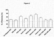
Figure 2 presents the recovery of exogenously-added IL-2 from dried blood spots applied to 903 Neonatal STD papers coated with various chemicals. -
Figure 3 presents the recovery of exogenously-added IL-2 from dried blood spots applied to DMPK-C papers coated with various chemicals. - Recombinant IL-2 ± carrier (R & D Systems; Cat. 202-IL-CF-10µg; lot AE4309112 and Cat. 202-IL-10µg; lot AE4309081 respectively) was dissolved in either Dulbecco's PBS without calcium and magnesium (PAA; Cat. H15-002, lot H00208-0673), EDTA-anti-coagulated human, rabbit or horse blood (TCS Biosciences) at 50 pg or 100 pg/µl.
- Aliquots (1 µl containing 0, 50 or 100 pg of IL-2) were applied to the following GE Healthcare filter papers; 903 Neonatal STD card, Cat. 10538069, lot 6833909 WO82; DMPK-A card, Cat. WB129241, lot FT6847509; DMPK-B card, Cat. WB129242, Lot FE6847609 and DMPK-C card, Cat. WB129243, Lot FE6847009. Samples were allowed to dry overnight at ambient temperature and humidity.
- Punches (3 mm diameter) were extracted from each paper type using the appropriately sized Harris Uni-core punch (Sigma, Cat.Z708860-25ea, lot 3110). Single punches were placed into individual wells of the IL-2 microplate derived from the Human IL-2 Quantikine ELISA (R & D Systems, Cat. D0250, lot 273275). These plates are coated with a mouse monoclonal antibody against IL-2. The IL-2 protein was eluted from the paper punch using the assay buffer (100 µl) supplied with the Quantikine kit. All subsequent steps were performed according to the instructions supplied with the Quantikine kit using a "paper in" method (paper punches are placed directly into the assay buffer and the analyte eluted directly in situ). On completion of the assay the optical density of the microplate was monitored at 450 nm using a Thermo Electron Corporation, Multiskan Ascent. The recovery of IL-2 was determined by comparing values to a standard curve of known IL-2 concentrations. A fresh IL-2 standard curve was prepared for each individual experiment.
- Additional experiments involved the addition of IL-2-spiked blood to the 903 Neonatal STD and DMPK-C cards after the cards had been saturation dipped in several chemical solutions (as described below).
- A list of the chemicals and their sources is given below.
Poly-ethyl-enemine, 50% in water (Fluka; Cat. P3143, lot 29k1492).
Poly-vinyl-pyrolodine, 1% in water (Sigma; Cat.PVP40-100 mg, lot 11pk0097).
Inulin, 1% in water (Sigma; Cat. I2255-100 g, lot 079F7110).
Poly-2-ethyl-2-oxazoline, 1 % in water (Aldrich Cat. 372846, lot 30498PJ). Albumin, 1% in water (Sigma, Cat A2153-10 g, lot 049k1586).
Caesin from bovine milk, 1% in water (Sigma, Cat. C5890-500 g, lot 089k0179).
Poly-ethylene glycol 1000, 1% in water (Biochemika, Cat. 81189, lot 1198969).
Poly-ethylene glycol 200, 1% in water (Fluka, Cat. 81150, lot 1384550). - When IL-2 was dissolved in EDTA-anti-coagulated blood, the 903 and DMPK-C cards facilitated the recovery of 45 - 55% of the cytokine, while only 2 -3 % was recovered from the DMPK-A and B cards (see Table 1 and
Figure 1 ). The 903 and DMPK-C cards are the basic base papers and have not been dipped or coated with any chemical, whilst the DMPK-A and B cards are coated with a proprietary mixture of chemicals that facilitate the denaturation and inactivation of proteins, micro-organisms and cells respectively. These cards have been designed to facilitate the transportation and prolonged storage of nucleic acids. Therefore the low IL-2 recovery levels observed when using the DMPK-A and B cards may actually be a reflection of the presence of these denaturing reagents and the ELISA-based antibody detection system used. The ELISA detection system requires the eluted IL-2 to exhibit an intact native structure.Table 1 - The Recovery of exogenously-added IL-2 from dried blood spots applied to various paper types. The p-value compares ± carrier for each paper type. The presence of the carrier had no significant effect on the recovery of IL-2 (p-value > 0.05). Paper type IL-2 recovery (%) p- value 903; minus carrier 46.9 ± 13.3 > 0.05 903; plus carrier 50.7 ± 5.8 DMPK A; minus carrier 2.0 ± 0.0 > 0.05 DM PK A; plus carrier 2.0 ± 0.0 DMPK B; minus carrier 2.0 ± 0.0 > 0.05 DM PK B; plus carrier 2.0 ± 0.0 DMPK C; minus carrier 53.9 ± 4.8 > 0.05 DMPK C; plus carrier 45.2 ± 5.4 - No IL-2 recovery was observed when the cytokine was dissolved in PBS irrespective of the paper type used (data not shown). The IL-2 recovery levels observed in the absence of added IL-2 were essentially equivalent to background levels indicating that the EDTA-anti-coagulated blood contain negligible amounts of endogenous IL-2 (data not shown).
- Several chemicals were used to saturation dip the 903 Neonatal STD and DMPK-C cards, some of which appeared to facilitate the recovery of elevated IL-2 levels compared to non-dipped papers (p-value < 0.05). For both the 903 Neonatal STD and DMPK-C cards (Tables 2 and 3;
Figures 2 and3 ), chemicals such as poly-vinyl-pyrolodine, poly-2-ethyl-2-oxazoline, albumin and casein facilitated a significant increase in IL-2 recovery levels (mean > 55 % compared to ∼ 45% observed for the corresponding un-dipped paper).Table 2 - The Recovery of exogenously-added IL-2 from dried blood spots applied to 903 Neonatal STD papers coated with various chemicals. The table is derived from 2 independent experiments (n = 6). The p-value compares the values derived from the dipped papers to those derived from the Un-dipped 903 paper. Chemical IL-2 recovery (%) p-value Un-dipped 44.9 ± 6.5 n/a Poly-ethyl-enemine (PEI) 41.8 ± 6.0 > 0.05 Poly-vinyl-pyrolodine (PVP) 62.0 ± 10.7 < 0.05 Inulin 50.4 ± 7.6 > 0.05 Poly-2-ethyl-2-oxazoline (PeOX) 66.1 ± 12.6 < 0.05 Albumin 73.8 ± 13.6 < 0.05 Caesin 55.0 ± 7.8 < 0.05 Poly-ethylene glycol 1000 (PEG 1000) 42.5 ± 9.1 > 0.05 Poly-ethylene glycol 200 (PEG 200) 43.3 ± 11.0 > 0.05 Table 3 - The Recovery of exogenously-added IL-2 from dried blood spots applied to DMPK-C coated with various chemicals (n = 3). The p-value compares the values derived from the dipped papers to those derived from the Un-dipped DMPK-C paper. Albumin* n = 1. Chemical IL-2 recovery (%) p-value Un-dipped 49.0 ± 2.1 n/a Poly-ethyl-enemine (PEI) 55.8 ± 12.2 > 0.05 Poly-vinyl-pyrolodine (PVP) 74.7 ± 7.8 < 0.05 Inulin 33.6 ± 15.4 > 0.05 Poly-2-ethyl-2-oxazoline (PeOX) 62.2 ± 2.0 < 0.05 Albumin* 63.7 increase Caesin 57.7 ± 1.5 < 0.05 Poly-ethylene glycol 1000 (PEG 1000) 31.0 ± 2.8 > 0.05 Poly-ethylene glycol 200 (PEG 200) 33.5 ± 15.7 > 0.05 - While preferred illustrative embodiments of the present invention are described, one skilled in the art will appreciate that the present invention can be practised by other than the described embodiments, which are presented for the purposes of illustration only and not by way of limitation. The present invention is limited only by the claims that follow.
Claims (11)
- A method of recovering a biological material from a solid support having at least one surface coated with a chemical selected from the group consisting of polyvinyl pyrrolidone (PVP), poly-2-ethyl-2-oxazoline (PEOX), albumin and casein, comprising the steps ofi) contacting a surface of a solid support with a sample containing a biological material;ii) drying said sample on said surface of said support;iii) storing the support; andiv) extracting said biological material from the surface.
- The method of claim 1, wherein said solid support is selected from the group consisting of paper, glass microfiber and membrane.
- The method according to claim 1, wherein the solid support is a paper.
- The method of claim 3, wherein said paper is a cellulose paper.
- The method of claim 2, wherein said membrane is selected from the group consisting of polyester, polyether sulfone (PES), polyamide (Nylon), polypropylene, polytetrafluoroethylene (PTFE), polycarbonate, cellulose nitrate, cellulose acetate and aluminium oxide.
- The method according to any preceding claim wherein step iii) comprises storing the solid support at a temperature in the range of 15 to 40°C.
- The method according to any preceding claim, wherein the sample is selected from the group consisting of tissue, cell, blood, plasma, saliva and urine.
- The method according to any preceding claim, wherein said biological material is selected from the group consisting of biomolecule, synthetically-derived biomolecule, cellular component and biopharmaceutical drug.
- The method according to any preceding claim, wherein said biological material is a biopharmaceutical drug.
- Use of a solid support having at least one surface coated with a chemical selected from the group consisting of polyvinyl pyrrolidone (PVP), poly-2-ethyl-2-oxazoline (PEOX), albumin and casein to facilitate the recovery of a biological material therefrom.
- Use of a solid support having at least one surface coated with a chemical selected from the group consisting of polyvinyl pyrrolidone (PVP), poly-2-ethyl-2-oxazoline (PEOX), albumin and casein to facilitate the recovery of a biopharmaceutical drug therefrom.
Applications Claiming Priority (2)
| Application Number | Priority Date | Filing Date | Title |
|---|---|---|---|
| GBGB1103258.8A GB201103258D0 (en) | 2011-02-25 | 2011-02-25 | Solid support and method of enhancing the recovery of biological material therefrom |
| PCT/EP2012/053163 WO2012113906A2 (en) | 2011-02-25 | 2012-02-24 | Solid support and method of enhancing the recovery of biological material therefrom |
Publications (2)
| Publication Number | Publication Date |
|---|---|
| EP2678692A2 EP2678692A2 (en) | 2014-01-01 |
| EP2678692B1 true EP2678692B1 (en) | 2016-06-22 |
Family
ID=43904184
Family Applications (1)
| Application Number | Title | Priority Date | Filing Date |
|---|---|---|---|
| EP12705857.6A Active EP2678692B1 (en) | 2011-02-25 | 2012-02-24 | Solid support and method of enhancing the recovery of biological material therefrom |
Country Status (9)
| Country | Link |
|---|---|
| US (1) | US10876938B2 (en) |
| EP (1) | EP2678692B1 (en) |
| JP (1) | JP6074369B2 (en) |
| CN (1) | CN103460051B (en) |
| AU (1) | AU2012219482B2 (en) |
| CA (1) | CA2828158C (en) |
| ES (1) | ES2592963T3 (en) |
| GB (1) | GB201103258D0 (en) |
| WO (1) | WO2012113906A2 (en) |
Families Citing this family (5)
| Publication number | Priority date | Publication date | Assignee | Title |
|---|---|---|---|---|
| CN107208136A (en) | 2014-12-18 | 2017-09-26 | 通用电气医疗集团英国有限公司 | Pass through the analysis analyte detection on the solid support for the nucleic acid amplification being coupled with immunoassay |
| FI4035762T3 (en) | 2015-09-09 | 2023-12-04 | Drawbridge Health Inc | Devices for sample collection, stabilization and preservation |
| MY196758A (en) | 2017-01-10 | 2023-05-03 | Drawbridge Health Inc | Devices, systems, and methods for sample collection |
| EP3521828A1 (en) * | 2018-01-31 | 2019-08-07 | Centogene AG | Method for the diagnosis of hereditary angioedema |
| JPWO2023181451A1 (en) * | 2022-03-23 | 2023-09-28 |
Family Cites Families (27)
| Publication number | Priority date | Publication date | Assignee | Title |
|---|---|---|---|---|
| US575126A (en) | 1897-01-12 | Watch-pocket guard | ||
| BE542544A (en) * | 1954-11-04 | |||
| US3227075A (en) | 1961-04-04 | 1966-01-04 | Fitchburg Paper | Planographic printing plates |
| JPS5319092A (en) | 1976-11-08 | 1978-02-21 | Shionogi Seiyaku Kk | Composite for liver desease detection |
| JPH01291164A (en) | 1988-05-19 | 1989-11-22 | Fujirebio Inc | Composition for standard filter paper |
| US5756126A (en) | 1991-05-29 | 1998-05-26 | Flinders Technologies Pty. Ltd. | Dry solid medium for storage and analysis of genetic material |
| US5985327A (en) | 1988-10-05 | 1999-11-16 | Flinders Technologies Pty. Ltd. | Solid medium and method for DNA storage |
| US5188938A (en) * | 1988-12-29 | 1993-02-23 | Microgenics Corporation | Enzyme quantitation wicking assay |
| US5756362A (en) * | 1993-10-12 | 1998-05-26 | Cornell Research Foundation, Inc. | Liposome-enhanced immunoaggregation assay and test device |
| US6132971A (en) * | 1995-06-27 | 2000-10-17 | The University Of North Carolina At Chapel Hill | Microelectronic device |
| US5804684A (en) * | 1995-08-24 | 1998-09-08 | The Theobald Smith Research Institute, Inc. | Method for isolating nucleic acids |
| JPH10179722A (en) | 1996-12-25 | 1998-07-07 | Sumitomo Bakelite Co Ltd | Surface treatment |
| US5939259A (en) | 1997-04-09 | 1999-08-17 | Schleicher & Schuell, Inc. | Methods and devices for collecting and storing clinical samples for genetic analysis |
| US7670768B1 (en) * | 1998-02-02 | 2010-03-02 | Qiagen North American Holdings, Inc. | Processes for isolating, amplifying and characterizing DNA |
| US6187540B1 (en) * | 1998-11-09 | 2001-02-13 | Identigene, Inc. | Method of newborn identification and tracking |
| EP1173623B1 (en) | 1999-03-11 | 2008-06-25 | Whatman, Inc. | Solid medium and process for the storage and rapid purification of nucleic acid |
| EP1423514A2 (en) | 2001-09-05 | 2004-06-02 | WHATMAN plc | Stable storage of proteins |
| US20030215358A1 (en) | 2002-01-15 | 2003-11-20 | Schulman Lloyd S. | Liquid permeable composition in dry reagent devices |
| US7156945B2 (en) | 2002-04-24 | 2007-01-02 | Sipix Imaging, Inc. | Process for forming a patterned thin film structure for in-mold decoration |
| ES2320875T5 (en) * | 2002-10-04 | 2012-10-30 | Ge Healthcare Bio-Sciences Corp. | Procedures and materials for using chemical compounds as a tool for the storage of nucleic acids on means of nucleic acid purification systems |
| US7521021B2 (en) * | 2003-11-26 | 2009-04-21 | Leica Biosvstems St. Louis Llc | System for in situ processing of a tissue specimen |
| US7045295B2 (en) | 2004-04-02 | 2006-05-16 | Hematologics, Inc. | Method for collecting purified cells |
| ATE497837T1 (en) * | 2004-04-09 | 2011-02-15 | Vivebio Llc | DEVICES AND METHODS FOR COLLECTION, STORAGE AND TRANSPORTATION OF BIOLOGICAL SAMPLES |
| US8062901B2 (en) * | 2005-04-30 | 2011-11-22 | Alere Switzerland Gmbh | Devices and methods for sample collection and analysis |
| DE602006020158D1 (en) * | 2005-11-08 | 2011-03-31 | Surmodics Inc | ULTRA-THIN PHOTOPOLYMER COATINGS AND USES THEREOF |
| CA2683729A1 (en) * | 2007-04-16 | 2008-10-23 | Mcmaster University | Method of producing bioactive paper |
| CN101892290A (en) * | 2009-05-19 | 2010-11-24 | 北京协和洛克生物技术研究开发中心 | Phenylalanine quantitative detection kit (enzyme quantitative method) |
-
2011
- 2011-02-25 GB GBGB1103258.8A patent/GB201103258D0/en not_active Ceased
-
2012
- 2012-02-24 US US13/985,089 patent/US10876938B2/en active Active
- 2012-02-24 CA CA2828158A patent/CA2828158C/en active Active
- 2012-02-24 ES ES12705857.6T patent/ES2592963T3/en active Active
- 2012-02-24 AU AU2012219482A patent/AU2012219482B2/en active Active
- 2012-02-24 EP EP12705857.6A patent/EP2678692B1/en active Active
- 2012-02-24 JP JP2013554907A patent/JP6074369B2/en active Active
- 2012-02-24 CN CN201280010442.3A patent/CN103460051B/en active Active
- 2012-02-24 WO PCT/EP2012/053163 patent/WO2012113906A2/en active Application Filing
Also Published As
| Publication number | Publication date |
|---|---|
| WO2012113906A3 (en) | 2012-12-20 |
| CA2828158C (en) | 2021-10-26 |
| WO2012113906A2 (en) | 2012-08-30 |
| GB201103258D0 (en) | 2011-04-13 |
| ES2592963T3 (en) | 2016-12-02 |
| US10876938B2 (en) | 2020-12-29 |
| CN103460051B (en) | 2015-10-07 |
| AU2012219482B2 (en) | 2016-10-27 |
| US20130330750A1 (en) | 2013-12-12 |
| JP6074369B2 (en) | 2017-02-01 |
| CA2828158A1 (en) | 2012-08-30 |
| CN103460051A (en) | 2013-12-18 |
| AU2012219482A1 (en) | 2013-08-29 |
| JP2014512516A (en) | 2014-05-22 |
| EP2678692A2 (en) | 2014-01-01 |
Similar Documents
| Publication | Publication Date | Title |
|---|---|---|
| EP2678681B1 (en) | Solid support and method of recovering biological material therefrom | |
| Edelbroek et al. | Dried blood spot methods in therapeutic drug monitoring: methods, assays, and pitfalls | |
| US20130323778A1 (en) | Paper support and method of recovering biological material therefrom | |
| EP2678692B1 (en) | Solid support and method of enhancing the recovery of biological material therefrom | |
| EP1461615A1 (en) | Diagnostic testing process | |
| Nevídalová et al. | Capillary electrophoresis–based immunoassay and aptamer assay: A review | |
| CN102135535A (en) | Immune colloidal metal detection technology capable of directly performing semi-quantitative analysis, preparation method and application | |
| US20040248181A1 (en) | Method and kit for enhancing extraction and quantification of target molecules using microdialysis | |
| US20220273209A1 (en) | Devices and methods for sample collection | |
| Ohnmacht et al. | Sample stabilization strategies: a case study review of unique sample collection and handling procedures | |
| Andrlova et al. | The dried blood spot sampling method in the laboratory medicine. | |
| US20200341005A1 (en) | Carrier and method for detecting an analyte in dried blood spots | |
| Laštovičková et al. | The dried blood spot sampling method in the laboratory medicine | |
| Oliveira et al. | Collection and Bioanalysis of Quantitative Microsamples: Technological Innovations and Practical Implications | |
| KR100437887B1 (en) | Western blot diagnostic kit using principle of chromatography | |
| RU2168725C2 (en) | Method for producing solid-state carrier for performing immune analysis | |
| Cheng et al. | Introduction to In Vitro Diagnostic Devices |
Legal Events
| Date | Code | Title | Description |
|---|---|---|---|
| PUAI | Public reference made under article 153(3) epc to a published international application that has entered the european phase |
Free format text: ORIGINAL CODE: 0009012 |
|
| 17P | Request for examination filed |
Effective date: 20130809 |
|
| AK | Designated contracting states |
Kind code of ref document: A2 Designated state(s): AL AT BE BG CH CY CZ DE DK EE ES FI FR GB GR HR HU IE IS IT LI LT LU LV MC MK MT NL NO PL PT RO RS SE SI SK SM TR |
|
| DAX | Request for extension of the european patent (deleted) | ||
| GRAP | Despatch of communication of intention to grant a patent |
Free format text: ORIGINAL CODE: EPIDOSNIGR1 |
|
| INTG | Intention to grant announced |
Effective date: 20150923 |
|
| GRAS | Grant fee paid |
Free format text: ORIGINAL CODE: EPIDOSNIGR3 |
|
| GRAA | (expected) grant |
Free format text: ORIGINAL CODE: 0009210 |
|
| AK | Designated contracting states |
Kind code of ref document: B1 Designated state(s): AL AT BE BG CH CY CZ DE DK EE ES FI FR GB GR HR HU IE IS IT LI LT LU LV MC MK MT NL NO PL PT RO RS SE SI SK SM TR |
|
| REG | Reference to a national code |
Ref country code: GB Ref legal event code: FG4D |
|
| REG | Reference to a national code |
Ref country code: CH Ref legal event code: EP |
|
| REG | Reference to a national code |
Ref country code: IE Ref legal event code: FG4D |
|
| REG | Reference to a national code |
Ref country code: AT Ref legal event code: REF Ref document number: 807971 Country of ref document: AT Kind code of ref document: T Effective date: 20160715 |
|
| REG | Reference to a national code |
Ref country code: DE Ref legal event code: R096 Ref document number: 602012019723 Country of ref document: DE |
|
| REG | Reference to a national code |
Ref country code: NL Ref legal event code: FP |
|
| REG | Reference to a national code |
Ref country code: LT Ref legal event code: MG4D |
|
| PG25 | Lapsed in a contracting state [announced via postgrant information from national office to epo] |
Ref country code: FI Free format text: LAPSE BECAUSE OF FAILURE TO SUBMIT A TRANSLATION OF THE DESCRIPTION OR TO PAY THE FEE WITHIN THE PRESCRIBED TIME-LIMIT Effective date: 20160622 Ref country code: NO Free format text: LAPSE BECAUSE OF FAILURE TO SUBMIT A TRANSLATION OF THE DESCRIPTION OR TO PAY THE FEE WITHIN THE PRESCRIBED TIME-LIMIT Effective date: 20160922 Ref country code: LT Free format text: LAPSE BECAUSE OF FAILURE TO SUBMIT A TRANSLATION OF THE DESCRIPTION OR TO PAY THE FEE WITHIN THE PRESCRIBED TIME-LIMIT Effective date: 20160622 |
|
| REG | Reference to a national code |
Ref country code: AT Ref legal event code: MK05 Ref document number: 807971 Country of ref document: AT Kind code of ref document: T Effective date: 20160622 |
|
| PG25 | Lapsed in a contracting state [announced via postgrant information from national office to epo] |
Ref country code: GR Free format text: LAPSE BECAUSE OF FAILURE TO SUBMIT A TRANSLATION OF THE DESCRIPTION OR TO PAY THE FEE WITHIN THE PRESCRIBED TIME-LIMIT Effective date: 20160923 Ref country code: LV Free format text: LAPSE BECAUSE OF FAILURE TO SUBMIT A TRANSLATION OF THE DESCRIPTION OR TO PAY THE FEE WITHIN THE PRESCRIBED TIME-LIMIT Effective date: 20160622 Ref country code: RS Free format text: LAPSE BECAUSE OF FAILURE TO SUBMIT A TRANSLATION OF THE DESCRIPTION OR TO PAY THE FEE WITHIN THE PRESCRIBED TIME-LIMIT Effective date: 20160622 Ref country code: SE Free format text: LAPSE BECAUSE OF FAILURE TO SUBMIT A TRANSLATION OF THE DESCRIPTION OR TO PAY THE FEE WITHIN THE PRESCRIBED TIME-LIMIT Effective date: 20160622 Ref country code: HR Free format text: LAPSE BECAUSE OF FAILURE TO SUBMIT A TRANSLATION OF THE DESCRIPTION OR TO PAY THE FEE WITHIN THE PRESCRIBED TIME-LIMIT Effective date: 20160622 |
|
| REG | Reference to a national code |
Ref country code: ES Ref legal event code: FG2A Ref document number: 2592963 Country of ref document: ES Kind code of ref document: T3 Effective date: 20161202 |
|
| PG25 | Lapsed in a contracting state [announced via postgrant information from national office to epo] |
Ref country code: RO Free format text: LAPSE BECAUSE OF FAILURE TO SUBMIT A TRANSLATION OF THE DESCRIPTION OR TO PAY THE FEE WITHIN THE PRESCRIBED TIME-LIMIT Effective date: 20160622 Ref country code: IS Free format text: LAPSE BECAUSE OF FAILURE TO SUBMIT A TRANSLATION OF THE DESCRIPTION OR TO PAY THE FEE WITHIN THE PRESCRIBED TIME-LIMIT Effective date: 20161022 Ref country code: CZ Free format text: LAPSE BECAUSE OF FAILURE TO SUBMIT A TRANSLATION OF THE DESCRIPTION OR TO PAY THE FEE WITHIN THE PRESCRIBED TIME-LIMIT Effective date: 20160622 Ref country code: EE Free format text: LAPSE BECAUSE OF FAILURE TO SUBMIT A TRANSLATION OF THE DESCRIPTION OR TO PAY THE FEE WITHIN THE PRESCRIBED TIME-LIMIT Effective date: 20160622 Ref country code: SK Free format text: LAPSE BECAUSE OF FAILURE TO SUBMIT A TRANSLATION OF THE DESCRIPTION OR TO PAY THE FEE WITHIN THE PRESCRIBED TIME-LIMIT Effective date: 20160622 |
|
| REG | Reference to a national code |
Ref country code: FR Ref legal event code: PLFP Year of fee payment: 6 |
|
| PG25 | Lapsed in a contracting state [announced via postgrant information from national office to epo] |
Ref country code: BE Free format text: LAPSE BECAUSE OF FAILURE TO SUBMIT A TRANSLATION OF THE DESCRIPTION OR TO PAY THE FEE WITHIN THE PRESCRIBED TIME-LIMIT Effective date: 20160622 Ref country code: AT Free format text: LAPSE BECAUSE OF FAILURE TO SUBMIT A TRANSLATION OF THE DESCRIPTION OR TO PAY THE FEE WITHIN THE PRESCRIBED TIME-LIMIT Effective date: 20160622 Ref country code: PT Free format text: LAPSE BECAUSE OF FAILURE TO SUBMIT A TRANSLATION OF THE DESCRIPTION OR TO PAY THE FEE WITHIN THE PRESCRIBED TIME-LIMIT Effective date: 20161024 Ref country code: PL Free format text: LAPSE BECAUSE OF FAILURE TO SUBMIT A TRANSLATION OF THE DESCRIPTION OR TO PAY THE FEE WITHIN THE PRESCRIBED TIME-LIMIT Effective date: 20160622 Ref country code: SM Free format text: LAPSE BECAUSE OF FAILURE TO SUBMIT A TRANSLATION OF THE DESCRIPTION OR TO PAY THE FEE WITHIN THE PRESCRIBED TIME-LIMIT Effective date: 20160622 |
|
| REG | Reference to a national code |
Ref country code: DE Ref legal event code: R097 Ref document number: 602012019723 Country of ref document: DE |
|
| PLBE | No opposition filed within time limit |
Free format text: ORIGINAL CODE: 0009261 |
|
| STAA | Information on the status of an ep patent application or granted ep patent |
Free format text: STATUS: NO OPPOSITION FILED WITHIN TIME LIMIT |
|
| 26N | No opposition filed |
Effective date: 20170323 |
|
| PG25 | Lapsed in a contracting state [announced via postgrant information from national office to epo] |
Ref country code: DK Free format text: LAPSE BECAUSE OF FAILURE TO SUBMIT A TRANSLATION OF THE DESCRIPTION OR TO PAY THE FEE WITHIN THE PRESCRIBED TIME-LIMIT Effective date: 20160622 |
|
| PG25 | Lapsed in a contracting state [announced via postgrant information from national office to epo] |
Ref country code: SI Free format text: LAPSE BECAUSE OF FAILURE TO SUBMIT A TRANSLATION OF THE DESCRIPTION OR TO PAY THE FEE WITHIN THE PRESCRIBED TIME-LIMIT Effective date: 20160622 |
|
| PG25 | Lapsed in a contracting state [announced via postgrant information from national office to epo] |
Ref country code: MC Free format text: LAPSE BECAUSE OF FAILURE TO SUBMIT A TRANSLATION OF THE DESCRIPTION OR TO PAY THE FEE WITHIN THE PRESCRIBED TIME-LIMIT Effective date: 20160622 |
|
| REG | Reference to a national code |
Ref country code: CH Ref legal event code: PL |
|
| PG25 | Lapsed in a contracting state [announced via postgrant information from national office to epo] |
Ref country code: LI Free format text: LAPSE BECAUSE OF NON-PAYMENT OF DUE FEES Effective date: 20170228 Ref country code: CH Free format text: LAPSE BECAUSE OF NON-PAYMENT OF DUE FEES Effective date: 20170228 |
|
| REG | Reference to a national code |
Ref country code: IE Ref legal event code: MM4A |
|
| PG25 | Lapsed in a contracting state [announced via postgrant information from national office to epo] |
Ref country code: LU Free format text: LAPSE BECAUSE OF NON-PAYMENT OF DUE FEES Effective date: 20170224 |
|
| REG | Reference to a national code |
Ref country code: FR Ref legal event code: PLFP Year of fee payment: 7 |
|
| PG25 | Lapsed in a contracting state [announced via postgrant information from national office to epo] |
Ref country code: IE Free format text: LAPSE BECAUSE OF NON-PAYMENT OF DUE FEES Effective date: 20170224 |
|
| PG25 | Lapsed in a contracting state [announced via postgrant information from national office to epo] |
Ref country code: MT Free format text: LAPSE BECAUSE OF NON-PAYMENT OF DUE FEES Effective date: 20170224 |
|
| PG25 | Lapsed in a contracting state [announced via postgrant information from national office to epo] |
Ref country code: AL Free format text: LAPSE BECAUSE OF FAILURE TO SUBMIT A TRANSLATION OF THE DESCRIPTION OR TO PAY THE FEE WITHIN THE PRESCRIBED TIME-LIMIT Effective date: 20160622 |
|
| PG25 | Lapsed in a contracting state [announced via postgrant information from national office to epo] |
Ref country code: HU Free format text: LAPSE BECAUSE OF FAILURE TO SUBMIT A TRANSLATION OF THE DESCRIPTION OR TO PAY THE FEE WITHIN THE PRESCRIBED TIME-LIMIT; INVALID AB INITIO Effective date: 20120224 |
|
| PG25 | Lapsed in a contracting state [announced via postgrant information from national office to epo] |
Ref country code: BG Free format text: LAPSE BECAUSE OF FAILURE TO SUBMIT A TRANSLATION OF THE DESCRIPTION OR TO PAY THE FEE WITHIN THE PRESCRIBED TIME-LIMIT Effective date: 20160622 |
|
| PG25 | Lapsed in a contracting state [announced via postgrant information from national office to epo] |
Ref country code: CY Free format text: LAPSE BECAUSE OF NON-PAYMENT OF DUE FEES Effective date: 20160622 |
|
| PG25 | Lapsed in a contracting state [announced via postgrant information from national office to epo] |
Ref country code: MK Free format text: LAPSE BECAUSE OF FAILURE TO SUBMIT A TRANSLATION OF THE DESCRIPTION OR TO PAY THE FEE WITHIN THE PRESCRIBED TIME-LIMIT Effective date: 20160622 |
|
| PG25 | Lapsed in a contracting state [announced via postgrant information from national office to epo] |
Ref country code: TR Free format text: LAPSE BECAUSE OF FAILURE TO SUBMIT A TRANSLATION OF THE DESCRIPTION OR TO PAY THE FEE WITHIN THE PRESCRIBED TIME-LIMIT Effective date: 20160622 |
|
| REG | Reference to a national code |
Ref country code: DE Ref legal event code: R081 Ref document number: 602012019723 Country of ref document: DE Owner name: GLOBAL LIFE SCIENCES SOLUTIONS OPERATIONS UK L, GB Free format text: FORMER OWNER: GE HEALTHCARE UK LIMITED, LITTLE CHALFONT, BUCKINGHAMSHIRE, GB |
|
| REG | Reference to a national code |
Ref country code: ES Ref legal event code: PC2A Owner name: GLOBAL LIFE SCIENCES SOLUTIONS OPERATIONS UK LTD Effective date: 20210115 |
|
| REG | Reference to a national code |
Ref country code: NL Ref legal event code: PD Owner name: GLOBAL LIFE SCIENCES SOLUTIONS OPERATIONS UK LTD; GB Free format text: DETAILS ASSIGNMENT: CHANGE OF OWNER(S), ASSIGNMENT; FORMER OWNER NAME: GE HEALTHCARE UK LIMITED Effective date: 20210218 |
|
| REG | Reference to a national code |
Ref country code: GB Ref legal event code: 732E Free format text: REGISTERED BETWEEN 20211210 AND 20211215 |
|
| P01 | Opt-out of the competence of the unified patent court (upc) registered |
Effective date: 20230526 |
|
| PGFP | Annual fee paid to national office [announced via postgrant information from national office to epo] |
Ref country code: NL Payment date: 20240108 Year of fee payment: 13 |
|
| PGFP | Annual fee paid to national office [announced via postgrant information from national office to epo] |
Ref country code: ES Payment date: 20240306 Year of fee payment: 13 |
|
| PGFP | Annual fee paid to national office [announced via postgrant information from national office to epo] |
Ref country code: DE Payment date: 20231229 Year of fee payment: 13 Ref country code: GB Payment date: 20240108 Year of fee payment: 13 |
|
| PGFP | Annual fee paid to national office [announced via postgrant information from national office to epo] |
Ref country code: IT Payment date: 20240111 Year of fee payment: 13 Ref country code: FR Payment date: 20240103 Year of fee payment: 13 |