EP2637705B1 - Konjugate und ihre verwendung in der molekularen bildgebung - Google Patents
Konjugate und ihre verwendung in der molekularen bildgebung Download PDFInfo
- Publication number
- EP2637705B1 EP2637705B1 EP11788554.1A EP11788554A EP2637705B1 EP 2637705 B1 EP2637705 B1 EP 2637705B1 EP 11788554 A EP11788554 A EP 11788554A EP 2637705 B1 EP2637705 B1 EP 2637705B1
- Authority
- EP
- European Patent Office
- Prior art keywords
- bifunctional
- bifunctional compound
- group
- molecule
- radionuclide
- Prior art date
- Legal status (The legal status is an assumption and is not a legal conclusion. Google has not performed a legal analysis and makes no representation as to the accuracy of the status listed.)
- Active
Links
- 238000003384 imaging method Methods 0.000 title claims description 76
- 230000001588 bifunctional effect Effects 0.000 claims description 201
- 150000001875 compounds Chemical class 0.000 claims description 145
- 238000000034 method Methods 0.000 claims description 73
- 230000008685 targeting Effects 0.000 claims description 66
- 230000027455 binding Effects 0.000 claims description 46
- 108090000765 processed proteins & peptides Proteins 0.000 claims description 46
- 108090000623 proteins and genes Proteins 0.000 claims description 43
- 102000004169 proteins and genes Human genes 0.000 claims description 42
- 239000003446 ligand Substances 0.000 claims description 34
- 239000000203 mixture Substances 0.000 claims description 34
- 239000000523 sample Substances 0.000 claims description 28
- 210000004027 cell Anatomy 0.000 claims description 25
- 238000001727 in vivo Methods 0.000 claims description 25
- 238000011362 radionuclide therapy Methods 0.000 claims description 18
- 210000001519 tissue Anatomy 0.000 claims description 18
- 206010028980 Neoplasm Diseases 0.000 claims description 16
- 229910052733 gallium Inorganic materials 0.000 claims description 14
- 239000002243 precursor Substances 0.000 claims description 13
- GYHNNYVSQQEPJS-UHFFFAOYSA-N Gallium Chemical compound [Ga] GYHNNYVSQQEPJS-UHFFFAOYSA-N 0.000 claims description 12
- -1 antibody Proteins 0.000 claims description 12
- 201000011510 cancer Diseases 0.000 claims description 12
- 150000003839 salts Chemical class 0.000 claims description 12
- 102000002110 C2 domains Human genes 0.000 claims description 10
- 108050009459 C2 domains Proteins 0.000 claims description 10
- 238000005859 coupling reaction Methods 0.000 claims description 10
- OAKJQQAXSVQMHS-UHFFFAOYSA-N Hydrazine Chemical compound NN OAKJQQAXSVQMHS-UHFFFAOYSA-N 0.000 claims description 9
- 239000000427 antigen Substances 0.000 claims description 9
- 108091007433 antigens Proteins 0.000 claims description 9
- 102000036639 antigens Human genes 0.000 claims description 9
- 230000008878 coupling Effects 0.000 claims description 9
- 238000010168 coupling process Methods 0.000 claims description 9
- 229910052739 hydrogen Inorganic materials 0.000 claims description 9
- 239000001257 hydrogen Substances 0.000 claims description 9
- 125000005647 linker group Chemical group 0.000 claims description 9
- 125000006701 (C1-C7) alkyl group Chemical group 0.000 claims description 8
- 201000010099 disease Diseases 0.000 claims description 8
- 208000037265 diseases, disorders, signs and symptoms Diseases 0.000 claims description 8
- 102000003137 synaptotagmin Human genes 0.000 claims description 7
- 108060008004 synaptotagmin Proteins 0.000 claims description 7
- 125000004435 hydrogen atom Chemical group [H]* 0.000 claims description 6
- 108020003175 receptors Proteins 0.000 claims description 6
- 102000005962 receptors Human genes 0.000 claims description 6
- IYMAXBFPHPZYIK-BQBZGAKWSA-N Arg-Gly-Asp Chemical compound NC(N)=NCCC[C@H](N)C(=O)NCC(=O)N[C@@H](CC(O)=O)C(O)=O IYMAXBFPHPZYIK-BQBZGAKWSA-N 0.000 claims description 5
- XEEYBQQBJWHFJM-UHFFFAOYSA-N Iron Chemical compound [Fe] XEEYBQQBJWHFJM-UHFFFAOYSA-N 0.000 claims description 5
- 125000000524 functional group Chemical group 0.000 claims description 5
- 239000003550 marker Substances 0.000 claims description 5
- 210000000056 organ Anatomy 0.000 claims description 5
- 238000001959 radiotherapy Methods 0.000 claims description 5
- 229910052727 yttrium Inorganic materials 0.000 claims description 5
- YBJHBAHKTGYVGT-ZKWXMUAHSA-N (+)-Biotin Chemical compound N1C(=O)N[C@@H]2[C@H](CCCCC(=O)O)SC[C@@H]21 YBJHBAHKTGYVGT-ZKWXMUAHSA-N 0.000 claims description 4
- UFHFLCQGNIYNRP-UHFFFAOYSA-N Hydrogen Chemical compound [H][H] UFHFLCQGNIYNRP-UHFFFAOYSA-N 0.000 claims description 4
- 206010021143 Hypoxia Diseases 0.000 claims description 4
- 150000001299 aldehydes Chemical class 0.000 claims description 4
- 125000005262 alkoxyamine group Chemical group 0.000 claims description 4
- 230000007954 hypoxia Effects 0.000 claims description 4
- 108090000672 Annexin A5 Proteins 0.000 claims description 3
- 102000004121 Annexin A5 Human genes 0.000 claims description 3
- 108091023037 Aptamer Proteins 0.000 claims description 3
- 108010051479 Bombesin Proteins 0.000 claims description 3
- 102000013585 Bombesin Human genes 0.000 claims description 3
- RYGMFSIKBFXOCR-UHFFFAOYSA-N Copper Chemical compound [Cu] RYGMFSIKBFXOCR-UHFFFAOYSA-N 0.000 claims description 3
- 108010052343 Gastrins Proteins 0.000 claims description 3
- 229910052765 Lutetium Inorganic materials 0.000 claims description 3
- PEEHTFAAVSWFBL-UHFFFAOYSA-N Maleimide Chemical compound O=C1NC(=O)C=C1 PEEHTFAAVSWFBL-UHFFFAOYSA-N 0.000 claims description 3
- 101710160107 Outer membrane protein A Proteins 0.000 claims description 3
- 108050001286 Somatostatin Receptor Proteins 0.000 claims description 3
- 102000011096 Somatostatin receptor Human genes 0.000 claims description 3
- 108010031372 Tissue Inhibitor of Metalloproteinase-2 Proteins 0.000 claims description 3
- 102000005354 Tissue Inhibitor of Metalloproteinase-2 Human genes 0.000 claims description 3
- QCWXUUIWCKQGHC-UHFFFAOYSA-N Zirconium Chemical compound [Zr] QCWXUUIWCKQGHC-UHFFFAOYSA-N 0.000 claims description 3
- 150000001336 alkenes Chemical class 0.000 claims description 3
- 150000001345 alkine derivatives Chemical class 0.000 claims description 3
- DNDCVAGJPBKION-DOPDSADYSA-N bombesin Chemical compound C([C@@H](C(=O)N[C@@H](CC(C)C)C(=O)N[C@@H](CCSC)C(N)=O)NC(=O)CNC(=O)[C@@H](NC(=O)[C@H](C)NC(=O)[C@H](CC=1NC2=CC=CC=C2C=1)NC(=O)[C@H](CCC(N)=O)NC(=O)[C@H](CC(N)=O)NC(=O)CNC(=O)[C@H](CC(C)C)NC(=O)[C@H](CCCNC(N)=N)NC(=O)[C@H](CCC(N)=O)NC(=O)[C@H]1NC(=O)CC1)C(C)C)C1=CN=CN1 DNDCVAGJPBKION-DOPDSADYSA-N 0.000 claims description 3
- 210000000988 bone and bone Anatomy 0.000 claims description 3
- AOXOCDRNSPFDPE-UKEONUMOSA-N chembl413654 Chemical compound C([C@H](C(=O)NCC(=O)N[C@H](CC=1C2=CC=CC=C2NC=1)C(=O)N[C@H](CCSC)C(=O)N[C@H](CC(O)=O)C(=O)N[C@H](CC=1C=CC=CC=1)C(N)=O)NC(=O)[C@@H](C)NC(=O)[C@@H](CCC(O)=O)NC(=O)[C@@H](CCC(O)=O)NC(=O)[C@@H](CCC(O)=O)NC(=O)[C@H](CCC(O)=O)NC(=O)[C@H](CCC(O)=O)NC(=O)[C@H](CC(C)C)NC(=O)[C@H](CC=1C2=CC=CC=C2NC=1)NC(=O)[C@H]1N(CCC1)C(=O)CNC(=O)[C@@H](N)CCC(O)=O)C1=CC=C(O)C=C1 AOXOCDRNSPFDPE-UKEONUMOSA-N 0.000 claims description 3
- 229910017052 cobalt Inorganic materials 0.000 claims description 3
- 239000010941 cobalt Substances 0.000 claims description 3
- GUTLYIVDDKVIGB-UHFFFAOYSA-N cobalt atom Chemical compound [Co] GUTLYIVDDKVIGB-UHFFFAOYSA-N 0.000 claims description 3
- 229910052802 copper Inorganic materials 0.000 claims description 3
- 239000010949 copper Substances 0.000 claims description 3
- 229910052738 indium Inorganic materials 0.000 claims description 3
- APFVFJFRJDLVQX-UHFFFAOYSA-N indium atom Chemical compound [In] APFVFJFRJDLVQX-UHFFFAOYSA-N 0.000 claims description 3
- OHSVLFRHMCKCQY-UHFFFAOYSA-N lutetium atom Chemical compound [Lu] OHSVLFRHMCKCQY-UHFFFAOYSA-N 0.000 claims description 3
- 229910052702 rhenium Inorganic materials 0.000 claims description 3
- WUAPFZMCVAUBPE-UHFFFAOYSA-N rhenium atom Chemical compound [Re] WUAPFZMCVAUBPE-UHFFFAOYSA-N 0.000 claims description 3
- 229910052706 scandium Inorganic materials 0.000 claims description 3
- SIXSYDAISGFNSX-UHFFFAOYSA-N scandium atom Chemical compound [Sc] SIXSYDAISGFNSX-UHFFFAOYSA-N 0.000 claims description 3
- 150000003384 small molecules Chemical class 0.000 claims description 3
- 229910052713 technetium Inorganic materials 0.000 claims description 3
- GKLVYJBZJHMRIY-UHFFFAOYSA-N technetium atom Chemical compound [Tc] GKLVYJBZJHMRIY-UHFFFAOYSA-N 0.000 claims description 3
- VWQVUPCCIRVNHF-UHFFFAOYSA-N yttrium atom Chemical compound [Y] VWQVUPCCIRVNHF-UHFFFAOYSA-N 0.000 claims description 3
- 229910052726 zirconium Inorganic materials 0.000 claims description 3
- 108091032973 (ribonucleotides)n+m Proteins 0.000 claims description 2
- 108020005544 Antisense RNA Proteins 0.000 claims description 2
- 108090001008 Avidin Proteins 0.000 claims description 2
- 229940122361 Bisphosphonate Drugs 0.000 claims description 2
- WQZGKKKJIJFFOK-GASJEMHNSA-N Glucose Natural products OC[C@H]1OC(O)[C@H](O)[C@@H](O)[C@@H]1O WQZGKKKJIJFFOK-GASJEMHNSA-N 0.000 claims description 2
- 108091052347 Glucose transporter family Proteins 0.000 claims description 2
- 108010090804 Streptavidin Proteins 0.000 claims description 2
- 108010072041 arginyl-glycyl-aspartic acid Proteins 0.000 claims description 2
- 229960002685 biotin Drugs 0.000 claims description 2
- 235000020958 biotin Nutrition 0.000 claims description 2
- 239000011616 biotin Substances 0.000 claims description 2
- 150000004663 bisphosphonates Chemical class 0.000 claims description 2
- 239000003184 complementary RNA Substances 0.000 claims description 2
- 150000002148 esters Chemical class 0.000 claims description 2
- 239000008103 glucose Substances 0.000 claims description 2
- 150000002429 hydrazines Chemical class 0.000 claims description 2
- 229910052500 inorganic mineral Inorganic materials 0.000 claims description 2
- 229910052742 iron Inorganic materials 0.000 claims description 2
- 230000002503 metabolic effect Effects 0.000 claims description 2
- 239000011707 mineral Substances 0.000 claims description 2
- 230000001766 physiological effect Effects 0.000 claims description 2
- 238000002603 single-photon emission computed tomography Methods 0.000 claims description 2
- 102000018711 Facilitative Glucose Transport Proteins Human genes 0.000 claims 1
- 229910052688 Gadolinium Inorganic materials 0.000 claims 1
- 102100021022 Gastrin Human genes 0.000 claims 1
- 229910052689 Holmium Inorganic materials 0.000 claims 1
- 101000914324 Homo sapiens Carcinoembryonic antigen-related cell adhesion molecule 5 Proteins 0.000 claims 1
- 101000914321 Homo sapiens Carcinoembryonic antigen-related cell adhesion molecule 7 Proteins 0.000 claims 1
- 101000617725 Homo sapiens Pregnancy-specific beta-1-glycoprotein 2 Proteins 0.000 claims 1
- 102100022019 Pregnancy-specific beta-1-glycoprotein 2 Human genes 0.000 claims 1
- 229910052771 Terbium Inorganic materials 0.000 claims 1
- ATJFFYVFTNAWJD-UHFFFAOYSA-N Tin Chemical compound [Sn] ATJFFYVFTNAWJD-UHFFFAOYSA-N 0.000 claims 1
- IVRMZWNICZWHMI-UHFFFAOYSA-N azide group Chemical group [N-]=[N+]=[N-] IVRMZWNICZWHMI-UHFFFAOYSA-N 0.000 claims 1
- JCXGWMGPZLAOME-RNFDNDRNSA-N bismuth-213 Chemical compound [213Bi] JCXGWMGPZLAOME-RNFDNDRNSA-N 0.000 claims 1
- UIWYJDYFSGRHKR-UHFFFAOYSA-N gadolinium atom Chemical compound [Gd] UIWYJDYFSGRHKR-UHFFFAOYSA-N 0.000 claims 1
- KJZYNXUDTRRSPN-UHFFFAOYSA-N holmium atom Chemical compound [Ho] KJZYNXUDTRRSPN-UHFFFAOYSA-N 0.000 claims 1
- 239000012678 infectious agent Substances 0.000 claims 1
- GZCRRIHWUXGPOV-UHFFFAOYSA-N terbium atom Chemical compound [Tb] GZCRRIHWUXGPOV-UHFFFAOYSA-N 0.000 claims 1
- 229910052718 tin Inorganic materials 0.000 claims 1
- 239000000243 solution Substances 0.000 description 81
- GYHNNYVSQQEPJS-YPZZEJLDSA-N Gallium-68 Chemical compound [68Ga] GYHNNYVSQQEPJS-YPZZEJLDSA-N 0.000 description 53
- OKKJLVBELUTLKV-UHFFFAOYSA-N Methanol Chemical compound OC OKKJLVBELUTLKV-UHFFFAOYSA-N 0.000 description 53
- 235000018102 proteins Nutrition 0.000 description 38
- 102000004196 processed proteins & peptides Human genes 0.000 description 32
- 125000002496 methyl group Chemical group [H]C([H])([H])* 0.000 description 30
- LFQSCWFLJHTTHZ-UHFFFAOYSA-N Ethanol Chemical compound CCO LFQSCWFLJHTTHZ-UHFFFAOYSA-N 0.000 description 29
- 239000000872 buffer Substances 0.000 description 29
- YMWUJEATGCHHMB-UHFFFAOYSA-N Dichloromethane Chemical compound ClCCl YMWUJEATGCHHMB-UHFFFAOYSA-N 0.000 description 28
- 238000000163 radioactive labelling Methods 0.000 description 28
- 229920001184 polypeptide Polymers 0.000 description 27
- 238000002347 injection Methods 0.000 description 25
- 239000007924 injection Substances 0.000 description 25
- 239000003981 vehicle Substances 0.000 description 25
- 238000011534 incubation Methods 0.000 description 24
- 239000002953 phosphate buffered saline Substances 0.000 description 22
- 238000002600 positron emission tomography Methods 0.000 description 22
- HEMHJVSKTPXQMS-UHFFFAOYSA-M Sodium hydroxide Chemical compound [OH-].[Na+] HEMHJVSKTPXQMS-UHFFFAOYSA-M 0.000 description 21
- 108010054176 apotransferrin Proteins 0.000 description 21
- 239000002904 solvent Substances 0.000 description 21
- 239000007787 solid Substances 0.000 description 20
- QTBSBXVTEAMEQO-UHFFFAOYSA-M Acetate Chemical compound CC([O-])=O QTBSBXVTEAMEQO-UHFFFAOYSA-M 0.000 description 19
- 238000002372 labelling Methods 0.000 description 19
- RTZKZFJDLAIYFH-UHFFFAOYSA-N Diethyl ether Chemical compound CCOCC RTZKZFJDLAIYFH-UHFFFAOYSA-N 0.000 description 18
- IAZDPXIOMUYVGZ-WFGJKAKNSA-N Dimethyl sulfoxide Chemical compound [2H]C([2H])([2H])S(=O)C([2H])([2H])[2H] IAZDPXIOMUYVGZ-WFGJKAKNSA-N 0.000 description 18
- 210000002966 serum Anatomy 0.000 description 18
- XLYOFNOQVPJJNP-UHFFFAOYSA-N water Substances O XLYOFNOQVPJJNP-UHFFFAOYSA-N 0.000 description 18
- 238000005160 1H NMR spectroscopy Methods 0.000 description 17
- VYPSYNLAJGMNEJ-UHFFFAOYSA-N Silicium dioxide Chemical compound O=[Si]=O VYPSYNLAJGMNEJ-UHFFFAOYSA-N 0.000 description 16
- HEDRZPFGACZZDS-MICDWDOJSA-N Trichloro(2H)methane Chemical compound [2H]C(Cl)(Cl)Cl HEDRZPFGACZZDS-MICDWDOJSA-N 0.000 description 16
- 239000000700 radioactive tracer Substances 0.000 description 16
- 238000004809 thin layer chromatography Methods 0.000 description 15
- 238000004458 analytical method Methods 0.000 description 14
- 238000004128 high performance liquid chromatography Methods 0.000 description 14
- BDAGIHXWWSANSR-UHFFFAOYSA-N methanoic acid Natural products OC=O BDAGIHXWWSANSR-UHFFFAOYSA-N 0.000 description 14
- 239000002738 chelating agent Substances 0.000 description 13
- FAPWRFPIFSIZLT-UHFFFAOYSA-M Sodium chloride Chemical compound [Na+].[Cl-] FAPWRFPIFSIZLT-UHFFFAOYSA-M 0.000 description 12
- 210000000952 spleen Anatomy 0.000 description 12
- TZCPCKNHXULUIY-RGULYWFUSA-N 1,2-distearoyl-sn-glycero-3-phosphoserine Chemical compound CCCCCCCCCCCCCCCCCC(=O)OC[C@H](COP(O)(=O)OC[C@H](N)C(O)=O)OC(=O)CCCCCCCCCCCCCCCCC TZCPCKNHXULUIY-RGULYWFUSA-N 0.000 description 11
- 101100060192 Anopheles gambiae ser4 gene Proteins 0.000 description 11
- ZWZWYGMENQVNFU-UHFFFAOYSA-N Glycerophosphorylserin Natural products OC(=O)C(N)COP(O)(=O)OCC(O)CO ZWZWYGMENQVNFU-UHFFFAOYSA-N 0.000 description 11
- 230000000694 effects Effects 0.000 description 11
- 241000699670 Mus sp. Species 0.000 description 10
- UIIMBOGNXHQVGW-UHFFFAOYSA-M Sodium bicarbonate Chemical compound [Na+].OC([O-])=O UIIMBOGNXHQVGW-UHFFFAOYSA-M 0.000 description 10
- WDLRUFUQRNWCPK-UHFFFAOYSA-N Tetraxetan Chemical compound OC(=O)CN1CCN(CC(O)=O)CCN(CC(O)=O)CCN(CC(O)=O)CC1 WDLRUFUQRNWCPK-UHFFFAOYSA-N 0.000 description 10
- 239000011575 calcium Substances 0.000 description 10
- 238000009826 distribution Methods 0.000 description 10
- 238000010828 elution Methods 0.000 description 10
- 239000012634 fragment Substances 0.000 description 10
- 230000003287 optical effect Effects 0.000 description 10
- 239000012071 phase Substances 0.000 description 10
- QKNYBSVHEMOAJP-UHFFFAOYSA-N 2-amino-2-(hydroxymethyl)propane-1,3-diol;hydron;chloride Chemical class Cl.OCC(N)(CO)CO QKNYBSVHEMOAJP-UHFFFAOYSA-N 0.000 description 9
- KRKNYBCHXYNGOX-UHFFFAOYSA-K Citrate Chemical compound [O-]C(=O)CC(O)(CC([O-])=O)C([O-])=O KRKNYBCHXYNGOX-UHFFFAOYSA-K 0.000 description 9
- 235000001014 amino acid Nutrition 0.000 description 9
- 150000001413 amino acids Chemical class 0.000 description 9
- 238000006243 chemical reaction Methods 0.000 description 9
- 239000000741 silica gel Substances 0.000 description 9
- 229910002027 silica gel Inorganic materials 0.000 description 9
- QTBSBXVTEAMEQO-UHFFFAOYSA-N Acetic acid Chemical compound CC(O)=O QTBSBXVTEAMEQO-UHFFFAOYSA-N 0.000 description 8
- OYPRJOBELJOOCE-UHFFFAOYSA-N Calcium Chemical compound [Ca] OYPRJOBELJOOCE-UHFFFAOYSA-N 0.000 description 8
- 241001465754 Metazoa Species 0.000 description 8
- 230000015572 biosynthetic process Effects 0.000 description 8
- 229910052791 calcium Inorganic materials 0.000 description 8
- 210000003743 erythrocyte Anatomy 0.000 description 8
- 230000000155 isotopic effect Effects 0.000 description 8
- 239000006228 supernatant Substances 0.000 description 8
- OSWFIVFLDKOXQC-UHFFFAOYSA-N 4-(3-methoxyphenyl)aniline Chemical compound COC1=CC=CC(C=2C=CC(N)=CC=2)=C1 OSWFIVFLDKOXQC-UHFFFAOYSA-N 0.000 description 7
- 241000699666 Mus <mouse, genus> Species 0.000 description 7
- PXHVJJICTQNCMI-UHFFFAOYSA-N Nickel Chemical compound [Ni] PXHVJJICTQNCMI-UHFFFAOYSA-N 0.000 description 7
- 108010029176 Sialic Acid Binding Ig-like Lectin 1 Proteins 0.000 description 7
- 102100032855 Sialoadhesin Human genes 0.000 description 7
- ZMANZCXQSJIPKH-UHFFFAOYSA-N Triethylamine Chemical compound CCN(CC)CC ZMANZCXQSJIPKH-UHFFFAOYSA-N 0.000 description 7
- 210000004369 blood Anatomy 0.000 description 7
- 239000008280 blood Substances 0.000 description 7
- 230000009920 chelation Effects 0.000 description 7
- BPYKHOKOZSDHJI-UHFFFAOYSA-N ditert-butyl 4-[3-[(2-methylpropan-2-yl)oxy]-3-oxopropyl]-4-nitroheptanedioate Chemical compound CC(C)(C)OC(=O)CCC(CCC(=O)OC(C)(C)C)(CCC(=O)OC(C)(C)C)[N+]([O-])=O BPYKHOKOZSDHJI-UHFFFAOYSA-N 0.000 description 7
- 238000002474 experimental method Methods 0.000 description 7
- 235000019253 formic acid Nutrition 0.000 description 7
- 239000012044 organic layer Substances 0.000 description 7
- 239000000047 product Substances 0.000 description 7
- 238000003756 stirring Methods 0.000 description 7
- 238000003786 synthesis reaction Methods 0.000 description 7
- WVDDGKGOMKODPV-UHFFFAOYSA-N Benzyl alcohol Chemical compound OCC1=CC=CC=C1 WVDDGKGOMKODPV-UHFFFAOYSA-N 0.000 description 6
- HEDRZPFGACZZDS-UHFFFAOYSA-N Chloroform Chemical compound ClC(Cl)Cl HEDRZPFGACZZDS-UHFFFAOYSA-N 0.000 description 6
- IAZDPXIOMUYVGZ-UHFFFAOYSA-N Dimethylsulphoxide Chemical compound CS(C)=O IAZDPXIOMUYVGZ-UHFFFAOYSA-N 0.000 description 6
- KDLHZDBZIXYQEI-UHFFFAOYSA-N Palladium Chemical compound [Pd] KDLHZDBZIXYQEI-UHFFFAOYSA-N 0.000 description 6
- 239000003054 catalyst Substances 0.000 description 6
- 239000013522 chelant Substances 0.000 description 6
- 239000003153 chemical reaction reagent Substances 0.000 description 6
- KRKNYBCHXYNGOX-UHFFFAOYSA-N citric acid Chemical compound OC(=O)CC(O)(C(O)=O)CC(O)=O KRKNYBCHXYNGOX-UHFFFAOYSA-N 0.000 description 6
- 230000021615 conjugation Effects 0.000 description 6
- 229960004132 diethyl ether Drugs 0.000 description 6
- 239000012153 distilled water Substances 0.000 description 6
- PFFIXGHIRWJVRO-UHFFFAOYSA-N ditert-butyl 4-amino-4-[3-[(2-methylpropan-2-yl)oxy]-3-oxopropyl]heptanedioate Chemical compound CC(C)(C)OC(=O)CCC(N)(CCC(=O)OC(C)(C)C)CCC(=O)OC(C)(C)C PFFIXGHIRWJVRO-UHFFFAOYSA-N 0.000 description 6
- 230000007717 exclusion Effects 0.000 description 6
- 210000002540 macrophage Anatomy 0.000 description 6
- 229910052751 metal Inorganic materials 0.000 description 6
- 239000002184 metal Substances 0.000 description 6
- 238000002360 preparation method Methods 0.000 description 6
- UBQKCCHYAOITMY-UHFFFAOYSA-N pyridin-2-ol Chemical compound OC1=CC=CC=N1 UBQKCCHYAOITMY-UHFFFAOYSA-N 0.000 description 6
- 239000011780 sodium chloride Substances 0.000 description 6
- 239000001509 sodium citrate Substances 0.000 description 6
- NLJMYIDDQXHKNR-UHFFFAOYSA-K sodium citrate Chemical compound O.O.[Na+].[Na+].[Na+].[O-]C(=O)CC(O)(CC([O-])=O)C([O-])=O NLJMYIDDQXHKNR-UHFFFAOYSA-K 0.000 description 6
- DGVVWUTYPXICAM-UHFFFAOYSA-N β‐Mercaptoethanol Chemical compound OCCS DGVVWUTYPXICAM-UHFFFAOYSA-N 0.000 description 6
- WSVIQCQIJLDTEK-UHFFFAOYSA-N 2-(chloromethyl)-5-hydroxypyran-4-one Chemical compound OC1=COC(CCl)=CC1=O WSVIQCQIJLDTEK-UHFFFAOYSA-N 0.000 description 5
- QOSSAOTZNIDXMA-UHFFFAOYSA-N Dicylcohexylcarbodiimide Chemical compound C1CCCCC1N=C=NC1CCCCC1 QOSSAOTZNIDXMA-UHFFFAOYSA-N 0.000 description 5
- 239000007983 Tris buffer Substances 0.000 description 5
- 108060008682 Tumor Necrosis Factor Proteins 0.000 description 5
- 102000000852 Tumor Necrosis Factor-alpha Human genes 0.000 description 5
- 239000008351 acetate buffer Substances 0.000 description 5
- 125000006297 carbonyl amino group Chemical group [H]N([*:2])C([*:1])=O 0.000 description 5
- 238000004440 column chromatography Methods 0.000 description 5
- 239000013078 crystal Substances 0.000 description 5
- 239000003814 drug Substances 0.000 description 5
- 229940079593 drug Drugs 0.000 description 5
- 230000014509 gene expression Effects 0.000 description 5
- 238000010438 heat treatment Methods 0.000 description 5
- 238000004895 liquid chromatography mass spectrometry Methods 0.000 description 5
- 210000004185 liver Anatomy 0.000 description 5
- 230000014759 maintenance of location Effects 0.000 description 5
- 230000005298 paramagnetic effect Effects 0.000 description 5
- 239000008363 phosphate buffer Substances 0.000 description 5
- 239000002244 precipitate Substances 0.000 description 5
- 230000008569 process Effects 0.000 description 5
- 239000011734 sodium Substances 0.000 description 5
- 229910000030 sodium bicarbonate Inorganic materials 0.000 description 5
- 239000011550 stock solution Substances 0.000 description 5
- 238000002560 therapeutic procedure Methods 0.000 description 5
- 125000003396 thiol group Chemical group [H]S* 0.000 description 5
- MZOFCQQQCNRIBI-VMXHOPILSA-N (3s)-4-[[(2s)-1-[[(2s)-1-[[(1s)-1-carboxy-2-hydroxyethyl]amino]-4-methyl-1-oxopentan-2-yl]amino]-5-(diaminomethylideneamino)-1-oxopentan-2-yl]amino]-3-[[2-[[(2s)-2,6-diaminohexanoyl]amino]acetyl]amino]-4-oxobutanoic acid Chemical compound OC[C@@H](C(O)=O)NC(=O)[C@H](CC(C)C)NC(=O)[C@H](CCCN=C(N)N)NC(=O)[C@H](CC(O)=O)NC(=O)CNC(=O)[C@@H](N)CCCCN MZOFCQQQCNRIBI-VMXHOPILSA-N 0.000 description 4
- PJRWTMJTMVSZOR-UHFFFAOYSA-N 2-[(1,6-dimethyl-4-oxo-3-phenylmethoxypyridin-2-yl)methyl]isoindole-1,3-dione Chemical compound O=C1C2=CC=CC=C2C(=O)N1CC=1N(C)C(C)=CC(=O)C=1OCC1=CC=CC=C1 PJRWTMJTMVSZOR-UHFFFAOYSA-N 0.000 description 4
- ACTBPFHIGMFYAH-UHFFFAOYSA-N 4-acetamido-4-(2-carboxyethyl)heptanedioic acid Chemical compound OC(=O)CCC(NC(=O)C)(CCC(O)=O)CCC(O)=O ACTBPFHIGMFYAH-UHFFFAOYSA-N 0.000 description 4
- VRHVBCJXPDMXPA-UHFFFAOYSA-N 4-acetamido-n,n'-bis[(1,6-dimethyl-4-oxo-3-phenylmethoxypyridin-2-yl)methyl]-4-[3-[(1,6-dimethyl-4-oxo-3-phenylmethoxypyridin-2-yl)methylamino]-3-oxopropyl]heptanediamide Chemical compound C=1C=CC=CC=1COC=1C(=O)C=C(C)N(C)C=1CNC(=O)CCC(CCC(=O)NCC=1N(C(C)=CC(=O)C=1OCC=1C=CC=CC=1)C)(NC(=O)C)CCC(=O)NCC(N(C(C)=CC1=O)C)=C1OCC1=CC=CC=C1 VRHVBCJXPDMXPA-UHFFFAOYSA-N 0.000 description 4
- IQXWFHDFTAZGNB-UHFFFAOYSA-N 5-hydroxy-2-methylpyran-4-one Chemical compound CC1=CC(=O)C(O)=CO1 IQXWFHDFTAZGNB-UHFFFAOYSA-N 0.000 description 4
- QGZKDVFQNNGYKY-UHFFFAOYSA-N Ammonia Chemical compound N QGZKDVFQNNGYKY-UHFFFAOYSA-N 0.000 description 4
- 0 C*=CC(N(*)C(*)=C(C)C1=O)=C1O Chemical compound C*=CC(N(*)C(*)=C(C)C1=O)=C1O 0.000 description 4
- OKKJLVBELUTLKV-MZCSYVLQSA-N Deuterated methanol Chemical compound [2H]OC([2H])([2H])[2H] OKKJLVBELUTLKV-MZCSYVLQSA-N 0.000 description 4
- KCXVZYZYPLLWCC-UHFFFAOYSA-N EDTA Chemical compound OC(=O)CN(CC(O)=O)CCN(CC(O)=O)CC(O)=O KCXVZYZYPLLWCC-UHFFFAOYSA-N 0.000 description 4
- XEKOWRVHYACXOJ-UHFFFAOYSA-N Ethyl acetate Chemical compound CCOC(C)=O XEKOWRVHYACXOJ-UHFFFAOYSA-N 0.000 description 4
- 108060003951 Immunoglobulin Proteins 0.000 description 4
- NDKBVBUGCNGSJJ-UHFFFAOYSA-M benzyltrimethylammonium hydroxide Chemical compound [OH-].C[N+](C)(C)CC1=CC=CC=C1 NDKBVBUGCNGSJJ-UHFFFAOYSA-M 0.000 description 4
- 230000030833 cell death Effects 0.000 description 4
- 238000005119 centrifugation Methods 0.000 description 4
- 238000001514 detection method Methods 0.000 description 4
- GNEPBDTZHNPWKZ-UHFFFAOYSA-N ditert-butyl 4-acetamido-4-[3-[(2-methylpropan-2-yl)oxy]-3-oxopropyl]heptanedioate Chemical compound CC(C)(C)OC(=O)CCC(NC(=O)C)(CCC(=O)OC(C)(C)C)CCC(=O)OC(C)(C)C GNEPBDTZHNPWKZ-UHFFFAOYSA-N 0.000 description 4
- 102000018358 immunoglobulin Human genes 0.000 description 4
- 238000011813 knockout mouse model Methods 0.000 description 4
- 125000005439 maleimidyl group Chemical group C1(C=CC(N1*)=O)=O 0.000 description 4
- 238000004949 mass spectrometry Methods 0.000 description 4
- 239000003208 petroleum Substances 0.000 description 4
- 230000002285 radioactive effect Effects 0.000 description 4
- 238000010992 reflux Methods 0.000 description 4
- 235000017557 sodium bicarbonate Nutrition 0.000 description 4
- 230000009870 specific binding Effects 0.000 description 4
- 238000001228 spectrum Methods 0.000 description 4
- 239000000126 substance Substances 0.000 description 4
- 239000012581 transferrin Substances 0.000 description 4
- SNUSZUYTMHKCPM-UHFFFAOYSA-N 1-hydroxypyridin-2-one Chemical compound ON1C=CC=CC1=O SNUSZUYTMHKCPM-UHFFFAOYSA-N 0.000 description 3
- 238000001644 13C nuclear magnetic resonance spectroscopy Methods 0.000 description 3
- YWDKTOKVJMCHGW-UHFFFAOYSA-N 2-(aminomethyl)-1,6-dimethyl-3-phenylmethoxypyridin-4-one Chemical compound CN1C(C)=CC(=O)C(OCC=2C=CC=CC=2)=C1CN YWDKTOKVJMCHGW-UHFFFAOYSA-N 0.000 description 3
- DFEBVJNVEANOGI-UHFFFAOYSA-N 2-(hydroxymethyl)-1,6-dimethyl-3-phenylmethoxypyridin-4-one Chemical compound CN1C(C)=CC(=O)C(OCC=2C=CC=CC=2)=C1CO DFEBVJNVEANOGI-UHFFFAOYSA-N 0.000 description 3
- NRHSKONRJVZHAX-UHFFFAOYSA-N 3-hydroxy-2-(hydroxymethyl)-6-methylpyran-4-one Chemical compound CC1=CC(=O)C(O)=C(CO)O1 NRHSKONRJVZHAX-UHFFFAOYSA-N 0.000 description 3
- CSCPPACGZOOCGX-UHFFFAOYSA-N Acetone Chemical compound CC(C)=O CSCPPACGZOOCGX-UHFFFAOYSA-N 0.000 description 3
- USFZMSVCRYTOJT-UHFFFAOYSA-N Ammonium acetate Chemical compound N.CC(O)=O USFZMSVCRYTOJT-UHFFFAOYSA-N 0.000 description 3
- 239000005695 Ammonium acetate Substances 0.000 description 3
- 108010022366 Carcinoembryonic Antigen Proteins 0.000 description 3
- VEXZGXHMUGYJMC-UHFFFAOYSA-M Chloride anion Chemical compound [Cl-] VEXZGXHMUGYJMC-UHFFFAOYSA-M 0.000 description 3
- 102000004127 Cytokines Human genes 0.000 description 3
- 108090000695 Cytokines Proteins 0.000 description 3
- XTHFKEDIFFGKHM-UHFFFAOYSA-N Dimethoxyethane Chemical compound COCCOC XTHFKEDIFFGKHM-UHFFFAOYSA-N 0.000 description 3
- WSFSSNUMVMOOMR-UHFFFAOYSA-N Formaldehyde Chemical compound O=C WSFSSNUMVMOOMR-UHFFFAOYSA-N 0.000 description 3
- 230000005526 G1 to G0 transition Effects 0.000 description 3
- 102000008100 Human Serum Albumin Human genes 0.000 description 3
- 108091006905 Human Serum Albumin Proteins 0.000 description 3
- 206010061218 Inflammation Diseases 0.000 description 3
- 239000002616 MRI contrast agent Substances 0.000 description 3
- 229910000564 Raney nickel Inorganic materials 0.000 description 3
- NPXOKRUENSOPAO-UHFFFAOYSA-N Raney nickel Chemical compound [Al].[Ni] NPXOKRUENSOPAO-UHFFFAOYSA-N 0.000 description 3
- YXFVVABEGXRONW-UHFFFAOYSA-N Toluene Chemical compound CC1=CC=CC=C1 YXFVVABEGXRONW-UHFFFAOYSA-N 0.000 description 3
- 229960000583 acetic acid Drugs 0.000 description 3
- 150000001412 amines Chemical class 0.000 description 3
- 229940043376 ammonium acetate Drugs 0.000 description 3
- 235000019257 ammonium acetate Nutrition 0.000 description 3
- VZTDIZULWFCMLS-UHFFFAOYSA-N ammonium formate Chemical compound [NH4+].[O-]C=O VZTDIZULWFCMLS-UHFFFAOYSA-N 0.000 description 3
- 230000006907 apoptotic process Effects 0.000 description 3
- 125000004429 atom Chemical group 0.000 description 3
- 230000001419 dependent effect Effects 0.000 description 3
- 238000010790 dilution Methods 0.000 description 3
- 239000012895 dilution Substances 0.000 description 3
- 238000002330 electrospray ionisation mass spectrometry Methods 0.000 description 3
- 238000005984 hydrogenation reaction Methods 0.000 description 3
- 238000011503 in vivo imaging Methods 0.000 description 3
- 208000015181 infectious disease Diseases 0.000 description 3
- 230000004054 inflammatory process Effects 0.000 description 3
- 239000002502 liposome Substances 0.000 description 3
- 239000012528 membrane Substances 0.000 description 3
- VLKZOEOYAKHREP-UHFFFAOYSA-N n-Hexane Chemical compound CCCCCC VLKZOEOYAKHREP-UHFFFAOYSA-N 0.000 description 3
- 239000002105 nanoparticle Substances 0.000 description 3
- 108020001580 protein domains Proteins 0.000 description 3
- 241000894007 species Species 0.000 description 3
- 238000003325 tomography Methods 0.000 description 3
- 238000011282 treatment Methods 0.000 description 3
- FAQYAMRNWDIXMY-UHFFFAOYSA-N trichloroborane Chemical compound ClB(Cl)Cl FAQYAMRNWDIXMY-UHFFFAOYSA-N 0.000 description 3
- XKRFYHLGVUSROY-UHFFFAOYSA-N Argon Chemical compound [Ar] XKRFYHLGVUSROY-UHFFFAOYSA-N 0.000 description 2
- 229910015844 BCl3 Inorganic materials 0.000 description 2
- CURLTUGMZLYLDI-UHFFFAOYSA-N Carbon dioxide Chemical compound O=C=O CURLTUGMZLYLDI-UHFFFAOYSA-N 0.000 description 2
- 102100025475 Carcinoembryonic antigen-related cell adhesion molecule 5 Human genes 0.000 description 2
- 108010078791 Carrier Proteins Proteins 0.000 description 2
- 206010009944 Colon cancer Diseases 0.000 description 2
- 102000011412 Complement 3d Receptors Human genes 0.000 description 2
- 108010023729 Complement 3d Receptors Proteins 0.000 description 2
- 108010047041 Complementarity Determining Regions Proteins 0.000 description 2
- 101000766307 Gallus gallus Ovotransferrin Proteins 0.000 description 2
- 102400000921 Gastrin Human genes 0.000 description 2
- VEXZGXHMUGYJMC-UHFFFAOYSA-N Hydrochloric acid Chemical compound Cl VEXZGXHMUGYJMC-UHFFFAOYSA-N 0.000 description 2
- 108010054477 Immunoglobulin Fab Fragments Proteins 0.000 description 2
- 102000001706 Immunoglobulin Fab Fragments Human genes 0.000 description 2
- 108010021625 Immunoglobulin Fragments Proteins 0.000 description 2
- 102000008394 Immunoglobulin Fragments Human genes 0.000 description 2
- 108010067060 Immunoglobulin Variable Region Proteins 0.000 description 2
- 102000017727 Immunoglobulin Variable Region Human genes 0.000 description 2
- KFZMGEQAYNKOFK-UHFFFAOYSA-N Isopropanol Chemical compound CC(C)O KFZMGEQAYNKOFK-UHFFFAOYSA-N 0.000 description 2
- 238000005481 NMR spectroscopy Methods 0.000 description 2
- CDBYLPFSWZWCQE-UHFFFAOYSA-L Sodium Carbonate Chemical compound [Na+].[Na+].[O-]C([O-])=O CDBYLPFSWZWCQE-UHFFFAOYSA-L 0.000 description 2
- PXIPVTKHYLBLMZ-UHFFFAOYSA-N Sodium azide Chemical compound [Na+].[N-]=[N+]=[N-] PXIPVTKHYLBLMZ-UHFFFAOYSA-N 0.000 description 2
- 102000005876 Tissue Inhibitor of Metalloproteinases Human genes 0.000 description 2
- 108010005246 Tissue Inhibitor of Metalloproteinases Proteins 0.000 description 2
- 108090000901 Transferrin Proteins 0.000 description 2
- 102000004338 Transferrin Human genes 0.000 description 2
- 238000009825 accumulation Methods 0.000 description 2
- WETWJCDKMRHUPV-UHFFFAOYSA-N acetyl chloride Chemical compound CC(Cl)=O WETWJCDKMRHUPV-UHFFFAOYSA-N 0.000 description 2
- 239000012346 acetyl chloride Substances 0.000 description 2
- 239000002253 acid Substances 0.000 description 2
- XPCTZQVDEJYUGT-UHFFFAOYSA-N allomaltol Natural products CC=1OC=CC(=O)C=1O XPCTZQVDEJYUGT-UHFFFAOYSA-N 0.000 description 2
- 125000002344 aminooxy group Chemical group [H]N([H])O[*] 0.000 description 2
- 238000013459 approach Methods 0.000 description 2
- 239000012131 assay buffer Substances 0.000 description 2
- 150000001540 azides Chemical class 0.000 description 2
- 230000008901 benefit Effects 0.000 description 2
- 235000019445 benzyl alcohol Nutrition 0.000 description 2
- 125000001797 benzyl group Chemical group [H]C1=C([H])C([H])=C(C([H])=C1[H])C([H])([H])* 0.000 description 2
- UCMIRNVEIXFBKS-UHFFFAOYSA-N beta-alanine Chemical compound NCCC(O)=O UCMIRNVEIXFBKS-UHFFFAOYSA-N 0.000 description 2
- 230000031018 biological processes and functions Effects 0.000 description 2
- 229910052796 boron Inorganic materials 0.000 description 2
- 238000002591 computed tomography Methods 0.000 description 2
- 239000002872 contrast media Substances 0.000 description 2
- 238000001816 cooling Methods 0.000 description 2
- XUJNEKJLAYXESH-UHFFFAOYSA-N cysteine Natural products SCC(N)C(O)=O XUJNEKJLAYXESH-UHFFFAOYSA-N 0.000 description 2
- 235000018417 cysteine Nutrition 0.000 description 2
- 125000000151 cysteine group Chemical group N[C@@H](CS)C(=O)* 0.000 description 2
- 239000000412 dendrimer Substances 0.000 description 2
- 229920000736 dendritic polymer Polymers 0.000 description 2
- 238000003745 diagnosis Methods 0.000 description 2
- IJKVHSBPTUYDLN-UHFFFAOYSA-N dihydroxy(oxo)silane Chemical compound O[Si](O)=O IJKVHSBPTUYDLN-UHFFFAOYSA-N 0.000 description 2
- LOKCTEFSRHRXRJ-UHFFFAOYSA-I dipotassium trisodium dihydrogen phosphate hydrogen phosphate dichloride Chemical compound P(=O)(O)(O)[O-].[K+].P(=O)(O)([O-])[O-].[Na+].[Na+].[Cl-].[K+].[Cl-].[Na+] LOKCTEFSRHRXRJ-UHFFFAOYSA-I 0.000 description 2
- 238000010494 dissociation reaction Methods 0.000 description 2
- 230000005593 dissociations Effects 0.000 description 2
- 239000003480 eluent Substances 0.000 description 2
- 238000005516 engineering process Methods 0.000 description 2
- 238000012869 ethanol precipitation Methods 0.000 description 2
- 235000019439 ethyl acetate Nutrition 0.000 description 2
- 239000000706 filtrate Substances 0.000 description 2
- CKHJYUSOUQDYEN-UHFFFAOYSA-N gallium(3+) Chemical compound [Ga+3] CKHJYUSOUQDYEN-UHFFFAOYSA-N 0.000 description 2
- 238000002523 gelfiltration Methods 0.000 description 2
- 239000011521 glass Substances 0.000 description 2
- 125000000623 heterocyclic group Chemical group 0.000 description 2
- NPZTUJOABDZTLV-UHFFFAOYSA-N hydroxybenzotriazole Substances O=C1C=CC=C2NNN=C12 NPZTUJOABDZTLV-UHFFFAOYSA-N 0.000 description 2
- 230000028993 immune response Effects 0.000 description 2
- 229940127121 immunoconjugate Drugs 0.000 description 2
- 238000010348 incorporation Methods 0.000 description 2
- 230000002757 inflammatory effect Effects 0.000 description 2
- NNPPMTNAJDCUHE-UHFFFAOYSA-N isobutane Chemical compound CC(C)C NNPPMTNAJDCUHE-UHFFFAOYSA-N 0.000 description 2
- 210000003734 kidney Anatomy 0.000 description 2
- 229910052747 lanthanoid Inorganic materials 0.000 description 2
- 150000002602 lanthanoids Chemical class 0.000 description 2
- 210000004072 lung Anatomy 0.000 description 2
- 230000005291 magnetic effect Effects 0.000 description 2
- 238000002595 magnetic resonance imaging Methods 0.000 description 2
- FPYJFEHAWHCUMM-UHFFFAOYSA-N maleic anhydride Chemical compound O=C1OC(=O)C=C1 FPYJFEHAWHCUMM-UHFFFAOYSA-N 0.000 description 2
- 238000005259 measurement Methods 0.000 description 2
- 208000023356 medullary thyroid gland carcinoma Diseases 0.000 description 2
- 229910021645 metal ion Inorganic materials 0.000 description 2
- 210000004165 myocardium Anatomy 0.000 description 2
- 229910052759 nickel Inorganic materials 0.000 description 2
- 125000000449 nitro group Chemical class [O-][N+](*)=O 0.000 description 2
- LYGJENNIWJXYER-UHFFFAOYSA-N nitromethane Chemical compound C[N+]([O-])=O LYGJENNIWJXYER-UHFFFAOYSA-N 0.000 description 2
- 239000008188 pellet Substances 0.000 description 2
- 229910052698 phosphorus Inorganic materials 0.000 description 2
- VVWRJUBEIPHGQF-UHFFFAOYSA-N propan-2-yl n-propan-2-yloxycarbonyliminocarbamate Chemical compound CC(C)OC(=O)N=NC(=O)OC(C)C VVWRJUBEIPHGQF-UHFFFAOYSA-N 0.000 description 2
- 238000000159 protein binding assay Methods 0.000 description 2
- 238000000746 purification Methods 0.000 description 2
- GGOZGYRTNQBSSA-UHFFFAOYSA-N pyridine-2,3-diol Chemical class OC1=CC=CN=C1O GGOZGYRTNQBSSA-UHFFFAOYSA-N 0.000 description 2
- 239000012217 radiopharmaceutical Substances 0.000 description 2
- 229940121896 radiopharmaceutical Drugs 0.000 description 2
- 230000002799 radiopharmaceutical effect Effects 0.000 description 2
- 230000009467 reduction Effects 0.000 description 2
- 239000012088 reference solution Substances 0.000 description 2
- 238000011160 research Methods 0.000 description 2
- 230000035945 sensitivity Effects 0.000 description 2
- 239000000377 silicon dioxide Substances 0.000 description 2
- 238000001542 size-exclusion chromatography Methods 0.000 description 2
- JHJLBTNAGRQEKS-UHFFFAOYSA-M sodium bromide Chemical compound [Na+].[Br-] JHJLBTNAGRQEKS-UHFFFAOYSA-M 0.000 description 2
- 239000012453 solvate Substances 0.000 description 2
- NHXLMOGPVYXJNR-ATOGVRKGSA-N somatostatin Chemical class C([C@H]1C(=O)N[C@H](C(N[C@@H](CO)C(=O)N[C@@H](CSSC[C@@H](C(=O)N[C@@H](CCCCN)C(=O)N[C@@H](CC(N)=O)C(=O)N[C@@H](CC=2C=CC=CC=2)C(=O)N[C@@H](CC=2C=CC=CC=2)C(=O)N[C@@H](CC=2C3=CC=CC=C3NC=2)C(=O)N[C@@H](CCCCN)C(=O)N[C@H](C(=O)N1)[C@@H](C)O)NC(=O)CNC(=O)[C@H](C)N)C(O)=O)=O)[C@H](O)C)C1=CC=CC=C1 NHXLMOGPVYXJNR-ATOGVRKGSA-N 0.000 description 2
- 230000003595 spectral effect Effects 0.000 description 2
- 125000001424 substituent group Chemical group 0.000 description 2
- 239000000725 suspension Substances 0.000 description 2
- 125000000999 tert-butyl group Chemical group [H]C([H])([H])C(*)(C([H])([H])[H])C([H])([H])[H] 0.000 description 2
- ISXSCDLOGDJUNJ-UHFFFAOYSA-N tert-butyl prop-2-enoate Chemical compound CC(C)(C)OC(=O)C=C ISXSCDLOGDJUNJ-UHFFFAOYSA-N 0.000 description 2
- FYSNRJHAOHDILO-UHFFFAOYSA-N thionyl chloride Chemical compound ClS(Cl)=O FYSNRJHAOHDILO-UHFFFAOYSA-N 0.000 description 2
- 239000003053 toxin Substances 0.000 description 2
- 231100000765 toxin Toxicity 0.000 description 2
- 150000005691 triesters Chemical class 0.000 description 2
- RIOQSEWOXXDEQQ-UHFFFAOYSA-N triphenylphosphine Chemical compound C1=CC=CC=C1P(C=1C=CC=CC=1)C1=CC=CC=C1 RIOQSEWOXXDEQQ-UHFFFAOYSA-N 0.000 description 2
- 238000005199 ultracentrifugation Methods 0.000 description 2
- 238000005406 washing Methods 0.000 description 2
- 238000005303 weighing Methods 0.000 description 2
- JKHVDAUOODACDU-UHFFFAOYSA-N (2,5-dioxopyrrolidin-1-yl) 3-(2,5-dioxopyrrol-1-yl)propanoate Chemical compound O=C1CCC(=O)N1OC(=O)CCN1C(=O)C=CC1=O JKHVDAUOODACDU-UHFFFAOYSA-N 0.000 description 1
- ZIIUUSVHCHPIQD-UHFFFAOYSA-N 2,4,6-trimethyl-N-[3-(trifluoromethyl)phenyl]benzenesulfonamide Chemical compound CC1=CC(C)=CC(C)=C1S(=O)(=O)NC1=CC=CC(C(F)(F)F)=C1 ZIIUUSVHCHPIQD-UHFFFAOYSA-N 0.000 description 1
- VFXZKNGPBLVKPC-UHFFFAOYSA-N 2-[4-(2-hydroxyethyl)piperazin-1-yl]ethanesulfonic acid;sodium Chemical compound [Na].OCCN1CCN(CCS(O)(=O)=O)CC1 VFXZKNGPBLVKPC-UHFFFAOYSA-N 0.000 description 1
- AOYNUTHNTBLRMT-SLPGGIOYSA-N 2-deoxy-2-fluoro-aldehydo-D-glucose Chemical compound OC[C@@H](O)[C@@H](O)[C@H](O)[C@@H](F)C=O AOYNUTHNTBLRMT-SLPGGIOYSA-N 0.000 description 1
- IZCVLYJVVCABBV-UHFFFAOYSA-N 2-hydrazinylpyridine-3-carboxylic acid Chemical class NNC1=NC=CC=C1C(O)=O IZCVLYJVVCABBV-UHFFFAOYSA-N 0.000 description 1
- ZUJVWTIQQMSESW-UHFFFAOYSA-N 3,4,5-trihydroxy-1h-pyridin-2-one Chemical compound OC1=CNC(=O)C(O)=C1O ZUJVWTIQQMSESW-UHFFFAOYSA-N 0.000 description 1
- LIPRKYKMVQPYPG-UHFFFAOYSA-N 3-Hydroxy-2H-pyran-2-one Chemical class OC1=CC=COC1=O LIPRKYKMVQPYPG-UHFFFAOYSA-N 0.000 description 1
- LQUSVSANJKHVTM-UHFFFAOYSA-N 3-hydroxy-3h-pyridin-4-one Chemical class OC1C=NC=CC1=O LQUSVSANJKHVTM-UHFFFAOYSA-N 0.000 description 1
- WDBQJSCPCGTAFG-QHCPKHFHSA-N 4,4-difluoro-N-[(1S)-3-[4-(3-methyl-5-propan-2-yl-1,2,4-triazol-4-yl)piperidin-1-yl]-1-pyridin-3-ylpropyl]cyclohexane-1-carboxamide Chemical compound FC1(CCC(CC1)C(=O)N[C@@H](CCN1CCC(CC1)N1C(=NN=C1C)C(C)C)C=1C=NC=CC=1)F WDBQJSCPCGTAFG-QHCPKHFHSA-N 0.000 description 1
- KRACPFDJGHBCTI-UHFFFAOYSA-N 4-acetamido-n,n'-bis[(3-hydroxy-1,6-dimethyl-4-oxopyridin-2-yl)methyl]-4-[3-[(3-hydroxy-1,6-dimethyl-4-oxopyridin-2-yl)methylamino]-3-oxopropyl]heptanediamide Chemical compound OC=1C(=O)C=C(C)N(C)C=1CNC(=O)CCC(CCC(=O)NCC=1N(C(C)=CC(=O)C=1O)C)(NC(=O)C)CCC(=O)NCC1=C(O)C(=O)C=C(C)N1C KRACPFDJGHBCTI-UHFFFAOYSA-N 0.000 description 1
- 102000011767 Acute-Phase Proteins Human genes 0.000 description 1
- 108010062271 Acute-Phase Proteins Proteins 0.000 description 1
- 229910000838 Al alloy Inorganic materials 0.000 description 1
- 102000000412 Annexin Human genes 0.000 description 1
- 108050008874 Annexin Proteins 0.000 description 1
- 208000037260 Atherosclerotic Plaque Diseases 0.000 description 1
- BVKZGUZCCUSVTD-UHFFFAOYSA-M Bicarbonate Chemical compound OC([O-])=O BVKZGUZCCUSVTD-UHFFFAOYSA-M 0.000 description 1
- 102000004506 Blood Proteins Human genes 0.000 description 1
- 108010017384 Blood Proteins Proteins 0.000 description 1
- 206010006187 Breast cancer Diseases 0.000 description 1
- 208000026310 Breast neoplasm Diseases 0.000 description 1
- JQUCWIWWWKZNCS-LESHARBVSA-N C(C1=CC=CC=C1)(=O)NC=1SC[C@H]2[C@@](N1)(CO[C@H](C2)C)C=2SC=C(N2)NC(=O)C2=NC=C(C=C2)OC(F)F Chemical compound C(C1=CC=CC=C1)(=O)NC=1SC[C@H]2[C@@](N1)(CO[C@H](C2)C)C=2SC=C(N2)NC(=O)C2=NC=C(C=C2)OC(F)F JQUCWIWWWKZNCS-LESHARBVSA-N 0.000 description 1
- ZPGYOQZARZNXKA-UHFFFAOYSA-N CC(C)(C)OC(CCC(CCC(OC(C)(C)C)=O)(CCC(OC(C)(C)I)=O)NC(C)=O)=O Chemical compound CC(C)(C)OC(CCC(CCC(OC(C)(C)C)=O)(CCC(OC(C)(C)I)=O)NC(C)=O)=O ZPGYOQZARZNXKA-UHFFFAOYSA-N 0.000 description 1
- IPQYZKMYECEQEE-UHFFFAOYSA-N CC(C)(CC(C)(C)NC(NN)=S)C(NC(C)(C)CC(C)(C)N)=O Chemical compound CC(C)(CC(C)(C)NC(NN)=S)C(NC(C)(C)CC(C)(C)N)=O IPQYZKMYECEQEE-UHFFFAOYSA-N 0.000 description 1
- VRLUKYLIRHYJCB-UHFFFAOYSA-N CC(C)(CN)C(NC(C)(C)CCNC(NN)=S)=O Chemical compound CC(C)(CN)C(NC(C)(C)CCNC(NN)=S)=O VRLUKYLIRHYJCB-UHFFFAOYSA-N 0.000 description 1
- OKTJSMMVPCPJKN-UHFFFAOYSA-N Carbon Chemical group [C] OKTJSMMVPCPJKN-UHFFFAOYSA-N 0.000 description 1
- 208000024172 Cardiovascular disease Diseases 0.000 description 1
- 102000014914 Carrier Proteins Human genes 0.000 description 1
- 102000016289 Cell Adhesion Molecules Human genes 0.000 description 1
- 108010067225 Cell Adhesion Molecules Proteins 0.000 description 1
- 208000001333 Colorectal Neoplasms Diseases 0.000 description 1
- 102000001711 Copine Human genes 0.000 description 1
- 108010054424 Copine Proteins 0.000 description 1
- 108020004414 DNA Proteins 0.000 description 1
- BUDQDWGNQVEFAC-UHFFFAOYSA-N Dihydropyran Chemical compound C1COC=CC1 BUDQDWGNQVEFAC-UHFFFAOYSA-N 0.000 description 1
- 102000004190 Enzymes Human genes 0.000 description 1
- 108090000790 Enzymes Proteins 0.000 description 1
- 241000588724 Escherichia coli Species 0.000 description 1
- 229910000608 Fe(NO3)3.9H2O Inorganic materials 0.000 description 1
- 238000005033 Fourier transform infrared spectroscopy Methods 0.000 description 1
- 102000042092 Glucose transporter family Human genes 0.000 description 1
- 102000003886 Glycoproteins Human genes 0.000 description 1
- 108090000288 Glycoproteins Proteins 0.000 description 1
- 208000018565 Hemochromatosis Diseases 0.000 description 1
- 101000934338 Homo sapiens Myeloid cell surface antigen CD33 Proteins 0.000 description 1
- 101001059454 Homo sapiens Serine/threonine-protein kinase MARK2 Proteins 0.000 description 1
- 206010020751 Hypersensitivity Diseases 0.000 description 1
- PSCMQHVBLHHWTO-UHFFFAOYSA-K Indium trichloride Inorganic materials Cl[In](Cl)Cl PSCMQHVBLHHWTO-UHFFFAOYSA-K 0.000 description 1
- 102000008133 Iron-Binding Proteins Human genes 0.000 description 1
- 108010035210 Iron-Binding Proteins Proteins 0.000 description 1
- 238000012897 Levenberg–Marquardt algorithm Methods 0.000 description 1
- 206010058467 Lung neoplasm malignant Diseases 0.000 description 1
- KDXKERNSBIXSRK-UHFFFAOYSA-N Lysine Natural products NCCCCC(N)C(O)=O KDXKERNSBIXSRK-UHFFFAOYSA-N 0.000 description 1
- 239000004472 Lysine Substances 0.000 description 1
- 102100037611 Lysophospholipase Human genes 0.000 description 1
- 102000002274 Matrix Metalloproteinases Human genes 0.000 description 1
- 108010000684 Matrix Metalloproteinases Proteins 0.000 description 1
- 208000009018 Medullary thyroid cancer Diseases 0.000 description 1
- 208000037196 Medullary thyroid carcinoma Diseases 0.000 description 1
- 102000005741 Metalloproteases Human genes 0.000 description 1
- 108010006035 Metalloproteases Proteins 0.000 description 1
- BAVYZALUXZFZLV-UHFFFAOYSA-N Methylamine Chemical compound NC BAVYZALUXZFZLV-UHFFFAOYSA-N 0.000 description 1
- 241000186367 Mycobacterium avium Species 0.000 description 1
- 102100025243 Myeloid cell surface antigen CD33 Human genes 0.000 description 1
- QPCDCPDFJACHGM-UHFFFAOYSA-N N,N-bis{2-[bis(carboxymethyl)amino]ethyl}glycine Chemical compound OC(=O)CN(CC(O)=O)CCN(CC(=O)O)CCN(CC(O)=O)CC(O)=O QPCDCPDFJACHGM-UHFFFAOYSA-N 0.000 description 1
- JRNVZBWKYDBUCA-UHFFFAOYSA-N N-Chlorosuccinimide Substances ClN1C(=O)CCC1=O JRNVZBWKYDBUCA-UHFFFAOYSA-N 0.000 description 1
- NQTADLQHYWFPDB-UHFFFAOYSA-N N-Hydroxysuccinimide Chemical compound ON1C(=O)CCC1=O NQTADLQHYWFPDB-UHFFFAOYSA-N 0.000 description 1
- LVDRREOUMKACNJ-BKMJKUGQSA-N N-[(2R,3S)-2-(4-chlorophenyl)-1-(1,4-dimethyl-2-oxoquinolin-7-yl)-6-oxopiperidin-3-yl]-2-methylpropane-1-sulfonamide Chemical compound CC(C)CS(=O)(=O)N[C@H]1CCC(=O)N([C@@H]1c1ccc(Cl)cc1)c1ccc2c(C)cc(=O)n(C)c2c1 LVDRREOUMKACNJ-BKMJKUGQSA-N 0.000 description 1
- 206010052399 Neuroendocrine tumour Diseases 0.000 description 1
- 229910000990 Ni alloy Inorganic materials 0.000 description 1
- 229910000545 Nickel–aluminium alloy Inorganic materials 0.000 description 1
- JCXJVPUVTGWSNB-UHFFFAOYSA-N Nitrogen dioxide Chemical compound O=[N]=O JCXJVPUVTGWSNB-UHFFFAOYSA-N 0.000 description 1
- JJBNCYLBHKHXAH-UHFFFAOYSA-N O.O.O.[Ga] Chemical compound O.O.O.[Ga] JJBNCYLBHKHXAH-UHFFFAOYSA-N 0.000 description 1
- 108010016076 Octreotide Proteins 0.000 description 1
- 238000012879 PET imaging Methods 0.000 description 1
- 206010061902 Pancreatic neoplasm Diseases 0.000 description 1
- 102000015439 Phospholipases Human genes 0.000 description 1
- 108010064785 Phospholipases Proteins 0.000 description 1
- 108010058864 Phospholipases A2 Proteins 0.000 description 1
- 241001085205 Prenanthella exigua Species 0.000 description 1
- 108090000315 Protein Kinase C Proteins 0.000 description 1
- 102000003923 Protein Kinase C Human genes 0.000 description 1
- 101000714574 Rattus norvegicus Synaptotagmin-1 Proteins 0.000 description 1
- 108020004511 Recombinant DNA Proteins 0.000 description 1
- 102100028904 Serine/threonine-protein kinase MARK2 Human genes 0.000 description 1
- 241000399119 Spio Species 0.000 description 1
- 208000005718 Stomach Neoplasms Diseases 0.000 description 1
- 101710172711 Structural protein Proteins 0.000 description 1
- 108010055170 Synaptotagmin I Proteins 0.000 description 1
- 102100036417 Synaptotagmin-1 Human genes 0.000 description 1
- 206010052779 Transplant rejections Diseases 0.000 description 1
- HCHKCACWOHOZIP-UHFFFAOYSA-N Zinc Chemical compound [Zn] HCHKCACWOHOZIP-UHFFFAOYSA-N 0.000 description 1
- CKHJYUSOUQDYEN-YPZZEJLDSA-N [68Ga+3] Chemical compound [68Ga+3] CKHJYUSOUQDYEN-YPZZEJLDSA-N 0.000 description 1
- 238000002835 absorbance Methods 0.000 description 1
- 159000000021 acetate salts Chemical class 0.000 description 1
- 230000021736 acetylation Effects 0.000 description 1
- 238000006640 acetylation reaction Methods 0.000 description 1
- 239000003929 acidic solution Substances 0.000 description 1
- 208000026935 allergic disease Diseases 0.000 description 1
- 230000007815 allergy Effects 0.000 description 1
- PNEYBMLMFCGWSK-UHFFFAOYSA-N aluminium oxide Inorganic materials [O-2].[O-2].[O-2].[Al+3].[Al+3] PNEYBMLMFCGWSK-UHFFFAOYSA-N 0.000 description 1
- 125000003368 amide group Chemical group 0.000 description 1
- 150000001408 amides Chemical group 0.000 description 1
- 229910021529 ammonia Inorganic materials 0.000 description 1
- 230000033115 angiogenesis Effects 0.000 description 1
- 238000005349 anion exchange Methods 0.000 description 1
- 239000002246 antineoplastic agent Substances 0.000 description 1
- 239000008346 aqueous phase Substances 0.000 description 1
- 229910052786 argon Inorganic materials 0.000 description 1
- 206010003246 arthritis Diseases 0.000 description 1
- 230000003143 atherosclerotic effect Effects 0.000 description 1
- 239000012298 atmosphere Substances 0.000 description 1
- 238000000376 autoradiography Methods 0.000 description 1
- 238000010533 azeotropic distillation Methods 0.000 description 1
- AGEZXYOZHKGVCM-UHFFFAOYSA-N benzyl bromide Chemical compound BrCC1=CC=CC=C1 AGEZXYOZHKGVCM-UHFFFAOYSA-N 0.000 description 1
- 229940000635 beta-alanine Drugs 0.000 description 1
- 230000017531 blood circulation Effects 0.000 description 1
- 230000036760 body temperature Effects 0.000 description 1
- 210000001185 bone marrow Anatomy 0.000 description 1
- 239000012267 brine Substances 0.000 description 1
- 239000007853 buffer solution Substances 0.000 description 1
- 230000003139 buffering effect Effects 0.000 description 1
- 229910052799 carbon Inorganic materials 0.000 description 1
- 239000001569 carbon dioxide Substances 0.000 description 1
- 229910002092 carbon dioxide Inorganic materials 0.000 description 1
- 125000006355 carbonyl methylene group Chemical group [H]C([H])([*:2])C([*:1])=O 0.000 description 1
- 125000002057 carboxymethyl group Chemical group [H]OC(=O)C([H])([H])[*] 0.000 description 1
- 208000002458 carcinoid tumor Diseases 0.000 description 1
- 230000000747 cardiac effect Effects 0.000 description 1
- 238000009903 catalytic hydrogenation reaction Methods 0.000 description 1
- 210000004534 cecum Anatomy 0.000 description 1
- 238000012512 characterization method Methods 0.000 description 1
- PBAYDYUZOSNJGU-UHFFFAOYSA-N chelidonic acid Natural products OC(=O)C1=CC(=O)C=C(C(O)=O)O1 PBAYDYUZOSNJGU-UHFFFAOYSA-N 0.000 description 1
- OEUUFNIKLCFNLN-LLVKDONJSA-N chembl432481 Chemical compound OC(=O)[C@@]1(C)CSC(C=2C(=CC(O)=CC=2)O)=N1 OEUUFNIKLCFNLN-LLVKDONJSA-N 0.000 description 1
- 239000003795 chemical substances by application Substances 0.000 description 1
- 238000002512 chemotherapy Methods 0.000 description 1
- 230000004087 circulation Effects 0.000 description 1
- 239000007979 citrate buffer Substances 0.000 description 1
- 238000010367 cloning Methods 0.000 description 1
- 230000015271 coagulation Effects 0.000 description 1
- 238000005345 coagulation Methods 0.000 description 1
- 210000001072 colon Anatomy 0.000 description 1
- 229940125898 compound 5 Drugs 0.000 description 1
- 230000001268 conjugating effect Effects 0.000 description 1
- 150000004696 coordination complex Chemical class 0.000 description 1
- 238000012937 correction Methods 0.000 description 1
- 239000012043 crude product Substances 0.000 description 1
- 229940127089 cytotoxic agent Drugs 0.000 description 1
- WHHGLZMJPXIBIX-UHFFFAOYSA-N decabromodiphenyl ether Chemical compound BrC1=C(Br)C(Br)=C(Br)C(Br)=C1OC1=C(Br)C(Br)=C(Br)C(Br)=C1Br WHHGLZMJPXIBIX-UHFFFAOYSA-N 0.000 description 1
- 238000010511 deprotection reaction Methods 0.000 description 1
- 238000011161 development Methods 0.000 description 1
- 230000018109 developmental process Effects 0.000 description 1
- 238000002059 diagnostic imaging Methods 0.000 description 1
- 239000013024 dilution buffer Substances 0.000 description 1
- 239000000539 dimer Substances 0.000 description 1
- 238000012377 drug delivery Methods 0.000 description 1
- 230000002526 effect on cardiovascular system Effects 0.000 description 1
- 238000000132 electrospray ionisation Methods 0.000 description 1
- 239000012149 elution buffer Substances 0.000 description 1
- 210000003038 endothelium Anatomy 0.000 description 1
- 238000011067 equilibration Methods 0.000 description 1
- 125000004185 ester group Chemical group 0.000 description 1
- VJYFKVYYMZPMAB-UHFFFAOYSA-N ethoprophos Chemical compound CCCSP(=O)(OCC)SCCC VJYFKVYYMZPMAB-UHFFFAOYSA-N 0.000 description 1
- 238000001704 evaporation Methods 0.000 description 1
- 230000008020 evaporation Effects 0.000 description 1
- 239000000284 extract Substances 0.000 description 1
- HOXINJBQVZWYGZ-UHFFFAOYSA-N fenbutatin oxide Chemical compound C=1C=CC=CC=1C(C)(C)C[Sn](O[Sn](CC(C)(C)C=1C=CC=CC=1)(CC(C)(C)C=1C=CC=CC=1)CC(C)(C)C=1C=CC=CC=1)(CC(C)(C)C=1C=CC=CC=1)CC(C)(C)C1=CC=CC=C1 HOXINJBQVZWYGZ-UHFFFAOYSA-N 0.000 description 1
- GNBHRKFJIUUOQI-UHFFFAOYSA-N fluorescein Chemical compound O1C(=O)C2=CC=CC=C2C21C1=CC=C(O)C=C1OC1=CC(O)=CC=C21 GNBHRKFJIUUOQI-UHFFFAOYSA-N 0.000 description 1
- 239000006260 foam Substances 0.000 description 1
- 230000006870 function Effects 0.000 description 1
- 230000004927 fusion Effects 0.000 description 1
- 229910021513 gallium hydroxide Inorganic materials 0.000 description 1
- CHPZKNULDCNCBW-UHFFFAOYSA-N gallium nitrate Inorganic materials [Ga+3].[O-][N+]([O-])=O.[O-][N+]([O-])=O.[O-][N+]([O-])=O CHPZKNULDCNCBW-UHFFFAOYSA-N 0.000 description 1
- 239000007789 gas Substances 0.000 description 1
- 206010017758 gastric cancer Diseases 0.000 description 1
- 239000012362 glacial acetic acid Substances 0.000 description 1
- 150000002303 glucose derivatives Chemical class 0.000 description 1
- 230000012010 growth Effects 0.000 description 1
- 150000004677 hydrates Chemical class 0.000 description 1
- 150000007857 hydrazones Chemical class 0.000 description 1
- 208000026278 immune system disease Diseases 0.000 description 1
- 238000000338 in vitro Methods 0.000 description 1
- 238000002329 infrared spectrum Methods 0.000 description 1
- 102000006495 integrins Human genes 0.000 description 1
- 108010044426 integrins Proteins 0.000 description 1
- 210000000936 intestine Anatomy 0.000 description 1
- 238000005040 ion trap Methods 0.000 description 1
- 150000002505 iron Chemical class 0.000 description 1
- 239000001282 iso-butane Substances 0.000 description 1
- 125000000468 ketone group Chemical group 0.000 description 1
- BEJNERDRQOWKJM-UHFFFAOYSA-N kojic acid Chemical compound OCC1=CC(=O)C(O)=CO1 BEJNERDRQOWKJM-UHFFFAOYSA-N 0.000 description 1
- 229960004705 kojic acid Drugs 0.000 description 1
- WZNJWVWKTVETCG-UHFFFAOYSA-N kojic acid Natural products OC(=O)C(N)CN1C=CC(=O)C(O)=C1 WZNJWVWKTVETCG-UHFFFAOYSA-N 0.000 description 1
- 150000002605 large molecules Chemical class 0.000 description 1
- 230000003902 lesion Effects 0.000 description 1
- 210000000265 leukocyte Anatomy 0.000 description 1
- 239000007788 liquid Substances 0.000 description 1
- 201000005202 lung cancer Diseases 0.000 description 1
- 208000020816 lung neoplasm Diseases 0.000 description 1
- 210000001165 lymph node Anatomy 0.000 description 1
- 229920002521 macromolecule Polymers 0.000 description 1
- 208000015486 malignant pancreatic neoplasm Diseases 0.000 description 1
- 238000013507 mapping Methods 0.000 description 1
- 238000001819 mass spectrum Methods 0.000 description 1
- 230000001394 metastastic effect Effects 0.000 description 1
- 206010061289 metastatic neoplasm Diseases 0.000 description 1
- 235000010755 mineral Nutrition 0.000 description 1
- 108091005601 modified peptides Proteins 0.000 description 1
- 102000035118 modified proteins Human genes 0.000 description 1
- 108091005573 modified proteins Proteins 0.000 description 1
- 238000012544 monitoring process Methods 0.000 description 1
- 108091005763 multidomain proteins Proteins 0.000 description 1
- 210000003205 muscle Anatomy 0.000 description 1
- 208000010125 myocardial infarction Diseases 0.000 description 1
- IFVGFQAONSKBCR-UHFFFAOYSA-N n-[bis(aziridin-1-yl)phosphoryl]pyrimidin-2-amine Chemical compound C1CN1P(N1CC1)(=O)NC1=NC=CC=N1 IFVGFQAONSKBCR-UHFFFAOYSA-N 0.000 description 1
- 210000000440 neutrophil Anatomy 0.000 description 1
- 238000002414 normal-phase solid-phase extraction Methods 0.000 description 1
- 238000009206 nuclear medicine Methods 0.000 description 1
- 238000011369 optimal treatment Methods 0.000 description 1
- 229910052760 oxygen Inorganic materials 0.000 description 1
- 201000002528 pancreatic cancer Diseases 0.000 description 1
- 208000008443 pancreatic carcinoma Diseases 0.000 description 1
- XKJCHHZQLQNZHY-UHFFFAOYSA-N phthalimide Chemical compound C1=CC=C2C(=O)NC(=O)C2=C1 XKJCHHZQLQNZHY-UHFFFAOYSA-N 0.000 description 1
- 230000004983 pleiotropic effect Effects 0.000 description 1
- 239000000843 powder Substances 0.000 description 1
- 238000001556 precipitation Methods 0.000 description 1
- 238000004094 preconcentration Methods 0.000 description 1
- 230000000770 proinflammatory effect Effects 0.000 description 1
- 238000000425 proton nuclear magnetic resonance spectrum Methods 0.000 description 1
- 238000003908 quality control method Methods 0.000 description 1
- 238000011002 quantification Methods 0.000 description 1
- 239000002096 quantum dot Substances 0.000 description 1
- 239000010453 quartz Substances 0.000 description 1
- 230000001105 regulatory effect Effects 0.000 description 1
- 230000009711 regulatory function Effects 0.000 description 1
- 230000004044 response Effects 0.000 description 1
- 238000012552 review Methods 0.000 description 1
- 229920006395 saturated elastomer Polymers 0.000 description 1
- 238000009738 saturating Methods 0.000 description 1
- 238000003998 size exclusion chromatography high performance liquid chromatography Methods 0.000 description 1
- 229940126586 small molecule drug Drugs 0.000 description 1
- 229910000029 sodium carbonate Inorganic materials 0.000 description 1
- HPALAKNZSZLMCH-UHFFFAOYSA-M sodium;chloride;hydrate Chemical compound O.[Na+].[Cl-] HPALAKNZSZLMCH-UHFFFAOYSA-M 0.000 description 1
- 229940075620 somatostatin analogue Drugs 0.000 description 1
- 210000000130 stem cell Anatomy 0.000 description 1
- 210000002784 stomach Anatomy 0.000 description 1
- 201000011549 stomach cancer Diseases 0.000 description 1
- 239000000758 substrate Substances 0.000 description 1
- 238000001356 surgical procedure Methods 0.000 description 1
- 238000011361 targeted radionuclide therapy Methods 0.000 description 1
- 229940124597 therapeutic agent Drugs 0.000 description 1
- 150000003573 thiols Chemical group 0.000 description 1
- 210000001541 thymus gland Anatomy 0.000 description 1
- 208000013818 thyroid gland medullary carcinoma Diseases 0.000 description 1
- 238000004448 titration Methods 0.000 description 1
- JOXIMZWYDAKGHI-UHFFFAOYSA-N toluene-4-sulfonic acid Chemical compound CC1=CC=C(S(O)(=O)=O)C=C1 JOXIMZWYDAKGHI-UHFFFAOYSA-N 0.000 description 1
- 230000032258 transport Effects 0.000 description 1
- ILWRPSCZWQJDMK-UHFFFAOYSA-N triethylazanium;chloride Chemical compound Cl.CCN(CC)CC ILWRPSCZWQJDMK-UHFFFAOYSA-N 0.000 description 1
- 238000000870 ultraviolet spectroscopy Methods 0.000 description 1
- 210000000689 upper leg Anatomy 0.000 description 1
- 230000008728 vascular permeability Effects 0.000 description 1
- 210000003462 vein Anatomy 0.000 description 1
- 239000003643 water by type Substances 0.000 description 1
Images


Classifications
-
- A—HUMAN NECESSITIES
- A61—MEDICAL OR VETERINARY SCIENCE; HYGIENE
- A61K—PREPARATIONS FOR MEDICAL, DENTAL OR TOILETRY PURPOSES
- A61K51/00—Preparations containing radioactive substances for use in therapy or testing in vivo
- A61K51/02—Preparations containing radioactive substances for use in therapy or testing in vivo characterised by the carrier, i.e. characterised by the agent or material covalently linked or complexing the radioactive nucleus
- A61K51/04—Organic compounds
- A61K51/041—Heterocyclic compounds
- A61K51/044—Heterocyclic compounds having nitrogen as a ring hetero atom, e.g. guanethidine, rifamycins
- A61K51/0455—Heterocyclic compounds having nitrogen as a ring hetero atom, e.g. guanethidine, rifamycins having six-membered rings with one nitrogen as the only ring hetero atom
-
- A—HUMAN NECESSITIES
- A61—MEDICAL OR VETERINARY SCIENCE; HYGIENE
- A61K—PREPARATIONS FOR MEDICAL, DENTAL OR TOILETRY PURPOSES
- A61K51/00—Preparations containing radioactive substances for use in therapy or testing in vivo
- A61K51/02—Preparations containing radioactive substances for use in therapy or testing in vivo characterised by the carrier, i.e. characterised by the agent or material covalently linked or complexing the radioactive nucleus
- A61K51/04—Organic compounds
- A61K51/0474—Organic compounds complexes or complex-forming compounds, i.e. wherein a radioactive metal (e.g. 111In3+) is complexed or chelated by, e.g. a N2S2, N3S, NS3, N4 chelating group
- A61K51/0478—Organic compounds complexes or complex-forming compounds, i.e. wherein a radioactive metal (e.g. 111In3+) is complexed or chelated by, e.g. a N2S2, N3S, NS3, N4 chelating group complexes from non-cyclic ligands, e.g. EDTA, MAG3
-
- A—HUMAN NECESSITIES
- A61—MEDICAL OR VETERINARY SCIENCE; HYGIENE
- A61K—PREPARATIONS FOR MEDICAL, DENTAL OR TOILETRY PURPOSES
- A61K51/00—Preparations containing radioactive substances for use in therapy or testing in vivo
- A61K51/02—Preparations containing radioactive substances for use in therapy or testing in vivo characterised by the carrier, i.e. characterised by the agent or material covalently linked or complexing the radioactive nucleus
- A61K51/04—Organic compounds
- A61K51/08—Peptides, e.g. proteins, carriers being peptides, polyamino acids, proteins
- A61K51/088—Peptides, e.g. proteins, carriers being peptides, polyamino acids, proteins conjugates with carriers being peptides, polyamino acids or proteins
-
- A—HUMAN NECESSITIES
- A61—MEDICAL OR VETERINARY SCIENCE; HYGIENE
- A61K—PREPARATIONS FOR MEDICAL, DENTAL OR TOILETRY PURPOSES
- A61K51/00—Preparations containing radioactive substances for use in therapy or testing in vivo
- A61K51/02—Preparations containing radioactive substances for use in therapy or testing in vivo characterised by the carrier, i.e. characterised by the agent or material covalently linked or complexing the radioactive nucleus
- A61K51/04—Organic compounds
- A61K51/08—Peptides, e.g. proteins, carriers being peptides, polyamino acids, proteins
- A61K51/10—Antibodies or immunoglobulins; Fragments thereof, the carrier being an antibody, an immunoglobulin or a fragment thereof, e.g. a camelised human single domain antibody or the Fc fragment of an antibody
- A61K51/1027—Antibodies or immunoglobulins; Fragments thereof, the carrier being an antibody, an immunoglobulin or a fragment thereof, e.g. a camelised human single domain antibody or the Fc fragment of an antibody against receptors, cell-surface antigens or cell-surface determinants
- A61K51/1033—Antibodies or immunoglobulins; Fragments thereof, the carrier being an antibody, an immunoglobulin or a fragment thereof, e.g. a camelised human single domain antibody or the Fc fragment of an antibody against receptors, cell-surface antigens or cell-surface determinants against receptors for cytokines, lymphokines or interferons
-
- A—HUMAN NECESSITIES
- A61—MEDICAL OR VETERINARY SCIENCE; HYGIENE
- A61P—SPECIFIC THERAPEUTIC ACTIVITY OF CHEMICAL COMPOUNDS OR MEDICINAL PREPARATIONS
- A61P35/00—Antineoplastic agents
Definitions
- the present invention relates to bifunctional compounds and in particular compounds for use in molecular imaging and therapy.
- the compounds may be conjugated to a targeting group so that the compounds target specific cells or tissues in a subject.
- Molecular imaging may be defined as the three-dimensional mapping of molecular processes, such as gene expression, blood flow, physiological changes (pH, [O 2 ] etc.), immune responses, and cell trafficking, in vivo. It can be used to detect and diagnose disease, select optimal treatments, and to monitor the effects of treatments to obtain an early readout of efficacy.
- a number of distinct technologies can in principle be used for molecular imaging, including positron emission tomography (PET), single photon emission tomography (SPET), optical (OI) and magnetic resonance imaging (MRI). Combinations of these modalities are emerging to provide improved clinical applications, e.g. PET/CT and SPET/CT ("multi-modal imaging").
- Radionuclide imaging with PET and SPET has the advantage of extremely high sensitivity and small amounts of administered contrast agents (e.g. picomolar in vivo), which do not perturb the in vivo molecular processes.
- the targeting principles for radionuclide imaging can be applied also in targeted delivery of radionuclide therapy.
- the isotope that is used as a radionuclide in molecular imaging is incorporated into a molecule to produce a radiotracer that is pharmaceutically acceptable to the subject.
- Many radiotracers have been developed with a range of properties. For example, fluorodeoxyglucose ( 18 F) is a labelled glucose derivative that is frequently used in molecular imaging with PET.
- WO 2009/021947 describes tripodal chelators for use as MRI contrast agents. Hydroxypyridinone chelating groups with a hydrophilic R group are described. The hydrophilic group is required to help solubilise the chelator. In addition, the chelator may be coupled to large molecules, such as a dendrimer, in order to increase the relativity of the MRI contrast agent. Fernandes et al., Microbes and Infection 12 (2010) 287-294 , describes a hexadentate iron chelator for use in the restriction of intramacrophage growth of Mycobacterium avium.
- radiotracers with sensitive functional moieties.
- incorporation of radioisotopes into the radiotracer may involve elevated temperatures that would disrupt protein structure.
- sensitive functional moieties into radiotracers and so it is a need to provide radiotracers that may be prepared using mild conditions. As a result imaging conjugates with improved functionality and improved molecular imaging properties may be produced.
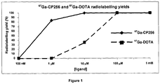
- 1,4,7,10-tetraazacyclododecane-N,N',N'',N''-tetraacetic acid is a common chelator for gallium-68 (and other metallic radioisotopes such as Ga-67, In-111, Cu-64, Lu-177, Y-90) used in molecular imaging and targeted radionuclide therapy.
- DOTA has a long radiolabelling time of around 30 minutes (relative to the half-life of 68 Ga ⁇ 68 minutes).
- chelation of gallium by DOTA derivatives often requires a high labelling temperature of around 95°C.
- the present inventors have found bifunctional molecules that are able to quickly chelate radionuclides at room temperature, whilst retaining adequate or even enhanced stability towards dissociation in the biological milieu.
- the bifunctional molecules have a reactive portion to couple the bifunctional molecule to a functional moiety, such as targeting group which can target, for example, cells, tissues or biological molecules in the body.
- the present invention provides bifunctional molecules for molecular imaging having a tripodal hexadentate tris(hydroxypyridinone) chelating portion to couple to a radionuclide or an imaging label and a reactive functionality to couple to a targeting group for targeting specific cells or tissues in a subject or to a delivery vehicle for delivering a drug, toxin or other such molecule so that the in vivo distribution and/or final location of the target group or delivery vehicle may be monitored.
- the present invention provides a bifunctional compound for use in an in vivo method of diagnostic molecular imaging wherein the bifunctional compound is represented by the formula: or salts thereof;
- the bifunctional compound of the first aspect may circulate the biological system to a targeted location. Then, when the radionuclide is introduced into the subject, the radionuclide may quickly pass though the system and chelate with the bifunctional compound. In this way, the radionuclide may be fixed in a location of interest within a short time of being introduced into the biological system so that the efficacy of the radionuclide is maximised.
- the present invention also provides a bifunctional molecule for use in an in vivo method of diagnostic molecular imaging, the bifunctional molecule comprises a bifunctional compound as defined above and a radionuclide bound through a chelating group of the bifunctional compound; and the method comprises the steps of:
- s is independently selected from 0 to 6
- each r is independently selected from 1 to 6
- q is selected from 1 to 6.
- the method of molecular imaging is PET or SPET.
- the present invention also provides a bifunctional compound or bifunctional molecule for use in a method of radionuclide therapy, wherein the bifunctional compound is represented by the formula: or salts thereof;
- the present invention provides a bifunctional compound precursor, a bifunctional compound or a bifunctional molecule, wherein:
- A is a reactive group for coupling to a biological moiety, a targeting group, a protein, a polypeptide or a delivery vehicle.
- the bifunctional conjugate compounds provide a tripodal hexadentate tris(hydroxypyridinone) chelating portion which is able to chelate metallic radionuclides in a very short time (in the order of 5 min or less) and is able to do so at room temperature in water at around physiological pH.
- the reactive portion is linked to the chelating portion and allows the bifunctional molecule to be conjugated with a targeting group or other vehicle, such as a polypeptide or other biomolecule, drug or nanoparticle.
- a temperature sensitive targeting group such as a polypeptide which may denature at radiolabelling temperatures above body temperature.
- the efficiency of the chelating reaction also allows labelling at very low delivery vehicle concentration. In this way, a significant proportion of the delivery vehicle is radiolabelled with the radionuclide and, as a result, leading to a very high specific activity. In this way, the bifunctional molecule provides an excellent precursor for a radiolabelling conjugate.
- the reactive group A is the protein-reactive functional group.
- the protein reactive group may react with proteins or modified proteins or peptides or other vehicles derivatised for the purpose.
- the protein-reactive group is a maleimide group, an aldehyde, an ester, or "click" reagent such as an alkyne, azide, alkene, hydrazine, hydrazine derivative, alkoxyamine, alkoxyamine derivative, aminoxy or thiol group.
- Maleimide, aldehyde and ester groups efficiently react with peptide thiol- or amine-containing residues (cysteine, lysine) and so a conjugate can easily form.
- Other bioorthogonal functional groups can be engineered into peptides and proteins for the purpose of conjugating them with alkyne, azide, alkene, hydrazine, aminoxy or thiol groups.
- R 1 is N-(2-aminoethyl)-2-aminoethyl-N-(2-aminoethyl)-2-aminoethyl-N-(2-aminoethyl)-2-aminoethyl-N-(2-aminoethyl)-2-aminoethyl-N-(2-aminoethyl)-2-aminoethyl
- R 2 , R 3 and R 4 are independently H or CH 3 . It is preferred to have relatively small substituents on the heterocyclic ring so that the substituents do not sterically hinder the chelation site of the chelating group and so that the chelator may not become too lipophilic. So in preferred embodiments, R 2 , R 3 and R 4 are H. However, one or more methyl substituents on the heterocyclic group may be used to tailor the solubility and/or the hydrophobicity of the molecule. Accordingly one or more of R 2 , R 3 and R 4 may be CH 3 .
- m is 2 or 3.
- the linker group B connects the tripodal chelating portion and the reactive functionality (and the e.g. targeting group or carrier vehicle when conjugated).
- the linking group may be tailored by varying the groups Q, q, r and/or s in order to vary, for example, the length of the linker.
- the linker group may be arranged in either direction so that either A or the quaternary carbon (attached to each arm having a chelating group) or both may be attached to Q.
- B is arranged so that the bifunctional compound is represented by the formula:
- the ordering of the atoms in the functional group represented by Q is not limited.
- the order of the group from the chelating portion towards the reactive functionality may be -NR 5 - then -C(O)- or vice versa.
- At least one Q is an amide (-C(O)NR 5 -).
- q is 1, r is 2 and/or s is 1. More preferably q is 1, r is 2 and s is 1.
- R 5 is hydrogen.
- B is represented by one of the following formulae: wherein r is selected from 1 to 6 and s selected from 0 to 6. Preferably r is 2 and/or s is 2.
- the bifunctional compound is represented by the following formula: wherein A, s, m, X, Y, n and R 1 are as defined herein.
- the radionuclide is an isotope of technetium, rhenium, copper, cobalt, gallium, yttrium, lutetium or other lanthanide, indium, zirconium, scandium.
- the radionuclide is Tc-99m, Re-186, Re-188, Co-57, In-111, Cu-60, Cu-61, Cu-62, Cu-64, Cu-67, Tc-94m, Ga-68, Ga-67, Ga-66, Y-90, Y-86, Sc-44, Sc-47, Fe-52, Sn-117m, Tb-149, Gd-153, Ho-166, Lu-177, Zr-89, Bi-213 or Co-55 and more preferably the radionuclide is gallium, and most preferably Ga-68 or Ga-67.
- the targeting group is a member of a specific binding pair that is capable of binding to a binding partner at the target of interest.
- the targeting group and target of interest are a receptor and ligand, or an antibody and antigen, or metabolic probe e.g. glucose transporter/glucose, hypoxia/hypoxia responsive moiety, or bone mineral/bisphosphonate, or avidin (or streptavidin or related protein) and biotin (or a biotin-like derivative), or RNA and antisense RNA.
- the targeting group is a peptide, protein, antibody, aptamer or small molecule ligand capable of binding to a binding partner at the target of interest. More preferably the targeting group is a polypeptide capable of binding to phosphatidylserine (PS) so that the bifunctional conjugate composition can be employed in apoptosis or cell death imaging studies. More preferably the targeting group includes Annexin V and the C2 domain of a synaptotagmin, TIMP-2, CEA, RGD peptide, somatostatin receptor targeting peptide, bombesin, gastrin or VCAM targeting peptide.
- PS phosphatidylserine
- the target of interest is an in vivo molecular target. More preferably the target of interest is a ligand or receptor expressed on diseased cells or tissue (of which the abundance or ligand occupancy is to be determined), a cell surface antigen associated with a disease state, tumour markers, such as a cancer specific marker or a tissue specific marker or a marker of a normal process which is up- or down-regulated in a disease state.
- the target of interest is a location, an organ, a tissue type or physiological property in a subject undergoing molecular imaging.
- Reactive group A may be for coupling to a delivery vehicle.
- a delivery vehicle as defined herein is a molecular structure for carrying a functional molecule, such as a small molecule drug or toxin with the purpose of delivering the functional molecule for example in a biological system.
- the functional molecule may be contained within or bonded to a surface of the delivery vehicle.
- the functional molecule is typically reversibly associated with the delivery vehicle so that the functional molecule may be released at a given location in the biological system.
- Many such delivery vehicles are known in the art.
- the delivery vehicle is a nanoparticle or liposome.
- Functional molecules, such as pharmaceutical drugs may be bound to the surface of nanoparticles or contained within liposomes.
- the bifunctional compounds of the present invention are coupled to the delivery vehicle and a radionuclide is chelated by the chelating group R 1 , the passage of a drug (carried by the delivery vehicle) through a subject may be monitored by imaging of the radionuclide.
- the reactive group may (in addition to a biological moiety, a targeting group, a protein, a polypeptide or a delivery vehicle) be for coupling to a secondary imaging label.
- a secondary imaging label is an optical label, a paramagnetic label, such as a SPIO, or a quantum dot.
- the imaging label is a paramagnetic label which is a MRI contrast agent with a paramagnetic probe.
- the imaging label is an optical label which is an organic molecule or metal complex with fluorescent or luminescent properties.
- the label may, for example in the case of an optical label or paramagnetic label, allow for the bifunctional compound to be used in multi-modal imaging.
- the reactive group A may couple with further biological moieties, targeting groups, proteins, polypeptides or delivery vehicles.
- the bifunctional compound may couple the radionuclide to a targeting group for targeting specific cells in a subject and a liposome containing a pharmaceutical drug. In this way, targeted drug delivery may be monitored during its passage through the biological system.
- the method of molecular imaging may include introducing the bifunctional compound (or conjugate thereof) into a biological system (e.g. a subject) either before the radionuclide is introduced into the biological system (i.e. pre-targeting) or after the radionuclide has been chelated to the chelating portion of the bifunctional compound (i.e. pre-assembled).
- a biological system e.g. a subject
- the bifunctional compound When the bifunctional compound (or conjugate) is introduced into the biological system before the radionuclide, the bifunctional compound may circulate the biological system to a preferred or targeted location. Then, when the radionuclide is introduced, the radionuclide may quickly pass though the system and chelate with the bifunctional molecule (or conjugate). In this way, the radionuclide may be fixed in a location of interest within a short time of being introduced into the biological system so that the efficacy of the radionuclide is maximised.
- the bifunctional compound When the radionuclide is chelated to the bifunctional compound (or conjugate) before introduction into the biological system, the bifunctional compound may be used to monitor the passage of the bifunctional compound or the conjugate having a biological moiety, a targeting group, a protein, a polypeptide a carrier vehicle or a secondary imaging label attached to the compound through the biological system.
- the biological moiety, targeting group, protein, polypeptide or delivery vehicle are bound to the bifunctional compound to form a bifunctional compound conjugate before introduction of the bifunctional compound composition into the biological system or subject.
- the biological system is a subject undergoing diagnosis or therapy for a disease or condition.
- the therapy may be radionuclide therapy.
- the therapy may be cancer treatment using the radionuclide or a chemotherapeutic agent associated with the bifunctional molecule, for example via the delivery vehicle to which it is attached.
- the bifunctional molecule is associated with or bound to a therapeutic agent.
- the subject may be a subject in whom the biodistribution of the targeting molecule or delivery vehicle is being evaluated.
- the present invention provides a kit for use in a method of in vivo diagnostic molecular imaging or for use in a method of radionuclide therapy, the kit comprising: a bifunctional compound composition having a bifunctional compound ; and a composition comprising a radionuclide as described herein.
- kits for use in a method of molecular imaging or therapy comprising: a bifunctional molecule composition having a bifunctional compound as described herein; and a biological moiety, a targeting group, a protein, a polypeptide or a delivery vehicle for coupling to the bifunctional molecule.
- the kit may also include an imaging composition comprising a radionuclide as described herein.
- a target of interest in a biological system comprising:
- a target of interest or passage of interest in a biological system comprising:
- the radionuclide is a radionuclide capable of delivering radiotherapy to the target of interest.
- the radionuclide is Cu-67, Re-186, Re-188, Y-90, Lu-177, Sc-47 or Bi-213.
- bifunctional compound is for coupling to a targeting molecule
- a wide range of primary targeting groups may be linked to the bifunctional compounds to direct the bifunctional compounds with the chelating group to different targets of interest in the biological system in question.
- the target of interest will comprise a member of a specific binding pair that is capable of specifically binding to the primary targeting group in the system, such that they will be members of a pair of molecules which have particular specificity for each other and which in normal conditions bind to each other in preference to binding to other molecules.
- specific binding pairs are well known in the art and include for example receptors and ligands, enzymes and substrates, antibodies and antigens.
- primary targeting groups may be peptides, proteins or other biological molecules, such as aptamers, or small molecule ligands, that bind to specific in vivo molecular targets.
- Classes of targets of interest include ligands or receptors or transporters expressed on diseased cells or tissue, cell surface antigen associated with disease states, or tumour markers, e.g. cancer specific markers or tissue specific markers.
- One group of primary targeting groups are polypeptides capable of binding to phosphatidylserine (PS) so that the bifunctional molecule conjugates of the present invention can be employed in apoptosis or cell death imaging studies.
- polypeptides include Annexin V and the C2 domain of a synaptotagmin.
- Polypeptides that comprise one or more C2 domains are well known in the art. While some polypeptides have only one C2 domain, others have two or more C2 domains, and the domains are generally described by attaching a letter (in alphabetical order) to the end of the name (e.g., C2A, C2B, and so on). For a protein that contains only one C2 domain, the domain is simply referred to as C2 domain.
- C2A domain of rat synaptotagmin I other C2 domains that are capable of binding to PS could be employed instead, for example a C2A domain of a synaptotagmin of another species.
- proteins that contain a C2 domain include but are not limited to synaptotagmin 1-13, protein kinase C family members of serine/threonine kinases, phospholipase A2, phospholipase ⁇ 1, cofactors in the coagulation cascade including factors V and VIII, and members of the copine family.
- Human synaptotagmins include synaptotagmin 1-7, 12 and 13.
- the present invention can employ an anti-CD33 antibody, or fragment thereof, for imaging cancer cells expressing CD33 such as cells of myelomonocytic lineage and leukaemic cells, see Emberson et al., J. Immunol. Methods. 305(2):135-51, 2005 .
- a further example is the use of a tissue inhibitor of metalloproteinases (TIMPs), such as TIMP-2, for imaging matrix metalloproteinase expression, as expression of metalloproteinases has been implicated in metastatic processes, see Giersing et al., Bioconjug Chem. 12(6): 964-71, 2001 .
- TIMP-2 tissue inhibitor of metalloproteinases
- a further example of a polypeptide that can be used to make conjugates according to the present invention is complement receptor 2 (CR2).
- CR2 complement receptor 2
- Antibodies capable of binding to the glycoprotein carcinoembryonic antigen (CEA) may also be used as primary targeting groups as members of this family of glycoproteins are expressed on colorectal cancer cells, gastric cancer cells, pancreatic cancer cells, lung cancer cells, medullary thyroid cancer cells and breast cancer cells.
- a further example may exploit the affinity or the peptide sequence arginine-glycine-aspartic acid (RGD) for the ⁇ v ⁇ 3 integrin expressed highly in the endothelium of tissues undergoing angiogenesis as is commonly seen in tumours, atherosclerotic plaque and repairing diseased tissue such as infarcted myocardium, by linking an RGD peptide derivative to the bifunctional compound by means of a suitable reactive group A.
- RGD arginine-glycine-aspartic acid
- a further example may exploit the affinity of the peptide octreotide or other related analogue of somatostatin, which may bind to the somatostatin receptor expressed highly at the surface of cancer cells e.g. in carcinoid, medullary thyroid carcinoma and other neuroendocrine tumours, by linking a somatostatin analogue peptide to the bifunctional compound by means of a suitable reactive group A.
- Another example may exploit the affinity of antibodies to cell adhesion molecules.
- One example is the affinity of the monoclonal antibody, SER 4, to the macrophage adhesion molecule, sialoadhesin.
- Sialoadhesin is found on the surface of macrophages, and, for example, in high amounts on macrophages of the spleen, liver, lymph node, bone marrow colon and lungs.
- the skilled person will be able to use other targeting groups such as bombesin, gastrin, or VCAM targeting peptide.
- the primary targeting group may be linked to the bifunctional compound as a conjugate having a functional group for binding to the bifunctional compound and the reactive group for linking to the primary targeting group.
- a reactive group for site-specific linkage to the primary targeting group may be employed, for example a maleimide group for site-specific linkage to thiol groups in a biomolecule such as a peptide, polypeptide or antibody.
- the maleimide group can then be site-specifically linked to a thiol group, for example of a cysteine residue incorporated site-specifically into the peptide or protein for the purpose.
- the reactive group in the bifunctional chelator would be an aldehyde or ketone group able to react site-specifically with a protein or peptide to which a hydrazine or similarly reactive group has been site-specifically incorporated (e.g. using a hydrazinonicotinic acid derivative) to form a hydrazone or similar link.
- the primary targeting group may comprise a suitable polypeptide or protein, or a fragment or domain thereof. Accordingly, while for convenience the methods herein are generally described by reference to “polypeptides", this should be taken to include shorter sequences of amino acids (e.g., from 3 to 10 amino acids in length to 30, 40 or 50 amino acids in length), sometimes referred to in the art as peptides. The term should also be taken to include polypeptides having secondary, tertiary or quaternary structure, generally referred to as proteins, as well as multidomain proteins. In some embodiments, and in the examples provided below, the polypeptides used as primary targeting groups are protein domains.
- Protein domains are fragments of a full length protein that have the ability to retain structure independent of the full length protein, typically forming a stable and folded three-dimensional structure. Many proteins consist of several structural protein domains and it is common for a particular domain to be found in a range of related proteins. Protein domains vary in length from between about 25 amino acids up to 500 amino acids in length, or from 50 amino acids to 250 amino acids, or from 75 amino acids to 150 amino acids.
- polypeptide is an antibody
- this term describes an immunoglobulin whether natural or partly or wholly synthetically produced.
- the term also covers any polypeptide or protein comprising the antigen binding domain of an antibody; antibody fragments which comprise an antigen binding domain such as Fab, scFv, Fv, dAb, Fd; and diabodies.
- Fab, scFv, Fv, dAb, Fd fragments which comprise an antigen binding domain
- Fab, scFv, Fv, dAb, Fd fragments which comprise an antigen binding domain
- Fab fragmentscFv, Fv, dAb, Fd
- diabodies antigen binding domain
- Antibodies can be modified in a number of ways and the term "antibody molecule" should be construed as covering any specific binding member or substance having an antibody antigen-binding domain with the required specificity. Thus, this term covers antibody fragments and derivatives, including any polypeptide comprising an immunoglobulin binding domain, whether natural or wholly or partially synthetic. Chimeric molecules comprising an immunoglobulin binding domain, or equivalent, fused to another polypeptide are therefore included. Cloning and expression of chimeric antibodies are described in EP 0 120 694 A and EP 0 125 023 A . It has been shown that fragments of a whole antibody can perform the function of binding antigens.
- binding fragments are (i) the Fab fragment consisting of VL, VH, CL and CH1 domains; (ii) the Fd fragment consisting of the VH and CH1 domains; (iii) the Fv fragment consisting of the VL and VH domains of a single antibody; (iv) the dAb fragment ( Ward, E.S.
- Fv, scFv or diabody molecules may be stabilised by the incorporation of disulphide bridges linking the VH and VL domains ( Reiter et al, Nature Biotech, 14: 1239-1245, 1996 ).
- Minibodies comprising a scFv joined to a CH3 domain may also be made ( Hu et al, Cancer Res., 56: 3055-3061, 1996 ).
- Affibodies may also be used as a primary targeting group.
- Affibodies are small high affinity proteins that may bind specifically to a large number of target proteins or peptides. Affibodies imitate monoclonal antibodies in many respects and are classed as antibody mimetics.
- the selection of affibody should not be limited and many affibodies are known in the art, such as unconjugated anti-TNF- ⁇ affibody.
- Anti-TNF- ⁇ affibody may be produced in E. coli and targets human cytokine tumor necrosis factor ⁇ , TNF- ⁇ . TNF- ⁇ is a pleiotropic inflammatory cytokine.
- the cytokine is produced by several types of cells, but especially by macrophage. It acts as a key mediary in the local inflammatory immune response and has further immune regulatory functions.
- TNF- ⁇ is an acute phase protein which initiates a cascade of cytokines and increases vascular permeability, thereby recruiting macrophage and neutrophils to a site of infection. Therefore, TNF- ⁇ is an important target for inflammation in atherosclerotic lesions and inflammations.
- Each bifunctional compound may be linked to a plurality of primary targeting groups, thereby increasing (a) the affinity of the primary targeting groups for the target of interest as the overall affinity increases as the product of the affinity of each targeting group for the target and/or (b) the rate of binding to the target, i.e. the likelihood of binding ensuing from each encounter between target and primary targeting group in vivo.
- the imaging probes used in accordance with the present invention have the property of binding to the chelating portion of the bifunctional compounds either in vitro or in vivo, in either the pre-targeted or pre-assembled approaches.
- One possibility for achieving this is to use radionuclides that have an intrinsic binding affinity for chelating portion of the bifunctional compounds.
- 68 Ga 3+ is capable of binding to the hydroxypyridinone groups of the chelating portion and can be used in PET imaging techniques.
- radionuclide probes used in accordance with the present invention may use a range of different radionuclides depending on the application for which the probes are intended.
- radionuclides that may form part of the probes of the present invention include radionuclides of technetium, rhenium, copper, cobalt, gallium, yttrium, lutetium, indium, zirconium, scandium, namely Tc-99m, Ga-67, In-111 (SPET), Cu-64, Cu-60, Cu-61, Cu-62, Tc-94m, Ga-68, Co-55, Zr-89, Sc-44 (PET), Cu-67, Re-186, Re-188, Y-90, Lu-177, Sc-47 or Bi-213 (radionuclide therapy).
- the present invention may employ the radionuclides alone or in combinations.
- the radionuclide may be in any form, such as a solvate, hydrate, salt or weak chelate. Solvates, hydrates, salts and weak chelates of radionuclides are known in the art.
- the radionuclide should be in a pharmaceutically acceptable form.
- the radionuclide may be a citrate or acetate salt of the metal ion.
- the present invention may also involve the use of secondary imaging labels, such as an optical label or a paramagnetic label (through the reactive group A) that may be linked to or associated with the bifunctional compounds, for example to enable multi-modal imaging to be carried out.
- optical probes include fluorophores such as fluorescein and luminescent molecules and complexes such as lanthanide complexes and tricarbonylrhenium-quinolinate conjugates.
- the possibility to incorporate optical probes as well as radionuclides and MR contrast agents provides the opportunity to combine modalities to enhance diagnosis and detection, for example the location of disease at the whole body level can be identified by whole body scanning with PET or SPECT while the same tracer may be detected visually during surgery to assist identification of diseased tissue.
- combined PET and MR imaging can provide the advantage of high sensitivity (PET, SPET), quantification of signal (PET) and anatomical resolution (MR) and measurement of the microenvironment (MR contrast enhancement).
- the bifunctional compounds of the present invention may use a range of different chemistries and techniques for linking the biological moiety, targeting group, label, protein or polypeptide to the bifunctional compounds and/or for introducing further groups and properties into the bifunctional compounds of the present invention.
- the reactive group provides a way of coupling the targeting group to the bifunctional compound, for example when the reactive group is a maleimide group (to bind to the thiol side chain of cysteine in the protein/peptide sequence)
- the bifunctional compounds of the present invention may be used for the molecular imaging of diseases, such as cancer, cardiovascular disease, immunological disease and associated conditions, such as transplant, inflammation, leukocyte tracking, stem cell tracking, allergy and infection.
- diseases such as cancer, cardiovascular disease, immunological disease and associated conditions, such as transplant, inflammation, leukocyte tracking, stem cell tracking, allergy and infection.
- the bifunctional compounds may also be employed for radionuclide therapy for treating cancer and arthritis.
- the applications of the bifunctional compounds and radionuclides of the present invention include a wide range of imaging and spectroscopic applications that can employ the radionuclide and/or a further label, for example, in multi-modal imaging studies.
- the bifunctional compounds are particularly useful for in vivo imaging applications such as cell death imaging, for example using bifunctional compounds for the detection of apoptosis. This might be useful in a number of different medical or research applications, for example in the fields of oncology (e.g. in monitoring response to chemotherapy), cardiovascular medicine (e.g. in imaging damaged myocardium post myocardial infarction) or graft rejection (e.g. in imaging cardiac allograft rejection).
- the present invention is particularly relevant to nuclear medicine imaging techniques, such as Single Photon Emission Tomography (SPET), an imaging technique that detects gamma rays emitted from a radionuclide to produce a three dimensional image of the distribution of the radionuclide in a sample or subject, and Positron Emission Tomography (PET), an imaging technique that provides three-dimensional images by detecting pairs of gamma rays emitted indirectly by a positron-emitting radionuclide introduced into a sample or subject.
- SPET studies can be carried out using In-111 and PET studies using Ga-68.
- the skilled person will be aware of other suitable SPET and PET radionuclides that can be employed in the present invention.
- the present invention may be employed for positron emission tomography (PET), single photon emission tomography (SPET), optical (OI) and/or magnetic resonance imaging (MRI) by appropriate selection of imaging probe, or for radionuclide therapy by selection of the appropriate radionuclide (e.g. Y-90, Lu-177).
- PET positron emission tomography
- SPET single photon emission tomography
- OI optical
- MRI magnetic resonance imaging
- the bifunctional compounds of the present invention may be used in methods of multi-modal imaging, that is where information or images are derived from two different techniques, either by the detection of the imaging probe capable of detection using two different techniques or by providing a second label at the site in the biological system where the bifunctional compounds become localised, most conveniently by linking or associating the second label with the bifunctional compounds as explained in detail above.
- Multi-modal studies will be co-registered and may entail simultaneous imaging with two modalities or may need to take place in two steps, but generally employ the same sample so that spatial information obtained using the two techniques can be compared. Examples of multi-modal imaging include PET/CT, SPET/CT, PET/MR and SPET/MR
- the tris tert-butyl esters 61 and 62 were synthesised by following established literature methods ( G. R. Newkome, R. K. Behera, C. N. Moorefield and G. R. Baker, J Org Chem, 1991, 56, 7162-7167 ).
- Nitro tripod 61 was produced by the reaction of nitromethane and tert-butyl acrylate in the presence of benzyltrimethylammonium hydroxide and recrystallised from ethanol in 57% yield.
- the nitro group of 61 was then reduced to an amine to form 62 by hydrogenation with a T-1 Raney Nickel catalyst, 5 which was specially prepared from a nickel/aluminium alloy.
- Acetylation of 62 with acetyl chloride and purification by column chromatography gave 63 in 72% yield.
- the tert-butyl groups were then removed with formic acid and the product, compound 64 was recrystallised from ethanol/petroleum spirit with a yield of 93%.
- Samples were dried in a vacuum oven (Gallenkamp, rated to ⁇ 250°C) connected to a vacuum pump (BOC-Edwards.) Samples were hydrogenated on a Parr hydrogenator (room temperature) at 40 psi with automatic shaking. Infrared Spectra were acquired on a Perkin Elmer Spectrum one FTIR using KBr disks. The software used was called "Spectrum.”
- Ga- CP256 and Fe- CP256 complexes Formation of the Ga- CP256 and Fe- CP256 complexes is evident from the mass spectral data shown in Table 1 and Table 2. The protonated form of each complex was detected and the isotopic distribution from the mass spectrum matches the calculated values recorded in table 1.
- the Ga- CP256 complex has two molecular species for (M + H) + , at m/z 806 and m/z 808, due to gallium having two stable isotopes with atomic masses of 69 and 71. No evidence of Ga- CP256 complexes with Ga:ligand ratios other than 1:1 was found.
- the mass spectral data for Fe- CP256 complex match the predicted isotopic distributions.
- Radiolabelling CP256 with Ga-67 gave a product whose HPLC showed a single radioactive peak with an elution time identical with that of the "cold" Ga-CP256 complex.
- Table 2 Calculated and experimental isotopic distributions of Ga-CP256 and Fe-CP256 and the CP256 ligand.
- CP256 1 mg was dissolved in 1 ml methanol to give a ligand concentration of 1.18 mM (based on tris HCl adduct of CP256 with RMM of 848). This solution was added to 0.3 mg anhydrous Ga(NO 3 ) 3 (RMM 255) to give a 1:1 metal to ligand stoichiometry at a concentration of 1.18 mM.
- Samples were analysed by LC-MS.
- the HPLC system was an Agilent 1200 system with degasser, quaternary pump, UV-visible variable wavelength detector, and an autosampler.
- the mass spectrometer was an Agilent 6520 Accurate-Mass Q-TOF LC/MS. Samples were acquired using Agilent Masshunter workstation acquisition software and data were analysed using Agilent Masshunter Qualitative analysis software.
- the HPLC column used was an Agilent Zorbax SB C18 cartridge column (2.1mm X 30mm, 3.5 ⁇ m.)
- the gradient is given in Table 3.
- HPLC analyses were carried out with FC3600 detector and ⁇ detector probe connected in series.
- the HPLC system was an Agilent 1200 series with quadruple pump, degasser, UV detector and manual syringe injector.
- the HPLC column used for all analyses was an Agilent Eclipse XDB-C18 (5 ⁇ m, 4.6 mm X 150 mm) with a guard column.
- the analytical software used for radio-TLC and radio-HPLC was Laura (Lablogic). All general reagents and consumables were purchased from Sigma-Aldrich or from Fisher Scientific.
- 67 Ga-Citrate solution for injection was obtained from Mallinkrodt Medical, 74 MBq/ml (at ref. date; contains 6.5 mM sodium citrate, 135 mM NaCl, 0.9% Benzyl alcohol).
- a 1 mg/ml stock solution of CP256 was prepared in PBS buffer in a 1 ml plastic microcentrifuge tube. Radiolabelling solutions were prepared by adding a 10 ⁇ l aliquot of the stock solution to 90 ⁇ l of labelling buffer in a plastic microcentrifuge tube to give a total ligand concentration of 0.1 mg/ml in 100 ⁇ l. This solution was radiolabelled by adding 10 ⁇ l 67 Ga-citrate. Radiolabelling conditions are shown in table 4. Table 4. Radiolabelling conditions for CP256 with 61 Ga LIGAND LABELLING BUFFER LABELLING TEMPERATURE LABELLING TIME LIGAND CONC. (M) MEASURED pH CP256 PBS 25°C 1 min 1.18 X 10 -4 6.6
- radiolabelling results in table 5 measured by Thin Layer Chromatography (TLC) show that CP256 radiolabels with 67 Ga at room temperature at lower concentrations than DOTA does at elevated temperature, with 100% radiolabelling at a concentration as low as 10 ⁇ M whereas the radiolabelling yield for DOTA at 10 ⁇ M is only 35%. These results are shown graphically in figure 1 . Table 5. Radiolabelling yields of 61 Ga-CP256 and 67 Ga-DOTA. Calculated from TLC analysis.
- Silica TLC plates and ITLC-SG strips were spotted with 1 ⁇ l of radiolabelled complex at the origin, which was marked at 5 mm in pencil. Spots were allowed to dry on silica gel TLC plates but not for ITLC-SG strips.
- the plates were placed in a developing chamber (100 ml wide necked sample bottle) filled with a 3 mm depth of the mobile phase. Stationary phases and mobile phases are listed in table 8.
- the TLC plate was developed until the solvent front had reached a distance of up to 65 mm.
- the plates were removed from the developing chamber and dried in an oven set at 80°C. Radiochemical yields were determined using either Laura software or the instant imager software.
- TLC and ITLC analyses were carried out on a Mini-Scan TLC scanner with FC3600 detector and ⁇ detector probe.
- the analytical software used was Laura (Lablogic). Radioactive samples were counted for 10 seconds (window 101-110) on a 1282 Compugamma Gamma Counter (LKB Wallac) using Ultroterm software.
- the dose calibrator used for measuring radioactivity in samples was a CRC-25R (Capintec.) Electronic autoradiography was performed on a Packard Instant Imager with Imager version 2.05 software.
- the gas mixture was 1% isobutane, 2.5% carbon dioxide and 96.5% argon (Air products.)
- 1.25 mM stock solutions of each ligand were prepared in the appropriate labelling buffer, as listed in table 6.
- the stock solution was diluted 10 fold by successive addition of the radiolabelling buffer to create extra solutions with ligand concentrations of 125 ⁇ M, 12.5 ⁇ M, 1.25 ⁇ M, 125 nM and 12.5 nM.
- Eighty microlitres of each solution was added to a plastic microcentrifuge tube and radiolabelled with 20 ⁇ l of 67 Ga-Citrate (1.5 MBq) to produce radiolabelled solutions with final ligand concentrations of 1 mM, 100 ⁇ M, 10 ⁇ M, 1 ⁇ M and 100 nM.
- a metal heater block (containing oil in the wells) was used to heat the 67 Ga-DOTA solution.
- Radiolabelling conditions are shown in table 6. Table 6. Radiolabelling conditions for each ligand. Final concentrations of each ligand in the labelling mixture were 1 mM, 100 ⁇ M, 10 ⁇ M, 1 ⁇ M, 100 nM and 10 nM. LIGAND LABELLING BUFFER LABELLING TEMPERATURE LABELLING TIME MEASURED pH DOTA Acetate 80°C 45 min 4.6 CP256 PBS 25°C 1 min 6.6
- Figure 2 shows the elution profiles of 67 Ga-CP256 from a PD10 column for radiolabelled solutions at 100 ⁇ M (A) and 10 ⁇ M (B) of CP256. After dilution and incubation in apo-transferrin, the concentrations of CP256 in these solutions were 227 ⁇ M and 2.27 nM respectively. After 4 hours incubation in apo-transferrin, the profile is unchanged from that of the incubation in the reference buffer, indicating no binding of 67 Ga to transferrin or loss of 67 Ga from the ligand, for either of the 2.27 ⁇ M or 227 nM solutions.
- 67 Ga-DTPA showed some dissociation over the 4 hour incubation period, with 10% extra binding of the radioactivity in this solution to the filter when compared with the solution in the reference buffer.
- Table 7 Binding of 67 Ga-citrate, 67 Ga-CP256, 67 Ga-DOTA and 67 Ga-DTPA to 30 kDa MWCO filters.
- a reference solution of 67 Ga-citrate was produced by incubating 20 ⁇ l 67 Ga-citrate in 200 ⁇ l PBS. This solution was incubated at 37°C in a metal heating block and samples were removed for analysis in the same manner as for the serum incubation.
- TRIS-HCl buffer pH 7.5
- Sodium bicarbonate was added to a concentration of 25 mM.
- a 2.5 mg/ml (32 ⁇ M) solution of apo-transferrin was prepared in this buffer.
- Apo-transferrin solution 200 ⁇ l in a plastic microcentrifuge tube was incubated at 37°C in a metal heating block. To this was added 20 ⁇ l of each pH-neutralised tracer. 20 ⁇ l of each tracer was also incubated in a reference buffer (containing no apo-transferrin) of 50 mM TRIS-HCl containing 25 mM sodium bicarbonate.
- the 50 mM TRIS-HCl solution (200 ⁇ l, not containing apo-transferrin) was incubated in a heating block at 37°C.
- the appropriate pH adjusted tracer (20 ⁇ l) was added to the incubation. Aliquots were taken at appropriate intervals for analysis.
- the elution profile of Transferrin from a PD10 column was determined by UV spectrophometry.
- Apo-transferrin non radioactive, 500 ⁇ l of the 2.5 mg/ml solution
- PBS buffer eluted with PBS buffer
- 0.5 ml fractions were collected.
- the samples were loaded into a 1 ml quartz cuvette and diluted to 0.9 ml with an extra 0.4 ml of PBS.
- Absorbance was determined at 280 nm with PBS as a reference standard.
- the analysis was done in triplicate after 4 hours for the apo-transferrin incubations. The analysis was done 6 times (6 tubes) for the incubations in the 50 mM TRIS-HCl reference buffer. Average values were taken as described above.
- C2A is a small protein that recognises phosphatidylserine displayed on cells undergoing apoptotis. It has been engineered with a cysteine residue to facilitate site-specific labelling with thiol-reactive agents (see Tavare R, Torres Martin de Rosales R, Blower PJ, Mullen GED. Efficient site-specific radiolabeling of a modified C2A domain of synaptotagmin I with [ 99m Tc(CO) 3 ] + . a new radiopharmaceutical for imaging cell death. Bioconjugate Chem 2009; 20:2071-2081 ). The resulting engineered protein (C2Ac) was conjugated with CP256 using either 6 or 7. The same product was obtained for both conjugation reactions.
- the average molecular weight of C2Ac is 14997 and so the C2Ac-(7) conjugate was expected to have an average molecular weight of 15918.44.
- the found average molecular weight was 15970.85, which corresponds to the C2Ac-(7) conjugate with an additional iron atom and without 3 hydrogen atoms.
- C2Ac (2 mg in a solution of 2mg/mL) was treated with a 3:1 molar excess of chlorosuccinimide-hexadentate chelator (6) or maleimide-hexadentate chelator (7) for 4 hours at room temperature.
- the mixture was purified using a PD-10 column pre-equilibrated with PBS that had been stored over Chelex and analysed by LC-MS.
- the C2Ac-(7) conjugate was radiolabelled with 68 Ga according to the method described herein.
- Figure 4 shows a high degree of chelation after only 15 minutes of incubation with 68 Ga in acetate buffer.
- the 68 Ga-labelled conjugate showed specific calcium dependent binding to phosphatidylserine (PS) in a red blood cell binding assay (see Fig. 5 )
- Unchelated 68 Ga in buffer was prepared as follows. The generator was eluted with in 2ml 0.1M HCl to give 25 MBq 68 Ga eluate. 1.5ml of conc. HCl (36%) was added to the elution to give a final concentration of 4M HCl to form gallium tetrachloride anion, and the solution passed through an anion exchange column (SAX SPEC, Chromafix 30-PS-HCO3, 45mg, Macherey-Nagel, Germany), trapping all activity.
- SAX SPEC anion exchange column
- the column was then washed with 250 ⁇ l 5M NaCl and the activity then eluted with 300 ⁇ l distilled water and the eluate (pH ⁇ 1, ⁇ 70 MBq) buffered by adding 5 ⁇ l of 1M NaOH and 30 ⁇ l of 1M ammonium acetate buffer, pH 6, raising pH to 7. 100 ⁇ l of PBS, pH 6 was then added.
- the cells were then centrifuged (3 min at 7500 RCF), the supernatant was removed, and the cells were resuspended in 1 ml assay buffer containing the same concentration of calcium used during the incubation step.
- the cells were centrifuged again, the supernatant was removed, and the pellet was resuspended in 0.7 ml assay buffer plus 10 mM ethylenediaminetetraacetic acid (EDTA) to release 68 Ga labelled C2Ac-(7) bound in a calcium-dependent manner.
- EDTA ethylenediaminetetraacetic acid
- Curve fitting was performed using a nonlinear curve fit by a routine based on the Levenberg-Marquardt algorithm using Kaleidagraph (Synergy Software, Reading, US).
- PET images taken at 1.5 h after injection of the 6B Ga-C2Ac- (7) conjugate and unchelated ("free") 68 Ga into mice are shown in Figs. 6a and 6b respectively.
- the presence of 68 Ga is shown by bright white areas in the image.
- Injection of the 68 Ga-C2Ac-(7) conjugate showed rapid clearance of radioactivity from blood and tissues with uptake almost exclusively in kidney with a small amount of activity excreted into the bladder ( Fig. 6a ). This contrasts with the images obtained with injection of "free" 68 Ga in which activity was seen slowly clearing from blood pool predominantly to remodelling bone ( Fig. 6b ).
- the 68 Ga labelled C2Ac-(7) conjugate was synthesised as described above. Unchelated 68 Ga in buffer, was prepared using the pre-concentration (and buffering to pH 7) method described in example 7. TLC analysis was performed. No gallium hydroxide precipitate was detected by the time of injection.
- Image reconstruction OSEM with SSRB 2D LOR, energy window: 400-600 keV, filter: Ramlak cutoff 1, number of iterations/subsets: 8/6. Animals were killed at 90 min and explanted organs counted in a gamma counter to determine biodistribution.
- Figure 7 shows size exclusion radiochromatography (PD10 column) of chelation of 68 Ga by CP256 in serum and control experiments to show 68 Ga-chloride incubated in serum, preformed 68 Ga-CP256-(7) complex incubated with serum and 69 Ga incubated in buffer.
- PD10 column size exclusion radiochromatography
- chelator CP-256, 2.35mM, 1.17mM, 589pM, 117 ⁇ M, 23 ⁇ M and 11.7 ⁇ M; pH 7 - 7.5 in PBS/bicarbonate buffer
- 100 ⁇ l of chelator CP-256, 2.35mM, 1.17mM, 589pM, 117 ⁇ M, 23 ⁇ M and 11.7 ⁇ M; pH 7 - 7.5 in PBS/bicarbonate buffer
- 20 ⁇ l of 68Ga eluate was added and the solution incubated at 37°C in a plastic tube.
- 20 ⁇ l of 68 Ga solution was added to the 300 ⁇ l of human blood serum.
- 100 ⁇ l of chelator Cp-256 was added.
- 111 In-chloride formed a complex with CP256 (589 ⁇ M) in about 90% yield after 5 min, as determined by ITLC-SG(HSA).
- the complex showed ⁇ 2% release of In-111 or binding to serum proteins when incubated in human serum for 2 h, as determined by PD10 size exclusion chromatography ( Fig. 8 ).
- Example 11 in vivo imaging of antibody-CP256 conjugate via the pre-targeting mode with 67 Ga .
- a monoclonal IgG antibody, SER4 against a sialoadhesin antigen expressed by pro-inflammatory macrophages was conjugated with CP256-malemide (7) by first reducing antibody disulfide bonds with 2-mercaptoethanol, then treating the thiol groups so formed with CP256-maleimide (7) to give CP256-SER4.
- mice of SER4 labelled with 99m Tc previously reflects rapid clearance from circulation with uptake predominantly in spleen and to some extent also liver.
- CP256-SER4 was labelled with 67 Ga to give 67 Ga-CP256-SER4.
- Groups of wild type mice were injected with either directly labelled 67 Ga-CP256-SER4 (group 1), or SER4 (unconjugated) followed 5 min later by 67 Ga citrate (group 2) or CP256-SER4 followed by 67 Ga citrate.
- biodistribution data for Group 1 are shown in left hand column
- biodistribution data for Group 2 are shown in the central column
- biodistribution data for Group 3 are shown in the right hand column.
- Group 1 showed strong targeting of 67 Ga-CP256-SER4 to spleen (36% ID/g), whereas group 2 showed very low spleen uptake of 67 Ga citrate (4% ID/g).
- group 3 showed that pre-injection of CP256-SER4 was able to increase spleen uptake of 67 Ga citrate from 4% ID/g to 10% ID/g, suggesting that the 67 Ga followed the location of antibody conjugate to a significant extent.
- Group 1 also showed high activity of 67 Ga-CP256-SER4 in circulating blood (31% ID/g), whereas 67 Ga-citrate (group 2) did not (7% ID/g); Pre-administration of CP256-SER4 (group 3) increased blood pool retention from 7% to 16% ID/g.
- the knockout mice did not show the spleen targeting behaviour, consistent with this pattern being associated with sialoadhesin expression.
- the antibody was then washed by centrifugation at 3000 rpm for 3 x 15 min in a 50,000 mwco Vivaspin2 ultracentrifugation tube.
- the antibody was resuspended between centrifugation steps and the volume adjusted to 2 ml by addition of 0.1M metal-free phosphate buffer, pH 7.
- the concentrated and washed antibody was collected in approximately 300 ⁇ L 0.1 M phosphate buffer.
- 2-mercaptoethanol (5 ⁇ L) was diluted to 20 ⁇ L in 0.1 M phosphate buffer, pH 7. 3.8 ⁇ L (1000 fold excess) of the diluted 2-mercaptoethanol was added to the antibody and the reduction allowed to proceed for 30 min at room temperature.
- the volume was adjusted to 1 ml by addition of 0.1 M phosphate buffer and the removed by applying the reduced antibody to a disposable size exclusion column (PD10).
- PD10 disposable size exclusion column
- Fractions containing the reduced antibody were combined and CP256-maleimide (0.77 mg dissolved in 8 ⁇ L DMSO, a 40 fold excess) was added immediately.
- the conjugation reaction was heated at 37°C for 30 min.
- the conjugated antibody was then washed using a 50,000 mwco Vivaspin2 ultracentrifugation tube as before but using 0.1 M ammonium acetate buffer, pH6 to remove the excess ligand.
- the concentrated CP256-antibody was collected in 0.1 M ammonium acetate buffer, pH6.
- Radiolabelling was performed by addition of 45 ⁇ L 67 Ga citrate (0.6-0.75 MBq) to 90 ⁇ L (90 ⁇ g) CP256-SER4. Radiolabelling was allowed to proceed for 10 min before dilution to 350 ⁇ L with phosphate buffered saline. Injections containing 25 ⁇ g of labelled antibody were drawn up. Analysis of the radiolabelled antibody was performed by size exclusion HPLC using 0.1 M phosphate buffer containing 2 mM EDTA as the mobile phase to ensure good radiolabelling had been achieved prior to injection into animals.
- Example 12 in vivo imaging of antibody-CP256 conjugate via the pretargeting mode with 68 Ga .
- SER4 conjugated with CP256-malemide (7) as described in Example 11, was labelled with 68 Ga as described above in example 7, to give 68 Ga-CP256-SER4.
- 68 Ga-CP256-SER4 group 1
- SER4 unconjugated
- biodistribution data for Group 2 are shown in left hand column
- biodistribution data for Group 3 are shown in the central column
- biodistribution data for Group 1 are shown in the right hand column.
- Group 1 showed strong targeting of 68 Ga-CP256-SER4 to spleen (20-50% ID/g over the three experiments), whereas group 2 showed very low spleen uptake of 68 Ga acetate (2-5% ID/g).
- group 3 showed that pre-injection of CP256-SER4 was able to increase spleen uptake of 68 Ga acetate from 2-5% ID/g to 17-40% ID/g, suggesting that the 68 Ga followed the location of antibody conjugate to a significant extent.
- FIG. 11A A NanoPET/CT image of a mouse from group 3 is shown in Fig. 11A .
- the mouse was image using the pre-targeting method of the invention using CP256-SER4: 8 week-old C57Bl/6 injected with CP256-SER4 followed by Ga-68 acetate.
- the figure shows targeted liver/spleen uptake.
- a mouse injected with Ga-68 acetate only is shown in Fig. 11 B , showing retention in blood pool and accumulation in joints.
- the antibody was conjugated to CP256 as described in Example 11.
- 68 Ga was eluted from a 68 Ge/ 68 Ga generator and concentrated as described in Example 7 to give 68 Ga acetate.
- 150 ⁇ L (105 ⁇ g) CP256-SER4 was added 150 ⁇ L 68 Ga acetate (16 MBq). The radiolabelling reaction was allowed to proceed for 5 min at room temperature. Injections containing 35 ⁇ g of the radiolabelled antibody were drawn up. Analysis of the radiolabelled antibody was carried out as described in Example 11.
- All animals were killed after 2-3 h and biodistribution of radioactivity determined by explanting tissues and weighing and counting. One animal from each group was scanned using a NanoPET/CT scanner (Bioscan). Scans were initiated immediately after injection of 68 Ga acetate or 68 Ga-CP256-SER4 and continued for 1.5 h.
Landscapes
- Health & Medical Sciences (AREA)
- Life Sciences & Earth Sciences (AREA)
- Proteomics, Peptides & Aminoacids (AREA)
- General Health & Medical Sciences (AREA)
- Public Health (AREA)
- Medicinal Chemistry (AREA)
- Veterinary Medicine (AREA)
- Pharmacology & Pharmacy (AREA)
- Chemical & Material Sciences (AREA)
- Animal Behavior & Ethology (AREA)
- Epidemiology (AREA)
- Physics & Mathematics (AREA)
- Optics & Photonics (AREA)
- Immunology (AREA)
- Chemical Kinetics & Catalysis (AREA)
- General Chemical & Material Sciences (AREA)
- Nuclear Medicine, Radiotherapy & Molecular Imaging (AREA)
- Organic Chemistry (AREA)
- Medicines Containing Antibodies Or Antigens For Use As Internal Diagnostic Agents (AREA)
- Pyridine Compounds (AREA)
- Medicinal Preparation (AREA)
- Medicines That Contain Protein Lipid Enzymes And Other Medicines (AREA)
Claims (16)
- Bifunktionelle Verbindung oder bifunktionelles Molekül zur Verwendung in einem In-vivo-Verfahren der diagnostischen molekularen Bildgebung, wobei(i) die bifunktionelle Verbundung durch die folgende Formel dargestellt ist:
 worin eines von X und Y C=O ist und das andere NR ist;worin die m und p jeweils unabhängig aus 0 bis 6 ausgewählt sind;worin R1 eine chelatbildende Gruppe ist, die dazu in der Lage ist, ein Radionuclid zu chelatieren und aus Folgendem ausgewählt ist:
 B eine Linkergruppe zum Binden der chelatierenden Gruppe an die reaktive Gruppe ist und durch die folgende Formel dargestellt ist:
 A* eine umgesetzte reaktive Gruppe ist, die an T gekuppelt ist, wobei T eine abzielende Gruppe ist, die dazu in der Lage ist, in dem Individuum an ein Ziel von Interesse zu binden;und wobei(ii) das bifunktionelle Molekül eine bifunktionelle Komponente wie oben definiert und ein Radionuclid umfasst, das über eine chelatbildende Gruppe der bifunktionellen Verbindung gebunden ist;und das Verfahren die folgenden Schritte umfasst:(a) Verabreichen der bifunktionellen Verbindung oder des bifunktionellen Moleküls an das Individuum, so dass die bifunktionelle Verbindung oder das bifunktionelle Molekül an das Ziel von Interesse bindet; und(b) wenn eine bifunktionelle Verbindung verabreicht wurde, das weitere Verabreichen einer ein Radionuclid umfassenden Bildgebungssondenzusammensetzung an das Individuum, so dass die chelatbildende Gruppe R1 der bifunktionellen Verbindung das Radionuclid an dem Ziel von Interesse chelatiert; und(c) Detektieren des Radionuclids, um das Ziel von Interesse in dem biologischen System bildlich darzustellen.
 - Bifunktionelle Verbindung oder bifunktionelles Molekül zur Verwendung in einem In-vivo-Verfahren für die diagnostische molekulare Bildgebung nach Anspruch 1, wobei das In-vivo-Verfahren der molekularen Bildgebung PET oder SPET ist.
- Bifunktionelle Verbindung oder bifunktionelles Molekül zur Verwendung in einem Verfahren der Radionuclidtherapie, wobei
die bifunktionelle Verbindung durch die folgende Formel dargestellt ist: worin eines von X und Y C=O ist und das andere NR ist;worin die m und p jeweils unabhängig aus 0 bis 6 ausgewählt sind;worin R1 eine chelatbildende Gruppe ist, die dazu in der Lage ist, ein Radionuclid zu chelatieren und aus Folgendem ausgewählt ist:
 B eine Linkergruppe zum Binden der chelatierenden Gruppe an die reaktive Gruppe ist und durch die folgende Formel dargestellt ist:
 A* eine umgesetzte reaktive Gruppe ist, die an T gekuppelt ist, wobei T eine abzielende Gruppe ist, die dazu in der Lage ist, in dem Individuum an ein Ziel von Interesse zu binden;und worin das bifunktionelle Molekül eine bifunktionelle Komponente wie oben definiert und ein Radionuclid umfasst, das dazu in der Lage ist, eine Strahlentherapie für das Ziel von Interesse bereitzustellen und über eine chelatbildende Gruppe der bifunktionellen Verbindung gebunden ist;und das Verfahren die folgenden Schritte umfasst:(a) Verabreichen der bifunktionellen Verbindung oder des bifunktionellen Moleküls an das Individuum, so dass die bifunktionelle Verbindung oder das bifunktionelle Molekül an das Ziel von Interesse bindet; und(b) wenn eine bifunktionelle Verbindung verabreicht wurde, das weitere Verabreichen einer Zusammensetzung an das Individuum, wobei die Zusammensetzung ein Radionuclid umfasst, das dazu in der Lage ist, für das Ziel von Interesse eine Strahlentherapie bereitzustellen, so dass die chelatbildende Gruppe R1 der bifunktionellen Verbindung das Radionuclid an dem Ziel von Interesse chelatiert.
 - Bifunktioneller Verbindungsvorläufer, bifunktionelle Verbindung oder bifunktionelles Molekül, wobei:(a) der bifunktionelle Verbindungsvorläufer durch die folgende Formel dargestellt ist:

worin:(i) X NR ist und Y C=O ist, m aus 0 bis 6 ausgewählt ist, und p aus 1 bis 6 ausgewählt ist; oder(ii) Y NR ist und X C=O ist, und m und p jeweils unabhängig aus 0 bis 6 ausgewählt sind;worin R1 und B wie in Anspruch 1 definiert sind;
A eine reaktive Gruppe zum Kuppeln an eine abzielende Gruppe ist, die dazu in der Lage ist, in dem Individuum an ein Ziel von Interesse zu binden;(b) die bifunktionelle Verbindung durch die folgende Formel dargestellt ist: worin eines von X und Y C=O ist und das andere NR ist;worin die m und p jeweils unabhängig aus 0 bis 6 ausgewählt sind;worin R1, B, A* und T wie in Anspruch 1 definiert sind.
 - Bifunktionelle Verbindung oder bifunktionelles Molekül zur Verwendung in einem In-vivo-Verfahren für die diagnostische molekulare Bildgebung nach Anspruch 1 oder 2, bifunktionelle Verbindung oder bifunktionelles Molekül zur Verwendung in einem Verfahren zur Radionuclidtherapie nach Anspruch 3 oder bifunktioneller Verbindungsvorläufer, bifunktionelle Verbindung oder bifunktionelles Molekül nach Anspruch 4, wobei s jeweils unabhängig aus 0 bis 6 ausgewählt ist, die r jeweils unabhängig aus 1 bis 6 ausgewählt sind und q aus 1 bis 6 ausgewählt ist.
- Bifunktionelle Verbindung oder bifunktionelles Molekül zur Verwendung in einem In-vivo-Verfahren für die diagnostische molekulare Bildgebung nach einem der Ansprüche 1, 2 oder 5, bifunktionelle Verbindung oder bifunktionelles Molekül zur Verwendung in einem Verfahren zur Radionuclidtherapie nach einem der Ansprüche 3 bis 5 oder bifunktioneller Verbindungsvorläufer, bifunktionelle Verbindung oder bifunktionelles Molekül nach Anspruch 4 oder 5, wobei die reaktive Gruppe A eine proteinreaktive funktionelle Gruppe ist und die reaktive Gruppe gegebenenfalls aus der Gruppe bestehend aus einer Maleimid-, Aldehyd-, Ester-, Hydrazin-, Hydrazinderivat-, Alkoxyamin-, Alkoxyaminderivat-, Alkin-, Alken- und Azidgruppe ausgewählt ist.
- Bifunktionelle Verbindung oder bifunktionelles Molekül zur Verwendung in einem In-vivo-Verfahren für die diagnostische molekulare Bildgebung nach einem der Ansprüche 1, 2, 5 oder 6, bifunktionelle Verbindung oder bifunktionelles Molekül zur Verwendung in einem Verfahren zur Radionuclidtherapie nach einem der Ansprüche 3, 5 oder 6 oder bifunktioneller Verbindungsvorläufer, bifunktionelle Verbindung oder bifunktionelles Molekül nach einem der Ansprüche 4 bis 6, wobei R1

- Bifunktionelle Verbindung oder bifunktionelles Molekül zur Verwendung in einem In-vivo-Verfahren für die diagnostische molekulare Bildgebung nach einem der Ansprüche 1, 2 oder 5 bis 7, bifunktionelle Verbindung oder bifunktionelles Molekül zur Verwendung in einem Verfahren zur Radionuclidtherapie nach einem der Ansprüche 3 oder 5 bis 7 oder bifunktioneller Verbindungsvorläufer, bifunktionelle Verbindung oder bifunktionelles Molekül nach einem der Ansprüche 4 bis 7, wobei B durch eine der folgenden Formeln dargestellt ist:


- Bifunktionelle Verbindung oder bifunktionelles Molekül zur Verwendung in einem In-vivo-Verfahren für die diagnostische molekulare Bildgebung nach einem der Ansprüche 1, 2 oder 5 bis 8, bifunktionelle Verbindung oder bifunktionelles Molekül zur Verwendung in einem Verfahren zur Radionuclidtherapie nach einem der Ansprüche 3 oder 5 bis 8 oder bifunktioneller Verbindungsvorläufer, bifunktionelle Verbindung oder bifunktionelles Molekül nach einem der Ansprüche 4 bis 8, wobei m = 2 und p = 1 ist.
- Bifunktionelle Verbindung oder bifunktionelles Molekül zur Verwendung in einem In-vivo-Verfahren für die diagnostische molekulare Bildgebung nach einem der Ansprüche 1, 2 oder 5 bis 9, bifunktionelle Verbindung oder bifunktionelles Molekül zur Verwendung in einem Verfahren zur Radionuclidtherapie nach einem der Ansprüche 3 oder 5 bis 9 oder bifunktioneller Verbindungsvorläufer, bifunktionelle Verbindung oder bifunktionelles Molekül nach einem der Ansprüche 4 bis 9, das eine bifunktionelle Verbindung oder ein bifunktionelles Molekül umfasst, das aus einem bifunktionellen Verbindungsvorläufer erhältlich ist, der durch die folgende Formel dargestellt ist:


- Bifunktionelle Verbindung oder bifunktionelles Molekül zur Verwendung in einem In-vivo-Verfahren für die diagnostische molekulare Bildgebung nach einem der Ansprüche 1, 2 oder 5 bis 10, bifunktionelle Verbindung oder bifunktionelles Molekül zur Verwendung in einem Verfahren zur Radionuclidtherapie nach einem der Ansprüche 3 oder 5 bis 10 oder bifunktioneller Verbindungsvorläufer, bifunktionelle Verbindung oder bifunktionelles Molekül nach einem der Ansprüche 4 bis 10, wobei die abzielende Gruppe und das Ziel von Interesse ein Rezeptor und ein Ligand oder ein Antikörper und ein Antigen oder eine Stoffwechselsonde, z.B. Glucosetransporter/Glucose, Hypoxie/Hypoxie-reaktive Gruppierung oder Knochenmineral/Bisphosphonat oder Avidin (oder Streptavidin oder verwandtes Protein)/Biotin oder ein biotinähnliches Derivat oder RNA/Antisense-RNA sind und wobei gegebenenfalls die abzielende Gruppe ein Peptid, ein Protein, ein Antikörper, ein Aptamer oder ein kleines Ligandenmolekül ist, die dazu in der Lage sind, an den Bindungspartner an dem Ziel von Interesse zu binden und wobei weiters gegebenenfalls die abzielende Gruppe Annexin V und die C2-Domäne eines Synaptotagmins, TIMP-2, CEA, RGD-Peptid, Somatostatinrezeptor-abzielendes Peptid, Bombesin, Gastrin oder VCAM-abzielendes Peptid ist.
- Bifunktionelle Verbindung oder bifunktionelles Molekül zur Verwendung in einem In-vivo-Verfahren für die diagnostische molekulare Bildgebung nach einem der Ansprüche 1, 2 oder 5 bis 11, bifunktionelle Verbindung oder bifunktionelles Molekül zur Verwendung in einem Verfahren zur Radionuclidtherapie nach einem der Ansprüche 3 oder 5 bis 11 oder bifunktioneller Verbindungsvorläufer, bifunktionelle Verbindung oder bifunktionelles Molekül nach einem der Ansprüche 4 bis 11, wobei das Ziel von Interesse ein Ligand oder Rezeptor, der auf erkrankten Zellen oder Gewebe exprimiert wird, ein Zellenoberflächenantigen, das mit einem Krankheitszustand in Zusammenhang steht, ein Tumormarker wie etwa ein krebsspezifischer Marker oder ein gewebespezifischer Marker ist oder das Ziel von Interesse eine Stelle, ein Organ, ein Gewebetyp, ein Infektionserreger oder eine physiologische Eigenschaft in einem Individuum ist, das molekularer Bildgebung unterzogen wird.
- Bifunktionelle Verbindung oder bifunktionelles Molekül zur Verwendung in einem In-vivo-Verfahren für die diagnostische molekulare Bildgebung nach einem der Ansprüche 1, 2 oder 5 bis 12, bifunktionelle Verbindung oder bifunktionelles Molekül zur Verwendung in einem Verfahren zur Radionuclidtherapie nach einem der Ansprüche 3 oder 5 bis 12 oder bifunktioneller Verbindungsvorläufer, bifunktionelle Verbindung oder bifunktionelles Molekül nach einem der Ansprüche 4 bis 12, wobei das Radionuclid ein Isotop von Scandium, Eisen, Cobalt, Kupfer, Gallium, Yttrium, Zirconium, Technetium, Indium, Zinn, Rhenium, Gadolinium, Terbium, Holmium, Bismuth-213 oder Lutetium ist, wobei gegebenenfalls das Radionuclid ein Isotop von Gallium, Ga-68, Ga-67 oder Bi-213 ist.
- Bifunktioneller Verbindungsvorläufer nach einem der Ansprüche 4 bis 9, wobei der bifunktionelle Verbindungsvorläufer durch die folgenden Formeln dargestellt ist:


- Set zur Verwendung in einem Verfahren für die diagnostische molekulare Invivo-Bildgebung oder zur Verwendung in einem Verfahren zur Radionuclidtherapie, wobei das Set Folgendes umfasst: eine bifunktionelle Verbindungszusammensetzung mit einer bifunktionellen Verbindung nach einem der Ansprüche 1 bis 13; und eine Zusammensetzung umfassend ein Radionuclid.
- Gallium-Liganden-Komplex, wobei der Gallium-Liganden-Komplex einen Liganden, der durch die folgende Formel dargestellt ist:

ein Gallium-Isotop, das mit dem Liganden komplexiert ist, umfasst.
Applications Claiming Priority (2)
| Application Number | Priority Date | Filing Date | Title |
|---|---|---|---|
| GB201019118A GB201019118D0 (en) | 2010-11-11 | 2010-11-11 | Conjugates and their uses in molecular imaging |
| PCT/GB2011/001599 WO2012063028A1 (en) | 2010-11-11 | 2011-11-11 | Conjugates and their uses in molecular imaging |
Publications (2)
| Publication Number | Publication Date |
|---|---|
| EP2637705A1 EP2637705A1 (de) | 2013-09-18 |
| EP2637705B1 true EP2637705B1 (de) | 2017-03-29 |
Family
ID=43431334
Family Applications (1)
| Application Number | Title | Priority Date | Filing Date |
|---|---|---|---|
| EP11788554.1A Active EP2637705B1 (de) | 2010-11-11 | 2011-11-11 | Konjugate und ihre verwendung in der molekularen bildgebung |
Country Status (7)
| Country | Link |
|---|---|
| US (1) | US9138495B2 (de) |
| EP (1) | EP2637705B1 (de) |
| JP (1) | JP5986095B2 (de) |
| AU (1) | AU2011327931B2 (de) |
| ES (1) | ES2625829T3 (de) |
| GB (1) | GB201019118D0 (de) |
| WO (1) | WO2012063028A1 (de) |
Families Citing this family (7)
| Publication number | Priority date | Publication date | Assignee | Title |
|---|---|---|---|---|
| US7098442B2 (en) | 2003-03-05 | 2006-08-29 | Raytheon Company | Thin micropolarizing filter, and a method for making it |
| CN103561771B (zh) | 2011-03-17 | 2019-01-04 | 伯明翰大学 | 重新定向的免疫治疗 |
| SG11201603403WA (en) | 2013-10-31 | 2016-05-30 | Beth Israel Hospital | Near-infrared fluorescent contrast bioimaging agents and methods of use thereof |
| US11001562B2 (en) | 2013-10-31 | 2021-05-11 | Beth Israel Deaconess Medical Center | Near-infrared fluorescent nerve contrast agents and methods of use thereof |
| GB201417067D0 (en) | 2014-09-26 | 2014-11-12 | South African Nuclear Energy | Radiopharmaceutical conjugate |
| GB201504064D0 (en) * | 2015-03-10 | 2015-04-22 | Accretion Biotechnology Ltd | Method and kits for preparing radionuclide complexes |
| JP7253762B2 (ja) * | 2017-12-27 | 2023-04-07 | 慈濟大學 | タンパク質を表面に発現させた小胞によるオートファジー細胞およびアポトーシス細胞への薬剤送達 |
Family Cites Families (10)
| Publication number | Priority date | Publication date | Assignee | Title |
|---|---|---|---|---|
| GB8308235D0 (en) | 1983-03-25 | 1983-05-05 | Celltech Ltd | Polypeptides |
| US4816567A (en) | 1983-04-08 | 1989-03-28 | Genentech, Inc. | Recombinant immunoglobin preparations |
| JPS61134325A (ja) | 1984-12-04 | 1986-06-21 | Teijin Ltd | ハイブリツド抗体遺伝子の発現方法 |
| GB8607679D0 (en) | 1986-03-27 | 1986-04-30 | Winter G P | Recombinant dna product |
| CA2150262C (en) | 1992-12-04 | 2008-07-08 | Kaspar-Philipp Holliger | Multivalent and multispecific binding proteins, their manufacture and use |
| ATE266006T1 (de) | 1995-06-14 | 2004-05-15 | Univ California | 3-hydroxy-2(1h)-pyridone als chelatbildner |
| US8926945B2 (en) * | 2005-10-07 | 2015-01-06 | Guerbet | Compounds comprising a biological target recognizing part, coupled to a signal part capable of complexing gallium |
| WO2008085064A2 (en) | 2007-01-11 | 2008-07-17 | Ge Healthcare As | Hydroxypyridinone chelating agents, their metal complexes and their use as mri contrast agents |
| US8475768B2 (en) | 2007-08-13 | 2013-07-02 | Ge Healthcare As | Paramagnetic chelates thereof and their use as contrast agents in magnetic resonance imaging (MRI) |
| US9209965B2 (en) | 2014-01-14 | 2015-12-08 | Microsemi Semiconductor Ulc | Network interface with clock recovery module on line card |
-
2010
- 2010-11-11 GB GB201019118A patent/GB201019118D0/en not_active Ceased
-
2011
- 2011-11-11 US US13/884,660 patent/US9138495B2/en active Active
- 2011-11-11 AU AU2011327931A patent/AU2011327931B2/en active Active
- 2011-11-11 WO PCT/GB2011/001599 patent/WO2012063028A1/en active Application Filing
- 2011-11-11 ES ES11788554.1T patent/ES2625829T3/es active Active
- 2011-11-11 JP JP2013538264A patent/JP5986095B2/ja active Active
- 2011-11-11 EP EP11788554.1A patent/EP2637705B1/de active Active
Non-Patent Citations (1)
| Title |
|---|
| TAO ZHOU ET AL: "Iron Binding Dendrimers: A Novel Approach for the Treatment of Haemochromatosis", JOURNAL OF MEDICINAL CHEMISTRY, vol. 49, no. 14, 1 July 2006 (2006-07-01), pages 4171 - 4182, XP055033048, ISSN: 0022-2623, DOI: 10.1021/jm0600949 * |
Also Published As
| Publication number | Publication date |
|---|---|
| GB201019118D0 (en) | 2010-12-29 |
| US9138495B2 (en) | 2015-09-22 |
| US20140056810A1 (en) | 2014-02-27 |
| WO2012063028A1 (en) | 2012-05-18 |
| AU2011327931A2 (en) | 2013-07-11 |
| EP2637705A1 (de) | 2013-09-18 |
| AU2011327931A1 (en) | 2013-07-04 |
| ES2625829T3 (es) | 2017-07-20 |
| JP5986095B2 (ja) | 2016-09-06 |
| AU2011327931B2 (en) | 2017-02-23 |
| JP2014501714A (ja) | 2014-01-23 |
Similar Documents
| Publication | Publication Date | Title |
|---|---|---|
| EP2637705B1 (de) | Konjugate und ihre verwendung in der molekularen bildgebung | |
| JP6368745B2 (ja) | プレターゲットキット、プレターゲット方法及びその使用試薬 | |
| Tinianow et al. | Site-specifically 89Zr-labeled monoclonal antibodies for ImmunoPET | |
| Chomet et al. | State of the art in radiolabeling of antibodies with common and uncommon radiometals for preclinical and clinical immuno-PET | |
| Vugts et al. | Synthesis of phosphine and antibody–azide probes for in vivo Staudinger ligation in a pretargeted imaging and therapy approach | |
| CA2822693C (en) | Radiolabled her2 binding peptides | |
| Moreau et al. | DOTAGA-trastuzumab. A new antibody conjugate targeting HER2/Neu antigen for diagnostic purposes | |
| Satpati | Recent breakthrough in 68Ga-radiopharmaceuticals cold kits for convenient PET radiopharmacy | |
| Escudero-Castellanos et al. | Synthesis and preclinical evaluation of the 177Lu-DOTA-PSMA (inhibitor)-Lys3-bombesin heterodimer designed as a radiotheranostic probe for prostate cancer | |
| US20150359913A1 (en) | Tetrazines/trans-cyclooctenes in solid phase synthesis of labeled peptides | |
| Paterson et al. | Bifunctional 64 Cu-labelled macrobicyclic cage amine isothiocyanates for immuno-positron emission tomography | |
| JP2002500629A (ja) | テトラアザ−またはN2S2−錯化剤(complexant)、および放射線診断または放射線治療におけるその使用 | |
| Reissig et al. | Modulating the pharmacokinetic profile of Actinium-225-labeled macropa-derived radioconjugates by dual targeting of PSMA and albumin | |
| Frindel et al. | Cyclam te1pa for 64 Cu PET imaging. Bioconjugation to antibody, radiolabeling and preclinical application in xenografted colorectal cancer | |
| Joaqui-Joaqui et al. | Catechol-based functionalizable ligands for gallium-68 positron emission tomography imaging | |
| Urbano et al. | Evaluation of a new biotin-DOTA conjugate for pretargeted antibody-guided radioimmunotherapy (PAGRIT®) | |
| Noor et al. | Imaging somatostatin positive tumors with Tyr3-octreotate/octreotide conjugated to desferrioxamine B squaramide radiolabeled with either zirconium-89 or gallium-68 | |
| Southcott et al. | Trastuzumab-conjugated oxine-based ligand for [89Zr] Zr4+ immunoPET | |
| Marquez-Nostra et al. | The radiopharmaceutical chemistry of zirconium-89 | |
| EP4380628A1 (de) | Verfahren zur markierung von biologischem material und medizinische bildgebung | |
| JP2024519603A (ja) | 神経内分泌セラノスティクスのための前駆体および放射性トレーサー | |
| US6844425B1 (en) | Combination of intercalating organometallic complexes and tumor seeking biomolecules for DNA cleavage and radiotherapy | |
| Ebenhan et al. | Radiochemistry | |
| Huynh | The Evaluation of Radiometal Labeled Biomolecules for Imaging and Therapeutic Applications | |
| WO2022156908A1 (en) | Method for preparing a lyophilized composition |
Legal Events
| Date | Code | Title | Description |
|---|---|---|---|
| PUAI | Public reference made under article 153(3) epc to a published international application that has entered the european phase |
Free format text: ORIGINAL CODE: 0009012 |
|
| 17P | Request for examination filed |
Effective date: 20130610 |
|
| AK | Designated contracting states |
Kind code of ref document: A1 Designated state(s): AL AT BE BG CH CY CZ DE DK EE ES FI FR GB GR HR HU IE IS IT LI LT LU LV MC MK MT NL NO PL PT RO RS SE SI SK SM TR |
|
| DAX | Request for extension of the european patent (deleted) | ||
| 17Q | First examination report despatched |
Effective date: 20150817 |
|
| GRAP | Despatch of communication of intention to grant a patent |
Free format text: ORIGINAL CODE: EPIDOSNIGR1 |
|
| STAA | Information on the status of an ep patent application or granted ep patent |
Free format text: STATUS: GRANT OF PATENT IS INTENDED |
|
| INTG | Intention to grant announced |
Effective date: 20161128 |
|
| GRAS | Grant fee paid |
Free format text: ORIGINAL CODE: EPIDOSNIGR3 |
|
| GRAA | (expected) grant |
Free format text: ORIGINAL CODE: 0009210 |
|
| STAA | Information on the status of an ep patent application or granted ep patent |
Free format text: STATUS: THE PATENT HAS BEEN GRANTED |
|
| AK | Designated contracting states |
Kind code of ref document: B1 Designated state(s): AL AT BE BG CH CY CZ DE DK EE ES FI FR GB GR HR HU IE IS IT LI LT LU LV MC MK MT NL NO PL PT RO RS SE SI SK SM TR |
|
| REG | Reference to a national code |
Ref country code: GB Ref legal event code: FG4D |
|
| REG | Reference to a national code |
Ref country code: CH Ref legal event code: EP |
|
| REG | Reference to a national code |
Ref country code: AT Ref legal event code: REF Ref document number: 879138 Country of ref document: AT Kind code of ref document: T Effective date: 20170415 |
|
| REG | Reference to a national code |
Ref country code: IE Ref legal event code: FG4D |
|
| RIN2 | Information on inventor provided after grant (corrected) |
Inventor name: MULLEN, GREGORY EDGAR DAVID Inventor name: MA, YONGMIN Inventor name: BERRY, DAVID Inventor name: BLOWER, PHILIP JOHN Inventor name: BALLINGER, JAMES RUSSELL |
|
| REG | Reference to a national code |
Ref country code: DE Ref legal event code: R096 Ref document number: 602011036486 Country of ref document: DE |
|
| REG | Reference to a national code |
Ref country code: SE Ref legal event code: TRGR |
|
| REG | Reference to a national code |
Ref country code: NL Ref legal event code: FP |
|
| REG | Reference to a national code |
Ref country code: ES Ref legal event code: FG2A Ref document number: 2625829 Country of ref document: ES Kind code of ref document: T3 Effective date: 20170720 |
|
| PG25 | Lapsed in a contracting state [announced via postgrant information from national office to epo] |
Ref country code: GR Free format text: LAPSE BECAUSE OF FAILURE TO SUBMIT A TRANSLATION OF THE DESCRIPTION OR TO PAY THE FEE WITHIN THE PRESCRIBED TIME-LIMIT Effective date: 20170630 Ref country code: NO Free format text: LAPSE BECAUSE OF FAILURE TO SUBMIT A TRANSLATION OF THE DESCRIPTION OR TO PAY THE FEE WITHIN THE PRESCRIBED TIME-LIMIT Effective date: 20170629 Ref country code: FI Free format text: LAPSE BECAUSE OF FAILURE TO SUBMIT A TRANSLATION OF THE DESCRIPTION OR TO PAY THE FEE WITHIN THE PRESCRIBED TIME-LIMIT Effective date: 20170329 Ref country code: LT Free format text: LAPSE BECAUSE OF FAILURE TO SUBMIT A TRANSLATION OF THE DESCRIPTION OR TO PAY THE FEE WITHIN THE PRESCRIBED TIME-LIMIT Effective date: 20170329 Ref country code: HR Free format text: LAPSE BECAUSE OF FAILURE TO SUBMIT A TRANSLATION OF THE DESCRIPTION OR TO PAY THE FEE WITHIN THE PRESCRIBED TIME-LIMIT Effective date: 20170329 |
|
| REG | Reference to a national code |
Ref country code: AT Ref legal event code: MK05 Ref document number: 879138 Country of ref document: AT Kind code of ref document: T Effective date: 20170329 |
|
| PG25 | Lapsed in a contracting state [announced via postgrant information from national office to epo] |
Ref country code: LV Free format text: LAPSE BECAUSE OF FAILURE TO SUBMIT A TRANSLATION OF THE DESCRIPTION OR TO PAY THE FEE WITHIN THE PRESCRIBED TIME-LIMIT Effective date: 20170329 Ref country code: BG Free format text: LAPSE BECAUSE OF FAILURE TO SUBMIT A TRANSLATION OF THE DESCRIPTION OR TO PAY THE FEE WITHIN THE PRESCRIBED TIME-LIMIT Effective date: 20170629 Ref country code: RS Free format text: LAPSE BECAUSE OF FAILURE TO SUBMIT A TRANSLATION OF THE DESCRIPTION OR TO PAY THE FEE WITHIN THE PRESCRIBED TIME-LIMIT Effective date: 20170329 |
|
| PG25 | Lapsed in a contracting state [announced via postgrant information from national office to epo] |
Ref country code: SK Free format text: LAPSE BECAUSE OF FAILURE TO SUBMIT A TRANSLATION OF THE DESCRIPTION OR TO PAY THE FEE WITHIN THE PRESCRIBED TIME-LIMIT Effective date: 20170329 Ref country code: EE Free format text: LAPSE BECAUSE OF FAILURE TO SUBMIT A TRANSLATION OF THE DESCRIPTION OR TO PAY THE FEE WITHIN THE PRESCRIBED TIME-LIMIT Effective date: 20170329 Ref country code: RO Free format text: LAPSE BECAUSE OF FAILURE TO SUBMIT A TRANSLATION OF THE DESCRIPTION OR TO PAY THE FEE WITHIN THE PRESCRIBED TIME-LIMIT Effective date: 20170329 Ref country code: AT Free format text: LAPSE BECAUSE OF FAILURE TO SUBMIT A TRANSLATION OF THE DESCRIPTION OR TO PAY THE FEE WITHIN THE PRESCRIBED TIME-LIMIT Effective date: 20170329 Ref country code: CZ Free format text: LAPSE BECAUSE OF FAILURE TO SUBMIT A TRANSLATION OF THE DESCRIPTION OR TO PAY THE FEE WITHIN THE PRESCRIBED TIME-LIMIT Effective date: 20170329 |
|
| REG | Reference to a national code |
Ref country code: FR Ref legal event code: PLFP Year of fee payment: 7 |
|
| PG25 | Lapsed in a contracting state [announced via postgrant information from national office to epo] |
Ref country code: SM Free format text: LAPSE BECAUSE OF FAILURE TO SUBMIT A TRANSLATION OF THE DESCRIPTION OR TO PAY THE FEE WITHIN THE PRESCRIBED TIME-LIMIT Effective date: 20170329 Ref country code: IS Free format text: LAPSE BECAUSE OF FAILURE TO SUBMIT A TRANSLATION OF THE DESCRIPTION OR TO PAY THE FEE WITHIN THE PRESCRIBED TIME-LIMIT Effective date: 20170729 Ref country code: PT Free format text: LAPSE BECAUSE OF FAILURE TO SUBMIT A TRANSLATION OF THE DESCRIPTION OR TO PAY THE FEE WITHIN THE PRESCRIBED TIME-LIMIT Effective date: 20170731 Ref country code: PL Free format text: LAPSE BECAUSE OF FAILURE TO SUBMIT A TRANSLATION OF THE DESCRIPTION OR TO PAY THE FEE WITHIN THE PRESCRIBED TIME-LIMIT Effective date: 20170329 |
|
| REG | Reference to a national code |
Ref country code: DE Ref legal event code: R097 Ref document number: 602011036486 Country of ref document: DE |
|
| PG25 | Lapsed in a contracting state [announced via postgrant information from national office to epo] |
Ref country code: DK Free format text: LAPSE BECAUSE OF FAILURE TO SUBMIT A TRANSLATION OF THE DESCRIPTION OR TO PAY THE FEE WITHIN THE PRESCRIBED TIME-LIMIT Effective date: 20170329 |
|
| PLBE | No opposition filed within time limit |
Free format text: ORIGINAL CODE: 0009261 |
|
| STAA | Information on the status of an ep patent application or granted ep patent |
Free format text: STATUS: NO OPPOSITION FILED WITHIN TIME LIMIT |
|
| 26N | No opposition filed |
Effective date: 20180103 |
|
| PG25 | Lapsed in a contracting state [announced via postgrant information from national office to epo] |
Ref country code: SI Free format text: LAPSE BECAUSE OF FAILURE TO SUBMIT A TRANSLATION OF THE DESCRIPTION OR TO PAY THE FEE WITHIN THE PRESCRIBED TIME-LIMIT Effective date: 20170329 |
|
| PG25 | Lapsed in a contracting state [announced via postgrant information from national office to epo] |
Ref country code: MC Free format text: LAPSE BECAUSE OF FAILURE TO SUBMIT A TRANSLATION OF THE DESCRIPTION OR TO PAY THE FEE WITHIN THE PRESCRIBED TIME-LIMIT Effective date: 20170329 |
|
| PG25 | Lapsed in a contracting state [announced via postgrant information from national office to epo] |
Ref country code: LU Free format text: LAPSE BECAUSE OF NON-PAYMENT OF DUE FEES Effective date: 20171111 |
|
| REG | Reference to a national code |
Ref country code: BE Ref legal event code: MM Effective date: 20171130 |
|
| REG | Reference to a national code |
Ref country code: IE Ref legal event code: MM4A |
|
| PG25 | Lapsed in a contracting state [announced via postgrant information from national office to epo] |
Ref country code: MT Free format text: LAPSE BECAUSE OF NON-PAYMENT OF DUE FEES Effective date: 20171111 |
|
| PG25 | Lapsed in a contracting state [announced via postgrant information from national office to epo] |
Ref country code: IE Free format text: LAPSE BECAUSE OF NON-PAYMENT OF DUE FEES Effective date: 20171111 |
|
| PG25 | Lapsed in a contracting state [announced via postgrant information from national office to epo] |
Ref country code: BE Free format text: LAPSE BECAUSE OF NON-PAYMENT OF DUE FEES Effective date: 20171130 |
|
| PG25 | Lapsed in a contracting state [announced via postgrant information from national office to epo] |
Ref country code: HU Free format text: LAPSE BECAUSE OF FAILURE TO SUBMIT A TRANSLATION OF THE DESCRIPTION OR TO PAY THE FEE WITHIN THE PRESCRIBED TIME-LIMIT; INVALID AB INITIO Effective date: 20111111 |
|
| PG25 | Lapsed in a contracting state [announced via postgrant information from national office to epo] |
Ref country code: CY Free format text: LAPSE BECAUSE OF NON-PAYMENT OF DUE FEES Effective date: 20170329 |
|
| PG25 | Lapsed in a contracting state [announced via postgrant information from national office to epo] |
Ref country code: MK Free format text: LAPSE BECAUSE OF FAILURE TO SUBMIT A TRANSLATION OF THE DESCRIPTION OR TO PAY THE FEE WITHIN THE PRESCRIBED TIME-LIMIT Effective date: 20170329 |
|
| PG25 | Lapsed in a contracting state [announced via postgrant information from national office to epo] |
Ref country code: TR Free format text: LAPSE BECAUSE OF FAILURE TO SUBMIT A TRANSLATION OF THE DESCRIPTION OR TO PAY THE FEE WITHIN THE PRESCRIBED TIME-LIMIT Effective date: 20170329 |
|
| PG25 | Lapsed in a contracting state [announced via postgrant information from national office to epo] |
Ref country code: AL Free format text: LAPSE BECAUSE OF FAILURE TO SUBMIT A TRANSLATION OF THE DESCRIPTION OR TO PAY THE FEE WITHIN THE PRESCRIBED TIME-LIMIT Effective date: 20170329 |
|
| P01 | Opt-out of the competence of the unified patent court (upc) registered |
Effective date: 20230429 |
|
| PGFP | Annual fee paid to national office [announced via postgrant information from national office to epo] |
Ref country code: NL Payment date: 20231122 Year of fee payment: 13 |
|
| PGFP | Annual fee paid to national office [announced via postgrant information from national office to epo] |
Ref country code: GB Payment date: 20231120 Year of fee payment: 13 |
|
| PGFP | Annual fee paid to national office [announced via postgrant information from national office to epo] |
Ref country code: ES Payment date: 20231215 Year of fee payment: 13 |
|
| PGFP | Annual fee paid to national office [announced via postgrant information from national office to epo] |
Ref country code: SE Payment date: 20231123 Year of fee payment: 13 Ref country code: IT Payment date: 20231130 Year of fee payment: 13 Ref country code: FR Payment date: 20231124 Year of fee payment: 13 Ref country code: DE Payment date: 20231120 Year of fee payment: 13 Ref country code: CH Payment date: 20231201 Year of fee payment: 13 |