EP2613705B1 - Verfahren und anordnung zur messung der mineraldichte eines knochens - Google Patents
Verfahren und anordnung zur messung der mineraldichte eines knochens Download PDFInfo
- Publication number
- EP2613705B1 EP2613705B1 EP11823123.2A EP11823123A EP2613705B1 EP 2613705 B1 EP2613705 B1 EP 2613705B1 EP 11823123 A EP11823123 A EP 11823123A EP 2613705 B1 EP2613705 B1 EP 2613705B1
- Authority
- EP
- European Patent Office
- Prior art keywords
- bone
- patient
- measurement signal
- parameters
- tubular
- Prior art date
- Legal status (The legal status is an assumption and is not a legal conclusion. Google has not performed a legal analysis and makes no representation as to the accuracy of the status listed.)
- Active
Links
Images

Classifications
-
- A—HUMAN NECESSITIES
- A61—MEDICAL OR VETERINARY SCIENCE; HYGIENE
- A61B—DIAGNOSIS; SURGERY; IDENTIFICATION
- A61B8/00—Diagnosis using ultrasonic, sonic or infrasonic waves
- A61B8/08—Clinical applications
- A61B8/0875—Clinical applications for diagnosis of bone
-
- A—HUMAN NECESSITIES
- A61—MEDICAL OR VETERINARY SCIENCE; HYGIENE
- A61B—DIAGNOSIS; SURGERY; IDENTIFICATION
- A61B5/00—Measuring for diagnostic purposes; Identification of persons
- A61B5/45—For evaluating or diagnosing the musculoskeletal system or teeth
- A61B5/4504—Bones
- A61B5/4509—Bone density determination
-
- A—HUMAN NECESSITIES
- A61—MEDICAL OR VETERINARY SCIENCE; HYGIENE
- A61B—DIAGNOSIS; SURGERY; IDENTIFICATION
- A61B5/00—Measuring for diagnostic purposes; Identification of persons
- A61B5/72—Signal processing specially adapted for physiological signals or for diagnostic purposes
- A61B5/7235—Details of waveform analysis
- A61B5/7246—Details of waveform analysis using correlation, e.g. template matching or determination of similarity
-
- A—HUMAN NECESSITIES
- A61—MEDICAL OR VETERINARY SCIENCE; HYGIENE
- A61B—DIAGNOSIS; SURGERY; IDENTIFICATION
- A61B6/00—Apparatus or devices for radiation diagnosis; Apparatus or devices for radiation diagnosis combined with radiation therapy equipment
-
- A—HUMAN NECESSITIES
- A61—MEDICAL OR VETERINARY SCIENCE; HYGIENE
- A61B—DIAGNOSIS; SURGERY; IDENTIFICATION
- A61B8/00—Diagnosis using ultrasonic, sonic or infrasonic waves
- A61B8/52—Devices using data or image processing specially adapted for diagnosis using ultrasonic, sonic or infrasonic waves
- A61B8/5215—Devices using data or image processing specially adapted for diagnosis using ultrasonic, sonic or infrasonic waves involving processing of medical diagnostic data
- A61B8/5223—Devices using data or image processing specially adapted for diagnosis using ultrasonic, sonic or infrasonic waves involving processing of medical diagnostic data for extracting a diagnostic or physiological parameter from medical diagnostic data
-
- A—HUMAN NECESSITIES
- A61—MEDICAL OR VETERINARY SCIENCE; HYGIENE
- A61B—DIAGNOSIS; SURGERY; IDENTIFICATION
- A61B8/00—Diagnosis using ultrasonic, sonic or infrasonic waves
- A61B8/52—Devices using data or image processing specially adapted for diagnosis using ultrasonic, sonic or infrasonic waves
- A61B8/5292—Devices using data or image processing specially adapted for diagnosis using ultrasonic, sonic or infrasonic waves using additional data, e.g. patient information, image labeling, acquisition parameters
-
- G—PHYSICS
- G16—INFORMATION AND COMMUNICATION TECHNOLOGY [ICT] SPECIALLY ADAPTED FOR SPECIFIC APPLICATION FIELDS
- G16H—HEALTHCARE INFORMATICS, i.e. INFORMATION AND COMMUNICATION TECHNOLOGY [ICT] SPECIALLY ADAPTED FOR THE HANDLING OR PROCESSING OF MEDICAL OR HEALTHCARE DATA
- G16H50/00—ICT specially adapted for medical diagnosis, medical simulation or medical data mining; ICT specially adapted for detecting, monitoring or modelling epidemics or pandemics
- G16H50/30—ICT specially adapted for medical diagnosis, medical simulation or medical data mining; ICT specially adapted for detecting, monitoring or modelling epidemics or pandemics for calculating health indices; for individual health risk assessment
-
- A—HUMAN NECESSITIES
- A61—MEDICAL OR VETERINARY SCIENCE; HYGIENE
- A61B—DIAGNOSIS; SURGERY; IDENTIFICATION
- A61B6/00—Apparatus or devices for radiation diagnosis; Apparatus or devices for radiation diagnosis combined with radiation therapy equipment
- A61B6/50—Apparatus or devices for radiation diagnosis; Apparatus or devices for radiation diagnosis combined with radiation therapy equipment specially adapted for specific body parts; specially adapted for specific clinical applications
- A61B6/505—Apparatus or devices for radiation diagnosis; Apparatus or devices for radiation diagnosis combined with radiation therapy equipment specially adapted for specific body parts; specially adapted for specific clinical applications for diagnosis of bone
-
- A—HUMAN NECESSITIES
- A61—MEDICAL OR VETERINARY SCIENCE; HYGIENE
- A61B—DIAGNOSIS; SURGERY; IDENTIFICATION
- A61B8/00—Diagnosis using ultrasonic, sonic or infrasonic waves
- A61B8/08—Clinical applications
- A61B8/0858—Clinical applications involving measuring tissue layers, e.g. skin, interfaces
Definitions
- the invention relates to a method and arrangement for the assessment of mineral density related to a patient's bone, such as the femoral head, neck and/or the lumbar spine.
- Musculoskeletal disorders are the leading cause of morbidity and the most common source of chronic long-term pain and disability all over the world. Osteoporosis or bone loss disease is an explosively increasing national MSD. Some predictions indicate a further increase in fracture numbers, resulting in higher-than-before costs for society.
- the prior art discloses a few methods of diagnosing bone mineral density or osteoporosis, such as, for example, central dual-energy X-ray absorptiometry (DXA), which is a so-called gold standard in osteoporosis diagnostics.
- DXA central dual-energy X-ray absorptiometry
- this measuring technique enables a diagnosis of osteoporosis from determined bone mineral density values (BMD) in the femoral neck or lumbar spine.
- BMD bone mineral density values
- the determination is performed by comparing the result determined from a patient with the normal values of young women. If the determined result is 1-2,5 normal distributions below average, the patient has osteopenia. If the result is more than 2,5 normal distributions below average, the patient is diagnosed for osteoporosis.
- US2008/194952 discloses an ultrasonic bone assessment apparatus and method showing a through-transmission technique in combination with a reflection mode technique, wherein the reflection of the wave from the soft tissue-cortical bone interface is measured by the transducer operating in pulse-echo mode.
- DXA peripheral DXA
- ultrasonic techniques useful for measuring extremities (for example the heel), which are potential methods for general healthcare level.
- One objective of the invention is to eliminate some of the drawbacks associated with the prior art. According to one embodiment, the invention seeks to improve the prognosis of mineral density related to the femoral head, neck and/or the lumbar spine and, at the same time, to eliminate or minimize a radiation dose applied to the patient. A further objective of the invention is to produce a parameter or estimate that would enable predicting a patient's bone fracture probability.
- the method of the invention is characterized by what is presented in claim 1 directed to a method.
- an arrangement of the invention is characterized by what is presented in claim 6 directed to the arrangement.
- the computer program product of the invention is characterized by what is presented in claim 9 directed to a computer program product.
- the invention involves determining an estimate for the mineral density related to a first bone of the patient, wherein said first bone is associated with the femoral head, neck and/or the lumbar spine.
- an ultrasonic pulse-echo method is used for determining a first parameter, which relates to a property change in an ultrasonic measurement signal launched towards a second bone other than the first bone after the measurement signal has been in interaction with said second bone.
- the second bone can be for example the calcaneus, tibia, finger, radius and/or ulna, and according to the invention, the cortical bone of a tubular bone connected.
- the femoral head region has typically a target of measurement covered by soft tissues, which interfere with the measurement and have a propensity for giving false results.
- the femoral head has a geometry that sets up challenges for the measurement.
- the applied measurement signal is an ultrasonic signal, which is launched for example with any ultrasonic transmitter known from the prior art.
- the method is an ultrasonic pulse-echo method.
- the method specifically based on an ultrasonic method alone, provides a remarkable benefit in terms of not exposing the patient to a harmful dose of radiation.
- the ultrasound-based installations present attractive price and compact size for example with respect to X-ray equipment.
- Said property change of a measurement signal is associated with, reflection, while the measurement signal is in interaction with said second bone.
- said property change of a measurement signal is the reflection of ultrasound from the first and second edges of a tubular bone's cortical bone, enabling the thickness of said cortical bone to be determined.
- the property change can be a time lapse detected when the measurement signal is in interaction with said second bone as opposed to a condition in which the measurement signal is not in interaction with the second bone.
- said property changes of a measurement signal enable determining for example the thickness of an entire bone, such as for example the calcaneus thickness, or for example the cortical layer thickness of ulna, radius or tibia. It should be noted that for example the thickness of cortical bone in tibia provides only a fairly rough prediction regarding for example the femoral neck bone mineral density in comparison with the femoral neck bone mineral density determined for example by DXA technology.
- the cortical layer thickness diminishes, so just the measurement of thickness in itself possesses diagnostic value.
- penetration measurement when measuring for example from the heel, the properties of both trabecular bone and cortical bone have an impact on the measured signal, making the traditional penetration measurement unfavorable. Changes in the properties (composition/structure) of various bone components (trabecular bone/cortical bone) may further lead in through-transmission measurements to opposite changes in the measurement signal. Still furthermore, the composition of bone marrow (yellow/red) has an independent impact on the measurement signal.
- these mentioned problems can be resolved with an ultrasonic pulse-echo measuring method of the invention.
- the penetration measurement requires at least two sensors while the tubular bone measurement can be conducted by using just one sensor. This also makes it possible to measure several parts of the skeletal system more easily, because the use of a single sensor in parts of varying anatomy is remarkably simple as opposed, for example, to the use of two sensors which must always be positioned at a certain minimum distance from each other and/or at a certain angle relative to each other.
- the penetration measurement is inevitably affected also by a soft tissue layer present on top of the bone, whereas, in the tubular bone thickness measurement, the thickness /composition of a soft tissue layer has no impact. Therefore, the method of the invention is considerably more accurate and simpler to carry out than methods based on penetration measurements.
- a set of second parameters comprises the age and weight of a particular patient.
- the patient's age and weight as such are rather weak or moderate predictors regarding the mineral densities of targets associated with the femoral head, but, it has now been discovered that the combination of certain parameters provides a high-quality correlation for the mineral density of targets (for example the neck) connected with the femoral head.
- such parameter comprises as follows:
- the estimate for mineral density regarding said patient's first bone is indeed determined by using said first parameter.
- Said first parameter relates to the thickness of cortical bone as measured from one or more points thereof, for example from the head, mid-section, and/or bottom.
- the cortical bone thickness is determined by using ultrasound technology.
- the determination of a mineral density estimate is conducted by additionally using other second parameters, which may comprise, in addition to the patient's age and weight, at least one of the following: the patient's height, body mass index, hormonal status (like how many years since menopause, estrogen level), femoral geometric parameter (for example cross-sectional area or diameter) as measured from at least one point, for example from the femoral shaft and/or neck, and the patient's hand grip strength.
- other second parameters may comprise, in addition to the patient's age and weight, at least one of the following: the patient's height, body mass index, hormonal status (like how many years since menopause, estrogen level), femoral geometric parameter (for example cross-sectional area or diameter) as measured from at least one point, for example from the femoral shaft and/or neck, and the patient's hand grip strength.
- the second parameter may comprise (either in addition to those mentioned above or by itself) a property change in an ultrasonic signal launched towards said first bone after said ultrasonic signal has been in interaction with said first bone, such as for example an AIB parameter (dB) (Apparent Integrated Backscatter) determined from the backscatter of ultrasound from the femoral head and/or neck.
- dB Current Integrated Backscatter
- any parameter among said set of second parameters by itself is only moderately capable of predicting the mineral density of a bone associated with the femoral head region, but, when these parameters with a poor or moderate capability of predicting the mineral density of targets in the femoral head region are combined with the above-mentioned first and/or second parameters, the result is most preferably a very strong prognosis for the mineral density of femoral head region bones.
- the composition and amount of soft tissue may cause an error of even more than 100% in the pulse echo measurement of a soft tissue-bone combination in the process of measuring the strength (dB) of ultrasonic backscatter from the femoral head and/or neck (obtaining a so-called AID parameter [dB]).
- the effect of soft tissue on the measurement can be corrected for example by DFUS technique (Dual Frequency Ultrasound, a multi-frequency measurement for example with frequencies 2.25 MHz and 5.0 MHz) and/or by accounting for attenuation occurring in the cortical bone layer.
- the attenuation of ultrasound in fat and muscle tissue is a frequency-dependent property, whereby these two reflection coefficients (determined from bone surface at two different frequencies) and a time signal can be used for calculating the amounts of fat and muscle from the soft tissue on top of the bone and for determining thereby the total soft tissue thickness.
- the effect of a soft tissue can be determined for example when the properties of an ultrasonic signal reflected or scattered from bone are compared to the properties of a signal reflected for example from a water-metal interface.
- the mineral density estimate is worked out by combining the foregoing parameters the first parameter and the patient's age and weight from the set of second parameters), by means of a regression analysis, such as by means of linear regression.
- a parameter representing the degree of a patient's osteoporosis can be determined by using a mineral density estimated related to the patient's first bone and by comparing said mineral density estimate to reference values regarded as normal. For example, in case the determined result is 1-2,5 normal distributions below average, the patient has osteopenia, and in case the result is more than 2,5 normal distributions below average, the patient is diagnosed for osteoporosis.
- the invention offers distinct benefits over what has been described above. For example, the assessment of mineral density and a subsequent osteoporosis diagnosis can be conducted in public health clinics with no need to send the patient to central hospitals. In addition, since all patients visiting health clinics and included in a risk group for osteoporosis can be tested, it is also possible to start medication with patients who require it and thereby to impede or at least slow down the advancement of osteoporosis. Thus, the costs resulting from bone fractures can also be minimized. Still further, the invention eliminates the dose of ionizing radiation applied to a patient and concentrating it in regions with no sensitive internal organs or genital cells.
- Fig. 1 shows one exemplary method 100 for the assessment of a patient's first bone density according to one preferred embodiment of the invention, wherein the first bone is associated for example with the femoral head, neck and/or the lumbar spine.
- Step 102 comprises determining a first parameter, which is related to a property change in a measurement signal launched towards a second bone of the patient other than the first bone after the measurement signal has been in interaction with said second bone.
- Said first parameter is a cortical bone thickness of the second bone and is determined based on a time gap between signals reflected from bone, namely the time gap between signals reflected from first and second bone surfaces in the direction of a measurement signal.
- Step 104 comprises determining a set of second parameters, a patient's age and weight.
- Step 106 may additionally comprise determining more precisely the set of second parameters, comprising, in addition to the patient's age and weight, at least one of the following: the patient's, body mass index, femoral geometric parameter (for example cross-sectional area or diameter) for example from the femoral shaft and/or neck, and the patient's hand grip strength.
- the set of second parameters may comprise (either in addition to those mentioned above or by itself) a property change in an ultrasonic signal launched towards said first bone after said ultrasonic signal has been in interaction with said first bone, such as for example an AIB parameter (dB) determined from the backscatter of ultrasound from the femoral head and/or neck.
- dB AIB parameter
- step 106 is optional.
- step 108 may comprise conducting possible parameter corrections, such as for example a correction of the ultrasonic signal property change or a DFUS correction regarding a measurement of the ultrasonic backscatter strength (dB) as measured from the femoral head and/or neck.
- the parameter corrections may be related to errors caused for example by the composition or amount of soft tissue.
- Step 108 is also optional.
- Step 110 comprises working out an estimate for the mineral density of a patient's first bone by combining the above-determined parameters (the first parameter and the patient's age and weight as second parameters).
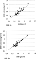
- the combination can be conducted for example by means of regression, such as for example by means of linear regression (cf. figs. 2a and 2b ).
- the estimate for mineral density can be further used for determining a parameter representing the degree of a patient's osteoporosis, as well as a fracture risk predicting parameter (for example fracture probability) in step 112, for example by comparing said mineral density estimate to reference values regarded as normal.
- a fracture risk predicting parameter for example fracture probability
- the thicknesses of cortical bone from lower and upper tibial segments (CTh_ds ja CTh_pr) and the ultrasound backscatter from femoral neck (AIB) were determined by pulse echo imaging (cf. table 1 below).
- the AIB was determined for 25 women and AIB was corrected by DFUS technique with the attenuation caused by soft tissue on top of the bone. Other parameters were determined for 29 women.
- the ultrasound backscatter (AIB) measured from femoral neck the cortical bone thicknesses measured from tibia (from distal end CTh_ds and proximal end CTh_pr), and initial information obtained about a patient (age, weight, body mass index (BMI)) are by themselves (linear correlation coefficient r ) just poor or moderate predictors of the femoral neck bone mineral density (BMD).
- the parameters can be processed with linear regression technique to provide a strong predictor for bone mineral density.
- BMD_estimate_2 3,624 ⁇ 0,015 * age + 0,070 * CTh_pr + 0,064 * CTh_ds ⁇ 0,061 * BMI ⁇ 0,005 * AIB ⁇ 0,016 * height + 0,027 * weight
- parameters of the above-presented estimates may have various coefficients in various embodiments, for example in accordance with the employed measurement material.
- Fig. 3 shows one exemplary arrangement 300 for the assessment of bone mineral density according to one preferred embodiment of the invention, wherein the arrangement 300 comprises transmission means 302 for a measurement signal (y pulse-echo measurement means for an ultrasound signal) for transmitting a measurement signal towards a patient's second bone 301 (other than the first bone), as well as reception means 304 for a measurement signal for receiving a measurement signal after its interaction with said second bone 301, preferably a reflected (such as reflected from cortical bone) ultrasound signal.
- the interacted signal is an ultrasound signal and its property to be measured being reflection.
- the reception device 304 is placed on the same side as the transmission device 302 with respect to the object of measurement.
- the transmission device 302 may also be the reception device 304 at the same time, whereby the transmitter/receiver device 302/304 may comprise for example a physically common crystal, which first generates an ultrasound signal and then receives said ultrasound signal after its interaction with an object of measurement.
- the pulse-echo measurement device comprises a single transceiver sensor (302/304), which is particularly handy in the process of measuring several different points.
- the transmission and reception means 302, 304 may also involve some control electronics 302a, 304a therefor.
- the control electronics are adapted to control the transmitter 302 and the receiver 304, such that the transmitter's transmission operation is discontinued for the duration of the receiver's reception operation in order to minimize interferences, especially in the embodiment using one and the same crystal to generate an ultrasound signal and to receive a signal reflected from an object.
- the arrangement preferably comprises elements 306 for feeding said second parameters into the arrangement, such as a keyboard or a graphical data entry tool.
- At least some further second parameters can be entered into the arrangement also by means of the elements 306.
- a parameter representing a property change in an ultrasound signal launched towards said first bone, after said ultrasound signal has been in interaction with said first bone such as for example an AIB parameter (dB) determined from ultrasound backscatter from the femoral head and/or neck, can be entered into the arrangement by means of the elements 306.
- an AIB parameter (dB) determined from ultrasound backscatter from the femoral head and/or neck can be entered into the arrangement by means of the elements 306.
- the arrangement comprises preferably also control elements 308 for controlling said measurement signal transmission means and reception means, for controlling the operation of the ultrasound transmitter 302 and receiver 304, such as, for example, for timing the operation of the transmission means' transmission and the reception means' operation.
- the control elements 308 may preferably control the operation of the transmission means 302 and/or the reception means 304 through the intermediary of the control electronics 302a, 304a associated therewith.
- the arrangement comprises also processing elements 310, which are adapted to determine an estimate for the mineral density regarding a first bone of the patient by using at least said first and second parameters.
- the processing elements 310 is adapted to use the second parameters in the process of determining the estimate.
- the estimate is worked out preferably by means of some regression technique, such as for example by means of linear regression.
- the processing elements 310 are adapted to determine a first parameter, which is related to a property change in a measurement signal launched towards some second bone of said patient which is other than the first bone after the measurement signal has been in interaction with said second bone.
- the processing elements 310 are adapted to determine the thickness of tibial or ulnar/radius cortical bone from one or several points thereof from a property change of the measurement signal, and to use this as said first parameter.
- the arrangement's processing elements 310 may be adapted to conduct a correction of parameters, such as for example a correction regarding a property change in a measurement signal after the signal has been in interaction with an object of measurement, or a correction regarding the strength (dB) of ultrasound backscatter from the femoral head and/or neck with the effect of soft tissue.
- the correction may be based for example on DFUS technique and/or on taking into account the attenuation taking place in the cortical bone layer.
- the arrangement's processing elements 310 can be adapted to determine a parameter representing the degree of a patient's osteoporosis by using a mineral density estimate related to a first bone of the patient and by comparing said mineral density estimate to reference values regarded as normal in a manner described elsewhere in this document.
- the arrangement may also comprise elements 312 for determining a femoral geometric parameter (such as cross-sectional area or diameter) from the femoral shaft and/or neck as one of the second parameters, such as for example ultrasound transceiver elements and software for interpreting the received ultrasound signal and calculating the surface area, as well as elements 314 for determining a property change in an ultrasound signal launched towards said first bone after said ultrasound signal has been in interaction with said first bone, such as, for example, for determining an AIB parameter (dB) from ultrasound backscatter from the femoral head and/or neck, and elements 316 for determining a hand grip strength of the patient and using said information as one of the second parameters.
- a femoral geometric parameter such as cross-sectional area or diameter
- the second parameters such as for example ultrasound transceiver elements and software for interpreting the received ultrasound signal and calculating the surface area
- elements 314 for determining a property change in an ultrasound signal launched towards said first bone after said ultrasound signal has been in interaction with said first bone, such
- At least some of the arrangement's elements and, especially, some of the functionalities of its processing elements 310 can be implemented programmatically for example by means of a computer program product capable of being run in the arrangement's data processing processor.
- said computer program can be adapted to determine an assessment of probability for bone fractures over the next 10 years, whereby, for example, the treatment guidelines for osteoporosis can be determined readily, quickly and precisely without an expensive separate measurement for example with a DXA apparatus.
- the computer software can be supplied for example with simple answers (yes or no) to some questions regarding fracture risk factors, after which the software is adapted to determine a probability for fracture and, according to one embodiment, to provide a guideline for the treatment, such as for example "no medication” or “measure mineral density” or “medicate”.
- a computer program product for the assessment of mineral density related to a patient's first bone, wherein said first bone is associated with the femoral head, neck and/or the lumbar spine, is adapted:
Landscapes
- Health & Medical Sciences (AREA)
- Life Sciences & Earth Sciences (AREA)
- Engineering & Computer Science (AREA)
- Medical Informatics (AREA)
- Public Health (AREA)
- Pathology (AREA)
- General Health & Medical Sciences (AREA)
- Biomedical Technology (AREA)
- Physics & Mathematics (AREA)
- Veterinary Medicine (AREA)
- Animal Behavior & Ethology (AREA)
- Surgery (AREA)
- Molecular Biology (AREA)
- Heart & Thoracic Surgery (AREA)
- Biophysics (AREA)
- Nuclear Medicine, Radiotherapy & Molecular Imaging (AREA)
- Radiology & Medical Imaging (AREA)
- Computer Vision & Pattern Recognition (AREA)
- Rheumatology (AREA)
- Orthopedic Medicine & Surgery (AREA)
- Physiology (AREA)
- Oral & Maxillofacial Surgery (AREA)
- Dentistry (AREA)
- Artificial Intelligence (AREA)
- Data Mining & Analysis (AREA)
- Databases & Information Systems (AREA)
- Epidemiology (AREA)
- Primary Health Care (AREA)
- Signal Processing (AREA)
- Psychiatry (AREA)
- High Energy & Nuclear Physics (AREA)
- Optics & Photonics (AREA)
- Ultra Sonic Daignosis Equipment (AREA)
- Measurement Of The Respiration, Hearing Ability, Form, And Blood Characteristics Of Living Organisms (AREA)
- Apparatus For Radiation Diagnosis (AREA)
Claims (9)
- Verfahren zur Bewertung der Mineraldichte (100) in Bezug auf einen ersten Knochen eines Patienten, wobei der erste Knochen einem Oberschenkelkopf, einem Oberschenkelhals und/oder einer Lendenwirbelsäule zugeordnet ist, das Verfahren umfassend:- Bestimmen eines ersten Parameters (102) mit einem Impulsechoverfahren, der sich auf eine Eigenschaftsänderung in einem Ultraschallmesssignal bezieht, das in Richtung eines kortikalen Knochens eines Röhrenknochens des Patienten ausgestrahlt wird, nachdem das Messsignal in Wechselwirkung mit dem kortikalen Knochen des Röhrenknochens gestanden hat,
wobei der kortikale Knochen des Röhrenknochens ein anderer zweiter Knochen als der erste Knochen ist, wobei der erste Parameter eine kortikale Knochendicke des zweiten Knochens ist und basierend auf einem zeitlichen Abstand zwischen Signalen bestimmt wird, die von ersten und zweiten Rändern des kortikalen Knochens des Röhrenknochens in Richtung des Ultraschallmesssignals reflektiert werden,- Bestimmen eines Satzes von zweiten Parametern (104), wobei der Satz von zweiten Parametern aus dem Alter und dem Gewicht des Patienten besteht und- Bestimmen einer Schätzung für die Mineraldichte (110), die sich auf den ersten Knochen des Patienten bezieht, unter Verwendung der ersten und zweiten Parameter, indem nur die Dicke der Knochenrinde des Röhrenknochens, das Alter und das Gewicht des Patienten durch eine durch eine Regressionstechnik bestimmte Gleichung kombiniert werden. - Verfahren nach Anspruch 1, wobei der Röhrenknochen mindestens eines von Folgendem umfasst: Calcaneus, Tibia, Finger und/oder Ulna, Radius.
- Verfahren nach einem der vorstehenden Ansprüche, wobei der erste Parameter die Dicke des tibialen oder radius/ulnaren Kortikalknochens an einem, vorzugsweise an mehreren Punkten davon ist.
- Verfahren nach einem der vorstehenden Ansprüche, wobei die Schätzung durch Kombinieren der Parameter mittels linearer Regression ermittelt wird.
- Verfahren nach einem der vorstehenden Ansprüche, wobei die aus einem Ultraschallsignal ableitbare Eigenschaft der Zeitlücke mit dem Effekt des weichen Gewebes zum Beispiel durch die DFUS-Technik korrigiert wird.
- Anordnung zur Bewertung der Mineraldichte in Bezug auf den ersten Knochen eines Patienten, wobei der erste Knochen einem Oberschenkelkopf, einem Oberschenkelhals und/oder einer Lendenwirbelsäule zugeordnet ist, wobei die Anordnung (300) Folgendes umfasst: Messsignalübertragungsmittel (302); Messsignalempfangsmittel (304); Elemente (306) zum Einspeisen von Parametern in die Anordnung (300); Steuermittel (308) zum Steuern der Messsignalübertragungsmittel (302) und der Empfangsmittel (304) und Verarbeitungselemente (310), wobei die Anordnung (300) geeignet ist zum:- Übertragen eines Ultraschallmesssignals zu einem kortikalen Knochen eines Röhrenknochens eines Patienten durch die Messsignalübertragungsmittel (302) mit einem Impulsechoverfahren sowie Empfangen des Ultraschallmesssignals nach seiner Wechselwirkung mit dem kortikalen Knochen des Röhrenknochens durch die Messsignalempfangsmittel (304), wobei der kortikale Knochen des Röhrenknochens ein anderer zweiter Knochen (301) als der erste Knochen ist,- Bestimmen eines ersten Parameters (102) durch die Verarbeitungselemente (310), wobei der erste Parameter eine kortikale Knochendicke des zweiten Knochens ist und basierend auf einem zeitlichen Abstand zwischen Signalen bestimmt wird, die von ersten und zweiten Rändern des kortikalen Knochens des Röhrenknochens in Richtung des Ultraschallmesssignals reflektiert werden,- durch Elemente (306), die einen Satz von zweiten Parametern (104) in die Anordnung einspeisen, wobei der Satz von zweiten Parametern aus dem Alter und dem Gewicht des Patienten besteht und- Bestimmen einer Schätzung der Mineraldichte (110), die sich auf den ersten Knochen des Patienten bezieht, durch die Verarbeitungselemente (310) unter Verwendung der ersten und zweiten Parameter, indem nur die Dicke der Knochenrinde des Röhrenknochens, das Alter und das Gewicht des Patienten durch eine durch eine Regressionstechnik bestimmte Gleichung kombiniert werden.
- Anordnung nach Anspruch 6, wobei die Anordnung geeignet ist, eine Schätzung durch Kombinieren der Parameter mittels linearer Regression zu berechnen.
- Anordnung nach Anspruch 6, wobei die Anordnung geeignet ist, die aus einem Ultraschallsignal ableitbare Zeitlückeneigenschaft mit dem Effekt des weichen Gewebes zum Beispiel durch DFUS-Technik zu korrigieren.
- Computerprogrammprodukt für eine Bewertung der Mineraldichte in Bezug auf einen ersten Knochen eines Patienten, wobei der erste Knochen einem Oberschenkelkopf, einem Oberschenkelhals und/oder einer Lendenwirbelsäule zugeordnet ist, wobei das Computerprogrammprodukt geeignet ist zum:- Bestimmen eines ersten Parameters (102) mit einem Impulsechoverfahren, der sich auf eine Eigenschaftsänderung in einem Ultraschallmesssignal bezieht, das in Richtung eines kortikalen Knochens eines Röhrenknochens des Patienten ausgestrahlt wird, nachdem das Messsignal in Wechselwirkung mit dem kortikalen Knochen des Röhrenknochens (301) gestanden hat,
wobei der kortikale Knochen des Röhrenknochens ein anderer zweiter Knochen als der erste Knochen ist, wobei der erste Parameter eine kortikale Knochendicke des zweiten Knochens ist und basierend auf einem zeitlichen Abstand zwischen Signalen bestimmt wird, die von ersten und zweiten Rändern des kortikalen Knochens des Röhrenknochens in Richtung des Ultraschallmesssignals reflektiert werden,- Bestimmen eines Satzes von zweiten Parametern (104), wobei der Satz von zweiten Parametern aus dem Alter und dem Gewicht des Patienten besteht und- Bestimmen einer Schätzung für die Mineraldichte (110), die sich auf den ersten Knochen des Patienten bezieht, unter Verwendung der ersten und zweiten Parameter, indem nur die Dicke der Knochenrinde des Röhrenknochens, das Alter und das Gewicht des Patienten durch eine durch eine Regressionstechnik bestimmte Gleichung kombiniert werden,wenn das Computerprogrammprodukt mit einer Anordnung nach Anspruch 6 ausgeführt wird.
Applications Claiming Priority (2)
| Application Number | Priority Date | Filing Date | Title |
|---|---|---|---|
| FI20105936A FI126104B (fi) | 2010-09-09 | 2010-09-09 | Menetelmä ja järjestelmä luun mineraalitiheyden arvioimiseksi |
| PCT/FI2011/050772 WO2012032225A1 (en) | 2010-09-09 | 2011-09-09 | Method and arrangement for estimating mineral density of a bone |
Publications (4)
| Publication Number | Publication Date |
|---|---|
| EP2613705A1 EP2613705A1 (de) | 2013-07-17 |
| EP2613705A4 EP2613705A4 (de) | 2018-01-17 |
| EP2613705B1 true EP2613705B1 (de) | 2024-12-11 |
| EP2613705C0 EP2613705C0 (de) | 2024-12-11 |
Family
ID=42829672
Family Applications (1)
| Application Number | Title | Priority Date | Filing Date |
|---|---|---|---|
| EP11823123.2A Active EP2613705B1 (de) | 2010-09-09 | 2011-09-09 | Verfahren und anordnung zur messung der mineraldichte eines knochens |
Country Status (9)
| Country | Link |
|---|---|
| US (1) | US9526472B2 (de) |
| EP (1) | EP2613705B1 (de) |
| JP (1) | JP5960699B2 (de) |
| CN (1) | CN103237501B (de) |
| ES (1) | ES3008695T3 (de) |
| FI (1) | FI126104B (de) |
| PL (1) | PL2613705T3 (de) |
| RU (1) | RU2598642C2 (de) |
| WO (1) | WO2012032225A1 (de) |
Families Citing this family (6)
| Publication number | Priority date | Publication date | Assignee | Title |
|---|---|---|---|---|
| CN103598875B (zh) * | 2013-11-16 | 2015-01-28 | 沈阳医学院 | 一种用于预测骨质疏松发生风险的人体骨密度预测装置 |
| WO2017083869A1 (en) * | 2015-11-13 | 2017-05-18 | Orthoforge | Medical devices, systems and methods for monitoring and stimulating osteogenesis |
| WO2017106485A1 (en) * | 2015-12-16 | 2017-06-22 | Hologic, Inc. | Systems and methods for presenting complex medical condition diagnoses |
| KR101840349B1 (ko) * | 2016-11-15 | 2018-03-21 | 강원대학교산학협력단 | 초음파 합주파수 성분을 이용한 골밀도 예측 장치 및 방법 |
| CN110769754B (zh) * | 2017-06-21 | 2023-06-27 | 夏里特柏林大学医学院 | 用于测定皮质骨的系统、方法和计算机程序产品 |
| RU2750976C1 (ru) * | 2020-10-16 | 2021-07-07 | Федеральное государственное автономное образовательное учреждение высшего образования "Новосибирский национальный исследовательский государственный университет" (Новосибирский государственный университет, НГУ) | Способ определения плотности костной ткани на основе выделения стоячих волн из микросейсм периферического скелета |
Family Cites Families (24)
| Publication number | Priority date | Publication date | Assignee | Title |
|---|---|---|---|---|
| US4913157A (en) * | 1986-06-03 | 1990-04-03 | Analog Devices, Inc. | Ultrasound method and apparatus for evaluating, in vivo, bone conditions |
| US5840029A (en) * | 1988-05-11 | 1998-11-24 | Lunar Corporation | Imaging ultrasonic densitometer |
| US4941474A (en) * | 1988-07-01 | 1990-07-17 | Massachusetts Institute Of Technology | Multivariable analysis of bone condition |
| US5218963A (en) * | 1991-10-15 | 1993-06-15 | Lunar Corporation | Ultrasonic bone analysis device and method |
| US6213934B1 (en) * | 1995-06-01 | 2001-04-10 | Hyper3D Corp. | Electromagnetic bone-assessment and treatment: apparatus and method |
| CA2211604A1 (en) * | 1995-11-29 | 1997-06-05 | Sekisui Kagaku Kogyo Kabushiki Kaisya | Apparatus and method for diagnosing osteoporosis |
| US20020006181A1 (en) | 1998-01-23 | 2002-01-17 | Mackenzie Innes K. | Method and device for estimating bone mineral content of the calcaneus |
| WO2001073116A2 (en) | 2000-03-28 | 2001-10-04 | Signalgene Inc. | Method for determining osteoporosis susceptibility and/or low bone density and reagents therefor |
| JP2002238904A (ja) | 2001-02-19 | 2002-08-27 | Tanita Corp | 骨密度推定方法および骨密度推定装置 |
| US6468215B1 (en) * | 2001-07-16 | 2002-10-22 | Artann Laboratories | Method and device for multi-parametric ultrasonic assessment of bone conditions |
| DE60228310D1 (de) * | 2001-11-30 | 2008-09-25 | Petro Moilanen | Verfahren für die nichtinvasive untersuchung von knochen |
| US7386337B2 (en) | 2002-05-06 | 2008-06-10 | University Of Rochester | Correction of dual energy X-ray absorptiometry measurements based on body lead levels |
| DK1393680T3 (da) * | 2002-08-30 | 2007-12-10 | L Acn L Accessorio Nucleare Sr | Apparat til måling af knoglemineraldensitet |
| US20050015002A1 (en) * | 2003-07-18 | 2005-01-20 | Dixon Gary S. | Integrated protocol for diagnosis, treatment, and prevention of bone mass degradation |
| RU2305491C2 (ru) * | 2003-12-05 | 2007-09-10 | Российская медицинская академия последипломного образования Министерства здравоохранения Российской Федерации (РМАПО МЗ РФ) | Способ исследования и диагностики патологии костной ткани при сахарном диабете |
| US8202219B2 (en) * | 2004-02-23 | 2012-06-19 | Cyberlogic, Inc. | Ultrasonic bone assessment apparatus and method |
| EP1789924A2 (de) * | 2004-09-16 | 2007-05-30 | Imaging Therapeutics, Inc. | System und verfahren zur vorhersage von zukünftigen brüchen |
| WO2008038159A2 (en) * | 2006-09-29 | 2008-04-03 | Odetect As | Ultrasound measurement techniques for bone analysis |
| CN101199429B (zh) * | 2006-12-13 | 2012-06-06 | 计算机逻辑公司 | 超声波骨评估的方法和装置 |
| US7862510B2 (en) * | 2007-02-09 | 2011-01-04 | Cyberlogic, Inc. | Ultrasonic bone assessment apparatus and method |
| JP5280647B2 (ja) | 2007-05-29 | 2013-09-04 | 古野電気株式会社 | 超音波を用いた骨強度診断装置及び超音波を用いた骨強度診断装置が作動する方法 |
| US8679019B2 (en) * | 2007-12-03 | 2014-03-25 | Bone Index Finland Oy | Method for measuring of thicknesses of materials using an ultrasound technique |
| WO2010093769A2 (en) * | 2009-02-13 | 2010-08-19 | Cyberlogic, Inc. | Ultrasonic bone assessment apparatus and method |
| US8880143B2 (en) * | 2012-01-17 | 2014-11-04 | Sectra Imtec Ab | Apparatus and method for estimating the bone mineral density to asses bone fractures risk |
-
2010
- 2010-09-09 FI FI20105936A patent/FI126104B/fi active IP Right Grant
-
2011
- 2011-09-09 JP JP2013527654A patent/JP5960699B2/ja active Active
- 2011-09-09 CN CN201180054044.7A patent/CN103237501B/zh active Active
- 2011-09-09 EP EP11823123.2A patent/EP2613705B1/de active Active
- 2011-09-09 PL PL11823123.2T patent/PL2613705T3/pl unknown
- 2011-09-09 WO PCT/FI2011/050772 patent/WO2012032225A1/en not_active Ceased
- 2011-09-09 ES ES11823123T patent/ES3008695T3/es active Active
- 2011-09-09 US US13/821,883 patent/US9526472B2/en active Active
- 2011-09-09 RU RU2013114988/14A patent/RU2598642C2/ru active
Non-Patent Citations (2)
| Title |
|---|
| KARJALAINEN J ET AL: "Ultrasonic assessment of cortical bone thickness in vitro and in vivo", IEEE TRANSACTIONS ON ULTRASONICS, FERROELECTRICS, AND FREQUENCY CONTROL, IEEE, USA, vol. 55, no. 10, 1 October 2008 (2008-10-01), pages 2191 - 2197, XP011235808, ISSN: 0885-3010, DOI: 10.1109/TUFFC.918 * |
| KARJALAINEN J P ET AL: "Ultrasound Backscatter Imaging Provides Frequency-Dependent Information on Structure, Composition and Mechanical Properties of Human Trabecular Bone", ULTRASOUND IN MEDICINE AND BIOLOGY, NEW YORK, NY, US, vol. 35, no. 8, 1 August 2009 (2009-08-01), pages 1376 - 1384, XP026321837, ISSN: 0301-5629, [retrieved on 20090613], DOI: 10.1016/J.ULTRASMEDBIO.2009.03.011 * |
Also Published As
| Publication number | Publication date |
|---|---|
| FI126104B (fi) | 2016-06-30 |
| ES3008695T3 (en) | 2025-03-24 |
| WO2012032225A1 (en) | 2012-03-15 |
| FI20105936A7 (fi) | 2012-03-10 |
| US20130245443A1 (en) | 2013-09-19 |
| EP2613705A1 (de) | 2013-07-17 |
| US9526472B2 (en) | 2016-12-27 |
| FI20105936L (fi) | 2012-03-10 |
| RU2013114988A (ru) | 2014-10-20 |
| CN103237501B (zh) | 2015-09-30 |
| RU2598642C2 (ru) | 2016-09-27 |
| FI20105936A0 (fi) | 2010-09-09 |
| EP2613705A4 (de) | 2018-01-17 |
| JP5960699B2 (ja) | 2016-08-02 |
| JP2013537055A (ja) | 2013-09-30 |
| CN103237501A (zh) | 2013-08-07 |
| EP2613705C0 (de) | 2024-12-11 |
| PL2613705T3 (pl) | 2025-04-28 |
Similar Documents
| Publication | Publication Date | Title |
|---|---|---|
| US11911175B2 (en) | Ultrasound apparatus for assessing the quality of a patient's bone tissue | |
| Oliveira et al. | Osteoporosis screening: applied methods and technological trends | |
| Pisani et al. | Screening and early diagnosis of osteoporosis through X-ray and ultrasound based techniques | |
| Karjalainen et al. | Multi-site bone ultrasound measurements in elderly women with and without previous hip fractures | |
| EP2613705B1 (de) | Verfahren und anordnung zur messung der mineraldichte eines knochens | |
| Egorov et al. | Osteoporosis detection in postmenopausal women using axial transmission multi-frequency bone ultrasonometer: Clinical findings | |
| Wan et al. | Summation of ossification ratios of radius, ulna and femur: a new parameter to evaluate bone age by ultrasound | |
| Raum et al. | Clinical devices for bone assessment | |
| Khy et al. | Bilateral stress fracture of the tibia diagnosed by ultrasound. A case report | |
| WO2010116374A2 (en) | Improved bone sonometer | |
| Wear | The effect of phase cancellation on estimates of calcaneal broadband ultrasound attenuation in vivo | |
| Bozkurt et al. | The diagnostic accuracy of ultrasonography in determining the reduction success of distal radius fractures | |
| Bi et al. | Ultrasonic backscatter measurements of human cortical and trabecular bone densities in a head-down bed-rest study | |
| Chen et al. | Quantitative assessment of osteoporosis from the tibia shaft by ultrasound techniques | |
| Rojo et al. | Classification of hip fragility fractures in older adults using an ultrasonic device | |
| KR100581229B1 (ko) | 램파를 이용한 경골의 골밀도 측정방법 | |
| As' ad | Axial skeletal assessment in osteoporosis using radiofrequency echographic Multi-spectrometry: diagnostic performance, clinical utility, and future directions | |
| RU2732697C1 (ru) | Способ определения жесткости костной мозоли ультразвуковой эластографией сдвиговой волны | |
| Mesquita et al. | Correlation between ultrasound velocity and densitometry in fresh and demineralized cortical bone | |
| Li et al. | Effect of spectral estimation on ultrasonic backscatter parameters in measurements of cancellous bones | |
| Ramiandrisoa et al. | In vivo estimation of cortical thickness and porosity by axial transmission: Comparison with high resolution computed tomography | |
| Minonzio et al. | Cortical bone parameters measured at the one-third distal radius obtained with axial transmission and HR-pQCT compared with anthropometric data in a representative population | |
| Karjalainen | Novel pulse-echo ultrasound methods for diagnostics of osteoporosis | |
| JP2014151175A (ja) | 骨内超音波伝播速度の測定による骨強度評価 | |
| Minonzio et al. | Bi-directional Axial Transmission measurements is an easy to apply methodology allowing risk assessment of fracture in elderly |
Legal Events
| Date | Code | Title | Description |
|---|---|---|---|
| PUAI | Public reference made under article 153(3) epc to a published international application that has entered the european phase |
Free format text: ORIGINAL CODE: 0009012 |
|
| 17P | Request for examination filed |
Effective date: 20130403 |
|
| AK | Designated contracting states |
Kind code of ref document: A1 Designated state(s): AL AT BE BG CH CY CZ DE DK EE ES FI FR GB GR HR HU IE IS IT LI LT LU LV MC MK MT NL NO PL PT RO RS SE SI SK SM TR |
|
| DAX | Request for extension of the european patent (deleted) | ||
| RIC1 | Information provided on ipc code assigned before grant |
Ipc: A61B 5/00 20060101AFI20171207BHEP Ipc: A61B 8/08 20060101ALI20171207BHEP Ipc: A61B 6/00 20060101ALI20171207BHEP |
|
| RA4 | Supplementary search report drawn up and despatched (corrected) |
Effective date: 20171215 |
|
| STAA | Information on the status of an ep patent application or granted ep patent |
Free format text: STATUS: EXAMINATION IS IN PROGRESS |
|
| 17Q | First examination report despatched |
Effective date: 20210401 |
|
| GRAP | Despatch of communication of intention to grant a patent |
Free format text: ORIGINAL CODE: EPIDOSNIGR1 |
|
| STAA | Information on the status of an ep patent application or granted ep patent |
Free format text: STATUS: GRANT OF PATENT IS INTENDED |
|
| INTG | Intention to grant announced |
Effective date: 20240704 |
|
| GRAS | Grant fee paid |
Free format text: ORIGINAL CODE: EPIDOSNIGR3 |
|
| GRAA | (expected) grant |
Free format text: ORIGINAL CODE: 0009210 |
|
| STAA | Information on the status of an ep patent application or granted ep patent |
Free format text: STATUS: THE PATENT HAS BEEN GRANTED |
|
| AK | Designated contracting states |
Kind code of ref document: B1 Designated state(s): AL AT BE BG CH CY CZ DE DK EE ES FI FR GB GR HR HU IE IS IT LI LT LU LV MC MK MT NL NO PL PT RO RS SE SI SK SM TR |
|
| REG | Reference to a national code |
Ref country code: GB Ref legal event code: FG4D |
|
| REG | Reference to a national code |
Ref country code: CH Ref legal event code: EP |
|
| REG | Reference to a national code |
Ref country code: IE Ref legal event code: FG4D |
|
| REG | Reference to a national code |
Ref country code: DE Ref legal event code: R096 Ref document number: 602011075151 Country of ref document: DE |
|
| U01 | Request for unitary effect filed |
Effective date: 20250107 |
|
| U07 | Unitary effect registered |
Designated state(s): AT BE BG DE DK EE FI FR IT LT LU LV MT NL PT RO SE SI Effective date: 20250115 |
|
| REG | Reference to a national code |
Ref country code: ES Ref legal event code: FG2A Ref document number: 3008695 Country of ref document: ES Kind code of ref document: T3 Effective date: 20250324 |
|
| PG25 | Lapsed in a contracting state [announced via postgrant information from national office to epo] |
Ref country code: HR Free format text: LAPSE BECAUSE OF FAILURE TO SUBMIT A TRANSLATION OF THE DESCRIPTION OR TO PAY THE FEE WITHIN THE PRESCRIBED TIME-LIMIT Effective date: 20241211 |
|
| PG25 | Lapsed in a contracting state [announced via postgrant information from national office to epo] |
Ref country code: NO Free format text: LAPSE BECAUSE OF FAILURE TO SUBMIT A TRANSLATION OF THE DESCRIPTION OR TO PAY THE FEE WITHIN THE PRESCRIBED TIME-LIMIT Effective date: 20250311 |
|
| PG25 | Lapsed in a contracting state [announced via postgrant information from national office to epo] |
Ref country code: RS Free format text: LAPSE BECAUSE OF FAILURE TO SUBMIT A TRANSLATION OF THE DESCRIPTION OR TO PAY THE FEE WITHIN THE PRESCRIBED TIME-LIMIT Effective date: 20250311 |
|
| REG | Reference to a national code |
Ref country code: GR Ref legal event code: EP Ref document number: 20250400427 Country of ref document: GR Effective date: 20250409 |
|
| PG25 | Lapsed in a contracting state [announced via postgrant information from national office to epo] |
Ref country code: SM Free format text: LAPSE BECAUSE OF FAILURE TO SUBMIT A TRANSLATION OF THE DESCRIPTION OR TO PAY THE FEE WITHIN THE PRESCRIBED TIME-LIMIT Effective date: 20241211 |
|
| PG25 | Lapsed in a contracting state [announced via postgrant information from national office to epo] |
Ref country code: IS Free format text: LAPSE BECAUSE OF FAILURE TO SUBMIT A TRANSLATION OF THE DESCRIPTION OR TO PAY THE FEE WITHIN THE PRESCRIBED TIME-LIMIT Effective date: 20250411 |
|
| PG25 | Lapsed in a contracting state [announced via postgrant information from national office to epo] |
Ref country code: SK Free format text: LAPSE BECAUSE OF FAILURE TO SUBMIT A TRANSLATION OF THE DESCRIPTION OR TO PAY THE FEE WITHIN THE PRESCRIBED TIME-LIMIT Effective date: 20241211 |
|
| PG25 | Lapsed in a contracting state [announced via postgrant information from national office to epo] |
Ref country code: CZ Free format text: LAPSE BECAUSE OF FAILURE TO SUBMIT A TRANSLATION OF THE DESCRIPTION OR TO PAY THE FEE WITHIN THE PRESCRIBED TIME-LIMIT Effective date: 20241211 |
|
| REG | Reference to a national code |
Ref country code: CH Ref legal event code: U11 Free format text: ST27 STATUS EVENT CODE: U-0-0-U10-U11 (AS PROVIDED BY THE NATIONAL OFFICE) Effective date: 20251001 |
|
| PGFP | Annual fee paid to national office [announced via postgrant information from national office to epo] |
Ref country code: GR Payment date: 20250917 Year of fee payment: 15 |
|
| PGFP | Annual fee paid to national office [announced via postgrant information from national office to epo] |
Ref country code: PL Payment date: 20250812 Year of fee payment: 15 Ref country code: TR Payment date: 20250822 Year of fee payment: 15 |
|
| PGFP | Annual fee paid to national office [announced via postgrant information from national office to epo] |
Ref country code: GB Payment date: 20250916 Year of fee payment: 15 |
|
| PLBE | No opposition filed within time limit |
Free format text: ORIGINAL CODE: 0009261 |
|
| STAA | Information on the status of an ep patent application or granted ep patent |
Free format text: STATUS: NO OPPOSITION FILED WITHIN TIME LIMIT |
|
| U20 | Renewal fee for the european patent with unitary effect paid |
Year of fee payment: 15 Effective date: 20250916 |
|
| 26N | No opposition filed |
Effective date: 20250912 |
|
| PGFP | Annual fee paid to national office [announced via postgrant information from national office to epo] |
Ref country code: CH Payment date: 20251001 Year of fee payment: 15 |
|
| PGFP | Annual fee paid to national office [announced via postgrant information from national office to epo] |
Ref country code: ES Payment date: 20251006 Year of fee payment: 15 |