EP2612926B1 - Methods of using mirna for detection of in vivo cell death - Google Patents
Methods of using mirna for detection of in vivo cell death Download PDFInfo
- Publication number
- EP2612926B1 EP2612926B1 EP13161985.0A EP13161985A EP2612926B1 EP 2612926 B1 EP2612926 B1 EP 2612926B1 EP 13161985 A EP13161985 A EP 13161985A EP 2612926 B1 EP2612926 B1 EP 2612926B1
- Authority
- EP
- European Patent Office
- Prior art keywords
- mirna
- urine
- specific
- disease
- death
- Prior art date
- Legal status (The legal status is an assumption and is not a legal conclusion. Google has not performed a legal analysis and makes no representation as to the accuracy of the status listed.)
- Active
Links
Images


Classifications
-
- C—CHEMISTRY; METALLURGY
- C12—BIOCHEMISTRY; BEER; SPIRITS; WINE; VINEGAR; MICROBIOLOGY; ENZYMOLOGY; MUTATION OR GENETIC ENGINEERING
- C12Q—MEASURING OR TESTING PROCESSES INVOLVING ENZYMES, NUCLEIC ACIDS OR MICROORGANISMS; COMPOSITIONS OR TEST PAPERS THEREFOR; PROCESSES OF PREPARING SUCH COMPOSITIONS; CONDITION-RESPONSIVE CONTROL IN MICROBIOLOGICAL OR ENZYMOLOGICAL PROCESSES
- C12Q1/00—Measuring or testing processes involving enzymes, nucleic acids or microorganisms; Compositions therefor; Processes of preparing such compositions
- C12Q1/70—Measuring or testing processes involving enzymes, nucleic acids or microorganisms; Compositions therefor; Processes of preparing such compositions involving virus or bacteriophage
-
- C—CHEMISTRY; METALLURGY
- C12—BIOCHEMISTRY; BEER; SPIRITS; WINE; VINEGAR; MICROBIOLOGY; ENZYMOLOGY; MUTATION OR GENETIC ENGINEERING
- C12Q—MEASURING OR TESTING PROCESSES INVOLVING ENZYMES, NUCLEIC ACIDS OR MICROORGANISMS; COMPOSITIONS OR TEST PAPERS THEREFOR; PROCESSES OF PREPARING SUCH COMPOSITIONS; CONDITION-RESPONSIVE CONTROL IN MICROBIOLOGICAL OR ENZYMOLOGICAL PROCESSES
- C12Q1/00—Measuring or testing processes involving enzymes, nucleic acids or microorganisms; Compositions therefor; Processes of preparing such compositions
- C12Q1/68—Measuring or testing processes involving enzymes, nucleic acids or microorganisms; Compositions therefor; Processes of preparing such compositions involving nucleic acids
- C12Q1/6876—Nucleic acid products used in the analysis of nucleic acids, e.g. primers or probes
- C12Q1/6883—Nucleic acid products used in the analysis of nucleic acids, e.g. primers or probes for diseases caused by alterations of genetic material
-
- C—CHEMISTRY; METALLURGY
- C12—BIOCHEMISTRY; BEER; SPIRITS; WINE; VINEGAR; MICROBIOLOGY; ENZYMOLOGY; MUTATION OR GENETIC ENGINEERING
- C12Q—MEASURING OR TESTING PROCESSES INVOLVING ENZYMES, NUCLEIC ACIDS OR MICROORGANISMS; COMPOSITIONS OR TEST PAPERS THEREFOR; PROCESSES OF PREPARING SUCH COMPOSITIONS; CONDITION-RESPONSIVE CONTROL IN MICROBIOLOGICAL OR ENZYMOLOGICAL PROCESSES
- C12Q1/00—Measuring or testing processes involving enzymes, nucleic acids or microorganisms; Compositions therefor; Processes of preparing such compositions
- C12Q1/70—Measuring or testing processes involving enzymes, nucleic acids or microorganisms; Compositions therefor; Processes of preparing such compositions involving virus or bacteriophage
- C12Q1/701—Specific hybridization probes
- C12Q1/705—Specific hybridization probes for herpetoviridae, e.g. herpes simplex, varicella zoster
-
- C—CHEMISTRY; METALLURGY
- C12—BIOCHEMISTRY; BEER; SPIRITS; WINE; VINEGAR; MICROBIOLOGY; ENZYMOLOGY; MUTATION OR GENETIC ENGINEERING
- C12Q—MEASURING OR TESTING PROCESSES INVOLVING ENZYMES, NUCLEIC ACIDS OR MICROORGANISMS; COMPOSITIONS OR TEST PAPERS THEREFOR; PROCESSES OF PREPARING SUCH COMPOSITIONS; CONDITION-RESPONSIVE CONTROL IN MICROBIOLOGICAL OR ENZYMOLOGICAL PROCESSES
- C12Q2600/00—Oligonucleotides characterized by their use
- C12Q2600/118—Prognosis of disease development
-
- C—CHEMISTRY; METALLURGY
- C12—BIOCHEMISTRY; BEER; SPIRITS; WINE; VINEGAR; MICROBIOLOGY; ENZYMOLOGY; MUTATION OR GENETIC ENGINEERING
- C12Q—MEASURING OR TESTING PROCESSES INVOLVING ENZYMES, NUCLEIC ACIDS OR MICROORGANISMS; COMPOSITIONS OR TEST PAPERS THEREFOR; PROCESSES OF PREPARING SUCH COMPOSITIONS; CONDITION-RESPONSIVE CONTROL IN MICROBIOLOGICAL OR ENZYMOLOGICAL PROCESSES
- C12Q2600/00—Oligonucleotides characterized by their use
- C12Q2600/158—Expression markers
-
- C—CHEMISTRY; METALLURGY
- C12—BIOCHEMISTRY; BEER; SPIRITS; WINE; VINEGAR; MICROBIOLOGY; ENZYMOLOGY; MUTATION OR GENETIC ENGINEERING
- C12Q—MEASURING OR TESTING PROCESSES INVOLVING ENZYMES, NUCLEIC ACIDS OR MICROORGANISMS; COMPOSITIONS OR TEST PAPERS THEREFOR; PROCESSES OF PREPARING SUCH COMPOSITIONS; CONDITION-RESPONSIVE CONTROL IN MICROBIOLOGICAL OR ENZYMOLOGICAL PROCESSES
- C12Q2600/00—Oligonucleotides characterized by their use
- C12Q2600/178—Oligonucleotides characterized by their use miRNA, siRNA or ncRNA
Definitions
- the invention provides non-invasive methods for isolation and detection of microRNA (miRNA) sequences in bodily fluid. More specifically, the present invention encompasses methods of detecting in vivo cell death by analyzing urine and other body fluids for miRNA levels for clinical diagnosis and treatment monitoring.
- miRNA microRNA
- Cell death is a normal component of development and functioning of multicellular organisms. Being a natural process, cell death is involved in the pathology of numerous diseases caused by internal factors. Cell death also accompanies diseases caused by external physical, chemical, of biological agents.
- necrosis is considered to be catastrophic metabolic failure resulting directly from severe molecular and/or structural damage and leads to inflammation and secondary damage to surrounding cells.
- Apoptosis is a much more prevalent biological phenomenon than necrosis and can be induced by specific signals such as hormones, cytokines, by absence of specific signal such as growth or adhesion factors, or by molecular damage that does not cause catastrophic loss of integrity.
- Apoptosis is a result of an active cellular response involving initiation of an orderly and specific cascade of molecular events.
- Apoptosis leads to the appearance of distinctive chromatin condensation and margination, nuclear fragmentation, cell shrinkage, membrane blebbing and enzymatic internucleosomal fragmentation of nuclear DNA ( Umansky et al., Biochim Biophys Acta. 655, 9-17 (1981 ); Arends et al., Am J Pathol. 136, 593-608(1990 )).
- Other more rare forms of cell death, characterized by specific morphology, for example, so called autophagic cell death have also been described ( Bredesen et al., Stroke. 38(2 Suppl):652-660 (2007 ).
- liver-specific enzymes in blood or in other bodily fluids.
- Measurement of the activity of liver-specific enzymes in blood is a widely used method for evaluation of hepatocyte death ( Amacher, et al., Regul Toxicol Pharmacol. Apr; 27(2):119-130 (1988 ); Salaspuro, et al., Enzyme.37:87-107 (1987 ); Herlong, Hosp. Pract. (Off Ed).29(11):32-38 (1994)).
- Tr-DNA transrenal DNA
- US 6251638 B1 relates to a non-invasive method of detecting DNA in urine in order to detect fetal DNA sequence.
- both cell-free plasma DNA and Tr-DNA may be used as diagnostic tools, they provide a rather limited approach when evaluating tissue specific events, such as cell death.
- analytical methods that are non-invasive, and provide a broader range of indications of specific pathology, due to their ability to detect levels of dying cells in particular tissues and organs, would be useful for diagnosing and monitoring the state of various diseases or pathological conditions in patients.
- tissue specific analytical methods that provide the means for monitoring the response of a patient to a disease therapy would be useful to determine the therapy effectiveness, and in the case of drug treatment, the optimum dosage required for drug administration.
- miRNA micro RNA
- the instant invention is focused on the use of micro RNA (miRNA) as a diagnostic tool to monitor in vivo cell death in bodily fluids, such as for example serum and urine.
- miRNA micro RNA
- many miRNAs exhibit cell, tissue and organ specific expression profiles ( Liang et al., Genomics, 8: 166 (2007 ); Lukiw et al, Neuroreport. 18:297-300 (2007 ); Lagos-Quintana et al., Curr Biol. 12:735-739 (2002 ); Chen et al., Nat Genet. 38:228-233 (2006 ); Beuvink et al., J. Nucleic Acids Res. 35:e52 (2007 )).
- the instant invention provides methods for measuring in vivo cell death by detection of tissue-specific miRNAs, characteristic of a pathogen infection, a brain stroke, Alzheimer's disease or Parkinson's disease, in body fluids, such as for example serum and urine.
- body fluids such as for example serum and urine.
- the instant methods based on detection of miRNAs in bodily fluids are used for further development of diagnostic or monitoring tests.
- the instant invention relates to a novel method for detecting and measuring in vivo cell-death by analyzing levels of specific miRNA sequences in cell-free nucleic acids obtained from bodily fluids, said miRNA originating from cells dying throughout the body, and using the obtained analytical result to determine state of a disease or abnormal medical condition in a patient.
- the methods of the instant invention are based on adsorption of cell-free nucleic acids on and elution from anion-exchangers, which makes it possible to concentrate and isolate nucleic acid fragments larger then 10 nucleotides.
- the instant invention demonstrates: (i) the presence of miRNA in body fluids; (ii) detection in urine of miRNA that originated from organs located outside of urinary system, which means that they have crossed the kidney barrier , such as for example, transrenal miRNA (Tr-miRNA); iii) detection of miRNA in serum (iv) pathology associated with cell death in a particular cell, tissue and/or organ is accompanied by changes in levels of miRNA specific for the said organ.
- Tr-miRNA transrenal miRNA
- the present invention provides a method of detecting and measuring in vivo cell death the method comprising:
- the body fluid is urine.
- the present method of analysis of a urine sample includes a technique selected from the group consisting of hybridization, cycling probe reaction, polymerase chain reaction, nested polymerase chain reaction, PCR to analyze single strand conformation polymorphisms and ligase chain reaction.
- nucleic acid degradation in said urine sample is reduced.
- the method of the present invention includes reducing nucleic acid degradation comprising inhibiting nuclease activity by addition of RNAse inhibitor(s), heat inactivation, or by treating said urine sample with a compound selected from the group consisting of: guanidine-HCl, guanidine isothiocyanate, N-lauroylsarcosine, and sodium dodecylsulphate.
- urine sample has been held in the bladder less than 12 hours.
- the body fluid is serum.
- the method of the present invention includes analysis of a serum sample including a technique selected from the group consisting of hybridization, cycling probe reaction, polymerase chain reaction, nested polymerase chain reaction, PCR to analyze single strand conformation polymorphisms and ligase chain reaction.
- the method of the instant invention involves detecting cell-free miRNAs, as a specific marker for the specific disorder associated with excessive or insufficient cell death in a tissue or organ.
- Said disorder is a pathogen infection.
- said pathogen is a virus. More preferably, said virus is an Epstein-Barr virus.
- said disorder is a brain stroke, Alzheimer's disease, or Parkinson's disease.
- the present invention further provides a method of disease and/or treatment monitoring in a patient by quantitative analysis of specific cell-free miRNAs in a urine sample, the method comprising:
- the present invention further provides a method for detecting and measuring miRNA originating from a specific cell type, tissue, or organ in the body of a patient, the method comprising:
- RNAs in particular specific micro RNAs (miRNAs), including transrenal miRNA (Tr-miRNA)
- miRNAs specific micro RNAs
- Tr-miRNA transrenal miRNA
- the presence of these nucleic acid sequences at levels lower or higher than that of a control group is therefore an indication that an abnormality or pathological condition is likely present in the patient from whom the sample was obtained.
- the methods of the present invention offer improvements over previous methods of diagnosis, detection and monitoring due to their inherently non-invasive nature.
- primer refers to an oligonucleotide which is capable of acting as a point of initiation of synthesis when placed under conditions in which primer extension is initiated.
- An oligonucleotide “primer” can occur naturally, as in a purified restriction digest or be produced synthetically.
- a primer is selected to be "substantially" complementary to a strand of specific sequence of the template.
- a primer must be sufficiently complementary to hybridize with a template strand for primer elongation to occur.
- a primer sequence need not reflect the exact sequence of the template.
- a non-complementary nucleotide fragment may be attached to the 5' end of the primer, with the remainder of the primer sequence being substantially complementary to the strand.
- Non-complementary bases or longer sequences can be interspersed into the primer, provided that the primer sequence has sufficient complementarity with the sequence of the template to hybridize and thereby form a template primer complex for synthesis of the extension product of the primer.
- a "target" nucleic acid is a miRNA sequence to be evaluated by hybridization, amplification or any other means of analyzing a nucleic acid sequence, including a combination of analysis methods.
- Hybridization methods involve the annealing of a complementary sequence to the target nucleic acid (the sequence to be analyzed). The ability of two polymers of nucleic acid containing complementary sequences to find each other and anneal through base pairing interaction is a well-recognized phenomenon.
- the initial observations of the "hybridization” process by Marmur and Lane, Proc. Natl. Acad. Sci. USA 46:453 (1960 ) and Doty et al., Proc. Natl. Acad. Sci. USA 46:461 (1960 ) have been followed by the refinement of this process into an essential tool of modern biology.
- Hybridization encompasses, but not be limited to, slot, dot and blot hybridization techniques.
- hybridization method it is important for some diagnostic applications to determine whether the hybridization represents complete or partial complementarity. For example, where it is desired to detect simply the presence or absence of pathogen miRNA, it is only important that the hybridization method ensures hybridization when the relevant sequence is present; conditions can be selected where both partially complementary probes and completely complementary probes will hybridize. Other diagnostic applications, however, may require that the hybridization method distinguish between partial and complete complementarity. It may be of interest to detect genetic polymorphisms.
- probe refers to an oligonucleotide (i.e., a sequence of nucleotides), whether occurring naturally as in a purified restriction digest or produced synthetically, which forms a duplex structure or other complex with a sequence of another nucleic acid, due to complementarity or other means of reproducible attractive interaction, of at least one sequence in the probe with a sequence in the other nucleic acid. Probes are useful in the detection, identification and isolation of particular gene sequences.
- any probe used in the present invention will be labeled with any "reporter molecule,” so that it is detectable in any detection system, including, but not limited to, enzyme (e.g ., ELISA, as well as enzyme--based histochemical assays), fluorescent, radioactive, and luminescent systems. It is further contemplated that the oligonucleotide of interest (i.e., to be detected) will be labeled with a reporter molecule. It is also contemplated that both the probe and oligonucleotide of interest will be labeled. It is not intended that the present invention be limited to any particular detection system or label.
- miRNA is a subclass of small non-coding single stranded RNA, approximately 18-23 nucleotides in length which plays an important role in regulation of metabolic processes, particularly due to their involvement in regulation of stability and translation of mRNA encoding specific proteins. miRNA also participate in other important processes, like heterochromatin formation and genome rearrangement.
- excessive and insufficient in vivo cell death describe the situation when the number of cells dying in a particular organ or tissue is respectively higher or lower than in age and gender matched controls.
- purified As used herein, the terms “purified”, “decontaminated” and “sterilized” refer to the removal of contaminant(s) from a sample.
- the terms “substantially purified” and “substantially isolated” refer to nucleic acid sequences that are removed from their natural environment, isolated or separated, and are preferably 60% free, more preferably 75% free, and most preferably 90% free from other components with which they are naturally associated.
- An "isolated polynucleotide” is therefore a substantially purified polynucleotide. It is contemplated that to practice the methods of the present invention polynucleotides can be, but need not be substantially purified. A variety of methods for the detection of nucleic acid sequences in unpurified form are known in the art.
- PCR product and “amplification product” refer to the resultant mixture of compounds after two or more cycles of the PCR steps of denaturation, annealing and extension are complete. These terms encompass the case where there has been amplification of one or more segments of one or more target sequences.
- urinary tract refers to the organs and ducts which participate in the secretion and elimination of urine from the body.
- Patient refers to the recipient of the treatment. Mammalian and non-mammalian patients are included. In a specific embodiment, the patient is a mammal, such as a human, canine, murine, feline, bovine, ovine, swine, or caprine. In a particular embodiment, the patient is a human.
- the detected miRNAs originate from and are specifically expressed in a specific cell type, tissue, or organ in the body, wherein alterations in the level of said miRNAs are indicative of acute pathology of said tissue, such as for example acute myocardial infarction associated with death of cardiomyocytes; brain stroke associated with death of neurons and glial cells; hepatitis or liver cirrhosis associated with hepatocyte death caused by a viral or other infection or by action of toxic agents; acute pancreatitis associated with death of different pancreatic cells; rejection of a transplanted organ associated with excessive cell death in the transplanted organ; traumatic damage of various organs; numerous acute infections, for example tuberculosis associated with cell death in lungs and/or other infected organs.
- acute pathology of said tissue such as for example acute myocardial infarction associated with death of cardiomyocytes; brain stroke associated with death of neurons and glial cells; hepatitis or liver cirrhosis associated with hepatocyte death caused by a
- the detected miRNAs originate from and are specifically expressed in a specific cell type, tissue, or organ in the body, wherein alterations in the level of said miRNAs are indicative of chronic pathology of said tissue, such as for example Alzheimer's disease, Parkinson disease, frontotemporal dementia and other diseases of the central nervous system that are caused or accompanied by neuronal death; chronic heart failure associated with the death of cardiomyocytes, emphysema associated with death of lung cells; diabetes type 1 associated with the death of pancreatic beta cells, glomerulonephritis associated with the death of kidney cells, precancerous conditions associated with the apoptotic death of actively proliferating precancerous cells, cancers associated with massive necrotic cell death due to insufficient blood supply, and cell death in chronically infected organs or tissues.
- alterations in the level of said miRNAs are indicative of chronic pathology of said tissue, such as for example Alzheimer's disease, Parkinson disease, frontotemporal dementia and other diseases of the central nervous system that are caused or accompanied by
- the detected miRNAs originate from and are specifically expressed in a specific cell type, tissue, or organ in the body and can be used for prognosis of disease outcome. Changes in the levels of respective miRNAs, that are indicative of disease progression/regression, success of therapeutic or surgical intervention, are used for disease and treatment monitoring.
- the detected miRNAs originate from a pathogen and are used for infection diagnosis and monitoring.
- the pathogen is a virus, for example Epstein-Barr virus.
- the detected miRNAs originate from cells of an infected organ and can be used for diagnosis support, evaluation of infected tissue damage, and further disease and treatment monitoring.
- the levels of cell- and/or tissue-specific miRNAs are normalized using the levels of ubiquitous miRNA in serum, the levels of albumin or creatinine in urine, or the levels of placenta-specific miRNAs for normalization of other tissue-specific fetal miRNAs.
- the step of analyzing said urine sample to detect specific miRNAs includes a technique selected from the group consisting of hybridization, cycling probe reaction, polymerase chain reaction, nested polymerase chain reaction, PCR to analyze single strand conformation polymorphisms and ligase chain reaction.
- the nucleic acid degradation in said urine sample is reduced.
- the method of reducing nucleic acid degradation comprises inhibiting nuclease activity by use of RNAse inhibitors, or by treating said urine sample with a compound selected from the group consisting of: guanidine-HCl, guanidine isothiocyanate, N-lauroylsarcosine, and sodium dodecylsulphate.
- said urine sample has been held in the bladder less than 12 hours.
- the miRNA sequences measured are specifically related to tissues in the body, which may be selected from but are not limited to, lung, heart, liver, nervous system, brain, blood, kidney, bone, eye or pancreas.
- a miRNA of interest may be detected in a body fluid such as blood, amniotic fluid, cerebrospinal fluid, plasma, milk, semen, serum, sputum, saliva and urine.
- a body fluid such as blood, amniotic fluid, cerebrospinal fluid, plasma, milk, semen, serum, sputum, saliva and urine.
- the miRNA is detected in urine.
- the miRNA is detected in serum.
- the instant method of the miRNA isolation of the instant invention can utilize commercially available anion exchange materials. Either strong or weak anion exchangers may be employed. By utilizing selected solutions for adsorption and elution, the miRNA can be purified, concentrated, and substantially isolated.
- the required ionic strength is reached by using known concentrations of a salt such as NaCl, which may be mixed with a buffer to control pH, ideally corresponding to the lowest ionic strength at which the nucleic acids will completely elute. Contaminating substances bound to the anion exchange resin with higher stringency than the nucleic acids may thereby be left within the column, i.e ., stronger bound contaminants are separated away from the nucleic acids.
- a salt such as NaCl
- a preferred weak exchanger is one in which primary, secondary, or tertiary amine groups (i.e ., protonatable amines) provide exchange sites.
- the strong base anion exchanger has quaternary ammonium groups (i.e., not protonatable and always positively charged) as exchange sites. Both exchangers are selected in relation to their respective absorption and elution ionic strengths and/or pH for the miRNA being separated. The solution strengths are higher than the binding strengths.
- a method for isolation tranrenal miRNA from urine comprising providing urine from a subject; optionally separating cells and cell debris from the urine by filtration or centrifugation; adding EDTA and Tris-HCl to the urine, adding silica free anion exchange resin to urine, incubating the mixture, removing the anion exchange medium from the urine, and eluting miRNA from the resin.
- the concentration of EDTA and Tris-HCl after it is added to the urine is in a range of 10-100 mM, and the pH of the solution is between about 8.0 and about 8.5.
- the body fluid is optionally pre-filtered through a membrane prior to adsorption onto the anion-exchange medium.
- the anion exchange medium is a sepharose-based resin functionalized with cationic quaternary ammonium groups.
- sepharose-based resins, functionalized with cationic ammonium groups include Q-SepharoseTM ANX-4 SepharoseTM Fast Flow, DEAE-Sepharose TM, and Q-Sepharose-XLTM DEAE Sepharose Fast Flow (GE Healthcare).
- the anion exchange medium is selected from sepharose-based quaternary ammonium anion exchange medium such as Q-filters or Q-resin.
- the anion-exchange medium is immobilized on an individualized carrier wherein such a carrier is a column, cartridge or portable filtering system which can be used for transport or storage of the medium/nucleoprotein bound complex.
- periodic analysis of miRNA sequences present, for example, in the urine samples of the same person can give early information about a pathological process in a particular organ or tissue.
- miRNA122 is synthesized in liver only and increases in its amount may be a marker of hepatitis or another liver pathology.
- Alzheimer's syndrome can be accompanied by increases in the concentration of miRNA specifically expressed in neurons.
- tissue-specific miRNA in the bodily fluid sample of the patient will be useful for estimation of a severity of the disease and for evaluation of effectiveness of therapeutic efforts.
- Urine collection For these experiments, urine specimens from patients or volunteers were collected in a sterile 110 ml urine collection cup and were immediately supplemented with EDTA up to final concentration between 10 and 150 mM, preferably 50 mM. Specimens were stored in 10 - 50 ml aliquots at -80°C. Optional filtration of urine was carried out on StericupTM (Millipore, Vacuum Driven Filtration System, 0.45 ⁇ DuraporeTM filter) immediately after specimen collection before the EDTA was added.
- StericupTM Micropore, Vacuum Driven Filtration System, 0.45 ⁇ DuraporeTM filter
- the resin pellet was resuspended in the remaining supernatant and transferred to a Micro Bio-Spin Chromatography Column (Bio-Rad) or equivalent, which was operated either by centrifugation of vacuum.
- the resin in the column was washed three times with 500 ⁇ l 2xSSC (300 mM NaCL/30 mM sodium citrate (pH 7.0)) or with buffer with comparable ionic strength (e.g . 300 mM NaCl or LiCl).
- Nucleic acids can be eluted from Q-Sepharose with high ionic strength (e.g . 1M NaCl or LiCl) but the methods described below preserves RNA better.
- Nucleic acid precipitation For nucleic acid precipitation, the above described preparation was supplemented with 1 ⁇ l of 20 mg/mL glycogen (Roche) and 300 ⁇ l of 100% isopropyl alcohol. Nucleic acids were collected by centrifugation, the pellet was washed twice with 200 ⁇ l of 70% ethanol, allowed to air dry for 5 min at room temperature, and then the nucleic acids were dissolved in 30 ⁇ l of 0.1 mM EDTA/1x RNA Secure (Ambion). The samples were incubated at 60 °C for 10 min to inactivate any residual RNase activity.
- silica Column cleaning of nucleic acids For binding to a silica column (Qiagen PCR clean columns or equivalent) 3 volumes of 96% ethanol were added to nucleic acid preparation from the TRIzol upper phase, and, after 3 minutes incubation at room temperature, the mixture was loaded onto the column. The column was washed twice with 500 ⁇ l 2 M LiCl/80% ethanol and twice with 500 ⁇ l 80% ethanol. Nucleic acids were eluted with 50 ⁇ l of 0.1 mM EDTA/1x RNA Secure (Ambion). The samples were incubated at 60 °C for 10 min to inactivate any residual RNase.
- DNase I and RNase A Digestion To verify the nucleic acid identity of the material extracted from urine with the above described protocol, the instant prep was digested with DNase I and/or RNase A. DNase I digestion was carried out in the DNase I Reaction Buffer (NEB) containing 2 units of RNase free DNase I (NEB). RNase A digestion was performed in TE buffer supplemented with 50 ng/mL boiled RNase A. Samples were incubated at 37 °C for 60 min and after addition of loading dye samples were subjected to electrophoresis on 5% polyacrylamide 1x TBE gels and stained with 1/10000 diluted SYBR® Gold (Invitrogen).
- NEB DNase I Reaction Buffer
- RNase A digestion was performed in TE buffer supplemented with 50 ng/mL boiled RNase A. Samples were incubated at 37 °C for 60 min and after addition of loading dye samples were subjected to electrophoresis on 5% polyacrylamide 1x TBE gels and stained
- the isolated material represents low molecular weight nucleic acids, mainly RNA and their fragments.
- nucleic acids from Q-resin were eluted by 3 M NaCl, lanes 2 and 3, and TrizolTM, lanes 4 and 5.
- lanes 1 and 5 represent nucleic acids isolated with high salt and TriZol elution from Q-Sepharose, respectively; lanes 2 and 6; 3 and 7; 4 and 8, represent nucleic acids after treatment with DNAse, RNAse, or DNAse plus RNAse, respectively.
- RNA aliquots of purified nucleic acids were digested with DNaseI, lanes 3 and 5.
- 1.2 ml of TRIzol LS were added to 0.4 ml of serum, and the mixture was centrifuged 10 at 14,000 rpm. The supernatant was transferred into a 2 ml Eppendorf tube, 0.3 ml of chloroform was added, and the mixture was shaken for 15 seconds. After centrifugation at 14,000 rpm for 15 min, the supernatant was transferred into a 5 ml tube and ethanol was added up to final concentration of 70%.
- the mixture was loaded on a Quiagen Quick column on a vacuum manifold, and the column was washed twice with 0.5 ml of 2M LiCl-80% EtOH, once with 0.5 ml of 80% ethanol-80 mM sodium acetate (pH 5.0), and finally with 0.5 ml of 95% ethanol.
- the column was centrifuged in 1.5 ml Eppendorf tube 3 min at 14,000 rpm, and RNA was eluted with 40 ⁇ l H 2 O.
- This Example demonstrates that miRNA, from dying cells, cross the kidney barrier and may be detected in the urine of a patient.
- Micro RNA species that were analyzed in this example can be grouped in three distinct types, namely ubiquitous miRNAs, which are expressed in all or multiple tissues, tissue-specific miRNAs, and miRNAs in which expression is significantly altered in a particular tissue or cell type.
- ubiquitous miRNAs which are expressed in all or multiple tissues
- tissue-specific miRNAs and miRNAs in which expression is significantly altered in a particular tissue or cell type.
- Table 1 20 different miRNAs were obtained from urine of 16 healthy volunteers and enrolled donors and later detected by real time RT-PCR using commercially available miRNA expression analysis kit (ABI).
- ABSI miRNA expression analysis kit
- Corresponding synthetic miRNA oligonucleotides were used as standards. Reactions were carried out strictly as recommended by the supplier. Table 1.
- the Example demonstrates that miRNA from human nasopharyngeal carcinoma (NPC) cells can cross the patient's kidney barrier and can be detected in patient's urine by real time RT-PCR.
- NPC nasopharyngeal carcinoma
- Epstein-Barr virus (EBV) is involved in development of nasopharyngeal carcinoma (NPC)
- EBV Epstein-Barr virus
- NPC nasopharyngeal carcinoma
- Urine samples from NPC patients were collected and stored according to the procedures described in the Example 1 of this application. EBV infection was confirmed by the detection of virus-specific DNA sequences in urine. Urine collected from healthy donor was negative for EBV specific DNA sequences.
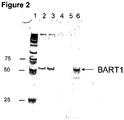
- BART3-3p and BART1-3p Two EBV-specific miRNAs BART3-3p and BART1-3p were analyzed in this study: BART3-3P CGC ACC ACU AGU CAC CAG GUG U SEQ ID NO:21 BART1-3P UAG CAC CGC UAU CCA CUA UGU CU SEQ ID NO:22
- lane 1 represents markers; lanes 2 and 3, represent patients with nasopharyngeal carcinoma, lanes 4 and 5, represent control patients, and lane 6, represents positive control, which represents respective synthetic miRNAs.
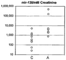
- This example demonstrates that the neuronal death caused by stroke can be registered in vivo by measurements of the concentrations of neuron-specific miRNA in the patient's urine.
- Urine samples were collected from patients accepted at a hospital through the emergency room. Diagnosis of brain stroke was based on clinical symptoms. Urine samples were collected at 12 and 24 hours after the stroke. Control urine samples were donated by age matched volunteers but without stroke symptoms. Samples were collected and stored according to the procedures described in the Example 1 of this application.
- miRNA from urine was extracted according to the procedure described in the Example 1. An amount of RNA equivalent to that isolated from 675 ⁇ l of urine underwent reverse transcription PCR and 1/10 of the RT-PCR mixture underwent final real time PCR, which was carried out using the protocol provided by the manufacturer. Data obtained were normalized for individual kidney filtration rates by re-calculation per creatinine concentration in urine. For these experiments, urine samples collected from healthy donors from same age group were used as baseline. Different miRNA species are presented as follows:
- Urine samples were collected from patients accepted at a hospital through the emergency room. Diagnosis of brain stroke was based on clinical symptoms and MRI analysis. Urine samples were collected at 12, 24, 48 hours and a week after the stroke. Patient clinical status was evaluated 30 days after stroke. Control urine samples were donated by age matched volunteers but without stroke symptoms. Samples were collected and stored according to the procedures described in the Example 1 of this application.
- miRNA from urine was extracted according to the procedure described in Example 1 and analyzed with TaqMan miRNA assays (Applied Biosystems). An amount of RNA equivalent to that isolated from 400 ⁇ l of urine underwent reverse transcription PCR and 1/10 of the RT-PCR mixture underwent final real time PCR, which was carried out using the protocol provided by the manufacturer. Data obtained were normalized for individual kidney filtration rates by re-calculation per creatinine concentration in urine. For these experiments, urine samples collected from healthy donors from same age group were used as baseline.
- Alzheimer's disease is a progressive neurological disease that is caused by the death of neurons, particularly in the cortex and hippocampus. The diagnosis is based on neurological examination and the exclusion of other causes of dementia whereas the definitive diagnosis can be made only at autopsy.
- the instant invention demonstrates that excessive neuronal death characterizing Alzheimer's disease may be monitored by measuring levels of specific brain miRNAs isolated from the patient's urine.
- Urine and serum samples were collected from patients diagnosed with various stages of the Alzheimer's disease. Control urine and serum samples were donated by age matched volunteers but without symptoms of Alzheimer's disease. Samples were collected and stored according to the procedures described in the Example 1 of this application. Some urine samples were filtered after collection as described in Example 1 to delete cells and cell debris.
- miRNA species RNA from urine and serum was extracted according to the procedures described in the Example 1.
- RNA equivalent to that isolated from 750 ⁇ l of urine underwent reverse transcription PCR and 1/10 of the RT-PCR mixture underwent final real time PCR, which was carried out using the protocol provided by the manufacturer (Applied Biosystems). Data obtained were normalized for individual kidney filtration rates by re-calculation per creatinine concentration in urine.
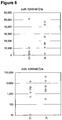
- Figure 5 clearly demonstrates that concentrations of several brain specific miRNAs is increased in the urine of Alzheimer's patients.
- RNA isolated from filtered urine or serum was analyzed.
- An amount of RNA equivalent to that isolated from 0.6 ml of urine or 50 ⁇ l of serum underwent reverse transcription PCR and 1/10 of the RT-PCR mixture underwent final real time PCR, which was carried out using the protocol provided by the manufacturer (Applied Biosystems).
- Data obtained for urinary miRNA were normalized for individual kidney filtration rates by re-calculation per creatinine concentration in urine.
- Data obtained for plasma miRNA were normalized per ubiquitous miRNA-16.
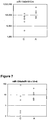
- Figures 6 and 7 show that the levels of some neuron-specific miRNAs are higher in both filtered urine and serum of the Alzheimer's patients compared to age-matched controls.
- Parkinson's disease is a degenerative disorder of the central nervous system that often impairs the sufferer's motor skills and speech.
- the instant invention demonstrates that excessive cellular death of dopaminergic neurons, characterizing Parkinson's disease may be monitored by measuring levels of specific brain miRNAs isolated from the patient's urine.
- Urine samples were collected from patients diagnosed with various stages of the Parkinson's disease. Control urine samples were donated by age matched volunteers without symptoms of Parkinson's disease. Samples were collected and stored according to the procedures described in the Example 1 of this application.
- RNA from urine was extracted according to the procedure described in the Example 1. Amount of RNA equivalent to that isolated from 750 ⁇ l of urine underwent reverse transcription PCR and 1/10 of the RT-PCR mixture underwent final real time PCR, which was carried out using the protocol provided by the manufacturer (Applied Biosystems). Data obtained were normalized for individual kidney filtration rates by re-calculation per creatinine concentration in urine.
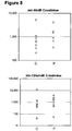
- Figure 8 demonstrates that concentrations of several brain specific miRNAs is increased in the urine of the patients with Parkinson disease.
- the principal finding of permeability of the kidney barrier for miRNA molecules opens the way for the use of maternal urine to perform completely noninvasive prenatal diagnosis of congenital diseases.
- a sample of urine is gathered from a pregnant patient.
- miRNA in the urine sample is then be isolated, purified and/or treated to prevent degradation using methods described above.
- MiRNA profiling is then performed using quantitative PCR or miRNA array and the data obtained are used to determine different fetal pathologies, as described for other pathologies above.
- Urine samples were collected from pregnant women diagnosed with Down syndrome by amniocentesis. Control urine samples were donated by age matched women with normal pregnancies. Samples were collected and stored according to the procedures described in the Example 1 of this application.
- miRNA from urine was extracted according to the procedure described in the Example 1. An amount of RNA equivalent to that isolated from 750 ⁇ l of urine underwent reverse transcription PCR and 1/10 of the RT-PCR mixture underwent final real time PCR, which was carried out using the protocol provided by the manufacturer. Data obtained were normalized per placenta-specific miRNA 518.
- Figure 9 demonstrates lower concentration the brain-specific miRNA 9 in urine of women pregnant with Down syndrome fetuses compared to urine of women with normal pregnancies, which indicates insufficient cell death compared to respective controls.
Description
- The invention provides non-invasive methods for isolation and detection of microRNA (miRNA) sequences in bodily fluid. More specifically, the present invention encompasses methods of detecting in vivo cell death by analyzing urine and other body fluids for miRNA levels for clinical diagnosis and treatment monitoring.
- Cell death is a normal component of development and functioning of multicellular organisms. Being a natural process, cell death is involved in the pathology of numerous diseases caused by internal factors. Cell death also accompanies diseases caused by external physical, chemical, of biological agents.
- There exist two major types of cell death, necrosis and apoptosis, marked by different morphological and molecular characteristics (Kerr et al., Br. J. Cancer. 26, 239-257 (1972); Umansky, Theor. Biol. 97, 591-602 (1982); Umansky et al., Adv Pharmacol. 41, 383-407 (1997); Ameisen, Cell Death Differ. 11, 4-10 (2004); Lockshin et al. Int J Biochem Cell Biol. 36, 2405-19 (2004); G. Kroemer, et al., Cell Death and Differentiation 12, 1463-1467 (2005)). Necrosis is considered to be catastrophic metabolic failure resulting directly from severe molecular and/or structural damage and leads to inflammation and secondary damage to surrounding cells. Apoptosis is a much more prevalent biological phenomenon than necrosis and can be induced by specific signals such as hormones, cytokines, by absence of specific signal such as growth or adhesion factors, or by molecular damage that does not cause catastrophic loss of integrity. Apoptosis is a result of an active cellular response involving initiation of an orderly and specific cascade of molecular events. Apoptosis leads to the appearance of distinctive chromatin condensation and margination, nuclear fragmentation, cell shrinkage, membrane blebbing and enzymatic internucleosomal fragmentation of nuclear DNA (Umansky et al., Biochim Biophys Acta. 655, 9-17 (1981); Arends et al., Am J Pathol. 136, 593-608(1990)). Other more rare forms of cell death, characterized by specific morphology, for example, so called autophagic cell death have also been described (Bredesen et al., Stroke. 38(2 Suppl):652-660 (2007).
- Independent of a specific mechanism and type of cell death, methods to detect dying cell types are important for diagnosis of various diseases, critical for disease and treatment monitoring, and helpful for differential diagnosis. Besides, the methods capable of detection of specific cell death in vivo are useful for developing drugs aiming at prevention or induction of cell death as well as for analysis of the cytotoxicity of the newly developed drugs.
- There are some clinical tests for diagnosis of disease-related excessive cell death based on detection of tissue specific markers, such as for example antigens, enzymes and other proteins in blood or in other bodily fluids. Measurement of the activity of liver-specific enzymes in blood, for example, is a widely used method for evaluation of hepatocyte death (Amacher, et al., Regul Toxicol Pharmacol. Apr; 27(2):119-130 (1988); Salaspuro, et al., Enzyme.37:87-107 (1987); Herlong, Hosp. Pract. (Off Ed).29(11):32-38 (1994)). Evaluation of the level of cardiomyocyte specific antigens has also been used for diagnosis of the myocardial infarction (Mair et al., Clin Chem Lab Med. 37:1077-1084 (1999); Nunes et al., Rev Port Cardiol. 20:785-788 (2001)). However, the number of such techniques is limited to diseases in which a marker and a method of detection are known in order for the analysis to provide meaningful, tissue-specific results. (Oh S et al., Curr Gastroenterol Rep. 3:12-18 (2001); Rochling et al., Clin Cornerstone. 3(6):1-12 (2001)). Other methods require invasive biopsy of specific tissues suspected of having a diseased condition to get a specimen for analysis. However, biopsy of some organs and tissues, for example brain is highly invasive and often difficult to perform.
- It is well known that apoptosis, or programmed cell death, which is a major form of cell death in the mamalian organism, is accompanied by internucleosomal fragmentation of nuclear DNA. Many laboratories have demonstrated that a portion of this DNA appears in blood (Lo Y.M. Ann N Y Acad Sci. 945:1-7 (2001); Lichtenstein et al., Ann N Y Acad Sci. 945:239-249 (2001); Taback et al., Curr Opin Mol Ther. 6:273-278 (2004); Bischoff et al., Hum Reprod Update.8:493-500,(2002)). It has also been shown that this fragmented DNA, called transrenal DNA (Tr-DNA) crosses the kidney barrier and can be detected in the urine. (Botezatu et al., Clin Chem. 46:1078-1084,(2000); Su et al., J Mol Diagn.6:101-107 (2004); Su et al., Ann N Y Acad Sci. 1022:81-89(2004).
-
US 6251638 B1 relates to a non-invasive method of detecting DNA in urine in order to detect fetal DNA sequence. - Although both cell-free plasma DNA and Tr-DNA may be used as diagnostic tools, they provide a rather limited approach when evaluating tissue specific events, such as cell death. Thus analytical methods that are non-invasive, and provide a broader range of indications of specific pathology, due to their ability to detect levels of dying cells in particular tissues and organs, would be useful for diagnosing and monitoring the state of various diseases or pathological conditions in patients. In addition, tissue specific analytical methods that provide the means for monitoring the response of a patient to a disease therapy would be useful to determine the therapy effectiveness, and in the case of drug treatment, the optimum dosage required for drug administration.
- J. Kemppainen et al. "miRNA as Biomarkers on Blood and other Biofluids", Keystone symposia on Molecular and Cellular Biology Poster Abstracts, Colorado, 28 January 2007, refers to the use of miRNA biomarker expression profiles to classify cancer patients.
- To address these problems, the instant invention is focused on the use of micro RNA (miRNA) as a diagnostic tool to monitor in vivo cell death in bodily fluids, such as for example serum and urine. Unlike cell-free plasma DNA and Tr-DNA, many miRNAs exhibit cell, tissue and organ specific expression profiles (Liang et al., Genomics, 8: 166 (2007); Lukiw et al, Neuroreport. 18:297-300 (2007); Lagos-Quintana et al., Curr Biol. 12:735-739 (2002); Chen et al., Nat Genet. 38:228-233 (2006); Beuvink et al., J. Nucleic Acids Res. 35:e52 (2007)). Furthermore, correlation of miRNA cell and tissue specific profiles with different pathologies and tumor types have been demonstrated (Visone R., et al. Oncogene. 26:7590-7595 (2007); Nelson et al., Neuropathol Exp Neurol. 66:461-468 (2007); Negrini et al., J Cell Sci. 120: 1833-1840 (2007); Chang et al., Annu Rev Genomics Hum Genet. 8:215-239 (2007); Jay et al., Cell Biol. 26:293-300 (2007)).
- Thus, the instant invention provides methods for measuring in vivo cell death by detection of tissue-specific miRNAs, characteristic of a pathogen infection, a brain stroke, Alzheimer's disease or Parkinson's disease, in body fluids, such as for example serum and urine. The instant methods based on detection of miRNAs in bodily fluids are used for further development of diagnostic or monitoring tests.
- The invention is defined by the appended claims.
- The instant invention relates to a novel method for detecting and measuring in vivo cell-death by analyzing levels of specific miRNA sequences in cell-free nucleic acids obtained from bodily fluids, said miRNA originating from cells dying throughout the body, and using the obtained analytical result to determine state of a disease or abnormal medical condition in a patient.
- The methods of the instant invention are based on adsorption of cell-free nucleic acids on and elution from anion-exchangers, which makes it possible to concentrate and isolate nucleic acid fragments larger then 10 nucleotides. Specifically, the instant invention demonstrates: (i) the presence of miRNA in body fluids; (ii) detection in urine of miRNA that originated from organs located outside of urinary system, which means that they have crossed the kidney barrier , such as for example, transrenal miRNA (Tr-miRNA); iii) detection of miRNA in serum (iv) pathology associated with cell death in a particular cell, tissue and/or organ is accompanied by changes in levels of miRNA specific for the said organ.
- The present invention provides a method of detecting and measuring in vivo cell death the method comprising:
- (a) analyzing levels of specific miRNA sequences in cell-free nucleic acids obtained from a sample of bodily fluid from a patient, wherein the bodily fluid sample is selected from blood, serum, and urine, said miRNA sequences being indicative of in vivo cell death and originating from cells dying throughout the patient's body; and
- (b) determining the state of a disease or abnormal medical condition in the patient,
- (i) a pathogen infection, preferably wherein the pathogen is a virus and more preferably wherein the virus is an Epstein-Barr virus;
- (ii) a brain stroke;
- (iii) Alzheimer's disease; or
- (iv) Parkinson's disease.
- In one embodiment of the present invention, the body fluid is urine. In another embodiment, the present method of analysis of a urine sample includes a technique selected from the group consisting of hybridization, cycling probe reaction, polymerase chain reaction, nested polymerase chain reaction, PCR to analyze single strand conformation polymorphisms and ligase chain reaction. In yet another embodiment, nucleic acid degradation in said urine sample is reduced.
- The method of the present invention includes reducing nucleic acid degradation comprising inhibiting nuclease activity by addition of RNAse inhibitor(s), heat inactivation, or by treating said urine sample with a compound selected from the group consisting of: guanidine-HCl, guanidine isothiocyanate, N-lauroylsarcosine, and sodium dodecylsulphate. In one embodiment of the present invention, urine sample has been held in the bladder less than 12 hours.
- In one embodiment of the present invention, the body fluid is serum. The method of the present invention includes analysis of a serum sample including a technique selected from the group consisting of hybridization, cycling probe reaction, polymerase chain reaction, nested polymerase chain reaction, PCR to analyze single strand conformation polymorphisms and ligase chain reaction.
- The method of the instant invention involves detecting cell-free miRNAs, as a specific marker for the specific disorder associated with excessive or insufficient cell death in a tissue or organ. Said disorder is a pathogen infection. Preferably, said pathogen is a virus. More preferably, said virus is an Epstein-Barr virus. Alternatively, said disorder is a brain stroke, Alzheimer's disease, or Parkinson's disease.
- The present invention further provides a method of disease and/or treatment monitoring in a patient by quantitative analysis of specific cell-free miRNAs in a urine sample, the method comprising:
- (a) periodically obtaining a urine sample from a patient, said miRNA sequences being indicative of in vivo cell death and originating from cells dying throughout the patient's body;and
- (b) analyzing said sample for one or more specific sequences of miRNA according to any one of the preceding claims, wherein said analyzing comprising detecting said one or more specific sequences of miRNA with probe(s) and /or probe(s) that are substantially complementary to a part of said specific sequences of miRNA,
- (i) a pathogen infection, preferably wherein the pathogen is a virus and more preferably wherein the virus is an Epstein-Barr virus;
- (ii) a brain stroke;
- (iii) Alzheimer's disease; or
- (iv) Parkinson's disease.
- The present invention further provides a method for detecting and measuring miRNA originating from a specific cell type, tissue, or organ in the body of a patient, the method comprising:
- (a) analyzing levels of specific miRNA sequences in cell-free nucleic acids obtained from a sample of bodily fluid from a patient, wherein the bodily fluid sample is selected from blood, serum, and urine, said miRNA sequences originating from a specific cell type, tissue or organ in the patient; and
- (b) detecting alterations in level of the specific miRNA sequences that are indicative of chronic pathology, or the cytotoxic effect of physical or chemical agents,
- The foregoing and other objects, features, and advantages of the invention will be apparent from the more particular description of embodiments of the invention, as illustrated in the accompanying drawings. The drawings are not necessary to scale, emphasis instead being placed upon illustrating the principles of the invention.
-
Figure 1 is a photograph of a polyacrylamide gel electrophoresis of nucleic acids extracted from filtered urine using Q-Sepharose™. -
Figure 2 is a photograph of a polyacrylamide gel analysis of EBV derived BART1 miRNA specific RT-PCR product. -
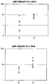
Figures 3A to 3G are dot plot representations of the normalized concentrations of miRNA in urine samples of patients at 12 and 24 hour time points after brain stroke. -
Figure 4 is a diagram representing correlation between changes inmiRNAs -
Figure 5 is a dot plot representation of the normalized concentrations of miRNA in unfiltered urine samples of patients with Alzheimer's disease and age matched controls. -
Figure 6 is a dot plot representation of the normalized concentrations of miRNA in filtered urine samples of patients with Alzheimer's disease and age matched controls. -
Figure 7 is a dot plot representation of the normalized concentrations of miRNA in serum samples of patients with Alzheimer's disease and age matched controls. -
Figure 8 is a dot plot representation of the normalized concentrations of miRNA in urine samples of patients with Parkinson's disease and age matched controls. -
Figure 9 is a dot plot representation of the normalized concentration of miRNA-9 in urine samples of women pregnant with Down syndrome and normal fetuses. - The technology of this invention is based on the discovery that small RNAs, in particular specific micro RNAs (miRNAs), including transrenal miRNA (Tr-miRNA), are presented in bodily fluids and their concentrations reflect cell death associated with organ damage or other pathology. The presence of these nucleic acid sequences at levels lower or higher than that of a control group is therefore an indication that an abnormality or pathological condition is likely present in the patient from whom the sample was obtained.
- The methods of the present invention offer improvements over previous methods of diagnosis, detection and monitoring due to their inherently non-invasive nature.
- To facilitate the understanding of the invention, a number of terms are defined below:
- The term "primer" refers to an oligonucleotide which is capable of acting as a point of initiation of synthesis when placed under conditions in which primer extension is initiated. An oligonucleotide "primer" can occur naturally, as in a purified restriction digest or be produced synthetically.
- A primer is selected to be "substantially" complementary to a strand of specific sequence of the template. A primer must be sufficiently complementary to hybridize with a template strand for primer elongation to occur. A primer sequence need not reflect the exact sequence of the template. For example, a non-complementary nucleotide fragment may be attached to the 5' end of the primer, with the remainder of the primer sequence being substantially complementary to the strand. Non-complementary bases or longer sequences can be interspersed into the primer, provided that the primer sequence has sufficient complementarity with the sequence of the template to hybridize and thereby form a template primer complex for synthesis of the extension product of the primer.
- A "target" nucleic acid is a miRNA sequence to be evaluated by hybridization, amplification or any other means of analyzing a nucleic acid sequence, including a combination of analysis methods.
- "Hybridization" methods involve the annealing of a complementary sequence to the target nucleic acid (the sequence to be analyzed). The ability of two polymers of nucleic acid containing complementary sequences to find each other and anneal through base pairing interaction is a well-recognized phenomenon. The initial observations of the "hybridization" process by Marmur and Lane, Proc. Natl. Acad. Sci. USA 46:453 (1960) and Doty et al., Proc. Natl. Acad. Sci. USA 46:461 (1960) have been followed by the refinement of this process into an essential tool of modern biology. Hybridization encompasses, but not be limited to, slot, dot and blot hybridization techniques.
- It is important for some diagnostic applications to determine whether the hybridization represents complete or partial complementarity. For example, where it is desired to detect simply the presence or absence of pathogen miRNA, it is only important that the hybridization method ensures hybridization when the relevant sequence is present; conditions can be selected where both partially complementary probes and completely complementary probes will hybridize. Other diagnostic applications, however, may require that the hybridization method distinguish between partial and complete complementarity. It may be of interest to detect genetic polymorphisms.
- The term "probe" as used herein refers to an oligonucleotide (i.e., a sequence of nucleotides), whether occurring naturally as in a purified restriction digest or produced synthetically, which forms a duplex structure or other complex with a sequence of another nucleic acid, due to complementarity or other means of reproducible attractive interaction, of at least one sequence in the probe with a sequence in the other nucleic acid. Probes are useful in the detection, identification and isolation of particular gene sequences. It is contemplated that any probe used in the present invention will be labeled with any "reporter molecule," so that it is detectable in any detection system, including, but not limited to, enzyme (e.g., ELISA, as well as enzyme--based histochemical assays), fluorescent, radioactive, and luminescent systems. It is further contemplated that the oligonucleotide of interest (i.e., to be detected) will be labeled with a reporter molecule. It is also contemplated that both the probe and oligonucleotide of interest will be labeled. It is not intended that the present invention be limited to any particular detection system or label.
- As used herein, the term "miRNA" is a subclass of small non-coding single stranded RNA, approximately 18-23 nucleotides in length which plays an important role in regulation of metabolic processes, particularly due to their involvement in regulation of stability and translation of mRNA encoding specific proteins. miRNA also participate in other important processes, like heterochromatin formation and genome rearrangement.
- The terms "excessive" and "insufficient" in vivo cell death describe the situation when the number of cells dying in a particular organ or tissue is respectively higher or lower than in age and gender matched controls.
- As used herein, the terms "purified", "decontaminated" and "sterilized" refer to the removal of contaminant(s) from a sample.
- As used herein, the terms "substantially purified" and "substantially isolated" refer to nucleic acid sequences that are removed from their natural environment, isolated or separated, and are preferably 60% free, more preferably 75% free, and most preferably 90% free from other components with which they are naturally associated. An "isolated polynucleotide" is therefore a substantially purified polynucleotide. It is contemplated that to practice the methods of the present invention polynucleotides can be, but need not be substantially purified. A variety of methods for the detection of nucleic acid sequences in unpurified form are known in the art.
- As used herein, the terms "PCR product" and "amplification product" refer to the resultant mixture of compounds after two or more cycles of the PCR steps of denaturation, annealing and extension are complete. These terms encompass the case where there has been amplification of one or more segments of one or more target sequences.
- The term "urinary tract" as used herein refers to the organs and ducts which participate in the secretion and elimination of urine from the body.
- "Patient" or "subject" as the terms are used herein, refer to the recipient of the treatment. Mammalian and non-mammalian patients are included. In a specific embodiment, the patient is a mammal, such as a human, canine, murine, feline, bovine, ovine, swine, or caprine. In a particular embodiment, the patient is a human.
- In one embodiment of the present disclosure, the detected miRNAs originate from and are specifically expressed in a specific cell type, tissue, or organ in the body, wherein alterations in the level of said miRNAs are indicative of acute pathology of said tissue, such as for example acute myocardial infarction associated with death of cardiomyocytes; brain stroke associated with death of neurons and glial cells; hepatitis or liver cirrhosis associated with hepatocyte death caused by a viral or other infection or by action of toxic agents; acute pancreatitis associated with death of different pancreatic cells; rejection of a transplanted organ associated with excessive cell death in the transplanted organ; traumatic damage of various organs; numerous acute infections, for example tuberculosis associated with cell death in lungs and/or other infected organs.
- In another embodiment of the present invention, the detected miRNAs originate from and are specifically expressed in a specific cell type, tissue, or organ in the body, wherein alterations in the level of said miRNAs are indicative of chronic pathology of said tissue, such as for example Alzheimer's disease, Parkinson disease, frontotemporal dementia and other diseases of the central nervous system that are caused or accompanied by neuronal death; chronic heart failure associated with the death of cardiomyocytes, emphysema associated with death of lung cells;
diabetes type 1 associated with the death of pancreatic beta cells, glomerulonephritis associated with the death of kidney cells, precancerous conditions associated with the apoptotic death of actively proliferating precancerous cells, cancers associated with massive necrotic cell death due to insufficient blood supply, and cell death in chronically infected organs or tissues. - In yet another embodiment of the present invention, the detected miRNAs originate from and are specifically expressed in a specific cell type, tissue, or organ in the body and can be used for prognosis of disease outcome. Changes in the levels of respective miRNAs, that are indicative of disease progression/regression, success of therapeutic or surgical intervention, are used for disease and treatment monitoring.
- In another embodiment of the invention, the detected miRNAs originate from a pathogen and are used for infection diagnosis and monitoring. In a specific embodiment of the instant invention, the pathogen is a virus, for example Epstein-Barr virus.
- In yet another embodiment of the invention, the detected miRNAs originate from cells of an infected organ and can be used for diagnosis support, evaluation of infected tissue damage, and further disease and treatment monitoring.
- In some embodiments, the levels of cell- and/or tissue-specific miRNAs are normalized using the levels of ubiquitous miRNA in serum, the levels of albumin or creatinine in urine, or the levels of placenta-specific miRNAs for normalization of other tissue-specific fetal miRNAs.
- In one aspect of the invention, the step of analyzing said urine sample to detect specific miRNAs includes a technique selected from the group consisting of hybridization, cycling probe reaction, polymerase chain reaction, nested polymerase chain reaction, PCR to analyze single strand conformation polymorphisms and ligase chain reaction.
- In certain aspects of the invention, the nucleic acid degradation in said urine sample is reduced. The method of reducing nucleic acid degradation comprises inhibiting nuclease activity by use of RNAse inhibitors, or by treating said urine sample with a compound selected from the group consisting of: guanidine-HCl, guanidine isothiocyanate, N-lauroylsarcosine, and sodium dodecylsulphate. In another aspect of the invention, said urine sample has been held in the bladder less than 12 hours.
- In one embodiment of the present invention, the miRNA sequences measured are specifically related to tissues in the body, which may be selected from but are not limited to, lung, heart, liver, nervous system, brain, blood, kidney, bone, eye or pancreas.
- The instant methods include isolation of miRNAs from the bodily fluids of the patients. In one aspect of the disclosure, a miRNA of interest may be detected in a body fluid such as blood, amniotic fluid, cerebrospinal fluid, plasma, milk, semen, serum, sputum, saliva and urine. In one aspect of the instant invention, the miRNA is detected in urine. In another embodiment, the miRNA is detected in serum.
- The instant method of the miRNA isolation of the instant invention can utilize commercially available anion exchange materials. Either strong or weak anion exchangers may be employed. By utilizing selected solutions for adsorption and elution, the miRNA can be purified, concentrated, and substantially isolated.
- By employing a solution at known ionic strength for the initial binding of the miRNA to the anion exchange column materials, most of the water soluble components including other electronegative molecules such as proteins (weakly-bound contaminants) can be washed through the column. For elution, the required ionic strength is reached by using known concentrations of a salt such as NaCl, which may be mixed with a buffer to control pH, ideally corresponding to the lowest ionic strength at which the nucleic acids will completely elute. Contaminating substances bound to the anion exchange resin with higher stringency than the nucleic acids may thereby be left within the column, i.e., stronger bound contaminants are separated away from the nucleic acids.
- A preferred weak exchanger is one in which primary, secondary, or tertiary amine groups (i.e., protonatable amines) provide exchange sites. The strong base anion exchanger has quaternary ammonium groups (i.e., not protonatable and always positively charged) as exchange sites. Both exchangers are selected in relation to their respective absorption and elution ionic strengths and/or pH for the miRNA being separated. The solution strengths are higher than the binding strengths.
- In one aspect of the invention, a method is provided for isolation tranrenal miRNA from urine, the method comprising providing urine from a subject; optionally separating cells and cell debris from the urine by filtration or centrifugation; adding EDTA and Tris-HCl to the urine, adding silica free anion exchange resin to urine, incubating the mixture, removing the anion exchange medium from the urine, and eluting miRNA from the resin.
- In one embodiment of the method of isolating miRNA from urine, the concentration of EDTA and Tris-HCl after it is added to the urine is in a range of 10-100 mM, and the pH of the solution is between about 8.0 and about 8.5.
- In a further embodiment, the body fluid is optionally pre-filtered through a membrane prior to adsorption onto the anion-exchange medium.
- In a further embodiment, the anion exchange medium is a sepharose-based resin functionalized with cationic quaternary ammonium groups. Examples of sepharose-based resins, functionalized with cationic ammonium groups include Q-Sepharose™ ANX-4 Sepharose™ Fast Flow, DEAE-Sepharose ™, and Q-Sepharose-XL™ DEAE Sepharose Fast Flow (GE Healthcare).
- In a further embodiment, the anion exchange medium is selected from sepharose-based quaternary ammonium anion exchange medium such as Q-filters or Q-resin.
- In a further embodiment of the invention, the anion-exchange medium is immobilized on an individualized carrier wherein such a carrier is a column, cartridge or portable filtering system which can be used for transport or storage of the medium/nucleoprotein bound complex.
- In another embodiment of the present invention, periodic analysis of miRNA sequences present, for example, in the urine samples of the same person can give early information about a pathological process in a particular organ or tissue. For example, miRNA122 is synthesized in liver only and increases in its amount may be a marker of hepatitis or another liver pathology. Alzheimer's syndrome can be accompanied by increases in the concentration of miRNA specifically expressed in neurons.
- In another embodiment, more detailed monitoring of tissue-specific miRNA in the bodily fluid sample of the patient will be useful for estimation of a severity of the disease and for evaluation of effectiveness of therapeutic efforts.
- Unless otherwise defined, all technical and scientific terms used herein have the same meaning as commonly understood by one of ordinary skill in the art to which this invention pertains.
- The examples are presented in order to more fully illustrate the various embodiments of the invention. These examples should in no way be construed as limiting the scope of the invention recited in the appended claims.
- Urine collection: For these experiments, urine specimens from patients or volunteers were collected in a sterile 110 ml urine collection cup and were immediately supplemented with EDTA up to final concentration between 10 and 150 mM, preferably 50 mM. Specimens were stored in 10 - 50 ml aliquots at -80°C. Optional filtration of urine was carried out on Stericup™ (Millipore, Vacuum Driven Filtration System, 0.45 µ Durapore™ filter) immediately after specimen collection before the EDTA was added.
- Q Binding: In a 50 ml tube 20 mL of urine was diluted with equal volume of 50 mM EDTA (pH 8.0) and 50 mM Tris-HCl (pH 8.0) which was then supplemented with 1-2 ml of Q-Sepharose™ (GE Healthcare; 17-0510-10) slurry and rigorously mixed 10 - 30 min at room temperature. The resin, with bound nucleic acids, was collected by centrifugation at 2000g for 5 minutes at room temperature in a table top clinical centrifuge using a swing bucket rotor. All but ∼ 500 µl of supernatant was removed by aspiration. The resin pellet was resuspended in the remaining supernatant and transferred to a Micro Bio-Spin Chromatography Column (Bio-Rad) or equivalent, which was operated either by centrifugation of vacuum. The resin in the column was washed three times with 500 µl 2xSSC (300 mM NaCL/30 mM sodium citrate (pH 7.0)) or with buffer with comparable ionic strength (e.g. 300 mM NaCl or LiCl). Nucleic acids can be eluted from Q-Sepharose with high ionic strength (e.g. 1M NaCl or LiCl) but the methods described below preserves RNA better.
- Elution from Q-Sepharose™ and TRIzol™ Phase Separation: Bound nucleic acids were further eluted with 500 µl of TRIzol™ reagent (Invitrogen). The extraction of nucleic acids from TRIzol was carried out according manufacturer's recommendations. Briefly, for phase separation TRIzol eluate was supplemented with 100 µl chloroform, mixed vigorously, incubated at room temperature for 3 - 5 minutes and centrifuged at 12,000xg for 15 min at 4°C. While avoiding touching the interphase, 300 µl of the upper phase was transferred into a fresh centrifuge tube. Then the nucleic acids were precipitated or additionally cleaned and desalted on a silica column.
- Nucleic acid precipitation: For nucleic acid precipitation, the above described preparation was supplemented with 1 µl of 20 mg/mL glycogen (Roche) and 300 µl of 100% isopropyl alcohol. Nucleic acids were collected by centrifugation, the pellet was washed twice with 200 µl of 70% ethanol, allowed to air dry for 5 min at room temperature, and then the nucleic acids were dissolved in 30 µl of 0.1 mM EDTA/1x RNA Secure (Ambion). The samples were incubated at 60 °C for 10 min to inactivate any residual RNase activity.
- Silica Column cleaning of nucleic acids: For binding to a silica column (Qiagen PCR clean columns or equivalent) 3 volumes of 96% ethanol were added to nucleic acid preparation from the TRIzol upper phase, and, after 3 minutes incubation at room temperature, the mixture was loaded onto the column. The column was washed twice with 500 µl 2 M LiCl/80% ethanol and twice with 500 µl 80% ethanol. Nucleic acids were eluted with 50 µl of 0.1 mM EDTA/1x RNA Secure (Ambion). The samples were incubated at 60 °C for 10 min to inactivate any residual RNase.
- DNase I and RNase A Digestion: To verify the nucleic acid identity of the material extracted from urine with the above described protocol, the instant prep was digested with DNase I and/or RNase A. DNase I digestion was carried out in the DNase I Reaction Buffer (NEB) containing 2 units of RNase free DNase I (NEB). RNase A digestion was performed in TE buffer supplemented with 50 ng/mL boiled RNase A. Samples were incubated at 37 °C for 60 min and after addition of loading dye samples were subjected to electrophoresis on 5% polyacrylamide 1x TBE gels and stained with 1/10000 diluted SYBR® Gold (Invitrogen). As shown in
Figure 1 , the isolated material represents low molecular weight nucleic acids, mainly RNA and their fragments. In addition, (seeFigure 1 ), for comparison nucleic acids from Q-resin were eluted by 3 M NaCl,lanes lanes - In the
Figure 1 ,lanes lanes - Also, to demonstrate existence and molecular size of RNA, RNA aliquots of purified nucleic acids were digested with DNaseI,
lanes - For these experiments, 1.2 ml of TRIzol LS were added to 0.4 ml of serum, and the mixture was centrifuged 10 at 14,000 rpm. The supernatant was transferred into a 2 ml Eppendorf tube, 0.3 ml of chloroform was added, and the mixture was shaken for 15 seconds. After centrifugation at 14,000 rpm for 15 min, the supernatant was transferred into a 5 ml tube and ethanol was added up to final concentration of 70%. The mixture was loaded on a Quiagen Quick column on a vacuum manifold, and the column was washed twice with 0.5 ml of 2M LiCl-80% EtOH, once with 0.5 ml of 80% ethanol-80 mM sodium acetate (pH 5.0), and finally with 0.5 ml of 95% ethanol. The column was centrifuged in 1.5
ml Eppendorf tube 3 min at 14,000 rpm, and RNA was eluted with 40 µl H2O. - This Example demonstrates that miRNA, from dying cells, cross the kidney barrier and may be detected in the urine of a patient.
- Micro RNA species that were analyzed in this example can be grouped in three distinct types, namely ubiquitous miRNAs, which are expressed in all or multiple tissues, tissue-specific miRNAs, and miRNAs in which expression is significantly altered in a particular tissue or cell type. As shown by Table 1, 20 different miRNAs were obtained from urine of 16 healthy volunteers and enrolled donors and later detected by real time RT-PCR using commercially available miRNA expression analysis kit (ABI). Corresponding synthetic miRNA oligonucleotides were used as standards. Reactions were carried out strictly as recommended by the supplier.
Table 1. Detected miRNA SEQ ID NO: ID Sequence Expression 1 hsa-miR-127 UCGGAUCCGUCUGAGCUUGGCU Brain overexpressed 2 hsa-miR-153 UUGCAUAGUCACAAAAGUGA Brain overexpressed 3 hsa-mlR-129 CUUUUUGCGGUCUGGGCUUGC Brain-specific 4 hsa-miR-137 UAUUGCUUAAGAAUACGCGUAG Brain overexpressed 5 hsa-miR-218 UUGUGCUUGAUCUAACCAUGU Ubiquitous, Brain overexpressed 6 hsa-miR-219 UGAUUGUCCAAACGCAAUUCU Brain-specific 7 hsa-miR-128a UCACAGUGAACCGGUCUCUUUU Brain-specific 8 hsa-miR-9 UCUUUGGUUAUCUAGCUGUAUGA Brain overexpressed 9 hsa-miR-138 AGCUGGUGUUGUGAAUC Brain, Thyroid 10 hsa-miR-134 UGUGACUGGUUGACCAGAGGG Brain and several other tissues 11 hsa-miR-124a UUAAGGCACGCGGUGAAUGCCA Brain-specific 12 hsa-miR-122a UGGAGUGUGACAAUGGUGUUUGU Liver-specific 13 hsa-miR-133a UUGGUCCCCUUCAACCAGCUGU Heart and Muscle overexpressed 14 hsa-miR-1 UGGAAUGUAAAGAAGUAUGUA Heart and Muscle overexpressed 15 hsa-miR-335 UCAAGAGCAAUAACGAAAAAUGU Ubiquitous 16 hsa-miR-16 UAGCAGCACGUAAAUAUUGGCG Ubiquitous 17 hsa-miR-215 AUGACCUAUGAAUUGACAGAC Small intestine and colon overexpressed 18 hsa-miR-135b UAUGGCUUUUCAUUCCUAUGUG Placenta-overexpressed 19 hsa-miR-517c AUCGUGCAUCCUUUUAGAGUGU Placenta-overexpressed 20 hsa-miR-518e AAAGCGCUUCCCUUCAGAGUGU Placenta-overexpressed - All three types of miRNA were detected in most preps of urinary RNA. The highest copy numbers were characteristic of ubiquitous miRNA. However, tissue-specific miRNA or miRNA over-expressed in a particular tissue or cell type were also detectable. It has been unequivocally demonstrated that a portion of miRNA from dying cells is not degraded but appears in the bloodstream and is finally excreted into urine.
- The Example demonstrates that miRNA from human nasopharyngeal carcinoma (NPC) cells can cross the patient's kidney barrier and can be detected in patient's urine by real time RT-PCR.
- It is known that some viruses also encode and produce miRNAs. Since Epstein-Barr virus (EBV) is involved in development of nasopharyngeal carcinoma (NPC), the instant system was used to find out if viral miRNA from NPC cells can reach a patient's urine and be detected there. Urine samples from NPC patients were collected and stored according to the procedures described in the Example 1 of this application. EBV infection was confirmed by the detection of virus-specific DNA sequences in urine. Urine collected from healthy donor was negative for EBV specific DNA sequences. Two EBV-specific miRNAs BART3-3p and BART1-3p were analyzed in this study:
BART3-3P CGC ACC ACU AGU CAC CAG GUG U SEQ ID NO:21 BART1-3P UAG CAC CGC UAU CCA CUA UGU CU SEQ ID NO:22 - Reverse transcription was performed in 15 µl, one tenth of the RT reaction was subjected to PCR amplification using JumpStart DNA polymerase from Sigma. The following primers were used at 500 nM concentration:
ID Sequence SEQ ID NO: BART3-3PRT GTC GTA TCC AGT GCA GGG TCC GAG GTA TTC GCA CTG GAT ACG ACA CAC CT 23 BART1-3PRT GTC GTA TCC AGT GCA GGG TCC GAG GTA TTC GCA CTG GAT ACG ACA GAC AT 24 BART3-3PF CGC CGC ACC ACT AGT CAC 25 BART1-3PF CGC TAG CAC CGC TAT CCA 26 miRNACR GTG CAG GGT CCG AGG T 27 - Products were analyzed in 15% polyacrylamide gel (PAAG).
- As demonstrated by
Figure 2 , both BART3 andBART 1 miRNA species were detected in urine from NPC patients but not in the urine sample from a healthy donor. These data again indicate that miRNA from dying cells located outside of urinary system can be detected in the urine. In theFigure 2 ,lane 1, represents markers;lanes lanes lane 6, represents positive control, which represents respective synthetic miRNAs. - This example demonstrates that the neuronal death caused by stroke can be registered in vivo by measurements of the concentrations of neuron-specific miRNA in the patient's urine.
- For these experiments, patients with brain stroke were investigated for analysis of changes in concentrations of brain-specific miRNA or miRNA which are over-expressed in brain, after stroke. Since currently it is not known in what brain cell types and in what brain areas these miRNA are expressed, 9 different brain specific miRNA were studied.
- Patients: Urine samples were collected from patients accepted at a hospital through the emergency room. Diagnosis of brain stroke was based on clinical symptoms. Urine samples were collected at 12 and 24 hours after the stroke. Control urine samples were donated by age matched volunteers but without stroke symptoms. Samples were collected and stored according to the procedures described in the Example 1 of this application.
- miRNA species: miRNA from urine was extracted according to the procedure described in the Example 1. An amount of RNA equivalent to that isolated from 675 µl of urine underwent reverse transcription PCR and 1/10 of the RT-PCR mixture underwent final real time PCR, which was carried out using the protocol provided by the manufacturer. Data obtained were normalized for individual kidney filtration rates by re-calculation per creatinine concentration in urine. For these experiments, urine samples collected from healthy donors from same age group were used as baseline. Different miRNA species are presented as follows:
- A. hsa-mir-128a
- B. hsa-mir-9
- C. hsa-mir-127
- D. hsa-mir-137
- E. hsa-mir-129
- F. hsa-mir-219
- G. hsa-mir-218
- Results summarized in
Figures 3A to 3G clearly demonstrate that after brain stroke, there is a significant increase in the levels of several brain specific miRNA (128a, 129, 218, 219)reflecting kinetics of the brain cell death. - This example demonstrates that kinetics of the miRNA concentrations in patient's urine after stroke provides information about disease outcome.
- For the experiments, patients with brain stroke were investigated for analysis of correlation between changes in concentrations of brain-specific miRNA and disease development.
- Patients: Urine samples were collected from patients accepted at a hospital through the emergency room. Diagnosis of brain stroke was based on clinical symptoms and MRI analysis. Urine samples were collected at 12, 24, 48 hours and a week after the stroke. Patient clinical status was evaluated 30 days after stroke. Control urine samples were donated by age matched volunteers but without stroke symptoms. Samples were collected and stored according to the procedures described in the Example 1 of this application.
- miRNA species: miRNA from urine was extracted according to the procedure described in Example 1 and analyzed with TaqMan miRNA assays (Applied Biosystems). An amount of RNA equivalent to that isolated from 400 µl of urine underwent reverse transcription PCR and 1/10 of the RT-PCR mixture underwent final real time PCR, which was carried out using the protocol provided by the manufacturer. Data obtained were normalized for individual kidney filtration rates by re-calculation per creatinine concentration in urine. For these experiments, urine samples collected from healthy donors from same age group were used as baseline.
- Results summarized in
Figures 4A and B clearly demonstrate that the dynamics of changes in Tr-miRNA 129 and Tr-miRNA 219 after brain stroke are different in different patients and correlates with the disease development. The increase in neuronal death a week after stroke inpatient # 3 corresponds to worsening in the patient clinical status. At the same time two patients, whose transrenal neuron-specific miRNA had tendency to normalization, demonstrated significant improvement. - Alzheimer's disease is a progressive neurological disease that is caused by the death of neurons, particularly in the cortex and hippocampus. The diagnosis is based on neurological examination and the exclusion of other causes of dementia whereas the definitive diagnosis can be made only at autopsy. The instant invention demonstrates that excessive neuronal death characterizing Alzheimer's disease may be monitored by measuring levels of specific brain miRNAs isolated from the patient's urine.
- For these experiments, patients diagnosed with Alzheimer's disease were investigated for analysis of changes in concentrations of brain-specific or over-expressed miRNA as a result of neuronal death.
- Patients: Urine and serum samples were collected from patients diagnosed with various stages of the Alzheimer's disease. Control urine and serum samples were donated by age matched volunteers but without symptoms of Alzheimer's disease. Samples were collected and stored according to the procedures described in the Example 1 of this application. Some urine samples were filtered after collection as described in Example 1 to delete cells and cell debris.
- miRNA species: RNA from urine and serum was extracted according to the procedures described in the Example 1.
- In one set of experiments an amount of RNA equivalent to that isolated from 750 µl of urine underwent reverse transcription PCR and 1/10 of the RT-PCR mixture underwent final real time PCR, which was carried out using the protocol provided by the manufacturer (Applied Biosystems). Data obtained were normalized for individual kidney filtration rates by re-calculation per creatinine concentration in urine.
Figure 5 clearly demonstrates that concentrations of several brain specific miRNAs is increased in the urine of Alzheimer's patients. - In another set of experiments, RNA isolated from filtered urine or serum was analyzed. An amount of RNA equivalent to that isolated from 0.6 ml of urine or 50 µl of serum underwent reverse transcription PCR and 1/10 of the RT-PCR mixture underwent final real time PCR, which was carried out using the protocol provided by the manufacturer (Applied Biosystems). Data obtained for urinary miRNA were normalized for individual kidney filtration rates by re-calculation per creatinine concentration in urine. Data obtained for plasma miRNA were normalized per ubiquitous miRNA-16.
Figures 6 and7 show that the levels of some neuron-specific miRNAs are higher in both filtered urine and serum of the Alzheimer's patients compared to age-matched controls. - Parkinson's disease is a degenerative disorder of the central nervous system that often impairs the sufferer's motor skills and speech. The instant invention demonstrates that excessive cellular death of dopaminergic neurons, characterizing Parkinson's disease may be monitored by measuring levels of specific brain miRNAs isolated from the patient's urine.
- For these experiments, patients diagnosed with Parkinson's were investigated for analysis of changes in concentrations of brain-specific miRNA or over-expressed miRNA as a result of neuronal death.
- Patients: Urine samples were collected from patients diagnosed with various stages of the Parkinson's disease. Control urine samples were donated by age matched volunteers without symptoms of Parkinson's disease. Samples were collected and stored according to the procedures described in the Example 1 of this application.
- miRNA species: For these experiments, RNA from urine was extracted according to the procedure described in the Example 1. Amount of RNA equivalent to that isolated from 750 µl of urine underwent reverse transcription PCR and 1/10 of the RT-PCR mixture underwent final real time PCR, which was carried out using the protocol provided by the manufacturer (Applied Biosystems). Data obtained were normalized for individual kidney filtration rates by re-calculation per creatinine concentration in urine.
Figure 8 demonstrates that concentrations of several brain specific miRNAs is increased in the urine of the patients with Parkinson disease. - The principal finding of permeability of the kidney barrier for miRNA molecules opens the way for the use of maternal urine to perform completely noninvasive prenatal diagnosis of congenital diseases. One can perform such a noninvasive screen as follows.
- First, a sample of urine is gathered from a pregnant patient. Where desired, miRNA in the urine sample is then be isolated, purified and/or treated to prevent degradation using methods described above. MiRNA profiling is then performed using quantitative PCR or miRNA array and the data obtained are used to determine different fetal pathologies, as described for other pathologies above.
- For the experiments, differences in concentrations of brain-specific miRNA in maternal urine between women pregnant with normal and Down syndrome fetuses were investigated.
- Patients: Urine samples were collected from pregnant women diagnosed with Down syndrome by amniocentesis. Control urine samples were donated by age matched women with normal pregnancies. Samples were collected and stored according to the procedures described in the Example 1 of this application.
- miRNA species: miRNA from urine was extracted according to the procedure described in the Example 1. An amount of RNA equivalent to that isolated from 750 µl of urine underwent reverse transcription PCR and 1/10 of the RT-PCR mixture underwent final real time PCR, which was carried out using the protocol provided by the manufacturer. Data obtained were normalized per placenta-
specific miRNA 518.Figure 9 demonstrates lower concentration the brain-specific miRNA 9 in urine of women pregnant with Down syndrome fetuses compared to urine of women with normal pregnancies, which indicates insufficient cell death compared to respective controls.
Claims (12)
- A method for detecting and measuring in vivo cell-death, the method comprising:(a) analyzing levels of specific miRNA sequences in cell-free nucleic acids obtained from a sample of bodily fluid from a patient, wherein the bodily fluid sample is selected from blood, serum, and urine, said miRNA sequences being indicative of in vivo cell death and originating from cells dying throughout the patient's body; and(b) determining the state of a disease or abnormal medical condition in the patient,wherein said disease or abnormal medical condition is:(i) a pathogen infection, preferably wherein the pathogen is a virus and more preferably wherein the virus is an Epstein-Barr virus;(ii) a brain stroke;(iii) Alzheimer's disease; or(iv) Parkinson's disease.
- A method according to claim 1, wherein said disease is a disorder associated with excessive or insufficient cell death in a tissue or organ, optionally wherein excessive or insufficient in vivo cell death is associated with a disorder of particular tissue.
- A method according to claim 1, wherein said in vivo cell death is associated with a disorder of a particular tissue and/or organ.
- A method according to any one of the preceding claims, wherein said analyzing comprises detecting said miRNA sequences with a primer and/or probe that is substantially complementary to a part of said specific miRNA sequences.
- A method according to claim 1, wherein said analyzing comprises a technique selected from hybridization, cycling probe reaction, polymerase chain reaction, nested polymerase chain reaction, PCR to analyze single strand conformation polymorphisms and ligase chain reaction.
- A method according to any one of the preceding claims, further comprising reducing nucleic acid degradation in said urine sample.
- A method according to claim 6 wherein reducing nucleic acid degradation in said urine sample comprises, addition of RNAse inhibitor(s), heat inactivation, or by treating said urine sample with a compound selected from guanidine-HCl, guanidine isothiocyanate, N-lauroylsarcosine, and sodium dodecylsulphate.
- A method according to any one of the preceding claims, wherein said urine sample has been held in the bladder less than 12 hours.
- A method according to any one of the preceding claims, wherein at least one of the detected miRNA sequences is selected from hsa-miR-129, hsa-miR-218, hsa-miR-219, hsa-miR-128a, hsa-miR-138, hsa-miR-134, and hsa-miR-124a.
- A method of monitoring of disease and/or treatment monitoring in a patient by quantitative analysis of specific cell-free miRNAs in a urine sample, the method comprising:(a) periodically obtaining a urine sample from a patient, said miRNA sequences being indicative of in vivo cell death and originating from cells dying throughout the patient's body; and(b) analyzing said sample for one or more specific sequences of miRNA according to any one of the preceding claims, wherein said analyzing comprising detecting said one or more specific sequences of miRNA with probe(s) and /or probe(s) that are substantially complementary to a part of said specific sequences of miRNA,wherein said disease and/or treatment monitoring thereof is:(i) a pathogen infection, preferably wherein the pathogen is a virus and more preferably wherein the virus is an Epstein-Barr virus;(ii) a brain stroke;(iii) Alzheimer's disease; or(iv) Parkinson's disease.
- A method for detecting and measuring miRNA originating from a specific cell type, tissue, or organ in the body of a patient, the method comprising:(a) analyzing levels of specific miRNA sequences in cell-free nucleic acids obtained from a sample of bodily fluid from a patient, wherein the bodily fluid sample is selected from blood, serum, and urine, said miRNA sequences originating from a specific cell type, tissue or organ in the patient; and(b) detecting alterations in level of the specific miRNA sequences that are indicative of chronic pathology, or the cytotoxic effect of physical or chemical agents,wherein the chronic pathology is selected from frontotemporal dementia, chronic heart failure associated with the death of cardiomyocytes, emphysema associated with death of lung cells, diabetes type 1 associated with the death of pancreatic beta cells, glomerulonephritis associated with the death of kidney cells, and cell death in chronically infected organs or tissues.
- A method according to claim 11, wherein the cytotoxic effect of physical or chemical agents is selected from radiation associated with doses that kill bone marrow cells, doses that lead to the death of epithelial cells of gastrointestinal system, and doses that kill brain neurons, and chemical cytotoxicity associated with cell death in different organ and tissues induced by natural or synthetic toxic compounds.
Priority Applications (1)
| Application Number | Priority Date | Filing Date | Title |
|---|---|---|---|
| PL13161985T PL2612926T3 (en) | 2007-08-22 | 2008-08-22 | Methods of using mirna for detection of in vivo cell death |
Applications Claiming Priority (2)
| Application Number | Priority Date | Filing Date | Title |
|---|---|---|---|
| US96587107P | 2007-08-22 | 2007-08-22 | |
| EP08795523.3A EP2195450B2 (en) | 2007-08-22 | 2008-08-22 | Methods of using mirna for detection of in vivo cell death |
Related Parent Applications (3)
| Application Number | Title | Priority Date | Filing Date |
|---|---|---|---|
| EP08795523.3 Division | 2008-08-22 | ||
| EP08795523.3A Division-Into EP2195450B2 (en) | 2007-08-22 | 2008-08-22 | Methods of using mirna for detection of in vivo cell death |
| EP08795523.3A Division EP2195450B2 (en) | 2007-08-22 | 2008-08-22 | Methods of using mirna for detection of in vivo cell death |
Publications (2)
| Publication Number | Publication Date |
|---|---|
| EP2612926A1 EP2612926A1 (en) | 2013-07-10 |
| EP2612926B1 true EP2612926B1 (en) | 2017-11-29 |
Family
ID=40378881
Family Applications (2)
| Application Number | Title | Priority Date | Filing Date |
|---|---|---|---|
| EP13161985.0A Active EP2612926B1 (en) | 2007-08-22 | 2008-08-22 | Methods of using mirna for detection of in vivo cell death |
| EP08795523.3A Not-in-force EP2195450B2 (en) | 2007-08-22 | 2008-08-22 | Methods of using mirna for detection of in vivo cell death |
Family Applications After (1)
| Application Number | Title | Priority Date | Filing Date |
|---|---|---|---|
| EP08795523.3A Not-in-force EP2195450B2 (en) | 2007-08-22 | 2008-08-22 | Methods of using mirna for detection of in vivo cell death |
Country Status (17)
| Country | Link |
|---|---|
| US (4) | US8486626B2 (en) |
| EP (2) | EP2612926B1 (en) |
| JP (1) | JP5416110B2 (en) |
| CN (2) | CN101861399B (en) |
| AU (2) | AU2008289447B2 (en) |
| CA (1) | CA2696403C (en) |
| CY (1) | CY1114111T1 (en) |
| DK (2) | DK2612926T3 (en) |
| ES (2) | ES2660785T3 (en) |
| HK (1) | HK1218767A1 (en) |
| HR (1) | HRP20130618T1 (en) |
| HU (1) | HUE019019T5 (en) |
| NO (1) | NO2612926T3 (en) |
| PL (2) | PL2612926T3 (en) |
| PT (2) | PT2195450E (en) |
| TR (1) | TR201802758T4 (en) |
| WO (1) | WO2009025852A2 (en) |
Families Citing this family (42)
| Publication number | Priority date | Publication date | Assignee | Title |
|---|---|---|---|---|
| EP2271931A4 (en) * | 2008-04-25 | 2011-10-12 | Merck Sharp & Dohme | Microrna biomarkers of tissue injury |
| WO2010004562A2 (en) * | 2008-07-09 | 2010-01-14 | Rosetta Genomics Ltd. | Methods and compositions for detecting colorectal cancer |
| US20100068711A1 (en) * | 2008-07-18 | 2010-03-18 | Xenomics, Inc. | Methods of PCR-Based Detection of "Ultra Short" Nucleic Acid Sequences |
| US20120034608A1 (en) * | 2009-04-14 | 2012-02-09 | Merck Sharp & Dohme Corp. | Microrna as a biomarker of pancreatic islet beta-cell engagement |
| WO2011006119A2 (en) | 2009-07-09 | 2011-01-13 | The Scripps Research Institute | Gene expression profiles associated with chronic allograft nephropathy |
| CA2780222C (en) * | 2009-11-04 | 2021-11-16 | Diamir, Llc | Methods of using small rna from bodily fluids for diagnosis and monitoring of neurodegenerative diseases |
| JP2013509883A (en) | 2009-11-06 | 2013-03-21 | ザ ボード オブ トラスティーズ オブ ザ リーランド スタンフォード ジュニア ユニバーシティ | Noninvasive diagnosis of graft rejection in organ transplant patients |
| US20140227691A1 (en) * | 2010-05-14 | 2014-08-14 | Fluidigm, Inc. | Nucleic acid isolation methods |
| EP3133147B1 (en) | 2011-04-18 | 2019-03-27 | DiamiR, LLC | Universal screening test based on mirna (ust) |
| CN102188719B (en) * | 2011-05-12 | 2012-12-19 | 南方医科大学 | Application of EB virus miR-BART3 antisense oligonucleotides in preparing medicament for treating nasopharyngeal darcinoma |
| CN103781919B9 (en) | 2011-06-27 | 2017-09-15 | 卫材R&D管理有限公司 | micro-RNA biomarkers indicative of Alzheimer's disease |
| WO2013022953A2 (en) * | 2011-08-08 | 2013-02-14 | Eugenia Wang | Biomarker for alzheimer's disease and/or mild cognitively impairment, and use thereof |
| AU2012312353A1 (en) * | 2011-09-22 | 2014-04-10 | Lineage Biosciences, Inc. | Compositions and methods for analyzing heterogeneous samples |
| ITRM20110685A1 (en) | 2011-12-23 | 2013-06-24 | Internat Ct For Genetic En Gineering And | MICRORNA FOR CARDIAC REGENERATION THROUGH THE INDUCTION OF THE PROLIFERATION OF CARDIAC MYCYCLES |
| JP6320302B2 (en) | 2012-01-27 | 2018-05-09 | ザ ボード オブ トラスティーズ オブ ザ リーランド スタンフォード ジュニア ユニバーシティ | Methods for profiling and quantifying cell-free RNA |
| US20140087964A1 (en) * | 2012-09-24 | 2014-03-27 | University Of Virginia Patent Foundation | Compositions and methods for detecting aberrant regulation, expression, and levels of hgh |
| US10011873B2 (en) | 2012-11-16 | 2018-07-03 | Universitat Des Saarlandes | Diagnostic miRNA markers for Parkinson disease |
| EP2733219B1 (en) | 2012-11-16 | 2017-09-20 | Siemens Aktiengesellschaft | Diagnostic miRNA markers for Alzheimer |
| EP2733220B1 (en) * | 2012-11-16 | 2017-10-18 | Siemens Aktiengesellschaft | Novel miRNA as a diagnostic marker for Alzheimer's Disease |
| CN103045736B (en) * | 2012-12-20 | 2014-09-17 | 徐勇 | Down syndrome 21 chromosome miRNA differential expression map model, modeling method and application |
| CN103045596A (en) * | 2012-12-20 | 2013-04-17 | 徐勇 | Down syndrome 21 chromosome-related miRNA, gene, screening method and application |
| EP2759602A1 (en) * | 2013-01-25 | 2014-07-30 | Charité - Universitätsmedizin Berlin | Non-invasive prenatal genetic diagnostic methods |
| EP3415626B1 (en) | 2013-03-15 | 2020-01-22 | Lineage Biosciences, Inc. | Methods and compositions for tagging and analyzing samples |
| US11098362B2 (en) | 2013-11-18 | 2021-08-24 | Diamir, Llc | Methods of using miRNAs from bodily fluids for detection and monitoring of Parkinson's disease (PD) |
| US11104951B2 (en) | 2014-05-22 | 2021-08-31 | The Scripps Research Institute | Molecular signatures for distinguishing liver transplant rejections or injuries |
| US10443100B2 (en) | 2014-05-22 | 2019-10-15 | The Scripps Research Institute | Gene expression profiles associated with sub-clinical kidney transplant rejection |
| TWI703216B (en) | 2014-07-18 | 2020-09-01 | 香港中文大學 | Methylation pattern analysis of tissues in a dna mixture |
| BR112017024747A2 (en) | 2015-05-18 | 2018-11-13 | Karius Inc | compositions and methods for enriching nucleic acid populations |
| WO2017019751A1 (en) | 2015-07-27 | 2017-02-02 | The Johns Hopkins University | Method to use viral and host methylation markers for cervical cancer screening and triage in liquid prep, serum/plasma, and urine: pcr and sequencing based process methods |
| CN108026570A (en) * | 2015-09-11 | 2018-05-11 | 康宁股份有限公司 | The composition and method of purification of nucleic acid from blood sample |
| US10975436B2 (en) | 2016-01-05 | 2021-04-13 | Diamir, Llc | Methods of using miRNA from bodily fluids for diagnosis and monitoring of neurodevelopmental disorders |
| EP3433381A4 (en) | 2016-03-21 | 2019-10-23 | Diamir, LLC | Methods of using mirnas from bodily fluids for detection and differentiation of neurodegenerative diseases |
| WO2018045359A1 (en) * | 2016-09-02 | 2018-03-08 | Karius, Inc. | Detection and treatment of infection during pregnancy |
| EP3538095A4 (en) | 2016-11-14 | 2020-06-17 | Berg LLC | Methods for treating parkinson's disease |
| KR20230062684A (en) | 2016-11-30 | 2023-05-09 | 더 차이니즈 유니버시티 오브 홍콩 | Analysis of cell-free dna in urine and other samples |
| US10907211B1 (en) | 2017-02-16 | 2021-02-02 | Quantgene Inc. | Methods and compositions for detecting cancer biomarkers in bodily fluids |
| US10781487B2 (en) | 2017-07-24 | 2020-09-22 | Diamir, Llc | miRNA-based methods for detecting and monitoring aging |
| WO2020168118A1 (en) | 2019-02-14 | 2020-08-20 | Mirvie, Inc. | Methods and systems for determining a pregnancy-related state of a subject |
| KR20220082914A (en) | 2019-10-29 | 2022-06-17 | 고쿠리츠켄큐카이하츠호진 고쿠리츠쥰칸키뵤 겐큐센터 | Cerebral infarction treatment support system |
| US11475981B2 (en) | 2020-02-18 | 2022-10-18 | Tempus Labs, Inc. | Methods and systems for dynamic variant thresholding in a liquid biopsy assay |
| US11211147B2 (en) | 2020-02-18 | 2021-12-28 | Tempus Labs, Inc. | Estimation of circulating tumor fraction using off-target reads of targeted-panel sequencing |
| US11211144B2 (en) | 2020-02-18 | 2021-12-28 | Tempus Labs, Inc. | Methods and systems for refining copy number variation in a liquid biopsy assay |
Citations (2)
| Publication number | Priority date | Publication date | Assignee | Title |
|---|---|---|---|---|
| US6251638B1 (en) * | 1997-05-30 | 2001-06-26 | Diagen Corporation | Methods for detection of nucleic acid sequences in urine |
| US20060204989A1 (en) * | 1998-09-22 | 2006-09-14 | Kopreski Michael S | Comparative analysis of extracellular RNA species |
Family Cites Families (25)
| Publication number | Priority date | Publication date | Assignee | Title |
|---|---|---|---|---|
| US5856088A (en) | 1989-07-11 | 1999-01-05 | Gen-Probe Incorporated | Detection of human immunodeficiency virus type 1 |
| US5168039A (en) | 1990-09-28 | 1992-12-01 | The Board Of Trustees Of The University Of Arkansas | Repetitive DNA sequence specific for mycobacterium tuberculosis to be used for the diagnosis of tuberculosis |
| US6020124A (en) | 1992-04-27 | 2000-02-01 | Trustees Of Dartmouth College | Detection of soluble gene sequences in biological fluids |
| WO1995003328A2 (en) | 1993-07-20 | 1995-02-02 | Boehringer Mannheim Gmbh | Melanoma-inhibiting protein |
| AU7375794A (en) | 1993-07-28 | 1995-02-28 | Akzo Nobel N.V. | Process for isolating nucleic acid from gram positive microorganisms |
| WO1995006652A1 (en) | 1993-08-30 | 1995-03-09 | Promega Corporation | Nucleic acid purification compositions and methods |
| US5631130A (en) | 1994-05-13 | 1997-05-20 | Abbott Laboratories | Materials and methods for the detection of Mycobacterium tuberculosis |
| DE69532675T2 (en) | 1994-06-09 | 2005-02-24 | Innogenetics N.V. | PROCESS FOR DETECTION OF THE ANTIBIOTIC RESISTANCE SPECTRUM OF MYCOBACTERIUM SPECIES |
| US5731150A (en) | 1995-11-01 | 1998-03-24 | Chiron Diagnostic Corporation | IS6110 based molecular detection of mycobacterium tuberculosis |
| US6027945A (en) | 1997-01-21 | 2000-02-22 | Promega Corporation | Methods of isolating biological target materials using silica magnetic particles |
| US6492144B1 (en) | 1997-05-30 | 2002-12-10 | Diagen Corporation | Methods for detection of nucleic acid sequences in urine |
| US20020119478A1 (en) | 1997-05-30 | 2002-08-29 | Diagen Corporation | Methods for detection of nucleic acid sequences in urine |
| US5962665A (en) | 1997-06-16 | 1999-10-05 | Abbott Laboratories | Nucleic acid primers and probes for detecting HIV-1 and HIV-2 |
| US7196183B2 (en) | 2001-08-31 | 2007-03-27 | Innogenetics N.V. | Hepatitis C virus genotype, and its use as prophylactic, therapeutic and diagnostic agent |
| US20040053264A1 (en) | 2002-02-01 | 2004-03-18 | Park Sung Sup | Clinical panel assay using DNA chips |
| EP2471922A1 (en) * | 2004-05-28 | 2012-07-04 | Asuragen, Inc. | Methods and compositions involving microRNA |
| US7914982B2 (en) | 2005-02-17 | 2011-03-29 | Trovagene, Inc. | Methods for detecting pathogen specific nucleic acids in urine |
| WO2006088895A2 (en) | 2005-02-17 | 2006-08-24 | Istituto Nazionale Per Le Malattie Infettive Irccs Lazzaro Spallanzani | Compositions and methods for detecting pathogen specific nucleic acids in urine |
| ITRM20050067A1 (en) | 2005-02-17 | 2006-08-18 | Istituto Naz Per Le Malattie I | METHOD FOR THE DETECTION OF VIRAL OR VIRAL NUCLEIC ACIDS IN URINE. |
| WO2006089203A2 (en) | 2005-02-17 | 2006-08-24 | Istituto Nazionale Per Le Malattie Infettive Irccs Lazzaro Spallanzani | Compositions and methods for detecting viral specific nucleic acids in urine |
| ITRM20050068A1 (en) | 2005-02-17 | 2006-08-18 | Istituto Naz Per Le Malattie I | METHOD FOR THE DETECTION OF NUCLEIC ACIDS OF BACTERIAL OR PATENT PATOGEN AGENTS IN URINE. |
| US20060292616A1 (en) * | 2005-06-23 | 2006-12-28 | U.S. Genomics, Inc. | Single molecule miRNA-based disease diagnostic methods |
| DE102005059315A1 (en) * | 2005-12-09 | 2007-06-14 | Qiagen Gmbh | Process for the enrichment of short-chain nucleic acids |
| US20070202511A1 (en) * | 2006-02-28 | 2007-08-30 | Sigma-Aldrich Co. | Methods and compositions for the rapid isolation of small RNA molecules |
| US20100068711A1 (en) | 2008-07-18 | 2010-03-18 | Xenomics, Inc. | Methods of PCR-Based Detection of "Ultra Short" Nucleic Acid Sequences |
-
2008
- 2008-08-22 ES ES13161985.0T patent/ES2660785T3/en active Active
- 2008-08-22 DK DK13161985.0T patent/DK2612926T3/en active
- 2008-08-22 PT PT87955233T patent/PT2195450E/en unknown
- 2008-08-22 ES ES08795523.3T patent/ES2415245T5/en active Active
- 2008-08-22 CN CN200880113585.0A patent/CN101861399B/en not_active Expired - Fee Related
- 2008-08-22 HU HUE08795523A patent/HUE019019T5/en unknown
- 2008-08-22 US US12/229,378 patent/US8486626B2/en not_active Expired - Fee Related
- 2008-08-22 DK DK08795523.3T patent/DK2195450T4/en active
- 2008-08-22 EP EP13161985.0A patent/EP2612926B1/en active Active
- 2008-08-22 CA CA2696403A patent/CA2696403C/en not_active Expired - Fee Related
- 2008-08-22 TR TR2018/02758T patent/TR201802758T4/en unknown
- 2008-08-22 PT PT131619850T patent/PT2612926T/en unknown
- 2008-08-22 WO PCT/US2008/009991 patent/WO2009025852A2/en active Application Filing
- 2008-08-22 PL PL13161985T patent/PL2612926T3/en unknown
- 2008-08-22 PL PL08795523T patent/PL2195450T3/en unknown
- 2008-08-22 EP EP08795523.3A patent/EP2195450B2/en not_active Not-in-force
- 2008-08-22 CN CN201510649676.8A patent/CN105400868B/en not_active Expired - Fee Related
- 2008-08-22 AU AU2008289447A patent/AU2008289447B2/en not_active Ceased
- 2008-08-22 JP JP2010521887A patent/JP5416110B2/en not_active Expired - Fee Related
- 2008-08-22 NO NO13161985A patent/NO2612926T3/no unknown
-
2013
- 2013-07-02 HR HRP20130618AT patent/HRP20130618T1/en unknown
- 2013-07-03 CY CY20131100546T patent/CY1114111T1/en unknown
- 2013-07-16 US US13/942,706 patent/US20150197805A1/en not_active Abandoned
-
2014
- 2014-08-11 AU AU2014210676A patent/AU2014210676B2/en not_active Ceased
-
2016
- 2016-06-13 HK HK16106720.6A patent/HK1218767A1/en unknown
-
2017
- 2017-01-06 US US15/400,157 patent/US20180135126A1/en not_active Abandoned
-
2018
- 2018-09-21 US US16/138,338 patent/US20200283851A1/en not_active Abandoned
Patent Citations (2)
| Publication number | Priority date | Publication date | Assignee | Title |
|---|---|---|---|---|
| US6251638B1 (en) * | 1997-05-30 | 2001-06-26 | Diagen Corporation | Methods for detection of nucleic acid sequences in urine |
| US20060204989A1 (en) * | 1998-09-22 | 2006-09-14 | Kopreski Michael S | Comparative analysis of extracellular RNA species |
Non-Patent Citations (2)
| Title |
|---|
| JON KEMPPAINEN ET AL: "miRNAs as Biomarkers in Blood and Other Biofluids", KEYSTONE SYMPOSIA ON MOLECULAR AND CELLULAR BIOLOGY POSTER ABSTRACTS - RNAI FOR TARGET VALIDATION AND AS A THERAPEUTIC JOHN M. MARAGANORE AND STEPHEN H. FRIEND KEYSTONE RESORT - KEYSTONE, COLORADO - JANUARY 28 - FEBRUARY 2, 2007, KEYSTONE, US, 28 January 2007 (2007-01-28), pages 1, XP007923108 * |
| KEMPPAINEN ET AL: "microRNAs as Biomarkers in Blood and Other Biofluids", MICRORNAS AS BIOMARKERS IN BLOOD AND OTHER BIOFLUIDS., 1 January 2007 (2007-01-01), XP055127728 * |
Also Published As
Similar Documents
| Publication | Publication Date | Title |
|---|---|---|
| EP2612926B1 (en) | Methods of using mirna for detection of in vivo cell death | |
| US10246747B2 (en) | Methods of using miRNA from bodily fluids for early detection and monitoring of mild cognitive impairment (MCI) and Alzheimer's disease (AD) | |
| JP2010536372A5 (en) | ||
| KR20210024986A (en) | Urinary exosome-derived miRNA gene biomarkers for diagnosis of antibody-mediated rejection in kidney allografts and use thereof | |
| KR20210024918A (en) | Urinary exosome-derived miRNA gene biomarkers for diagnosis of T cell-mediated rejection in kidney allografts and use thereof | |
| CN111363808B (en) | Biomarker related to Alzheimer disease and application thereof | |
| LU500371B1 (en) | Application of GAS5 in the diagnosis of severe asthma | |
| KR102545543B1 (en) | Urinary exosome-derived miRNA gene biomarkers for diagnosis of BK virus nephropathy in kidney allografts and use thereof | |
| US20160369340A1 (en) | Assay to measure the levels of circulating demethylated dna | |
| KR101542934B1 (en) | Composition and kit for detecting a laminitis in a subject, method for detecting a laminitis in a subject and method for screening a therapeutic agent for a laminitis | |
| CN110951867A (en) | Biomarkers and their use in alzheimer's disease |
Legal Events
| Date | Code | Title | Description |
|---|---|---|---|
| PUAI | Public reference made under article 153(3) epc to a published international application that has entered the european phase |
Free format text: ORIGINAL CODE: 0009012 |
|
| AC | Divisional application: reference to earlier application |
Ref document number: 2195450 Country of ref document: EP Kind code of ref document: P |
|
| AK | Designated contracting states |
Kind code of ref document: A1 Designated state(s): AT BE BG CH CY CZ DE DK EE ES FI FR GB GR HR HU IE IS IT LI LT LU LV MC MT NL NO PL PT RO SE SI SK TR |
|
| 17P | Request for examination filed |
Effective date: 20140428 |
|
| RBV | Designated contracting states (corrected) |
Designated state(s): AT BE BG CH CY CZ DE DK EE ES FI FR GB GR HR HU IE IS IT LI LT LU LV MC MT NL NO PL PT RO SE SI SK TR |
|
| 17Q | First examination report despatched |
Effective date: 20150605 |
|
| STAA | Information on the status of an ep patent application or granted ep patent |
Free format text: STATUS: EXAMINATION IS IN PROGRESS |
|
| GRAP | Despatch of communication of intention to grant a patent |
Free format text: ORIGINAL CODE: EPIDOSNIGR1 |
|
| STAA | Information on the status of an ep patent application or granted ep patent |
Free format text: STATUS: GRANT OF PATENT IS INTENDED |
|
| INTG | Intention to grant announced |
Effective date: 20170622 |
|
| GRAS | Grant fee paid |
Free format text: ORIGINAL CODE: EPIDOSNIGR3 |
|
| GRAA | (expected) grant |
Free format text: ORIGINAL CODE: 0009210 |
|
| STAA | Information on the status of an ep patent application or granted ep patent |
Free format text: STATUS: THE PATENT HAS BEEN GRANTED |
|
| AC | Divisional application: reference to earlier application |
Ref document number: 2195450 Country of ref document: EP Kind code of ref document: P |
|
| AK | Designated contracting states |
Kind code of ref document: B1 Designated state(s): AT BE BG CH CY CZ DE DK EE ES FI FR GB GR HR HU IE IS IT LI LT LU LV MC MT NL NO PL PT RO SE SI SK TR |
|
| REG | Reference to a national code |
Ref country code: CH Ref legal event code: EP |
|
| REG | Reference to a national code |
Ref country code: AT Ref legal event code: REF Ref document number: 950448 Country of ref document: AT Kind code of ref document: T Effective date: 20171215 |
|
| REG | Reference to a national code |
Ref country code: IE Ref legal event code: FG4D |
|
| REG | Reference to a national code |
Ref country code: DE Ref legal event code: R096 Ref document number: 602008053212 Country of ref document: DE |
|
| REG | Reference to a national code |
Ref country code: PT Ref legal event code: SC4A Ref document number: 2612926 Country of ref document: PT Date of ref document: 20180302 Kind code of ref document: T Free format text: AVAILABILITY OF NATIONAL TRANSLATION Effective date: 20180226 |
|
| REG | Reference to a national code |
Ref country code: DK Ref legal event code: T3 Effective date: 20180305 |
|
| REG | Reference to a national code |
Ref country code: NL Ref legal event code: FP |
|
| REG | Reference to a national code |
Ref country code: SE Ref legal event code: TRGR |
|
| REG | Reference to a national code |
Ref country code: ES Ref legal event code: FG2A Ref document number: 2660785 Country of ref document: ES Kind code of ref document: T3 Effective date: 20180326 |
|
| REG | Reference to a national code |
Ref country code: LT Ref legal event code: MG4D |
|
| PG25 | Lapsed in a contracting state [announced via postgrant information from national office to epo] |
Ref country code: LT Free format text: LAPSE BECAUSE OF FAILURE TO SUBMIT A TRANSLATION OF THE DESCRIPTION OR TO PAY THE FEE WITHIN THE PRESCRIBED TIME-LIMIT Effective date: 20171129 |
|
| REG | Reference to a national code |
Ref country code: CH Ref legal event code: NV Representative=s name: MICHELI AND CIE SA, CH |
|
| REG | Reference to a national code |
Ref country code: NO Ref legal event code: CREP Representative=s name: NORDIC PATENT SERVICE A/S, BREDGADE 30, DK-1260 |
|
| PG25 | Lapsed in a contracting state [announced via postgrant information from national office to epo] |
Ref country code: LV Free format text: LAPSE BECAUSE OF FAILURE TO SUBMIT A TRANSLATION OF THE DESCRIPTION OR TO PAY THE FEE WITHIN THE PRESCRIBED TIME-LIMIT Effective date: 20171129 Ref country code: BG Free format text: LAPSE BECAUSE OF FAILURE TO SUBMIT A TRANSLATION OF THE DESCRIPTION OR TO PAY THE FEE WITHIN THE PRESCRIBED TIME-LIMIT Effective date: 20180228 Ref country code: HR Free format text: LAPSE BECAUSE OF FAILURE TO SUBMIT A TRANSLATION OF THE DESCRIPTION OR TO PAY THE FEE WITHIN THE PRESCRIBED TIME-LIMIT Effective date: 20171129 Ref country code: GR Free format text: LAPSE BECAUSE OF FAILURE TO SUBMIT A TRANSLATION OF THE DESCRIPTION OR TO PAY THE FEE WITHIN THE PRESCRIBED TIME-LIMIT Effective date: 20180301 |
|
| REG | Reference to a national code |
Ref country code: NO Ref legal event code: T2 Effective date: 20171129 |
|
| PG25 | Lapsed in a contracting state [announced via postgrant information from national office to epo] |
Ref country code: CY Free format text: LAPSE BECAUSE OF FAILURE TO SUBMIT A TRANSLATION OF THE DESCRIPTION OR TO PAY THE FEE WITHIN THE PRESCRIBED TIME-LIMIT Effective date: 20171129 Ref country code: EE Free format text: LAPSE BECAUSE OF FAILURE TO SUBMIT A TRANSLATION OF THE DESCRIPTION OR TO PAY THE FEE WITHIN THE PRESCRIBED TIME-LIMIT Effective date: 20171129 Ref country code: SK Free format text: LAPSE BECAUSE OF FAILURE TO SUBMIT A TRANSLATION OF THE DESCRIPTION OR TO PAY THE FEE WITHIN THE PRESCRIBED TIME-LIMIT Effective date: 20171129 Ref country code: CZ Free format text: LAPSE BECAUSE OF FAILURE TO SUBMIT A TRANSLATION OF THE DESCRIPTION OR TO PAY THE FEE WITHIN THE PRESCRIBED TIME-LIMIT Effective date: 20171129 |
|
| REG | Reference to a national code |
Ref country code: DE Ref legal event code: R097 Ref document number: 602008053212 Country of ref document: DE |
|
| PG25 | Lapsed in a contracting state [announced via postgrant information from national office to epo] |
Ref country code: RO Free format text: LAPSE BECAUSE OF FAILURE TO SUBMIT A TRANSLATION OF THE DESCRIPTION OR TO PAY THE FEE WITHIN THE PRESCRIBED TIME-LIMIT Effective date: 20171129 |
|
| PLBE | No opposition filed within time limit |
Free format text: ORIGINAL CODE: 0009261 |
|
| STAA | Information on the status of an ep patent application or granted ep patent |
Free format text: STATUS: NO OPPOSITION FILED WITHIN TIME LIMIT |
|
| 26N | No opposition filed |
Effective date: 20180830 |
|
| PG25 | Lapsed in a contracting state [announced via postgrant information from national office to epo] |
Ref country code: SI Free format text: LAPSE BECAUSE OF FAILURE TO SUBMIT A TRANSLATION OF THE DESCRIPTION OR TO PAY THE FEE WITHIN THE PRESCRIBED TIME-LIMIT Effective date: 20171129 |
|
| REG | Reference to a national code |
Ref country code: DE Ref legal event code: R119 Ref document number: 602008053212 Country of ref document: DE |
|
| REG | Reference to a national code |
Ref country code: DK Ref legal event code: EBP Effective date: 20180831 Ref country code: NO Ref legal event code: MMEP |
|
| PG25 | Lapsed in a contracting state [announced via postgrant information from national office to epo] |
Ref country code: MC Free format text: LAPSE BECAUSE OF FAILURE TO SUBMIT A TRANSLATION OF THE DESCRIPTION OR TO PAY THE FEE WITHIN THE PRESCRIBED TIME-LIMIT Effective date: 20171129 |
|
| REG | Reference to a national code |
Ref country code: CH Ref legal event code: PL |
|
| REG | Reference to a national code |
Ref country code: SE Ref legal event code: EUG |
|
| REG | Reference to a national code |
Ref country code: NL Ref legal event code: MM Effective date: 20180901 |
|
| GBPC | Gb: european patent ceased through non-payment of renewal fee |
Effective date: 20180822 |
|
| PG25 | Lapsed in a contracting state [announced via postgrant information from national office to epo] |
Ref country code: NO Free format text: LAPSE BECAUSE OF NON-PAYMENT OF DUE FEES Effective date: 20180831 Ref country code: FI Free format text: LAPSE BECAUSE OF NON-PAYMENT OF DUE FEES Effective date: 20180822 Ref country code: LU Free format text: LAPSE BECAUSE OF NON-PAYMENT OF DUE FEES Effective date: 20180822 Ref country code: LI Free format text: LAPSE BECAUSE OF NON-PAYMENT OF DUE FEES Effective date: 20180831 Ref country code: CH Free format text: LAPSE BECAUSE OF NON-PAYMENT OF DUE FEES Effective date: 20180831 |
|
| REG | Reference to a national code |
Ref country code: BE Ref legal event code: MM Effective date: 20180831 |
|
| PG25 | Lapsed in a contracting state [announced via postgrant information from national office to epo] |
Ref country code: SE Free format text: LAPSE BECAUSE OF NON-PAYMENT OF DUE FEES Effective date: 20180823 |
|
| PG25 | Lapsed in a contracting state [announced via postgrant information from national office to epo] |
Ref country code: NL Free format text: LAPSE BECAUSE OF NON-PAYMENT OF DUE FEES Effective date: 20180901 |
|
| PG25 | Lapsed in a contracting state [announced via postgrant information from national office to epo] |
Ref country code: DE Free format text: LAPSE BECAUSE OF NON-PAYMENT OF DUE FEES Effective date: 20190301 Ref country code: DK Free format text: LAPSE BECAUSE OF NON-PAYMENT OF DUE FEES Effective date: 20180831 Ref country code: PT Free format text: LAPSE BECAUSE OF NON-PAYMENT OF DUE FEES Effective date: 20190522 Ref country code: IT Free format text: LAPSE BECAUSE OF NON-PAYMENT OF DUE FEES Effective date: 20180822 |
|
| PG25 | Lapsed in a contracting state [announced via postgrant information from national office to epo] |
Ref country code: BE Free format text: LAPSE BECAUSE OF NON-PAYMENT OF DUE FEES Effective date: 20180831 Ref country code: FR Free format text: LAPSE BECAUSE OF NON-PAYMENT OF DUE FEES Effective date: 20180831 |
|
| REG | Reference to a national code |
Ref country code: ES Ref legal event code: FD2A Effective date: 20190918 |
|
| REG | Reference to a national code |
Ref country code: AT Ref legal event code: MM01 Ref document number: 950448 Country of ref document: AT Kind code of ref document: T Effective date: 20180822 |
|
| PG25 | Lapsed in a contracting state [announced via postgrant information from national office to epo] |
Ref country code: GB Free format text: LAPSE BECAUSE OF NON-PAYMENT OF DUE FEES Effective date: 20180822 Ref country code: ES Free format text: LAPSE BECAUSE OF NON-PAYMENT OF DUE FEES Effective date: 20180823 |
|
| PG25 | Lapsed in a contracting state [announced via postgrant information from national office to epo] |
Ref country code: AT Free format text: LAPSE BECAUSE OF NON-PAYMENT OF DUE FEES Effective date: 20180822 |
|
| PG25 | Lapsed in a contracting state [announced via postgrant information from national office to epo] |
Ref country code: MT Free format text: LAPSE BECAUSE OF NON-PAYMENT OF DUE FEES Effective date: 20180822 |
|
| PG25 | Lapsed in a contracting state [announced via postgrant information from national office to epo] |
Ref country code: PL Free format text: LAPSE BECAUSE OF NON-PAYMENT OF DUE FEES Effective date: 20180822 |
|
| PG25 | Lapsed in a contracting state [announced via postgrant information from national office to epo] |
Ref country code: HU Free format text: LAPSE BECAUSE OF FAILURE TO SUBMIT A TRANSLATION OF THE DESCRIPTION OR TO PAY THE FEE WITHIN THE PRESCRIBED TIME-LIMIT; INVALID AB INITIO Effective date: 20080822 |
|
| REG | Reference to a national code |
Ref country code: AT Ref legal event code: UEP Ref document number: 950448 Country of ref document: AT Kind code of ref document: T Effective date: 20171129 |
|
| PG25 | Lapsed in a contracting state [announced via postgrant information from national office to epo] |
Ref country code: IE Free format text: LAPSE BECAUSE OF NON-PAYMENT OF DUE FEES Effective date: 20180822 |
|
| PG25 | Lapsed in a contracting state [announced via postgrant information from national office to epo] |
Ref country code: IS Free format text: LAPSE BECAUSE OF FAILURE TO SUBMIT A TRANSLATION OF THE DESCRIPTION OR TO PAY THE FEE WITHIN THE PRESCRIBED TIME-LIMIT Effective date: 20180329 |