EP2582382B1 - Therapeutische und kosmetische verwendungen und anwendungen von calreticulin - Google Patents
Therapeutische und kosmetische verwendungen und anwendungen von calreticulin Download PDFInfo
- Publication number
- EP2582382B1 EP2582382B1 EP11796554.1A EP11796554A EP2582382B1 EP 2582382 B1 EP2582382 B1 EP 2582382B1 EP 11796554 A EP11796554 A EP 11796554A EP 2582382 B1 EP2582382 B1 EP 2582382B1
- Authority
- EP
- European Patent Office
- Prior art keywords
- calreticulin
- wound
- wounds
- crt
- treated
- Prior art date
- Legal status (The legal status is an assumption and is not a legal conclusion. Google has not performed a legal analysis and makes no representation as to the accuracy of the status listed.)
- Active
Links
Images


Classifications
-
- A—HUMAN NECESSITIES
- A61—MEDICAL OR VETERINARY SCIENCE; HYGIENE
- A61K—PREPARATIONS FOR MEDICAL, DENTAL OR TOILETRY PURPOSES
- A61K38/00—Medicinal preparations containing peptides
- A61K38/16—Peptides having more than 20 amino acids; Gastrins; Somatostatins; Melanotropins; Derivatives thereof
- A61K38/17—Peptides having more than 20 amino acids; Gastrins; Somatostatins; Melanotropins; Derivatives thereof from animals; from humans
- A61K38/1703—Peptides having more than 20 amino acids; Gastrins; Somatostatins; Melanotropins; Derivatives thereof from animals; from humans from vertebrates
- A61K38/1709—Peptides having more than 20 amino acids; Gastrins; Somatostatins; Melanotropins; Derivatives thereof from animals; from humans from vertebrates from mammals
- A61K38/1738—Calcium binding proteins, e.g. calmodulin
-
- A—HUMAN NECESSITIES
- A61—MEDICAL OR VETERINARY SCIENCE; HYGIENE
- A61K—PREPARATIONS FOR MEDICAL, DENTAL OR TOILETRY PURPOSES
- A61K8/00—Cosmetics or similar toiletry preparations
- A61K8/02—Cosmetics or similar toiletry preparations characterised by special physical form
- A61K8/0208—Tissues; Wipes; Patches
-
- A—HUMAN NECESSITIES
- A61—MEDICAL OR VETERINARY SCIENCE; HYGIENE
- A61K—PREPARATIONS FOR MEDICAL, DENTAL OR TOILETRY PURPOSES
- A61K8/00—Cosmetics or similar toiletry preparations
- A61K8/18—Cosmetics or similar toiletry preparations characterised by the composition
- A61K8/30—Cosmetics or similar toiletry preparations characterised by the composition containing organic compounds
- A61K8/64—Proteins; Peptides; Derivatives or degradation products thereof
-
- A—HUMAN NECESSITIES
- A61—MEDICAL OR VETERINARY SCIENCE; HYGIENE
- A61K—PREPARATIONS FOR MEDICAL, DENTAL OR TOILETRY PURPOSES
- A61K8/00—Cosmetics or similar toiletry preparations
- A61K8/18—Cosmetics or similar toiletry preparations characterised by the composition
- A61K8/72—Cosmetics or similar toiletry preparations characterised by the composition containing organic macromolecular compounds
- A61K8/73—Polysaccharides
- A61K8/735—Mucopolysaccharides, e.g. hyaluronic acid; Derivatives thereof
-
- A—HUMAN NECESSITIES
- A61—MEDICAL OR VETERINARY SCIENCE; HYGIENE
- A61P—SPECIFIC THERAPEUTIC ACTIVITY OF CHEMICAL COMPOUNDS OR MEDICINAL PREPARATIONS
- A61P17/00—Drugs for dermatological disorders
-
- A—HUMAN NECESSITIES
- A61—MEDICAL OR VETERINARY SCIENCE; HYGIENE
- A61P—SPECIFIC THERAPEUTIC ACTIVITY OF CHEMICAL COMPOUNDS OR MEDICINAL PREPARATIONS
- A61P17/00—Drugs for dermatological disorders
- A61P17/02—Drugs for dermatological disorders for treating wounds, ulcers, burns, scars, keloids, or the like
-
- A—HUMAN NECESSITIES
- A61—MEDICAL OR VETERINARY SCIENCE; HYGIENE
- A61P—SPECIFIC THERAPEUTIC ACTIVITY OF CHEMICAL COMPOUNDS OR MEDICINAL PREPARATIONS
- A61P19/00—Drugs for skeletal disorders
-
- A—HUMAN NECESSITIES
- A61—MEDICAL OR VETERINARY SCIENCE; HYGIENE
- A61Q—SPECIFIC USE OF COSMETICS OR SIMILAR TOILETRY PREPARATIONS
- A61Q19/00—Preparations for care of the skin
- A61Q19/08—Anti-ageing preparations
-
- A—HUMAN NECESSITIES
- A61—MEDICAL OR VETERINARY SCIENCE; HYGIENE
- A61Q—SPECIFIC USE OF COSMETICS OR SIMILAR TOILETRY PREPARATIONS
- A61Q7/00—Preparations for affecting hair growth
-
- A—HUMAN NECESSITIES
- A61—MEDICAL OR VETERINARY SCIENCE; HYGIENE
- A61K—PREPARATIONS FOR MEDICAL, DENTAL OR TOILETRY PURPOSES
- A61K2800/00—Properties of cosmetic compositions or active ingredients thereof or formulation aids used therein and process related aspects
- A61K2800/80—Process related aspects concerning the preparation of the cosmetic composition or the storage or application thereof
- A61K2800/91—Injection
Definitions
- the present invention relates to cosmetic uses and applications of calreticulin.
- the invention relates to the cosmetic uses and applications of calreticulin to a patient in need of such treatment including reducing or eliminating wrinkles including preventing fibroblast senescence (aging)..
- Wounds can be divided into two major categories: acute wounds, such as those associated with surgical incisions and excisions, bites, burns, cuts and abrasions, as well as more traumatic wounds such as lacerations and those caused by crush or gun shot injuries, and chronic or impaired non-healing wounds, such as those associated with diabetes, venous and arterial stasis leg ulcers, foot ulcers, and pressure sores to name a few.
- Acute wound healing has been categorized into four phases: coagulation, inflammation, proliferation, and remodeling ( Singer, A.J. and Clark, R.A. (1999) N. Engl. J. Med. 341:738-746 ).
- coagulation is initiated by activated platelets binding thrombin and forming a plug.
- Vasoconstriction and cytokine release also occur.
- PDGF platelet-derived growth factor
- FGF fibroblast growth factor
- VEGF vascular endothelial growth factor
- TGF-ß transforming growth factor-beta
- inflammatory cells such as macrophages and polymorphonuclear (PMN) cells are recruited, which phagocytose (engulf) bacteria, remove dead tissue/cells (wound debridement), and produce additional cytokines and growth factors such as IL-6 and TGF- ⁇ .
- Fibroblasts are also recruited and produce matrix components such as fibronectin and collagen.
- keratinocytes proliferate at the wound margin and subsequently migrate both, over the wound and upward from any remaining hair follicles and sweat ducts to begin wound resurfacing, termed re-epithelialization.
- Matrix metalloproteinases are produced by inflammatory cells, and help prepare the wound for angiogenesis (new blood vessel formation).
- fibroblasts and endothelial cells proliferate, and fibroblasts secrete extracellular matrix proteins forming granulation tissue.
- fibroblasts remodel tissue, macrophages continue to debride the wound, fibroblasts continue to synthesize and release growth factors and extracellular matrix (ECM) proteins, such as collagens and fibronectin, and a subpopulation of fibroblasts differentiate into myofibroblasts that produce more ECM and cause wound contraction.
- ECM extracellular matrix
- the granulation tissue serves as a matrix over which the keratinocytes migrate to create the new epidermal surface across the wound.
- MMPs collagen fibrils in the scar.
- numerous cell types and complex molecular events and biologic processes must stochastically interact to bring about [cutaneous] repair of injury.
- the most critical molecular events involved in normal wound healing are cell migration, cell proliferation, and wound contraction.
- the major cell types involved in the [cutaneous] wound healing process are keratinocytes, fibroblasts, endothelial cells, and immune cells, and mesenchymal stem cells.
- General tissue repair involves similar cellular processes in a more regenerative sense largely involving proliferation and differentiation of the particular cell types composing a damaged organ, bone, cartilage, tendon, ligament etc and angiogenesis to supply the repairing tissue with nutrients.
- the migration of mesenchymal stem cells endothelial stem cells are shown to be an important cell type involved in the wound healing and tissue repair process.
- Stem cells normally involved in development, are released from the bone marrow when cytokines are released into the circulation upon injury.
- progenitor cells including mesenchymal stem cells (MSCs), fibrocytes (derived from MSCs; CD34+, Col I+, CD11b+, CD13+, MHC class II+), and endothelial progenitors cells (EPCs) provide important contributions to the wound healing/tissue repair and regeneration process ( Liu, Z.J. et al (2009) J Cell Biochem. 106:984-991 ; Abe, R. et al (2001) J. Immunol. 166:7556-7562 ).
- MSCs mesenchymal stem cells
- fibrocytes derived from MSCs
- EPCs endothelial progenitors cells
- Chronic wound healing does not proceed normally through the four healing phases.
- Chronic wounds are characterized by a lack of continuity and integrity of healing with wounds lasting more than 8 weeks, no healing, or a recurring wound ( Liu, Z.J. and Velasquez, O. C. (2008) Antiox. Redox. Signal. 10:1869-1882 ).
- These wounds are arrested in the inflammatory phase of healing and demonstrate persistent infection concomitant with a constant influx of neutrophils that perpetuate the release of cytotoxic enzymes, free oxygen radicals and other inflammatory mediators.
- MMPs matrix metalloproteinases
- the inflammatory excess is characterized by excessive production of Interleukin-6 (IL-6), tumor necrosis factor-alpha (TNF- ⁇ ), and MMPs).
- IL-6 Interleukin-6
- TNF- ⁇ tumor necrosis factor-alpha
- MMPs tumor necrosis factor-alpha
- Other defects are a deficiency of important growth factors needed for proper healing, and bacterial overgrowth and senescence of fibroblasts.
- the epithelial layer fails to cover the entire surface of the wound and, consequently, a chronic wound remains open and subject to infection. Bacteria colonize the chronic wound beneath a biofilm layer (which they secrete), activate virulence factors, and trigger NF ⁇ B-dependent inflammatory pathways, thereby continuing the process of inflammatory excess that prevents proper healing of the wound.
- a resulting dead tissue accumulates completely retarding healing and therefore, chronic wounds require frequent surgical debridement to remove debris. Failure of mesenchymal stem cells to home to injured sites is a problem in chronic wound healing leading to lack of proper cell differentiation and angiogenesis. It has been shown that chronic-impaired wounds, such as diabetic wounds, contain less stromal-derived factor (SDF-1 ⁇ ), a protein required for homing of EPCs to the wound.
- SDF-1 ⁇ stromal-derived factor
- a type of chronic wound is a diabetic wound, which are largely diabetic foot ulcers (DFUs). Similar to other chronic wounds but more severe, these wounds are defective in cell proliferation, the migration of cells into the wound including macrophage infiltration, extracellular matrix production, clearance of dead tissue and apoptotic cells, and fibromyoblast_differentiation ( Ochoa, O. et al (2007) Vasc 15:350-355 ). It is also proposed that high glucose levels (hyperglycemia) in diabetics cause cell wall rigidity, which impedes red blood cell permeability, and impairs blood flow through the microvasculature causing ischemia at the wound surface. New blood vessel growth is impaired by lack of VEGF production ( Galiano, R.D. et al (2004) Am. J. Pathol. 161:19351947 .
- Novel therapies/agents to heal all types of chronic non-healing wounds and extensively injured tissue, including epidermal and dermal skin substitutes have largely failed causing an insurmountable and unsolved medical problem ( Clark, R.A. et al (2007) J. Invest. Dermatol. 127:1018-1029 ).
- Existing pharmaceutical agents, such as Regranex® gel are currently used to treat acute and chronic wounds.
- Regranex® gel contains becaplermin, a recombinant human platelet-derived growth factor isoform dimer, BB (PDGF-BB), which promotes cellular proliferation of the cells of the dermis, which are mainly fibroblasts, and angiogenesis.
- VEGF vascular endothelial growth factor
- FGF fibroblast growth factor
- EGF epidermal growth factor
- TGF-ß transforming growth factor-ß
- Calreticulin is a highly conserved major calcium-binding protein of the endoplasmic reticulum (ER) consisting of three structurally and functionally distinct domains- the N, P and the C domains, as shown in Figure 36 .
- the middle P and C-terminal domains contain a number of high- and low-affinity calcium interacting sites, respectively.
- the N-terminal domain contains a signal sequence for targeting to the ER and the C-terminal domain has a KDEL sequence at its C-terminus, for retrieval/retention in the ER.
- CRT in concert with other ER-resident chaperones mainly, 1) ensures proper folding of proteins and glycoproteins mainly via its lectin-binding site, 2) prevents protein aggregation and 3) is engaged in protein quality control through identifying and banning misfolded proteins from the ER for ubiquitin-mediated destruction.
- Another important function for CRT directed from the ER is in the regulation of calcium metabolism, which influences a variety of cellular functions including cell signaling, particularly through integrins.
- the heralded functions of calreticulin are intracellular, in calcium homeostasis and in binding N-linked oligosaccharide protein intermediates to ensure proper glycoprotein conformation in the ER. Johnson, S. et al.
- CRT enhances cutaneous wound repair by enhancing the proliferation of various endothelial cell types.
- US 2008/045909 describes the improvement of skin appearance by the delivery of a nitric oxide (NO) donor.
- NO nitric oxide
- US 5,591,716 discloses a method for treating chronic diabetic wounds using CRT.
- US 2010/048484 teaches that hyaluronan associated proteins (e.g. CRT) promote the accelerated and relatively scarless healing of wounds.
- the present invention addresses cosmetic needs as it provides cosmetic methods which involve the use of calreticulin for treatment of wrinkles including preventing fibroblast senescence (aging).
- a method of repairing damaged bone and/or cartilage in a patient in need thereof comprises administering to the damaged bone and/or cartilage of the patient a therapeutically effective amount of calreticulin or a functional fragment or derivative thereof.
- the present disclosure provides a method of stimulating regeneration of an epidermal appendage (e.g., stimulating hair follicle regeneration) in a wound or skin of a patient in need thereof, which method comprises administering to the wound or skin of the patient a therapeutically effective amount of calreticulin or a functional fragment or derivative thereof.
- an epidermal appendage e.g., stimulating hair follicle regeneration
- a method for treating a wound in a patient suffering from delayed wound healing comprises administering to the wound of the patient a therapeutically effective amount of calreticulin or a functional fragment or derivative thereof.
- the patient suffering from delayed wound healing is suffering from sickle cell disease and the wound is a skin ulcer (e.g., located on the leg).
- the patient suffering from delayed wound healing is suffering from epidermolysis bullosa and the wound is an open wound upon epidermal sloughing.
- Wounds treatable by the methods of the present disclosure include acute wounds and chronic wounds.
- a method for treating a corneal wound e.g., corneal abrasion
- a corneal wound e.g., corneal abrasion
- which method comprises administering to the corneal wound of the patient a therapeutically effective amount of calreticulin or a functional fragment or derivative thereof.
- a further aspect of the present disclosure relates to a method for treating or preventing a surgical adhesion in a patient in need thereof, which method comprises administering to the site of surgery in the patient a therapeutically effective amount of calreticulin or a functional fragment or derivative thereof.
- calreticulin or a functional fragment or derivative thereof can be administered to the site of action, e.g., by a route selected from the group consisting of topical, subcutaneous (e.g., by injection), intradermal, transdermal (e.g., via a transdermal patch), and intracorporal.
- calreticulin or a functional fragment or derivative thereof can be administered in an amount ranging between about 0.001 milligram and about 100 grams (e.g., between about 0.01 milligram and about 50 milligrams).
- calreticulin or a functional fragment or derivative thereof can be administered in combination with another active agent such as, e.g., a cytokine, a growth factor (e.g., platelet-derived growth factor, vascular endothelial growth factor, fibroblast growth factor, epidermal growth factor, transforming growth factor-beta, and any mixtures thereof), a glycosaminoglycan (e.g., hyaluronic acid), a proteoglycan (e.g., perlecan or heparin sulfate), syndecan, or any mixtures thereof.
- a cytokine e.g., a growth factor (e.g., platelet-derived growth factor, vascular endothelial growth factor, fibroblast growth factor, epidermal growth factor, transforming growth factor-beta, and any mixtures thereof), a glycosaminoglycan (e.g., hyaluronic acid), a proteoglycan (e
- the present invention provides methods for using calreticulin for cosmetic applications comprising administering a therapeutically effective amount of calreticulin to a patient in need of such treatment.
- the present disclosure also provides methods for accelerating or improving the quality of wound repair, preventing scarring or keloid formation, prevent surgical adhesions, treat burn wounds, repair corneal wounds, treat wounds as a result of a patient having sickle cell anemia, treat wounds of a patient having epidermylosis bullosa, and promote hair follicle regeneration within a wound or in the skin.
- the present invention provides methods for eradicating wrinkles and prevent senescence of fibroblasts. Further disclosed are methods for bone and cartilage repair.
- the present disclosure provides methods for treating incisional wounds that heal by first intention as well as excisional full-thickness or partial-thickness wounds that heal by second intention and also, for general tissue repair and regeneration.
- the wound may be an open wound, a closed wound, a cut, or a wound derived from facial plastic surgery or full-body plastic surgery.
- open wounds include, but are not limited to, an incision, a laceration, an abrasion, a puncture wound, a penetration wound, a gunshot wound, a stabbing wound, extensive shrapnel wound, and a burn wound.
- closed wounds include, but are not limited to, a contusion or a hematoma. In certain aspects, the healing of the wound, re-epithelialization, or reduction of scarring during healing is accelerated and the quality of the process is improved.
- the wound is covered by a scab in whole or in part, contains active fibroblasts, or is an acute or chronic wound.
- the cut is an incision of the epidermis.
- the wound is a corneal wound.
- a wound may be derived from cosmetic surgery, such facial plastic surgery or full-body plastic surgery.
- facial plastic surgery include, but are not limited to, rhytidectomy, blepharoplasty, rhinoplasty, otoplasty, mentoplasty, face lift, fore head lift, brow lift, facial scar revision, facial scar removal, laser surgery, skin resurfacing, wrinkle treatment, plasma skin regeneration, facial fat grafting, skin tightening, tattoo removal hair replacement, and tissue reconstruction.
- full-body plastic surgery include, but are not limited to, abdominoplasty, breast reduction, breast enhancement, body lift procedures, spider vein treatment, stretch mark treatment, liposuction, excess skin removal surgery, cellulite reduction treatment, body contouring, body resurfacing and body implants.
- calreticulin as a cosmecutical, for the reduction or eradication of wrinkles.
- calreticulin is administered for bone and cartilage repair immediately following a surgical procedure before the wound is closed or by injection into the repaired site.
- the wound is a chronic wound such as but not limited to a chronic diabetic wound, a venous or arterial stasis ulcer or pressure ulcer (bed sores).
- the calreticulin is administered topically to a chronic diabetic wound of the patient.
- a specific type of chronic diabetic wound may be a diabetic foot ulcer.
- the methods of the present invention include topically, or by injection, administering calreticulin to a patient in need thereof in an amount between about 0.001 milligram and about 1 gram, preferably between about 0.01 milligram and about 50 milligrams, and most preferably between 0.01 milligram and 10 milligrams.
- Calreticulin can be administered in combination with a cytokine, a growth factor, any agonist of wound healing (or effective wound healing agent), including but not limited to small molecule agonists, peptide agonists, chemical agonists, or mixtures thereof.
- a growth factor according to the present invention can be, for example, platelet-derived growth factor, vascular endothelial growth factor, fibroblast growth factor, epidermal growth factor, TGF- ⁇ , and mixtures thereof.
- Calreticulin may also be administered in combination with other wound healing agents or anti-scarring agents or anti-wrinkle or bone and cartilage repair agents. Examples of such agents include but are not limited to bone morphogenetic proteins, TGF- ⁇ s, and other growth factors and cytokines.
- Calreticulin may also be used to treat "wound healing cells" such as keratinocytes and fibroblasts that will be added to cell based therapies or skin equivalents for the treatment of wounds.
- Calreticulin can be embedded or complexed chemically or ionically (electrostatically) to any chemical, polymer or natural matrix or scaffold for that can be be applied to a cutaneous wound or tissse such as bone and cartilage.
- a therapeutically effective amount of a functional fragment of calreticulin is administered to the patient in order to treat the wound of a patient.
- the wound is an acute wound injury or chronic wound.
- a therapeutically effective amount of calreticulin is administered to the wound of a patient, such that the rate of wound healing is increased and/or the quality of the wound is improved relative to the rate of wound healing prior to the administration of calreticulin.
- the wound is an acute wound injury or a chronic wound.
- the present disclosure provides a method for inducing re-epithelialization of a wound of a patient, which comprises topically administering a therapeutically effective amount of calreticulin to the wound of the patient.
- the wound is an acute wound injury or a chronic wound.
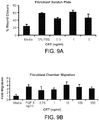
- macrophage migration into a wound of a patient is induced by topically administering a therapeutically effective amount of calreticulin to the wound of the patient.
- the present disclosure further provides a method for inducing keratinocyte migration (for wound re-epithelialzation, as described above) into a wound of a patient by topically administering a therapeutically effective amount of calreticulin to the wound of the patient.
- a method for inducing endothelial cell migration into a wound of a patient is disclosed, which comprises topically administering a therapeutically effective amount of calreticulin to the wound of the patient.
- a method for inducing monocyte migration into a wound of a patient comprises topically administering a therapeutically effective amount of calreticulin to the wound of the patient.
- fibroblast migration into a wound of a patient is induced by topically administering a therapeutically effective amount of calreticulin to the wound of the patientA method for inducing human mesenchymal stem cell migration into the wound is disclosed, which comprises topically administering a therapeutically effective amount of calreticulin to the wound of the patient.
- the wound is an acute wound injury or a chronic wound.
- a method for inducing TGF- ⁇ 3 expression (for induction of extracellular matrix proteins required) for granulation tissue formation and wound remodeling, and for wound healing without a scar) in a wound of a patient comprises topically administering a therapeutically effective amount of calreticulin to the wound of the patient.
- the wound is an acute wound injury or a chronic wound.
- the present disclosure provides a method for increasing extracellular matrix formation (to fill in and heal the wound defect) by inducing fibroblasts to produce fibronectin and collagens, which comprises topically administering or injecting a therapeutically effective amount of calreticulin into a wound of a patient.
- the wound is an acute wound injury or chronic wound.
- calreticulin can be administered to a patient for cosmetic purposes for the reduction of wrinkles and scarring.
- Calreticulin can be administered with or without hyaluronic acid for the reduction of wrinkles.
- the present disclosure provides a method for increasing alpha smooth muscle actin expression by fibroblasts for wound contraction in the wound of a patient by topically administering a therapeutically effective amount of calreticuliln into a wound of a patient.
- the wound is an acute wound injury or a chronic wound.
- alpha5 and betal integrins are induced to be expressed by keratinocytes and fibroblasts by administering a therapeutically effective amount of calreticulin to the wound of a patient.
- the presence of calreticulin in this context will mediate their migration of these cells into a wound.
- the wound is an acute wound injury or chronic wound.
- the present disclosure provides a method for enhancing phagocytosis of bacteria by phagocytes for reduction of bacterial burden (and biofilm) within the wound of a patient by administering a therapeutically effective amount of calreticulin to the wound.
- the wound is an acute wound injury or chronic wound.
- cell proliferation is induced in a wound of a patient by topically administering a therapeutically effective amount of calreticulin to the wound of the patient.
- the cell which is induced to proliferate can be, for example, a keratinocyte, a fibroblast, a dermal cell, and an endothelial cell.
- the wound is an acute wound injury or a chronic wound.
- Methods for increasing granulation tissue formation in a wound of a patient are desclosed, which comprise topically administering a therapeutically effective amount of calreticulin to the patient, wherein the amount of granulation tissue is increased relative to the amount of granulation tissue present in the wound prior to calreticulin administration.
- the wound is an acute wound injury or a chronic wound.
- a method for increasing the rate and/or quality of wound healing in a wound of a patient which method comprises administering a therapeutically effective amount of calreticulin to the patient, wherein the rate and/or quality of wound healing is increased relative to the rate and/or quality of wound healing prior to administration of calreticulin.
- a method of attracting a patient's own cells for producing extracellular matrix proteins such as collagen for the reduction or elimination of wrinkles, which comprises administering a therapeutically effective amount of calreticulin to the site of the patient.
- calreticulin improves several of the healing defects that prevent healing of a chronic wound including but not limited to a diabetic wound.
- calreticulin affects the most important functions required for general wound healing and tissue repair.
- Calreticulin induces matrix proteins including fibronectin and collagen and the factor, TGF- ⁇ 3, which itself induces these matrix proteins and also elastin, proteoglycans, glycosaminoglycans, and perlecan and others. These qualities make calreticulin suitable and important for treating wrinkles and other other cosmetic indications and also, for the treatment of deep tissue injury in which substantial removal of tissue or extensive tissue injury requires filling-in the wound defect or extensive tissue remodeling.
- calreticulin treatment facilitates the progression of chronic diabetic wound healing beyond the inflammatory phase, when these wounds are generally halted.
- the present inventors have discovered that calreticulin induces monocyte, macrophage, fibroblast, and keratinocyte migration both in vitro and in vivo in a wound, induces cellular proliferation in the wound both in vitro and in vivo in a wound, and induces TGF- ⁇ 3 both in vitro and in vivo in a wound.
- the TGF- ⁇ 3 mammalian isoform is known for its anti-scarring effects ( Ferguson, M.W. et al (2009) Lancet 373:1264-1274 ).
- the present invention also provides the use of calreticulin for treating wrinkles (including reducing the noticeability and/or improving the appearance and/or decreasing the depth of and/or decreasing the number of fine lines and/or wrinkles [including, e.g., facial lines and/or wrinkles, facial wrinkles on the cheeks, forehead, perpendicular wrinkles between the eyes, horizontal wrinkles above the eyes, and around the mouth, marionette lines, deep wrinkles or creases, expression lines, suborbital lines, periorbital lines, crow's feet, etc.] as well as preventing fibroblast senescence (aging) and more generally for tissue reconstruction including filling in and remodeling of defects or deformities in skin, vaginal reconstruction or other areas of the body in need of restoring normal anatomy, cartilage, bone as well as other uses of calreticulin disclosed herein.
- wrinkles including reducing the noticeability and/or improving the appearance and/or decreasing the depth of and/or decreasing the number of fine lines and/or wrinkles [including, e
- Calreticulin is based, at least in part, on its ability to induce collagen, fibronectin and TGF- ⁇ 3 (which, in turn, induces elastin and laminin and other matrix proteins). Calreticulin can be beneficially used in combination with hyaluronic acid as it occurs naturally in the body with hyaluronic acid with application to any needs or body parts described above.
- the present invention provides the use of calreticulin for prevention or treatment of cellular senescence and aging as in the treatment of wrinkles as well as other used of calreticuli disclosed herein.
- calreticulin can be used for treating corneal wounds (e.g., abrasions). This is based at least in part on calreticulin's unique ability to enhance healing without inducing angiogenesis. Angiogenesis in the cornea causes blindness.
- calreticulin can be used to treat epidermolysis bullosa.
- Calreticulin's ability to treat epidermolysis bullosa can be attributed at least in part to its ability to induce keratinocyte migration and proliferation which helps to resurface these broad pediatric wounds in which large areas of the epidermis are separated from the dermis appearing as large blisters.
- this disclosure provides the use of clareticulin to treat surgical adhesions.
- Calreticulin's ability to treat surgical adheshions can be attributed at least in part to its ability to induce TGF- ⁇ 3 which prevents scaring by mediating collagen organization. Calreticulin also decreases TGF- ⁇ 1 and 2, which are known to cause scaring.
- the disclosure provides the use of calreticulin to promote cartilage and bone repair.
- Calreticulin's ability to promote cartilage and bone repair can be attributed at least in part to its ability to induce chondrocytes to produce TGF- ⁇ -3, collagen and other matrix proteins.
- the human calreticulin protein has been previously described and cloned, and has protein accession number NP_004334 (SEQ ID NO:1) ( Fliegel, L. et al. (1989) J. Biol. Chem. 264:21522-21528 ; Baksh, S. et al., (1991) J. Biol. Chem. 266:21458-21465 ; Rokeach, L. A. et al., (1991) Prot. Engineering 4:981-987 ; Baksh, S. et al. (1992) Prot. Express. Purific. 3:322-331 ; Michalak, M. et al., (1992) Biochem. J.
- Calreticulin has an amino terminal signal sequence, a carboxy-terminal KDEL ER retrieval sequence, multiple calcium-binding sites, and harbors three distinct domains N, P, and C within its 46,000 dalton molecular mass (401 amino acids) ( Michalak, M. et al. (1999) Biochem. J. 344: Pt 2:281-292 ).
- Calreticulin is localized to the surface of a variety of cells including platelets, fibroblasts, apoptotic cells, endothelial cells, and cancer cells and is required for the phagocytosis of apoptotic cells by all phagocytes ( Gardai, S.J. et al (2005) Cell 123:321-334 ).
- calreticulin functions in the removal of dead cells and tissue from wounds (debridement).
- the presence of dead tissue in a wound is a significant deterrent to the wound healing process.
- the presence of bacterial infection is also a critical deterrent to the healing of an acute wound injury or a chronic wound.
- Calreticulin enhances the uptake and ingestion of Staph. aureus by human neutrophils. This quality implicates a role for calreticulin as a bacteriacidal agent to fight infections in the wound bed. Calreticulin is also dynamically expressed during wound healing indicating its inherent importance in this process.
- Treating means improving the rate of wound healing or completely healing a wound.
- Methods for measuring the rate of wound healing include, for example, observing increased epithelialization and/or granulation tissue formation, or lessening of the wound diameter and/or depth.
- Increased epithelialization can be measured by methods known in the art such as by, for example, the appearance of new epithelium at the wound edges and/or new epithelial islands migrating upward from hair follicles and sweat glands.
- Granulation tissue is necessary for proper healing and for providing a scaffold for the migration of keratinocytes over the wound for resurfacing and for tissue remodeling including filling in the wound defect.
- the amount of area of granulation tissue formation can be measured by morphometric analysis by measing the area of the granulation tissue or neodermis.
- the terms “treat” or “treatment” mean reducing the noticeability and/or improving the appearance and/or decreasing the depth of and/or decreasing the number of facial lines and/or wrinkles, facial wrinkles on the cheeks, forehead, perpendicular wrinkles between the eyes, horizontal wrinkles above the eyes, and around the mouth, marionette lines, deep wrinkles or creases, expression lines, suborbital lines, periorbital lines, crow's feet, etc. as well as preventing fibroblast senescence (aging).
- calreticulin Because of the ability of calreticulin to induce extracellular matrix proteins and stimulate cells such as fibroblasts and macrophages to migrate into the area of treatment such as in tissue reconstruction, these cells produce cytokines, growth factors, and other proteins that aid in filling in or replenishing the tissue in need of restoring, such as in tissue reconstruction.
- Chronic wound as used herein means a wound that has not completely closed in eight weeks since the occurrence of the wound in a patient having a condition, disease or therapy associated with defective healing.
- Conditions, diseases or therapies associated with defective healing include, for example, diabetes, arterial insufficiency, venous insufficiency, chronic steroid use, cancer chemotherapy, radiotherapy, radiation exposure, and malnutrition.
- a chronic wound includes defects resulting in inflammatory excess (e.g ., excessive production of Interleukin-6 (IL-6), tumor necrosis factor-alpha (TNF- ⁇ ), and MMPs), a deficiency of important growth factors needed for proper healing, bacterial overgrowth and senescence of fibroblasts.
- IL-6 Interleukin-6
- TNF- ⁇ tumor necrosis factor-alpha
- MMPs a deficiency of important growth factors needed for proper healing, bacterial overgrowth and senescence of fibroblasts.
- a chronic wound has an epithelial layer that fails to cover the entire
- Chronic diabetic wound means a chronic wound in a patient with diabetes.
- a chronic diabetic wound may be associated with peripheral neuropathy and/or macro- and micro- vascular insufficiency.
- a diabetic foot ulcer is one type of chronic diabetic wound.
- hyaluronic acid refers to hyaluronic acid or salts of hyaluronic acid, such as the sodium, potassium, magnesium and calcium salts, among others.
- hyaluronic acid is also intended to include not only elemental hyaluronic acid, but hyaluronic acid with other trace of elements or in various compositions with other elements, as long as the chemical and physical properties of hyaluronic acid remain unchanged.
- hyaluronic acid as used in the present application is intended to include natural formulas, synthetic formulas or combination of these natural and synthetic formulas.
- Non-limiting examples of useful hyaluronic acid preparations which can be used in the methods of the present invention include, for example, Juvederm® (a highly-crosslinked hyaluronic acid product sold by Allergan, Inc.) and RESTYLANE®, Perlane® (a non-animal stabilized hyaluronic acid product sold by Q-Med AB).
- Patient or “subject” refers to mammals and includes human and veterinary subjects.
- a “therapeutically effective amount” means the amount of a compound that, when administered to a mammal for treating a chronic diabetic wound, is sufficient to effect such treatment.
- the “therapeutically effective amount” may vary depending on the size of the wound, and the age, weight, physical condition and responsiveness of the mammal to be treated.
- promote wound healing is used to describe an agent that increases the rate at which a wound heals and the quality of wound repair.
- growth factor can be a naturally occurring, endogenous or exogenous protein, or recombinant protein, capable of stimulating cellular proliferation and/or cellular differentiation and cellular migration.
- the term “about” or “approximately” means within an acceptable range for the particular value as determined by one of ordinary skill in the art, which will depend in part on how the value is measured or determined, e.g ., the limitations of the measurement system.
- “about” can mean a range of up to 20 %, preferably up to 10 %, more preferably up to 5 %, and more preferably still up to 1 % of a given value.
- the term can mean within an order of magnitude, preferably within 5-fold, and more preferably within 2-fold, of a value.
- the term 'about' means within an acceptable error range for the particular value.
- John Wiley and Sons, Inc. Hoboken, NJ ; Coligan et al. eds. (2005) Current Protocols in Immunology, John Wiley and Sons, Inc. : Hoboken, NJ ; Coico et al. eds. (2005) Current Protocols in Microbiology, John Wiley and Sons, Inc. : Hoboken, NJ ; Coligan et al. eds. (2005) Current Protocols in Protein Science, John Wiley and Sons, Inc. : Hoboken, NJ ; and Enna et al. eds. (2005) Current Protocols in Pharmacology, John Wiley and Sons, Inc.: Hoboken, NJ .
- Methods for the preparation and analysis of calreticulin such as tissue extraction, recombinant protein technology in bacteria or yeast, anion and cation exchange and hydrophobic interaction chromatography, alcohol precipitation, cellulose acetate electrophoresis, polyacrylamide gel electrophoresis (PAGE), measurement of protein concentration, and microanalysis of SDS-PAGE electroblotted protein reverse phase HPLC, mass spectometry, are well known in the art and are described in detail in U.S. Patent No. 5,591,716 .
- the present invention encompasses calreticulin peptide fragments and other functional derivatives of calreticulin which have the functional activity of promoting healing of a chronic wound or the function of affecting a process associated with enhancing acute wound healing and chronic or impaired wound healing or tissue repair.
- the invention provides "functional derivatives" of calreticulin.
- “functional derivative” is meant a “fragment,” “variant,” “analog,” or “chemical derivative” of calreticulin.
- a functional derivative retains at least a portion of the function of calreticulin, such as the activity of promoting chronic wound healing, upregulating TGF- ⁇ 3 expression in skin, inducing cell migration, stimulating cell proliferation, or binding to a specific anti-calreticulin antibody, which permits its utility in accordance with the present invention.
- a “fragment” of calreticulin refers to any subset of the molecule, that is, a shorter peptide.
- a "variant" of calreticulin refers to a molecule substantially similar to either the entire protein or a fragment thereof. Variant peptides may be conveniently prepared by direct chemical synthesis of the variant peptide or producing the peptide by genetic recombinant technology, using methods well-known in the art.
- the protein useful in the methods and compositions of the present invention can be biochemically purified from a cell or tissue source.
- any of a number of tissues of adult or of fetal origin can be used.
- the gene encoding human calreticulin is known (GenBank Accession No. NC_000019.8, (SEQ ID NO: 2); Fliegel et al., supra ; Baksh et al., (1991) supra ; Rokeach et al., supra ; Baksh et al. (1992) supra ; Michalak et al., (1992), supra ); McCauliffe et al., J Clin Invest.
- polypeptide can be synthesized substantially free of other proteins or glycoproteins of mammalian origin in a prokaryotic organism, in a non-mammalian eukaryotic organism, by a yeast, or by a baculovirus system, if desired.
- methods are well known for the synthesis of polypeptides of desired sequence on solid phase supports and their subsequent separation from the support.
- amino acid sequence variants of the protein or peptide can be prepared by mutations in the DNA which encodes the synthesized peptide.
- Such variants include, for example, deletions from, or insertions or substitutions of, residues within the amino acid sequence. Any combination of deletion, insertion, and substitution may also be made to arrive at the final construct, provided that the final construct possesses the desired functional activity.
- the mutations that will be made in the DNA encoding the variant peptide must not alter the reading frame and preferably will not create complementary regions that could produce secondary mRNA structure (see European Patent Publication No. EP 75,444 ).
- these variants ordinarily are prepared by site-directed mutagenesis (as exemplified by Adelman et al., DNA 2:183 (1983 )) of nucleotides in the DNA encoding the calreticulin protein or a peptide fragment thereof, thereby producing DNA encoding the variant, and thereafter expressing the DNA (cDNA, RNA, and protein) in recombinant cell culture (see below).
- site-directed mutagenesis as exemplified by Adelman et al., DNA 2:183 (1983 )
- the variants typically exhibit the same qualitative biological activity as the nonvariant peptide.
- a preferred group of variants of calreticulin are those in which at least one amino acid residue in the protein or in a peptide fragment thereof, and preferably, only one, has been removed and a different residue inserted in its place.
- substitutions which may be made in the protein or peptide molecule of the present invention may be based on analysis of the frequencies of amino acid changes between a homologous protein of different species, such as those presented in Table 1-2 of Schulz et al. (supra) and FIGS. 3-9 of Creighton (supra). Based on such an analysis, conservative substitutions are defined herein as exchanges within one of the following five groups:
- Preferred deletions and insertions, and substitutions, according to the present invention are those which do not produce radical changes in the characteristics of the protein or peptide molecule.
- substitutions, deletion, or insertion in advance of doing so, one skilled in the art will appreciate that the effect will be evaluated by routine screening assays which are described in more detail below. For example, a change in the immunological character of the protein peptide molecule, such as binding to a given antibody, is measured by a competitive type immunoassay. Biological activity is screened in an appropriate bioassay, as described below.
- an "analog" of calreticulin refers to a non-natural molecule substantially similar to either the entire molecule or a fragment thereof.
- a "chemical derivative" of calreticulin contains additional chemical moieties not normally a part of the peptide.
- Covalent modifications of the peptide are included within the scope of this invention. Such modifications may be introduced into the molecule by reacting targeted amino acid residues of the peptide with an organic derivatizing agent that is capable of reacting with selected side chains or terminal residues.
- modified amino acids or chemical derivatives of amino acids of calreticulin or fragments thereof, according to the present invention may be provided, which polypeptides contain additional chemical moieties or modified amino acids not normally a part of the protein. Covalent modifications of the peptide are thus included within the scope of the present invention.
- the following examples of chemical derivatives are provided by way of illustration and not by way of limitation.
- Aromatic amino acids may be replaced with D- or L-naphthylalanine, D- or L-phenylglycine, D- or L-2-thienylalanine, D- or L-1-, 2-, 3- or 4-pyrenylalanine, D- or L-3-thienylalanine, D- or L-(2-pyridinyl)-alanine, D- or L-(3-pyridinyl)-alanine, D- or L-(2-pyrazinyl)-alanine, D- or L-(4-isopropyl)-phenylglycine, D-(trifluoromethyl)-phenylglycine, D-(trifluoromethyl)-phenylalanine, D-p-fluorophenylalanine, D- or L-p-biphenylphenylalanine, D- or L-p-methoxybiphenylphenylalanine, D- or L-2-indole(al
- Acidic amino acids can be substituted with non-carboxylate amino acids while maintaining a negative charge, and derivatives or analogs thereof, such as the non-limiting examples of (phosphono)-alanine, glycine, leucine, isoleucine, threonine, or serine; or sulfated (for example, --SO 3 H) threonine, serine, tyrosine.
- substitutions may include unnatural hydroxylated amino acids may made by combining "alkyl" with any natural amino acid.
- Basic amino acids may be substituted with alkyl groups at any position of the naturally occurring amino acids lysine, arginine, ornithine, citrulline, or (guanidino)-acetic acid, or other (guanidino)alkyl-acetic acids, where "alkyl” is define as above.
- Nitrile derivatives (for example, containing the CN-moiety in place of COOH) may also be substituted for asparagine or glutamine, and methionine sulfoxide may be substituted for methionine. Methods of preparation of such peptide derivatives are well known to one skilled in the art.
- any amide linkage the polypeptides can be replaced by a ketomethylene moiety.
- Such derivatives are expected to have the property of increased stability to degradation by enzymes, and therefore possess advantages for the formulation of compounds which may have increased in vivo half lives, as administered by various routes as described herein.
- any amino acid representing a component of the peptides can be replaced by the same amino acid but of the opposite chirality.
- any amino acid naturally occurring in the L-configuration (which may also be referred to as the R or S, depending upon the structure of the chemical entity) may be replaced with an amino acid of the same chemical structural type, but of the opposite chirality, generally referred to as the D-amino acid but which can additionally be referred to as the R- or the S-, depending upon its composition and chemical configuration.
- Such derivatives have the property of greatly increased stability to degradation by enzymes, and therefore are advantageous in the formulation of compounds which may have longer in vivo half lives, when administered by various routes.
- Additional amino acid modifications in calreticulin or in a peptide thereof may include the following.
- Cysteinyl residues most commonly are reacted with ⁇ -haloacetates (and corresponding amines), such as chloroacetic acid or chloroacetamide, to give carboxymethyl or carboxyamidomethyl derivatives. Cysteinyl residues also are derivatized by reaction with bromotrifluoroacetone, ⁇ -bromo- ⁇ -(5-imidozoyl)propionic acid, chloroacetyl phosphate, N-alkylmaleimides, 3-nitro-2-pyridyl disulfide, methyl 2-pyridyl disulfide, p-chloromercuribenzoate, 2-chloromercuri-4-nitrophenol, or chloro-7-nitrobenzo-2-oxa-1,3-diazole.
- Histidyl residues are derivatized by reaction with diethylprocarbonate at pH 5.5-7.0 because this agent is relatively specific for the histidyl side chain.
- Para-bromophenacyl bromide also is useful; the reaction is preferably performed in 0.1M sodium cacodylate at pH 6.0.
- Lysinyl and amino terminal residues are reacted with succinic or other carboxylic acid anhydrides, which reverses the charge of the lysinyl residues.
- suitable reagents for derivatizing ⁇ -amino-containing residues include imidoesters such as methyl picolinimidate; pyridoxal phosphate; pyridoxal; chloroborohydride; trinitrobenzenesulfonic acid; O-methylisourea; 2,4 pentanedione; and transaminase-catalyzed reaction with glyoxylate.
- Arginyl residues are modified by reaction with one or several conventional reagents, among them phenylglyoxal, 2,3-butanedione, 1,2-cyclohexanedione, and ninhydrin. Derivatization of arginine residues requires that the reaction be performed in alkaline conditions because of the high pK ⁇ of the guanidine functional group. Furthermore, these reagents may react with the groups of lysine as well as the arginine ⁇ -amino group.
- Carboxyl side groups (aspartyl or glutamyl) are selectively modified by reaction with carbodiimides (R'-N-C-N-R') such as 1-cyclohexyl-3-(2-morpholinyl-(4-ethyl) carbodiimide or 1-ethyl-3-(4-azonia-4,4-dimethylpentyl) carbodiimide. Furthermore, aspartyl and glutamyl residues are converted to asparaginyl and glutaminyl residues by reaction with ammonium ions.
- Glutaminyl and asparaginyl residues are deamidated to the corresponding glutamyl and aspartyl residues. Alternatively, these residues are deamidated under mildly acidic conditions. Either form of these residues falls within the scope of this invention.
- Derivatization with bifunctional agents is useful for cross-linking the peptide to a water-insoluble support matrix or to other macromolecular carriers.
- Commonly used cross-linking agents include, e.g ., 1,1-bis(diazoacetyl)-2-phenylethane, glutaraldehyde, N-hydroxysuccinimide esters, for example, esters with 4-azidosalicylic acid, homobifunctional imidoesters, including disuccinimidyl esters such as 3,3'-dithiobis-(succinimidyl-propionate), and bifunctional maleimides such as bis-N-maleimido-1,8-octane.
- Derivatizing agents such as methyl-3-[(p-azidophenyl)dithio]propioimidate yield photoactivatable intermediates that are capable of forming crosslinks in the presence of light.
- reactive water-insoluble matrices such as cyanogen bromide-activated carbohydrates and the reactive substrates described in U.S. Pat. Nos. 3,969,287 ; 3,691,016 ; 4,195,128 ; 4,247,642 ; 4,229,537 ; and 4,330,440 are employed for protein immobilization.
- Such derivatized moieties may improve the solubility, absorption, biological half life, and the like.
- the moieties may alternatively eliminate or attenuate any undesirable side effect of the protein and the like.
- Moieties capable of mediating such effects are disclosed, for example, in Remington's Pharmaceutical Sciences, 16th ed., Mack Publishing Co., Easton, Pa. (1980 ).
- Calreticulin may be purified from a tissue source using conventional biochemical techniques, or produced recombinantly in either prokaryotic or eukaryotic cells using methods well-known in the art. See , Sambrook, J. et al., MOLECULAR CLONING: A LABORATORY MANUAL, 2nd Edition, Cold Spring Harbor Press, Cold Spring Harbor, N.Y., 1989 . Various references describing the cloning and expression of calreticulin have been noted above.
- Fusion proteins representing different polypeptide regions in calreticulin may be used to identify regions of the protein that have the desired functional activity (binding, stimulating wound healing, specoific functions associated with wound healing, etc .).
- PCR polymerase chain reaction
- live insects are to be used, caterpillars are presently preferred hosts for large scale production according to the invention.
- Fragments of calreticulin are purified by conventional affinity chromatography using antibodies, preferably monoclonal antibodies (mAbs) that recognize the appropriate regions of calreticulin.
- mAbs monoclonal antibodies
- the mAbs specific for the most highly conserved regions in calreticulin can be used to purify calreticulin protein from mixtures.
- the preferred animal subject of the present invention is a mammal.
- mammal an individual belonging to the class Mammalia.
- the invention is particularly useful in the treatment of human subjects.
- the present invention provides for methods of treatment of wounds, and cosmetic applications of calreticulin, which methods comprise administering to a subject in need of such treatment a therapeutically effective amount of calreticulin, or a functional derivative thereof.
- the disorders that may be treated according to this invention include, but are not limited to acute wounds, chronic wounds, corneal wounds, bone and cartilage repair, injury due to surgical procedures, wrinkles, and alopecia as well as other uses of calreticulin disclosed herein.
- the present invention provides a pharmaceutical composition or formulation comprising at least one active composition, or a pharmaceutically acceptable derivative thereof, in association with a pharmaceutically acceptable excipient, diluent, and/or carrier.
- the excipient, diluent and/or carrier must be "acceptable" in the sense of being compatible with the other ingredients of the formulation and not deleterious to the recipient thereof.
- the topical preparation is an ointment wherein about 0.01 to about 50 mg of active ingredient is used per cc of ointment base.
- the dosage of the therapeutic formulation may vary widely, depending upon the size of the wound, the patient's medical history, the frequency of administration, the manner of administration, the clearance of the agent from the host, and the like.
- the dose may be administered with each wound dressing change.
- the dose may be administered once daily, more than once daily, or as infrequently as weekly or biweekly.
- Calreticulin may be administered in any pharmaceutically acceptable carrier or excipient.
- carrier refers to a diluent, adjuvant, excipient, or vehicle with which the compound is administered.
- pharmaceutically acceptable refers to molecular entities and compositions that are generally believed to be physiologically tolerable and do not typically produce an allergic or similar untoward reaction, such as gastric upset, dizziness and the like, when administered to a human.
- Pharmaceutically acceptable carriers can be sterile liquids, such as water and oils, including those of petroleum, animal, vegetable or synthetic origin, such as peanut oil, soybean oil, mineral oil, sesame oil and the like.
- gels such as hydrogels, hyaluronic acid (HA), collagen, materials consisting of naturally occurring or synthetic substances, or any other matrix protein such as perlecan, proteoglycans, glycoaminoglycans, fibrin gels, and polymers.
- Water or aqueous solution saline solutions and aqueous dextrose and glycerol solutions are preferably employed as carriers, particularly for injectable solutions.
- the carrier can be a solid dosage form carrier, including but not limited to one or more of a binder (for compressed pills), a glidant, an encapsulating agent, a flavorant, and a colorant.
- Suitable pharmaceutical carriers are described in "Remington's Pharmaceutical Sciences" by E.W. Martin.
- peptide sequences from calreticulin are inserted into or replace sequences within "scaffold" proteins.
- a "scaffold protein" of the present invention is a protein which includes a functional calreticulin sequence, either as an inserted sequence or as a replacement sequence for a homologous (corresponding) sequence of the scaffold protein.
- the scaffold protein adopts a native conformation.
- the calreticulin and scaffold can alternate positions; these terms are used to indicate the source of sequences introduced into the "scaffold.”
- functional peptide sequences from calreticulin can be inserted into a chemical or natural matrix.
- pharmaceutically acceptable derivative means any pharmaceutically acceptable salt, solvate or prodrug, e.g. ester, of a compound of the invention, which upon administration to the recipient is capable of providing (directly or indirectly) a compound of the invention, or an active metabolite or residue thereof.
- Such derivatives are recognizable to those skilled in the art, without undue experimentation. Nevertheless, reference is made to the teaching of Burger's Medicinal Chemistry and Drug Discovery, 5th Edition, Vol. 1 : Principles and Practice.
- Preferred pharmaceutically acceptable derivatives are salts, solvates, esters, carbamates, and phosphate esters.
- Particularly preferred pharmaceutically acceptable derivatives are salts, solvates, and esters.
- Most preferred pharmaceutically acceptable derivatives are salts and esters.
- the administration route may be any mode of administration known in the art, including but not limited to topically, subcutaneously (e.g, by injection), intradermally, transdermally (e.g., by transdermal patch), via intracorporal application during surgery, parenterally, intramuscularly, intraperitoneally, buccally, intravenously, intrathecally, intracranially, by injection into involved tissue, intraarterially, orally, or via an implanted device.
- the present invention also provides pharmaceutical and cosmetic compositions comprising an amount of calreticulin, or a functional derivative or fragment thereof, effective to promote the healing of a wound or exert any other therapeutic or cosmetic effect relevant for the present invention, in a pharmaceutically or cosmetically acceptable carrier.
- the pharmaceutical composition of the present invention is preferably applied to site of action (e.g., topically, subcutaneously [e.g, by injection], intradermally, transdermally [e.g., by transdermal patch], or via intracorporal application during surgery).
- site of action e.g., topically, subcutaneously [e.g, by injection], intradermally, transdermally [e.g., by transdermal patch], or via intracorporal application during surgery).
- compositions of the present invention may be incorporated into topically applied vehicles such as salves or ointments, which have both a soothing effect on the skin as well as a means for administering the active ingredient directly to the affected area.
- the carrier for the active ingredient in a topical formulation may be either in sprayable or non-sprayable form.
- Non-sprayable forms can be semi-solid or solid forms comprising a carrier indigenous to topical application and having a dynamic viscosity preferably greater than that of water.
- Suitable formulations include, but are not limited to, solution, suspensions, emulsions, creams, ointments, powders, liniments, salves, and the like. If desired, these may be sterilized or mixed with auxiliary agents, e.g ., preservatives, stabilizers, wetting agents, buffers, or salts for influencing osmotic pressure and the like.
- Preferred vehicles for non-sprayable topical preparations include ointment bases, e.g ., polyethylene glycol-1000 (PEG-1000); conventional creams such as HEB cream; gels; as well as petroleum jelly and the like.
- PEG-1000 polyethylene glycol-1000
- a preferred vehicle is a petrolatum/lanolin vehicle.
- sprayable aerosol preparations wherein the active ingredient, preferably in combination with a solid or liquid inert carrier material, is packaged in a squeeze bottle or in admixture with a pressurized volatile, normally gaseous propellant.
- the aerosol preparations can contain solvents, buffers, surfactants, perfumes, and/or antioxidants in addition to the compounds of the invention.
- the dosage administered will be dependent upon the age, health, and weight of the recipient, kind of concurrent treatment, if any, frequency of treatment, and the nature of the effect desired.
- Effective doses of calreticulin for therapeutic uses discussed above may be determined using methods known to one skilled in the art. Effective doses may be determined, preferably in vitro, in order to identify the optimal dose range using any of the various methods described herein.
- an aqueous solution of a calreticulin protein or peptide is administered by intravenous injection.
- Each dose may range from about 0.001 ⁇ g/kg body weight to about 100 mg/kg body weight, or more preferably, from about 0.1 ⁇ g/kg to 10 mg/kg body weight.
- the dosing schedule may vary from one time only to once a week to daily or twice (or more) daily depending on a number of clinical factors, including the type of wound, its severity, and the subject's sensitivity to the protein.
- Non-limiting examples of dosing schedules are 3 ⁇ g/kg administered twice a week, three times a week or daily; a dose of 7 ⁇ g/kg twice a week, three times a week or daily; a dose of 10 ⁇ g/kg twice a week, three times a week or daily; or a dose of 30 ⁇ g/kg twice a week, three times a week or daily.
- doses such as those described above by alternate routes, including intravenously, intramuscularly, intraperitoneally or intrathecally. Continuous infusion may also be appropriate.
- Calreticulin or a functional derivative may also be administered in combination with an effective amount of at least one other agent that is, itself, capable of promoting the healing of wounds or treating accompanying symptoms.
- agents include growth factors, anti-infectives, including anti-bacterial, anti-viral and anti-fungal agents, local anesthetics, and analgesics, collagens, fibrin gels, glycosaminoglycans (e.g., hyaluronic acid), proteoglycans (e.g., perlecan, heparin sulfate), syndecan, suitable chemical or natural polymers, or a combination thereof.
- Other agents that can be applied to a wound include but, are not limited to, calreticulin as part of a living skin substitute (skin device) or a synthetic, chemical or natural scaffold or matrix or polymer thereof.
- Combination treatment according to the present invention includes administering the calreticulin and one or more additional agent in the same or separate dosage forms.
- additional agents include, among others, agents which are known to promote wound healing or to treat problems or symptoms associated with chronic wounds. Examples of such agents include hyaluronic acid, disinfectants such as antibacterial agents or antiviral agents, anti-fungal agents, anti-inflammatory agents, agents which induce relief from pain or itching, and the like.
- growth factors which promote wound healing including, but not limited to, transforming growth factor- ⁇ , transforming growth factor- ⁇ , fibroblast growth factor- ⁇ , fibroblast growth factor- ⁇ , FGFs in general, epidermal growth factor, platelet-derived growth factor, endothelial cell-derived growth factor, insulin-like growth factors, VEGF, and granulocyte colony-stimulating factor.
- calreticulin administered in combination with an additional agent includes any overlapping or sequential administration of the calreticulin and the additional agent.
- methods according to the present invention encompass administering calreticulin and an additional agent simultaneously or non-simultaneously.
- calreticulin and an additional agent can be administered by the same route (e.g., both are administered topically) or by different routes (e.g., calreticulin is administered topically and an additional agent is administered orally).
- compositions of the present invention may be administered by any means that achieve their intended purpose. Amounts and regimens for the administration of calreticulin, or a derivative thereof, can be determined readily by those with ordinary skill in the clinical art of treating wounds.
- the positive control reagent may be interchangeably referred to as either Regranex® gel or PDGF-BB. These two terms are understood to be the same reagent used in the porcine wound healing studies.
- VEGF was used as a positive control in the murine wound healing studies.
- Recombinant rabbit calreticulin (from M. Michalak, University of Alberta) was expressed in E. coli as a his-tagged protein that was purified to homogeneity by Nickel-Sepharose chromatography. The rabbit calreticulin was shown to be properly folded and migrated as a single band at approximately Mr 50,000 by SDS-PAGE, as described in Guo et al., J Biol Chem. 2003;278:50645-50653 . This protein was prepared in pBAD and E. coli, and his tagged with five amino acids at the N-terminus (composed of SEQ iD NO:5 and SEQ ID NO:7).
- human calreticulin was produced from the human gene sequence inserted into the pBAD plasmid and expressed in E. coli.
- the recombinant human calreticulin was found to be a mixture of Michalak 5-CRT + tag and Michalak 23-CRT + tag (later experiments were performed using Michalak 5-CRT and Michalak 23-CRT).
- human calreticulin was obtained from GenWay Biotech (#10-288-22432F; San Diego, CA). The calreticulin was stored at minus 80C in 10 mM Tris containing 3.0 mM calcium, pH 7.0 ("buffer”), to maintain proper conformation of this calcium-binding molecule.
- Anti-peptide antibodies (purified IgG) specific for each isoform of TGF-ß (TGF-ß1, TGF-ß2, AND TGF-ß3) have been described by Levine et al., Amer J Pathol. 1993;143:368-380 ; Pelton et al. J Cell Biol. 1991;115:1091-1105 .
- Isoform-specific cytokeratin 14 antibody was obtained from Accurate Scientific (Westbury, NY).
- Goat anti-calreticulin pantropic; BIOCAN/Jackson Immunochemicals
- Rabbit anti-human Ki-67 was obtained from Nova Castra Laboratories Ltd. (Newcastle, UK) and a monoclonal mouse anti-human antibody specific for macrophages (MAC387) was obtained from Serotec, Ltd. (UK).
- Collagen type I antibody was purchased from Santa Cruz Biotech, catalogue no. sc-28657 (Santa Cruz, CA). Integrin ⁇ -5 antibody was purchased from Santa Cruz Biotech, catalogue no. sc-10729 (Santa Cruz, CA). Integrin ⁇ -1 antibody was purchased from Santa Cruz Biotech, catalogue no. sc-8978 (Santa Cruz, CA).
- Alpha smooth muscle actin antibody was purchased from Sigma-Aldrich, catalogue no. A5228 (St. Loius, MO).
- porcine wound healing is a well-known and accepted model for studying human wound healing because the pig heals similarly to humans.
- porcine and human skin share similar epidermal and dermal-epidermal thickness ratios, mosaic hair growth, and have similar hair and blood vessel distribution.
- pigs lack the muscular layer (panniculous carnosus) found in loose skin animals (e.g., rodents), which contracts the wound.
- Adolescent Yorkshire pigs weighing about 50-60 lbs. were housed, fed and treated in accordance with protocols approved by the IACUC at Vanderbilt University Medical Center.
- the pigs Prior to surgery, the pigs were anesthetized with a mixture of Ketamine anesthetic (2.2 mg/kg), Telazol® anesthetic/tranquilizer (4.4 mg/kg) and Xylazine anesthetic (2.2 mg/kg) by intramuscular injection, intubated and maintained on an inhalation of oxygen and isofluorene.
- Cefazolin antibiotic was administered intramuscularly immediately before surgery and on subsequent post-operative days.
- four longitudinal partial thickness wounds were created along the paravertebral region to a depth of 1560 ⁇ m using a Zimmer dermatome (Warsaw, IN).
- Calreticulin at 1.0 mg/ml and 5.0 mg/ml was topically applied to the wounds.
- the treated wound tissue was harvested at 5 and 10 days after wounding.
- the gel formulation Regranex® (0.01 % PDGF-BB; Ethicon, Inc., Sommerville, NJ), a commercially available wound healing agent, was used as a positive control. Two wounds per treatment group per pig were used. In order to prevent the liquid from rolling off, 0.05 ml of calreticulin or buffer was applied to the pig lying on its side and allowed to dry for one minute prior to application of a KY gel formulation, which maintained moist wound healing conditions. The wounds were covered with OpSiteTM (Smith & Nephew, Mt Waverly, Victoria, Australia) semi-occlusive bandages. The wounds were cleansed daily; and the bandages were replaced.
- OpSiteTM Smith & Nephew, Mt Waverly, Victoria, Australia
- Buprenex analgesic was administered intramuscularly for pain control in the initial post-operative period.
- Duragesic® patches 25 ⁇ g/hr transdermal fentanyl were placed for 3 days for sustained analgesia.
- Topical treatments of calreticulin and buffer controls were repeated daily for the first 4 days; PDGF-BB was applied once at the time of wounding.
- wound placement patterns from the various treatment groups were randomized.
- pigs were administered 1 mg/kg of methylprednisolone acetate (DepoMedrol®, Henry-Schein, Melville, NY) intramuscularly 48 hours before wound creation.
- the wounds were prepared and treated as described above. Treatment groups were the same for both the normal and impaired healing models. Animals were euthanized and tissues collected for study after 6 days or 7 days of healing.
- This model eliminated this effect by stenting open the wound to prevent this contraction, thus facilitating observations of re-epithelialization and enabling measurement of the area of wound resurfacing over time during wound healing (reduction in epithelial gap between the edges of the wound) this model mimics human cutaneous wound healing more closely. Therefore, all healing in this murine model was mediated by granulation tissue formation and epithelial migration, more closely mimicking human skin wound healing.
- db/db mice Eight- to 12-week old C57BL/6J mice (Jackson Laboratories stock #000664, Bar Harbor, ME) were used as a control of unimpaired healing; db/db mice (BKS.Cg-m+/+Leprdb, Jackson Laboratories stock #000642) were used in the model of impaired wound healing.
- the db/db mice are leptin receptor deficient and are a model of type II diabetes mellitus characterized by hyperglycemia, obesity, hypoinsulinemia, and impaired wound healing. The animals were housed five animals per cage prior to surgery and alone post-procedure in a temperature-controlled animal facility with a 12-hour light/dark cycle.
- mice were acclimated to their environment for at least 1 week prior to the procedure and were given food and water ad libitum. This experimental protocol was approved by the Institutional Animal Care and Use Committee (IACUC) of New York University School of Medicine. The animals utilized in this experiment all received humane care.
- IACUC Institutional Animal Care and Use Committee
- mice were individually anesthetized using an intraperitoneal injection of ketamine (75 mg/kg), xylazine (15 mg/kg), and acepromazine (2.5 mg/kg).
- the dorsal surface was shaved with an electric clipper followed by a depilatory agent to remove any remaining hair.
- the mice were rinsed with an alcohol swab and sterilely prepped with betadine and draped.
- a sterile 6-mm punch biopsy tool was used to outline a pattern for the wounds on the dorsum of the C57/BL6J and db/db mice.
- a 6-mm wound was chosen for the db/db mice because of their large dorsal surface. Two wounds were patterned, one on each side of midline.
- calreticulin 5.0 mg/mL
- 10mM Tris containing 3mM calcium was applied to each wound for the first four days of the experiment.
- the buffer alone was used on the control animals.
- the wounds were covered in an occlusive dressing (TegadermTM, 3M, St. Paul, MN) to protect them from infection and trauma.
- the animals were placed in individual cages under a warming lamp and allowed to recover fully from anesthesia.
- the dressing was changed daily after each calreticulin or buffer application.
- Time to closure was defined as the time at which the wound bed was completely filled in with new tissue and fully closed.
- the wound area was analyzed by tracing the wound margin with a fine-resolution computer mouse and calculating pixel area using SigmaScan® Pro Image Analysis Version 5.0.0 digital analysis software (Aspire Software International, Leesburg, VA). The wound area was calculated as a percent of the original wound area. A completely closed wound was considered equal to its area measured zero (grossly). Because the splint has a constant area, it was used to normalize the wound sizes.
- the wounds were excised, bisected, and fixed in 10% neutral formalin for 24 hours.
- the sections were embedded in paraffin and sectioned transversely through the wound bed, allowing for analysis of the epithelial gap remaining and wound depth to be calculated.
- the samples underwent routine histological processing with hematoxylin and eosin. Under light microscopy, the sections were photographed using a mounted digital camera (Olympus, Melville, NY). The images were analyzed for epithelial gap (EG) and total area of granulation tissue (GT) using digital analysis software.
- EG epithelial gap
- GT total area of granulation tissue
- EG was defined as the distance in area of non-epithelialized wound between the advancing edges of keratinocyte (epithelial cell) migration to close the wound. Three serial sections were averaged to determine EG at each time point. An EG of zero represents a completely re-epithelialized wound. Area of GT was calculated by tracing regions of GT and calculating pixel area. (Note: A wound is re-epithelialized by light microscopy whereas wound closure indicates that the skin has completely closed.)
- the methods for histological preparation and morphometric analysis used in the Examples are as follows. At the termination of each experiment, wounds with an adjacent margin of normal skin were excised, divided vertically into three full-thickness tissue sections per wound, fixed in 10 % neutral buffered formalin for 24 hours, embedded in paraffin, and mounted in 5.0 ⁇ m thick tissue sections on glass slides for histological analysis and immunohistochemistry (IHC). The tissue sections were stained (described below) and the extent of re-epithelialization and dermal depth (granulation tissue formation) of the wounds was determined by morphometric analysis. Serial images of the wounds were captured under a light microscope and displayed on a videoscreen using an Olympus model AHBT camera. Quantitative measurements were performed using Image-Pro Plus scientific image analysis software (Media Cybernetic, Inc., Silver Spring, MD).
- Re-epithelialization was assessed using the wounds of normal pigs. Antibodies to cytokeratin 14 were used to selectively highlight the newly resurfaced epithelial islands and epidermal margins. The extent of re-epithelialization was determined after 5 days of healing in normal pigs by measuring a composite of newly resurfaced epidermis that migrated over the wounds from the wound edges and epithelial islands derived from surviving epithelium that migrated upward from hair follicles and sweat ducts, compared to total wound length. The data are expressed as a percent of resurfacing as described in Okwueze et al., J Invest Dermatol. 2007;127:1030-1041 .
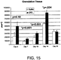
- Granulation tissue thickness was measured in trichrome stained tissue slides extending from the non-re-epithelialized surface of the granulating wound down to its intersection with the underlying unwounded dermis.
- the granulation tissue becomes converted into a neodermis as re-eptithelialization is nearly complete, which is measured as dermal depth.
- Dermal depth measurements extend from the dermo-epidermal junction down to the intersection of the newly formed granulation tissue with the adjacent underlying unwounded dermis of these partial thickness wound beds.
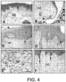
- the temporal and spatial expression of calreticulin during wound healing was determined at 5 and 10 days of healing by immunohistochemical localization using a polyclonal goat anti-calreticulin.
- Slides were baked overnight at 56°C and passed through graded alcohol with the final concentration being 30 % ethanol.
- the slides were placed in Tris -buffered saline (TBS) containing 0.3 % Triton X-100 for 15 minutes, followed by 100 % methanol for one minute and then, peroxidase activity was quenched with 0.6 % H 2 O 2 for 30 minutes followed by 100 % methanol for one minute.
- TBS Tris -buffered saline
- the tissues were blocked with normal rabbit serum (Vector Labs, #S5000; Burlingame, CA) in TBS containing 0.5 % BSA (blocking buffer) for 20 minutes at room temperature.
- the calreticulin antibody diluted at 1:1000 buffer, was incubated with the slides overnight at 4 °C, in humido.
- biotinylated rabbit anti-goat IgG secondary antibody (Vector #BA5000) was applied to the slides for one hour at room temperature.
- the slides were washed and then incubated with ABC Reagent (Vectastain kit #PK6200, Vector Laboratories, Burlingame, CA) for one hour.
- the slides were dipped in the substrate 0.05 % 3,3-diaminobenzidine HCL (DAB; Sigma Chemical #D5637) solution until a brown color appeared, counterstained with hematoxylin (Fisher #CS401-1D), dehydrated through increasing concentrations of alcohol, and mounted with Permount (Fisher #SP15-100).
- DAB 3,3-diaminobenzidine HCL
- TGF-ß isoform expression was induced in calreticulin-treated porcine and murine wounds.
- tissue slides were incubated separately with antibodies to TGF-ß1, TGF-ß2, and TGF-ß3.
- the antisera was produced in rabbits to individual peptides of each isoform and the IgG purified by peptide affinity chromatography as described in Pelton et al., J Cell Biol. 1991;115:1091-1105 .
- the following protocol was used: actively proliferating cells in the epidermis and neodermis were immunostained for Ki67 antigen.
- the tissue slides were subjected to antigen retrieval. Endogenous peroxidase activity was neutralized with 6 % H 2 O 2 for 20 minutes followed by blocking non-specific reactivity with a casein-based protein block (DAKO, Carpintera, CA) for 10 minutes.
- DAKO casein-based protein block
- the slides were incubated with rabbit anti-human Ki-67 (NovaCastra Laboratories Ltd., Newcastle, UK) diluted at 1:1400 for 60 minutes in TBS.
- the rabbit Envision HRP System (DAKO) was used with DAB as substrate and the slides were counterstained with hematoxylin.
- mice were injected i.p.
- BrDU bromodeoxyuridine
- Macrophage infiltration into the porcine wounds was assessed by immunostaining using a specific antisera for tissue monocytes/macrophages (MAC387AbD; Serotec, Raleigh, NC).
- tissue sections underwent antigen retrieval by boiling the slides in 0.01 M Tris/HCL pH 10. Both quenching peroxidase activity and blocking non-specific immunoreactivity were performed as described above.
- a monoclonal mouse anti-human antibody to a macrophage epitope (MAC387) was used at 1:1000 for one hour.
- the mouse Envision system HRP kit (DAKO) was used for detection as described above.
- H&E and Trichrome stains of the murine and porcine wounds were performed by the department of pathology at NYU. Trichrome staining was used to visualize collagen content staining as cyano blue fibrils and cell nuclei stain red.
- Keratinocytes Primary adult human epidermal keratinocytes (#CC-2501; Cambrex-Lonza, Inc., Walkersville, MD) were cultured in Keratinocyte Growth Media (KGM) containing additives from the BulletKit [Singlequots] (Cat # CC-4131; Cambrex-Lonza, Walkersville, MD), including Gentamycin-1000 (Lonza). The cells were subcultured at 50 % confluency by washing with 30mM HEPES buffered saline, treating with trypsin-EDTA (0.025 % trypsin- 0.02 % EDTA; Lonza) and neutralizing the trypsin with neutralizing solution (TNS; Lonza). Following slow centrifugation, the cells were resuspended in fresh media and seeded at different cell densities depending on the experiments described below.
- KGM Keratinocyte Growth Media
- TMS neutralizing solution
- Fibroblasts Primary human low passage foreskin fibroblasts (CCD 1070SK; ATCC, Manassas, VA) were grown in complete Eagles Minimal Essential Media (MEM, Gibco/Invitrogen, Carlsbad, CA) containing 10 % fetal bovine serum (FBS; HyClone, Logan, UT), 2mM Glutamine (Mediatech, Manassas, VA), and antibiotic-antimycotic (ABAM; Mediatech).
- MEM complete Eagles Minimal Essential Media
- FBS HyClone, Logan, UT
- 2mM Glutamine Mediatech, Manassas, VA
- ABAM antibiotic-antimycotic
- the cells were washed with PBS, removed for re-plating with 0.25 % trypsin-2.21 mM EDTA (Mediatech), the trypsin neutralized with 10 % FBS in MEM, and the cells centrifuged and resuspended in complete MEM at the cell densities described in the assays below.
- HMVECs Human dermal microvascular endothelial cells (HMVECs; Cambrex-Lonza) were cultured in complete Endothelial Cell Medium (EGM; Lonza) supplemented with the EGM-MV BulletKit (CC-3125; Lonza). The cells were subcultured when approximately 70 % confluent by washing with HEPES Buffered Saline solution, treating with 0.025 % trypsin-0.01 % EDTA, followed by neutralization with TNS, and the cells were centrifuged and resuspended in complete media.
- EGM Endothelial Cell Medium
- CC-3125 EGM-MV BulletKit
- THP-1 The human monocyte cell line, THP-1 (ATCC-#TIB-202, Manassas, VA), was cultured in suspension in RPMI 1640 media (GIBCO/Invitrogen, Carlsbad, CA) supplemented with 10 % FBS, 1 % L-Glutamine, 1 % Penicillin-Streptomycin. The cells were grown by removing media from the cells and replenishing with fresh media every 3-4 days.
- the THP-1 monocytes at a concentration of 5 X 10 5 /ml in 10 ml of complete media were differentiated into macrophages by the addition of phorbol myristyl acetate (PMA) at 10 ng/ml for 48 hours.
- PMA phorbol myristyl acetate
- Human mesenchymal stem cells were derived from bone marrow and maintained in culture in alpha minimal essential media containing 20% fetal bovine serum exactly as described by Sekitya, I. et. al., (2002) Stem Cells. 20:530-541 . The cells were only grown to 50% confluency prior to harvesting for experimental use to avoid differentiation.
- Murine diabetic and normal fibroblasts human primary fibroblasts isolated from Human Skin : Dorsal skin from diabetic mice (lep-/lep-), genetically identical to those used in the in vivo studies, was harvested, and fibroblasts isolated. Fibroblasts from human skin were isolated as follows: macerated foreskin tissue samples were pressed through a cell dissociation sieve from a tissue grinder kit and washed with PBS. For mouse skin, dorsal depiliated skin from the dorsum was chopped finely with scissors in MEM media and pressed through a cell dissociation sieve from a tissue grinder kit and washed in PBS.
- the cell suspension was then incubated with 1.0 ml of Liberase 3 (Roche) in DMEM media for one hour at 37 °C. Following incubation, the suspension was passed through a disposable strainer (100 ⁇ m nylon; Falcon), and then applied to CD-31 labeled magnetic beads (Dynatech), to remove contaminating endothelial cells. The remaining cell suspension was plated onto 0.5% bovine gelatin (Sigma)-coated plates in Dulbeccos Modified Eagles Media (DMEM; Gibco) containing 10% fetal bovine serum (FBS; Gibco) and 1% Penstrep antibiotics (Mediatech).
- DMEM Dulbeccos Modified Eagles Media
- FBS fetal bovine serum
- Penstrep antibiotics Mediatech
- Calreticulins used in the various experiments described herein were rabbit Michalak 5-CRT +/- tag, human Michalak 5-CRT +/- tag and Michalak 23-CRT +/tag, human GenWay CRT, and NAT-CRT.
- the CRT source used in each experiment is identified below in the Examples.
- Keratinocytes The primary human keratinocytes were seeded in 96-well tissue culture plates at a density of 2.0 X 10 3 in Keratinocyte Growth Media (KGM) and incubated for 48 hours or until the cells reached 50-60 % confluency. The cells were washed with Keratinocyte Basal Media (KBM, Lonza) and treated with increasing concentrations of calreticulin (0-200 pg/ml) in KBM in triplicate. In certain experiments, keratinocytes were synchronized by growing in KBM for 24 hours prior to treating. Human EGF (10 ng/ml; Invitrogen, Carlsbad, CA) was used as a positive control and KBM served as a negative control.
- KGM Keratinocyte Growth Media
- Fibroblasts The primary human dermal fibroblasts in complete MEM were seeded in 96-well tissue culture plate at a cell density of 2.0 X 10 3 cells per well. At between 50-60 % confluency (approximately 48 hours), the cells were switched to serum-free MEM for 24 hours, treated with increasing concentrations of rabbit or human calreticulin (0-200 ng/ml) in triplicate for 72 hours, and cellular proliferation assessed by the MTS assay. Human FGF (5.0 ng/ml; R & D Systems, Minneapolis, MN) and serum-free media were positive and negative controls, respectively.
- HMVECs Primary human microvascular endothelial cells (HMVECs) were seeded in 96-well plates at a cell density of 2 X 10 3 /well in complete EGM. Upon reaching approximately 60 % confluency, the cells were switched to basal EGF containing 0.5 % serum overnight and then, increasing concentrations of calreticulin (0-50 pg/ml) were added, and cellular proliferation assessed by the MTS assay after 24 hours. VEGF (10 ng/ml; Genway Biotech, San Diego, CA), and basal EGM were positive and negative controls, respectively. The concentrations of calreticulin described for each of the three cell types were predetermined by initially using a wider range of doses.
- the primary human keratinocytes, dermal fibroblasts, and human mesenchymal stem cells were seeded in 24-well tissue culture plates at 2.0 X 10 4 /well for the keratinoctes and at, 1.0 X 10 4 /well, for the fibroblasts and stem cells, in their respective complete media and the cells grown to approximately 70-80 % confluency for the keratinocytes and fibroblasts and 50% confluency for the stem cells.
- the keratinocytes were washed with KBM and incubated in ketatinocyte basal media (KBM) for 18 hours prior to wounding.
- KBM ketatinocyte basal media
- Wounds were created in each well by drawing a line down the center of the well with a 200 ⁇ l plastic pipette and the plate washed with KBM or serum-free MEM to remove the displaced cells. To denote the edges of the original wound, a dot was marked with a black pen on the underside of the plate. Following washing with media, increasing concentrations of calreticulin in KBM or MEM were added to keratinocytes (0-100 pg/ml) and to fibroblasts (0-10 ng/ml), respectively, and 0.1 to 50 ng/ml added to the stems cells, in duplicate wells.
- human EGF (10 ng/ml) and 5 % FBS were added to the keratinocytes and fibroblasts, respectively, and 0.1 and 2% FBS were added to the stem cells.
- Negative controls were KBM for keratinocytes and MEM for fibroblasts and stem cells. After 48 incubation for the keratinocytes and 24 hours incubation for the fibroblasts and stem cells, the cells were washed and stained with 0.025 % Coomassie blue in 10 % acetic acid:45 % methanol for 10 minutes and washed twice with PBS or water.
- Wound closure cellular migration was determined using NIH Image J version 1.37 software, by outlining the front of cell migration into the wounds, calculating the area of the scratch remaining unoccupied by the cells and comparing this area to area in the original scratch at time zero.
- percent wound closure (migration) of the wound was determined by counting the number of cells in 16 rectangles of set dimensions that had migrated over the line of the original wound at time zero, using Image J software.
- a thin membrane ChemoTx® system (Neuroprobe Inc, Gaithersburg, MD) in a 96-well plate format and two different cell-labeling methods were used to determine whether calreticulin mediates directed migration of the keratinocytes, fibroblasts, monocytes, and macrophages.
- the assay was performed according to the manufacturer's instructions. Fibroblasts and adherent macrophages were washed with PBS and removed from the plate with 0.25 % trypsin/2.21 mM EDTA in Hanks Balanced Salt Solution (HBSS; Cellgro, Herndon, VA), the trypsin activity neutralized with serum-containing media, and the cells centrifuged at 235 X g for 5 minutes.
- HBSS Hanks Balanced Salt Solution
- Keratinocytes were washed with HEPES-BSS (Lonza, Walkersville, MD) and trypsinized, suspended in TNS, and centrifuged as described above. All cell pellets were suspended in their respective serum-free media.
- the migration wells in the bottom chamber were loaded with 330 ⁇ l of increasing dilutions of calreticulin in serum-free media for keratinocytes, fibroblasts, monocytes, and macrophages. Treatments were performed in triplicate.
- Serum-free media was used as a negative control and EGF (10 ng/ml; keratinocytes), FGF (5.0 ng/ml; fibroblasts) the phlogistic agent N-formyl-Met-Leu-Phe (fMLP 1-100 nM, monocytes; Sigma Chemical Co, St Louis, MO), and VEGF (100 ng/ml; Fisher Scientific) or fMLP (100 nM macrophages) were used as positive controls, as shown in individual experiments.
- EGF 10 ng/ml; keratinocytes
- FGF 5.0 ng/ml; fibroblasts
- fMLP 1-100 nM, monocytes monocytes
- VEGF 100 ng/ml; Fisher Scientific
- fMLP 100 nM macrophages
- the frame of the apparatus containing the membrane was carefully placed on top of the wells, 50 ⁇ l of cell suspension was loaded onto the membrane above each well bordered by a rubber gasket, the lid replaced, and the cells incubated at 37°C, 5 % CO2.
- the number of cells per well, pore size of the polycarbonate Neuroprobe membrane, and migration time period varied for each cell type as follows: keratinocytes at 2.5 X 10 4 /well, 8 ⁇ m pore size, 4 hours; fibroblasts at 5.0 X10 4 / well, 8.0 ⁇ m, 4 hours; THP-1 monocytes at 5 X 10 4 /well, 5 ⁇ m pore size, one hour; THP-1 macrophages at 2.5 X 10 4 /well, 5.0 ⁇ m pore size, 2 hours.
- the chambers were dismantled, the membranes washed with PBS, the cells fixed with 4 % paraformaldehyde for 5 minutes, and the membranes applied to a cover slips, which were sealed and stained using Vectashield® and DAPI (Vector Labs, Burlingame, CA). Each membrane was photographed at 200X magnification in at least 6 fields and an average of three high power fields (hpf) calculated for the number of cells per well using Kodak ID software.
- the THP-1 monocytes and PMA-induced differentiated adherent macrophages were labeled with 2-4 ⁇ M Calcein AM (Molecular Probes, Eugene, OR) prior to applying the cells to the membrane.
- the concentration of the fluorochrome and incubation times varied for each cell type as follows: macrophages, 4 ⁇ M Calcein for 30 minutes and monocytes, 2 ⁇ M Calcein for 40 minutes. Following the incubation times, the remaining cell suspension was aspirated, the membrane carefully wiped with a cotton swap dipped in PBS, treated with 20 ⁇ M EDTA in PBS for 20 minutes at 4 °C, and the plate centrifuged at 1,500 RPM (Beckman Model J-6M, Fullerton, CA) for 10 minutes at 4 °C to detach the cells in the membrane into the lower chamber.
- RPM Beckman Model J-6M, Fullerton, CA
- the membrane was removed and fluorescence reflecting the number of cells that migrated in and through the membrane into to the bottom chamber was determined in a fluorimeter, Victor3 VTM Multilabel Counter (Perkin Elmer, Waltham, MA) using excitation and emission wavelengths of 485 nm and 535 nm, respectively.
- the calreticulin sources used for individual experiments and compared for biological activity are described in each experiment and identified in the Examples below.
- Keratinocytes and fibroblasts were treated separately with increasing concentrations of calreticulin for 24 to 48 hours and cell lysates prepared with RIPA lysis buffer. Protein concentrations of cellular supernatants were determined by the DC Protein Assay kit (Bio-Rad Laboratories) and equal concentrations of protein (5-40 ⁇ gs) in Laemmli were applied to SDS-PAGE gradient gel (5-20% acrylamide) for separation by molecular weight by electrophoresis. The proteins were transferred to Hybond polyvinylidene difluoride (PVDF) membranes. The procedures for immunoblotting were according to the individual antibodies used as directed by each manufacturer.
- PVDF Hybond polyvinylidene difluoride
- the blots were exposed to BioMax X-ray film for protein detection and densitometry performed for quantitation of the protein bands using an EDAS 290 scanner and Kodak 1D image analysis software. By densitometric scanning, the amount of protein in each well was normalized to an actin control.
- Reference Example 1 Calreticulin enhances porcine wound healing in normal pigs and a porcine diabetic model
- An accelerated rate of re-epithelialization of wounds is one of the indicators of enhanced wound repair.
- epithelial islands derived from keratinocytes migrating upward from hair follicles and sweat glands, and epithelial wound edges in calreticulin-treated wounds displayed a higher degree of resurfacing and epidermal stratification compared to either the wounds treated with buffer or Regranex ®, PDGF-BB.
- Regranex® (PDGF-BB) the first and only FDA approved cytokine treatment for cutaneous wound repair, was used as a positive control in the porcine wound healing experiments. See , Clark et al., J Invest Dermatol. 2007;127:1018-1029 ; Embil et al., Wound Repair Regen. 2000;8:162-168 ; Meier K, Nanney LB, Expert Opin Emerg Drugs 2006;11:23-37 .
- Figure 1 shows graphs of quantitative morphometric analysis of calreticulin-treated (mixture of rabbit 5-CRT + tag and 23-CRT + tag) porcine wounds at 5 and 10 days after injury.
- re-epithelialization expressed as percent healed, of wounds in normal pigs after 5 days and 10 days of healing was analyzed.
- the exogenously applied treatments were: 1.0 mg/ml or 5.0 mg/ml calreticulin, buffer or PDGF-BB.
- the percent of epithelial resurfacing (percent re-epithelialized) with 1.0 mg/ml calreticulin (200 ⁇ g/wound) was 58 % compared to 41 % and 44 % for the PDGF-BB-treated and buffer-treated wounds, respectively, at 5 days after injury ( Fig. 1A ).
- the percent healed is presented as the mean ⁇ SEM.
- the calreticulin-treated and control wounds were 100% re-epithelilialized.
- neodermal connective tissue is a dynamic process essential for remodeling the wound bed.
- the accrual of neodermal connective tissue is largely mediated by fibroblasts recruited into the wound, the continued migration, proliferation, and production of extracellular matrix proteins by these cells, capillary ingrowth, and an influx of inflammatory cells and progenitor cells from outside the confines of the wound.
- fibroblasts recruited into the wound, the continued migration, proliferation, and production of extracellular matrix proteins by these cells, capillary ingrowth, and an influx of inflammatory cells and progenitor cells from outside the confines of the wound.
- the extent of matrix formation is a composite assessment reflecting the quality of the dermal response.
- the wound bed is filled with a loose granulation tissue sparsely populated with cells, which progressively forms a mature, comparatively dense, irregular connective tissue containing increasing amounts of collagen.
- granulation tissue/neodermal depths measurements of treated (as in A) wounds in normal and steroid-impaired pigs were analyzed after 5 and 10 days of healing.
- the dermal depths are presented as the mean ⁇ SEM.
- FIG. 2 shows photomicrographs of trichrome stained calreticulin-treated porcine wounds after 10 days of healing.
- Treatments were as follows: 5.0 mg/ml calreticulin (mixture of rabbit 5-CRT + tag and 23-CRT + tag) ( Fig. 2A,D,G ); buffer ( Fig. 2B,E,H ); PDGF-BB (Regranex®) ( Fig. 2C,F,I ).
- Treatment with calreticulin resulted in a more mature, stratified epidermis ( Fig. 2A,D ) as compared to buffer ( Fig. 2B,E ) and PDGF-BB ( Fig. 2C,F ) treated wounds, showing incomplete re-epithelialization (arrows).
- neodermal depth In wounds examined 5 days after wounding, the neodermal depth (granulation tissue) was thicker in the 5 mg/ml compared to 1.0 mg/ml calreticulin- treated wounds ( Fig. 1B ; p ⁇ 0.058). The higher dose of calreticulin induced a statistically significant greater neodermal depth than PDGF-BB ( Fig. 1B ; *p ⁇ 0.04). By 10 days of healing, there was a dose-dependent compaction of neodermal depth in the 1.0 mg/ml and 5.0 mg/ml calreticulin-treated wounds, which was statistically significant compared with the buffer and PDGF-BB-treated wounds ( Fig.
- Steroid-treatment methylprednisolone in these studies is a well-established model for simulating diabetic impaired wound healing. This model is useful for detecting effects in the dermis and thus, is applicable to events that frequently impair human healing. See , Leibovich SJ, Ross R, Am J Pathol. 1975;78:71-100 .
- the wounds were harvested after 7 days of healing. Upon histological examination, re-epithelialization of the wounds was 100 % complete, thereby obviating the ability to determine the effect of the treatments on rate of wound resurfacing.
- calreticulin at 10 mg/ml induced a greater dermal depth than the buffer and PDGF-BB controls, this apparent trend representing a small number of wounds, was not statistically significant.
- Fig. 1 The quantitative ( Fig. 1 ) and qualitative effects of calreticulin on the granulation tissue/neodermis suggested that the calreticulin-treated wounds might have greater integrity of wound strength compared to the buffer and PDGF-BB-treated wounds.
- the impact of calreticulin on the wound tensile strength was analyzed using a well-established rat incisional model for wound breaking tensile strength. See , Ballas CB, Davidson JM, Wound Repair Regen. 2001; 9:223-237 .
- basal and suprabasal keratinocyte layers of normal skin showed slight to no immunoreactivity in contrast to the more differentiated stratum corneum, granulosum, and spinosum upper layers of epidermis, which demonstrated intense immunoreactivity.
- Fig. 4B At 5 days following wounding ( Fig. 4B ), in the buffer-treated control wounds, there was a notable absence of calreticulin in the migrating keratinocytes both, emanating from the wound margins and those migrating upward from the hair follicles and sweat ducts; these are the keratinocytes responsible for the formation of the epithelial islands within a partial thickness skin injury.
- TGF- ⁇ isoforms are important regulators of many aspects of wound healing including induction of extracellular matrix proteins, such as collagens and fibronectin, and chemoattraction of cells into the wound. See , Ogawa et al., Growth Factors 1991, 5:57-68 ; Ksander et al., Ann N Y Acad Sci. 1990;593:135-147 ; Kinbara et al., J Cell Physiol. 2002;190:375-381 ; Roberts AB, Sporn MB: Transforming Growth Factor-beta. Edited by Clark RAF. New York, Plenum Press, 1996, 275-308 .
- TGF- ⁇ isoforms are differentially upregulated, temporally and spatially, during wound repair including in a porcine model of repair indicating specific biological effects of the individual isoforms. See , Kinbara et al., J Cell Physiol 2002, 190:375-381 ; Levine et al., Am J Pathol. 1993;143:368-380 ; McMullen et al., Wound Repair Regen. 1995;3:141-156 .
- TGF- ⁇ 2 immunoreactivity was shown in the newly forming epidermis ( Fig. 5D ), no further induction by calreticulin was observed.
- this experiment showed that the specific expression of the TGF- ⁇ 3 isoform, but not TGF- ⁇ 1 nor TGF- ⁇ 2, is strongly increased in the dermal cells of the calreticulin-treated wounds compared to the buffer-treated wounds at 5 days post-wounding.
- PDGF-BB- and buffer-treated wounds showed equal intensity of immunostaining at this time point.
- a dose-dependent increase in TGF- ⁇ 3 immunoreactivity was observed in wounds at 10 days post- wounding, the intensity of TGF- ⁇ 3 immunoreactivity was diminished compared to the earlier 5-day post-injury wounds.
- the specific upregulation of the expression of the TGF- ⁇ 3 isoform by calreticulin is significant in terms of the unique ability of TGF- ⁇ 3 to mediate collagen gel matrix contraction (to stimulate wound contraction), motogenic behavior of cells, acceleration of wound healing with decreased scar formation, induction of hyaluronan, which is important in neodermal formation, and increasing the expression of col GmbHases MMP2 and MMP9 for wound extracellular matrix remodeling ( O'Kane S (1997) Int J Biochem Cell Biol 29:63-78 ; Li WY (2006) Wound Repair Regen.
- TGF- ⁇ 3 isoform is considered as the master regulator of migration of epidermal and dermal cells ("traffic control") during cutaneous repair ( Bandyopadhyay B (2006) Cell Biol 172:1093-1105 .
- Reference Example 5 Topical application of calreticulin to wounds stimulates cellular proliferation of basal keratinocytes and presumptive fibroblasts of the dermis
- keratinocytes After wounding, keratinocytes first migrate over the wound and do not show evidence of proliferation in the regenerative suprabasal and basal layers until resurfacing is complete.
- Marked proliferation i.e., Ki67 immunoreactivity
- proliferation in the neodermis subsided being replaced by matrix production.
- calreticulin-treatment induced a strong dose-dependent effect on the proliferation of basal and suprabasal keratinocytes in the 7-day post-injury re-epithelialized wounds and in dermal cells ( Fig. 6E-G ).
- the buffer-treated and PDGF-BB-treated wounds showed equal numbers of proliferating cells in the basal layer of the epidermis, albeit notably less than the calreticulin-treated wounds.
- the intensity of immunoreactivity in the dermal cells was similar in the Regranex® ( Fig.
- Reference Example 6 Calreticulin stimulates cellular proliferation of human keratinocyte, fibroblasts, and microvascular endothelial cells
- HMVECs human microvascular endothelial cells
- Reference Example 7 Calreticulin induces migration of human keratinocytes, fibroblasts, and mesenchymal stem cells
- the dose-dependent response to calreticulin was shown by the DAPI-stained nuclei of the keratinocytes on the bottom side of the membrane. It is notable that the identical peak response was obtained with both the scratch plate and directed migration assays using keratinocytes prelabeled with the fluoroprobe, Calcein, or nuclei stained with DAPI following migration of the keratinocytes through the membrane in response to calreticulin in the lower chamber..
- calreticulin induced a dose-dependent increase in migration of human fibroblasts in the scratch plate assay, with a maximal response of 62 % ⁇ 5.3 closure of the wound at 24 hours with 1 ng/ml calreticulin ( Fig. 9A ).
- a photomicrograph of the scratch plate assay using fibroblast and reflected by the graph in Fig. 9A is shown in Fig. 9C .
- keratinocytes demonstrated greater sensitivity to calreticulin than the fibroblasts as a 1,000 times lower dose was required for the maximal migratory response (10 pg/ml versus 1 ng/ml; compare Fig. 8A and 9A ) in the scratch plate assays.
- the migratory response was of a higher magnitude with the fibroblasts than the keratinocytes (60 % versus 18 %) ( Figs. 8A and 9A ). This difference is likely related to the greater cell density at the time of performing the scratch on the plate as it was noted that the intensity of the response was not so disparate between these two cell types in the chamber migration assay.
- the proliferation and migration responses obtained in vitro provide mechanistic support for the histological effects shown in the calreticulin-treated wounds and importantly, specifically shows that calreticulin affects two important biological functions necessary for acute normal wound healing and also, the defects of chronic [diabetic] wounds. In the latter, the lack of wound cell proliferation and migration into the wounds is denoted by their hypocellularity. Furthermore, migration of keratinocytes over a wound for resurfacing (re-epithelialization) and fibroblasts into a wound to produce matrix proteins for filling in/remodeling the wound defect, are critical functions for wound healing and tissue repair, in general. These in vitro biological functions underscore the underlying mechanisms consistent with an agent that has widespread vulnerary effects to enhance healing of acute wounds with deep tissue loss and those characterized as chronic wounds.
- the in vitro scratch plate assay was used to test whether human mesenchymal stem cells (CD34+/Col+)/fibrocytes were induced to migrate by calreticulin. The assay was performed as described above and percent wound closure was measured after 24 hrs. It is known that mesenchymal stem cells (MSCs) migrate from the bone marrow to sites of cutaneous injury contribute to the wound repair and regeneration process; they are involved in collagen induction and matrix formation and are required for normal wound healing ---- mesenchymal and endothelial stem cell migration into wounds is a known defect in diabetic healing because of lack of SDF-1 alpha needed for homing to wounds.
- MSCs mesenchymal stem cells
- Focal Adhesion Disassembly is important for cell migration. This response was shown to be mediated by the binding of N-terminal amino acids 19-36 in the N-domain of calreticulin to amino acids 17-35 of thrombospondin 1 (TSP1) in a co-receptor complex with lipoprotein receptor-related protein 1 (LRP1) that signals through Gi phosphoinositide-3 kinase-dependent ERK activation ( Orr, A.W. et al (2003) J Cell Sci. 116:2917 ; Orr, A.W. (2002) J. Biol. Chem. 277:20453-20460 .
- TSP1 thrombospondin 1
- LRP1 lipoprotein receptor-related protein 1
- fibroblasts (CC-1070Sk) were tested for their response to increasing concentrations of calreticulin in the presence of 30M excess of the 19-36 peptide.
- scratch plate assay left side of figure 35
- ChemoTx thin layer migration chambers right side of figure 35
- calreticulin-induced fibroblast migration induced by calreticulin was inhibited by the 19-36 peptide.
- TSP-1 mediated focal adhesion disassembly is involved in calreticulin-inudced scratch plate wound closure and in calreticulin-induced concentration-dependent directed migration ( Figure 35 ) and that the N-Domain of calreticulin induces TSP-1-mediated migration.
- Calreticulin is the obligate mediator of apoptotic cell clearance by both professional and non-professional phagocytes. See, Gardai et al., Cell 2005; 123:321-334 . Since accumulation of dead cells and tissue are important retardants to the wound healing process, such a functional role for calreticulin in wound healing would be significant. Therefore, the effect of calreticulin on the influx of monocytes/macrophages, one of the major professional phagocytic cell types, into the porcine wounds was tested.
- FIG. 10H,I A statistically significant effect of both 1 mg/ml and 5 mg/ml calreticulin in recruiting macrophages within the vascular compartment (p ⁇ 0.001) and extracellular matrix of the neodermis (p ⁇ 0.09) was obtained compared to the PDGF-BB- and buffer-treated wounds ( Fig. 10H,I ).
- Figure 10A,B , E show a clear example of the macrophage infiltration into the porcine wounds treated with calreticulin (1.0 mg/ml calreticulin [A]; 5.0 mg/ml calreticulin [B,E] compared to buffer [C,F] and PDGF-BB [D].
- calreticulin 5.0 mg/ml is shown to induce macrophage infiltration into the steroid-challenged pig wounds [E].
- macrophage infiltration and other inflammatory cells are an important characteristic of the chronic wounds (wounds that do not demonstrate a normal rate of wound healing or do not heal), greatly contributing to lack of infection control and the absence of important cytokines and growth factors, these results are highly significant in terms of calreticulin as a wound healing agent specifically for chronic diabetic wounds since calreticulin specifically affects the migratory behavior of peripheral blood monocytes (precursors to differentiated macrophages) and macrophages ( Fig. 10 and Fig. 11A and 11B , below in Example 9).
- ChemoTx® chamber migration assays were performed using both human THP-1 monocytes and their PMA-induced differentiated macrophage counterparts either pre-labeled with Calcein or stained at termination of the migration assay with DAPI.
- Monocytes that were pre-labeled with the Calcein fluorescent probe, prior to performing the chamber migration assay, migrate in a concentration-dependent directed manner in response to calreticulin (mixture of rabbit 5-CRT + tag and 23-CRT + tag) (0.5-50 ng/ml with a peak response of 1.0 ng/ml which is equal to the highest dose of fMLP (100 nM) positive control (n 3) ( Fig. 11A ). As shown by DAPI-stained macrophages that have migrated within the membranes to 0.5-50 ng/ml of calreticulin, calreticulin induces a concentration-dependent directed migration of macrophages with a peak response between 1.0 and 5.0 ng/ml ( Fig.
- CRT CRT attracts monocytes into the wound from the circulation and adjacent tissues by inducing migration
- Reference Example 10 Calreticulin accelerates and improves wound healing and induces hair follicle formation in an in vivo diabetic mouse model.
- Figure 33A shows examples of gross wounds treated with calreticulin or buffer, for the first 4 days following experimentally-induced injury, at 10 and 28 days post-wounding. The method is described in Michaels et al (2007) Wound repair and Regeneration 15:665 . The calreticulin-treated wounds (lower panels) healed more rapidly and appeard more mature than the buffer-treated controls (upper panels) (shown are two examples of a total of 12 wounds). The histology of the calreticulin-treated wounds shown in Figure 33B demonstrates granulation tissue formation and far better wound closure in the calreticulin-treated wounds at 10 days following injury (Hematoxylin and Eosin staining).
- calreticulin induces murine hair re-growth after full-thickness excisional wound injury.
- the two major components of wound healing are granulation tissue formation, produced by fibroblasts and other cells in the dermis that have migrated into the wound, and accelerated migration of the keratinocytes over the wound to close the epithelial gap.
- Re-epithelialization is a critical early marker of successful wound healing and it is aided by abundant granulation tissue formation, termed the 'microexudate carpet'.
- Ki-67 staining (a marker of proliferation) of diabetic tissues showed intense areas of proliferating cells in the basal layer of the keratinocytes as well as in the fibroblasts explaining the high cellularity in the dermis of the wounds treated with calreticulin (not shown). In contrast, this was not observed in the buffer treated control wounds (not shown). Ki-67 immunostaining of basal keratinocytes and fibroblasts was identical in the calreticulin treated wounds of both the pig and mouse.
- Reference Example 11 Calreticulin induced keratinocyte, fibroblast and macrophage migration, and keratinocyte and fibroblast proliferation in an in vitro diabetic model
- human fibroblasts and ketatinocytes were used as markers of granulation tissue formation and epithelialization, respectively.
- the experiments used a model of high (4.5 g/L) and normal level glucose environments to simulate the diabetic milieu in vivo. Cells grown in a high glucose environment for a minimum of 3 weeks are similar to isolated diabetic cells (cells derived from diabetic mammals) and display impaired proliferation, migration, and cytokine production, as in diabetes.
- calreticulin-mediated functions were compared to wound cells grown in normal glucose conditions in in vitro migration and proliferation assays.
- Calreticulin mixture of rabbit 5-CRT + tag and 23-CRT + tag
- Fig. 16 human fibroblasts
- macrophages Fig. 17
- a maximal effect at 10 ng/ml and 0.5 and 50 ng/ml (a biphasic response), respectively, in normal levels of glucose.
- calreticulin partially restored the migratory capacity of these wound cells assayed under high glucose conditions, which exhibited decreased migration relative to positive controls ( Figs. 16-17 ).
- calreticulin stimulated proliferation of fibroblasts ( Fig. 18 ) with a maximal induction of 1.9-fold at 1 ng/ml for normal glucose conditions and 1.2-fold (the same response was obtained for the FGF positive control) at 1.0 ng/ml under high glucose conditions.
- diabetic murine fibroblasts responded to calreticulin in a dose-dependent manner, these cells were less sensitive requiring a ten times higher concentration of calreticulin for the peak response and moreover, the cells gave a less robust response (440cell/hpf compared to 270 cells/hpf).
- diabetic fibroblasts While studies have shown that diabetic fibroblasts are impaired in their capacity to undergo migration (Lerman, O.Z. et al. supra ), this experiment revealed that they may require higher doses of calreticulin to significantly increase their migration. Whereas the diabetic fibroblasts responded less well to calreticulin in these migration experiments, these studies suggest that calreticulin has the potential to improve diabetic wound healing, as was observed in vivo, by increasing migration of fibroblasts into the diabetic wound.
- calreticulin affects a specific trait of diabetic wounds, namely that they are poorly re-epithelialized due to lack of keratinocyte proliferation and migration.
- calreticulin improves wound healing in diabetic animal models and the in vitro results of the strong effect of calreticulin on increasing both the migratory and proliferative response of keratinocytes and fibroblasts from diabetic skin, support the use of calreticulin as a therapeutic agent for diabetic (and impaired) wound healing and sets an example for its positive wound healing effect for other chronic wounds such as, but not limited to, pressure ulcers and venous and arterial stasis ulcers.
- Example 12 Calreticulin induced ⁇ -smooth muscle actin ( ⁇ -SMA) as a biphasic response in human fibroblasts
- fibroblasts into a myofibroblast phenotype are consistent with wound healing as these cells are involved in wound contraction and in the deposition of collagen as well as other extracellular matrix proteins.
- Smooth muscle cell actin (SMA) is expressed by fibroblasts that have migrated into the wound (myofibroblasts). Myofibroblasts are distinguished by their expression of ⁇ -smooth muscle actin ( ⁇ -SMA).
- ⁇ -SMA smooth muscle actin
- human dermal fibroblasts were treated in vitro with increasing concentrations of calreticulin (mixture of rabbit or human 5-CRT + tag and 23 CRT + tag) for 24 hours and cell lysates prepared with RIPA buffer.
- Fibroblasts are treated in the presence of 0.5% fetal bovine serum as a requirement for their vitality. Equal protein concentrations were subjected to SDS-PAGE analysis (5-20% acrylamide gradient gel) followed by immunoblot analysis using an antibody to ⁇ -SMA ( Fig. 20A ). As shown, calreticulin induces a biphasic (50 and 500 ng/ml) response of ⁇ -SMA expression with peak responses shown by the Fig. 20B graph, which represents a densitometric scan of the blot. SMA expression is normalized to the quantity of ⁇ -tubulin in each sample (well). Thus, calreticulin induced the myofibroblast phenotype, important in wound contraction (for wound closure), thereby contributing to accelerated and enhanced wound repair.
- Example 12 Calreticulin increased integrin expression for cellular migration (alpha5 on keratinocytes and beta1 on fibroblasts)
- Integrins composed of two chains (alpha and beta), mediate cell adhesion and migration on most adherent cell types. ⁇ 5 integrin and ⁇ 1 integrin are upregulated during migration of keratinocytes and fibroblasts into the wound bed. Since calreticulin induced migration of these cells, this experiment was performed to determine whether calreticulin could induce integrin expression in vitro.
- the intensity of the bands (quantity) of integrin was measured by densitometric scans normalized to ⁇ -actin or ⁇ -tubulin ( Figs. 21C,D ).
- calreticulin induces the expression of integrins involved in migration of keratinocytes and fibroblasts into the wound with peak responses of 50 pg/ml for ⁇ 5 integrin by keratinocytes and 5-10 ng/ml of ⁇ 1 integrin by fibroblasts, as shown.
- KBM keratinocyte basal media.
- keratinocytes and fibroblasts produce fibronectin in response to calreticulin. This suggests that alpha5 and beta1 integrins are upregulated by these cells to migrate over the fibronectin that they secrete.
- Example 13 Calreticulin induced the expression of fibronectin in human keratinocytes and dermal fibroblasts
- Fibronectin is an important extracellular matrix protein. Increased fibronectin expression is important for granulation tissue formation (formation of the neodermis) critical to wound remodeling.
- keratinocytes and fibroblasts were treated with increasing concentrations of calreticulin for 24 hours and cell lysates analyzed by immunoblotting, using a polyclonal antibody to fibronectin. As shown, calreticulin (mixture of rabbit or human 5-CRT + tag and 23-CRT + tag) induced fibronectin expression in both keratinocytes ( Fig.
- calreticulin induced fibronectin expression with the same peak concentration that induces migration of keratinocytes (5-10 pg/ml) and fibroblasts (10 ng/ml), substantiating the role for calreticulin in enhancing wound healing in vivo through the migration of cells into the wound.
- Reference Example 14 Calreticulin induced the expression of TGF- ⁇ 3 isoform but, not TGF- ⁇ 1 and TGF- ⁇ 2 in human dermal fibroblasts.
- TGF- ⁇ isoforms are the master regulators of extracellular matrix formation and therefore, important to granulation tissue formation. Since the TGF- ⁇ 3 isoform compared to TGF- ⁇ 1 and TGF- ⁇ 2, has specific positive effects on wound healing, as described in paragrpah 00138 and we show that topical application of calreticulin specifically increased TGF- ⁇ 3 but not TGF- ⁇ 1 nor TGF- ⁇ 2 in the porcine wounds (Example 4, Figure 5 ) and murine wounds (not shown), TGF- ⁇ isoform expression was analyzed in human fibroblasts in vitro.
- TGF- ⁇ isoform antibodies were using at concentration of 2.0 ⁇ g/ml. The antibodies are described above.
- TGF- ⁇ 3 expression ( Figure 23C , F ), but not TGF- ⁇ 1 ( Figure 23A , D ) nor TGF- ⁇ 2 ( Figure 23B , E ), was induced by calreticulin.
- the peak response of induction of TGF- ⁇ 3 protein was at 10ng/ml calreticulin.
- the increase in expression of TGF- ⁇ 3 by fibroblasts in response to calreticulin suggests that calreticulin has anti-scarring effects ( Ferguson, M.W. (2009) Lancet. 373:1264-1274 ).
- the quantitative graphs below each Western blot represent the densitometric scan of the protein bands and reflect the levels of TGF- ⁇ isoforms normalized to ⁇ -actin or ⁇ -tubulin, as shown.
- Example 15 Calreticulin induced the expression of collagen type I in human dermal fibroblasts.
- Collagen Type I is upregulated during normal wound healing and is important in providing a scaffold for cellular migration into the wound early in repair and remodeling of the neodermis later in the wound healing process.
- human dermal fibroblasts grown to 80-90% confluent, were treated with increasing concentrations of calreticulin (mixture of human 5-CRT + tag and 23-CRT + tag) in serum-free media for 24 hours and cell lysates prepared in RIPA buffer. Equal amounts of protein were loaded into the wells and the samples subjected to immunoblot analysis using an antibody to collagen type I.
- each band on the blot was determined by densitometric scanning of the collagen and ⁇ -actin in each well to normalize expression to actin.
- Collagen type I expression is shown by the graph to the right.
- calreticulin induces collagen type I expression in fibroblasts with a peak response (10-100 ng/ml) similar to calreticulin induction of migration.
- Fig. 24B shows a quantitative graph of the blot and represents the densitometric scan of the collagen type I protein band normalized to ⁇ -actin. Induction of collagen by calreticulin might be both direct and indirect through the induction of TGF- ⁇ 3 expression.
- Example 16 Various calreticulin molecules stimulate cellular proliferation of human keratinocytes, fibroblasts, and microvascular endothelial cells
- the cells were synchronized for 24 hours in 0.5% fetal bovine serum in MEM and subsequently, increasing concentrations of calreticulin were added to the subconfluent primary fibroblasts. After 72 hours the MTS Proliferation assay (CellTiter96®) was performed in triplicate.
- EGF Epidermal Growth Factor
- Fibroblast Growth Factor at increasing concentration of 2.5 ng/ml, 5 ng/ml, 10 ng/ml, and 20 ng/ml
- Example 17 Native/natural calreticulin (NAT-CRT) and 23-CRT both from Michalak induces proliferation of human dermal fibroblasts in vitro.
- NAT-CRT Native/natural calreticulin
- 23-CRT both from Michalak induces proliferation of human dermal fibroblasts in vitro.
- Human dermal fibroblasts were synchronized in MEM media containing 0.2% fetal bovine serum (FBS) for 24 hours and subsequently treated with increasing concentrations of native calreticulin isolated from dog pancreas (NAT-CRT; obtained from Michalak) or calreticulin with 23 amino acids of the gene III periplasmic targeting sequence (pBAD/E. coli expression system) extended from the N-terminus of calreticulin (23-CRT; [not his-tagged] Michalak) in MEM media containing 0.2% serum. After 48 hours, proliferation was determined by the MTS assay (see Example 6). Figure 27 shows that both NAT-CRT and 23-CRT stimulate proliferation; a triphasic response was obtained.
- FBS fetal bovine serum
- Example 18 Native/natural calreticulin (NAT-CRT) and 23-CRT from Michalak induce proliferation of mouse embro fibroblasts in vitro.
- NAT-CRT Native/natural calreticulin
- 23-CRT from Michalak induce proliferation of mouse embro fibroblasts in vitro.
- Mouse embro fibroblasts (MEFs [K41 cells] obtained Marek Michalak, University of Alberta) were assayed for the ability of calreticulin to stimulate proliferation.
- the MEFs were treated exactly as described above and according to the method described in Example 6 and Materials and Methods.
- the MEFs were synchronized in MEM media containing 0.2% fetal bovine serum (FBS), treated with NAT-CRT or 23-CRT (Michalak) in MEM containing 0.2% serum, and analyzed for proliferation.
- Figure 28 shows that, similar to the human fibroblasts (Example 17), both NAT-CRT and 23-CRT (Michalak) stimulated MEF proliferation with a triphasic response.
- This type of response represents the concentration-dependent gradient effects in the local wound environment. It was notable that both NAT-CRT and Michalak 23-CRT yielded more than a 1.8-2-fold induction of proliferation at the peak concentrations shown.
- the MEFs may have been more sensitive to the 23-CRT than the NAT-CRT because a greater response was achieved with the former at 500 pg/ml and 1 ng/ml.
- Example 19 The C-domain of calreticulin stimulates proliferation of human dermal fibroblasts.
- FIG. 29 shows that the C-domain of calreticulin contains the structure that dictates the function of stimulating cell proliferation.
- the data show that the C-domain of calreticulin may have higher specific activitity than the entire molecule since it induces proliferation at a lower concentration (peak responses: 50 ng/ml versus 1.0 ng/ml) and stimulates a more robust response.
- Reference Example 20 Human recombinant calreticulin from GenWay (GenWay CRT) and the Michalak Lab (mixture of 5-CRT + tag and 23-CRT + tag from Michalak) stimulate cellular migration of human dermal fibroblasts with similar peaks of activity- scratch plate assay.
- the purpose of this experiment was to compare the biological activities in inducing migration of human dermal fibroblasts between recombinant human calreticulin purchased from GenWay Biotech (containing a 2 amino acid extension at the N-terminus) (SEQ ID NO:3) (GenWay CRT) and recombinant human calreticulin from the Michalak lab-University of Alberta Canada (containing a mixture of the five amino acid (5-CRT) (SEQ ID NO:4) and twenty-three amino acid (23-CRT) (SEQ ID NO:5) extension at the N-terminus of calreticulin + tags) using the scratch plate assay.
- the scratch plate assay is the in vitro standard for wound healing. The cells migrate to cover a scratch made on the plate.
- Example 7 The method is described in Example 7 and Materials and Methods. Briefly, the cells are grown to 80% confluency and the cells were scratched off to make a wound by drawing a line down the center of the well with a pipette tip. This assay for cell migration was performed under serum-free conditions for 24 h. Fetal bovine serum serum (containing many growth factors and proteins that induce migration) was a positive control and serum-free media (SFM) was a negative [untreated] control.
- SFM serum-free media
- Figure 30 shows that both the recombinant human calreticulins from GenWay (GenWay-CRT) and Michalak (a mixture of 5-CRT + tag and 23-CRT + tag) resulted in similar induction of migration of HDFs.
- the data are expressed as per cent healed (wound closure).
- the human CRT from Marek Michalak and GenWay appear to induce a peak of 40% wound closure at 0.1- 1.0 ng/ml compared to the SFM control of 25.5% (approximately 2-fold).
- Reference Example 21 Natural calreticulin isolated from dog pancreas (NAT-CRT) and clareticulin with a 23 amino acid extension (23-CRT [not his-tagged]) induce migration of human dermal fibroblast with similar peaks of activity using an in vitro assay of wound closure- scratch plate assay.
- the fibroblasts were treated exactly as described above and in Example 7 and Materials and Methods. After 20 h, the per cent wound closure (cell migration) was determined as described in Example 7.
- the positive control was 5% fetal bovine serum and serum-free media (SFM) served as a negative [untreated] control.
- the 24-well plates used for this assay contained separate negative and positive controls, each for 23-CRT (SEQ ID NO:9) and NAT-CRT (SEQ ID NO:6) [on separate plates].
- Figure 31 shows that compared to SFM, which induced 46-52% wound closure by the fibroblasts, both NAT-CRT and 23-CRT induced a similar peak response of 74% wound closure at 10 ng/ml.
- NAT-CRT purified natural calreticulin isolated from dog pancreas
- 23-CRT recombinant calreticulin
- Reference Example 22 Calreticulin induces migration of human mesenchymal stem cells (fibrocytes) in the scratch plate in vitro wound healing assay
- Bone-marrow derived mesenchymal stem cells termed fibrocytes, which have the cell surface markers (CD34+/ColI+), have been shown to migrate to sites of cutaneous wound injury and are important in wound healing. These cells proliferate and produce extracellular matrix proteins such as collagen to fill in the wound defect [among other functions]. Having shown that recombinant human calreticulin mediates the migration of keratinocytes, fibroblasts, monocytes, and macrophages using the sctrach plate assay and migration chambers (see Nanney et al., Am J Pathol.
- GenWay-CRT human recombinant calreticulin from GenWay Biotech
- GenWay-CRT human recombinant calreticulin from GenWay Biotech
- SEQ ID NO:3 GenWay-CRT
- the experiments were performed according to the methods described in Example 7 and Materials and Methods.
- the negative control was serum-free media (sfm) and the positive control was fetal bovine serum serum (FBS) at 0.1% and 2%.
- Figure 32 shows that calreticulin induced migration of (hMSCs) above the sfm control.
- calreticulin exemplified by human calreticulin from GenWay (GenWay-CRT) likely recruits stem cells from the bone marrow to aid in wound healing (cell proliferation and extracellular matrix induction).
- Reference Example 23 Calreticulin-treated wounds increase collagen deposition in wounds in a dose-dependent fashion compared to buffer-treated wounds.
- TGF- ⁇ 3 The increase in collagen organization is consistent with the increased expression of TGF- ⁇ 3 observed in vivo in the calreticulin-treated porcine and murine wounds shown in Figure 5 and in human fibroblasts treated with calreticulin in vitro, as shown by the Western Blot in Figure 23C .
- TGF-beta3 is known for its antiscarring effects and is being tested clinically to prevent scar formation after injury ( Ferguson, M.W. (2009) Lancet. 373:1264-1274 ). Therefore, both the induction of expression of TGF-beta3 and the increased collagen organization in the mouse and porcine wounds treated with calreticulin indicates that calreticulin may have an anti-scarring effect on wounds.
- Reference Example 24 Calreticulin enhances the uptake of heat killed Staph Aureus by human polymorphonucleated neutrophils.
- Reference Example 25 Use of calreticulin (CRT) to treat skin ulcers of sickle cell disease patients.
- CRT calreticulin
- SCD Sickle Cell Disease
- SCD is considered a rare and neglected disease and serious unmet medical need as to date, there is no agent that can successfully treat the impaired and delayed healing associated with this disease.
- SCD is a genetic disease caused by a single amino acid substitution in the hemoglobin gene (S hemoglobin).
- the pathology is characterized by hemoglobin polymerization, red blood cell rigidity that decreases microvascular blood flow leading to tissue ischemia and infarction.
- other pathologies of the vasculature are abnormal vascular tone, activated adhesive endothelium (lining of the blood vessels), and vasculitis.
- this vascular occlusive disease causes very painful chronic leg ulcerations in 30% of patients globally.
- the ulcers usually occur on the medial and lateral malleoli and occur with no previous trauma. Less than 5% of SCU patients have complete wound closure. The chronic ulcers can last from 6 months to many years with repeated closures and re-opening. Mechanical obstruction resulting from the aggregated sickle cells, venous pathologies described above including vasoconstriction, bacterial infections, thrombosis, anemia with a decrease in oxygen capacity and decreased nitric oxide lead to endothelial malfunction. Decreased Nitric Oxide stimulates endogenous calreticulin synthesis ( Gold et al., FASEB J., 2010, 24:665-683 ).
- atriovenous shunting is considered to be a major contributing factor to the pathogenesis of ankle ulcerations in SCD patients ( Minniti et al., 2010, Amer J of Hematol., 85: 831-833 ).
- EB Epidermolysis bullosa
- calreticulin Since, as demonstrated herein, calreticulin has wide-ranging diverse effects on the most important aspects of wound healing namely, stimulation of proliferation and migration of most wound cells and induction of matrix constituents composing granulation tissue thereby causing accelerated wound resurfacing, maturity and tissue remodeling, this protein is best suited to heal the most difficult chronic wounds characterized by delayed healing such as those of Sickle Cell Disease (SCD) and epidermolysis bullosa (EB). Calreticulin is unique in its wound healing capacity as other wound healing agents do not possess the same array of mechanisms of action in healing wounds.
- SCD Sickle Cell Disease
- EB epidermolysis bullosa
- Example 26 Use of calreticulin (CRT) to treat wrinkles
- Both subjective scoring by subjects themselves and objective scoring of wrinkle appearance is performed on the first day of the study, once a week during the administration and in three months and in six months following the end of administration.
- Objective scoring is performed using the CANFIELDTM clinical photography platform. Objective scoring is performed by non-biased observers who rate the pictures on a graded scale (0-12) based on the presence of lines and wrinkles. This rating is performed blinded, or without knowledge as to whether the picture is of calreticulin-treated or control skin.
- Subjective scoring is performed by the patients themselves, on a graded scale (0-12). Average scores for treated (groups 2 and 3) or untreated (control, group 1) skin as well as percent change are determined.
- Fibroblast senescence assay Fibroblasts are divided into two groups. Group 1 is treated with UVB light to cause damage to the cells analogous to skin aging (damage to cells from the sun exposure). Group 2 (control) does not receive UVB treatment. Each group is further subdivided into three subgroups, of which the first one is treated with calreticulin, the second one (negative control) receives only PBS, and the third one (positive control) is treated with 0.005% Trolox. Senescence from the UVB is measured by fixing the cells and staining with potassium ferricyanide, potassium ferrocyanide and X-gal solution in DMSO.
- Reference Example 27 Use of calreticulin (CRT) to treat corneal abrasions
- Bilateral 6-mm diameter corneal epithelial abrasions are made in each of six rabbits.
- a calreticulin-containing formulation is applied topically four times per day in right eye of each rabbit for one week, and buffer alone is placed in left (control) eye of each rabbit.
- the wound size is determined by staining with 1% fluorescein and photographed at the slit lamp with a digital camera at 0, 1, 2, 3 days postoperatively.
- Rabbit corneas are collected for histological examination on day 7. Time to complete closure of corneal wound and thickness of the central corneal epithelium as well as epithelial and stromal organization is measured.
- the present inventors have discovered that topical application of calreticulin to partial and full thickness excisional porcine wounds positively affects both epidermal and dermal aspects of cutaneous wound repair.
- the calreticulin-treated wounds showed an increase in the rate of re-epithelialization and a greater degree of stratification of the epidermal layer and amount of granulation tissue, reaching wound maturity earlier than PDFG-BB -treated (Regranex®) wounds, used as positive control.
- calreticulin is an important new factor that may be used to promote healing of both acute wounds with deep and/or extensive tissue injury and chronic wounds, such as chronic diabetic wounds/ulcers, venous- and arterial-statsis wounds/ulcers, and pressure ulcers (bed sores).
- TGF- ⁇ 3 an important protein in driving matrix formation and inducing cellular migration, including the influx of macrophages into the wounds, and also known for prevention of post-injury scarring (due to its ability to induce collagen organization) was markedly increased in the dermis.
- the calreticulin-treated wounds showed a comparatively remarkable increase in proliferating basal keratinocytes and cells of the neodermis (e.g., fibroblasts).
- calreticulin treated wounds closed significantly faster than the buffer treated controls and resulted in increased granulation tissue formation.
- the ability of calreticulin to induce granulation tissue within 3 days of wounding supports a role for this protein in tissue remodeling of deep tissue wounds (e.g., wounds sustained by military in combat).
- the epithelial gap was significantly smaller in calreticulin treated wounds than buffer treated controls.
- Calreticulin treated wounds also exhibited increased proliferation of basal keratinocytes and fibroblasts. Calreticulin induced a decrease in time to closure of the diabetic wounds, which was statistically significant from day 3 after injury until final closure (day 17 vs.
- calreticulin should have anti-scarring effects.
- the quality of collagen cross-linking is also shown by calreticulin's ability to increase wound tensile strength in a rat model.
- the dose of calreticulin for optimal wound healing was identical in both the porcine and murine wound healing models (5.0 mg/ml in saline).
- the histology of the wounds treated with calreticulin was remarkably similar in both animal models; this histology shows particular characteristics in calreticulin-treated wounds. Calreticulin targets many more aspects of wound repair than PDGF-BB (Regranex®).
- Calreticulin should be able to heal wounds with large areas of epidermal denudation since it induces wound resurfacing and early epidermal stratification (e.g., thermal and chemical burn wounds).
- calreticulin induced chemotaxis concentration-dependent directed migration of human fibroblasts, keratinocytes monocytes, and macrophages with maximal induction at 1-10 ng/ml, 10 pg/ml 1 ng/ml, and 5.0 ng/ml, respectively, which was greater than positive controls (p ⁇ 0.05).
- calreticulin maximally stimulated proliferation of keratinocytes (100 pg/ml) and fibroblasts (100 ng/ml) by 2.2-fold and 2.4-fold, respectively over the untreated controls.
- calreticulin dose-dependently induced fibronectin protein by keratinocytes and fibroblasts and collagen, TGF- ⁇ 3, and alpha smooth muscle actin in fibroblasts. These proteins are the most important constituents of granulation tissue necessary for wound remodeling and to provide a matrix for keratinocyte migration over the wound for resurfacing.
- Alpha smooth muscle actin is important in wound contraction/closure.
- calreticulin accelerates and improves the quality of wound repair demonstrated in the animal models of wound repair and supports its utility as a successful agent for the treatment of deep tissue wounds requiring granulation tissue/neodermis to fill in the wound defect.
- the stimulation of proliferation of keratinocytes and fibroblasts and the stimulation of migration of keratinocytes, monocytes, and macrophages by calreticulin has not been previously described and moreover, the identification of the amino acid sequence responsible for cellular proliferation (the C-domain of recombinant rabbit calreticulin, residues 285-400) also has not been previously described.
- calreticulin induced migration and proliferation of fibroblasts under high glucose conditions (simulating the diabetic milieu of hyperglycemia etc.) and migration of diabetic fibroblasts isolated from diabetic mouse skin, as well as stimulated migration of macrophages under high glucose conditions; these are serious defective processes contributing to the abnormal and retarded healing of diabetic wounds.
- the cells assayed under high glucose conditions exhibited decreased migration and proliferation compared to positive controls, calreticulin nonetheless restored their proliferative and migratory capacity.
- the responses obtained in vitro support the physiological mechanisms involved in calreticulin-induced enhanced wound closure, cellularity, and thus healing of the diabetic and other chronic wounds that demonstrate impaired wound healing.
- calreticulin should be useful for the treatment of all acute extensive and deep tissue injuries caused by severe trauma, for burn wounds, and for all chronic wounds.
- Another type of wound is a leg skin ulcer that results from sickle cell disease, classified as a rare, genetic and neglected disease. No therapeutic agent has successfully allowed healing of these wounds. Therefore, wound ulcers of sickle cell disease remain classified as a serious unmet medical need.
- this protein should be particularly useful for improving the poor/impaired healing and reducing the pain of chronic ulcers associated with sickle cell disease
- calreticulin induces collagen, fibronectin and TGF- ⁇ 3, it should have utility for cosmetic use in the treatment of skin wrinkles and prevent cell senescence, bone and cartilage repair, and tissue remodeling and reconstruction, in general. Since calreticulin is not angiogenic, it is believed that it would be useful for the treatment of corneal abrasions, in which blood vessel growth causes loss of vision.
- N-terminal sequences added to the natural CRT sequence as a consequence of the recombinant process and histadine tags added to aid in purification do not interfere with the beneficial effects of CRT on chronic wound healing.
- calreticulin has the potential to be a powerful therapeutic for the treatment of both acute wounds with extensive tissue damage and chronic wounds through multiple biological effects.
- the effects demonstrated in vivo are substantiated by in vitro bioactivities showing that calreticulin stimulates proliferation, migration of, and the production of extracellular matrix proteins by, cells critical to both wound resurfacing and remodeling.
Claims (7)
- Ein kosmetisches Verfahren zur Behandlung von Falten und/oder feiner Linien in einem Subjekt, das dessen bedarf, wobei das Verfahren die Verabreichung einer effektiven Menge von Calreticulin, eines funktionellen Fragments oder Derivats davon, oder eines Fusionsproteins, welches Calreticulin oder ein funktionelles Fragment von Calreticulin umfasst, an die genannten Falten und/oder feinen Linien umfasst.
- Das kosmetische Verfahren aus Anspruch 1, wobei die Behandlung in wenigstens einem Effekt auf Falten und/oder feinen Linien resultiert, ausgewählt aus der Gruppe bestehend aus Reduktion der Wahrnehmbarkeit, Verbesserung des Erscheinungsbilds, Verringerung der Tiefe, Verringerung der Anzahl, sowie jegliche Kombination davon.
- Das kosmetische Verfahren gemäß Anspruch 1 oder 2, wobei das genannte Calreticulin oder ein funktionales Fragment oder Derivat davon auf einem Weg ausgewählt aus der Gruppe bestehend aus topisch, subkutan, intradermal, transdermal und intrakorporal zu verabreichen ist.
- Das kosmetische Verfahren gemäß Anspruch 1 oder 2, wobei das genannte Calreticulin oder ein funktionales Fragment oder Derivat davon intradermal zu verabreichen ist.
- Das kosmetische Verfahren gemäß Anspruch 1 oder 2, ferner umfassend die Verabreichung einer Verbindung ausgewählt aus der Gruppe bestehend aus einem Zytokin, einem Wachstumsfaktor, einem Kollagen, einem Glykosaminoglykan, einem Proteoglykan und Syndekan, oder jegliche Mischungen von diesen.
- Das kosmetische Verfahren gemäß Anspruch 5, wobei das genannte Glykosaminoglykan Hyualuronsäure ist.
- Das Verfahren gemäß einem beliebigen der Ansprüche 1 bis 6, wobei das funktionelle Calreticulin Derivat eine Variante von Calreticulin mit einer Aminosäuresubstitution, ein chemisches Derivat, oder ein pharmazeutisch verträgliches Derivat von Calreticulin ist.
Priority Applications (2)
| Application Number | Priority Date | Filing Date | Title |
|---|---|---|---|
| EP17166248.9A EP3272352B1 (de) | 2010-06-17 | 2011-06-17 | Therapeutische und kosmetische verwendungen und anwendungen von calreticulin |
| EP22165566.5A EP4056193A1 (de) | 2010-06-17 | 2011-06-17 | Therapeutische und kosmetische verwendungen und anwendungen von calreticulin |
Applications Claiming Priority (2)
| Application Number | Priority Date | Filing Date | Title |
|---|---|---|---|
| US35598710P | 2010-06-17 | 2010-06-17 | |
| PCT/US2011/040979 WO2011160082A2 (en) | 2010-06-17 | 2011-06-17 | Therapeutic and cosmetic uses and applications of calreticulin |
Related Child Applications (2)
| Application Number | Title | Priority Date | Filing Date |
|---|---|---|---|
| EP22165566.5A Division EP4056193A1 (de) | 2010-06-17 | 2011-06-17 | Therapeutische und kosmetische verwendungen und anwendungen von calreticulin |
| EP17166248.9A Division EP3272352B1 (de) | 2010-06-17 | 2011-06-17 | Therapeutische und kosmetische verwendungen und anwendungen von calreticulin |
Publications (3)
| Publication Number | Publication Date |
|---|---|
| EP2582382A2 EP2582382A2 (de) | 2013-04-24 |
| EP2582382A4 EP2582382A4 (de) | 2014-04-02 |
| EP2582382B1 true EP2582382B1 (de) | 2017-04-19 |
Family
ID=45348915
Family Applications (3)
| Application Number | Title | Priority Date | Filing Date |
|---|---|---|---|
| EP22165566.5A Pending EP4056193A1 (de) | 2010-06-17 | 2011-06-17 | Therapeutische und kosmetische verwendungen und anwendungen von calreticulin |
| EP11796554.1A Active EP2582382B1 (de) | 2010-06-17 | 2011-06-17 | Therapeutische und kosmetische verwendungen und anwendungen von calreticulin |
| EP17166248.9A Active EP3272352B1 (de) | 2010-06-17 | 2011-06-17 | Therapeutische und kosmetische verwendungen und anwendungen von calreticulin |
Family Applications Before (1)
| Application Number | Title | Priority Date | Filing Date |
|---|---|---|---|
| EP22165566.5A Pending EP4056193A1 (de) | 2010-06-17 | 2011-06-17 | Therapeutische und kosmetische verwendungen und anwendungen von calreticulin |
Family Applications After (1)
| Application Number | Title | Priority Date | Filing Date |
|---|---|---|---|
| EP17166248.9A Active EP3272352B1 (de) | 2010-06-17 | 2011-06-17 | Therapeutische und kosmetische verwendungen und anwendungen von calreticulin |
Country Status (4)
| Country | Link |
|---|---|
| US (4) | US9254310B2 (de) |
| EP (3) | EP4056193A1 (de) |
| CA (2) | CA2805412C (de) |
| WO (1) | WO2011160082A2 (de) |
Families Citing this family (2)
| Publication number | Priority date | Publication date | Assignee | Title |
|---|---|---|---|---|
| WO2014164727A1 (en) * | 2013-03-12 | 2014-10-09 | Wisconsin Alumni Research Foundation | A method of treating fungal infection |
| GB201914516D0 (en) * | 2019-10-08 | 2019-11-20 | Uab Baltymas | Treatment of eye disease |
Family Cites Families (27)
| Publication number | Priority date | Publication date | Assignee | Title |
|---|---|---|---|---|
| US3691016A (en) | 1970-04-17 | 1972-09-12 | Monsanto Co | Process for the preparation of insoluble enzymes |
| CA1023287A (en) | 1972-12-08 | 1977-12-27 | Boehringer Mannheim G.M.B.H. | Process for the preparation of carrier-bound proteins |
| US4195128A (en) | 1976-05-03 | 1980-03-25 | Bayer Aktiengesellschaft | Polymeric carrier bound ligands |
| US4330440A (en) | 1977-02-08 | 1982-05-18 | Development Finance Corporation Of New Zealand | Activated matrix and method of activation |
| CA1093991A (en) | 1977-02-17 | 1981-01-20 | Hideo Hirohara | Enzyme immobilization with pullulan gel |
| US4229537A (en) | 1978-02-09 | 1980-10-21 | New York University | Preparation of trichloro-s-triazine activated supports for coupling ligands |
| NZ201918A (en) | 1981-09-18 | 1987-04-30 | Genentech Inc | N-terminal methionyl analogues of bovine growth hormone |
| US5591716A (en) | 1993-11-19 | 1997-01-07 | New York University | Beneficial wound healing applications of calreticulin and other hyaluronan-associated proteins |
| AU5186396A (en) | 1995-03-09 | 1996-10-02 | Paravax, Inc. | Hematophagous insect calreticulin nucleic acid molecules, proteins and uses thereof |
| US6171864B1 (en) | 1996-07-05 | 2001-01-09 | Pioneer Hi-Bred International, Inc. | Calreticulin genes and promoter regions and uses thereof |
| EP1109569A2 (de) | 1998-09-11 | 2001-06-27 | President And Fellows Of Harvard College | Regulierung der pulmonaren gewebe mittels hedgehog-ähnlichen polypeptiden und dazu geeignete formulierungen und anwendungen |
| US6596690B2 (en) * | 1998-10-06 | 2003-07-22 | The United States Of America As Represented By The Department Of Health And Human Services | Vasostatin as marrow protectant |
| WO2000020577A1 (en) * | 1998-10-06 | 2000-04-13 | The Government Of The United States Of America, Represented By The Secretary, Dept. Of Health And Huuman Services, The National Institutes Of Health | Use of calreticulin and calreticulin fragments to inhibit endothelial cell growth and angiogenesis, and suppress tumor growth |
| US6703225B1 (en) | 1999-01-12 | 2004-03-09 | Sumitomo Pharmaceuticals Company, Limited | Interferon-α |
| US20020119921A1 (en) | 1999-03-31 | 2002-08-29 | Michael Streit | Thrombospondin-2 and uses thereof |
| US7645588B2 (en) | 2003-03-04 | 2010-01-12 | Calcimedica, Inc. | Composition comprising a cell comprising a STIM1 protein and an agent that modulates intracellular calcium and methods of use |
| EP1732577B1 (de) * | 2004-02-23 | 2013-04-10 | Strategic Science & Technologies, LLC | Topische verabreichung von l-arginin zur verbesserung des aussehens von körper und haut |
| MX2007004176A (es) | 2004-10-06 | 2007-06-15 | Mayo Foundation | B7-h1 y metodos de diagnosis, prognosis, y tratamiento de cancer. |
| US7351745B2 (en) | 2004-12-22 | 2008-04-01 | Avon Products, Inc | Compositions and methods of their use for improving the condition and appearance of skin |
| US7514092B2 (en) | 2004-12-22 | 2009-04-07 | Avon Products, Inc. | Compositions and methods of their use for improving the condition and appearance of skin |
| US7491709B2 (en) | 2005-07-01 | 2009-02-17 | Wayne Carey | Treatment with hyaluronic acid |
| FR2889449B1 (fr) | 2005-08-05 | 2011-06-10 | Khorionyx | Preparations implantables |
| EP1884231A1 (de) * | 2006-08-01 | 2008-02-06 | Auriga International S.A. | Kosmetische oder Pharmazeutische Zusammensetzung, die Hyaluronsäure enthält |
| DK2063907T3 (da) | 2006-09-07 | 2017-11-13 | Arthur E Frankel | Fremgangsmåder og sammensætninger baseret på difteritoksin-interleukin-3-konjugater |
| WO2008046251A1 (fr) | 2006-10-19 | 2008-04-24 | Sunbio Biotech Pharmaceuticals(Tianjin) Co., Ltd. | Protéines hybrides contenant un domaine n de la calréticuline humaine et des protéines e6 ou e7 du papillomavirus humain de type 16, et utilisations associées |
| JP2010524849A (ja) | 2007-03-21 | 2010-07-22 | ラプトール ファーマシューティカル インコーポレイテッド | 環状受容体関連蛋白ペプチド |
| WO2010027802A2 (en) | 2008-08-25 | 2010-03-11 | New York University | Methods for treating diabetic wounds |
-
2011
- 2011-06-17 CA CA2805412A patent/CA2805412C/en active Active
- 2011-06-17 EP EP22165566.5A patent/EP4056193A1/de active Pending
- 2011-06-17 US US13/717,349 patent/US9254310B2/en active Active
- 2011-06-17 EP EP11796554.1A patent/EP2582382B1/de active Active
- 2011-06-17 EP EP17166248.9A patent/EP3272352B1/de active Active
- 2011-06-17 WO PCT/US2011/040979 patent/WO2011160082A2/en active Application Filing
- 2011-06-17 CA CA3111806A patent/CA3111806A1/en active Pending
-
2016
- 2016-01-22 US US15/004,372 patent/US10314888B2/en active Active
-
2019
- 2019-03-04 US US16/291,390 patent/US11045521B2/en active Active
-
2021
- 2021-05-14 US US17/321,002 patent/US11723954B2/en active Active
Non-Patent Citations (1)
| Title |
|---|
| None * |
Also Published As
| Publication number | Publication date |
|---|---|
| US11723954B2 (en) | 2023-08-15 |
| US20160354438A1 (en) | 2016-12-08 |
| US20210393739A1 (en) | 2021-12-23 |
| US20130150285A1 (en) | 2013-06-13 |
| US9254310B2 (en) | 2016-02-09 |
| WO2011160082A2 (en) | 2011-12-22 |
| EP2582382A2 (de) | 2013-04-24 |
| US20190343923A1 (en) | 2019-11-14 |
| EP4056193A1 (de) | 2022-09-14 |
| EP3272352B1 (de) | 2022-04-13 |
| US10314888B2 (en) | 2019-06-11 |
| EP2582382A4 (de) | 2014-04-02 |
| WO2011160082A3 (en) | 2012-03-29 |
| CA3111806A1 (en) | 2011-12-22 |
| EP3272352A1 (de) | 2018-01-24 |
| US11045521B2 (en) | 2021-06-29 |
| CA2805412C (en) | 2021-04-27 |
| CA2805412A1 (en) | 2011-12-22 |
Similar Documents
| Publication | Publication Date | Title |
|---|---|---|
| Xie et al. | Hyaluronic acid: evaluation as a potential delivery vehicle for vitronectin: growth factor complexes in wound healing applications | |
| Gold et al. | Overview of the role for calreticulin in the enhancement of wound healing through multiple biological effects | |
| Nanney et al. | Calreticulin enhances porcine wound repair by diverse biological effects | |
| US11723954B2 (en) | Therapeutic and cosmetic uses and applications of calreticulin | |
| US20160310563A1 (en) | Methods for Treating Diabetic Wounds | |
| RU2602861C2 (ru) | Синтез эластического волокна in vivo | |
| KR20080031405A (ko) | 상피 재생의 촉진 | |
| WO1995013828A1 (en) | Beneficial wound healing applications of calreticulin and other hyaluronan-associated proteins | |
| Ohto-Fujita et al. | Solubilized eggshell membrane supplies a type III collagen-rich elastic dermal papilla | |
| JP2016513107A (ja) | 創傷治癒を促進する生物活性短鎖ペプチド | |
| JP2015513905A (ja) | カドヘリン調節に基づく組成物および処置 | |
| JP6846063B2 (ja) | 異常な皮膚瘢痕化の治療 | |
| CA2946032A1 (en) | Use of apc analogue for wound healing | |
| US20210260167A1 (en) | Lubricin for use in wound healing | |
| WO2016174269A1 (en) | A novel skin medical and cosmetic care product | |
| WO2024091513A1 (en) | Biomimetic extracellular matrix nanofibers electrospun with calreticulin | |
| JP2013530990A (ja) | 創傷治療に係るラクトフェリンの配列、組成物、及び方法 | |
| KR20230136597A (ko) | 상처 치료를 위한 조성물들 및 방법들 | |
| Colella et al. | Amino acid-enriched sodium hyaluronate enhances keratinocyte scattering, chemotaxis and wound healing through integrin β1-dependent mechanisms | |
| KR20160006995A (ko) | 섬유모세포성장인자 유래의 펩타이드 단편을 포함하는 상처 치료 또는 피부 주름형성 억제용 조성물 | |
| WO1999042132A1 (en) | Use of anti-cd44 monoclonal antibody to enhance wound healing |
Legal Events
| Date | Code | Title | Description |
|---|---|---|---|
| PUAI | Public reference made under article 153(3) epc to a published international application that has entered the european phase |
Free format text: ORIGINAL CODE: 0009012 |
|
| 17P | Request for examination filed |
Effective date: 20130117 |
|
| AK | Designated contracting states |
Kind code of ref document: A2 Designated state(s): AL AT BE BG CH CY CZ DE DK EE ES FI FR GB GR HR HU IE IS IT LI LT LU LV MC MK MT NL NO PL PT RO RS SE SI SK SM TR |
|
| DAX | Request for extension of the european patent (deleted) | ||
| A4 | Supplementary search report drawn up and despatched |
Effective date: 20140305 |
|
| RIC1 | Information provided on ipc code assigned before grant |
Ipc: A61K 8/02 20060101ALI20140227BHEP Ipc: A61P 17/00 20060101ALI20140227BHEP Ipc: A61K 8/73 20060101ALI20140227BHEP Ipc: A61Q 19/08 20060101ALI20140227BHEP Ipc: A61K 8/64 20060101ALI20140227BHEP Ipc: A61K 38/17 20060101ALI20140227BHEP Ipc: A61K 38/00 20060101AFI20140227BHEP |
|
| 17Q | First examination report despatched |
Effective date: 20150629 |
|
| REG | Reference to a national code |
Ref country code: DE Ref legal event code: R079 Ref document number: 602011037188 Country of ref document: DE Free format text: PREVIOUS MAIN CLASS: A61K0038000000 Ipc: A61K0008640000 |
|
| GRAJ | Information related to disapproval of communication of intention to grant by the applicant or resumption of examination proceedings by the epo deleted |
Free format text: ORIGINAL CODE: EPIDOSDIGR1 |
|
| GRAP | Despatch of communication of intention to grant a patent |
Free format text: ORIGINAL CODE: EPIDOSNIGR1 |
|
| GRAP | Despatch of communication of intention to grant a patent |
Free format text: ORIGINAL CODE: EPIDOSNIGR1 |
|
| STAA | Information on the status of an ep patent application or granted ep patent |
Free format text: STATUS: GRANT OF PATENT IS INTENDED |
|
| RIC1 | Information provided on ipc code assigned before grant |
Ipc: A61K 8/73 20060101ALI20161005BHEP Ipc: A61K 8/64 20060101AFI20161005BHEP Ipc: A61K 8/02 20060101ALI20161005BHEP Ipc: A61Q 19/08 20060101ALI20161005BHEP |
|
| INTG | Intention to grant announced |
Effective date: 20161104 |
|
| GRAS | Grant fee paid |
Free format text: ORIGINAL CODE: EPIDOSNIGR3 |
|
| GRAA | (expected) grant |
Free format text: ORIGINAL CODE: 0009210 |
|
| STAA | Information on the status of an ep patent application or granted ep patent |
Free format text: STATUS: THE PATENT HAS BEEN GRANTED |
|
| AK | Designated contracting states |
Kind code of ref document: B1 Designated state(s): AL AT BE BG CH CY CZ DE DK EE ES FI FR GB GR HR HU IE IS IT LI LT LU LV MC MK MT NL NO PL PT RO RS SE SI SK SM TR |
|
| REG | Reference to a national code |
Ref country code: GB Ref legal event code: FG4D |
|
| REG | Reference to a national code |
Ref country code: CH Ref legal event code: EP |
|
| REG | Reference to a national code |
Ref country code: FR Ref legal event code: PLFP Year of fee payment: 7 |
|
| REG | Reference to a national code |
Ref country code: AT Ref legal event code: REF Ref document number: 885248 Country of ref document: AT Kind code of ref document: T Effective date: 20170515 |
|
| REG | Reference to a national code |
Ref country code: IE Ref legal event code: FG4D |
|
| REG | Reference to a national code |
Ref country code: DE Ref legal event code: R096 Ref document number: 602011037188 Country of ref document: DE |
|
| REG | Reference to a national code |
Ref country code: NL Ref legal event code: MP Effective date: 20170419 |
|
| RAP2 | Party data changed (patent owner data changed or rights of a patent transferred) |
Owner name: NEW YORK UNIVERSITY |
|
| REG | Reference to a national code |
Ref country code: LT Ref legal event code: MG4D |
|
| REG | Reference to a national code |
Ref country code: AT Ref legal event code: MK05 Ref document number: 885248 Country of ref document: AT Kind code of ref document: T Effective date: 20170419 |
|
| PG25 | Lapsed in a contracting state [announced via postgrant information from national office to epo] |
Ref country code: NL Free format text: LAPSE BECAUSE OF FAILURE TO SUBMIT A TRANSLATION OF THE DESCRIPTION OR TO PAY THE FEE WITHIN THE PRESCRIBED TIME-LIMIT Effective date: 20170419 |
|
| PG25 | Lapsed in a contracting state [announced via postgrant information from national office to epo] |
Ref country code: LT Free format text: LAPSE BECAUSE OF FAILURE TO SUBMIT A TRANSLATION OF THE DESCRIPTION OR TO PAY THE FEE WITHIN THE PRESCRIBED TIME-LIMIT Effective date: 20170419 Ref country code: HR Free format text: LAPSE BECAUSE OF FAILURE TO SUBMIT A TRANSLATION OF THE DESCRIPTION OR TO PAY THE FEE WITHIN THE PRESCRIBED TIME-LIMIT Effective date: 20170419 Ref country code: NO Free format text: LAPSE BECAUSE OF FAILURE TO SUBMIT A TRANSLATION OF THE DESCRIPTION OR TO PAY THE FEE WITHIN THE PRESCRIBED TIME-LIMIT Effective date: 20170719 Ref country code: GR Free format text: LAPSE BECAUSE OF FAILURE TO SUBMIT A TRANSLATION OF THE DESCRIPTION OR TO PAY THE FEE WITHIN THE PRESCRIBED TIME-LIMIT Effective date: 20170720 Ref country code: AT Free format text: LAPSE BECAUSE OF FAILURE TO SUBMIT A TRANSLATION OF THE DESCRIPTION OR TO PAY THE FEE WITHIN THE PRESCRIBED TIME-LIMIT Effective date: 20170419 Ref country code: FI Free format text: LAPSE BECAUSE OF FAILURE TO SUBMIT A TRANSLATION OF THE DESCRIPTION OR TO PAY THE FEE WITHIN THE PRESCRIBED TIME-LIMIT Effective date: 20170419 Ref country code: ES Free format text: LAPSE BECAUSE OF FAILURE TO SUBMIT A TRANSLATION OF THE DESCRIPTION OR TO PAY THE FEE WITHIN THE PRESCRIBED TIME-LIMIT Effective date: 20170419 |
|
| PG25 | Lapsed in a contracting state [announced via postgrant information from national office to epo] |
Ref country code: BG Free format text: LAPSE BECAUSE OF FAILURE TO SUBMIT A TRANSLATION OF THE DESCRIPTION OR TO PAY THE FEE WITHIN THE PRESCRIBED TIME-LIMIT Effective date: 20170719 Ref country code: PL Free format text: LAPSE BECAUSE OF FAILURE TO SUBMIT A TRANSLATION OF THE DESCRIPTION OR TO PAY THE FEE WITHIN THE PRESCRIBED TIME-LIMIT Effective date: 20170419 Ref country code: IS Free format text: LAPSE BECAUSE OF FAILURE TO SUBMIT A TRANSLATION OF THE DESCRIPTION OR TO PAY THE FEE WITHIN THE PRESCRIBED TIME-LIMIT Effective date: 20170819 Ref country code: SE Free format text: LAPSE BECAUSE OF FAILURE TO SUBMIT A TRANSLATION OF THE DESCRIPTION OR TO PAY THE FEE WITHIN THE PRESCRIBED TIME-LIMIT Effective date: 20170419 Ref country code: RS Free format text: LAPSE BECAUSE OF FAILURE TO SUBMIT A TRANSLATION OF THE DESCRIPTION OR TO PAY THE FEE WITHIN THE PRESCRIBED TIME-LIMIT Effective date: 20170419 Ref country code: LV Free format text: LAPSE BECAUSE OF FAILURE TO SUBMIT A TRANSLATION OF THE DESCRIPTION OR TO PAY THE FEE WITHIN THE PRESCRIBED TIME-LIMIT Effective date: 20170419 |
|
| REG | Reference to a national code |
Ref country code: DE Ref legal event code: R097 Ref document number: 602011037188 Country of ref document: DE |
|
| PG25 | Lapsed in a contracting state [announced via postgrant information from national office to epo] |
Ref country code: DK Free format text: LAPSE BECAUSE OF FAILURE TO SUBMIT A TRANSLATION OF THE DESCRIPTION OR TO PAY THE FEE WITHIN THE PRESCRIBED TIME-LIMIT Effective date: 20170419 Ref country code: SK Free format text: LAPSE BECAUSE OF FAILURE TO SUBMIT A TRANSLATION OF THE DESCRIPTION OR TO PAY THE FEE WITHIN THE PRESCRIBED TIME-LIMIT Effective date: 20170419 Ref country code: CZ Free format text: LAPSE BECAUSE OF FAILURE TO SUBMIT A TRANSLATION OF THE DESCRIPTION OR TO PAY THE FEE WITHIN THE PRESCRIBED TIME-LIMIT Effective date: 20170419 Ref country code: MC Free format text: LAPSE BECAUSE OF FAILURE TO SUBMIT A TRANSLATION OF THE DESCRIPTION OR TO PAY THE FEE WITHIN THE PRESCRIBED TIME-LIMIT Effective date: 20170419 Ref country code: EE Free format text: LAPSE BECAUSE OF FAILURE TO SUBMIT A TRANSLATION OF THE DESCRIPTION OR TO PAY THE FEE WITHIN THE PRESCRIBED TIME-LIMIT Effective date: 20170419 Ref country code: RO Free format text: LAPSE BECAUSE OF FAILURE TO SUBMIT A TRANSLATION OF THE DESCRIPTION OR TO PAY THE FEE WITHIN THE PRESCRIBED TIME-LIMIT Effective date: 20170419 |
|
| REG | Reference to a national code |
Ref country code: CH Ref legal event code: PL |
|
| PLBE | No opposition filed within time limit |
Free format text: ORIGINAL CODE: 0009261 |
|
| STAA | Information on the status of an ep patent application or granted ep patent |
Free format text: STATUS: NO OPPOSITION FILED WITHIN TIME LIMIT |
|
| PG25 | Lapsed in a contracting state [announced via postgrant information from national office to epo] |
Ref country code: IT Free format text: LAPSE BECAUSE OF FAILURE TO SUBMIT A TRANSLATION OF THE DESCRIPTION OR TO PAY THE FEE WITHIN THE PRESCRIBED TIME-LIMIT Effective date: 20170419 Ref country code: SM Free format text: LAPSE BECAUSE OF FAILURE TO SUBMIT A TRANSLATION OF THE DESCRIPTION OR TO PAY THE FEE WITHIN THE PRESCRIBED TIME-LIMIT Effective date: 20170419 |
|
| REG | Reference to a national code |
Ref country code: IE Ref legal event code: MM4A |
|
| 26N | No opposition filed |
Effective date: 20180122 |
|
| PG25 | Lapsed in a contracting state [announced via postgrant information from national office to epo] |
Ref country code: CH Free format text: LAPSE BECAUSE OF NON-PAYMENT OF DUE FEES Effective date: 20170630 Ref country code: LU Free format text: LAPSE BECAUSE OF NON-PAYMENT OF DUE FEES Effective date: 20170617 Ref country code: IE Free format text: LAPSE BECAUSE OF NON-PAYMENT OF DUE FEES Effective date: 20170617 Ref country code: LI Free format text: LAPSE BECAUSE OF NON-PAYMENT OF DUE FEES Effective date: 20170630 |
|
| REG | Reference to a national code |
Ref country code: FR Ref legal event code: PLFP Year of fee payment: 8 |
|
| PG25 | Lapsed in a contracting state [announced via postgrant information from national office to epo] |
Ref country code: SI Free format text: LAPSE BECAUSE OF FAILURE TO SUBMIT A TRANSLATION OF THE DESCRIPTION OR TO PAY THE FEE WITHIN THE PRESCRIBED TIME-LIMIT Effective date: 20170419 |
|
| REG | Reference to a national code |
Ref country code: BE Ref legal event code: MM Effective date: 20170630 |
|
| PG25 | Lapsed in a contracting state [announced via postgrant information from national office to epo] |
Ref country code: BE Free format text: LAPSE BECAUSE OF NON-PAYMENT OF DUE FEES Effective date: 20170630 |
|
| PG25 | Lapsed in a contracting state [announced via postgrant information from national office to epo] |
Ref country code: MT Free format text: LAPSE BECAUSE OF NON-PAYMENT OF DUE FEES Effective date: 20170617 |
|
| REG | Reference to a national code |
Ref country code: DE Ref legal event code: R082 Ref document number: 602011037188 Country of ref document: DE Representative=s name: GRUENECKER PATENT- UND RECHTSANWAELTE PARTG MB, DE Ref country code: DE Ref legal event code: R081 Ref document number: 602011037188 Country of ref document: DE Owner name: NEW YORK UNIVERSITY, NEW YORK, US Free format text: FORMER OWNERS: CALREGEN INC., HARRISON, N.Y., US; NEW YORK UNIVERSITY, NEW YORK, N.Y., US |
|
| REG | Reference to a national code |
Ref country code: GB Ref legal event code: 732E Free format text: REGISTERED BETWEEN 20180906 AND 20180912 |
|
| PG25 | Lapsed in a contracting state [announced via postgrant information from national office to epo] |
Ref country code: HU Free format text: LAPSE BECAUSE OF FAILURE TO SUBMIT A TRANSLATION OF THE DESCRIPTION OR TO PAY THE FEE WITHIN THE PRESCRIBED TIME-LIMIT; INVALID AB INITIO Effective date: 20110617 |
|
| PG25 | Lapsed in a contracting state [announced via postgrant information from national office to epo] |
Ref country code: CY Free format text: LAPSE BECAUSE OF NON-PAYMENT OF DUE FEES Effective date: 20170419 |
|
| PG25 | Lapsed in a contracting state [announced via postgrant information from national office to epo] |
Ref country code: MK Free format text: LAPSE BECAUSE OF FAILURE TO SUBMIT A TRANSLATION OF THE DESCRIPTION OR TO PAY THE FEE WITHIN THE PRESCRIBED TIME-LIMIT Effective date: 20170419 |
|
| PG25 | Lapsed in a contracting state [announced via postgrant information from national office to epo] |
Ref country code: TR Free format text: LAPSE BECAUSE OF FAILURE TO SUBMIT A TRANSLATION OF THE DESCRIPTION OR TO PAY THE FEE WITHIN THE PRESCRIBED TIME-LIMIT Effective date: 20170419 |
|
| PG25 | Lapsed in a contracting state [announced via postgrant information from national office to epo] |
Ref country code: PT Free format text: LAPSE BECAUSE OF FAILURE TO SUBMIT A TRANSLATION OF THE DESCRIPTION OR TO PAY THE FEE WITHIN THE PRESCRIBED TIME-LIMIT Effective date: 20170419 |
|
| PG25 | Lapsed in a contracting state [announced via postgrant information from national office to epo] |
Ref country code: AL Free format text: LAPSE BECAUSE OF FAILURE TO SUBMIT A TRANSLATION OF THE DESCRIPTION OR TO PAY THE FEE WITHIN THE PRESCRIBED TIME-LIMIT Effective date: 20170419 |
|
| REG | Reference to a national code |
Ref country code: FR Ref legal event code: PLFP Year of fee payment: 13 |
|
| P01 | Opt-out of the competence of the unified patent court (upc) registered |
Effective date: 20230525 |
|
| PGFP | Annual fee paid to national office [announced via postgrant information from national office to epo] |
Ref country code: FR Payment date: 20230411 Year of fee payment: 13 Ref country code: DE Payment date: 20230425 Year of fee payment: 13 |
|
| PGFP | Annual fee paid to national office [announced via postgrant information from national office to epo] |
Ref country code: GB Payment date: 20230427 Year of fee payment: 13 |