EP2576825B1 - Methods for assessing endometrium receptivity of a patient - Google Patents
Methods for assessing endometrium receptivity of a patient Download PDFInfo
- Publication number
- EP2576825B1 EP2576825B1 EP11725888.9A EP11725888A EP2576825B1 EP 2576825 B1 EP2576825 B1 EP 2576825B1 EP 11725888 A EP11725888 A EP 11725888A EP 2576825 B1 EP2576825 B1 EP 2576825B1
- Authority
- EP
- European Patent Office
- Prior art keywords
- endometrial
- endometrium
- genes
- patient
- competent
- Prior art date
- Legal status (The legal status is an assumption and is not a legal conclusion. Google has not performed a legal analysis and makes no representation as to the accuracy of the status listed.)
- Active
Links
Images


Classifications
-
- C—CHEMISTRY; METALLURGY
- C12—BIOCHEMISTRY; BEER; SPIRITS; WINE; VINEGAR; MICROBIOLOGY; ENZYMOLOGY; MUTATION OR GENETIC ENGINEERING
- C12Q—MEASURING OR TESTING PROCESSES INVOLVING ENZYMES, NUCLEIC ACIDS OR MICROORGANISMS; COMPOSITIONS OR TEST PAPERS THEREFOR; PROCESSES OF PREPARING SUCH COMPOSITIONS; CONDITION-RESPONSIVE CONTROL IN MICROBIOLOGICAL OR ENZYMOLOGICAL PROCESSES
- C12Q1/00—Measuring or testing processes involving enzymes, nucleic acids or microorganisms; Compositions therefor; Processes of preparing such compositions
- C12Q1/68—Measuring or testing processes involving enzymes, nucleic acids or microorganisms; Compositions therefor; Processes of preparing such compositions involving nucleic acids
- C12Q1/6813—Hybridisation assays
- C12Q1/6834—Enzymatic or biochemical coupling of nucleic acids to a solid phase
- C12Q1/6837—Enzymatic or biochemical coupling of nucleic acids to a solid phase using probe arrays or probe chips
-
- A—HUMAN NECESSITIES
- A61—MEDICAL OR VETERINARY SCIENCE; HYGIENE
- A61B—DIAGNOSIS; SURGERY; IDENTIFICATION
- A61B17/00—Surgical instruments, devices or methods
- A61B17/42—Gynaecological or obstetrical instruments or methods
- A61B17/425—Gynaecological or obstetrical instruments or methods for reproduction or fertilisation
- A61B17/435—Gynaecological or obstetrical instruments or methods for reproduction or fertilisation for embryo or ova transplantation
-
- C—CHEMISTRY; METALLURGY
- C12—BIOCHEMISTRY; BEER; SPIRITS; WINE; VINEGAR; MICROBIOLOGY; ENZYMOLOGY; MUTATION OR GENETIC ENGINEERING
- C12Q—MEASURING OR TESTING PROCESSES INVOLVING ENZYMES, NUCLEIC ACIDS OR MICROORGANISMS; COMPOSITIONS OR TEST PAPERS THEREFOR; PROCESSES OF PREPARING SUCH COMPOSITIONS; CONDITION-RESPONSIVE CONTROL IN MICROBIOLOGICAL OR ENZYMOLOGICAL PROCESSES
- C12Q1/00—Measuring or testing processes involving enzymes, nucleic acids or microorganisms; Compositions therefor; Processes of preparing such compositions
- C12Q1/68—Measuring or testing processes involving enzymes, nucleic acids or microorganisms; Compositions therefor; Processes of preparing such compositions involving nucleic acids
- C12Q1/6876—Nucleic acid products used in the analysis of nucleic acids, e.g. primers or probes
- C12Q1/6883—Nucleic acid products used in the analysis of nucleic acids, e.g. primers or probes for diseases caused by alterations of genetic material
-
- C—CHEMISTRY; METALLURGY
- C40—COMBINATORIAL TECHNOLOGY
- C40B—COMBINATORIAL CHEMISTRY; LIBRARIES, e.g. CHEMICAL LIBRARIES
- C40B40/00—Libraries per se, e.g. arrays, mixtures
- C40B40/04—Libraries containing only organic compounds
- C40B40/06—Libraries containing nucleotides or polynucleotides, or derivatives thereof
- C40B40/08—Libraries containing RNA or DNA which encodes proteins, e.g. gene libraries
-
- C—CHEMISTRY; METALLURGY
- C12—BIOCHEMISTRY; BEER; SPIRITS; WINE; VINEGAR; MICROBIOLOGY; ENZYMOLOGY; MUTATION OR GENETIC ENGINEERING
- C12Q—MEASURING OR TESTING PROCESSES INVOLVING ENZYMES, NUCLEIC ACIDS OR MICROORGANISMS; COMPOSITIONS OR TEST PAPERS THEREFOR; PROCESSES OF PREPARING SUCH COMPOSITIONS; CONDITION-RESPONSIVE CONTROL IN MICROBIOLOGICAL OR ENZYMOLOGICAL PROCESSES
- C12Q2600/00—Oligonucleotides characterized by their use
- C12Q2600/158—Expression markers
Definitions
- the present invention relates to methods for assessing the endometrium receptivity of a patient.
- endometrium receptivity Several parameters have been suggested for assessing endometrium receptivity, including endometrial thickness which is a traditional criterion, endometrial morphological aspect and endometrial and subendometrial blood flow. However, their positive predictive value is still limited.
- transcriptomic approaches have been driven to identify bio-markers of the human implantation window.
- microarray technology in human biopsy samples, several authors have observed modifications in gene expression profile associated to the transition of the human endometrium from a pre-receptive (early-secretory phase) to a receptive (mid-secretory phase) state (Carson et al., 2002; Riesewijk et al., 2003; Mirkin et al., 2005; Talbi et al., 2006).
- head-secretory phase pre-receptive
- du-secretory phase receptive (mid-secretory phase) state
- the present invention relates to a method for assessing the endometrium receptivity of a patient, comprising a step consisting of measuring the expression level of eleven genes in an endometrial biopsy sample obtained from said patient wherein said genes are MFAP5, ANGPTL1, PROK1, NLF2, LAMB3, BCL2L10, CD68, TRPC4, SORCS1, FST and KRT80.
- the inventors aimed at identifying genes expressed in human endometrium during the implantation window that could be used as such markers.
- ICSI intra cytoplasmic sperm injection
- MFAP5 microfibrillar associated protein 5
- ANGPTL1 angiopoietin-like 1
- EG-VEGF also called PROK1
- NLF2 also called C2CD4B (nuclear localized factor 2 or C2 calcium-dependent domain containing 4B)
- LAMB3 laminin beta 3
- the six unpublished genes were BCL2L10 (BCL2-like 10), CD68 antigen, TRPC4 (transient receptor potential cation channel, subfamily C, member 4), SORCS1 (sortilin-related VPS10 domain containing receptor 1), FST (follistatin), and KRT80 (keratin 80) which were also over-expressed during the implantation window by a factor 4.7, 5.3, 7.7, 9, 9 and 14.6 respectively (Table A).
- Eleven genes (MFAP5, ANGPTL1, PROK1, NLF2, LAMB3, BCL2L10, CD68, TRPC4, SORCS1, FST and KRT80) have been selected for the first time and represent new biomarkers for exploration of endometrial receptiveness.
- the present invention represents accordingly a novel strategy in patients with poor implantation after IVF or ICSI.
- the present invention relates to a method for assessing the endometrium receptivity of a patient, comprising a step consisting of measuring the expression level of eleven genes in an endometrial biopsy sample obtained from said patient wherein said genes are MFAP5, ANGPTL1, PROK1, NLF2, LAMB3, BCL2L10, CD68, TRPC4, SORCS1, FST and KRT80.
- the term "patient” refers to a mammalian female to which the present invention may be applied.
- said mammal is a human (i.e a woman), but may concern other mammals such as primates, dogs, cats, pigs, sheep, cows....
- the methods of the invention further comprise a step consisting of comparing the expression level of the genes in the endometrial biopsy sample with a control, wherein detecting differential in the expression level of the genes between the endometrial biopsy sample and the control is indicative whether the endometrium is receptive.
- the control may consist in an endometrial biopsy sample obtained form a receptive endometrium or may consist of an endometrial biopsy sample obtained form a non-receptive endometrium.
- the patient has observed a natural cycle.
- the inventors indeed believe that stimulated cycle or natural modified cycle has an impact on endometrium receptivity.
- the term "natural cycle” refers to the natural cycle by which the female or patient produces one oocyte.
- modified natural cycle refers to the process by which, the female or patient produces between two and five oocytes under a mild ovarian stimulation with GnRH antagonists associated with recombinant FSH or hMG.
- the term “stimulated cycle” refers to the process by which a female or a patient produces more than one oocyte under stimulation with GnRH agonists or antagonists associated with recombinant FSH or hMG.
- the inventors have indeed observed that gonadotrophin treatments in controlled ovarian hyperstimulation (COS) cycles led to disruptions of the transcriptional activation of genes involved in normal endometrium receptivity. Accordingly, the present invention opens new perspectives, particularly in patients with multiple implantation failures. In this case, analysis of the endometrial profile could reveal a strongly altered profile during COS protocols, prompting the clinician to either adapt the IVF stimulation protocol or to perform embryo transfer later during a natural cycle. More particularly, when the receptiveness of the endometrium is seriously compromised by the COS protocol, fresh embryo replacement should be cancelled, the embryo frozen and thawed embryo replacement should be performed under natural cycles.
- COS controlled ovarian hyperstimulation
- the method of the present invention is also particularly suitable to understand why a patient undergoes multiple implantation failures.
- the methods of the invention are particularly suitable for enhancing the pregnancy outcome of a patient. Accordingly the present disclosure also relates to a method for enhancing the pregnancy outcome of a patient comprising:
- the embryo of step ii) may be obtained through a classical in vitro fertilization (cIVF) protocol or under an intracytoplasmic sperm injection (ICSI) protocol.
- cIVF classical in vitro fertilization
- ICSI intracytoplasmic sperm injection
- the term "classical in vitro fertilization” or “cIVF” refers to a process by which oocytes are fertilised by sperm outside of the body, in vitro. IVF is a major treatment in infertility when in vivo conception has failed.
- intracytoplasmic sperm injection or "ICSI” refers to an in vitro fertilization procedure in which a single sperm is injected directly into an oocyte. This procedure is most commonly used to overcome male infertility factors, although it may also be used when oocytes cannot easily be penetrated by sperm, and occasionally as a method of in vitro fertilization, especially that associated with sperm donation.
- Determination of the expression level of the genes as above described in Table A can be performed by a variety of techniques. Generally, the expression level as determined is a relative expression level.
- the determination comprises contacting the endometrial biopsy sample with selective reagents such as probes, primers or ligands, and thereby detecting the presence, or measuring the amount, of polypeptide or nucleic acids of interest originally in the endometrial biopsy sample.
- Contacting may be performed in any suitable device, such as a plate, microtiter dish, test tube, well, glass, column, and so forth.
- the contacting is performed on a substrate coated with the reagent, such as a nucleic acid array or a specific ligand array.
- the substrate may be a solid or semi-solid substrate such as any suitable support comprising glass, plastic, nylon, paper, metal, polymers and the like.
- the substrate may be of various forms and sizes, such as a slide, a membrane, a bead, a column, a gel, etc.
- the contacting may be made under any condition suitable for a detectable complex, such as a nucleic acid hybrid or an antibody-antigen complex, to be formed between the reagent and the nucleic acids or polypeptides of the endometrial biopsy sample.
- the expression level may be determined by determining the quantity of mRNA.
- nucleic acid contained in the endometrial biopsy sample is first extracted according to standard methods, for example using lytic enzymes or chemical solutions or extracted by nucleic-acid-binding resins following the manufacturer's instructions.
- the extracted mRNA is then detected by hybridization (e. g., Northern blot analysis) and/or amplification (e.g., RT-PCR).
- hybridization e. g., Northern blot analysis
- amplification e.g., RT-PCR
- RT-PCR e.g., RT-PCR
- quantitative or semi-quantitative RT-PCR is preferred. Real-time quantitative or semi-quantitative RT-PCR is particularly advantageous.
- LCR ligase chain reaction
- TMA transcription-mediated amplification
- SDA strand displacement amplification
- NASBA nucleic acid sequence based amplification
- Nucleic acids having at least 10 nucleotides and exhibiting sequence complementarity or homology to the mRNA of interest herein find utility as hybridization probes or amplification primers. It is understood that such nucleic acids need not be identical, but are typically at least about 80% identical to the homologous region of comparable size, more preferably 85% identical and even more preferably 90-95% identical. In certain embodiments, it will be advantageous to use nucleic acids in combination with appropriate means, such as a detectable label, for detecting hybridization. A wide variety of appropriate indicators are known in the art including, fluorescent, radioactive, enzymatic or other ligands (e. g. avidin/biotin).
- Probes typically comprise single-stranded nucleic acids of between 10 to 1000 nucleotides in length, for instance of between 10 and 800, more preferably of between 15 and 700, typically of between 20 and 500.
- Primers typically are shorter single-stranded nucleic acids, of between 10 to 25 nucleotides in length, designed to perfectly or almost perfectly match a nucleic acid of interest, to be amplified.
- the probes and primers are "specific" to the nucleic acids they hybridize to, i.e. they preferably hybridize under high stringency hybridization conditions (corresponding to the highest melting temperature Tm, e.g., 50 % formamide, 5x or 6x SCC.
- SCC is a 0.15 M NaCl, 0.015 M Na-citrate).
- the nucleic acid primers or probes used in the above amplification and detection method may be assembled as a kit.
- a kit includes consensus primers and molecular probes.
- a preferred kit also includes the components necessary to determine if amplification has occurred.
- the kit may also include, for example, PCR buffers and enzymes; positive control sequences, reaction control primers; and instructions for amplifying and detecting the specific sequences.
- the methods of the invention comprise the steps of providing total RNAs extracted from an endometrial biopsy samples and subjecting the RNAs to amplification and hybridization to specific probes, more particularly by means of a quantitative or semi-quantitative RT-PCR.
- the expression level is determined by DNA chip analysis.
- DNA chip or nucleic acid microarray consists of different nucleic acid probes that are chemically attached to a substrate, which can be a microchip, a glass slide or a microsphere-sized bead.
- a microchip may be constituted of polymers, plastics, resins, polysaccharides, silica or silica-based materials, carbon, metals, inorganic glasses, or nitrocellulose.
- Probes comprise nucleic acids such as cDNAs or oligonucleotides that may be about 10 to about 60 base pairs.
- an endometrial biopsy sample from a test patient optionally first subjected to a reverse transcription, is labelled and contacted with the microarray in hybridization conditions, leading to the formation of complexes between target nucleic acids that are complementary to probe sequences attached to the microarray surface.
- the labelled hybridized complexes are then detected and can be quantified or semi-quantified. Labelling may be achieved by various methods, e.g. by using radioactive or fluorescent labelling.
- Many variants of the microarray hybridization technology are available to the man skilled in the art (see e.g. the review by Hoheisel, Nature Reviews, Genetics, 2006, 7:200-210 ).
- the present disclosure further relates to a DNA chip comprising a solid support which carries nucleic acids that are specific to the genes listed in table A.
- Other methods for determining the expression level of said genes include the determination of the quantity of proteins encoded by said genes.
- Such methods comprise contacting the endometrial biopsy sample with a binding partner capable of selectively interacting with a marker protein present in the endometrial biopsy sample.
- the binding partner is generally an antibody that may be polyclonal or monoclonal, preferably monoclonal.
- the presence of the protein can be detected using standard electrophoretic and immunodiagnostic techniques, including immunoassays such as competition, direct reaction, or sandwich type assays.
- immunoassays include, but are not limited to, Western blots; agglutination tests; enzyme-labeled and mediated immunoassays, such as ELISAs; biotin/avidin type assays; radioimmunoassays; immunoelectrophoresis; immunoprecipitation, etc.
- the reactions generally include revealing labels such as fluorescent, chemiluminescent, radioactive, enzymatic labels or dye molecules, or other methods for detecting the formation of a complex between the antigen and the antibody or antibodies reacted therewith.
- the aforementioned assays generally involve separation of unbound protein in a liquid phase from a solid phase support to which antigen-antibody complexes are bound.
- Solid supports which can be used in the practice of the invention include substrates such as nitrocellulose (e. g., in membrane or microtiter well form); polyvinylchloride (e. g., sheets or microtiter wells); polystyrene latex (e.g., beads or microtiter plates); polyvinylidine fluoride; diazotized paper; nylon membranes; activated beads, magnetically responsive beads, and the like.
- an ELISA method can be used, wherein the wells of a microtiter plate are coated with an antibody against the protein to be tested. An endometrial biopsy sample containing or suspected of containing the marker protein is then added to the coated wells. After a period of incubation sufficient to allow the formation of antibody-antigen complexes, the plate (s) can be washed to remove unbound moieties and a detectably labeled secondary binding molecule added. The secondary binding molecule is allowed to react with any captured sample marker protein, the plate washed and the presence of the secondary binding molecule detected using methods well known in the art.
- IHC immunohistochemistry
- IHC specifically provides a method of detecting targets in the endometrial biopsy sample in situ. The overall cellular integrity of the endometrial biopsy sample is maintained in IHC, thus allowing detection of both the presence and location of the targets of interest.
- a endometrial biopsy sample is fixed with formalin, embedded in paraffin and cut into sections for staining and subsequent inspection by light microscopy.
- Current methods of IHC use either direct labeling or secondary antibody-based or hapten-based labeling.
- IHC systems include, for example, EnVision(TM) (DakoCytomation), Powervision(R) (Immunovision, Springdale, AZ), the NBA(TM) kit (Zymed Laboratories Inc., South San Francisco, CA), HistoFine(R) (Nichirei Corp, Tokyo, Japan).
- a tissue section i.e. endometrial biopsy sample
- a tissue section may be mounted on a slide or other support after incubation with antibodies directed against the proteins encoded by the genes of interest. Then, microscopic inspections in the sample mounted on a suitable solid support may be performed.
- sections comprising endometrial biopsy sample may be mounted on a glass slide or other planar support, to highlight by selective staining the presence of the proteins of interest.
- IHC endometrial biopsy samples may include, for instance: (a) preparations comprising endometrial cells (b) fixed and embedded said cells and (c) detecting the proteins of interest in said endometrial biopsy samples.
- an IHC staining procedure may comprise steps such as: cutting and trimming tissue, fixation, dehydration, paraffin infiltration, cutting in thin sections, mounting onto glass slides, baking, deparaffination, rehydration, antigen retrieval, blocking steps, applying primary antibodies, washing, applying secondary antibodies (optionally coupled to a suitable detectable label), washing, counter staining, and microscopic examination.
- the invention also relates to the use of a kit for performing the methods as above described, wherein said kit comprises means for measuring the expression levels of the genes of Table A that are indicative whether endometrium is receptive.
- Another object of the disclosure relates to an endometrial explant obtainable from an endometrium which has been considered as receptive according to the method of the present invention.
- said endometrial explant is characterized in that it has endometrial cells (e.g. epithelial, stromal or glandular cells) that overexpress at least one gene selected from the group consisting of MFAP5, ANGPTL1, PROK1, NLF2, LAMB3, BCL2L10, CD68, TRPC4, SORCS1, FST and KRT80.
- endometrial cells e.g. epithelial, stromal or glandular cells
- said endometrial explant overexpresses all the genes described in Table A.
- the endometrial explants may be thus obtained from an endometrial biopsy during which a small piece of the uterine lining of the patient was removed.
- the endometrial explant according to the disclosure is particularly suitable for preparing endometrial coculture system for endometrium-embryo coculture during IVF.
- said endometrium-embryo coculture is autologous (the endometrial explants and the embryo result from the same patient).
- the endometrial explant is treated to get a population of endometrial cells.
- the treatment of the endometrial explant may be performed as described in US 2008064100 ; Eyheremendy et al., 2010; Spandorfer et al., 2004.
- the obtained endometrial cells may be characterized as competent endometrial cells.
- the present invention further relates to a method for identifying competent endometrial cells.
- competent endometrial cells refers to endometrial cells obtained from an endometrial explant presenting overexpression of at least one gene selected from the group consisting of MFAP5, ANGPTL1, PROK1, NLF2, LAMB3, BCL2L10, CD68, TRPC4, SORCS1, FST and KRT80.
- said competent endometrial cells overexpress all the genes described in Table A.
- Another object of the invention relates to a competent endometrial cell for use in a method of in vitro fertilization (IVF), wherein said method comprises the steps of i) identifying a competent endometrial cell according to the method of claim 2; ii) culturing said competent cell identified in step i) in order to obtain a population of endometrial cells; and iii) coculturing said population of endometrial cells with an embryo.
- IVF in vitro fertilization
- the disclosure thus concerns a method of growing an embryo to a blastocyst stage of development comprising the step of coculturing said embryo in the presence of a population of competent endometrial cells as above defined.
- step iii) is consists in coculturing said embryo on a cell culture surface coated with a layer of competent endometrial cells obtained as above defined.
- cell culture surface or “cell culture matrix” refers to every type of surface or matrix suitable for cell culture.
- the term “cell culture surface” includes but is not limited to tissue culture plate, dish, well or bottle. In a particular embodiment, the culture surface is plastic surface of the culture plate, dish, well or bottle.
- the cell culture surface is to be compatible with the coating of competent endometrial cells. According to an embodiment of the invention, the cell culture surface is selected in the manner that competent endometrial cells may naturally adhere on it.
- Various materials of cell culture surface may be selected. Examples of such materials include but are not limited to tissue culture dishes or dishes coated with collagen.
- the competent endometrial cells are first coated on the cell culture surface with a culture medium containing collagen. After a sufficient time for allowing adhesion of competent endometrial cell on the cell culture surface, the culture medium containing collagen is removed and replace by a medium that allows expansion of said competent endometrial cells.
- competent endometrial cells are previously treated to stop their proliferation before to in be in contact with the embryo. Therefore, the competent endometrial cells are inactivated by gamma irradiation or with a cell cycle blocking agent.
- the competent endometrial cells may be immortalized to get competent endometrial cell lines.
- Conditions e.g. temperature, CO2 levels
- culture medium for endometrium-embryo coculture are well known in the art and are described for example in Eyheremendy et al., 2010 and Spandorfer et al., 2004.
- the endometrial coculture system of the present invention is thus particularly suitable for increasing the in vivo implantation potential of an in vitro fertilization embryo.
- "Implantation potential” is the ability of the embryos to implant in the uterus.
- the present disclosure relates to a method for increasing the in vivo implantation potential of an in vitro fertilization embryo. This method includes carrying out one of the above-described embodiments for growing an embryo to a blastocyst stage of development, such that complete hatching of the embryo in culture is achieved or hatching is enhanced, compared to other IVF methods.
- the balstocyst is then introduced into the uterus of a mammalian host, such than enhanced implantation of the embryo is achieved.
- complete hatching of the embryo in vitro correlates with establishment of a viable pregnancy.
- a patient has been deemed an appropriate candidate for the procedure, she undergoes the method of the invention for determining endometrium receptivity. If the endometrium is considered receptive an endometrial biopsy is performed by during which a small piece of her uterine lining is removed to get an endometrium explants as describe supra.
- the endometrium explants may be then sent to a lab or company, where it is treated, purified and frozen.
- the patient then undergoes a typical IVF cycle and is given medication to stimulate egg growth in her ovaries.
- the patient's eggs are retrieved and mixed with the sperm.
- the lab begins thawing and growing her endometrial cells derived from the endometrium explants as above prepared. Once fertilization is confirmed, the patient's embryos are cocultured endometrial cells. When blastocyst stage is reached, patient's embryo(s) are transferred into her uterus for implantation and pregnancy.
- Live birth potential refers to the ability of an embryo to yield a live birth.
- the method comprises growing an embryo to a blastocyst stage of development, as described above, such that enhanced hatching potential or complete hatching of the embryos in culture is achieved.
- the blastocyst is then transferred to the uterus of a mammalian host; and the embryo is allowed to implant and grow in vivo, such that the ability of the embryo to yield a live birth is enhanced relative to that of an embryo that is not cultured according to the invention.
- the method of the invention is also particularly suitable for limiting multiple pregnancies because it can provides a higher implantation rate and therefore fewer embryos could be transferred at each cycle, resulting in a decreased incidence of multiple pregnancies.
- FIGURES are a diagrammatic representation of FIGURES.
- Patient characteristics and endometrial biopsies This project has received institutional review board approval.
- the study population included 31 patients (age 30.4 years ⁇ 3.2), recruited after written informed consent. All patients had normal serum FSH, LH, estradiol and AMH levels on day 3 and were normal responders during a previous first ICSI attempt. They were referred for ICSI for male infertility factor.
- two endometrial biopsies were obtained in all women at day 2 (LH+2) and day 7 (LH+7) after the LH peak. The LH surge was estimated by patient herself according to the first day of their menstruation. Histologic analysis was not performed to verify that the LH timing was accurate.
- RNA Complementary RNA
- cRNA Complementary RNA
- HG-U133 plus 2.0 GeneChip pangenomic oligonucleotide arrays Affymetrix, Santa Clara, CA, USA
- HG-U133 plus 2.0 arrays contain 54,675 oligonucleotide probe sets, which correspond to ⁇ 30,000 unique human genes or predicted genes.
- Array analysis was performed with the GeneChip Operating Software 1.2 (Affymetrix) to measure significant RNA detection (detection call "present” or “absent”) and to evaluate the signal intensity for each probe set.
- SAM provides mean or median fold change values (FC) and a false discovery rate (FDR) confidence percentage based on data permutation.
- Predictor construction This 3-step process extensively described elsewhere (Rème et al., 2008) has been modified for paired samples. Affymetrix detection calls were used throughout with only two levels of expression, "Present” as 1 and "Else” as 0. As recommended by others, probe sets were filtered by selecting half the size of a sample class as the minimal number of present calls across all samples. Probe sets with poorly informative signals were further eliminated using a minimal variation coefficient of 40%, leading to a final 16,130 probe sets out of a 54,613-probe sets U133P chip.
- the capacity of such a list to separate sample classes is evaluated as described previously by maximizing the significance of sample to sample comparisons using a ⁇ 2 test with Bonferroni correction for multiple testing and Yates correction for small sample numbers in two class comparisons. If the significance threshold is reached, the samples are not in the same class. This is repeated for comparison of each class sample paired to any sample of the other class and the initial number and strength of non-significant comparisons can be determined. Reducing the list is achieved by minimizing the number of non-significant comparisons by successive deletions of the probe set giving the best improvement. The process stops when no criterion can be further improved by probe set removal, the remaining list being the predictor.
- each sample pair in turn is removed, and the whole process of dimensionality reduction and predictor building is run with Bonferroni correction on the remaining samples as described for initial classes.
- Each predictor build in this way is tested for its capacity to generate misclassification errors when the left-out sample is returned to its class, where the number of non-significant comparisons should be 0.
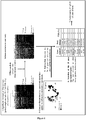
- New candidate gene selection of the implantation window To identify new markers of endometrium receptivity, we have first intersected our gene list significantly modulated between the LH+2 and LH+7 sample groups with those from four other transcriptomic studies which compared the same natural endometrium cycle phases (Carson et al., 2002; Riesewijk et al., 2003; Mirkin et al., 2005; Talbi et al., 2006) to identify a list of genes comprising 797 genes specifically modulated during the implantation window and exclusive to the present study. We performed a hierarchical clustering with the same data (797 genes). This list of genes allowed the separation of the two endometrium sample groups. Interestingly, the majority of these genes were up-regulated during the implantation window (746 up-regulated genes, 51 down-regulated genes).
- MFAP5 microfibrillar associated protein 5
- ANGPTL1 angiopoietin-like 1
- EG-VEGF also called PROK1
- prokineticin 1 endocrine-gland-derived vascular endothelial growth factor or prokineticin 1
- NLF2 nuclear localized factor 2
- This gene is LAMB3 and is over-expressed by a factor 20.4 in our study, and a factor 15 and 6.6 in the Riesewijk and Tablbi's studies respectively.
- the six unpublished genes were BCL2L10 (BCL2-like 10), CD68, TRPC4 (transient receptor potential cation channel, subfamily C, member 4), SORCS1 (sortilin-related VPS10 domain containing receptor 1), FST (follistatin), and KRT80 (keratin 80) which were also over-expressed during the implantation window by a factor 4.7, 5.3, 7.7, 9, 9 and 14.6 respectively.
- MFAP5 also called MAGP2
- MAGP2 extracellular matrix protein
- This gene encodes a microfibril-associated glycoprotein which is a component of microfibrils, an important structural component of elastic tissues such as vasculature.
- extracellular matrix such as collagen
- cell-associated proteins such as integrins
- this protein is therefore positioned to potentially modulate cell matrix interactions and to participate in cell signaling pathways.
- MFAP5 has a role in Notch signaling activation, a pathway involved in vasculature during embryogenesis, development and normal homeostasis.
- EG-VEGF vascular endothelial growth factor family
- PROK1 vascular endothelial growth factor family
- EG-VEGF is a newly identified angiogenic and permeability enhancing factor predominantly expressed in steroidogenic tissues.
- EG-VEGF is also expressed in the normal peri-implantation endometrial samples from patients of reproductive ages, and rarely detected in the endometrial samples from the post-menopausal patients and patients with endometrial carcinoma.
- EG-VEGF is predominantly expressed in the glandular epithelial cells with a peak protein expression at the mid luteal phase of the menstrual cycle.
- ANGPTL1 angiopoietin-like 1
- ANGPTL1 has anti-apoptotic activities through the phosphatidylinositol 3-kinase/Akt pathway and regulates angiogenesis.
- ANGPTL1 mRNA is increased after estradiol treatment.
- NLF2 gene expression was strongly expressed in the LH+7 sample group, suggesting that it has a role in endometrium remodeling during the implantation window. Invasion into the endometrial stroma is facilitated by inflammation.
- NLF2 also called C2CD4B (C2 calcium-dependent domain containing 4B)
- C2CD4B C2 calcium-dependent domain containing 4B
- Follistatin is a single-chain gonadal glycoprotein that specifically inhibits follicle-stimulating hormone (FSH) release. Follistatin has been previously described to be present in the endometrium and was localized in stromal and epithelial cells. Secretion of FSH from epithelial cells might be important for restricting the bioavailability of activin within the uterine lumen. Follistatin might also have activin independent effects and can also bind other members of the TGF-beta superfamily, including inhibin and certain members of the BMP (bone morphogenetic protein) family.
- BMP bone morphogenetic protein
- the expression patterns of the gene encoding FST is consistent with a role in decidualization, a key event for blastocyst implantation and successful pregnancy outcome after IVF.
- Keratins are intermediate filament proteins responsible for the cellular architecture of epithelial cells, which is necessary to achieve specific function. These proteins are required on a cellular level for phagocytosis, pinocytosis, cell adhesion, cell motility, subcellular organization and cell division. On a tissue level, structural proteins are necessary for contraction and intact epithelium. Structural proteins may also play a role in intracellular trafficking through the microtubule network.
- Apoptosis plays a critical role in maintaining cellular homeostasis during the menstrual by eliminating senescent cells from the functional layer of the uterine endometrium during the late secretory and menstrual phase of the cycle. Apoptosis was detected in the glandular epithelium of late secretory and menstruating endometrium, while very little apoptosis was detected during the proliferative phase or at the beginning of the secretory phase.
- Members of the Bcl2 family of proteins are fundamental elements in the pathways that control apoptosis and act as pro- and anti-apoptotic regulators.
- BCL2L10 which has been previously shown to suppress cell apoptosis, was found, in the present study, to be over-expressed in the human receptive endometrium in comparison with the pre- receptive endometrium. This finding was coherent with the period of apoptosis detection through the menstrual cycle.
- TRPC4 is one members of the transient receptor potential cation channel family which may facilitate store operated calcium entry (SOCE) to calcium signaling in the human myometrium.
- TRPC4 mRNA and protein have been previously described to be over-expressed in term pregnant human myometrium. However, we reported for the first time an over-expression of this gene in the receptive endometrium in comparison with the pre-receptive endometrium, suggesting a potential role of TRPC4 in implantation process.
- SORCS1 which is over-expressed in this study during the human implantation window, is the first identified member of a subgroup of the mammalian Vps10p-domain receptor family that comprises an N-terminal Vps10p-D (named after the yeast vacuolar protein sorting 10 protein), a leucine-rich domain, a single transmembrane domain, and a short cytoplasmic domain. Functions of this gene were not elucidated because it has been reported that sorCS1 was synthesized as a proprotein that is cleaved to mature forms in the trans-Golgi network and expressed in three isoforms with different cytoplasmic domains capable of mediating different trafficking of the receptor.
- EXAMPLE 2 Exemplary profile of patient with endometrial receptivity assessment in natural cycle.
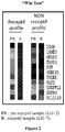
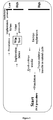
- assessing the endometrium receptivity of a patient comprise a first step consisting of measuring the mRNA expression of the 11 bio-markers (Win Test: window implantation test) during the implantation window (LH+7) under natural cycle ( Figure 2 ) and a second step comprising two scenarios : (i) the patient presents a delay of her implantation window and, in this case, Win Test could help in the detection of the implantation window; (ii) Win test could allow to identify patients never presenting a receptive endometrium and, in this case, they are oriented towards an adoption procedure ( Figure 3 : Consequences for the patient care management during IVF procedure).
- EXAMPLE 3 Exemplary profile of patient with endometrial receptivity assessment in stimulated cycle.
- the comparisons of gene expression from the same patients between natural and stimulated cycles revealed endometrial profiles associated either with a moderately altered receptivity in most cases (86%) or a strongly altered receptivity during the COH protocol in a few cases (14%) ( Figure 4 ).
- the invention provide two consequence of the Win Test: i) Fresh embryo replacement could be reconsidered during IVF procedure.
- Embryos freezing enable the IVF attempt to be saved and the embryo transfer can be done later during a natural cycle; ii) For patients with multiple implantation failures, analysis of the endometrial profile (Win test) could reveal a strongly altered profile during COH protocols, prompting the clinician to either adapt the IVF stimulation protocol or to perform embryo transfer later during a natural cycle or to orient the patient in a adoption procedure ( Figure 5 ).
- EXAMPLE 4 Win Test as means to set up “à la carte” “the adequate” treatments.
- EXAMPLE 5 Win Test to check the receptive status of infertile women with gynecological diseases.
- Win Test could be used as markers to evaluate endometrial receptivity in women with gynecological diseases.
- the potential relationship between several gynecological diseases for example, endometriosis and adenomyosis
- abnormal endometrial receptivity as a possible cause of sub-fertility in these patients can be tested with Win Test ( Figure 7 ).
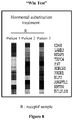
- EXAMPLE 6 Win Test to check the impact of hormonal substitution treatment during endometrial preparation in recipient patients for oocyte donation.
- Win test can be used to ameliorate hormonal treatments for endometrial maturation.
- Win Test reveals an altered endometrial profile at the time of the implantation window in oocyte recipient patients under hormonal substitution treatment (estrogen and progesterone treatments) compared with natural cycle ( Figure 8 ).
- "Win Test” (comprising a set of 11 genes issued from both their exclusive gene list and their list of predictor) is a diagnostic tool particularly relevant as several genes issued from their exclusive list have been found by other studies at proteomic levels (Table B).
Landscapes
- Chemical & Material Sciences (AREA)
- Health & Medical Sciences (AREA)
- Life Sciences & Earth Sciences (AREA)
- Organic Chemistry (AREA)
- Molecular Biology (AREA)
- Proteomics, Peptides & Aminoacids (AREA)
- Engineering & Computer Science (AREA)
- Genetics & Genomics (AREA)
- General Health & Medical Sciences (AREA)
- Wood Science & Technology (AREA)
- Zoology (AREA)
- Biochemistry (AREA)
- Analytical Chemistry (AREA)
- Microbiology (AREA)
- Physics & Mathematics (AREA)
- Biophysics (AREA)
- Biotechnology (AREA)
- Immunology (AREA)
- Bioinformatics & Cheminformatics (AREA)
- General Engineering & Computer Science (AREA)
- Surgery (AREA)
- Chemical Kinetics & Catalysis (AREA)
- Medicinal Chemistry (AREA)
- General Chemical & Material Sciences (AREA)
- Pathology (AREA)
- Biomedical Technology (AREA)
- Reproductive Health (AREA)
- Animal Behavior & Ethology (AREA)
- Medical Informatics (AREA)
- Heart & Thoracic Surgery (AREA)
- Veterinary Medicine (AREA)
- Nuclear Medicine, Radiotherapy & Molecular Imaging (AREA)
- Public Health (AREA)
- Pregnancy & Childbirth (AREA)
- Gynecology & Obstetrics (AREA)
- Transplantation (AREA)
- Measuring Or Testing Involving Enzymes Or Micro-Organisms (AREA)
- Micro-Organisms Or Cultivation Processes Thereof (AREA)
- Investigating Or Analysing Biological Materials (AREA)
- Medicines Containing Material From Animals Or Micro-Organisms (AREA)
Priority Applications (1)
| Application Number | Priority Date | Filing Date | Title |
|---|---|---|---|
| EP11725888.9A EP2576825B1 (en) | 2010-05-27 | 2011-05-27 | Methods for assessing endometrium receptivity of a patient |
Applications Claiming Priority (4)
| Application Number | Priority Date | Filing Date | Title |
|---|---|---|---|
| EP10305561 | 2010-05-27 | ||
| US35431010P | 2010-06-14 | 2010-06-14 | |
| PCT/EP2011/058757 WO2011147976A1 (en) | 2010-05-27 | 2011-05-27 | Methods for assessing endometrium receptivity of a patient |
| EP11725888.9A EP2576825B1 (en) | 2010-05-27 | 2011-05-27 | Methods for assessing endometrium receptivity of a patient |
Publications (2)
| Publication Number | Publication Date |
|---|---|
| EP2576825A1 EP2576825A1 (en) | 2013-04-10 |
| EP2576825B1 true EP2576825B1 (en) | 2017-11-08 |
Family
ID=42396421
Family Applications (1)
| Application Number | Title | Priority Date | Filing Date |
|---|---|---|---|
| EP11725888.9A Active EP2576825B1 (en) | 2010-05-27 | 2011-05-27 | Methods for assessing endometrium receptivity of a patient |
Country Status (6)
| Country | Link |
|---|---|
| US (1) | US9260748B2 (enExample) |
| EP (1) | EP2576825B1 (enExample) |
| JP (2) | JP6219164B2 (enExample) |
| ES (1) | ES2656962T3 (enExample) |
| PT (1) | PT2576825T (enExample) |
| WO (1) | WO2011147976A1 (enExample) |
Families Citing this family (13)
| Publication number | Priority date | Publication date | Assignee | Title |
|---|---|---|---|---|
| FR2987446B1 (fr) * | 2012-02-28 | 2016-01-01 | Univ Nice Sophia Antipolis | Test diagnostic de la resistance a l'azacitidine |
| EP2687851A1 (en) | 2012-07-20 | 2014-01-22 | Matricelab Innove | Method for increasing implantation success in assisted fertilization |
| US10143682B2 (en) * | 2014-10-28 | 2018-12-04 | Koushi Yamaguchi | Medicine for improving state of pregnancy, and use thereof |
| US11541040B2 (en) | 2014-10-28 | 2023-01-03 | Kouchi Yamaguchi | Medicine for improving state of pregnancy, and use thereof |
| CN114927178A (zh) * | 2015-06-12 | 2022-08-19 | 格尼亚Ip控股私人有限公司 | 患者和生物样本识别和追踪的方法和系统 |
| CN105784983B (zh) * | 2016-01-13 | 2017-10-31 | 深圳中山生殖与遗传研究所 | 一种评估子宫内膜容受性的试剂盒及其使用方法 |
| US10918327B2 (en) | 2017-02-02 | 2021-02-16 | Coopersurgical, Inc. | Compositions and methods for determining receptivity of an endometrium for embryonic implantation |
| IT201700045856A1 (it) * | 2017-04-27 | 2018-10-27 | Molipharma Srl | Kit di determinazione della recettivita’ endometriale |
| EP3569718A1 (en) * | 2018-05-16 | 2019-11-20 | Integrated Genetic Lab Services SLU | Kit and method for determining the receptivity status of an endometrium |
| WO2021160597A1 (en) * | 2020-02-10 | 2021-08-19 | ObsEva S.A. | Biomarkers for oxytocin receptor antagonist therapy |
| CN111778326B (zh) * | 2020-07-14 | 2021-10-22 | 和卓生物科技(上海)有限公司 | 用于子宫内膜容受性评估的基因标志物组合及其应用 |
| JP2023136665A (ja) * | 2022-03-17 | 2023-09-29 | 国立大学法人 東京大学 | 着床能の検出方法および着床障害の予測方法 |
| EP4311862A1 (en) | 2022-07-29 | 2024-01-31 | Ivi Rma Global, Sl. | Methods for detection of embryo implantation failure of endometrial origen |
Family Cites Families (3)
| Publication number | Priority date | Publication date | Assignee | Title |
|---|---|---|---|---|
| US6196965B1 (en) * | 1998-05-21 | 2001-03-06 | Cryofacets, Inc. | Compositions methods and devices for embryo implantation for in vitro fertilization |
| US7354742B2 (en) * | 2002-02-22 | 2008-04-08 | Ortho-Mcneil Pharmaceutical, Inc. | Method for generating amplified RNA |
| FR2876701B1 (fr) | 2004-10-15 | 2010-09-17 | Genevrier Lab | Procede de preparation d'un systeme de culture d'endometre autologue pour la co-culture endometre-embryon |
-
2011
- 2011-05-27 WO PCT/EP2011/058757 patent/WO2011147976A1/en not_active Ceased
- 2011-05-27 ES ES11725888.9T patent/ES2656962T3/es active Active
- 2011-05-27 US US13/699,814 patent/US9260748B2/en active Active
- 2011-05-27 EP EP11725888.9A patent/EP2576825B1/en active Active
- 2011-05-27 JP JP2013511700A patent/JP6219164B2/ja active Active
- 2011-05-27 PT PT117258889T patent/PT2576825T/pt unknown
-
2016
- 2016-04-22 JP JP2016086087A patent/JP2016171802A/ja active Pending
Non-Patent Citations (1)
| Title |
|---|
| "Affymetrix GeneChip Human Genome U133 Array Set HG-U133A", GEO,, 11 March 2002 (2002-03-11), XP002254749 * |
Also Published As
| Publication number | Publication date |
|---|---|
| PT2576825T (pt) | 2018-02-02 |
| JP2016171802A (ja) | 2016-09-29 |
| US20130072748A1 (en) | 2013-03-21 |
| ES2656962T3 (es) | 2018-03-01 |
| JP2013528052A (ja) | 2013-07-08 |
| JP6219164B2 (ja) | 2017-10-25 |
| WO2011147976A1 (en) | 2011-12-01 |
| EP2576825A1 (en) | 2013-04-10 |
| US9260748B2 (en) | 2016-02-16 |
Similar Documents
| Publication | Publication Date | Title |
|---|---|---|
| EP2576825B1 (en) | Methods for assessing endometrium receptivity of a patient | |
| EP2419526B1 (en) | Methods for selecting oocytes and competent embryos with high potential for pregnancy outcome | |
| US20100021898A1 (en) | Mammalian oocyte development competency granulosa markers and uses thereof | |
| JP5916241B2 (ja) | 妊娠成績についての高い能力を有するコンピテントな卵母細胞およびコンピテントな胚を選択するための方法 | |
| US7842464B2 (en) | Use of ADAM 12 for diagnosis and therapy of preeclampsia | |
| EP2768976B1 (en) | Methods for assessing endometrial receptivity of a patient after controlled ovarian hyperstimulation | |
| EP4332242A1 (en) | Method for predicting prognosis of gastric cancer | |
| CN108624678A (zh) | 一种用于子痫前期诊治的生物标志物 | |
| Class et al. | Patent application title: METHODS FOR ASSESSING ENDOMETRIUM RECEPTIVITY OF A PATIENT Inventors: Samir Hamamah (Montpellier, FR) Delphine Haouzi (Montpellier, FR) | |
| CN114686582A (zh) | Gdf15和itih3在孕早期自然流产预测中的应用 |
Legal Events
| Date | Code | Title | Description |
|---|---|---|---|
| PUAI | Public reference made under article 153(3) epc to a published international application that has entered the european phase |
Free format text: ORIGINAL CODE: 0009012 |
|
| 17P | Request for examination filed |
Effective date: 20121213 |
|
| AK | Designated contracting states |
Kind code of ref document: A1 Designated state(s): AL AT BE BG CH CY CZ DE DK EE ES FI FR GB GR HR HU IE IS IT LI LT LU LV MC MK MT NL NO PL PT RO RS SE SI SK SM TR |
|
| DAX | Request for extension of the european patent (deleted) | ||
| 17Q | First examination report despatched |
Effective date: 20130925 |
|
| RAP1 | Party data changed (applicant data changed or rights of an application transferred) |
Owner name: CENTRE HOSPITALIER UNIVERSITAIRE DE MONTPELLIER Owner name: INSERM - INSTITUT NATIONAL DE LA SANTE ET DE LA RE Owner name: UNIVERSITE MONTPELLIER 1 |
|
| STAA | Information on the status of an ep patent application or granted ep patent |
Free format text: STATUS: EXAMINATION IS IN PROGRESS |
|
| GRAP | Despatch of communication of intention to grant a patent |
Free format text: ORIGINAL CODE: EPIDOSNIGR1 |
|
| STAA | Information on the status of an ep patent application or granted ep patent |
Free format text: STATUS: GRANT OF PATENT IS INTENDED |
|
| INTG | Intention to grant announced |
Effective date: 20170508 |
|
| GRAS | Grant fee paid |
Free format text: ORIGINAL CODE: EPIDOSNIGR3 |
|
| GRAA | (expected) grant |
Free format text: ORIGINAL CODE: 0009210 |
|
| STAA | Information on the status of an ep patent application or granted ep patent |
Free format text: STATUS: THE PATENT HAS BEEN GRANTED |
|
| AK | Designated contracting states |
Kind code of ref document: B1 Designated state(s): AL AT BE BG CH CY CZ DE DK EE ES FI FR GB GR HR HU IE IS IT LI LT LU LV MC MK MT NL NO PL PT RO RS SE SI SK SM TR |
|
| RAP1 | Party data changed (applicant data changed or rights of an application transferred) |
Owner name: UNIVERSITE MONTPELLIER Owner name: CENTRE HOSPITALIER UNIVERSITAIRE DE MONTPELLIER Owner name: INSERM - INSTITUT NATIONAL DE LA SANTE ET DE LA RE |
|
| REG | Reference to a national code |
Ref country code: GB Ref legal event code: FG4D |
|
| REG | Reference to a national code |
Ref country code: CH Ref legal event code: EP Ref country code: AT Ref legal event code: REF Ref document number: 944201 Country of ref document: AT Kind code of ref document: T Effective date: 20171115 |
|
| REG | Reference to a national code |
Ref country code: IE Ref legal event code: FG4D |
|
| REG | Reference to a national code |
Ref country code: DE Ref legal event code: R096 Ref document number: 602011043137 Country of ref document: DE |
|
| REG | Reference to a national code |
Ref country code: PT Ref legal event code: SC4A Ref document number: 2576825 Country of ref document: PT Date of ref document: 20180202 Kind code of ref document: T Free format text: AVAILABILITY OF NATIONAL TRANSLATION Effective date: 20180125 |
|
| REG | Reference to a national code |
Ref country code: NL Ref legal event code: FP |
|
| REG | Reference to a national code |
Ref country code: ES Ref legal event code: FG2A Ref document number: 2656962 Country of ref document: ES Kind code of ref document: T3 Effective date: 20180301 |
|
| REG | Reference to a national code |
Ref country code: LT Ref legal event code: MG4D |
|
| REG | Reference to a national code |
Ref country code: AT Ref legal event code: MK05 Ref document number: 944201 Country of ref document: AT Kind code of ref document: T Effective date: 20171108 |
|
| REG | Reference to a national code |
Ref country code: FR Ref legal event code: PLFP Year of fee payment: 8 |
|
| PG25 | Lapsed in a contracting state [announced via postgrant information from national office to epo] |
Ref country code: LT Free format text: LAPSE BECAUSE OF FAILURE TO SUBMIT A TRANSLATION OF THE DESCRIPTION OR TO PAY THE FEE WITHIN THE PRESCRIBED TIME-LIMIT Effective date: 20171108 Ref country code: FI Free format text: LAPSE BECAUSE OF FAILURE TO SUBMIT A TRANSLATION OF THE DESCRIPTION OR TO PAY THE FEE WITHIN THE PRESCRIBED TIME-LIMIT Effective date: 20171108 Ref country code: NO Free format text: LAPSE BECAUSE OF FAILURE TO SUBMIT A TRANSLATION OF THE DESCRIPTION OR TO PAY THE FEE WITHIN THE PRESCRIBED TIME-LIMIT Effective date: 20180208 Ref country code: SE Free format text: LAPSE BECAUSE OF FAILURE TO SUBMIT A TRANSLATION OF THE DESCRIPTION OR TO PAY THE FEE WITHIN THE PRESCRIBED TIME-LIMIT Effective date: 20171108 |
|
| PG25 | Lapsed in a contracting state [announced via postgrant information from national office to epo] |
Ref country code: BG Free format text: LAPSE BECAUSE OF FAILURE TO SUBMIT A TRANSLATION OF THE DESCRIPTION OR TO PAY THE FEE WITHIN THE PRESCRIBED TIME-LIMIT Effective date: 20180208 Ref country code: IS Free format text: LAPSE BECAUSE OF FAILURE TO SUBMIT A TRANSLATION OF THE DESCRIPTION OR TO PAY THE FEE WITHIN THE PRESCRIBED TIME-LIMIT Effective date: 20180308 Ref country code: HR Free format text: LAPSE BECAUSE OF FAILURE TO SUBMIT A TRANSLATION OF THE DESCRIPTION OR TO PAY THE FEE WITHIN THE PRESCRIBED TIME-LIMIT Effective date: 20171108 Ref country code: RS Free format text: LAPSE BECAUSE OF FAILURE TO SUBMIT A TRANSLATION OF THE DESCRIPTION OR TO PAY THE FEE WITHIN THE PRESCRIBED TIME-LIMIT Effective date: 20171108 Ref country code: AT Free format text: LAPSE BECAUSE OF FAILURE TO SUBMIT A TRANSLATION OF THE DESCRIPTION OR TO PAY THE FEE WITHIN THE PRESCRIBED TIME-LIMIT Effective date: 20171108 Ref country code: LV Free format text: LAPSE BECAUSE OF FAILURE TO SUBMIT A TRANSLATION OF THE DESCRIPTION OR TO PAY THE FEE WITHIN THE PRESCRIBED TIME-LIMIT Effective date: 20171108 Ref country code: GR Free format text: LAPSE BECAUSE OF FAILURE TO SUBMIT A TRANSLATION OF THE DESCRIPTION OR TO PAY THE FEE WITHIN THE PRESCRIBED TIME-LIMIT Effective date: 20180209 |
|
| PG25 | Lapsed in a contracting state [announced via postgrant information from national office to epo] |
Ref country code: DK Free format text: LAPSE BECAUSE OF FAILURE TO SUBMIT A TRANSLATION OF THE DESCRIPTION OR TO PAY THE FEE WITHIN THE PRESCRIBED TIME-LIMIT Effective date: 20171108 Ref country code: SK Free format text: LAPSE BECAUSE OF FAILURE TO SUBMIT A TRANSLATION OF THE DESCRIPTION OR TO PAY THE FEE WITHIN THE PRESCRIBED TIME-LIMIT Effective date: 20171108 Ref country code: EE Free format text: LAPSE BECAUSE OF FAILURE TO SUBMIT A TRANSLATION OF THE DESCRIPTION OR TO PAY THE FEE WITHIN THE PRESCRIBED TIME-LIMIT Effective date: 20171108 Ref country code: CY Free format text: LAPSE BECAUSE OF FAILURE TO SUBMIT A TRANSLATION OF THE DESCRIPTION OR TO PAY THE FEE WITHIN THE PRESCRIBED TIME-LIMIT Effective date: 20171108 Ref country code: CZ Free format text: LAPSE BECAUSE OF FAILURE TO SUBMIT A TRANSLATION OF THE DESCRIPTION OR TO PAY THE FEE WITHIN THE PRESCRIBED TIME-LIMIT Effective date: 20171108 |
|
| REG | Reference to a national code |
Ref country code: DE Ref legal event code: R097 Ref document number: 602011043137 Country of ref document: DE |
|
| PG25 | Lapsed in a contracting state [announced via postgrant information from national office to epo] |
Ref country code: SM Free format text: LAPSE BECAUSE OF FAILURE TO SUBMIT A TRANSLATION OF THE DESCRIPTION OR TO PAY THE FEE WITHIN THE PRESCRIBED TIME-LIMIT Effective date: 20171108 Ref country code: PL Free format text: LAPSE BECAUSE OF FAILURE TO SUBMIT A TRANSLATION OF THE DESCRIPTION OR TO PAY THE FEE WITHIN THE PRESCRIBED TIME-LIMIT Effective date: 20171108 Ref country code: RO Free format text: LAPSE BECAUSE OF FAILURE TO SUBMIT A TRANSLATION OF THE DESCRIPTION OR TO PAY THE FEE WITHIN THE PRESCRIBED TIME-LIMIT Effective date: 20171108 |
|
| PLBE | No opposition filed within time limit |
Free format text: ORIGINAL CODE: 0009261 |
|
| STAA | Information on the status of an ep patent application or granted ep patent |
Free format text: STATUS: NO OPPOSITION FILED WITHIN TIME LIMIT |
|
| 26N | No opposition filed |
Effective date: 20180809 |
|
| PG25 | Lapsed in a contracting state [announced via postgrant information from national office to epo] |
Ref country code: SI Free format text: LAPSE BECAUSE OF FAILURE TO SUBMIT A TRANSLATION OF THE DESCRIPTION OR TO PAY THE FEE WITHIN THE PRESCRIBED TIME-LIMIT Effective date: 20171108 |
|
| PG25 | Lapsed in a contracting state [announced via postgrant information from national office to epo] |
Ref country code: MC Free format text: LAPSE BECAUSE OF FAILURE TO SUBMIT A TRANSLATION OF THE DESCRIPTION OR TO PAY THE FEE WITHIN THE PRESCRIBED TIME-LIMIT Effective date: 20171108 |
|
| REG | Reference to a national code |
Ref country code: IE Ref legal event code: MM4A |
|
| PG25 | Lapsed in a contracting state [announced via postgrant information from national office to epo] |
Ref country code: IE Free format text: LAPSE BECAUSE OF NON-PAYMENT OF DUE FEES Effective date: 20180527 |
|
| PG25 | Lapsed in a contracting state [announced via postgrant information from national office to epo] |
Ref country code: MT Free format text: LAPSE BECAUSE OF NON-PAYMENT OF DUE FEES Effective date: 20180527 |
|
| PG25 | Lapsed in a contracting state [announced via postgrant information from national office to epo] |
Ref country code: HU Free format text: LAPSE BECAUSE OF FAILURE TO SUBMIT A TRANSLATION OF THE DESCRIPTION OR TO PAY THE FEE WITHIN THE PRESCRIBED TIME-LIMIT; INVALID AB INITIO Effective date: 20110527 |
|
| PG25 | Lapsed in a contracting state [announced via postgrant information from national office to epo] |
Ref country code: MK Free format text: LAPSE BECAUSE OF NON-PAYMENT OF DUE FEES Effective date: 20171108 |
|
| PG25 | Lapsed in a contracting state [announced via postgrant information from national office to epo] |
Ref country code: AL Free format text: LAPSE BECAUSE OF FAILURE TO SUBMIT A TRANSLATION OF THE DESCRIPTION OR TO PAY THE FEE WITHIN THE PRESCRIBED TIME-LIMIT Effective date: 20171108 |
|
| PGFP | Annual fee paid to national office [announced via postgrant information from national office to epo] |
Ref country code: LU Payment date: 20240418 Year of fee payment: 14 |
|
| PGFP | Annual fee paid to national office [announced via postgrant information from national office to epo] |
Ref country code: NL Payment date: 20240418 Year of fee payment: 14 |
|
| PGFP | Annual fee paid to national office [announced via postgrant information from national office to epo] |
Ref country code: CH Payment date: 20240602 Year of fee payment: 14 |
|
| PGFP | Annual fee paid to national office [announced via postgrant information from national office to epo] |
Ref country code: PT Payment date: 20240423 Year of fee payment: 14 |
|
| PGFP | Annual fee paid to national office [announced via postgrant information from national office to epo] |
Ref country code: TR Payment date: 20240430 Year of fee payment: 14 Ref country code: BE Payment date: 20240418 Year of fee payment: 14 |
|
| PGFP | Annual fee paid to national office [announced via postgrant information from national office to epo] |
Ref country code: DE Payment date: 20250423 Year of fee payment: 15 |
|
| PGFP | Annual fee paid to national office [announced via postgrant information from national office to epo] |
Ref country code: GB Payment date: 20250423 Year of fee payment: 15 Ref country code: ES Payment date: 20250602 Year of fee payment: 15 |
|
| PGFP | Annual fee paid to national office [announced via postgrant information from national office to epo] |
Ref country code: IT Payment date: 20250423 Year of fee payment: 15 |
|
| PGFP | Annual fee paid to national office [announced via postgrant information from national office to epo] |
Ref country code: FR Payment date: 20250423 Year of fee payment: 15 |
|
| PG25 | Lapsed in a contracting state [announced via postgrant information from national office to epo] |
Ref country code: PT Free format text: LAPSE BECAUSE OF NON-PAYMENT OF DUE FEES Effective date: 20251127 |
|
| REG | Reference to a national code |
Ref country code: CH Ref legal event code: H13 Free format text: ST27 STATUS EVENT CODE: U-0-0-H10-H13 (AS PROVIDED BY THE NATIONAL OFFICE) Effective date: 20251223 |
|
| REG | Reference to a national code |
Ref country code: NL Ref legal event code: MM Effective date: 20250601 |
|
| PG25 | Lapsed in a contracting state [announced via postgrant information from national office to epo] |
Ref country code: LU Free format text: LAPSE BECAUSE OF NON-PAYMENT OF DUE FEES Effective date: 20250527 |
|
| PG25 | Lapsed in a contracting state [announced via postgrant information from national office to epo] |
Ref country code: CH Free format text: LAPSE BECAUSE OF NON-PAYMENT OF DUE FEES Effective date: 20250531 |
|
| REG | Reference to a national code |
Ref country code: BE Ref legal event code: MM Effective date: 20250531 |
|
| PG25 | Lapsed in a contracting state [announced via postgrant information from national office to epo] |
Ref country code: NL Free format text: LAPSE BECAUSE OF NON-PAYMENT OF DUE FEES Effective date: 20250601 |
|
| PG25 | Lapsed in a contracting state [announced via postgrant information from national office to epo] |
Ref country code: BE Free format text: LAPSE BECAUSE OF NON-PAYMENT OF DUE FEES Effective date: 20250531 |