EP2534483B1 - Testanordnung - Google Patents
Testanordnung Download PDFInfo
- Publication number
- EP2534483B1 EP2534483B1 EP11706460.0A EP11706460A EP2534483B1 EP 2534483 B1 EP2534483 B1 EP 2534483B1 EP 11706460 A EP11706460 A EP 11706460A EP 2534483 B1 EP2534483 B1 EP 2534483B1
- Authority
- EP
- European Patent Office
- Prior art keywords
- enzyme
- arrangement according
- laccase
- matrix
- siloxane
- Prior art date
- Legal status (The legal status is an assumption and is not a legal conclusion. Google has not performed a legal analysis and makes no representation as to the accuracy of the status listed.)
- Not-in-force
Links
Images


Classifications
-
- C—CHEMISTRY; METALLURGY
- C12—BIOCHEMISTRY; BEER; SPIRITS; WINE; VINEGAR; MICROBIOLOGY; ENZYMOLOGY; MUTATION OR GENETIC ENGINEERING
- C12Q—MEASURING OR TESTING PROCESSES INVOLVING ENZYMES, NUCLEIC ACIDS OR MICROORGANISMS; COMPOSITIONS OR TEST PAPERS THEREFOR; PROCESSES OF PREPARING SUCH COMPOSITIONS; CONDITION-RESPONSIVE CONTROL IN MICROBIOLOGICAL OR ENZYMOLOGICAL PROCESSES
- C12Q1/00—Measuring or testing processes involving enzymes, nucleic acids or microorganisms; Compositions therefor; Processes of preparing such compositions
- C12Q1/001—Enzyme electrodes
-
- C—CHEMISTRY; METALLURGY
- C12—BIOCHEMISTRY; BEER; SPIRITS; WINE; VINEGAR; MICROBIOLOGY; ENZYMOLOGY; MUTATION OR GENETIC ENGINEERING
- C12Q—MEASURING OR TESTING PROCESSES INVOLVING ENZYMES, NUCLEIC ACIDS OR MICROORGANISMS; COMPOSITIONS OR TEST PAPERS THEREFOR; PROCESSES OF PREPARING SUCH COMPOSITIONS; CONDITION-RESPONSIVE CONTROL IN MICROBIOLOGICAL OR ENZYMOLOGICAL PROCESSES
- C12Q1/00—Measuring or testing processes involving enzymes, nucleic acids or microorganisms; Compositions therefor; Processes of preparing such compositions
- C12Q1/02—Measuring or testing processes involving enzymes, nucleic acids or microorganisms; Compositions therefor; Processes of preparing such compositions involving viable microorganisms
- C12Q1/04—Determining presence or kind of microorganism; Use of selective media for testing antibiotics or bacteriocides; Compositions containing a chemical indicator therefor
Definitions
- microorganisms in general and the identification of the detected microorganisms in a sample is of considerable importance in many areas.
- microorganisms may be responsible for the spoilage or short shelf life of food.
- microorganisms also play an important role in many diseases (e.g., inflammation) of humans and animals. It is therefore of great importance to detect the presence of microorganisms in time to take appropriate action.
- the state of the art describes various systems and methods which are suitable for determining microorganisms in a sample. Some of these systems can also be used to detect specific substances (e.g., proteins such as enzymes) in a sample.
- specific substances e.g., proteins such as enzymes
- WO 2006/065350 A2 the detection of microorganisms by means of a dye, which shows a visible color change in the presence of one or more microorganisms. This color change is based on differences in the polarity of the solvent or acid-base reactions, redox reactions, which are caused by the interaction between microorganism and sensor.
- WO 98/010556 A1 a method is disclosed by means of which molecules can be bound to binding sites within a gel droplet.
- molecules which are secreted by cells located within the gel droplet are bound to the free binding sites of the gel droplets.
- WO 03/033691 A1 A microchip is disclosed that can be used to test the antibacterial activity of antibiotics.
- prokaryotic and eukaryotic cells are immobilized in a gel by polymerizing it.
- a matrix consisting of a protein "backbone" cross-linked with polyethylene glycol. This matrix is particularly suitable for tissue regeneration.
- the matrix may additionally comprise a biologically active agent which is released by the degradation of the matrix at the site of action to the surrounding tissue. To test the extent to which this matrix releases the biological active substance contained therein, colored fibrinogen fragments were incorporated into the matrix.
- the US 2008/0057534 A1 describes the visual detection of a Microbial contamination through the degradation of a topcoat consisting of microbe-sensitive dyes. These microbe-sensitive dyes change color in the presence of microbes in the sample, either causing loss of color or color change.
- the dyes can be applied to any surface. A certain, albeit very limited, selectivity is made possible by the nature of the dyes.
- Such systems can, for example, in a system such as in US 2004/0018641 A1 described, are used.
- the appearance or disappearance of computer-readable barcodes can indicate contamination in the food sector.
- the indication is based on the detection of gases, temperature and pH differences, as well as toxins or other metabolites from bacteria.
- the US 6,297,059 B1 describes a method for measuring microbial contamination by fluorescence built on a liquid bilayer membrane. By addressing polyvalent or polyvalent target biomolecules, two or more fluorescence quenchers or a transmission of the extinction energy to a fluorescence acceptor are triggered per molecule and thus signal amplification is achieved.
- the present invention relates to an assembly comprising a solid support and a matrix disposed on the solid support comprising at least one enzymatically convertible or modifiable molecule, preferably polymer or oligomer, which matrix is at least one enzyme releasable by reacting or modifying the molecule, preferably polymer or oligomer which is capable of converting at least one color-producing substrate located in the matrix and / or on the solid support.
- the arrangement according to the invention comprises, in addition to a solid support and an optional semipermeable membrane opposite thereto, a matrix which is arranged on the solid support or optionally between the solid support and the semipermeable membrane.
- the matrix comprises at least one molecule, preferably polymer, which is degraded, reacted or modified by substances present in the sample (eg enzymes) can be.
- the degradation, the conversion or the modification of such molecules or polymers in the matrix change their properties.
- the reaction or modification of the molecule or polymer or oligomer can lead to their structural properties changing, which can lead to a reduction in the degree of crosslinking or viscosity, to change the pores in the polymer or to degradation of the polymer chains.
- the releasable enzyme in the matrix becomes more mobile and can move more freely within the matrix, especially in the space between the solid support and the semipermeable membrane.
- this makes it possible to react or liberate a color-producing substrate located in the matrix and / or on the solid support and / or to degrade the molecule or polymer present in the matrix.
- Such a system is suitable, for example, for even the smallest amounts of microorganisms, such as bacteria or Mushrooms to prove in a sample.
- An enzyme secreted by a contaminating bacterium or fungus penetrates the protective, semi-permeable membrane, degrades a matrix applied to a support material (eg, a polysaccharide, an enzyme, or combinations thereof, with or without cross-linking), thus initiating the release of the enzyme. which accelerates the release as an enzymatic amplification system and / or enables the detection of a microbial contamination.
- Bioresponsive behavior is imparted to components by coating with biomaterials that can be degraded by extracellular enzymes.
- Extracellular microbial enzymes can act as so-called triggers and thus initiate the release of active enzymes included and / or bound in the polymer.
- the type of polymer (polypeptide, polysaccharide, synthetic polymer ...) defines the type of trigger enzymes required.
- Microorganisms produce a variety of extracellular enzymes that they release to the environment, among many other functions, e.g. To digest food components, in order to be able to take these into the cell. Extracellular enzymes thus serve, inter alia, the degradation of large molecules and polymers. Hydrolysis of a variety of biopolymers is catalyzed by hydrolases which, depending on the substrate, are converted into lipases, proteases, esterases, glycosidases, and the like. can be divided.
- polypeptide as used herein includes polypeptides with a minimum size of 50 amino acids and thus also includes proteins.
- a "polymer-releasable enzyme” (polypeptide) is an enzyme which is incorporated into the polymer of the matrix and which can not diffuse due to cross-linking of the polymer or which is bound to the polymer by covalent and / or non-covalent bonds. In both cases, the enzymes are immobilized in the polymer prior to degradation of the polymer or only limited mobility. As a result of the degradation, modification or conversion of the polymer, its long-chain or crosslinked chains are shortened or split so that the enzymes in the matrix are released, i. be more mobile, and be able to diffuse around in it.
- “Semipermeable membrane” as used herein refers to membranes that allow access of the triggers or enzymes from the outside into the arrangement of the invention, but prevent the diffusion of the enzymes and other matrix constituents present in the matrix outwards.
- the enzyme present in the polymer and the color-producing substrate is chosen so that when contacting both substances, the substrate is reacted and it comes to color formation.
- in situ dye synthesis or dye staining may also take place by the enzymatic reaction of one or more substrates, e.g. Polymerization or depolymerization of a phenol by oxidoreductase.
- the indicator system may also be that the enhancer enzyme is capable of hydrolytically degrading a masking polymer layer to reveal the hidden color layer.
- polymer Degrading trigger enzyme Included polypeptide (enhancer enzyme) or bound dye Color reaction by turnover of pectin pectinase PEG protease Siloxane -N succinyl Ala-Ala-Pro-Leu-p-nitroanilide pectin pectinase protease Siloxane -N succinyl Ala-Ala-Pro-Leu-p-nitroanilide pectin pectinase PVA protease Siloxane -N succinyl Ala-Ala-Pro-Leu-p-nitroanilide pectin pectinase laccase Siloxane ferulic acid pectin pectinase laccase Siloxane caffeic acid pectin pectinase laccase Siloxane 3,4, dihydroxy
- Oxidoreductases such as laccases or peroxidases, proteases, lipases, esterases, peptidases, etc. can be used as amplifier enzymes for the amplification of the display.
- the matrix is arranged as a layer, in the form of capsules or as a hydrogel, on the solid support.
- the matrix between the solid support and the semipermeable membrane may have different shapes.
- the matrix can be applied, for example, as a layer or capsules.
- the use of capsules or multiple layers (at least two, three or four) has the advantage that the capsules and layers can have different compositions.
- other capsules or layers may comprise enzymes which are capable of converting color-producing substrates.
- the polymer composition of the various capsules or layers can also vary.
- the matrix may comprise a degradable, modifiable molecule or polymer which can be degraded or modified by enzymes.
- a degradable, modifiable molecule or polymer which can be degraded or modified by enzymes.
- molecules or polymers which are degradable in the course of a pH change or by changing the ionic strength.
- the enzymatically decomposable, metabolizable or modifiable polymer is preferably a polysaccharide, polypeptide, polyester, polyamide or a combination thereof.
- the degradable polymer may, for example, be esterified to control the degradation or modification by enzymes present in the sample.
- the polysaccharide is selected from the group consisting of pectin, amylose, amylopectin, agarose, alginate, carrageenan, chitin, chitosan, dextran, glycogen, guar, locust bean gum, levan, pectin, pollulan, tamarind kernel flour, xanthan gum, Cellulose and xylan.
- the polymers can be provided with specific sequences (e.g., amino acid or oligosaccharide sequences) which are cleavage sites for particular enzymes. This makes it possible to produce polymers which are cleaved in contact with the substances to be detected (e.g., enzymes).
- specific sequences e.g., amino acid or oligosaccharide sequences
- the degradable polymer is selected from the group consisting of gum arabic, agar, agarose, maltodextrins, alginic acid and its salts, in particular sodium alginate or calcium alginate, liposomes, fats, cetyl alcohol, collagen, chitosan, peptidoglycan, Leithine, Gelatin, albumin, shellac, polysaccharides, especially starch or dextran, cyclodextrins, pectin, carrageenan and waxes.
- the polymer degrading compound is an enzyme.
- the enzyme which is included in the matrix is preferably selected from the group consisting of the groups of the hydrolases and oxidoreductases such as, for example, proteases, laccases or peroxidases.
- the color-producing substrate is selected from the group consisting of phenolic compounds and azo dyes such as ferulic acid, caffeic acid, 3,4-dihydroxybenzoic acid, reactive blue, indigo carmine, ABTS or guaiacol.
- a semi-permeable character is achieved by modification of the surface of a side made more hydrophilic or hydrophobic, for example. This can be achieved by, for example, chemical, physical (plasma) or enzymatic treatment (eg Guebitz, GM, Cavaco-Paulo, A., 2008. Trends in Biotechnology 26, 32-38 ).
- plasma chemical, physical
- enzymatic treatment eg Guebitz, GM, Cavaco-Paulo, A., 2008. Trends in Biotechnology 26, 32-38 ).
- the solid support comprises a material selected from the group consisting of inorganic materials, preferably silica gel, aluminum, silicon or glass, organic materials, preferably polyester, polystyrene, polyamide, polyacrylamide or polyvinyl alcohol, or biopolymers , preferably paper.
- inorganic materials preferably silica gel, aluminum, silicon or glass
- organic materials preferably polyester, polystyrene, polyamide, polyacrylamide or polyvinyl alcohol, or biopolymers , preferably paper.
- the carrier material can be chemically modified.
- functional groups are applied to the surface, which can then covalently cross-link with components of the matrix.
- trimethylsilyl methacrylate may be mentioned at this point.
- trimethylsilyl groups with hydroxy groups can form covalent ether bridges on surfaces as they occur in glass but also in other polymers.
- the methacrylic group can then crosslink with methacrylic groups of the matrix.
- microorganisms to be determined and / or characterized are selected from the group consisting of bacteria and mushrooms.
- the arrangement according to the invention can be used according to a further aspect for the detection of at least one enzyme in a sample.
- Yet another aspect of the present invention relates to the use of an inventive wound infection detection assembly by determining the presence of at least one sore-specific enzyme.
- "sore-specific" enzymes are enzymes that commonly occur in infections of wounds.
- Exemplary enzymes are those enzymes secreted by the microorganisms causing the infection or those secreted by the body in response to an infection.
- a 5% solution of pectin derived from citrus peel with a degree of esterification of 50-60% was prepared by dissolving in water overnight at 50 ° C.
- pectin from apple peel with a degree of esterification of 70-75% can be used.
- the polysaccharide was dyed with various dyes such as Alizarin, Cibacrom, Remazol, Victoria Blue or others.
- various dyes such as Alizarin, Cibacrom, Remazol, Victoria Blue or others.
- 10 g of pectin were suspended in acetone with 5 mM dye, heated to reflux overnight and then washed several times with acetone.
- the pectin solution was polymerized by dropping into a 200mM CaCl 2 solution and the resulting pectin beads were washed with water.
- silica gel 10g was stirred in 30mL of a 3-9% aminopropyltriethoxysilane in ethanol (95%) for 4 hours at 40 ° C. Subsequently, the aminated silica gel was decanted, washed 3 times with 70% ethanol and dried in a desiccator to constant weight.
- N-succinyl-Ala-Ala-Pro-Leu-p-nitroanilide, N-succinyl-Ala-Ala-Pro-Val-p-nitroanilide or L-leucine p-nitroanilide can be immobilized in the same manner.
- silica gel 10 g was stirred in 30 mL of a 3-9% mercaptopropyltriethoxysilane in ethanol (95%) for 4 hours at 40 ° C. The silica gel was then decanted off, washed 3 times with 2-propanol and dried in a desiccator to constant weight. To 5 g pretreated silica gel suspended in 20 mL dichloromethane, 155 mg ferulic acid and 38 mg dimethylaminopyridine are added. To the reaction mixture cooled with ice to 0 ° C was added 165 mg DCC and stirred for 3 hours. The temperature was left at room temperature (about 20 ° C). Subsequently, the solid, modified silica gel was filtered off, washed 3 times with dichloromethane and dried overnight in a desiccator.
- a mixture consisting of 4.5 g of pectin and 0.5 g of sodium alginate in 100 ml of water was used.
- the gel preparation was carried out by dropping the pectin alginate solution in a submitted 200mM CaCl 2 solution. The resulting pectin beads were washed with water.
- the polymer After mixing the polysaccharide solution with the enzyme, the polymer was dripped into 200mM CaCl 2 solution and gelled. The polymer beads thus obtained were screened off, washed 3 times with 50 mM Tris-HCl buffer pH 7.5 and each of 1 g wet mass in reaction vessels with 5mL 50mM Tris-HCl buffer pH 7.5 portioned.
- biopolymer B (Example 4) was loaded with a protease of Aspergillus oryzae.
- the biopolymer may be loaded with any other enzyme, such as laccases, from the group of oxidoreductases.
- laccases from the group of oxidoreductases.
- the activity of the liberated laccase is determined accordingly by means of ABTS ( Fig. 3 ).
- Example 2 10 mg silica gel with immobilized N-succinyl-Ala-Ala-Pro-Leu-p-nitroanilide or L-leucine p-nitroanilide or N-succinyl-Ala-Ala-Pro-Val-p-nitroanilide as protease substrates (Example 2) were suspended in 1300 ⁇ L 50mM Tris-HCl buffer pH8.3 and incubated with commercially available protease from Aspergillus oryzae or wound fluid at room temperature. The activity was determined by UV absorption of the cleaved p-nitroanilide in the supernatant at 375 nm or at 405 nm ( Fig. 4 ).
- silica gel with immobilized ferulic acid as Laccasesubstrat (Example 2) or with immobilized Fast Blue RR were suspended in 1300 ⁇ L 50mM succinate buffer pH4.5 and incubated with laccase from Trametes hirsuta or wound fluid at room temperature. The activity is determined by means of a color measurement (yellow-orange) by means of spectrophotometer.
- the polymer spheres were screened off, washed 3 times with 50 mM succinate buffer pH4.5 and portioned to 1 g wet mass in reaction vessels with 5 mM 50 mM succinate buffer pH4.5 in the presence of 10 mg silica gel with immobilized ferulic acid as Laccaseubstrat from Example 2.
- the enzymatic degradation was started by adding commercially available pectinase at room temperature and with shaking. After the incubation, the color change to yellow-orange was determined by means of a color measurement by means of a spectrophotometer ( Fig. 5 ).
- the biopolymer may be loaded with a protease of Aspergillus oryzae.
- the activity of the released protease is then determined by UV absorption of the cleaved p-nitroanilide in the supernatant at 375 nm.
- the response of the bioresponsive polymers can be adjusted via the diffusion behavior of both the trigger and the amplifier enzymes.
- chemically or genetically modifying e.g., increasing
- correspondingly lower levels of cross-linking of the biopolymer may be employed, while minimizing the outdiffusion of the enhancer enzyme.
- Example 6 the biopolymer was loaded with modified protease.
- the polymer spheres obtained in this way were screened off, washed 3 times with 50 mM succinate buffer pH4.5 and portioned with 1 g wet mass in reaction vessels with 5 mM 50 mM succinate buffer pH4.5 in the presence of 10 mg silica gel with immobilized ferulic acid as Laccase substrate from Example 2.
- Enzymatic degradation at room temperature and with shaking was started by adding commercial pectinase. After the incubation, the color change to yellow-orange was determined by means of a color measurement by means of a spectrophotometer ( Fig. 5 ).
- the molecular weight of the trigger enzyme can be increased by genetic engineering.
- a system that can be used in the medical field uses the enzyme lysozyme as a trigger enzyme.
- This enzyme of the body's native immune response is formed and secreted in the case of infection.
- the main task of the enzyme is a destruction of bacteria by degradation of peptidoglycan, a component of the bacterial cell wall. It was demonstrated that increased amounts of enzyme are present in the case of wound infection.
- 3.12 mg of micrococcus lysodeicticus cell wall from Sigma were suspended with 1 ml of 1% agarose in phosphate buffer pH 7.00. 100 ⁇ l of this suspension were mixed in a microtiter plate with 50 ⁇ l of a PEG-modified laccase.
- PES fabric is modified with trimethylsilyl methacrylate and overcoated with a modified with glycidyl methacrylate matrix and polymerized free-radically and covalently cross-linked.
- Extracellular enzymes such as pectinases or cellulases, can now overcome the PES tissue by diffusion and functional molecules, e.g. Release enzymes, which in turn trigger a color reaction.
Description
- Die vorliegende Erfindung betrifft eine Anordnung, welche geeignet ist, u.a. Mikroorganismen in einer Probe zu detektieren.
- Der Nachweis von Mikroorganismen im Allgemeinen und die Identifizierung der nachgewiesenen Mikroorganismen in einer Probe ist in vielen Bereichen von erheblicher Wichtigkeit. So können beispielsweise Mikroorganismen für den Verderb oder die kurze Haltbarkeit von Lebensmitteln verantwortlich sein. Ferner spielen Mikroorganismen auch eine wichtige Rolle bei vielen Erkrankungen (z.B. Entzündungen) des Menschen und der Tiere. Es ist daher von großer Wichtigkeit das Vorhandensein von Mikroorganismen rechtzeitig zu erkennen, um entsprechende Maßnahmen zu setzen.
- Im Stand der Technik sind verschiedenste Systeme und Verfahren beschrieben, die geeignet sind Mikroorganismen in einer Probe zu bestimmen. Diese Systeme können teilweise auch dazu eingesetzt werden spezielle Substanzen (z.B. Proteine wie Enzyme) in einer Probe nachzuweisen.
- Zur Bestimmung mikrobieller Kontaminationen, beispielsweise, können Systeme eingesetzt werden die simple Adsorption erfordern. So wird zum Beispiel in der
US 2001/017270 A1 eine Goldoberfläche mit immobilisierten Kohlenhydraten oder einem entsprechenden Derivat beschrieben, wobei die Bindung von Proteinen, Viren oder Bakterienzellen ein detektierbares Signal bewirkt. - In anderen Systemen werden andere äußere Einflüsse, wie zum Beispiel pH-Wert, ionische Stärke und Polarität zunutze gemacht. Eine durch die Anwesenheit von Mikroorganismen bedingte Änderung dieser Parameter kann z.B. zu einer Farbentwicklung oder - änderung führen.
- In der
WO 2006/065350 A2 erfolgt die Detektion von Mikroorganismen mittels eines Farbstoffes, welcher einen sichtbaren Farbwechsel bei Anwesenheit eines oder mehrerer Mikroorganismen zeigt. Dieser Farbwechsel basiert auf Unterschieden in der Polarität des Lösungsmittels bzw. Säure-Base-Reaktionen, Redox-Reaktionen, welche durch die Wechselwirkung zwischen Mikroorganismus und Sensor entstehen. - Ein weiteres, sehr einfaches System basiert auf der Detektion einer pH-Wert-Verschiebung während der mikrobiellen Kontamination (
IE 20060034 - In der
WO 98/010556 A1 - In der
WO 03/033691 A1 - In der
US 5,366,881 A werden polymerisierbare Lipide und Mischungen davon geoffenbart, in welche Wirkstoffe, Enzyme, fluoreszierende Substanzen und dgl. eingebracht werden können. Durch Stimulantien von außen, wie pH-Änderung oder Ionen, werden die eingekapselten Substanzen freigesetzt. - In der
US 2006/0233854 A wird eine Matrix geoffenbart, welche aus einem Protein-"Backbone" besteht, welches mit Polyethylenglykol quervernetzt ist. Diese Matrix eignet sich insbesondere zur Gewebsregeneration. Die Matrix kann dabei zusätzlich einen biologisch aktiven Wirkstoff umfassen, der durch den Abbau der Matrix am Wirkort an das umliegenden Gewebe abgegeben wird. Um zu testen inwieweit diese Matrix den darin enthaltenen biologischen aktiven Wirkstoff freigibt, wurden gefärbte Fibrinogen-Fragmente in die Matrix aufgenommen. - Yan et al. (Biosens Bioelectron. 24 (8) (2009):2604-2610) beschreibt Biosensoren, die geeignet sind Wasserstoffperoxid in einer Probe zu bestimmen. Diese Biosensoren umfassen ein Hydrogel auf Polyethylenglykolbasis, welche zusätzlich mit Meerrettichperoxidase versetzt ist. Um schlussendlich am Biosensor direkt die Anwesenheit von Wasserstoffperoxid in einer Probe zu bestimmen, wird zusätzlich noch ein Substrat der Meerrettichperoxidase, nämlich Amplex Red, zugesetzt.
- Ulijn et al. (Materials Today 10 (4) (2007):40-48) befassen sich mit bioresponsiven Hydrogelen. Darin werden verschiedenste Hydrogele diskutiert die je nach Mittel mit denen sie in Kontakt gebracht werden unterschiedliche Eigenschaften aufweisen.
- Die
US 2008/0057534 A1 beschreibt die visuelle Detektion einer mikrobiellen Kontamination durch den Abbau einer Deckschicht bestehend aus mikroben-sensitiven Farbstoffen. Diese mikroben-sensitiven Farbstoffe verändern bei Anwesenheit von Mikroben in der Probe die Farbe, so dass die Farbe entweder verloren geht oder ein Farbwechsel erfolgt. Die Farbstoffe können auf jede beliebige Unterlage aufgebracht werden. Eine gewisse, wenn auch sehr eingeschränkte, Selektivität wird durch die Art der Farbstoffe möglich. - Neben den Änderungen der unmittelbaren Umgebung können auch sekretierte Moleküle wie zum Beispiel Metabolite oder Enzyme zur Detektion von Mikroorganismen herangezogen werden. So beschreibt die
EP 0 347 771 , beispielsweise, ein Verfahren zur Charakterisierung von Enzymen verschiedener Bakterien, welche häufig in Blut und anderen Körperflüssigkeiten vorkommen. Sechsundvierzig verschiedene fluoreszierende Substrate können in einem derartigen Test verwendet werden. Anhand der Vielzahl an unterschiedlich fluoreszierenden Substrate und enzymatischen Reaktionsprofilen können mit diesem Verfahren unterschiedliche Mikroorganismen identifiziert werden. - Derartige Systeme können, z.B. in einem System wie in der
US 2004/0018641 A1 beschrieben, zum Einsatz kommen. Durch Erscheinen oder Verschwinden von computerlesbaren Barcodes kann eine im Lebensmittelbereich vorhandene Kontamination aufgezeigt werden. Die Indikation beruht auf dem Nachweis von Gasen, Temperatur- und pH-Wert-Unterschieden, sowie Toxinen oder anderer Metaboliten aus Bakterien. - Auf der XVIth International Conference on Bioencapsulation, Dublin, Irland (4.-6. September 2008) präsentierten K. Schneider et al. Trigger-Enzyme für Pektin-basierte bioresponsive Polymere. In der der Präsentation zu Grunde liegenden Studie wurde eine Auswahl von Mikroorganismen im Hinblick auf deren Pectinase-Aktivität (d.h. Trigger-Enzym-Aktivität) durchrastert. Eine Matrix umfassend ein freisetzbares Enzym, das in der Lage ist ein farbänderndes Substrat umzusetzen, wie in Anspruch 1 definiert, wird darin jedoch weder offenbart noch nahegelegt.
- Neben den visuell sichtbaren Farbstoffen kann auch Fluoreszenz herangezogen werden. Sehr oft werden hier Systeme basierend auf einer PCR Methode genutzt. Das beispielsweise in der
US 2007/0122831 A1 beschriebene System nutzt die unterschiedlichen DNA Sequenzen verschiedener Mikroorganismen für die Detektion von mikrobieller Kontamination und zur Identifikation deren Ursprungs in wässrigen Proben. - Die
US 6,297,059 B1 beschreibt ein Verfahren zur Messung mikrobieller Kontamination mittels Fluoreszenz aufgebaut auf eine flüssige Zweischichtmembran. Durch das Ansprechen mehrwertiger oder polyvalenter Zielbiomoleküle werden pro Molekül zwei oder mehrere Fluoreszenzquencher bzw. eine Übertragung der Extinktionsenergie auf einen Fluoreszenzakzeptor ausgelöst und somit eine Signalverstärkung erzielt. - Anordnungen und Vorrichtungen der oben beschriebenen Art weisen eine Reihe von Nachteilen auf. Einer der wesentlichsten Nachteile vieler der oben beschriebenen Systeme ist deren Trägheit und geringe Nachweisempfindlichkeit, da die Reaktionen in diesen Vorrichtungen, welche durch unterschiedliche Substanzen ("Trigger") ausgelöst werden, nicht verstärkt werden und dies nur langsam zu einer Signalgebung führt.
- Weiters ist in vielen Fällen keine Schutzschicht vorhanden, sodass das System nur begrenzt stabil ist. Einfache Anzeigensysteme basierend auf Adsorption oder Verschiebung des pH-Wertes besitzen sehr geringe Selektivität und eine hohe Empfindlichkeit gegenüber äußeren Einflüssen. Alternativ hierzu können Systeme basierend auf Antikörpern verwendet werden. Diese haben jedoch den Nachteil hoher Kosten und den Bedarf an entsprechenden Instrumenten.
- Es ist somit eine Aufgabe der vorliegenden Erfindung Vorrichtungen bzw. Anordnungen zur Verfügung zu stellen, welche die oben genannten Nachteile des Standes der Technik überwinden.
- Die vorliegende Erfindung betrifft eine Anordnung umfassend einen festen Träger und eine an dem festen Träger angeordnete Matrix umfassend mindestens ein enzymatisch umsetzbares oder modifizierbares Molekül, vorzugsweise Polymer oder Oligomer, welche Matrix mindestens ein durch Umsetzung oder Modifikation des Moleküls, vorzugsweise Polymers oder Oligomers, freisetzbares Enzym umfasst, das in der Lage ist zumindest ein in der Matrix und/oder am festen Träger befindliches farberzeugendes Substrat umzusetzen.
- Mit der erfindungsgemäßen Anordnung wird ein robustes System zur Verfügung gestellt, mit dessen Hilfe in einer Probe vorhandene Substanzen, insbesondere Enzyme, welche beispielsweise von Mikroorganismen sekretiert werden oder von anderen Quellen, wie beispielsweise dem Immunsystem stammen, zuverlässig detektiert werden können.
- Die erfindungsgemäße Anordnung umfasst neben einem festen Träger und einer optionalen diesem gegenüberliegenden semipermeablen Membran eine Matrix, welche auf dem festen Träger bzw. gegebenenfalls zwischen dem festen Träger und der semipermeablen Membran angeordnet ist. Die Matrix umfasst mindestens ein Molekül, vorzugsweise Polymer, welches durch in der Probe vorhandene Substanzen (z.B. Enzyme), abgebaut, umgesetzt bzw. modifiziert werden kann. Durch den Abbau, die Umsetzung bzw. der Modifizierung derartiger Moleküle bzw. Polymere in der Matrix verändern sich deren Eigenschaften. Insbesondere kann die Umsetzung bzw. Modifikation des Moleküls bzw. Polymers oder Oligomers dazu führen, dass sich deren strukturellen Eigenschaften ändern, was zu einer Reduktion des Vernetzungsgrades bzw. der Viskosität, zur Änderung der Poren im Polymer oder zu einem Abbau der Polymerketten führen kann. Durch diese Modifikationen wird das in der Matrix befindliche freisetzbare Enzym mobiler und kann sich innerhalb der Matrix, insbesondere im Zwischenraum zwischen festem Träger und semipermeabler Membran, freier bewegen bzw. diffundieren. Durch die erhöhte Mobilität des Enzyms innerhalb der Anordnung wird diesem ermöglicht ein in der Matrix und/oder am festen Träger befindliches farberzeugendes Substrat umzusetzen bzw. freizusetzen und/oder das in der Matrix befindliche Molekül bzw. Polymer abzubauen.
- Durch das Vorsehen von Enzymen in der erfindungsgemäßen Matrix, welches das Molekül bzw. Polymer in der Matrix umsetzt, abbaut oder modifiziert, wird die Mobilität der Enzyme in der Matrix vergrößert, sobald das Molekül bzw. Polymer durch extrinsische Faktoren aus der Probe abgebaut wird. Es kommt somit zu einer Farbreaktion und gegebenenfalls zu einer Verstärkung der Detektionsreaktion, da die Freisetzung der Molekül- bzw. Polymermodifizierenden bzw. -umsetzenden Enzyme in der Matrix, falls vorhanden, den Abbau des Moleküls bzw. Polymers verstärkt und beschleunigt. Dadurch ist es erstmals möglich geringste Konzentrationen solcher Stoffe bzw. Mikroorganismen in einer Probe nachzuweisen.
- In der Matrix bzw. im Polymer können neben den Enzymen, die ein farberzeugendes Substrat umsetzen, auch Enzyme enthalten sein, die die Modifikation, Umsetzung oder den Abbau des Polymers unterstützen. Diese Enzyme sind entweder in das Polymer eingelagert oder an das Polymer über kovalente Bindungen gebunden. Im Zuge des Abbaus des Polymers werden diese Enzyme mobilisiert und können mit deren Substrat, welches am festen Träger gebunden ist oder sich im Polymer befindet, reagieren und eine Farbreaktion (z.B. Bildung von Farbe oder Änderung der Farbe) hervorrufen.
- Ein derartiges System eignet sich beispielsweise um bereits geringste Mengen an Mikroorganismen, wie z.B. Bakterien oder Pilze, in einer Probe nachzuweisen. Dabei durchdringt ein von einem kontaminierenden Bakterium oder Pilz sekretiertes Enzym die schützende, semipermeable Membran, baut eine auf einem Trägermaterial aufgebrachte Matrix (z.B. ein Polysaccharid, ein Enzym oder Kombinationen davon, mit oder ohne Quervernetzung) ab und leitet somit die Freisetzung des Enzyms ein, welches als enzymatisches Verstärkungssystem, die Freisetzung beschleunigt und/oder die Detektion einer mikrobiellen Kontamination erst ermöglicht.
- Es gibt eine Vielzahl von Möglichkeiten um das Polymer in der Matrix abzubauen, wie z.B. chemische, physikalische sowie biochemische. Beim chemischen Abbau wie z.B. einer Änderung des pH-Wertes, der ionischen Stärke oder chemischer Reagenzien werden die Wechselwirkungen zwischen den Polymerketten untereinander oder der Ketten mit den Lösungsmitteln auf molekularer Ebene verändert. Bei der physikalischen Stimulation (z.B. Temperatur, elektromagnetisches Feld und mechanischer Stress) erfolgt eine Veränderung molekularer Wechselwirkungen. Generell können biosensible Polymere und Mikrokapseln einfach auf pH-Wert-Änderungen in verschiedenen Umgebungen reagieren (Khayat, Int. J. Pharma 2006 317: 175-186; Li and Szoka Pharmaceutical Res. 2007 24:438-449; Nagareskar J. Biomed. Mater. Res. 2002 62: 195-203).
- Bioresponsives Verhalten wird Bauteilen durch eine Beschichtung mit Biomaterialien, welche durch extrazelluläre Enzyme abgebaut werden können, verliehen. Extrazelluläre mikrobielle Enzyme können als sog. Trigger fungieren und somit die Freisetzung von im Polymer eingeschlossenen und/oder gebundenen aktiven Enzymen initiieren. Dabei definiert die Art des Polymers (Polypeptid, Polysaccharid, synthetisches Polymer...) die Art der benötigten Trigger-Enzyme. Mikroorganismen produzieren eine Vielzahl extrazellulärer Enzyme, die sie an die Umgebung abgeben um neben vielen anderen Funktionen, z.B. Nahrungsbestandteile, aufzuschließen um diese in die Zelle aufnehmen zu können. Extrazelluläre Enzyme dienen somit unter anderem dem Abbau von großen Molekülen und Polymeren. Die Hydrolyse einer Vielzahl von Biopolymeren wird von Hydrolasen katalysiert, die je nach Substrat in Lipasen, Proteasen, Esterasen, Glycosidasen, u.a. eingeteilt werden können.
- Der Begriff "Polypeptid", wie hier verwendet, umfasst Polypeptide mit einer Mindestgröße von 50 Aminosäuren und schließt somit auch Proteine ein.
- Ein "durch Abbau des Polymers freisetzbares Enzym" (Polypeptid) ist ein Enzym, welches in das Polymer der Matrix eingelagert ist und aufgrund der Quervernetzung des Polymers nicht diffundieren kann oder welches an das Polymer durch kovalente und/oder nicht kovalente Bindungen gebunden ist. In beiden Fällen sind die Enzyme vor Abbau des Polymers im Wesentlichen im Polymer immobilisiert bzw. nur beschränkt beweglich. Durch den Abbau, die Modifikation bzw. Umsetzung des Polymers werden dessen langkettige bzw. quervernetzte Ketten verkürzt bzw. gespalten, sodass die Enzyme in der Matrix freigesetzt, d.h. beweglicher, werden und darin herumdiffundieren können.
- "Semipermeable Membran", wie hier verwendet, bezieht sich auf Membranen, die einen Zugang der Trigger bzw. Enzyme von außen in die erfindungsgemäße Anordnung erlaubt, aber die Diffusion der in der Matrix befindlichen Enzyme und anderer Matrixbestandteile nach außen verhindert.
- Bei der Herstellung der erfindungsgemäßen Anordnung wird das im Polymer befindliche Enzym und das farberzeugende Substrat so gewählt, dass bei Inkontaktbringen beider Stoffe das Substrat umgesetzt wird und es dadurch zur Farbbildung kommt.
- Das farberzeugende Substrat wird durch im Polymer befindliche Enzyme umgesetzt, wodurch es zu einer Farbbildung oder Farbänderung kommt. So kann z.B. durch Abspaltung einer chemischen Gruppe aus dem Substratmolekül oder durch Änderung des Redoxzustandes eine Farbbildung erfolgen. Die Farbbildung oder - änderung kann auch durch enzymatische Abspaltungen in der Umgebungsmatrix erfolgen (z.B. durch Abspaltung eines Quenchers, Änderung des pH-Wertes durch Umesterung).
- Weiters kann auch eine in-situ Farbstoffsynthese oder Farbstoffentfärbung durch die enzymatische Umsetzung eines oder mehreren Substraten stattfinden, z.B. Polymerisation oder Depolymerisation eines Phenols mittels Oxidoreduktase.
- Das Indikatorsystem kann auch darin bestehen, dass das Verstärker-Enzym in der Lage ist, eine verdeckende Polymerschicht hydrolytisch abzubauen und somit die verdeckte Farbschicht in Erscheinung treten zu lassen.
- In der folgenden Tabelle A sind besonders bevorzugte Kombinationen von Polymer (und dessen abbauendes Enzym), farbänderndes Enzym und dessen Substrat angeführt:
Polymer Abbauendes Trigger Enzym Eingeschlossenes Polypeptid (Verstärkerenzym) bzw. gebundener Farbstoff Farbreaktion durch Umsatz von Pektin Pektinase PEG-Protease Siloxan -N Succinyl Ala-Ala-Pro-Leu-p-Nitroanilid Pektin Pektinase Protease Siloxan -N Succinyl Ala-Ala-Pro-Leu-p-Nitroanilid Pektin Pektinase PVA -Protease Siloxan -N Succinyl Ala-Ala-Pro-Leu-p-Nitroanilid Pektin Pektinase Laccase Siloxan-Ferulasäure Pektin Pektinase Laccase Siloxan-Kaffeesäure Pektin Pektinase Laccase Siloxan 3,4, Dihydroxybenzoesäure Pektin Pektinase PEG- Laccase Siloxan-Ferulasäure Pektin Pektinase PEG- Laccase Siloxan-Kaffeesäure Pektin Pektinase PEG -Laccase Siloxan 3,4, Dihydroxybenzoesäure Pektin Pektinase PEG Laccase ABTS Pektin/Alginat Pektinase PEG-Protease Siloxan -N Succinyl Ala-Ala-Pro-Leu-p-Nitroanilid Pektin/Alginat Pektinase Protease Siloxan -N Succinyl Ala-Ala-Pro-Leu-p-Nitroanilide Pektin/Alginat Pektinase PVA -Protease Siloxan -N Succinyl Ala-Ala-Pro-Leu-p-Nitroanilide Pektin/Alginat Pektinase Laccase Siloxan-Ferulasäure Pektin/Alginat Pektinase Laccase Siloxan-Kaffeesäure Pektin/Alginat Pektinase Laccase Siloxan 3,4, Dihydroxybenzoesäure Pektin/Alginat Pektinase PEG Laccase Siloxan-Ferulasäure Pektin/Alginat Pektinase PEG Laccase Siloxan-Kaffeesäure Pektin/Alginat Pektinase PEG Laccase Siloxan 3,4, Dihydroxybenzoesäure Pektin/Alginat Pektinase PEG Laccase ABTS Pektin/Alginat Pektinase Laccase ABTS Peptidoglycan/Alginat Lysozym Laccase ABTS Peptidoglycan/Alginat Lysozym PEG-Laccase ABTS Peptidoglycan/Alginat Lysozym PEG Laccase Siloxan-Ferulasäure Peptidoglycan/Alginat Lysozym PEG Laccase Siloxan-Kaffesäure Peptidoglycan/Alginat Lysozym PEG Laccase Siloxan 3,4, Dihydroxybenzoesäure Peptidoglycan/Alginat Lysozym PEG-Protease Siloxan -N Succinyl Ala-Ala-Pro-Leu-p-Nitroanilide Peptidoglycan/Alginat Lysozym Protease Siloxan -N Succinyl Ala-Ala-Pro-Leu-p-Nitroanilide Peptidoglycan/Alginat Lysozym PVA -Protease Siloxan -N Succinyl Ala-Ala-Pro-Leu-p-Nitroanilide Peptidoglycan/Agarose Lysozym Laccase ABTS Peptidoglycan/Agarose Lysozym PEG-Laccase ABTS Peptidoglycan/Agarose Lysozym PEG Laccase Siloxan-Ferulasäure Peptidoglycan/Agarose Lysozym PEG Laccase Siloxan-Kaffeesäure Amylopektin Amylase PEG Laccase Siloxan 3,4, Dihydroxybenzoesäure Peptidoglycan/Agarose Lysozym PEG-Protease Siloxan -N Succinyl Ala-Ala-Pro-Leu-p-Nitroanilide Peptidoglycan/Agarose Lysozym Protease Siloxan -N Succinyl Ala-Ala-Pro-Leu-p-Nitroanilide Peptidoglycan/Agarose Lysozym PVA -Protease Silocxan -N Succinyl Ala-Ala-Pro-Leu-p-Nitroanilide Elastin/Agarose Elastase PEG-Protease Siloxan -N Succinyl Ala-Ala-Pro-Leu-p-Nitroanilid Elastin/Agarose Elastase Protease Silocxan -N Succinyl Ala-Ala-Pro-Leu-p-Nitroanilid Elastin/Agarose Elastase PVA -Protease Siloxan -N Succinyl Ala-Ala-Pro-Leu-p-Nitroanilide Elastin/Agarose Elastase Laccase Siloxan-Ferulasäure Elastin/Agarose Elastase Laccase Siloxan-Kaffeesäure Elastin/Agarose Elastase Laccase Siloxan 3,4, Dihydroxybenzoesäure Elastin/Agarose Elastase PEG-Laccase ABTS Elastin/Agarose Elastase PEG Laccase Siloxan-Ferulasäure Elastin/Agarose Elastase PEG Laccase Siloxan-Kaffesäure Elastin/Agarose Elastase PEG Laccase Siloxan 3,4, Dihydroxybenzoesäure Silk/Agarose Elastase PEG-Protease Siloxan -N Succinyl Ala-Ala-Pro-Leu-p-Nitroanilide Silk/Agarose Elastase Protease Siloxan -N Succinyl Ala-Ala-Pro-Leu-p-Nitroanilide Silk/Agarose Elastase PVA -Protease Silocxan -N Succinyl Ala-Ala-Pro-Leu-p-Nitroanilide Silk/Agarose Elastase Laccase Siloxan-Ferulasäure Silk/Agarose Elastase Laccase Siloxan-Kaffesäure Silk/Agarose Elastase Laccase Siloxan 3,4, Dihydroxybenzoesäure Silk/Agarose Elastase PEG-Laccase ABTS Silk/Agarose Elastase PEG Laccase Siloxan-Ferulasäure Silk/Agarose Elastase PEG Laccase Siloxan-Kaffeesäure Silk/Agarose Elastase PEG Laccase Siloxan 3,4, Dihydroxybenzoesäure Chitosan /Agarose Lysozym PEG-Laccase ABTS Chitosan /Agarose Lysozym PEG Laccase Siloxan-Ferulasäure Chitosan /Agarose Lysozym PEG Laccase Siloxan-Kaffesäure Chitosan /Agarose Amylase PEG Laccase Siloxan 3,4, Dihydroxybenzoesäure Chitosan /Agarose Lysozym PEG-Protease Siloxan -N Succinyl Ala-Ala-Pro-Leu-p-Nitroanilide Chitosan /Agarose Lysozym Protease Siloxan -N Succinyl Ala-Ala-Pro-Leu-p-Nitroanilide Chitosan /Agarose Lysozym PVA -Protease Siloxan -N Succinyl Ala-Ala-Pro-Leu-p-Nitroanilide Gelatine/Agarose Gelatinase Laccase ABTS Gelatine/Agarose Gelatinase PEG-Laccase ABTS Gelatine/Agarose Gelatinase PEG Laccase Siloxan-Ferulasäure Gelatine/Agarose Gelatinase PEG Laccase Siloxan-Kaffesäure Gelatine/Agarose Gelatinase PEG Laccase Siloxan 3,4,Dihydroxybenz oesäure Gelatine/Agarose Gelatinase PEG-Protease Siloxan -N Succinyl Ala-Ala-Pro-Leu-p-Nitroanilide Gelatine/Agarose Gelatinase Protease Siloxan -N Succinyl Ala-Ala-Pro-Leu-p-Nitroanilide Gelatine/Agarose Gelatinase PVA -Protease Siloxan -N Succinyl Ala-Ala-Pro-Leu-p-Nitroanilide Gelatine/Alginat Gelatinase Laccase ABTS Gelatine/Alginat Gelatinase PEG-Laccase ABTS Gelatine/Alginat Gelatinase PEG Laccase Siloxan-Ferulasäure Gelatine/Alginat Gelatinase PEG Laccase Siloxan-Kaffesäure Gelatine/Alginat Gelatinase PEG Laccase Siloxan 3,4,Dihydroxybenz oesäure Gelatine/Alginat Gelatinase PEG-Protease Siloxan -N Succinyl Ala-Ala-Pro-Leu-p-Nitroanilide Gelatine/Alginat Gelatinase Protease Siloxan -N Succinyl Ala-Ala-Pro-Leu-p-Nitroanilide Xylan Xylanase Laccase ABTS Xylan Xylanase Xylan ABTS Xylan Xylanase PEG Laccase Siloxan-Ferulasäure Xylan Xylanase PEG Laccase Siloxan-Kaffesäure Xylan Xylanase PEG Laccase Siloxan 3,4,Dihydroxybenz oesäure Xylan Xylanase PEG-Protease Siloxan -N Succinyl Ala-Ala-Pro-Leu-p-Nitroanilide Xylan Xylanase Protease Siloxan -N Succinyl Ala-Ala-Pro-Leu-p-Nitroanilide Cellulose Cellulase Laccase ABTS Cellulose Cellulase Xylan ABTS Cellulose Cellulase PEG Laccase Siloxan-Ferulasäure Cellulose Cellulase PEG Laccase Siloxan-Kaffesäure Cellulose Cellulase PEG Laccase Siloxan 3,4,Dihydroxybenz oesäure Cellulose Cellulase PEG-Protease Siloxan -N Succinyl Ala-Ala-Pro-Leu-p-Nitroanilide Cellulose Cellulase Protease Siloxan -N Succinyl Ala-Ala-Pro-Leu-p-Nitroanilide Dextran Amylase Laccase ABTS Dextran Amylase Xylan ABTS Dextran Amylase PEG Laccase Siloxan-Ferulasäure Dextran Amylase PEG Laccase Siloxan-Kaffesäure Dextran Amylase PEG Laccase Siloxan 3,4,Dihydroxybenz oesäure Dextran Amylase PEG-Protease Siloxan -N Succinyl Ala-Ala-Pro-Leu-p-Nitroanilide Dextran Amylase Protease Siloxan -N Succinyl Ala-Ala-Pro-Leu-p-Nitroanilide - Als Verstärker-Enzyme für die Amplifikation der Anzeige können Oxidoreduktasen wie Laccasen oder Peroxidasen,Proteasen, Lipasen, Esterasen, Peptidasen, etc. eingesetzt werden.
- Gemäß einer bevorzugten Ausführungsform der vorliegenden Erfindung ist die Matrix als Schicht, in Form von Kapseln oder als Hydrogel, auf dem festen Träger angeordnet.
- Die Matrix zwischen dem festen Träger und der semipermeablen Membran kann unterschiedliche Formen aufweisen. Die Matrix kann beispielsweise als Schicht oder Kapseln aufgebracht werden. Die Verwendung von Kapseln bzw. mehrerer Schichten (mindestens zwei, drei oder vier) hat den Vorteil, dass die Kapseln und Schichten unterschiedliche Zusammensetzungen aufweisen können. So ist es möglich eine Kapselart bzw. eine Schicht zur Verfügung zu stellen, die Enzyme umfassen, die in der Lage sind das Polymer der Matrix abzubauen. Andere Kapseln bzw. Schichten können hingegen Enzyme umfassen, die in der Lage sind farberzeugende Substrate umzusetzen. Auch die Polymerzusammensetzung der verschiedenen Kapseln bzw. Schichten können variieren.
- Gemäß einer bevorzugten Ausführungsform der vorliegenden Erfindung kann die Matrix ein abbaubares, modifizierbares Molekül bzw. Polymer umfassen, welches durch Enzyme abgebaut bzw. modifiziert werden kann. Selbstverständlich können aber auch Moleküle bzw. Polymere eingesetzt werden, die im Zuge einer pH-Wert-Änderung oder durch Änderung der Ionenstärke abbaubar sind.
- Das enzymatisch zersetzbare, umsetzbare oder modifizierbare bzw. abbaubare Polymer ist vorzugsweise ein Polysaccharid, Polypeptid, Polyester, Polyamid oder eine Kombination davon.
- Das abbaubare bzw. modifizierbare Polymer kann, um den Abbau bzw. die Modifikation durch in der Probe vorhandenen Enzymen zu steuern, beispielsweise, verestert sein.
- Gemäß einer bevorzugten Ausführungsform der vorliegenden Erfindung ist das Polysaccharid ausgewählt aus der Gruppe bestehend aus Pektin, Amylose, Amylopektin, Agarose, Alginat, Carraghenan, Chitin, Chitosan, Dextran, Glycogen, Guar, Johannisbrotkernmehl, Laevan, Pektin, Pollulan, Tamarindenkernmehl, Xanthan, Zellulose und Xylan.
- Um eine weitere Variationsmöglichkeit zu besitzen besteht ebenfalls die Möglichkeit das Polymer mit funktionellen Seitenketten zu modifizieren. Diese Seitenketten sind in der Lage duch kovalentes Quervernetzen ein gegen Autohydrolyse stabiles Polymernetzwerk zu bilden. Die Abbaubarkeit durch die bereits beschriebenen Trigger Enzyme wird dadurch nicht beeinflusst. Möglichkeiten für derartige Seitenketten wären radikalisch polymerisierende Monomere wie Acrylate bzw. Methacrylate.
- Polymere auf Proteinbasis, welche erfindungsgemäß eingesetzt werden können, können Seide und Elastin ähnliche Proteinpolymere sein.
- Um eine verbesserte Spezifität der Polymere zu erreichen, können die Polymere mit spezifischen Sequenzen (z.B. Aminosäure-oder Oligosaccharidsequenzen) versehen werden, welche Spaltstellen für bestimmte Enzyme darstellen. Damit wird ermöglicht Polymere herzustellen, die in Kontakt mit den nachzuweisenden Substanzen (z.B. Enzymen) gespalten werden.
- Gemäß einer weiteren bevorzugten Ausführungsform der vorliegenden Erfindung ist das abbaubare Polymer ausgewählt aus der Gruppe bestehend aus Gummiarabicum, Agar, Agarose, Maltodextrine, Alginsäure und deren Salze, insbesondere Natriumalginat oder Calciumalginat, Liposome, Fette, Cetylalkohol, Collagen, Chitosan, Peptidoglycan, Leithine, Gelatine, Albumin, Schellack, Polysaccaride, insbesondere Stärke oder Dextran, Cyclodextrine, Pektin, Carragenan und Wachse.
- Gemäß einer bevorzugten Ausführungsform der vorliegenden Erfindung ist die polymerabbauende Verbindung ein Enzym.
- Das Enzym welches in der Matrix umfasst ist, ist vorzugsweise ausgewählt aus der Gruppe bestehend aus den Gruppen der Hydrolasen und Oxidoreduktasen wie zum Beispiel Proteasen, Laccasen oder Peroxidasen.
- Um die Mobilität der Enzyme im Polymer vor dessen Abbau einzuschränken, um den vorzeitigen Abbau des Polymers zu verhindern bzw. zu unterbinden und um zu verhindern, dass das farberzeugende Substrat vorzeitig umgesetzt wird, ist das durch die Umsetzung oder Modifikation bzw. den Abbau des Moleküls bzw. Polymers freisetzbare Enzym chemisch oder adsorptiv, z.B., an Polyvinylalkohol, Polyethylenglykol (PEG) oder Peptide gebunden oder dessen Molekulargewicht genetisch durch Fusion, z.B. mit Elastin oder anderen Peptiden oder Proteinen vergrößert. Durch die Bindung des Polypeptids an diese hochmolekularen Gruppen wird die Diffusion im Polymer im Wesentlichen verhindert.
- Gemäß einer bevorzugten Ausführungsform der vorliegenden Erfindung ist das farberzeugende Substrat ausgewählt aus der Gruppe bestehend aus phenolischen Verbindungen und Azo-Farbstoffen wie Ferulasäure, Kaffeesäure, 3,4-Dihydroxybenzoesäure, Reactive Blue, Indigo Carmine, ABTS oder Guaiacol.
- Die semipermeable Membran ist vorzugsweise ausgewählt aus der Gruppe bestehend aus Cellulosederivate, Polyamide, Polyacrylamide und Polyester.
- Ein semipermeabler Charakter wird durch Modifikation der Oberfläche einer Seite erreicht die z.B. hydrophiler oder hydrophober gemacht wird. Dies kann durch z.B. durch chemische, physikalische (Plasma) oder enzymatische Behandlung erzielt werden (z.B. Guebitz,G.M.,Cavaco-Paulo,A., 2008. Trends in Biotechnology 26, 32-38).
- Gemäß einer bevorzugten Ausführungsform der vorliegenden Erfindung umfasst der feste Träger ein Material, welches ausgewählt ist aus der Gruppe bestehend aus anorganischen Materialien, vorzugsweise Silicagel, Aluminium, Silizium oder Glas, organischen Materialien, vorzugsweise Polyester, Polystyrol, Polyamid, Polyacrylamid oder Polyvinylalkohol, oder Biopolymeren, vorzugsweise Papier.
- Um die Bindung zwischen Trägermaterial und Matrix zu verbessern kann das Trägermaterial chemisch modifiziert werden. Bei der Modifikation werden funktionelle Gruppe auf die Oberfläche aufgebracht die anschließend mit Bestandteilen der Matrix kovalent quervernetzen können. Als Beispiel sei an dieser Stelle Trimethylsilyl methacrylate genannt. Wie an andere Stelle beschrieben können Trimethylsilyl Gruppen mit Hydroxygruppen an Oberflächen wie sie bei Glas aber auch bei andere Polymeren vorkommen kovalente Etherbrücken bilden. Die Metacrylgruppe kann im Anschluss mit Methacrylgruppen der Matrix quervernetzen.
- Ein weiterer Aspekt der vorliegenden Erfindung betrifft die Verwendung einer erfindungsgemäßen Anordnung zur Bestimmung der Anwesenheit und/oder Charakterisierung von Zellen, vorzugsweise von Mikroorganismen, in einer Probe.
- Die zu bestimmenden und/oder charakterisierenden Mikroorganismen sind ausgewählt aus der Gruppe bestehend aus Bakterien und Pilzen.
- Die erfindungsgemäße Anordnung kann gemäß einem weiteren Aspekt zur Detektion mindestens eines Enzyms in einer Probe verwendet werden. Ein noch weiterer Aspekt der vorliegenden Erfindung betrifft die Verwendung einer erfindungsgemäßen Anordnung zur Detektion einer Wundinfektion durch Bestimmen des Vorhandenseins mindestens eines wund-spezifischen Enzyms.
- Gemäß der vorliegenden Erfindung sind "wund-spezifische" Enzyme Enzyme, die üblicherweise bei Infektionen von Wunden auftreten. Beispielhafte Enzyme sind solche Enzyme, die von den die Infektion auslösenden Mikroorganismen sekretiert werden oder solche Enzyme, die vom Körper als Antwort auf eine Infektion ausgeschüttet werden.
- Gemäß einer bevorzugten Ausführungsform der vorliegenden Erfindung ist das mindestens eine Enzym ausgewählt aus der Gruppe der Hydrolasen bestehend aus Amylase, Cellulase, Xylanase, Mannanase, Protease, Lysozym, Lipase und Esterase, Oxidase.
- Die gegenständliche Erfindung wird ferner anhand der folgenden Figuren und Beispiele näher illustriert.
-
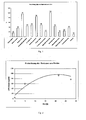
Fig. 1 zeigt die Freisetzung von Alizarinfarbstoff mittels verschiedenen Mikroorganismen nach 72 Stunden Inkubation (Beispiel 3). -
Fig. 2 zeigt den Zeitverlauf der Freisetzung von Proteasen aus dem Biopolymer durch Einwirkung von Pektinasen (Beispiel 7). -
Fig. 3 zeigt den Zeitverlauf der Freisetzung des Verstärkerenzyms Laccase aus dem Biopolymer durch Einwirkung eines Triggerenzyms (Pektinasen) (Beispiel 7). -
Fig. 4 zeigt die Freisetzung von p-Nitroanilid des immobilisierten Substrats mittels kommerzieller Protease (Beispiel 8). -
Fig. 5 zeigt den Farbumschlag immobilisierter Ferulasäure. Durch Einwirkung des Triggerenzyms (Pektinase) wird das Verstärkerenzym (Laccase) aus dem Biopolymer freigesetzt und induziert die Farbreaktion (Farbgebung durch Oxidation immobilisierter Ferulasäure) (Beispiel 10). -
Fig. 6 zeigt die Umsetzung von ABTS (Diammonium-2,2'-azinobis-(3-ethylbenzthiazolin-6-sulfonsäure)) durch Laccase infolge deren Freisetzung aus einer Peptidoglycan Matrix nach Inkubation mit Lysozym (5000 U/mL) oder Puffer (Kontrolle) nach verschiedenen Zeitpunkten. -
Fig. 7 zeigt ein erfindungsgemäßes bioresponsives System mit enzymatischer Verstärkungsreaktion zur kontrollierten Freisetzung und Sensorik. - Eine 5%ige Lösung aus Pektin, gewonnen aus Citrusschalen mit einem Veresterungsgrad von 50-60%, wurde durch Auflösen in Wasser über Nacht bei 50°C hergestellt. Alternativ kann auch Pektin aus Apfelschalen mit einem Veresterungsgrad von 70-75% verwendet werden.
- Im Vorfeld wurde das Polysaccharid mit verschiedenen Farbstoffen wie z.B. Alizarin, Cibacrom, Remazol, Viktoriablau oder anderen gefärbt. Dazu wurden 10g Pektin in Aceton mit 5mM Farbstoff suspendiert, über Nacht unter Rückfluss erhitzt und anschließend mehrere Male mit Aceton gewaschen. Die Pektinlösung wurde durch Eintropfen in eine 200mM CaCl2 Lösung auspolymerisiert und die erhaltenen Pektinkugeln wurden mit Wasser gewaschen.
- 1g (Feuchtmasse) des Biopolymers A wurde mit Alizarin gefärbt und in 10mL Puffer (50mM, pH6,0) für 24h bei Raumtemperatur unter leichtem Schütteln unter Verwendung verschiedener kommerziell erhältlicher Pektinasen inkubiert. Anschließend wurden die Überstände mit 1M NaOH auf pH14 eingestellt und die Adsorption bei 550nm mit Hilfe eines UV/VIS Photometers gemessen.
- Unterschiedliche potentiell kontaminierende Mikroorganismen wurden mittels Vorkultur angezüchtet. Zu 100mL der Hauptkultur wurden jeweils 1g Feuchtmasse des mit Alizarin gefärbten Biopolymers A zugegeben und mit 100µL Vorkultur inokuliert. Die Inkubation wurde für 72 Stunden bei 33°C durchgeführt. Anschließend wurde die Biomasse abzentrifugiert und die Absorption des Überstandes bei geeigneter Wellenlänge gemessen (
Fig. 1 ). - 10g Kieselgel wurden in 30mL einer 3-9% Aminopropyltriethoxysilan in Ethanol (95%) für 4 Stunden bei 40°C gerührt. Anschließend wurde das aminierte Kieselgel abdekantiert, 3-mal mit 70% Ethanol gewaschen und im Exsikkator bis zur Gewichtskonstanz getrocknet.
- 50mg N-(3-Dimethylaminopropyl)-N-ethylcarbodiimid hydrochlorid, 5mg (1-Hydroxybenzotriazole hydrate) und 50mg Ferulasäure wurden in 20mL absolutem Ethanol gelöst. Anschließend wurde 5g aminiertes Kieselgel zugegeben und 30min gerührt. Nach Zentrifugation wurde das Kieselgel mit der gekoppelten Ferulasäure mit 70% Ethanol gewaschen und im Exsikkator bis zur Gewichtskonstanz getrocknet. Alternativ zur Ferulasäure kann Kaffeesäure, 3,4-Dihydroxybenzoesäure, oder Fast Blue RR etc. verwendet werden. Weiters können auch Proteasesubstrate wie z.B. N-Succinyl-Ala-Ala-Pro-Leu-p-nitroanilid, N-Succinyl-Ala-Ala-Pro-Val-p-nitroanilid oder L-Leucin-p-nitroanilid auf die gleiche Art und Weise immobilisiert werden.
- 10g Kieselgel wurde in 30mL einer 3-9% Mercaptopropyltriethoxysilan in Ethanol (95%) für 4 Stunden bei 40°C gerührt. Anschließend wurde das Kieselgel abdekantiert, 3-mal mit 2-Propanol gewaschen und im Exsikkator bis zur Gewichtskonstanz getrocknet. Zu 5g vorbehandeltem Kieselgel, suspendiert in 20mL Dichlormethan, werden 155mg Ferulasäure und 38mg Dimethylaminopyridin zugegeben. Zu der mit Eis auf 0°C gekühlten Reaktionsmischung wurden 165mg DCC zugegeben und über 3 Stunden gerührt. Dabei wurde die Temperatur auf Raumtemperatur (ca. 20°C) belassen. Anschließend wurde das feste, modifizierte Kieselgel abfiltriert, 3-mal mit Dichlormethan gewaschen und über Nacht im Exsikkator getrocknet.
- Pektin, gewonnen aus Citrusschalen mit einem Veresterungsgrad von 50-60%, wurde unter leichtem Erhitzen über Nacht in Wasser gelöst. Zum Pektin als Hauptbestandteil der bioresponsiven Matrix wurde Alginat in verschiedenen Konzentrationen (1-20%) beigemengt.
- Vorzugsweise wurde eine Mischung bestehend aus 4,5g Pektin und 0,5g Natriumalginat in 100mL Wasser verwendet. Die Gelherstellung erfolgte durch Eintropfen der Pektin-Alginat-Lösung in eine vorgelegte 200mM CaCl2 Lösung. Die erhaltenen Pektinkugeln wurden mit Wasser gewaschen.
- Zu einer Mischung bestehend aus 4,5g Pektin und 0,5g Natriumalginat in 100mL Wasser wurden 5mL kommerziell erhältlicher Protease von Aspergillus oryzae zugegeben.
- Nach dem Vermischen der Polysaccharidlösung mit dem Enzym wurde das Polymer in 200mM CaCl2-Lösung eingetropft und ausgeliert. Die so gewonnenen Polymerkügelchen wurden abgesiebt, 3-mal mit 50mM Tris-HCl Puffer pH7,5 gewaschen und zu je 1g Feuchtmasse in Reaktionsgefäßen mit 5mL 50mM Tris-HCl Puffer pH7,5 portioniert.
- Entsprechend können auch andere Enzyme wie z.B. Laccasen verschiedener Trametes sp. immobilisiert werden. Weiters können auch Proteine z.B. Casein, Collagen etc. entsprechend eingebaut werden.
- Als Testpolymer wurde das Biopolymer B (Beispiel 4) mit einer Protease von Aspergillus oryzae beladen.
- Die so gewonnenen Polymerkugeln wurden abgesiebt, 3-mal mit 50mM Tris-HCl Puffer pH7,5 gewaschen und zu je 1g Feuchtmasse in Reaktionsgefäßen mit 5mL 50mM Tris-HCl Puffer pH7,5 portioniert. Der enzymatische Abbau wurde durch Zugabe einer kommerziellen Pektinase gestartet. Während der Inkubation bei Raumtemperatur und unter Schütteln wurden in bestimmten Zeitintervallen Proben aus dem Überstand genommen. Von diesen Proben wurden die Proteaseaktivitäten mittels Azocasein Assay und der Proteingehalt bestimmt (
Fig. 2 ). - Alternativ dazu kann das Biopolymer mit jedem anderen Enzym, wie z.B. Laccasen, aus der Gruppe der Oxidoreduktasen beladen werden. Hier wird dann entsprechend die Aktivität der freigesetzten Laccase mittels ABTS bestimmt (
Fig. 3 ). - 10mg Kieselgel mit immobilisiertem N-Succinyl-Ala-Ala-Pro-Leu-p-nitroanilid bzw. L-Leucin-p-nitroanilid oder N-Succinyl-Ala-Ala-Pro-Val-p-nitroanilid als Proteasesubstrate (Beispiel 2) wurden in 1300µL 50mM Tris-HCl Puffer pH8,3 suspendiert und mit kommerziell erhältlicher Protease von Aspergillus oryzae bzw. mit Wundflüssigkeit bei Raumtemperatur inkubiert. Die Aktivität wurde mittels UV-Absorption des abgespaltenen p-Nitroanilides im Überstand bei 375nm bzw. bei 405nm bestimmt (
Fig. 4 ). - 10mg Kieselgel mit immobilisierter Ferulasäure als Laccasesubstrat (Beispiel 2) bzw. mit immobilisiertem Fast Blue RR wurden in 1300µL 50mM Succinat Puffer pH4,5 suspendiert und mit Laccase vom Trametes hirsuta bzw. mit Wundflüssigkeit bei Raumtemperatur inkubiert. Die Aktivität wird anhand einer Farbmessung (gelb-orange) mittels Spectrophotometer bestimmt.
- Als Testpolymer diente das Biopolymer aus Beispiel 4 beladen mit Laccasen verschiedener Trametes sp.
- Die Polymerkugeln wurden abgesiebt, 3-mal mit 50mM Succinat Puffer pH4,5 gewaschen und zu je 1g Feuchtmasse in Reaktionsgefäßen mit 5mL 50mM Succinat Puffer pH4,5 in Anwesenheit von 10mg Kieselgel mit immobilisierter Ferulasäure als Laccasesubstrat aus Beispiel 2 portioniert. Der enzymatische Abbau wurde durch Zugabe kommerziell erhältlicher Pektinase bei Raumtemperatur und unter Schütteln gestartet. Nach der Inkubation wurde der Farbumschlag zu gelb-orange anhand einer Farbmessung mittels Spectrophotometer bestimmt (
Fig. 5 ). - Alternativ dazu kann das Biopolymer mit einer Protease von Aspergillus oryzae beladen werden. Hier wird dann entsprechend die Aktivität der freigesetzten Protease mittels UV-Absorption des abgespaltenen p-Nitroanilides im Überstand bei 375 nm bestimmt.
- Das Ansprechverhalten der bioresponsiven Polymere kann über das Diffusionsverhalten sowohl der Trigger- als auch der Verstärkerenzyme eingestellt werden. Durch eine chemische oder genetische Modifikation (z.B. Vergrößerung) der Verstärkerenzyme können entsprechend geringere Vernetzungsgrade des Biopolymers eingesetzt werden, bei einer gleichzeitigen Minimierung der Ausdiffusion des Verstärkerenzyms.
- Zur chemischen Modifikation von Verstärkerenzymen mit wasserlöslichen Polymeren wurden 1g Methoxypolyethylenglykol und 0,4g Cuyanurchlorid in 100mL trockenem Toluol gelöst und für 40 Stunden bei 40°C gerührt. Anschließend wurde das aktivierte Polymer in Hexan gefällt, filtriert und unter Vakuum getrocknet. Durch Verwendung unterschiedlicher Molekulargewichte (350, 550, 1100, 2000, 5000 etc.) kann mittels der Länge des Polymers das Diffusionsverhalten des Konjugates eingestellt werden. Das Polymer wurde in schwach basischem Medium Boratpuffer pH9,3 an das Enzym gehängt. Nach der Reaktion wurde ungebundenes Polymer mittels Ultrafiltration entfernt und das Konjugat ohne weitere Aufreinigung verwendet.
- Entsprechend Beispiel 6 wurde das Biopolymer mit modifizierter Protease beladen. Die so gewonnenen Polymerkugeln wurden abgesiebt, 3-mal mit 50mM Succinat Puffer pH4,5 gewaschen und zu je 1g Feuchtmasse in Reaktionsgefäßen mit 5mL 50mM Succinat Puffer pH4,5 in Anwesenheit von 10mg Kieselgel mit immobilisierter Ferulasäure als Laccasesubstrat aus Beispiel 2 portioniert. Der enzymatische Abbau bei Raumtemperatur und unter Schütteln wurde durch Zugabe kommerzieller Pektinase gestartet. Nach der Inkubation wurde der Farbumschlag zu gelb-orange anhand einer Farbmessung mittels Spectrophotometer bestimmt (
Fig. 5 ). Alternativ hierzu kann das Molekulargewicht des Triggerenzyms gentechnisch erhöht werden. - Ein System das im medizinischen Bereich zur Anwendung gebracht werden kann benützt das Enzym Lysozym als Trigger Enzym. Dieses Enzym der körpereigenen, nativen Immunantwort wird im Falle von Infektion gebildet und sekretiert. Hauptaufgabe des Enzyms ist eine Zerstörung von Bakterien durch Abbau von Peptidoglycan, einem Bestandteil der bakteriellen Zellwand. Es wurde gezeigt, dass erhöhte Enzymmengen im Falle einer Wundinfektion vorliegen. Im folgenden System wurden 3,12mg Mikrokokkus lysodeicticus Zellwand von Sigma mit 1mL 1%iger Agarose in Phosphat-Puffer pH7,00 suspendiert. 100µl dieser Suspension wurden in einer Mikrotiterplatte mit 50µL einer PEG modifizierten Laccase vermischt. Nach Aushärten wird das Polymer mit Puffer gewaschen und 100µl einer Lysozymlösung (200-5000U/mL) aufgetragen. Die Inkubation erfolgte bei 37°C. Alle 30 Minuten wurden 25µl des Überstandes entnommen. Laccaseaktivität wurde mittels des ABTS Assays nachgewiesen (1400µL Saccharose Puffer + 45µL 1% H2O2 + 30µL ABTS 40mM): 25µL Überstand + 75µL ABTS-Lösung). Eine Grünfärbung trat nach einigen Minuten Inkubation mit Lysozym durch Umsetzung von ABTS durch die freigesetzte Laccase auf (
Fig. 6 ). - 5 g Pektine oder/und Cellulosederivate werden in 100 mL Wasser gelöst und mit 3 3mL einer 97% Glycidylmethacrylate Lösung in Gegenwart von 0,5 mL 6 M HCl gekoppelt.
- PES Gewebe wird mit Trimethylsilyl methacrylate modifiziert und mit einer mit Glycidylmethacrylate modifizierten Matrix überschichtet und radikalisch auspolymerisiert und damit kovalent quervernetzt.
- Extrazellulare Enzyme wie Pektinasen oder Cellulasen können nun das PES Geweben durch Diffusion überwinden und durch Abbau der Matrix darin eingeschlossene funktionelle Moleküle wie z.B. Enzyme freisetzen, die ihrerseits eine Farbreaktion auslösen.
- Ein weiteres System, das im medizinischen Bereich zur Anwendung gebracht werden kann, benützt das Enzym Elastase als Trigger Enzym. Dieses Enzym wird von manchen Bakterienarten, aber auch von der körpereigenen Immunantwort im Falle von Infektion gebildet und sekretiert, wobei fast alle Arten von Protein gespalten werden können. Im Falle von Wundinfektion konnten eindeutig erhöhte Enzymmmengen im Wundsekret nachgewiesen werden. Damit kann dieses Enzym als Markerenzym für beginnende Wundinfektion herangezogen werden. Im folgenden System wurden 3,12mg Chitosan mit 1mL 1%iger Agarose in Phosphat-Puffer pH 7,0 suspendiert. Das Chitosan wurde zuvor mit GMBS aktiviert, um es beidseits über die SH Gruppe eines Cysteins und der Peptidsequenz Ala-Ala-Pro-Val querzuvernetzen. 100µl dieser Suspension wurden in einer Mikrotiterplatte mit 50µL einer PEG modifizierten Laccase vermischt. Nach Aushärten wird das Polymer mit Puffer gewaschen und 100µl einer Elastaselösung (2-5 U/mL) oder mit Wundsekret aufgetragen. Die Inkubation erfolgte bei 37°C. Alle 10 Minuten wurden 25µl des Überstandes entnommen. Laccaseaktivität wurde mittels des ABTS Assays nachgewiesen (1400µL Saccharose Puffer + 45µL 1% H2O2 + 30uL ABTS 40mM) : 25µL Überstand + 75µL ABTS-Lösung). Eine Grünfärbung trat nach einigen Minuten Inkubation mit Elastase durch Umsetzung von ABTS durch die freigesetze Laccase auf. Ohne Elastase wurde keine Farbänderung festgestellt.
Claims (15)
- Anordnung umfassend einen festen Träger und eine an dem festen Träger angeordnete Matrix umfassend mindestens ein enzymatisch umsetzbares oder modifizierbares Molekül, welche Matrix mindestens ein durch Umsetzung oder Modifikation des Moleküls freisetzbares Enzym umfasst, das in der Lage ist zumindest ein in der Matrix und/oder am festen Träger befindliches farbänderndes Substrat umzusetzen.
- Anordnung nach Anspruch 1, dadurch gekennzeichnet, dass die Matrix als Schicht, in Form von Kapseln oder als Hydrogel auf den festen Träger angeordnet ist.
- Anordnung nach Anspruch 1 oder 2, dadurch gekennzeichnet, dass das enzymatisch umsetzbare oder modifizierbare Molekül ein Polymer, vorzugsweise ein Polysaccharid, Polypeptid, Polyester, Polyamid oder eine Kombination davon ist, ist.
- Anordnung nach Anspruch 3, dadurch gekennzeichnet, dass das Polysaccharid ausgewählt aus der Gruppe bestehend aus Pektin, Amylose, Amylopektin, Agarose, Alginat, Carraghenan, Chitin, Chitosan, Dextran, Glycogen, Guar, Johannisbrotkernmehl, Laevan, Pektin, Pollulan, Tamarindenkernmehl, Xanthan und Xylan.
- Anordnung nach einem der Ansprüche 1 bis 4, dadurch gekennzeichnet, dass die Matrix zusätzlich durch Umsetzung oder Modifikation des Moleküls freisetzbares Enzym umfasst, das in der Lage ist das in der Matrix befindliche Molekül ebenfalls umzusetzen oder zu modifizieren.
- Anordnung nach Anspruch 5, dadurch gekennzeichnet, dass das Enzym ausgewählt ist aus der Gruppe bestehend aus den Gruppen der Hydrolasen und Oxidoreduktasen, wie Proteasen, Laccasen oder Peroxidasen.
- Anordnung nach einem der Ansprüche 1 bis 6, dadurch gekennzeichnet, dass das durch die Umsetzung oder Modifikation des Moleküls freisetzbare Enzyme an Polyvinylalkohol, Polyethylenglykol (PEG), Polypeptide, insbesondere Elastin, oder Peptide gebunden ist.
- Anordnung nach Anspruch 7, dadurch gekennzeichnet, dass das freisetzbare Enzym, welches mittels eines Polypeptids oder Peptids am enzymatisch umsetzbaren oder modifizierbaren Molekül gebunden ist, durch ein Enzym, vorzugsweise einem mikrobiellen Enzym oder einem Enzym des Immunsystems, wie Elastase, freisetzbar ist.
- Anordnung nach einem der Ansprüche 1 bis 8, dadurch gekennzeichnet, dass das farberzeugende Substrat ausgewählt ist aus der Gruppe bestehend aus phenolischen Verbindungen und Azo-Farbstoffen.
- Anordnung nach Anspruch 9, dadurch gekennzeichnet, dass das farberzeugende Substrat direkt von Enzymen, vorzugsweise mikrobiellen Enzymen, wie Laccasen oder Peroxidasen oder Enzymen des Immunsystems, wie Myeloperoxidase, umsetzbar ist.
- Anordnung nach einem der Ansprüche 1 bis 10, dadurch gekennzeichnet, dass die Anordnung eine dem festen Träger gegenüberliegende semipermeable Membran aufweist, wobei die semipermeable Membran vorzugsweise ausgewählt ist aus der Gruppe bestehend aus Cellulosederivate, Polyamide, Polyacrylamide und Polyester.
- Anordnung nach einem der Ansprüche 3 bis 11, dadurch gekennzeichnet, dass das Polymer, das Enzym und das farbändernde Substrat ausgewählt sind aus Tabelle A.
- Verwendung einer Anordnung nach einem der Ansprüche 1 bis 12 zur Bestimmung der Anwesenheit und/oder Charakterisierung von Zellen, vorzugsweise von Mikroorganismen, vorzugsweise ausgewählt aus der Gruppe bestehend aus Bakterien und Pilzen, in einer Probe.
- Verwendung einer Anordnung nach einem der Ansprüche 1 bis 12 zur Detektion mindestens eines Enzyms, vorzugsweise ausgewählt aus der Gruppe der Hydrolasen bestehend aus Amylase, Cellulase, Xylanase, Mannanase, Protease, Lysozym, Elastase, Collagenase, Cathepsin, Myeloperoxidase, Lipase und Esterase, in einer Probe.
- Verwendung einer Anordnung nach einem der Ansprüche 1 bis 12 zur Detektion einer Wundinfektion durch Bestimmen des Vorhandenseins mindestens eines wund-spezifischen Enzyms, vorzugsweise aus der Gruppe der Hydrolasen bestehend aus Amylase, Cellulase, Xylanase, Mannanase, Protease, Lysozym, Elastase, Collagenase, Cathepsin, Myeloperoxidase, Lipase und Esterase.
Applications Claiming Priority (2)
| Application Number | Priority Date | Filing Date | Title |
|---|---|---|---|
| ATA184/2010A AT509355B1 (de) | 2010-02-10 | 2010-02-10 | Testanordnung |
| PCT/AT2011/000074 WO2011097664A2 (de) | 2010-02-10 | 2011-02-09 | Testanordnung |
Publications (2)
| Publication Number | Publication Date |
|---|---|
| EP2534483A2 EP2534483A2 (de) | 2012-12-19 |
| EP2534483B1 true EP2534483B1 (de) | 2016-09-14 |
Family
ID=44352351
Family Applications (1)
| Application Number | Title | Priority Date | Filing Date |
|---|---|---|---|
| EP11706460.0A Not-in-force EP2534483B1 (de) | 2010-02-10 | 2011-02-09 | Testanordnung |
Country Status (5)
| Country | Link |
|---|---|
| US (1) | US8785142B2 (de) |
| EP (1) | EP2534483B1 (de) |
| JP (1) | JP5859984B2 (de) |
| AT (1) | AT509355B1 (de) |
| WO (1) | WO2011097664A2 (de) |
Families Citing this family (3)
| Publication number | Priority date | Publication date | Assignee | Title |
|---|---|---|---|---|
| CN109310528B (zh) | 2016-03-30 | 2021-07-20 | 康沃特克科技公司 | 检测伤口中的微生物感染 |
| US11453905B2 (en) | 2016-04-18 | 2022-09-27 | Trubac, LTD. | Method of detecting bacterial infection in a biological sample |
| US11802838B2 (en) * | 2020-03-23 | 2023-10-31 | Bay State Milling Company | Rapid high amylose wheat seed purity test |
Family Cites Families (24)
| Publication number | Priority date | Publication date | Assignee | Title |
|---|---|---|---|---|
| US4888287A (en) * | 1986-09-24 | 1989-12-19 | Eastman Kodak Company | Rapid differentiation of fungi from bacteria using polyene antibiotics |
| DE68920319D1 (de) | 1988-04-22 | 1995-02-09 | Massachusetts Inst Technology | Verfahren zum formen und zum gebrauch von mikrotröpchen. |
| NL8801073A (nl) * | 1988-04-26 | 1989-11-16 | Univ Twente | Detectiemethode. |
| AU616957B2 (en) | 1988-06-20 | 1991-11-14 | Becton Dickinson & Company | Device for enhancing fluorescence and kinetics and methods of using the device |
| AU647421B2 (en) * | 1989-11-28 | 1994-03-24 | Convatec Technologies Inc. | Dressing including an indicator |
| JPH0786497B2 (ja) * | 1989-12-22 | 1995-09-20 | 国税庁長官 | 非多孔固定化酵素膜を使用する酵素電極 |
| US5366881A (en) * | 1993-02-23 | 1994-11-22 | The United States Of America As Represented By The Secretary Of The Navy | Polymerizable lipids for preparing vesicles that controllably release an encapsulant |
| HUT73393A (en) * | 1993-04-14 | 1996-07-29 | Litmus Concepts | Reporter enzyme release technology methods of assaying for the presence of aspartic proteases and other hydrolitic enzyme activities |
| SE9301270D0 (sv) | 1993-04-19 | 1993-04-17 | Biosensor | |
| US7157048B2 (en) | 1993-05-19 | 2007-01-02 | Sira Technologies, Inc. | Detection of contaminants |
| IL109079A (en) * | 1994-03-22 | 1998-02-22 | Israel Fiber Inst State Of Isr | Polyionic hydrogels |
| EP0922341B1 (de) | 1996-09-02 | 2002-11-13 | STMicroelectronics N.V. | Verbesserungen bei, oder in bezug auf, mehrträgerübertragungssysteme |
| AU4829099A (en) | 1998-06-22 | 2000-01-10 | Regents Of The University Of California, The | Triggered optical biosensor |
| WO2003033691A1 (fr) * | 2001-10-19 | 2003-04-24 | Institut Molekulyarnoi Biologii Im. V.A. Engelgardta Rossiiskoi Akademii Nauk | Micropuce cellulaire et son utilisation dans un procede d'analyse de cellules vivantes |
| WO2003063693A2 (en) * | 2002-01-31 | 2003-08-07 | Expressive Constructs, Inc. | Method for detecting microorganisms |
| DK1762626T3 (da) * | 2003-09-02 | 2011-03-14 | Systagenix Wound Man Ip Co Bv | Signalamplifikation under anvendelse af et syntetisk zymogen |
| US7399608B2 (en) | 2003-12-16 | 2008-07-15 | Kimberly-Clark Worldwide, Inc. | Microbial detection and quantification |
| WO2005061018A1 (en) * | 2003-12-22 | 2005-07-07 | Regentis Biomaterials Ltd. | Matrix comprising naturally-occurring protein backbone |
| US20050197554A1 (en) * | 2004-02-26 | 2005-09-08 | Michael Polcha | Composite thin-film glucose sensor |
| US20070122831A1 (en) | 2005-11-12 | 2007-05-31 | Dave Bachoon | Detection and source identification of microbial contaminants in water samples |
| US20090215079A1 (en) * | 2005-12-05 | 2009-08-27 | Technische Universitat Dresden | Method and Devices for the Detection of Microorganisms and/or the Activity Thereof |
| WO2007139854A2 (en) * | 2006-05-22 | 2007-12-06 | George John | Method for preparing hydro/organo gelators from disaccharide sugars by biocatalysis and their use in enzyme-triggered drug delivery |
| US20080057534A1 (en) | 2006-08-31 | 2008-03-06 | Kimberly-Clark Worldwide, Inc. | Microbe-sensitive indicators and use of the same |
| JP5508861B2 (ja) * | 2007-03-07 | 2014-06-04 | エコー セラピューティクス, インコーポレイテッド | 経皮的被検体モニタリングシステム |
-
2010
- 2010-02-10 AT ATA184/2010A patent/AT509355B1/de not_active IP Right Cessation
-
2011
- 2011-02-09 JP JP2012552209A patent/JP5859984B2/ja not_active Expired - Fee Related
- 2011-02-09 EP EP11706460.0A patent/EP2534483B1/de not_active Not-in-force
- 2011-02-09 WO PCT/AT2011/000074 patent/WO2011097664A2/de active Application Filing
- 2011-02-09 US US13/577,366 patent/US8785142B2/en not_active Expired - Fee Related
Non-Patent Citations (1)
| Title |
|---|
| K SCHNEIDER ET AL: "Trigger enzymes for pectin based bioresponsive polymers", XVITH INTERNATIONAL CONFERENCE ON BIOENCAPSULATION, 4 September 2008 (2008-09-04), XP055222948, Retrieved from the Internet <URL:http://impascience.eu/bioencapsulation/340_contribution_texts/2008-09-04_P76.pdf> [retrieved on 20151022] * |
Also Published As
| Publication number | Publication date |
|---|---|
| US8785142B2 (en) | 2014-07-22 |
| WO2011097664A3 (de) | 2012-03-22 |
| JP5859984B2 (ja) | 2016-02-16 |
| EP2534483A2 (de) | 2012-12-19 |
| US20120309036A1 (en) | 2012-12-06 |
| JP2013518600A (ja) | 2013-05-23 |
| AT509355B1 (de) | 2012-04-15 |
| WO2011097664A8 (de) | 2012-09-20 |
| WO2011097664A2 (de) | 2011-08-18 |
| AT509355A1 (de) | 2011-08-15 |
Similar Documents
| Publication | Publication Date | Title |
|---|---|---|
| US11236323B2 (en) | Biofunctional materials | |
| Lu et al. | Stabilization of enzymes in silk films | |
| Peniche et al. | Formation and stability of shark liver oil loaded chitosan/calcium alginate capsules | |
| Mohammadi et al. | Activated alginate-montmorillonite beads as an efficient carrier for pectinase immobilization | |
| Yi et al. | Amino acid modified chitosan beads: improved polymer supports for immobilization of lipase from Candida rugosa | |
| HU220197B (hu) | Eljárás fenolpolimerek viszkozitásának növelésére vagy gélezésére lakkáz enzimmel | |
| Mendez‐Encinas et al. | Partial removal of protein associated with arabinoxylans: Impact on the viscoelasticity, crosslinking content, and microstructure of the gels formed | |
| EP1468968A1 (de) | Eine Laccase enthaltender Biokatalysator | |
| EP2534483B1 (de) | Testanordnung | |
| Kadri et al. | Nanoencapsulation and release study of enzymes from Alkanivorax borkumensis in chitosan-tripolyphosphate formulation | |
| Romero et al. | Architecture and physicochemical characterization of Bacillus biofilm as a potential enzyme immobilization factory | |
| de Souza et al. | Enzymatic hydrolysis of starch into sugars is influenced by microgel assembly | |
| Popović et al. | Dopamine-modified pectin for a Streptomyces cyaneus laccase induced microbeads formation, immobilization, and textile dyes decolorization | |
| Cerón et al. | Study of stability, kinetic parameters and release of lysozyme immobilized on chitosan microspheres by crosslinking and covalent attachment for cotton fabric functionalization | |
| Schneider et al. | Bioresponsive systems based on polygalacturonate containing hydrogels | |
| DD294729A5 (de) | Verfahren zur herstellung von immobilisaten mit biologisch aktiven, makromolekularen verbindungen | |
| CH680668A5 (de) | ||
| DD263663A7 (de) | Verfahren zur herstellung eines immobilisierten cholinestetase-praeparates | |
| WO2019013100A1 (ja) | 複合組成物及びその製造方法、並びにバイオセンサ | |
| DE4005927A1 (de) | Immobilisierung von proteinen an traegern | |
| RU2771183C1 (ru) | Способ получения препарата фицина в геле на основе карбоксиметилцеллюлозы | |
| Eser et al. | Immobilization of Subtilisin Carlsberg and its use for transesterification of N-acetyl-L-phenylalanine ethyl ester in organic medium | |
| Kullmann | Polymer-Enzyme Thin Films–Assembly, Characterization and Stability of Bio-functionalized Surfaces |
Legal Events
| Date | Code | Title | Description |
|---|---|---|---|
| PUAI | Public reference made under article 153(3) epc to a published international application that has entered the european phase |
Free format text: ORIGINAL CODE: 0009012 |
|
| 17P | Request for examination filed |
Effective date: 20120910 |
|
| AK | Designated contracting states |
Kind code of ref document: A2 Designated state(s): AL AT BE BG CH CY CZ DE DK EE ES FI FR GB GR HR HU IE IS IT LI LT LU LV MC MK MT NL NO PL PT RO RS SE SI SK SM TR |
|
| DAX | Request for extension of the european patent (deleted) | ||
| RIN1 | Information on inventor provided before grant (corrected) |
Inventor name: ROLLETT, ALEXANDRA Inventor name: GUEBITZ, GEORG Inventor name: KAUFMANN, FRANZ Inventor name: SIGL, EVA Inventor name: SCHROEDER, MARC Inventor name: SCHNEIDER, KONSTANTIN Inventor name: HAFNER, ANDREAS Inventor name: HASMANN, ANDREA |
|
| 17Q | First examination report despatched |
Effective date: 20140924 |
|
| GRAP | Despatch of communication of intention to grant a patent |
Free format text: ORIGINAL CODE: EPIDOSNIGR1 |
|
| INTG | Intention to grant announced |
Effective date: 20151111 |
|
| GRAP | Despatch of communication of intention to grant a patent |
Free format text: ORIGINAL CODE: EPIDOSNIGR1 |
|
| INTG | Intention to grant announced |
Effective date: 20160414 |
|
| RIN1 | Information on inventor provided before grant (corrected) |
Inventor name: GUEBITZ, GEORG Inventor name: KAUFMANN, FRANZ Inventor name: HASMANN, ANDREA Inventor name: ROLLETT, ALEXANDRA Inventor name: SCHROEDER, MARC Inventor name: HAFNER, ANDREAS Inventor name: SCHNEIDER, KONSTANTIN Inventor name: SIGL, EVA |
|
| GRAS | Grant fee paid |
Free format text: ORIGINAL CODE: EPIDOSNIGR3 |
|
| GRAA | (expected) grant |
Free format text: ORIGINAL CODE: 0009210 |
|
| AK | Designated contracting states |
Kind code of ref document: B1 Designated state(s): AL AT BE BG CH CY CZ DE DK EE ES FI FR GB GR HR HU IE IS IT LI LT LU LV MC MK MT NL NO PL PT RO RS SE SI SK SM TR |
|
| REG | Reference to a national code |
Ref country code: GB Ref legal event code: FG4D Free format text: NOT ENGLISH |
|
| REG | Reference to a national code |
Ref country code: CH Ref legal event code: EP |
|
| REG | Reference to a national code |
Ref country code: IE Ref legal event code: FG4D Free format text: LANGUAGE OF EP DOCUMENT: GERMAN |
|
| REG | Reference to a national code |
Ref country code: AT Ref legal event code: REF Ref document number: 829544 Country of ref document: AT Kind code of ref document: T Effective date: 20161015 |
|
| REG | Reference to a national code |
Ref country code: DE Ref legal event code: R096 Ref document number: 502011010676 Country of ref document: DE |
|
| REG | Reference to a national code |
Ref country code: LT Ref legal event code: MG4D |
|
| REG | Reference to a national code |
Ref country code: NL Ref legal event code: MP Effective date: 20160914 |
|
| REG | Reference to a national code |
Ref country code: FR Ref legal event code: PLFP Year of fee payment: 7 |
|
| PG25 | Lapsed in a contracting state [announced via postgrant information from national office to epo] |
Ref country code: NO Free format text: LAPSE BECAUSE OF FAILURE TO SUBMIT A TRANSLATION OF THE DESCRIPTION OR TO PAY THE FEE WITHIN THE PRESCRIBED TIME-LIMIT Effective date: 20161214 Ref country code: HR Free format text: LAPSE BECAUSE OF FAILURE TO SUBMIT A TRANSLATION OF THE DESCRIPTION OR TO PAY THE FEE WITHIN THE PRESCRIBED TIME-LIMIT Effective date: 20160914 Ref country code: LT Free format text: LAPSE BECAUSE OF FAILURE TO SUBMIT A TRANSLATION OF THE DESCRIPTION OR TO PAY THE FEE WITHIN THE PRESCRIBED TIME-LIMIT Effective date: 20160914 Ref country code: FI Free format text: LAPSE BECAUSE OF FAILURE TO SUBMIT A TRANSLATION OF THE DESCRIPTION OR TO PAY THE FEE WITHIN THE PRESCRIBED TIME-LIMIT Effective date: 20160914 Ref country code: RS Free format text: LAPSE BECAUSE OF FAILURE TO SUBMIT A TRANSLATION OF THE DESCRIPTION OR TO PAY THE FEE WITHIN THE PRESCRIBED TIME-LIMIT Effective date: 20160914 |
|
| REG | Reference to a national code |
Ref country code: CH Ref legal event code: NV Representative=s name: RENTSCH PARTNER AG, CH |
|
| PG25 | Lapsed in a contracting state [announced via postgrant information from national office to epo] |
Ref country code: LV Free format text: LAPSE BECAUSE OF FAILURE TO SUBMIT A TRANSLATION OF THE DESCRIPTION OR TO PAY THE FEE WITHIN THE PRESCRIBED TIME-LIMIT Effective date: 20160914 Ref country code: GR Free format text: LAPSE BECAUSE OF FAILURE TO SUBMIT A TRANSLATION OF THE DESCRIPTION OR TO PAY THE FEE WITHIN THE PRESCRIBED TIME-LIMIT Effective date: 20161215 Ref country code: NL Free format text: LAPSE BECAUSE OF FAILURE TO SUBMIT A TRANSLATION OF THE DESCRIPTION OR TO PAY THE FEE WITHIN THE PRESCRIBED TIME-LIMIT Effective date: 20160914 Ref country code: SE Free format text: LAPSE BECAUSE OF FAILURE TO SUBMIT A TRANSLATION OF THE DESCRIPTION OR TO PAY THE FEE WITHIN THE PRESCRIBED TIME-LIMIT Effective date: 20160914 |
|
| PG25 | Lapsed in a contracting state [announced via postgrant information from national office to epo] |
Ref country code: EE Free format text: LAPSE BECAUSE OF FAILURE TO SUBMIT A TRANSLATION OF THE DESCRIPTION OR TO PAY THE FEE WITHIN THE PRESCRIBED TIME-LIMIT Effective date: 20160914 Ref country code: RO Free format text: LAPSE BECAUSE OF FAILURE TO SUBMIT A TRANSLATION OF THE DESCRIPTION OR TO PAY THE FEE WITHIN THE PRESCRIBED TIME-LIMIT Effective date: 20160914 |
|
| PG25 | Lapsed in a contracting state [announced via postgrant information from national office to epo] |
Ref country code: SK Free format text: LAPSE BECAUSE OF FAILURE TO SUBMIT A TRANSLATION OF THE DESCRIPTION OR TO PAY THE FEE WITHIN THE PRESCRIBED TIME-LIMIT Effective date: 20160914 Ref country code: ES Free format text: LAPSE BECAUSE OF FAILURE TO SUBMIT A TRANSLATION OF THE DESCRIPTION OR TO PAY THE FEE WITHIN THE PRESCRIBED TIME-LIMIT Effective date: 20160914 Ref country code: BG Free format text: LAPSE BECAUSE OF FAILURE TO SUBMIT A TRANSLATION OF THE DESCRIPTION OR TO PAY THE FEE WITHIN THE PRESCRIBED TIME-LIMIT Effective date: 20161214 Ref country code: PT Free format text: LAPSE BECAUSE OF FAILURE TO SUBMIT A TRANSLATION OF THE DESCRIPTION OR TO PAY THE FEE WITHIN THE PRESCRIBED TIME-LIMIT Effective date: 20170116 Ref country code: IS Free format text: LAPSE BECAUSE OF FAILURE TO SUBMIT A TRANSLATION OF THE DESCRIPTION OR TO PAY THE FEE WITHIN THE PRESCRIBED TIME-LIMIT Effective date: 20170114 Ref country code: BE Free format text: LAPSE BECAUSE OF NON-PAYMENT OF DUE FEES Effective date: 20170228 Ref country code: CZ Free format text: LAPSE BECAUSE OF FAILURE TO SUBMIT A TRANSLATION OF THE DESCRIPTION OR TO PAY THE FEE WITHIN THE PRESCRIBED TIME-LIMIT Effective date: 20160914 Ref country code: SM Free format text: LAPSE BECAUSE OF FAILURE TO SUBMIT A TRANSLATION OF THE DESCRIPTION OR TO PAY THE FEE WITHIN THE PRESCRIBED TIME-LIMIT Effective date: 20160914 Ref country code: PL Free format text: LAPSE BECAUSE OF FAILURE TO SUBMIT A TRANSLATION OF THE DESCRIPTION OR TO PAY THE FEE WITHIN THE PRESCRIBED TIME-LIMIT Effective date: 20160914 |
|
| REG | Reference to a national code |
Ref country code: DE Ref legal event code: R097 Ref document number: 502011010676 Country of ref document: DE |
|
| PG25 | Lapsed in a contracting state [announced via postgrant information from national office to epo] |
Ref country code: IT Free format text: LAPSE BECAUSE OF FAILURE TO SUBMIT A TRANSLATION OF THE DESCRIPTION OR TO PAY THE FEE WITHIN THE PRESCRIBED TIME-LIMIT Effective date: 20160914 |
|
| PLBE | No opposition filed within time limit |
Free format text: ORIGINAL CODE: 0009261 |
|
| STAA | Information on the status of an ep patent application or granted ep patent |
Free format text: STATUS: NO OPPOSITION FILED WITHIN TIME LIMIT |
|
| PG25 | Lapsed in a contracting state [announced via postgrant information from national office to epo] |
Ref country code: DK Free format text: LAPSE BECAUSE OF FAILURE TO SUBMIT A TRANSLATION OF THE DESCRIPTION OR TO PAY THE FEE WITHIN THE PRESCRIBED TIME-LIMIT Effective date: 20160914 |
|
| 26N | No opposition filed |
Effective date: 20170615 |
|
| PG25 | Lapsed in a contracting state [announced via postgrant information from national office to epo] |
Ref country code: MC Free format text: LAPSE BECAUSE OF FAILURE TO SUBMIT A TRANSLATION OF THE DESCRIPTION OR TO PAY THE FEE WITHIN THE PRESCRIBED TIME-LIMIT Effective date: 20160914 |
|
| REG | Reference to a national code |
Ref country code: CH Ref legal event code: PCAR Free format text: NEW ADDRESS: BELLERIVESTRASSE 203 POSTFACH, 8034 ZUERICH (CH) |
|
| REG | Reference to a national code |
Ref country code: IE Ref legal event code: MM4A |
|
| PG25 | Lapsed in a contracting state [announced via postgrant information from national office to epo] |
Ref country code: SI Free format text: LAPSE BECAUSE OF FAILURE TO SUBMIT A TRANSLATION OF THE DESCRIPTION OR TO PAY THE FEE WITHIN THE PRESCRIBED TIME-LIMIT Effective date: 20160914 |
|
| PG25 | Lapsed in a contracting state [announced via postgrant information from national office to epo] |
Ref country code: LU Free format text: LAPSE BECAUSE OF NON-PAYMENT OF DUE FEES Effective date: 20170209 |
|
| REG | Reference to a national code |
Ref country code: FR Ref legal event code: PLFP Year of fee payment: 8 |
|
| REG | Reference to a national code |
Ref country code: BE Ref legal event code: MM Effective date: 20170228 |
|
| PG25 | Lapsed in a contracting state [announced via postgrant information from national office to epo] |
Ref country code: IE Free format text: LAPSE BECAUSE OF NON-PAYMENT OF DUE FEES Effective date: 20170209 |
|
| REG | Reference to a national code |
Ref country code: AT Ref legal event code: MM01 Ref document number: 829544 Country of ref document: AT Kind code of ref document: T Effective date: 20170209 |
|
| PG25 | Lapsed in a contracting state [announced via postgrant information from national office to epo] |
Ref country code: AT Free format text: LAPSE BECAUSE OF NON-PAYMENT OF DUE FEES Effective date: 20170209 |
|
| PG25 | Lapsed in a contracting state [announced via postgrant information from national office to epo] |
Ref country code: MT Free format text: LAPSE BECAUSE OF FAILURE TO SUBMIT A TRANSLATION OF THE DESCRIPTION OR TO PAY THE FEE WITHIN THE PRESCRIBED TIME-LIMIT Effective date: 20160914 |
|
| PG25 | Lapsed in a contracting state [announced via postgrant information from national office to epo] |
Ref country code: AL Free format text: LAPSE BECAUSE OF FAILURE TO SUBMIT A TRANSLATION OF THE DESCRIPTION OR TO PAY THE FEE WITHIN THE PRESCRIBED TIME-LIMIT Effective date: 20160914 |
|
| PG25 | Lapsed in a contracting state [announced via postgrant information from national office to epo] |
Ref country code: HU Free format text: LAPSE BECAUSE OF FAILURE TO SUBMIT A TRANSLATION OF THE DESCRIPTION OR TO PAY THE FEE WITHIN THE PRESCRIBED TIME-LIMIT; INVALID AB INITIO Effective date: 20110209 |
|
| PG25 | Lapsed in a contracting state [announced via postgrant information from national office to epo] |
Ref country code: CY Free format text: LAPSE BECAUSE OF NON-PAYMENT OF DUE FEES Effective date: 20160914 |
|
| PG25 | Lapsed in a contracting state [announced via postgrant information from national office to epo] |
Ref country code: MK Free format text: LAPSE BECAUSE OF FAILURE TO SUBMIT A TRANSLATION OF THE DESCRIPTION OR TO PAY THE FEE WITHIN THE PRESCRIBED TIME-LIMIT Effective date: 20160914 |
|
| PG25 | Lapsed in a contracting state [announced via postgrant information from national office to epo] |
Ref country code: TR Free format text: LAPSE BECAUSE OF FAILURE TO SUBMIT A TRANSLATION OF THE DESCRIPTION OR TO PAY THE FEE WITHIN THE PRESCRIBED TIME-LIMIT Effective date: 20160914 |
|
| PGFP | Annual fee paid to national office [announced via postgrant information from national office to epo] |
Ref country code: FR Payment date: 20201130 Year of fee payment: 11 |
|
| PGFP | Annual fee paid to national office [announced via postgrant information from national office to epo] |
Ref country code: CH Payment date: 20210217 Year of fee payment: 11 |
|
| PGFP | Annual fee paid to national office [announced via postgrant information from national office to epo] |
Ref country code: GB Payment date: 20210222 Year of fee payment: 11 Ref country code: DE Payment date: 20210225 Year of fee payment: 11 |
|
| REG | Reference to a national code |
Ref country code: DE Ref legal event code: R119 Ref document number: 502011010676 Country of ref document: DE |
|
| REG | Reference to a national code |
Ref country code: CH Ref legal event code: PL |
|
| GBPC | Gb: european patent ceased through non-payment of renewal fee |
Effective date: 20220209 |
|
| PG25 | Lapsed in a contracting state [announced via postgrant information from national office to epo] |
Ref country code: FR Free format text: LAPSE BECAUSE OF NON-PAYMENT OF DUE FEES Effective date: 20220228 |
|
| PG25 | Lapsed in a contracting state [announced via postgrant information from national office to epo] |
Ref country code: LI Free format text: LAPSE BECAUSE OF NON-PAYMENT OF DUE FEES Effective date: 20220228 Ref country code: GB Free format text: LAPSE BECAUSE OF NON-PAYMENT OF DUE FEES Effective date: 20220209 Ref country code: DE Free format text: LAPSE BECAUSE OF NON-PAYMENT OF DUE FEES Effective date: 20220901 Ref country code: CH Free format text: LAPSE BECAUSE OF NON-PAYMENT OF DUE FEES Effective date: 20220228 |