EP2518164A1 - Probe for detecting mutation in exon 12 of NPM1 gene and use thereof - Google Patents
Probe for detecting mutation in exon 12 of NPM1 gene and use thereof Download PDFInfo
- Publication number
- EP2518164A1 EP2518164A1 EP20120166071 EP12166071A EP2518164A1 EP 2518164 A1 EP2518164 A1 EP 2518164A1 EP 20120166071 EP20120166071 EP 20120166071 EP 12166071 A EP12166071 A EP 12166071A EP 2518164 A1 EP2518164 A1 EP 2518164A1
- Authority
- EP
- European Patent Office
- Prior art keywords
- nucleotide
- seq
- probe
- sequence
- oligonucleotide
- Prior art date
- Legal status (The legal status is an assumption and is not a legal conclusion. Google has not performed a legal analysis and makes no representation as to the accuracy of the status listed.)
- Granted
Links
Images







Classifications
-
- C—CHEMISTRY; METALLURGY
- C12—BIOCHEMISTRY; BEER; SPIRITS; WINE; VINEGAR; MICROBIOLOGY; ENZYMOLOGY; MUTATION OR GENETIC ENGINEERING
- C12Q—MEASURING OR TESTING PROCESSES INVOLVING ENZYMES, NUCLEIC ACIDS OR MICROORGANISMS; COMPOSITIONS OR TEST PAPERS THEREFOR; PROCESSES OF PREPARING SUCH COMPOSITIONS; CONDITION-RESPONSIVE CONTROL IN MICROBIOLOGICAL OR ENZYMOLOGICAL PROCESSES
- C12Q1/00—Measuring or testing processes involving enzymes, nucleic acids or microorganisms; Compositions therefor; Processes of preparing such compositions
- C12Q1/68—Measuring or testing processes involving enzymes, nucleic acids or microorganisms; Compositions therefor; Processes of preparing such compositions involving nucleic acids
- C12Q1/6876—Nucleic acid products used in the analysis of nucleic acids, e.g. primers or probes
- C12Q1/6883—Nucleic acid products used in the analysis of nucleic acids, e.g. primers or probes for diseases caused by alterations of genetic material
- C12Q1/6886—Nucleic acid products used in the analysis of nucleic acids, e.g. primers or probes for diseases caused by alterations of genetic material for cancer
-
- C—CHEMISTRY; METALLURGY
- C12—BIOCHEMISTRY; BEER; SPIRITS; WINE; VINEGAR; MICROBIOLOGY; ENZYMOLOGY; MUTATION OR GENETIC ENGINEERING
- C12Q—MEASURING OR TESTING PROCESSES INVOLVING ENZYMES, NUCLEIC ACIDS OR MICROORGANISMS; COMPOSITIONS OR TEST PAPERS THEREFOR; PROCESSES OF PREPARING SUCH COMPOSITIONS; CONDITION-RESPONSIVE CONTROL IN MICROBIOLOGICAL OR ENZYMOLOGICAL PROCESSES
- C12Q1/00—Measuring or testing processes involving enzymes, nucleic acids or microorganisms; Compositions therefor; Processes of preparing such compositions
- C12Q1/68—Measuring or testing processes involving enzymes, nucleic acids or microorganisms; Compositions therefor; Processes of preparing such compositions involving nucleic acids
-
- G—PHYSICS
- G01—MEASURING; TESTING
- G01N—INVESTIGATING OR ANALYSING MATERIALS BY DETERMINING THEIR CHEMICAL OR PHYSICAL PROPERTIES
- G01N33/00—Investigating or analysing materials by specific methods not covered by groups G01N1/00 - G01N31/00
- G01N33/48—Biological material, e.g. blood, urine; Haemocytometers
- G01N33/50—Chemical analysis of biological material, e.g. blood, urine; Testing involving biospecific ligand binding methods; Immunological testing
- G01N33/53—Immunoassay; Biospecific binding assay; Materials therefor
- G01N33/5302—Apparatus specially adapted for immunological test procedures
-
- G—PHYSICS
- G01—MEASURING; TESTING
- G01N—INVESTIGATING OR ANALYSING MATERIALS BY DETERMINING THEIR CHEMICAL OR PHYSICAL PROPERTIES
- G01N33/00—Investigating or analysing materials by specific methods not covered by groups G01N1/00 - G01N31/00
- G01N33/48—Biological material, e.g. blood, urine; Haemocytometers
- G01N33/50—Chemical analysis of biological material, e.g. blood, urine; Testing involving biospecific ligand binding methods; Immunological testing
- G01N33/53—Immunoassay; Biospecific binding assay; Materials therefor
- G01N33/5308—Immunoassay; Biospecific binding assay; Materials therefor for analytes not provided for elsewhere, e.g. nucleic acids, uric acid, worms, mites
-
- C—CHEMISTRY; METALLURGY
- C12—BIOCHEMISTRY; BEER; SPIRITS; WINE; VINEGAR; MICROBIOLOGY; ENZYMOLOGY; MUTATION OR GENETIC ENGINEERING
- C12Q—MEASURING OR TESTING PROCESSES INVOLVING ENZYMES, NUCLEIC ACIDS OR MICROORGANISMS; COMPOSITIONS OR TEST PAPERS THEREFOR; PROCESSES OF PREPARING SUCH COMPOSITIONS; CONDITION-RESPONSIVE CONTROL IN MICROBIOLOGICAL OR ENZYMOLOGICAL PROCESSES
- C12Q2600/00—Oligonucleotides characterized by their use
- C12Q2600/156—Polymorphic or mutational markers
Definitions
- the present invention relates to a method for detecting a mutation(s) in exon 12 of the NPM1 gene, and a nucleic acid probe and a kit therefor.
- the present invention aims to provide probes effective for detecting mutations in exon 12 of the NPM1 gene and to provide a method for detecting the mutations in exon 12 of the NPM1 gene and a kit therefor.
- the present inventors discovered that, by designing probes based on specific regions containing mutations in exon 12 of the NPM1 gene and carrying out melting curve analysis using the probes, the mutations can be detected, thereby completing the present invention.
- the present invention is as follows.
- the probes of the present invention have high specificity and high detection sensitivity. Since, by using the method of the present invention, the operation of recovery of an amplification product can be eliminated even in cases where PCR is carried out, there is hardly any risk of contamination. Further, since the operations in the method of the present invention are simple, they can be easily automated. By using the probes of the present invention, 7 mutations can be identified among the 8 representative types of mutations which have been reported so far. Further, 2 representative mutant types (Types A and E) can be detected even in cases where each of these mutations exists in a proportion of as low as 10% relative to the wild-type.
- Tm analysis melting curve analysis
- the probe of the present invention is a probe for detecting a mutation(s) in exon 12 of the NPM1 gene, comprising at least one fluorescently labeled oligonucleotide selected from the group consisting of (P5), (P5'), (P6), (P6'), (P7), (P7'), (P1), (P1'), (P2) and (P2') below:
- the probe of the present invention can be prepared in the same manner as the quenching probes described in JP 2001-286300 A and JP 2002-119291 A except that the probe of the present invention has the sequence specified in the above-described (P5), (P5'), (P6), (P6'), (P7), (P7') in the nucleotide sequence shown in SEQ ID NO:2 (having the mt type A nucleotide sequence of exon 12 of the NPM1 gene), and in (P6) and (P6') the nucleotide corresponding to the nucleotide at position 153 is a guanine, and in (P7) and (P7') the nucleotide corresponding to the nucleotide at position 154 is a thymine.
- the probe of the present invention can be prepared in the same manner as the quenching probes described in JP 2001-286300 A and JP 2002-119291 A except that the probe of the present invention has the sequence specified in the above-described (P1), (P1'), (P2) or (P2') in the nucleotide sequence shown in SEQ ID NO:1 (having the wild-type nucleotide sequence of exon 12 of the NPM1 gene).
- the sequence shown in SEQ ID NO:1 in the present invention corresponds to nucleotides 27689 to 28278 in GenBank accession number NG_016018.
- SEQ ID NO:2 and SEQ ID NO:1 are the same except that four nucleotides at nucleotides 153 to 157 are added in SEQ ID NO:2.
- the length of the probes P5, P5', P6, P6', P7, and P7' of the present invention is, for example, 12 to 50 consecutive nucleotides, 12 to 35 consecutive nucleotides, or 12 to 30 consecutive nucleotides.
- the length of the probes P1 and P1' of the present invention is, for example, 16 to 50 consecutive nucleotides, 16 to 35 consecutive nucleotides, or 16 to 30 consecutive nucleotides.
- the length of the probes P2 and P2' of the present invention is, for example, 19 to 50 consecutive nucleotides, 19 to 35 consecutive nucleotides, or 19 to 30 consecutive nucleotides.
- the probe P5, P5', P6, P6', P7, and P7'of the present invention is a probe having at its 5' end the nucleotide corresponding to any of the nucleotides 160 to 163 in the nucleotide sequence shown in SEQ ID NO:2, or having the nucleotide at position 162 at its 5' end and the nucleotide at position 145 at its 3' end.
- the probe of the present invention is a probe having at its end the nucleotide corresponding to any of the nucleotides 153 to 156 (gcag in Table 1 below) in the nucleotide sequence shown in SEQ ID NO:1, and P1 and P1' has at its 5' end any of the nucleotides at positions 153 to 156 and P2 and P2' has at its 3' end any of the nucleotides at positions 153 to 156.
- P1 and P1' has at its 5' end the nucleotide at position 155 in the nucleotide sequence shown in SEQ ID NO:1 and P2 and P2' has at its 3' end the nucleotide at position 156 in the nucleotide sequence shown in SEQ ID NO:1.
- homologous sequence or “homologous” herein means that a nucleotide sequence comprises a sequence having an identity of not less than 80%, 85%, 90%, 95%, 96%, 97%, 98% or 99% to a particular nucleotide sequence (e.g. SEQ ID NO: 1 or 2) or to the complementary strand of a particular nucleotide sequence (or the part of said sequence corresponding to the homologous sequence). In the present invention, 100% identity may be included.
- the hybridization herein can be carried out according to a known method or a method corresponding thereto, such as the method described in Molecular Cloning 3rd (J. Sambrook et al., Cold Spring Harbor Lab. Press, 2001 ).
- hybridization is under stringent conditions.
- the stringent conditions mean conditions under which a specific hybrid is formed while nonspecific hybrids are not formed.
- Typical examples of stringent conditions include conditions under which hybridization is performed with a potassium concentration of about 25 mM to about 50 mM and a magnesium concentration of about 1.0 mM to about 5.0 mM.
- Examples of the conditions in the present invention include conditions under which hybridization is performed in Tris-HCl (pH 8.6), 25 mM KCl and 1.5 mM MgCl 2 , but the conditions are not limited thereto.
- Other examples of stringent conditions include those described in Molecular Cloning 3rd (J. Sambrook et al., Cold Spring Harbor Lab. Press, 2001 ). This literature is hereby incorporated in the present specification by reference. Those skilled in the art can easily select such conditions by controlling the hybridization reaction and/or changing the salt conditions of the hybridization reaction solution.
- the fluorescently labeled oligonucleotides P5, P5', P6, P6', P7, P7', P1, P1', P2 and P2' of the present invention include fluorescently labeled oligonucleotides P5, P5', P6, P6', P7, P7', P1, P1', P2 andP2' with nucleotides added, deleted, or substituted, respectively.
- the fluorescently labeled oligonucleotides P5, P5', P6, P6', P7, P7', P1, P1, P2 and P2' with nucleotides added, deleted, or substituted of the present invention can show the same effect as the fluorescently labeled oligonucleotides P5, P5', P6, P6', P7, P7', P1, P1', P2 and P2', those oligonucleotides can be included in the present invention.
- the position of addition, deletion, or substitution is not particularly limited.
- the number of nucleotides to be added, deleted, or substituted is one or two nucleotides, for example. Although the number differs according to the whole length of the fluorescently labeled oligonucleotide, the number of nucleotides to be added, deleted, or substituted is 1 to 10, or 1 to 5, for example.
- the fluorescently labeled oligonucleotides P5, P5', P6, P6', P7, P7', P1, P1', P2 and P2' of the present invention include fluorescently labeled oligonucleotides P5, P5', P6, P6', P7, P7', P1, P1', P2 and P2' wherein nucleotides in the fluorescently labeled oligonucleotide are substituted.
- the position to be substituted is not particularly limited.
- nucleotides corresponding to nucleotides other than nucleotides 152 to 166 in the nucleotide sequence of SEQ ID NO:2 and nucleotides 152 to 162 in the nucleotide sequence of SEQ ID NO:1 are substituted.
- the number of nucleotides to be substituted is 1, 2 or more.
- the number of nucleotides to be substituted depends on the whole number of the fluorescently labeled oligonucleotide, the number is 1 to 5 nucleotides or 1 to 3 nucleotides, for example.
- Oligonucleotides according to the above-mentioned fluorescently labeled oligonucleotides include modified oligonucleotides as well as oligonucleotides.
- Examples of the nucleotides or units making up the oligonucleotides include ribonucleotides, deoxylibonucleotides, and artificial nucleic acids.
- the artificial nucleic acids include DNA; RNA; RNA analogue LNA (Locked Nucleic Acid); PNA (Peptide Nucleic Acid); BNA (Bridged Nucleic Acid) etc.
- the above-mentioned oligonucleotides can be comprised of one or more kinds of the nucleotides or units.
- nucleotide sequence of the probe for detection of a mutation(s) in exon 12 of the NPM1 gene used in the present invention examples include, as P5, 5'-cactgcCAGAcagagatc-3' (SEQ ID NO:56), as P6, 5'-cactgcCATGcagagatc-3' (SEQ ID NO:57), as P7, 5'-cactgcCAGGcagagatc-3' (SEQ ID NO:58), as P1, 5'-tgccagagatcttgaatagcc-3' (SEQ ID NO:4), and, as P2, 5'-ctattttcttaaagagacttcctccac-3' (SEQ ID NO:5).
- fluorescent dye those described in JP 2001-286300 A and JP 2002-119291 A may be used, and specific examples of the fluorescent dye include Pacific Blue (trademark), FAM (trademark), TAMRA (trademark) and BODIPY FL (trademark).
- fluorescent dye examples include Pacific Blue (trademark), FAM (trademark), TAMRA (trademark) and BODIPY FL (trademark).
- methods for binding the fluorescent dye to the oligonucleotide include conventional methods such as the methods described in JP 2001-286300 A and JP 2002-119291 A .
- any one of the oligonucleotides (P5), (P5'), (P6), (P6'), (P7), (P7'), (P1), (P1'), (P2) and (P2') of the present invention by using three oligonucleotides, for example, (P5), (P6) and (P7), or (P5'), (P6') and (P7'); by using two oligonucleotides, for example, (P1) and (P2), or (P1') and (P2'); by using the probes shown in SEQ ID NOs: 56 to 58 described in Examples 3 and 4 in the present specification; or by using the probes shown in SEQ ID NOs: 4 and 5 described in Examples 1 and 2 in the present specification; a mutation(s) in exon 12 of the NPM1 gene can be detected ( Fig. 3 ).
- the probe of the present invention emits fluorescence from a fluorescent dye when the probe is not hybridized with the target sequence, and the fluorescence from the fluorescent dye decreases or increases when the probe is hybridized with the target sequence.
- the probe of the present invention is a quenching probe which emits fluorescence from a fluorescent dye when the probe is not hybridized, and the fluorescence from the fluorescent dye is quenched when the probe is hybridized.
- the probe of the present invention preferably has a base labeled with a fluorescent dye at the first, second or third position from the 5' or 3' end, and more preferably the 3' end of the probe is labeled with a fluorescent dye.
- the nucleotide labeled with a fluorescent dye in the probe of the present invention is the nucleotide at the position corresponding to position 145 in SEQ ID NO:2 in respect of P5, P5', P6, P6', P7 and P7', the nucleotide at the position corresponding to position 135 in SEQ ID NO:1 in respect of P1 and P1', and the nucleotide at the position corresponding to position 182 in SEQ ID NO:1 in respect of P2 and P2'.
- the mutations in exon 12 of the NPM1 gene which can be detected by the oligonucleotides of the present invention are, for example, described in Table 1 below, and at least one mutation selected from the group consisting of Type A, Type B, Type D, Type C, Type E, Type Gm, Type Km, Type Nm, Type Om, Type Qm, Type 3, Type 4, Type 6, Type 7, Type 10, Type 13, Type G+, Type H+, Type I+, Type J+, and Type I can be detected.
- Table 1 shows comparison of sequences of regions in exon 12 among the wild-type and various mutants of the NPM1 gene.
- Various mutant sequences have been reported for exon 12 of the NPM1 gene.
- the numbers of cases reported in several documents are summarized and described in the left side of the table as "number of cases of mutation".
- the number of reported cases of the mutant sequence A is considerably large, and the number of reported cases of the mutant sequences B and C are second largest.
- the mutant type A has "tctg" inserted at the site 4.
- the mutant type E has "ctcttgcccc” inserted at the site 9 and deletion of "ggagg" at the site 10.
- the mutants whose detection was studied in the Examples are marked with " ⁇ ".
- Type A In cases where a probe such as the oligonucleotide (P5), (P5'), (P6), (P6'), (P7) or (P7'), is used, Type A, Type B, Type D, Type C, Type Gm, Type Km, Type Nm, Type Om, Type Qm, Type 3, Type 4, Type 13, Type G+, Type H+, Type I+, Type J+, and Type I and the like can be detected.
- the presence/absence of mutations in exon 12 of the NPM1 gene can be detected, and therefore the risk of developing acute myeloid leukemia, and/or the diseased state and/or prognosis of acute myeloid leukemia can be diagnosed.
- SEQ ID NOs:56, 57, 58, 4 and 5 shown in the Examples of the present specification, mutations in exon 12 of the NPM1 gene can be detected.
- the probe shown in SEQ ID NO:56-58, 4 is labeled with a fluorescent dye at its 3' end
- the probe shown in SEQ ID NO:5 is labeled with a fluorescent dye at its 5' end.
- the detection method of the present invention is characterized in that a probe(s) of the present invention as described above is/are used. That is, in the detection method of the present invention, one or more probes each having the region of nucleotides 157 to 160 in the nucleotide sequence shown in SEQ ID NO:2 (corresponding to the region of nucleotides 153 to 156 in the nucleotide sequence shown in SEQ ID NO:1) may be used, or one or more probes selected from the above-described P5, P5', P6, P6', P7 and P7' may be used.
- one or more probes each having at its end the region of nucleotides 153 to 156 in the nucleotide sequence shown in SEQ ID NO:1 may be used, or one or more probes selected from the above-described P1, P1', P2 and P2' may be used.
- the detection method of the present invention uses the probe of the present invention and comprises the following:
- the detection method of the present invention can be carried out in the same manner as conventional methods for nucleic acid amplification and melting curve analysis (Tm analysis) except that the probe of the present invention is used. Further, the detection method of the present invention may also comprise amplifying nucleic acid before Step (I) or at the same time as Step (I).
- the method of nucleic acid amplification preferably uses a polymerase, and examples of the method include PCR, ICAN and LAMP.
- the amplification is preferably carried out in the presence of the probe of the present invention.
- Those skilled in the art can easily control reaction conditions and the like of the amplification depending on the probe to be used.
- the detection can be carried out just by analyzing the Tm value of the probe after the amplification of the nucleic acid, so that the amplification product does not need to be handled after the reaction. Therefore, there is no risk of contamination of the amplification product. Further, since the detection can be carried out with the same apparatus as the one necessary for the amplification, it is not necessary to transfer the container. Therefore, automation can also be easily done.
- the determination of the Tm value in Step (III) includes not only determination of the temperature of Tm but also determination of the height of the peak at Tm. Using the height of the peak, the abundance ratio of a nucleotide sequence having a polymorphism can be determined. For a more quantitative determination of the abundance ratio of a nucleotide sequence having a polymorphism, it is preferred to prepare a calibration curve as described below and determine the abundance ratio based on the prepared calibration curve.
- the method of quantitative determination of the abundance ratios of the nucleotide sequences having a polymorphism is shown below by way of an example of the determination of the abundance ratios of the wild-type and a particular variant. However, this is merely an example, and the method of determination of the abundance ratios of nucleotide sequences having a polymorphism is not restricted thereto.
- a plurality of nucleic acid mixtures in which two types of nucleic acids, that is, the wild-type nucleic acid Wt and a variant nucleic acid Mt, contained at various abundance ratios are prepared, and a melting curve is obtained for each of the plurality of nucleic acid mixtures using a melting curve analysis device or the like.
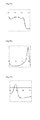
- Fig. 1 (A) shows a melting curve represented as the relationship between the temperature and the fluorescence intensity for a certain nucleic acid mixture
- Fig. 1 (B) shows a melting curve represented as the relationship between the temperature and the differential value of the fluorescence intensity (also referred to as a differential melting curve).
- Tm w which is the melting temperature of the nucleic acid Wt
- Tm M which is the melting temperature of the nucleic acid Mt
- ⁇ T w which is the temperature range including Tm w
- the temperature range whose lower limit is the temperature at which the differential value of the fluorescence intensity is lowest between Tm w and Tm M and whose upper limit is the temperature corresponding to the skirt of the peak of the fluorescence intensity can be set.
- ⁇ T M which is the temperature range including Tm M
- the temperature range whose upper limit is the temperature at which the differential value of the fluorescence intensity is lowest between Tm w and Tm M and whose lower limit is the temperature corresponding to the skirt of the peak of the fluorescence intensity
- the temperature range ⁇ T w and the temperature range ⁇ T M may be set such that these have either the same width (e.g., 10°C) or different widths (e.g., a temperature range ⁇ T w of 10°C and a temperature range ⁇ T M of 7°C).
- the temperature range ⁇ T w and the temperature range - ⁇ T M may be set such that each of these has a width ranging from X°C higher than the melting temperature to X°C lower than the melting temperature (e.g., X°C is not more than 15°C, preferably not more than 10°C).
- the area surrounded by the line passing through the point corresponding to the lower limit and the point corresponding to the upper limit of the temperature range of the differential melting curve, and the differential melting curve (shaded portion in Fig. 1(B) ) is calculated. More particularly, for example, the area can be calculated as follows. By defining the differential value of the fluorescence intensity at temperature T as f(T) and the base value at temperature T as B(T), the area is calculated by the Equation (1) below.
- T s represents the lower limit value of each temperature range
- T e represents the upper limit value
- the base value B(T) at each temperature T is a value calculated by the Equation (2) below and represents the background level contained in the detection signal of the fluorescence intensity. By subtracting this base value from the differential value of the fluorescence intensity, the effect of the background contained in the detection signal of the fluorescence intensity is removed.
- B T a ⁇ T - T s + f T s
- a ⁇ f(T e )-f(T s ) ⁇ /(T e -T s ).
- the area S w in the temperature range ⁇ T W and the area S M in the temperature range ⁇ T W are calculated, to prepare a calibration curve representing the relationship between the area ratio and the abundance ratio of the respective mixtures.
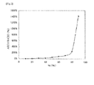
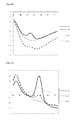
- Fig. 2 shows an example of the calibration curve prepared by plotting the abundance ratio (the ratio of the nucleic acid Mt with respect to the total amount of nucleic acids in the nucleic acid mixture) along the abscissa and the area ratio (S M /S W ) along the ordinate.
- the area ratio may also be defined as S W /S M .
- the abundance ratio of a nucleotide sequence having a polymorphism contained in the actual sample can be determined based on the prepared calibration curve.
- the DNA in the sample may be either single-stranded DNA or double-stranded DNA.
- the step of dissociating the double-stranded DNA in the sample by heating is preferably included before the hybridization step. By dissociating the double-stranded DNA into single-stranded DNA, hybridization with a detection probe is possible in the subsequent hybridization step.
- the ratio (molar ratio) of the probe of the present invention to be added with respect to the DNA in the sample is not restricted, and the ratio is preferably not more than 1, more preferably not more than 0.1 with respect to the DNA in the sample in view of to ensure a sufficient detection signal.
- the DNA in the sample may be either the total amount of the DNA having the polymorphism to be detected and DNA which does not have the polymorphism to be detected, or the total amount of the amplification product containing the sequence having the polymorphism to be detected and amplification products containing sequences which do not have the polymorphism to be detected.
- the ratio of the DNA to be detected relative to the DNA in the sample is usually not known, the ratio (molar ratio) of the probe to be added with respect to the DNA to be detected (the amplification product containing the sequence to be detected) is, for example, not more than 10, not more than 5, or not more than 3 as a result.
- the lower limit of the ratio is not restricted, and the ratio is, for example, not less than 0.001, not less than 0.01, or not less than 0.1.
- the ratio of the probe of the present invention to be added with respect to the DNA may be, for example, either the molar ratio with respect to the double-stranded DNA or the molar ratio with respect to the single-stranded DNA.
- the Tm value will now be described. Heating a solution containing double-stranded DNA causes an increase in absorbance at 260 nm. This is caused because hydrogen bonds between the strands of the double-stranded DNA are broken by the heat and the double-stranded DNA is dissociated into single-stranded DNA (melting of DNA). Based on this phenomenon, the melting temperature Tm can be generally defined as the temperature at which the increase in the absorbance reached 50% of the total increase in the absorbance.
- the difference in the Tm value is 5°C or more
- the above-mentioned mutants can be detected with high sensitivity.
- the difference in the Tm value 5°C or more, 7°C or more is exemplified.
- a method for increasing the difference in the Tm value a method wherein a probe is designed to include nucleotides mismatched to a nucleotide sequence to be hybridized, or a method described in Nature Biotech (1997) vol15, p.331-335 is exemplified.
- measurement of the signal fluctuation due to the temperature change for determination of the Tm value can be carried out also by measuring the absorbance at 260 nm based on the above-mentioned principle, but the measurement is preferably carried out based on a signal from a label added to the probe of the present invention, which signal fluctuates depending on the state of hybrid formation between the DNA and the probe. Therefore, as the probe of the present invention, the above-mentioned labeled probe is preferably used.
- the labeled probe examples include a fluorescently labeled oligonucleotide probe which emits fluorescence when it is not hybridized with the target sequence, whose fluorescence intensity decreases (the fluorescence is quenched) when the probe is hybridized with the target sequence, and a fluorescently labeled oligonucleotide probe which emits fluorescence when it is not hybridized with the target sequence, whose fluorescence intensity increases when the probe is hybridized with the target sequence.
- the probe shows no signal or a weak signal when it is forming a hybrid (double-stranded DNA) with the sequence to be detected, while the probe shows a signal or the signal increases when the probe is released by heating.
- the probe shows a signal by forming a hybrid (double-stranded DNA) with the sequence to be detected, while the signal decreases (disappears) when the probe is released by heating. Therefore, by detecting the change in the signal due to the label under conditions specific to the signal (absorbance and the like), determination of the progress of melting and the Tm value can be carried out in a similar manner to the measurement of the absorbance at 260 nm.
- the labeling substance in the labeled probe is as mentioned above, and the probe is preferably labeled with a fluorescent dye.
- the nucleic acid to be used as a template for carrying out the nucleic acid amplification is not restricted as long as it contains nucleic acid, and examples of the nucleic acid include those derived from, or those which may be derived from, arbitrary biological origins such as blood; oral mucosal suspensions; somatic cells of nails, hair and the like; germ cells; milk; ascitic fluid; paraffin-embedded tissues; gastric juices; fluids obtained by gastric lavage; peritoneal fluid; amniotic fluid; and cell cultures.
- the nucleic acid as a template may be used as it is directly after being obtained from any of the above-described origins, or may be pretreated to modify properties of the sample before being used.
- the method of nucleic acid amplification is further described by way of an example using PCR.
- the primer pair used in the PCR may be designed in the same manner as in the method for designing a primer pair for conventional PCR, except that the primer pair is designed such that a region with which the probe of the present invention can hybridize is amplified.
- the length and the Tm value of each primer is usually 12 mer to 40 mer and 40 to 70°C, or 16 mer to 30 mer and 55 to 60°C, respectively.
- the length of the respective primers of the primer pair does not need to be the same, but the Tm values of the both primers are preferably almost the same (the difference is usually not more than 2°C).
- the Tm value is a value calculated by the Nearest Neighbor method. Examples of the primer pair include the one composed of the primers having the nucleotide sequences shown in SEQ ID NOs:16 and 17.
- the PCR is preferably carried out in the presence of the probe of the present invention to be used in the present invention.
- the Tm analysis can be carried out without subjecting the amplified product to purification and/or the like after the amplification reaction.
- Those skilled in the art can easily control the Tm values of the primers and the reaction conditions for the PCR depending on the probe used.
- the detection of a mutation(s) in exon 12 of the NPM1 gene based on the result of Tm analysis can be carried out according to a conventional method.
- the detection herein includes detection of the presence/absence of a mutation(s) and determination of the abundance ratio(s) of a nucleic acid(s) having a polymorphism(s).
- a mutation(s) in exon 12 of the NPM1 gene can be detected, and the risk of developing acute myeloid leukemia, and/or the diseased state and/or prognosis of acute myeloid leukemia can be diagnosed based on the detected presence/absence of the mutation(s).
- the probes or kits of the present invention may be used in the diagnosis of acute myeloid leukemia (or for methods of diagnosis of said condition using methods of the invention).
- the kit of the present invention is a kit to be used for the detection method of the present invention.
- This kit comprises the probe of the present invention for detecting a polymorphism(s).
- the kit of the present invention can also be used for judging the risk of developing acute myeloid leukemia, and/or the diseased state and/or prognosis of acute myeloid leukemia.
- the probe is as described above as the probe of the present invention.
- the detection kit of the present invention may further comprise, in addition to the probe, reagents required for nucleic acid amplification in the detection method of the present invention, especially the above-described primers for amplification using a DNA polymerase.
- the probe, primers and other reagents may be contained separately, or a mixture of a part of them may be contained in the kit.
- the probes having C at their ends shown in Table 2 were designed.
- the position of each probe is indicated by its nucleotides in the nucleotide sequence shown in SEQ ID NO:1.
- "P" at the 3' end indicates phosphorylation.
- Labeling with TAMRA was carried out according to a conventional method.
- the sequences of the complementary strand oligonucleotides used as the subjects of detection are shown in Table 2.
- the position of each oligonucleotide is indicated by its nucleotides in the nucleotide sequence shown in SEQ ID NO:1.
- the complementary strands of the wild-type sequence and various mutant sequences of the NPM1 exon 12 region were prepared, and the following reagent solution comprising a probe and a complementary strand oligonucleotide was prepared.
- WT or mt type A, B, D, or 7 was added as the complementary strand oligonucleotide
- WT or mt type Q, 10, E, or 6 was added as the complementary strand oligonucleotide.
- the conditions of the Tm analysis were as follows.
- the excitation wavelength and the detection wavelength in the Tm analysis were 520 to 555 nm and 585 to 700 nm (TAMRA), respectively.
- the prepared reagent solution was added to an i-densy reaction tube, and 30 ⁇ L of mineral oil were overlaid on the solution to prevent evaporation of the reagent.
- Tm analysis was carried out under the following reaction conditions to analyze the amount of change in the fluorescence value of TAMRA.
- Example 1 Based on the results of Example 1, when the probes P1 (SEQ ID NO:4) and P2 (SEQ ID NO:5) were used, changes in the fluorescence intensity which can be analyzed by Tm analysis were observed for the polymorphisms of mutations (Type A, Type, B, Type D, Type 7, Type 10, Type E and Type 6) in exon 12 of the NPM1 gene. That is, each of the mutant types has another peak in addition to the peak for the wild-type, and a unique pattern of the amount of change in the fluorescence intensity exists. Therefore, by using the probes P1 (SEQ ID NO:4) and P2 (SEQ ID NO: 5), the polymorphisms of mutations in exon 12 of the NPM1 gene can be detected.
- the complementary strands of the wild-type sequence and various mutant sequences of the NPM1 exon 12 region were prepared, and the following reagent solution comprising the probe and each complementary strand oligonucleotide was prepared.
- the reagent solution was the same as in Example 1 except that the probe described below was used. Tm analysis was also carried out in the same manner as in Example 1.
- the sequence of the probe used for detection of the mutations is shown in Table 7. In Table 7, the position of the probe is indicated as nucleotides in the nucleotide sequence shown in SEQ ID NO:1.
- the reaction was performed using a blood sample. Further, in order to study how much nucleic acid comprising a mutation in exon 12 of the NPM1 gene is required in a sample to allow detection of the mutation, detection waveforms obtained with various mixing ratios between artificial nucleic acid plasmids of the wild-type sequence and a mutant sequence (Type A or Type E) were observed.
- plasmids were prepared by insertion of the wild-type sequence around exon 12 of the NPM1 gene (590 bp, SEQ ID NO:1), the mutant type A (594 bp, SEQ ID NO:2) or the mutant type E (594 bp, SEQ ID NO:3) to pUC plasmid vector.
- the PCR and Tm analysis were carried out using a fully automatic SNPs testing device (trade name: i-densy IS-5310, manufactured by ARKRAY, Inc.).
- the conditions of the PCR and Tm analysis were as shown in Table 11 below.
- the sequences of the probes and the primers used for detection of mutations are shown in Table 12.
- the positions of the probes and the primers are indicated as nucleotides in the nucleotide sequence shown in SEQ ID NO:1.
- the excitation wavelength and the detection wavelength in the Tm analysis were 420 to 485 nm and 520 to 555 nm, respectively (BODIPY FL), or 520 to 555 nm and 585 to 700 nm, respectively (TAMRA).
- the reagent solution was as follows.
- the wild-type and a mutant were mixed to prepare an artificial nucleic acid plasmid at a concentration of 500 copies/ ⁇ l, and 4 ⁇ l of the resulting artificial nucleic acid plasmid were used.
- a detection peak was observed without any problem ( Fig. 23 ).
- the detection waveform obtained using the artificial nucleic acid plasmid was observed, and, as a result, the detection peak of the wild type was found at 63°C in the case of BODIPY FL and similarly at 63°C in the case of TAMRA ( Fig. 24 ).
- a detection peak with BODIPY FL was found at 56 to 57°C ( Fig. 25 , left)
- a detection peak with TAMRA was found at 50 to 51 °C ( Fig. 29 , right).
- the 3T-NPMl-el2-R3 probe was used as a comparative example. Whether detection by Tm analysis is possible after nucleic acid amplification using the following solution was studied.
- the primer sequences and the reaction conditions were the same as in Example 2.
- 4 pL of a plasmid solution 500 copies/pL were added, and the reaction was carried out under the following reaction conditions using i-densy.
- Tm analysis the amount of change in the fluorescence value of TAMRA was analyzed.
- exon 12 of the NPM1 gene many mutated sequences are reported and most of them are Type A, Type B or Type D (See Table 1).
- the important aspect of a probe for detecting exon 12 mutations in the NPM1 gene is the ability to detect Type A, Type B and Type D clearly.
- SEQ ID NO:2 Type A
- the probes having C at their ends shown in Table 15 were designed. In Table 15, the position of each probe is indicated by its nucleotide positions in the nucleotide sequence shown in SEQ ID NO:2. Labeling with Pacific Blue, TAMRA and PODIPY FL was carried out according to a conventional method.
- Example 1 The reagent solution was the same with those used in Example 1 except that the following probes were used. Further, Tm analysis was carried out as in Example 1. The sequences of the complementary strand oligonucleotides used as the subjects of detection are shown in Table 1. These complementary strand oligonucleotides are sequences with mutated nucleotides corresponding to section 4 in Table 1 added.
- the excitation wavelength and the detection wavelength in the Tm analysis were 365 to 415 nm and 445 to 480 nm ( Pacific Blue), 520 to 555 nm and 585 to 700 nm (TAMRA), 420 to 485 nm and 520 to 555 nm (TAMRA), respectively.
- Tm values detection peaks
- 3PB-NPM1-mtA-R4 SEQ ID NO:56
- the difference in the Tm value ( ⁇ Tm) between WT and mutant Type M, N, Gm, 12, and I was 4°C or less.
- ⁇ Tm the difference in the Tm value between WT and mutant Type M, N, Gm, 12, and I was 4°C or less.
- the probe was proven to be suitable for detecting the mutations.
- mtA-R4 probe is a probe with a sequence complementary to a nucleotide sequence wherein a four nucleotide sequence "tctg", which is specific to Type A, was inserted into WT sequence (14 mer). Since mtA-R4 probe is identical to Type A, Type B, and Type D sequences in 18 mer, 16 mer, and 17 mer, respectively, a higher Tm value (higher affinity) is obtained compared with the WT complementary strand (identical in 14 mer). Thus, these mutants can be detected.
- nucleic acid comprising a mutation in exon 12 of the NPM1 gene should be contained in a sample to allow detection of the mutation.
- detection waveforms obtained with various mixing ratios between artificial nucleic acid plasmids with the wild-type sequence and a mutant sequence (Type A) were observed.
- plasmids were prepared by insertion of the wild-type sequence around exon 12 of the NPM1 gene (590 bp, SEQ ID NO:1) or the mutant type A (594 bp, SEQ ID NO:2) to pUC plasmid vector.
- the PCR and Tm analysis were carried out as described in Table 11 of Example 2.
- the excitation wavelength and the detection wavelength in the Tm analysis were 365 to 415 nm and 445 to 480 nm ( Pacific Blue), 520 to 555 nm and 585 to 700 nm (TAMRA), or 420 to 485 nm and 520 to 555 nm (BODIPY FL), respectively.
- mutant type A The detection peaks of mutant type A were confirmed using probes described in Table 15. In particular, a detection peak was found with mtA-R4 probe at a ratio of 10% or 3% of mutant type A at 61 to 62°C ( Fig. 39 ), a detection peak was found with mtB-R5 probe at a ratio of 10% of mutant type A at 53°C ( Fig. 40 ), and a detection peak was found with mtD-R6 probe at a ratio of 10% or 3% of mutant type A at 59 to 60°C ( Fig. 41 ).
- Example 4 Based on the results of Example 4, when mtA-R4 (SEQ ID NO:56), mtB-R5 (SEQ ID NO:57) and mtD-R6 (SEQ ID NO:58) were used, changes in the fluorescence intensity which can be analyzed by Tm analysis were observed for the polymorphisms of mutations (Type A) in exon 12 of the NPM1 gene. That is, the mutant type A has another peak in addition to the peak for the wild-type, and a unique pattern of the amount of change in the fluorescence intensity exists.
- the polymorphisms (Type A) of mutations in exon 12 of the NPM1 gene can be detected with a high sensitivity from a small amount of sample.
- the risk of developing acute myeloid leukemia, and/or the diseased state and/or prognosis of acute myeloid leukemia can be diagnosed.
Landscapes
- Health & Medical Sciences (AREA)
- Life Sciences & Earth Sciences (AREA)
- Chemical & Material Sciences (AREA)
- Immunology (AREA)
- Engineering & Computer Science (AREA)
- Organic Chemistry (AREA)
- Proteomics, Peptides & Aminoacids (AREA)
- Analytical Chemistry (AREA)
- Molecular Biology (AREA)
- Zoology (AREA)
- Pathology (AREA)
- Wood Science & Technology (AREA)
- Genetics & Genomics (AREA)
- Physics & Mathematics (AREA)
- General Health & Medical Sciences (AREA)
- Biotechnology (AREA)
- Microbiology (AREA)
- Biochemistry (AREA)
- Bioinformatics & Cheminformatics (AREA)
- General Engineering & Computer Science (AREA)
- Biophysics (AREA)
- Hematology (AREA)
- Biomedical Technology (AREA)
- Urology & Nephrology (AREA)
- Oncology (AREA)
- Hospice & Palliative Care (AREA)
- Cell Biology (AREA)
- Food Science & Technology (AREA)
- Medicinal Chemistry (AREA)
- General Physics & Mathematics (AREA)
- Tropical Medicine & Parasitology (AREA)
- Measuring Or Testing Involving Enzymes Or Micro-Organisms (AREA)
Abstract
(P5) an oligonucleotide comprising a nucleotide sequence complementary to a nucleotide sequence of 12 to 50 consecutive nucleotides containing nucleotides 145 to 156 in SEQ ID NO:2 or a homologous sequence thereof, wherein the nucleotide corresponding to the nucleotide at position 145 is a cytosine labeled with a fluorescent dye; and
(P5') an oligonucleotide comprising a nucleotide sequence complementary to a nucleotide sequence of 12 to 50 consecutive nucleotides containing nucleotides 145 to 156 in SEQ ID NO:2 or a sequence which hybridizes with the nucleotide sequence in SEQ ID NO:2 under stringent conditions, wherein the nucleotide corresponding to the nucleotide at position 145 is a cytosine labeled with a fluorescent dye.
Description
- The present invention relates to a method for detecting a mutation(s) in exon 12 of the NPM1 gene, and a nucleic acid probe and a kit therefor.
- Many DNA mutations involved in causes of acute myeloid leukemia (AML) have been discovered so far. Mutations of the NPM1 gene are found in adult patients suffering from acute myeloid leukemia, and it has been reported that these can be used for prognosis prediction by taking those mutations into consideration in combination with the result of analysis of the FLT3-ITD gene mutation (Blood. 2007, 109: 874-885). Examples of the type of mutation of the NPM1 gene include those described in New Eng J Med 352: 254-266, 2005; Blood. 2005, 106: 1419-1422; Blood. 2005, 106: 3740-3746; Blood. 2005, 106: 3733-3739; Blood. 2006, 108: 1999-2005; and Blood. 2005, 106: 2854-2861, and representative examples of the mutations include Type A, Type, B, Type D, Type 7, Type Q,
Type 10, Type E andType 6 in view of the number of cases reported and the positions in nucleotide sequences at which the mutations occur. - As methods for detecting polymorphisms of nucleotides, the following methods are known.
Blood. 2005, 106: 2854-2861 describes a method wherein amplification by PCR is carried out and the resulting amplification product is separated by electrophoresis, followed by cutting out a part of the gel, purifying the amplification product from the gel and subjecting the purified product to direct sequencing. Haematologica 2007, 92: 1268-1269 describes a method by detection using the DHPLC method and the sequencing method.
However, in these methods, (1) since an amplification product needs to be recovered, there is a risk of contamination; (2) since the operations are not automated, and since each step requires an operation, the methods are laborious and costly; (3) special knowledge and special skills are required for analysis of results; and (4) since the detection specificity in sequencing analysis is as low as about 20%, detection is difficult when the ratio of normal cells contained together with cancer cells is high; which are problematic. - On the other hand, methods wherein a region containing a mutation is amplified by PCR and a fluorescently labeled nucleic acid probe is used to carry out melting curve analysis, followed by analyzing the mutation based on the result of the melting curve analysis are known (
JP 2001-286300 A JP 2002-119291 A - The present invention aims to provide probes effective for detecting mutations in exon 12 of the NPM1 gene and to provide a method for detecting the mutations in exon 12 of the NPM1 gene and a kit therefor.
- The present inventors discovered that, by designing probes based on specific regions containing mutations in exon 12 of the NPM1 gene and carrying out melting curve analysis using the probes, the mutations can be detected, thereby completing the present invention.
- That is, the present invention is as follows.
- (1) A probe for detecting a mutation(s) in exon 12 of the NPM1 gene, comprising at least one fluorescently labeled oligonucleotide selected from the group consisting of P5, P5', P6, P6', P7, P7', P1, P1', P2 and P2' below:
- (P5) an oligonucleotide comprising a nucleotide sequence complementary to a nucleotide sequence of 12 to 50 consecutive nucleotides containing nucleotides 145 to 156 in SEQ ID NO:2 or a homologous sequence thereof, wherein the nucleotide corresponding to the nucleotide at position 145 is a cytosine labeled with a fluorescent dye;
- (P5') an oligonucleotide comprising a nucleotide sequence complementary to a nucleotide sequence of 12 to 50 consecutive nucleotides containing nucleotides 145 to 156 in SEQ ID NO:2 or a sequence which hybridizes with the nucleotide sequence in SEQ ID NO:2 under stringent conditions, wherein the nucleotide corresponding to the nucleotide at position 145 is a cytosine labeled with a fluorescent dye;
- (P6) an oligonucleotide comprising a nucleotide sequence complementary to a nucleotide sequence of 12 to 50 consecutive nucleotides containing nucleotides 145 to 156 in SEQ ID NO:2 or a homologous sequence thereof, wherein the nucleotide corresponding to the nucleotide at position 145 is a cytosine labeled with a fluorescent dye and the nucleotide corresponding to the nucleotide at position 153 is a guanine;
- (P6') an oligonucleotide comprising a nucleotide sequence complementary to a nucleotide sequence of 12 to 50 consecutive nucleotides containing nucleotides 145 to 156 in SEQ ID NO:2 or a sequence which hybridizes with the nucleotide sequence in SEQ ID NO:2 under stringent conditions, wherein the nucleotide corresponding to the nucleotide at position 145 is a cytosine labeled with a fluorescent dye and the nucleotide corresponding to the nucleotide at position 153 is a guanine;
- (P7) an oligonucleotide comprising a nucleotide sequence complementary to a nucleotide sequence of 12 to 50 consecutive nucleotides containing nucleotides 145 to 156 in SEQ ID NO:2 or a homologous sequence thereof, wherein the nucleotide corresponding to the nucleotide at position 145 is a cytosine labeled with a fluorescent dye, the nucleotide corresponding to the nucleotide at position 153 is a guanine, and the nucleotide corresponding to the nucleotide at position 154 is a thymine;
- (P7') an oligonucleotide comprising a nucleotide sequence complementary to a nucleotide sequence of 12 to 50 consecutive nucleotides containing nucleotides 145 to 156 in SEQ ID NO:2 or a sequence which hybridizes with the nucleotide sequence in SEQ ID NO:2 under stringent conditions, wherein the nucleotide corresponding to the nucleotide at position 145 is a cytosine labeled with a fluorescent dye, the nucleotide corresponding to the nucleotide at position 153 is a guanine, and the nucleotide corresponding to the nucleotide at position 154 is a thymine;
- (P1) an oligonucleotide comprising a nucleotide sequence complementary to a nucleotide sequence of 16 to 50 consecutive nucleotides containing nucleotides 135 to 150 in SEQ ID NO:1 or a homologous sequence thereof, wherein the nucleotide corresponding to the nucleotide at position 135 is a cytosine labeled with a fluorescent dye;
- (P1') an oligonucleotide comprising a nucleotide sequence complementary to a nucleotide sequence of 16 to 50 consecutive nucleotides containing nucleotides 135 5 to 150 in SEQ ID NO:1 or a sequence which hybridizes with the nucleotide sequence in SEQ ID NO:1 under stringent conditions, wherein the nucleotide at position 135 is a cytosine labeled with a fluorescent dye;
- (P2) an oligonucleotide comprising a nucleotide sequence complementary to a nucleotide sequence of 19 to 50 consecutive nucleotides containing nucleotides 164 to 182 in SEQ ID NO:1 or a homologous sequence thereof, wherein the nucleotide corresponding to the nucleotide at position 182 is a cytosine labeled with a fluorescent dye; and
- (P2') an oligonucleotide comprising a nucleotide sequence complementary to a nucleotide sequence of 19 to 50 consecutive nucleotides containing nucleotides 164 to 182 in SEQ ID NO:1 or a sequence which hybridizes with the nucleotide sequence in SEQ ID NO:1 under stringent conditions, wherein the nucleotide corresponding to the nucleotide at position 182 is a cytosine labeled with a fluorescent dye.
- (2) The probe according to (1), wherein
said oligonucleotides P5, P5', P6, P6', P7, and P7' have the nucleotide corresponding to the nucleotide at position 145 labeled with a fluorescent dye at the first, second or third position from the 3' end;
said oligonucleotides P1 and P1' have the nucleotide corresponding to the nucleotide at position 135 labeled with a fluorescent dye at the first, second or third position from the 3' end; and
said oligonucleotides P2 and P2' have the nucleotide corresponding to the nucleotide at position 182 labeled with a fluorescent dye at the first, second or third position from the 5' end. - (3) The probe according to (1), wherein
said oligonucleotides P5, P5', P6, P6', P7, and P7' have the nucleotide corresponding to the nucleotide at position 145 labeled with a fluorescent dye at the 3' end;
said oligonucleotides P1 and P1' have the nucleotide corresponding to the nucleotide at position 135 labeled with a fluorescent dye at the 3' end; and
said oligonucleotides P2 and P2' have the base corresponding to the nucleotide at position 182 labeled with a fluorescent dye at the 5' end. - (4) The probe according to (1), wherein
said oligonucleotides P1 and P1' have the nucleotide corresponding to any one of the nucleotides at positions 153 to 156 at the 5' end and the nucleotide corresponding to the nucleotide at position 135 labeled with a fluorescent dye at the 3' end; and
said oligonucleotides P2 and P2' have the nucleotide corresponding to any one of the nucleotides at positions 153 to 156 at the 3' end and the nucleotide corresponding to the nucleotide at position 182 labeled with a fluorescent dye at the 5' end. - (5) The probe according to (1), wherein
said oligonucleotides P5, P5', P6, P6', P7, and P7' have the nucleotide corresponding to the nucleotide at position 162 at the 5' end and the nucleotide corresponding to the nucleotide at position 145 labeled with a fluorescent dye at the 3' end;
said oligonucleotides P1 and P1' have the nucleotide corresponding to the nucleotide at position 155 at the 5' end and the nucleotide corresponding to the nucleotide at position 135 labeled with a fluorescent dye at the 3' end; and
said oligonucleotides P2 and P2' have the nucleotide corresponding to the nucleotide at position 156 at the 3' end and the nucleotide corresponding to the nucleotide at position 182 labeled with a fluorescent dye at the 5' end. - (6) The probe according to any one of (1) to (5), wherein said oligonucleotide emits fluorescence when said probe is not hybridized with a target sequence and the fluorescence intensity decreases or increases when said probe is hybridized with said target sequence.
- (7) The probe according to (6), wherein said oligonucleotide emits fluorescence when said probe is not hybridized with a target sequence and the fluorescence intensity decreases when said probe is hybridized with said target sequence.
- (8) The probe according to any one of (1) to (7), wherein said probe is a probe for melting curve analysis.
- (9) The probe for detecting a polymorphism according to any one of (1) to (8), wherein said oligonucleotides P5, P5', P6, P6', P7, and P7' have 12 to 35 consecutive nucleotides, said oligonucleotides P1 and P1' have 16 to 35 consecutive nucleotides, and said oligonucleotides P2 and P2' have 19 to 35 consecutive nucleotides.
- (10) A method for detecting a polymorphism(s) in exon 12 of the NPM1 gene, which method uses at least one probe according to any one of (1) to (9).
- (11) The method for detecting a polymorphism(s) in exon 12 of the NPM1 gene, preferably according to (10), comprising:
- (I) adding the probe according to any one of (1) to (9) to a sample comprising a nucleic acid, to allow said probe to hybridize with said nucleic acid;
- (II) changing the temperature to dissociate the hybridization complex between said nucleic acid and said probe, and measuring the fluctuation of the signal due to the dissociation of saidhybridization complex;
- (III) analyzing said fluctuation of the signal to detect the Tm value of the single-stranded nucleic acid in said sample; and
- (IV) determining based on said Tm value the presence or absence of said polymorphism(s) of interest or the abundance ratio(s) of single-stranded nucleic acid having said polymorphism(s) relative to the single-stranded nucleic acids in said sample.
- (12) The method according to (11), further comprising amplifying the nucleic acid (e.g. DNA) before Step (I) or at the same time as Step (I).
- (13) A method for analyzing the risk of developing acute myeloid leukemia, and/or the diseased state and/or prognosis of acute myeloid leukemia by using the method according to any one of (10) to (12), comprising detecting a polymorphism(s) in exon 12 of the NPM1 gene and determining the presence/absence of the polymorphism(s). Said method may be performed in a sample from a subject (e.g. a human) in whom the assessment is to be made.
- (14) A reagent kit for detecting a polymorphism(s) in the NPM1 gene, comprising the probe according to any one of (1) to (9).
- (15) The reagent kit according to (14), comprising primers for amplifying a region(s) comprising a sequence(s) in the nucleotide sequence shown in SEQ ID NO:1 in the NPM1 gene, with which said oligonucleotide(s) P5, P5', P6, P6', P7, P7', P1, P1', P2 and/or P2' hybridize(s).
- (16) The reagent kit according to (14) or (15), wherein said primers are for detecting a polymorphism(s), selected from P3 and P4, or P3' and P4':
- (P3) an oligonucleotide of 10 to 50 consecutive nucleotides having T at position 106 at the 3' end, which is homologous to SEQ ID NO:1; and
- (P4) an oligonucleotide of 10 to 50 consecutive nucleotides having T at position 205 at the 3' end, which is homologous to the complementary strand of SEQ ID NO:1; or
- (P3') an oligonucleotide of 10 to 50 consecutive nucleotides having T at position 106 at the 3' end, which hybridizes with the complementary strand of SEQ ID NO:1 under stringent conditions; and
- (P4') an oligonucleotide of 10 to 50 consecutive nucleotides having T at 205 position at the 3' end, which hybridizes with the nucleotide sequence of SEQ ID NO:1 under stringent conditions. The invention also extends to the use of said kit for detecting said polymorphism(s).
- Only by adding a probe of the present invention and carrying out melting curve analysis (Tm analysis), can a mutation(s) in exon 12 of the NPM1 gene be detected. The probes of the present invention have high specificity and high detection sensitivity. Since, by using the method of the present invention, the operation of recovery of an amplification product can be eliminated even in cases where PCR is carried out, there is hardly any risk of contamination. Further, since the operations in the method of the present invention are simple, they can be easily automated. By using the probes of the present invention, 7 mutations can be identified among the 8 representative types of mutations which have been reported so far. Further, 2 representative mutant types (Types A and E) can be detected even in cases where each of these mutations exists in a proportion of as low as 10% relative to the wild-type.
-
-
Fig. 1 is a diagram showing examples of (A) a melting curve of a nucleic acid mixture and (B) a differential melting curve. -
Fig. 2 is a diagram showing an example of a calibration curve. -
Fig. 3 is a schematic view of the design of the probes of the present invention. The positions of the probes used in the Examples are shown. The positions of the probes used in the Comparative Examples are also shown. -
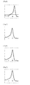
Fig. 4 is a diagram showing the relationship between the amount of change in the fluorescence intensity of TAMRA (3T-NPM1-e12-R1) per unit time (d the amount of increase in the fluorescence intensity/t) and the temperature in the Tm analysis in Example 1 for WT (complementary strand oligonucleotide). The amount of change in the fluorescence intensity per unit time is plotted along the ordinate and the temperature is plotted along the abscissa. This relationship between the ordinate and the abscissa also applies to the diagrams below. -
Fig. 5 shows the result of Tm analysis for mt type A (complementary strand oligonucleotide) in Example 1 using TAMRA(3T-NPM1-e12-R1) as the probe. -
Fig. 6 shows the result of Tm analysis for mt type B (complementary strand oligonucleotide) in Example 1 using TAMRA(3T-NPM1-e12-R1) as the probe. -
Fig. 7 shows the result of Tm analysis for mt type D (complementary strand oligonucleotide) in Example 1 using TAMRA(3T-NPM1-e12-R1) as the probe. -
Fig. 8 shows the result of Tm analysis for mt type 7 (complementary strand oligonucleotide) in Example 1 using TAMRA(3T-NPM1-e12-R1) as the probe. -
Fig. 9 shows the result of Tm analysis for WT (complementary strand oligonucleotide) in Example 1 using TAMRA(5T-NPM1-e12-R2) as the probe. -
Fig. 10 shows the result of Tm analysis for mt type Q (complementary strand oligonucleotide) in Example 1 using TAMRA(5T-NPM1-e12-R2) as the probe. -
Fig. 11 shows the result of Tm analysis for mt type 10 (complementary strand oligonucleotide) in Example 1 using TAMRA(5T-NPM1-e12-R2) as the probe. -
Fig. 12 shows the result of Tm analysis for mt type E (complementary strand oligonucleotide) in Example 1 using TAMRA(5T-NPM1-e12-R2) as the probe. -
Fig. 13 shows the result of Tm analysis for mt type 6 (complementary strand oligonucleotide) in Example 1 using TAMRA(5T-NPM1-e12-R2) as the probe. -
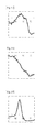
Fig. 14 shows the result of Tm analysis for WT (complementary strand oligonucleotide) in Comparative Example 1 using TAMRA(3T-NPM1-e12-R3) as the probe. -
Fig. 15 shows the result of Tm analysis for mt type A (complementary strand oligonucleotide) in Comparative Example 1 using TAMRA(3T-NPM1-e12-R3) as the probe. -
Fig. 16 shows the result of Tm analysis for mt type B (complementary strand oligonucleotide) in Comparative Example 1 using TAMRA(3T-NPM1-e12-R3) as the probe. -
Fig. 17 shows the result of Tm analysis for mt type D (complementary strand oligonucleotide) in Comparative Example 1 using TAMRA(3T-NPM1-e12-R3) as the probe. -
Fig. 18 shows the result of Tm analysis for mt type 7 (complementary strand oligonucleotide) in Comparative Example 1 using TAMRA(3T-NPM1-e12-R3) as the probe. -
Fig. 19 shows the result of Tm analysis for mt type Q (complementary strand oligonucleotide) in Comparative Example 1 using TAMRA(3T-NPM1-e12-R3) as the probe. -
Fig. 20 shows the result of Tm analysis for mt type 10 (complementary strand oligonucleotide) in Comparative Example 1 using TAMRA(3T-NPM1-e12-R3) as the probe. -
Fig. 21 shows the result of Tm analysis for mt type E (complementary strand oligonucleotide) in Comparative Example 1 using TAMRA(3T-NPM1-e12-R3) as the probe. -
Fig. 22 shows the result of Tm analysis for mt type 6 (complementary strand oligonucleotide) in Comparative Example 1 using TAMRA(3T-NPM1-e12-R3) as the probe. -
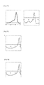
Fig. 23 shows the results of Tm analysis after PCR reaction for the blood sample in Example 2 using BODIPY FL(3FL-NPM1-e12-R1) (left) and TAMRA(5T-NPM1-e12-R2) (right) as the probes. -
Fig. 24 shows the results of Tm analysis after PCR reaction forWT 100% (plasmid) in Example 2 using BODIPY FL(3FL-NPM1-e12-R1) (left) and TAMRA(5T-NPM1-e12-R2) (right) as the probes. -
Fig. 25 shows the results of Tm analysis after PCR reaction for mt type A 100% (plasmid) in Example 2 using BODIPY FL(3FL-NPM1-e12-R1) (left) and TAMRA(5T-NPM1-e12-R2) (right) as the probes. -
Fig. 26 shows the results of Tm analysis after PCR reaction for mt type A 30% andWt 70% (plasmids) in Example 2 using BODIPY FL(3FL-NPM1-e12-R1) (left) and TAMRA(5T-NPM1-e12-R2) (right) as the probes. -
Fig. 27 shows the results of Tm analysis after PCR reaction for mt type A 20% andWt 80% (plasmids) in Example 2 using BODIPY FL(3FL-NPM1-e12-R1) (left) and TAMRA(5T-NPM1-e12-R2) (right) as the probes. -
Fig. 28 shows the results of Tm analysis after PCR reaction for mt type A 10% and Wt 90% (plasmids) in Example 2 using BODIPY FL(3FL-NPM1-e12-R1) (left) and TAMRA(5T-NPM1-e12-R2) (right) as the probes. -
Fig. 29 shows the results of Tm analysis after PCR reaction formt type E 100% (plasmid) in Example 2 using BODIPY FL(3FL-NPM1-e12-R1) (left) and TAMRA(5T-NPM1-e12-R2) (right) as the probes. -
Fig. 30 shows the results of Tm analysis after PCR reaction for mt type E 30% andWt 70% (plasmids) in Example 2 using BODIPY FL(3FL-NPM1-e12-R1) (left) and TAMRA(5T-NPM1-e12-R2) (right) as the probes. -
Fig. 31 shows the results of Tm analysis after PCR reaction formt type E 20% andWt 80% (plasmids) in Example 2 using BODIPY FL(3FL-NPM1-e12-R1) (left) and TAMRA(5T-NPM1-e12-R2) (right) as the probes. -
Fig. 32 shows the results of Tm analysis after PCR reaction formt type E 10% and Wt 90% (plasmids) in Example 2 using BODIPY FL(3FL-NPM1-e12-R1) (left) and TAMRA(5T-NPM1-e12-R2) (right) as the probes. -
Fig. 33 shows the result of Tm analysis after PCR reaction for the blood sample in Comparative Example 2 using TAMRA(3T-NPM1-e12-R3) as the probe. -
Fig. 34 shows the result of Tm analysis after PCR reaction forWt 100% (plasmid) in Comparative Example 2 using TAMRA(3T-NPM1-e12-R3) as the probe. -
Fig. 35 shows the result of Tm analysis after PCR reaction for mt type A 100% (plasmid) in Comparative Example 2 using TAMRA(3T-NPM1-e12-R3) as the probe. -
Fig. 36 shows the result of Tm analysis after PCR reaction for mt type A 20% andWt 80% (plasmids) in Comparative Example 2 using TAMRA(3T-NPM1-e12-R3) as the probe. -
Fig. 37 shows the result of Tm analysis after PCR reaction formt type E 100% (plasmid) in Comparative Example 2 using TAMRA(3T-NPM1-e12-R3) as the probe. -
Fig. 38 shows the result of Tm analysis after PCR reaction formt type E 20% andWt 80% (plasmids) in Comparative Example 2 using TAMRA(3T-NPM1-e12-R3) as the probe. -
Fig. 39 shows the result of Tm analysis after PCR reaction for mt type A (plasimid) in Example 4 using Pacific Blue(mtA-R4) as the probe. -
Fig. 40 shows the result of Tm analysis after PCR reaction for mt type A (plasmid) in Example 4 using TAMRA(mtB-R5) as the probe. -
Fig. 41 shows the result of Tm analysis after PCR reaction for mt type A (plasmid) in Example 4 using BODIPY FL(mtD-R6) as the probe. - The probe of the present invention is a probe for detecting a mutation(s) in exon 12 of the NPM1 gene, comprising at least one fluorescently labeled oligonucleotide selected from the group consisting of (P5), (P5'), (P6), (P6'), (P7), (P7'), (P1), (P1'), (P2) and (P2') below:
- (P5) an oligonucleotide comprising a nucleotide sequence complementary to a nucleotide sequence of 12 to 50 consecutive nucleotides containing nucleotides 145 to 156 in SEQ ID NO:2 or a homologous sequence thereof, wherein the nucleotide corresponding to the nucleotide at position 145 is a cytosine labeled with a fluorescent dye;
- (P5') an oligonucleotide comprising a nucleotide sequence complementary to a nucleotide sequence of 12 to 50 consecutive nucleotides containing nucleotides 145 to 156 in SEQ ID NO:2 or a sequence which hybridizes with the nucleotide sequence in SEQ ID NO:2 under stringent conditions, wherein the nucleotide corresponding to the nucleotide at position 145 is a cytosine labeled with a fluorescent dye;
- (P6) an oligonucleotide comprising a nucleotide sequence complementary to a nucleotide sequence of 12 to 50 consecutive nucleotides containing nucleotides 145 to 156 in SEQ ID NO:2 or a homologous sequence thereof, wherein the nucleotide corresponding to the nucleotide at position 145 is a cytosine labeled with a fluorescent dye and the nucleotide corresponding to the nucleotide at position 153 is a guanine;
- (P6') an oligonucleotide comprising a nucleotide sequence complementary to a nucleotide sequence of 12 to 50 consecutive nucleotides containing nucleotides 145 to 156 in SEQ ID NO:2 or a sequence which hybridizes with the nucleotide sequence in SEQ ID NO:2 under stringent conditions, wherein the nucleotide corresponding to the nucleotide at position 145 is a cytosine labeled with a fluorescent dye and the nucleotide corresponding to the nucleotide at position 153 is a guanine;
- (P7) an oligonucleotide comprising a nucleotide sequence complementary to a nucleotide sequence of 12 to 50 consecutive nucleotides containing nucleotides 145 to 156 in SEQ ID NO:2 or a homologous sequence thereof, wherein the nucleotide corresponding to the nucleotide at position 145 is a cytosine labeled with a fluorescent dye, the nucleotide corresponding to the nucleotide at position 153 is a guanine, and the nucleotide corresponding to the nucleotide at position 154 is a thymine;
- (P7') an oligonucleotide comprising a nucleotide sequence complementary to a nucleotide sequence of 12 to 50 consecutive nucleotides containing nucleotides 145 to 156 in SEQ ID NO:2 or a sequence which hybridizes with the nucleotide sequence in SEQ ID NO:2 under stringent conditions, wherein the nucleotide corresponding to the nucleotide at position 145 is a cytosine labeled with a fluorescent dye, the nucleotide corresponding to the nucleotide at position 153 is a guanine, and the nucleotide corresponding to the nucleotide at position 154 is a thymine;
- (P1) an oligonucleotide comprising a nucleotide sequence complementary to a nucleotide sequence of 16 to 50 consecutive nucleotides containing nucleotides 135 to 150 in SEQ ID NO:1 or a homologous sequence thereof, wherein the nucleotide corresponding to the nucleotide at position 135 is a cytosine labeled with a fluorescent dye;
- (P1') an oligonucleotide comprising a nucleotide sequence complementary to a nucleotide sequence of 16 to 50 consecutive nucleotides containing nucleotides 135 to 150 in SEQ ID NO:1 or a sequence which hybridizes with the nucleotide sequence in SEQ ID NO:1 under stringent conditions, wherein the nucleotide at position 135 is a cytosine labeled with a fluorescent dye;
- (P2) an oligonucleotide comprising a nucleotide sequence complementary to a nucleotide sequence of 19 to 50 consecutive nucleotides containing nucleotides 164 to 182 in SEQ ID NO:1 or a homologous sequence thereof, wherein the nucleotide corresponding to the nucleotide at position 182 is a cytosine labeled with a fluorescent dye; and
- (P2') an oligonucleotide comprising a nucleotide sequence complementary to a nucleotide sequence of 19 to 50 consecutive nucleotides containing nucleotides 164 to 182 in SEQ ID NO:1 or a sequence which hybridizes with the nucleotide sequence in SEQ ID NO:1 under stringent conditions, wherein the nucleotide corresponding to the nucleotide at position 182 is a cytosine labeled with a fluorescent dye.
- The probe of the present invention can be prepared in the same manner as the quenching probes described in
JP 2001-286300 A JP 2002-119291 A JP 2001-286300 A JP 2002-119291 A - The length of the probes P5, P5', P6, P6', P7, and P7' of the present invention is, for example, 12 to 50 consecutive nucleotides, 12 to 35 consecutive nucleotides, or 12 to 30 consecutive nucleotides. The length of the probes P1 and P1' of the present invention is, for example, 16 to 50 consecutive nucleotides, 16 to 35 consecutive nucleotides, or 16 to 30 consecutive nucleotides. The length of the probes P2 and P2' of the present invention is, for example, 19 to 50 consecutive nucleotides, 19 to 35 consecutive nucleotides, or 19 to 30 consecutive nucleotides.
For example, the probe P5, P5', P6, P6', P7, and P7'of the present invention is a probe having at its 5' end the nucleotide corresponding to any of thenucleotides 160 to 163 in the nucleotide sequence shown in SEQ ID NO:2, or having the nucleotide at position 162 at its 5' end and the nucleotide at position 145 at its 3' end.
For example, the probe of the present invention is a probe having at its end the nucleotide corresponding to any of the nucleotides 153 to 156 (gcag in Table 1 below) in the nucleotide sequence shown in SEQ ID NO:1, and P1 and P1' has at its 5' end any of the nucleotides at positions 153 to 156 and P2 and P2' has at its 3' end any of the nucleotides at positions 153 to 156. For example, in the probe of the present invention, P1 and P1' has at its 5' end the nucleotide at position 155 in the nucleotide sequence shown in SEQ ID NO:1 and P2 and P2' has at its 3' end the nucleotide at position 156 in the nucleotide sequence shown in SEQ ID NO:1. - The term "homologous sequence" or "homologous" herein means that a nucleotide sequence comprises a sequence having an identity of not less than 80%, 85%, 90%, 95%, 96%, 97%, 98% or 99% to a particular nucleotide sequence (e.g. SEQ ID NO: 1 or 2) or to the complementary strand of a particular nucleotide sequence (or the part of said sequence corresponding to the homologous sequence). In the present invention, 100% identity may be included.
The hybridization herein can be carried out according to a known method or a method corresponding thereto, such as the method described in Molecular Cloning 3rd (J. Sambrook et al., Cold Spring Harbor Lab. Press, 2001). This literature is hereby incorporated in the present specification by reference. Preferably said hybridization is under stringent conditions.
The stringent conditions mean conditions under which a specific hybrid is formed while nonspecific hybrids are not formed. Typical examples of stringent conditions include conditions under which hybridization is performed with a potassium concentration of about 25 mM to about 50 mM and a magnesium concentration of about 1.0 mM to about 5.0 mM. Examples of the conditions in the present invention include conditions under which hybridization is performed in Tris-HCl (pH 8.6), 25 mM KCl and 1.5 mM MgCl2, but the conditions are not limited thereto. Other examples of stringent conditions include those described in Molecular Cloning 3rd (J. Sambrook et al., Cold Spring Harbor Lab. Press, 2001).
This literature is hereby incorporated in the present specification by reference.
Those skilled in the art can easily select such conditions by controlling the hybridization reaction and/or changing the salt conditions of the hybridization reaction solution. - The fluorescently labeled oligonucleotides P5, P5', P6, P6', P7, P7', P1, P1', P2 and P2' of the present invention include fluorescently labeled oligonucleotides P5, P5', P6, P6', P7, P7', P1, P1', P2 andP2' with nucleotides added, deleted, or substituted, respectively.
As long as the fluorescently labeled oligonucleotides P5, P5', P6, P6', P7, P7', P1, P1, P2 and P2' with nucleotides added, deleted, or substituted of the present invention, can show the same effect as the fluorescently labeled oligonucleotides P5, P5', P6, P6', P7, P7', P1, P1', P2 and P2', those oligonucleotides can be included in the present invention. When nucleotides are added, deleted, or substituted, the position of addition, deletion, or substitution is not particularly limited. The number of nucleotides to be added, deleted, or substituted is one or two nucleotides, for example. Although the number differs according to the whole length of the fluorescently labeled oligonucleotide, the number of nucleotides to be added, deleted, or substituted is 1 to 10, or 1 to 5, for example. - Among the addition, deletion, or substitution, the fluorescently labeled oligonucleotides P5, P5', P6, P6', P7, P7', P1, P1', P2 and P2' of the present invention include fluorescently labeled oligonucleotides P5, P5', P6, P6', P7, P7', P1, P1', P2 and P2' wherein nucleotides in the fluorescently labeled oligonucleotide are substituted. The position to be substituted is not particularly limited. For example, in view of detection sensitivity, the nucleotides corresponding to nucleotides other than nucleotides 152 to 166 in the nucleotide sequence of SEQ ID NO:2 and nucleotides 152 to 162 in the nucleotide sequence of SEQ ID NO:1 are substituted. For example, the number of nucleotides to be substituted is 1, 2 or more. Although the number of nucleotides to be substituted depends on the whole number of the fluorescently labeled oligonucleotide, the number is 1 to 5 nucleotides or 1 to 3 nucleotides, for example.
- Oligonucleotides according to the above-mentioned fluorescently labeled oligonucleotides include modified oligonucleotides as well as oligonucleotides.
Examples of the nucleotides or units making up the oligonucleotides include ribonucleotides, deoxylibonucleotides, and artificial nucleic acids. The artificial nucleic acids include DNA; RNA; RNA analogue LNA (Locked Nucleic Acid); PNA (Peptide Nucleic Acid); BNA (Bridged Nucleic Acid) etc.
The above-mentioned oligonucleotides can be comprised of one or more kinds of the nucleotides or units. - Examples of the nucleotide sequence of the probe for detection of a mutation(s) in exon 12 of the NPM1 gene used in the present invention include, as P5, 5'-cactgcCAGAcagagatc-3' (SEQ ID NO:56), as P6, 5'-cactgcCATGcagagatc-3'
(SEQ ID NO:57), as P7, 5'-cactgcCAGGcagagatc-3' (SEQ ID NO:58), as P1, 5'-tgccagagatcttgaatagcc-3' (SEQ ID NO:4), and, as P2, 5'-ctattttcttaaagagacttcctccac-3' (SEQ ID NO:5).
As the fluorescent dye, those described inJP 2001-286300 A JP 2002-119291 A JP 2001-286300 A JP 2002-119291 A - By using any one of the oligonucleotides (P5), (P5'), (P6), (P6'), (P7), (P7'), (P1), (P1'), (P2) and (P2') of the present invention; by using three oligonucleotides, for example, (P5), (P6) and (P7), or (P5'), (P6') and (P7'); by using two oligonucleotides, for example, (P1) and (P2), or (P1') and (P2'); by using the probes shown in SEQ ID NOs: 56 to 58 described in Examples 3 and 4 in the present specification; or by using the probes shown in SEQ ID NOs: 4 and 5 described in Examples 1 and 2 in the present specification; a mutation(s) in exon 12 of the NPM1 gene can be detected (
Fig. 3 ). - Preferably, the probe of the present invention emits fluorescence from a fluorescent dye when the probe is not hybridized with the target sequence, and the fluorescence from the fluorescent dye decreases or increases when the probe is hybridized with the target sequence. For example, the probe of the present invention is a quenching probe which emits fluorescence from a fluorescent dye when the probe is not hybridized, and the fluorescence from the fluorescent dye is quenched when the probe is hybridized.
Further, the probe of the present invention preferably has a base labeled with a fluorescent dye at the first, second or third position from the 5' or 3' end, and more preferably the 3' end of the probe is labeled with a fluorescent dye. In the present specification, when the term "first, second or third position from the 5' end" is mentioned, the 5' end is counted as the first position, and, when the term "first, second or third position from the 3' end" is mentioned, the 3' end is counted as the first position.
The nucleotide labeled with a fluorescent dye in the probe of the present invention is the nucleotide at the position corresponding to position 145 in SEQ ID NO:2 in respect of P5, P5', P6, P6', P7 and P7', the nucleotide at the position corresponding to position 135 in SEQ ID NO:1 in respect of P1 and P1', and the nucleotide at the position corresponding to position 182 in SEQ ID NO:1 in respect of P2 and P2'. - The mutations in exon 12 of the NPM1 gene which can be detected by the oligonucleotides of the present invention are, for example, described in Table 1 below, and at least one mutation selected from the group consisting of Type A, Type B, Type D, Type C, Type E, Type Gm, Type Km, Type Nm, Type Om, Type Qm,
Type 3, Type 4,Type 6, Type 7,Type 10, Type 13, Type G+, Type H+, Type I+, Type J+, and Type I can be detected. -


Various mutant sequences have been reported for exon 12 of the NPM1 gene. The numbers of cases reported in several documents are summarized and described in the left side of the table as "number of cases of mutation". The number of reported cases of the mutant sequence A is considerably large, and the number of reported cases of the mutant sequences B and C are second largest.
In the nucleotide sequences shown in Table 1, for example, the mutant type A has "tctg" inserted at the site 4. Further, the mutant type E has "ctcttgccc" inserted at the site 9 and deletion of "ggagg" at thesite 10.
The mutants whose detection was studied in the Examples are marked with "☆". - In cases where a probe such as the oligonucleotide (P5), (P5'), (P6), (P6'), (P7) or (P7'), is used, Type A, Type B, Type D, Type C, Type Gm, Type Km, Type Nm, Type Om, Type Qm,
Type 3, Type 4, Type 13, Type G+, Type H+, Type I+, Type J+, and Type I and the like can be detected.
In cases where a probe positioned on the 5' side of the region of nucleotides 153 to 156 in the nucleotide sequence shown in SEQ ID NO:1, such as the oligonucleotide (P1) or (P1'), is used, Type A, Type B, Type D, Type 7 and the like can be detected.
In cases where a probe positioned on the 3'side of the region of nucleotides 153 to 156 in the nucleotide sequence shown in SEQ ID NO:1, such as the oligonucleotide (P2) or (P2'), is used,Type 10, Type E,Type 6 and the like can be detected.
Further, by using any one of the oligonucleotides (P5), (P5'), (P6), (P6'), (P7) and (P7') or the oligonucleotides (P1) or (P1') and (P2) or (P2') of the present invention, the presence/absence of mutations in exon 12 of the NPM1 gene can be detected, and therefore the risk of developing acute myeloid leukemia, and/or the diseased state and/or prognosis of acute myeloid leukemia can be diagnosed. - By using the probes of SEQ ID NOs:56, 57, 58, 4 and 5 shown in the Examples of the present specification, mutations in exon 12 of the NPM1 gene can be detected. The probe shown in SEQ ID NO:56-58, 4 is labeled with a fluorescent dye at its 3' end, and the probe shown in SEQ ID NO:5 is labeled with a fluorescent dye at its 5' end.
- The detection method of the present invention is characterized in that a probe(s) of the present invention as described above is/are used. That is, in the detection method of the present invention, one or more probes each having the region of nucleotides 157 to 160 in the nucleotide sequence shown in SEQ ID NO:2 (corresponding to the region of nucleotides 153 to 156 in the nucleotide sequence shown in SEQ ID NO:1) may be used, or one or more probes selected from the above-described P5, P5', P6, P6', P7 and P7' may be used. Further, one or more probes each having at its end the region of nucleotides 153 to 156 in the nucleotide sequence shown in SEQ ID NO:1 may be used, or one or more probes selected from the above-described P1, P1', P2 and P2' may be used.
- For example, the detection method of the present invention uses the probe of the present invention and comprises the following:
- (I) adding the probe of the present invention to a sample comprising a nucleic acid, to allow the probe to hybridize with the nucleic acid;
- (II) changing the temperature to dissociate the hybridization complex between the nucleic acid and the probe, and measuring the fluctuation of the signal due to the dissociation of said hybridization complex;
- (III) analyzing the fluctuation of the signal to detect the Tm value of single-stranded nucleic acid in the sample; and
- (IV) determining based on the Tm value the presence or absence of the polymorphism(s) of interest or the abundance ratio(s) of single-stranded nucleic acid having the polymorphism(s) relative to the single-stranded nucleic acids in the sample.
- The detection method of the present invention can be carried out in the same manner as conventional methods for nucleic acid amplification and melting curve analysis (Tm analysis) except that the probe of the present invention is used. Further, the detection method of the present invention may also comprise amplifying nucleic acid before Step (I) or at the same time as Step (I).
- The method of nucleic acid amplification preferably uses a polymerase, and examples of the method include PCR, ICAN and LAMP. When the amplification is carried out by a method using a polymerase, the amplification is preferably carried out in the presence of the probe of the present invention. Those skilled in the art can easily control reaction conditions and the like of the amplification depending on the probe to be used. By this, the detection can be carried out just by analyzing the Tm value of the probe after the amplification of the nucleic acid, so that the amplification product does not need to be handled after the reaction. Therefore, there is no risk of contamination of the amplification product. Further, since the detection can be carried out with the same apparatus as the one necessary for the amplification, it is not necessary to transfer the container. Therefore, automation can also be easily done.
- The determination of the Tm value in Step (III) includes not only determination of the temperature of Tm but also determination of the height of the peak at Tm. Using the height of the peak, the abundance ratio of a nucleotide sequence having a polymorphism can be determined. For a more quantitative determination of the abundance ratio of a nucleotide sequence having a polymorphism, it is preferred to prepare a calibration curve as described below and determine the abundance ratio based on the prepared calibration curve.
- The method of quantitative determination of the abundance ratios of the nucleotide sequences having a polymorphism is shown below by way of an example of the determination of the abundance ratios of the wild-type and a particular variant. However, this is merely an example, and the method of determination of the abundance ratios of nucleotide sequences having a polymorphism is not restricted thereto.
- First, a plurality of nucleic acid mixtures in which two types of nucleic acids, that is, the wild-type nucleic acid Wt and a variant nucleic acid Mt, contained at various abundance ratios are prepared, and a melting curve is obtained for each of the plurality of nucleic acid mixtures using a melting curve analysis device or the like.
Fig. 1 (A) shows a melting curve represented as the relationship between the temperature and the fluorescence intensity for a certain nucleic acid mixture, andFig. 1 (B) shows a melting curve represented as the relationship between the temperature and the differential value of the fluorescence intensity (also referred to as a differential melting curve). By detecting a peak from this differential melting curve, Tmw, which is the melting temperature of the nucleic acid Wt, and TmM, which is the melting temperature of the nucleic acid Mt, are detected, and each of the temperature ranges including Tmw and TmM are set. For example, as ΔTw, which is the temperature range including Tmw, the temperature range whose lower limit is the temperature at which the differential value of the fluorescence intensity is lowest between Tmw and TmM and whose upper limit is the temperature corresponding to the skirt of the peak of the fluorescence intensity can be set. Further, for example, as ΔTM, which is the temperature range including TmM, the temperature range whose upper limit is the temperature at which the differential value of the fluorescence intensity is lowest between Tmw and TmM and whose lower limit is the temperature corresponding to the skirt of the peak of the fluorescence intensity can be set. The temperature range ΔTw and the temperature range ΔTM may be set such that these have either the same width (e.g., 10°C) or different widths (e.g., a temperature range ΔTw of 10°C and a temperature range ΔTM of 7°C). Further, the temperature range ΔTw and the temperature range -ΔTM may be set such that each of these has a width ranging from X°C higher than the melting temperature to X°C lower than the melting temperature (e.g., X°C is not more than 15°C, preferably not more than 10°C). - Subsequently, for each of the temperature range ΔTw and the temperature range ΔTM, the area surrounded by the line passing through the point corresponding to the lower limit and the point corresponding to the upper limit of the temperature range of the differential melting curve, and the differential melting curve (shaded portion in
Fig. 1(B) ) is calculated. More particularly, for example, the area can be calculated as follows. By defining the differential value of the fluorescence intensity at temperature T as f(T) and the base value at temperature T as B(T), the area is calculated by the Equation (1) below.
In the equation, Ts represents the lower limit value of each temperature range, and Te represents the upper limit value. The base value B(T) at each temperature T is a value calculated by the Equation (2) below and represents the background level contained in the detection signal of the fluorescence intensity. By subtracting this base value from the differential value of the fluorescence intensity, the effect of the background contained in the detection signal of the fluorescence intensity is removed.
In this equation, a= {f(Te)-f(Ts)}/(Te-Ts). - According to the above Equation (1) and Equation (2), the area Sw in the temperature range ΔTW and the area SM in the temperature range ΔTW are calculated, to prepare a calibration curve representing the relationship between the area ratio and the abundance ratio of the respective mixtures.
Fig. 2 shows an example of the calibration curve prepared by plotting the abundance ratio (the ratio of the nucleic acid Mt with respect to the total amount of nucleic acids in the nucleic acid mixture) along the abscissa and the area ratio (SM/SW) along the ordinate. The area ratio may also be defined as SW/SM.
By calculating the area ratio from the melting curve and the differential melting curve obtained using an actual sample and preparing a calibration curve as described above in advance, the abundance ratio of a nucleotide sequence having a polymorphism contained in the actual sample can be determined based on the prepared calibration curve. - In the present invention, the DNA in the sample may be either single-stranded DNA or double-stranded DNA. In cases where the DNA is double-stranded DNA, for example, the step of dissociating the double-stranded DNA in the sample by heating is preferably included before the hybridization step. By dissociating the double-stranded DNA into single-stranded DNA, hybridization with a detection probe is possible in the subsequent hybridization step.
- In the present invention, the ratio (molar ratio) of the probe of the present invention to be added with respect to the DNA in the sample is not restricted, and the ratio is preferably not more than 1, more preferably not more than 0.1 with respect to the DNA in the sample in view of to ensure a sufficient detection signal. In this case, for example, the DNA in the sample may be either the total amount of the DNA having the polymorphism to be detected and DNA which does not have the polymorphism to be detected, or the total amount of the amplification product containing the sequence having the polymorphism to be detected and amplification products containing sequences which do not have the polymorphism to be detected. Although the ratio of the DNA to be detected relative to the DNA in the sample is usually not known, the ratio (molar ratio) of the probe to be added with respect to the DNA to be detected (the amplification product containing the sequence to be detected) is, for example, not more than 10, not more than 5, or not more than 3 as a result. The lower limit of the ratio is not restricted, and the ratio is, for example, not less than 0.001, not less than 0.01, or not less than 0.1.
- The ratio of the probe of the present invention to be added with respect to the DNA may be, for example, either the molar ratio with respect to the double-stranded DNA or the molar ratio with respect to the single-stranded DNA.
- The Tm value will now be described. Heating a solution containing double-stranded DNA causes an increase in absorbance at 260 nm. This is caused because hydrogen bonds between the strands of the double-stranded DNA are broken by the heat and the double-stranded DNA is dissociated into single-stranded DNA (melting of DNA). Based on this phenomenon, the melting temperature Tm can be generally defined as the temperature at which the increase in the absorbance reached 50% of the total increase in the absorbance.
- In the present invention, the difference in the Tm value when the above-mentioned fluorescently labeled oligonucleotides P5, P5', P6, P6', P7, P7', P1, P1', ', P2, and P2' hybridized with a nucleotide which is complementary thereto and when the above-mentioned fluorescently labeled oligonucleotides P5, P5', P6, P6', P7, P7', P1, P1', P2, and P2' hybridized with a nucleotide which is not complementary thereto, is more than 5°C, for example. When the difference in the Tm value is 5°C or more, the above-mentioned mutants can be detected with high sensitivity.
As for the difference in the Tm value, 5°C or more, 7°C or more is exemplified.
As for a method for increasing the difference in the Tm value, a method wherein a probe is designed to include nucleotides mismatched to a nucleotide sequence to be hybridized, or a method described in Nature Biotech (1997) vol15, p.331-335 is exemplified. - In the present invention, measurement of the signal fluctuation due to the temperature change for determination of the Tm value can be carried out also by measuring the absorbance at 260 nm based on the above-mentioned principle, but the measurement is preferably carried out based on a signal from a label added to the probe of the present invention, which signal fluctuates depending on the state of hybrid formation between the DNA and the probe. Therefore, as the probe of the present invention, the above-mentioned labeled probe is preferably used. Examples of the labeled probe include a fluorescently labeled oligonucleotide probe which emits fluorescence when it is not hybridized with the target sequence, whose fluorescence intensity decreases (the fluorescence is quenched) when the probe is hybridized with the target sequence, and a fluorescently labeled oligonucleotide probe which emits fluorescence when it is not hybridized with the target sequence, whose fluorescence intensity increases when the probe is hybridized with the target sequence. In the case of the former probe, the probe shows no signal or a weak signal when it is forming a hybrid (double-stranded DNA) with the sequence to be detected, while the probe shows a signal or the signal increases when the probe is released by heating. In the case of the latter probe, the probe shows a signal by forming a hybrid (double-stranded DNA) with the sequence to be detected, while the signal decreases (disappears) when the probe is released by heating. Therefore, by detecting the change in the signal due to the label under conditions specific to the signal (absorbance and the like), determination of the progress of melting and the Tm value can be carried out in a similar manner to the measurement of the absorbance at 260 nm. For example, the labeling substance in the labeled probe is as mentioned above, and the probe is preferably labeled with a fluorescent dye.
- The nucleic acid to be used as a template for carrying out the nucleic acid amplification is not restricted as long as it contains nucleic acid, and examples of the nucleic acid include those derived from, or those which may be derived from, arbitrary biological origins such as blood; oral mucosal suspensions; somatic cells of nails, hair and the like; germ cells; milk; ascitic fluid; paraffin-embedded tissues; gastric juices; fluids obtained by gastric lavage; peritoneal fluid; amniotic fluid; and cell cultures. The nucleic acid as a template may be used as it is directly after being obtained from any of the above-described origins, or may be pretreated to modify properties of the sample before being used.
- The method of nucleic acid amplification is further described by way of an example using PCR. The primer pair used in the PCR may be designed in the same manner as in the method for designing a primer pair for conventional PCR, except that the primer pair is designed such that a region with which the probe of the present invention can hybridize is amplified. The length and the Tm value of each primer is usually 12 mer to 40 mer and 40 to 70°C, or 16 mer to 30 mer and 55 to 60°C, respectively. The length of the respective primers of the primer pair does not need to be the same, but the Tm values of the both primers are preferably almost the same (the difference is usually not more than 2°C). The Tm value is a value calculated by the Nearest Neighbor method. Examples of the primer pair include the one composed of the primers having the nucleotide sequences shown in SEQ ID NOs:16 and 17.
- The PCR is preferably carried out in the presence of the probe of the present invention to be used in the present invention. By this, the Tm analysis can be carried out without subjecting the amplified product to purification and/or the like after the amplification reaction. Those skilled in the art can easily control the Tm values of the primers and the reaction conditions for the PCR depending on the probe used.
- The detection of a mutation(s) in exon 12 of the NPM1 gene based on the result of Tm analysis can be carried out according to a conventional method. The detection herein includes detection of the presence/absence of a mutation(s) and determination of the abundance ratio(s) of a nucleic acid(s) having a polymorphism(s).
- By using the probe and the method for detecting a polymorphism(s) of the present invention, a mutation(s) in exon 12 of the NPM1 gene can be detected, and the risk of developing acute myeloid leukemia, and/or the diseased state and/or prognosis of acute myeloid leukemia can be diagnosed based on the detected presence/absence of the mutation(s). Thus the probes or kits of the present invention may be used in the diagnosis of acute myeloid leukemia (or for methods of diagnosis of said condition using methods of the invention).
- The kit of the present invention is a kit to be used for the detection method of the present invention. This kit comprises the probe of the present invention for detecting a polymorphism(s). The kit of the present invention can also be used for judging the risk of developing acute myeloid leukemia, and/or the diseased state and/or prognosis of acute myeloid leukemia.
The probe is as described above as the probe of the present invention. - The detection kit of the present invention may further comprise, in addition to the probe, reagents required for nucleic acid amplification in the detection method of the present invention, especially the above-described primers for amplification using a DNA polymerase.
In the detection kit of the present invention, the probe, primers and other reagents may be contained separately, or a mixture of a part of them may be contained in the kit. - In the present invention, in terms of the individual sequences in the sample nucleic acids, probes for detecting a polymorphism(s) and primers, matters described based on the complementary relationship between these are applied to the respective sequences and also to the sequences complementary thereto unless otherwise specified. When the matters of the present invention are applied to the sequence complementary to each sequence, the sequence recognized by the complementary sequence is read as the sequence complementary to the corresponding sequence described in the present specification throughout the specification according to the common technical knowledge.
- The present invention will now be described more concretely by way of Examples. However, these Examples are merely examples, and the present invention is not restricted to the Examples.
- Based on the nucleotide sequence of exon 12 of the NPM1 gene (SEQ ID NO:1 (wild-type)), the probes having C at their ends shown in Table 2 were designed. In Table 2, the position of each probe is indicated by its nucleotides in the nucleotide sequence shown in SEQ ID NO:1. "P" at the 3' end indicates phosphorylation. Labeling with TAMRA was carried out according to a conventional method.
The sequences of the complementary strand oligonucleotides used as the subjects of detection are shown in Table 2. In Table 2, the position of each oligonucleotide is indicated by its nucleotides in the nucleotide sequence shown in SEQ ID NO:1. -
[Table 2] SEQ ID NO: Probe name Sequence(5'→3') Positions Mer SEQ ID NO:4 3T-NPM1-e12 2-R1 tgccagagatcttgaatagcc-(TAMRA) 155-135 21 SEQ ID NO:5 5T-NPM1-e1 2-R2 (TAMRA)-ctattttcttaaagagacttcctccac-P 182-156 27 SEQ ID NO: Name of complemantary strand oligonucleotide Sequence (5'→3') Positions mer SEQ ID NO:6 WT ccaggctattcaagatctctggcagtggaggaagtctctttaag aaaatagttt 132-185 54 SEQ ID NO:7 mt type A ccaggctattcaagatctctgTCTGgcagtggaggaagtct ctttaagaaaatagttt 58 SEQ ID NO:8 mt type B ccaggctattcaagatctctgCATGgcagtggaggaagtct ctttaagaaaatagttt 58 SEQ ID NO:9 mt type D ccaggctattcaagatctctgCCTGgcagtggaggaagtct ctttaagaaaatagttt 58 SEQ ID NO:10 mt type 7 ccaggctattcaagatctATGCctggcagtggaggaagtct ctttaagaaaatagttt 58 SEQ ID NO:11 mt type Q caggctattcaagatctctggcagAGGAtggaggaagtct ctttaagaaaatagttt 58 SEQ ID NO:12 mt type 10caggctattcaagatctctggcagtgCTGCTCCCaagt ctctttaagaaaatagttt 58 SEQIDNO:13 mt type E ccaggctattcaagatctctggcagtCTCTTGCCCaagt ctctttaagaaaatagttt 58 SEQ ID NO:14 mt type 6ccaggctattcaagatctctggcaAGATTTCTTAAA TCgtctctttaagaaaatagttt 59 *Bases represented by uppercase letters indicate the position of mutation which is characteristic of each mutation. - In order to confirm the performance of probes designed for detecting mutations in the exon 12 region of the NPM1 gene with 2 probes, the complementary strands of the wild-type sequence and various mutant sequences of the NPM1 exon 12 region were prepared, and the following reagent solution comprising a probe and a complementary strand oligonucleotide was prepared.
To the 3T-NPM1-e12-R1 probe, WT or mt type A, B, D, or 7 was added as the complementary strand oligonucleotide, and, to the 5T-NPM1-e12-R2 probe, WT or mt type Q, 10, E, or 6 was added as the complementary strand oligonucleotide. -
[Table 3] Formulation: total amount, 25 µl 1xGeneTaq Buffer Probe * 0.2 µM Template oligonucleotide(WT,mt) 0.5 µM - The conditions of the Tm analysis were as follows. The excitation wavelength and the detection wavelength in the Tm analysis were 520 to 555 nm and 585 to 700 nm (TAMRA), respectively.
The prepared reagent solution was added to an i-densy reaction tube, and 30 µL of mineral oil were overlaid on the solution to prevent evaporation of the reagent. Tm analysis was carried out under the following reaction conditions to analyze the amount of change in the fluorescence value of TAMRA. -
[Table 4] Conditions for Tm analysis 95°C,1 sec ↓ 40°C,60 sec ↓ 40°C→80°C 1°C/3 sec - As a result of the Tm analysis using the probes shown in Table 2, peaks of TAMRA were observed with 3T-NPM1-e12-R1 (SEQ ID NO:4) at about 65°C (WT), 59°C (Type A), 60°C (Type B), 60°C (Type D) and 55°C (Type 7) (
Figs. 4 to 8 ).
Thus, a detection peak was observed for WT and each of the mutations. Further, it was proven that the difference in the Tm value (ΔTm) between WT and each mutation was 5 to 10°C, which is sufficient for detecting the mutation. Thus, the probe was proven to be suitable for detecting the mutations.
The results are shown in Table 5. -
[Table 5] Tm(°C) ΔTm(°C) WT 65 - A 59 6 B 60 5 D 60 5 7 55 10 - Further, peaks of TAMRA were observed with 5T-NPM1-e12-R2 (SEQ ID NO:5) at about 64°C (WT), 62°C (Type Q), 52°C (Type 10), 52°C (Type E) and 49°C (Type 6) (
Figs. 9 to 13 ).
Thus, a detection peak was observed for WT and each of the mutations. Further, it was proven that, although the difference in the Tm value (ΔTm) between WT and the Type Q mutation was 2°C, the other mutant types showed sufficient difference in the Tm value, suggesting that the probe is suitable for detecting the mutations.
The results are shown in Table 6. -
[Table 6] Tm(°C) ΔTm(°C) WT 64 - Q 62 2 10 52 12 E 52 12 6 49 15 - Based on the results of Example 1, when the probes P1 (SEQ ID NO:4) and P2 (SEQ ID NO:5) were used, changes in the fluorescence intensity which can be analyzed by Tm analysis were observed for the polymorphisms of mutations (Type A, Type, B, Type D, Type 7,
Type 10, Type E and Type 6) in exon 12 of the NPM1 gene. That is, each of the mutant types has another peak in addition to the peak for the wild-type, and a unique pattern of the amount of change in the fluorescence intensity exists. Therefore, by using the probes P1 (SEQ ID NO:4) and P2 (SEQ ID NO: 5), the polymorphisms of mutations in exon 12 of the NPM1 gene can be detected. - In order to confirm the performance of a probe designed for detecting mutations in the exon 12 region of the NPM1 gene with a single probe, the complementary strands of the wild-type sequence and various mutant sequences of the NPM1 exon 12 region were prepared, and the following reagent solution comprising the probe and each complementary strand oligonucleotide was prepared.
The reagent solution was the same as in Example 1 except that the probe described below was used. Tm analysis was also carried out in the same manner as in Example 1.
The sequence of the probe used for detection of the mutations is shown in Table 7. In Table 7, the position of the probe is indicated as nucleotides in the nucleotide sequence shown in SEQ ID NO:1. -
[Table 7] SEQ ID NO: Probe name Sequence (5'→3') Positions mer SEQ ID NO:15 3T-NPM1-e12-R3 gagacttcctccactgccagagatc-(TAMRA) 169-145 25 - Although the detection was achieved without any problem for WT and some of the mutant types, no detection peak was found for the mutant types A, D and 7, and it was therefore proven that the probe is not suitable for detection of various mutations (
Figs. 14 to 22 ).
The results are shown in Table 8. -
[Table 8] Tm(°C) ΔTm(°C) WT 70 - A Undetectable B 63 7 D Undetectable 7 Undetectable Q 63 7 10 55 15 E 50 20 6 44 26 - In order to study whether detection by Tm analysis is possible after nucleic amplification, the reaction was performed using a blood sample. Further, in order to study how much nucleic acid comprising a mutation in exon 12 of the NPM1 gene is required in a sample to allow detection of the mutation, detection waveforms obtained with various mixing ratios between artificial nucleic acid plasmids of the wild-type sequence and a mutant sequence (Type A or Type E) were observed. As the artificial nucleic acids, plasmids were prepared by insertion of the wild-type sequence around exon 12 of the NPM1 gene (590 bp, SEQ ID NO:1), the mutant type A (594 bp, SEQ ID NO:2) or the mutant type E (594 bp, SEQ ID NO:3) to pUC plasmid vector.
The PCR and Tm analysis were carried out using a fully automatic SNPs testing device (trade name: i-densy IS-5310, manufactured by ARKRAY, Inc.). The conditions of the PCR and Tm analysis were as shown in Table 11 below.
The sequences of the probes and the primers used for detection of mutations are shown in Table 12. In Table 12, the positions of the probes and the primers are indicated as nucleotides in the nucleotide sequence shown in SEQ ID NO:1.
The excitation wavelength and the detection wavelength in the Tm analysis were 420 to 485 nm and 520 to 555 nm, respectively (BODIPY FL), or 520 to 555 nm and 585 to 700 nm, respectively (TAMRA).
The reagent solution was as follows. -
[Table 9] (Reaction solution volume:50 µl) 1xPCR buffer dNTP 0.2 mM MgCl2 1.5 mM Taq polymerase (manufactured by ARKRAY, Inc.) 1.88 U/test NPM1-F2 1µM NPM1-R4 0.5 µM 3FL-NPM1-e12-R1 0.8 µM 5T-NPM1-e12-R2 0.8 µM template 4µl - To 70 µl of a diluent (1), 10 µl of whole blood were added, and the resulting mixture was mixed well, followed by adding 10 µl of the resulting mixture to 70 µ1 of a diluent (2). A 17-µl aliquot of the mixture was then heated at 95°C for 10 minutes, to obtain 4 µl of pretreated whole blood. This was used as a template for each test.
-
[Table 10] Diluent(1) Tris-HCl (pH8.0) 10 mM EDTA (pH8.0) 0.1 mM SDS 0.30% Diluent(2) Tris-HCl (pH8.0) 10 mM 500mM EDTA (pH8.0) 0.1 mM - The wild-type and a mutant were mixed to prepare an artificial nucleic acid plasmid at a concentration of 500 copies/µl, and 4 µl of the resulting artificial nucleic acid plasmid were used.
-
[Table 11] Conditions for PCR and Tm analysis 95°C,60sec ↓ (95°C,1 sec, 58°C,15 sec)x50 ↓ 95°C,1 sec ↓ 40°C,60 sec ↓ 40°→75°C, 1°C/3 sec -
[Table 12] SEQ ID NO: Name sequence (5'→3') Positions mer SEQ ID NO:4 3FL-NPM1-e12-R1 tgccagagatcttgaatagcc-(BODIPY FL) 155-135 21 SEQ ID NO:5 5T-NPM1-e12-R2 (TAMRA)-ctattttcttaaagagacttcctccac-P 182-156 27 SEQ ID NO:16 NPM1-F2 gatgtctatgaagtgttgtggttcct 81-116 26 SEQ ID NO:17 NPM1-R4 caactgttacagaaatgaaataagacggaaa 235-205 31 - As a result of measurement with the blood sample using i-densy, a detection peak was observed without any problem (
Fig. 23 ).
The detection waveform obtained using the artificial nucleic acid plasmid was observed, and, as a result, the detection peak of the wild type was found at 63°C in the case of BODIPY FL and similarly at 63°C in the case of TAMRA (Fig. 24 ).
In the case of the mutant type A, a detection peak with BODIPY FL was found at 56 to 57°C (Fig. 25 , left), and in the case of the mutant type E, a detection peak with TAMRA was found at 50 to 51 °C (Fig. 29 , right). In the cases of a plasmid with an arbitrary mixing ratio, a detection peak of either the mutant type A or E was found even in the state wherein the mutant sequence was mixed at a ratio of 10% (Fig. 28 , left;Fig. 32 , right). - Based on the results of Example 2, when the probes P1 (SEQ ID NO:4) and P2 (SEQ ID NO:5) were used, when a blood sample was used, and even when a mutant sequence was mixed at a ratio of 10%, changes in the fluorescence intensity which can be analyzed by Tm analysis were observed for the polymorphisms of mutations (Type A and Type E) in exon 12 of the NPM1 gene. Therefore, by using the probes P1 (SEQ ID NO:4) and P2 (SEQ ID NO:5), the polymorphisms of mutations in exon 12 of the NPM1 gene can be detected.
- As a comparative example, the 3T-NPMl-el2-R3 probe was used. Whether detection by Tm analysis is possible after nucleic acid amplification using the following solution was studied. The primer sequences and the reaction conditions were the same as in Example 2. In a reaction tube, 4 pL of a plasmid solution (500 copies/pL) were added, and the reaction was carried out under the following reaction conditions using i-densy. In Tm analysis, the amount of change in the fluorescence value of TAMRA was analyzed.
-


-
[Table 14] Conditions for PCR and Tm analysis 95°C,60 sec ↓ (95°C,1 sec, 58°C,15 sec)x50 ↓ 95°C,1 sec ↓ 40°C,60 sec ↓ 40°C→75°C, 1°C/3 sec - In the case where blood was used and in the cases where the wild-type and the mutant type E as the artificial nucleic acid plasmids were used, detection peaks were obtained without any problem (
Figs. 33, 34 and37 ). However, the detection peak could not be found in the case where the mutant type A 100% was used as a sample (Fig. 35 ). Further, in the cases where an artificial nucleic acid in which the mutant type A or E was mixed at a ratio of 20% was used, the detection peak for the mutant type was not observed, and only the detection peak for the wild-type was observed (Figs. 36 and38 ). From these results, it is judged that, in contrast to the results in Example 2, the 3T-NPM1-e12-R3 probe is unsuitable for detection of mutations in exon 12 of NPM1. - As for exon 12 of the NPM1 gene, many mutated sequences are reported and most of them are Type A, Type B or Type D (See Table 1). Thus, the important aspect of a probe for detecting exon 12 mutations in the NPM1 gene is the ability to detect Type A, Type B and Type D clearly.
Based on the nucleotide sequence of exon 12 of the NPM1 gene (SEQ ID NO:2 (Type A)), the probes having C at their ends shown in Table 15 were designed. In Table 15, the position of each probe is indicated by its nucleotide positions in the nucleotide sequence shown in SEQ ID NO:2. Labeling with Pacific Blue, TAMRA and PODIPY FL was carried out according to a conventional method.
The reagent solution was the same with those used in Example 1 except that the following probes were used. Further, Tm analysis was carried out as in Example 1.
The sequences of the complementary strand oligonucleotides used as the subjects of detection are shown in Table 1. These complementary strand oligonucleotides are sequences with mutated nucleotides corresponding to section 4 in Table 1 added. -
[Table 15] SEQ ID NO: Probe name Sequence(5'→3') Positions Mer SEQ ID NO:56 3PB-NPM1-mtA-R4 cactgcCAGAcagagatc-(Pacific Blue) 162-145 18 SEQ ID NO:57 3T-NPM1-mtB-R5 cactgcCATGcagagatc-(TAMRA) 162-145 18 SEQ ID NO:58 3FL-NPM1-mtD-R6 cactgcCAGGcagagatc-(BODIPY FL) 162-145 18 - The excitation wavelength and the detection wavelength in the Tm analysis were 365 to 415 nm and 445 to 480 nm (Pacific Blue), 520 to 555 nm and 585 to 700 nm (TAMRA), 420 to 485 nm and 520 to 555 nm (TAMRA), respectively.
- By using the above-mentioned probes, as in Example 1, detection peaks (Tm values) were confirmed by Tm analysis. Further, the difference between the Tm value of the nucleotide sequence of wild-type and the Tm value of the nucleotide sequence of respective mutants were confirmed.
As for 3PB-NPM1-mtA-R4 (SEQ ID NO:56), the difference in the Tm value (ΔTm) between WT and mutant Type M, N, Gm, 12, and I was 4°C or less. However, in relation to the other mutants, a significant difference in the Tm value was confirmed. Thus, the probe was proven to be suitable for detecting the mutations.
As for 3T-NPMl-mtB-RS (SEQ ID NO:57), the difference in the Tm value (ΔTm) between WT and mutant Type M, N, and 12 was 4°C or less. However, in relation to the other mutants, a significant difference in Tm value was confirmed. Thus, the probe was proven to be suitable for detecting the mutations.
As for 3FL-NPM1-mtD-R6 (SEQ ID NO:58), the difference in the Tm value (ΔTm) between WT and mutant Type M, N, 3, 12, and I was 4°C or less. However, in other mutants, a significant difference in Tm value was found. Thus, the probe was proven to be suitable for detecting the mutations.
The results are shown in Table 16. -


- mtA-R4 probe is a probe with a sequence complementary to a nucleotide sequence wherein a four nucleotide sequence "tctg", which is specific to Type A, was inserted into WT sequence (14 mer). Since mtA-R4 probe is identical to Type A, Type B, and Type D sequences in 18 mer, 16 mer, and 17 mer, respectively, a higher Tm value (higher affinity) is obtained compared with the WT complementary strand (identical in 14 mer). Thus, these mutants can be detected.
Similarly, since mtB-R5 probe, which is complementary to Type B sequence, is identical to Type A, Type B, and Type D sequences in 16 mer, 18 mer, and 17 mer, respectively, a higher Tm value is obtained compared with the WT complementary strand.
Similarly, since mtD-R6 probe, which is complementary to Type D sequence, is identical to Type A, Type B, and Type D sequences in 17 mer, 17 mer, and 18 mer, respectively, a higher Tm value is obtained compared with WT complementary strand. - In order to study how much nucleic acid comprising a mutation in exon 12 of the NPM1 gene should be contained in a sample to allow detection of the mutation, detection waveforms obtained with various mixing ratios between artificial nucleic acid plasmids with the wild-type sequence and a mutant sequence (Type A) were observed. As the artificial nucleic acids, plasmids were prepared by insertion of the wild-type sequence around exon 12 of the NPM1 gene (590 bp, SEQ ID NO:1) or the mutant type A (594 bp, SEQ ID NO:2) to pUC plasmid vector.
-
[Table 17] (Reaction solution volume:50 µl) 1xPCR buffer dNTP 0.2 mM MgCl2 1.5 mM Taq polymerase (manufactured by ARKRAY, Inc.) 1.88 U/test NPM1-F2 1 µM NPM1-R4 0.5 µM probe 0.8 µM Template plasmid 4 µl ratios 10%,3%, or 0% (WT100%) to prepare an artificial nucleic acid plasmid at a concentration of 500 copies/µl, and 4 µl of the resulting artificial nucleic acid plasmid were used as a template plasmid. - The PCR and Tm analysis were carried out as described in Table 11 of Example 2. The excitation wavelength and the detection wavelength in the Tm analysis were 365 to 415 nm and 445 to 480 nm (Pacific Blue), 520 to 555 nm and 585 to 700 nm (TAMRA), or 420 to 485 nm and 520 to 555 nm (BODIPY FL), respectively.
- The detection peaks of mutant type A were confirmed using probes described in Table 15. In particular, a detection peak was found with mtA-R4 probe at a ratio of 10% or 3% of mutant type A at 61 to 62°C (
Fig. 39 ), a detection peak was found with mtB-R5 probe at a ratio of 10% of mutant type A at 53°C (Fig. 40 ), and a detection peak was found with mtD-R6 probe at a ratio of 10% or 3% of mutant type A at 59 to 60°C (Fig. 41 ). - Based on the results of Example 4, when mtA-R4 (SEQ ID NO:56), mtB-R5 (SEQ ID NO:57) and mtD-R6 (SEQ ID NO:58) were used, changes in the fluorescence intensity which can be analyzed by Tm analysis were observed for the polymorphisms of mutations (Type A) in exon 12 of the NPM1 gene. That is, the mutant type A has another peak in addition to the peak for the wild-type, and a unique pattern of the amount of change in the fluorescence intensity exists. Therefore, by using the probes mtA-R4 (SEQ ID NO:56), mtB-R5 (SEQ ID NO:57) and mtD-R6 (SEQ ID NO:58), the polymorphisms (Type A) of mutations in exon 12 of the NPM1 gene can be detected with a high sensitivity from a small amount of sample.
- By using the probe of the present invention, the risk of developing acute myeloid leukemia, and/or the diseased state and/or prognosis of acute myeloid leukemia can be diagnosed.


Claims (15)
- A probe for detecting a mutation(s) in exon 12 of the NPM1 gene, comprising at least one fluorescently labeled oligonucleotide selected from the group consisting of P5, P5', P6, P6', P7, P7', P1, P1', P2 and P2' below:(P5) an oligonucleotide comprising a nucleotide sequence complementary to a nucleotide sequence of 12 to 50 consecutive nucleotides containing nucleotides 145 to 156 in SEQ ID NO:2 or a homologous sequence thereof, wherein the nucleotide corresponding to the nucleotide at position 145 is a cytosine labeled with a fluorescent dye;(P5') an oligonucleotide comprising a nucleotide sequence complementary to a nucleotide sequence of 12 to 50 consecutive nucleotides containing nucleotides 145 to 156 in SEQ ID NO:2 or a sequence which hybridizes with the nucleotide sequence in SEQ ID NO:2 under stringent conditions, wherein the nucleotide corresponding to the nucleotide at position 145 is a cytosine labeled with a fluorescent dye;(P6) an oligonucleotide comprising a nucleotide sequence complementary to a nucleotide sequence of 12 to 50 consecutive nucleotides containing nucleotides 145 to 156 in SEQ ID NO:2 or a homologous sequence thereof, wherein the nucleotide corresponding to the nucleotide at position 145 is a cytosine labeled with a fluorescent dye and the nucleotide corresponding to the nucleotide at position 153 is a guanine;(P6') an oligonucleotide comprising a nucleotide sequence complementary to a nucleotide sequence of 12 to 50 consecutive nucleotides containing nucleotides 145 to 156 in SEQ ID NO:2 or a sequence which hybridizes with the nucleotide sequence in SEQ ID NO:2 under stringent conditions, wherein the nucleotide corresponding to the nucleotide at position 145 is a cytosine labeled with a fluorescent dye and the nucleotide corresponding to the nucleotide at position 153 is a guanine;(P7) an oligonucleotide comprising a nucleotide sequence complementary to a nucleotide sequence of 12 to 50 consecutive nucleotides containing nucleotides 145 to 156 in SEQ ID NO:2 or a homologous sequence thereof, wherein the nucleotide corresponding to the nucleotide at position 145 is a cytosine labeled with a fluorescent dye, the nucleotide corresponding to the nucleotide at position 153 is a guanine, and the nucleotide corresponding to the nucleotide at position 154 is a thymine;(P7') an oligonucleotide comprising a nucleotide sequence complementary to a nucleotide sequence of 12 to 50 consecutive nucleotides containing nucleotides 145 to 156 in SEQ ID NO:2 or a sequence which hybridizes with the nucleotide sequence in SEQ ID NO:2 under stringent conditions, wherein the nucleotide corresponding to the nucleotide at position 145 is a cytosine labeled with a fluorescent dye, the nucleotide corresponding to the nucleotide at position 153 is a guanine, and the nucleotide corresponding to the nucleotide at position 154 is a thymine;(P1) an oligonucleotide comprising a nucleotide sequence complementary to a nucleotide sequence of 16 to 50 consecutive nucleotides containing nucleotides 135 to 150 in SEQ ID NO:1 or a homologous sequence thereof, wherein the nucleotide corresponding to the nucleotide at position 135 is a cytosine labeled with a fluorescent dye;(P1') an oligonucleotide comprising a nucleotide sequence complementary to a nucleotide sequence of 16 to 50 consecutive nucleotides containing nucleotides 135 to 150 in SEQ ID NO:1 or a sequence which hybridizes with the nucleotide sequence in SEQ ID NO:1 under stringent conditions, wherein the nucleotide at position 135 is a cytosine labeled with a fluorescent dye;(P2) an oligonucleotide comprising a nucleotide sequence complementary to a nucleotide sequence of 19 to 50 consecutive nucleotides containing nucleotides 164 to 182 in SEQ ID NO:1 or a homologous sequence thereof, wherein the nucleotide corresponding to the nucleotide at position 182 is a cytosine labeled with a fluorescent dye; and(P2') an oligonucleotide comprising a nucleotide sequence complementary to a nucleotide sequence of 19 to 50 consecutive nucleotides containing nucleotides 164 to 182 in SEQ ID NO:1 or a sequence which hybridizes with the nucleotide sequence in SEQ ID NO:1 under stringent conditions, wherein the nucleotide corresponding to the nucleotide at position 182 is a cytosine labeled with a fluorescent dye.
- The probe according to claim 1, wherein
said oligonucleotides P5, P5', P6, P6', P7, and P7' have the nucleotide corresponding to the nucleotide at position 145 labeled with a fluorescent dye at the first, second or third position from the 3' end;
said oligonucleotides P1 and P1' have the nucleotide corresponding to the nucleotide at position 135 labeled with a fluorescent dye at the first, second or third position from the 3' end; and
said oligonucleotides P2 and P2' have the nucleotide corresponding to the nucleotide at position 182 labeled with a fluorescent dye at the first, second or third position from the 5' end. - The probe according to claim 1, wherein
said oligonucleotides P5, P5', P6, P6', P7, and P7' have the nucleotide corresponding to the nucleotide at position 145 labeled with a fluorescent dye at the 3' end;
said oligonucleotides P1 and P1' have the nucleotide corresponding to the nucleotide at position 135 labeled with a fluorescent dye at the 3' end; and
said oligonucleotides P2 and P2' have the base corresponding to the nucleotide at position 182 labeled with a fluorescent dye at the 5' end. - The probe according to claim 1, wherein
said oligonucleotides P1 and P1' have the nucleotide corresponding to any one of the nucleotides at positions 153 to 156 at the 5' end and the nucleotide corresponding to the nucleotide at position 135 labeled with a fluorescent dye at the 3' end; and
said oligonucleotides P2 and P2' have the nucleotide corresponding to any one of the nucleotides at positions 153 to 156 at the 3' end and the nucleotide corresponding to the nucleotide at position 182 labeled with a fluorescent dye at the 5' end. - The probe according to claim 1, wherein
said oligonucleotides P5, P5', P6, P6', P7, and P7' have the nucleotide corresponding to the nucleotide at position 162 at the 5' end and the nucleotide corresponding to the nucleotide at position 145 labeled with a fluorescent dye at the 3' end;
said oligonucleotides P1 and P1' have the nucleotide corresponding to the nucleotide at position 155 at the 5' end and the nucleotide corresponding to the nucleotide at position 135 labeled with a fluorescent dye at the 3' end; and
said oligonucleotides P2 and P2' have the nucleotide corresponding to the nucleotide at position 156 at the 3' end and the nucleotide corresponding to the nucleotide at position 182 labeled with a fluorescent dye at the 5' end. - The probe according to any one of claims 1 to 5, wherein said oligonucleotide emits fluorescence when said probe is not hybridized with a target sequence and the fluorescence intensity decreases or increases when said probe is hybridized with said target sequence, preferably wherein said oligonucleotide emits fluorescence when said probe is not hybridized with a target sequence and the fluorescence intensity decreases when said probe is hybridized with said target sequence.
- The probe according to any one of claims 1 to 6, wherein said probe is a probe for melting curve analysis.
- The probe for detecting a polymorphism according to any one of claims 1 to 7, wherein said oligonucleotides P5, P5', P6, P6', P7, and P7' have 12 to 35 consecutive nucleotides, said oligonucleotides P1 and P1' have 16 to 35 consecutive nucleotides, and said oligonucleotides P2 and P2' have 19 to 35 consecutive nucleotides.
- A method for detecting a polymorphism(s) in exon 12 of the NPM1 gene in a sample comprising nucleic acid which may contain said polymorphism(s), comprising contacting at least one probe according to any one of claims 1 to 8 with said sample and detecting the presence or absence of said polymorphism(s).
- The method for detecting a polymorphism(s) in exon 12 of the NPM1 gene according to claim 9, comprising:(I) adding the probe according to any one of claims 1 to 8 to said sample comprising said nucleic acid, to allow said probe to hybridize with said nucleic acid;(II) changing the temperature to dissociate the hybridization complex between said nucleic acid and said probe, and measuring the fluctuation of the signal due to the dissociation of said hybridization complex;(III) analyzing said fluctuation of the signal to detect the Tm value of the single-stranded nucleic acid in said sample; and(IV) determining based on said Tm value the presence or absence of said polymorphism(s) of interest or the abundance ratio(s) of single-stranded nucleic acid having said polymorphism(s) relative to the single-stranded nucleic acids in said sample.
- The method according to claim 10, further comprising amplifying the nucleic acid before Step (I) or at the same time as Step (I).
- A method for analyzing the risk of developing acute myeloid leukemia, and/or the diseased state and/or prognosis of acute myeloid leukemia in a subject by using the method according to any one of claims 9 to 11, comprising detecting a polymorphism(s) in exon 12 of the NPM1 gene in a sample from said subject and determining the presence/absence of the polymorphism(s).
- A reagent kit for detecting a polymorphism(s) in the NPM1 gene, comprising the probe according to any one of claims 1 to 8, wherein preferably said reagent kit further comprises primers for amplifying a region(s) comprising a sequence(s) in the nucleotide sequence shown in SEQ ID NO:1 in the NPM1 gene, with which said oligonucleotide(s) P5, P5', P6, P6', P7, P7', P1, P1', P2 and/or P2' hybridize(s).
- The reagent kit according to claim 13, wherein said primers are for detecting a polymorphism(s), selected from P3 and P4, or P3' and P4':(P3) an oligonucleotide of 10 to 50 consecutive nucleotides having T at position 106 at the 3' end, which is homologous to SEQ ID NO:1; and(P4) an oligonucleotide of 10 to 50 consecutive nucleotides having T at position 205 at the 3' end, which is homologous to the complementary strand of SEQ ID NO:1; or(P3') an oligonucleotide of 10 to 50 consecutive nucleotides having T at position 106 at the 3' end, which hybridizes with the complementary strand of SEQ ID NO:1 under stringent conditions; and(P4') an oligonucleotide of 10 to 50 consecutive nucleotides having T at 205 position at the 3' end, which hybridizes with the nucleotide sequence of SEQ ID NO:1 under stringent conditions.
- The probe according to any one of claims 1 to 9 or the reagent kit of claim 12 or 13 for use in the diagnosis of acute myeloid leukemia.
Applications Claiming Priority (1)
| Application Number | Priority Date | Filing Date | Title |
|---|---|---|---|
| JP2011102327 | 2011-04-28 |
Publications (2)
| Publication Number | Publication Date |
|---|---|
| EP2518164A1 true EP2518164A1 (en) | 2012-10-31 |
| EP2518164B1 EP2518164B1 (en) | 2015-07-29 |
Family
ID=46027766
Family Applications (1)
| Application Number | Title | Priority Date | Filing Date |
|---|---|---|---|
| EP12166071.6A Active EP2518164B1 (en) | 2011-04-28 | 2012-04-27 | Probe for detecting mutation in exon 12 of NPM1 gene and use thereof |
Country Status (5)
| Country | Link |
|---|---|
| US (1) | US8877910B2 (en) |
| EP (1) | EP2518164B1 (en) |
| JP (1) | JP5940874B2 (en) |
| KR (1) | KR101660322B1 (en) |
| CN (1) | CN102758007A (en) |
Cited By (2)
| Publication number | Priority date | Publication date | Assignee | Title |
|---|---|---|---|---|
| CN107287301A (en) * | 2017-06-27 | 2017-10-24 | 西北农林科技大学 | Utilize the molecule labelling method of Nucleophosmin gene selects goat growth trait |
| CN112662772A (en) * | 2020-12-31 | 2021-04-16 | 武汉海希生物科技有限公司 | NPM1 gene mutation detection kit |
Families Citing this family (6)
| Publication number | Priority date | Publication date | Assignee | Title |
|---|---|---|---|---|
| WO2017201276A1 (en) * | 2016-05-18 | 2017-11-23 | Cornell University | Methods of detecting a mutated gene by multiplex digital pcr |
| CN110408697A (en) * | 2016-07-08 | 2019-11-05 | 中山大学达安基因股份有限公司 | NPM1 gene mutation typing detection method and kit |
| CN107893108A (en) * | 2018-01-17 | 2018-04-10 | 陈超 | A kind of acute myeloid leukemia NPM1 mutator parting detecting reagents |
| CN108624687B (en) * | 2018-05-11 | 2019-05-17 | 南京先声医学检验有限公司 | Primers, primer combinations, kits and their applications |
| CN111560438B (en) * | 2020-06-11 | 2024-01-19 | 迈杰转化医学研究(苏州)有限公司 | Primer composition and kit for detecting AML prognosis related gene mutation and application thereof |
| CN117795091A (en) * | 2021-08-05 | 2024-03-29 | 香港大学 | Compositions and methods for rapid multiplex screening and monitoring of NPM1 mutations |
Citations (6)
| Publication number | Priority date | Publication date | Assignee | Title |
|---|---|---|---|---|
| EP1046717A2 (en) * | 1999-04-20 | 2000-10-25 | Japan Bioindustry Association | Method and probes for determining a concentration of target nucleic acid molecules and method for analyzing data obtained by the method |
| JP2001286300A (en) | 1999-04-20 | 2001-10-16 | Japan Bioindustry Association | Method for measuring nucleic acid, nucleic acid probe used therefor, and method for analyzing data obtained by the method |
| JP2002119291A (en) | 2000-08-03 | 2002-04-23 | Japan Bioindustry Association | Method for measuring nucleic acid, nucleic acid probe used therefor, and method for analyzing data obtained by the method |
| EP1295941A1 (en) * | 2000-06-27 | 2003-03-26 | National Institute of Advanced Industrial Science and Technology | Novel nucleic acid probes and method of assaying nucleic acid by using the same |
| WO2006046270A2 (en) * | 2004-10-29 | 2006-05-04 | Brunagelo Falini | Nucleophosmin protein (npm) mutants, corresponding gene sequences and uses thereof |
| WO2010045354A1 (en) * | 2008-10-17 | 2010-04-22 | Quest Diagnostics Investments Incorporated | Detection of npm1 nucleic acid in acellular body fluids |
Family Cites Families (4)
| Publication number | Priority date | Publication date | Assignee | Title |
|---|---|---|---|---|
| EP1836313A4 (en) | 2004-12-17 | 2010-06-30 | Veridex Llc | METHOD OF ESTIMATING PATIENTS SUFFERING FROM ACUTE MYELOID LEUKEMIA |
| WO2008078969A1 (en) * | 2006-12-27 | 2008-07-03 | Snu R&Db Foundation | Data processing, analysis method of gene expression data to identify endogenous reference genes |
| JP4942508B2 (en) * | 2007-02-20 | 2012-05-30 | アークレイ株式会社 | Probe for detecting abl gene mutation and use thereof |
| CN101955994B (en) * | 2010-06-17 | 2013-01-23 | 江苏迈迪基因生物科技有限公司 | Joint detection method and diagnostic kit of NPM1 (Nucleophosmin 1) gene mutation |
-
2012
- 2012-04-26 KR KR1020120043790A patent/KR101660322B1/en active Active
- 2012-04-27 EP EP12166071.6A patent/EP2518164B1/en active Active
- 2012-04-27 JP JP2012102626A patent/JP5940874B2/en active Active
- 2012-04-27 US US13/458,832 patent/US8877910B2/en active Active
- 2012-04-28 CN CN2012101368613A patent/CN102758007A/en active Pending
Patent Citations (6)
| Publication number | Priority date | Publication date | Assignee | Title |
|---|---|---|---|---|
| EP1046717A2 (en) * | 1999-04-20 | 2000-10-25 | Japan Bioindustry Association | Method and probes for determining a concentration of target nucleic acid molecules and method for analyzing data obtained by the method |
| JP2001286300A (en) | 1999-04-20 | 2001-10-16 | Japan Bioindustry Association | Method for measuring nucleic acid, nucleic acid probe used therefor, and method for analyzing data obtained by the method |
| EP1295941A1 (en) * | 2000-06-27 | 2003-03-26 | National Institute of Advanced Industrial Science and Technology | Novel nucleic acid probes and method of assaying nucleic acid by using the same |
| JP2002119291A (en) | 2000-08-03 | 2002-04-23 | Japan Bioindustry Association | Method for measuring nucleic acid, nucleic acid probe used therefor, and method for analyzing data obtained by the method |
| WO2006046270A2 (en) * | 2004-10-29 | 2006-05-04 | Brunagelo Falini | Nucleophosmin protein (npm) mutants, corresponding gene sequences and uses thereof |
| WO2010045354A1 (en) * | 2008-10-17 | 2010-04-22 | Quest Diagnostics Investments Incorporated | Detection of npm1 nucleic acid in acellular body fluids |
Non-Patent Citations (17)
| Title |
|---|
| BLOOD, vol. 106, 2005, pages 1419 - 1422 |
| BLOOD, vol. 106, 2005, pages 2854 - 2861 |
| BLOOD, vol. 106, 2005, pages 3733 - 3739 |
| BLOOD, vol. 106, 2005, pages 3740 - 3746 |
| BLOOD, vol. 108, 2006, pages 1999 - 2005 |
| BLOOD, vol. 109, 2007, pages 874 - 885 |
| CHI H T ET AL: "Detection of exon 12 type A mutation of NPM1 gene in IMS-M2 cell line", LEUKEMIA RESEARCH, NEW YORK,NY, US, vol. 34, no. 2, 1 February 2010 (2010-02-01), pages 261 - 262, XP026915694, ISSN: 0145-2126, [retrieved on 20091024], DOI: 10.1016/J.LEUKRES.2009.09.019 * |
| FALINI BRUNANGELO ET AL: "Acute myeloid leukemia carrying cytoplasmic/mutated nucleophosmin (NPMc+ AML): biologic and clinical features.", BLOOD 1 FEB 2007 LNKD- PUBMED:17008539, vol. 109, no. 3, 1 February 2007 (2007-02-01), pages 874 - 885, XP002680455, ISSN: 0006-4971 * |
| GORELLO P ET AL: "Quantitative assessment of minimal residue disease in acute myeloid leukemia carrying nucleophosmin (NPM1) gene mutations", LEUKEMIA, MACMILLAN PRESS LTD, US, 16 March 2006 (2006-03-16), pages 1 - 6, XP002376520, ISSN: 0887-6924 * |
| HAEMATOLOGICA, vol. 92, 2007, pages 1268 - 1269 |
| J. SAMBROOK ET AL.: "Molecular Cloning 3rd", 2001, COLD SPRING HARBOR LAB. PRESS |
| LAUGHLIN TODD S ET AL: "Rapid method for detection of mutations in the nucleophosmin gene in acute myeloid leukemia.", THE JOURNAL OF MOLECULAR DIAGNOSTICS : JMD JUL 2008 LNKD- PUBMED:18556765, vol. 10, no. 4, July 2008 (2008-07-01), pages 338 - 345, XP002680454, ISSN: 1525-1578 * |
| NATURE BIOTECH, vol. 15, 1997, pages 331 - 335 |
| NEW ENG J MED, vol. 352, 2005, pages 254 - 266 |
| PALMISANO MICHELA ET AL: "NPM1 mutations are more stable than FLT3 mutations during the course of disease in patients with acute myeloid leukemia.", HAEMATOLOGICA SEP 2007 LNKD- PUBMED:17768124, vol. 92, no. 9, September 2007 (2007-09-01), pages 1268 - 1269, XP002680456, ISSN: 1592-8721 * |
| SCHOLL ET AL: "Rapid screening and sensitive detection of NPM1 (nucleophosmin) exon 12 mutations in acute myeloid leukaemia", LEUKEMIA RESEARCH, NEW YORK,NY, US, vol. 31, no. 9, 7 June 2007 (2007-06-07), pages 1205 - 1211, XP022118039, ISSN: 0145-2126, DOI: 10.1016/J.LEUKRES.2006.12.011 * |
| TAN ANGELA YC ET AL: "Detection of NPM1 exon 12 mutations and FLT3 â internal tandem duplications by high resolution melting analysis in normal karyotype acute myeloid leukemia", JOURNAL OF HEMATOLOGY & ONCOLOGY, BIOMED CENTRAL LTD, LONDON UK, vol. 1, no. 1, 29 July 2008 (2008-07-29), pages 10, XP021040982, ISSN: 1756-8722 * |
Cited By (3)
| Publication number | Priority date | Publication date | Assignee | Title |
|---|---|---|---|---|
| CN107287301A (en) * | 2017-06-27 | 2017-10-24 | 西北农林科技大学 | Utilize the molecule labelling method of Nucleophosmin gene selects goat growth trait |
| CN107287301B (en) * | 2017-06-27 | 2020-08-04 | 西北农林科技大学 | Molecular marker method for selection of growth traits in goat using nucleolar phosphoprotein gene |
| CN112662772A (en) * | 2020-12-31 | 2021-04-16 | 武汉海希生物科技有限公司 | NPM1 gene mutation detection kit |
Also Published As
| Publication number | Publication date |
|---|---|
| KR101660322B1 (en) | 2016-09-29 |
| US20120276534A1 (en) | 2012-11-01 |
| JP2012235777A (en) | 2012-12-06 |
| JP5940874B2 (en) | 2016-06-29 |
| US8877910B2 (en) | 2014-11-04 |
| CN102758007A (en) | 2012-10-31 |
| KR20120122930A (en) | 2012-11-07 |
| EP2518164B1 (en) | 2015-07-29 |
Similar Documents
| Publication | Publication Date | Title |
|---|---|---|
| EP2518164A1 (en) | Probe for detecting mutation in exon 12 of NPM1 gene and use thereof | |
| EP2518165A1 (en) | Method for simultaneously detecting gene mutations of acetaldehyde dehydrogenase 2 and alcohol dehydrogenase 2 | |
| JP5509075B2 (en) | Nucleic acid amplification control detection method and use thereof | |
| US20130084568A1 (en) | Probe, and polymorphism detection method using the same | |
| EP2570492B1 (en) | Method for detecting mutations at genes IL28B (RS8099917) and ITPA (RS1127354) | |
| EP2447376A1 (en) | Polymorphism detection probe, polymorphism detection method, evaluation of drug efficacy, and polymorphism detection kit | |
| EP2392676B1 (en) | Method for detecting polymorphism at nucleotide position -1639 of VKORC1 gene, and nucleic acid probe and kit therefor | |
| EP2520665B1 (en) | Probe, polymorphism detection method, method of evaluating drug efficacy or tolerance, and reagent kit | |
| AU2013204483B2 (en) | Probe, and polymorphism detection method using the same | |
| EP2383347B1 (en) | Method for detecting mutation in Exon 12 of JAK2 gene, and nucleic acid probe and kit therefor | |
| EP2615182A1 (en) | Method for detecting PON1 gene polymorphism (Q192R) | |
| EP2711435B1 (en) | Mutation detection probe, mutation detection method, method of evaluating drug efficacy, and mutation detection kit | |
| EP2407560B1 (en) | Probe for detection of polymorphism in abl gene, and use thereof | |
| EP2447377B1 (en) | Primer set for detecting EGFR exon 21 polymorphism and application thereof |
Legal Events
| Date | Code | Title | Description |
|---|---|---|---|
| PUAI | Public reference made under article 153(3) epc to a published international application that has entered the european phase |
Free format text: ORIGINAL CODE: 0009012 |
|
| AK | Designated contracting states |
Kind code of ref document: A1 Designated state(s): AL AT BE BG CH CY CZ DE DK EE ES FI FR GB GR HR HU IE IS IT LI LT LU LV MC MK MT NL NO PL PT RO RS SE SI SK SM TR |
|
| AX | Request for extension of the european patent |
Extension state: BA ME |
|
| 17P | Request for examination filed |
Effective date: 20130430 |
|
| 17Q | First examination report despatched |
Effective date: 20131111 |
|
| GRAP | Despatch of communication of intention to grant a patent |
Free format text: ORIGINAL CODE: EPIDOSNIGR1 |
|
| INTG | Intention to grant announced |
Effective date: 20150317 |
|
| GRAS | Grant fee paid |
Free format text: ORIGINAL CODE: EPIDOSNIGR3 |
|
| GRAA | (expected) grant |
Free format text: ORIGINAL CODE: 0009210 |
|
| AK | Designated contracting states |
Kind code of ref document: B1 Designated state(s): AL AT BE BG CH CY CZ DE DK EE ES FI FR GB GR HR HU IE IS IT LI LT LU LV MC MK MT NL NO PL PT RO RS SE SI SK SM TR |
|
| REG | Reference to a national code |
Ref country code: GB Ref legal event code: FG4D |
|
| REG | Reference to a national code |
Ref country code: CH Ref legal event code: EP |
|
| RAP2 | Party data changed (patent owner data changed or rights of a patent transferred) |
Owner name: ARKRAY, INC. |
|
| REG | Reference to a national code |
Ref country code: AT Ref legal event code: REF Ref document number: 739343 Country of ref document: AT Kind code of ref document: T Effective date: 20150815 |
|
| REG | Reference to a national code |
Ref country code: IE Ref legal event code: FG4D |
|
| REG | Reference to a national code |
Ref country code: DE Ref legal event code: R096 Ref document number: 602012009068 Country of ref document: DE |
|
| REG | Reference to a national code |
Ref country code: AT Ref legal event code: MK05 Ref document number: 739343 Country of ref document: AT Kind code of ref document: T Effective date: 20150729 |
|
| REG | Reference to a national code |
Ref country code: LT Ref legal event code: MG4D |
|
| REG | Reference to a national code |
Ref country code: NL Ref legal event code: MP Effective date: 20150729 |
|
| REG | Reference to a national code |
Ref country code: DE Ref legal event code: R081 Ref document number: 602012009068 Country of ref document: DE Owner name: ARKRAY, INC., JP Free format text: FORMER OWNER: ARKRAY, INC., KYOTO-SHI, KYOTO, JP Ref country code: DE Ref legal event code: R082 Ref document number: 602012009068 Country of ref document: DE Representative=s name: DEHNS, GB |
|
| PG25 | Lapsed in a contracting state [announced via postgrant information from national office to epo] |
Ref country code: FI Free format text: LAPSE BECAUSE OF FAILURE TO SUBMIT A TRANSLATION OF THE DESCRIPTION OR TO PAY THE FEE WITHIN THE PRESCRIBED TIME-LIMIT Effective date: 20150729 Ref country code: LT Free format text: LAPSE BECAUSE OF FAILURE TO SUBMIT A TRANSLATION OF THE DESCRIPTION OR TO PAY THE FEE WITHIN THE PRESCRIBED TIME-LIMIT Effective date: 20150729 Ref country code: LV Free format text: LAPSE BECAUSE OF FAILURE TO SUBMIT A TRANSLATION OF THE DESCRIPTION OR TO PAY THE FEE WITHIN THE PRESCRIBED TIME-LIMIT Effective date: 20150729 Ref country code: NO Free format text: LAPSE BECAUSE OF FAILURE TO SUBMIT A TRANSLATION OF THE DESCRIPTION OR TO PAY THE FEE WITHIN THE PRESCRIBED TIME-LIMIT Effective date: 20151029 Ref country code: GR Free format text: LAPSE BECAUSE OF FAILURE TO SUBMIT A TRANSLATION OF THE DESCRIPTION OR TO PAY THE FEE WITHIN THE PRESCRIBED TIME-LIMIT Effective date: 20151030 |
|
| PG25 | Lapsed in a contracting state [announced via postgrant information from national office to epo] |
Ref country code: HR Free format text: LAPSE BECAUSE OF FAILURE TO SUBMIT A TRANSLATION OF THE DESCRIPTION OR TO PAY THE FEE WITHIN THE PRESCRIBED TIME-LIMIT Effective date: 20150729 Ref country code: AT Free format text: LAPSE BECAUSE OF FAILURE TO SUBMIT A TRANSLATION OF THE DESCRIPTION OR TO PAY THE FEE WITHIN THE PRESCRIBED TIME-LIMIT Effective date: 20150729 Ref country code: PT Free format text: LAPSE BECAUSE OF FAILURE TO SUBMIT A TRANSLATION OF THE DESCRIPTION OR TO PAY THE FEE WITHIN THE PRESCRIBED TIME-LIMIT Effective date: 20151130 Ref country code: SE Free format text: LAPSE BECAUSE OF FAILURE TO SUBMIT A TRANSLATION OF THE DESCRIPTION OR TO PAY THE FEE WITHIN THE PRESCRIBED TIME-LIMIT Effective date: 20150729 Ref country code: RS Free format text: LAPSE BECAUSE OF FAILURE TO SUBMIT A TRANSLATION OF THE DESCRIPTION OR TO PAY THE FEE WITHIN THE PRESCRIBED TIME-LIMIT Effective date: 20150729 Ref country code: PL Free format text: LAPSE BECAUSE OF FAILURE TO SUBMIT A TRANSLATION OF THE DESCRIPTION OR TO PAY THE FEE WITHIN THE PRESCRIBED TIME-LIMIT Effective date: 20150729 Ref country code: IS Free format text: LAPSE BECAUSE OF FAILURE TO SUBMIT A TRANSLATION OF THE DESCRIPTION OR TO PAY THE FEE WITHIN THE PRESCRIBED TIME-LIMIT Effective date: 20151129 Ref country code: ES Free format text: LAPSE BECAUSE OF FAILURE TO SUBMIT A TRANSLATION OF THE DESCRIPTION OR TO PAY THE FEE WITHIN THE PRESCRIBED TIME-LIMIT Effective date: 20150729 |
|
| PG25 | Lapsed in a contracting state [announced via postgrant information from national office to epo] |
Ref country code: NL Free format text: LAPSE BECAUSE OF FAILURE TO SUBMIT A TRANSLATION OF THE DESCRIPTION OR TO PAY THE FEE WITHIN THE PRESCRIBED TIME-LIMIT Effective date: 20150729 |
|
| REG | Reference to a national code |
Ref country code: FR Ref legal event code: PLFP Year of fee payment: 5 |
|
| PG25 | Lapsed in a contracting state [announced via postgrant information from national office to epo] |
Ref country code: DK Free format text: LAPSE BECAUSE OF FAILURE TO SUBMIT A TRANSLATION OF THE DESCRIPTION OR TO PAY THE FEE WITHIN THE PRESCRIBED TIME-LIMIT Effective date: 20150729 Ref country code: EE Free format text: LAPSE BECAUSE OF FAILURE TO SUBMIT A TRANSLATION OF THE DESCRIPTION OR TO PAY THE FEE WITHIN THE PRESCRIBED TIME-LIMIT Effective date: 20150729 Ref country code: SK Free format text: LAPSE BECAUSE OF FAILURE TO SUBMIT A TRANSLATION OF THE DESCRIPTION OR TO PAY THE FEE WITHIN THE PRESCRIBED TIME-LIMIT Effective date: 20150729 Ref country code: CZ Free format text: LAPSE BECAUSE OF FAILURE TO SUBMIT A TRANSLATION OF THE DESCRIPTION OR TO PAY THE FEE WITHIN THE PRESCRIBED TIME-LIMIT Effective date: 20150729 |
|
| REG | Reference to a national code |
Ref country code: DE Ref legal event code: R097 Ref document number: 602012009068 Country of ref document: DE |
|
| PG25 | Lapsed in a contracting state [announced via postgrant information from national office to epo] |
Ref country code: RO Free format text: LAPSE BECAUSE OF FAILURE TO SUBMIT A TRANSLATION OF THE DESCRIPTION OR TO PAY THE FEE WITHIN THE PRESCRIBED TIME-LIMIT Effective date: 20150729 |
|
| PLBE | No opposition filed within time limit |
Free format text: ORIGINAL CODE: 0009261 |
|
| STAA | Information on the status of an ep patent application or granted ep patent |
Free format text: STATUS: NO OPPOSITION FILED WITHIN TIME LIMIT |
|
| 26N | No opposition filed |
Effective date: 20160502 |
|
| PG25 | Lapsed in a contracting state [announced via postgrant information from national office to epo] |
Ref country code: BE Free format text: LAPSE BECAUSE OF NON-PAYMENT OF DUE FEES Effective date: 20160430 Ref country code: SI Free format text: LAPSE BECAUSE OF FAILURE TO SUBMIT A TRANSLATION OF THE DESCRIPTION OR TO PAY THE FEE WITHIN THE PRESCRIBED TIME-LIMIT Effective date: 20150729 |
|
| REG | Reference to a national code |
Ref country code: CH Ref legal event code: PL |
|
| PG25 | Lapsed in a contracting state [announced via postgrant information from national office to epo] |
Ref country code: BE Free format text: LAPSE BECAUSE OF FAILURE TO SUBMIT A TRANSLATION OF THE DESCRIPTION OR TO PAY THE FEE WITHIN THE PRESCRIBED TIME-LIMIT Effective date: 20150729 Ref country code: LU Free format text: LAPSE BECAUSE OF FAILURE TO SUBMIT A TRANSLATION OF THE DESCRIPTION OR TO PAY THE FEE WITHIN THE PRESCRIBED TIME-LIMIT Effective date: 20160427 |
|
| REG | Reference to a national code |
Ref country code: IE Ref legal event code: MM4A |
|
| PG25 | Lapsed in a contracting state [announced via postgrant information from national office to epo] |
Ref country code: LI Free format text: LAPSE BECAUSE OF NON-PAYMENT OF DUE FEES Effective date: 20160430 Ref country code: CH Free format text: LAPSE BECAUSE OF NON-PAYMENT OF DUE FEES Effective date: 20160430 |
|
| REG | Reference to a national code |
Ref country code: FR Ref legal event code: PLFP Year of fee payment: 6 |
|
| PG25 | Lapsed in a contracting state [announced via postgrant information from national office to epo] |
Ref country code: IE Free format text: LAPSE BECAUSE OF NON-PAYMENT OF DUE FEES Effective date: 20160427 |
|
| REG | Reference to a national code |
Ref country code: FR Ref legal event code: PLFP Year of fee payment: 7 |
|
| PG25 | Lapsed in a contracting state [announced via postgrant information from national office to epo] |
Ref country code: HU Free format text: LAPSE BECAUSE OF FAILURE TO SUBMIT A TRANSLATION OF THE DESCRIPTION OR TO PAY THE FEE WITHIN THE PRESCRIBED TIME-LIMIT; INVALID AB INITIO Effective date: 20120427 Ref country code: SM Free format text: LAPSE BECAUSE OF FAILURE TO SUBMIT A TRANSLATION OF THE DESCRIPTION OR TO PAY THE FEE WITHIN THE PRESCRIBED TIME-LIMIT Effective date: 20150729 Ref country code: CY Free format text: LAPSE BECAUSE OF FAILURE TO SUBMIT A TRANSLATION OF THE DESCRIPTION OR TO PAY THE FEE WITHIN THE PRESCRIBED TIME-LIMIT Effective date: 20150729 |
|
| PG25 | Lapsed in a contracting state [announced via postgrant information from national office to epo] |
Ref country code: MT Free format text: LAPSE BECAUSE OF NON-PAYMENT OF DUE FEES Effective date: 20160430 Ref country code: MC Free format text: LAPSE BECAUSE OF FAILURE TO SUBMIT A TRANSLATION OF THE DESCRIPTION OR TO PAY THE FEE WITHIN THE PRESCRIBED TIME-LIMIT Effective date: 20150729 Ref country code: TR Free format text: LAPSE BECAUSE OF FAILURE TO SUBMIT A TRANSLATION OF THE DESCRIPTION OR TO PAY THE FEE WITHIN THE PRESCRIBED TIME-LIMIT Effective date: 20150729 Ref country code: MK Free format text: LAPSE BECAUSE OF FAILURE TO SUBMIT A TRANSLATION OF THE DESCRIPTION OR TO PAY THE FEE WITHIN THE PRESCRIBED TIME-LIMIT Effective date: 20150729 |
|
| PG25 | Lapsed in a contracting state [announced via postgrant information from national office to epo] |
Ref country code: BG Free format text: LAPSE BECAUSE OF FAILURE TO SUBMIT A TRANSLATION OF THE DESCRIPTION OR TO PAY THE FEE WITHIN THE PRESCRIBED TIME-LIMIT Effective date: 20150729 |
|
| PG25 | Lapsed in a contracting state [announced via postgrant information from national office to epo] |
Ref country code: AL Free format text: LAPSE BECAUSE OF FAILURE TO SUBMIT A TRANSLATION OF THE DESCRIPTION OR TO PAY THE FEE WITHIN THE PRESCRIBED TIME-LIMIT Effective date: 20150729 |
|
| REG | Reference to a national code |
Ref country code: DE Ref legal event code: R082 Ref document number: 602012009068 Country of ref document: DE |
|
| PGFP | Annual fee paid to national office [announced via postgrant information from national office to epo] |
Ref country code: DE Payment date: 20250422 Year of fee payment: 14 |
|
| PGFP | Annual fee paid to national office [announced via postgrant information from national office to epo] |
Ref country code: GB Payment date: 20250423 Year of fee payment: 14 |
|
| PGFP | Annual fee paid to national office [announced via postgrant information from national office to epo] |
Ref country code: IT Payment date: 20250424 Year of fee payment: 14 |
|
| PGFP | Annual fee paid to national office [announced via postgrant information from national office to epo] |
Ref country code: FR Payment date: 20250425 Year of fee payment: 14 |