EP2570492B1 - Method for detecting mutations at genes IL28B (RS8099917) and ITPA (RS1127354) - Google Patents
Method for detecting mutations at genes IL28B (RS8099917) and ITPA (RS1127354) Download PDFInfo
- Publication number
- EP2570492B1 EP2570492B1 EP12184426.0A EP12184426A EP2570492B1 EP 2570492 B1 EP2570492 B1 EP 2570492B1 EP 12184426 A EP12184426 A EP 12184426A EP 2570492 B1 EP2570492 B1 EP 2570492B1
- Authority
- EP
- European Patent Office
- Prior art keywords
- oligonucleotide
- probe
- nucleotide
- gene
- itpa
- Prior art date
- Legal status (The legal status is an assumption and is not a legal conclusion. Google has not performed a legal analysis and makes no representation as to the accuracy of the status listed.)
- Not-in-force
Links
Images


Classifications
-
- C—CHEMISTRY; METALLURGY
- C12—BIOCHEMISTRY; BEER; SPIRITS; WINE; VINEGAR; MICROBIOLOGY; ENZYMOLOGY; MUTATION OR GENETIC ENGINEERING
- C12Q—MEASURING OR TESTING PROCESSES INVOLVING ENZYMES, NUCLEIC ACIDS OR MICROORGANISMS; COMPOSITIONS OR TEST PAPERS THEREFOR; PROCESSES OF PREPARING SUCH COMPOSITIONS; CONDITION-RESPONSIVE CONTROL IN MICROBIOLOGICAL OR ENZYMOLOGICAL PROCESSES
- C12Q1/00—Measuring or testing processes involving enzymes, nucleic acids or microorganisms; Compositions therefor; Processes of preparing such compositions
- C12Q1/68—Measuring or testing processes involving enzymes, nucleic acids or microorganisms; Compositions therefor; Processes of preparing such compositions involving nucleic acids
- C12Q1/6876—Nucleic acid products used in the analysis of nucleic acids, e.g. primers or probes
- C12Q1/6883—Nucleic acid products used in the analysis of nucleic acids, e.g. primers or probes for diseases caused by alterations of genetic material
-
- C—CHEMISTRY; METALLURGY
- C12—BIOCHEMISTRY; BEER; SPIRITS; WINE; VINEGAR; MICROBIOLOGY; ENZYMOLOGY; MUTATION OR GENETIC ENGINEERING
- C12Q—MEASURING OR TESTING PROCESSES INVOLVING ENZYMES, NUCLEIC ACIDS OR MICROORGANISMS; COMPOSITIONS OR TEST PAPERS THEREFOR; PROCESSES OF PREPARING SUCH COMPOSITIONS; CONDITION-RESPONSIVE CONTROL IN MICROBIOLOGICAL OR ENZYMOLOGICAL PROCESSES
- C12Q1/00—Measuring or testing processes involving enzymes, nucleic acids or microorganisms; Compositions therefor; Processes of preparing such compositions
- C12Q1/68—Measuring or testing processes involving enzymes, nucleic acids or microorganisms; Compositions therefor; Processes of preparing such compositions involving nucleic acids
- C12Q1/6876—Nucleic acid products used in the analysis of nucleic acids, e.g. primers or probes
- C12Q1/6881—Nucleic acid products used in the analysis of nucleic acids, e.g. primers or probes for tissue or cell typing, e.g. human leukocyte antigen [HLA] probes
-
- C—CHEMISTRY; METALLURGY
- C12—BIOCHEMISTRY; BEER; SPIRITS; WINE; VINEGAR; MICROBIOLOGY; ENZYMOLOGY; MUTATION OR GENETIC ENGINEERING
- C12Q—MEASURING OR TESTING PROCESSES INVOLVING ENZYMES, NUCLEIC ACIDS OR MICROORGANISMS; COMPOSITIONS OR TEST PAPERS THEREFOR; PROCESSES OF PREPARING SUCH COMPOSITIONS; CONDITION-RESPONSIVE CONTROL IN MICROBIOLOGICAL OR ENZYMOLOGICAL PROCESSES
- C12Q1/00—Measuring or testing processes involving enzymes, nucleic acids or microorganisms; Compositions therefor; Processes of preparing such compositions
- C12Q1/68—Measuring or testing processes involving enzymes, nucleic acids or microorganisms; Compositions therefor; Processes of preparing such compositions involving nucleic acids
- C12Q1/6876—Nucleic acid products used in the analysis of nucleic acids, e.g. primers or probes
-
- C—CHEMISTRY; METALLURGY
- C12—BIOCHEMISTRY; BEER; SPIRITS; WINE; VINEGAR; MICROBIOLOGY; ENZYMOLOGY; MUTATION OR GENETIC ENGINEERING
- C12Q—MEASURING OR TESTING PROCESSES INVOLVING ENZYMES, NUCLEIC ACIDS OR MICROORGANISMS; COMPOSITIONS OR TEST PAPERS THEREFOR; PROCESSES OF PREPARING SUCH COMPOSITIONS; CONDITION-RESPONSIVE CONTROL IN MICROBIOLOGICAL OR ENZYMOLOGICAL PROCESSES
- C12Q2527/00—Reactions demanding special reaction conditions
- C12Q2527/101—Temperature
-
- C—CHEMISTRY; METALLURGY
- C12—BIOCHEMISTRY; BEER; SPIRITS; WINE; VINEGAR; MICROBIOLOGY; ENZYMOLOGY; MUTATION OR GENETIC ENGINEERING
- C12Q—MEASURING OR TESTING PROCESSES INVOLVING ENZYMES, NUCLEIC ACIDS OR MICROORGANISMS; COMPOSITIONS OR TEST PAPERS THEREFOR; PROCESSES OF PREPARING SUCH COMPOSITIONS; CONDITION-RESPONSIVE CONTROL IN MICROBIOLOGICAL OR ENZYMOLOGICAL PROCESSES
- C12Q2563/00—Nucleic acid detection characterized by the use of physical, structural and functional properties
- C12Q2563/107—Nucleic acid detection characterized by the use of physical, structural and functional properties fluorescence
-
- C—CHEMISTRY; METALLURGY
- C12—BIOCHEMISTRY; BEER; SPIRITS; WINE; VINEGAR; MICROBIOLOGY; ENZYMOLOGY; MUTATION OR GENETIC ENGINEERING
- C12Q—MEASURING OR TESTING PROCESSES INVOLVING ENZYMES, NUCLEIC ACIDS OR MICROORGANISMS; COMPOSITIONS OR TEST PAPERS THEREFOR; PROCESSES OF PREPARING SUCH COMPOSITIONS; CONDITION-RESPONSIVE CONTROL IN MICROBIOLOGICAL OR ENZYMOLOGICAL PROCESSES
- C12Q2600/00—Oligonucleotides characterized by their use
- C12Q2600/106—Pharmacogenomics, i.e. genetic variability in individual responses to drugs and drug metabolism
-
- C—CHEMISTRY; METALLURGY
- C12—BIOCHEMISTRY; BEER; SPIRITS; WINE; VINEGAR; MICROBIOLOGY; ENZYMOLOGY; MUTATION OR GENETIC ENGINEERING
- C12Q—MEASURING OR TESTING PROCESSES INVOLVING ENZYMES, NUCLEIC ACIDS OR MICROORGANISMS; COMPOSITIONS OR TEST PAPERS THEREFOR; PROCESSES OF PREPARING SUCH COMPOSITIONS; CONDITION-RESPONSIVE CONTROL IN MICROBIOLOGICAL OR ENZYMOLOGICAL PROCESSES
- C12Q2600/00—Oligonucleotides characterized by their use
- C12Q2600/156—Polymorphic or mutational markers
Definitions
- the present invention relates to a method for detecting mutations at IL28B (rs8099917) and ITPA (rs1127354), nucleic acid probes and a kit therefor.
- Hepatitis C is a viral hepatitis caused by infection with hepatitis C virus (HCV).
- HCV hepatitis C virus
- the number of patients suffering from HCV infection is about 2,000,000, and progression to chronic hepatitis occurs in 60 to 80% of HCV patients.
- progression to liver cirrhosis/liver cancer occurs in 30 to 40% of patients.
- antiviral therapies for removal of HCV include interferon therapy, whose treatment results have been improved by the combined use of ribavirin and the pegylation of interferon.
- Ribavirin-induced anemia is a major factor that forces anti-hepatitis C virus (HCV) therapy to be terminated or reduced ( Hepatol Res. 2010 Nov;40(11):1063-1071 ).
- HCV hepatitis C virus
- rs1127354 which is a functional SNP in an ITPA exon, is a useful predictive factor for ribavirin-induced anemia ( Gastroenterology. 2010 Oct; 139(4):1190-7. Epub 2010 Jul 14 ).
- PLoS One. 2010 Oct 29; 5(10): e13771 reports that mutation at IL28B (rs8099917) is strongly involved in the effect of the pegylated interferon/ribavirin combination therapy in hepatitis C patients, and detection of the presence/absence of a mutation at IL28B (rs8099917) was carried out by sequence analysis.
- Hepatology, Vol. 53, Issue 2, pages 415-421, 2011 reports that mutation at ITPA (rsl 127354) is strongly involved in anemia during treatment with the pegylated interferon/ribavirin combination therapy, and detection of the presence/absence of a mutation at ITPA (rs1127354) was carried out by sequence analysis.
- JP 2002-119291 A describes a method wherein a nucleic acid probe labeled with a fluorescent dye is hybridized with a target nucleic acid and the amount of decrease in the luminescence from the fluorescent dye is measured.
- a nucleic acid probe labeled with a fluorescent dye is hybridized with a target nucleic acid and the amount of decrease in the luminescence from the fluorescent dye is measured, the sequence of the nucleic acid probe cannot be arbitrary and an appropriate sequence must be found for each mutation.
- Melis et al discloses simultaneous genotyping of rs8099917 and another polymorphism in the IL28B gene using anchor and sensor probes which bind adjacent to one another and are labeled to allow FRET. Ito et al (Journal of Clinical Microbiology, Vol. 49, No. 5, pages 1853-1860, 2011 ) identifies the rs8099917 polymorphism in the IL28B gene as a predictor of response to peglyated alpha interferon/ribavirin therapy and refers to various methods of genotyping including the use of hybridization and invader probes which sandwich the polymorphism site.
- Tani et al Journal of Agricultural and Food Chemistry, Vol. 53, pages 2535-2540, 2005 ) discloses the use of fluorescence quenching of a labeled probe on binding to target DNA as a means of detection during PCR.
- the present invention aims to provide probes effective for detecting a polymorphism rs8099917 in the IL28B gene, to provide a method for detecting the polymorphism rs8099917 in the IL28B gene and optionally, in addition the polymorphism rs1127354 in the ITPA gene, and a kit therefor.
- the present inventors discovered that, by designing probes based on a specific region comprising the polymorphism rs8099917 in the IL28B gene and a specific region comprising the polymorphism rs1127354 in the ITPA gene, and detecting changes in a signal due to formation of a hybrid of the probes with target nucleic acids or due to dissociation of the probes from target nucleic acids, mutations at these sites can be detected, thereby completing the present invention.
- the present invention is as follows.
- typing of the polymorphism rs8099917 in the IL28B gene can be carried out, optionally in conjunction with typing of the polymorphism rs1127354 in the ITPA gene. Further, since whole blood, an oral mucosa suspension and the like can be directly tested, the labour and the costs can be reduced.
- the probe of the present invention has high specificity.
- the method of the present invention By using the method of the present invention, even in cases where PCR is carried out, the amplification product does not need to be extracted, so that there is hardly any risk of contamination. Further, since the method of the present invention can be carried out by a simple procedure, it can be easily automated.
- the polymorphism rs8099917 in the IL28B gene and the polymorphism rs1127354 in the ITPA gene can be simultaneously detected.
- the polymorphism rs8099917 in the IL28B gene and the polymorphism rs1127354 in the ITPA gene is of clinical significance, since the two polymorphisms are greatly involved in the effect of the therapy.
- the (P1) probe of the present invention is a probe for detecting the polymorphism rs8099917 in the IL28B gene, and is composed of an oligonucleotide comprising a nucleotide sequence of 7 to 28 consecutive nucleotides containing nucleotides 301 to 307 in SEQ ID NO:1, wherein the nucleotide corresponding to the nucleotide at position 307 is a cytosine and is labeled with a fluorescent dye.
- the 7 to 28 nucleotides are all contained, consecutively in SEQ ID NO:1.
- the polymorphism rs8099917 in the IL28B gene described herein is located at nucleotide position 301 in SEQ ID NO:1.
- the rs number represents a registration number for the dbSNP database curated by the National Center for Biotechnology Information (//www.ncbi.nlm.nih.gov/projects/SNP/).
- the nucleotide at position 301 in SEQ ID NO:1 is represented as k, which represents T in the wild-type and G in the mutant type.
- the nucleotide at position 301 in SEQ ID NO:1 is G, which is the mutant type.
- the (P1) probes of the present invention can be prepared in the same manner as the probe described in JP 2002-119291 A except that the probes of the present invention have the above-described specific sequence in the nucleotide sequence shown in SEQ ID NO:1.
- Examples of the length of the probe (P1) include 7 to 48 consecutive nucleotides, 7 to 28 consecutive nucleotides, 7 to 23 consecutive nucleotides and 7 to 18 consecutive nucleotides.
- nucleotide sequences of the (P1) probes used in the present invention include 5'-ctgtgagcaatGtcaccc-3' (SEQ ID NO:3), and the nucleotide denoted by the upper case letter G corresponds to the nucleotide at position 301.
- the (P2) probe used in accordance with the present invention is a probe for detecting the polymorphism rs1127354 in the ITPA gene, and is composed of an oligonucleotide comprising a complement of a nucleotide sequence of 13 to 28 consecutive nucleotides containing nucleotides 239 to 251 in SEQ ID NO:2, wherein the nucleotide corresponding (i.e. complementary) to the nucleotide at position 239 is a cytosine and is labeled with a fluorescent dye.
- the 13 to 28 nucleotides are all contained, consecutively in SEQ ID NO:2.
- the polymorphism rs1127354 in the ITPA gene described herein is located at nucleotide position 251 in SEQ ID NO:2.
- the nucleotide at position 25 in SEQ ID NO:2 is represented as v, which represents C in the wild-type and A in the mutant type.
- Three types of nucleotides, C/A/G, are registered in the SNP site of NCBI for the polymorphism rs1127354 in the ITPA gene, and it is described that the allele frequency of C is 87.5% and the allele frequency of A is 12.5% in the Japanese population (no Japanese person has the G-type allele).
- the (P2) probe for use in accordance with the present invention is complementary to the nucleotide sequence shown in SEQ ID NO:2 wherein the nucleotide at position 251 is A, which corresponds to the mutant type.
- the (P2) probes for use in accordance with the present invention can be prepared in the same manner as the quenching probe described in JP 2002-119291 A except that the probes for use in accordance with the present invention have the above-described specific sequence in the nucleotide sequence shown in SEQ ID NO:2.
- Examples of the length of the probes for use in accordance with the present invention include 13 to 48 consecutive nucleotides, 13 to 28 consecutive nucleotides, 13 to 23 consecutive nucleotides and 13 to 18 consecutive nucleotides.
- nucleotide sequences of the (P2) probes used in accordance with the present invention include 5'-gcatgTaaacttatctcc-3' (SEQ ID NO:4), and the nucleotide denoted by the upper case letter T corresponds to (i.e. is complementary to) the nucleotide at position 251.
- the (P3) probe for use in accordance with the present invention is a probe for detecting the polymorphism rs1127354 in the ITPA gene, and is composed of an oligonucleotide comprising a nucleotide sequence of 6 to 42 consecutive nucleotides containing nucleotides 251 to 256 in SEQ ID NO:2, wherein the nucleotide corresponding to the nucleotide at position 256 is a cytosine and is labeled with a fluorescent dye.
- the 6 to 42 nucleotides are all contained, consecutively in SEQ ID NO:2.
- the polymorphism rs1127354 in the ITPA gene described herein is located at nucleotide position 251 in SEQ ID NO:2.
- the nucleotide at position 251 in SEQ ID NO:2 is C, which is the wild-type nucleotide.
- the (P3) probes for use in accordance with the present invention can be prepared in the same manner as the quenching probe described in JP 2002-119291 A except that the probes for use in accordance with the present invention have the above-described specific sequence in the nucleotide sequence shown in SEQ ID NO:2.
- Examples of the length of the probes for use in accordance with the present invention include 6 to 72 consecutive nucleotides, 6 to 42 consecutive nucleotides, 6 to 27 consecutive nucleotides and 6 to 22 consecutive nucleotides.
- nucleotide sequences of the (P3) probes used in accordance with the present invention include 5'-tctaggagataagtttCcatgc-3' (SEQ ID NO 5), and the nucleotide denoted by the upper case letter C corresponds to the nucleotide at position 251.
- the fluorescent dye is not restricted, and examples of the fluorescent dye include fluorescein, phosphor, rhodamine and polymethine dye derivatives.
- Examples of commercially available fluorescent dyes include BODIPY FL (trademark; manufactured by Molecular Probes), FluorePrime (trade name; manufactured by Amersham Pharmacia), Fluoredite (trade name; manufactured by Millipore), FAM (manufactured by ABI), Cy3 and Cy5 (manufactured by Amersham Pharmacia) and TARMA (manufactured by Molecular Probes).
- BODIPY FL trademark; manufactured by Molecular Probes
- FluorePrime trade name; manufactured by Amersham Pharmacia
- Fluoredite trade name; manufactured by Millipore
- FAM manufactured by ABI
- Cy3 and Cy5 manufactured by Amersham Pharmacia
- TARMA manufactured by Molecular Probes
- the conditions for detection using the probe are not restricted, and may be appropriately determined depending on the fluorescent dye used.
- the detection may be carried out, for example, with a detection wavelength of 445 to 480 nm in the case of Pacific Blue, with a detection wavelength of 585 to 700 nm in the case of TAMRA, and with a detection wavelength of 520 to 555 nm in the case of BODIPY FL.
- a detection wavelength of 445 to 480 nm in the case of Pacific Blue with a detection wavelength of 585 to 700 nm in the case of TAMRA, and with a detection wavelength of 520 to 555 nm in the case of BODIPY FL.
- hybridization described herein can be carried out according to a known method or a method corresponding thereto, such as the method described in Molecular Cloning 3rd (J. Sambrook et al., Cold Spring Harbor Lab. Press, 2001 ). Preferably said hybridization is under stringent conditions.
- stringent conditions means conditions under which a specific hybrid is formed while nonspecific hybrids are not formed.
- Typical examples of stringent conditions include conditions under which hybridization is performed with a potassium concentration of about 25 mM to about 50 mM and a magnesium concentration of about 1.0 mM to about 5.0 mM.
- Examples of the conditions in the present invention include conditions under which hybridization is performed in Tris-HCl (pH 8.6), 25 mM KCl and 1.5 mM MgCl 2 , but the conditions are not limited thereto.
- Other examples of stringent conditions include those described in Molecular Cloning 3rd (J. Sambrook et al., Cold Spring Harbor Lab. Press, 2001 ). Those skilled in the art can easily select such conditions by, for example, controlling the hybridization reaction and/or changing salt concentrations of the hybridization reaction solution.
- oligonucleotide of the above fluorescently labeled oligonucleotide examples include oligonucleotides as well as modified oligonucleotides.
- Examples of the constituent units of the above oligonucleotide include ribonucleotides, deoxyribonucleotides and artificial nucleic acids.
- Examples of the artificial nucleic acids include DNA; RNA; LNA (Locked Nucleic Acid), which is an RNA analogue; PNA (Peptide Nucleic Acid), which is a peptide nucleic acid; and BNA (Bridged Nucleic Acid), which is a bridged nucleic acid.
- the oligonucleotide may be constituted by either a single type of constituent unit or a plurality of types of constituent units among the above constituent units.
- the fluorescence intensity of the fluorescently labeled oligonucleotide decreases (or the fluorescence is quenched) or increases when the oligonucleotide is hybridized with a complementary sequence, relative to the fluorescence intensity observed when the oligonucleotide is not hybridized with the complementary sequence.
- the fluorescence intensity of the fluorescently labeled oligonucleotide decreases when the oligonucleotide is hybridized with a complementary sequence, relative to the fluorescence intensity observed when the oligonucleotide is not hybridized with the complementary sequence.
- a probe that utilizes such fluorescence quenching phenomenon is generally called a guanine quenching probe, and is known as a QProbe (registered trademark).
- the probe is an oligonucleotide which is designed such that the oligonucleotide has C at its 3'-end or 5'-end and which is fluorescently labeled such that the emission decreases when the terminal C is close to G.
- the probe of (or for use in accordance with) the present invention is labeled at its 3'-end with a fluorescent dye.
- the 3'-end is regarded as the first position.
- nucleic acid comprising the polymorphism rs8099917 in the IL28B gene and/or the polymorphism rs1 127354 in the ITPA gene is analyzed using a nucleic acid probe(s) labeled with a fluorescent dye(s) by measuring fluorescence from the fluorescent dye(s), and melting curve analysis is carried out, followed by detecting the polymorphism(s) based on the result of the melting curve analysis, which nucleic acid probe(s) is/are probe(s) of the present invention or for use in accordance with the present invention.
- the detection method of the present invention uses the probe of the present invention and comprises the following steps:
- the evaluation of the Tm value in (III) includes not only evaluation of the dissociation temperature of the hybridization complex but also evaluation of the differential value of the fluorescence signal, which fluctuates upon melting of the hybridization complex depending on the temperature. Based on the differential value, the abundance ratio of the nucleotide sequence (DNA) having the polymorphism can be evaluated.
- Examples of the method of nucleic acid amplification include methods using a polymerase, such as PCR, ICAN and LAMP.
- a polymerase such as PCR, ICAN and LAMP.
- the amplification may be carried out in the presence of the probe of the present invention or for use in accordance with the present invention.
- Those skilled in the art can easily control reaction conditions and the like of the amplification depending on the probe to be used.
- the detection can be carried out just by analyzing the Tm value of the probe after the amplification of the nucleic acid, so that the amplification product does not need to be handled after the reaction. Therefore, there is no risk of contamination of the amplification product.
- the detection can be carried out with the same apparatus as the one necessary for the amplification, it is not necessary to transfer the container. Therefore, automation can also be easily done.
- DNA polymerase to be used for the PCR method a conventional DNA polymerase may be used without any limitation.
- the DNA polymerase include GeneTaq (manufactured by Nippon Gene Co., Ltd.), PrimeSTAR Max DNA Polymerase (manufactured by Takara Bio Inc.) and Taq Polymerase.
- the amount of the polymerase to be used is not restricted as long as the polymerase is used at a concentration which is normally employed.
- the concentration of the polymerase may be 0.01 U to 100 U per 50 ⁇ l of the reaction solution.
- Use of the polymerase in such an amount has an advantage, for example, in that a sufficient amount of amplified product can be obtained.
- the DNA in the sample may be either single-stranded DNA or double-stranded DNA.
- the step of dissociating the double-stranded DNA in the sample by heating may be included before the hybridization step.
- the ratio (molar ratio) of the probe to be added with respect to the DNA in the sample is not restricted, and the ratio is, for example, not more than 1 or not more than 0.3 with respect to the DNA in the sample to ensure a sufficient detection signal.
- the DNA in the sample may be either: the sum of the target DNA, wherein the polymorphism to be detected is present, and non-target DNA, wherein the polymorphism to be detected is absent; or the sum of the amplification product containing the target sequence, wherein the polymorphism to be detected is present, and the amplification product containing the non-target sequence, wherein the polymorphism is absent.
- the ratio of the DNA to be detected relative to the DNA in the sample is usually not known, the ratio (molar ratio) of the probe to be added with respect to the DNA to be detected (the amplification product containing the sequence to be detected) is, for example, not more than 100, not more than 50, or not more than 30, as a result.
- the lower limit of the ratio is not restricted, and the ratio is, for example, not less than 0.001, not less than 0.01, or not less than 0.2.
- the ratio of the probe to be added with respect to the DNA may be, for example, either the molar ratio with respect to the double-stranded DNA or the molar ratio with respect to the single-stranded DNA.
- the Tm value will now be described. Heating a solution containing double-stranded DNA causes an increase in absorbance at 260 nm. This is caused because the hydrogen bonds between the strands of the double-stranded DNA are broken by the heat and the double-stranded DNA is dissociated into single-stranded DNA (melting of DNA). When the double-stranded DNA is completely dissociated into single-stranded DNA, the absorbance is about 1.5-fold higher than the absorbance at the beginning of heating (absorbance for only the double-stranded DNA), and completion of the melting can be judged by such a change in absorbance. Based on this phenomenon, the melting temperature Tm is generally defined as the temperature at which the increase in absorbance reached 50% of the total increase in absorbance.
- measurement of the signal fluctuation due to the temperature change for determination of the Tm value can be carried out also by measuring absorbance at 260 nm based on the above-mentioned principle, but the measurement may be carried out based on a signal from a label added to the probe used in accordance with the present invention, which signal fluctuates depending on the state of hybrid formation between the DNA and the probe.
- the probe of, and in accordance with, the present invention is labeled with a fluorescent dye.
- the labeled probe examples include a fluorescently labeled oligonucleotide probe which emits fluorescence when it is not hybridized with the target sequence, whose fluorescence intensity decreases (or the fluorescence is quenched) when the probe is hybridized with the target sequence, and a fluorescently labeled oligonucleotide probe which emits fluorescence when it is not hybridized with the target sequence, whose fluorescence intensity increases when the probe is hybridized with the target sequence.
- the probe shows no signal or shows a weak signal when it forms a hybrid (double-stranded DNA) with the sequence to be detected, while the probe shows a signal or the signal increases when the probe is released by heating.
- the probe shows a signal when forming a hybrid (double-stranded DNA) with the sequence to be detected, while the signal decreases (disappears) when the probe is released by heating. Therefore, by detecting the change in the signal from the label under conditions specific to the signal (by investigating absorbance or the like), the progress of melting and the Tm value can be determined in a similar manner to the measurement of absorbance at 260 nm.
- the method of the present invention for detecting a polymorphism will now be described focusing on the method of detection of changes in the signal from a fluorescent dye. It should be noted that the method of the present invention for detecting a polymorphism is characterized by the use per se of the probe for detection of a polymorphism, and other steps and conditions are not restricted.
- the range of the temperature at which fluctuation of the fluorescence intensity is measured is not restricted, and the starting temperature may be, for example, room temperature to 85°C, or 25°C to 70°C, and the end temperature may be, for example, 40°C to 105°C.
- the heating rate is not restricted, and may be, for example, 0.1°C/second to 20°C/second, or 0.3°C/second to 5°C/second.
- the fluctuation of the signal is then analyzed to determine the Tm value. More particularly, the differential value at each temperature (-d fluorescence intensity/dt) is calculated based on the obtained fluorescence intensity, and the temperature at which the value is lowest may be determined as the Tm value. Further, the point at which the amount of increase in the fluorescence intensity per unit time (the amount of increase in the fluorescence intensity/t) is largest may also be determined as the Tm value. In cases where, as the labeled probe, a probe with which the signal intensity increases upon hybrid formation is used instead of a quenching probe, the amount of decrease in the fluorescence intensity may be measured instead.
- the hybridization complex is heated and fluorescence signal fluctuation (for example, increase in the fluorescence intensity) due to the increase in the temperature is measured as described above, but, instead of employing this method, signal fluctuation upon formation of the hybrid may be measured, for example. That is, the fluorescence signal fluctuation caused by formation of the hybridization complex due to the decrease in the temperature of the sample to which the probe was added may be measured.
- fluorescence signal fluctuation for example, increase in the fluorescence intensity
- the fluorescence intensity is high upon addition of the probe to the sample since the probe is in the dissociated state, while the fluorescence decreases (or is quenched) upon formation of a hybridization complex due to the decrease in the temperature. Therefore, for example, the temperature of the heated sample may be gradually decreased, to measure the decrease in the fluorescence intensity due to the temperature decrease.
- the fluorescence intensity is low (or the fluorescence is quenched) upon addition of the probe to the sample since the probe is in the dissociated state, while the fluorescence intensity increases upon formation of a hybridization complex due to the decrease in the temperature. Therefore, for example, the temperature of the sample may be gradually decreased, to measure the increase in the fluorescence intensity due to the temperature decrease.
- the nucleic acid to be used as a template for carrying out the nucleic acid amplification is not restricted as long as it contains a nucleic acid, and examples of the nucleic acid include those derived from, or those which may be derived from, arbitrary biological origins such as blood; oral mucosal suspensions; somatic cells of nails, hair and the like; germ cells; milk; ascitic fluid; paraffin-embedded tissues; gastric juices; fluids obtained by gastric lavage; peritoneal fluid; amniotic fluid; and cell cultures.
- the nucleic acid as a template may be used as it is directly after being obtained from any of the above-described origins, or may be pretreated to modify properties of the sample before being used.
- genomic DNA isolation kit (trade name, GFX Genomic Blood DNA Purification kit; manufactured by GE Healthcare Bio-Sciences KK) or the like may be employed.
- the primer pair used in the PCR is not restricted as long as a region with which the probe of the present invention (or the probe used in accordance with the invention) can be hybridized is amplified.
- Those skilled in the art can easily design such a primer pair based on the nucleotide sequences of SEQ ID NO:1 or SEQ ID NO:2.
- the length and the Tm value of each primer are usually 12-mer to 40-mer and 40 to 70°C, or 16-mer to 30-mer and 55 to 60°C, respectively.
- the primers in the primer pair do not need to have the same length, but the Tm values of these primers are almost the same (or the difference is not more than 5°C), for example.
- the Tm analysis can be carried out in the same manner as the conventional method except that fluorescence from the fluorescent dye of the probe for use in accordance with the present invention is measured.
- the fluorescence can be measured using the excitation light having a wavelength dependent on the fluorescent dye, and measuring the light having the emission wavelength.
- the heating rate in the Tm analysis is usually 0.1 to 1°C/second.
- the composition of the reaction solution used for carrying out the Tm analysis is not restricted as long as the probe can hybridize with a nucleic acid having the complementary sequence of the nucleotide sequence of the probe, and usually, the concentration of monovalent cations is 1.5 to 5 mM, and the pH is 7 to 9. Since the reaction solution in an amplification method using a DNA polymerase, such as PCR, usually satisfies these conditions, the reaction solution after the amplification can be used as it is for the Tm analysis.
- Detection of the polymorphism rs8099917 in the IL28B gene and/or the polymorphism rs1127354 in the ITPA gene can be carried out based on the result of the Tm analysis, by a conventional method.
- the detection in the present invention includes detection of the presence/absence of a mutation.
- the present invention also includes judgment (or prediction) of the pharmacological effect of a therapeutic agent against hepatitis C virus and judgment (or prediction) of tolerance to a therapeutic agent against hepatitis C virus.
- the pegylated interferon+ribavirin combination therapy is suggested to be effective, while in cases where rs8099917 has G, which corresponds to the mutant type (G/G or T/G), the therapy is suggested to be ineffective.
- the nucleotides at the polymorphism rs1127354 in the ITPA gene comprise A, which corresponds to the mutant type (A/A or C/A)
- it is suggested that severe anemia as a side effect of ribavirin is unlikely to occur and therefore administration of ribavirin at a high dose is possible.
- the kit of the present invention is a kit to be used for the detection method of the present invention.
- This kit comprises a nucleic acid probe labeled with a fluorescent dye whose fluorescence changes upon hybridization (e.g., quenching probe), which probe is composed of the above-described oligonucleotide.
- the kit of the present invention can also be used for judging (or predicting) the pharmacological effect of a therapeutic agent against hepatitis C virus and tolerance to a therapeutic agent against hepatitis C virus.
- the detection kit of the present invention may further comprise, in addition to the probe, reagents required for nucleic acid amplification in the detection method of the present invention, especially primers for amplification using a DNA polymerase.
- the primers of the present invention are primers for amplifying a region in the nucleotide sequence shown in SEQ ID NO:1 in the IL28B gene, which region comprises a sequence with which the P1 oligonucleotide hybridizes, and/or primers for amplifying a region in the nucleotide sequence shown in SEQ ID NO:2 in the ITPA gene, which region comprises a sequence with which the P2 or P3 oligonucleotide hybridizes.
- the detection kit of the present invention may comprise the probe, primers and other reagents separately or as a mixture(s) of a part of these components.
- the probe shown in Table 1 having C at its 3'-end was designed.
- the position of the probe is represented by nucleotide positions in the nucleotide sequence shown in SEQ ID NO:1. Labeling with BODIPY FL was carried out according to a conventional method.
- the sequences of the template oligonucleotides used as the subject sequences to be detected are shown in Table 1.
- the position of each oligonucleotide is represented by nucleotide positions in the nucleotide sequence shown in SEQ ID NO:1.
- a mixture of the template oligonucleotides of SEQ ID NOs:10 and 11 at a ratio of 1:1 was used as a sample for studying the heterozygous sample.
- Tm analysis was carried out to evaluate the performance of the fluorescently labeled oligonucleotide (SEQ ID NO:3).
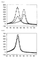
- the peak for BODIPY FL was found at about 55°C in the case of the wild-type oligonucleotide (squares), and at about 64°C in the case of the mutant type oligonucleotide (diamonds) ( Fig. 2 ).
- the heterozygous oligonucleotide (triangles) wherein the wild-type and the mutant type are mixed showed peaks for BODIPY FL at about 55°C and about 64°C ( Fig. 2 ).
- T/T wild-type
- T/G heterozygous type
- G/G mutant type
- the probe shown in Table 3 having C at its 3'-end was designed.
- the position of the probe is represented by nucleotide positions in the nucleotide sequence shown in SEQ ID NO:1. Labeling with BODIPY FL was carried out according to a conventional method.
- sequences of the template oligonucleotides used as the subject sequences to be detected are shown in Table 3.
- the position of each oligonucleotide is represented by nucleotide positions in the nucleotide sequence shown in SEQ ID NO:1.
- a mixture of the template oligonucleotides of SEQ ID NOs:13 and 14 at a ratio of 1:1 was used as a sample for studying the heterozygous sample.
- the peak for BODIPY FL was found at about 56°C in the case of the wild-type oligonucleotide (squares), and at about 55°C in the case of the mutant type oligonucleotide (diamonds) ( Fig. 3 ).
- ⁇ of the Tm value i.e. the difference between the Tm values
- the heterozygous oligonucleotide (triangles) wherein the wild-type and the mutant type are mixed showed a peak for BODIPY FL only at about 56°C ( Fig. 3 ). Therefore, it was revealed that the heterozygous type produces only a single detection peak and hence the wild-type and the mutant type cannot be distinguished from each other.
- the probe shown in Table 5 having C at its 3'-end was designed.
- the position of the probe is represented by nucleotide positions in the nucleotide sequence shown in SEQ ID NO:2. Labeling with TAMRA was carried out according to a conventional method.
- sequences of the template oligonucleotides used as the subject sequences to be detected are shown in Table 5.
- the position of each oligonucleotide is represented by nucleotide positions in the nucleotide sequence shown in SEQ ID NO:2.
- a mixture of the template oligonucleotides of SEQ ID NOs:15 and 16 at a ratio of 1:1 was used as a sample for studying the heterozygous sample.
- Tm analysis was carried out to evaluate the performance of the fluorescently labeled oligonucleotide (SEQ ID NO:5).
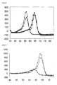
- the peak for TAMRA was found at about 64°C in the case of the wild-type oligonucleotide (squares), and at about 56°C in the case of the mutant type oligonucleotide (diamonds) ( Fig. 4 ). Thus, it was revealed that the wild-type and the mutant type can be distinguished from each other.
- the heterozygous oligonucleotide (triangles) wherein the wild-type and the mutant type are mixed showed peaks for TAMRA at about 56°C and about 64°C ( Fig. 4 ). Therefore, it was revealed that the heterozygous type can be distinguished from the wild-type and the mutant type.
- the probe shown in Table 7 having C at its 3'-end was designed.
- the position of the probe is represented by nucleotide positions in the nucleotide sequence shown in SEQ ID NO:2. Labeling with TAMRA was carried out according to a conventional method.
- sequences of the template oligonucleotides used as the subject sequences to be detected are shown in Table 7.
- the position of each oligonucleotide is represented by nucleotide positions in the nucleotide sequence shown in SEQ ID NO:2.
- a mixture of the template oligonucleotides of SEQ ID NOs:18 and 19 at a ratio of 1:1 was used as a sample for studying the heterozygous sample.
- Tm analysis was carried out to evaluate the performance of the fluorescently labeled oligonucleotide (SEQ ID NO:17).
- the peak for TAMRA was found at about 63°C in the case of the wild-type oligonucleotide (triangles), and at about 59°C in the case of the mutant type oligonucleotide (squares) ( Fig. 5 ).
- the heterozygous oligonucleotide (diamonds) wherein the wild-type and the mutant type are mixed showed a peak for TAMRA only at about 64°C ( Fig. 5 ). Therefore, it was revealed that the heterozygous type produces only a single detection peak and hence cannot be distinguished from either the wild-type or the mutant type.
- Example 3 (Use of a Plurality of Probes for Detection from Purified Human Genomic DNA or Whole Blood)
- Polymorphic regions were amplified as described below from purified human genomic DNA or whole blood by PCR using the primers described below, and Tm analysis was carried out using the probes shown in SEQ ID NOs:3 and 4.
- the primers shown in Table 9 were designed based on the nucleotide sequence having the polymorphic site rs8099917 in the IL28B gene (SEQ ID NO:1), such that the polymorphic site can be amplified with the primers.
- the positions of the primers are represented by nucleotide positions in the nucleotide sequence shown in SEQ ID NO:1.
- the primers shown in Table 10 were designed based on the nucleotide sequence having the polymorphic site rs1127354 in the ITPA gene (SEQ ID NO:2), such that the polymorphic site can be amplified with the primers.
- the positions of the primers are represented by nucleotide positions in the nucleotide sequence shown in SEQ ID NO:2.
- PCR and Tm analysis were then carried out using a fully automatic SNPs testing device (trade name: i-densy IS-5310, manufactured by ARKRAY, Inc.).
- the composition of the PCR reaction solution was as described below.
- whole blood or purified human genomic DNA as described below was used.
- the PCR and Tm analysis were carried out under the following conditions: 95°C for 60 seconds ⁇ (95°C for 1 second ⁇ 58°C for 30 seconds) ⁇ 50 cycles ⁇ 95°C for 1 second ⁇ 40°C for 60 seconds ⁇ (40°C-85°C, 1°C/3 seconds).
- the excitation wavelength and detection wavelength in the Tm analysis were 420 to 485 nm and 520 to 555 nm, respectively (BODIPY FL), or 520 to 555 nm and 585 to 700 nm, respectively (TAMRA).
- BODIPY FL 520 to 555 nm
- TAMRA 520 to 555 nm and 585 to 700 nm, respectively.
- [Table 9] Name Sequence (5' ⁇ 3') Position SEQ ID NO: mer IL28B(917)F caacatggagagttaaagtaagtcttgtatttcacc 197-232 6 36 IL28B(917)R cagctaccaaactgtatacagcatggttc 342-314 7 29
- Purified DNA was added as a template to the PCR reaction solution such that 100 copies/test of the DNA was contained therein.
- the purified human genomic DNA and the whole blood having the heterozygous genotype T/G showed two peaks (53°C and 62°C) and hence exhibited a unique pattern in respect of the amount of change in the fluorescence intensity.
- the purified human genomic DNA and the whole blood showed a single peak (48°C) and hence exhibited a unique pattern in respect of the amount of change in the fluorescence intensity.
- the IL28B gene polymorphism and the ITPA gene polymorphism can be detected simultaneously.
- probe of SEQ ID NO:5 is labeled at C at its 3'-end as in the case of the probe of SEQ ID NO:4 and the effect of the label has been demonstrated in Example 2, it is possible to simultaneously detect the IL28B gene polymorphism and the ITPA gene polymorphism also by simultaneous use of the probe of SEQ ID NO:5 and the probe of SEQ ID NO:3.
- the present invention can be suitably used in fields such as healthcare, diagnosis and research.
Description
- The present invention relates to a method for detecting mutations at IL28B (rs8099917) and ITPA (rs1127354), nucleic acid probes and a kit therefor.
- Hepatitis C is a viral hepatitis caused by infection with hepatitis C virus (HCV). In Japan, the number of patients suffering from HCV infection is about 2,000,000, and progression to chronic hepatitis occurs in 60 to 80% of HCV patients. In cases where treatment is not carried out and chronic hepatitis is maintained for 10 to 30 years, progression to liver cirrhosis/liver cancer occurs in 30 to 40% of patients. Examples of antiviral therapies for removal of HCV include interferon therapy, whose treatment results have been improved by the combined use of ribavirin and the pegylation of interferon.
- As a result of analysis of about 900,000 sites in human genes, which are said to be different among individuals, in 314 Japanese patients for whom the pegylated interferon+ribavirin combination therapy is effective or ineffective, it has been reported that SNPs existing in the gene for IL28B, which is an interferon (IFN), and in the vicinity of the gene are involved in the therapeutic effect (Nature Genetics 41, 1105-1109 (2009)). Further, it has been reported that out of the 6 SNPs that were predicted to be involved in the effect of the pegylated interferon+ribavirin combination therapy, the SNP identified as rs8099917 has the largest influence on the therapeutic effect (PLoS One. 2010 Oct 29;5(10):e13771).
- Ribavirin-induced anemia is a major factor that forces anti-hepatitis C virus (HCV) therapy to be terminated or reduced (Hepatol Res. 2010 Nov;40(11):1063-1071). As a result of the evaluation of the clinical significance of ITPA gene variations in Japanese hepatitis C patients treated with the pegylated interferon (PEG-IFN)/ribavirin combination therapy, it has been reported that rs1127354, which is a functional SNP in an ITPA exon, is a useful predictive factor for ribavirin-induced anemia (Gastroenterology. 2010 Oct; 139(4):1190-7. Epub 2010 Jul 14).
- PLoS One. 2010 Oct 29; 5(10): e13771 reports that mutation at IL28B (rs8099917) is strongly involved in the effect of the pegylated interferon/ribavirin combination therapy in hepatitis C patients, and detection of the presence/absence of a mutation at IL28B (rs8099917) was carried out by sequence analysis. Hepatology, Vol. 53, reports that mutation at ITPA (rsl 127354) is strongly involved in anemia during treatment with the pegylated interferon/ribavirin combination therapy, and detection of the presence/absence of a mutation at ITPA (rs1127354) was carried out by sequence analysis.
- However, it is very laborious and costly to carry out genomic DNA extraction from whole blood at actual clinical sites for investigating SNP variation in hepatitis C patients as described in PLoS One. 2010 Oct 29; 5(10): e13771 and Hepatology, Vol. 53, . Therefore, it is assumed that the demand for a technology with which the presence/absence of mutations can be automatically and directly assayed using whole blood will increase. Further, because of the involvement of IL28B (rs8099917) and ITPA (rs1127354) in the effect of the pegylated interferon/ribavirin combination therapy, it is highly likely that a technology that allows the simultaneous assay of the presence/absence of mutations in IL28B (rs8099917) and ITPA (rs1127354) will be required in the future in the clinical field, but such simultaneous detection cannot be done by either PLoS One. 2010 Oct 29; 5(10): e13771 or Hepatology, Vol. 53, , since detection of the presence/absence of mutations in IL28B (rs8099917) and ITPA (rs1127354) was carried out by sequence analysis.
-
JP 2002-119291 A - Melis et al (Journal of Molecular Diagnostics, Vol. 13, No. 4, pages 446-451, 2011) discloses simultaneous genotyping of rs8099917 and another polymorphism in the IL28B gene using anchor and sensor probes which bind adjacent to one another and are labeled to allow FRET. Ito et al (Journal of Clinical Microbiology, Vol. 49, No. 5, pages 1853-1860, 2011) identifies the rs8099917 polymorphism in the IL28B gene as a predictor of response to peglyated alpha interferon/ribavirin therapy and refers to various methods of genotyping including the use of hybridization and invader probes which sandwich the polymorphism site. Tani et al (Journal of Agricultural and Food Chemistry, Vol. 53, pages 2535-2540, 2005) discloses the use of fluorescence quenching of a labeled probe on binding to target DNA as a means of detection during PCR.
- The present invention aims to provide probes effective for detecting a polymorphism rs8099917 in the IL28B gene, to provide a method for detecting the polymorphism rs8099917 in the IL28B gene and optionally, in addition the polymorphism rs1127354 in the ITPA gene, and a kit therefor.
- The present inventors discovered that, by designing probes based on a specific region comprising the polymorphism rs8099917 in the IL28B gene and a specific region comprising the polymorphism rs1127354 in the ITPA gene, and detecting changes in a signal due to formation of a hybrid of the probes with target nucleic acids or due to dissociation of the probes from target nucleic acids, mutations at these sites can be detected, thereby completing the present invention.
- That is, the present invention is as follows.
- (1) A probe for detecting a polymorphism in the IL28B gene, comprising the fluorescently labeled oligonucleotide P1 below:
- (P1) an oligonucleotide comprising a nucleotide sequence of 7 to 28 consecutive nucleotides containing nucleotides 301 to 307 in SEQ ID NO:1, wherein the nucleotide corresponding to the nucleotide at position 307 is a cytosine and is labeled with a fluorescent dye.
- (2) The probe according to (1), wherein
said oligonucleotide P1 has the nucleotide corresponding to the nucleotide at position 307 labeled with a fluorescent dye at the first, second or third position counted from the 3'-end. - (3) The probe according to (1) or (2), wherein
said oligonucleotide P1 has the nucleotide corresponding to the nucleotide at position 307 labeled with a fluorescent dye at the 3'-end. - (4) The probe according to any one of (1) to (3), wherein said fluorescently labeled oligonucleotide emits fluorescence when said oligonucleotide is not hybridized with a target sequence, and the fluorescence intensity decreases or increases when said oligonucleotide is hybridized with said target sequence.
- (5) The probe according to (4), wherein said fluorescently labeled oligonucleotide emits fluorescence when said oligonucleotide is not hybridized with a target sequence, and the fluorescence intensity decreases when said oligonucleotide is hybridized with said target sequence.
- (6) The probe according to any one of (1) to (5), wherein
said oligonucleotide P1 has 7 to 23 consecutive nucleotides. - (7) The probe according to any one of (1) to (5), wherein
said oligonucleotide P1 has 7 to 18 consecutive nucleotides. - (8) The probe according to any one of (1) to (7), wherein said probe is a probe for melting curve analysis.
- (9) A method for detecting a polymorphism in the IL28B gene using the probe for the IL28B gene according to any one of (1) to (8). The method may be conducted on a sample comprising nucleic acid which may contain said polymorphism and comprises contacting the probe with said sample and detecting the presence or absence of the polymorphism. The invention also extends to use of said probe for detecting said polymorphism.
- (10) The method according to (9) for detecting polymorphisms in the IL28B gene and the ITPA gene by using the probe for the IL28B gene according to any one of (1) to (8) and a probe for detecting a polymorphism in the ITPA gene, wherein said probe comprises at least one fluorescently labelled oligonucleotide selected from P2 or P3 below:
- (P2) an oligonucleotide comprising a complement of a nucleotide sequence of 13 to 28 consecutive nucleotides containing nucleotides 239 to 251 in SEQ ID NO:2, wherein the nucleotide corresponding (i.e. complementary) to the nucleotide at position 239 is a cytosine and is labeled with a fluorescent dye;
- (P3) an oligonucleotide comprising a nucleotide sequence of 6 to 42 consecutive nucleotides containing nucleotides 251 to 256 in SEQ ID NO:2, wherein the nucleotide corresponding to the nucleotide at position 256 is a cytosine and is labeled with a fluorescent dye. The invention also extends to use of said probes for detecting said polymorphisms.
- (11) The method according to (10), wherein
said oligonucleotide P2 has the nucleotide corresponding (i.e. complementary) to the nucleotide at position 239 labeled with a fluorescent dye at the first, second or third position counted from the 3'-end; and
said oligonucleotide P3 has the nucleotide corresponding to the nucleotide at position 256 labeled with a fluorescent dye at the first, second or third position counted from the 3'-end,
wherein preferably
said oligonucleotide P2 has the nucleotide corresponding (i.e. complementary) to the nucleotide at position 239 labeled with a fluorescent dye at the 3'-end; and
said oligonucleotide P3 has the nucleotide corresponding to the nucleotide at position 256 labeled with a fluorescent dye at the 3'-end. - (12) The method according to (10) or (11), wherein said fluorescently labeled oligonucleotide in said probe for the ITPA gene emits fluorescence when said oligonucleotide is not hybridized with a target sequence, and the fluorescence intensity decreases or increases when said oligonucleotide is hybridized with said target sequence, wherein preferably said fluorescently labeled oligonucleotide emits fluorescence when said oligonucleotide is not hybridized with a target sequence, and the fluorescence intensity decreases when said oligonucleotide is hybridized with said target sequence.
- (13) The method according to any one of (10) to (12), wherein
said oligonucleotide P2 has 13 to 23 consecutive nucleotides; and
said oligonucleotide P3 has 6 to 27 consecutive nucleotides. - (14) The method according to any one of (10) to (12), wherein
said oligonucleotide P2 has 13 to 18 consecutive nucleotides; and
said oligonucleotide P3 has 6 to 22 consecutive nucleotides. - (15) The method according to any one of (10) to (13), wherein said probe for the ITPA gene is a probe for melting curve analysis.
- (16) A method for detecting polymorphisms in the IL28B gene and the ITPA gene separately or in a single reaction system, comprising the Steps (I) to (IV) below:
- (I) adding the probe for the IL28B gene according to any one of (1) to (8) and the probe for the ITPA gene according to any one of (10) to (15) to a sample containing DNA and allowing hybridization of said probe with said DNA;
- (II) changing the temperature to dissociate the hybridization complex between said DNA and said probe, and measuring the fluctuation of the signal due to said dissociation of said hybridization complex;
- (III) analyzing said fluctuation of the signal to determine the Tm value; and
- (IV) determining based on said Tm value the presence/absence of said polymorphisms of interest or the abundance ratios of the nucleotide sequences having said polymorphisms.
- (17) The method according to (16), further comprising amplifying DNA before Step (I) or at the same time as Step (I).
- (18) A method for judging (or predicting) a pharmacological effect of a therapeutic agent against hepatitis C virus, comprising detecting polymorphisms in the IL28B gene and the ITPA gene separately or in a single reaction system by the method according to any one of (10) to (17) and judging (or predicting) the resistance to said agent or judging (or predicting) a pharmacological effect of said agent based on the presence/absence of said polymorphisms of interest. Said method may be performed in a sample from a subject (e.g. a human) in relation to whom the judgement/prediction is to be made. A sample (as used herein) comprises nucleic acid (e.g. DNA) which may contain said polymorphism.
- (19) A reagent kit for detecting polymorphisms in the IL28B gene and the ITPA gene separately or in a single reaction system, comprising the probe for the IL28B gene according to any one of (1) to (8) and the probe for the ITPA gene as defined in any one of (10) to (15).
- (20) The reagent kit according to (19), further comprising:
- a primer for amplifying a region comprising a sequence, in the nucleotide sequence shown in SEQ ID NO:1 in the IL28B gene, with which said oligonucleotide P1 hybridizes; and/or
- a primer for amplifying a region comprising a sequence, in the nucleotide sequence shown in SEQ ID NO:2 in the ITPA gene, with which said oligonucleotide P2 or P3 hybridizes. The invention also extends to the use of said kit for detecting said polymorphism(s).
- By including the probe of the present invention in a gene amplification system such as PCR and only carrying out Tm analysis after the gene amplification reaction, typing of the polymorphism rs8099917 in the IL28B gene can be carried out, optionally in conjunction with typing of the polymorphism rs1127354 in the ITPA gene. Further, since whole blood, an oral mucosa suspension and the like can be directly tested, the labour and the costs can be reduced.
- The probe of the present invention has high specificity.
- By using the method of the present invention, even in cases where PCR is carried out, the amplification product does not need to be extracted, so that there is hardly any risk of contamination. Further, since the method of the present invention can be carried out by a simple procedure, it can be easily automated.
- By using the method of the present invention, the polymorphism rs8099917 in the IL28B gene and the polymorphism rs1127354 in the ITPA gene can be simultaneously detected. For the pegylated interferon+ribavirin combination therapy, being able to simultaneously detect the polymorphism rs8099917 in the IL28B gene and the polymorphism rs1127354 in the ITPA gene is of clinical significance, since the two polymorphisms are greatly involved in the effect of the therapy.
-
-
Fig. 1 is a diagram showing examples of (A) a melting curve of a nucleic acid mixture and (B) a differential melting curve. -
Fig. 2 shows melting curves obtained using the probes of Example 1 of the present invention for detecting a polymorphism. The amount of change in the fluorescence intensity per unit time is plotted along the ordinate (d the amount of increase in the fluorescence intensity/t) and the temperature (°C) is plotted along the abscissa. Squares represent the wild-type, diamonds represent the mutant type, and triangles represent the heterozygous type (this also applies to the figures below). -
Fig. 3 shows melting curves obtained using the probes of Comparative Example 1 for detecting a polymorphism. -
Fig. 4 shows melting curves obtained using the probes of Example 2 used in methods of the present invention for detecting a polymorphism. -
Fig. 5 shows melting curves obtained using the probes of Comparative Example 2 for detecting a polymorphism. -
Fig. 6 shows melting curves obtained using the probes of Example 3 of the present invention for detecting polymorphisms, together with purified human genomic DNA. The left panel shows a melting curve obtained by detection of a polymorphism in the IL28B gene, and the right panel shows a melting curve obtained by detection of a polymorphism in the ITPA gene (this also applies to the next figure). -
Fig. 7 is a melting curve obtained using the probes of Example 3 of the present invention for detecting polymorphisms, together with whole blood. - The (P1) probe of the present invention is a probe for detecting the polymorphism rs8099917 in the IL28B gene, and is composed of an oligonucleotide comprising a nucleotide sequence of 7 to 28 consecutive nucleotides containing nucleotides 301 to 307 in SEQ ID NO:1, wherein the nucleotide corresponding to the nucleotide at position 307 is a cytosine and is labeled with a fluorescent dye. The 7 to 28 nucleotides are all contained, consecutively in SEQ ID NO:1.
- The polymorphism rs8099917 in the IL28B gene described herein is located at nucleotide position 301 in SEQ ID NO:1. The rs number represents a registration number for the dbSNP database curated by the National Center for Biotechnology Information (//www.ncbi.nlm.nih.gov/projects/SNP/). The nucleotide at position 301 in SEQ ID NO:1 is represented as k, which represents T in the wild-type and G in the mutant type. For example, in the probe (P1) of the present invention, the nucleotide at position 301 in SEQ ID NO:1 is G, which is the mutant type.
- The (P1) probes of the present invention can be prepared in the same manner as the probe described in
JP 2002-119291 A - Examples of the nucleotide sequences of the (P1) probes used in the present invention include 5'-ctgtgagcaatGtcaccc-3' (SEQ ID NO:3), and the nucleotide denoted by the upper case letter G corresponds to the nucleotide at position 301.
- The (P2) probe used in accordance with the present invention is a probe for detecting the polymorphism rs1127354 in the ITPA gene, and is composed of an oligonucleotide comprising a complement of a nucleotide sequence of 13 to 28 consecutive nucleotides containing nucleotides 239 to 251 in SEQ ID NO:2, wherein the nucleotide corresponding (i.e. complementary) to the nucleotide at position 239 is a cytosine and is labeled with a fluorescent dye. The 13 to 28 nucleotides are all contained, consecutively in SEQ ID NO:2.
- The polymorphism rs1127354 in the ITPA gene described herein is located at nucleotide position 251 in SEQ ID NO:2. The nucleotide at position 25 in SEQ ID NO:2 is represented as v, which represents C in the wild-type and A in the mutant type. Three types of nucleotides, C/A/G, are registered in the SNP site of NCBI for the polymorphism rs1127354 in the ITPA gene, and it is described that the allele frequency of C is 87.5% and the allele frequency of A is 12.5% in the Japanese population (no Japanese person has the G-type allele). Further, only one person (tissue) who reported the existence of G carried out the assay (see http://www.ncbi.nlm.nih.gov/projects/SNP/snp_refcgi?rs=1127354) (this also applies to the case of the P3 probe). For example, the (P2) probe for use in accordance with the present invention is complementary to the nucleotide sequence shown in SEQ ID NO:2 wherein the nucleotide at position 251 is A, which corresponds to the mutant type.
- The (P2) probes for use in accordance with the present invention can be prepared in the same manner as the quenching probe described in
JP 2002-119291 A - Examples of the nucleotide sequences of the (P2) probes used in accordance with the present invention include 5'-gcatgTaaacttatctcc-3' (SEQ ID NO:4), and the nucleotide denoted by the upper case letter T corresponds to (i.e. is complementary to) the nucleotide at position 251.
- The (P3) probe for use in accordance with the present invention is a probe for detecting the polymorphism rs1127354 in the ITPA gene, and is composed of an oligonucleotide comprising a nucleotide sequence of 6 to 42 consecutive nucleotides containing nucleotides 251 to 256 in SEQ ID NO:2, wherein the nucleotide corresponding to the nucleotide at position 256 is a cytosine and is labeled with a fluorescent dye. The 6 to 42 nucleotides are all contained, consecutively in SEQ ID NO:2.
- The polymorphism rs1127354 in the ITPA gene described herein is located at nucleotide position 251 in SEQ ID NO:2. For example, in the (P3) probes for use in accordance with the present invention, the nucleotide at position 251 in SEQ ID NO:2 is C, which is the wild-type nucleotide.
- The (P3) probes for use in accordance with the present invention can be prepared in the same manner as the quenching probe described in
JP 2002-119291 A - Examples of the nucleotide sequences of the (P3) probes used in accordance with the present invention include 5'-tctaggagataagtttCcatgc-3' (SEQ ID NO 5), and the nucleotide denoted by the upper case letter C corresponds to the nucleotide at position 251.
- The fluorescent dye is not restricted, and examples of the fluorescent dye include fluorescein, phosphor, rhodamine and polymethine dye derivatives. Examples of commercially available fluorescent dyes include BODIPY FL (trademark; manufactured by Molecular Probes), FluorePrime (trade name; manufactured by Amersham Pharmacia), Fluoredite (trade name; manufactured by Millipore), FAM (manufactured by ABI), Cy3 and Cy5 (manufactured by Amersham Pharmacia) and TARMA (manufactured by Molecular Probes). The conditions for detection using the probe are not restricted, and may be appropriately determined depending on the fluorescent dye used. The detection may be carried out, for example, with a detection wavelength of 445 to 480 nm in the case of Pacific Blue, with a detection wavelength of 585 to 700 nm in the case of TAMRA, and with a detection wavelength of 520 to 555 nm in the case of BODIPY FL. By using such a probe, hybridization and dissociation can be easily detected based on fluctuation of the signal. The fluorescent dye can be bound to the oligonucleotide by a conventional method such as the one described in
JP 2002-119291 A - The hybridization described herein can be carried out according to a known method or a method corresponding thereto, such as the method described in Molecular Cloning 3rd (J. Sambrook et al., Cold Spring Harbor Lab. Press, 2001). Preferably said hybridization is under stringent conditions.
- The term "stringent conditions" means conditions under which a specific hybrid is formed while nonspecific hybrids are not formed. Typical examples of stringent conditions include conditions under which hybridization is performed with a potassium concentration of about 25 mM to about 50 mM and a magnesium concentration of about 1.0 mM to about 5.0 mM. Examples of the conditions in the present invention include conditions under which hybridization is performed in Tris-HCl (pH 8.6), 25 mM KCl and 1.5 mM MgCl2, but the conditions are not limited thereto. Other examples of stringent conditions include those described in Molecular Cloning 3rd (J. Sambrook et al., Cold Spring Harbor Lab. Press, 2001). Those skilled in the art can easily select such conditions by, for example, controlling the hybridization reaction and/or changing salt concentrations of the hybridization reaction solution.
- Examples of the oligonucleotide of the above fluorescently labeled oligonucleotide include oligonucleotides as well as modified oligonucleotides.
- Examples of the constituent units of the above oligonucleotide include ribonucleotides, deoxyribonucleotides and artificial nucleic acids. Examples of the artificial nucleic acids include DNA; RNA; LNA (Locked Nucleic Acid), which is an RNA analogue; PNA (Peptide Nucleic Acid), which is a peptide nucleic acid; and BNA (Bridged Nucleic Acid), which is a bridged nucleic acid.
- The oligonucleotide may be constituted by either a single type of constituent unit or a plurality of types of constituent units among the above constituent units.
- For example, the fluorescence intensity of the fluorescently labeled oligonucleotide decreases (or the fluorescence is quenched) or increases when the oligonucleotide is hybridized with a complementary sequence, relative to the fluorescence intensity observed when the oligonucleotide is not hybridized with the complementary sequence. For example, the fluorescence intensity of the fluorescently labeled oligonucleotide decreases when the oligonucleotide is hybridized with a complementary sequence, relative to the fluorescence intensity observed when the oligonucleotide is not hybridized with the complementary sequence. A probe that utilizes such fluorescence quenching phenomenon is generally called a guanine quenching probe, and is known as a QProbe (registered trademark). For example, the probe is an oligonucleotide which is designed such that the oligonucleotide has C at its 3'-end or 5'-end and which is fluorescently labeled such that the emission decreases when the terminal C is close to G.
- For example, the probe of (or for use in accordance with) the present invention is labeled at its 3'-end with a fluorescent dye.
- In the present specification, when the term "first, second or third position counted from the 3'-end" is mentioned, the 3'-end is regarded as the first position.
- In the detection method of the present invention, nucleic acid comprising the polymorphism rs8099917 in the IL28B gene and/or the polymorphism rs1 127354 in the ITPA gene is analyzed using a nucleic acid probe(s) labeled with a fluorescent dye(s) by measuring fluorescence from the fluorescent dye(s), and melting curve analysis is carried out, followed by detecting the polymorphism(s) based on the result of the melting curve analysis, which nucleic acid probe(s) is/are probe(s) of the present invention or for use in accordance with the present invention.
- For example, the detection method of the present invention uses the probe of the present invention and comprises the following steps:
- (I) adding the probe of the present invention to a sample containing DNA and allowing hybridization of the probe with the DNA;
- (II) changing the temperature to dissociate the hybridization complex between the DNA and the probe, and measuring the fluctuation of the signal due to the dissociation of the hybridization complex;
- (III) analyzing the fluctuation of the signal to determine the Tm value; and
- (IV) determining based on the Tm value the presence/absence of the polymorphism of interest or the abundance ratio of the nucleotide sequence having the polymorphism.
- The evaluation of the Tm value in (III) includes not only evaluation of the dissociation temperature of the hybridization complex but also evaluation of the differential value of the fluorescence signal, which fluctuates upon melting of the hybridization complex depending on the temperature. Based on the differential value, the abundance ratio of the nucleotide sequence (DNA) having the polymorphism can be evaluated.
- Examples of the method of nucleic acid amplification include methods using a polymerase, such as PCR, ICAN and LAMP. When the amplification is carried out by a method using a polymerase, the amplification may be carried out in the presence of the probe of the present invention or for use in accordance with the present invention. Those skilled in the art can easily control reaction conditions and the like of the amplification depending on the probe to be used. By this, the detection can be carried out just by analyzing the Tm value of the probe after the amplification of the nucleic acid, so that the amplification product does not need to be handled after the reaction. Therefore, there is no risk of contamination of the amplification product. Further, since the detection can be carried out with the same apparatus as the one necessary for the amplification, it is not necessary to transfer the container. Therefore, automation can also be easily done.
- As the DNA polymerase to be used for the PCR method, a conventional DNA polymerase may be used without any limitation. Examples of the DNA polymerase include GeneTaq (manufactured by Nippon Gene Co., Ltd.), PrimeSTAR Max DNA Polymerase (manufactured by Takara Bio Inc.) and Taq Polymerase.
- The amount of the polymerase to be used is not restricted as long as the polymerase is used at a concentration which is normally employed. For example, in cases where Taq polymerase is used, the concentration of the polymerase may be 0.01 U to 100 U per 50 µl of the reaction solution. Use of the polymerase in such an amount has an advantage, for example, in that a sufficient amount of amplified product can be obtained.
- In the present invention, the DNA in the sample may be either single-stranded DNA or double-stranded DNA. In cases where the DNA is double-stranded DNA, for example, the step of dissociating the double-stranded DNA in the sample by heating may be included before the hybridization step. By dissociating the double-stranded DNA into single-stranded DNA, hybridization with the fluorescently labeled oligonucleotide is possible.
- In the present invention, the ratio (molar ratio) of the probe to be added with respect to the DNA in the sample is not restricted, and the ratio is, for example, not more than 1 or not more than 0.3 with respect to the DNA in the sample to ensure a sufficient detection signal. In this case, for example, the DNA in the sample may be either: the sum of the target DNA, wherein the polymorphism to be detected is present, and non-target DNA, wherein the polymorphism to be detected is absent; or the sum of the amplification product containing the target sequence, wherein the polymorphism to be detected is present, and the amplification product containing the non-target sequence, wherein the polymorphism is absent. Although the ratio of the DNA to be detected relative to the DNA in the sample is usually not known, the ratio (molar ratio) of the probe to be added with respect to the DNA to be detected (the amplification product containing the sequence to be detected) is, for example, not more than 100, not more than 50, or not more than 30, as a result. The lower limit of the ratio is not restricted, and the ratio is, for example, not less than 0.001, not less than 0.01, or not less than 0.2. The ratio of the probe to be added with respect to the DNA may be, for example, either the molar ratio with respect to the double-stranded DNA or the molar ratio with respect to the single-stranded DNA.
- The Tm value will now be described. Heating a solution containing double-stranded DNA causes an increase in absorbance at 260 nm. This is caused because the hydrogen bonds between the strands of the double-stranded DNA are broken by the heat and the double-stranded DNA is dissociated into single-stranded DNA (melting of DNA). When the double-stranded DNA is completely dissociated into single-stranded DNA, the absorbance is about 1.5-fold higher than the absorbance at the beginning of heating (absorbance for only the double-stranded DNA), and completion of the melting can be judged by such a change in absorbance. Based on this phenomenon, the melting temperature Tm is generally defined as the temperature at which the increase in absorbance reached 50% of the total increase in absorbance.
- In the present invention, measurement of the signal fluctuation due to the temperature change for determination of the Tm value can be carried out also by measuring absorbance at 260 nm based on the above-mentioned principle, but the measurement may be carried out based on a signal from a label added to the probe used in accordance with the present invention, which signal fluctuates depending on the state of hybrid formation between the DNA and the probe. The probe of, and in accordance with, the present invention is labeled with a fluorescent dye. Examples of the labeled probe include a fluorescently labeled oligonucleotide probe which emits fluorescence when it is not hybridized with the target sequence, whose fluorescence intensity decreases (or the fluorescence is quenched) when the probe is hybridized with the target sequence, and a fluorescently labeled oligonucleotide probe which emits fluorescence when it is not hybridized with the target sequence, whose fluorescence intensity increases when the probe is hybridized with the target sequence. In the case of the former probe, the probe shows no signal or shows a weak signal when it forms a hybrid (double-stranded DNA) with the sequence to be detected, while the probe shows a signal or the signal increases when the probe is released by heating. In the case of the latter probe, the probe shows a signal when forming a hybrid (double-stranded DNA) with the sequence to be detected, while the signal decreases (disappears) when the probe is released by heating. Therefore, by detecting the change in the signal from the label under conditions specific to the signal (by investigating absorbance or the like), the progress of melting and the Tm value can be determined in a similar manner to the measurement of absorbance at 260 nm.
- The method of the present invention for detecting a polymorphism will now be described focusing on the method of detection of changes in the signal from a fluorescent dye. It should be noted that the method of the present invention for detecting a polymorphism is characterized by the use per se of the probe for detection of a polymorphism, and other steps and conditions are not restricted.
- The range of the temperature at which fluctuation of the fluorescence intensity is measured is not restricted, and the starting temperature may be, for example, room temperature to 85°C, or 25°C to 70°C, and the end temperature may be, for example, 40°C to 105°C. The heating rate is not restricted, and may be, for example, 0.1°C/second to 20°C/second, or 0.3°C/second to 5°C/second.
- The fluctuation of the signal is then analyzed to determine the Tm value. More particularly, the differential value at each temperature (-d fluorescence intensity/dt) is calculated based on the obtained fluorescence intensity, and the temperature at which the value is lowest may be determined as the Tm value. Further, the point at which the amount of increase in the fluorescence intensity per unit time (the amount of increase in the fluorescence intensity/t) is largest may also be determined as the Tm value. In cases where, as the labeled probe, a probe with which the signal intensity increases upon hybrid formation is used instead of a quenching probe, the amount of decrease in the fluorescence intensity may be measured instead.
- In the present invention, the hybridization complex is heated and fluorescence signal fluctuation (for example, increase in the fluorescence intensity) due to the increase in the temperature is measured as described above, but, instead of employing this method, signal fluctuation upon formation of the hybrid may be measured, for example. That is, the fluorescence signal fluctuation caused by formation of the hybridization complex due to the decrease in the temperature of the sample to which the probe was added may be measured.
- For example, in cases where a fluorescently labeled oligonucleotide probe whose fluorescence intensity is lower (or whose fluorescence is quenched) when the probe is hybridized with the complementary sequence than when the probe is not hybridized with the complementary sequence (e.g., QProbe) is used, the fluorescence intensity is high upon addition of the probe to the sample since the probe is in the dissociated state, while the fluorescence decreases (or is quenched) upon formation of a hybridization complex due to the decrease in the temperature. Therefore, for example, the temperature of the heated sample may be gradually decreased, to measure the decrease in the fluorescence intensity due to the temperature decrease.
- On the other hand, in cases where a labeled probe whose signal increases upon formation of the hybrid is used, the fluorescence intensity is low (or the fluorescence is quenched) upon addition of the probe to the sample since the probe is in the dissociated state, while the fluorescence intensity increases upon formation of a hybridization complex due to the decrease in the temperature. Therefore, for example, the temperature of the sample may be gradually decreased, to measure the increase in the fluorescence intensity due to the temperature decrease.
- The nucleic acid to be used as a template for carrying out the nucleic acid amplification is not restricted as long as it contains a nucleic acid, and examples of the nucleic acid include those derived from, or those which may be derived from, arbitrary biological origins such as blood; oral mucosal suspensions; somatic cells of nails, hair and the like; germ cells; milk; ascitic fluid; paraffin-embedded tissues; gastric juices; fluids obtained by gastric lavage; peritoneal fluid; amniotic fluid; and cell cultures. The nucleic acid as a template may be used as it is directly after being obtained from any of the above-described origins, or may be pretreated to modify properties of the sample before being used. For example, in cases where whole blood is used as a sample, isolation of the genomic DNA from the whole blood may be carried out by a known method. For example, a commercially available genomic DNA isolation kit (trade name, GFX Genomic Blood DNA Purification kit; manufactured by GE Healthcare Bio-Sciences KK) or the like may be employed.
- The primer pair used in the PCR is not restricted as long as a region with which the probe of the present invention (or the probe used in accordance with the invention) can be hybridized is amplified. Those skilled in the art can easily design such a primer pair based on the nucleotide sequences of SEQ ID NO:1 or SEQ ID NO:2. The length and the Tm value of each primer are usually 12-mer to 40-mer and 40 to 70°C, or 16-mer to 30-mer and 55 to 60°C, respectively.
- The primers in the primer pair do not need to have the same length, but the Tm values of these primers are almost the same (or the difference is not more than 5°C), for example.
- The Tm analysis can be carried out in the same manner as the conventional method except that fluorescence from the fluorescent dye of the probe for use in accordance with the present invention is measured. The fluorescence can be measured using the excitation light having a wavelength dependent on the fluorescent dye, and measuring the light having the emission wavelength. The heating rate in the Tm analysis is usually 0.1 to 1°C/second. The composition of the reaction solution used for carrying out the Tm analysis is not restricted as long as the probe can hybridize with a nucleic acid having the complementary sequence of the nucleotide sequence of the probe, and usually, the concentration of monovalent cations is 1.5 to 5 mM, and the pH is 7 to 9. Since the reaction solution in an amplification method using a DNA polymerase, such as PCR, usually satisfies these conditions, the reaction solution after the amplification can be used as it is for the Tm analysis.
- Detection of the polymorphism rs8099917 in the IL28B gene and/or the polymorphism rs1127354 in the ITPA gene can be carried out based on the result of the Tm analysis, by a conventional method. The detection in the present invention includes detection of the presence/absence of a mutation.
- Since detection of the presence/absence of a mutation is possible by the method of the present invention using the probe of the present invention or the probe for use in accordance with the present invention, the present invention also includes judgment (or prediction) of the pharmacological effect of a therapeutic agent against hepatitis C virus and judgment (or prediction) of tolerance to a therapeutic agent against hepatitis C virus. More specifically, in cases where the nucleotides at the polymorphism rs8099917 in the IL28B gene are T (T/T), which corresponds to the wild-type, the pegylated interferon+ribavirin combination therapy is suggested to be effective, while in cases where rs8099917 has G, which corresponds to the mutant type (G/G or T/G), the therapy is suggested to be ineffective. In cases where the nucleotides at the polymorphism rs1127354 in the ITPA gene comprise A, which corresponds to the mutant type (A/A or C/A), it is suggested that severe anemia as a side effect of ribavirin is unlikely to occur and therefore administration of ribavirin at a high dose is possible.
- Further, by the method of the present invention, simultaneous detection of the polymorphism rs8099917 in the IL28B gene and the polymorphism rs1127354 in the ITPA gene is also possible.
- The kit of the present invention is a kit to be used for the detection method of the present invention. This kit comprises a nucleic acid probe labeled with a fluorescent dye whose fluorescence changes upon hybridization (e.g., quenching probe), which probe is composed of the above-described oligonucleotide. The kit of the present invention can also be used for judging (or predicting) the pharmacological effect of a therapeutic agent against hepatitis C virus and tolerance to a therapeutic agent against hepatitis C virus.
- The detection kit of the present invention may further comprise, in addition to the probe, reagents required for nucleic acid amplification in the detection method of the present invention, especially primers for amplification using a DNA polymerase.
- More specifically, the primers of the present invention are primers for amplifying a region in the nucleotide sequence shown in SEQ ID NO:1 in the IL28B gene, which region comprises a sequence with which the P1 oligonucleotide hybridizes, and/or primers for amplifying a region in the nucleotide sequence shown in SEQ ID NO:2 in the ITPA gene, which region comprises a sequence with which the P2 or P3 oligonucleotide hybridizes.
- The detection kit of the present invention may comprise the probe, primers and other reagents separately or as a mixture(s) of a part of these components.
- The present invention is described more concretely by way of Examples below. However, these Examples are merely examples and the present invention is not limited to the Examples.
- In the present invention, in terms of the individual sequences of the sample nucleic acid in the sample to be subjected to the detection, and the probes and primers for detecting polymorphisms, matters described herein based on the complementary relationship between these sequences are applied to the respective sequences and also to the sequences complementary thereto unless otherwise specified. When the matters of the present invention are applied to the sequence complementary to each sequence, the sequence recognized by the complementary sequence is read as the sequence complementary to the corresponding sequence described in the present specification throughout the specification according to the common technical knowledge.
- Based on the nucleotide sequence (SEQ ID NO:1) comprising the polymorphic site rs8099917 in the IL28B gene, the probe shown in Table 1 having C at its 3'-end was designed. In Table 1, the position of the probe is represented by nucleotide positions in the nucleotide sequence shown in SEQ ID NO:1. Labeling with BODIPY FL was carried out according to a conventional method.
- The sequences of the template oligonucleotides used as the subject sequences to be detected (mutant type (SEQ ID NO:10) and wild-type (SEQ ID NO:11)) are shown in Table 1. In Table 1, the position of each oligonucleotide is represented by nucleotide positions in the nucleotide sequence shown in SEQ ID NO:1. A mixture of the template oligonucleotides of SEQ ID NOs:10 and 11 at a ratio of 1:1 was used as a sample for studying the heterozygous sample.
[Table 1] Name Sequence (5'→3') Position SEQ ID NO: mer GC content (%) Tm (mt) Tm (WT) ΔTm IL28B(917) probe ctgtgagcaatGtcaccc-(BODIPY FL) 290-307 3 18 55.6 52.1 44.3 7.8 Template oligonucleotide Sequence (5'→3') Position SEQ ID NO: mer Mutant type tggttccaatttgggtgaCattgctcacagaaaggaa 319-283 10 37 Wild-type tggttccaatttgggtgaAattgctcacagaaaggaa 319-283 11 37 *The nucleotide denoted by the upper case letter indicates the position of the mutation.
*The nucleotides denoted by the upper case letters indicate the position of the mutation. - Using the reagents described below and a fully automatic SNPs testing device (trade name: i-densy (registered trademark), manufactured by ARKRAY, Inc.), Tm analysis was carried out to evaluate the performance of the fluorescently labeled oligonucleotide (SEQ ID NO:3).
- In the Tm analysis, treatment was carried out at 95°C for 1 second and then at 40°C for 60 seconds, followed by increasing the temperature from 40°C to 85°C with a heating rate of 1°C/3 seconds, while measuring changes in the fluorescence intensity with time. The excitation wavelength and detection wavelength in the Tm analysis were 420 to 485 nm and 520 to 555 nm, respectively (BODIPY FL).
[Table 2] (Reaction solution volume: 50 µl) 1×gene Taq Universal buffer (manufactured by Nippon Gene Co., Ltd.) IL28B(917) probe 0.2 µM Template oligonucleotide 0.2 µM - As a result of the Tm analysis using the probe shown in Table 1, the peak for BODIPY FL was found at about 55°C in the case of the wild-type oligonucleotide (squares), and at about 64°C in the case of the mutant type oligonucleotide (diamonds) (
Fig. 2 ). The heterozygous oligonucleotide (triangles) wherein the wild-type and the mutant type are mixed showed peaks for BODIPY FL at about 55°C and about 64°C (Fig. 2 ). - As described above, it was revealed that the wild-type (T/T), heterozygous type (T/G) and mutant type (G/G) can be distinguished from one another since these clearly show unique patterns of peaks.
- Based on the nucleotide sequence (SEQ ID NO:1) comprising the polymorphic site rs8099917 in the IL28B gene, the probe shown in Table 3 having C at its 3'-end was designed. In Table 3, the position of the probe is represented by nucleotide positions in the nucleotide sequence shown in SEQ ID NO:1. Labeling with BODIPY FL was carried out according to a conventional method.
- The sequences of the template oligonucleotides used as the subject sequences to be detected (mutant type (SEQ ID NO:13) and wild-type (SEQ ID NO:14)) are shown in Table 3. In Table 3, the position of each oligonucleotide is represented by nucleotide positions in the nucleotide sequence shown in SEQ ID NO:1. A mixture of the template oligonucleotides of SEQ ID NOs:13 and 14 at a ratio of 1:1 was used as a sample for studying the heterozygous sample.

- Using the reagents described below and a fully automatic SNPs testing device (trade name: i-densy (registered trademark), manufactured by ARKRAY, Inc.), Tm analysis was carried out to evaluate the performance of the fluorescently labeled oligonucleotide (SEQ ID NO:12).
- In the Tm analysis, treatment was carried out at 95°C for 1 second and then at 40°C for 60 seconds, followed by increasing the temperature from 40°C to 85°C with a heating rate of 1°C/3 seconds, while measuring changes in the fluorescence intensity with time. The excitation wavelength and detection wavelength in the Tm analysis were 420 to 485 nm and 520 to 555 nm, respectively (BODIPY FL).
[Table 4] (Reaction solution volume: 50 µl) 1×Gene Taq Universal buffer (manufactured by Nippon Gene Co., Ltd.) IL28B(917) probe 20.2 µM Template oligonucleotide 0.2 µM - As a result of the Tm analysis using the probe shown in Table 3, the peak for BODIPY FL was found at about 56°C in the case of the wild-type oligonucleotide (squares), and at about 55°C in the case of the mutant type oligonucleotide (diamonds) (
Fig. 3 ). Thus, it was revealed that Δ of the Tm value (i.e. the difference between the Tm values) is too small to distinguish between the types. - The heterozygous oligonucleotide (triangles) wherein the wild-type and the mutant type are mixed showed a peak for BODIPY FL only at about 56°C (
Fig. 3 ). Therefore, it was revealed that the heterozygous type produces only a single detection peak and hence the wild-type and the mutant type cannot be distinguished from each other. - Accordingly, taking the above result into account together with the result of Example 1, it can be understood that fluorescent labeling of C at the 3'-end of the probe sequence is not necessarily effective, and that fluorescent labeling of C at position 307 in the nucleotide sequence shown in SEQ ID NO:1, as is the case of the probe shown in SEQ ID NO:3, is important.
- Based on the nucleotide sequence (SEQ ID NO:2) comprising the polymorphic site rs1127354 in the ITPA gene, the probe shown in Table 5 having C at its 3'-end was designed. In Table 5, the position of the probe is represented by nucleotide positions in the nucleotide sequence shown in SEQ ID NO:2. Labeling with TAMRA was carried out according to a conventional method.
- The sequences of the template oligonucleotides used as the subject sequences to be detected (mutant type (SEQ ID NO:15) and wild-type (SEQ ID NO:16)) are shown in Table 5. In Table 5, the position of each oligonucleotide is represented by nucleotide positions in the nucleotide sequence shown in SEQ ID NO:2. A mixture of the template oligonucleotides of SEQ ID NOs:15 and 16 at a ratio of 1:1 was used as a sample for studying the heterozygous sample.
[Table 5] Name Sequence (5'→3') Position SEQ ID NO: mer GC content (%) Tm (mt) Tm (WT) ΔTm ITPA(354) probe2 tctaggagataagtttCcatgc-(TAMRA) 235-256 5 22 40.9 41.3 50.9 9.6 Template oligonucleotide Sequence (5'→3') Position SEQ ID NO: mer Mutant type ggtcaattttctgtgccaccaaagtgcatgTaaacttatctcctagaatctgaacgacct 281-222 15 60 Wild-type ggtcaattttctgtgccaccaaagtgcatgGaaacttatctcctagaatctgaacgacct 281-222 16 60 *The nucleotide denoted by the upper case letter indicates the position of the mutation.
*The nucleotides denoted by the upper case letters indicate the position of the mutation. - Using the reagents described below and a fully automatic SNPs testing device (trade name: i-densy (registered trademark), manufactured by ARKRAY, Inc.), Tm analysis was carried out to evaluate the performance of the fluorescently labeled oligonucleotide (SEQ ID NO:5).
- In the Tm analysis, treatment was carried out at 95°C for 1 second and then at 40°C for 60 seconds, followed by increasing the temperature from 40°C to 85°C with a heating rate of 1°C/3 seconds, while measuring changes in the fluorescence intensity with time. The excitation wavelength and detection wavelength in the Tm analysis were 520 to 555 nm and 585 to 700 nm, respectively (TAMRA).
[Table 6] (Reaction solution volume: 50 µl) 1×Gene Taq Universal buffer (manufactured by Nippon Gene Co., Ltd.) ITPA(354) probe 20.2 µM Template oligonucleotide 0.2 µM - As a result of the Tm analysis using the probe shown in Table 5, the peak for TAMRA was found at about 64°C in the case of the wild-type oligonucleotide (squares), and at about 56°C in the case of the mutant type oligonucleotide (diamonds) (
Fig. 4 ). Thus, it was revealed that the wild-type and the mutant type can be distinguished from each other. - The heterozygous oligonucleotide (triangles) wherein the wild-type and the mutant type are mixed showed peaks for TAMRA at about 56°C and about 64°C (
Fig. 4 ). Therefore, it was revealed that the heterozygous type can be distinguished from the wild-type and the mutant type. - Based on the nucleotide sequence (SEQ ID NO:2) comprising the polymorphic site rs1127354 in the ITPA gene, the probe shown in Table 7 having C at its 3'-end was designed. In Table 7, the position of the probe is represented by nucleotide positions in the nucleotide sequence shown in SEQ ID NO:2. Labeling with TAMRA was carried out according to a conventional method.
- The sequences of the template oligonucleotides used as the subject sequences to be detected (wild-type (SEQ ID NO:18) and mutant type (SEQ ID NO:19)) are shown in Table 7. In Table 7, the position of each oligonucleotide is represented by nucleotide positions in the nucleotide sequence shown in SEQ ID NO:2. A mixture of the template oligonucleotides of SEQ ID NOs:18 and 19 at a ratio of 1:1 was used as a sample for studying the heterozygous sample.
[Table 7] Name Sequence (5'→3') Position SEQ ID NO: mer GC content (%) Tm (mt) Tm (WT) ΔTm 3T-TTPA-94A-F1-TK cagattctaggagataagtttCc-(TAMRA) 230-252 17 23 39.1 40.4 49.6 9.2 Template oligonucleotide Sequence (5'→3') Position SEQ ID NO: mer Wild-type (ITPA-RG) tgGaaacttatctcctagaatctga 253-229 18 25 Mutant type (ITPA-RT) tgTaaacttatctcctagaatctga 253-229 19 25 *The nucleotide denoted by the upper case letter indicates the position of the mutation.
*The nucleotides denoted by the upper case letters indicate the position of the mutation. - Using the reagents described below and a fully automatic SNPs testing device (trade name: i-densy (registered trademark), manufactured by ARKRAY, Inc.), Tm analysis was carried out to evaluate the performance of the fluorescently labeled oligonucleotide (SEQ ID NO:17).
- In the Tm analysis, treatment was carried out at 95°C for 1 second and then at 40°C for 60 seconds, followed by increasing the temperature from 40°C to 75°C with a heating rate of 1°C/3 seconds, while measuring changes in the fluorescence intensity with time. The excitation wavelength and detection wavelength in the Tm analysis were 520 to 555 nm and 585 to 700 nm, respectively (TAMRA).
[Table 8] (Reaction solution volume: 50 µl) 1×Gene Taq Universal buffer (manufactured by Nippon Gene Co., Ltd.) 3T-ITPA-94A-F1-TK 0.2 µM Template oligonucleotide 0.2 µM - As a result of the Tm analysis using the probe shown in Table 7, the peak for TAMRA was found at about 63°C in the case of the wild-type oligonucleotide (triangles), and at about 59°C in the case of the mutant type oligonucleotide (squares) (
Fig. 5 ). - The heterozygous oligonucleotide (diamonds) wherein the wild-type and the mutant type are mixed showed a peak for TAMRA only at about 64°C (
Fig. 5 ). Therefore, it was revealed that the heterozygous type produces only a single detection peak and hence cannot be distinguished from either the wild-type or the mutant type. - Accordingly, taking the above result into account together with the result of Example 2, it can be understood that fluorescent labeling of C at the 3'-end of the probe sequence is not necessarily effective, and that fluorescent labeling of C at position 256 in the nucleotide sequence shown in SEQ ID NO:2, as is the case of the probe shown in SEQ ID NO:5, is important.
- Polymorphic regions were amplified as described below from purified human genomic DNA or whole blood by PCR using the primers described below, and Tm analysis was carried out using the probes shown in SEQ ID NOs:3 and 4.
- First, the primers shown in Table 9 were designed based on the nucleotide sequence having the polymorphic site rs8099917 in the IL28B gene (SEQ ID NO:1), such that the polymorphic site can be amplified with the primers. In Table 9, the positions of the primers are represented by nucleotide positions in the nucleotide sequence shown in SEQ ID NO:1.
- Further, the primers shown in Table 10 were designed based on the nucleotide sequence having the polymorphic site rs1127354 in the ITPA gene (SEQ ID NO:2), such that the polymorphic site can be amplified with the primers. In Table 10, the positions of the primers are represented by nucleotide positions in the nucleotide sequence shown in SEQ ID NO:2.
- PCR and Tm analysis were then carried out using a fully automatic SNPs testing device (trade name: i-densy IS-5310, manufactured by ARKRAY, Inc.).
The composition of the PCR reaction solution was as described below. As a sample, whole blood or purified human genomic DNA as described below was used. The PCR and Tm analysis were carried out under the following conditions: 95°C for 60 seconds→(95°C for 1 second→58°C for 30 seconds) × 50 cycles→95°C for 1 second →40°C for 60 seconds→(40°C-85°C, 1°C/3 seconds). - The excitation wavelength and detection wavelength in the Tm analysis were 420 to 485 nm and 520 to 555 nm, respectively (BODIPY FL), or 520 to 555 nm and 585 to 700 nm, respectively (TAMRA).
[Table 9] Name Sequence (5'→3') Position SEQ ID NO: mer IL28B(917)F caacatggagagttaaagtaagtcttgtatttcacc 197-232 6 36 IL28B(917)R cagctaccaaactgtatacagcatggttc 342-314 7 29 Name Sequence (5'→3') Position SEQ ID NO: mer GC content (%) Tm (mt) Tm (WT) ΔTm IL28B(917)probe ctgtgagcaatGtcaccc-(BODIPY FL) 290-307 3 18 55.6 52.1 44.3 7.8 *The nucleotide denoted by the upper case letter indicates the position of the mutation. [Table 10] Name Sequence (5'→3') Position SEQ ID NO: mer ITPA(354)F aagtgttctcttttctcttggaacag 198-223 8 26 ITPA(354)R agagacatacggtcaattttctgtg 291-267 9 25 Name Sequence (5'→3') Position SEQ ID NO: mer GC content (%) Tm (mt) Tm (WT) ΔTm ITPA(354) probe gcatgTaaacttatctcc-(TAMRA) 256-239 (Complementary strand) 4 18 38.9 43.9 34.1 9.8 *The nucleotide denoted by the upper case letter indicates the position of the mutation. [Table 11] « For use of purified human genomic DNA as template» (Reaction solution volume: 50 µl) 1×PCR buffer dNTP 0.2 mM MgCl2 1.5 mM Taq polymerase (manufactured by ARRAY, Inc.) 1.88U/test ITPA(354)F 1 µM ITPA(354)R 0.5 µM IL28B(917)F 0.5 µM IL28B(917)R 1 µM IL28B(917) probe 0.2 µM ITPA(354) probe 0.2 µM Purified human genomic DNA* 100 copies *Human genomic DNA purified from whole blood was used [Table 12] «For use of whole blood as template» (Reaction solution volume: 50 µl) 1×PCR buffer dNTP 0.2 mM MgCl2 1.5 mM Taq polymerase (manufactured by ARKRAY, Inc.) 1.88 U/test ITPA(354)F 1 µM ITPA(354)R 0.5 µM IL28B(917)F 0.5 µM IL28B(917)R 1 µM IL28B(917) probe 0.2 µM ITPA(354) probe 0.2 µM Pretreated whole blood (4 µl*) *Pretreated whole blood was used. - Purified DNA was added as a template to the PCR reaction solution such that 100 copies/test of the DNA was contained therein.
- To 70 µl of dilution buffer (1), 10 µl of whole blood was added, and the resulting mixture was mixed well, followed by adding 10 µl of the resulting mixture to 70 µl of dilution buffer (2). By heating 17 µl of the resulting mixture at 95°C for 10 minutes, 4 µl of pretreated whole blood was obtained. The pretreated whole blood was added to the PCR reaction solution, to use DNA originating from the pretreated whole blood as a template.
[Table 13] Dilution buffer (1) Tris-HCl (pH8.0) 10 mM EDTA (pH8.0) 0.1 mM SDS 0.30% Dilution buffer (2) Tris-HCl (pH8.0) 10 mM 500mM EDTA (pH8.0) 0.1 mM - As a result of the evaluation of the IL28B gene with the fluorescence of BODIPY FL, the peaks for both the wild-type and the mutant type were found for the purified human genomic DNA (
Fig. 6 , left panel) and the blood (Fig. 7 , left panel). Thus, the samples were revealed to have the heterozygous genotype (T/G). - Further, as a result of the evaluation of the ITPA gene with the fluorescence of TAMRA, only the peak for the wild-type was found for the purified human genomic DNA (
Fig. 6 , right panel) and the blood (Fig. 7 , right panel). Thus, the samples were revealed to have the wild-type genotype (C/C). - As can be seen from the results shown in
Figs. 6 and 7 , when the probes shown in SEQ ID NOs:3 and 4 were used, changes in the fluorescence intensity that can be analyzed by Tm analysis were observed for the IL28B gene polymorphism and the ITPA gene polymorphism. - That is, in terms of the IL28B gene polymorphism, the purified human genomic DNA and the whole blood having the heterozygous genotype T/G showed two peaks (53°C and 62°C) and hence exhibited a unique pattern in respect of the amount of change in the fluorescence intensity.
- Further, also in terms of the ITPA gene polymorphism, the purified human genomic DNA and the whole blood showed a single peak (48°C) and hence exhibited a unique pattern in respect of the amount of change in the fluorescence intensity.
- Thus, by using the probes of SEQ ID NOs:3 and 4 simultaneously, the IL28B gene polymorphism and the ITPA gene polymorphism can be detected simultaneously.
- Further, since the probe of SEQ ID NO:5 is labeled at C at its 3'-end as in the case of the probe of SEQ ID NO:4 and the effect of the label has been demonstrated in Example 2, it is possible to simultaneously detect the IL28B gene polymorphism and the ITPA gene polymorphism also by simultaneous use of the probe of SEQ ID NO:5 and the probe of SEQ ID NO:3.
- The present invention can be suitably used in fields such as healthcare, diagnosis and research.
-
- <110> Arkray, Inc.
- <120> METHOD FOR DETECTING MUTATIONS AT IL28B (RS8099917) AND ITPA
(RS1127354) - <130> 42.13.114059
- <150>
JP2011-202261
<151> 2011-09-15 - <160> 19
- <170> PatentIn version 3.5
- <210> 1
<211> 601
<212> DNA
<213> Homo sapiens - <400> 1

- <210> 2
<211> 501
<212> DNA
<213> Homo sapiens - <400> 2


- <210> 3
<211> 18
<212> DNA
<213> Artificial Sequence - <220>
<223> probe - <400> 3
ctgtgagcaa tgtcaccc 18 - <210> 4
<211> 18
<212> DNA
<213> Artificial Sequence - <220>
<223> probe - <400> 4
gcatgtaaac ttatctcc 18 - <210> 5
<211> 22
<212> DNA
<213> Artificial Sequence - <220>
<223> probe - <400> 5
tctaggagat aagtttccat gc 22 - <210> 6
<211> 36
<212> DNA
<213> Artificial Sequence - <220>
<223> primer - <400> 6
caacatggag agttaaagta agtcttgtat ttcacc 36 - <210> 7
<211> 29
<212> DNA
<213> Artificial Sequence - <220>
<223> primer - <400> 7
cagctaccaa actgtataca gcatggttc 29 - <210> 8
<211> 26
<212> DNA
<213> Artificial Sequence - <220>
<223> primer - <400> 8
aagtgttctc ttttctcttg gaacag 26 - <210> 9
<211> 25
<212> DNA
<213> Artificial Sequence - <220>
<223> primer - <400> 9
agagacatac ggtcaatttt ctgtg 25 - <210> 10
<211> 37
<212> DNA
<213> Artificial Sequence - <220>
<223> oligonucleotide - <400> 10
tggttccaat ttgggtgaca ttgctcacag aaaggaa 37 - <210> 11
<211> 37
<212> DNA
<213> Artificial Sequence - <220>
<223> oligonucleotide - <400> 11
tggttccaat ttgggtgaaa ttgctcacag aaaggaa 37 - <210> 12
<211> 20
<212> DNA
<213> Artificial Sequence - <220>
<223> probe - <400> 12
caatttgggt gacattgctc 20 - <210> 13
<211> 37
<212> DNA
<213> Artificial Sequence - <220>
<223> oligonucleotide - <400> 13
ttcctttctg tgagcaatgt cacccaaatt ggaacca 37 - <210> 14
<211> 37
<212> DNA
<213> Artificial Sequence - <220>
<223> oligonucleotide - <400> 14
ttcctttctg tgagcaattt cacccaaatt ggaacca 37 - <210> 15
<211> 60
<212> DNA
<213> Artificial Sequence - <220>
<223> oligonucleotide - <400> 15
ggtcaatttt ctgtgccacc aaagtgcatg taaacttatc tcctagaatc tgaacgacct 60 - <210> 16
<211> 60
<212> DNA
<213> Artificial Sequence - <220>
<223> oligonucleotide - <400> 16
ggtcaatttt ctgtgccacc aaagtgcatg gaaacttatc tcctagaatc tgaacgacct 60 - <210> 17
<211> 23
<212> DNA
<213> Artificial Sequence - <220>
<223> probe - <400> 17
cagattctag gagataagtt tcc 23 - <210> 18
<211> 25
<212> DNA
<213> Artificial Sequence - <220>
<223> oligonucleotide - <400> 18
tggaaactta tctcctagaa tctga 25 - <210> 19
<211> 25
<212> DNA
<213> Artificial Sequence - <220>
<223> oligonucleotide - <400> 19
tgtaaactta tctcctagaa tctga 25
Claims (19)
- A probe for detecting a polymorphism in the IL28B gene, comprising the fluorescently labeled oligonucleotide P1 below:(P1) an oligonucleotide comprising a nucleotide sequence of 7 to 28 consecutive nucleotides containing nucleotides 301 to 307 in SEQ ID NO:1, wherein the nucleotide corresponding to the nucleotide at position 307 is a cytosine and is labeled with a fluorescent dye.
- The probe according to claim 1, wherein
said oligonucleotide P1 has the nucleotide corresponding to the nucleotide at position 307 labeled with a fluorescent dye at the first, second or third position counted from the 3'-end. - The probe according to claim 1, wherein
said oligonucleotide P1 has the nucleotide corresponding to the nucleotide at position 307 labeled with a fluorescent dye at the 3'-end. - The probe according to any one of claims 1 to 3, wherein said fluorescently labeled oligonucleotide emits fluorescence when said oligonucleotide is not hybridized with a target sequence, and the fluorescence intensity decreases or increases when said oligonucleotide is hybridized with said target sequence, wherein preferably said fluorescently labeled oligonucleotide emits fluorescence when said oligonucleotide is not hybridized with a target sequence, and the fluorescence intensity decreases when said oligonucleotide is hybridized with said target sequence.
- The probe according to any one of claims 1 to 4, wherein
said oligonucleotide P1 has 7 to 23 consecutive nucleotides. - The probe according to any one of claims 1 to 4, wherein
said oligonucleotide P1 has 7 to 18 consecutive nucleotides. - The probe according to any one of claims 1 to 6, wherein said probe is a probe for melting curve analysis.
- A method for detecting a polymorphism in the IL28B gene in a sample comprising nucleic acid which may contain said polymorphism, comprising contacting the probe for the IL28B gene according to any one of claims 1 to 7 with said sample and detecting the presence or absence of the polymorphism.
- The method according to claim 8 for detecting polymorphisms in the IL28B gene and the ITPA gene by using the probe for the IL28B gene according to any one of claims 1 to 7 and a probe for detecting a polymorphism in the ITPA gene, wherein said probe comprises at least one fluorescently labeled oligonucleotide selected from P2 or P3 below:(P2) an oligonucleotide comprising a complement of a nucleotide sequence of 13 to 28 consecutive nucleotides containing nucleotides 239 to 251 in SEQ ID NO:2, wherein the nucleotide complementary to the nucleotide at position 239 is a cytosine and is labeled with a fluorescent dye;(P3) an oligonucleotide comprising a nucleotide sequence of 6 to 42 consecutive nucleotides containing nucleotides 251 to 256 in SEQ ID NO:2, wherein the nucleotide corresponding to the nucleotide at position 256 is a cytosine and is labeled with a fluorescent dye.
- The method according to claim 9 wherein said oligonucleotide P2 has the nucleotide complementary to the nucleotide at position 239 labeled with a fluorescent dye at the first, second or third position counted from the 3'-end, preferably at the 3' end; and said oligonucleotide P3 has the nucleotide corresponding to the nucleotide at position 256 labeled with a fluorescent dye at the first, second or third position counted from the 3'-end, preferably at the 3' end.
- The method according to claim 9 or 10 wherein said fluorescently labeled oligonucleotide in said probe for the ITPA gene emits fluorescence when said oligonucleotide is not hybridized with a target sequence, and the fluorescence intensity decreases or increases when said oligonucleotide is hybridized with said target sequence, wherein preferably said fluorescently labeled oligonucleotide emits fluorescence when said oligonucleotide is not hybridized with a target sequence, and the fluorescence intensity decreases when said oligonucleotide is hybridized with said target sequence.
- The method according to any one of claims 9 to 11, wherein
said oligonucleotide P2 has 13 to 23 consecutive nucleotides; and
said oligonucleotide P3 has 6 to 27 consecutive nucleotides. - The method according to any one of claims 9 to 11, wherein
said oligonucleotide P2 has 13 to 18 consecutive nucleotides; and
said oligonucleotide P3 has 6 to 22 consecutive nucleotides. - The method according to any one of claims 9 to 13, wherein said probe for the ITPA gene is a probe for melting curve analysis.
- The method according to any one of claims 9 to 14 for detecting polymorphisms in the IL28B gene and the ITPA gene separately or in a single reaction system, comprising the Steps (I) to (IV) below:(I) adding the probe for the IL28B gene according to any one of claims 1 to 7 and the probe for the ITPA gene as defined in any one of claims 9-14 to said sample containing DNA and allowing hybridization of said probe with said DNA;(II) changing the temperature to dissociate the hybridization complex between said DNA and said probe, and measuring the fluctuation of the signal due to said dissociation of said hybridization complex;(III) analyzing said fluctuation of the signal to determine the Tm value; and(IV) determining based on said Tm value the presence/absence of said polymorphisms of interest or the abundance ratios of the nucleotide sequences having said polymorphisms.
- The method according to claim15, further comprising amplifying the DNA in the sample before Step (I) or at the same time as Step (I).
- A method for judging a pharmacological effect of a therapeutic agent against hepatitis C virus in a subject, comprising detecting polymorphisms in the IL28B gene and the ITPA gene in a sample from said subject separately or in a single reaction system by the method according to any one of claims 9 to 16 and judging the resistance to said agent or judging a pharmacological effect of said agent based on the presence or absence of said polymorphisms of interest.
- A reagent kit for detecting polymorphisms in the IL28B gene and the ITPA gene separately or in a single reaction system, comprising the probe for the IL28B gene according to any one of claims 1 to 7 and the probe for the ITPA gene as defined in any one of claims 9 to 14, wherein preferably said reagent kit further comprises:a primer for amplifying a region comprising a sequence, in the nucleotide sequence shown in SEQ ID NO:1 in the IL28B gene, with which said oligonucleotide P1 hybridizes; and/ora primer for amplifying a region comprising a sequence, in the nucleotide sequence shown in SEQ ID NO:2 in the ITPA gene, with which said oligonucleotide P2 or P3 hybridizes.
- Use of the reagent kit according to claim 18 in the detection of polymorphisms in the IL28B gene and the ITPA gene.
Applications Claiming Priority (1)
| Application Number | Priority Date | Filing Date | Title |
|---|---|---|---|
| JP2011202261 | 2011-09-15 |
Publications (3)
| Publication Number | Publication Date |
|---|---|
| EP2570492A2 EP2570492A2 (en) | 2013-03-20 |
| EP2570492A3 EP2570492A3 (en) | 2013-03-27 |
| EP2570492B1 true EP2570492B1 (en) | 2014-07-16 |
Family
ID=46963483
Family Applications (1)
| Application Number | Title | Priority Date | Filing Date |
|---|---|---|---|
| EP12184426.0A Not-in-force EP2570492B1 (en) | 2011-09-15 | 2012-09-14 | Method for detecting mutations at genes IL28B (RS8099917) and ITPA (RS1127354) |
Country Status (5)
| Country | Link |
|---|---|
| US (1) | US9057103B2 (en) |
| EP (1) | EP2570492B1 (en) |
| JP (1) | JP2013074888A (en) |
| KR (1) | KR20130029748A (en) |
| CN (1) | CN102994629A (en) |
Families Citing this family (6)
| Publication number | Priority date | Publication date | Assignee | Title |
|---|---|---|---|---|
| EP3012326A4 (en) * | 2013-03-26 | 2016-12-21 | Nippon Gene Co Ltd | Primer and probe set used in identifying gene polymorphism, and use thereof |
| JP6286904B2 (en) * | 2013-07-11 | 2018-03-07 | 東洋紡株式会社 | Probe for detecting SNP (rs8103142) |
| GB201319180D0 (en) | 2013-10-30 | 2013-12-11 | Mast Group Ltd | Nucleic acid probe |
| CN105543349A (en) * | 2015-12-30 | 2016-05-04 | 广州金域检测科技股份有限公司 | Primers and method for simultaneously detecting IL28B and ITPA gene polymorphism |
| CN105506100A (en) * | 2015-12-30 | 2016-04-20 | 广州金域检测科技股份有限公司 | Primer and method for simultaneously detecting gene polymorphisms of IL28B and ITPA (inosineadenosine triphatase) |
| CN106868109A (en) * | 2017-01-13 | 2017-06-20 | 中山大学 | One kind detection ITPA94C>The primer and its PCR method of T gene pleiomorphisms |
Family Cites Families (8)
| Publication number | Priority date | Publication date | Assignee | Title |
|---|---|---|---|---|
| US6582908B2 (en) * | 1990-12-06 | 2003-06-24 | Affymetrix, Inc. | Oligonucleotides |
| DE60140804D1 (en) | 2000-06-27 | 2010-01-28 | Nat Inst Of Advanced Ind Scien | NEW NUCLEIC ACID EASTS AND METHOD FOR TESTING NUCLEIC ACIDS UNDER USE |
| JP3963422B2 (en) | 2000-08-03 | 2007-08-22 | 日鉄環境エンジニアリング株式会社 | Nucleic acid measurement method |
| JP5213297B2 (en) | 2000-08-11 | 2013-06-19 | ユニバーシティ オブ ユタ リサーチ ファウンデーション | Single label oligonucleotide probe |
| CN102199662B (en) * | 2006-11-30 | 2013-10-23 | 爱科来株式会社 | Primer set for amplification of SULT1A1 gene, reagent for amplification of SULT1A1 gene containing same, and uses thereof |
| JP4942508B2 (en) | 2007-02-20 | 2012-05-30 | アークレイ株式会社 | Probe for detecting abl gene mutation and use thereof |
| US20100316608A1 (en) * | 2009-06-15 | 2010-12-16 | Vijayaprakash Suppiah | Method of Determining A Response To Treatment With Immunomodulatory Composition |
| KR20120040725A (en) * | 2009-07-31 | 2012-04-27 | 위니버르시티 오브 베른 | Methods for diagnosing or predicting hepatitis c outcome in hcv infected patients |
-
2012
- 2012-09-12 JP JP2012200972A patent/JP2013074888A/en not_active Withdrawn
- 2012-09-14 CN CN2012103532909A patent/CN102994629A/en active Pending
- 2012-09-14 KR KR1020120102098A patent/KR20130029748A/en not_active Application Discontinuation
- 2012-09-14 EP EP12184426.0A patent/EP2570492B1/en not_active Not-in-force
- 2012-09-14 US US13/617,591 patent/US9057103B2/en not_active Expired - Fee Related
Also Published As
| Publication number | Publication date |
|---|---|
| US20130309663A1 (en) | 2013-11-21 |
| US9057103B2 (en) | 2015-06-16 |
| KR20130029748A (en) | 2013-03-25 |
| EP2570492A3 (en) | 2013-03-27 |
| JP2013074888A (en) | 2013-04-25 |
| CN102994629A (en) | 2013-03-27 |
| EP2570492A2 (en) | 2013-03-20 |
Similar Documents
| Publication | Publication Date | Title |
|---|---|---|
| EP2518165A1 (en) | Method for simultaneously detecting gene mutations of acetaldehyde dehydrogenase 2 and alcohol dehydrogenase 2 | |
| US9115391B2 (en) | Method of detecting a polymorphism at a polymorphism site | |
| EP2570492B1 (en) | Method for detecting mutations at genes IL28B (RS8099917) and ITPA (RS1127354) | |
| US8877910B2 (en) | Probe for detecting polymorphism in exon 12 of NPM1 gene and use thereof | |
| EP2578701A1 (en) | Probe, polymorphism detection method, method of evaluating drug efficacy or tolerance, and reagent kit | |
| EP2450443B1 (en) | Target sequence amplification method, polymorphism detection method, and reagents for use in the methods | |
| JP4228041B2 (en) | Nucleotide polymorphism detection method | |
| US8758997B2 (en) | Method for detecting polymorphism at nucleotide position-1639 of VKORC1 gene, and nucleic acid probe and kit therefor | |
| EP2520663A1 (en) | Gene mutation detection probe | |
| EP2615182A1 (en) | Method for detecting PON1 gene polymorphism (Q192R) | |
| US9255298B2 (en) | Probe, and polymorphism detection method using the same | |
| EP2520665B1 (en) | Probe, polymorphism detection method, method of evaluating drug efficacy or tolerance, and reagent kit | |
| EP2520666A1 (en) | Oligonucleotide for detection test of polymorphism of EGFR exon 19 and use thereof | |
| JP5860667B2 (en) | Primer set for detecting EGFR exon 21L858R gene polymorphism and use thereof | |
| EP2407560B1 (en) | Probe for detection of polymorphism in abl gene, and use thereof | |
| EP2111465B1 (en) | Reagents and methods for detecting cyp2c9 polymorphisms |
Legal Events
| Date | Code | Title | Description |
|---|---|---|---|
| PUAL | Search report despatched |
Free format text: ORIGINAL CODE: 0009013 |
|
| PUAI | Public reference made under article 153(3) epc to a published international application that has entered the european phase |
Free format text: ORIGINAL CODE: 0009012 |
|
| AK | Designated contracting states |
Kind code of ref document: A2 Designated state(s): AL AT BE BG CH CY CZ DE DK EE ES FI FR GB GR HR HU IE IS IT LI LT LU LV MC MK MT NL NO PL PT RO RS SE SI SK SM TR |
|
| AX | Request for extension of the european patent |
Extension state: BA ME |
|
| AK | Designated contracting states |
Kind code of ref document: A3 Designated state(s): AL AT BE BG CH CY CZ DE DK EE ES FI FR GB GR HR HU IE IS IT LI LT LU LV MC MK MT NL NO PL PT RO RS SE SI SK SM TR |
|
| AX | Request for extension of the european patent |
Extension state: BA ME |
|
| RIC1 | Information provided on ipc code assigned before grant |
Ipc: C12Q 1/68 20060101AFI20130219BHEP |
|
| 17P | Request for examination filed |
Effective date: 20130927 |
|
| RBV | Designated contracting states (corrected) |
Designated state(s): AL AT BE BG CH CY CZ DE DK EE ES FI FR GB GR HR HU IE IS IT LI LT LU LV MC MK MT NL NO PL PT RO RS SE SI SK SM TR |
|
| 17Q | First examination report despatched |
Effective date: 20131017 |
|
| GRAP | Despatch of communication of intention to grant a patent |
Free format text: ORIGINAL CODE: EPIDOSNIGR1 |
|
| INTG | Intention to grant announced |
Effective date: 20140423 |
|
| GRAS | Grant fee paid |
Free format text: ORIGINAL CODE: EPIDOSNIGR3 |
|
| GRAA | (expected) grant |
Free format text: ORIGINAL CODE: 0009210 |
|
| AK | Designated contracting states |
Kind code of ref document: B1 Designated state(s): AL AT BE BG CH CY CZ DE DK EE ES FI FR GB GR HR HU IE IS IT LI LT LU LV MC MK MT NL NO PL PT RO RS SE SI SK SM TR |
|
| REG | Reference to a national code |
Ref country code: GB Ref legal event code: FG4D |
|
| REG | Reference to a national code |
Ref country code: CH Ref legal event code: EP |
|
| REG | Reference to a national code |
Ref country code: IE Ref legal event code: FG4D |
|
| REG | Reference to a national code |
Ref country code: AT Ref legal event code: REF Ref document number: 677720 Country of ref document: AT Kind code of ref document: T Effective date: 20140815 |
|
| REG | Reference to a national code |
Ref country code: DE Ref legal event code: R096 Ref document number: 602012002430 Country of ref document: DE Effective date: 20140828 |
|
| REG | Reference to a national code |
Ref country code: NL Ref legal event code: VDEP Effective date: 20140716 |
|
| REG | Reference to a national code |
Ref country code: AT Ref legal event code: MK05 Ref document number: 677720 Country of ref document: AT Kind code of ref document: T Effective date: 20140716 |
|
| REG | Reference to a national code |
Ref country code: LT Ref legal event code: MG4D |
|
| PG25 | Lapsed in a contracting state [announced via postgrant information from national office to epo] |
Ref country code: ES Free format text: LAPSE BECAUSE OF FAILURE TO SUBMIT A TRANSLATION OF THE DESCRIPTION OR TO PAY THE FEE WITHIN THE PRESCRIBED TIME-LIMIT Effective date: 20140716 Ref country code: GR Free format text: LAPSE BECAUSE OF FAILURE TO SUBMIT A TRANSLATION OF THE DESCRIPTION OR TO PAY THE FEE WITHIN THE PRESCRIBED TIME-LIMIT Effective date: 20141017 Ref country code: SE Free format text: LAPSE BECAUSE OF FAILURE TO SUBMIT A TRANSLATION OF THE DESCRIPTION OR TO PAY THE FEE WITHIN THE PRESCRIBED TIME-LIMIT Effective date: 20140716 Ref country code: BG Free format text: LAPSE BECAUSE OF FAILURE TO SUBMIT A TRANSLATION OF THE DESCRIPTION OR TO PAY THE FEE WITHIN THE PRESCRIBED TIME-LIMIT Effective date: 20141016 Ref country code: LT Free format text: LAPSE BECAUSE OF FAILURE TO SUBMIT A TRANSLATION OF THE DESCRIPTION OR TO PAY THE FEE WITHIN THE PRESCRIBED TIME-LIMIT Effective date: 20140716 Ref country code: PT Free format text: LAPSE BECAUSE OF FAILURE TO SUBMIT A TRANSLATION OF THE DESCRIPTION OR TO PAY THE FEE WITHIN THE PRESCRIBED TIME-LIMIT Effective date: 20141117 Ref country code: FI Free format text: LAPSE BECAUSE OF FAILURE TO SUBMIT A TRANSLATION OF THE DESCRIPTION OR TO PAY THE FEE WITHIN THE PRESCRIBED TIME-LIMIT Effective date: 20140716 Ref country code: NO Free format text: LAPSE BECAUSE OF FAILURE TO SUBMIT A TRANSLATION OF THE DESCRIPTION OR TO PAY THE FEE WITHIN THE PRESCRIBED TIME-LIMIT Effective date: 20141016 |
|
| PG25 | Lapsed in a contracting state [announced via postgrant information from national office to epo] |
Ref country code: LV Free format text: LAPSE BECAUSE OF FAILURE TO SUBMIT A TRANSLATION OF THE DESCRIPTION OR TO PAY THE FEE WITHIN THE PRESCRIBED TIME-LIMIT Effective date: 20140716 Ref country code: RS Free format text: LAPSE BECAUSE OF FAILURE TO SUBMIT A TRANSLATION OF THE DESCRIPTION OR TO PAY THE FEE WITHIN THE PRESCRIBED TIME-LIMIT Effective date: 20140716 Ref country code: CY Free format text: LAPSE BECAUSE OF FAILURE TO SUBMIT A TRANSLATION OF THE DESCRIPTION OR TO PAY THE FEE WITHIN THE PRESCRIBED TIME-LIMIT Effective date: 20140716 Ref country code: AT Free format text: LAPSE BECAUSE OF FAILURE TO SUBMIT A TRANSLATION OF THE DESCRIPTION OR TO PAY THE FEE WITHIN THE PRESCRIBED TIME-LIMIT Effective date: 20140716 Ref country code: IS Free format text: LAPSE BECAUSE OF FAILURE TO SUBMIT A TRANSLATION OF THE DESCRIPTION OR TO PAY THE FEE WITHIN THE PRESCRIBED TIME-LIMIT Effective date: 20141116 Ref country code: PL Free format text: LAPSE BECAUSE OF FAILURE TO SUBMIT A TRANSLATION OF THE DESCRIPTION OR TO PAY THE FEE WITHIN THE PRESCRIBED TIME-LIMIT Effective date: 20140716 Ref country code: NL Free format text: LAPSE BECAUSE OF FAILURE TO SUBMIT A TRANSLATION OF THE DESCRIPTION OR TO PAY THE FEE WITHIN THE PRESCRIBED TIME-LIMIT Effective date: 20140716 |
|
| REG | Reference to a national code |
Ref country code: DE Ref legal event code: R097 Ref document number: 602012002430 Country of ref document: DE |
|
| PG25 | Lapsed in a contracting state [announced via postgrant information from national office to epo] |
Ref country code: SK Free format text: LAPSE BECAUSE OF FAILURE TO SUBMIT A TRANSLATION OF THE DESCRIPTION OR TO PAY THE FEE WITHIN THE PRESCRIBED TIME-LIMIT Effective date: 20140716 Ref country code: RO Free format text: LAPSE BECAUSE OF FAILURE TO SUBMIT A TRANSLATION OF THE DESCRIPTION OR TO PAY THE FEE WITHIN THE PRESCRIBED TIME-LIMIT Effective date: 20140716 Ref country code: MC Free format text: LAPSE BECAUSE OF FAILURE TO SUBMIT A TRANSLATION OF THE DESCRIPTION OR TO PAY THE FEE WITHIN THE PRESCRIBED TIME-LIMIT Effective date: 20140716 Ref country code: DK Free format text: LAPSE BECAUSE OF FAILURE TO SUBMIT A TRANSLATION OF THE DESCRIPTION OR TO PAY THE FEE WITHIN THE PRESCRIBED TIME-LIMIT Effective date: 20140716 Ref country code: CZ Free format text: LAPSE BECAUSE OF FAILURE TO SUBMIT A TRANSLATION OF THE DESCRIPTION OR TO PAY THE FEE WITHIN THE PRESCRIBED TIME-LIMIT Effective date: 20140716 Ref country code: LU Free format text: LAPSE BECAUSE OF FAILURE TO SUBMIT A TRANSLATION OF THE DESCRIPTION OR TO PAY THE FEE WITHIN THE PRESCRIBED TIME-LIMIT Effective date: 20140914 Ref country code: EE Free format text: LAPSE BECAUSE OF FAILURE TO SUBMIT A TRANSLATION OF THE DESCRIPTION OR TO PAY THE FEE WITHIN THE PRESCRIBED TIME-LIMIT Effective date: 20140716 |
|
| PLBE | No opposition filed within time limit |
Free format text: ORIGINAL CODE: 0009261 |
|
| STAA | Information on the status of an ep patent application or granted ep patent |
Free format text: STATUS: NO OPPOSITION FILED WITHIN TIME LIMIT |
|
| REG | Reference to a national code |
Ref country code: IE Ref legal event code: MM4A |
|
| 26N | No opposition filed |
Effective date: 20150417 |
|
| PG25 | Lapsed in a contracting state [announced via postgrant information from national office to epo] |
Ref country code: BE Free format text: LAPSE BECAUSE OF NON-PAYMENT OF DUE FEES Effective date: 20140930 |
|
| PG25 | Lapsed in a contracting state [announced via postgrant information from national office to epo] |
Ref country code: IE Free format text: LAPSE BECAUSE OF NON-PAYMENT OF DUE FEES Effective date: 20140914 |
|
| REG | Reference to a national code |
Ref country code: FR Ref legal event code: PLFP Year of fee payment: 4 |
|
| PGFP | Annual fee paid to national office [announced via postgrant information from national office to epo] |
Ref country code: DE Payment date: 20150922 Year of fee payment: 4 |
|
| PG25 | Lapsed in a contracting state [announced via postgrant information from national office to epo] |
Ref country code: SI Free format text: LAPSE BECAUSE OF FAILURE TO SUBMIT A TRANSLATION OF THE DESCRIPTION OR TO PAY THE FEE WITHIN THE PRESCRIBED TIME-LIMIT Effective date: 20140716 |
|
| PGFP | Annual fee paid to national office [announced via postgrant information from national office to epo] |
Ref country code: FR Payment date: 20150922 Year of fee payment: 4 |
|
| PGFP | Annual fee paid to national office [announced via postgrant information from national office to epo] |
Ref country code: IT Payment date: 20150930 Year of fee payment: 4 |
|
| REG | Reference to a national code |
Ref country code: CH Ref legal event code: PL |
|
| PG25 | Lapsed in a contracting state [announced via postgrant information from national office to epo] |
Ref country code: SM Free format text: LAPSE BECAUSE OF FAILURE TO SUBMIT A TRANSLATION OF THE DESCRIPTION OR TO PAY THE FEE WITHIN THE PRESCRIBED TIME-LIMIT Effective date: 20140716 |
|
| PG25 | Lapsed in a contracting state [announced via postgrant information from national office to epo] |
Ref country code: MT Free format text: LAPSE BECAUSE OF FAILURE TO SUBMIT A TRANSLATION OF THE DESCRIPTION OR TO PAY THE FEE WITHIN THE PRESCRIBED TIME-LIMIT Effective date: 20140716 |
|
| PG25 | Lapsed in a contracting state [announced via postgrant information from national office to epo] |
Ref country code: BE Free format text: LAPSE BECAUSE OF FAILURE TO SUBMIT A TRANSLATION OF THE DESCRIPTION OR TO PAY THE FEE WITHIN THE PRESCRIBED TIME-LIMIT Effective date: 20140716 Ref country code: LI Free format text: LAPSE BECAUSE OF NON-PAYMENT OF DUE FEES Effective date: 20150930 Ref country code: HR Free format text: LAPSE BECAUSE OF FAILURE TO SUBMIT A TRANSLATION OF THE DESCRIPTION OR TO PAY THE FEE WITHIN THE PRESCRIBED TIME-LIMIT Effective date: 20140716 Ref country code: HU Free format text: LAPSE BECAUSE OF FAILURE TO SUBMIT A TRANSLATION OF THE DESCRIPTION OR TO PAY THE FEE WITHIN THE PRESCRIBED TIME-LIMIT; INVALID AB INITIO Effective date: 20120914 Ref country code: TR Free format text: LAPSE BECAUSE OF FAILURE TO SUBMIT A TRANSLATION OF THE DESCRIPTION OR TO PAY THE FEE WITHIN THE PRESCRIBED TIME-LIMIT Effective date: 20140716 Ref country code: CH Free format text: LAPSE BECAUSE OF NON-PAYMENT OF DUE FEES Effective date: 20150930 |
|
| REG | Reference to a national code |
Ref country code: DE Ref legal event code: R119 Ref document number: 602012002430 Country of ref document: DE |
|
| GBPC | Gb: european patent ceased through non-payment of renewal fee |
Effective date: 20160914 |
|
| REG | Reference to a national code |
Ref country code: FR Ref legal event code: ST Effective date: 20170531 |
|
| PG25 | Lapsed in a contracting state [announced via postgrant information from national office to epo] |
Ref country code: FR Free format text: LAPSE BECAUSE OF NON-PAYMENT OF DUE FEES Effective date: 20160930 Ref country code: GB Free format text: LAPSE BECAUSE OF NON-PAYMENT OF DUE FEES Effective date: 20160914 Ref country code: DE Free format text: LAPSE BECAUSE OF NON-PAYMENT OF DUE FEES Effective date: 20170401 |
|
| PG25 | Lapsed in a contracting state [announced via postgrant information from national office to epo] |
Ref country code: IT Free format text: LAPSE BECAUSE OF NON-PAYMENT OF DUE FEES Effective date: 20160914 |
|
| PG25 | Lapsed in a contracting state [announced via postgrant information from national office to epo] |
Ref country code: MK Free format text: LAPSE BECAUSE OF FAILURE TO SUBMIT A TRANSLATION OF THE DESCRIPTION OR TO PAY THE FEE WITHIN THE PRESCRIBED TIME-LIMIT Effective date: 20140716 |
|
| PG25 | Lapsed in a contracting state [announced via postgrant information from national office to epo] |
Ref country code: AL Free format text: LAPSE BECAUSE OF FAILURE TO SUBMIT A TRANSLATION OF THE DESCRIPTION OR TO PAY THE FEE WITHIN THE PRESCRIBED TIME-LIMIT Effective date: 20140716 |