EP2492684A1 - Protein surface remodeling - Google Patents
Protein surface remodeling Download PDFInfo
- Publication number
- EP2492684A1 EP2492684A1 EP20120155208 EP12155208A EP2492684A1 EP 2492684 A1 EP2492684 A1 EP 2492684A1 EP 20120155208 EP20120155208 EP 20120155208 EP 12155208 A EP12155208 A EP 12155208A EP 2492684 A1 EP2492684 A1 EP 2492684A1
- Authority
- EP
- European Patent Office
- Prior art keywords
- protein
- residues
- supercharged
- interest
- variant
- Prior art date
- Legal status (The legal status is an assumption and is not a legal conclusion. Google has not performed a legal analysis and makes no representation as to the accuracy of the status listed.)
- Granted
Links
Images


Classifications
-
- C—CHEMISTRY; METALLURGY
- C07—ORGANIC CHEMISTRY
- C07K—PEPTIDES
- C07K14/00—Peptides having more than 20 amino acids; Gastrins; Somatostatins; Melanotropins; Derivatives thereof
- C07K14/435—Peptides having more than 20 amino acids; Gastrins; Somatostatins; Melanotropins; Derivatives thereof from animals; from humans
- C07K14/43504—Peptides having more than 20 amino acids; Gastrins; Somatostatins; Melanotropins; Derivatives thereof from animals; from humans from invertebrates
- C07K14/43595—Peptides having more than 20 amino acids; Gastrins; Somatostatins; Melanotropins; Derivatives thereof from animals; from humans from invertebrates from coelenteratae, e.g. medusae
-
- C—CHEMISTRY; METALLURGY
- C12—BIOCHEMISTRY; BEER; SPIRITS; WINE; VINEGAR; MICROBIOLOGY; ENZYMOLOGY; MUTATION OR GENETIC ENGINEERING
- C12N—MICROORGANISMS OR ENZYMES; COMPOSITIONS THEREOF; PROPAGATING, PRESERVING, OR MAINTAINING MICROORGANISMS; MUTATION OR GENETIC ENGINEERING; CULTURE MEDIA
- C12N9/00—Enzymes; Proenzymes; Compositions thereof; Processes for preparing, activating, inhibiting, separating or purifying enzymes
- C12N9/93—Ligases (6)
-
- C—CHEMISTRY; METALLURGY
- C12—BIOCHEMISTRY; BEER; SPIRITS; WINE; VINEGAR; MICROBIOLOGY; ENZYMOLOGY; MUTATION OR GENETIC ENGINEERING
- C12Y—ENZYMES
- C12Y603/00—Ligases forming carbon-nitrogen bonds (6.3)
- C12Y603/02—Acid—amino-acid ligases (peptide synthases)(6.3.2)
- C12Y603/02003—Glutathione synthase (6.3.2.3)
Definitions
- Proteins are the workhorses of the cell. Proteins catalyze chemical reactions, transduce signals in biological systems, provide structural elements in cells and the extracellular matrix, act as messengers, etc .
- One of the major causes of misbehavior of proteins is aggregation. This is not only a problem in the laboratory but also a problem in many diseases such as Alzheimer's disease. Aggregation is a particularly vexing problem when it comes to computationally designed proteins.
- TOP7 is a computationally designed protein with a novel fold. A longer version of TOP7, TOP7 extended, is very prone to aggregation. TOP7ex is expressed predominantly as insoluble aggregates.
- proteins are either designed or modified to be used a tools to study biological systems or as more proteins - wild type or modified - are used as therapeutic agents, there needs to be a system for routinely modifying these proteins to be more stable and/or to prevent aggregation.
- the present invention provides a system for modifying proteins to make them more stable.
- the invention stems from the recognition that modifying the hydrophobic areas on the surface of a protein can improve the extrathermodynamic properties of the protein.
- the inventive system is particularly useful in improving the solubility of a protein of interest, improving the protein's resistance to aggregation, and/or improving the protein's ability to renature. All of these properties are particularly useful in protein production, protein purification, and the use of proteins as therapeutic agents and research tools.
- the invention provides a method of altering the primary sequence of a protein in order to increase the protein's resistance to aggregation, solubility, ability to refold, and/or general stability under a wide range of conditions;
- the activity of the modified protein is preferably approximately or substantially the same as the protein without modification.
- the modified protein retains at least 50%, 75%, 90%, or 95% of the wild type protein's activity.
- the method includes the steps of (a) identifying the surface residues of a protein of interest; (b) identifying the particular surface residues that are not highly conserved among other proteins related to the protein of interest (i.e ., determining which amino acids are not essential for the activity or function of the protein); (c) determining the hydrophobicity of the identified non-conserved surface residues; and (e) replacing at least one or more of the identified hydrophobic, non-conserved residues with an amino acid that is more polar or is charged at physiological pH,
- Each of the above steps may be carried out using any technique, computer software, algorithm, paradigm, etc . known in the art.
- the modified protein After the modified protein is created, it may be tested for its activity and/or the desired property being sought.
- the modified protein is more stable.
- the modified protein is less susceptible to aggregation.
- the inventive method typically increases the net charge (positive or negative) on the protein at physiological pH.
- the invention provides a method of altering the primary sequence of a protein in order to increase the protein's resistance to aggregation, solubility, ability to refold, and/or general stability under a wide range of conditions by "supercharging" the protein. That is, the overall net charge on the modified protein is increased (either positive charge or negative charge) compared to the wild type protein.
- the activity of the modified protein is approximately or substantially the same as the protein without modification.
- the method includes the steps of (a) identifying the surface residues of a protein of interest; (b) identifying the particular surface residues that are not highly conserved among other proteins related to the protein of interest (i.e ., determining which amino acids are not essential for the activity or function of the protein); (c) determining the hydrophilicity of the identified non-conserved surface residues; and (e) replacing at least one or more of the identified charged or polar, solvent-exposed, non-conserved residues with a charged amino acid that is charged at physiological pH.
- the residues identified for modification are mutated either to aspartate (Asp) or glutamate (Glu) residues.
- the residues identified for modification are mutated either to lysine (Lys) or arginine (Arg) residues.
- lysine Lys
- Arg arginine residues.
- Each af the above steps may be carried out using any technique, computer software, algorithm, paradigm, etc. known in the art.
- the modified protein (“supercharged pratein”) is more stable.
- the modified protein is less susceptible to aggregation.
- the inventive method typically increases the net charge (positive or negative) on the protein at physiological pH.
- the modified protein created by the present invention typically have a net charge less than -10 or greater than +10. In certain embodiments, the modified protein has a net charge less than -20 or greater than +20. In certain embodiments, the modified protein has a net charge less than -30 or greater than +30. In certain embodiments, the modified protein has a net charge less than -40 or greater than +40. In certain embodiments, the modified protein has a net charge less than -50 or greater than +30.
- the modified proteins are able to fold correctly and retain their biological activity.
- any protein may be modified using the inventive system, and protein variants created by the inventive system are considered to be part of the present invention, as well as polynucleotides or vectors encoding the variant protein and cells expressing the variant protein.
- the inventive system has been used to create several new variants of green fluorescent protein (GFP). These variants retain their fluorescence; however, they are more stable than current versions of GFP under a wide range of environments.
- the inventive GFPs are immune to aggregation even over long periods oftime and in environments that induce aggregation and are capable of refolding into a fluorescent protein even after being denatured by boiling.
- the inventive system has also been used to create new variants of streptavidin and glutathione- S -transferase (GST).
- the invention also includes polynucleotide sequences encoding the inventive GFP, streptavidin, and GST protein sequences, vectors including any of these nucleotide sequences, and cells that include such a polynucleotide sequence or vector, or express the inventive variants.
- the invention includes bacteria or other cells that overexpress an inventive variant.
- the inventive variants may be used in a variety of biological assays known in the art. For example, supercharged GYPS may be used in any assay that currently uses GFP as a reporter protein.
- the invention provides other proteins that have been modified by the inventive system. These modified proteins preferably retain a significant portion of their original activity. In certain embodiments, the modified protein retains at least 99%, 98%, 95%, or 90% of the activity of the unmodified version.
- the modified protein may be more soluble, resistant to aggregation, have a increased ability to refold, and/or have greater stability under a variety of conditions.
- the proteins modified by the inventive system include hydrophobic proteins, recombinant proteins, membrane proteins, structural proteins, enzymes, extracellular proteins, therapeutic proteins (e.g ., insulin, cytokines, immunoglobulins, fragments of immunoglobulins, etc .), receptors, cell signaling proteins, cytoplasmic proteins, nuclear proteins, transcription factors, etc .
- therapeutic proteins e.g ., insulin, cytokines, immunoglobulins, fragments of immunoglobulins, etc .
- receptors e.g ., cell signaling proteins, cytoplasmic proteins, nuclear proteins, transcription factors, etc .
- the proteins are therapeutic proteins for use in human or veterinary medicine.
- the proteins are unnatural proteins, for example, computationally designed proteins.
- the proteins are hybrid proteins, fusion proteins, altered proteins, mutated proteins, genetically engineered proteins, or any other protein that has been altered by the hands of man.
- kits are also provided for the practice of the invention.
- the kits may include the reagents needed to modify a protein of interest to make it more resistant to aggregation, increase its ability to renature, or increase its stability overall.
- kits may include all or some of the following: polynucleotides, computer software, nucleotides, primers, vectors, cell lines, instructions, plates, media, buffers, enzymes, Eppendorf tubes, site-directed mutagenesis kits, etc.
- the kit is conveniently packaged for use in a laboratory setting. The researcher typically provides the DNA coding sequence of the protein to be modified using the inventive technique.
- amino acid refers to the basic structural subunits of proteins.
- An alpha-amino acid consists of an amino group, a carboxyl group, a hydrogen atom, and a side chain ( i.e ., R group) all bonded to a central carbon atom. This central carbon atom is referred to as the alpha carbon because it is adjacent to the carboxyl group.
- amino acids there are twenty natural amino acids including glycine, alanine, valine, leucine, isoleucine, phenylalanine, tyrosine, trypotphan, cysteine, methionine, serine, threonine, lysine, arginine, histidine, aspartate, glutamate, asparagine, glutamate, and proline.
- Hydrophobic amino acids include alanine, valine, leucine, isoleucine, and phenylalanine.
- Aromatic amino acids includes phenylalanine, tyrosine, tryptophan, and histine.
- Polar amino acids include tyrosine, cysteine, serine, threonine, lysine, arginine, histidine, aspartate, glutamate, asparagine, and glutamine.
- Sulfur-containing amino acids include cysteine and methionine.
- Basic amino acids include lysine, arginine, and histidine.
- Acidic amino acids include aspartate and glutamate.
- Unnatural amino acids have also been inserted into proteins. In certain embodiments, the twenty natural amino acids are referred to when the term "amino acid" is used.
- Antibody refers to an immunoglobulin, whether natural or wholly or partially synthetically produced. All derivatives thereof which maintain specific binding ability are also included in the term. The term also covers any protein having a binding domain which is homologous or largely homologous to an immunoglobulin binding domain. These proteins may be derived from natural sources, or partly or wholly synthetically produced. An antibody may be monoclonal or polyclonal. The antibody may be a member of any immunoglobulin class, including any of the human classes: IgG, IgM, IgA, IgD, and IgE.
- Constant refers nucleotides or amino acid residues of a polynucleotide sequence or amino acid sequence, respectively, that are those that occur unaltered in the same position of two or more related sequences being compared. Nucleotides or amino acids that are relatively conserved are those that are conserved amongst more related sequences than nucleotides or amino acids appearing elsewhere in the sequences.
- homologous The term “homologous”, as used herein is an art-understood term that refers to nucleic acids or proteins that are highly related at the level of nucleotide or amino acid sequence. Nucleic acids or proteins that are homologous to each other are termed homologues. Homologous may refer to the degree of sequence similarity between two sequences ( i.e ., nucleotide sequence or amino acid). The homology percentage figures referred to herein reflect the maximal homology possible between two sequences, i.e., the percent homology when the two sequences are so aligned as to have the greatest number of matched (homologous) positions.
- Homology can be readily calculated by known methods such as those described in: Computational Molecular Biology, Lesk, A. M., ed., Oxford University Press, New York, 1988 ; Biocomputing: Informatics and Genome Projects, Smith, D. W., ed., Academic Press, New York, 1993 ; Sequence Analysis in Molecular Biology, von Heinje, G., Academic Press, 1987 ; Computer Analysis of Sequence Data, Part I, Griffin, A. M., and Griffin, H. G., eds., Humana Press, New Jersey, 1994 ; and Sequence Analysis Primer, Gribskov, M.
- homologous necessarily refers to a comparison between at least two sequences (nucleotides sequences or amino acid sequences).
- two nucleotide sequences are considered to be homologous if the polypeptides they encode are at least about 50-60% identical, preferably about 70% identical, for at least one stretch of at least 20 amino acids.
- homologous nucleotide sequences are also characterized by the ability to encode a stretch of at least 4-5 uniquely specified amino acids. Both the identity and the approximate spacing of these amino acids relative to one another must be considered for nucleotide sequences to be considered homologous.
- homology is determined by the ability to encode a stretch of at least 4-5 uniquely specified amino acids.
- a “peptide” or “protein” comprises a string of at least three amino acids linked together by peptide bonds.
- the terms “protein” and “peptide” may be used interchangeably.
- Inventive peptides preferably contain only natural amino acids, although non-natural amino acids (i.e., compounds that do not occur in nature but that can be incorporated into a polypeptide chain) and/or amino acid analogs as are known in the art may alternatively be employed.
- one or more of the amino acids in an inventive peptide may be modified, for example, by the addition of a chemical entity such as a carbohydrate group, a phosphate group, a farnesyl group, an isofarnesyl group, a fatty acid group, a linker for conjugation, functionalization, or other modification ( e.g ., alpha amindation), etc .
- the modifications of the peptide lead to a more stable peptide ( e.g ., greater half-life in vivo ). These modifications may include cyclization of the peptide, the incorporation of-amino acids, etc. None of the modifications should substantially interfere with the desired biological activity of the peptide.
- the modifications of the peptide lead to a more biologically active peptide.
- Polynucleotide or oligonucleotide Polynucleotide or oligonucleotide refers to a polymer of nucleotides. Typically, a polynucleotide comprises at least three nucleotides.
- the polymer may include natural nucleosides (i.e ., adenosine, thymidine, guanosine, cytidine, uridine, deoxyadenosine, deoxythymidine, deoxyguanosine, and deoxycytidine), nucleoside analogs (e.g ., 2-aminoadenosine, 2-thiothymidine, inosine, pyrrolo-pyrimidine, 3-methyl adenosine, C5-propynylcytidine, C5-propynyluridine, C5-bromouxidine, C5-fluorouridine, C5-lodouridine, C5-methylcytidine, 7-deazaadenosine, 7-deazaguanosine, 8-oxoadenosine, 8-oxoguanosine, O(6)-methylguanine, and 2-thiocytidine), chemically modified bases, biologically
- Small molecule refers to a non-peptidic, non-oligomeric organic compound either prepared in the laboratory or found in nature. Small molecules, as used herein, can refer to compounds that are "natural product-like,” however, the term “small molecule” is not limited to "natural product-like” compounds. Rather, a small molecule is typically characterized in that it contains several carbon-carbon bonds, and has a molecular weight of less than 1500, although this characterization is not intended to be limiting for the purposes of the present invention. In certain other preferred embodiments, natural-product-like small molecules are utilized.
- stable The term “stable” as used herein to refer to a protein refers to any aspect of protein stability.
- the stable modified protein as compared to the original wild type protein possesses any one or more of the following characteristics: more soluble, more resistant to aggregation, more resistant to denaturation, more resistant to unfolding, more resistant to improper or undesired folding, greater ability to renature, increased thermal stability, increased stability in a variety of environments ( e.g ., pH, salt concentration, presence of detergents, presence of denaturing agents, etc .), and increased stability in non-aqueous environments.
- the stable modified protein exhibits at least two of the above characteristics.
- the stable modified protein exhibits at least three of the above characteristics.
- Such characteristics may allow the active protein to be produced at higher levels.
- the modified protein can be overexpressed at a higher level without aggregation than the unmodified version of the protein.
- Such characteristics may also allow the protein to be used as a therapeutic agent or a research tool.
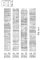
- FIG. 1 Supercharged green fluorescent proteins (GFPs).
- GFPs Supercharged green fluorescent proteins
- (a) Protein sequences of GFP variants, with fluorophore-forming residues highlighted green, negatively charged residues highlighted red, and positively charged residues highlighted blue.
- FIG. 2 Intramolecular properties of GFP variants, (a) Staining and UV fluorescence of purified GFP variants. Each lane and tube contains 0.2 ⁇ g of protein. (b) Circular dichroism spectra of GFP variants. (c) Thermodynamic stability of GFP variants, measured by guanidinium-induced unfolding.
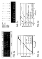
- FIG. 3 Intermolecular properties of supercharged proteins.
- Sample 1 6 ⁇ g of GFP(+36) in 30 ⁇ l of 25 mM Tris pH 7.0 and 100 mM NaCl.
- Sample 3 30 ⁇ g of salmon sperm DNA added to sample 1.
- Sample 4 20 ⁇ g of E . coli tRNA added to sample 1.
- Sample 5 Addition of NaCl to 1 M to sample 4.
- Samples 6-8 identical to samples 1, 2, and 4, respectively, except using sfGFP instead of GFP(+36). All samples were spun briefly in a microcentrifuge and visualized under UV light, (d) Enzymatic assays ofGST variants. Reactions contained 0.5 mg/mL of GST variant, 20 mM chlorodinitrobenzene, 20 mM glutathione, and 100 mM potassium phosphate pH 6.5. Product formation was monitored at 340 nm, resulting in observed reaction rates (k obs ) of 6 min -1 for wild-type GST, 2.2 min -1 for GST(-40), and 0.9 min -1 for GST(-40) after being boiled and cooled.
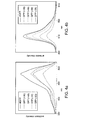
- FIG. 5 Biotin-binding activity of streptavidin variants, measured as described previously ( Kada et al., Rapid estimation of avidin and streptavidin by fluorescence quenching or fluorescence polarization. Biochim. Biophys. Acta 1427, 44-48 (1999 ); incorporated herein by reference) by monitoring binding-dependent of biotin-4-fluorescein (Invitrogen). Protein samples were titrated into 0.3 ⁇ M biotin-4-fluoreseein (B4F), 100 mM NaCl, 1 mM EDTA, 0.1 mg/mL bovine serum albumin (BSA), 50 mM potassium phosphate pH 7.5.
- B4F biotin-4-fluoreseein
- BSA bovine serum albumin
- the invention provides a system for modifying proteins to be more stable.
- the system is thought to work by changing non-conserved amino acids on the surface of a protein to more polar or charged amino acid residues.
- the amino acids residues to be modified may be hydrophobic, hydrophilic, charged, or a combination thereof.
- Any protein may be modified using the inventive system to produce a more stable variant. These modifications of surface residues have been found to improve the extrathermodynamic properties of proteins.
- proteins are increasingly used as therapeutic agents and as they continue to be used as research tools, a system for altering a protein to make it more stable is important and useful.
- Proteins modified by the inventive method typically are resistant to aggregation, have an increased ability to refold, resist improper folding, have improved solubility, and are generally more stable under a wide range of conditions including denaturing conditions such as heat or the presence of a detergent.
- Any protein may be modified to create a more stable variant using the inventive system.
- Natural as well as unnatural proteins e.g ., engineered proteins
- Example of proteins that may be modified include receptors, membrane bound proteins, transmembrane proteins, enzymes, transcription factors, extracellular proteins, therapeutic proteins, cytokines, messenger proteins, DNA-binding proteins, RNA-binding proteins, proteins involved in signal transduction, structural proteins, cytoplasmic proteins, nuclear proteins, hydrophobic proteins, hydrophilic proteins, etc.
- the protein to be modified may be derived from any species of plant, animal, or microorganism, In certain embodiments, the protein is a mammalian protein. In certain embodiments, the protein is a human protein.
- the proteins is derived from an organism typically used in research.
- the protein to be modified may be from a primate (e.g ., ape, monkey), rodent (e.g ., rabbit, hamster, gerbil), pig, dog, cat, fish ( e.g., zebrafish), nematode ( e.g., C. elegans ), yeast ( e.g., Saccharomyces cervisiae), or bacteria ( e.g., E. coli ).
- a primate e.g ., ape, monkey
- rodent e.g ., rabbit, hamster, gerbil
- pig e.g., dog, cat
- fish e.g., zebrafish
- nematode e.g., C. elegans
- yeast e.g., Saccharomyces cervisiae
- bacteria e.g., E. coli
- the inventive system is particularly useful in modifying proteins that are susceptible to aggregation or have stability issues.
- the system may also be used to modify proteins that are being overexpressed.
- therapeutic proteins that are being produced recombinantly may benefit from being modified by the inventive system.
- modified therapeutic proteins are not only easier to produce and purify but also may be more stable with respect to storage and use of the protein.
- the inventive system involves identifying non-conserved surface residues of a protein of interest and replacing some of those residues with a residue that is hydrophilic, polar, or charged at physiological pH.
- the inventive system includes not only methods for modifying a protein but also reagents and kits that are useful in modifying a protein to make it more stable.
- the surface residues of the protein to be modified are identified using any method(s) known in the art.
- the surface residues are identified by computer modeling of the protein.
- the three-dimensional structure of the protein is known and/or determined, and the surface residues are identified by visualizing the structure of the protein.
- the surface residues are predicted using computer software.
- Average Neighbor Atoms per Sidechain Atom (AvNAPSA) is used to predict surface exposure.
- AvNAPSA is an automated measure of surface exposure which has been implemented as a computer program. See Appendix A. A low AvNAPSA value indicates a surface exposed residue, whereas a high value indicates a residue in the interior of the protein.
- the software is used to predict the secondary structure and/or tertiary structure of a protein and the surface residues are identified based on this prediction.
- the prediction of surface residues is based on hydrophobicity and hydrophilicity of the residues and their clustering in the primary sequence of the protein.
- the surface residues of the protein may also be identified using various biochemical techniques, for example, protease cleavage, surface modification, etc.
- the conserved residues are identified by aligning the primary sequence of the protein of interest with related proteins. These related proteins may be from the same family of proteins. For example, if the protein is an immunoglobulin, other immunoglobulin sequences may be used. The related proteins may also be the same protein from a different species. For example, the conserved residues may be identified by aligning the sequences of the same protein from different species. To give but another example, proteins of similar function or biological activity may be aligned.
- the residue is considered conserved if over 50%, 60%, 70%, 75%, 80%, or 90% of the sequences have the same amino acid in a particular position. In other embodiments, the residue is considered conserved if over 50%, 60%, 70%, 75%, 80%, or 90% of the sequences have the same or a similar ( e.g ., valine, leucine, and isoleucine; glycine and alanine; glutamine and asparagine; or aspartate and glutamate) amino acid in a particular position.
- Many software packages are available for aligning and comparing protein sequences as described herein.
- conserved residues may be determined first or the surface residues may be determined first.
- the order does not matter.
- a computer software package may determine surface residues and conserved residues simultaneously.
- Important residues in the protein may also be identified by mutagenesis of the protein. For example, alanine scanning of the protein can be used to determine the important amino acid residues in the protein. In other embodiments, site-directed mutagenesis may be used.
- each of the residues is identified as hydrophobic or hydrophilic.
- the residues is assigned a hydrophobicity score.
- each non-conserved surface residue may be assigned an octanol/water logP value. Other hydrophobicity parameters may also be used.
- At least one identified non-conserved or non-vital surface residue is then chosen for modification.
- hydrophobic residue(s) are chosen for modification.
- hydrophilic and/or charged residue(s) are chosen for modification.
- more than one residue is chosen for modification.
- 1, 2, 3, 4, 5, 6, 7, 8, 9, or 10 of the identified residues are chosen for modification.
- over 10, over 15, or over 20 residues are chosen for modification.
- the larger the protein the more residues that will need to be modified.
- the more hydrophobic or susceptible to aggregation or precipitation the protein is the more residues will need to be modified.
- multiple variants of the protein, each with different modifications are produced and tested to determine the best variant in terms of biological activity and stability.
- the residues chosen for modification are mutated into more hydrophilic residues (including charged residues).
- the residues are mutated into more hydrophilic natural amino acids.
- the residues are mutated into amino acids that are charged at physiological pH.
- the residue may be changed to an arginine, aspartate, glutamate, histidine, or lysine.
- all the residues to be modified are changed into the same different residue.
- all the chosen residues are changed to a glutamate residue.
- the chosen residues are changed into different residues; however, all the final residues may be either positively charged or negatively charged at physiological pH.
- all the residues to be mutated are converted to glutamate and/or aspartate residues.
- all the residues to be mutated are converted to lysine residues.
- all the chosen residues for modification are asparagine, glutamine, lysine, and/or arginine, and these residues are mutated into aspartate or glutamate residues.
- all the chosen residues for modification are aspartate, glutamate, asparagine, and/or glutamine, and these residues are mutated into lysine. This approach allows for modifying the net charge on the protein to the greatest extent.
- the protein may be modified to keep the net charge on the modified protein the same as on the unmodified protein.
- the protein may be modified to decrease the overall net charge on the protein while increasing the total number of charged residues on the surface.
- the theoretical net charge is increased by at least +1, +2, +3, +4, +5, +10, +15, +20, +25, +30, or +35.
- the theoretical net charge is decreased by at least -1, -2, -3, -4, -5, -10, -15, -20, -25, -30, or -35.
- the chosen amino acids are changed into non-ionic, polar residues (e.g ., cysteine, serine, threonine, tyrosine, glutamine, asparagine).
- modification or mutations in the protein may be accomplished using any technique known in the art.
- Recombinant DNA techniques for introducing such changes in a protein sequence are well known in the art.
- the modifications are made by site-directed mutagenesis of the polynucleotide encoding the protein.
- Other techniques for introducing mutations are discussed in Molecular Cloning: A Laboratory Manual, 2nd Ed., ed. by Sambrook, Fritsch, and Maniatis (Cold Spring Harbor Laboratory Press: 1989 ); the treatise, Methods in Enzymology (Academic Press, Inc., N.Y .); Ausubel et al.
- the modified protein is expressed and tested.
- a series of variants is prepared and each variant is tested to determine its biological activity and its stability.
- the variant chosen for subsequent use may be the most stable one, the most active one, or the one with the greatest overall combination of activity and stability.
- an additional set of variants may be prepared based on what is learned from the first set.
- the variants are typically created and overexpressed using recombinant techniques known in the art.
- the inventive system has been used to created variants of GFP. These variants have been shown to be more stable and to retain their fluorescence.
- a GFP from Aequorea victoria is described in GenBank Accession Number P42212, incorporated herein by reference.
- the amino acid sequence of this wild type GFP is as follows: Wild type GFP has a theoretical net charge of -7.
- variants with a theoretical net charge of-29, -30, -25, +36, +48, and +49 have been created. Even after heating the +36 GFP to 95°C, 100% of the variant protein is soluble and the protein retains ⁇ 70% of its fluorescence.
- amino acid sequences of the variants of GFP that have been created include:
- Any DNA sequence that encodes the above GFP variants is also include within the scope of the invention.
- Exemplary DNA sequences which encode each of the variants above are as follows;
- polynucleotide sequence homologous to the above sequences are also within the scope of the present invention, in certain embodiments, the polynucleotide sequence include a stretch of 50, 100, or 150 nucleotides that are 60%, 70%, 80%, 90%, 95%, 98%, 99%, or 100% homologous to any one of the above sequence.
- the present invention also includes sequence where one or more nucleotides is inserted or deleted from one of the above sequences. Any polynucleotide sequence with a mutation as shown in any of the sequences above is considered part of the invention. In certain embodiments, the sequence includes 2, 3, 4, 5, 6, 7, 8, 9, 10, or more mutations as shown in any of the sequences above.
- the present invention also provides vector (e.g., plasmids, cosmids, viruses, etc .) that comprise any of the inventive sequences herein or any other sequence (DNA or protein) modified using the inventive system.
- the vector includes elements such as promoter, enhancer, ribosomal binding sites, etc. sequences useful in overexpressing the inventive GFP variant in a cell.
- the invention also includes cells comprising the inventive sequences or vectors. In certain embodiments, the cells overexpress the variant GFP.
- the cells may be bacterial cells (e.g ., E. coli ), fungal cells ( e.g., P. pastoris ), yeast cells ( e.g., S. cerevisiae ), mammalian cells ( e.g., CHO cells), or human cells.
- the inventive system has been used to created variants of streptavidin. These variants have been shown to form soluble tetramers that bind biotin.
- the amino acid sequence of this wild type streptavidin is as follows: Wild type streptavidin has a theoretical net charge of -4. Using the inventive system, variants with a theoretical net charge of -40 and +52 have been created. Even after heating the variants to 100°C, the proteins remained soluble.
- amino acid sequences of the variants of streptavidin that have been created include:
- polynucleotide sequence homologous to the above sequences are also within the scope of the present invention.
- the polynucleotide sequence include a stretch of 50, 100, or 150 nucleotides that are 60%, 70%, 80%, 90%, 95%, 98%, 99%, or 100% homologous to any one of the above sequence.
- the present invention also includes sequence where one or more nucleotides is inserted or deleted from one of the above sequences. Any polynucleotide sequence with a mutation as shown in any of the sequences above is considered part of the invention. In certain embodiments, the sequence includes 2, 3, 4, 5, 6, 7, 8, 9, 10, or more mutations as shown in any of the sequences above.
- the present invention also provides vector (e.g. , plasmids, cosmids, viruses, etc .) that comprise any of the inventive sequences herein or any other sequence (DNA or protein) modified using the inventive system.
- the vector includes elements such as promoter, enhancer, ribosomal binding sites, etc. sequences useful in overexpressing the inventive streptavidin variant in a cell.
- the invention also includes cells comprising the inventive sequences or vectors. In certain embodiments, the cells overexpress the variant streptavidin.
- the cells may be bacterial cells ( e.g., E. coli ), fungal cells ( e.g., P. pastors ), yeast cells ( e.g., S. cerevisiae ), mammalian cells ( e.g., CHO cells), or human cells,
- the inventive system has been used to created variants of glutathione- S- transferase (GST). These variants have been shown to retain the catalytic activity of wild type GST.
- the amino acid sequence of this wild type GST is as follows: Wild type GST has a theoretical net charge of +2.
- a variant with a theoretical net charge of -40 has been created.
- This variant catalyzes the addition of glutathione to chloronitrobenzene with a specific activity only 2.7-fold lower than that of wild type GST. Even after heating the variant to 100 °C, the protein remained soluble, and the protein recovered 40% of its catalytic activity upon cooling.
- amino acid sequences of variants of GST include:
- the present invention also provides vector (e.g., plasmids, cosmids, viruses, etc. ) that comprise any of the inventive sequences herein or any other sequence (DNA or protein) modified using the inventive system.
- the vector includes elements such as promoter, enhancer, ribosomal binding sites, etc. sequences useful in overexpressing the inventive GST variant in a cell.
- the invention also includes cells comprising the inventive sequences or vectors. In certain embodiments, the cells overexpress the variant GST.
- the cells may be bacterial cells (e.g., E. coli ), fungal cells ( e.g., P. pastoris ), yeast cells ( e.g., S. cerevisiae ), mammalian cells ( e.g., CHO cells), or human cells,
- kits for modifying proteins of interest to produce more stable variants of the protein typically include all or most of the reagents needed create a more stable variant of a protein.
- the kit includes computer software to aid a researcher in designing the more stable variant protein based on the inventive method.
- the kit may also include all of some of the following: reagents, primers, oligonucleotides, nucleotides, enzymes, buffers, cells, media, plates, tubes, instructions, vectors, etc.
- the research using the kit typically provides the DNA sequence for mutating to create the more stable variant.
- the contents are typically packaged for convenience use in a laboratory.
- Protein aggregation a well known culprit in human disease ( Cohen, F. E.; Kelly, J. W., Nature 2003, 426, (6968), 905-9 ; Chiti, F.; Dobson, C. M., Annu Rev Biochem 2006, 75, 333-66 ; each of which is incorporated herein by reference), is also a major problem facing the use of proteins as therapeutic or diagnostic agents ( Frokjaer, S.; Otzen, D. E., Nat Rev Drug Discov 2005, 4, (4), 298-306 ; Fowler, S. B.; Poon, S.; Muff, R.; Chiti, F.; Dobson, C.
- Superfolder GFP has a net charge of-7, similar to that of wild-type GFP.
- sfGFP green fluorescent protein
- sfGFP is the product of a long history of GFP optimization ( Giepmans et al., Science 2006, 312, (5771), 217-24 ; incorporated herein by reference), it remains susceptible to aggregation induced by thermal or chemical unfolding. Heating sfGFP to 100°C induced its quantitative precipitation and the irreversible loss of fluorescence ( Figure 3a ). In contrast, supercharged GFP(+36) and GFP(-30) remained soluble when heated to 100°C, and recovered significant fluorescence upon cooling ( Figure 3a ).
- Glutathione-S-transferase (GST), a dimer with a total net charge of +2, was supercharged to yield a dimer with net charge of -40 that catalyzed the addition of glutathione to chlorodinitrobenzene with a specific activity only 2.7-fold lower than that of wild-type GST ( Figure 3d ).
- the supercharged streptavidins and supercharged GST remained soluble when heated to 100°C, in contrast to their wild-type counterparts, which, like sfGFP, precipitated quantitatively and irreversibly (Table 1).
- GST(-40) recovered 40% of its catalytic activity upon cooling ( Figure 3d ).
- Protein supercharging illustrates the remarkable plasticity of protein surfaces and highlights the opportunities that arise from the mutational tolerance of solvent-exposed residues. For example, it was recently shown that the thermodynamic stability of some proteins can be enhanced by rationally engineering charge-charge interactions ( Strickler et al., Biochemistry 2006, 45, (9), 2761-6 ; incorporated herein by reference). Protein supercharging demonstrates how this plasticity can be exploited in a different way to impart extraordinary resistance to protein aggregation. Our findings are consistent with the results of a complementary study in which removal of all charges from ubiquitin left its folding intact but significantly impaired its solubility ( Loladze et al, Protein Sci 2002, 11, (1), 174-7 ; incorporated herein by reference).
- GFP green fluorescent protein
- Protein expression and purification Protein expression and purification .
- Synthetic genes optimized for E. coli codon usage were purchased from DNA 2.0, cloned into a pET expression vector (Novagen), and overexpressed in E . coli BL21(DE3)pLysS for 5-10 hours at 15°C. Cells were harvested by centrifugation and lysed by sonication. Proteins were prized by Ni-NTA agarose chromotography (Qiagen), buffer-exchanged into 100 mM NaCl, 50 mM potassium phosphate pH 7.5, and concentrated by ultrafiltration (Millipore). All GFP variants were purified under native conditions. Wild-type streptavidin was purchased from Promega.
- Supercharged streptavidin variants were purified under denaturing conditions and refolded as reported previously for wild-type streptavidin ( Thompson et al. Construction and expression of a synthetic streptavidin-encoding gene in Escherichia coli. Gene 136, 243-246 (1993 ); incorporated herein by reference), as was supercharged GST. Wild-type GST was purified under either native or denaturing conditions, yielding protein of comparable activity.
- Electrostatic surface potential calculations ( Figure 1b ) . Models of -30 and +48 supercharged GFP variants were based on the crystal structure of superfolder GFP ( Pedelacq et al., Engineering and characterization of a superfolder green fluorescent protein. Nat Biotechnol 24, 79-88 (2006 ); incorporated herein by reference). Electrostatic potentials were calculated using APBS ( Baker et al., Electrostatics of nanosystems: application to microtubules and the ribosome.
- Protein staining and UV-induced fluorescence ( Figure 2a ) .
- 0.2 ⁇ g of each GFP variant was analyzed by electrophoresis in a 10% denaturing polyacrylamide gel and stained with Coomassie brilliant blue dye.
- 0.2 ⁇ g of the same protein samples in 25 mM Tris pH 8.0 with 100 mM NaCl was placed in a 0.2 mL Eppendorftube and photographed under UV light (360 nm).
- Size-exclusion chromotography (Table 1) . The multimeric state of SAV and GST variants was determined by analyzing 20-50 ⁇ g of protein on a Superdex 75 gel-filtration column. Buffer was 100 mM NaCl, 50 mM potassium phosphate pH 7.5. Molecular weights were determined by comparison with a set of monomeric protein standards of known molecular weights analyzed separately under identical conditions. Table 1 . Calculated and experimentally determined protein properties.
- the present invention also provides:
Abstract


Description
- The present application claims priority under 35 U.S.C. § 119(e) to U.S. provisional patent application,
USSN 60/810,364, filed June 2, 2006 - The work described herein was supported, in part, by grants from the National Institutes of Health (GM065400). The United States government may have certain rights in the invention.
- Proteins are the workhorses of the cell. Proteins catalyze chemical reactions, transduce signals in biological systems, provide structural elements in cells and the extracellular matrix, act as messengers, etc. One of the major causes of misbehavior of proteins is aggregation. This is not only a problem in the laboratory but also a problem in many diseases such as Alzheimer's disease. Aggregation is a particularly vexing problem when it comes to computationally designed proteins. For example, TOP7 is a computationally designed protein with a novel fold. A longer version of TOP7, TOP7 extended, is very prone to aggregation. TOP7ex is expressed predominantly as insoluble aggregates.
- As more proteins are either designed or modified to be used a tools to study biological systems or as more proteins - wild type or modified - are used as therapeutic agents, there needs to be a system for routinely modifying these proteins to be more stable and/or to prevent aggregation.
- The present invention provides a system for modifying proteins to make them more stable. The invention stems from the recognition that modifying the hydrophobic areas on the surface of a protein can improve the extrathermodynamic properties of the protein. The inventive system is particularly useful in improving the solubility of a protein of interest, improving the protein's resistance to aggregation, and/or improving the protein's ability to renature. All of these properties are particularly useful in protein production, protein purification, and the use of proteins as therapeutic agents and research tools.
- In one aspect, the invention provides a method of altering the primary sequence of a protein in order to increase the protein's resistance to aggregation, solubility, ability to refold, and/or general stability under a wide range of conditions; The activity of the modified protein is preferably approximately or substantially the same as the protein without modification. In certain embodiments, the modified protein retains at least 50%, 75%, 90%, or 95% of the wild type protein's activity. In one embodiments, the method includes the steps of (a) identifying the surface residues of a protein of interest; (b) identifying the particular surface residues that are not highly conserved among other proteins related to the protein of interest (i.e., determining which amino acids are not essential for the activity or function of the protein); (c) determining the hydrophobicity of the identified non-conserved surface residues; and (e) replacing at least one or more of the identified hydrophobic, non-conserved residues with an amino acid that is more polar or is charged at physiological pH, Each of the above steps may be carried out using any technique, computer software, algorithm, paradigm, etc. known in the art. After the modified protein is created, it may be tested for its activity and/or the desired property being sought. In certain embodiments, the modified protein is more stable. In certain embodiments, the modified protein is less susceptible to aggregation. The inventive method typically increases the net charge (positive or negative) on the protein at physiological pH.
- In another aspect, the invention provides a method of altering the primary sequence of a protein in order to increase the protein's resistance to aggregation, solubility, ability to refold, and/or general stability under a wide range of conditions by "supercharging" the protein. That is, the overall net charge on the modified protein is increased (either positive charge or negative charge) compared to the wild type protein. Preferably, the activity of the modified protein is approximately or substantially the same as the protein without modification. In certain embodiments, the method includes the steps of (a) identifying the surface residues of a protein of interest; (b) identifying the particular surface residues that are not highly conserved among other proteins related to the protein of interest (i.e., determining which amino acids are not essential for the activity or function of the protein); (c) determining the hydrophilicity of the identified non-conserved surface residues; and (e) replacing at least one or more of the identified charged or polar, solvent-exposed, non-conserved residues with a charged amino acid that is charged at physiological pH. In certain embodiments, to make a negatively charged "supercharged" protein, the residues identified for modification are mutated either to aspartate (Asp) or glutamate (Glu) residues. In certain other embodiments, to make a positively charged "supercharged" protein, the residues identified for modification are mutated either to lysine (Lys) or arginine (Arg) residues. Each af the above steps may be carried out using any technique, computer software, algorithm, paradigm, etc. known in the art. After the modified protein is created, it may be tested for its activity and/or the desired property being sought. In certain embodiments, the modified protein ("supercharged pratein") is more stable. In certain embodiments, the modified protein is less susceptible to aggregation. The inventive method typically increases the net charge (positive or negative) on the protein at physiological pH.
- The theoretical net charge on over 80% of the proteins catalogued in the Protein Data Bank (PDB) fall within ±10. The modified protein created by the present invention typically have a net charge less than -10 or greater than +10. In certain embodiments, the modified protein has a net charge less than -20 or greater than +20. In certain embodiments, the modified protein has a net charge less than -30 or greater than +30. In certain embodiments, the modified protein has a net charge less than -40 or greater than +40. In certain embodiments, the modified protein has a net charge less than -50 or greater than +30. The modified proteins are able to fold correctly and retain their biological activity.
- Any protein may be modified using the inventive system, and protein variants created by the inventive system are considered to be part of the present invention, as well as polynucleotides or vectors encoding the variant protein and cells expressing the variant protein. The inventive system has been used to create several new variants of green fluorescent protein (GFP). These variants retain their fluorescence; however, they are more stable than current versions of GFP under a wide range of environments. The inventive GFPs are immune to aggregation even over long periods oftime and in environments that induce aggregation and are capable of refolding into a fluorescent protein even after being denatured by boiling. The inventive system has also been used to create new variants of streptavidin and glutathione-S-transferase (GST). These variants retain their biological activity and remain soluble when heated. The invention also includes polynucleotide sequences encoding the inventive GFP, streptavidin, and GST protein sequences, vectors including any of these nucleotide sequences, and cells that include such a polynucleotide sequence or vector, or express the inventive variants. In certain embodiments, the invention includes bacteria or other cells that overexpress an inventive variant. The inventive variants may be used in a variety of biological assays known in the art. For example, supercharged GYPS may be used in any assay that currently uses GFP as a reporter protein.
- In another aspect, the invention provides other proteins that have been modified by the inventive system. These modified proteins preferably retain a significant portion of their original activity. In certain embodiments, the modified protein retains at least 99%, 98%, 95%, or 90% of the activity of the unmodified version. The modified protein may be more soluble, resistant to aggregation, have a increased ability to refold, and/or have greater stability under a variety of conditions. The proteins modified by the inventive system include hydrophobic proteins, recombinant proteins, membrane proteins, structural proteins, enzymes, extracellular proteins, therapeutic proteins (e.g., insulin, cytokines, immunoglobulins, fragments of immunoglobulins, etc.), receptors, cell signaling proteins, cytoplasmic proteins, nuclear proteins, transcription factors, etc. In certain specific embodiments, the proteins are therapeutic proteins for use in human or veterinary medicine. In certain embodiments, the proteins are unnatural proteins, for example, computationally designed proteins. In other embodiments, the proteins are hybrid proteins, fusion proteins, altered proteins, mutated proteins, genetically engineered proteins, or any other protein that has been altered by the hands of man.
- Kits are also provided for the practice of the invention. The kits may include the reagents needed to modify a protein of interest to make it more resistant to aggregation, increase its ability to renature, or increase its stability overall. Such kits may include all or some of the following: polynucleotides, computer software, nucleotides, primers, vectors, cell lines, instructions, plates, media, buffers, enzymes, Eppendorf tubes, site-directed mutagenesis kits, etc. Preferably, the kit is conveniently packaged for use in a laboratory setting. The researcher typically provides the DNA coding sequence of the protein to be modified using the inventive technique.
- "Amino acid"; The term "amino acid" refers to the basic structural subunits of proteins. An alpha-amino acid consists of an amino group, a carboxyl group, a hydrogen atom, and a side chain (i.e., R group) all bonded to a central carbon atom. This central carbon atom is referred to as the alpha carbon because it is adjacent to the carboxyl group. There are twenty natural amino acids including glycine, alanine, valine, leucine, isoleucine, phenylalanine, tyrosine, trypotphan, cysteine, methionine, serine, threonine, lysine, arginine, histidine, aspartate, glutamate, asparagine, glutamate, and proline. Hydrophobic amino acids include alanine, valine, leucine, isoleucine, and phenylalanine. Aromatic amino acids includes phenylalanine, tyrosine, tryptophan, and histine. Polar amino acids include tyrosine, cysteine, serine, threonine, lysine, arginine, histidine, aspartate, glutamate, asparagine, and glutamine. Sulfur-containing amino acids include cysteine and methionine. Basic amino acids include lysine, arginine, and histidine. Acidic amino acids include aspartate and glutamate. Unnatural amino acids have also been inserted into proteins. In certain embodiments, the twenty natural amino acids are referred to when the term "amino acid" is used.
- "Antibody": The term "antibody" refers to an immunoglobulin, whether natural or wholly or partially synthetically produced. All derivatives thereof which maintain specific binding ability are also included in the term. The term also covers any protein having a binding domain which is homologous or largely homologous to an immunoglobulin binding domain. These proteins may be derived from natural sources, or partly or wholly synthetically produced. An antibody may be monoclonal or polyclonal. The antibody may be a member of any immunoglobulin class, including any of the human classes: IgG, IgM, IgA, IgD, and IgE.
- "Conserved": The term "conserved" refers nucleotides or amino acid residues of a polynucleotide sequence or amino acid sequence, respectively, that are those that occur unaltered in the same position of two or more related sequences being compared. Nucleotides or amino acids that are relatively conserved are those that are conserved amongst more related sequences than nucleotides or amino acids appearing elsewhere in the sequences.
- "Homologous": The term "homologous", as used herein is an art-understood term that refers to nucleic acids or proteins that are highly related at the level of nucleotide or amino acid sequence. Nucleic acids or proteins that are homologous to each other are termed homologues. Homologous may refer to the degree of sequence similarity between two sequences (i.e., nucleotide sequence or amino acid). The homology percentage figures referred to herein reflect the maximal homology possible between two sequences, i.e., the percent homology when the two sequences are so aligned as to have the greatest number of matched (homologous) positions. Homology can be readily calculated by known methods such as those described in: Computational Molecular Biology, Lesk, A. M., ed., Oxford University Press, New York, 1988; Biocomputing: Informatics and Genome Projects, Smith, D. W., ed., Academic Press, New York, 1993; Sequence Analysis in Molecular Biology, von Heinje, G., Academic Press, 1987; Computer Analysis of Sequence Data, Part I, Griffin, A. M., and Griffin, H. G., eds., Humana Press, New Jersey, 1994; and Sequence Analysis Primer, Gribskov, M. and Devereux, J., eds., M Stockton Press, New York, 1991; each of which is incorporated herein by reference. Methods commonly employed to determinehomology between sequences include, but are not limited to those disclosed in Carillo, H., and Lipman, D., SIAM J Applied Math., 48:1073 (1988); incorporated herein by reference. Techniques for determining homology are codified in publicly available computer programs. Exemplary computer software to determine homology between two sequences include, but are not limited to, GCG program package, Devereux, J., et al., Nucleic Acids Research, 12(1), 387 (1984)), BLASTP, BLASTN, and FASTA Atschul, S. F. et al., J Molec. Biol., 215, 403 (1990)).
- The term "homologous" necessarily refers to a comparison between at least two sequences (nucleotides sequences or amino acid sequences). In accordance with the invention, two nucleotide sequences are considered to be homologous if the polypeptides they encode are at least about 50-60% identical, preferably about 70% identical, for at least one stretch of at least 20 amino acids. Preferably, homologous nucleotide sequences are also characterized by the ability to encode a stretch of at least 4-5 uniquely specified amino acids. Both the identity and the approximate spacing of these amino acids relative to one another must be considered for nucleotide sequences to be considered homologous. For nucleotide sequences less than 60 nucleotides in length, homology is determined by the ability to encode a stretch of at least 4-5 uniquely specified amino acids.
- "Peptide" or "protein": According to the present invention, a "peptide" or "protein" comprises a string of at least three amino acids linked together by peptide bonds. The terms "protein" and "peptide" may be used interchangeably. Inventive peptides preferably contain only natural amino acids, although non-natural amino acids (i.e., compounds that do not occur in nature but that can be incorporated into a polypeptide chain) and/or amino acid analogs as are known in the art may alternatively be employed. Also, one or more of the amino acids in an inventive peptide may be modified, for example, by the addition of a chemical entity such as a carbohydrate group, a phosphate group, a farnesyl group, an isofarnesyl group, a fatty acid group, a linker for conjugation, functionalization, or other modification (e.g., alpha amindation), etc. In a preferred embodiment, the modifications of the peptide lead to a more stable peptide (e.g., greater half-life in vivo). These modifications may include cyclization of the peptide, the incorporation of-amino acids, etc. None of the modifications should substantially interfere with the desired biological activity of the peptide. In certain embodiments, the modifications of the peptide lead to a more biologically active peptide.
- "Polynucleotide" or "oligonucleotide": Polynucleotide or oligonucleotide refers to a polymer of nucleotides. Typically, a polynucleotide comprises at least three nucleotides. The polymer may include natural nucleosides (i.e., adenosine, thymidine, guanosine, cytidine, uridine, deoxyadenosine, deoxythymidine, deoxyguanosine, and deoxycytidine), nucleoside analogs (e.g., 2-aminoadenosine, 2-thiothymidine, inosine, pyrrolo-pyrimidine, 3-methyl adenosine, C5-propynylcytidine, C5-propynyluridine, C5-bromouxidine, C5-fluorouridine, C5-lodouridine, C5-methylcytidine, 7-deazaadenosine, 7-deazaguanosine, 8-oxoadenosine, 8-oxoguanosine, O(6)-methylguanine, and 2-thiocytidine), chemically modified bases, biologically modified bases (e.g., methylated bases), intercalated bases, modified sugars (e.g., 2'-fluororibose, ribose, 2'-deoxyribose, arabinose, and hexose), and/or modified phosphate groups (e.g., phosphorothioates and 5'-N-phosphoramidite linkages),
- "Small molecule": The term "small molecule," as used herein, refers to a non-peptidic, non-oligomeric organic compound either prepared in the laboratory or found in nature. Small molecules, as used herein, can refer to compounds that are "natural product-like," however, the term "small molecule" is not limited to "natural product-like" compounds. Rather, a small molecule is typically characterized in that it contains several carbon-carbon bonds, and has a molecular weight of less than 1500, although this characterization is not intended to be limiting for the purposes of the present invention. In certain other preferred embodiments, natural-product-like small molecules are utilized.
- "Stable": The term "stable" as used herein to refer to a protein refers to any aspect of protein stability. The stable modified protein as compared to the original wild type protein possesses any one or more of the following characteristics: more soluble, more resistant to aggregation, more resistant to denaturation, more resistant to unfolding, more resistant to improper or undesired folding, greater ability to renature, increased thermal stability, increased stability in a variety of environments (e.g., pH, salt concentration, presence of detergents, presence of denaturing agents, etc.), and increased stability in non-aqueous environments. In certain embodiments, the stable modified protein exhibits at least two of the above characteristics. In certain embodiments, the stable modified protein exhibits at least three of the above characteristics. Such characteristics may allow the active protein to be produced at higher levels. For example, the modified protein can be overexpressed at a higher level without aggregation than the unmodified version of the protein. Such characteristics may also allow the protein to be used as a therapeutic agent or a research tool.
-
Figure 1 . Supercharged green fluorescent proteins (GFPs). (a) Protein sequences of GFP variants, with fluorophore-forming residues highlighted green, negatively charged residues highlighted red, and positively charged residues highlighted blue. (b) Electrostatic surface potentials of sfGFP (left), GFP(+36) (middle), and GFP(-30) (right), colored from -25 kT/e (red) to +25 kT/e (blue). -
Figure 2 . Intramolecular properties of GFP variants, (a) Staining and UV fluorescence of purified GFP variants. Each lane and tube contains 0.2 µg of protein. (b) Circular dichroism spectra of GFP variants. (c) Thermodynamic stability of GFP variants, measured by guanidinium-induced unfolding. -
Figure 3 . Intermolecular properties of supercharged proteins. (a) UV-illuminated samples of purified GFP variants ("native"), those samples heated 1 min at 100 °C ("boiled"), and those samples subsequently cooled for 2 h at 25°C ("cooled"). (b) Aggregation of GFP variants was induced with 40% TFE at 25 °C and monitored by right-angle light scattering. (c) Supercharged GFPs adhere reversibly to oppositely charged macromolecules. Sample 1:6 µg of GFP(+36) in 30 µl of 25 mM Tris pH 7.0 and 100 mM NaCl. Sample 2: 6 µg of GFP(-30) added tosample 1. Sample 3: 30 µg of salmon sperm DNA added tosample 1. Sample 4: 20 µg of E. coli tRNA added tosample 1. Sample 5: Addition of NaCl to 1 M to sample 4. Samples 6-8: identical tosamples -
Figure 4. (a) Excitation and (b) emission spectra of GFP variants. Each sample contained an equal amount of protein as quantitated by chromophore absorbance at 490 nm, -
Figure 5 . Biotin-binding activity of streptavidin variants, measured as described previously (Kada et al., Rapid estimation of avidin and streptavidin by fluorescence quenching or fluorescence polarization. Biochim. Biophys. Acta 1427, 44-48 (1999); incorporated herein by reference) by monitoring binding-dependent of biotin-4-fluorescein (Invitrogen). Protein samples were titrated into 0.3 µM biotin-4-fluoreseein (B4F), 100 mM NaCl, 1 mM EDTA, 0.1 mg/mL bovine serum albumin (BSA), 50 mM potassium phosphate pH 7.5. Quenching of fluorescence at 526 nm was measured on a Perkin-ELmer LS50B luminescence spectrometer with excitation at 470 nm. Measurements were normalized to control titrations that contained a 600-fold excess of non-fluorescent biotin. The three proteins in the bottom of the legend are included as negative controls. - The invention provides a system for modifying proteins to be more stable. The system is thought to work by changing non-conserved amino acids on the surface of a protein to more polar or charged amino acid residues. The amino acids residues to be modified may be hydrophobic, hydrophilic, charged, or a combination thereof. Any protein may be modified using the inventive system to produce a more stable variant. These modifications of surface residues have been found to improve the extrathermodynamic properties of proteins. As proteins are increasingly used as therapeutic agents and as they continue to be used as research tools, a system for altering a protein to make it more stable is important and useful. Proteins modified by the inventive method typically are resistant to aggregation, have an increased ability to refold, resist improper folding, have improved solubility, and are generally more stable under a wide range of conditions including denaturing conditions such as heat or the presence of a detergent.
- Any protein may be modified to create a more stable variant using the inventive system. Natural as well as unnatural proteins (e.g., engineered proteins) may be modified. Example of proteins that may be modified include receptors, membrane bound proteins, transmembrane proteins, enzymes, transcription factors, extracellular proteins, therapeutic proteins, cytokines, messenger proteins, DNA-binding proteins, RNA-binding proteins, proteins involved in signal transduction, structural proteins, cytoplasmic proteins, nuclear proteins, hydrophobic proteins, hydrophilic proteins, etc. The protein to be modified may be derived from any species of plant, animal, or microorganism, In certain embodiments, the protein is a mammalian protein. In certain embodiments, the protein is a human protein. In certain embodiments, the proteins is derived from an organism typically used in research. For example, the protein to be modified may be from a primate (e.g., ape, monkey), rodent (e.g., rabbit, hamster, gerbil), pig, dog, cat, fish (e.g., zebrafish), nematode (e.g., C. elegans), yeast (e.g., Saccharomyces cervisiae), or bacteria (e.g., E. coli).
- The inventive system is particularly useful in modifying proteins that are susceptible to aggregation or have stability issues. The system may also be used to modify proteins that are being overexpressed. For example, therapeutic proteins that are being produced recombinantly may benefit from being modified by the inventive system. Such modified therapeutic proteins are not only easier to produce and purify but also may be more stable with respect to storage and use of the protein.
- The inventive system involves identifying non-conserved surface residues of a protein of interest and replacing some of those residues with a residue that is hydrophilic, polar, or charged at physiological pH. The inventive system includes not only methods for modifying a protein but also reagents and kits that are useful in modifying a protein to make it more stable.
- The surface residues of the protein to be modified are identified using any method(s) known in the art. In certain embodiments, the surface residues are identified by computer modeling of the protein. In certain embodiments, the three-dimensional structure of the protein is known and/or determined, and the surface residues are identified by visualizing the structure of the protein. In other embodiments, the surface residues are predicted using computer software. In certain particular embodiments, Average Neighbor Atoms per Sidechain Atom (AvNAPSA) is used to predict surface exposure. AvNAPSA is an automated measure of surface exposure which has been implemented as a computer program. See Appendix A. A low AvNAPSA value indicates a surface exposed residue, whereas a high value indicates a residue in the interior of the protein. In certain embodiments, the software is used to predict the secondary structure and/or tertiary structure of a protein and the surface residues are identified based on this prediction. In other embodiments, the prediction of surface residues is based on hydrophobicity and hydrophilicity of the residues and their clustering in the primary sequence of the protein. Besides in silico methods, the surface residues of the protein may also be identified using various biochemical techniques, for example, protease cleavage, surface modification, etc.
- Of the surface residues, it is then determined which are conserved or important to the functioning of the protein. The identification of conserved residues can be determined using any method known in the art. In certain embodiments, the conserved residues are identified by aligning the primary sequence of the protein of interest with related proteins. These related proteins may be from the same family of proteins. For example, if the protein is an immunoglobulin, other immunoglobulin sequences may be used. The related proteins may also be the same protein from a different species. For example, the conserved residues may be identified by aligning the sequences of the same protein from different species. To give but another example, proteins of similar function or biological activity may be aligned. Preferably, 2, 3, 4, 5, 6, 7, 8, 9, or 10 different sequences are used to determine the conserved amino acids in the protein. In certain embodiments, the residue is considered conserved if over 50%, 60%, 70%, 75%, 80%, or 90% of the sequences have the same amino acid in a particular position. In other embodiments, the residue is considered conserved if over 50%, 60%, 70%, 75%, 80%, or 90% of the sequences have the same or a similar (e.g., valine, leucine, and isoleucine; glycine and alanine; glutamine and asparagine; or aspartate and glutamate) amino acid in a particular position. Many software packages are available for aligning and comparing protein sequences as described herein. As would be appreciated by one of skill in the art, either the conserved residues may be determined first or the surface residues may be determined first. The order does not matter. In certain embodiments, a computer software package may determine surface residues and conserved residues simultaneously. Important residues in the protein may also be identified by mutagenesis of the protein. For example, alanine scanning of the protein can be used to determine the important amino acid residues in the protein. In other embodiments, site-directed mutagenesis may be used.
- Once non-conserved surface residues of the protein have been identified, each of the residues is identified as hydrophobic or hydrophilic. In certain embodiments, the residues is assigned a hydrophobicity score. For example, each non-conserved surface residue may be assigned an octanol/water logP value. Other hydrophobicity parameters may also be used. Such scales for amino acids have been discussed in: Janin, "Surface and Inside Volumes in Globular Proteins," Nature 277;491-92, 1979; Wolfenden et al., "Affinities of Amino Acid Side Chains for Solvent Water," Biochemistry 20:849-855, 1981; Kyte et al., "A Simple Method for Displaying the Hydropathic Character of a Protein," J. Mol. Biol. 157: 105-132, 1982; Rose et al., "Hydrophobicity of Amino Acid Residues in Globular Proteins," Science 229:834-838, 1985; Cornette et al., "Hydrophobicity Scales and Computational Techniques for Detecting Amphipathic Structures in Proteins," J. Mol. Biol. 195:659-685,1987; Charton and Charton, "The Structure Dependence of Amino Acid Hydrophobicity Parameters," J. Theor, Biol. 99:629-644,1982; each of which is incorporated by reference. Any of these hydrophobicity parameters may be used in the inventive method to determine which non-conserved residues to modify. In certain embodiments, hydrophilic or charged residues are identified for modification.
- At least one identified non-conserved or non-vital surface residue is then chosen for modification. In certain embodiments, hydrophobic residue(s) are chosen for modification. In other embodiments, hydrophilic and/or charged residue(s) are chosen for modification. In certain embodiments, more than one residue is chosen for modification. In certain embodiments, 1, 2, 3, 4, 5, 6, 7, 8, 9, or 10 of the identified residues are chosen for modification. In certain embodiments, over 10, over 15, or over 20 residues are chosen for modification. As would be appreciated by one of skill in the art, the larger the protein the more residues that will need to be modified. Also, the more hydrophobic or susceptible to aggregation or precipitation the protein is, the more residues will need to be modified. In certain embodiments, multiple variants of the protein, each with different modifications, are produced and tested to determine the best variant in terms of biological activity and stability.
- In certain embodiments, the residues chosen for modification are mutated into more hydrophilic residues (including charged residues). Typically, the residues are mutated into more hydrophilic natural amino acids. In certain embodiments, the residues are mutated into amino acids that are charged at physiological pH. For example, the residue may be changed to an arginine, aspartate, glutamate, histidine, or lysine. In certain embodiments, all the residues to be modified are changed into the same different residue. For example, all the chosen residues are changed to a glutamate residue. In other embodiments, the chosen residues are changed into different residues; however, all the final residues may be either positively charged or negatively charged at physiological pH. In certain embodiments, to create a negatively charged protein, all the residues to be mutated are converted to glutamate and/or aspartate residues. In certain embodiments, to create a positively charged protein, all the residues to be mutated are converted to lysine residues. For example, all the chosen residues for modification are asparagine, glutamine, lysine, and/or arginine, and these residues are mutated into aspartate or glutamate residues. To give but another example, all the chosen residues for modification are aspartate, glutamate, asparagine, and/or glutamine, and these residues are mutated into lysine. This approach allows for modifying the net charge on the protein to the greatest extent.
- In other embodiments, the protein may be modified to keep the net charge on the modified protein the same as on the unmodified protein. In still other embodiments, the protein may be modified to decrease the overall net charge on the protein while increasing the total number of charged residues on the surface. In certain embodiments, the theoretical net charge is increased by at least +1, +2, +3, +4, +5, +10, +15, +20, +25, +30, or +35. In certain embodiments, the theoretical net charge is decreased by at least -1, -2, -3, -4, -5, -10, -15, -20, -25, -30, or -35. In certain embodiments, the chosen amino acids are changed into non-ionic, polar residues (e.g., cysteine, serine, threonine, tyrosine, glutamine, asparagine).
- These modification or mutations in the protein may be accomplished using any technique known in the art. Recombinant DNA techniques for introducing such changes in a protein sequence are well known in the art. In certain embodiments, the modifications are made by site-directed mutagenesis of the polynucleotide encoding the protein. Other techniques for introducing mutations are discussed in Molecular Cloning: A Laboratory Manual, 2nd Ed., ed. by Sambrook, Fritsch, and Maniatis (Cold Spring Harbor Laboratory Press: 1989); the treatise, Methods in Enzymology (Academic Press, Inc., N.Y.); Ausubel et al. Current Protocols in Molecular Biology (John Wiley & Sons, Inc., New York, 1999); each of which is incorporated herein by reference. The modified protein is expressed and tested. In certain embodiments, a series of variants is prepared and each variant is tested to determine its biological activity and its stability. The variant chosen for subsequent use may be the most stable one, the most active one, or the one with the greatest overall combination of activity and stability. After a first set of variants is prepared an additional set of variants may be prepared based on what is learned from the first set. The variants are typically created and overexpressed using recombinant techniques known in the art.
- The inventive system has been used to created variants of GFP. These variants have been shown to be more stable and to retain their fluorescence. A GFP from Aequorea victoria is described in GenBank Accession Number P42212, incorporated herein by reference. The amino acid sequence of this wild type GFP is as follows:Wild type GFP has a theoretical net charge of -7. Using the inventive system, variants with a theoretical net charge of-29, -30, -25, +36, +48, and +49 have been created. Even after heating the +36 GFP to 95°C, 100% of the variant protein is soluble and the protein retains ≥70% of its fluorescence.

- The amino acid sequences of the variants of GFP that have been created include:
- GFP-NEG25

- GFP-NEG29

- GFP-NEG30


- GFP-POS36)

- GFP-POS42

- GFP-POS49

- Any DNA sequence that encodes the above GFP variants is also include within the scope of the invention. Exemplary DNA sequences which encode each of the variants above are as follows;
- GFP-NEG25


- GFP-NEG29

- GFP-NEG30

- GFP-POS36

- GFP-POS44

- GFP-POS49


- Polynucleotide sequence homologous to the above sequences are also within the scope of the present invention, In certain embodiments, the polynucleotide sequence include a stretch of 50, 100, or 150 nucleotides that are 60%, 70%, 80%, 90%, 95%, 98%, 99%, or 100% homologous to any one of the above sequence. The present invention also includes sequence where one or more nucleotides is inserted or deleted from one of the above sequences. Any polynucleotide sequence with a mutation as shown in any of the sequences above is considered part of the invention. In certain embodiments, the sequence includes 2, 3, 4, 5, 6, 7, 8, 9, 10, or more mutations as shown in any of the sequences above.
- The present invention also provides vector (e.g., plasmids, cosmids, viruses, etc.) that comprise any of the inventive sequences herein or any other sequence (DNA or protein) modified using the inventive system. In certain embodiments, the vector includes elements such as promoter, enhancer, ribosomal binding sites, etc. sequences useful in overexpressing the inventive GFP variant in a cell. The invention also includes cells comprising the inventive sequences or vectors. In certain embodiments, the cells overexpress the variant GFP. The cells may be bacterial cells (e.g., E. coli), fungal cells (e.g., P. pastoris), yeast cells (e.g., S. cerevisiae), mammalian cells (e.g., CHO cells), or human cells.
- The inventive system has been used to created variants of streptavidin. These variants have been shown to form soluble tetramers that bind biotin. The amino acid sequence of this wild type streptavidin is as follows:Wild type streptavidin has a theoretical net charge of -4. Using the inventive system, variants with a theoretical net charge of -40 and +52 have been created. Even after heating the variants to 100°C, the proteins remained soluble.

- The amino acid sequences of the variants of streptavidin that have been created include:
- SAV-NEG40

- SAV-POS32

- Any DNA sequence that encodes the above streptavidin variants is also included within the scope of the invention. Exemplary DNA sequences which encode each of the variants above are as follows:
- SAV-NEG40

- SAV-POS52


- Polynucleotide sequence homologous to the above sequences are also within the scope of the present invention. In certain embodiments, the polynucleotide sequence include a stretch of 50, 100, or 150 nucleotides that are 60%, 70%, 80%, 90%, 95%, 98%, 99%, or 100% homologous to any one of the above sequence. The present invention also includes sequence where one or more nucleotides is inserted or deleted from one of the above sequences. Any polynucleotide sequence with a mutation as shown in any of the sequences above is considered part of the invention. In certain embodiments, the sequence includes 2, 3, 4, 5, 6, 7, 8, 9, 10, or more mutations as shown in any of the sequences above.
- The present invention also provides vector (e.g., plasmids, cosmids, viruses, etc.) that comprise any of the inventive sequences herein or any other sequence (DNA or protein) modified using the inventive system. In certain embodiments, the vector includes elements such as promoter, enhancer, ribosomal binding sites, etc. sequences useful in overexpressing the inventive streptavidin variant in a cell. The invention also includes cells comprising the inventive sequences or vectors. In certain embodiments, the cells overexpress the variant streptavidin. The cells may be bacterial cells (e.g., E. coli), fungal cells (e.g., P. pastors), yeast cells (e.g., S. cerevisiae), mammalian cells (e.g., CHO cells), or human cells,
- The inventive system has been used to created variants of glutathione-S-transferase (GST). These variants have been shown to retain the catalytic activity of wild type GST. The amino acid sequence of this wild type GST is as follows:Wild type GST has a theoretical net charge of +2. Using the inventive system, a variant with a theoretical net charge of -40 has been created. This variant catalyzes the addition of glutathione to chloronitrobenzene with a specific activity only 2.7-fold lower than that of wild type GST. Even after heating the variant to 100 °C, the protein remained soluble, and the protein recovered 40% of its catalytic activity upon cooling.

- The amino acid sequences of variants of GST include:
- GST-NEG40

- GST-POS50

- Any DNA sequence that encodes the above GST variants is also included within the scope of the invention. Exemplary DNA sequences which encode each of the variants above are as follows:
- GST-NEG40


- GST-POS50

- The present invention also provides vector (e.g., plasmids, cosmids, viruses, etc.) that comprise any of the inventive sequences herein or any other sequence (DNA or protein) modified using the inventive system. In certain embodiments, the vector includes elements such as promoter, enhancer, ribosomal binding sites, etc. sequences useful in overexpressing the inventive GST variant in a cell. The invention also includes cells comprising the inventive sequences or vectors. In certain embodiments, the cells overexpress the variant GST. The cells may be bacterial cells (e.g., E. coli), fungal cells (e.g., P. pastoris), yeast cells (e.g., S. cerevisiae), mammalian cells (e.g., CHO cells), or human cells,
- The present invention also includes kits for modifying proteins of interest to produce more stable variants of the protein. These kits typically include all or most of the reagents needed create a more stable variant of a protein. In certain embodiments, the kit includes computer software to aid a researcher in designing the more stable variant protein based on the inventive method. The kit may also include all of some of the following: reagents, primers, oligonucleotides, nucleotides, enzymes, buffers, cells, media, plates, tubes, instructions, vectors, etc. The research using the kit typically provides the DNA sequence for mutating to create the more stable variant. The contents are typically packaged for convenience use in a laboratory.
- These and other aspects of the present invention will be further appreciated upon consideration of the following Examples, which are intended to illustrate certain particular embodiments of the invention but are not intended to limit its scope, as defined by the claims.
- Protein aggregation, a well known culprit in human disease (Cohen, F. E.; Kelly, J. W., Nature 2003, 426, (6968), 905-9; Chiti, F.; Dobson, C. M., ; each of which is incorporated herein by reference), is also a major problem facing the use of proteins as therapeutic or diagnostic agents (Frokjaer, S.; Otzen, D. E., Nat ; Fowler, S. B.; Poon, S.; Muff, R.; Chiti, F.; Dobson, C. M.; Zurdo, J., Proc Natl Acad Sci USA 2005, 102, (29), 10105-10; each of which is incorporated herein by reference). Insights into the protein aggregation problem have been garnered from the study of natural proteins. It has been known for some time that proteins are least soluble at their isoelectric point, where they bear a net charge of zero (Loeb, J., ; incorporated herein by reference). More recently, small differences in net charge (±3 charge units) have been shown to predict aggregation tendencies among variants of a globular protein (Chiti, F.; Stefani, M.; Taddei, N.; Ramponi, G.; Dobson, C. M., Nature 2003, 424, (6950), 805-8; incorporated herein by reference), and also among intrinsically disordered peptides (Pawar, A. P.; Dubay, K. F.; Zurdo, J.; Chiti, F.; Vendruscolo, M.; Dobson, C. M., ; incorporated herein by reference). Together with recent evidence that some proteins can tolerate significant changes in net charge (for example, the finding that carbonic anhydrase retains catalytic activity after exhaustive chemical acetylation of its surface lysines (Gudiksen et al., J Am Chem Soc 2005, 127, (13), 4707-14; incorporated herein by reference)), these observations led us to conclude that the solubility and aggregation resistance of some proteins might be significantly enhanced, without abolishing their folding or function, by extensively mutating their surfaces to dramatically increase their net charge, a process we refer to herein as "supercharging".
- We began with a recently reported state-of-the-art variant of green fluorescent protein (GFP) called "superfolder GFP" (sfGFP), which has been highly optimized for folding efficiency and resistance to denaturants (Pedelacq et al., Nat Biotechnol 2006, 24, (1), 79-88; incorporated herein by reference). Superfolder GFP has a net charge of-7, similar to that of wild-type GFP. Guided by a simple algorithm to calculate solvent exposure of amino acids (see Materials and Methods), we designed a supercharged variant of GFP having a theoretical net charge of +36 by mutating 29 of its most solvent-exposed residues to positively charged amino acids (
Figure 1 ). The expression of genes encoding either sfGFP or GFP(+36) yielded intensely green-fluorescent bacteria. Following protein purification, the fluorescence properties of GFP(+36) were measured and found to be very similar to those of sfGFP. Encouraged by this finding, we designed and purified additional supercharged GFPs having net charges of +48, -25, and -30, all of which were also found to exhibit sfGFP-like fluorescence (Figure 2a ). All supercharged GFP variants showed circular dichroism spectra similar to that of sfGFP, indicating that the proteins have similar secondary structure content (Figure 2b ). The thermodynamic stabilities of the supercharged GFP variants were only modestly lower than that of sfGFP (1.0-4.1 kcal/mol,Figure 2c and Table 1) despite the presence of as many as 36 mutations. - Although sfGFP is the product of a long history of GFP optimization (Giepmans et al., Science 2006, 312, (5771), 217-24; incorporated herein by reference), it remains susceptible to aggregation induced by thermal or chemical unfolding. Heating sfGFP to 100°C induced its quantitative precipitation and the irreversible loss of fluorescence (
Figure 3a ). In contrast, supercharged GFP(+36) and GFP(-30) remained soluble when heated to 100°C, and recovered significant fluorescence upon cooling (Figure 3a ). Importantly, while 40% 2.,2,2-trifluoroethanol (TFE) induced the complete aggregation of sfGFP at 25 °C within minutes, the +36 and -30 supercharged GFP variants suffered no significant aggregation or loss of fluorescence under the same conditions for hours (Figure 3b ). - In addition to this remarkable aggregation resistance, supercharged GFP variants show a strong, reversible avidity for highly charged macromolecules of the opposite charge (
Figure 3c ). When mixed together in 1:1 stoichiometry, GFP(+36) and GFP(-30) immediately formed a green fluorescent co-precipitate, indicating the association of folded proteins. GFP(+36) similarly co-precipitated with high concentrations of RNA or DNA. The addition of NaCl was sufficient to dissolve these complexes, consistent with the electrostatic basis of their formation. In contrast, sfGFP was unaffected by the addition of GFP(-30), RNA, or DNA (Figure 3c ). - We next sought to determine whether the supercharging principle could apply to proteins other than GFP, which is monomeric and has a well-shielded fluorophore. To this end, we applied the supercharging process to two proteins unrelated to GFP. Streptavidin is a tetramer with a total net charge of -4. Using the solvent-exposure algorithm, we designed two supercharged streptavidin variants with net charges of -40 or +52. Both supercharged streptavidin variants were capable of forming soluble tetramers that bind biotin, albeit with reduced affinity.
- Glutathione-S-transferase (GST), a dimer with a total net charge of +2, was supercharged to yield a dimer with net charge of -40 that catalyzed the addition of glutathione to chlorodinitrobenzene with a specific activity only 2.7-fold lower than that of wild-type GST (
Figure 3d ). Moreover, the supercharged streptavidins and supercharged GST remained soluble when heated to 100°C, in contrast to their wild-type counterparts, which, like sfGFP, precipitated quantitatively and irreversibly (Table 1). In addition, GST(-40) recovered 40% of its catalytic activity upon cooling (Figure 3d ). - In summary, we have demonstrated that monomeric and multimeric proteins of varying structures and functions can be "supercharged" by simply replacing their most solvent-exposed residues with like-charged amino acids. Supercharging profoundly alters the intermolecular properties of proteins, imparting remarkable aggregation resistance and the ability to associate in folded form with oppositely charged macromolecules like "molecular Velcro." We note that these unusual intermolecular properties arise from high net charge, rather than from the total number of charged amino acids, which was not significantly changed by the supercharging process (Table 1).
- In contrast to these dramatic intermolecular effects, the intramolecular properties of the seven supercharged proteins studied here, including folding, fluorescence, ligand binding, and enzymatic catalysis, remained largely intact. Supercharging therefore may represent a useful approach for reducing the aggregation tendency and improving the solubility of proteins without abolishing their function. These principles may be particularly useful in de novo protein design efforts, where unpredictable protein handling properties including aggregation remain a significant challenge. In light of the above results of supercharging natural proteins, it is tempting to speculate that the aggregation resistance of designed proteins could also be improved by biasing the design process to increase the frequency of like-charged amino acids at positions predicted to lie on the outside of the folded protein.
- Protein supercharging illustrates the remarkable plasticity of protein surfaces and highlights the opportunities that arise from the mutational tolerance of solvent-exposed residues. For example, it was recently shown that the thermodynamic stability of some proteins can be enhanced by rationally engineering charge-charge interactions (Strickler et al., Biochemistry 2006, 45, (9), 2761-6; incorporated herein by reference). Protein supercharging demonstrates how this plasticity can be exploited in a different way to impart extraordinary resistance to protein aggregation. Our findings are consistent with the results of a complementary study in which removal of all charges from ubiquitin left its folding intact but significantly impaired its solubility (Loladze et al, Protein Sci 2002, 11, (1), 174-7; incorporated herein by reference).
- These observations may also illuminate the modest net-charge distribution of natural proteins (Knight et al., Proc Natl Acad Sci USA 2004, 101, (22), 8390-5; Gitlin et al., Angew Chem Int Ed Engl 2006, 45, (19), 3022-60; each of which is incorporated herein by reference): the net charge of 84% of Protein Data Bank (PDB) polypeptides, for example, falls within ±10. Our results argue against the hypothesis that high net charge creates sufficient electrostatic repulsion to force unfolding. Indeed, GFP(+48) has a higher positive net charge than any polypeptide currently in the PDB, yet retains the ability to fold and fluoresce. Instead, our findings suggest that nonspecific intermolecular adhesions may have disfavored the evolution of too many highly charged natural proteins. Almost all natural proteins with very high net charge, such as ribosomal proteins L3 (+36) and L15 (+44), which bind RNA, or calsequestrin (-80), which binds calcium cations, associate with oppositely charged species as part of their essential cellular functions.
- Design procedure and supercharged protein sequences. Solvent-exposed residues (shown in grey below) were identified from published structural data (Weber, P.C., Ohlendorf, D.H., Wendoloski, J.J. & Salemme, F.R. Structural origins of high-affinity biotin binding to streptavidin. Science 243, 85-88 (1989); Dirr, H., Reinemer, P. & Huber, R. Refined crystal structure of porcine class Pi glutathione S-transferase (pGST P1-1) at 2.1 A resolution. J Mol Biol 243, 72-92 (1994); Pedelacq, J.D., Cabantous, S., Tran, T., Terwilliger, T.C. & Waldo, G.S. Engineering and characterization of a superfolder green fluorescent protein, Nat Biotechnol 24, 79-88 (2006); each of which is incorporated herein by reference) as those having AvNAPSA < 150, where AvNAPSA is average neighbor atoms (within 10 Å) per sidechain atom. Charged or highly polar solvent-exposed residues (DERKNQ) were mutated either to Asp or Glu, for negative-supercharging (red); or to Lys or Arg, for positive-supercharging (blue). Additional surface-exposed positions to mutate in green fluorescent protein (GFP) variants were chosen on the basis of sequence variability at these positions among GFP homologues. The supercharging design process for streptavidin (SAV) and glutathione-S-transferase (GST) was fully automated: residues were first sorted by solvent exposure, and then the most solvent-exposed charged or highly polar residues were mutated either to Lys for positive supercharging, or to Glu (unless the starting residue was Asn, in which case to Asp) for negative supercharging.

- Protein expression and purification. Synthetic genes optimized for E. coli codon usage were purchased from DNA 2.0, cloned into a pET expression vector (Novagen), and overexpressed in E. coli BL21(DE3)pLysS for 5-10 hours at 15°C. Cells were harvested by centrifugation and lysed by sonication. Proteins were prized by Ni-NTA agarose chromotography (Qiagen), buffer-exchanged into 100 mM NaCl, 50 mM potassium phosphate pH 7.5, and concentrated by ultrafiltration (Millipore). All GFP variants were purified under native conditions. Wild-type streptavidin was purchased from Promega. Supercharged streptavidin variants were purified under denaturing conditions and refolded as reported previously for wild-type streptavidin (Thompson et al. Construction and expression of a synthetic streptavidin-encoding gene in Escherichia coli. Gene 136, 243-246 (1993); incorporated herein by reference), as was supercharged GST. Wild-type GST was purified under either native or denaturing conditions, yielding protein of comparable activity.
- Electrostatic surface potential calculations (
Figure 1b ). Models of -30 and +48 supercharged GFP variants were based on the crystal structure of superfolder GFP (Pedelacq et al., Engineering and characterization of a superfolder green fluorescent protein. Nat Biotechnol 24, 79-88 (2006); incorporated herein by reference). Electrostatic potentials were calculated using APBS (Baker et al., Electrostatics of nanosystems: application to microtubules and the ribosome. Proc Natl Acad Sci USA 98, 10037-10041 (2001); incorporated herein by reference) and rendered with PyMol (Delano, W.L., The PyMOL Molecular Graphics System, www.pymol.org (2002); incorporated herein by reference) using a scale of -25 kT/e (red) to +25kT/e (blue). - Protein staining and UV-induced fluorescence (
Figure 2a ). 0.2 µg of each GFP variant was analyzed by electrophoresis in a 10% denaturing polyacrylamide gel and stained with Coomassie brilliant blue dye. 0.2 µg of the same protein samples in 25 mM Tris pH 8.0 with 100 mM NaCl was placed in a 0.2 mL Eppendorftube and photographed under UV light (360 nm). - Thermal denaturation and aggregation (
Figure 3a ). Purified GFP variants were diluted to 2 mg/mL in 25 mM Tris pH 8.0, 100 mM NaCl, and 10 mM beta-mercaptoethanol (BME), then photographed under UV illumination ("native"). The samples were heated to 100 °C for 1 minute, then photographed again under UV illumination ("boiled"). Finally, the samples were cooled 2 h at room temperature and photographed again under UV illumination ("cooled"). - Chemically induced aggregation (
Figure 3b ). 2,2,2-trifluoroethanol (TFE) was added to produce solutions with 1.5 mg/mL protein, 25 mM Tris pH 7.0, 10 mM BME, and 40% TFE. Aggregation at 25 °C was monitored by right-angle light scattering. - Size-exclusion chromotography (Table 1). The multimeric state of SAV and GST variants was determined by analyzing 20-50 µg of protein on a Superdex 75 gel-filtration column. Buffer was 100 mM NaCl, 50 mM potassium phosphate pH 7.5. Molecular weights were determined by comparison with a set of monomeric protein standards of known molecular weights analyzed separately under identical conditions.
Table 1. Calculated and experimentally determined protein properties. name MW (kD) length (aa) npos nneg ncharged Qnet pl ΔG (kcal/mol)a native MW (kD)b % voluble after boilingc GFP (-30) 27.8 248 19 49 68 -30 4,8 10.2 n.d. 98 GFP (-26) 27.8 248 21 46 67 -25 5.0 n.d. n.d. n.d. sfGFP 27.8 248 27 34 61 -7 6,6 11.2 n.d. 4 GFP (+36) 28.6 248 56 20 76 +36 10.4 8.8 n.d. 97 GFP (+48) 28.6 248 63 15 78 +48 10.8 7.1 n.d. n.d, SAV (-40) 14.3 137 5 15 20 -10 5.1 n.d. 55 ± 5 (tetramer) 99 wtSAV 13.3 128 8 9 17 -1 6.5 n.d. 50 ± 5 (tetramer) 7 SAV (+52) 14.5 137 16 3 19 +13 10.3 n.d. 55 ± 5 (tetramer) 97 GST (-40) 24.7 217 17 37 54 -20 4.8 n.d. 50 ± 5 (dimer) 96 wtGST 24.6 217 24 23 47 +1 7.9 n.d. 50 ± 5 (dimer) 3 GST (+50)d 24.7 217 39 14 53 +25 10.0 n.d. n.d. n.d. npos, number of positively charged amino acids (per monomer)
nneg, number of negatively charge amino acids
ncharged, total number of charged amino acids
Qnet theroretical net charge at neutral pH
pI, calculated isoelectric point
n.d., not determined
ameasured by guanidinium denaturation (Figure 2c ).
bmeasured by size-exclusion chromatography.
cpercent protein remaining in supernatant after 5 min at 100 °C, cooling to 25 °C, and brief centrifugation.
dprotein failed to express in E. coli. - Those of ordinary skill in the art will readily appreciate that the foregoing represents merely certain preferred embodiments of the invention. Various changes and modifications to the procedures and compositions described above can be made without departing from the spirit or scope of the present invention, as set forth in the following claims.
- The present invention also provides:
- 1. A method of improving the stability of a protein of interest, the method comprising steps of:
- identifying the surface residues of a protein of interest that are not highly conserved among other proteins related to the protein of interest; and
- replacing a plurality of non-conserved, surface residue with an amino acid residue that is positively charged at physiological pH.
- 2. A method of improving the stability of a protein of interest, the method comprising steps of:
- identifying the surface residues of a protein of interest that are not highly conserved among other proteins related to the protein of interest; and
- replacing a plurality of non-conserved, surface residue with an amino acid residue that is negatively charged at physiological pH.
- 3. The method of
embodiment - 4. The method of
embodiment 1, wherein the non-conserved, surface residues are hydrophilic or negatively charged. - 5. The method of
embodiment 2, wherein the non-conserved, surface residues are hydrophilic or positively charged. - 6. A method of improving the stability of a protein of interest, the method comprising steps of:
- identifying surface residues of a protein of interest;
- identifying the surface residues of the protein of interest that are not highly conserved among other proteins related to the protein of interest;
- assigning a hydrophobicity value to each of the identified non-conserved surface residues; and
- replacing at least one surface residue with an amino acid residue that is charged at physiological pH, wherein the residues being replaced are all mutated into the same type of residue, wherein the type of residue is either positively charged residues or negatively charged residues.
- 7. The method of
embodiment 6, wherein the step of replacing comprises replacing at least one hydrophobic surface residue. - 8. The method of
embodiment 6, wherein the step of replacing comprises replacing at least one hydrophilic surface residue. - 9. The method of
embodiment 6, wherein the step of replacing comprises replacing at least one charged surface residue. - 10. The method of
embodiment 6, wherein the step of replacing comprises replacing at least one surface residue with a lysine residue. - 11. The method of
embodiment 6, wherein the step of replacing comprises replacing at least one surface residue with an aspartate or glutamate residue. - 12. The method of
embodiment - 13. The method of
embodiment - 14. The method of
embodiment - 15. The method of
embodiment - 16. The method of
embodiment - 17. The method of
embodiment - 18. The method of
embodiment - 19. The method of
embodiment - 20. The method of
embodiment - 21. The method of
embodiment - 22. The method of
embodiment - 23. The method of
embodiment - 24. The method of
embodiment - 25. The method of
embodiment - 26. The method of
embodiment - 27. The method of
embodiment - 28. The method of
embodiment - 29. The method of
embodiment - 30. The method of
embodiment - 31. The method of
embodiment - 32. The method of
embodiment - 33. The method of
embodiment - 34. The method of
embodiment - 35. The method of
embodiment - 36. The method of
embodiment - 37. The method of
embodiment - 38. The method of
embodiment - 39. The method of
embodiment - 40. The method of
embodiment - 41. The method of
embodiment - 42. The method of
embodiment - 43. The method of
embodiment - 44. The method of
embodiment - 45. The method of
embodiment - 46. The method of
embodiment - 47. The method of embodiment 45 or 46, wherein the alignment is performed with at least 2 other protein sequences.
- 48. The method of embodiment 45 or 46, wherein the alignment is performed with at least 3 other protein sequences.
- 49. The method of embodiment 45 or 46, wherein the alignment is performed with at least 5 other protein sequences.
- 50. The method of
embodiment 6, wherein the step of assigning a hydrophobicity value comprises using octanol/water P values. - 51. The method of
embodiment - 52. The method of embodiment 51, wherein the natural amino acid residue is selected from the group consisting of lysine, glutamate, aspartate, histidine, and arginine.
- 53. The method of embodiment 51, wherein the natural amino acid residue is selected from the group consisting of lysine, histidine, and arginine.
- 54. The method of embodiment 51, wherein the natural amino acid residue is lysine.
- 55. The method of embodiment 51, wherein the natural amino acid residue is selected from the group consisting of glutamate and aspartate.
- 56. The method of
embodiment - 57. The method of
embodiment - 58. A green fluorescent protein (+36 GFP) of amino acid sequence:

- 59. A polynucleotide encoding the green fluorescent protein of embodiment 58.
- 60. The polynucleotide of embodiment 59 of sequence:


- 61. A green fluorescent protein (+49 GFP) of amino acid sequence:

- 62. A polynucleotide encoding the green fluorescent protein of embodiment 61.
- 63. The polynucleotide of embodiment 62 of sequence:


- 64. A green fluorescent protein (-29 GFP) of amino acid sequence:

- 65. A polynucleotide encoding the green fluorescent protein of embodiment 64.
- 66. The polynucleotide of embodiment 65 of sequence:

- 67. A streptavidin (-40 SAV) of amino acid sequence:

- 68. A polynucleotide encoding the streptavidin of embodiment 67.
- 69. The polynucleotide of embodiment 68 of sequence:

- 70. A streptavidin (+52 SAV) of amino acid sequence:

- 71. A polynucleotide encoding the streptavidin of embodiment 70.
- 72. The polynucleotide of embodiment 71 of sequence:

- 73. A glutathione-S-transferase (-40 GST) of amino acid sequence:

- 74. A polynucleotide encoding the streptavidin of embodiment 73.
- 75. The polynucleotide of embodiment 74 of sequence:

- 76. A protein modified by the method of any one of embodiments 1-57.
- 77. A polynucleotide encoding a protein of embodiment 76.
- 78. A kit for carrying out the method of any one of embodiments 1-57.


Claims (19)
- A supercharged protein variant of a wild-type protein, wherein the supercharged protein variant comprises a modified primary amino acid sequence as compared to the wild-type sequence, resulting in a net charge on the supercharged protein variant of less than -10 or greater than +10 at physiological pH.
- The supercharged protein variant of claim 1, wherein the net charge ranges from -40 to -10 at physiological pH.
- The supercharged protein variant of claim 1, wherein the net charge ranges from +10 to +52 at physiological pH.
- The supercharged protein of claim 1, wherein the net charge at physiological pH of the supercharged protein variant is increased by at least +3, at least +4, at least +5, at least +10, at least +15, at least +20, at least +25, at least +30, or at least +35 as compared to the wild-type sequence.
- The supercharged protein of claim 1, wherein the net charge at physiological pH of the supercharged protein variant is decreased by at least -3, at least -4, at least -5, at least -10, at least -15, at least -20, at least -25, at least -30, or at least -35 as compared to the wild-type sequence.
- The supercharged protein variant of claim 1, wherein the net charge of the supercharged protein is different from the net charge of the wild type protein.
- The supercharged protein variant of claim 1, wherein the supercharged protein variant retains at least 50%, at least 75%, at least 90%, or at least 95% of the activity of the wild-type protein.
- The supercharged protein variant of claim 1, wherein the wild-type protein is a human protein.
- The supercharged protein variant of claim 1, wherein the wild-type protein is an immunoglobulin or a fragment thereof and preferably wherein the immunoglobulin or fragment thereof is human or humanized.
- The supercharged protein variant of claim 1, wherein the variant is a fusion protein and preferably wherein the fusion protein comprises a linker.
- The supercharged protein variant of claim 1, wherein the modified primary amino acid sequence of the supercharged protein variant comprises a replacement of at least one surface residue of the wild-type protein with a lysine, histidine or arginine residue.
- The supercharged protein variant of claim 1, wherein the modified primary amino acid sequence of the supercharged protein variant comprises a replacement of at least one surface residue of the wild-type protein with an aspartate or glutamate residue.
- The supercharged protein variant of claim 1, wherein the modified primary amino acid sequence of the supercharged protein variant comprises a replacement of at least two, at least five, at least ten, at least twenty, or at least thirty surface residues of the wild-type protein with a different residue.
- A method of preparing the supercharged protein variant of claim 1, the method comprising steps of:identifying the surface residues of a protein of interest that are not highly conserved among other proteins related to the protein of interest; andreplacing a plurality of non-conserved, surface residues with an amino acid residue that is positively charged at physiological pH, orreplacing a plurality of non-conserved, surface residues with an amino acid residue that is negatively charged at physiological pH.
- A method of improving the stability of a protein of interest, the method comprising steps of:identifying the surface residues of a protein of interest that are not highly conserved among other proteins related to the protein of interest; andreplacing a plurality of non-conserved, surface residues with an amino acid residue that is positively charged at physiological pH.
- A method of improving the stability of a protein of interest, the method comprising steps of:identifying surface residues of a protein of interest;identifying the surface residues of the protein of interest that are not highly conserved among other proteins related to the protein of interest;assigning a hydrophobicity value to each of the identified non-conserved surface residues; andreplacing at least one surface residues with an amino acid residue that is charged at physiological pH, wherein the residues being replaced are all mutated into the same type of residue, wherein the type of residue is either positively charged residues or negatively charged residues.
- A streptavidin protein (+52 SAV) of amino acid sequence:MGHHHHHHGGAKAGITGTWYNQLGSTFIVTAGAKGALTGTYESAVGNAKSRYVLT GRYDSAPATKGSGTALGWTVAWKNKYRNAHSATTWSGQYVGGAKARINTQWLLT SGTTKAKAWKSTLVGHDTFTKVKPSAAS.
- A polynucleotide encoding the streptavidin protein (+52 SAV) of claim 17.
- A vector comprising the polynucleotide of claim 18.
Priority Applications (1)
| Application Number | Priority Date | Filing Date | Title |
|---|---|---|---|
| EP15193384.3A EP3045532A1 (en) | 2006-06-02 | 2007-06-01 | Protein surface remodeling |
Applications Claiming Priority (3)
| Application Number | Priority Date | Filing Date | Title |
|---|---|---|---|
| US81036406P | 2006-06-02 | 2006-06-02 | |
| US83660706P | 2006-08-09 | 2006-08-09 | |
| EP07784283.9A EP2030015B1 (en) | 2006-06-02 | 2007-06-01 | Protein surface remodeling |
Related Parent Applications (2)
| Application Number | Title | Priority Date | Filing Date |
|---|---|---|---|
| EP07784283.9A Division EP2030015B1 (en) | 2006-06-02 | 2007-06-01 | Protein surface remodeling |
| EP07784283.9A Division-Into EP2030015B1 (en) | 2006-06-02 | 2007-06-01 | Protein surface remodeling |
Related Child Applications (2)
| Application Number | Title | Priority Date | Filing Date |
|---|---|---|---|
| EP15193384.3A Division EP3045532A1 (en) | 2006-06-02 | 2007-06-01 | Protein surface remodeling |
| EP15193384.3A Division-Into EP3045532A1 (en) | 2006-06-02 | 2007-06-01 | Protein surface remodeling |
Publications (2)
| Publication Number | Publication Date |
|---|---|
| EP2492684A1 true EP2492684A1 (en) | 2012-08-29 |
| EP2492684B1 EP2492684B1 (en) | 2016-12-28 |
Family
ID=38801824
Family Applications (3)
| Application Number | Title | Priority Date | Filing Date |
|---|---|---|---|
| EP07784283.9A Revoked EP2030015B1 (en) | 2006-06-02 | 2007-06-01 | Protein surface remodeling |
| EP12155208.7A Not-in-force EP2492684B1 (en) | 2006-06-02 | 2007-06-01 | Protein surface remodeling |
| EP15193384.3A Withdrawn EP3045532A1 (en) | 2006-06-02 | 2007-06-01 | Protein surface remodeling |
Family Applications Before (1)
| Application Number | Title | Priority Date | Filing Date |
|---|---|---|---|
| EP07784283.9A Revoked EP2030015B1 (en) | 2006-06-02 | 2007-06-01 | Protein surface remodeling |
Family Applications After (1)
| Application Number | Title | Priority Date | Filing Date |
|---|---|---|---|
| EP15193384.3A Withdrawn EP3045532A1 (en) | 2006-06-02 | 2007-06-01 | Protein surface remodeling |
Country Status (7)
| Country | Link |
|---|---|
| US (3) | US9150626B2 (en) |
| EP (3) | EP2030015B1 (en) |
| CN (1) | CN104356230A (en) |
| AU (1) | AU2007256780B2 (en) |
| CA (1) | CA2656298A1 (en) |
| ES (1) | ES2570334T3 (en) |
| WO (1) | WO2007143574A1 (en) |
Families Citing this family (51)
| Publication number | Priority date | Publication date | Assignee | Title |
|---|---|---|---|---|
| AU2007256780B2 (en) | 2006-06-02 | 2013-08-29 | President And Fellows Of Harvard College | Protein surface remodeling |
| EP2297182A4 (en) * | 2008-04-28 | 2012-08-15 | Harvard College | Supercharged proteins for cell penetration |
| US9221886B2 (en) | 2009-04-28 | 2015-12-29 | President And Fellows Of Harvard College | Supercharged proteins for cell penetration |
| EP3578205A1 (en) | 2010-08-06 | 2019-12-11 | ModernaTX, Inc. | A pharmaceutical formulation comprising engineered nucleic acids and medical use thereof |
| EP2857499A1 (en) | 2010-10-01 | 2015-04-08 | Moderna Therapeutics, Inc. | Engineered nucleic acids and methods of use thereof |
| US9436685B2 (en) | 2010-12-23 | 2016-09-06 | Microsoft Technology Licensing, Llc | Techniques for electronic aggregation of information |
| EP2691101A2 (en) | 2011-03-31 | 2014-02-05 | Moderna Therapeutics, Inc. | Delivery and formulation of engineered nucleic acids |
| US10323236B2 (en) | 2011-07-22 | 2019-06-18 | President And Fellows Of Harvard College | Evaluation and improvement of nuclease cleavage specificity |
| US9464124B2 (en) | 2011-09-12 | 2016-10-11 | Moderna Therapeutics, Inc. | Engineered nucleic acids and methods of use thereof |
| SG10201602654SA (en) | 2011-10-03 | 2016-05-30 | Moderna Therapeutics Inc | Modified nucleosides,nucleotides,and nucleic acids,and uses thereof |
| CA3018046A1 (en) | 2011-12-16 | 2013-06-20 | Moderna Therapeutics, Inc. | Modified nucleoside, nucleotide, and nucleic acid compositions |
| US9303079B2 (en) | 2012-04-02 | 2016-04-05 | Moderna Therapeutics, Inc. | Modified polynucleotides for the production of cytoplasmic and cytoskeletal proteins |
| US9572897B2 (en) | 2012-04-02 | 2017-02-21 | Modernatx, Inc. | Modified polynucleotides for the production of cytoplasmic and cytoskeletal proteins |
| US9283287B2 (en) | 2012-04-02 | 2016-03-15 | Moderna Therapeutics, Inc. | Modified polynucleotides for the production of nuclear proteins |
| AU2013243950A1 (en) | 2012-04-02 | 2014-10-30 | Moderna Therapeutics, Inc. | Modified polynucleotides |
| CN105073770B (en) * | 2012-11-16 | 2019-08-06 | Iba股份有限公司 | Streptavidin mutain and its application method |
| PT2922554T (en) | 2012-11-26 | 2022-06-28 | Modernatx Inc | Terminally modified rna |
| US8980864B2 (en) | 2013-03-15 | 2015-03-17 | Moderna Therapeutics, Inc. | Compositions and methods of altering cholesterol levels |
| US9163284B2 (en) | 2013-08-09 | 2015-10-20 | President And Fellows Of Harvard College | Methods for identifying a target site of a Cas9 nuclease |
| US9359599B2 (en) | 2013-08-22 | 2016-06-07 | President And Fellows Of Harvard College | Engineered transcription activator-like effector (TALE) domains and uses thereof |
| US9340799B2 (en) | 2013-09-06 | 2016-05-17 | President And Fellows Of Harvard College | MRNA-sensing switchable gRNAs |
| US9737604B2 (en) | 2013-09-06 | 2017-08-22 | President And Fellows Of Harvard College | Use of cationic lipids to deliver CAS9 |
| US9322037B2 (en) | 2013-09-06 | 2016-04-26 | President And Fellows Of Harvard College | Cas9-FokI fusion proteins and uses thereof |
| WO2015048744A2 (en) | 2013-09-30 | 2015-04-02 | Moderna Therapeutics, Inc. | Polynucleotides encoding immune modulating polypeptides |
| US10323076B2 (en) | 2013-10-03 | 2019-06-18 | Modernatx, Inc. | Polynucleotides encoding low density lipoprotein receptor |
| KR102380245B1 (en) | 2013-11-07 | 2022-03-30 | 에디타스 메디신, 인코포레이티드 | CRISPR-RELATED METHODS AND COMPOSITIONS WITH GOVERNING gRNAS |
| US11053481B2 (en) | 2013-12-12 | 2021-07-06 | President And Fellows Of Harvard College | Fusions of Cas9 domains and nucleic acid-editing domains |
| EP3177718B1 (en) | 2014-07-30 | 2022-03-16 | President and Fellows of Harvard College | Cas9 proteins including ligand-dependent inteins |
| EP3212165B1 (en) | 2014-10-30 | 2024-02-28 | President and Fellows of Harvard College | Delivery of negatively charged proteins using cationic lipids |
| US9816080B2 (en) | 2014-10-31 | 2017-11-14 | President And Fellows Of Harvard College | Delivery of CAS9 via ARRDC1-mediated microvesicles (ARMMs) |
| CN104804100A (en) * | 2015-05-15 | 2015-07-29 | 中国科学技术大学 | Fusion protein and preparation method thereof |
| CA3002827A1 (en) | 2015-10-23 | 2017-04-27 | President And Fellows Of Harvard College | Nucleobase editors and uses thereof |
| IL308426A (en) | 2016-08-03 | 2024-01-01 | Harvard College | Adenosine nucleobase editors and uses thereof |
| CN109804066A (en) | 2016-08-09 | 2019-05-24 | 哈佛大学的校长及成员们 | Programmable CAS9- recombination enzyme fusion proteins and application thereof |
| WO2018039438A1 (en) | 2016-08-24 | 2018-03-01 | President And Fellows Of Harvard College | Incorporation of unnatural amino acids into proteins using base editing |
| US11730823B2 (en) | 2016-10-03 | 2023-08-22 | President And Fellows Of Harvard College | Delivery of therapeutic RNAs via ARRDC1-mediated microvesicles |
| WO2018071868A1 (en) | 2016-10-14 | 2018-04-19 | President And Fellows Of Harvard College | Aav delivery of nucleobase editors |
| US10745677B2 (en) | 2016-12-23 | 2020-08-18 | President And Fellows Of Harvard College | Editing of CCR5 receptor gene to protect against HIV infection |
| US11898179B2 (en) | 2017-03-09 | 2024-02-13 | President And Fellows Of Harvard College | Suppression of pain by gene editing |
| EP3592777A1 (en) | 2017-03-10 | 2020-01-15 | President and Fellows of Harvard College | Cytosine to guanine base editor |
| CA3057192A1 (en) | 2017-03-23 | 2018-09-27 | President And Fellows Of Harvard College | Nucleobase editors comprising nucleic acid programmable dna binding proteins |
| US11560566B2 (en) | 2017-05-12 | 2023-01-24 | President And Fellows Of Harvard College | Aptazyme-embedded guide RNAs for use with CRISPR-Cas9 in genome editing and transcriptional activation |
| CN111801345A (en) | 2017-07-28 | 2020-10-20 | 哈佛大学的校长及成员们 | Methods and compositions using an evolved base editor for Phage Assisted Continuous Evolution (PACE) |
| EP3676376A2 (en) | 2017-08-30 | 2020-07-08 | President and Fellows of Harvard College | High efficiency base editors comprising gam |
| KR102084054B1 (en) | 2017-09-07 | 2020-03-03 | 한국과학기술원 | method of increasing signal sequence-mediated secretion of recombinant proteins |
| US11723985B2 (en) | 2017-09-08 | 2023-08-15 | Cytoseek Limited | Protein delivery to membranes |
| KR20200121782A (en) | 2017-10-16 | 2020-10-26 | 더 브로드 인스티튜트, 인코퍼레이티드 | Uses of adenosine base editor |
| WO2019126734A1 (en) | 2017-12-22 | 2019-06-27 | Pacific Biosciences Of California, Inc. | Modified biotin-binding proteins for immobilization |
| CN109212053B (en) * | 2018-07-07 | 2021-05-14 | 丁立平 | Gas chromatography for determining trace glufosinate, glyphosate and aminomethylphosphonic acid in drinking water |
| MX2021011325A (en) | 2019-03-19 | 2022-01-06 | Broad Inst Inc | Methods and compositions for editing nucleotide sequences. |
| JP2023525304A (en) | 2020-05-08 | 2023-06-15 | ザ ブロード インスティテュート,インコーポレーテッド | Methods and compositions for simultaneous editing of both strands of a target double-stranded nucleotide sequence |
Citations (3)
| Publication number | Priority date | Publication date | Assignee | Title |
|---|---|---|---|---|
| WO1991000345A1 (en) * | 1989-06-26 | 1991-01-10 | Novo Nordisk A/S | A mutated subtilisin protease |
| US20040102606A1 (en) * | 2001-04-24 | 2004-05-27 | Danuta Balicki | Histone H2A -derived peptides useful in gene delivery |
| WO2005078074A2 (en) * | 2004-02-13 | 2005-08-25 | Novozymes A/S | Protease variants |
Family Cites Families (68)
| Publication number | Priority date | Publication date | Assignee | Title |
|---|---|---|---|---|
| US1000000A (en) * | 1910-04-25 | 1911-08-08 | Francis H Holton | Vehicle-tire. |
| US4270537A (en) * | 1979-11-19 | 1981-06-02 | Romaine Richard A | Automatic hypodermic syringe |
| US4596556A (en) * | 1985-03-25 | 1986-06-24 | Bioject, Inc. | Hypodermic injection apparatus |
| US4902505A (en) | 1986-07-30 | 1990-02-20 | Alkermes | Chimeric peptides for neuropeptide delivery through the blood-brain barrier |
| CA1283827C (en) * | 1986-12-18 | 1991-05-07 | Giorgio Cirelli | Appliance for injection of liquid formulations |
| GB8704027D0 (en) * | 1987-02-20 | 1987-03-25 | Owen Mumford Ltd | Syringe needle combination |
| US4940460A (en) * | 1987-06-19 | 1990-07-10 | Bioject, Inc. | Patient-fillable and non-invasive hypodermic injection device assembly |
| US4790824A (en) * | 1987-06-19 | 1988-12-13 | Bioject, Inc. | Non-invasive hypodermic injection device |
| US4941880A (en) * | 1987-06-19 | 1990-07-17 | Bioject, Inc. | Pre-filled ampule and non-invasive hypodermic injection device assembly |
| US5339163A (en) * | 1988-03-16 | 1994-08-16 | Canon Kabushiki Kaisha | Automatic exposure control device using plural image plane detection areas |
| FR2638359A1 (en) * | 1988-11-03 | 1990-05-04 | Tino Dalto | SYRINGE GUIDE WITH ADJUSTMENT OF DEPTH DEPTH OF NEEDLE IN SKIN |
| US5312335A (en) * | 1989-11-09 | 1994-05-17 | Bioject Inc. | Needleless hypodermic injection device |
| US5064413A (en) * | 1989-11-09 | 1991-11-12 | Bioject, Inc. | Needleless hypodermic injection device |
| US6005087A (en) * | 1995-06-06 | 1999-12-21 | Isis Pharmaceuticals, Inc. | 2'-modified oligonucleotides |
| US6399754B1 (en) * | 1991-12-24 | 2002-06-04 | Isis Pharmaceuticals, Inc. | Sugar modified oligonucleotides |
| US5190521A (en) * | 1990-08-22 | 1993-03-02 | Tecnol Medical Products, Inc. | Apparatus and method for raising a skin wheal and anesthetizing skin |
| US5527288A (en) * | 1990-12-13 | 1996-06-18 | Elan Medical Technologies Limited | Intradermal drug delivery device and method for intradermal delivery of drugs |
| GB9118204D0 (en) * | 1991-08-23 | 1991-10-09 | Weston Terence E | Needle-less injector |
| SE9102652D0 (en) * | 1991-09-13 | 1991-09-13 | Kabi Pharmacia Ab | INJECTION NEEDLE ARRANGEMENT |
| US5258453A (en) | 1992-01-21 | 1993-11-02 | University Of Utah | Drug delivery system for the simultaneous delivery of drugs activatable by enzymes and light |
| US5328483A (en) * | 1992-02-27 | 1994-07-12 | Jacoby Richard M | Intradermal injection device with medication and needle guard |
| US5383851A (en) * | 1992-07-24 | 1995-01-24 | Bioject Inc. | Needleless hypodermic injection device |
| US5569189A (en) * | 1992-09-28 | 1996-10-29 | Equidyne Systems, Inc. | hypodermic jet injector |
| US5334144A (en) * | 1992-10-30 | 1994-08-02 | Becton, Dickinson And Company | Single use disposable needleless injector |
| US5574142A (en) | 1992-12-15 | 1996-11-12 | Microprobe Corporation | Peptide linkers for improved oligonucleotide delivery |
| US5798340A (en) * | 1993-09-17 | 1998-08-25 | Gilead Sciences, Inc. | Nucleotide analogs |
| WO1995024176A1 (en) * | 1994-03-07 | 1995-09-14 | Bioject, Inc. | Ampule filling device |
| US5466220A (en) * | 1994-03-08 | 1995-11-14 | Bioject, Inc. | Drug vial mixing and transfer device |
| US5596091A (en) * | 1994-03-18 | 1997-01-21 | The Regents Of The University Of California | Antisense oligonucleotides comprising 5-aminoalkyl pyrimidine nucleotides |
| US5599302A (en) * | 1995-01-09 | 1997-02-04 | Medi-Ject Corporation | Medical injection system and method, gas spring thereof and launching device using gas spring |
| US5730723A (en) | 1995-10-10 | 1998-03-24 | Visionary Medical Products Corporation, Inc. | Gas pressured needle-less injection device and method |
| US5893397A (en) * | 1996-01-12 | 1999-04-13 | Bioject Inc. | Medication vial/syringe liquid-transfer apparatus |
| GB9607549D0 (en) | 1996-04-11 | 1996-06-12 | Weston Medical Ltd | Spring-powered dispensing device |
| US5922695A (en) * | 1996-07-26 | 1999-07-13 | Gilead Sciences, Inc. | Antiviral phosphonomethyoxy nucleotide analogs having increased oral bioavarilability |
| US6127533A (en) * | 1997-02-14 | 2000-10-03 | Isis Pharmaceuticals, Inc. | 2'-O-aminooxy-modified oligonucleotides |
| US5993412A (en) * | 1997-05-19 | 1999-11-30 | Bioject, Inc. | Injection apparatus |
| US6897297B1 (en) * | 1997-12-03 | 2005-05-24 | Curis, Inc. | Hydrophobically-modified protein compositions and methods |
| IT1298087B1 (en) | 1998-01-08 | 1999-12-20 | Fiderm S R L | DEVICE FOR CHECKING THE PENETRATION DEPTH OF A NEEDLE, IN PARTICULAR APPLICABLE TO A SYRINGE FOR INJECTIONS |
| US6403779B1 (en) * | 1999-01-08 | 2002-06-11 | Isis Pharmaceuticals, Inc. | Regioselective synthesis of 2′-O-modified nucleosides |
| US20030236214A1 (en) * | 1999-06-09 | 2003-12-25 | Wolff Jon A. | Charge reversal of polyion complexes and treatment of peripheral occlusive disease |
| US20050020525A1 (en) * | 2002-02-20 | 2005-01-27 | Sirna Therapeutics, Inc. | RNA interference mediated inhibition of gene expression using chemically modified short interfering nucleic acid (siNA) |
| US20050032733A1 (en) * | 2001-05-18 | 2005-02-10 | Sirna Therapeutics, Inc. | RNA interference mediated inhibition of gene expression using chemically modified short interfering nucleic acid (SiNA) |
| US8202979B2 (en) * | 2002-02-20 | 2012-06-19 | Sirna Therapeutics, Inc. | RNA interference mediated inhibition of gene expression using chemically modified short interfering nucleic acid |
| CA2406233A1 (en) | 2000-04-12 | 2001-10-18 | Implyx Ltd. | Compositions for drug delivery |
| CA2417432C (en) | 2000-09-01 | 2010-11-02 | The Center For Blood Research, Inc. | Modified polypeptides stabilized in a desired conformation and methods for producing same |
| US20030175950A1 (en) * | 2001-05-29 | 2003-09-18 | Mcswiggen James A. | RNA interference mediated inhibition of HIV gene expression using short interfering RNA |
| EP2385123B1 (en) * | 2001-09-28 | 2018-04-25 | Max-Planck-Gesellschaft zur Förderung der Wissenschaften e.V. | Microrna molecules |
| US20030134352A1 (en) * | 2002-01-04 | 2003-07-17 | Freimuth Paul I. | Facilitating protein folding and solubility by use of peptide extensions |
| US7306937B2 (en) * | 2002-01-16 | 2007-12-11 | Genencor International, Inc. | Multiply-substituted protease variants |
| US7271241B2 (en) * | 2002-04-24 | 2007-09-18 | Los Alamos National Security, Llc | Directed evolution methods for improving polypeptide folding and solubility and superfolder fluorescent proteins generated thereby |
| US9637528B2 (en) * | 2002-04-24 | 2017-05-02 | Los Alamos National Security, Llc | Method of generating ploynucleotides encoding enhanced folding variants |
| US6835810B2 (en) * | 2002-05-13 | 2004-12-28 | Geneshuttle Biopharma, Inc. | Fusion protein for use as vector |
| US20040092470A1 (en) * | 2002-06-18 | 2004-05-13 | Leonard Sherry A. | Dry powder oligonucleotide formualtion, preparation and its uses |
| CA2474143C (en) * | 2002-09-26 | 2006-03-14 | Marko I.R.D.C. Inc. | Elastomeric film with anti-skid additive |
| JP3996028B2 (en) * | 2002-09-30 | 2007-10-24 | 株式会社日本触媒 | Method for intracellular introduction of protein or peptide |
| CA2513072A1 (en) | 2003-01-09 | 2004-07-29 | Invitrogen Corporation | Cellular delivery and activation polypeptide-nucleic acid complexes |
| AU2004205643A1 (en) * | 2003-01-21 | 2004-08-05 | The Trustees Of The University Of Pennsylvania | Computational design of a water-soluble analog of a protein, such as phospholamban and potassium channel KcsA |
| US20040162235A1 (en) * | 2003-02-18 | 2004-08-19 | Trubetskoy Vladimir S. | Delivery of siRNA to cells using polyampholytes |
| US7311701B2 (en) | 2003-06-10 | 2007-12-25 | Cierra, Inc. | Methods and apparatus for non-invasively treating atrial fibrillation using high intensity focused ultrasound |
| DE10342670A1 (en) | 2003-09-16 | 2005-04-21 | Bayer Healthcare Ag | Isolated photoprotein mtClytin, as well as its use |
| US7417131B2 (en) * | 2005-11-04 | 2008-08-26 | Evrogen Joint Stock Company | Modified green fluorescent proteins and methods for using same |
| US7452973B2 (en) * | 2005-11-07 | 2008-11-18 | Wisconsin Alumni Research Foundation | Cell-permeable fluorescent proteins |
| WO2008054544A2 (en) | 2006-05-22 | 2008-05-08 | Immune Disease Institute, Inc. | Method for delivery across the blood brain barrier |
| AU2007256780B2 (en) | 2006-06-02 | 2013-08-29 | President And Fellows Of Harvard College | Protein surface remodeling |
| JP5295242B2 (en) | 2007-09-04 | 2013-09-18 | プロセル セラピューティックス インコーポレーティッド | Cell-permeable Nm23 recombinant protein, polynucleotide encoding the same, and composition for suppressing cancer metastasis containing the same as an active ingredient |
| US7910700B2 (en) | 2007-12-21 | 2011-03-22 | Los Alamos National Security, Llc | Highly thermostable fluorescent proteins |
| EP2297182A4 (en) * | 2008-04-28 | 2012-08-15 | Harvard College | Supercharged proteins for cell penetration |
| US9221886B2 (en) * | 2009-04-28 | 2015-12-29 | President And Fellows Of Harvard College | Supercharged proteins for cell penetration |
-
2007
- 2007-06-01 AU AU2007256780A patent/AU2007256780B2/en not_active Ceased
- 2007-06-01 EP EP07784283.9A patent/EP2030015B1/en not_active Revoked
- 2007-06-01 US US12/303,047 patent/US9150626B2/en not_active Expired - Fee Related
- 2007-06-01 CA CA002656298A patent/CA2656298A1/en not_active Abandoned
- 2007-06-01 EP EP12155208.7A patent/EP2492684B1/en not_active Not-in-force
- 2007-06-01 ES ES07784283T patent/ES2570334T3/en active Active
- 2007-06-01 EP EP15193384.3A patent/EP3045532A1/en not_active Withdrawn
- 2007-06-01 WO PCT/US2007/070254 patent/WO2007143574A1/en active Application Filing
- 2007-06-01 CN CN201410575610.4A patent/CN104356230A/en active Pending
-
2011
- 2011-12-30 US US13/341,231 patent/US9434774B2/en active Active
-
2016
- 2016-09-02 US US15/255,582 patent/US10407474B2/en not_active Expired - Fee Related
Patent Citations (3)
| Publication number | Priority date | Publication date | Assignee | Title |
|---|---|---|---|---|
| WO1991000345A1 (en) * | 1989-06-26 | 1991-01-10 | Novo Nordisk A/S | A mutated subtilisin protease |
| US20040102606A1 (en) * | 2001-04-24 | 2004-05-27 | Danuta Balicki | Histone H2A -derived peptides useful in gene delivery |
| WO2005078074A2 (en) * | 2004-02-13 | 2005-08-25 | Novozymes A/S | Protease variants |
Non-Patent Citations (43)
| Title |
|---|
| "Biocomputing: Informatics and Genome Projects", 1993, ACADEMIC PRESS |
| "Computational Molecular Biology", 1988, OXFORD UNIVERSITY PRESS |
| "Computer Analysis of Sequence Data", 1994, HUMANA PRESS |
| "Methods in Enzymology", ACADEMIC PRESS, INC. |
| "Sequence Analysis in Molecular Biology", 1987, ACADEMIC PRESS |
| "Sequence Analysis Primer", 1991, M STOCKTON PRESS |
| ATSCHUL, S. F. ET AL., J MOLEC. BIOL., vol. 215, 1990, pages 403 |
| AUSUBEL ET AL.: "Current Protocols in Molecular Biology", 1999, JOHN WILEY & SONS, INC. |
| BAKER ET AL.: "Electrostatics of nanosystems: application to microtubules and the ribosome.", PROC NATL ACAD SOL USA, vol. 98, 2001, pages 10037 - 10041 |
| CARILLO, H.; LIPMAN, D., SIAM J APPLIED MATH., vol. 48, 1988, pages 1073 |
| CHARTON; CHAITON: "The Structure Dependence of Amino Acid Hydrophobicity Parameters", J. THEOR. BIOL., vol. 99, 1982, pages 629 - 644 |
| CHITI, F.; DOBSON, C. M., ANNU REV BIOCHEM, vol. 75, 2006, pages 333 - 66 |
| CHITI, F.; STEFANI, M; TADDEI, N.; RAMPONI, G.; DOBSON, C. M., NATURE, vol. 424, no. 6950, 2003, pages 805 - 8 |
| COHEN, F. E.; KELLY, J. W., NATURE, vol. 426, no. 6968, 2003, pages 905 - 9 |
| CORNETTE ET AL.: "Hydrophobicity Scales and Computational Techniques for Detecting Amphipathic Structures in Proteins", J. MOL. BIOL., vol. 195, 1987, pages 659 - 685, XP024010479, DOI: doi:10.1016/0022-2836(87)90189-6 |
| DELANO, W.L., THE PYMOL MOLECULAR GRAPHICS SYSTEM, 2002, Retrieved from the Internet <URL:www.pymol.org> |
| DEVEREUX, J. ET AL., NUCLEIC ACIDS RESEARCH, vol. 12, no. 1, 1984, pages 387 |
| DIRR, H.; REINEMER, P.; HUBER, R.: "Refined crystal structure of porcine class Pi glutathione S-transferase (pGST P1-1) at 2.1 A resolution.", JMOL BIOL, vol. 243, 1994, pages 72 - 92, XP024008184, DOI: doi:10.1006/jmbi.1994.1631 |
| FOWLER, S. B.; POON, S.; MUFF, R.; CHITI, F.; DOBSON, C. M.; ZURDO, J., PROC NATL ACAD SCI USA, vol. 102, no. 29, 2005, pages 10105 - 10 |
| FROKJAER, S.; OTZEN, D. E., NAT REV DRUG DISCOV, vol. 4, no. 4, 2005, pages 298 - 306 |
| GIEPMANS ET AL., SCIENCE, vol. 312, no. 5771, 2006, pages 217 - 24 |
| GITLIN ET AL., ANGEW CHEM INT EDENGL, vol. 45, no. 19, 2006, pages 3022 - 60 |
| GUDIKSEN ET AL., JAM CHEM SOC, vol. 127, no. 13, 2005, pages 4707 - 14 |
| JANIN: "Surface and Inside Volumes in Globular Proteins", NATURE, vol. 277, 1979, pages 491 - 92 |
| KADA ET AL.: "Rapid estimation ofavidin and streptavidin by fluorescence quenching or fluorescence polarization.", BIOCHIM. BIOPHYS. ACTA, vol. 1427, 1999, pages 44 - 48 |
| KNIGHT, PROC NATL ACAD SCI USA, vol. 101, no. 22, 2004, pages 8390 - 5 |
| KYTE ET AL.: "A Simple Method for Displaying the Hydropathic Character of a Protein", J MOL. BIOL., vol. 157, 1982, pages 145 - 132 |
| LAWRENCE MICHAEL S ET AL: "Supercharging proteins can impart unusual resilience", JOURNAL OF THE AMERICAN CHEMICAL SOCIETY, vol. 129, no. 33, August 2007 (2007-08-01), pages 10110 - 10112, XP002679970, ISSN: 0002-7863 * |
| LOEB, J., J GEN PHYSIOL, vol. 4, 1921, pages 547 - 555 |
| LOLADZE, PROTEIN SCI, vol. 11, no. 1, 2002, pages 174 - 7 |
| PACE C NICK ET AL: "Charge-charge interactions influence the denatured state ensemble and contribute to protein stability", PROTEIN SCIENCE, vol. 9, no. 7, July 2000 (2000-07-01), pages 1395 - 1398, XP002679969 * |
| PAWAR, A. P.; ZURDO, J.; CHITI, F.; VENDRUSCOLO, M.; DOBSON, C. M., JMOL BIOL, vol. 350, no. 2, 2005, pages 379 - 92 |
| PEDELACQ ET AL., NAT BIOTECHNOL, vol. 24, no. 1, 2006, pages 79 - 88 |
| PEDELACQ ET AL.: "Engineering and characterization of a superfolder green fluorescent protein.", NAT BIOTECHNOL, vol. 24, 2006, pages 79 - 88, XP002368364, DOI: doi:10.1038/nbt1172 |
| PEDELACQ, J.D.; CABANTOUS, S.; TRAN, T.; TERWILLIGER, T.C.; WALDO, G.S.: "Engineering and characterization of a superfolder green fluorescent protein", NAT BIOTECHNOL, vol. 24, 2006, pages 79 - 88, XP002368364, DOI: doi:10.1038/nbt1172 |
| ROSE ET AL.: "Hydrophobicity of Amino Acid Residues in Globular Proteins", SCIENCE, vol. 229, 1985, pages 834 - 838 |
| SAMBROOK; FRITSCH; MANIATIS: "Molecular Cloning: A Laboratoty Manual", 1989, COLD SPRING HARBOR LABORATORY PRESS |
| SANCHEZ-RUIZ J M ET AL: "To charge or not to charge?", TRENDS IN BIOTECHNOLOGY, vol. 19, no. 4, 1 April 2001 (2001-04-01), ELSEVIER PUBLICATIONS, CAMBRIDGE, GB, pages 132 - 135, XP004230200, ISSN: 0167-7799, DOI: 10.1016/S0167-7799(00)01548-1 * |
| SIMEONOV PETER ET AL: "Surface supercharged human enteropeptidase light chain shows improved solubility and refolding yield", PROTEIN ENGINEERING DESIGN & SELECTION, vol. 24, no. 3, March 2011 (2011-03-01), pages 261 - 268, XP002679971, ISSN: 1741-0126 * |
| STRICKLER ET AL., BIOCHEMISTRY, vol. 45, no. 9, 2006, pages 2761 - 6 |
| THOMPSON ET AL.: "Construction and expression of a synthetic streptavidin-encoding gene in Escherichia coli.", GENE, vol. 136, 1993, pages 243 - 246, XP023797211, DOI: doi:10.1016/0378-1119(93)90472-F |
| WEBER, P.C.; OHLENDORF, D.H.; WENDOLOSKI, J.J.; SALEMME, F.R.: "Structural origins of high-affinity biotin binding to streptavidin.", SCIENCE, vol. 243, 1989, pages 85 - 88, XP055341143, DOI: doi:10.1126/science.2911722 |
| WOLFENDEN ET AL.: "Affinities of Amino Acid Side Chains for Solvent Water", BIOCHEMISTRY, vol. 20, 1981, pages 849 - 855, XP055015490, DOI: doi:10.1021/bi00507a030 |
Also Published As
| Publication number | Publication date |
|---|---|
| EP2030015A4 (en) | 2009-11-11 |
| CN104356230A (en) | 2015-02-18 |
| US10407474B2 (en) | 2019-09-10 |
| CA2656298A1 (en) | 2007-12-13 |
| AU2007256780A1 (en) | 2007-12-13 |
| US20170088592A1 (en) | 2017-03-30 |
| US20120129759A1 (en) | 2012-05-24 |
| ES2570334T3 (en) | 2016-05-17 |
| WO2007143574A1 (en) | 2007-12-13 |
| AU2007256780B2 (en) | 2013-08-29 |
| EP3045532A1 (en) | 2016-07-20 |
| US9434774B2 (en) | 2016-09-06 |
| EP2030015A1 (en) | 2009-03-04 |
| US9150626B2 (en) | 2015-10-06 |
| EP2492684B1 (en) | 2016-12-28 |
| WO2007143574A8 (en) | 2008-10-16 |
| EP2030015B1 (en) | 2016-02-17 |
| US20100209994A1 (en) | 2010-08-19 |
Similar Documents
| Publication | Publication Date | Title |
|---|---|---|
| EP2492684B1 (en) | Protein surface remodeling | |
| Schliebs et al. | Recombinant human peroxisomal targeting signal receptor PEX5: structural basis for interaction of PEX5 with PEX14 | |
| US7139665B2 (en) | Computational method for designing enzymes for incorporation of non natural amino acids into proteins | |
| Yao et al. | Replication factor C clamp loader subunit arrangement within the circular pentamer and its attachment points to proliferating cell nuclear antigen | |
| Bienert et al. | Crystal structure of NblA from Anabaena sp. PCC 7120, a small protein playing a key role in phycobilisome degradation | |
| US7666591B2 (en) | Single stranded DNA binding proteins from Archaea and uses therefor | |
| JP5396270B2 (en) | Protein surface remodeling | |
| Sosa et al. | Crystal structure and mutational analysis of the human TRIM7 B30. 2 domain provide insights into the molecular basis of its binding to glycogenin-1 | |
| AU2013263759B2 (en) | Protein surface remodeling | |
| Ponce et al. | Discovery of two distinct aminoacyl-tRNA synthetase complexes anchored to the Plasmodium surface tRNA import protein | |
| Maier et al. | Dynamitin mutagenesis reveals protein–protein interactions important for dynactin structure | |
| DuPrez et al. | Structural insights into chaperone-activity enhancement by a K354E mutation in tomato acidic leucine aminopeptidase | |
| Arnold et al. | Semisynthesis of ribonuclease A using intein-mediated protein ligation | |
| Černý et al. | Redesigning protein cavities as a strategy for increasing affinity in protein-protein interaction: Interferon-γ receptor 1 as a model | |
| Walsh | Testing the Effects of Synonymous Codon Usage on Cotranslational Protein Folding Using Novel Experimental and Computational Techniques | |
| Korf et al. | Archaeal self-activating GPN-loop GTPases involve a lock-switch-rock mechanism for GTP hydrolysis | |
| Dyer | Hydrophobic Surface Patch Disruption to Produce Water-Soluble G-Protein Coupled Receptors | |
| Calver | The role of calmodulin in the regulation of calcium signalling proteins in health and disease | |
| Tripathi | Subunit architecture of the yeast Dsl1p tethering complex and its interaction with SNAREs | |
| Herný et al. | Research Article Redesigning Protein Cavities as a Strategy for Increasing Affinity in Protein-Protein Interaction: Interferon-� Receptor 1 as a Model | |
| Cho et al. | MICHAEL SCHRÄML, DOROTHEE AMBROSIUS, JAN STRACKE, Martin LanzenDÖRFER | |
| Hakoshima | Protein Modification for Crystallization | |
| Suzuki et al. | Structural and functional compensation by proteins for the RNA deficit of animal mitochondrial translation systems | |
| Herný et al. | Redesigning Protein Cavities as a Strategy for Increasing Affinity in Protein-Protein Interaction: Interferon-𝛾 Receptor 1 as a Model | |
| Hammamieh | Nucleic acid-protein interactions: Elucidation using synthetic peptides from aspartyl-tRNA synthetase and elongation factor 1alpha |
Legal Events
| Date | Code | Title | Description |
|---|---|---|---|
| PUAI | Public reference made under article 153(3) epc to a published international application that has entered the european phase |
Free format text: ORIGINAL CODE: 0009012 |
|
| AC | Divisional application: reference to earlier application |
Ref document number: 2030015 Country of ref document: EP Kind code of ref document: P |
|
| AK | Designated contracting states |
Kind code of ref document: A1 Designated state(s): AT BE BG CH CY CZ DE DK EE ES FI FR GB GR HU IE IS IT LI LT LU LV MC MT NL PL PT RO SE SI SK TR |
|
| 17P | Request for examination filed |
Effective date: 20130227 |
|
| 17Q | First examination report despatched |
Effective date: 20140407 |
|
| GRAP | Despatch of communication of intention to grant a patent |
Free format text: ORIGINAL CODE: EPIDOSNIGR1 |
|
| INTG | Intention to grant announced |
Effective date: 20160322 |
|
| GRAS | Grant fee paid |
Free format text: ORIGINAL CODE: EPIDOSNIGR3 |
|
| GRAJ | Information related to disapproval of communication of intention to grant by the applicant or resumption of examination proceedings by the epo deleted |
Free format text: ORIGINAL CODE: EPIDOSDIGR1 |
|
| GRAL | Information related to payment of fee for publishing/printing deleted |
Free format text: ORIGINAL CODE: EPIDOSDIGR3 |
|
| GRAP | Despatch of communication of intention to grant a patent |
Free format text: ORIGINAL CODE: EPIDOSNIGR1 |
|
| INTC | Intention to grant announced (deleted) | ||
| INTG | Intention to grant announced |
Effective date: 20160728 |
|
| GRAA | (expected) grant |
Free format text: ORIGINAL CODE: 0009210 |
|
| AC | Divisional application: reference to earlier application |
Ref document number: 2030015 Country of ref document: EP Kind code of ref document: P |
|
| AK | Designated contracting states |
Kind code of ref document: B1 Designated state(s): AT BE BG CH CY CZ DE DK EE ES FI FR GB GR HU IE IS IT LI LT LU LV MC MT NL PL PT RO SE SI SK TR |
|
| REG | Reference to a national code |
Ref country code: GB Ref legal event code: FG4D |
|
| REG | Reference to a national code |
Ref country code: CH Ref legal event code: EP |
|
| REG | Reference to a national code |
Ref country code: AT Ref legal event code: REF Ref document number: 857749 Country of ref document: AT Kind code of ref document: T Effective date: 20170115 |
|
| REG | Reference to a national code |
Ref country code: IE Ref legal event code: FG4D |
|
| REG | Reference to a national code |
Ref country code: DE Ref legal event code: R096 Ref document number: 602007049366 Country of ref document: DE |
|
| PG25 | Lapsed in a contracting state [announced via postgrant information from national office to epo] |
Ref country code: LV Free format text: LAPSE BECAUSE OF FAILURE TO SUBMIT A TRANSLATION OF THE DESCRIPTION OR TO PAY THE FEE WITHIN THE PRESCRIBED TIME-LIMIT Effective date: 20161228 |
|
| REG | Reference to a national code |
Ref country code: LT Ref legal event code: MG4D |
|
| PG25 | Lapsed in a contracting state [announced via postgrant information from national office to epo] |
Ref country code: GR Free format text: LAPSE BECAUSE OF FAILURE TO SUBMIT A TRANSLATION OF THE DESCRIPTION OR TO PAY THE FEE WITHIN THE PRESCRIBED TIME-LIMIT Effective date: 20170329 Ref country code: SE Free format text: LAPSE BECAUSE OF FAILURE TO SUBMIT A TRANSLATION OF THE DESCRIPTION OR TO PAY THE FEE WITHIN THE PRESCRIBED TIME-LIMIT Effective date: 20161228 Ref country code: LT Free format text: LAPSE BECAUSE OF FAILURE TO SUBMIT A TRANSLATION OF THE DESCRIPTION OR TO PAY THE FEE WITHIN THE PRESCRIBED TIME-LIMIT Effective date: 20161228 |
|
| REG | Reference to a national code |
Ref country code: NL Ref legal event code: MP Effective date: 20161228 |
|
| REG | Reference to a national code |
Ref country code: AT Ref legal event code: MK05 Ref document number: 857749 Country of ref document: AT Kind code of ref document: T Effective date: 20161228 |
|
| PG25 | Lapsed in a contracting state [announced via postgrant information from national office to epo] |
Ref country code: FI Free format text: LAPSE BECAUSE OF FAILURE TO SUBMIT A TRANSLATION OF THE DESCRIPTION OR TO PAY THE FEE WITHIN THE PRESCRIBED TIME-LIMIT Effective date: 20161228 |
|
| PG25 | Lapsed in a contracting state [announced via postgrant information from national office to epo] |
Ref country code: NL Free format text: LAPSE BECAUSE OF FAILURE TO SUBMIT A TRANSLATION OF THE DESCRIPTION OR TO PAY THE FEE WITHIN THE PRESCRIBED TIME-LIMIT Effective date: 20161228 |
|
| PG25 | Lapsed in a contracting state [announced via postgrant information from national office to epo] |
Ref country code: CZ Free format text: LAPSE BECAUSE OF FAILURE TO SUBMIT A TRANSLATION OF THE DESCRIPTION OR TO PAY THE FEE WITHIN THE PRESCRIBED TIME-LIMIT Effective date: 20161228 Ref country code: SK Free format text: LAPSE BECAUSE OF FAILURE TO SUBMIT A TRANSLATION OF THE DESCRIPTION OR TO PAY THE FEE WITHIN THE PRESCRIBED TIME-LIMIT Effective date: 20161228 Ref country code: IS Free format text: LAPSE BECAUSE OF FAILURE TO SUBMIT A TRANSLATION OF THE DESCRIPTION OR TO PAY THE FEE WITHIN THE PRESCRIBED TIME-LIMIT Effective date: 20170428 Ref country code: RO Free format text: LAPSE BECAUSE OF FAILURE TO SUBMIT A TRANSLATION OF THE DESCRIPTION OR TO PAY THE FEE WITHIN THE PRESCRIBED TIME-LIMIT Effective date: 20161228 Ref country code: EE Free format text: LAPSE BECAUSE OF FAILURE TO SUBMIT A TRANSLATION OF THE DESCRIPTION OR TO PAY THE FEE WITHIN THE PRESCRIBED TIME-LIMIT Effective date: 20161228 |
|
| PG25 | Lapsed in a contracting state [announced via postgrant information from national office to epo] |
Ref country code: IT Free format text: LAPSE BECAUSE OF FAILURE TO SUBMIT A TRANSLATION OF THE DESCRIPTION OR TO PAY THE FEE WITHIN THE PRESCRIBED TIME-LIMIT Effective date: 20161228 Ref country code: AT Free format text: LAPSE BECAUSE OF FAILURE TO SUBMIT A TRANSLATION OF THE DESCRIPTION OR TO PAY THE FEE WITHIN THE PRESCRIBED TIME-LIMIT Effective date: 20161228 Ref country code: PL Free format text: LAPSE BECAUSE OF FAILURE TO SUBMIT A TRANSLATION OF THE DESCRIPTION OR TO PAY THE FEE WITHIN THE PRESCRIBED TIME-LIMIT Effective date: 20161228 Ref country code: BE Free format text: LAPSE BECAUSE OF FAILURE TO SUBMIT A TRANSLATION OF THE DESCRIPTION OR TO PAY THE FEE WITHIN THE PRESCRIBED TIME-LIMIT Effective date: 20161228 Ref country code: PT Free format text: LAPSE BECAUSE OF FAILURE TO SUBMIT A TRANSLATION OF THE DESCRIPTION OR TO PAY THE FEE WITHIN THE PRESCRIBED TIME-LIMIT Effective date: 20170428 Ref country code: ES Free format text: LAPSE BECAUSE OF FAILURE TO SUBMIT A TRANSLATION OF THE DESCRIPTION OR TO PAY THE FEE WITHIN THE PRESCRIBED TIME-LIMIT Effective date: 20161228 Ref country code: BG Free format text: LAPSE BECAUSE OF FAILURE TO SUBMIT A TRANSLATION OF THE DESCRIPTION OR TO PAY THE FEE WITHIN THE PRESCRIBED TIME-LIMIT Effective date: 20170328 |
|
| REG | Reference to a national code |
Ref country code: DE Ref legal event code: R097 Ref document number: 602007049366 Country of ref document: DE |
|
| PLBE | No opposition filed within time limit |
Free format text: ORIGINAL CODE: 0009261 |
|
| STAA | Information on the status of an ep patent application or granted ep patent |
Free format text: STATUS: NO OPPOSITION FILED WITHIN TIME LIMIT |
|
| PG25 | Lapsed in a contracting state [announced via postgrant information from national office to epo] |
Ref country code: DK Free format text: LAPSE BECAUSE OF FAILURE TO SUBMIT A TRANSLATION OF THE DESCRIPTION OR TO PAY THE FEE WITHIN THE PRESCRIBED TIME-LIMIT Effective date: 20161228 |
|
| 26N | No opposition filed |
Effective date: 20170929 |
|
| REG | Reference to a national code |
Ref country code: DE Ref legal event code: R119 Ref document number: 602007049366 Country of ref document: DE |
|
| PG25 | Lapsed in a contracting state [announced via postgrant information from national office to epo] |
Ref country code: MC Free format text: LAPSE BECAUSE OF FAILURE TO SUBMIT A TRANSLATION OF THE DESCRIPTION OR TO PAY THE FEE WITHIN THE PRESCRIBED TIME-LIMIT Effective date: 20161228 |
|
| REG | Reference to a national code |
Ref country code: CH Ref legal event code: PL |
|
| GBPC | Gb: european patent ceased through non-payment of renewal fee |
Effective date: 20170601 |
|
| PG25 | Lapsed in a contracting state [announced via postgrant information from national office to epo] |
Ref country code: SI Free format text: LAPSE BECAUSE OF FAILURE TO SUBMIT A TRANSLATION OF THE DESCRIPTION OR TO PAY THE FEE WITHIN THE PRESCRIBED TIME-LIMIT Effective date: 20161228 |
|
| REG | Reference to a national code |
Ref country code: IE Ref legal event code: MM4A |
|
| REG | Reference to a national code |
Ref country code: FR Ref legal event code: ST Effective date: 20180228 |
|
| PG25 | Lapsed in a contracting state [announced via postgrant information from national office to epo] |
Ref country code: IE Free format text: LAPSE BECAUSE OF NON-PAYMENT OF DUE FEES Effective date: 20170601 Ref country code: DE Free format text: LAPSE BECAUSE OF NON-PAYMENT OF DUE FEES Effective date: 20180103 Ref country code: GB Free format text: LAPSE BECAUSE OF NON-PAYMENT OF DUE FEES Effective date: 20170601 Ref country code: LI Free format text: LAPSE BECAUSE OF NON-PAYMENT OF DUE FEES Effective date: 20170630 Ref country code: CH Free format text: LAPSE BECAUSE OF NON-PAYMENT OF DUE FEES Effective date: 20170630 Ref country code: LU Free format text: LAPSE BECAUSE OF NON-PAYMENT OF DUE FEES Effective date: 20170601 |
|
| PG25 | Lapsed in a contracting state [announced via postgrant information from national office to epo] |
Ref country code: FR Free format text: LAPSE BECAUSE OF NON-PAYMENT OF DUE FEES Effective date: 20170630 |
|
| PG25 | Lapsed in a contracting state [announced via postgrant information from national office to epo] |
Ref country code: MT Free format text: LAPSE BECAUSE OF NON-PAYMENT OF DUE FEES Effective date: 20170601 |
|
| PG25 | Lapsed in a contracting state [announced via postgrant information from national office to epo] |
Ref country code: HU Free format text: LAPSE BECAUSE OF FAILURE TO SUBMIT A TRANSLATION OF THE DESCRIPTION OR TO PAY THE FEE WITHIN THE PRESCRIBED TIME-LIMIT; INVALID AB INITIO Effective date: 20070601 |
|
| PG25 | Lapsed in a contracting state [announced via postgrant information from national office to epo] |
Ref country code: CY Free format text: LAPSE BECAUSE OF NON-PAYMENT OF DUE FEES Effective date: 20161228 |
|
| PG25 | Lapsed in a contracting state [announced via postgrant information from national office to epo] |
Ref country code: TR Free format text: LAPSE BECAUSE OF FAILURE TO SUBMIT A TRANSLATION OF THE DESCRIPTION OR TO PAY THE FEE WITHIN THE PRESCRIBED TIME-LIMIT Effective date: 20161228 |