EP2437684B1 - Anterior segment drug delivery - Google Patents
Anterior segment drug delivery Download PDFInfo
- Publication number
- EP2437684B1 EP2437684B1 EP10784102.5A EP10784102A EP2437684B1 EP 2437684 B1 EP2437684 B1 EP 2437684B1 EP 10784102 A EP10784102 A EP 10784102A EP 2437684 B1 EP2437684 B1 EP 2437684B1
- Authority
- EP
- European Patent Office
- Prior art keywords
- eye
- ocular insert
- drug
- annulus
- insert
- Prior art date
- Legal status (The legal status is an assumption and is not a legal conclusion. Google has not performed a legal analysis and makes no representation as to the accuracy of the status listed.)
- Active
Links
- 238000012377 drug delivery Methods 0.000 title description 19
- 239000003814 drug Substances 0.000 claims description 84
- 229940079593 drug Drugs 0.000 claims description 82
- 239000000463 material Substances 0.000 claims description 29
- 238000000576 coating method Methods 0.000 claims description 20
- -1 polypropylene Polymers 0.000 claims description 17
- 239000011248 coating agent Substances 0.000 claims description 15
- 230000003287 optical effect Effects 0.000 claims description 14
- 229920000642 polymer Polymers 0.000 claims description 14
- 238000011282 treatment Methods 0.000 claims description 13
- 238000010828 elution Methods 0.000 claims description 12
- 229960004114 olopatadine Drugs 0.000 claims description 12
- 239000011159 matrix material Substances 0.000 claims description 11
- JBIMVDZLSHOPLA-LSCVHKIXSA-N olopatadine Chemical compound C1OC2=CC=C(CC(O)=O)C=C2C(=C/CCN(C)C)\C2=CC=CC=C21 JBIMVDZLSHOPLA-LSCVHKIXSA-N 0.000 claims description 11
- 210000003786 sclera Anatomy 0.000 claims description 10
- 150000001875 compounds Chemical class 0.000 claims description 8
- 229910052751 metal Inorganic materials 0.000 claims description 7
- 239000002184 metal Substances 0.000 claims description 7
- 108010093965 Polymyxin B Proteins 0.000 claims description 6
- 239000000739 antihistaminic agent Substances 0.000 claims description 6
- 230000002209 hydrophobic effect Effects 0.000 claims description 6
- 229920000024 polymyxin B Polymers 0.000 claims description 6
- 229960005266 polymyxin b Drugs 0.000 claims description 6
- 150000003180 prostaglandins Chemical class 0.000 claims description 6
- 239000012781 shape memory material Substances 0.000 claims description 6
- 208000003556 Dry Eye Syndromes Diseases 0.000 claims description 5
- 206010013774 Dry eye Diseases 0.000 claims description 5
- 208000010412 Glaucoma Diseases 0.000 claims description 5
- HVRLZEKDTUEKQH-NOILCQHBSA-N Olopatadine hydrochloride Chemical compound Cl.C1OC2=CC=C(CC(O)=O)C=C2C(=C/CCN(C)C)\C2=CC=CC=C21 HVRLZEKDTUEKQH-NOILCQHBSA-N 0.000 claims description 5
- CGIGDMFJXJATDK-UHFFFAOYSA-N indomethacin Chemical compound CC1=C(CC(O)=O)C2=CC(OC)=CC=C2N1C(=O)C1=CC=C(Cl)C=C1 CGIGDMFJXJATDK-UHFFFAOYSA-N 0.000 claims description 5
- 229920003023 plastic Polymers 0.000 claims description 5
- 239000004033 plastic Substances 0.000 claims description 5
- 150000003431 steroids Chemical class 0.000 claims description 5
- 108010001478 Bacitracin Proteins 0.000 claims description 4
- ULGZDMOVFRHVEP-RWJQBGPGSA-N Erythromycin Chemical compound O([C@@H]1[C@@H](C)C(=O)O[C@@H]([C@@]([C@H](O)[C@@H](C)C(=O)[C@H](C)C[C@@](C)(O)[C@H](O[C@H]2[C@@H]([C@H](C[C@@H](C)O2)N(C)C)O)[C@H]1C)(C)O)CC)[C@H]1C[C@@](C)(OC)[C@@H](O)[C@H](C)O1 ULGZDMOVFRHVEP-RWJQBGPGSA-N 0.000 claims description 4
- 239000004743 Polypropylene Substances 0.000 claims description 4
- MUMGGOZAMZWBJJ-DYKIIFRCSA-N Testostosterone Chemical compound O=C1CC[C@]2(C)[C@H]3CC[C@](C)([C@H](CC4)O)[C@@H]4[C@@H]3CCC2=C1 MUMGGOZAMZWBJJ-DYKIIFRCSA-N 0.000 claims description 4
- WYTGDNHDOZPMIW-RCBQFDQVSA-N alstonine Natural products C1=CC2=C3C=CC=CC3=NC2=C2N1C[C@H]1[C@H](C)OC=C(C(=O)OC)[C@H]1C2 WYTGDNHDOZPMIW-RCBQFDQVSA-N 0.000 claims description 4
- 239000003242 anti bacterial agent Substances 0.000 claims description 4
- 229940125715 antihistaminic agent Drugs 0.000 claims description 4
- 229960003071 bacitracin Drugs 0.000 claims description 4
- 229930184125 bacitracin Natural products 0.000 claims description 4
- CLKOFPXJLQSYAH-ABRJDSQDSA-N bacitracin A Chemical compound C1SC([C@@H](N)[C@@H](C)CC)=N[C@@H]1C(=O)N[C@@H](CC(C)C)C(=O)N[C@H](CCC(O)=O)C(=O)N[C@@H]([C@@H](C)CC)C(=O)N[C@@H]1C(=O)N[C@H](CCCN)C(=O)N[C@@H]([C@@H](C)CC)C(=O)N[C@H](CC=2C=CC=CC=2)C(=O)N[C@@H](CC=2N=CNC=2)C(=O)N[C@H](CC(O)=O)C(=O)N[C@@H](CC(N)=O)C(=O)NCCCC1 CLKOFPXJLQSYAH-ABRJDSQDSA-N 0.000 claims description 4
- 229960002470 bimatoprost Drugs 0.000 claims description 4
- AQOKCDNYWBIDND-FTOWTWDKSA-N bimatoprost Chemical compound CCNC(=O)CCC\C=C/C[C@H]1[C@@H](O)C[C@@H](O)[C@@H]1\C=C\[C@@H](O)CCC1=CC=CC=C1 AQOKCDNYWBIDND-FTOWTWDKSA-N 0.000 claims description 4
- MYSWGUAQZAJSOK-UHFFFAOYSA-N ciprofloxacin Chemical compound C12=CC(N3CCNCC3)=C(F)C=C2C(=O)C(C(=O)O)=CN1C1CC1 MYSWGUAQZAJSOK-UHFFFAOYSA-N 0.000 claims description 4
- 239000003246 corticosteroid Substances 0.000 claims description 4
- 229960001334 corticosteroids Drugs 0.000 claims description 4
- 229960003449 epinastine Drugs 0.000 claims description 4
- WHWZLSFABNNENI-UHFFFAOYSA-N epinastine Chemical compound C1C2=CC=CC=C2C2CN=C(N)N2C2=CC=CC=C21 WHWZLSFABNNENI-UHFFFAOYSA-N 0.000 claims description 4
- JYGXADMDTFJGBT-VWUMJDOOSA-N hydrocortisone Chemical compound O=C1CC[C@]2(C)[C@H]3[C@@H](O)C[C@](C)([C@@](CC4)(O)C(=O)CO)[C@@H]4[C@@H]3CCC2=C1 JYGXADMDTFJGBT-VWUMJDOOSA-N 0.000 claims description 4
- 229960003630 ketotifen fumarate Drugs 0.000 claims description 4
- YNQQEYBLVYAWNX-WLHGVMLRSA-N ketotifen fumarate Chemical compound OC(=O)\C=C\C(O)=O.C1CN(C)CCC1=C1C2=CC=CC=C2CC(=O)C2=C1C=CS2 YNQQEYBLVYAWNX-WLHGVMLRSA-N 0.000 claims description 4
- 229960001160 latanoprost Drugs 0.000 claims description 4
- GGXICVAJURFBLW-CEYXHVGTSA-N latanoprost Chemical compound CC(C)OC(=O)CCC\C=C/C[C@H]1[C@@H](O)C[C@@H](O)[C@@H]1CC[C@@H](O)CCC1=CC=CC=C1 GGXICVAJURFBLW-CEYXHVGTSA-N 0.000 claims description 4
- 229960001798 loteprednol Drugs 0.000 claims description 4
- YPZVAYHNBBHPTO-MXRBDKCISA-N loteprednol Chemical compound O=C1C=C[C@]2(C)[C@H]3[C@@H](O)C[C@](C)([C@@](CC4)(O)C(=O)OCCl)[C@@H]4[C@@H]3CCC2=C1 YPZVAYHNBBHPTO-MXRBDKCISA-N 0.000 claims description 4
- CMWTZPSULFXXJA-VIFPVBQESA-N naproxen Chemical compound C1=C([C@H](C)C(O)=O)C=CC2=CC(OC)=CC=C21 CMWTZPSULFXXJA-VIFPVBQESA-N 0.000 claims description 4
- 229940094443 oxytocics prostaglandins Drugs 0.000 claims description 4
- QYSPLQLAKJAUJT-UHFFFAOYSA-N piroxicam Chemical compound OC=1C2=CC=CC=C2S(=O)(=O)N(C)C=1C(=O)NC1=CC=CC=N1 QYSPLQLAKJAUJT-UHFFFAOYSA-N 0.000 claims description 4
- 229920001155 polypropylene Polymers 0.000 claims description 4
- WVYADZUPLLSGPU-UHFFFAOYSA-N salsalate Chemical compound OC(=O)C1=CC=CC=C1OC(=O)C1=CC=CC=C1O WVYADZUPLLSGPU-UHFFFAOYSA-N 0.000 claims description 4
- UCSJYZPVAKXKNQ-HZYVHMACSA-N streptomycin Chemical compound CN[C@H]1[C@H](O)[C@@H](O)[C@H](CO)O[C@H]1O[C@@H]1[C@](C=O)(O)[C@H](C)O[C@H]1O[C@@H]1[C@@H](NC(N)=N)[C@H](O)[C@@H](NC(N)=N)[C@H](O)[C@H]1O UCSJYZPVAKXKNQ-HZYVHMACSA-N 0.000 claims description 4
- 229960002368 travoprost Drugs 0.000 claims description 4
- MKPLKVHSHYCHOC-AHTXBMBWSA-N travoprost Chemical compound CC(C)OC(=O)CCC\C=C/C[C@H]1[C@@H](O)C[C@@H](O)[C@@H]1\C=C\[C@@H](O)COC1=CC=CC(C(F)(F)F)=C1 MKPLKVHSHYCHOC-AHTXBMBWSA-N 0.000 claims description 4
- HEFNNWSXXWATRW-UHFFFAOYSA-N Ibuprofen Chemical compound CC(C)CC1=CC=C(C(C)C(O)=O)C=C1 HEFNNWSXXWATRW-UHFFFAOYSA-N 0.000 claims description 3
- BLXXJMDCKKHMKV-UHFFFAOYSA-N Nabumetone Chemical compound C1=C(CCC(C)=O)C=CC2=CC(OC)=CC=C21 BLXXJMDCKKHMKV-UHFFFAOYSA-N 0.000 claims description 3
- 230000003110 anti-inflammatory effect Effects 0.000 claims description 3
- 229940088710 antibiotic agent Drugs 0.000 claims description 3
- RZEKVGVHFLEQIL-UHFFFAOYSA-N celecoxib Chemical compound C1=CC(C)=CC=C1C1=CC(C(F)(F)F)=NN1C1=CC=C(S(N)(=O)=O)C=C1 RZEKVGVHFLEQIL-UHFFFAOYSA-N 0.000 claims description 3
- 230000008859 change Effects 0.000 claims description 3
- 230000001684 chronic effect Effects 0.000 claims description 3
- HUPFGZXOMWLGNK-UHFFFAOYSA-N diflunisal Chemical compound C1=C(O)C(C(=O)O)=CC(C=2C(=CC(F)=CC=2)F)=C1 HUPFGZXOMWLGNK-UHFFFAOYSA-N 0.000 claims description 3
- 210000003630 histaminocyte Anatomy 0.000 claims description 3
- 230000002401 inhibitory effect Effects 0.000 claims description 3
- 231100001032 irritation of the eye Toxicity 0.000 claims description 3
- DKYWVDODHFEZIM-UHFFFAOYSA-N ketoprofen Chemical compound OC(=O)C(C)C1=CC=CC(C(=O)C=2C=CC=CC=2)=C1 DKYWVDODHFEZIM-UHFFFAOYSA-N 0.000 claims description 3
- BWHLPLXXIDYSNW-UHFFFAOYSA-N ketorolac tromethamine Chemical compound OCC(N)(CO)CO.OC(=O)C1CCN2C1=CC=C2C(=O)C1=CC=CC=C1 BWHLPLXXIDYSNW-UHFFFAOYSA-N 0.000 claims description 3
- 229960003139 olopatadine hydrochloride Drugs 0.000 claims description 3
- OFPXSFXSNFPTHF-UHFFFAOYSA-N oxaprozin Chemical compound O1C(CCC(=O)O)=NC(C=2C=CC=CC=2)=C1C1=CC=CC=C1 OFPXSFXSNFPTHF-UHFFFAOYSA-N 0.000 claims description 3
- 229960002702 piroxicam Drugs 0.000 claims description 3
- 239000003381 stabilizer Substances 0.000 claims description 3
- 239000003356 suture material Substances 0.000 claims description 3
- UPSPUYADGBWSHF-UHFFFAOYSA-N tolmetin Chemical compound C1=CC(C)=CC=C1C(=O)C1=CC=C(CC(O)=O)N1C UPSPUYADGBWSHF-UHFFFAOYSA-N 0.000 claims description 3
- QCHFTSOMWOSFHM-WPRPVWTQSA-N (+)-Pilocarpine Chemical compound C1OC(=O)[C@@H](CC)[C@H]1CC1=CN=CN1C QCHFTSOMWOSFHM-WPRPVWTQSA-N 0.000 claims description 2
- BLSQLHNBWJLIBQ-OZXSUGGESA-N (2R,4S)-terconazole Chemical compound C1CN(C(C)C)CCN1C(C=C1)=CC=C1OC[C@@H]1O[C@@](CN2N=CN=C2)(C=2C(=CC(Cl)=CC=2)Cl)OC1 BLSQLHNBWJLIBQ-OZXSUGGESA-N 0.000 claims description 2
- NWIUTZDMDHAVTP-KRWDZBQOSA-N (S)-betaxolol Chemical compound C1=CC(OC[C@@H](O)CNC(C)C)=CC=C1CCOCC1CC1 NWIUTZDMDHAVTP-KRWDZBQOSA-N 0.000 claims description 2
- XUBOMFCQGDBHNK-JTQLQIEISA-N (S)-gatifloxacin Chemical compound FC1=CC(C(C(C(O)=O)=CN2C3CC3)=O)=C2C(OC)=C1N1CCN[C@@H](C)C1 XUBOMFCQGDBHNK-JTQLQIEISA-N 0.000 claims description 2
- TWBNMYSKRDRHAT-RCWTXCDDSA-N (S)-timolol hemihydrate Chemical compound O.CC(C)(C)NC[C@H](O)COC1=NSN=C1N1CCOCC1.CC(C)(C)NC[C@H](O)COC1=NSN=C1N1CCOCC1 TWBNMYSKRDRHAT-RCWTXCDDSA-N 0.000 claims description 2
- PDNHLCRMUIGNBV-UHFFFAOYSA-N 1-pyridin-2-ylethanamine Chemical compound CC(N)C1=CC=CC=N1 PDNHLCRMUIGNBV-UHFFFAOYSA-N 0.000 claims description 2
- FUFLCEKSBBHCMO-UHFFFAOYSA-N 11-dehydrocorticosterone Natural products O=C1CCC2(C)C3C(=O)CC(C)(C(CC4)C(=O)CO)C4C3CCC2=C1 FUFLCEKSBBHCMO-UHFFFAOYSA-N 0.000 claims description 2
- GCKMFJBGXUYNAG-UHFFFAOYSA-N 17alpha-methyltestosterone Natural products C1CC2=CC(=O)CCC2(C)C2C1C1CCC(C)(O)C1(C)CC2 GCKMFJBGXUYNAG-UHFFFAOYSA-N 0.000 claims description 2
- JJOFNSLZHKIJEV-UHFFFAOYSA-N 2-amino-2-(hydroxymethyl)propane-1,3-diol;2-[2-chloro-5-cyano-3-(oxaloamino)anilino]-2-oxoacetic acid Chemical compound OCC(N)(CO)CO.OCC(N)(CO)CO.OC(=O)C(=O)NC1=CC(C#N)=CC(NC(=O)C(O)=O)=C1Cl JJOFNSLZHKIJEV-UHFFFAOYSA-N 0.000 claims description 2
- GSDSWSVVBLHKDQ-UHFFFAOYSA-N 9-fluoro-3-methyl-10-(4-methylpiperazin-1-yl)-7-oxo-2,3-dihydro-7H-[1,4]oxazino[2,3,4-ij]quinoline-6-carboxylic acid Chemical compound FC1=CC(C(C(C(O)=O)=C2)=O)=C3N2C(C)COC3=C1N1CCN(C)CC1 GSDSWSVVBLHKDQ-UHFFFAOYSA-N 0.000 claims description 2
- XYLJNLCSTIOKRM-UHFFFAOYSA-N Alphagan Chemical compound C1=CC2=NC=CN=C2C(Br)=C1NC1=NCCN1 XYLJNLCSTIOKRM-UHFFFAOYSA-N 0.000 claims description 2
- BSYNRYMUTXBXSQ-UHFFFAOYSA-N Aspirin Chemical compound CC(=O)OC1=CC=CC=C1C(O)=O BSYNRYMUTXBXSQ-UHFFFAOYSA-N 0.000 claims description 2
- MFYSYFVPBJMHGN-ZPOLXVRWSA-N Cortisone Chemical compound O=C1CC[C@]2(C)[C@H]3C(=O)C[C@](C)([C@@](CC4)(O)C(=O)CO)[C@@H]4[C@@H]3CCC2=C1 MFYSYFVPBJMHGN-ZPOLXVRWSA-N 0.000 claims description 2
- MFYSYFVPBJMHGN-UHFFFAOYSA-N Cortisone Natural products O=C1CCC2(C)C3C(=O)CC(C)(C(CC4)(O)C(=O)CO)C4C3CCC2=C1 MFYSYFVPBJMHGN-UHFFFAOYSA-N 0.000 claims description 2
- PMATZTZNYRCHOR-CGLBZJNRSA-N Cyclosporin A Chemical compound CC[C@@H]1NC(=O)[C@H]([C@H](O)[C@H](C)C\C=C\C)N(C)C(=O)[C@H](C(C)C)N(C)C(=O)[C@H](CC(C)C)N(C)C(=O)[C@H](CC(C)C)N(C)C(=O)[C@@H](C)NC(=O)[C@H](C)NC(=O)[C@H](CC(C)C)N(C)C(=O)[C@H](C(C)C)NC(=O)[C@H](CC(C)C)N(C)C(=O)CN(C)C1=O PMATZTZNYRCHOR-CGLBZJNRSA-N 0.000 claims description 2
- 108010036949 Cyclosporine Proteins 0.000 claims description 2
- 229930182566 Gentamicin Natural products 0.000 claims description 2
- CEAZRRDELHUEMR-URQXQFDESA-N Gentamicin Chemical compound O1[C@H](C(C)NC)CC[C@@H](N)[C@H]1O[C@H]1[C@H](O)[C@@H](O[C@@H]2[C@@H]([C@@H](NC)[C@@](C)(O)CO2)O)[C@H](N)C[C@@H]1N CEAZRRDELHUEMR-URQXQFDESA-N 0.000 claims description 2
- 108010026389 Gramicidin Proteins 0.000 claims description 2
- GSDSWSVVBLHKDQ-JTQLQIEISA-N Levofloxacin Chemical compound C([C@@H](N1C2=C(C(C(C(O)=O)=C1)=O)C=C1F)C)OC2=C1N1CCN(C)CC1 GSDSWSVVBLHKDQ-JTQLQIEISA-N 0.000 claims description 2
- GZENKSODFLBBHQ-ILSZZQPISA-N Medrysone Chemical compound C([C@@]12C)CC(=O)C=C1[C@@H](C)C[C@@H]1[C@@H]2[C@@H](O)C[C@]2(C)[C@@H](C(C)=O)CC[C@H]21 GZENKSODFLBBHQ-ILSZZQPISA-N 0.000 claims description 2
- GCKMFJBGXUYNAG-HLXURNFRSA-N Methyltestosterone Chemical compound C1CC2=CC(=O)CC[C@]2(C)[C@@H]2[C@@H]1[C@@H]1CC[C@](C)(O)[C@@]1(C)CC2 GCKMFJBGXUYNAG-HLXURNFRSA-N 0.000 claims description 2
- CMWTZPSULFXXJA-UHFFFAOYSA-N Naproxen Natural products C1=C(C(C)C(O)=O)C=CC2=CC(OC)=CC=C21 CMWTZPSULFXXJA-UHFFFAOYSA-N 0.000 claims description 2
- 229930193140 Neomycin Natural products 0.000 claims description 2
- 229930182555 Penicillin Natural products 0.000 claims description 2
- JGSARLDLIJGVTE-MBNYWOFBSA-N Penicillin G Chemical compound N([C@H]1[C@H]2SC([C@@H](N2C1=O)C(O)=O)(C)C)C(=O)CC1=CC=CC=C1 JGSARLDLIJGVTE-MBNYWOFBSA-N 0.000 claims description 2
- QCHFTSOMWOSFHM-UHFFFAOYSA-N SJ000285536 Natural products C1OC(=O)C(CC)C1CC1=CN=CN1C QCHFTSOMWOSFHM-UHFFFAOYSA-N 0.000 claims description 2
- 239000004098 Tetracycline Substances 0.000 claims description 2
- 229960001138 acetylsalicylic acid Drugs 0.000 claims description 2
- 230000001154 acute effect Effects 0.000 claims description 2
- 239000000048 adrenergic agonist Substances 0.000 claims description 2
- 229940126575 aminoglycoside Drugs 0.000 claims description 2
- 229960003022 amoxicillin Drugs 0.000 claims description 2
- LSQZJLSUYDQPKJ-NJBDSQKTSA-N amoxicillin Chemical compound C1([C@@H](N)C(=O)N[C@H]2[C@H]3SC([C@@H](N3C2=O)C(O)=O)(C)C)=CC=C(O)C=C1 LSQZJLSUYDQPKJ-NJBDSQKTSA-N 0.000 claims description 2
- 239000003098 androgen Substances 0.000 claims description 2
- 229940030486 androgens Drugs 0.000 claims description 2
- 229940121363 anti-inflammatory agent Drugs 0.000 claims description 2
- 239000002260 anti-inflammatory agent Substances 0.000 claims description 2
- 229940006133 antiglaucoma drug and miotics carbonic anhydrase inhibitors Drugs 0.000 claims description 2
- 229960002610 apraclonidine Drugs 0.000 claims description 2
- IEJXVRYNEISIKR-UHFFFAOYSA-N apraclonidine Chemical compound ClC1=CC(N)=CC(Cl)=C1NC1=NCCN1 IEJXVRYNEISIKR-UHFFFAOYSA-N 0.000 claims description 2
- 229960004335 azelastine hydrochloride Drugs 0.000 claims description 2
- YEJAJYAHJQIWNU-UHFFFAOYSA-N azelastine hydrochloride Chemical compound Cl.C1CN(C)CCCC1N1C(=O)C2=CC=CC=C2C(CC=2C=CC(Cl)=CC=2)=N1 YEJAJYAHJQIWNU-UHFFFAOYSA-N 0.000 claims description 2
- 229960004099 azithromycin Drugs 0.000 claims description 2
- MQTOSJVFKKJCRP-BICOPXKESA-N azithromycin Chemical compound O([C@@H]1[C@@H](C)C(=O)O[C@@H]([C@@]([C@H](O)[C@@H](C)N(C)C[C@H](C)C[C@@](C)(O)[C@H](O[C@H]2[C@@H]([C@H](C[C@@H](C)O2)N(C)C)O)[C@H]1C)(C)O)CC)[C@H]1C[C@@](C)(OC)[C@@H](O)[C@H](C)O1 MQTOSJVFKKJCRP-BICOPXKESA-N 0.000 claims description 2
- 239000002876 beta blocker Substances 0.000 claims description 2
- 229940097320 beta blocking agent Drugs 0.000 claims description 2
- 229960002537 betamethasone Drugs 0.000 claims description 2
- UREBDLICKHMUKA-DVTGEIKXSA-N betamethasone Chemical compound C1CC2=CC(=O)C=C[C@]2(C)[C@]2(F)[C@@H]1[C@@H]1C[C@H](C)[C@@](C(=O)CO)(O)[C@@]1(C)C[C@@H]2O UREBDLICKHMUKA-DVTGEIKXSA-N 0.000 claims description 2
- 229960004324 betaxolol Drugs 0.000 claims description 2
- NWIUTZDMDHAVTP-UHFFFAOYSA-N betaxolol Chemical compound C1=CC(OCC(O)CNC(C)C)=CC=C1CCOCC1CC1 NWIUTZDMDHAVTP-UHFFFAOYSA-N 0.000 claims description 2
- 208000010217 blepharitis Diseases 0.000 claims description 2
- 229960003679 brimonidine Drugs 0.000 claims description 2
- 229960000722 brinzolamide Drugs 0.000 claims description 2
- HCRKCZRJWPKOAR-JTQLQIEISA-N brinzolamide Chemical compound CCN[C@H]1CN(CCCOC)S(=O)(=O)C2=C1C=C(S(N)(=O)=O)S2 HCRKCZRJWPKOAR-JTQLQIEISA-N 0.000 claims description 2
- 239000003489 carbonate dehydratase inhibitor Substances 0.000 claims description 2
- 229960001222 carteolol Drugs 0.000 claims description 2
- LWAFSWPYPHEXKX-UHFFFAOYSA-N carteolol Chemical compound N1C(=O)CCC2=C1C=CC=C2OCC(O)CNC(C)(C)C LWAFSWPYPHEXKX-UHFFFAOYSA-N 0.000 claims description 2
- 229960000590 celecoxib Drugs 0.000 claims description 2
- 229960001265 ciclosporin Drugs 0.000 claims description 2
- 229960003405 ciprofloxacin Drugs 0.000 claims description 2
- 229960002626 clarithromycin Drugs 0.000 claims description 2
- AGOYDEPGAOXOCK-KCBOHYOISA-N clarithromycin Chemical compound O([C@@H]1[C@@H](C)C(=O)O[C@@H]([C@@]([C@H](O)[C@@H](C)C(=O)[C@H](C)C[C@](C)([C@H](O[C@H]2[C@@H]([C@H](C[C@@H](C)O2)N(C)C)O)[C@H]1C)OC)(C)O)CC)[C@H]1C[C@@](C)(OC)[C@@H](O)[C@H](C)O1 AGOYDEPGAOXOCK-KCBOHYOISA-N 0.000 claims description 2
- 229960004544 cortisone Drugs 0.000 claims description 2
- 229960000265 cromoglicic acid Drugs 0.000 claims description 2
- 229930182912 cyclosporin Natural products 0.000 claims description 2
- POZRVZJJTULAOH-LHZXLZLDSA-N danazol Chemical compound C1[C@]2(C)[C@H]3CC[C@](C)([C@](CC4)(O)C#C)[C@@H]4[C@@H]3CCC2=CC2=C1C=NO2 POZRVZJJTULAOH-LHZXLZLDSA-N 0.000 claims description 2
- 229960000766 danazol Drugs 0.000 claims description 2
- 229960003957 dexamethasone Drugs 0.000 claims description 2
- UREBDLICKHMUKA-CXSFZGCWSA-N dexamethasone Chemical compound C1CC2=CC(=O)C=C[C@]2(C)[C@]2(F)[C@@H]1[C@@H]1C[C@@H](C)[C@@](C(=O)CO)(O)[C@@]1(C)C[C@@H]2O UREBDLICKHMUKA-CXSFZGCWSA-N 0.000 claims description 2
- 229960001259 diclofenac Drugs 0.000 claims description 2
- DCOPUUMXTXDBNB-UHFFFAOYSA-N diclofenac Chemical compound OC(=O)CC1=CC=CC=C1NC1=C(Cl)C=CC=C1Cl DCOPUUMXTXDBNB-UHFFFAOYSA-N 0.000 claims description 2
- 229960000616 diflunisal Drugs 0.000 claims description 2
- 229940052760 dopamine agonists Drugs 0.000 claims description 2
- 239000003136 dopamine receptor stimulating agent Substances 0.000 claims description 2
- 229960003933 dorzolamide Drugs 0.000 claims description 2
- IAVUPMFITXYVAF-XPUUQOCRSA-N dorzolamide Chemical compound CCN[C@H]1C[C@H](C)S(=O)(=O)C2=C1C=C(S(N)(=O)=O)S2 IAVUPMFITXYVAF-XPUUQOCRSA-N 0.000 claims description 2
- 229960004677 emedastine difumarate Drugs 0.000 claims description 2
- FWLKKPKZQYVAFR-SPIKMXEPSA-N emedastine difumarate Chemical compound OC(=O)\C=C/C(O)=O.OC(=O)\C=C/C(O)=O.N=1C2=CC=CC=C2N(CCOCC)C=1N1CCCN(C)CC1 FWLKKPKZQYVAFR-SPIKMXEPSA-N 0.000 claims description 2
- 229960003276 erythromycin Drugs 0.000 claims description 2
- 230000000193 eyeblink Effects 0.000 claims description 2
- 229960003469 flumetasone Drugs 0.000 claims description 2
- WXURHACBFYSXBI-GQKYHHCASA-N flumethasone Chemical compound C1([C@@H](F)C2)=CC(=O)C=C[C@]1(C)[C@]1(F)[C@@H]2[C@@H]2C[C@@H](C)[C@@](C(=O)CO)(O)[C@@]2(C)C[C@@H]1O WXURHACBFYSXBI-GQKYHHCASA-N 0.000 claims description 2
- 229940043075 fluocinolone Drugs 0.000 claims description 2
- FEBLZLNTKCEFIT-VSXGLTOVSA-N fluocinolone acetonide Chemical compound C1([C@@H](F)C2)=CC(=O)C=C[C@]1(C)[C@]1(F)[C@@H]2[C@@H]2C[C@H]3OC(C)(C)O[C@@]3(C(=O)CO)[C@@]2(C)C[C@@H]1O FEBLZLNTKCEFIT-VSXGLTOVSA-N 0.000 claims description 2
- FAOZLTXFLGPHNG-KNAQIMQKSA-N fluorometholone Chemical compound C([C@@]12C)=CC(=O)C=C1[C@@H](C)C[C@@H]1[C@]2(F)[C@@H](O)C[C@]2(C)[C@@](O)(C(C)=O)CC[C@H]21 FAOZLTXFLGPHNG-KNAQIMQKSA-N 0.000 claims description 2
- 229960003923 gatifloxacin Drugs 0.000 claims description 2
- 229960002518 gentamicin Drugs 0.000 claims description 2
- 239000003862 glucocorticoid Substances 0.000 claims description 2
- 229960004905 gramicidin Drugs 0.000 claims description 2
- ZWCXYZRRTRDGQE-SORVKSEFSA-N gramicidina Chemical compound C1=CC=C2C(C[C@H](NC(=O)[C@@H](CC(C)C)NC(=O)[C@H](CC=3C4=CC=CC=C4NC=3)NC(=O)[C@@H](CC(C)C)NC(=O)[C@H](CC=3C4=CC=CC=C4NC=3)NC(=O)[C@@H](CC(C)C)NC(=O)[C@H](CC=3C4=CC=CC=C4NC=3)NC(=O)[C@H](C(C)C)NC(=O)[C@H](C(C)C)NC(=O)[C@@H](C(C)C)NC(=O)[C@H](C)NC(=O)[C@H](NC(=O)[C@H](C)NC(=O)CNC(=O)[C@@H](NC=O)C(C)C)CC(C)C)C(=O)NCCO)=CNC2=C1 ZWCXYZRRTRDGQE-SORVKSEFSA-N 0.000 claims description 2
- 229960000890 hydrocortisone Drugs 0.000 claims description 2
- 229960001680 ibuprofen Drugs 0.000 claims description 2
- 229960000905 indomethacin Drugs 0.000 claims description 2
- 229960000991 ketoprofen Drugs 0.000 claims description 2
- 229960004384 ketorolac tromethamine Drugs 0.000 claims description 2
- 229960004771 levobetaxolol Drugs 0.000 claims description 2
- 229960003376 levofloxacin Drugs 0.000 claims description 2
- 229960000558 lodoxamide tromethamine Drugs 0.000 claims description 2
- 229960001011 medrysone Drugs 0.000 claims description 2
- 229960001810 meprednisone Drugs 0.000 claims description 2
- PIDANAQULIKBQS-RNUIGHNZSA-N meprednisone Chemical compound C1CC2=CC(=O)C=C[C@]2(C)[C@@H]2[C@@H]1[C@@H]1C[C@H](C)[C@@](C(=O)CO)(O)[C@@]1(C)CC2=O PIDANAQULIKBQS-RNUIGHNZSA-N 0.000 claims description 2
- 229960001566 methyltestosterone Drugs 0.000 claims description 2
- 230000003547 miosis Effects 0.000 claims description 2
- 239000003604 miotic agent Substances 0.000 claims description 2
- 229960001664 mometasone Drugs 0.000 claims description 2
- QLIIKPVHVRXHRI-CXSFZGCWSA-N mometasone Chemical compound C1CC2=CC(=O)C=C[C@]2(C)[C@]2(Cl)[C@@H]1[C@@H]1C[C@@H](C)[C@@](C(=O)CCl)(O)[C@@]1(C)C[C@@H]2O QLIIKPVHVRXHRI-CXSFZGCWSA-N 0.000 claims description 2
- 229960003702 moxifloxacin Drugs 0.000 claims description 2
- FABPRXSRWADJSP-MEDUHNTESA-N moxifloxacin Chemical compound COC1=C(N2C[C@H]3NCCC[C@H]3C2)C(F)=CC(C(C(C(O)=O)=C2)=O)=C1N2C1CC1 FABPRXSRWADJSP-MEDUHNTESA-N 0.000 claims description 2
- 230000003551 muscarinic effect Effects 0.000 claims description 2
- 229960004270 nabumetone Drugs 0.000 claims description 2
- 229960002009 naproxen Drugs 0.000 claims description 2
- 229960002259 nedocromil sodium Drugs 0.000 claims description 2
- 229960004927 neomycin Drugs 0.000 claims description 2
- 239000000041 non-steroidal anti-inflammatory agent Substances 0.000 claims description 2
- 229940021182 non-steroidal anti-inflammatory drug Drugs 0.000 claims description 2
- 229960001699 ofloxacin Drugs 0.000 claims description 2
- 229960002739 oxaprozin Drugs 0.000 claims description 2
- LSQZJLSUYDQPKJ-UHFFFAOYSA-N p-Hydroxyampicillin Natural products O=C1N2C(C(O)=O)C(C)(C)SC2C1NC(=O)C(N)C1=CC=C(O)C=C1 LSQZJLSUYDQPKJ-UHFFFAOYSA-N 0.000 claims description 2
- 229960004811 pemirolast potassium Drugs 0.000 claims description 2
- 229940049954 penicillin Drugs 0.000 claims description 2
- 229960001416 pilocarpine Drugs 0.000 claims description 2
- 229940096013 polymyxin b / trimethoprim Drugs 0.000 claims description 2
- NMMVKSMGBDRONO-UHFFFAOYSA-N potassium;9-methyl-3-(1,2,4-triaza-3-azanidacyclopenta-1,4-dien-5-yl)pyrido[1,2-a]pyrimidin-4-one Chemical compound [K+].CC1=CC=CN(C2=O)C1=NC=C2C1=NN=N[N-]1 NMMVKSMGBDRONO-UHFFFAOYSA-N 0.000 claims description 2
- 229960005205 prednisolone Drugs 0.000 claims description 2
- OIGNJSKKLXVSLS-VWUMJDOOSA-N prednisolone Chemical compound O=C1C=C[C@]2(C)[C@H]3[C@@H](O)C[C@](C)([C@@](CC4)(O)C(=O)CO)[C@@H]4[C@@H]3CCC2=C1 OIGNJSKKLXVSLS-VWUMJDOOSA-N 0.000 claims description 2
- 229960004618 prednisone Drugs 0.000 claims description 2
- XOFYZVNMUHMLCC-ZPOLXVRWSA-N prednisone Chemical compound O=C1C=C[C@]2(C)[C@H]3C(=O)C[C@](C)([C@@](CC4)(O)C(=O)CO)[C@@H]4[C@@H]3CCC2=C1 XOFYZVNMUHMLCC-ZPOLXVRWSA-N 0.000 claims description 2
- 229960001487 rimexolone Drugs 0.000 claims description 2
- QTTRZHGPGKRAFB-OOKHYKNYSA-N rimexolone Chemical compound C1CC2=CC(=O)C=C[C@]2(C)[C@@H]2[C@@H]1[C@@H]1C[C@@H](C)[C@@](C(=O)CC)(C)[C@@]1(C)C[C@@H]2O QTTRZHGPGKRAFB-OOKHYKNYSA-N 0.000 claims description 2
- 229960000953 salsalate Drugs 0.000 claims description 2
- 229960005322 streptomycin Drugs 0.000 claims description 2
- MLKXDPUZXIRXEP-MFOYZWKCSA-N sulindac Chemical compound CC1=C(CC(O)=O)C2=CC(F)=CC=C2\C1=C/C1=CC=C(S(C)=O)C=C1 MLKXDPUZXIRXEP-MFOYZWKCSA-N 0.000 claims description 2
- 239000004094 surface-active agent Substances 0.000 claims description 2
- 229940037128 systemic glucocorticoids Drugs 0.000 claims description 2
- 229960000580 terconazole Drugs 0.000 claims description 2
- 229960003604 testosterone Drugs 0.000 claims description 2
- 229960002180 tetracycline Drugs 0.000 claims description 2
- 229930101283 tetracycline Natural products 0.000 claims description 2
- 235000019364 tetracycline Nutrition 0.000 claims description 2
- 150000003522 tetracyclines Chemical class 0.000 claims description 2
- 229960004605 timolol Drugs 0.000 claims description 2
- 229960000707 tobramycin Drugs 0.000 claims description 2
- NLVFBUXFDBBNBW-PBSUHMDJSA-N tobramycin Chemical compound N[C@@H]1C[C@H](O)[C@@H](CN)O[C@@H]1O[C@H]1[C@H](O)[C@@H](O[C@@H]2[C@@H]([C@@H](N)[C@H](O)[C@@H](CO)O2)O)[C@H](N)C[C@@H]1N NLVFBUXFDBBNBW-PBSUHMDJSA-N 0.000 claims description 2
- 229960001017 tolmetin Drugs 0.000 claims description 2
- 229960005294 triamcinolone Drugs 0.000 claims description 2
- GFNANZIMVAIWHM-OBYCQNJPSA-N triamcinolone Chemical compound O=C1C=C[C@]2(C)[C@@]3(F)[C@@H](O)C[C@](C)([C@@]([C@H](O)C4)(O)C(=O)CO)[C@@H]4[C@@H]3CCC2=C1 GFNANZIMVAIWHM-OBYCQNJPSA-N 0.000 claims description 2
- 230000003115 biocidal effect Effects 0.000 claims 1
- IMZMKUWMOSJXDT-UHFFFAOYSA-N cromoglycic acid Chemical compound O1C(C(O)=O)=CC(=O)C2=C1C=CC=C2OCC(O)COC1=CC=CC2=C1C(=O)C=C(C(O)=O)O2 IMZMKUWMOSJXDT-UHFFFAOYSA-N 0.000 claims 1
- 229960005293 etodolac Drugs 0.000 claims 1
- XFBVBWWRPKNWHW-UHFFFAOYSA-N etodolac Chemical compound C1COC(CC)(CC(O)=O)C2=N[C]3C(CC)=CC=CC3=C21 XFBVBWWRPKNWHW-UHFFFAOYSA-N 0.000 claims 1
- 229920001477 hydrophilic polymer Polymers 0.000 claims 1
- 229960004752 ketorolac Drugs 0.000 claims 1
- OZWKMVRBQXNZKK-UHFFFAOYSA-N ketorolac Chemical compound OC(=O)C1CCN2C1=CC=C2C(=O)C1=CC=CC=C1 OZWKMVRBQXNZKK-UHFFFAOYSA-N 0.000 claims 1
- 239000002395 mineralocorticoid Substances 0.000 claims 1
- 229940037129 plain mineralocorticoids for systemic use Drugs 0.000 claims 1
- 239000000583 progesterone congener Substances 0.000 claims 1
- 229960000894 sulindac Drugs 0.000 claims 1
- 230000008719 thickening Effects 0.000 claims 1
- 210000001508 eye Anatomy 0.000 description 98
- 230000001225 therapeutic effect Effects 0.000 description 51
- 238000003780 insertion Methods 0.000 description 42
- 230000037431 insertion Effects 0.000 description 42
- 239000007943 implant Substances 0.000 description 20
- 238000000034 method Methods 0.000 description 16
- 210000004087 cornea Anatomy 0.000 description 14
- 210000000744 eyelid Anatomy 0.000 description 10
- 239000006196 drop Substances 0.000 description 9
- 210000003717 douglas' pouch Anatomy 0.000 description 8
- 239000003889 eye drop Substances 0.000 description 8
- 229940012356 eye drops Drugs 0.000 description 8
- 239000012530 fluid Substances 0.000 description 8
- 239000000017 hydrogel Substances 0.000 description 8
- 230000008901 benefit Effects 0.000 description 6
- 230000000694 effects Effects 0.000 description 6
- 229960002800 prednisolone acetate Drugs 0.000 description 6
- 239000007787 solid Substances 0.000 description 6
- 239000000243 solution Substances 0.000 description 6
- 206010020751 Hypersensitivity Diseases 0.000 description 5
- LRJOMUJRLNCICJ-JZYPGELDSA-N Prednisolone acetate Chemical compound C1CC2=CC(=O)C=C[C@]2(C)[C@@H]2[C@@H]1[C@@H]1CC[C@@](C(=O)COC(=O)C)(O)[C@@]1(C)C[C@@H]2O LRJOMUJRLNCICJ-JZYPGELDSA-N 0.000 description 5
- 238000004364 calculation method Methods 0.000 description 5
- 239000010410 layer Substances 0.000 description 5
- 229920002635 polyurethane Polymers 0.000 description 5
- 239000004814 polyurethane Substances 0.000 description 5
- 230000004913 activation Effects 0.000 description 4
- 229910001000 nickel titanium Inorganic materials 0.000 description 4
- 229920001296 polysiloxane Polymers 0.000 description 4
- 229920003171 Poly (ethylene oxide) Polymers 0.000 description 3
- 229920000954 Polyglycolide Polymers 0.000 description 3
- 241000219793 Trifolium Species 0.000 description 3
- 238000010521 absorption reaction Methods 0.000 description 3
- 208000030961 allergic reaction Diseases 0.000 description 3
- 210000000695 crystalline len Anatomy 0.000 description 3
- 230000002354 daily effect Effects 0.000 description 3
- 238000013461 design Methods 0.000 description 3
- 239000000499 gel Substances 0.000 description 3
- 230000007246 mechanism Effects 0.000 description 3
- 238000002483 medication Methods 0.000 description 3
- 210000004379 membrane Anatomy 0.000 description 3
- 239000012528 membrane Substances 0.000 description 3
- 150000002739 metals Chemical class 0.000 description 3
- HLXZNVUGXRDIFK-UHFFFAOYSA-N nickel titanium Chemical compound [Ti].[Ti].[Ti].[Ti].[Ti].[Ti].[Ti].[Ti].[Ti].[Ti].[Ti].[Ni].[Ni].[Ni].[Ni].[Ni].[Ni].[Ni].[Ni].[Ni].[Ni].[Ni].[Ni].[Ni].[Ni] HLXZNVUGXRDIFK-UHFFFAOYSA-N 0.000 description 3
- 229920001432 poly(L-lactide) Polymers 0.000 description 3
- 229920001343 polytetrafluoroethylene Polymers 0.000 description 3
- 239000004810 polytetrafluoroethylene Substances 0.000 description 3
- 230000008569 process Effects 0.000 description 3
- 210000001525 retina Anatomy 0.000 description 3
- 210000001519 tissue Anatomy 0.000 description 3
- 201000009487 Amblyopia Diseases 0.000 description 2
- 102000008186 Collagen Human genes 0.000 description 2
- 108010035532 Collagen Proteins 0.000 description 2
- KCXVZYZYPLLWCC-UHFFFAOYSA-N EDTA Chemical compound OC(=O)CN(CC(O)=O)CCN(CC(O)=O)CC(O)=O KCXVZYZYPLLWCC-UHFFFAOYSA-N 0.000 description 2
- JVTAAEKCZFNVCJ-REOHCLBHSA-N L-lactic acid Chemical compound C[C@H](O)C(O)=O JVTAAEKCZFNVCJ-REOHCLBHSA-N 0.000 description 2
- 239000004677 Nylon Substances 0.000 description 2
- 239000004696 Poly ether ether ketone Substances 0.000 description 2
- 239000004698 Polyethylene Substances 0.000 description 2
- 239000002202 Polyethylene glycol Substances 0.000 description 2
- 230000009471 action Effects 0.000 description 2
- 230000007815 allergy Effects 0.000 description 2
- 230000001387 anti-histamine Effects 0.000 description 2
- 229920002988 biodegradable polymer Polymers 0.000 description 2
- 239000004621 biodegradable polymer Substances 0.000 description 2
- 239000001913 cellulose Substances 0.000 description 2
- 229920002678 cellulose Polymers 0.000 description 2
- 239000003795 chemical substances by application Substances 0.000 description 2
- 229920001436 collagen Polymers 0.000 description 2
- 229920001577 copolymer Polymers 0.000 description 2
- 239000002552 dosage form Substances 0.000 description 2
- 230000006870 function Effects 0.000 description 2
- 208000015181 infectious disease Diseases 0.000 description 2
- 230000007794 irritation Effects 0.000 description 2
- 239000007788 liquid Substances 0.000 description 2
- 238000011866 long-term treatment Methods 0.000 description 2
- 239000000203 mixture Substances 0.000 description 2
- 229920001778 nylon Polymers 0.000 description 2
- 239000002245 particle Substances 0.000 description 2
- 229920003229 poly(methyl methacrylate) Polymers 0.000 description 2
- 229920001610 polycaprolactone Polymers 0.000 description 2
- 239000004632 polycaprolactone Substances 0.000 description 2
- 229920002530 polyetherether ketone Polymers 0.000 description 2
- 229920000573 polyethylene Polymers 0.000 description 2
- 229920001223 polyethylene glycol Polymers 0.000 description 2
- 229920000139 polyethylene terephthalate Polymers 0.000 description 2
- 239000005020 polyethylene terephthalate Substances 0.000 description 2
- 239000004633 polyglycolic acid Substances 0.000 description 2
- 239000004926 polymethyl methacrylate Substances 0.000 description 2
- 239000011148 porous material Substances 0.000 description 2
- 108090000623 proteins and genes Proteins 0.000 description 2
- 102000004169 proteins and genes Human genes 0.000 description 2
- 230000002207 retinal effect Effects 0.000 description 2
- 229910001220 stainless steel Inorganic materials 0.000 description 2
- 239000010935 stainless steel Substances 0.000 description 2
- 238000002560 therapeutic procedure Methods 0.000 description 2
- NNYBQONXHNTVIJ-QGZVFWFLSA-N (R)-etodolac Chemical compound C1CO[C@](CC)(CC(O)=O)C2=C1C(C=CC=C1CC)=C1N2 NNYBQONXHNTVIJ-QGZVFWFLSA-N 0.000 description 1
- LQIAZOCLNBBZQK-UHFFFAOYSA-N 1-(1,2-Diphosphanylethyl)pyrrolidin-2-one Chemical compound PCC(P)N1CCCC1=O LQIAZOCLNBBZQK-UHFFFAOYSA-N 0.000 description 1
- 206010027654 Allergic conditions Diseases 0.000 description 1
- 229930003347 Atropine Natural products 0.000 description 1
- OYPRJOBELJOOCE-UHFFFAOYSA-N Calcium Chemical compound [Ca] OYPRJOBELJOOCE-UHFFFAOYSA-N 0.000 description 1
- 102100026735 Coagulation factor VIII Human genes 0.000 description 1
- 229910000684 Cobalt-chrome Inorganic materials 0.000 description 1
- 206010011715 Cyclitis Diseases 0.000 description 1
- 229920004934 Dacron® Polymers 0.000 description 1
- 206010012689 Diabetic retinopathy Diseases 0.000 description 1
- 206010013975 Dyspnoeas Diseases 0.000 description 1
- 108010007979 Glycocholic Acid Proteins 0.000 description 1
- AEMRFAOFKBGASW-UHFFFAOYSA-N Glycolic acid Polymers OCC(O)=O AEMRFAOFKBGASW-UHFFFAOYSA-N 0.000 description 1
- 101000911390 Homo sapiens Coagulation factor VIII Proteins 0.000 description 1
- WOBHKFSMXKNTIM-UHFFFAOYSA-N Hydroxyethyl methacrylate Chemical compound CC(=C)C(=O)OCCO WOBHKFSMXKNTIM-UHFFFAOYSA-N 0.000 description 1
- RKUNBYITZUJHSG-UHFFFAOYSA-N Hyosciamin-hydrochlorid Natural products CN1C(C2)CCC1CC2OC(=O)C(CO)C1=CC=CC=C1 RKUNBYITZUJHSG-UHFFFAOYSA-N 0.000 description 1
- 241000736305 Marsilea quadrifolia Species 0.000 description 1
- 229920005689 PLLA-PGA Polymers 0.000 description 1
- 229920002732 Polyanhydride Polymers 0.000 description 1
- 229920000331 Polyhydroxybutyrate Polymers 0.000 description 1
- 239000004642 Polyimide Substances 0.000 description 1
- 229920001710 Polyorthoester Polymers 0.000 description 1
- 229910001260 Pt alloy Inorganic materials 0.000 description 1
- FAPWRFPIFSIZLT-UHFFFAOYSA-M Sodium chloride Chemical compound [Na+].[Cl-] FAPWRFPIFSIZLT-UHFFFAOYSA-M 0.000 description 1
- 229920002472 Starch Polymers 0.000 description 1
- 239000004809 Teflon Substances 0.000 description 1
- 229920006362 Teflon® Polymers 0.000 description 1
- GUGOEEXESWIERI-UHFFFAOYSA-N Terfenadine Chemical compound C1=CC(C(C)(C)C)=CC=C1C(O)CCCN1CCC(C(O)(C=2C=CC=CC=2)C=2C=CC=CC=2)CC1 GUGOEEXESWIERI-UHFFFAOYSA-N 0.000 description 1
- 229910001069 Ti alloy Inorganic materials 0.000 description 1
- RTAQQCXQSZGOHL-UHFFFAOYSA-N Titanium Chemical compound [Ti] RTAQQCXQSZGOHL-UHFFFAOYSA-N 0.000 description 1
- 239000004699 Ultra-high molecular weight polyethylene Substances 0.000 description 1
- 150000001252 acrylic acid derivatives Chemical class 0.000 description 1
- 230000006978 adaptation Effects 0.000 description 1
- 230000002411 adverse Effects 0.000 description 1
- 206010064930 age-related macular degeneration Diseases 0.000 description 1
- 229940060515 aleve Drugs 0.000 description 1
- 208000026935 allergic disease Diseases 0.000 description 1
- 229910045601 alloy Inorganic materials 0.000 description 1
- 239000000956 alloy Substances 0.000 description 1
- 229940035674 anesthetics Drugs 0.000 description 1
- 239000003963 antioxidant agent Substances 0.000 description 1
- 239000007864 aqueous solution Substances 0.000 description 1
- QVGXLLKOCUKJST-UHFFFAOYSA-N atomic oxygen Chemical compound [O] QVGXLLKOCUKJST-UHFFFAOYSA-N 0.000 description 1
- RKUNBYITZUJHSG-SPUOUPEWSA-N atropine Chemical compound O([C@H]1C[C@H]2CC[C@@H](C1)N2C)C(=O)C(CO)C1=CC=CC=C1 RKUNBYITZUJHSG-SPUOUPEWSA-N 0.000 description 1
- 229960000396 atropine Drugs 0.000 description 1
- 229940120638 avastin Drugs 0.000 description 1
- 230000001580 bacterial effect Effects 0.000 description 1
- 230000009286 beneficial effect Effects 0.000 description 1
- 229960000397 bevacizumab Drugs 0.000 description 1
- 239000003833 bile salt Substances 0.000 description 1
- 229940093761 bile salts Drugs 0.000 description 1
- 230000000975 bioactive effect Effects 0.000 description 1
- 229920013641 bioerodible polymer Polymers 0.000 description 1
- 230000015572 biosynthetic process Effects 0.000 description 1
- 239000011575 calcium Substances 0.000 description 1
- 229910052791 calcium Inorganic materials 0.000 description 1
- 229940047495 celebrex Drugs 0.000 description 1
- 239000002738 chelating agent Substances 0.000 description 1
- 239000011247 coating layer Substances 0.000 description 1
- 239000010952 cobalt-chrome Substances 0.000 description 1
- 238000010276 construction Methods 0.000 description 1
- 238000013267 controlled drug release Methods 0.000 description 1
- 229920003020 cross-linked polyethylene Polymers 0.000 description 1
- 239000004703 cross-linked polyethylene Substances 0.000 description 1
- 229940124570 cycloplegic agent Drugs 0.000 description 1
- 230000003500 cycloplegic effect Effects 0.000 description 1
- 229940070230 daypro Drugs 0.000 description 1
- 230000003247 decreasing effect Effects 0.000 description 1
- 229940009976 deoxycholate Drugs 0.000 description 1
- KXGVEGMKQFWNSR-LLQZFEROSA-N deoxycholic acid Chemical compound C([C@H]1CC2)[C@H](O)CC[C@]1(C)[C@@H]1[C@@H]2[C@@H]2CC[C@H]([C@@H](CCC(O)=O)C)[C@@]2(C)[C@@H](O)C1 KXGVEGMKQFWNSR-LLQZFEROSA-N 0.000 description 1
- 230000001419 dependent effect Effects 0.000 description 1
- 238000000502 dialysis Methods 0.000 description 1
- KPHWPUGNDIVLNH-UHFFFAOYSA-M diclofenac sodium Chemical compound [Na+].[O-]C(=O)CC1=CC=CC=C1NC1=C(Cl)C=CC=C1Cl KPHWPUGNDIVLNH-UHFFFAOYSA-M 0.000 description 1
- VLARUOGDXDTHEH-UHFFFAOYSA-L disodium cromoglycate Chemical compound [Na+].[Na+].O1C(C([O-])=O)=CC(=O)C2=C1C=CC=C2OCC(O)COC1=CC=CC2=C1C(=O)C=C(C([O-])=O)O2 VLARUOGDXDTHEH-UHFFFAOYSA-L 0.000 description 1
- 238000006073 displacement reaction Methods 0.000 description 1
- 229940072701 dolobid Drugs 0.000 description 1
- 229920001971 elastomer Polymers 0.000 description 1
- 229910000701 elgiloys (Co-Cr-Ni Alloy) Inorganic materials 0.000 description 1
- 230000003203 everyday effect Effects 0.000 description 1
- 230000004907 flux Effects 0.000 description 1
- 239000006260 foam Substances 0.000 description 1
- 239000003193 general anesthetic agent Substances 0.000 description 1
- RFDAIACWWDREDC-FRVQLJSFSA-N glycocholic acid Chemical compound C([C@H]1C[C@H]2O)[C@H](O)CC[C@]1(C)[C@@H]1[C@@H]2[C@@H]2CC[C@H]([C@@H](CCC(=O)NCC(O)=O)C)[C@@]2(C)[C@@H](O)C1 RFDAIACWWDREDC-FRVQLJSFSA-N 0.000 description 1
- KIUKXJAPPMFGSW-MNSSHETKSA-N hyaluronan Chemical compound CC(=O)N[C@H]1[C@H](O)O[C@H](CO)[C@@H](O)C1O[C@H]1[C@H](O)[C@@H](O)[C@H](O[C@H]2[C@@H](C(O[C@H]3[C@@H]([C@@H](O)[C@H](O)[C@H](O3)C(O)=O)O)[C@H](O)[C@@H](CO)O2)NC(C)=O)[C@@H](C(O)=O)O1 KIUKXJAPPMFGSW-MNSSHETKSA-N 0.000 description 1
- 229920002674 hyaluronan Polymers 0.000 description 1
- 229940099552 hyaluronan Drugs 0.000 description 1
- 230000036571 hydration Effects 0.000 description 1
- 238000006703 hydration reaction Methods 0.000 description 1
- 230000003100 immobilizing effect Effects 0.000 description 1
- 238000002513 implantation Methods 0.000 description 1
- 229940089536 indocin Drugs 0.000 description 1
- 230000004968 inflammatory condition Effects 0.000 description 1
- 230000002452 interceptive effect Effects 0.000 description 1
- 239000002085 irritant Substances 0.000 description 1
- 231100000021 irritant Toxicity 0.000 description 1
- 206010023332 keratitis Diseases 0.000 description 1
- 210000004561 lacrimal apparatus Anatomy 0.000 description 1
- 229940076783 lucentis Drugs 0.000 description 1
- 208000002780 macular degeneration Diseases 0.000 description 1
- 238000004519 manufacturing process Methods 0.000 description 1
- 229910000734 martensite Inorganic materials 0.000 description 1
- 230000004048 modification Effects 0.000 description 1
- 238000012986 modification Methods 0.000 description 1
- 229940072709 motrin Drugs 0.000 description 1
- 229940090008 naprosyn Drugs 0.000 description 1
- 210000004083 nasolacrimal duct Anatomy 0.000 description 1
- 230000000324 neuroprotective effect Effects 0.000 description 1
- 239000002547 new drug Substances 0.000 description 1
- 231100000252 nontoxic Toxicity 0.000 description 1
- 230000003000 nontoxic effect Effects 0.000 description 1
- 230000003204 osmotic effect Effects 0.000 description 1
- 229910052760 oxygen Inorganic materials 0.000 description 1
- 239000001301 oxygen Substances 0.000 description 1
- 230000035515 penetration Effects 0.000 description 1
- 238000001020 plasma etching Methods 0.000 description 1
- 229920001308 poly(aminoacid) Polymers 0.000 description 1
- 239000005015 poly(hydroxybutyrate) Substances 0.000 description 1
- 229920000636 poly(norbornene) polymer Polymers 0.000 description 1
- 229920002463 poly(p-dioxanone) polymer Polymers 0.000 description 1
- 239000004417 polycarbonate Substances 0.000 description 1
- 229920000515 polycarbonate Polymers 0.000 description 1
- 229920001692 polycarbonate urethane Polymers 0.000 description 1
- 239000000622 polydioxanone Substances 0.000 description 1
- 229920000728 polyester Polymers 0.000 description 1
- 229920001721 polyimide Polymers 0.000 description 1
- 229920001195 polyisoprene Polymers 0.000 description 1
- 239000004626 polylactic acid Substances 0.000 description 1
- 230000002035 prolonged effect Effects 0.000 description 1
- 238000005086 pumping Methods 0.000 description 1
- 229960003876 ranibizumab Drugs 0.000 description 1
- 229940087462 relafen Drugs 0.000 description 1
- 201000004700 rosacea Diseases 0.000 description 1
- 150000003839 salts Chemical group 0.000 description 1
- 229920002379 silicone rubber Polymers 0.000 description 1
- 239000004945 silicone rubber Substances 0.000 description 1
- 239000011780 sodium chloride Substances 0.000 description 1
- 239000007779 soft material Substances 0.000 description 1
- 238000000638 solvent extraction Methods 0.000 description 1
- 229910001256 stainless steel alloy Inorganic materials 0.000 description 1
- 235000019698 starch Nutrition 0.000 description 1
- 239000008107 starch Substances 0.000 description 1
- 239000000126 substance Substances 0.000 description 1
- 239000000758 substrate Substances 0.000 description 1
- 238000004381 surface treatment Methods 0.000 description 1
- 238000001356 surgical procedure Methods 0.000 description 1
- 239000000725 suspension Substances 0.000 description 1
- 238000013269 sustained drug release Methods 0.000 description 1
- 230000002459 sustained effect Effects 0.000 description 1
- 238000013268 sustained release Methods 0.000 description 1
- 239000012730 sustained-release form Substances 0.000 description 1
- 230000008685 targeting Effects 0.000 description 1
- AWDRATDZQPNJFN-VAYUFCLWSA-N taurodeoxycholic acid Chemical compound C([C@H]1CC2)[C@H](O)CC[C@]1(C)[C@@H]1[C@@H]2[C@@H]2CC[C@H]([C@@H](CCC(=O)NCCS(O)(=O)=O)C)[C@@]2(C)[C@@H](O)C1 AWDRATDZQPNJFN-VAYUFCLWSA-N 0.000 description 1
- 230000002123 temporal effect Effects 0.000 description 1
- 238000012360 testing method Methods 0.000 description 1
- 229940124597 therapeutic agent Drugs 0.000 description 1
- 239000010936 titanium Substances 0.000 description 1
- 229910052719 titanium Inorganic materials 0.000 description 1
- 238000011269 treatment regimen Methods 0.000 description 1
- 229920000785 ultra high molecular weight polyethylene Polymers 0.000 description 1
- 230000003612 virological effect Effects 0.000 description 1
- 229940063674 voltaren Drugs 0.000 description 1
- PAPBSGBWRJIAAV-UHFFFAOYSA-N ε-Caprolactone Chemical compound O=C1CCCCCO1 PAPBSGBWRJIAAV-UHFFFAOYSA-N 0.000 description 1
Images


Classifications
-
- A—HUMAN NECESSITIES
- A61—MEDICAL OR VETERINARY SCIENCE; HYGIENE
- A61F—FILTERS IMPLANTABLE INTO BLOOD VESSELS; PROSTHESES; DEVICES PROVIDING PATENCY TO, OR PREVENTING COLLAPSING OF, TUBULAR STRUCTURES OF THE BODY, e.g. STENTS; ORTHOPAEDIC, NURSING OR CONTRACEPTIVE DEVICES; FOMENTATION; TREATMENT OR PROTECTION OF EYES OR EARS; BANDAGES, DRESSINGS OR ABSORBENT PADS; FIRST-AID KITS
- A61F2/00—Filters implantable into blood vessels; Prostheses, i.e. artificial substitutes or replacements for parts of the body; Appliances for connecting them with the body; Devices providing patency to, or preventing collapsing of, tubular structures of the body, e.g. stents
- A61F2/02—Prostheses implantable into the body
- A61F2/14—Eye parts, e.g. lenses, corneal implants; Implanting instruments specially adapted therefor; Artificial eyes
- A61F2/16—Intraocular lenses
-
- A—HUMAN NECESSITIES
- A61—MEDICAL OR VETERINARY SCIENCE; HYGIENE
- A61F—FILTERS IMPLANTABLE INTO BLOOD VESSELS; PROSTHESES; DEVICES PROVIDING PATENCY TO, OR PREVENTING COLLAPSING OF, TUBULAR STRUCTURES OF THE BODY, e.g. STENTS; ORTHOPAEDIC, NURSING OR CONTRACEPTIVE DEVICES; FOMENTATION; TREATMENT OR PROTECTION OF EYES OR EARS; BANDAGES, DRESSINGS OR ABSORBENT PADS; FIRST-AID KITS
- A61F9/00—Methods or devices for treatment of the eyes; Devices for putting-in contact lenses; Devices to correct squinting; Apparatus to guide the blind; Protective devices for the eyes, carried on the body or in the hand
- A61F9/0008—Introducing ophthalmic products into the ocular cavity or retaining products therein
- A61F9/0026—Ophthalmic product dispenser attachments to facilitate positioning near the eye
-
- A—HUMAN NECESSITIES
- A61—MEDICAL OR VETERINARY SCIENCE; HYGIENE
- A61F—FILTERS IMPLANTABLE INTO BLOOD VESSELS; PROSTHESES; DEVICES PROVIDING PATENCY TO, OR PREVENTING COLLAPSING OF, TUBULAR STRUCTURES OF THE BODY, e.g. STENTS; ORTHOPAEDIC, NURSING OR CONTRACEPTIVE DEVICES; FOMENTATION; TREATMENT OR PROTECTION OF EYES OR EARS; BANDAGES, DRESSINGS OR ABSORBENT PADS; FIRST-AID KITS
- A61F9/00—Methods or devices for treatment of the eyes; Devices for putting-in contact lenses; Devices to correct squinting; Apparatus to guide the blind; Protective devices for the eyes, carried on the body or in the hand
- A61F9/0008—Introducing ophthalmic products into the ocular cavity or retaining products therein
- A61F9/0017—Introducing ophthalmic products into the ocular cavity or retaining products therein implantable in, or in contact with, the eye, e.g. ocular inserts
-
- A—HUMAN NECESSITIES
- A61—MEDICAL OR VETERINARY SCIENCE; HYGIENE
- A61F—FILTERS IMPLANTABLE INTO BLOOD VESSELS; PROSTHESES; DEVICES PROVIDING PATENCY TO, OR PREVENTING COLLAPSING OF, TUBULAR STRUCTURES OF THE BODY, e.g. STENTS; ORTHOPAEDIC, NURSING OR CONTRACEPTIVE DEVICES; FOMENTATION; TREATMENT OR PROTECTION OF EYES OR EARS; BANDAGES, DRESSINGS OR ABSORBENT PADS; FIRST-AID KITS
- A61F2/00—Filters implantable into blood vessels; Prostheses, i.e. artificial substitutes or replacements for parts of the body; Appliances for connecting them with the body; Devices providing patency to, or preventing collapsing of, tubular structures of the body, e.g. stents
-
- A—HUMAN NECESSITIES
- A61—MEDICAL OR VETERINARY SCIENCE; HYGIENE
- A61K—PREPARATIONS FOR MEDICAL, DENTAL OR TOILETRY PURPOSES
- A61K31/00—Medicinal preparations containing organic active ingredients
- A61K31/185—Acids; Anhydrides, halides or salts thereof, e.g. sulfur acids, imidic, hydrazonic or hydroximic acids
- A61K31/19—Carboxylic acids, e.g. valproic acid
-
- A—HUMAN NECESSITIES
- A61—MEDICAL OR VETERINARY SCIENCE; HYGIENE
- A61K—PREPARATIONS FOR MEDICAL, DENTAL OR TOILETRY PURPOSES
- A61K31/00—Medicinal preparations containing organic active ingredients
- A61K31/33—Heterocyclic compounds
- A61K31/335—Heterocyclic compounds having oxygen as the only ring hetero atom, e.g. fungichromin
-
- A—HUMAN NECESSITIES
- A61—MEDICAL OR VETERINARY SCIENCE; HYGIENE
- A61K—PREPARATIONS FOR MEDICAL, DENTAL OR TOILETRY PURPOSES
- A61K31/00—Medicinal preparations containing organic active ingredients
- A61K31/33—Heterocyclic compounds
- A61K31/395—Heterocyclic compounds having nitrogen as a ring hetero atom, e.g. guanethidine or rifamycins
- A61K31/40—Heterocyclic compounds having nitrogen as a ring hetero atom, e.g. guanethidine or rifamycins having five-membered rings with one nitrogen as the only ring hetero atom, e.g. sulpiride, succinimide, tolmetin, buflomedil
- A61K31/407—Heterocyclic compounds having nitrogen as a ring hetero atom, e.g. guanethidine or rifamycins having five-membered rings with one nitrogen as the only ring hetero atom, e.g. sulpiride, succinimide, tolmetin, buflomedil condensed with other heterocyclic ring systems, e.g. ketorolac, physostigmine
-
- A—HUMAN NECESSITIES
- A61—MEDICAL OR VETERINARY SCIENCE; HYGIENE
- A61K—PREPARATIONS FOR MEDICAL, DENTAL OR TOILETRY PURPOSES
- A61K31/00—Medicinal preparations containing organic active ingredients
- A61K31/33—Heterocyclic compounds
- A61K31/395—Heterocyclic compounds having nitrogen as a ring hetero atom, e.g. guanethidine or rifamycins
- A61K31/41—Heterocyclic compounds having nitrogen as a ring hetero atom, e.g. guanethidine or rifamycins having five-membered rings with two or more ring hetero atoms, at least one of which being nitrogen, e.g. tetrazole
- A61K31/4164—1,3-Diazoles
- A61K31/4178—1,3-Diazoles not condensed 1,3-diazoles and containing further heterocyclic rings, e.g. pilocarpine, nitrofurantoin
-
- A—HUMAN NECESSITIES
- A61—MEDICAL OR VETERINARY SCIENCE; HYGIENE
- A61K—PREPARATIONS FOR MEDICAL, DENTAL OR TOILETRY PURPOSES
- A61K31/00—Medicinal preparations containing organic active ingredients
- A61K31/33—Heterocyclic compounds
- A61K31/395—Heterocyclic compounds having nitrogen as a ring hetero atom, e.g. guanethidine or rifamycins
- A61K31/41—Heterocyclic compounds having nitrogen as a ring hetero atom, e.g. guanethidine or rifamycins having five-membered rings with two or more ring hetero atoms, at least one of which being nitrogen, e.g. tetrazole
- A61K31/425—Thiazoles
- A61K31/429—Thiazoles condensed with heterocyclic ring systems
- A61K31/43—Compounds containing 4-thia-1-azabicyclo [3.2.0] heptane ring systems, i.e. compounds containing a ring system of the formula, e.g. penicillins, penems
-
- A—HUMAN NECESSITIES
- A61—MEDICAL OR VETERINARY SCIENCE; HYGIENE
- A61K—PREPARATIONS FOR MEDICAL, DENTAL OR TOILETRY PURPOSES
- A61K31/00—Medicinal preparations containing organic active ingredients
- A61K31/33—Heterocyclic compounds
- A61K31/395—Heterocyclic compounds having nitrogen as a ring hetero atom, e.g. guanethidine or rifamycins
- A61K31/535—Heterocyclic compounds having nitrogen as a ring hetero atom, e.g. guanethidine or rifamycins having six-membered rings with at least one nitrogen and one oxygen as the ring hetero atoms, e.g. 1,2-oxazines
- A61K31/5375—1,4-Oxazines, e.g. morpholine
- A61K31/5377—1,4-Oxazines, e.g. morpholine not condensed and containing further heterocyclic rings, e.g. timolol
-
- A—HUMAN NECESSITIES
- A61—MEDICAL OR VETERINARY SCIENCE; HYGIENE
- A61K—PREPARATIONS FOR MEDICAL, DENTAL OR TOILETRY PURPOSES
- A61K31/00—Medicinal preparations containing organic active ingredients
- A61K31/33—Heterocyclic compounds
- A61K31/395—Heterocyclic compounds having nitrogen as a ring hetero atom, e.g. guanethidine or rifamycins
- A61K31/54—Heterocyclic compounds having nitrogen as a ring hetero atom, e.g. guanethidine or rifamycins having six-membered rings with at least one nitrogen and one sulfur as the ring hetero atoms, e.g. sulthiame
- A61K31/5415—Heterocyclic compounds having nitrogen as a ring hetero atom, e.g. guanethidine or rifamycins having six-membered rings with at least one nitrogen and one sulfur as the ring hetero atoms, e.g. sulthiame ortho- or peri-condensed with carbocyclic ring systems, e.g. phenothiazine, chlorpromazine, piroxicam
-
- A—HUMAN NECESSITIES
- A61—MEDICAL OR VETERINARY SCIENCE; HYGIENE
- A61K—PREPARATIONS FOR MEDICAL, DENTAL OR TOILETRY PURPOSES
- A61K31/00—Medicinal preparations containing organic active ingredients
- A61K31/33—Heterocyclic compounds
- A61K31/395—Heterocyclic compounds having nitrogen as a ring hetero atom, e.g. guanethidine or rifamycins
- A61K31/55—Heterocyclic compounds having nitrogen as a ring hetero atom, e.g. guanethidine or rifamycins having seven-membered rings, e.g. azelastine, pentylenetetrazole
-
- A—HUMAN NECESSITIES
- A61—MEDICAL OR VETERINARY SCIENCE; HYGIENE
- A61K—PREPARATIONS FOR MEDICAL, DENTAL OR TOILETRY PURPOSES
- A61K31/00—Medicinal preparations containing organic active ingredients
- A61K31/557—Eicosanoids, e.g. leukotrienes or prostaglandins
-
- A—HUMAN NECESSITIES
- A61—MEDICAL OR VETERINARY SCIENCE; HYGIENE
- A61K—PREPARATIONS FOR MEDICAL, DENTAL OR TOILETRY PURPOSES
- A61K31/00—Medicinal preparations containing organic active ingredients
- A61K31/557—Eicosanoids, e.g. leukotrienes or prostaglandins
- A61K31/5575—Eicosanoids, e.g. leukotrienes or prostaglandins having a cyclopentane, e.g. prostaglandin E2, prostaglandin F2-alpha
-
- A—HUMAN NECESSITIES
- A61—MEDICAL OR VETERINARY SCIENCE; HYGIENE
- A61K—PREPARATIONS FOR MEDICAL, DENTAL OR TOILETRY PURPOSES
- A61K31/00—Medicinal preparations containing organic active ingredients
- A61K31/56—Compounds containing cyclopenta[a]hydrophenanthrene ring systems; Derivatives thereof, e.g. steroids
- A61K31/565—Compounds containing cyclopenta[a]hydrophenanthrene ring systems; Derivatives thereof, e.g. steroids not substituted in position 17 beta by a carbon atom, e.g. estrane, estradiol
- A61K31/568—Compounds containing cyclopenta[a]hydrophenanthrene ring systems; Derivatives thereof, e.g. steroids not substituted in position 17 beta by a carbon atom, e.g. estrane, estradiol substituted in positions 10 and 13 by a chain having at least one carbon atom, e.g. androstanes, e.g. testosterone
-
- A—HUMAN NECESSITIES
- A61—MEDICAL OR VETERINARY SCIENCE; HYGIENE
- A61K—PREPARATIONS FOR MEDICAL, DENTAL OR TOILETRY PURPOSES
- A61K31/00—Medicinal preparations containing organic active ingredients
- A61K31/56—Compounds containing cyclopenta[a]hydrophenanthrene ring systems; Derivatives thereof, e.g. steroids
- A61K31/57—Compounds containing cyclopenta[a]hydrophenanthrene ring systems; Derivatives thereof, e.g. steroids substituted in position 17 beta by a chain of two carbon atoms, e.g. pregnane or progesterone
- A61K31/573—Compounds containing cyclopenta[a]hydrophenanthrene ring systems; Derivatives thereof, e.g. steroids substituted in position 17 beta by a chain of two carbon atoms, e.g. pregnane or progesterone substituted in position 21, e.g. cortisone, dexamethasone, prednisone or aldosterone
-
- A—HUMAN NECESSITIES
- A61—MEDICAL OR VETERINARY SCIENCE; HYGIENE
- A61K—PREPARATIONS FOR MEDICAL, DENTAL OR TOILETRY PURPOSES
- A61K31/00—Medicinal preparations containing organic active ingredients
- A61K31/56—Compounds containing cyclopenta[a]hydrophenanthrene ring systems; Derivatives thereof, e.g. steroids
- A61K31/58—Compounds containing cyclopenta[a]hydrophenanthrene ring systems; Derivatives thereof, e.g. steroids containing heterocyclic rings, e.g. danazol, stanozolol, pancuronium or digitogenin
-
- A—HUMAN NECESSITIES
- A61—MEDICAL OR VETERINARY SCIENCE; HYGIENE
- A61K—PREPARATIONS FOR MEDICAL, DENTAL OR TOILETRY PURPOSES
- A61K31/00—Medicinal preparations containing organic active ingredients
- A61K31/70—Carbohydrates; Sugars; Derivatives thereof
- A61K31/7028—Compounds having saccharide radicals attached to non-saccharide compounds by glycosidic linkages
- A61K31/7034—Compounds having saccharide radicals attached to non-saccharide compounds by glycosidic linkages attached to a carbocyclic compound, e.g. phloridzin
- A61K31/7036—Compounds having saccharide radicals attached to non-saccharide compounds by glycosidic linkages attached to a carbocyclic compound, e.g. phloridzin having at least one amino group directly attached to the carbocyclic ring, e.g. streptomycin, gentamycin, amikacin, validamycin, fortimicins
-
- A—HUMAN NECESSITIES
- A61—MEDICAL OR VETERINARY SCIENCE; HYGIENE
- A61K—PREPARATIONS FOR MEDICAL, DENTAL OR TOILETRY PURPOSES
- A61K38/00—Medicinal preparations containing peptides
-
- A—HUMAN NECESSITIES
- A61—MEDICAL OR VETERINARY SCIENCE; HYGIENE
- A61K—PREPARATIONS FOR MEDICAL, DENTAL OR TOILETRY PURPOSES
- A61K9/00—Medicinal preparations characterised by special physical form
-
- A—HUMAN NECESSITIES
- A61—MEDICAL OR VETERINARY SCIENCE; HYGIENE
- A61K—PREPARATIONS FOR MEDICAL, DENTAL OR TOILETRY PURPOSES
- A61K9/00—Medicinal preparations characterised by special physical form
- A61K9/0012—Galenical forms characterised by the site of application
- A61K9/0048—Eye, e.g. artificial tears
- A61K9/0051—Ocular inserts, ocular implants
-
- A—HUMAN NECESSITIES
- A61—MEDICAL OR VETERINARY SCIENCE; HYGIENE
- A61M—DEVICES FOR INTRODUCING MEDIA INTO, OR ONTO, THE BODY; DEVICES FOR TRANSDUCING BODY MEDIA OR FOR TAKING MEDIA FROM THE BODY; DEVICES FOR PRODUCING OR ENDING SLEEP OR STUPOR
- A61M31/00—Devices for introducing or retaining media, e.g. remedies, in cavities of the body
- A61M31/002—Devices for releasing a drug at a continuous and controlled rate for a prolonged period of time
-
- A—HUMAN NECESSITIES
- A61—MEDICAL OR VETERINARY SCIENCE; HYGIENE
- A61P—SPECIFIC THERAPEUTIC ACTIVITY OF CHEMICAL COMPOUNDS OR MEDICINAL PREPARATIONS
- A61P27/00—Drugs for disorders of the senses
- A61P27/02—Ophthalmic agents
-
- A—HUMAN NECESSITIES
- A61—MEDICAL OR VETERINARY SCIENCE; HYGIENE
- A61F—FILTERS IMPLANTABLE INTO BLOOD VESSELS; PROSTHESES; DEVICES PROVIDING PATENCY TO, OR PREVENTING COLLAPSING OF, TUBULAR STRUCTURES OF THE BODY, e.g. STENTS; ORTHOPAEDIC, NURSING OR CONTRACEPTIVE DEVICES; FOMENTATION; TREATMENT OR PROTECTION OF EYES OR EARS; BANDAGES, DRESSINGS OR ABSORBENT PADS; FIRST-AID KITS
- A61F2230/00—Geometry of prostheses classified in groups A61F2/00 - A61F2/26 or A61F2/82 or A61F9/00 or A61F11/00 or subgroups thereof
- A61F2230/0002—Two-dimensional shapes, e.g. cross-sections
- A61F2230/0004—Rounded shapes, e.g. with rounded corners
- A61F2230/0006—Rounded shapes, e.g. with rounded corners circular
Definitions
- the invention relates to ocular inserts adapted to be worn along the front surface of the eye outside the optical zone, and can deliver one or more drugs at a safe, therapeutically-effective level for at least thirty days.
- Eye drops and gels can be effective drug delivery vehicles, but can also have significant disadvantages. Specifically, eye drops mix with fluid in the tear film, but may have a residence time of only 2-5 minutes in the tear film. As little as 5% of the drug may be absorbed locally; some or all of the rest being carried from the lacrimal sac into the lacrimal duct and eventually absorbed into the bloodstream. The absorption into the bloodstream can have at least two adverse effects: first, most of the drug is wasted and, second, the presence of the drug in the bloodstream may have harmful side effects on the rest of the body.
- US 4,540,417 discloses an eye-medicating haptic construction which is initially flat but axially flexible as to be self-adapting to the surface of the cornea and to be self-retaining of its position, via moisture at the surface of the cornea.
- US 6,001,386 discloses an implantable sustained release drug delivery device with an inner core containing an effective amount of a low solubility agent covered by a non-bioerodible polymer coating layer that is permeable to the low solubility agent.
- Such solid ocular dosage forms appear to present significant potential advantages over drop-administered drug treatments of the eyes.
- eye drug delivery implants might help overcome low patient compliance, the difficult application and frequent misapplication of traditional eye drops and other dosage forms, and limited effective drug absorption presented by eye drops, while potentially facilitating the advantageous application of advances in polymer chemistry and introduction of the concepts of sustained/controlled drug release from other known drug-delivery systems.
- new drug delivery devices, systems, and methods would be beneficial, particularly for delivering therapeutic compounds to the anterior segment of the eye. It would be particularly advantageous to provide improved ocular inserts so as to gain both physician and user acceptance, with such inserts ideally being easy to insert and remove, providing patient comfort, being non-toxic and not interfering with vision or oxygen penetration, allowing for reproducible release kinetics, and/or being easy to manufacture at a reasonable price.
- the present invention provides an ocular insert as defined in claim 1.
- Exemplary embodiments are defined in the dependent claims.
- the invention provides an ocular insert for use in an eye, the eye having upper and lower lids extendable along an anterior eye surface with an optical zone therebetween, the ocular insert comprising:
- the first structure functions as a skeletal frame which maintains positioning of the implant along the anterior portion of the eye and provides support to the second, cushioning structure. This first structure maintains the attachment of the therapeutic system to the anterior portion of the eye for at least thirty days.
- the first structure remains a constant size and shape, e.g. a ring shape, a ring with haptics, or a curvilinear ring that is confined to and restrainingly engages the inferior and superior conjunctival fornices so as to retain the implant within the tear fluid and/or against the tissues of the eye.
- the first structure stretches or changes shape so as to maximize its attachment to the anterior structure of the eye.
- the drug may be dispersed on or in the second structure, or, also, in or on the first structure.
- the therapeutic system is designed for easy insertion and removal by the patient.
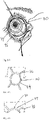
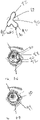
- Figs. 1-1 and 1-2 show an anatomical tissue structure of an eye 2 suitable for treatment with ocular inserts.
- the eye 2 includes a cornea 4, an iris 6, and a white-colored sclera 8.
- a substantially transparent conjunctival layer 10 covers the sclera 8.
- Posterior to the cornea 4 lies a crystalline lens 12.
- a retina 14 that responds to light is located in the posterior portion of the eye.
- a fovea 16 is a part of the retina that provides sharp focused vision.
- the cornea 4 and lens 12 refract light to form an image on the fovea 16 and retina 14.
- Fig. 1-2 shows the lacrimal system 18 which is responsible for producing and draining the tear fluid.
- the lacrimal system consists of two general areas: first, the lacrimal gland 20, which secretes the tears, and its excretory ducts 22, which transport the fluid to the surface of the eye and, second, the lacrimal canaliculi 24, the lacrimal sac 26, and the nasolacrimal duct 28, which bring the tear fluid is conveyed into the nose cavity.
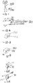
- Fig. 2-1 shows an exemplary embodiment of a therapeutic system 30.
- the therapeutic system 30 comprises an ocular insert 31, and may also include an insertion device, not being part of the invention, a configuration altering material that dissolves (or swells, weakens, tightens, or effects some other activation mechanism) to reconfigure the implant from an insertion configuration to a deployed configuration, or the like.
- activation of the insertion device may also reconfigure the insert from the insertion configuration to the deployed configuration, or may simply releasably hold the insert in a manner so as to assist insertion.
- the ocular insert may not undergo significant changes in shape or other properties before, during, or after deployment.
- the ocular insert is eventually positioned on a region outside an optical zone of an eye.
- the ocular insert comprises two structures: a first structure 32 and a second structure 34.
- Fig. 2-1 shows the exemplary therapeutic system 30 placed outside the optical zone of the eye.
- the first structure functions as a skeleton which largely holds the implant in place relative to the structures of the eye, thereby attaches the implant to the eye, and thus provides support for the cushioning structure relative to the anterior portion of the eye.
- This first or skeletal structure preferably maintains the attachment of the therapeutic system to the anterior portion of the eye for at least thirty days. Should it become medically desirable or should a patient so desire, the therapeutic system may be removed sooner than the thirty days; however, from a physical standpoint, it is capable of maintaining the ocular insert of the anterior surface of the eye for at least thirty days.
- the first structure may continue to help maintain the overall implant in the eye for sixty days or more, for ninety days or more, or even for 180 days or more, ideally with safe and effective delivery of therapeutic agents continuing throughout such implant periods.
- Alternative treatment devices and methods may benefit from shorter implant periods, optionally for periods of one or more days, at least a plurality of days, a week or more, two weeks or more, or the like.
- the first structure may determine the overall shape of the ocular insert.
- the first structure comprises a shape-memory material, a thin metal wire, a hard plastic such as nylon, PMMA, polycarbonate, polyethylene terepthalate, and/or another polymer, polypropylene or other synthetic suture material capable of providing the structural support to maintain the therapeutic system attached to the eye.
- the first structure may also comprise a coated plastic or metal such that the coating contains the therapeutic medication or provides easier attachment of the second, cushioning element to the skeletal member.
- the first structure may have a surface treatment such as plasma etching or the like to enable the second structure to be suitably attached to the skeletal member.
- Fig. 2-1 shows a basic embodiment of the first structure.
- the first structure 32 is annular or ring-shaped and, has a diameter of at least 8mm, and is sized to fit outside the optical zone of the cornea so as not to interfere with patient vision.
- the annulus of first structure 32 will preferably comprise a complete ring or torroid, but may have some gap along its circumference. The arc angle of the annulus in such embodiments will be over 180°.
- Figs. 2-2 and 2-3 show a top view and cross-sectional view of the therapeutic system shown in Fig. 2-1 .
- the therapeutic system shown in Figs. 2-1 to 2-3 can be sized much larger so that the edges of the structure will lie within the cul-de-sac of the eye.
- the therapeutic system In the case where the therapeutic system is intended to be located within the cul-de-sac of the eye, the therapeutic system will desirably be produced in at least two sizes to accommodate varying sizes of eyes (e.g. pediatric versus adult, and optionally different adult eye sizes).
- Alternative shapes of the first structure may include those of the inserts shown and described in U.S. Patent No. 3,995,635 .
- Fig. 2-4 shows an embodiment 36 of the therapeutic system 30 where the ring comprises two radially outwardly and/or anteriorly extending protrusions or bumps 42 on opposed portions of its surface.
- the lids "trap" the two bumps between the lids and push the ocular implant (which otherwise can freely glide on the surface of the eye) back into its therapeutically effective position outside the optical zone of the cornea.
- Fig. 2-5 shows an alternative embodiment 40 of the ring-shaped therapeutic device system 30.
- a crescent or banana-shaped reservoir 42 is attached to the inferior portion of the ocular insert.
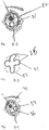
- Figs. 3-1 to 3-3 show another embodiment 44 of the therapeutic system 30 again including a ring-shaped structure with a diameter of at least 8 mm, sized to fit outside the optical zone of the cornea, and also having two or more haptics 46, each radiating from the ring-shaped structure across to the cul-de-sac of the eye, thus providing an additional support point for the therapeutic system.
- Fig. 3-1 shows the ring-shaped therapeutic system with haptics placed on the anterior structure of the eye.
- Figs. 3-2 and 3-3 show a top- and a cross-sectional view, respectively, of ocular insert 44.
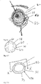
- Figs. 4-1 to 4-2 show an alternate embodiment 48 of the therapeutic system 30 in which two or more concentric ring-shaped structures 52 are held together by four or more haptics 50.
- the inner ring-shaped structure has a diameter of at least 8 mm and is sized to fit outside the optical zone of the cornea.
- the next (and subsequent) outer ring-shaped structures have progressively larger diameters, the outermost ring-shaped structure optionally having a diameter of at least 12 mm and being sized to fit on the sclera, fornix or cul-de-sac of the eye.
- Fig. 4-1 shows the embodiment 48 of the therapeutic system placed on the eye.
- Fig. 4-2 shows the embodiment 48 of the therapeutic system before insertion on the eye.
- the embodiment 48 has the advantage of providing a larger surface area for drug delivery, due to the presence of the two or more rings and four or more haptics. Additional insert shapes having enhanced surface areas may be seen in U.S. Patent No. 4,540,417 .
- Fig. 4-3 shows a related embodiment 49 that employs an eccentric design such that the one or more ring portions or arc segments 54 are present in the inferior area of the ring to target delivery to the area of the eye where tears may more readily pool, as in the cul-de-sac.
- This eccentric design may also stabilize the device in a more fixed position and be less likely to rotate out of position or move into the optical zone of the eye.
- targeting delivery to the cul-de-sac may enable more effective delivery of some medications to the nasolacrimal system in addition to the ocular surface, such as in the case of nasal allergy medications.
- the first structure typically remains of a constant size and shape, e.g. a ring-shape, or a ring with haptics that anchor/attach to the sclera, fornix or cul-de-sac of the eye.
- the first structure can expand or change shape so as to enhance its attachment to the anterior structure of the eye.
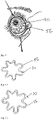
- Figs. 5-1 through 5-3 show a serpentine embodiment 56 of therapeutic system 30 which shows an expandable ocular insert.
- Fig. 5-1 shows the embodiment 56 inserted on the surface of the eye;
- Fig. 5-2 shows the embodiment 56 before insertion, and
- Fig. 5-3 shows the embodiment in its expanded state.
- a variety of alternative serpentine configurations may be developed or modified so as to take advantage of the cushioning and/or configuration-changing techniques described herein, including those of U.S. Patent No. 4,540,417 , the disclosure of which is incorporated herein by reference.
- the skeletal member can be shaped to conform to the radius of curvature of the eye.
- the first structure can stretch through a spring action mechanism.
- materials that may stretch through spring action include platinum alloys, titanium alloys, all stainless steel alloys & tempers, various clad metals and insulated wires.
- the first structure may comprise a shape- memory material, such as nitinol, which will allow it to change to a desired shape using thermal, magnetic or electromagnetic activation, from a martensitic to an austenitic state.
- shape memory materials include shape memory polyurethanes, crosslinked trans-polyoctylene rubber, polynorbornene polymers, nitinol, polyethylene, PMMA, polyurethane, cross-linked polyethylene, cross-linked polyisoprene, polycycloocetene, polycaprolactone, copolymers of (oligo)caprolactone, PLLA, PL/DLA copolymers, PLLA PGA copolymers, and other shape memory materials well-known to those of ordinary skill in the art.
- Figs. 7-1 shows a close-up of an exemplary ocular insert 31 of the therapeutic device system 30 in which the second structure 34 is disposed throughout the circumferential length of the first structure 32.
- the second structure 34 provides cushioning to facilitate extended implantation or wearing of the device, optionally inhibiting irritation to the eye sufficiently to encourage a patient to wear the therapeutic system for at least thirty days.
- the cushioning effect may be achieved at least in part by the material used in the second structure, as well as by the shape of the surfaces and/or edges of the second structure.
- the second structure may comprise a coating.
- the material of the second structure is soft, biocompatible, and non-irritant.
- examples of such material comprise polymers such as hydrogel or silicone.
- Fig. 7-2 shows a cross-section of a therapeutic device system comprising a second structure 34 with a tapered outer and/or inner edge 64.
- Fig. 7-3 shows a cross-section of a therapeutic device system comprising a second structure 34 with a beveled edge 66.
- Fig. 7-4 shows a cross-section of a therapeutic device system comprising a second structure 34 with a rounded edge 68.
- Fig. 8-1 shows a therapeutic device system 30 with a second structure 34 that may have an anterior and/or posterior surface 70 that can be shaped as well to the radius of curvature of the eye 70.
- the second, cushioning structure 74 is disposed only over certain discrete portions along the length of the first structure, desirably at locations where sharper edges or bends may provoke irritation to the eye.
- Fig. 9-1 shows the second, cushioning structure 74 disposed over discrete portions of the length of the first supporting structure 32.
- the second structure may also comprise a coating, partially disposed on the second structure, which prevents the expansion of the otherwise expandable, desirably hydratable, second structure.
- Figs. 10-1 and 10-2 show an embodiment 76 where the coating 78 is partially dispersed around the second structure to allow for preferential expansion of the second structure in certain areas.
- Fig. 10-1 shows an embodiment where the coating is partially dispersed around the second structure 80, with the first structure 32 in an unhydrated state.
- Fig. 10-2 shows the embodiment of the second structure 80 of Fig. 10-1 in a hydrated, thus expanded, state 76'.
- the first and second structure may comprise similar compositions or materials having differing durometers and/or other characteristics, particularly where the material can be processed so as to exhibit the desired properties for both the first and second structures.
- the drug used in the therapeutic system will often be placed on, embedded, encapsulated or otherwise incorporated into a delivery matrix.
- the delivery matrix may be included in or on either the first skeletal structure or the second cushioning structure, or both.
- the delivery matrix in turn, comprises either a biodegradable or a non-biodegradable material.
- the delivery matrix may include, although it is not limited to, a polymer.
- biodegradable polymers include protein, hydrogel, polyglycolic acid (PGA), polylactic acid (PLA), poly(L-lactic acid) (PLLA), poly(L-glycolic acid) (PLGA), polyglycolide, poly-L-lactide, poly-D-lactide, poly(amino acids), polydioxanone, polycaprolactone, polygluconate, polylactic acid- polyethylene oxide copolymers, modified cellulose, collagen, polyorthoesters, polyhydroxybutyrate, polyanhydride, polyphosphoester, poly(alpha-hydroxy acid), and combinations thereof.
- Non-biodegradable polymers may comprise silicone, acrylates, polyethylenes, polyurethane, polyurethane, hydrogel, polyester (e.g., DACRON ® from E. I. Du Pont de Nemours and Company, Wilmington, Del.), polypropylene, polytetrafluoroethylene (PTFE), expanded PTFE (ePTFE), polyether ether ketone (PEEK), nylon, extruded collagen, polymer foam, silicone rubber, polyethylene terephthalate, ultra high molecular weight polyethylene, polycarbonate urethane, polyurethane, polyimides, stainless steel, nickel-titanium alloy (e.g., Nitinol), titanium, stainless steel, cobalt-chrome alloy (e.g., ELGILOY ® from Elgin Specialty Metals, Elgin, 111.; CONICHROME ® from Carpenter Metals Corp., Wyomissing, Pa.).
- polyester e.g., DACRON ® from E
- the ocular insert desirably will comprise a hypoaller genie, material.
- the first and /or second structure may comprise materials such as hydrogels, polyethylene glycol (PEG), or polyethylene oxide (PEO) that prevent adhesion of proteins and thus minimize the chance of developing an allergic reaction.
- the drug delivery matrix of the ocular insert may comprise an anti-allergenic and/or antihistaminic compound to prevent an allergic reaction to the ocular insert.
- the delivery matrix may also include other materials known in the art.
- drugs may be delivered to the eye using the therapeutic system. Desirably these drugs will include drugs needed for long-term treatment to the eye. Examples of conditions that require long-term treatment include: dry eye, glaucoma, allergies, infections, bacterial, viral and other infections, chronic inflammatory conditions such as acne rosacea keratitis, cyclitis, and blepharitis, selected retinal conditions such as diabetic retinopathy, age related macular degeneration and other retinal conditions, post-surgery, amblyopia, etc.
- Some drug families used in the treatment of the above-mentioned conditions comprise: steroids, antiinflammatories, antibiotics, glaucoma treatment compounds, antihistamines, dry eye medication, neuroprotectives, retinoids, antineovasculars, antioxidants, and biologies.
- steroids examples include glucocorticoids, aprogestins, amineralocorticoids, or corticosteroids.
- exemplary corticosteroids include cortisone, hydrocortisone, prednisone, prednisolone, methylprednisone, triamcinolone, fluoromethalone, dexamethasone, medrysone, betamethasone, loteprednol, fluocinolone, flumethasone, rimexolone or mometasone.
- steroids include androgens, such as testosterone, methyltestosterone, or danazol.
- anti-inflamatories examples include NSAIDs such as piroxicam, aspirin, salsalate (Amigesic), diflunisal (Dolobid), ibuprofen (Motrin), ketoprofen (Orudis), nabumetone (Relafen), piroxicam (Feldene), naproxen (Aleve, Naprosyn), diclofenac (Voltaren), indomethacin (Indocin), sulindac (Clinoril), tolmetin (Tolectin), etodolac (Lodine), ketorolac (Toradol), oxaprozin (Daypro), and celecoxib (Celebrex).
- NSAIDs such aspiroxicam, aspirin, salsalate (Amigesic), diflunisal (Dolobid), ibuprofen (Motrin), ketoprofen (Orudis), nabumetone (Relafen), piroxicam (
- antibiotics examples include amoxicillin, penicillin, sulfa drugs, erythromycin, streptomycin, tetracycline, clarithromycin, terconazole, azithromycin, bacitracin, ciprofloxacin, evofloxacin, ofloxacin, levofloxacin, moxifloxacin, gatifloxacin, aminoglycosides, tobramycin, gentamicin, as well as polymyxin B combinations including polymyxin B/trimethoprim, polymyxin B/bacitracin, polymyxin B/neomycin/ gramicidin.
- Glaucoma treatment medications include beta-blockers, such as timolol, betaxolol, levobetaxolol, and carteolol; miotics, such as pilocarpine; carbonic anhydrase inhibitors, such as brinzolamide and dorzolamide; prostaglandins, such as travoprost, bimatoprost, and latanoprost; seretonergics; muscarinics; dopaminergic agonists; and adrenergic agonists, such as apraclonidine and brimonidine, and prostaglandins or prostaglandin analogs such as latanoprost, bimatoprost, or travoprost.
- beta-blockers such as timolol, betaxolol, levobetaxolol, and carteolol
- miotics such as pilocarpine
- carbonic anhydrase inhibitors such as
- Antihistamines and mast cell stabilizers include Olopatadine and epinastine, the acute care anti-allergenic products ketorolac tromethamine, ketotifen fumarate, loteprednol, epinastine HCl, emedastine difumarate, azelastine hydrochloride, Olopatadine hydrochloride, ketotifen fumarate; while the chronic care anti-allergenic products include pemirolast potassium, nedocromil sodium, lodoxamide tromethamine, cromolyn sodium.
- Antineovasculars include biologies, Ranibizumab (Lucentis) and Bevacizumab (Avastin).
- Amblyopia medicine includes anesthetics and cycloplegics such as atropine.
- Dry eye medication includes cyclosporine.
- Drug elution can be controlled either through concentration of the drug present, or by embedding into or combining the drug with various other compounds.
- the drug's particular solubility characteristic, whether hydrophobic or hydrophilic, will determine the means of controlling the rate of elution for that particular drug.
- the drug may be finely ground up and dispersed into the second cushioning structure comprising silicone or a polymer such as hydrogel that is highly hydrophilic.
- Hydrophilic drugs can either be immobilized in a first structure, e.g. a plastic, or a second structure such as a hydrogel.
- the specific choice of polymers used for immobilizing depends on the drug and its characteristics, the rate of elution desired, and the wall thickness of the coating that contains the drag which may also alter the rate of elution. For instance, if the drug is embedded in a first polymer, then the wall thickness of the second polymer may at least in part control the rate at which the drug passes through. Conversely, the wall thickness of the coating may be used to control drug release if the drug is embedded into the skeletal element.
- surfactants comprising bile salts (e.g. deoxycholate, taurodeoxycholate, and glycocholate) or calcium chelators, such as ethylenediaminetetraacetic acid (EDTA), may be added to increase their solubility.
- bile salts e.g. deoxycholate, taurodeoxycholate, and glycocholate
- calcium chelators such as ethylenediaminetetraacetic acid (EDTA)
- EDTA ethylenediaminetetraacetic acid
- the drug particles may be coated, a less soluble salt form of the drug may be produced, or a rate-limiting coating, polymer, or other material may be incorporated in and/or on the delivery matrix such that the distance the drug travels to exit the device or the resistance of the material to passage of the drug restricts flow of the drug from the device.
- aqueous/tear fluid such as a sponge-like or naturally porous material or a material with artificially created pores or other materials that saturate such that an osmotic pumping effect occurs.
- the surface area and geometric configuration of the therapeutic system can also be used to control the elution rate of the drug.
- the geometric configuration of the therapeutic device can be thus designed to maximize, or minimize, the flow of tear fluid over the therapeutic system, according to the specific need. For example, an increased surface area will increase the contact area between the drug and the tear fluid.
- the device could also be constructed such that it has more delivery area/surface area in the lower or upper fornix depending on if a targeted delivery is desired. Conversely, to decrease the elution rate of the drug, the contact area between the eye and the drug particles should be decreased.
- the second structure also comprises a coating to further soothe the patient's eye.
- the coating may comprise a lubricious material, e.g., Hydak ® hyaluronan-based coating from Biocoat.
- Hydak a lubricious material
- the advantages of Hydak include that it is lubricious when wet, biocompatible, highly hydrophilic, may be applied using thin, flexible coatings, and it is a carrier for bioactive substances.
- Other coatings could also include either hydrophilic or drug delivery coatings from SurModics (hydrophilic or drug delivery) and Hydromer.
- some embodiments will be coated so that the ocular inserts will have a firm texture during insertion. Once such ocular inserts are in place, the coating will dissolve to allow the ocular insert to become more comfortable for every-day use.
- the therapeutic system may first be placed onto the eye by a physician and then, once a desired drug-delivery time period is complete, subsequently be removed from the anterior surface of the eye by the same or a different physician.
- the physician may optionally then teach the patient how to insert and then take out the ocular implant by him- or herself.
- a challenge to insertion and removal of the device comes from maintaining the fine balance between rigidity and flexibility.
- a device that is too flexible will be very difficult to insert; while a device that is too rigid will be uncomfortable to wear for extended periods of time.
- One way of maintaining the fine balance between rigidity and flexibility is by folding or pinching the device into various shapes.
- the folds in the device create a structure that maintains its shape more effectively during insertion and thus is more "pushable" than a ring structure that deforms easier.
- One alternative for maintaining the folds in the device for purposes of insertion is to tether the folds with a dissolvable material until the device is placed in the eye.
- the dissolvable material allows for a slow release of the shape back into a ring.
- Fig. 11-1 shows a ring-shaped ocular insert 31 in which separated and/or opposed portions are approximate or pinched in toward the center to form a non-planar taco shape 82.
- the pinched ring-shaped device can be positioned such that one end can be slipped under the lower or upper lid, then the other end can be positioned under the other lid.
- Fig. 11-2 shows said ocular insert 31 upon insertion on the surface of the eye, before a dissolvable material has allowed for a slow release back into a ring-shape;
- Fig. 11-7 shows said device on the surface of the eye, in its fully expanded state 94.
- Fig. 11-3 shows one embodiment 84 where the annular shape includes a serpenting shape or series of bends such that radially outer portions or protrusions are interspersed with radially inner portions.
- This embodiment has four protrusions and a clover leaf shape is produced such that each of the four protrusions 86 would facilitate placement into the upper and lower lids as well as the nasal and temporal aspects of the eye.
- the inner portions of this shape could also be held together (or near each other) with a dissolvable material, but it may not be necessary since a good initial position could be achieved.
- Fig. 11-4 shows said embodiment 84 upon insertion on the surface of the eye;
- Fig. 11-7 shows said device on the surface of the eye, in its fully expanded state 94.
- Fig. 11-5 shows an alternative embodiment 88 where a three-leaf clover shape 90 is produced.
- the top protrusion 92 would first be inserted behind the top eyelid and then the bottom two protrusions would be inserted behind the bottom eyelid.
- this shape could also be held in place with a dissolvable material, but it may not be necessary since a good initial position could be achieved.
- Fig. 11-6 shows said device upon insertion on the surface of the eye;
- Fig. 11-7 shows said device on the surface of the eye, in its fully expanded state 94.
- Insertion of the device can also be facilitated by use of delivery instruments, the delivery instruments not being part of the invention.
- Figs. 12-1 and 12-2 show two alternatives of a modified grasping tool 150 and 160, the grasping tool also not being part of the invention, with a notch or groove 170 on the end to facilitate grasping the device.
- the jaws have an antramatic distal surface with, for example, a top layer comprising silicone or Teflon so that if they come in contact with the surface of the eye, they will not scratch it.
- a top layer comprising silicone or Teflon
- FIG. 12-2 an example of a delivery instrument that can accommodate two or more jaws is shown, there are preferably three or four jaws 162 (the drawing shows two jaws for simplicity) that create the three or four leaf clover shape when the device 160 is clamped onto the ring.
- the three or four jaws 162 come together simultaneously via an outer tube that forces the tines of the jaws to compress from their more relaxed outward position.
- Figs 12-3 to 12-5 show different examples for jaws 162 of the grasping tool (not being part of the invention) modified to have a specific shape on the notch to help fold the device into a shape that matches that of the eye.
- Fig. 12-3 shows jaws with a groove 172 that runs horizontally across the jaws.
- Fig. 12-4 shows jaws with a groove 175 that is curved so as to bend the ring into a shape that conforms more easily to the shape of the eye.
- Fig. 12-5 shows an alternative antramatic example of a jaw with a curved groove 176.
- the jaw consists of three adjacent slabs with a horizontal groove.
- the middle slab 178 is raised slightly, to allow the formation of a curved groove for the ring material.
- the middle slab is pushed down so that all the slabs are flush with each other and the groove running on the side of the three slabs is now horizontal; the ring will now be easily released from the grasping tool.
- Figs. 13-1 to 13-4 show an alternative manner of releasing the ring from the grasping tool.
- Fig. 13-1 shows a modified grasping tool 160 (not part of the invention) comprising four jaws 162, each jaw comprising a groove 170 positioned at the anterior end of each jaw and facing the center of the grasping tool.
- Fig. 13-2 shows a modified grasping tool 160 that has grasped an ocular insert 31 in the grooves 170, tightened the jaws 162 on the insert, creating four protrusions to ease insertion of the ocular insert behind a patient's eye lids.
- Fig. 13-3 shows the modified grasping tool 160 where each jaw has turned approximately 180° so that each groove 170 now faces away from the center of the grasping tool. The 180° turn of the grooves has resulted in a release of the ocular insert 31.
- Fig. 13-4 shows the ocular insert 131 released from the modified grasping tool 160.
- Figs. 14-1 to 14-5 show an alternative device (not part of the invention), along with a method (not part of the invention) for, inserting the ring-shaped device.
- Fig. 14-1 shows a syringe-shaped device 180 comprising a barrel 182 with a flat tip 184 through which ocular insert 31 can be pushed, and a plunger 186.
- the plunger of the syringe is taken out, the ring-shaped device is folded flat and inserted into the body of the syringe and the plunger is placed back into the syringe.
- FIG. 14-2 shows a syringe-shaped device 180 where and ocular insert 31 has been folded flat and inserted into the barrel 182.
- the plunger 186 has been inserted into the barrel, with the barrel comprising a lumen and the open tip comprising a port through which insert 31 can be pushed.
- the plunger pushed into the body of the syringe so as to allow the ring-shaped device to slowly squeeze through the flat tip of the syringe.
- Fig. 14-3 shows the loop of the ocular insert 31 substantially extruded from the tip 184 of the syringe-shaped device 180 and ready for placement on the sclera of the eye.
- FIG. 14-4 shows the loop of the ocular insert 31 partially placed on the sclera of the eye; the rest of the ocular insert will be pushed through the tip 184 of the syringe-shaped device 180 and released onto the eye.
- Fig. 14-5 shows the ocular insert 31 placed on the sclera of the eye.
- Figs. 15-1 to 15-3 show another alternative ocular insert insertion device (not part of the invention) that resembles a classical bike horn.
- Fig. 15-1 shows a bike horn-shaped insertion device 180 comprising two parts: a trumpet 182 and a squeeze bulb 184.
- Fig. 15-2 shows a close-up of the trumpet 182.
- the trumpet comprises a soft material 182 with channels 188 that connect the squeeze bulb to the outer rim of the insertion device.
- the outer rim of the trumpet comprises a groove 186 which is sized to fit a ring-shaped device.
- Fig. 15-3 shows a cross-section of the bike-horn shaped insertion device comprising a trumpet and a squeeze bulb.
- the squeeze bulb comprises a vacuum source and a reservoir for liquid 190, desirably saline.
- the squeeze bulb is attached to the trumpet.
- the squeeze bulb is squeezed, thereby creating a vacuum seal which picks up and holds the ring-shaped device in the groove of the outer rim of the trumpet.
- the trumpet is gently inserted under both eyelids of an eye and the squeeze bulb is gently squeezed, causing the liquid from the squeezed bulb's reservoir to flow through the channels of the trumpet, breaking the vacuum seal between the ring-shaped device and the outer rim of the trumpet. The trumpet is then gently pulled out from underneath the eyelids.
- Figs. 16-1 to 16-6 show an alternative insertion device 192 (not part of the invention).
- Fig. 16-1 shows an alternative insertion device 192 comprising a soft flexible cone 194 that supports the ring-shaped device along its outer rim 196 and a method of using it to place an ocular insert on the sclera 8 of the eye 2.
- Fig. 16-2 shows a close-up view of the outer rim of the delivery device 196.
- the outer rim comprises a groove 198 designed to fit the ring-shaped ocular insert 31.
- Fig. 16-3 shows a front view of the insertion device 192 comprising a soft flexible cone.
- the soft flexible cone comprises two slits 198, to allow the diameter of the cone to be modifiable.
- FIG. 16-4 to 16-6 show a method for using the insertion device 192 to insert the ocular insert 31.
- Fig. 16-4 shows the insertion device 192 loaded with the ring-shaped ocular insert 31 being gently tucked under the top 202 and bottom eyelid 204.
- Fig. 16-5 shows the soft flexible cone 194 being pinched or pulled, so as to lower the circumference of the cone and release the ring-shaped ocular insert 31 from the outer rim 196 of the cone.
- Fig. 16-6 shows the ring-shaped ocular insert 31 left in the eye, while the cone 194 is pulled away from under the eyelids.
- Figs. 17-1 to 17-2 show an alternative insertion device (not part of the invention).
- Fig. 17-1 shows the insertion device 206 comprising a flexible curved band 208.
- the curved band comprises a curved groove that can support ocular insert 31 , while the ocular insert is gently slid behind at least one of the eyelids.
- Example 1 Calculation of a Drug's Therapeutically Effective Dosage for an Ocular Insert - - Olopatadine
- the drug Olopatadine for treatment of allergic conditions, will demonstrate a method that can be used in calculating a drug's therapeutically effective plant-delivered dosage based on a drop-administered treatment regimen for that drug.
- the calculation method involves the following steps: 1.) determining the number of drops desired per application; 2.) multiplying the number of drops by 30 uL (the volume of one drop); 3.) determining the amount of solid drug per uL; 4.) multiplying the results from step 2 by the result from step 3, to find out the amount of solid drug to be applied to the eye on a daily basis; 5.) multiplying the result in step 4 by the number of days of therapy desired for the particular drug; and 6) multiplying by the efficiency of drug delivery. This final amount will preferably be dispersed from an ocular insert.
- Olopatadine is an ophthalmic antihistamine (H 1 -receptor) and mast cell stabilizer.
- the usual adult dosage for Olopatadine may be, for example, one drop in each affected eye twice a day, when using the 0.1 % solution and one drop per day in each affected eye, when using the 0.2% solution.
- Olopatadine HCl (OH) for ophthalmic applications can be formulated as a 0.2% (2 mg/mL) solution.
- a single 50 uL drop containing 100 ug of OH may be instilled in the eye once a day for 2 weeks.
- At least 70 ug in dry form could be loaded into the implant and released into the tear film by partitioning the drug reservoir with a membrane (e.g. HEMA, PVA, PVP, GMA, dialysis tubing of cellulose, etc) or embedding the drug within the implant.
- a membrane e.g. HEMA, PVA, PVP, GMA, dialysis tubing of cellulose, etc
- the release rate could be controlled by altering the surface area exposed to the tear film to tailor the desired 5 ug/day (0.21ug/hour), by altering a drug release controlling membrane, or the like. It is again assumed in this calculation that 100% of the targeted dose gets to its target location without being washed out with tear film, and more accurate calculations can be performed using wash-out data.
- the outside of the implant could be coated for either example with a bolus of the drug for immediate dosing while the hydration process, and thus flux of drug across the membrane or through the reservoir can take place.
- These coatings could be in solid drug form with a readily dissolvable layer (e.g. starch, sugar) to maintain placement of the solid drug upon the exterior of the implant.
- Example 3 Drug Delivery Procedure & Elution Rate Control for a Hydrophobic Drug - Prednisolone acetate
- a 1% Prednisolone acetate suspension (10 mg/mL) is given 2 drops (total of approximately 100 uL volume) 4 times daily for a week. Working with the estimate that 5% of a dose is actually available for absorption into the cornea, this amounts to 20 ug/day of Prednisolone acetate. A week's available dose is then 140 ug.
- the solubility of Prednisolone acetate in aqueous solutions is approximately 240 ug/mL.
- At least 140 ug of solid Prednisolone acetate could be loaded into the implant, allowing the Prednisolone acetate to dissolve into the tear layer at a rate of about 0.83 ug/hour. The rate could be controlled by the porosity of the implant as well as the surface area exposed to the tear film.
Description
- 1.Field of the Invention. The invention relates to ocular inserts adapted to be worn along the front surface of the eye outside the optical zone, and can deliver one or more drugs at a safe, therapeutically-effective level for at least thirty days.
- 2. Background of the Invention. A variety of ophthalmic and non-ophthalmic conditions necessitate administration of various drugs to the eye. Eye drops and gels can be effective drug delivery vehicles, but can also have significant disadvantages. Specifically, eye drops mix with fluid in the tear film, but may have a residence time of only 2-5 minutes in the tear film. As little as 5% of the drug may be absorbed locally; some or all of the rest being carried from the lacrimal sac into the lacrimal duct and eventually absorbed into the bloodstream. The absorption into the bloodstream can have at least two adverse effects: first, most of the drug is wasted and, second, the presence of the drug in the bloodstream may have harmful side effects on the rest of the body. Gels may adhere more effectively to the eye, but can also blur the patient's vision for prolonged time periods. Both eye drops and gels need to be reapplied frequently for some therapies. Thus, a need remains for an improved drug delivery method to the eye that is neither cleared out of its targeted location, nor needs frequent reapplication.
- In light of the disadvantages of eye drops, it is understandable that a variety of alternatives have been proposed. Among the known alternatives to drops include treatments in which structures containing or impregnated with drugs have been placed under the eyelid.
-
US 4,540,417 discloses an eye-medicating haptic construction which is initially flat but axially flexible as to be self-adapting to the surface of the cornea and to be self-retaining of its position, via moisture at the surface of the cornea.US 6,001,386 discloses an implantable sustained release drug delivery device with an inner core containing an effective amount of a low solubility agent covered by a non-bioerodible polymer coating layer that is permeable to the low solubility agent. - Such solid ocular dosage forms appear to present significant potential advantages over drop-administered drug treatments of the eyes. In particular, eye drug delivery implants might help overcome low patient compliance, the difficult application and frequent misapplication of traditional eye drops and other dosage forms, and limited effective drug absorption presented by eye drops, while potentially facilitating the advantageous application of advances in polymer chemistry and introduction of the concepts of sustained/controlled drug release from other known drug-delivery systems.
- Despite the tremendous potential advantages of drug delivery implants, drug application to the front of the eyes remains dominated by eye drops. Factors that may have contributed to the limited acceptance of prior ocular inserts include their lack of comfort, their propensity for displacement or movement around the eye, their excessive incidents of inadvertent expulsion during sleep or rubbing of eyes, their interference with vision, and/or the difficulty in placing and removing the known drug delivery implants.
- In light of the above, new drug delivery devices, systems, and methods would be beneficial, particularly for delivering therapeutic compounds to the anterior segment of the eye. It would be particularly advantageous to provide improved ocular inserts so as to gain both physician and user acceptance, with such inserts ideally being easy to insert and remove, providing patient comfort, being non-toxic and not interfering with vision or oxygen penetration, allowing for reproducible release kinetics, and/or being easy to manufacture at a reasonable price.
- The present invention provides an ocular insert as defined in claim 1. Exemplary embodiments are defined in the dependent claims.
- In a first aspect, the invention provides an ocular insert for use in an eye, the eye having upper and lower lids extendable along an anterior eye surface with an optical zone therebetween, the ocular insert comprising:
- a first structure comprising an annulus having a circumferential length and disposable along the anterior surface of the eye of a patient, wherein the annulus has a diameter of at least 8mm and is sized to fit around and remain outside the optical zone, the first structure being a skeletal structure configured to help maintain the ocular insert in contact with the eye for a plurality of days;
- a second structure formed of a material, wherein the second structure is supported by the first structure relative to the anterior portion of the eye and disposed along at least part of the circumferential length of the first structure, wherein the second structure is configured to cushion engagement between the first structure and the eye so as to inhibit irritation of the eye;
- at least one drug dispersed in or on the second structure so as release a safe and therapeutically effective quantity of the drug to the eye for each of the plurality of days; and
- wherein the first structure of ocular insert comprises a shape-memory material or a thin metal wire or a hard plastic and/or a polymer, polypropylene or synthetic suture material that is capable of providing the structural support to maintain attachment of the ocular insert to the anterior surface of the eye.
- The first structure functions as a skeletal frame which maintains positioning of the implant along the anterior portion of the eye and provides support to the second, cushioning structure. This first structure maintains the attachment of the therapeutic system to the anterior portion of the eye for at least thirty days. In some embodiments the first structure remains a constant size and shape, e.g. a ring shape, a ring with haptics, or a curvilinear ring that is confined to and restrainingly engages the inferior and superior conjunctival fornices so as to retain the implant within the tear fluid and/or against the tissues of the eye.
- In many embodiments, the first structure stretches or changes shape so as to maximize its attachment to the anterior structure of the eye. The drug may be dispersed on or in the second structure, or, also, in or on the first structure.
- In exemplary embodiments of the invention, the therapeutic system is designed for easy insertion and removal by the patient.
-
-
Figs. 1-1 and 1-2 show an anatomical tissue structure of aneye 2 suitable for treatment with ocular inserts; -
Fig. 2-1 shows a therapeutic system comprising an ocular insert, that may also include an insertion device, a configuration altering material that dissolves (or swells, weakens, tightens, or effects some other activation mechanism) to reconfigure the implant from an insertion configuration to a deployed configuration, or the like; -
Figs. 2-2 and 2-3 show a top view and cross-sectional view of the therapeutic system shown inFig. 2-1 ; -
Fig. 2-4 shows a therapeutic system where the ring comprises two radially outwardly and/or anteriorly extending protrusions or bumps on opposed portions of its surface; -
Fig. 2-5 shows a the ring-shaped therapeutic device system, in which a crescent or banana-shaped reservoir is attached to the inferior portion of the ocular insert; -
Figs. 3-1 to 3-3 show a therapeutic system including a ring- shaped structure with a diameter of at least 8 mm, sized to fit outside the optical zone of the cornea, and also having two or more haptics; -
Figs. 4-1 to 4-2 show a therapeutic system in which two or more concentric ring-shaped structures are held together by four or more haptics; -
Fig. 4-3 shows an eccentric design such that the one or more ring portions or arc segments are present in the inferior area of the ring to target delivery to the area of the eye where tears may more readily pool, as in the cul-de-sac; -
Figs. 5-1 through5-3 show a serpentine therapeutic system which shows an expandable ocular insert; -
Figs. 6-1 and 6-2 show an example, not part of the invention, where the second cushioning structure comprises two hydrogel scleral contact lenses attached to each other, so as to sandwich the first rigid structure between them; -
Figs. 7-1 shows a close-up of an exemplary ocular insert of the therapeutic device system in which the second structure is disposed throughout the circumferential length of the first structure; -
Fig. 7-2 shows a cross-section of a therapeutic device system comprising a second structure with a tapered outer and/or inner edge; -
Fig. 7-3 shows a cross-section of a therapeutic device system comprising a second structure with a beveled edge; -
Fig. 7-4 shows a cross-section of a therapeutic device system comprising a second structure with a rounded edge; -
Fig. 8-1 shows a therapeutic device system with a second structure that may have an anterior and/or posterior surface that can be shaped as well to the radius of curvature of the eye; -
Fig. 9-1 shows the second, cushioning structure disposed over discrete portions of the length of the first supporting structure; -
Figs. 10-1 and 10-2 show an example where the coating is partially dispersed around the second structure to allow for preferential expansion of the second structure in certain areas; -
Fig. 11-1 shows a ring-shaped ocular insert in which separated and/or opposed portions are approximate or pinched in toward the center to form a non-planar taco shape; -
Fig. 11-2 shows the ring-shaped ocular insert upon insertion on the surface of the eye, before a dissolvable material has allowed for a slow release back into a ring-shape; -
Fig. 11-3 and 11-4 show an example where the annular shape includes a serpenting shape or series of bends such that radially outer portions or protrusions are interspersed with radially inner portions; -
Fig. 11-5 and 11-6 show an example 88 where a three-leaf clover shape is produced; -
Fig. 11-7 shows a fully expanded ocular insert positioned on the surface of the eye; -
Figs. 12-1 and 12-2 show two alternatives of a modified grasping tool, with a notch or groove on the end to facilitate grasping the device, the grasping tool not being part of the invention; -
Figs 12-3 to 12-6 show different jaws of the grasping tool modified to have a specific shape on the notch to help fold the device into a shape that matches that of the eye and a method of using it, the grasping tool not being part of the invention; -
Figs. 13-1 to 13-4 show an alternative manner of releasing the ring from the grasping tool, the grasping tool not being part of the invention; -
Figs. 14-1 to 14-5 show an alternative syringe-shaped insertion device, along with a method for, inserting the ring-shaped ocular insert, the alternative syringe-shaped insertion device not being part of the invention; -
Figs. 15-1 to 15-3 show another alternative ocular insert insertion device that resembles a classical bike horn, the alternative ocular insertion device not being part of the invention; -
Figs. 16-1 to16-6 show an alternative insertion device comprising a soft flexible cone that supports the ring-shaped device along its outer rim and a method of using it to place an ocular insert on the sclera of the eye, the alternative insertion device not being part of the invention; and -
Figs. 17-1 to 17-2 show an alternative insertion device comprising a flexible curved band, the alternative insertion device not being part of the invention. -
Figs. 1-1 and 1-2 show an anatomical tissue structure of aneye 2 suitable for treatment with ocular inserts. Theeye 2 includes a cornea 4, aniris 6, and a white-colored sclera 8. A substantially transparentconjunctival layer 10 covers thesclera 8. Posterior to the cornea 4 lies a crystalline lens 12. Aretina 14 that responds to light is located in the posterior portion of the eye. Afovea 16 is a part of the retina that provides sharp focused vision. The cornea 4 and lens 12 refract light to form an image on thefovea 16 andretina 14. -
Fig. 1-2 shows thelacrimal system 18 which is responsible for producing and draining the tear fluid. The lacrimal system consists of two general areas: first, thelacrimal gland 20, which secretes the tears, and its excretory ducts 22, which transport the fluid to the surface of the eye and, second, thelacrimal canaliculi 24, thelacrimal sac 26, and thenasolacrimal duct 28, which bring the tear fluid is conveyed into the nose cavity. -
Fig. 2-1 shows an exemplary embodiment of atherapeutic system 30. Thetherapeutic system 30 comprises anocular insert 31, and may also include an insertion device, not being part of the invention, a configuration altering material that dissolves (or swells, weakens, tightens, or effects some other activation mechanism) to reconfigure the implant from an insertion configuration to a deployed configuration, or the like. In alternative examples, activation of the insertion device (or some other tool) may also reconfigure the insert from the insertion configuration to the deployed configuration, or may simply releasably hold the insert in a manner so as to assist insertion. In still further disclosed examples, the ocular insert may not undergo significant changes in shape or other properties before, during, or after deployment. Regardless, the ocular insert is eventually positioned on a region outside an optical zone of an eye. The ocular insert comprises two structures: afirst structure 32 and asecond structure 34.Fig. 2-1 shows the exemplarytherapeutic system 30 placed outside the optical zone of the eye. - The first structure functions as a skeleton which largely holds the implant in place relative to the structures of the eye, thereby attaches the implant to the eye, and thus provides support for the cushioning structure relative to the anterior portion of the eye. This first or skeletal structure preferably maintains the attachment of the therapeutic system to the anterior portion of the eye for at least thirty days. Should it become medically desirable or should a patient so desire, the therapeutic system may be removed sooner than the thirty days; however, from a physical standpoint, it is capable of maintaining the ocular insert of the anterior surface of the eye for at least thirty days. In some examples, the first structure may continue to help maintain the overall implant in the eye for sixty days or more, for ninety days or more, or even for 180 days or more, ideally with safe and effective delivery of therapeutic agents continuing throughout such implant periods. Alternative treatment devices and methods may benefit from shorter implant periods, optionally for periods of one or more days, at least a plurality of days, a week or more, two weeks or more, or the like.
- Due to its role as skeleton for the
insert 31 oftherapeutic system 30, the first structure may determine the overall shape of the ocular insert. The first structure comprises a shape-memory material, a thin metal wire, a hard plastic such as nylon, PMMA, polycarbonate, polyethylene terepthalate, and/or another polymer, polypropylene or other synthetic suture material capable of providing the structural support to maintain the therapeutic system attached to the eye. The first structure may also comprise a coated plastic or metal such that the coating contains the therapeutic medication or provides easier attachment of the second, cushioning element to the skeletal member. The first structure may have a surface treatment such as plasma etching or the like to enable the second structure to be suitably attached to the skeletal member. -
Fig. 2-1 shows a basic embodiment of the first structure. Here thefirst structure 32 is annular or ring-shaped and, has a diameter of at least 8mm, and is sized to fit outside the optical zone of the cornea so as not to interfere with patient vision. The annulus offirst structure 32 will preferably comprise a complete ring or torroid, but may have some gap along its circumference. The arc angle of the annulus in such embodiments will be over 180°.Figs. 2-2 and 2-3 show a top view and cross-sectional view of the therapeutic system shown inFig. 2-1 . The therapeutic system shown inFigs. 2-1 to 2-3 can be sized much larger so that the edges of the structure will lie within the cul-de-sac of the eye. In the case where the therapeutic system is intended to be located within the cul-de-sac of the eye, the therapeutic system will desirably be produced in at least two sizes to accommodate varying sizes of eyes (e.g. pediatric versus adult, and optionally different adult eye sizes). Alternative shapes of the first structure may include those of the inserts shown and described inU.S. Patent No. 3,995,635 . -
Fig. 2-4 shows anembodiment 36 of thetherapeutic system 30 where the ring comprises two radially outwardly and/or anteriorly extending protrusions or bumps 42 on opposed portions of its surface. When the eye blinks, the lids "trap" the two bumps between the lids and push the ocular implant (which otherwise can freely glide on the surface of the eye) back into its therapeutically effective position outside the optical zone of the cornea. -
Fig. 2-5 shows analternative embodiment 40 of the ring-shapedtherapeutic device system 30. In this embodiment, a crescent or banana-shapedreservoir 42 is attached to the inferior portion of the ocular insert. -
Figs. 3-1 to 3-3 show anotherembodiment 44 of thetherapeutic system 30 again including a ring-shaped structure with a diameter of at least 8 mm, sized to fit outside the optical zone of the cornea, and also having two ormore haptics 46, each radiating from the ring-shaped structure across to the cul-de-sac of the eye, thus providing an additional support point for the therapeutic system.Fig. 3-1 shows the ring-shaped therapeutic system with haptics placed on the anterior structure of the eye.Figs. 3-2 and 3-3 show a top- and a cross-sectional view, respectively, ofocular insert 44. -
Figs. 4-1 to 4-2 show analternate embodiment 48 of thetherapeutic system 30 in which two or more concentric ring-shapedstructures 52 are held together by four ormore haptics 50. The inner ring-shaped structure has a diameter of at least 8 mm and is sized to fit outside the optical zone of the cornea. The next (and subsequent) outer ring-shaped structures have progressively larger diameters, the outermost ring-shaped structure optionally having a diameter of at least 12 mm and being sized to fit on the sclera, fornix or cul-de-sac of the eye.Fig. 4-1 shows theembodiment 48 of the therapeutic system placed on the eye.Fig. 4-2 shows theembodiment 48 of the therapeutic system before insertion on the eye. Theembodiment 48 has the advantage of providing a larger surface area for drug delivery, due to the presence of the two or more rings and four or more haptics. Additional insert shapes having enhanced surface areas may be seen inU.S. Patent No. 4,540,417 .Fig. 4-3 shows arelated embodiment 49 that employs an eccentric design such that the one or more ring portions orarc segments 54 are present in the inferior area of the ring to target delivery to the area of the eye where tears may more readily pool, as in the cul-de-sac. This eccentric design may also stabilize the device in a more fixed position and be less likely to rotate out of position or move into the optical zone of the eye. In addition, targeting delivery to the cul-de-sac may enable more effective delivery of some medications to the nasolacrimal system in addition to the ocular surface, such as in the case of nasal allergy medications. - In the embodiments described above, the first structure typically remains of a constant size and shape, e.g. a ring-shape, or a ring with haptics that anchor/attach to the sclera, fornix or cul-de-sac of the eye.
- In other embodiments, the first structure can expand or change shape so as to enhance its attachment to the anterior structure of the eye.
Figs. 5-1 through5-3 show aserpentine embodiment 56 oftherapeutic system 30 which shows an expandable ocular insert.Fig. 5-1 shows theembodiment 56 inserted on the surface of the eye;Fig. 5-2 shows theembodiment 56 before insertion, andFig. 5-3 shows the embodiment in its expanded state. A variety of alternative serpentine configurations may be developed or modified so as to take advantage of the cushioning and/or configuration-changing techniques described herein, including those ofU.S. Patent No. 4,540,417 , the disclosure of which is incorporated herein by reference. - With respect to the already described embodiments, the skeletal member can be shaped to conform to the radius of curvature of the eye.
- The first structure can stretch through a spring action mechanism. Examples of materials that may stretch through spring action include platinum alloys, titanium alloys, all stainless steel alloys & tempers, various clad metals and insulated wires. The first structure may comprise a shape- memory material, such as nitinol, which will allow it to change to a desired shape using thermal, magnetic or electromagnetic activation, from a martensitic to an austenitic state. Other examples of shape memory materials include shape memory polyurethanes, crosslinked trans-polyoctylene rubber, polynorbornene polymers, nitinol, polyethylene, PMMA, polyurethane, cross-linked polyethylene, cross-linked polyisoprene, polycycloocetene, polycaprolactone, copolymers of (oligo)caprolactone, PLLA, PL/DLA copolymers, PLLA PGA copolymers, and other shape memory materials well-known to those of ordinary skill in the art.
-
Figs. 7-1 shows a close-up of an exemplaryocular insert 31 of thetherapeutic device system 30 in which thesecond structure 34 is disposed throughout the circumferential length of thefirst structure 32. Thesecond structure 34 provides cushioning to facilitate extended implantation or wearing of the device, optionally inhibiting irritation to the eye sufficiently to encourage a patient to wear the therapeutic system for at least thirty days. The cushioning effect may be achieved at least in part by the material used in the second structure, as well as by the shape of the surfaces and/or edges of the second structure. In some embodiments, the second structure may comprise a coating. - Desirably the material of the second structure is soft, biocompatible, and non-irritant. Examples of such material comprise polymers such as hydrogel or silicone.
- Regardless of its overall shape and configuration, edges of the second structure are often shaped so as to inhibit friction between them and the inside portion of the eyelid.
Fig. 7-2 shows a cross-section of a therapeutic device system comprising asecond structure 34 with a tapered outer and/orinner edge 64.Fig. 7-3 shows a cross-section of a therapeutic device system comprising asecond structure 34 with abeveled edge 66.Fig. 7-4 shows a cross-section of a therapeutic device system comprising asecond structure 34 with a rounded edge 68.Fig. 8-1 shows atherapeutic device system 30 with asecond structure 34 that may have an anterior and/orposterior surface 70 that can be shaped as well to the radius of curvature of theeye 70. - In some
embodiments 72 the second, cushioningstructure 74 is disposed only over certain discrete portions along the length of the first structure, desirably at locations where sharper edges or bends may provoke irritation to the eye.Fig. 9-1 shows the second, cushioningstructure 74 disposed over discrete portions of the length of the first supportingstructure 32. - The second structure may also comprise a coating, partially disposed on the second structure, which prevents the expansion of the otherwise expandable, desirably hydratable, second structure.
Figs. 10-1 and 10-2 show an embodiment 76 where thecoating 78 is partially dispersed around the second structure to allow for preferential expansion of the second structure in certain areas.Fig. 10-1 shows an embodiment where the coating is partially dispersed around thesecond structure 80, with thefirst structure 32 in an unhydrated state.Fig. 10-2 shows the embodiment of thesecond structure 80 ofFig. 10-1 in a hydrated, thus expanded, state 76'. - In one embodiment, the first and second structure may comprise similar compositions or materials having differing durometers and/or other characteristics, particularly where the material can be processed so as to exhibit the desired properties for both the first and second structures.
- The drug used in the therapeutic system will often be placed on, embedded, encapsulated or otherwise incorporated into a delivery matrix. The delivery matrix may be included in or on either the first skeletal structure or the second cushioning structure, or both. The delivery matrix, in turn, comprises either a biodegradable or a non-biodegradable material. The delivery matrix may include, although it is not limited to, a polymer. Examples of biodegradable polymers include protein, hydrogel, polyglycolic acid (PGA), polylactic acid (PLA), poly(L-lactic acid) (PLLA), poly(L-glycolic acid) (PLGA), polyglycolide, poly-L-lactide, poly-D-lactide, poly(amino acids), polydioxanone, polycaprolactone, polygluconate, polylactic acid- polyethylene oxide copolymers, modified cellulose, collagen, polyorthoesters, polyhydroxybutyrate, polyanhydride, polyphosphoester, poly(alpha-hydroxy acid), and combinations thereof. Non-biodegradable polymers may comprise silicone, acrylates, polyethylenes, polyurethane, polyurethane, hydrogel, polyester (e.g., DACRON® from E. I. Du Pont de Nemours and Company, Wilmington, Del.), polypropylene, polytetrafluoroethylene (PTFE), expanded PTFE (ePTFE), polyether ether ketone (PEEK), nylon, extruded collagen, polymer foam, silicone rubber, polyethylene terephthalate, ultra high molecular weight polyethylene, polycarbonate urethane, polyurethane, polyimides, stainless steel, nickel-titanium alloy (e.g., Nitinol), titanium, stainless steel, cobalt-chrome alloy (e.g., ELGILOY® from Elgin Specialty Metals, Elgin, 111.; CONICHROME® from Carpenter Metals Corp., Wyomissing, Pa.).
- To prevent a potential allergic reaction to the ocular insert in a patient, the ocular insert, desirably will comprise a hypoaller genie, material. Desirably, either or both the first and /or second structure may comprise materials such as hydrogels, polyethylene glycol (PEG), or polyethylene oxide (PEO) that prevent adhesion of proteins and thus minimize the chance of developing an allergic reaction. Alternatively, the drug delivery matrix of the ocular insert may comprise an anti-allergenic and/or antihistaminic compound to prevent an allergic reaction to the ocular insert. In certain embodiments, the delivery matrix may also include other materials known in the art.
- A variety of drugs may be delivered to the eye using the therapeutic system. Desirably these drugs will include drugs needed for long-term treatment to the eye. Examples of conditions that require long-term treatment include: dry eye, glaucoma, allergies, infections, bacterial, viral and other infections, chronic inflammatory conditions such as acne rosacea keratitis, cyclitis, and blepharitis, selected retinal conditions such as diabetic retinopathy, age related macular degeneration and other retinal conditions, post-surgery, amblyopia, etc.
- Some drug families used in the treatment of the above-mentioned conditions comprise: steroids, antiinflammatories, antibiotics, glaucoma treatment compounds, antihistamines, dry eye medication, neuroprotectives, retinoids, antineovasculars, antioxidants, and biologies.
- Examples of steroids include glucocorticoids, aprogestins, amineralocorticoids, or corticosteroids. Exemplary corticosteroids include cortisone, hydrocortisone, prednisone, prednisolone, methylprednisone, triamcinolone, fluoromethalone, dexamethasone, medrysone, betamethasone, loteprednol, fluocinolone, flumethasone, rimexolone or mometasone. Other examples of steroids include androgens, such as testosterone, methyltestosterone, or danazol.
- Examples of anti-inflamatories include NSAIDs such as piroxicam, aspirin, salsalate (Amigesic), diflunisal (Dolobid), ibuprofen (Motrin), ketoprofen (Orudis), nabumetone (Relafen), piroxicam (Feldene), naproxen (Aleve, Naprosyn), diclofenac (Voltaren), indomethacin (Indocin), sulindac (Clinoril), tolmetin (Tolectin), etodolac (Lodine), ketorolac (Toradol), oxaprozin (Daypro), and celecoxib (Celebrex).
- Examples of antibiotics include amoxicillin, penicillin, sulfa drugs, erythromycin, streptomycin, tetracycline, clarithromycin, terconazole, azithromycin, bacitracin, ciprofloxacin, evofloxacin, ofloxacin, levofloxacin, moxifloxacin, gatifloxacin, aminoglycosides, tobramycin, gentamicin, as well as polymyxin B combinations including polymyxin B/trimethoprim, polymyxin B/bacitracin, polymyxin B/neomycin/ gramicidin.
- Glaucoma treatment medications include beta-blockers, such as timolol, betaxolol, levobetaxolol, and carteolol; miotics, such as pilocarpine; carbonic anhydrase inhibitors, such as brinzolamide and dorzolamide; prostaglandins, such as travoprost, bimatoprost, and latanoprost; seretonergics; muscarinics; dopaminergic agonists; and adrenergic agonists, such as apraclonidine and brimonidine, and prostaglandins or prostaglandin analogs such as latanoprost, bimatoprost, or travoprost.
- Antihistamines and mast cell stabilizers include Olopatadine and epinastine, the acute care anti-allergenic products ketorolac tromethamine, ketotifen fumarate, loteprednol, epinastine HCl, emedastine difumarate, azelastine hydrochloride, Olopatadine hydrochloride, ketotifen fumarate; while the chronic care anti-allergenic products include pemirolast potassium, nedocromil sodium, lodoxamide tromethamine, cromolyn sodium.
- Antineovasculars include biologies, Ranibizumab (Lucentis) and Bevacizumab (Avastin). Amblyopia medicine includes anesthetics and cycloplegics such as atropine. Dry eye medication includes cyclosporine.
- Drug elution can be controlled either through concentration of the drug present, or by embedding into or combining the drug with various other compounds. The drug's particular solubility characteristic, whether hydrophobic or hydrophilic, will determine the means of controlling the rate of elution for that particular drug. In some embodiments where the drag is hydrophobic, the drug may be finely ground up and dispersed into the second cushioning structure comprising silicone or a polymer such as hydrogel that is highly hydrophilic. Hydrophilic drugs can either be immobilized in a first structure, e.g. a plastic, or a second structure such as a hydrogel. The specific choice of polymers used for immobilizing depends on the drug and its characteristics, the rate of elution desired, and the wall thickness of the coating that contains the drag which may also alter the rate of elution. For instance, if the drug is embedded in a first polymer, then the wall thickness of the second polymer may at least in part control the rate at which the drug passes through. Conversely, the wall thickness of the coating may be used to control drug release if the drug is embedded into the skeletal element.
- Other considerations may include choice of substrate material for the skeleton and whether the drug can be incorporated into the skeleton, then cast into the specific skeletal shape, and then coated with a hydrogel or other polymer.
- In the case of hydrophobic drugs, surfactants comprising bile salts (e.g. deoxycholate, taurodeoxycholate, and glycocholate) or calcium chelators, such as ethylenediaminetetraacetic acid (EDTA), may be added to increase their solubility.
- Conversely, to decrease the rate of elution, the drug particles may be coated, a less soluble salt form of the drug may be produced, or a rate-limiting coating, polymer, or other material may be incorporated in and/or on the delivery matrix such that the distance the drug travels to exit the device or the resistance of the material to passage of the drug restricts flow of the drug from the device.
- Other variables include whether or not the polymer absorbs enough aqueous/tear fluid to force the drug out of the matrix, such as a sponge-like or naturally porous material or a material with artificially created pores or other materials that saturate such that an osmotic pumping effect occurs.
- The surface area and geometric configuration of the therapeutic system can also be used to control the elution rate of the drug. The geometric configuration of the therapeutic device can be thus designed to maximize, or minimize, the flow of tear fluid over the therapeutic system, according to the specific need. For example, an increased surface area will increase the contact area between the drug and the tear fluid. The device could also be constructed such that it has more delivery area/surface area in the lower or upper fornix depending on if a targeted delivery is desired. Conversely, to decrease the elution rate of the drug, the contact area between the eye and the drug particles should be decreased.
- In some embodiments, the second structure also comprises a coating to further soothe the patient's eye. The coating may comprise a lubricious material, e.g., Hydak® hyaluronan-based coating from Biocoat. The advantages of Hydak include that it is lubricious when wet, biocompatible, highly hydrophilic, may be applied using thin, flexible coatings, and it is a carrier for bioactive substances. Other coatings could also include either hydrophilic or drug delivery coatings from SurModics (hydrophilic or drug delivery) and Hydromer.
- To ease the insertion process, some embodiments will be coated so that the ocular inserts will have a firm texture during insertion. Once such ocular inserts are in place, the coating will dissolve to allow the ocular insert to become more comfortable for every-day use.
- The therapeutic system may first be placed onto the eye by a physician and then, once a desired drug-delivery time period is complete, subsequently be removed from the anterior surface of the eye by the same or a different physician. The physician may optionally then teach the patient how to insert and then take out the ocular implant by him- or herself.
- A challenge to insertion and removal of the device comes from maintaining the fine balance between rigidity and flexibility. A device that is too flexible will be very difficult to insert; while a device that is too rigid will be uncomfortable to wear for extended periods of time.
- One way of maintaining the fine balance between rigidity and flexibility is by folding or pinching the device into various shapes. The folds in the device create a structure that maintains its shape more effectively during insertion and thus is more "pushable" than a ring structure that deforms easier.
- One alternative for maintaining the folds in the device for purposes of insertion is to tether the folds with a dissolvable material until the device is placed in the eye. The dissolvable material allows for a slow release of the shape back into a ring.
-
Fig. 11-1 shows a ring-shaped ocular insert 31 in which separated and/or opposed portions are approximate or pinched in toward the center to form anon-planar taco shape 82. The pinched ring-shaped device can be positioned such that one end can be slipped under the lower or upper lid, then the other end can be positioned under the other lid.Fig. 11-2 shows saidocular insert 31 upon insertion on the surface of the eye, before a dissolvable material has allowed for a slow release back into a ring-shape;Fig. 11-7 shows said device on the surface of the eye, in its fully expandedstate 94. -
Fig. 11-3 shows oneembodiment 84 where the annular shape includes a serpenting shape or series of bends such that radially outer portions or protrusions are interspersed with radially inner portions. This embodiment has four protrusions and a clover leaf shape is produced such that each of the fourprotrusions 86 would facilitate placement into the upper and lower lids as well as the nasal and temporal aspects of the eye. The inner portions of this shape could also be held together (or near each other) with a dissolvable material, but it may not be necessary since a good initial position could be achieved.Fig. 11-4 shows saidembodiment 84 upon insertion on the surface of the eye;Fig. 11-7 shows said device on the surface of the eye, in its fully expandedstate 94. -
Fig. 11-5 shows analternative embodiment 88 where a three-leaf clover shape 90 is produced. In this case thetop protrusion 92 would first be inserted behind the top eyelid and then the bottom two protrusions would be inserted behind the bottom eyelid. As in the previous embodiment, this shape could also be held in place with a dissolvable material, but it may not be necessary since a good initial position could be achieved.Fig. 11-6 shows said device upon insertion on the surface of the eye;Fig. 11-7 shows said device on the surface of the eye, in its fully expandedstate 94. - Insertion of the device can also be facilitated by use of delivery instruments, the delivery instruments not being part of the invention.
Figs. 12-1 and 12-2 show two alternatives of a modified graspingtool Fig. 12-2 , an example of a delivery instrument that can accommodate two or more jaws is shown, there are preferably three or four jaws 162 (the drawing shows two jaws for simplicity) that create the three or four leaf clover shape when thedevice 160 is clamped onto the ring. The three or fourjaws 162 come together simultaneously via an outer tube that forces the tines of the jaws to compress from their more relaxed outward position. -
Figs 12-3 to 12-5 show different examples forjaws 162 of the grasping tool (not being part of the invention) modified to have a specific shape on the notch to help fold the device into a shape that matches that of the eye.Fig. 12-3 shows jaws with agroove 172 that runs horizontally across the jaws.Fig. 12-4 shows jaws with a groove 175 that is curved so as to bend the ring into a shape that conforms more easily to the shape of the eye. -
Fig. 12-5 shows an alternative antramatic example of a jaw with acurved groove 176. In this example, the jaw consists of three adjacent slabs with a horizontal groove. As shown inFig. 12-5 , to grab the ring-shapedocular insert 31, themiddle slab 178 is raised slightly, to allow the formation of a curved groove for the ring material. As shown inFig. 12-6 , to release theocular insert 31 , once the protruding fold of the ring has touched the surface of the eye, the middle slab is pushed down so that all the slabs are flush with each other and the groove running on the side of the three slabs is now horizontal; the ring will now be easily released from the grasping tool. -
Figs. 13-1 to 13-4 show an alternative manner of releasing the ring from the grasping tool.Fig. 13-1 shows a modified grasping tool 160 (not part of the invention) comprising fourjaws 162, each jaw comprising agroove 170 positioned at the anterior end of each jaw and facing the center of the grasping tool.Fig. 13-2 shows a modified graspingtool 160 that has grasped anocular insert 31 in thegrooves 170, tightened thejaws 162 on the insert, creating four protrusions to ease insertion of the ocular insert behind a patient's eye lids. Once the protruding folds of the ring- shaped device have been placed on the eye, the arms with the jaws each turn 180° so that the groove is not located along the external circumference of the gasping tool; the folded ring can now slide out easily, as it is no longer maintained in the groove.Fig. 13-3 shows the modified graspingtool 160 where each jaw has turned approximately 180° so that eachgroove 170 now faces away from the center of the grasping tool. The 180° turn of the grooves has resulted in a release of theocular insert 31.Fig. 13-4 shows the ocular insert 131 released from the modified graspingtool 160. -
Figs. 14-1 to 14-5 show an alternative device (not part of the invention), along with a method (not part of the invention) for, inserting the ring-shaped device.Fig. 14-1 shows a syringe-shapeddevice 180 comprising abarrel 182 with aflat tip 184 through which ocular insert 31 can be pushed, and aplunger 186. To insert the ring-shaped device in the syringe, the plunger of the syringe is taken out, the ring-shaped device is folded flat and inserted into the body of the syringe and the plunger is placed back into the syringe.Fig. 14-2 shows a syringe-shapeddevice 180 where andocular insert 31 has been folded flat and inserted into thebarrel 182. Theplunger 186 has been inserted into the barrel, with the barrel comprising a lumen and the open tip comprising a port through which insert 31 can be pushed. To insert the ring-shaped device into the eye, the plunger pushed into the body of the syringe so as to allow the ring-shaped device to slowly squeeze through the flat tip of the syringe.Fig. 14-3 shows the loop of theocular insert 31 substantially extruded from thetip 184 of the syringe-shapeddevice 180 and ready for placement on the sclera of the eye.Fig. 14-4 shows the loop of theocular insert 31 partially placed on the sclera of the eye; the rest of the ocular insert will be pushed through thetip 184 of the syringe-shapeddevice 180 and released onto the eye.Fig. 14-5 shows theocular insert 31 placed on the sclera of the eye. -
Figs. 15-1 to 15-3 show another alternative ocular insert insertion device (not part of the invention) that resembles a classical bike horn.Fig. 15-1 shows a bike horn-shapedinsertion device 180 comprising two parts: atrumpet 182 and asqueeze bulb 184.Fig. 15-2 shows a close-up of thetrumpet 182. The trumpet comprises asoft material 182 withchannels 188 that connect the squeeze bulb to the outer rim of the insertion device. The outer rim of the trumpet comprises agroove 186 which is sized to fit a ring-shaped device.Fig. 15-3 shows a cross-section of the bike-horn shaped insertion device comprising a trumpet and a squeeze bulb. The squeeze bulb comprises a vacuum source and a reservoir forliquid 190, desirably saline. The squeeze bulb is attached to the trumpet. - To pick up a ring-shaped device for insertion on the surface of the eye, the squeeze bulb is squeezed, thereby creating a vacuum seal which picks up and holds the ring-shaped device in the groove of the outer rim of the trumpet. To place the ring-shaped device on the anterior surface of the eye, the trumpet is gently inserted under both eyelids of an eye and the squeeze bulb is gently squeezed, causing the liquid from the squeezed bulb's reservoir to flow through the channels of the trumpet, breaking the vacuum seal between the ring-shaped device and the outer rim of the trumpet. The trumpet is then gently pulled out from underneath the eyelids.
-
Figs. 16-1 to16-6 show an alternative insertion device 192 (not part of the invention).Fig. 16-1 shows analternative insertion device 192 comprising a softflexible cone 194 that supports the ring-shaped device along itsouter rim 196 and a method of using it to place an ocular insert on thesclera 8 of theeye 2.Fig. 16-2 shows a close-up view of the outer rim of thedelivery device 196. The outer rim comprises agroove 198 designed to fit the ring-shapedocular insert 31.Fig. 16-3 shows a front view of theinsertion device 192 comprising a soft flexible cone. The soft flexible cone comprises twoslits 198, to allow the diameter of the cone to be modifiable.Fig. 16-4 to16-6 show a method for using theinsertion device 192 to insert theocular insert 31.Fig. 16-4 shows theinsertion device 192 loaded with the ring-shaped ocular insert 31 being gently tucked under the top 202 andbottom eyelid 204..Fig. 16-5 shows the softflexible cone 194 being pinched or pulled, so as to lower the circumference of the cone and release the ring-shaped ocular insert 31 from theouter rim 196 of the cone.Fig. 16-6 shows the ring-shaped ocular insert 31 left in the eye, while thecone 194 is pulled away from under the eyelids. -
Figs. 17-1 to 17-2 show an alternative insertion device (not part of the invention).Fig. 17-1 shows theinsertion device 206 comprising a flexiblecurved band 208. As shown inFig. 17-2 , the curved band comprises a curved groove that can supportocular insert 31 , while the ocular insert is gently slid behind at least one of the eyelids. Example 1: Calculation of a Drug's Therapeutically Effective Dosage for an Ocular Insert - - Olopatadine - The drug Olopatadine, for treatment of allergic conditions, will demonstrate a method that can be used in calculating a drug's therapeutically effective plant-delivered dosage based on a drop-administered treatment regimen for that drug. The calculation method involves the following steps: 1.) determining the number of drops desired per application; 2.) multiplying the number of drops by 30 uL (the volume of one drop); 3.) determining the amount of solid drug per uL; 4.) multiplying the results from
step 2 by the result fromstep 3, to find out the amount of solid drug to be applied to the eye on a daily basis; 5.) multiplying the result in step 4 by the number of days of therapy desired for the particular drug; and 6) multiplying by the efficiency of drug delivery. This final amount will preferably be dispersed from an ocular insert. - Olopatadine is an ophthalmic antihistamine (H 1 -receptor) and mast cell stabilizer. The usual adult dosage for Olopatadine may be, for example, one drop in each affected eye twice a day, when using the 0.1 % solution and one drop per day in each affected eye, when using the 0.2% solution.
- Estimating use of the 0.1% Olopatadine solution, 1 mL of the drug corresponds to lmg of the drug. One drop is 30 uL, which corresponds to 0.03 mL of the solution and 30 ug of Olopatadine. Since the 0.1% solution is applied twice a day, the daily dosage of Olopatadine is 60ug. Due to the inefficiency of eye drops in drug delivery 90-95% of the Olopatadine applied to the eye is washed out. This leaves only 3-6ug of Olopatadine in the eye. 3-6ug per day for 30 days amounts to about 90-180ug of Olopatadine that may be delivered to one eye within a period of one month. Example 2: Drug Delivery Procedure & Elution Rate Control for a Hydrophilic Drug - Olopatadine hydrochloride
- Olopatadine HCl (OH) for ophthalmic applications can be formulated as a 0.2% (2 mg/mL) solution. A single 50 uL drop containing 100 ug of OH may be instilled in the eye once a day for 2 weeks. Estimating 5% availability, this gives a 5 ug/day dose delivered to the cornea, for a total of 70 ug over the course of the 2 week treatment. At least 70 ug in dry form could be loaded into the implant and released into the tear film by partitioning the drug reservoir with a membrane (e.g. HEMA, PVA, PVP, GMA, dialysis tubing of cellulose, etc) or embedding the drug within the implant. The release rate could be controlled by altering the surface area exposed to the tear film to tailor the desired 5 ug/day (0.21ug/hour), by altering a drug release controlling membrane, or the like. It is again assumed in this calculation that 100% of the targeted dose gets to its target location without being washed out with tear film, and more accurate calculations can be performed using wash-out data.
- For both examples, the outside of the implant could be coated for either example with a bolus of the drug for immediate dosing while the hydration process, and thus flux of drug across the membrane or through the reservoir can take place. These coatings could be in solid drug form with a readily dissolvable layer (e.g. starch, sugar) to maintain placement of the solid drug upon the exterior of the implant. Example 3: Drug Delivery Procedure & Elution Rate Control for a Hydrophobic Drug - Prednisolone acetate
- Generally, a 1% Prednisolone acetate suspension (10 mg/mL) is given 2 drops (total of approximately 100 uL volume) 4 times daily for a week. Working with the estimate that 5% of a dose is actually available for absorption into the cornea, this amounts to 20 ug/day of Prednisolone acetate. A week's available dose is then 140 ug. The solubility of Prednisolone acetate in aqueous solutions is approximately 240 ug/mL. At least 140 ug of solid Prednisolone acetate could be loaded into the implant, allowing the Prednisolone acetate to dissolve into the tear layer at a rate of about 0.83 ug/hour. The rate could be controlled by the porosity of the implant as well as the surface area exposed to the tear film.
- For these simplified calculations, it has been assumed that 100% of the dose hits the target (the cornea) and is absorbed completely and not lost by tear layer flow away from the cornea. Adjustments can be made based on test data, modeling, or the like.
- While the exemplary embodiments have been described in some detail, by way of example and for clarity of understanding, those of skill in the art will recognize that a variety of modifications, adaptations, and changes may be employed. Hence, the scope of the present invention should be limited solely by the appended claims.
Claims (15)
- An ocular insert (31) for use in an eye (2), the eye (2) having upper and lower lids extendable along an anterior eye surface with an optical zone therebetween, the ocular insert (31) comprising:a first structure (32) comprising an annulus having a circumferential length and disposable along the anterior surface of the eye (2) of a patient, wherein the annulus has a diameter of at least 8mm and is sized to fit around and remain outside the optical zone, the first structure (32) being a skeletal structure configured to help maintain the ocular insert (31) in contact with the eye (2) for a plurality of days;a second structure (34) formed of a material, wherein the second structure (34) is supported by the first structure (32) relative to the anterior portion of the eye (2) and disposed along at least part of the circumferential length of the first structure (32), wherein the second structure (34) is configured to cushion engagement between the first structure (32) and the eye (2) so as to inhibit irritation of the eye (2);at least one drug dispersed in or on the second structure (34) so as release a safe and therapeutically effective quantity of the drug to the eye (2) for each of the plurality of days; andwherein the first structure (32) of ocular insert (31) comprises a shape-memory material or a thin metal wire or a hard plastic and/or a polymer, polypropylene or synthetic suture material that is capable of providing the structural support to maintain attachment of the ocular insert (31) to the anterior surface of the eye (2).
- The ocular insert of claim 1, wherein the ocular insert (31) has an annular expulsion-inhibiting shape with a posterior surface for engagement with the anterior surface of the eye (2), an anterior surface, and radially inner and outer edges extending therebetween, the first structure (32) extending along a circumference of the annular shape so as to maintain the shape of the annulus sufficiently to capture the ocular insert (31) between the upper lid and the anterior surface of the eye (2), and between the lower lid and the anterior surface of the eye (2), the second structure (34) disposed between the first structure (32) and an eye tissue, the eye tissue comprising the sclera (8) or at least one of the lids.
- The ocular insert of claim 1, wherein the second structure (34) releases the drug to the eye (2) for at least 30 days.
- The ocular insert of claim 1, wherein the drug comprises one or more member selected from the group consisting of:
steroids, anti-inflammatories, antibiotics, glaucoma treatment compounds, antihistamines, or compounds to treat dry eye. - The ocular insert of claim 1, wherein the drug comprises one or more member selected from the group consisting of:a steroid, including glucocorticoids, progestins, mineralocorticoids, corticosteroids, corticosteroids including cortisone, hydrocortisone, prednisone, prednisolone, methylprednisone, triamcinolone, fluoromethalone, dexamethasone, medrysone, betamethasone, loteprednol, fluocinolone, flumethasone, rimexolone, mometasone, androgens, testosterone, methyltestosterone, or danazol;an NSAID including piroxicam, aspirin, salsalate, diflunisal, ibuprofen, ketoprofen, nabumetone, piroxicam, naproxen, diclofenac, indomethacin, sulindac, tolmetin, etodolac, ketorolac, oxaprozin, or celecoxib;an antibiotic including amoxicillin, penicillin, sulfa drugs, erythromycin, streptomycin, tetracycline, clarithromycin, terconazole, azithromycin, bacitracin, ciprofloxacin, evofloxacin, ofloxacin, levofloxacin, moxifloxacin, gatifloxacin, aminoglycosides, tobramycin, gentamicin, polymyxin B combinations, polymyxin B/trimethoprim, polymyxin B/bacitracin, or polymyxin B/neomycin/ gramicidin;a glaucoma treatment medication including beta-blockers, timolol, betaxolol, levobetaxolol, carteolol, miotics, pilocarpine; carbonic anhydrase inhibitors, brinzolamide, dorzolamide; prostaglandins, travoprost, bimatoprost, latanoprost; seretonergics; muscarinics; dopaminergic agonists; adrenergic agonists, apraclonidine, brimonidine, prostaglandins, prostaglandin analogs, latanoprost, bimatoprost, or travoprost; antihistamines and mast cell stabilizers including Olopatadine and epinastine, acute care anti-allergenic products, ketorolac tromethamine, ketotifen fumarate, loteprednol, epinastine HCl, emedastine difumarate, azelastine hydrochloride, Olopatadine hydrochloride, ketotifen fumarate; chronic care anti-allergenic products, pemirolast potassium, nedocromil sodium, lodoxamide tromethamine, cromolyn sodium; or compounds to treat dry eye, including cyclosporine.
- The ocular insert of claim 1, wherein the drug is hydrophilic, and wherein the drug is dispersed in a hydrophilic polymer.
- The ocular insert of claim 1, wherein the drug is hydrophobic, and wherein the insert comprises surfactants to increase the drug solubility.
- The ocular insert of claim 1, wherein the drug is hydrophobic, and wherein the ocular insert (31) comprises an elution rate decrease material, the elution rate decrease material comprising a coating over the drug, a component of a delivery matrix or a coating on the delivery matrix.
- The ocular insert of claim 1, wherein the annulus has a member selected from the group consisting of:two radial protrusions (42) extending radially outwardly from the annulus so as to engage the lids and position the insert around the optical zone when the eye blinks;a local thickening along at least one portion of the annulus;a plurality of haptics (46) extending radially from the annulus;at least one arc radially offset from the annulus, and a plurality of radial members extending between the at least one arc and annulus; ora serpentine annulus having radially inward portions interspersed with radially outward portions.
- The ocular insert of claim 1, wherein the annulus has inner and outer edges, at least one of the edges being atraumatically shaped to avoid sharp-edge irritation of the eye.
- The ocular insert of claim 1, wherein the annulus has a non-planar eye-engagement surface.
- The ocular insert of claim 1, wherein the second structure (34) swells when hydrated, and further comprises a separated series of swell-inhibiting bands disposed over the first and second structures.
- The ocular insert of claim 1, wherein the annulus is shaped to restrainingly engage the eye (2) so as to retain the ocular insert (31) along the anterior surface of the eye (2).
- The ocular insert of claim 1, wherein the first structure (32) has a serpentine configuration to expand or change shape to enhance its attachment to the anterior surface of the eye (2).
- The ocular insert of claim 1, wherein the drug comprises a blepharitis treatment compound.
Priority Applications (1)
| Application Number | Priority Date | Filing Date | Title |
|---|---|---|---|
| EP22177944.0A EP4108216A1 (en) | 2009-06-03 | 2010-06-03 | Anterior segment drug delivery |
Applications Claiming Priority (2)
| Application Number | Priority Date | Filing Date | Title |
|---|---|---|---|
| US18383909P | 2009-06-03 | 2009-06-03 | |
| PCT/US2010/037268 WO2010141729A1 (en) | 2009-06-03 | 2010-06-03 | Anterior segment drug delivery |
Related Child Applications (1)
| Application Number | Title | Priority Date | Filing Date |
|---|---|---|---|
| EP22177944.0A Division EP4108216A1 (en) | 2009-06-03 | 2010-06-03 | Anterior segment drug delivery |
Publications (3)
| Publication Number | Publication Date |
|---|---|
| EP2437684A1 EP2437684A1 (en) | 2012-04-11 |
| EP2437684A4 EP2437684A4 (en) | 2013-12-18 |
| EP2437684B1 true EP2437684B1 (en) | 2022-06-15 |
Family
ID=43298150
Family Applications (2)
| Application Number | Title | Priority Date | Filing Date |
|---|---|---|---|
| EP22177944.0A Pending EP4108216A1 (en) | 2009-06-03 | 2010-06-03 | Anterior segment drug delivery |
| EP10784102.5A Active EP2437684B1 (en) | 2009-06-03 | 2010-06-03 | Anterior segment drug delivery |
Family Applications Before (1)
| Application Number | Title | Priority Date | Filing Date |
|---|---|---|---|
| EP22177944.0A Pending EP4108216A1 (en) | 2009-06-03 | 2010-06-03 | Anterior segment drug delivery |
Country Status (9)
| Country | Link |
|---|---|
| US (5) | US9421126B2 (en) |
| EP (2) | EP4108216A1 (en) |
| JP (2) | JP5695035B2 (en) |
| KR (1) | KR101429881B1 (en) |
| CN (2) | CN104887388B (en) |
| AU (1) | AU2010256558B2 (en) |
| CA (1) | CA2764063C (en) |
| ES (1) | ES2921527T3 (en) |
| WO (1) | WO2010141729A1 (en) |
Families Citing this family (75)
| Publication number | Priority date | Publication date | Assignee | Title |
|---|---|---|---|---|
| US7431710B2 (en) | 2002-04-08 | 2008-10-07 | Glaukos Corporation | Ocular implants with anchors and methods thereof |
| EP1933852B1 (en) | 2005-09-27 | 2018-12-19 | TissueTech, Inc. | Amniotic membrane preparations and purified compositions and methods of use |
| US8323296B2 (en) | 2007-03-15 | 2012-12-04 | Boris Malyugin | Ring used in a small pupil phacoemulsification procedure |
| US9918710B2 (en) * | 2007-03-15 | 2018-03-20 | Microsurgical Technology, Inc. | Expansion ring for eyeball tissue |
| US8623395B2 (en) | 2010-01-29 | 2014-01-07 | Forsight Vision4, Inc. | Implantable therapeutic device |
| CN104887389B (en) | 2009-01-29 | 2017-06-23 | 弗赛特影像4股份有限公司 | Posterior segment drug delivery |
| US10206813B2 (en) | 2009-05-18 | 2019-02-19 | Dose Medical Corporation | Implants with controlled drug delivery features and methods of using same |
| WO2010141729A1 (en) | 2009-06-03 | 2010-12-09 | Forsight Labs, Llc | Anterior segment drug delivery |
| US8939948B2 (en) | 2010-06-01 | 2015-01-27 | Forsight Vision5, Inc. | Ocular insert apparatus and methods |
| LT2600812T (en) | 2010-08-05 | 2021-11-10 | Forsight Vision4, Inc. | Apparatus to treat an eye |
| JP6111194B2 (en) | 2010-08-05 | 2017-04-05 | フォーサイト・ビジョン フォー・インコーポレーテッド | Combination drug delivery method and apparatus |
| SI2600930T1 (en) | 2010-08-05 | 2021-08-31 | Forsight Vision4, Inc. | Injector apparatus for drug delivery |
| CA2818612C (en) | 2010-11-19 | 2020-12-29 | Forsight Vision4, Inc. | Therapeutic agent formulations for implanted devices |
| US10245178B1 (en) | 2011-06-07 | 2019-04-02 | Glaukos Corporation | Anterior chamber drug-eluting ocular implant |
| WO2013003620A2 (en) | 2011-06-28 | 2013-01-03 | Forsight Vision4, Inc. | Diagnostic methods and apparatus |
| EP2739252A4 (en) | 2011-08-05 | 2015-08-12 | Forsight Vision4 Inc | Small molecule delivery with implantable therapeutic device |
| EP3903733A1 (en) | 2011-09-16 | 2021-11-03 | ForSight Vision4, Inc. | Fluid exchange apparatus |
| US9241829B2 (en) * | 2011-12-20 | 2016-01-26 | Abbott Medical Optics Inc. | Implantable intraocular drug delivery apparatus, system and method |
| JP5852449B2 (en) * | 2012-01-20 | 2016-02-03 | 原 裕 | Conjunctival sac fitting |
| US10010448B2 (en) | 2012-02-03 | 2018-07-03 | Forsight Vision4, Inc. | Insertion and removal methods and apparatus for therapeutic devices |
| KR20140143207A (en) * | 2012-03-27 | 2014-12-15 | 듀크 유니버시티 | Ophthalmic drug delivery device and methods of use |
| US9089397B2 (en) * | 2012-10-01 | 2015-07-28 | Gerald Paul Clarke | Iris shield |
| JP6298068B2 (en) * | 2012-10-26 | 2018-03-20 | フォーサイト・ビジョン5・インコーポレイテッドForsight Vision5,Inc. | Ophthalmic system for sustained drug release to the eye |
| US9968603B2 (en) | 2013-03-14 | 2018-05-15 | Forsight Vision4, Inc. | Systems for sustained intraocular delivery of low solubility compounds from a port delivery system implant |
| US20160022695A1 (en) * | 2013-03-27 | 2016-01-28 | Forsight Vision5, Inc. | Bimatoprost Ocular Silicone Inserts and Methods of Use Thereof |
| US9526654B2 (en) | 2013-03-28 | 2016-12-27 | Forsight Vision4, Inc. | Ophthalmic implant for delivering therapeutic substances |
| CN105813578A (en) * | 2013-10-15 | 2016-07-27 | 弗赛特影像5股份有限公司 | Formulations and methods for increasing or reducing mucus |
| AU2015266850B2 (en) | 2014-05-29 | 2019-12-05 | Glaukos Corporation | Implants with controlled drug delivery features and methods of using same |
| RU2695563C2 (en) | 2014-07-15 | 2019-07-24 | Форсайт Вижн4, Инк. | Method and device for eye implant delivery |
| EP3177289A4 (en) | 2014-08-08 | 2018-03-21 | Forsight Vision4, Inc. | Stable and soluble formulations of receptor tyrosine kinase inhibitors, and methods of preparation thereof |
| US10940109B2 (en) * | 2014-08-27 | 2021-03-09 | The Regents Of The University Of Colorado | Multilayer polymeric matrix based medical devices |
| WO2016031093A1 (en) * | 2014-08-28 | 2016-03-03 | 株式会社ユニバーサルビュー | Ocular insert device |
| JP5643461B1 (en) * | 2014-08-28 | 2014-12-17 | 株式会社ユニバーサルビュー | Eyelid interior tools |
| JP5643460B1 (en) * | 2014-08-28 | 2014-12-17 | 株式会社ユニバーサルビュー | Eyelid interior tools |
| JP5671183B1 (en) * | 2014-08-28 | 2015-02-18 | 株式会社ユニバーサルビュー | Eyelid interior tools |
| WO2016061125A1 (en) | 2014-10-15 | 2016-04-21 | California Institute Of Technology | Contact lens with metered liquid system |
| CN110478119B (en) | 2014-11-10 | 2022-04-15 | 弗赛特影像4股份有限公司 | Expandable drug delivery device and method of use |
| CN104644414B (en) * | 2015-02-15 | 2016-09-28 | 建德市康华医疗器材有限公司 | Eyes clean device |
| US20160243288A1 (en) | 2015-02-23 | 2016-08-25 | Tissuetech, Inc. | Apparatuses and methods for treating ophthalmic diseases and disorders |
| WO2016168141A1 (en) | 2015-04-13 | 2016-10-20 | Forsight Vision5, Inc. | Ocular insert composition of semi-crystalline or crystalline pharmaceutically active agent |
| US9504459B1 (en) | 2015-06-30 | 2016-11-29 | Ravi Nallakrishnan Revocable Trust | Surgical apparatus and method of use thereof |
| AR106018A1 (en) | 2015-08-26 | 2017-12-06 | Achillion Pharmaceuticals Inc | ARYL, HETEROARYL AND HETEROCYCLIC COMPOUNDS FOR THE TREATMENT OF MEDICAL DISORDERS |
| ES2908479T3 (en) | 2015-08-26 | 2022-04-29 | Achillion Pharmaceuticals Inc | Compounds for the treatment of immune and inflammatory disorders |
| US11925578B2 (en) | 2015-09-02 | 2024-03-12 | Glaukos Corporation | Drug delivery implants with bi-directional delivery capacity |
| WO2017053885A1 (en) | 2015-09-25 | 2017-03-30 | Glaukos Corporation | Punctal implants with controlled drug delivery features and methods of using same |
| WO2017062770A1 (en) | 2015-10-08 | 2017-04-13 | Silverberg Noah | Punctal plug and bioadhesives |
| US11077462B2 (en) | 2015-10-23 | 2021-08-03 | California Institute Of Technology | Reliable deposition of thin parylene |
| WO2017087902A1 (en) | 2015-11-20 | 2017-05-26 | Forsight Vision4, Inc. | Porous structures for extended release drug delivery devices |
| CA3019822A1 (en) | 2016-04-05 | 2017-10-12 | Forsight Vision4, Inc. | Implantable ocular drug delivery devices |
| CN115120405A (en) | 2016-04-20 | 2022-09-30 | 多斯医学公司 | Delivery device for bioabsorbable ocular drugs |
| CN105963677A (en) * | 2016-05-03 | 2016-09-28 | 宋钦 | Eye nursing gel and preparation method thereof |
| EP3939591A1 (en) | 2016-06-27 | 2022-01-19 | Achillion Pharmaceuticals, Inc. | Quinazoline and indole compounds to treat medical disorders |
| CA3082891A1 (en) | 2017-11-21 | 2019-05-31 | Forsight Vision4, Inc. | Fluid exchange apparatus for expandable port delivery system and methods of use |
| US11337854B2 (en) * | 2018-02-21 | 2022-05-24 | Francis Roy | Intraocular iris protector and method of using same |
| CN111902141A (en) | 2018-03-26 | 2020-11-06 | C4医药公司 | Glucocerebroside binders for IKAROS degradation |
| JP6501096B1 (en) * | 2018-03-30 | 2019-04-17 | 真一 芦田 | Drug supply eye equipment |
| JP6603903B2 (en) * | 2018-03-30 | 2019-11-13 | 真一 芦田 | Ophthalmic device, drug supply ophthalmic device, and ophthalmic attachment |
| WO2019226682A1 (en) | 2018-05-22 | 2019-11-28 | California Institute Of Technology | Miniature fixed and adjustable flow restrictor for the body |
| WO2019227207A1 (en) * | 2018-05-29 | 2019-12-05 | Armstrong James Jacob Bruvall | Compositions and methods for treating ocular inflammation and ocular scarring |
| US11051977B2 (en) | 2018-06-11 | 2021-07-06 | California Institute Of Technology | Eye treatment device having a ring like shape |
| US20230022157A1 (en) | 2018-08-20 | 2023-01-26 | Achillion Pharmaceuticals, Inc. | Pharmaceutical compounds for the treatment of complement factor d medical disorders |
| US11395761B2 (en) | 2018-10-03 | 2022-07-26 | Realens, Inc. | Iris shield |
| WO2020081723A1 (en) | 2018-10-16 | 2020-04-23 | Georgia State University Research Foundation, Inc. | Carbon monoxide prodrugs for the treatment of medical disorders |
| CN111184602B (en) * | 2018-11-14 | 2022-12-02 | 财团法人工业技术研究院 | Wearing device for eyes |
| US11471143B2 (en) * | 2019-02-06 | 2022-10-18 | Srinivas Sai Appala Kondapalli | Eyelid retractor and irrigation tool |
| MX2021015760A (en) | 2019-06-27 | 2022-04-06 | Layerbio Inc | Ocular device delivery methods and systems. |
| BR112022002467A2 (en) | 2019-09-18 | 2022-07-19 | Alcon Inc | WET PACKED SOFT HYDROGEL EYE INSERTS |
| CA3149866A1 (en) | 2019-10-24 | 2021-04-29 | Denis E. Labombard | Ocular device and drug delivery system, with case |
| US11103446B2 (en) | 2019-12-31 | 2021-08-31 | Industrial Technology Research Institute | Ophthalmic drug delivery device and method for fabricating the same |
| US20210251893A1 (en) | 2020-02-06 | 2021-08-19 | Ocular Therapeutix, Inc. | Compositions and Methods for Treating Ocular Diseases |
| US11399977B2 (en) * | 2020-06-04 | 2022-08-02 | SpyGlass Pharma, Inc. | Ophthalmic implant system for drug delivery |
| WO2023073706A1 (en) * | 2021-10-27 | 2023-05-04 | Avisi Ltd. | Ophthalmic device |
| EP4233836A1 (en) | 2022-02-24 | 2023-08-30 | Eyed Pharma | Intraocular drug delivery device comprising an eyelet |
| KR20230161194A (en) * | 2022-05-18 | 2023-11-27 | 유한회사 웰씨 | A member for inserting an eyeball and a method for inserting a member for inserting an eyeball |
| KR20230172951A (en) * | 2022-06-16 | 2023-12-26 | 가톨릭대학교 산학협력단 | An anterior segment stabilization device |
Family Cites Families (271)
| Publication number | Priority date | Publication date | Assignee | Title |
|---|---|---|---|---|
| US3113076A (en) | 1956-07-03 | 1963-12-03 | Henry R Jacobs | Medicinal tablets |
| US3312215A (en) | 1963-08-02 | 1967-04-04 | Max N Silber | Uterocervical cannula |
| US3416530A (en) | 1966-03-02 | 1968-12-17 | Richard A. Ness | Eyeball medication dispensing tablet |
| US3545439A (en) | 1968-01-04 | 1970-12-08 | Upjohn Co | Medicated devices and methods |
| US3566874A (en) | 1968-08-13 | 1971-03-02 | Nat Patent Dev Corp | Catheter |
| US3993073A (en) | 1969-04-01 | 1976-11-23 | Alza Corporation | Novel drug delivery device |
| US3626940A (en) | 1969-05-02 | 1971-12-14 | Alza Corp | Ocular insert |
| US3618604A (en) | 1969-06-09 | 1971-11-09 | Alza Corp | Ocular insert |
| US3903880A (en) | 1972-08-17 | 1975-09-09 | Alza Corp | Intrauterine device for managing the reproductive process |
| US3760805A (en) | 1971-01-13 | 1973-09-25 | Alza Corp | Osmotic dispenser with collapsible supply container |
| SU404474A1 (en) | 1971-04-29 | 1973-10-22 | VACUUM MASSAGING CAPSULE | |
| US3710796A (en) | 1971-05-14 | 1973-01-16 | C Neefe | Corneal drug delivery method |
| US3960150A (en) | 1971-09-09 | 1976-06-01 | Alza Corporation | Bioerodible ocular device |
| US3995635A (en) | 1971-09-09 | 1976-12-07 | Alza Corporation | Ocular insert |
| BE788575A (en) * | 1971-09-09 | 1973-01-02 | Alza Corp | OCULAR DEVICE FOR THE ADMINISTRATION OF A |
| US3828777A (en) | 1971-11-08 | 1974-08-13 | Alza Corp | Microporous ocular device |
| US3920805A (en) | 1971-12-09 | 1975-11-18 | Upjohn Co | Pharmaceutical devices and method |
| US3826258A (en) | 1972-02-07 | 1974-07-30 | S Abraham | Gradual release medicine carrier |
| US3845201A (en) | 1972-04-24 | 1974-10-29 | S Loucas | Solid state ophthalmic medication delivery method |
| US3867519A (en) | 1972-04-27 | 1975-02-18 | Alza Corp | Bioerodible drug delivery device |
| US3962414A (en) | 1972-04-27 | 1976-06-08 | Alza Corporation | Structured bioerodible drug delivery device |
| US4016251A (en) | 1972-08-17 | 1977-04-05 | Alza Corporation | Vaginal drug dispensing device |
| US3811444A (en) | 1972-12-27 | 1974-05-21 | Alza Corp | Bioerodible ocular device |
| US4177256A (en) | 1973-04-25 | 1979-12-04 | Alza Corporation | Osmotic bursting drug delivery device |
| US3916899A (en) | 1973-04-25 | 1975-11-04 | Alza Corp | Osmotic dispensing device with maximum and minimum sizes for the passageway |
| US4179497A (en) | 1973-12-17 | 1979-12-18 | Merck & Co., Inc. | Solid state ophthalmic medication |
| US3961628A (en) | 1974-04-10 | 1976-06-08 | Alza Corporation | Ocular drug dispensing system |
| US3963025A (en) | 1974-09-16 | 1976-06-15 | Alza Corporation | Ocular drug delivery device |
| IL48277A (en) | 1974-10-18 | 1978-03-10 | Schering Ag | Vaginal ring |
| US4155991A (en) | 1974-10-18 | 1979-05-22 | Schering Aktiengesellschaft | Vaginal ring |
| US3926188A (en) | 1974-11-14 | 1975-12-16 | Alza Corp | Laminated drug dispenser |
| US4131648A (en) | 1975-01-28 | 1978-12-26 | Alza Corporation | Structured orthoester and orthocarbonate drug delivery devices |
| US4093709A (en) | 1975-01-28 | 1978-06-06 | Alza Corporation | Drug delivery devices manufactured from poly(orthoesters) and poly(orthocarbonates) |
| AU1147476A (en) | 1975-03-19 | 1977-09-01 | Procter & Gamble | Controlled release article |
| US4014335A (en) | 1975-04-21 | 1977-03-29 | Alza Corporation | Ocular drug delivery device |
| US4052505A (en) | 1975-05-30 | 1977-10-04 | Alza Corporation | Ocular therapeutic system manufactured from copolymer |
| US4057619A (en) | 1975-06-30 | 1977-11-08 | Alza Corporation | Ocular therapeutic system with selected membranes for administering ophthalmic drug |
| NL188266C (en) | 1975-07-29 | 1992-05-18 | Merck & Co Inc | PROCESS FOR THE PREPARATION OF AN ORGANIC IMPLANT. |
| US3995633A (en) | 1975-12-02 | 1976-12-07 | The Procter & Gamble Company | Vaginal Medicament dispensing device |
| US3991760A (en) | 1975-12-02 | 1976-11-16 | The Procter & Gamble Company | Vaginal medicament dispensing means |
| US3995634A (en) | 1975-12-02 | 1976-12-07 | The Procter & Gamble Company | Vaginal cavity dispensing means and method |
| US4008719A (en) | 1976-02-02 | 1977-02-22 | Alza Corporation | Osmotic system having laminar arrangement for programming delivery of active agent |
| US4014334A (en) | 1976-02-02 | 1977-03-29 | Alza Corporation | Laminated osmotic system for dispensing beneficial agent |
| US4201210A (en) | 1976-06-22 | 1980-05-06 | The United States Of America As Represented By The Secretary Of Agriculture | Veterinary ocular ring device for sustained drug release |
| US4164560A (en) | 1977-01-05 | 1979-08-14 | Folkman Moses J | Systems for the controlled release of macromolecules |
| US4157864A (en) | 1977-02-28 | 1979-06-12 | Friedrich Kilb | Contact lens eyeball centering support of soft material for use in conjunction with hard contact lens |
| US4160452A (en) | 1977-04-07 | 1979-07-10 | Alza Corporation | Osmotic system having laminated wall comprising semipermeable lamina and microporous lamina |
| GB1529143A (en) | 1977-08-22 | 1978-10-18 | Alza Corp | Ocular inserts |
| US4190642A (en) | 1978-04-17 | 1980-02-26 | Alza Corporation | Ocular therapeutic system for dispensing a medication formulation |
| US4215691A (en) | 1978-10-11 | 1980-08-05 | Alza Corporation | Vaginal contraceptive system made from block copolymer |
| JPS6124692Y2 (en) | 1978-10-20 | 1986-07-24 | ||
| US4237885A (en) | 1978-10-23 | 1980-12-09 | Alza Corporation | Delivery system with mated members for storing and releasing a plurality of beneficial agents |
| US4285987A (en) | 1978-10-23 | 1981-08-25 | Alza Corporation | Process for manufacturing device with dispersion zone |
| JPS588248B2 (en) * | 1978-10-26 | 1983-02-15 | アルザ・コ−ポレ−シヨン | ophthalmological treatment equipment |
| US4292965A (en) | 1978-12-29 | 1981-10-06 | The Population Council, Inc. | Intravaginal ring |
| US4249531A (en) | 1979-07-05 | 1981-02-10 | Alza Corporation | Bioerodible system for delivering drug manufactured from poly(carboxylic acid) |
| US4303637A (en) | 1980-04-04 | 1981-12-01 | Alza Corporation | Medication indicated for ocular hypertension |
| US4281654A (en) | 1980-04-07 | 1981-08-04 | Alza Corporation | Drug delivery system for controlled ocular therapy |
| US4304765A (en) | 1980-10-14 | 1981-12-08 | Alza Corporation | Ocular insert housing steroid in two different therapeutic forms |
| DE3040978A1 (en) | 1980-10-28 | 1982-05-27 | Schering Ag, 1000 Berlin Und 4619 Bergkamen | VAGINAL RING |
| US4322323A (en) | 1980-12-01 | 1982-03-30 | Alza Corporation | Erodible device comprising surfactant for modifying the rate of erosion of the device |
| US4678466A (en) | 1981-06-25 | 1987-07-07 | Rosenwald Peter L | Internal medication delivery method and vehicle |
| US4484922A (en) * | 1981-06-25 | 1984-11-27 | Rosenwald Peter L | Occular device |
| US4439198A (en) | 1981-07-09 | 1984-03-27 | University Of Illinois Foundation | Biodegradable ocular insert for controlled delivery of ophthalmic medication |
| US4432964A (en) | 1981-08-24 | 1984-02-21 | Alza Corporation | Topical composition containing steroid in two forms released independently from polymeric carrier |
| US5248700A (en) | 1982-05-14 | 1993-09-28 | Akzo Nv | Active agent containing solid structures for prolonged release of active agents |
| US4961931A (en) | 1982-07-29 | 1990-10-09 | Alza Corporation | Method for the management of hyperplasia |
| US4469671A (en) | 1983-02-22 | 1984-09-04 | Eli Lilly And Company | Contraceptive device |
| US4540417A (en) * | 1983-05-02 | 1985-09-10 | Stanley Poler | Eye-medicating haptic |
| JPS59216802A (en) | 1983-05-20 | 1984-12-06 | Shin Etsu Chem Co Ltd | Slow-releasing preparation of volatile substance |
| DE3333240A1 (en) | 1983-09-12 | 1985-03-28 | Schering AG, 1000 Berlin und 4709 Bergkamen | MEDIUM FOR TRANSDERMAL APPLICATION OF MEDICINAL PRODUCTS |
| US4524776A (en) | 1983-10-27 | 1985-06-25 | Withers Stanley J | Split carrier for eyelid sensor and the like |
| GB8403138D0 (en) | 1984-02-07 | 1984-03-14 | Graham N B | Sustained release of active ingredient |
| US4652099A (en) | 1984-05-30 | 1987-03-24 | Lichtman William M | Scleral ring |
| CA1311686C (en) * | 1986-06-25 | 1992-12-22 | John Weldon Shell | Controlled release bioerodible drug delivery system |
| US5989579A (en) | 1986-10-02 | 1999-11-23 | Escalon Medical Corp. | Ocular insert with anchoring protrusions |
| US5147647A (en) | 1986-10-02 | 1992-09-15 | Sohrab Darougar | Ocular insert for the fornix |
| US5322691A (en) | 1986-10-02 | 1994-06-21 | Sohrab Darougar | Ocular insert with anchoring protrusions |
| FR2618329B1 (en) | 1987-07-22 | 1997-03-28 | Dow Corning Sa | METHOD OF MANUFACTURING A RING CAPABLE OF ENSURING THE RELEASE OF A THERAPEUTIC AGENT, AND RING MANUFACTURED BY THIS METHOD |
| US5137728A (en) | 1988-03-01 | 1992-08-11 | Bausch & Lomb Incorporated | Ophthalmic article |
| US5098443A (en) | 1989-03-23 | 1992-03-24 | University Of Miami | Method of implanting intraocular and intraorbital implantable devices for the controlled release of pharmacological agents |
| US5474780A (en) | 1990-04-27 | 1995-12-12 | Allergan, Inc. | Monolithic maleic anhydride drug delivery systems |
| JPH05501979A (en) | 1990-05-22 | 1993-04-15 | スティーヴンス フレデリック オスカル | head support pillow |
| US5378475A (en) | 1991-02-21 | 1995-01-03 | University Of Kentucky Research Foundation | Sustained release drug delivery devices |
| US5178635A (en) | 1992-05-04 | 1993-01-12 | Allergan, Inc. | Method for determining amount of medication in an implantable device |
| ATE179585T1 (en) | 1992-08-07 | 1999-05-15 | Keravision Inc | INTRASTROMAL CORNEAL RING |
| US6966927B1 (en) | 1992-08-07 | 2005-11-22 | Addition Technology, Inc. | Hybrid intrastromal corneal ring |
| AU680813B2 (en) * | 1992-08-28 | 1997-08-14 | Pharmos Corporation | Submicron emulsions as ocular drug delivery vehicles |
| US5370607A (en) | 1992-10-28 | 1994-12-06 | Annuit Coeptis, Inc. | Glaucoma implant device and method for implanting same |
| US5314419A (en) | 1992-10-30 | 1994-05-24 | Pelling George E | Method for dispensing ophthalmic drugs to the eye |
| FI95768C (en) | 1993-06-17 | 1996-03-25 | Leiras Oy | Intravaginal dosing system |
| SG87083A1 (en) | 1993-08-02 | 2002-03-19 | Keravision Inc | Segmented preformed intrastromal corneal insert |
| US5443505A (en) | 1993-11-15 | 1995-08-22 | Oculex Pharmaceuticals, Inc. | Biocompatible ocular implants |
| JP3720386B2 (en) | 1993-12-27 | 2005-11-24 | 住友製薬株式会社 | Drug release controlled formulation |
| US5773021A (en) | 1994-03-14 | 1998-06-30 | Vetoquinol S.A. | Bioadhesive ophthalmic insert |
| US5618274A (en) | 1994-04-08 | 1997-04-08 | Rosenthal; Kenneth J. | Method and device for deep pressurized topical, fornix applied "nerve block" anesthesia |
| US5472436A (en) | 1994-07-26 | 1995-12-05 | Fremstad; Daria A. | Ocular appliance for delivering medication |
| IL116433A (en) | 1994-12-19 | 2002-02-10 | Galen Chemicals Ltd | INTRAVAGINAL DRUG DELIVERY DEVICES FOR THE ADMINISTRATION OF 17β-OESTRADIOL PRECURSORS |
| US5605696A (en) | 1995-03-30 | 1997-02-25 | Advanced Cardiovascular Systems, Inc. | Drug loaded polymeric material and method of manufacture |
| DE69616551T2 (en) | 1995-07-04 | 2002-05-29 | Akzo Nobel Nv | RING-SHAPED DEVICE |
| US5773019A (en) * | 1995-09-27 | 1998-06-30 | The University Of Kentucky Research Foundation | Implantable controlled release device to deliver drugs directly to an internal portion of the body |
| FR2745180B1 (en) | 1996-02-23 | 1998-05-07 | Dow Corning Sa | METHOD FOR MANUFACTURING CONTROLLED RELEASE DEVICES |
| US20090005864A1 (en) | 1996-03-18 | 2009-01-01 | Eggleston Harry C | Modular intraocular implant |
| WO1997043984A1 (en) * | 1996-05-17 | 1997-11-27 | Helmut Payer | An ocular implant |
| US5972372A (en) | 1996-07-31 | 1999-10-26 | The Population Council, Inc. | Intravaginal rings with insertable drug-containing core |
| US6120460A (en) | 1996-09-04 | 2000-09-19 | Abreu; Marcio Marc | Method and apparatus for signal acquisition, processing and transmission for evaluation of bodily functions |
| TW358031B (en) | 1997-04-11 | 1999-05-11 | Akze Nobel N V | Drug delivery system for 2 or more active substances |
| US6135118A (en) | 1997-05-12 | 2000-10-24 | Dailey; James P. | Treatment with magnetic fluids |
| JP4049411B2 (en) | 1997-06-27 | 2008-02-20 | 株式会社メニコン | Rigid contact lens ring and hard contact lens composite using the same |
| US5902598A (en) | 1997-08-28 | 1999-05-11 | Control Delivery Systems, Inc. | Sustained release drug delivery devices |
| ATE313422T1 (en) | 1998-05-01 | 2006-01-15 | Duramed Pharmaceuticals Inc | METHOD FOR INJECTION MOLDING DEVICES WITH CONTROLLED RELEASE OF ACTIVE INGREDIENTS AND DEVICE PRODUCED THEREFROM |
| US6939569B1 (en) | 1998-06-19 | 2005-09-06 | Oxibio, Inc. | Medical device having anti-infective and contraceptive properties |
| US6146366A (en) | 1998-11-03 | 2000-11-14 | Ras Holding Corp | Device for the treatment of macular degeneration and other eye disorders |
| US6361780B1 (en) | 1998-11-12 | 2002-03-26 | Cardiac Pacemakers, Inc. | Microporous drug delivery system |
| US6309380B1 (en) | 1999-01-27 | 2001-10-30 | Marian L. Larson | Drug delivery via conformal film |
| US6109537A (en) | 1999-02-18 | 2000-08-29 | The United States Of America As Represented By The Secretary Of Agriculture | Release rate modulator and method for producing and using same |
| US20040121014A1 (en) | 1999-03-22 | 2004-06-24 | Control Delivery Systems, Inc. | Method for treating and/or preventing retinal diseases with sustained release corticosteroids |
| US6217896B1 (en) | 1999-04-01 | 2001-04-17 | Uab Research Foundation | Conjunctival inserts for topical delivery of medication or lubrication |
| US6149685A (en) | 1999-07-01 | 2000-11-21 | Sigoloff; Bruce | Human eye prosthesis |
| US6485735B1 (en) | 1999-08-31 | 2002-11-26 | Phelps Dodge High Performance Conductors Of Sc & Ga, Inc. | Multilayer thermoset polymer matrix and structures made therefrom |
| EP1473003B1 (en) | 1999-10-21 | 2008-11-19 | Alcon, Inc. | Drug delivery device |
| US6416777B1 (en) | 1999-10-21 | 2002-07-09 | Alcon Universal Ltd. | Ophthalmic drug delivery device |
| US6264971B1 (en) | 1999-11-04 | 2001-07-24 | Btg International Limited | Ocular insert |
| JP5000826B2 (en) | 2000-01-24 | 2012-08-15 | バイオコンパテイブルズ・ユーケイ・リミテツド | Coated implant |
| IL143477A (en) | 2001-05-31 | 2009-07-20 | Finetech Pharmaceutical Ltd | Process for the preparation of 17-phenyl-18,19,20-trinor-pgf2?? and its derivatives |
| US6375642B1 (en) | 2000-02-15 | 2002-04-23 | Grieshaber & Co. Ag Schaffhausen | Method of and device for improving a drainage of aqueous humor within the eye |
| US6375972B1 (en) | 2000-04-26 | 2002-04-23 | Control Delivery Systems, Inc. | Sustained release drug delivery devices, methods of use, and methods of manufacturing thereof |
| JP4471568B2 (en) | 2000-08-30 | 2010-06-02 | ジョンズ・ホプキンス・ユニバーシティ | Intraocular drug delivery device |
| AU8847101A (en) | 2000-08-31 | 2002-03-13 | Rtp Pharma Inc | Milled particles |
| JP2004521882A (en) | 2001-01-03 | 2004-07-22 | ボシュ・アンド・ロム・インコーポレイテッド | Sustained release drug delivery device with assembled permeable plug |
| EP1365738A2 (en) * | 2001-01-26 | 2003-12-03 | Bausch & Lomb Incorporated | Improved process for the production of sustained release drug delivery devices |
| US20040220660A1 (en) | 2001-02-05 | 2004-11-04 | Shanley John F. | Bioresorbable stent with beneficial agent reservoirs |
| CN1536986B (en) | 2001-03-27 | 2012-07-04 | 沃纳奇尔科特(爱尔兰)有限公司 | Intravaginal drug delivery devices for administration of antimicrobial agent |
| US20050197651A1 (en) | 2001-04-25 | 2005-09-08 | Chen Hai L. | Vaginal ring preparation and its application |
| DK1385452T3 (en) | 2001-07-23 | 2007-01-15 | Alcon Inc | Device for administering an ophthalmic drug |
| DK1409065T3 (en) | 2001-07-23 | 2007-05-21 | Alcon Inc | Ophthalmic drug delivery device |
| RU2311892C2 (en) | 2001-08-29 | 2007-12-10 | КАРВАЛХО Рикардо А. П. ДЕ | Implantable sealable system for one-way delivery of therapeutic preparations to tissues |
| FR2831423B1 (en) * | 2001-10-31 | 2004-10-15 | Bausch & Lomb | INTRAOCULAR LENSES PROVIDED WITH ANGULAR EDGES IN ORDER TO AVOID POSTERIOR CAPSULAR OPACIFICATION |
| US20030088307A1 (en) | 2001-11-05 | 2003-05-08 | Shulze John E. | Potent coatings for stents |
| WO2003061625A2 (en) | 2002-01-18 | 2003-07-31 | Snyder Michael E | Sustained release ophthalmological device and method of making and using the same |
| CN1638743B (en) | 2002-03-11 | 2012-11-07 | 爱尔康公司 | Implantable drug delivery system |
| CA2479351C (en) | 2002-03-18 | 2012-05-15 | Novartis Ag | Topical composition comprising a cyclofructan, a carrier and a drug |
| US9993424B2 (en) | 2002-03-27 | 2018-06-12 | Allergan Pharmaceuticals International Limited | Intravaginal matrix drug delivery devices |
| US20040115234A1 (en) | 2002-09-24 | 2004-06-17 | Gewirtz Joan T. | Cosmetic composition |
| US7785578B2 (en) | 2002-10-11 | 2010-08-31 | Aciont, Inc. | Non-invasive ocular drug delivery |
| US6841574B2 (en) | 2003-01-03 | 2005-01-11 | Nexmed Holdings, Inc. | Topical stabilized prostaglandin E compound dosage forms |
| US20050048099A1 (en) | 2003-01-09 | 2005-03-03 | Allergan, Inc. | Ocular implant made by a double extrusion process |
| EP1603485A4 (en) | 2003-02-26 | 2011-03-30 | Medivas Llc | Bioactive stents and methods for use thereof |
| EP1605913A2 (en) | 2003-03-21 | 2005-12-21 | Nexmed (Holdings), Inc. | Angiogenesis promotion by prostaglandin compositions and methods |
| CA2523859C (en) | 2003-04-29 | 2011-08-02 | The General Hospital Corporation | Methods and devices for the sustained release of multiple drugs |
| US8246974B2 (en) | 2003-05-02 | 2012-08-21 | Surmodics, Inc. | Medical devices and methods for producing the same |
| JP4824549B2 (en) | 2003-05-02 | 2011-11-30 | サーモディクス,インコーポレイティド | Controlled release bioactive substance delivery device |
| US7017580B2 (en) | 2003-05-22 | 2006-03-28 | Clarity Corporation | Punctum plug system including a punctum plug and passive insertion tool therefor |
| US20040249364A1 (en) | 2003-06-03 | 2004-12-09 | Ilya Kaploun | Device and method for dispensing medication to tissue lining a body cavity |
| US8404272B2 (en) | 2003-06-26 | 2013-03-26 | Poly-Med, Inc. | Fiber-reinforced composite rings for intravaginal controlled drug delivery |
| US8399013B2 (en) | 2003-06-26 | 2013-03-19 | Poly-Med, Inc. | Partially absorbable fiber-reinforced composites for controlled drug delivery |
| CA2536454C (en) | 2003-08-26 | 2013-03-26 | Vista Scientific | Ocular drug delivery device |
| US7488343B2 (en) | 2003-09-16 | 2009-02-10 | Boston Scientific Scimed, Inc. | Medical devices |
| US20050228482A1 (en) | 2003-09-26 | 2005-10-13 | William Herzog | Stent covered by a layer having a layer opening |
| JP5009623B2 (en) | 2003-12-05 | 2012-08-22 | イノレーン リミティド ライアビリティ カンパニー | Improved intraocular lens |
| CN101018541A (en) | 2004-01-26 | 2007-08-15 | 普西维达公司 | Controlled and sustained delivery of nucleic acid-based therapeutic agents |
| US20050228473A1 (en) | 2004-04-05 | 2005-10-13 | David Brown | Device and method for delivering a treatment to an artery |
| US20050232972A1 (en) | 2004-04-15 | 2005-10-20 | Steven Odrich | Drug delivery via punctal plug |
| US7993634B2 (en) | 2004-04-30 | 2011-08-09 | Allergan, Inc. | Oil-in-oil emulsified polymeric implants containing a hypotensive lipid and related methods |
| US8673341B2 (en) | 2004-04-30 | 2014-03-18 | Allergan, Inc. | Intraocular pressure reduction with intracameral bimatoprost implants |
| US20060182781A1 (en) | 2004-04-30 | 2006-08-17 | Allergan, Inc. | Methods for treating ocular conditions with cyclic lipid contraining microparticles |
| US20050244461A1 (en) | 2004-04-30 | 2005-11-03 | Allergan, Inc. | Controlled release drug delivery systems and methods for treatment of an eye |
| US7799336B2 (en) | 2004-04-30 | 2010-09-21 | Allergan, Inc. | Hypotensive lipid-containing biodegradable intraocular implants and related methods |
| US8128954B2 (en) | 2004-06-07 | 2012-03-06 | California Institute Of Technology | Biodegradable drug-polymer delivery system |
| EP1763336A1 (en) | 2004-06-08 | 2007-03-21 | Ocularis Pharma, Inc. | Hydrophobic ophthalmic compositions and methods of use |
| WO2006002366A2 (en) | 2004-06-24 | 2006-01-05 | Surmodics, Inc. | Biodegradable ocular devices, methods and systems |
| WO2006023130A2 (en) | 2004-08-12 | 2006-03-02 | Surmodics, Inc. | Biodegradable controlled release bioactive agent delivery device |
| BRPI0519087B8 (en) | 2004-12-16 | 2021-06-22 | Iscience Interventional Corp | instrument for inserting an implant into the schlemm's canal of the eye |
| US20060140867A1 (en) | 2004-12-28 | 2006-06-29 | Helfer Jeffrey L | Coated stent assembly and coating materials |
| WO2006084083A2 (en) | 2005-02-03 | 2006-08-10 | Duramed Pharmaceuticals, Inc. | Devices for delivering agents to a vaginal tract |
| US20060204548A1 (en) | 2005-03-01 | 2006-09-14 | Allergan, Inc. | Microimplants for ocular administration |
| AR050764A1 (en) | 2005-03-18 | 2006-11-22 | Maldonado Bas Arturo | INTRACORNEAL RING AND THE PROCEDURE FOR PLACEMENT |
| CN102293774A (en) | 2005-03-31 | 2011-12-28 | 旭硝子株式会社 | Application of prostaglandin f2 alpha derivative |
| GB2425259B (en) | 2005-04-12 | 2008-09-24 | Castex Prod | Controlled release devices and structural elements for use in their manufacture |
| WO2006113310A2 (en) | 2005-04-13 | 2006-10-26 | Elan Pharma International Limited | Nanoparticulate and controlled release compositions comprising prostaglandin derivatives |
| US20060235513A1 (en) | 2005-04-18 | 2006-10-19 | Price Francis W Jr | Corneal implants and methods of use |
| US7862552B2 (en) | 2005-05-09 | 2011-01-04 | Boston Scientific Scimed, Inc. | Medical devices for treating urological and uterine conditions |
| WO2007002184A1 (en) | 2005-06-21 | 2007-01-04 | Bausch & Lomb Incorporated | Drug delivery device having zero or near zero-order release kinetics |
| EP1928407B1 (en) | 2005-09-21 | 2009-03-18 | Aston University | Chronotherapeutic ocular delivery system comprising a combination of prostaglandin and a beta-blocker for treating primary glaucoma |
| US20090012836A1 (en) | 2005-12-05 | 2009-01-08 | Steffen Weissbach | Handling Exceptional Situations in a Warehouse Management |
| WO2007066339A1 (en) | 2005-12-07 | 2007-06-14 | Ramot At Tel Aviv University Ltd. | Drug-delivering composite structures |
| US7544371B2 (en) | 2005-12-20 | 2009-06-09 | Bausch + Lomb Incorporated | Drug delivery systems |
| JP2007167358A (en) * | 2005-12-22 | 2007-07-05 | Taketoshi Suzuki | Intraocular drug administration appliance |
| AU2007206638B2 (en) * | 2006-01-17 | 2011-07-14 | Nulens Ltd | Intraocular drug dispenser |
| US20070202150A1 (en) | 2006-02-24 | 2007-08-30 | Vipul Dave | Implantable device formed from polymer and plasticizer blends |
| EP1834636A1 (en) | 2006-03-08 | 2007-09-19 | Sahajanand Medical Technologies PVT. ltd | Compositions comprising porous articles and uses in implantable medical devices |
| RU2441756C2 (en) | 2006-03-20 | 2012-02-10 | Тева Вуменс Хелс, Инк. | Flexible press-formed intravaginal rings, methods of production and application, production unit |
| BRPI0709663A2 (en) * | 2006-03-31 | 2011-07-26 | Qlt Plug Delivery Inc | Drug release methods, structures and compositions for a nasolacrimal system |
| US8216603B2 (en) | 2006-05-04 | 2012-07-10 | Herbert Edward Kaufman | Method, device, and system for delivery of therapeutic agents to the eye |
| US7762662B1 (en) | 2006-05-08 | 2010-07-27 | Eno Robert C | Colored and ring-patterned contact lenses |
| GB0613333D0 (en) | 2006-07-05 | 2006-08-16 | Controlled Therapeutics Sct | Hydrophilic polyurethane compositions |
| IL177762A0 (en) | 2006-08-29 | 2006-12-31 | Arieh Gutman | Bimatoprost crystalline form i |
| US7560489B2 (en) | 2006-10-11 | 2009-07-14 | Nexmed Holdings, Inc. | Stabilized prostaglandin E composition |
| US20080097591A1 (en) | 2006-10-20 | 2008-04-24 | Biosensors International Group | Drug-delivery endovascular stent and method of use |
| US20080103584A1 (en) | 2006-10-25 | 2008-05-01 | Biosensors International Group | Temporal Intraluminal Stent, Methods of Making and Using |
| US7951781B2 (en) | 2006-11-02 | 2011-05-31 | University Of Iowa Research Foundation | Methods and compositions related to PLUNC surfactant polypeptides |
| WO2008076544A2 (en) | 2006-12-18 | 2008-06-26 | Alcon Research, Ltd. | Devices and methods for ophthalmic drug delivery |
| AU2013213742B2 (en) | 2006-12-19 | 2016-05-12 | Allergan, Inc. | Processes for making cyclic lipid implants for intraocular use |
| US8846073B2 (en) | 2006-12-19 | 2014-09-30 | Allergan, Inc. | Low temperature processes for making cyclic lipid implants for intraocular use |
| CA2674076A1 (en) | 2006-12-26 | 2008-07-10 | Qlt Plug Delivery, Inc. | Drug delivery implants for inhibition of optical defects |
| CN201012180Y (en) * | 2007-01-12 | 2008-01-30 | 崔浩 | Conjunctiva sac medicament sustained release film |
| US20120116505A1 (en) | 2007-01-24 | 2012-05-10 | Mohsen Shahinpoor | System and device for correcting hyperopia, myopia and presbyopia |
| UY30883A1 (en) | 2007-01-31 | 2008-05-31 | Alcon Res | PUNCTURAL PLUGS AND METHODS OF RELEASE OF THERAPEUTIC AGENTS |
| CA2680831A1 (en) | 2007-03-14 | 2008-09-18 | University Of Washington | Device and method for intraocular drug delivery |
| WO2008118938A1 (en) | 2007-03-26 | 2008-10-02 | Theta Research Consultants, Llc | Method and apparatus for ophthalmic medication delivery and ocular wound recovery |
| US20100178316A1 (en) | 2007-05-30 | 2010-07-15 | Anuj Chauhan | Extended release of bioactive molecules from silicone hydrogels |
| ES2493641T3 (en) | 2007-06-28 | 2014-09-12 | Cydex Pharmaceuticals, Inc. | Nasal administration of aqueous corticosteroid solutions |
| EP2207529B1 (en) | 2007-09-07 | 2014-12-31 | Mati Therapeutics Inc. | Drug cores for sustained release of therapeutic agents |
| NZ583861A (en) | 2007-09-07 | 2012-08-31 | Quadra Logic Tech Inc | Lacrimal implants with an angled intersection between its sections so as to bias a portion of the body against the tissue surface |
| TW200930343A (en) | 2007-09-21 | 2009-07-16 | Organon Nv | Drug delivery system |
| US7740604B2 (en) | 2007-09-24 | 2010-06-22 | Ivantis, Inc. | Ocular implants for placement in schlemm's canal |
| US8974814B2 (en) | 2007-11-12 | 2015-03-10 | California Institute Of Technology | Layered drug delivery polymer monofilament fibers |
| US20090143752A1 (en) | 2007-12-03 | 2009-06-04 | Higuchi John W | Passive intraocular drug delivery devices and associated methods |
| US20090162417A1 (en) | 2007-12-21 | 2009-06-25 | Cook Incorporated | Drug eluting ocular conformer |
| US8414646B2 (en) | 2007-12-27 | 2013-04-09 | Forsight Labs, Llc | Intraocular, accommodating lens and methods of use |
| WO2009088448A2 (en) | 2008-01-03 | 2009-07-16 | Forsight Labs, Llc | Intraocular, accomodating lens and methods of use |
| WO2009097468A2 (en) | 2008-01-29 | 2009-08-06 | Kliman Gilbert H | Drug delivery devices, kits and methods therefor |
| EP2255788B1 (en) | 2008-02-29 | 2015-07-22 | Nagoya Industrial Science Research Institute | Liposome for delivery to posterior segment of eye and pharmaceutical composition for disease in posterior segment of eye |
| KR20110005268A (en) | 2008-05-09 | 2011-01-17 | 큐엘티 플러그 딜리버리, 인코포레이티드 | Sustained release delivery of active agents to treat glaucoma and ocular hypertension |
| WO2009140246A2 (en) | 2008-05-12 | 2009-11-19 | University Of Utah Research Foundation | Intraocular drug delivery device and associated methods |
| WO2009140430A1 (en) | 2008-05-14 | 2009-11-19 | Peter Thomas Roth Labs, Llc | Prostaglandin based compositions and method of use thereof |
| WO2009140345A2 (en) | 2008-05-15 | 2009-11-19 | Tate & Lyle Technology Ltd | Buffer rinsed sucralose crystals |
| US8273404B2 (en) | 2008-05-19 | 2012-09-25 | Cordis Corporation | Extraction of solvents from drug containing polymer reservoirs |
| EP2135860A1 (en) | 2008-06-20 | 2009-12-23 | Sandoz AG | Improved process for the production of bimatoprost |
| WO2010008883A1 (en) | 2008-06-24 | 2010-01-21 | Qlt Plug Delivery, Inc. | Combination treatment of glaucoma |
| CN102137658A (en) | 2008-06-30 | 2011-07-27 | 斯兰斯德有限公司 | Methods, compositions and systems for local delivery of drugs |
| US20100040671A1 (en) | 2008-08-12 | 2010-02-18 | Ahmed Salah U | Intravaginal Devices With a Rigid Support, Methods of Making, and Uses Thereof |
| JP2010051763A (en) | 2008-08-27 | 2010-03-11 | Chodayu Suzuki | Shoe with vent hole |
| US8932639B2 (en) | 2008-09-03 | 2015-01-13 | Elc Management Llc | Compositions comprising solid particles encapsulated in a cross-linked silicone matrix, and methods of making the same |
| ES2640187T3 (en) | 2008-09-03 | 2017-11-02 | Elc Management Llc | Compositions comprising solid particles encapsulated in a crosslinked silicone matrix, and manufacturing processes thereof |
| US7985208B2 (en) | 2008-09-18 | 2011-07-26 | Oasis Research LLC | Ring shaped contoured collagen shield for ophthalmic drug delivery |
| US9095506B2 (en) | 2008-11-17 | 2015-08-04 | Allergan, Inc. | Biodegradable alpha-2 agonist polymeric implants and therapeutic uses thereof |
| TWI506333B (en) | 2008-12-05 | 2015-11-01 | Novartis Ag | Ophthalmic devices for delivery of hydrophobic comfort agents and preparation method thereof |
| US20100158980A1 (en) | 2008-12-18 | 2010-06-24 | Casey Kopczynski | Drug delivery devices for delivery of therapeutic agents |
| US8109997B2 (en) | 2009-01-18 | 2012-02-07 | Eyeon Medical Ltd. | Hydrophobic pseudo-endothelial implants for treating corneal edema |
| US20100209477A1 (en) | 2009-01-23 | 2010-08-19 | Qlt Plug Delivery Inc. | Sustained release delivery of one or more agents |
| US20110280909A1 (en) | 2009-01-29 | 2011-11-17 | Kambiz Thomas Moazed | Method and system for effecting changes in pigmented tissue |
| US9486362B2 (en) | 2009-02-10 | 2016-11-08 | Senju Pharmaceutical Co., Ltd. | Ring-shaped device |
| CN102395401B (en) | 2009-02-12 | 2015-08-19 | 因赛普特有限责任公司 | Via the drug delivery of water-setting plug |
| US8197490B2 (en) | 2009-02-23 | 2012-06-12 | Ellipse Technologies, Inc. | Non-invasive adjustable distraction system |
| WO2010101758A1 (en) | 2009-03-03 | 2010-09-10 | Alcon Research, Ltd. | Peri-corneal drug delivery device |
| US20100233241A1 (en) | 2009-03-13 | 2010-09-16 | Vista Scientific Llc | Ophthalmic drug delivery system and applications |
| WO2010141729A1 (en) * | 2009-06-03 | 2010-12-09 | Forsight Labs, Llc | Anterior segment drug delivery |
| JP5635605B2 (en) | 2009-07-09 | 2014-12-03 | イバンティス インコーポレイテッド | Intraocular implant and method for delivering an intraocular implant into an eyeball |
| WO2011008897A2 (en) | 2009-07-14 | 2011-01-20 | Board Of Regents, The University Of Texas System | Methods for making controlled delivery devices having zero order kinetics |
| US20110105986A1 (en) | 2009-09-21 | 2011-05-05 | Ben Bronstein | Uveoscleral drainage device |
| SE534411C2 (en) | 2009-11-02 | 2011-08-09 | Stanley Wissmar | Electronic Finger Ring and manufacture of the same |
| JP5734302B2 (en) | 2009-11-02 | 2015-06-17 | 上海天偉生物制薬有限公司 | Bimatoprost crystals and their production and use |
| EP2335735A1 (en) | 2009-12-14 | 2011-06-22 | Novaliq GmbH | Pharmaceutical composition for treatment of dry eye syndrome |
| US20120245505A1 (en) | 2009-12-16 | 2012-09-27 | Robinson Michael R | Intracameral devices for sustained delivery |
| JP5681472B2 (en) | 2009-12-28 | 2015-03-11 | ロート製薬株式会社 | Ophthalmic composition |
| WO2011094170A1 (en) | 2010-01-27 | 2011-08-04 | Alcon Research, Ltd. | Pulsatile peri-corneal drug delivery device |
| RU2012146630A (en) | 2010-04-06 | 2014-05-20 | Аллерган, Инк. | IMPLANTS IN THE TYPE OF RESERVOIRS WITH SLOW RELEASE FOR IN-CAMERA DELIVERY OF MEDICINES |
| US9028860B2 (en) | 2010-04-28 | 2015-05-12 | Poly-Med, Inc. | Partially microcellular, selectively hydrophilic composite construct for ocular drug delivery |
| US20130142858A1 (en) | 2010-05-17 | 2013-06-06 | Aerie Pharmaceuticals, Inc. | Drug delivery devices for delivery of ocular therapeutic agents |
| US8939948B2 (en) | 2010-06-01 | 2015-01-27 | Forsight Vision5, Inc. | Ocular insert apparatus and methods |
| WO2012021108A1 (en) | 2010-08-12 | 2012-02-16 | Nanyang Technological University | A biodegradable ocular implant |
| US9370444B2 (en) | 2010-10-12 | 2016-06-21 | Emmett T. Cunningham, JR. | Subconjunctival conformer device and uses thereof |
| US20120109054A1 (en) | 2010-10-29 | 2012-05-03 | Vista Scientific Llc | Devices with an erodible surface for delivering at least one active agent to tissue over a prolonged period of time |
| WO2012106673A1 (en) | 2011-02-04 | 2012-08-09 | Forsight Labs, Llc | Intraocular accommodating lens |
| WO2012112636A1 (en) | 2011-02-18 | 2012-08-23 | Valeant International (Barbados) Srl | Cylindrical ocular inserts |
| US10391179B2 (en) | 2011-03-21 | 2019-08-27 | Momentive Performance Materials Inc. | Organomodified carbosiloxane monomers containing compositions and uses thereof |
| EP2717813B1 (en) | 2011-06-06 | 2020-05-20 | Auritec Pharmaceuticals | Drug delivery device employing wicking release window |
| EP2540261A1 (en) | 2011-06-30 | 2013-01-02 | Sanofi-Aventis Deutschland GmbH | Intraocular medicament delivery device |
| US9241829B2 (en) | 2011-12-20 | 2016-01-26 | Abbott Medical Optics Inc. | Implantable intraocular drug delivery apparatus, system and method |
| JP6298068B2 (en) | 2012-10-26 | 2018-03-20 | フォーサイト・ビジョン5・インコーポレイテッドForsight Vision5,Inc. | Ophthalmic system for sustained drug release to the eye |
-
2010
- 2010-06-03 WO PCT/US2010/037268 patent/WO2010141729A1/en active Application Filing
- 2010-06-03 EP EP22177944.0A patent/EP4108216A1/en active Pending
- 2010-06-03 CN CN201510185782.5A patent/CN104887388B/en not_active Expired - Fee Related
- 2010-06-03 CN CN201080031182.9A patent/CN102596097B/en not_active Expired - Fee Related
- 2010-06-03 AU AU2010256558A patent/AU2010256558B2/en not_active Ceased
- 2010-06-03 JP JP2012514133A patent/JP5695035B2/en not_active Expired - Fee Related
- 2010-06-03 EP EP10784102.5A patent/EP2437684B1/en active Active
- 2010-06-03 ES ES10784102T patent/ES2921527T3/en active Active
- 2010-06-03 CA CA2764063A patent/CA2764063C/en not_active Expired - Fee Related
- 2010-06-03 KR KR1020127000188A patent/KR101429881B1/en active IP Right Grant
-
2011
- 2011-06-01 US US13/151,001 patent/US9421126B2/en active Active
-
2015
- 2015-02-05 JP JP2015021188A patent/JP5956626B2/en not_active Expired - Fee Related
-
2016
- 2016-08-05 US US15/230,275 patent/US10004636B2/en active Active
-
2018
- 2018-06-08 US US16/003,857 patent/US10736774B2/en active Active
-
2020
- 2020-07-16 US US16/930,678 patent/US20210128351A1/en not_active Abandoned
-
2022
- 2022-05-06 US US17/739,037 patent/US20230102547A1/en active Pending
Also Published As
| Publication number | Publication date |
|---|---|
| CA2764063A1 (en) | 2010-12-09 |
| US10004636B2 (en) | 2018-06-26 |
| WO2010141729A1 (en) | 2010-12-09 |
| CN104887388A (en) | 2015-09-09 |
| CN104887388B (en) | 2018-01-09 |
| EP2437684A4 (en) | 2013-12-18 |
| AU2010256558B2 (en) | 2013-10-03 |
| US10736774B2 (en) | 2020-08-11 |
| JP5695035B2 (en) | 2015-04-01 |
| US20120136322A1 (en) | 2012-05-31 |
| US20230102547A1 (en) | 2023-03-30 |
| EP2437684A1 (en) | 2012-04-11 |
| CA2764063C (en) | 2019-05-14 |
| KR20120020197A (en) | 2012-03-07 |
| AU2010256558A1 (en) | 2012-01-12 |
| US20210128351A1 (en) | 2021-05-06 |
| CN102596097B (en) | 2015-05-20 |
| US20190105198A1 (en) | 2019-04-11 |
| JP5956626B2 (en) | 2016-07-27 |
| KR101429881B1 (en) | 2014-08-13 |
| CN102596097A (en) | 2012-07-18 |
| ES2921527T3 (en) | 2022-08-29 |
| US9421126B2 (en) | 2016-08-23 |
| JP2015107369A (en) | 2015-06-11 |
| US20170056242A1 (en) | 2017-03-02 |
| EP4108216A1 (en) | 2022-12-28 |
| JP2012528695A (en) | 2012-11-15 |
Similar Documents
| Publication | Publication Date | Title |
|---|---|---|
| US20210128351A1 (en) | Anterior segment drug delivery | |
| US20230181356A1 (en) | Ophthalmic system for sustained release of drug to eye | |
| US10835416B2 (en) | Ocular insert apparatus and methods | |
| JP2014061434A (en) | Composite lacrimal insert and related methods | |
| JP2011525388A (en) | Combination treatment for glaucoma | |
| AU2015203456B2 (en) | Anterior segment drug delivery | |
| AU2014200016B2 (en) | Anterior segment drug delivery |
Legal Events
| Date | Code | Title | Description |
|---|---|---|---|
| PUAI | Public reference made under article 153(3) epc to a published international application that has entered the european phase |
Free format text: ORIGINAL CODE: 0009012 |
|
| 17P | Request for examination filed |
Effective date: 20111214 |
|
| AK | Designated contracting states |
Kind code of ref document: A1 Designated state(s): AL AT BE BG CH CY CZ DE DK EE ES FI FR GB GR HR HU IE IS IT LI LT LU LV MC MK MT NL NO PL PT RO SE SI SK SM TR |
|
| DAX | Request for extension of the european patent (deleted) | ||
| A4 | Supplementary search report drawn up and despatched |
Effective date: 20131114 |
|
| RIC1 | Information provided on ipc code assigned before grant |
Ipc: A61F 9/00 20060101AFI20131108BHEP |
|
| STAA | Information on the status of an ep patent application or granted ep patent |
Free format text: STATUS: EXAMINATION IS IN PROGRESS |
|
| 17Q | First examination report despatched |
Effective date: 20180302 |
|
| STAA | Information on the status of an ep patent application or granted ep patent |
Free format text: STATUS: EXAMINATION IS IN PROGRESS |
|
| GRAP | Despatch of communication of intention to grant a patent |
Free format text: ORIGINAL CODE: EPIDOSNIGR1 |
|
| STAA | Information on the status of an ep patent application or granted ep patent |
Free format text: STATUS: GRANT OF PATENT IS INTENDED |
|
| INTG | Intention to grant announced |
Effective date: 20210820 |
|
| GRAJ | Information related to disapproval of communication of intention to grant by the applicant or resumption of examination proceedings by the epo deleted |
Free format text: ORIGINAL CODE: EPIDOSDIGR1 |
|
| STAA | Information on the status of an ep patent application or granted ep patent |
Free format text: STATUS: EXAMINATION IS IN PROGRESS |
|
| GRAP | Despatch of communication of intention to grant a patent |
Free format text: ORIGINAL CODE: EPIDOSNIGR1 |
|
| STAA | Information on the status of an ep patent application or granted ep patent |
Free format text: STATUS: GRANT OF PATENT IS INTENDED |
|
| INTC | Intention to grant announced (deleted) | ||
| INTG | Intention to grant announced |
Effective date: 20211210 |
|
| GRAS | Grant fee paid |
Free format text: ORIGINAL CODE: EPIDOSNIGR3 |
|
| RAP1 | Party data changed (applicant data changed or rights of an application transferred) |
Owner name: FORSIGHT VISION5, INC. |
|
| RAP3 | Party data changed (applicant data changed or rights of an application transferred) |
Owner name: FORSIGHT VISION5, INC. |
|
| GRAA | (expected) grant |
Free format text: ORIGINAL CODE: 0009210 |
|
| STAA | Information on the status of an ep patent application or granted ep patent |
Free format text: STATUS: THE PATENT HAS BEEN GRANTED |
|
| AK | Designated contracting states |
Kind code of ref document: B1 Designated state(s): AL AT BE BG CH CY CZ DE DK EE ES FI FR GB GR HR HU IE IS IT LI LT LU LV MC MK MT NL NO PL PT RO SE SI SK SM TR |
|
| REG | Reference to a national code |
Ref country code: CH Ref legal event code: EP Ref country code: GB Ref legal event code: FG4D |
|
| REG | Reference to a national code |
Ref country code: IE Ref legal event code: FG4D |
|
| REG | Reference to a national code |
Ref country code: DE Ref legal event code: R096 Ref document number: 602010068302 Country of ref document: DE |
|
| REG | Reference to a national code |
Ref country code: AT Ref legal event code: REF Ref document number: 1497943 Country of ref document: AT Kind code of ref document: T Effective date: 20220715 |
|
| REG | Reference to a national code |
Ref country code: ES Ref legal event code: FG2A Ref document number: 2921527 Country of ref document: ES Kind code of ref document: T3 Effective date: 20220829 |
|
| REG | Reference to a national code |
Ref country code: LT Ref legal event code: MG9D |
|
| REG | Reference to a national code |
Ref country code: NL Ref legal event code: MP Effective date: 20220615 |
|
| PG25 | Lapsed in a contracting state [announced via postgrant information from national office to epo] |
Ref country code: SE Free format text: LAPSE BECAUSE OF FAILURE TO SUBMIT A TRANSLATION OF THE DESCRIPTION OR TO PAY THE FEE WITHIN THE PRESCRIBED TIME-LIMIT Effective date: 20220615 Ref country code: NO Free format text: LAPSE BECAUSE OF FAILURE TO SUBMIT A TRANSLATION OF THE DESCRIPTION OR TO PAY THE FEE WITHIN THE PRESCRIBED TIME-LIMIT Effective date: 20220915 Ref country code: LT Free format text: LAPSE BECAUSE OF FAILURE TO SUBMIT A TRANSLATION OF THE DESCRIPTION OR TO PAY THE FEE WITHIN THE PRESCRIBED TIME-LIMIT Effective date: 20220615 Ref country code: HR Free format text: LAPSE BECAUSE OF FAILURE TO SUBMIT A TRANSLATION OF THE DESCRIPTION OR TO PAY THE FEE WITHIN THE PRESCRIBED TIME-LIMIT Effective date: 20220615 Ref country code: GR Free format text: LAPSE BECAUSE OF FAILURE TO SUBMIT A TRANSLATION OF THE DESCRIPTION OR TO PAY THE FEE WITHIN THE PRESCRIBED TIME-LIMIT Effective date: 20220916 Ref country code: FI Free format text: LAPSE BECAUSE OF FAILURE TO SUBMIT A TRANSLATION OF THE DESCRIPTION OR TO PAY THE FEE WITHIN THE PRESCRIBED TIME-LIMIT Effective date: 20220615 Ref country code: BG Free format text: LAPSE BECAUSE OF FAILURE TO SUBMIT A TRANSLATION OF THE DESCRIPTION OR TO PAY THE FEE WITHIN THE PRESCRIBED TIME-LIMIT Effective date: 20220915 |
|
| REG | Reference to a national code |
Ref country code: AT Ref legal event code: MK05 Ref document number: 1497943 Country of ref document: AT Kind code of ref document: T Effective date: 20220615 |
|
| PG25 | Lapsed in a contracting state [announced via postgrant information from national office to epo] |
Ref country code: LV Free format text: LAPSE BECAUSE OF FAILURE TO SUBMIT A TRANSLATION OF THE DESCRIPTION OR TO PAY THE FEE WITHIN THE PRESCRIBED TIME-LIMIT Effective date: 20220615 |
|
| PG25 | Lapsed in a contracting state [announced via postgrant information from national office to epo] |
Ref country code: NL Free format text: LAPSE BECAUSE OF FAILURE TO SUBMIT A TRANSLATION OF THE DESCRIPTION OR TO PAY THE FEE WITHIN THE PRESCRIBED TIME-LIMIT Effective date: 20220615 |
|
| PG25 | Lapsed in a contracting state [announced via postgrant information from national office to epo] |
Ref country code: SM Free format text: LAPSE BECAUSE OF FAILURE TO SUBMIT A TRANSLATION OF THE DESCRIPTION OR TO PAY THE FEE WITHIN THE PRESCRIBED TIME-LIMIT Effective date: 20220615 Ref country code: SK Free format text: LAPSE BECAUSE OF FAILURE TO SUBMIT A TRANSLATION OF THE DESCRIPTION OR TO PAY THE FEE WITHIN THE PRESCRIBED TIME-LIMIT Effective date: 20220615 Ref country code: RO Free format text: LAPSE BECAUSE OF FAILURE TO SUBMIT A TRANSLATION OF THE DESCRIPTION OR TO PAY THE FEE WITHIN THE PRESCRIBED TIME-LIMIT Effective date: 20220615 Ref country code: PT Free format text: LAPSE BECAUSE OF FAILURE TO SUBMIT A TRANSLATION OF THE DESCRIPTION OR TO PAY THE FEE WITHIN THE PRESCRIBED TIME-LIMIT Effective date: 20221017 Ref country code: EE Free format text: LAPSE BECAUSE OF FAILURE TO SUBMIT A TRANSLATION OF THE DESCRIPTION OR TO PAY THE FEE WITHIN THE PRESCRIBED TIME-LIMIT Effective date: 20220615 Ref country code: CZ Free format text: LAPSE BECAUSE OF FAILURE TO SUBMIT A TRANSLATION OF THE DESCRIPTION OR TO PAY THE FEE WITHIN THE PRESCRIBED TIME-LIMIT Effective date: 20220615 Ref country code: AT Free format text: LAPSE BECAUSE OF FAILURE TO SUBMIT A TRANSLATION OF THE DESCRIPTION OR TO PAY THE FEE WITHIN THE PRESCRIBED TIME-LIMIT Effective date: 20220615 |
|
| PG25 | Lapsed in a contracting state [announced via postgrant information from national office to epo] |
Ref country code: PL Free format text: LAPSE BECAUSE OF FAILURE TO SUBMIT A TRANSLATION OF THE DESCRIPTION OR TO PAY THE FEE WITHIN THE PRESCRIBED TIME-LIMIT Effective date: 20220615 Ref country code: IS Free format text: LAPSE BECAUSE OF FAILURE TO SUBMIT A TRANSLATION OF THE DESCRIPTION OR TO PAY THE FEE WITHIN THE PRESCRIBED TIME-LIMIT Effective date: 20221015 |
|
| REG | Reference to a national code |
Ref country code: DE Ref legal event code: R097 Ref document number: 602010068302 Country of ref document: DE |
|
| PG25 | Lapsed in a contracting state [announced via postgrant information from national office to epo] |
Ref country code: AL Free format text: LAPSE BECAUSE OF FAILURE TO SUBMIT A TRANSLATION OF THE DESCRIPTION OR TO PAY THE FEE WITHIN THE PRESCRIBED TIME-LIMIT Effective date: 20220615 |
|
| PLBE | No opposition filed within time limit |
Free format text: ORIGINAL CODE: 0009261 |
|
| STAA | Information on the status of an ep patent application or granted ep patent |
Free format text: STATUS: NO OPPOSITION FILED WITHIN TIME LIMIT |
|
| PG25 | Lapsed in a contracting state [announced via postgrant information from national office to epo] |
Ref country code: DK Free format text: LAPSE BECAUSE OF FAILURE TO SUBMIT A TRANSLATION OF THE DESCRIPTION OR TO PAY THE FEE WITHIN THE PRESCRIBED TIME-LIMIT Effective date: 20220615 |
|
| 26N | No opposition filed |
Effective date: 20230316 |
|
| PG25 | Lapsed in a contracting state [announced via postgrant information from national office to epo] |
Ref country code: SI Free format text: LAPSE BECAUSE OF FAILURE TO SUBMIT A TRANSLATION OF THE DESCRIPTION OR TO PAY THE FEE WITHIN THE PRESCRIBED TIME-LIMIT Effective date: 20220615 |
|
| P01 | Opt-out of the competence of the unified patent court (upc) registered |
Effective date: 20230320 |
|
| PGFP | Annual fee paid to national office [announced via postgrant information from national office to epo] |
Ref country code: IT Payment date: 20230613 Year of fee payment: 14 Ref country code: FR Payment date: 20230509 Year of fee payment: 14 Ref country code: DE Payment date: 20230509 Year of fee payment: 14 |
|
| PGFP | Annual fee paid to national office [announced via postgrant information from national office to epo] |
Ref country code: GB Payment date: 20230510 Year of fee payment: 14 Ref country code: ES Payment date: 20230703 Year of fee payment: 14 |
|
| PG25 | Lapsed in a contracting state [announced via postgrant information from national office to epo] |
Ref country code: MC Free format text: LAPSE BECAUSE OF FAILURE TO SUBMIT A TRANSLATION OF THE DESCRIPTION OR TO PAY THE FEE WITHIN THE PRESCRIBED TIME-LIMIT Effective date: 20220615 |
|
| PG25 | Lapsed in a contracting state [announced via postgrant information from national office to epo] |
Ref country code: MC Free format text: LAPSE BECAUSE OF FAILURE TO SUBMIT A TRANSLATION OF THE DESCRIPTION OR TO PAY THE FEE WITHIN THE PRESCRIBED TIME-LIMIT Effective date: 20220615 |
|
| REG | Reference to a national code |
Ref country code: CH Ref legal event code: PL |
|
| REG | Reference to a national code |
Ref country code: BE Ref legal event code: MM Effective date: 20230630 |
|
| PG25 | Lapsed in a contracting state [announced via postgrant information from national office to epo] |
Ref country code: LU Free format text: LAPSE BECAUSE OF NON-PAYMENT OF DUE FEES Effective date: 20230603 |
|
| REG | Reference to a national code |
Ref country code: IE Ref legal event code: MM4A |
|
| PG25 | Lapsed in a contracting state [announced via postgrant information from national office to epo] |
Ref country code: LU Free format text: LAPSE BECAUSE OF NON-PAYMENT OF DUE FEES Effective date: 20230603 |