EP2347713B1 - Ultraschallbildverbesserung in einem Ultraschallsystem - Google Patents
Ultraschallbildverbesserung in einem Ultraschallsystem Download PDFInfo
- Publication number
- EP2347713B1 EP2347713B1 EP11151475.8A EP11151475A EP2347713B1 EP 2347713 B1 EP2347713 B1 EP 2347713B1 EP 11151475 A EP11151475 A EP 11151475A EP 2347713 B1 EP2347713 B1 EP 2347713B1
- Authority
- EP
- European Patent Office
- Prior art keywords
- ultrasound
- mask
- images
- value
- frame data
- Prior art date
- Legal status (The legal status is an assumption and is not a legal conclusion. Google has not performed a legal analysis and makes no representation as to the accuracy of the status listed.)
- Active
Links
Images

Classifications
-
- A—HUMAN NECESSITIES
- A61—MEDICAL OR VETERINARY SCIENCE; HYGIENE
- A61B—DIAGNOSIS; SURGERY; IDENTIFICATION
- A61B8/00—Diagnosis using ultrasonic, sonic or infrasonic waves
-
- A—HUMAN NECESSITIES
- A61—MEDICAL OR VETERINARY SCIENCE; HYGIENE
- A61B—DIAGNOSIS; SURGERY; IDENTIFICATION
- A61B8/00—Diagnosis using ultrasonic, sonic or infrasonic waves
- A61B8/52—Devices using data or image processing specially adapted for diagnosis using ultrasonic, sonic or infrasonic waves
- A61B8/5215—Devices using data or image processing specially adapted for diagnosis using ultrasonic, sonic or infrasonic waves involving processing of medical diagnostic data
- A61B8/5223—Devices using data or image processing specially adapted for diagnosis using ultrasonic, sonic or infrasonic waves involving processing of medical diagnostic data for extracting a diagnostic or physiological parameter from medical diagnostic data
-
- A—HUMAN NECESSITIES
- A61—MEDICAL OR VETERINARY SCIENCE; HYGIENE
- A61B—DIAGNOSIS; SURGERY; IDENTIFICATION
- A61B8/00—Diagnosis using ultrasonic, sonic or infrasonic waves
- A61B8/52—Devices using data or image processing specially adapted for diagnosis using ultrasonic, sonic or infrasonic waves
- A61B8/5215—Devices using data or image processing specially adapted for diagnosis using ultrasonic, sonic or infrasonic waves involving processing of medical diagnostic data
- A61B8/5238—Devices using data or image processing specially adapted for diagnosis using ultrasonic, sonic or infrasonic waves involving processing of medical diagnostic data for combining image data of patient, e.g. merging several images from different acquisition modes into one image
- A61B8/5246—Devices using data or image processing specially adapted for diagnosis using ultrasonic, sonic or infrasonic waves involving processing of medical diagnostic data for combining image data of patient, e.g. merging several images from different acquisition modes into one image combining images from the same or different imaging techniques, e.g. color Doppler and B-mode
- A61B8/5253—Devices using data or image processing specially adapted for diagnosis using ultrasonic, sonic or infrasonic waves involving processing of medical diagnostic data for combining image data of patient, e.g. merging several images from different acquisition modes into one image combining images from the same or different imaging techniques, e.g. color Doppler and B-mode combining overlapping images, e.g. spatial compounding
-
- A—HUMAN NECESSITIES
- A61—MEDICAL OR VETERINARY SCIENCE; HYGIENE
- A61B—DIAGNOSIS; SURGERY; IDENTIFICATION
- A61B8/00—Diagnosis using ultrasonic, sonic or infrasonic waves
- A61B8/52—Devices using data or image processing specially adapted for diagnosis using ultrasonic, sonic or infrasonic waves
- A61B8/5269—Devices using data or image processing specially adapted for diagnosis using ultrasonic, sonic or infrasonic waves involving detection or reduction of artifacts
-
- G—PHYSICS
- G01—MEASURING; TESTING
- G01S—RADIO DIRECTION-FINDING; RADIO NAVIGATION; DETERMINING DISTANCE OR VELOCITY BY USE OF RADIO WAVES; LOCATING OR PRESENCE-DETECTING BY USE OF THE REFLECTION OR RERADIATION OF RADIO WAVES; ANALOGOUS ARRANGEMENTS USING OTHER WAVES
- G01S15/00—Systems using the reflection or reradiation of acoustic waves, e.g. sonar systems
- G01S15/88—Sonar systems specially adapted for specific applications
- G01S15/89—Sonar systems specially adapted for specific applications for mapping or imaging
- G01S15/8906—Short-range imaging systems; Acoustic microscope systems using pulse-echo techniques
- G01S15/8995—Combining images from different aspect angles, e.g. spatial compounding
-
- G—PHYSICS
- G16—INFORMATION AND COMMUNICATION TECHNOLOGY [ICT] SPECIALLY ADAPTED FOR SPECIFIC APPLICATION FIELDS
- G16H—HEALTHCARE INFORMATICS, i.e. INFORMATION AND COMMUNICATION TECHNOLOGY [ICT] SPECIALLY ADAPTED FOR THE HANDLING OR PROCESSING OF MEDICAL OR HEALTHCARE DATA
- G16H50/00—ICT specially adapted for medical diagnosis, medical simulation or medical data mining; ICT specially adapted for detecting, monitoring or modelling epidemics or pandemics
- G16H50/30—ICT specially adapted for medical diagnosis, medical simulation or medical data mining; ICT specially adapted for detecting, monitoring or modelling epidemics or pandemics for calculating health indices; for individual health risk assessment
Definitions
- the present disclosure generally relates to ultrasound image processing, and more particularly to enhancing ultrasound images without lowering a frame rate in an ultrasound system.
- An ultrasound system has been extensively used in the medical field due to its non-invasive and non-destructive nature.
- Modern high-performance ultrasound imaging diagnostic systems and techniques are commonly used to produce two-dimensional or three-dimensional ultrasound images of internal features of patients.
- the spatial compounding is implemented by compounding a plurality of ultrasound images (e.g., three ultrasound images), which have been successively formed at different steering angles of scan lines, to form a compound image.
- a plurality of ultrasound images e.g., three ultrasound images
- border lines of the ultrasound images may appear in a compound image formed by spatial compounding of the ultrasound images, i.e., seam artifact occurs in the compound image, which may degrade the compound image.
- An ultrasound system according to the preamble of claim 1 and a method of forming an ultrasound spatial compound image in this ultrasound system is known from XP031336625 "Suppression of Gradient Across Seam Line Using a Smoothing Filter in Spatially Compounded Ultrasonic Diagnostic Images” by Myoung Hwan Choi .
- an ultrasound system comprises the features according to claim 1.
- a method of forming an ultrasound spatial compound image in an ultrasound system comprises the features according to claim 4.
- the invention relates to a computer-readable storage medium according to claim 7.
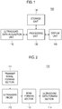
- the ultrasound system 100 includes an ultrasound data acquisition unit 110, a processing unit 120, a storage unit 130 and a display unit 140.
- the ultrasound data acquisition unit 110 is configured to transmit ultrasound beams to a target object and receive ultrasound echoes reflected from the target object to thereby form ultrasound data representative of the target object. An operation of the ultrasound acquisition unit will be described in detail by referring to FIG. 2 .
- FIG. 2 is a block diagram showing an illustrative embodiment of the ultrasound data acquisition unit 120.
- the ultrasound data acquisition unit 110 includes a transmit (Tx) signal forming section 111.
- the Tx signal forming section 121 generates a plurality of Tx signals and apply delays to the Tx signals.
- the delays of the Tx signals is controlled according to a steering angle of scan lines.
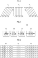
- the Tx signals includes first Tx signals for obtaining a first frame P 1 in which scan lines S 1 -S 6 are not steered, second Tx signals for obtaining a second frame P 2 in which scan lines S 1 -S 6 are steered at a first steering angle of ⁇ 1 and third Tx signals for obtaining a third frame P 3 in which scan lines S 1 -S 6 are steered at a second steering angle of ⁇ 2 , as shown in FIG. 3 .
- the ultrasound data acquisition unit 110 further includes an ultrasound probe 112, which is coupled to the Tx signal forming section 111.
- the ultrasound probe 112 includes an array transducer containing a plurality of transducer elements for reciprocal conversion between electric signals and ultrasound signals.
- the ultrasound probe 112 is configured to transmit ultrasound signals in response to the Tx signals.
- the ultrasound probe 112 is further configured to receive ultrasound echoes reflected from the target object to thereby output receive signals.
- the receive signals includes first receive signals obtained in response to the first Tx signals, second receive signals obtained in response to the second Tx signals and third receive signals obtained in response to the third Tx signals.
- the ultrasound data acquisition unit 110 further includes a beam forming section 113, which is coupled to the ultrasound probe 112.
- the beam forming section 113 is configured to digitize the electrical receive signals to obtain digital signals.
- the beam forming section 113 also applies delays to the digital signals in consideration of distances between the elements of the ultrasound probe 112 and focal points.
- the beam forming section 113 further sums the delayed digital signals to form receive-focused beams.
- the beam forming section 113 forms a first receive-focused beam based on the first receive signals, a second receive-focused beam based on the second receive signals and a third receive-focused beam based on the third receive signals.
- the ultrasound data acquisition unit 110 further includes an ultrasound data forming section 114, which is coupled to the beam forming section 113.
- the ultrasound data forming section 114 is configured to form ultrasound data corresponding to a plurality of frames based on the receive-focused beams.
- the ultrasound data are stored in the storage unit 130.
- the ultrasound data forming section 114 is configured to form a first ultrasound frame data set corresponding to the respective scan lines S 1 -S 6 of the first frame P 1 based on the first receive-focused beams.
- the ultrasound data forming section 114 is configured to form a second ultrasound frame data set corresponding to the respective scan lines S 1 -S 6 of the second frame P 2 based on the second receive-focused beams.
- the ultrasound data forming section 114 is configured to form a third ultrasound frame data set corresponding to the respective scan lines S 1 -S 6 of the third frame P 3 based on the first receive-focused beams.
- the ultrasound data forming section 114 is configured to perform a variety of signal processing (e.g., gain adjustment, etc.), which is necessary for forming the ultrasound data, upon the receive-focused beams.
- the processing unit 120 which is coupled to the ultrasound data acquisition unit 110, is configured to form ultrasound images and mask images corresponding to the frames of the plurality of steering angles based on the plurality of ultrasound frame data sets.
- the mask images are formed to remove seam artifacts appearing in a spatial compound image of the ultrasound images.
- the processing unit 120 is configured to spatially compound a plurality ultrasound images by using the mask images to from an ultrasound spatial compound image. An operation of the processing unit 120 will be described in detail by referring to FIG. 4.
- FIG. 4 is a block diagram showing an illustrative embodiment of the processing unit 120.
- the processing unit 120 includes a mask forming section 121, a mask setting section 122, an image forming section 123 and a spatial compounding section 124.
- the mask forming section 121 is configured to a plurality of masks corresponding to a plurality of frames, respectively, based on the plurality of ultrasound frame data sets. Each of the masks has a size and a pixel number identical to those of each of the frames.
- the mask forming section 121 is configured to a first mask 211 corresponding to the first frame P 1 based on the first ultrasound frame data set as shown in FIG. 5 .
- the mask forming section 121 is configured to a second mask 212 corresponding to the first frame P 2 based on the second ultrasound frame data set.
- the mask forming section 121 is configured to a third mask 213 corresponding to the first frame P 3 based on the third ultrasound frame data set.
- the mask setting section 122 which is coupled to the mask forming section 121, is configured to determine a pixel value corresponding to each of pixels in each of the masks by using the ultrasound frame data sets. More particularly, the mask setting section 122 is configured to detect an intensity of the ultrasound frame data corresponding to the scan line S i at each of the frame by using the ultrasound frame data sets. The mask setting section 122 is further configured to compare the detected intensity with a predetermined threshold. If the intensity is equal to or greater than the predetermined threshold, then it is determined that the ultrasound probe 122 is properly contacted with the surface of the target object. This is so that the mask setting section 122 may assign a value of 1 to pixels corresponding to the scan line S i .
- the mask setting section 122 may assign a value of 0 to pixels corresponding to the scan line S i .
- the mask setting section 122 is configured to detect intensities of the ultrasound frame data corresponding to each of the scan lines S 1 -S 6 of the first frame P 1 and compare the intensities with the predetermined threshold to thereby assign a value of 1 to pixels corresponding to the scan line S 1 -S 6 , which have the intensities equal to or greater than the predetermined threshold value, as shown in FIG. 5 .
- the mask setting section 122 is configured to detect intensities of the ultrasound frame data corresponding to each of the scan lines S 1 -S 6 of the second frame P 2 and compare the intensities with the predetermined threshold.
- the mask setting section 122 is configured to assign a value of 1 to pixels of the second mask 212 corresponding to the scan line S 1 -S 4 , which have the intensities equal to or greater than the predetermined threshold value, and a value of 0 to pixels of the second mask 212 corresponding to the scan line S 5 -S 6 , which have the intensities less than the predetermined threshold value.
- the mask setting section 122 is configured to detect intensities of the ultrasound frame data corresponding to each of the scan lines S 1 -S 6 of the third frame P 3 and compare the intensities with the predetermined threshold.
- the mask setting section 122 is configured to assign a value of 1 to pixels of the third mask 213 corresponding to the scan line S 2 -S 6 , which have the intensities equal to or greater than the predetermined threshold value, and a value of 0 to pixels of the third mask 213 corresponding to the scan line S 1 , which have the intensities less than the predetermined threshold value.
- the image forming section 123 which is coupled to the mask setting unit 122, is configured to form a plurality of mask images based on the plurality of masks provided from the mask setting unit 122. Also, the image forming section 123 is further configured to form a plurality of ultrasound images corresponding to the plurality of mask images by using the ultrasound frame data sets provided from the ultrasound data acquisition unit 110. In one embodiment, the image forming section 123 is configured to form a first mask image 311, a second mask image 312 and a third mask image 313 by using the first mask 211, the second mask 212 and the third mask 213, as shown in FIG. 6 . Further, the image forming section 123 is configured to form first to third ultrasound images 321 to 323 corresponding to the first to third frames P 1 -P 3 by using the first to third ultrasound frame data sets.
- the spatial compounding section 124 is configured to form an ultrasound spatial compound image by compounding the plurality of ultrasound images by using the plurality of mask images.
- the spatial compounding section 124 is configured to sum values of pixels identically positioned in the plurality of ultrasound images to obtain first summation values, and sum values of pixels identically positioned in the plurality of mask images to obtain second summation values.
- the spatial compound section 124 is configured to determine pixel values of the ultrasound spatial compound image.
- the spatial compound image 124 is configured to sum a pixel value of a pixel P 1,1 of the first ultrasound image 321, a pixel value of a pixel S 1,1 of the second ultrasound image 322 and a pixel value of a pixel U 1,1 of the third ultrasound image 323, with respect to a pixel C 1,1 of a ultrasound spatial compound image 330, thereby obtaining a first summation value.
- the spatial compound image 124 is further configured to sum a pixel value of 1 of the first mask image 311, which corresponds to the pixel P 1,1 of the first ultrasound image 321, a pixel value of 1 of the second mask image 312, which corresponds to the pixel S 1,1 of the second ultrasound image, and a pixel value of 0 of the third mask image 313, which corresponds to the pixel U 1,1 of the third ultrasound image, thereby obtaining a second summation value of 2.
- the spatial compounding section 124 is further configured to divide the first summation value by the second summation value (i.e., first summation value/second summation value) to thereby determine a pixel value of a pixel C 1,1 of the ultrasound spatial compound image 330. In the same manner, the spatial compounding section 124 is configured to determine pixel values of pixels C1,2 and C 1,3 of the ultrasound spatial compound image 330.
- the spatial compounding section 124 is configured to sum a pixel value of a pixel P 1,4 of the first ultrasound image 321 and a pixel value of a pixel S 2,4 of the second ultrasound image 322, thereby obtaining a first summation value.
- the spatial compound image 124 is further configured to sum a pixel value of 1 of the first mask image 311, which corresponds to the pixel P 1,4 of the first ultrasound image 321, and a pixel value of 1 of the second mask image 312, which corresponds to the pixel S 2,4 of the second ultrasound image, thereby obtaining a second summation value of 2.
- the spatial compounding section 124 is further configured to divide the first summation value by the second summation value (i.e., first summation value/second summation value) to thereby determine a pixel value of a pixel C 1,4 of the ultrasound spatial compound image 330.
- the spatial compounding section 124 is configured to determine pixel values of pixels C 1,5 and C 1,6 of the ultrasound spatial compound image 330.
- the spatial compound image 124 is configured to sum a pixel value of a pixel P 2,1 of the first ultrasound image 321, a pixel value of a pixel S2,1 of the second ultrasound image 322 and a pixel value of a pixel U 2,1 of the third ultrasound image 323, thereby obtaining a first summation value.
- the spatial compound image 124 is further configured to sum a pixel value of 1 of the first mask image 311, which corresponds to the pixel P 2,1 of the first ultrasound image 321, a pixel value of 1 of the second mask image 312, which corresponds to the pixel S 2,1 of the second ultrasound image, and a pixel value of 1 of the third mask image 313, which corresponds to the pixel U 2,1 of the third ultrasound image, thereby obtaining a second summation value of 3.
- the spatial compounding section 124 is further configured to divide the first summation value by the second summation value (i.e., first summation value/second summation value) to thereby determine a pixel value of a pixel C 2,1 of the ultrasound spatial compound image 330.
- the spatial compounding section 124 is configured to determine pixel values of pixels C 2,2 , C 2,3 , C 2,4 , C 2,5 , C 2,6 , C 3,1 , C 3,2 , C 3,3 , C 3,4 , C 3,5 , C 3,6 , C 4,1 , C 4,2 , C 4,3 , C 4,4 , C 4,5 , C 4,6 , C 5,1 , C 5,2 , C 5,3 , C 5,4 , C 5,5 , C 5,6 , C 6,1 , C 6,2 and C 6,3 of the ultrasound spatial compound image 330.
- the spatial compounding section 124 is configured to sum a pixel value of a pixel P 6,4 of the first ultrasound image 321 and a pixel value of a pixel U 5,4 of the second ultrasound image 323, thereby obtaining a first summation value.
- the spatial compound image 124 is further configured to sum a pixel value of 1 of the first mask image 311, which corresponds to the pixel P 6,4 of the first ultrasound image 321, and a pixel value of 1 of the third mask image 313, which corresponds to the pixel U 5,4 of the third ultrasound image, thereby obtaining a second summation value of 2.
- the spatial compounding section 124 is further configured to divide the first summation value by the second summation value (i.e., first summation value/second summation value) to thereby determine a pixel value of a pixel C 6,4 of the ultrasound spatial compound image 330.
- the spatial compounding section 124 is configured to determine pixel values of pixels C 6,5 and C 6,6 of the ultrasound spatial compound image 330.
- the storage unit 130 which is coupled to the processing unit 120, is configured to store the ultrasound data sets, which have been acquired in the ultrasound data acquisition unit 110.
- the storage unit 130 further stores the plurality of ultrasound images, which have been formed in the processing unit 120.
- the display unit 140 displays the ultrasound spatial compound image.
- a computer-readable storage medium storing instructions that, when executed by a computer, cause the computer to provide a method of spatially compounding ultrasound images based on a plurality of ultrasound frame data sets acquired from a target object and at different steering angles of scan lines in an ultrasound system, the method comprising: setting a plurality of masks corresponding to the respective frames based on the plurality of ultrasound frame date sets for removing seam artifact to form a plurality of mask images corresponding to the respective frames; forming a plurality of ultrasound images corresponding to the plurality of frames based on the plurality of frame data sets; and spatially compounding the plurality of ultrasound image based on the plurality of mask images to form an ultrasound spatial compound image.
Landscapes
- Health & Medical Sciences (AREA)
- Life Sciences & Earth Sciences (AREA)
- Engineering & Computer Science (AREA)
- Physics & Mathematics (AREA)
- Public Health (AREA)
- Medical Informatics (AREA)
- Biomedical Technology (AREA)
- General Health & Medical Sciences (AREA)
- Pathology (AREA)
- Animal Behavior & Ethology (AREA)
- Radiology & Medical Imaging (AREA)
- Molecular Biology (AREA)
- Surgery (AREA)
- Nuclear Medicine, Radiotherapy & Molecular Imaging (AREA)
- Biophysics (AREA)
- Veterinary Medicine (AREA)
- Heart & Thoracic Surgery (AREA)
- Remote Sensing (AREA)
- Computer Vision & Pattern Recognition (AREA)
- Radar, Positioning & Navigation (AREA)
- Acoustics & Sound (AREA)
- Computer Networks & Wireless Communication (AREA)
- General Physics & Mathematics (AREA)
- Physiology (AREA)
- Data Mining & Analysis (AREA)
- Databases & Information Systems (AREA)
- Epidemiology (AREA)
- Primary Health Care (AREA)
- Ultra Sonic Daignosis Equipment (AREA)
Claims (7)
- Ultraschallsystem (100), welches Folgendes aufweist:eine Ultraschalldatenerlangungseinheit (110), die dafür vorgesehen ist, Ultraschallstrahlen zu einem Zielobjekt zu übertragen, von dem Zielobjekt reflektierte Ultraschallechos zu empfangen und eine Vielzahl von Ultraschallrahmendatensätze für eine Vielzahl von Rahmen zur Verfügung zu stellen, wobei die Vielzahl von Ultraschallrahmendaten an unterschiedlichen Steuerungswinkeln von Scanlinien erlangt werden; undeine Verarbeitungseinheit (120), die dafür vorgesehen ist, eine Vielzahl von Ultraschallabbildungen zu erzeugen,wobei die Verarbeitungseinheit (120) Folgendes aufweist:einen Maskenerzeugungsabschnitt, der dafür vorgesehen ist, dem jeweiligen Rahmen entsprechende Masken basierend auf den Ultraschallrahmendatensätzen zu erzeugen,einen Maskenfestlegungsabschnitt, der dafür vorgesehen ist, Pixelwerte von Pixeln von jeder der Masken basierend auf Intensitäten der Ultraschallrahmendatensätze zu ermitteln,einen Abbildungserzeugungsabschnitt, der dafür vorgesehen ist, eine Vielzahl von Maskenabbildungen basierend auf den Masken und die Vielzahl von Ultraschallabbildungen basierend auf den Ultraschallrahmendatensätzen zu erzeugen, undeinen räumlichen Zusammensetzungsabschnitt, der dafür vorgesehen ist, die Vielzahl von Ultraschallabbildungen basierend auf der Vielzahl von Maskenabbildungen räumlich zusammenzusetzen, um ein räumliches, zusammengesetztes Ultraschallbild zu erzeugen,wobei der räumliche Zusammensetzungsabschnitt dafür vorgesehen ist, Pixelwerte von an den Ultraschallabbildungen überlappten Pixeln zu summieren, um einen ersten Additionswert zu erlangen, Pixelwerte von an den Maskenabbildungen überlappten Pixeln zu summieren, um einen zweiten Additionswert zu erlangen, und den ersten Additionswert durch den zweiten Additionswert zu teilen, um einen Pixelwert an einem korrespondierenden Pixel der räumlichen zusammengesetzten Ultraschallabbildung zu ermitteln.
- Ultraschallsystem (100) nach Anspruch 1, wobei die Maskenabbildungen identische Größen und Pixelanzahlen wie diejenigen der entsprechenden Ultraschallabbildungen aufweisen.
- Ultraschallsystem (100) nach Anspruch 1, wobei der Maskenfestlegungsabschnitt dafür vorgesehen ist, eine Intensität von jedem der Rahmen, die den jeweiligen Scanlinien entsprechen, durch Verwenden der Ultraschallrahmendatensätze zu detektieren, die detektierten Intensitäten mit einem vorbestimmten Grenzwert zu vergleichen, wenn die Intensität gleich oder größer als ein vorbestimmter Grenzwert ist, den Pixeln, die der Scanlinie entsprechen, einen Wert von 1 zuzuordnen, und, wenn die Intensität geringer ist als der vorbestimmte Grenzwert, den Pixeln, die der Scanlinie entsprechen, einen Wert von 0 zuzuordnen.
- Verfahren zum Erzeugen einer räumlichen, zusammengesetzten Ultraschallabbildung in einem Ultraschallsystem (100), welches Folgendes aufweist:a) Übertragen von Ultraschallstrahlen zu einem Zielobjekt, Empfangen von Ultraschallechos, die von dem Zielobjekt reflektiert wurden, und zur Verfügung Stellen einer Vielzahl von Ultraschallrahmendatensätzen für eine Vielzahl von Rahmen, wobei die Vielzahl von Ultraschallrahmendaten an unterschiedlichen Steuerungswinkeln von Scanlinien erlangt werden;b) Erzeugen von Masken, welche den jeweiligen Rahmen entsprechen, basierend auf den Ultraschallrahmendatensätzen;c) Ermitteln von Pixelwerten von Pixeln von jeder der Maske basierend auf Intensitäten der Ultraschallrahmendatensätze zum Entfernen von Randartifakten, um eine Vielzahl von Maskenabbildungen zu erzeugen, die den jeweiligen Rahmen entsprechen;d) Erzeugen einer Vielzahl von Ultraschallabbildungen, welche der Vielzahl von Rahmen entsprechen, basierend auf der Vielzahl von Rahmendatensätzen; unde) räumliches Zusammensetzen der Vielzahl von Ultraschallabbildungen basierend auf der Vielzahl von Maskenabbildungen, um ein räumliches, zusammengesetztes Ultraschallbild zu erzeugen,wobei e) Folgendes aufweist:Summieren von Pixelwerten von Pixeln, die an den Ultraschallabbildungen überlappen, um einen ersten Additionswert zu erlangen;Summieren von Pixelwerten von Pixeln, die an den Maskenabbildungen überlappen, um einen zweiten Additionswert zu erlangen; undTeilen des ersten Additionswerts durch den zweiten Additionswert, um einen Pixelwert an einem korrespondierenden Pixel der räumlichen zusammengesetzten Ultraschallabbildung zu ermitteln.
- Verfahren nach Anspruch 4, wobei die Masken identische Größen und Pixelanzahlen wie diejenigen der entsprechenden Ultraschallabbildungen aufweisen.
- Verfahren nach Anspruch 4, wobei c) Folgendes aufweist:Detektieren einer Intensität von jedem der Rahmen, die den jeweiligen Scanlinien entsprechen, unter Verwendung der Ultraschallrahmendatensätze;Vergleichen der detektierten Intensität mit einem vorbestimmten Grenzwert, wenn die Intensität gleich oder größer als ein vorbestimmter Grenzwert ist, Zuordnen eines Werts von 1 zu den Pixeln, die der Scanlinie entsprechen; undwenn die Intensität geringer als ein vorbestimmter Grenzwert ist, Zuordnen eines Werts von 0 zu den Pixeln, welche der Scanlinie entsprechen.
- Computerlesbares Speichermedium, welches Befehle speichert, die, wenn sie von einem Computer ausgeführt werden, bewirken, dass der Computer ein Verfahren zum Erzeugen einer räumlichen, zusammengesetzten Ultraschallabbildung basierend auf einer Vielzahl von Ultraschallrahmendatensätzen, die von einem Zielobjekt und an unterschiedlichen Steuerungswinkeln von Scanlinien in einem Ultraschallsystem (100) erlangt werden, ausführt, wobei das Verfahren Folgendes aufweist:Erzeugen von Masken, welche den jeweiligen Rahmen entsprechen, basierend auf den Ultraschallrahmendatensätzen;Ermitteln von Pixelwerten von Pixeln von jeder der Maske basierend auf Intensitäten der Ultraschallrahmendatensätze zum Entfernen von Randartefakten, um eine Vielzahl von Maskenabbildungen zu erzeugen, die den jeweiligen Rahmen entsprechen;Erzeugen einer Vielzahl von Ultraschallabbildungen, welche der Vielzahl von Rahmen entsprechen, basierend auf der Vielzahl von Rahmendatensätzen; undräumliches Zusammensetzen der Vielzahl von Ultraschallabbildungen basierend auf der Vielzahl von Maskenabbildungen, um ein räumliches, zusammengesetztes Ultraschallbild zu erzeugen,wobei das Ermitteln der Pixelwerte von Pixeln von jeder der Maske Folgendes aufweist:Detektieren einer Intensität von jedem der Rahmen, die den jeweiligen Scanlinien entsprechen, unter Verwendung der Ultraschallrahmendatensätze;Vergleichen der detektierten Intensität mit einem vorbestimmten Grenzwert, wenn die Intensität gleich oder größer als ein vorbestimmter Grenzwert ist, Zuordnen eines Werts von 1 zu den Pixeln, die der Scanlinie entsprechen; undwenn die Intensität geringer als ein vorbestimmter Grenzwert ist, Zuordnen eines Werts von 0 zu den Pixeln, welche der Scanlinie entsprechen,wobei das räumliche Zusammensetzen der Vielzahl von Ultraschallabbildungen Folgendes aufweist:Summieren von Pixelwerten von Pixeln, die an den Ultraschallabbildungen überlappen, um einen ersten Additionswert zu erhalten;Summieren von Pixelwerten von Pixeln, die an den Maskenabbildungen überlappen, um einen zweiten Additionswert zu erhalten; undTeilen des ersten Additionswerts durch den zweiten Additionswert, um einen Pixelwert an einem entsprechenden Pixel der räumlichen zusammengesetzten Ultraschallabbildung zu ermitteln.
Applications Claiming Priority (1)
| Application Number | Priority Date | Filing Date | Title |
|---|---|---|---|
| KR1020100006412A KR101183003B1 (ko) | 2010-01-25 | 2010-01-25 | 마스크에 기초하여 초음파 공간 합성 영상을 제공하는 초음파 시스템 및 방법 |
Publications (2)
| Publication Number | Publication Date |
|---|---|
| EP2347713A1 EP2347713A1 (de) | 2011-07-27 |
| EP2347713B1 true EP2347713B1 (de) | 2021-03-03 |
Family
ID=43981727
Family Applications (1)
| Application Number | Title | Priority Date | Filing Date |
|---|---|---|---|
| EP11151475.8A Active EP2347713B1 (de) | 2010-01-25 | 2011-01-20 | Ultraschallbildverbesserung in einem Ultraschallsystem |
Country Status (4)
| Country | Link |
|---|---|
| US (1) | US20110184292A1 (de) |
| EP (1) | EP2347713B1 (de) |
| JP (1) | JP5756639B2 (de) |
| KR (1) | KR101183003B1 (de) |
Cited By (1)
| Publication number | Priority date | Publication date | Assignee | Title |
|---|---|---|---|---|
| US20230010317A1 (en) * | 2021-07-12 | 2023-01-12 | Fujifilm Corporation | Medical image processing device and operation method thereof |
Families Citing this family (7)
| Publication number | Priority date | Publication date | Assignee | Title |
|---|---|---|---|---|
| JP5272084B2 (ja) * | 2012-01-11 | 2013-08-28 | 日立アロカメディカル株式会社 | 超音波診断装置 |
| CN102551809A (zh) * | 2012-02-29 | 2012-07-11 | 飞依诺科技(苏州)有限公司 | 一种超声诊断中图形扩展的成像方法 |
| KR101956308B1 (ko) * | 2016-11-04 | 2019-03-08 | 서강대학교산학협력단 | 광음향 영상의 배경 노이즈를 제거하는 시스템 및 그 방법 |
| US11439369B1 (en) | 2017-10-26 | 2022-09-13 | United States Of America As Represented By The Secretary Of The Air Force | Method and apparatus for imaging with reduced level of off-axis artifacts |
| KR102655278B1 (ko) * | 2018-07-24 | 2024-04-08 | 삼성메디슨 주식회사 | 초음파 영상 장치 및 그 표시 방법 |
| EP4412532B1 (de) * | 2021-10-08 | 2025-01-29 | Koninklijke Philips N.V. | Verbesserung der ultraschallherzbildgebung |
| US12588898B2 (en) * | 2023-06-05 | 2026-03-31 | GE Precision Healthcare LLC | Ultrasound imaging techniques for shear-wave elastography |
Citations (1)
| Publication number | Priority date | Publication date | Assignee | Title |
|---|---|---|---|---|
| US6181810B1 (en) * | 1998-07-30 | 2001-01-30 | Scimed Life Systems, Inc. | Method and apparatus for spatial and temporal filtering of intravascular ultrasonic image data |
Family Cites Families (9)
| Publication number | Priority date | Publication date | Assignee | Title |
|---|---|---|---|---|
| US5821915A (en) * | 1995-10-11 | 1998-10-13 | Hewlett-Packard Company | Method and apparatus for removing artifacts from scanned halftone images |
| US6224552B1 (en) * | 1998-10-01 | 2001-05-01 | Atl Ultrasound | Ultrasonic diagnostic imaging system with reduced spatial compounding seam artifacts |
| US6364835B1 (en) * | 1998-11-20 | 2002-04-02 | Acuson Corporation | Medical diagnostic ultrasound imaging methods for extended field of view |
| US6895077B2 (en) * | 2001-11-21 | 2005-05-17 | University Of Massachusetts Medical Center | System and method for x-ray fluoroscopic imaging |
| US20040077946A1 (en) * | 2002-10-15 | 2004-04-22 | Jun Ohmiya | Image processing apparatus, method and program |
| US6858010B2 (en) * | 2003-05-06 | 2005-02-22 | Siemens Medical Solutions Usa, Inc. | Identifying clinical markers in spatial compounding ultrasound imaging |
| JP4095494B2 (ja) * | 2003-05-30 | 2008-06-04 | キヤノン株式会社 | 眼科画像処理装置及び処理方法 |
| US20050124886A1 (en) * | 2003-11-21 | 2005-06-09 | Koninklijke Philips Electronics N.V. | System and method for generating ultrasound images having variable spatial compounding |
| US7780601B2 (en) * | 2007-06-05 | 2010-08-24 | Siemens Medical Solutions Usa, Inc. | Adaptive clinical marker preservation in spatial compound ultrasound imaging |
-
2010
- 2010-01-25 KR KR1020100006412A patent/KR101183003B1/ko active Active
-
2011
- 2011-01-20 EP EP11151475.8A patent/EP2347713B1/de active Active
- 2011-01-21 US US13/011,470 patent/US20110184292A1/en not_active Abandoned
- 2011-01-24 JP JP2011012246A patent/JP5756639B2/ja not_active Expired - Fee Related
Patent Citations (1)
| Publication number | Priority date | Publication date | Assignee | Title |
|---|---|---|---|---|
| US6181810B1 (en) * | 1998-07-30 | 2001-01-30 | Scimed Life Systems, Inc. | Method and apparatus for spatial and temporal filtering of intravascular ultrasonic image data |
Cited By (1)
| Publication number | Priority date | Publication date | Assignee | Title |
|---|---|---|---|---|
| US20230010317A1 (en) * | 2021-07-12 | 2023-01-12 | Fujifilm Corporation | Medical image processing device and operation method thereof |
Also Published As
| Publication number | Publication date |
|---|---|
| JP2011152415A (ja) | 2011-08-11 |
| US20110184292A1 (en) | 2011-07-28 |
| JP5756639B2 (ja) | 2015-07-29 |
| KR101183003B1 (ko) | 2012-09-18 |
| EP2347713A1 (de) | 2011-07-27 |
| KR20110086989A (ko) | 2011-08-02 |
Similar Documents
| Publication | Publication Date | Title |
|---|---|---|
| EP2347713B1 (de) | Ultraschallbildverbesserung in einem Ultraschallsystem | |
| US8792690B2 (en) | Enhancing quality of ultrasound spatial compound image based on beam profile in ultrasound system | |
| US8506488B2 (en) | Ultrasound image enhancement in an ultrasound system | |
| US8968199B2 (en) | Spatial compound imaging in an ultrasound system | |
| US9008383B2 (en) | Enhancing quality of ultrasound image in ultrasound system | |
| US20120101378A1 (en) | Providing an ultrasound spatial compound image based on a phased array probe in an ultrasound system | |
| CN106600550B (zh) | 超声图像处理方法及系统 | |
| US8727990B2 (en) | Providing an ultrasound spatial compound image in an ultrasound system | |
| US20110054324A1 (en) | Ultrasound system and method for providing multiple plane images for a plurality of views | |
| US9081097B2 (en) | Component frame enhancement for spatial compounding in ultrasound imaging | |
| US9151841B2 (en) | Providing an ultrasound spatial compound image based on center lines of ultrasound images in an ultrasound system | |
| US20110137171A1 (en) | Providing an ultrasound spatial compound image in an ultrasound system | |
| US8037767B2 (en) | Transmit apodization using a sinc function in an ultrasound system | |
| US20100280383A1 (en) | Color Doppler Mode Image Processing In An Ultrasound System | |
| US20110054323A1 (en) | Ultrasound system and method for providing an ultrasound spatial compound image considering steering angle | |
| WO2015136534A1 (en) | Strong echoes in ultrasound images | |
| EP2422705B1 (de) | Ultraschall-Elastizitätsbildgebung | |
| US8864668B2 (en) | Formation of an elastic image in an ultrasound system | |
| US8503714B2 (en) | Dropout correction in ultrasound strain imaging | |
| CN113243936A (zh) | 超声波波束合成方法、装置、超声设备及存储介质 | |
| CN119700183B (zh) | 一种超声设备以及超声成像的空间复合方法 | |
| Teixeira Prado et al. | The Use of Instantaneous Phase for Improving Saft Images |
Legal Events
| Date | Code | Title | Description |
|---|---|---|---|
| PUAI | Public reference made under article 153(3) epc to a published international application that has entered the european phase |
Free format text: ORIGINAL CODE: 0009012 |
|
| AK | Designated contracting states |
Kind code of ref document: A1 Designated state(s): AL AT BE BG CH CY CZ DE DK EE ES FI FR GB GR HR HU IE IS IT LI LT LU LV MC MK MT NL NO PL PT RO RS SE SI SK SM TR |
|
| AX | Request for extension of the european patent |
Extension state: BA ME |
|
| RIN1 | Information on inventor provided before grant (corrected) |
Inventor name: KIM, JEONG SIK Inventor name: YOO, JAE HEUNG |
|
| RTI1 | Title (correction) |
Free format text: ULTRASOUND IMAGE ENHANCEMENT IN AN ULTRASOUND SYSTEM |
|
| 17P | Request for examination filed |
Effective date: 20120126 |
|
| 17Q | First examination report despatched |
Effective date: 20130530 |
|
| STAA | Information on the status of an ep patent application or granted ep patent |
Free format text: STATUS: EXAMINATION IS IN PROGRESS |
|
| GRAP | Despatch of communication of intention to grant a patent |
Free format text: ORIGINAL CODE: EPIDOSNIGR1 |
|
| STAA | Information on the status of an ep patent application or granted ep patent |
Free format text: STATUS: GRANT OF PATENT IS INTENDED |
|
| INTG | Intention to grant announced |
Effective date: 20200924 |
|
| GRAS | Grant fee paid |
Free format text: ORIGINAL CODE: EPIDOSNIGR3 |
|
| RAP1 | Party data changed (applicant data changed or rights of an application transferred) |
Owner name: SAMSUNG MEDISON CO., LTD. |
|
| GRAA | (expected) grant |
Free format text: ORIGINAL CODE: 0009210 |
|
| STAA | Information on the status of an ep patent application or granted ep patent |
Free format text: STATUS: THE PATENT HAS BEEN GRANTED |
|
| AK | Designated contracting states |
Kind code of ref document: B1 Designated state(s): AL AT BE BG CH CY CZ DE DK EE ES FI FR GB GR HR HU IE IS IT LI LT LU LV MC MK MT NL NO PL PT RO RS SE SI SK SM TR |
|
| REG | Reference to a national code |
Ref country code: GB Ref legal event code: FG4D |
|
| REG | Reference to a national code |
Ref country code: AT Ref legal event code: REF Ref document number: 1366333 Country of ref document: AT Kind code of ref document: T Effective date: 20210315 Ref country code: CH Ref legal event code: EP |
|
| REG | Reference to a national code |
Ref country code: DE Ref legal event code: R096 Ref document number: 602011070278 Country of ref document: DE |
|
| REG | Reference to a national code |
Ref country code: IE Ref legal event code: FG4D |
|
| REG | Reference to a national code |
Ref country code: LT Ref legal event code: MG9D |
|
| PG25 | Lapsed in a contracting state [announced via postgrant information from national office to epo] |
Ref country code: LT Free format text: LAPSE BECAUSE OF FAILURE TO SUBMIT A TRANSLATION OF THE DESCRIPTION OR TO PAY THE FEE WITHIN THE PRESCRIBED TIME-LIMIT Effective date: 20210303 Ref country code: FI Free format text: LAPSE BECAUSE OF FAILURE TO SUBMIT A TRANSLATION OF THE DESCRIPTION OR TO PAY THE FEE WITHIN THE PRESCRIBED TIME-LIMIT Effective date: 20210303 Ref country code: GR Free format text: LAPSE BECAUSE OF FAILURE TO SUBMIT A TRANSLATION OF THE DESCRIPTION OR TO PAY THE FEE WITHIN THE PRESCRIBED TIME-LIMIT Effective date: 20210604 Ref country code: HR Free format text: LAPSE BECAUSE OF FAILURE TO SUBMIT A TRANSLATION OF THE DESCRIPTION OR TO PAY THE FEE WITHIN THE PRESCRIBED TIME-LIMIT Effective date: 20210303 Ref country code: BG Free format text: LAPSE BECAUSE OF FAILURE TO SUBMIT A TRANSLATION OF THE DESCRIPTION OR TO PAY THE FEE WITHIN THE PRESCRIBED TIME-LIMIT Effective date: 20210603 Ref country code: NO Free format text: LAPSE BECAUSE OF FAILURE TO SUBMIT A TRANSLATION OF THE DESCRIPTION OR TO PAY THE FEE WITHIN THE PRESCRIBED TIME-LIMIT Effective date: 20210603 |
|
| REG | Reference to a national code |
Ref country code: NL Ref legal event code: MP Effective date: 20210303 |
|
| REG | Reference to a national code |
Ref country code: AT Ref legal event code: MK05 Ref document number: 1366333 Country of ref document: AT Kind code of ref document: T Effective date: 20210303 |
|
| PG25 | Lapsed in a contracting state [announced via postgrant information from national office to epo] |
Ref country code: SE Free format text: LAPSE BECAUSE OF FAILURE TO SUBMIT A TRANSLATION OF THE DESCRIPTION OR TO PAY THE FEE WITHIN THE PRESCRIBED TIME-LIMIT Effective date: 20210303 Ref country code: LV Free format text: LAPSE BECAUSE OF FAILURE TO SUBMIT A TRANSLATION OF THE DESCRIPTION OR TO PAY THE FEE WITHIN THE PRESCRIBED TIME-LIMIT Effective date: 20210303 Ref country code: RS Free format text: LAPSE BECAUSE OF FAILURE TO SUBMIT A TRANSLATION OF THE DESCRIPTION OR TO PAY THE FEE WITHIN THE PRESCRIBED TIME-LIMIT Effective date: 20210303 Ref country code: PL Free format text: LAPSE BECAUSE OF FAILURE TO SUBMIT A TRANSLATION OF THE DESCRIPTION OR TO PAY THE FEE WITHIN THE PRESCRIBED TIME-LIMIT Effective date: 20210303 |
|
| PG25 | Lapsed in a contracting state [announced via postgrant information from national office to epo] |
Ref country code: NL Free format text: LAPSE BECAUSE OF FAILURE TO SUBMIT A TRANSLATION OF THE DESCRIPTION OR TO PAY THE FEE WITHIN THE PRESCRIBED TIME-LIMIT Effective date: 20210303 |
|
| PG25 | Lapsed in a contracting state [announced via postgrant information from national office to epo] |
Ref country code: AT Free format text: LAPSE BECAUSE OF FAILURE TO SUBMIT A TRANSLATION OF THE DESCRIPTION OR TO PAY THE FEE WITHIN THE PRESCRIBED TIME-LIMIT Effective date: 20210303 Ref country code: SM Free format text: LAPSE BECAUSE OF FAILURE TO SUBMIT A TRANSLATION OF THE DESCRIPTION OR TO PAY THE FEE WITHIN THE PRESCRIBED TIME-LIMIT Effective date: 20210303 Ref country code: CZ Free format text: LAPSE BECAUSE OF FAILURE TO SUBMIT A TRANSLATION OF THE DESCRIPTION OR TO PAY THE FEE WITHIN THE PRESCRIBED TIME-LIMIT Effective date: 20210303 Ref country code: EE Free format text: LAPSE BECAUSE OF FAILURE TO SUBMIT A TRANSLATION OF THE DESCRIPTION OR TO PAY THE FEE WITHIN THE PRESCRIBED TIME-LIMIT Effective date: 20210303 |
|
| PG25 | Lapsed in a contracting state [announced via postgrant information from national office to epo] |
Ref country code: IS Free format text: LAPSE BECAUSE OF FAILURE TO SUBMIT A TRANSLATION OF THE DESCRIPTION OR TO PAY THE FEE WITHIN THE PRESCRIBED TIME-LIMIT Effective date: 20210703 Ref country code: RO Free format text: LAPSE BECAUSE OF FAILURE TO SUBMIT A TRANSLATION OF THE DESCRIPTION OR TO PAY THE FEE WITHIN THE PRESCRIBED TIME-LIMIT Effective date: 20210303 Ref country code: PT Free format text: LAPSE BECAUSE OF FAILURE TO SUBMIT A TRANSLATION OF THE DESCRIPTION OR TO PAY THE FEE WITHIN THE PRESCRIBED TIME-LIMIT Effective date: 20210705 Ref country code: SK Free format text: LAPSE BECAUSE OF FAILURE TO SUBMIT A TRANSLATION OF THE DESCRIPTION OR TO PAY THE FEE WITHIN THE PRESCRIBED TIME-LIMIT Effective date: 20210303 Ref country code: ES Free format text: LAPSE BECAUSE OF FAILURE TO SUBMIT A TRANSLATION OF THE DESCRIPTION OR TO PAY THE FEE WITHIN THE PRESCRIBED TIME-LIMIT Effective date: 20210303 |
|
| REG | Reference to a national code |
Ref country code: DE Ref legal event code: R097 Ref document number: 602011070278 Country of ref document: DE |
|
| PLBE | No opposition filed within time limit |
Free format text: ORIGINAL CODE: 0009261 |
|
| STAA | Information on the status of an ep patent application or granted ep patent |
Free format text: STATUS: NO OPPOSITION FILED WITHIN TIME LIMIT |
|
| PG25 | Lapsed in a contracting state [announced via postgrant information from national office to epo] |
Ref country code: DK Free format text: LAPSE BECAUSE OF FAILURE TO SUBMIT A TRANSLATION OF THE DESCRIPTION OR TO PAY THE FEE WITHIN THE PRESCRIBED TIME-LIMIT Effective date: 20210303 Ref country code: AL Free format text: LAPSE BECAUSE OF FAILURE TO SUBMIT A TRANSLATION OF THE DESCRIPTION OR TO PAY THE FEE WITHIN THE PRESCRIBED TIME-LIMIT Effective date: 20210303 |
|
| 26N | No opposition filed |
Effective date: 20211206 |
|
| PG25 | Lapsed in a contracting state [announced via postgrant information from national office to epo] |
Ref country code: SI Free format text: LAPSE BECAUSE OF FAILURE TO SUBMIT A TRANSLATION OF THE DESCRIPTION OR TO PAY THE FEE WITHIN THE PRESCRIBED TIME-LIMIT Effective date: 20210303 |
|
| PG25 | Lapsed in a contracting state [announced via postgrant information from national office to epo] |
Ref country code: IS Free format text: LAPSE BECAUSE OF FAILURE TO SUBMIT A TRANSLATION OF THE DESCRIPTION OR TO PAY THE FEE WITHIN THE PRESCRIBED TIME-LIMIT Effective date: 20210703 |
|
| PG25 | Lapsed in a contracting state [announced via postgrant information from national office to epo] |
Ref country code: MC Free format text: LAPSE BECAUSE OF FAILURE TO SUBMIT A TRANSLATION OF THE DESCRIPTION OR TO PAY THE FEE WITHIN THE PRESCRIBED TIME-LIMIT Effective date: 20210303 |
|
| REG | Reference to a national code |
Ref country code: CH Ref legal event code: PL |
|
| GBPC | Gb: european patent ceased through non-payment of renewal fee |
Effective date: 20220120 |
|
| REG | Reference to a national code |
Ref country code: BE Ref legal event code: MM Effective date: 20220131 |
|
| PG25 | Lapsed in a contracting state [announced via postgrant information from national office to epo] |
Ref country code: LU Free format text: LAPSE BECAUSE OF NON-PAYMENT OF DUE FEES Effective date: 20220120 Ref country code: GB Free format text: LAPSE BECAUSE OF NON-PAYMENT OF DUE FEES Effective date: 20220120 |
|
| PG25 | Lapsed in a contracting state [announced via postgrant information from national office to epo] |
Ref country code: BE Free format text: LAPSE BECAUSE OF NON-PAYMENT OF DUE FEES Effective date: 20220131 |
|
| PG25 | Lapsed in a contracting state [announced via postgrant information from national office to epo] |
Ref country code: LI Free format text: LAPSE BECAUSE OF NON-PAYMENT OF DUE FEES Effective date: 20220131 Ref country code: CH Free format text: LAPSE BECAUSE OF NON-PAYMENT OF DUE FEES Effective date: 20220131 |
|
| PG25 | Lapsed in a contracting state [announced via postgrant information from national office to epo] |
Ref country code: IE Free format text: LAPSE BECAUSE OF NON-PAYMENT OF DUE FEES Effective date: 20220120 |
|
| PG25 | Lapsed in a contracting state [announced via postgrant information from national office to epo] |
Ref country code: HU Free format text: LAPSE BECAUSE OF FAILURE TO SUBMIT A TRANSLATION OF THE DESCRIPTION OR TO PAY THE FEE WITHIN THE PRESCRIBED TIME-LIMIT; INVALID AB INITIO Effective date: 20110120 |
|
| PG25 | Lapsed in a contracting state [announced via postgrant information from national office to epo] |
Ref country code: MK Free format text: LAPSE BECAUSE OF FAILURE TO SUBMIT A TRANSLATION OF THE DESCRIPTION OR TO PAY THE FEE WITHIN THE PRESCRIBED TIME-LIMIT Effective date: 20210303 Ref country code: CY Free format text: LAPSE BECAUSE OF FAILURE TO SUBMIT A TRANSLATION OF THE DESCRIPTION OR TO PAY THE FEE WITHIN THE PRESCRIBED TIME-LIMIT Effective date: 20210303 |
|
| PG25 | Lapsed in a contracting state [announced via postgrant information from national office to epo] |
Ref country code: TR Free format text: LAPSE BECAUSE OF FAILURE TO SUBMIT A TRANSLATION OF THE DESCRIPTION OR TO PAY THE FEE WITHIN THE PRESCRIBED TIME-LIMIT Effective date: 20210303 |
|
| PG25 | Lapsed in a contracting state [announced via postgrant information from national office to epo] |
Ref country code: MT Free format text: LAPSE BECAUSE OF FAILURE TO SUBMIT A TRANSLATION OF THE DESCRIPTION OR TO PAY THE FEE WITHIN THE PRESCRIBED TIME-LIMIT Effective date: 20210303 |
|
| PGFP | Annual fee paid to national office [announced via postgrant information from national office to epo] |
Ref country code: FR Payment date: 20251209 Year of fee payment: 16 |
|
| PGFP | Annual fee paid to national office [announced via postgrant information from national office to epo] |
Ref country code: DE Payment date: 20251208 Year of fee payment: 16 |
|
| PGFP | Annual fee paid to national office [announced via postgrant information from national office to epo] |
Ref country code: IT Payment date: 20251209 Year of fee payment: 16 |