EP2346503B1 - Kv1.3-kanal-blockierstoffe zur behandlung von erkrankungen in zusammenhang mit intimaler hyperplasie - Google Patents
Kv1.3-kanal-blockierstoffe zur behandlung von erkrankungen in zusammenhang mit intimaler hyperplasie Download PDFInfo
- Publication number
- EP2346503B1 EP2346503B1 EP09783852.8A EP09783852A EP2346503B1 EP 2346503 B1 EP2346503 B1 EP 2346503B1 EP 09783852 A EP09783852 A EP 09783852A EP 2346503 B1 EP2346503 B1 EP 2346503B1
- Authority
- EP
- European Patent Office
- Prior art keywords
- cells
- vsmcs
- vascular
- pap
- treatment
- Prior art date
- Legal status (The legal status is an assumption and is not a legal conclusion. Google has not performed a legal analysis and makes no representation as to the accuracy of the status listed.)
- Not-in-force
Links
Images



Classifications
-
- A—HUMAN NECESSITIES
- A61—MEDICAL OR VETERINARY SCIENCE; HYGIENE
- A61K—PREPARATIONS FOR MEDICAL, DENTAL OR TOILETRY PURPOSES
- A61K45/00—Medicinal preparations containing active ingredients not provided for in groups A61K31/00 - A61K41/00
- A61K45/06—Mixtures of active ingredients without chemical characterisation, e.g. antiphlogistics and cardiaca
-
- A—HUMAN NECESSITIES
- A61—MEDICAL OR VETERINARY SCIENCE; HYGIENE
- A61K—PREPARATIONS FOR MEDICAL, DENTAL OR TOILETRY PURPOSES
- A61K31/00—Medicinal preparations containing organic active ingredients
- A61K31/33—Heterocyclic compounds
- A61K31/335—Heterocyclic compounds having oxygen as the only ring hetero atom, e.g. fungichromin
- A61K31/365—Lactones
- A61K31/366—Lactones having six-membered rings, e.g. delta-lactones
- A61K31/37—Coumarins, e.g. psoralen
-
- A—HUMAN NECESSITIES
- A61—MEDICAL OR VETERINARY SCIENCE; HYGIENE
- A61K—PREPARATIONS FOR MEDICAL, DENTAL OR TOILETRY PURPOSES
- A61K38/00—Medicinal preparations containing peptides
- A61K38/16—Peptides having more than 20 amino acids; Gastrins; Somatostatins; Melanotropins; Derivatives thereof
- A61K38/168—Peptides having more than 20 amino acids; Gastrins; Somatostatins; Melanotropins; Derivatives thereof from plants
-
- A—HUMAN NECESSITIES
- A61—MEDICAL OR VETERINARY SCIENCE; HYGIENE
- A61K—PREPARATIONS FOR MEDICAL, DENTAL OR TOILETRY PURPOSES
- A61K38/00—Medicinal preparations containing peptides
- A61K38/16—Peptides having more than 20 amino acids; Gastrins; Somatostatins; Melanotropins; Derivatives thereof
- A61K38/17—Peptides having more than 20 amino acids; Gastrins; Somatostatins; Melanotropins; Derivatives thereof from animals; from humans
- A61K38/1767—Peptides having more than 20 amino acids; Gastrins; Somatostatins; Melanotropins; Derivatives thereof from animals; from humans from invertebrates
-
- A—HUMAN NECESSITIES
- A61—MEDICAL OR VETERINARY SCIENCE; HYGIENE
- A61P—SPECIFIC THERAPEUTIC ACTIVITY OF CHEMICAL COMPOUNDS OR MEDICINAL PREPARATIONS
- A61P13/00—Drugs for disorders of the urinary system
- A61P13/12—Drugs for disorders of the urinary system of the kidneys
-
- A—HUMAN NECESSITIES
- A61—MEDICAL OR VETERINARY SCIENCE; HYGIENE
- A61P—SPECIFIC THERAPEUTIC ACTIVITY OF CHEMICAL COMPOUNDS OR MEDICINAL PREPARATIONS
- A61P9/00—Drugs for disorders of the cardiovascular system
-
- A—HUMAN NECESSITIES
- A61—MEDICAL OR VETERINARY SCIENCE; HYGIENE
- A61P—SPECIFIC THERAPEUTIC ACTIVITY OF CHEMICAL COMPOUNDS OR MEDICINAL PREPARATIONS
- A61P9/00—Drugs for disorders of the cardiovascular system
- A61P9/10—Drugs for disorders of the cardiovascular system for treating ischaemic or atherosclerotic diseases, e.g. antianginal drugs, coronary vasodilators, drugs for myocardial infarction, retinopathy, cerebrovascula insufficiency, renal arteriosclerosis
Definitions
- the present invention is comprised in the field of cardiovascular pathologies, particularly in the field of diseases derived from the hyperplasia of the tunica intima.
- the wall of blood vessels is made up of three concentric strata or tunicae (the tunica intima, media and adventitia) with well distinguished structure and composition.
- the tunica intima is made up of a single layer of endothelial cells supported on a basement membrane rich in collagen and proteoglycans and separated from the media by the internal elastic lamina.
- the tunica media is formed by vascular smooth muscle cells (VSMCs) and extracellular matrix, and the adventitia, the outermost layer, is essentially made up of connective tissue and fibroblasts.
- the VSMCs are physiologically located in the tunica media. However, VSMCs can be found in the intimal layer as a result of a lack of organization of the structure of the vascular wall due to vascular pathologies or lesions such as atherosclerosis, hyperplasia of the tunica intima.
- Intimal hyperplasia is a change in the vascular structure which occurs as a result of the biological repair mechanisms after a vascular lesion, either mechanical, surgical, inflammatory or immunological.
- the most characteristic finding of this structural change is the thickening of the intimal layer, due both to an increase in the number of cells and to an increase in the synthesis of extracellular matrix in which these cells are found ( Davies MG, Hagen PO. Pathobiology of intimal hyperplasia. British Journal of Surgery 1994;81:1254-1269 ).
- this process results in narrowing or stenosis of the vascular lumen.
- intimal hyperplasia There are various pathological situations associated with intimal hyperplasia and the triggering factors can generally be grouped together as physical lesions (in many cases iatrogenic lesions due to vascular surgery), inflammatory lesions (as in atherosclerotic lesions) or due to an increase of the tension of the wall (as in the case of pulmonary hypertension or the use of vein grafts in by-pass surgery).
- Some of the diseases associated with intimal hyperplasia include:
- Revascularization with vein grafts is the standard treatment for occlusive arterial diseases when the occluded segment is large and an endarterectomy cannot be performed.
- the most used technique is the autogenic transplant of a segment of the saphenous vein, and it is used both in coronary surgery and in peripheral vascular surgery (intermittent claudication, thromboangiitis obliterans of the tibial artery, etc.).
- the vein graft suffers chronic maladaptive response to an arterial environment in which the primary component is the uncontrolled proliferation of vascular smooth muscle cells, giving rise to intimal hyperplasia which can later become complicated due to the development of atherosclerosis and thrombosis ( Murphy GJ, Angelini GD. Cardiovasc Ultrasound 2004;21:2-8 ).
- Post-transplant coronary vasculopathy is the main factor limiting long-term survival after a heart transplant. It manifests as an especially aggressive form of coronary artery disease that is different from conventional arteriosclerosis, which is caused by the combination of physical, chemical and immunological factors causing an endothelial lesion which in turn triggers the proliferation of vascular smooth muscle cells and intimal hyperplasia
- the pathological analysis of the lesions shows a thickening of the intima in which undifferentiated cells of the smooth muscle and macrophages and lymphocytes participate.
- the factors triggering coronary vasculopathy are also present in other post-transplant vasculopathies, as is the case of post-transplant nephropathy, which again represents the most frequent cause of renal transplant failure.
- intimal hyperplasia there are other diseases resulting from intimal hyperplasia.
- the person skilled in the art will use imaging techniques. Specifically, coronary angiography is used in the coronary tree, injecting contrasting fluid into the ostium of said arteries.
- the 64-slice multislice CT and cardiac resonance angiography are also able to display atherosclerotic lesions in the coronary circulation.
- Angiographic techniques are complemented with ultrasound techniques, such as intravascular ultrasound, which allow displaying the thickness of the arterial intimal layer, and determining if there is an atherosclerotic lesion and its characteristics.
- ultrasound techniques such as intravascular ultrasound
- the thickness of the intima cannot be measured microscopically because it is a single layer of cells.
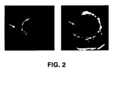
- any measurable thickness (which is greater than a single cell layer) is pathological ( Figure 1 ).
- Kv1.3 voltage-activated potassium channel
- Kv1.3 was first identified in T-cells ( Hu et al., The Journal of Immunology, 2007, 179: 4563-4570 . Grissmer et al., Proc. Nat. Acad. Sci. 87: 9411-9415, 1990 ).
- Kv1.3 channel is tetra-homomeric, whereas in other types of cells, Kv1.3 is associated with subunits of other voltage-activated channels of the Kv1 family to form heteromeric potassium channels ( Hu et al., The Journal of Immunology, 2007, 179: 4563-4570 ).
- the regulation of K + flows through the membrane of the cells is often the combinatorial result of multiple molecular signaling pathways.
- the importance of Kv1.3 in immune system-related diseases has been proven in several studies.
- Document W02008088422 discloses the use of peptide inhibitors of Kv1.3 for the treatment of autoimmune diseases, allergies, diabetes and obesity and describes the manufacture and purification of derivatives of the toxins ShK, MgTx 1 , MTX 1 , HsTxl, wGVIA, HaTxl, etc. According to the data shown, the peptide derivatives of ShK are able to inhibit the human Kv1.3 current with an IC of -150 pM.
- ShK is, furthermore, a potent inhibitor of the proliferation of T-cells so their use in therapies for the treatment of immunological diseases such as multiple sclerosis, rheumatoid arthritis, dermatitis, diabetes type I, etc., is proposed.
- Potassium channels also play a very relevant role in the immediate and long-term regulation of the function of the vascular smooth muscle cells.
- the VSMCs of the walls of the vessels are cells which express a unique repertoire of contractile proteins, ion channels and signaling molecules aimed at maintaining vascular tone.
- K + channels such as Kv1.2, Kv1.3, Kv1.5, Kv1.6 and Kv2.1, calcium-activated K + channels (such as the maxiK or BK channels), of inward-rectifying K + channels (such as the K IR channels and K ATP channels) and two-pore K + channels, which are responsible for the background currents and the TASK and TWIK channels (reviewed in Jackson WF, Microcirculation 12,113-127, 2005 ) .
- Kv voltage-dependent K + channels
- Kv voltage-dependent K + channels
- the field of the art cannot provide effective therapies for the treatment of intimal hyperplasia. It would therefore be convenient to develop a therapeutic tool aimed at controlling the factors determining hyperplasia of the tunica intima and which is able to stop the migration and proliferation of the VSMCs therein, as well as the secretion of components of the cell matrix.
- the inventors propose the use of Kv1.3 channel blocking substances in the manufacture of a pharmacological tool for the treatment of diseases relating to intimal hyperplasia.
- An advantage of the present invention is the selective blocking of the Kv1.3 channels of the VSMCs by means of the use of blocking substances belonging to the group of margatoxin, ShK toxin, psoralen, kaliotoxin, correolide, J123, and the derivatives and combinations thereof, for the manufacture of a pharmacological tool for the treatment of diseases relating to hyperplasia of the vascular tunica intima.
- a possible embodiment of the present invention is the use of Kv1.3 blockers in the manufacture of a pharmacological tool for the treatment of vascular diseases which cause restenosis and comprise post-transplant chronic nephropathy, late by-pass occlusion and post-transplant coronary vasculopathy, and the equivalents and combinations thereof.
- the present invention comprises the use of Shk-L5, PAP-1 or both, in the manufacture of a pharmacological tool for the treatment of said vascular diseases.
- An advantageous embodiment of the present invention is the use of said blocking substances in the manufacture of a pharmacological tool for the treatment and/or prevention of restenosis.
- a possible embodiment of the present invention consists of a method for the prevention or treatment of intimal hyperplasia, comprising the administration of a Kv1.3 blocking substance.
- this method is used for the selective blocking of the Kv1.3 channels of the VSMCs by means of the use of pharmacological tools manufactured with blocking substances belonging to the group of margatoxin, ShK toxin, psoralen, kaliotoxin, correolide, J123, and the derivatives and combinations thereof, preferably Shk-L5, PAP-1 or both.
- this method can be applied in the treatment of diseases which cause restenosis, such as post-transplant chronic nephropathy, late by-pass occlusion and post-transplant coronary vasculopathy, and the equivalents and combinations thereof.
- Cardiovascular pharmacology lacks effective tools designed for the treatment of diseases resulting from hyperplasia of the tunica intima.
- a possible embodiment of the present invention consists of a metering apparatus for implanting in an arterial vessel of an animal, which can be a human, comprising a containing system for containing a Kv1.3 blocking substance.
- An advantage of this apparatus is that the active ingredient it meters is a selective Kv1.3 channel blocker of the VSMCs belonging to the group of margatoxin, ShK toxin, psoralen, kaliotoxin, correolide, J123, and the derivatives and combinations thereof.
- the present invention preferably comprises the substances Shk-L5, PAP-1 or both.
- this apparatus can be applied in the treatment of diseases which cause restenosis, such as post-transplant chronic nephropathy, late by-pass occlusion and post-transplant coronary vasculopathy.
- the inventors propose the use of Kv1.3 blocking substances, as described below, for the treatment of the diseases resulting from the thickening of the intima.
- the invention is based on the discovery that the smooth muscle cells located in the intimal hyperplasia preferably express Kv1.3 ( Figures 3 , 4 , 5 and 6 ) and that by using blocking substances specific for the Kv1.3 channels, the rate of cell division and migration of the VSMCs ( Figures 7 and 8 ) and the subsequent thickening of the intima after a vascular lesion are reduced.
- the Kv1.3 channel blocking substances can be used as pharmacological tools for the treatment of vascular diseases.
- a pharmacological tool is any substance, composition, utensil, device, mechanism, contrivance, contraption, instrument, machine or apparatus adapted, studied or designed to be used in the treatment, cure, prevention or diagnosis of a disease, or to prevent the onset of an unwanted physiological process.
- Vascular diseases can be a consequence of the lack of organization of the normal structure of the vascular wall.
- the three concentric layers of the vascular wall or tunicae can become unorganized during pathological processes or as the consequence of physical lesions, causing the vascular lumen to narrow ( Figure 1 ).
- the lack of organization of the structure of the vascular wall manifests by means of hyperplasia of the tunica intima ( Figure 1 ).
- hyperplasia relates to any assessable thickening of the intimal layer (beyond a single layer of cells).
- the cells which can be found in the hyperplastic intima include, besides the single layer of endothelial cells, cells from the immune system and cardiovascular smooth muscle cells.
- the latter which are absent in the tunica intima in physiological situations, have experienced cell migration and proliferation processes in the tunica intima. Due to this cell activity, which entails the secretion of cell matrix, the mentioned thickening and, accordingly, the reduction of the vascular lumen occur.
- restenosis relates to this reduction in diameter of the vascular lumen. Restenosis is a consequence of the lack of organization of the vascular wall and is therefore common in many vascular diseases, including post-transplant chronic nephropathy, late by-pass occlusion and post-transplant coronary vasculopathy. In the context of the present invention, therefore, diseases equivalent to post-transplant chronic nephropathy, late by-pass occlusion and post-transplant coronary vasculopathy are those which present a lack of organization of the structure of the vascular wall which manifests by means of intimal hyperplasia and causes the vascular lumen to narrow, or restenosis. In the context of the present invention, it is also considered that stenosis and restenosis are equivalent terms and relate to an obstruction ⁇ : 50% of the vascular lumen in a given segment.
- Kv1.3 blocking substances in therapies against diseases relating to intimal hyperplasia are explained below. This document includes in its different sections the teachings so that the person skilled in the art can identify this type of disease and the substances acting as Kv1.3 channel blockers.
- the Kv1.3 channel blockers must be able to decrease in a dose-dependent manner the flow of current through these channels evoked by a depolarizing voltage pulse.
- the present application describes one of these known methods in Example 1.
- Kv1.3 channel blocker In order to prove that a substance acts as a Kv1.3 channel blocker, the person skilled in the art uses a heterologous expression system. Some stable cell lanes expressing the Kv1.3 channel have been described and are available in the field of the art, for example those described in Grissmer et al., Mol. Pharmacol 44, 1227-34, 1994 . These or other equivalent cells can be requested from their inventors.
- a cell line can be purchased from the ATCC cell collection, such as for example HEK293 cells (Human Embryonic Kidney, catalogue no. ATCC CRC-1573), CHO-K1 cells (Chinese Hamster Ovary, catalogue no. ATCC CCL-61) or COS-7 cells (monkey kidney fibroblasts catalogue no. ATCC CRC-1651) marketed in Europe through LGC Standards, UK.
- ATCC cell collection such as for example HEK293 cells (Human Embryonic Kidney, catalogue no. ATCC CRC-1573), CHO-K1 cells (Chinese Hamster Ovary, catalogue no. ATCC CCL-61) or COS-7 cells (monkey kidney fibroblasts catalogue no. ATCC CRC-1651) marketed in Europe through LGC Standards, UK.
- the Kv1.3 channel can be expressed in HEK293 cells by means of transfection with Lipofectamine of a suitable plasmid for expression in mammals, which contains the cDNA of the encoding sequence of the Kv1.3 channel.
- the person skilled in the art can perform co-transfection with marker molecules to identify the cells which effectively express the transfected channel.
- Kd values for Kv1.3 channel blocker peptides and small molecules Abbreviations: 4-AP, 4-aminopyridine; ShK-Dap22, Stichodactyla helianthus toxin with diaminopropionic acid in position 22 substituting a native Lys; ShK-F6CA, Stichodactyla helianthus toxin-fluorescein-6-carboxylic acid; TEA, tetraethylammonium chloride.
- Blocking substance Affinity Stichodactyla helianthus toxin (ShK) 11 pM Heteroflowers spinifer toxin 1 12 pM ShK-F6CA 48 pM Pandinus imperator toxin 2 50 pM ShK-Dap22 52 pM Hongotoxin 86 pM Margatoxin 110 pM Agiotoxin-2 200 pM Pandinus imperator toxin 3 500 pM Kaliotoxin 650 pM Buthus martensii Karsch toxin (J123 peptide) 790 pM Noxiustoxin 1 nM Pap-1 2 nM Psora-4 3 nM Charybdotoxin 3 nM Titystoxin-K ⁇ 4 nM Odonthobthus doriae toxin 2 (OdK2) 7 nM Pandinus imperator toxin 1 11 nM Bunodosoma
- the blockers described above can be used according to the present invention in the manufacture of a pharmacological tool which can commonly be a medicament containing any pharmacologically acceptable excipient and the Kv1.3 blocker as the active ingredient, alone or in combination with other active ingredients.
- the present invention comprises all the effective doses in any of the comprised diseases as well as any of the possible methods of administration independently of the race, sex, biotype, weight or age of the individual to be treated.
- a direct application of the present invention is the development of a new method for the prevention and treatment of vascular diseases which are a consequence of the lack of organization of the normal structure of the vascular wall.
- This method consists of the administration or the implantation of a pharmacological tool comprising at least one active ingredient having a blocking effect on Kv1.3 channels.
- the method of the present invention consists of the administration of a Kv1.3 channel blocker from the group of margatoxin, ShK toxin, psoralen, kaliotoxin, correolide, J123, and the derivatives and combinations thereof.
- the blocking substances comprise the ShK-L5 or PAP-1 toxins.
- Example 4 Figures 6 , 7 and 8 , illustrate the effect of these toxins on the VSMCs and Example 5 describes the administration protocol.
- Another important application of the present invention consists of the design and preparation of a system which allows the controlled release of the Kv1.3 blocking substance, such as a stent coated with a biopolymer in which the blocker resides for its gradual release.
- Example 5 describes the details of this application.
- Example 1 Assay to determine if a substance is a Kv1.3 channel blocker.
- Kv1.3 a substance is capable of dose-dependently reducing the flow of current through Kv1.3 (i.e., if it is a Kv1.3 blocker).
- the currents through the Kv1.3 channels expressed in the heterologous system were studied by means of the patch-clamp technique in the whole-cell configuration. The currents were recorded at room temperature (21-25°-C).
- the cells, placed in a recording chamber located in an inverted microscope, were perfused with a physiological saline solution (in mM: 141 NaCl, 4.7 KCl, 1.2 MgCl 2 , 1.8 CaCl 2 , 10 glucose and 10 HEPES (pH 7.4 with NaOH).
- the recording pipettes were made from borosilicate glass (2.0 mm OD, WPI) with a pipette puller (Narishige PP-83) so that they have resistances between 2 and 5 M ⁇ upon filling them with an internal solution containing (in mM) 125 KCl, 4 MgCl 2 , 10 HEPES, 10 EGTA, 5 MgATP; (pH 7.2 with KOH).
- the whole-cell currents were recorded with an Axopatch 200 amplifier or the like, filtering at 2 kHz and sampling at 10 kHz.
- the records were digitalized with a Digidata 1200 A/D interface executed by the CLAMPEX 8 software (Axon Instruments) or an equivalent similar system, in a computer.
- the cells were maintained at a resting potential of -80 mV and from here depolarizing pulses of 200 ms in duration from -60 mV up to +80 mV in intervals of 10 mV every 15 seconds were applied.
- depolarizing pulses 200 ms in duration from -60 mV up to +80 mV in intervals of 10 mV every 15 seconds were applied.
- the person skilled ion the art can construct a current-voltage curve for the Kv1.3 channel.
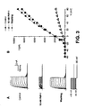
- Figure 3 shows an example of this type of experiment.
- the application of PAP-1 at a concentration of 10 nM caused a reduction of the current of about 80-85% for potentials from +10 up to +80 mV.
- the effect of repeated depolarizing pulses from -80 mV up to +40 mV applied every second was evaluated for the purpose of characterizing the characteristic cumulative inactivation of the Kv1.3 currents.
- the potential blocking substances are applied dissolved in the external solution to study the effect of their application with the protocols previously described and known in the field of the art, such that the possible effects of these substances on the amplitude of the current, the time course and the voltage dependence of their activation and inactivation, can be characterized.
- the dose-response curve of the blocking effect as well as the reversibility of the effect after washing the blocker have to be analyzed.
- the careful analysis of the rate of the establishment and the washout of the effects allows obtaining an estimate of the affinity constant of the substance to the canal.
- the percent reduction of the amplitude of the current in the presence of the blocker as a value between 0 and 1 can be defined. If there is a reduction of the current in the presence of the substance under analysis, a dose-response curve is constructed to define the IC 50 (or Kd) of the blocking of the Kv1.3 current by the substance under analysis.
- the changes in the expression of the Kv1.3 channels were studied by comparing VSMCs of mouse femoral arteries in contractile and proliferative phenotype.
- the contractile phenotype are the cells isolated directly from the medial layer of the femoral artery, and the effect of the phenotypic change was studied in two different proliferation models: an in vivo model in which intimal hyperplasia is induced by endoluminal lesion with the previously described procedure ( Roqué et al., Arterioscler Thromb Vasc Biol 20, 335-342, 2000 ) and an in vitro model in which VSMCs are obtained in culture from explants of mouse femoral artery ( Miguel-Velado et al., Circ Res 97, 1280-1287, 2005 ).
- BPN mice were obtained from Jackson Laboratories and maintained by means of inbreeding in the animal house of the School of Medicine or the animal house of the IDIBAPS, in temperature-controlled conditions (21°C) with 12 hour light/dark cycles and with free access to food and water.
- VSMCs model of cells in contractile phenotype
- the animals were sacrificed by decapitation after anesthesia with isoflurane (5% at 2.5 I O 2 min -1 ).
- the femoral arteries were dissected and the connective and endothelial tissue was removed and the cells were obtained by means of enzyme dispersion with the previously described protocol ( Miguel-Velado et al., Circ Res 97, 1280-1287, 2005 ).
- VSMCs in culture in vitro model of proliferative phenotype
- small fragments (0.5 mm) of femoral arteries free of endothelium and connective tissue were placed in 35 mm Petri culture dishes covered with 2% gelatin (Type B, Sigma) in a DMEM culture medium with 20% of fetal bovine serum, 100 U/ml penicillin, 100 U/ml streptomycin and 2 mM L-glutamine at 37°C in a 5% CO 2 atmosphere.
- the migration and proliferation of the VSMCs of these explants was clear at 6-10 days, but the explants were maintained until the single layer of VSMC was confluent. At this time, by means of mechanical scraping with a rubber spatula the cells were shed and seeded in a new culture dish at 1/3 of their original density.
- the VSMCs were thus subjected to several passes (3-4) without showing apparent morphological changes.
- the BPN mice were subjected to unilateral transluminal lesion of the femoral artery at the age of 12-16 weeks by means of the repeated pass of an angioplasty catheter 0.25 mm in diameter following the previously described protocol ( Roqué et al., Arterioscler Thromb Vasc Biol 20, 335-342, 2000 ). The mice were sacrificed after 48 hours, 1 week or 4 weeks by means of pentobarbital overdose to extract the control and damaged femoral arteries.
- RNA samples were processed with the previously described protocols ( Miguel-Velado et al., Circ Res 97, 1280-1287, 2005 ) and the mRNA levels were determined using Taqman probes for Kv1.3 which were obtained from Applied Biosystems.
- the VSMCs freshly dissociated or in culture were seeded in glass coverslips 12 mm in diameter, fixed with 4% paraformaldehyde in phosphate buffer at pH 7.5 for 15 minutes at 20oC, washed with PBSTx (0.1 % PBS Triton X-100) and blocked with PBSTx containing 10 mg/ml of bovine albumin and 2% non-immune goat serum for 10 minutes.
- the anti-Kv1.3 antibody was diluted in the blocking solution at a concentration of 1:100 for 40-60 min at 21 °C.
- PBSTx After three 5-minute washings in PBSTx the cells were incubated with the secondary antibody (Alexa 488 goat anti-mouse from Molecular Probes) at a dilution of 1:1000 in blocking solution for 30 minutes. After three other washings with PBSTx the coverslips were assembled with VectaShield H100-DAPI (vector Labs) and the cells were examined in a Nikon fluorescence microscope with a wavelength filter of 488 nm.
- the secondary antibody Alexa 488 goat anti-mouse from Molecular Probes
- the euthanized animals were fixed with 4% paraformaldehyde intravenous perfusion in PBS for 5 minutes at 100 mmHg to obtain paraffin sections of the femoral arteries as described ( Roqué et al., Arterioscler Thromb Vasc Biol 20, 335-342, 2000 ). The sections were subsequently deparaffinized, rehydrated and treated with 1 % citric acid and blocked with 3% hydrogen peroxide (DakoCytomatation) before incubation with the primary antibody for 12 hours at 4oC.
- DakoCytomatation hydrogen peroxide
- the drugs were prepared at the final concentrations indicated in the saline solution of the bath following the supplier's instructions and were applied to the cells for a period of 1-2 minutes.
- the currents were recorded before applying the drug (control) and in the presence of the drug, and the percentage of reduction in the amplitude of the currents induced by different blockers was calculated.
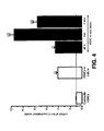
- the analysis of the expression of the mRNA of the Kv1.3 channel shows a very significant increase of the expression levels in the two proliferation conditions (culture and endoluminal lesion) with respect to the contractile cells as illustrated in Figure 4 .
- the effect of these blockers was significantly greater in the VSMCs in culture in all the cases.
- the current blocked by this drug was 10.6 ⁇ 2.03 of the Kv current in the contractile VSMCs and 58.15 ⁇ 4.54 of the Kv current in the VSMCs in culture
- PAP-1 i.e., the Kv1.3 channels contribute with 10% of the total current in the differentiated cells and almost 60% of the total current in the cells in culture.
- the migration studies have been performed in cultures of smooth muscle cells obtained from explants of confluent human uterine arteries, maintained in DMEM culture medium supplemented with 20% fetal bovine serum, 100 U/ml of penicillin and streptomycin and 2 mM of L-glutamine at 37oC in a humidified incubator with a 5% CO 2 atmosphere. In these cultures, a small surface of the single layer is scraped by means of the tip of a sterile P10 pipette creating a space without cells and they are left for 24-48 hours in the same culture medium but without supplementing with fetal bovine serum to block proliferation.
- the area of the surface without cells created by the tip of the pipette was quantified by means of the image analysis software of the Image J program, measuring it at 0, 24 and 48 hours.
- the cell-free area at 0 hours was taken as control and the area at 24 or 48 hours is expressed as a fraction of the control area, which allows determining the degree of migration as the percentage of cell-free area at 24 or 48 hours with respect to the cell-free area at 0 hours (100%).
- the migration in these cultures in control situation and in the presence of margatoxin (10 nM) or PAP-1 (10 nM) was studied.
- the drugs are prepared in sterile saline solution at a concentration of 10 ⁇ M and added to the culture medium without serum (1 ⁇ l/ml to obtain the final concentration of 10 nM.
- BrdU 5-bromo-2'-deoxyuridine
- the cells incorporating BrdU can be detected by means of observation under a fluorescence microscope upon using a monoclonal antibody against this molecule and a secondary antibody conjugated with a fluorochrome.
- the assay was performed with a commercial kit (5-Bromo-2'-deoxy-uridine labeling and Detection Kit I; Roche Applied Science, Germany).
- the human uterine artery smooth muscle cells were grown for 48 hours in glass coverslips 12 mm in diameter in DMEM medium supplemented with serum and antibiotics as has been previously described, until reaching a confluence close to 50%. At this time, the coverslips are maintained for 24 hours at 37oC and in 5% CO 2 atmosphere in culture medium with 5% fetal bovine serum (control), medium with 5% serum and 10 nM of PAP-1 and medium with 5% serum and 10 nM of Margatoxin, after which the BrdU incorporation assay is performed following the instructions of the commercial company.
- Expandable stainless steel stents (Cordis Co) 18 mm in length covered with a thin layer of a poly-n-butyl methacrylate and polyethylene-vinyl acetate copolymer containing 200 ⁇ g of PAP-1 or 200 ⁇ g of ShK-L5 are used.
- the total weight of the polymer with the drug is about 500 ⁇ g and the drug polymer ratio is 30%.
- the stents are packed individually, coded with a bar code (to do a double blind experiment) and sterilized with ethylene oxide.
- 36 stents (12 with the polymer alone, 12 with PAP and 12 with ShK) are implanted in 12 young pigs (25-35 kg) in the anterior descending or circumflex branches of the left coronary artery or in the right coronary artery (at a proportion of 3 stents per animal).
- the animals recover from the operation and are sent back to the animal house where they receive a normal diet supplemented with aspirin (325 mg/d). Blood samples of the animals are obtained at 6, 12, 24, 48 and 96 hours to determine the plasma levels of PAP and ShK.
- the animals are euthanized after performing a coronary arteriography for the quantitative analysis.
- the coronary arteries are dissected, the stent is removed and the samples are frozen in liquid nitrogen in order to determine different markers by means of Western blot of the tissues and to determine the levels of PAP and ShK in the arterial wall.
- the hearts are collected and the coronary arteries are fixed by perfusion of formalin at 10% for 30 minutes. Then the coronary vessels are dissected and processed for their immunohistological and morphometric analysis performing sections of the area where the stent and the superior and inferior margins are located.
- a morphometric analysis is performed measuring the degree of endothelialization of the stent area, the content of VSMCs in the intima and its thickness in relation to the thickness of the vessel and the lumen area (see Figure 9 ).
- the pharmacokinetics studies show that the blood levels of PAP-1 and ShK are maximal in the initial measure at 6 hours after implanting the stent (about 3 ng/ml) and decrease and are maintained below the limit of detection at 4 days ( ⁇ 0.2 ng/ml).
- the content of PAP and ShK in the arterial tissue is 97 and 83 ng/artery at 7 days and 30 and 25 ng/artery at 28 days.
Landscapes
- Health & Medical Sciences (AREA)
- Life Sciences & Earth Sciences (AREA)
- Chemical & Material Sciences (AREA)
- Public Health (AREA)
- Animal Behavior & Ethology (AREA)
- Medicinal Chemistry (AREA)
- General Health & Medical Sciences (AREA)
- Pharmacology & Pharmacy (AREA)
- Veterinary Medicine (AREA)
- Epidemiology (AREA)
- Bioinformatics & Cheminformatics (AREA)
- Engineering & Computer Science (AREA)
- Proteomics, Peptides & Aminoacids (AREA)
- Immunology (AREA)
- Gastroenterology & Hepatology (AREA)
- General Chemical & Material Sciences (AREA)
- Organic Chemistry (AREA)
- Nuclear Medicine, Radiotherapy & Molecular Imaging (AREA)
- Chemical Kinetics & Catalysis (AREA)
- Botany (AREA)
- Zoology (AREA)
- Urology & Nephrology (AREA)
- Heart & Thoracic Surgery (AREA)
- Cardiology (AREA)
- Vascular Medicine (AREA)
- Medicines That Contain Protein Lipid Enzymes And Other Medicines (AREA)
- Acyclic And Carbocyclic Compounds In Medicinal Compositions (AREA)
- Pharmaceuticals Containing Other Organic And Inorganic Compounds (AREA)
Claims (5)
- Verwendung von PAP-1 als Kv1.3-Kanal-Blockierstoff in der Herstellung eines pharmakologischen Instruments zur Behandlung von Gefäßerkrankungen in Zusammenhang mit Intimahyperplasie.
- Verwendung nach Anspruch 1, umfassend PAP-1 und mindestens einen pharmakologisch akzeptablen Hilfsstoff.
- Verwendung nach Anspruch 1, wobei die Gefäßerkrankungen Restenose, chronische Nephropathie nach Transplantation, Bypass-Spätverschluss, koronare Vaskulopathie nach Transplantation und deren Äquivalente und Kombinationen umfassen.
- Abgabevorrichtung zur Implantation in einem arteriellen Gefäß eines Tieres, auch eines Menschen, welche eine PAP-1 enthaltende Vorratseinrichtung umfasst.
- Abgabevorrichtung nach Anspruch 4 zur Verwendung in der Behandlung von Restenose, chronischer Nephropathie nach Transplantation, Bypass-Spätverschluss, koronarer Vaskulopathie nach Transplantation und deren Äquivalenten und Kombinationen.
Priority Applications (1)
| Application Number | Priority Date | Filing Date | Title |
|---|---|---|---|
| EP09783852.8A EP2346503B1 (de) | 2008-10-08 | 2009-10-08 | Kv1.3-kanal-blockierstoffe zur behandlung von erkrankungen in zusammenhang mit intimaler hyperplasie |
Applications Claiming Priority (3)
| Application Number | Priority Date | Filing Date | Title |
|---|---|---|---|
| EP08105518 | 2008-10-08 | ||
| EP09783852.8A EP2346503B1 (de) | 2008-10-08 | 2009-10-08 | Kv1.3-kanal-blockierstoffe zur behandlung von erkrankungen in zusammenhang mit intimaler hyperplasie |
| PCT/EP2009/063099 WO2010040803A2 (en) | 2008-10-08 | 2009-10-08 | Kv1.3 channel blocking substances for the treatment of diseases associated with intimal hyperplasia |
Publications (2)
| Publication Number | Publication Date |
|---|---|
| EP2346503A2 EP2346503A2 (de) | 2011-07-27 |
| EP2346503B1 true EP2346503B1 (de) | 2013-07-17 |
Family
ID=41360303
Family Applications (1)
| Application Number | Title | Priority Date | Filing Date |
|---|---|---|---|
| EP09783852.8A Not-in-force EP2346503B1 (de) | 2008-10-08 | 2009-10-08 | Kv1.3-kanal-blockierstoffe zur behandlung von erkrankungen in zusammenhang mit intimaler hyperplasie |
Country Status (4)
| Country | Link |
|---|---|
| US (1) | US8629110B2 (de) |
| EP (1) | EP2346503B1 (de) |
| ES (2) | ES2345527B1 (de) |
| WO (1) | WO2010040803A2 (de) |
Families Citing this family (7)
| Publication number | Priority date | Publication date | Assignee | Title |
|---|---|---|---|---|
| WO2014028502A1 (en) * | 2012-08-13 | 2014-02-20 | ImmunGene Inc. | Engineered antibody-interferon fusion molecules for treatment of autoimmune diseases |
| TWI598325B (zh) | 2012-10-12 | 2017-09-11 | H 朗德貝克公司 | 苯甲醯胺類 |
| TW201427947A (zh) | 2012-10-12 | 2014-07-16 | Lundbeck & Co As H | 環狀胺 |
| TWI698438B (zh) | 2015-03-13 | 2020-07-11 | 德商4Sc製藥公司 | Kv1.3抑制劑及其醫學應用 |
| TWI701249B (zh) | 2015-03-13 | 2020-08-11 | 德商4Sc製藥公司 | Kv1.3抑制劑及其醫學應用 |
| EP4003329A1 (de) * | 2019-07-31 | 2022-06-01 | Sea4Us - Biotecnologia e Recursos Marinhos, Lda. | Kv1.3 antagonisten zur verwendung bei der behandlung von chronischem und akutem schmerz |
| CN114616230A (zh) | 2019-07-31 | 2022-06-10 | 海洋生物技术与海洋资源有限公司 | 黄烷类似物化合物及其在治疗慢性和急性疼痛中的用途 |
Family Cites Families (5)
| Publication number | Priority date | Publication date | Assignee | Title |
|---|---|---|---|---|
| US5116864A (en) * | 1991-04-09 | 1992-05-26 | Indiana University Foundation | Method for preventing restenosis following reconfiguration of body vessels |
| WO1993007748A2 (en) * | 1991-10-23 | 1993-04-29 | The General Hospital Corporation | Laser-based inhibition of smooth muscle cell hyperproliferation |
| US5354774A (en) * | 1992-12-24 | 1994-10-11 | Yale University | Inhibition of smooth muscle cell proliferation by 8-methoxypsoralen photoactivated by visible light |
| US6008211A (en) * | 1995-07-27 | 1999-12-28 | Pdt Pharmaceuticals, Inc. | Photoactivatable compounds comprising benzochlorin and furocoumarin |
| AR063384A1 (es) | 2006-10-25 | 2009-01-28 | Amgen Inc | Agentes terapeuticos a base de peptidos derivados de toxinas |
-
2009
- 2009-01-30 ES ES200900281A patent/ES2345527B1/es not_active Expired - Fee Related
- 2009-10-08 WO PCT/EP2009/063099 patent/WO2010040803A2/en not_active Ceased
- 2009-10-08 EP EP09783852.8A patent/EP2346503B1/de not_active Not-in-force
- 2009-10-08 US US13/123,220 patent/US8629110B2/en not_active Expired - Fee Related
- 2009-10-08 ES ES09783852T patent/ES2436035T3/es active Active
Also Published As
| Publication number | Publication date |
|---|---|
| WO2010040803A2 (en) | 2010-04-15 |
| US8629110B2 (en) | 2014-01-14 |
| US20110275571A1 (en) | 2011-11-10 |
| ES2345527A1 (es) | 2010-09-24 |
| ES2345527B1 (es) | 2011-09-08 |
| WO2010040803A3 (en) | 2010-06-03 |
| EP2346503A2 (de) | 2011-07-27 |
| ES2436035T3 (es) | 2013-12-26 |
Similar Documents
| Publication | Publication Date | Title |
|---|---|---|
| Krishna et al. | Development of a two-stage limb ischemia model to better simulate human peripheral artery disease | |
| US8629110B2 (en) | Kv1.3 channel blocking substances for the treatment of diseases associated with intimal hyperplasia | |
| Qin et al. | Downregulation of circulating MOTS-c levels in patients with coronary endothelial dysfunction | |
| Westvik et al. | Limb ischemia after iliac ligation in aged mice stimulates angiogenesis without arteriogenesis | |
| Wang et al. | Glucagon-like peptide-1 protects against cardiac microvascular injury in diabetes via a cAMP/PKA/Rho-dependent mechanism | |
| Tarabra et al. | Effect of the monocyte chemoattractant protein-1/CC chemokine receptor 2 system on nephrin expression in streptozotocin-treated mice and human cultured podocytes | |
| Oparil et al. | Estrogen attenuates the adventitial contribution to neointima formation in injured rat carotid arteries | |
| Hu et al. | Biochanin A protect against lipopolysaccharide-induced acute lung injury in mice by regulating TLR4/NF-κB and PPAR-γ pathway | |
| Parker-Duffen et al. | Divergent roles for adiponectin receptor 1 (AdipoR1) and AdipoR2 in mediating revascularization and metabolic dysfunction in vivo | |
| Fan et al. | Endothelial deletion of mTORC1 protects against hindlimb ischemia in diabetic mice via activation of autophagy, attenuation of oxidative stress and alleviation of inflammation | |
| JPH11510479A (ja) | タモキシフェン類似体による心臓血管疾病の予防及び治療 | |
| US11274306B2 (en) | Method for inhibiting calcification of a smooth muscle cell (SMC) | |
| WO1994026303A1 (en) | Prevention and treatment of pathologies associated with abnormally proliferative smooth muscle cells | |
| Chu et al. | Involvement of POMC neurons in LEAP2 regulation of food intake and body weight | |
| Zhang et al. | RETRACTED ARTICLE: The Synergistic Effects of Astragalus mongholicus and Salvia miltiorrhiza on Coronary Heart Disease Identified by Network Pharmacology and Experiment | |
| Li et al. | Nagilactone D ameliorates experimental pulmonary fibrosis in vitro and in vivo via modulating TGF-β/Smad signaling pathway | |
| Jiang et al. | ZC3H4 mediates silica-induced EndoMT via ER stress and autophagy | |
| Calegari et al. | Suppressor of cytokine signaling 3 is induced by angiotensin II in heart and isolated cardiomyocytes, and participates in desensitization | |
| Chen et al. | Colchicine inhibits myocardial pyroptosis and reduces myocardial cell injury after myocardial infarction through the ESR1-PI3K-Akt-NF-κB signaling pathway | |
| Pätilä et al. | Vascular endothelial growth factor C–induced collateral formation in a model of myocardial ischemia | |
| Ichihara et al. | Inhibition of ischemia-induced angiogenesis by benzo [a] pyrene in a manner dependent on the aryl hydrocarbon receptor | |
| He et al. | Susceptibility to intracerebral hemorrhage-induced brain injury segregates with low aerobic capacity in rats | |
| Williams et al. | Acute nitric oxide synthase inhibition accelerates transendothelial insulin efflux in vivo | |
| WO2018196870A1 (zh) | 靶向前列腺素e2及其受体的药物与应用 | |
| Jover et al. | Expression of the lymphangiogenic reelin is associated with sex-dependent calcific aortic stenosis in men |
Legal Events
| Date | Code | Title | Description |
|---|---|---|---|
| PUAI | Public reference made under article 153(3) epc to a published international application that has entered the european phase |
Free format text: ORIGINAL CODE: 0009012 |
|
| 17P | Request for examination filed |
Effective date: 20110414 |
|
| AK | Designated contracting states |
Kind code of ref document: A2 Designated state(s): AT BE BG CH CY CZ DE DK EE ES FI FR GB GR HR HU IE IS IT LI LT LU LV MC MK MT NL NO PL PT RO SE SI SK SM TR |
|
| AX | Request for extension of the european patent |
Extension state: AL BA RS |
|
| DAX | Request for extension of the european patent (deleted) | ||
| 17Q | First examination report despatched |
Effective date: 20120824 |
|
| GRAP | Despatch of communication of intention to grant a patent |
Free format text: ORIGINAL CODE: EPIDOSNIGR1 |
|
| RIC1 | Information provided on ipc code assigned before grant |
Ipc: A61K 35/56 20060101ALI20130118BHEP Ipc: A61K 38/17 20060101ALI20130118BHEP Ipc: A61P 9/10 20060101ALI20130118BHEP Ipc: A61K 31/37 20060101AFI20130118BHEP Ipc: A61K 45/06 20060101ALI20130118BHEP |
|
| GRAS | Grant fee paid |
Free format text: ORIGINAL CODE: EPIDOSNIGR3 |
|
| GRAA | (expected) grant |
Free format text: ORIGINAL CODE: 0009210 |
|
| AK | Designated contracting states |
Kind code of ref document: B1 Designated state(s): AT BE BG CH CY CZ DE DK EE ES FI FR GB GR HR HU IE IS IT LI LT LU LV MC MK MT NL NO PL PT RO SE SI SK SM TR |
|
| REG | Reference to a national code |
Ref country code: GB Ref legal event code: FG4D |
|
| REG | Reference to a national code |
Ref country code: CH Ref legal event code: EP |
|
| REG | Reference to a national code |
Ref country code: IE Ref legal event code: FG4D |
|
| REG | Reference to a national code |
Ref country code: AT Ref legal event code: REF Ref document number: 621742 Country of ref document: AT Kind code of ref document: T Effective date: 20130815 |
|
| REG | Reference to a national code |
Ref country code: DE Ref legal event code: R096 Ref document number: 602009017269 Country of ref document: DE Effective date: 20130912 |
|
| REG | Reference to a national code |
Ref country code: AT Ref legal event code: MK05 Ref document number: 621742 Country of ref document: AT Kind code of ref document: T Effective date: 20130717 |
|
| REG | Reference to a national code |
Ref country code: NL Ref legal event code: VDEP Effective date: 20130717 |
|
| REG | Reference to a national code |
Ref country code: ES Ref legal event code: FG2A Ref document number: 2436035 Country of ref document: ES Kind code of ref document: T3 Effective date: 20131226 |
|
| REG | Reference to a national code |
Ref country code: LT Ref legal event code: MG4D |
|
| PG25 | Lapsed in a contracting state [announced via postgrant information from national office to epo] |
Ref country code: SE Free format text: LAPSE BECAUSE OF FAILURE TO SUBMIT A TRANSLATION OF THE DESCRIPTION OR TO PAY THE FEE WITHIN THE PRESCRIBED TIME-LIMIT Effective date: 20130717 Ref country code: NO Free format text: LAPSE BECAUSE OF FAILURE TO SUBMIT A TRANSLATION OF THE DESCRIPTION OR TO PAY THE FEE WITHIN THE PRESCRIBED TIME-LIMIT Effective date: 20131017 Ref country code: BE Free format text: LAPSE BECAUSE OF FAILURE TO SUBMIT A TRANSLATION OF THE DESCRIPTION OR TO PAY THE FEE WITHIN THE PRESCRIBED TIME-LIMIT Effective date: 20130717 Ref country code: PT Free format text: LAPSE BECAUSE OF FAILURE TO SUBMIT A TRANSLATION OF THE DESCRIPTION OR TO PAY THE FEE WITHIN THE PRESCRIBED TIME-LIMIT Effective date: 20131118 Ref country code: AT Free format text: LAPSE BECAUSE OF FAILURE TO SUBMIT A TRANSLATION OF THE DESCRIPTION OR TO PAY THE FEE WITHIN THE PRESCRIBED TIME-LIMIT Effective date: 20130717 Ref country code: CY Free format text: LAPSE BECAUSE OF FAILURE TO SUBMIT A TRANSLATION OF THE DESCRIPTION OR TO PAY THE FEE WITHIN THE PRESCRIBED TIME-LIMIT Effective date: 20130626 Ref country code: LT Free format text: LAPSE BECAUSE OF FAILURE TO SUBMIT A TRANSLATION OF THE DESCRIPTION OR TO PAY THE FEE WITHIN THE PRESCRIBED TIME-LIMIT Effective date: 20130717 Ref country code: HR Free format text: LAPSE BECAUSE OF FAILURE TO SUBMIT A TRANSLATION OF THE DESCRIPTION OR TO PAY THE FEE WITHIN THE PRESCRIBED TIME-LIMIT Effective date: 20130717 Ref country code: IS Free format text: LAPSE BECAUSE OF FAILURE TO SUBMIT A TRANSLATION OF THE DESCRIPTION OR TO PAY THE FEE WITHIN THE PRESCRIBED TIME-LIMIT Effective date: 20131117 |
|
| PG25 | Lapsed in a contracting state [announced via postgrant information from national office to epo] |
Ref country code: NL Free format text: LAPSE BECAUSE OF FAILURE TO SUBMIT A TRANSLATION OF THE DESCRIPTION OR TO PAY THE FEE WITHIN THE PRESCRIBED TIME-LIMIT Effective date: 20130717 Ref country code: FI Free format text: LAPSE BECAUSE OF FAILURE TO SUBMIT A TRANSLATION OF THE DESCRIPTION OR TO PAY THE FEE WITHIN THE PRESCRIBED TIME-LIMIT Effective date: 20130717 Ref country code: LV Free format text: LAPSE BECAUSE OF FAILURE TO SUBMIT A TRANSLATION OF THE DESCRIPTION OR TO PAY THE FEE WITHIN THE PRESCRIBED TIME-LIMIT Effective date: 20130717 Ref country code: GR Free format text: LAPSE BECAUSE OF FAILURE TO SUBMIT A TRANSLATION OF THE DESCRIPTION OR TO PAY THE FEE WITHIN THE PRESCRIBED TIME-LIMIT Effective date: 20131018 Ref country code: PL Free format text: LAPSE BECAUSE OF FAILURE TO SUBMIT A TRANSLATION OF THE DESCRIPTION OR TO PAY THE FEE WITHIN THE PRESCRIBED TIME-LIMIT Effective date: 20130717 Ref country code: SI Free format text: LAPSE BECAUSE OF FAILURE TO SUBMIT A TRANSLATION OF THE DESCRIPTION OR TO PAY THE FEE WITHIN THE PRESCRIBED TIME-LIMIT Effective date: 20130717 |
|
| PG25 | Lapsed in a contracting state [announced via postgrant information from national office to epo] |
Ref country code: CY Free format text: LAPSE BECAUSE OF FAILURE TO SUBMIT A TRANSLATION OF THE DESCRIPTION OR TO PAY THE FEE WITHIN THE PRESCRIBED TIME-LIMIT Effective date: 20130717 |
|
| PG25 | Lapsed in a contracting state [announced via postgrant information from national office to epo] |
Ref country code: SK Free format text: LAPSE BECAUSE OF FAILURE TO SUBMIT A TRANSLATION OF THE DESCRIPTION OR TO PAY THE FEE WITHIN THE PRESCRIBED TIME-LIMIT Effective date: 20130717 Ref country code: RO Free format text: LAPSE BECAUSE OF FAILURE TO SUBMIT A TRANSLATION OF THE DESCRIPTION OR TO PAY THE FEE WITHIN THE PRESCRIBED TIME-LIMIT Effective date: 20130717 Ref country code: DK Free format text: LAPSE BECAUSE OF FAILURE TO SUBMIT A TRANSLATION OF THE DESCRIPTION OR TO PAY THE FEE WITHIN THE PRESCRIBED TIME-LIMIT Effective date: 20130717 Ref country code: EE Free format text: LAPSE BECAUSE OF FAILURE TO SUBMIT A TRANSLATION OF THE DESCRIPTION OR TO PAY THE FEE WITHIN THE PRESCRIBED TIME-LIMIT Effective date: 20130717 Ref country code: CZ Free format text: LAPSE BECAUSE OF FAILURE TO SUBMIT A TRANSLATION OF THE DESCRIPTION OR TO PAY THE FEE WITHIN THE PRESCRIBED TIME-LIMIT Effective date: 20130717 |
|
| PLBE | No opposition filed within time limit |
Free format text: ORIGINAL CODE: 0009261 |
|
| STAA | Information on the status of an ep patent application or granted ep patent |
Free format text: STATUS: NO OPPOSITION FILED WITHIN TIME LIMIT |
|
| PG25 | Lapsed in a contracting state [announced via postgrant information from national office to epo] |
Ref country code: MC Free format text: LAPSE BECAUSE OF FAILURE TO SUBMIT A TRANSLATION OF THE DESCRIPTION OR TO PAY THE FEE WITHIN THE PRESCRIBED TIME-LIMIT Effective date: 20130717 Ref country code: IT Free format text: LAPSE BECAUSE OF FAILURE TO SUBMIT A TRANSLATION OF THE DESCRIPTION OR TO PAY THE FEE WITHIN THE PRESCRIBED TIME-LIMIT Effective date: 20130717 |
|
| REG | Reference to a national code |
Ref country code: CH Ref legal event code: PL |
|
| 26N | No opposition filed |
Effective date: 20140422 |
|
| REG | Reference to a national code |
Ref country code: IE Ref legal event code: MM4A |
|
| PG25 | Lapsed in a contracting state [announced via postgrant information from national office to epo] |
Ref country code: CH Free format text: LAPSE BECAUSE OF NON-PAYMENT OF DUE FEES Effective date: 20131031 Ref country code: LI Free format text: LAPSE BECAUSE OF NON-PAYMENT OF DUE FEES Effective date: 20131031 |
|
| REG | Reference to a national code |
Ref country code: DE Ref legal event code: R097 Ref document number: 602009017269 Country of ref document: DE Effective date: 20140422 |
|
| PG25 | Lapsed in a contracting state [announced via postgrant information from national office to epo] |
Ref country code: IE Free format text: LAPSE BECAUSE OF NON-PAYMENT OF DUE FEES Effective date: 20131008 |
|
| PG25 | Lapsed in a contracting state [announced via postgrant information from national office to epo] |
Ref country code: SM Free format text: LAPSE BECAUSE OF FAILURE TO SUBMIT A TRANSLATION OF THE DESCRIPTION OR TO PAY THE FEE WITHIN THE PRESCRIBED TIME-LIMIT Effective date: 20130717 |
|
| PG25 | Lapsed in a contracting state [announced via postgrant information from national office to epo] |
Ref country code: TR Free format text: LAPSE BECAUSE OF FAILURE TO SUBMIT A TRANSLATION OF THE DESCRIPTION OR TO PAY THE FEE WITHIN THE PRESCRIBED TIME-LIMIT Effective date: 20130717 |
|
| PG25 | Lapsed in a contracting state [announced via postgrant information from national office to epo] |
Ref country code: LU Free format text: LAPSE BECAUSE OF NON-PAYMENT OF DUE FEES Effective date: 20131008 Ref country code: HU Free format text: LAPSE BECAUSE OF FAILURE TO SUBMIT A TRANSLATION OF THE DESCRIPTION OR TO PAY THE FEE WITHIN THE PRESCRIBED TIME-LIMIT; INVALID AB INITIO Effective date: 20091008 Ref country code: MK Free format text: LAPSE BECAUSE OF FAILURE TO SUBMIT A TRANSLATION OF THE DESCRIPTION OR TO PAY THE FEE WITHIN THE PRESCRIBED TIME-LIMIT Effective date: 20130717 Ref country code: BG Free format text: LAPSE BECAUSE OF FAILURE TO SUBMIT A TRANSLATION OF THE DESCRIPTION OR TO PAY THE FEE WITHIN THE PRESCRIBED TIME-LIMIT Effective date: 20130717 |
|
| PG25 | Lapsed in a contracting state [announced via postgrant information from national office to epo] |
Ref country code: MT Free format text: LAPSE BECAUSE OF FAILURE TO SUBMIT A TRANSLATION OF THE DESCRIPTION OR TO PAY THE FEE WITHIN THE PRESCRIBED TIME-LIMIT Effective date: 20130717 |
|
| REG | Reference to a national code |
Ref country code: FR Ref legal event code: PLFP Year of fee payment: 7 |
|
| REG | Reference to a national code |
Ref country code: FR Ref legal event code: PLFP Year of fee payment: 8 |
|
| REG | Reference to a national code |
Ref country code: FR Ref legal event code: PLFP Year of fee payment: 9 |
|
| REG | Reference to a national code |
Ref country code: FR Ref legal event code: PLFP Year of fee payment: 10 |
|
| PGFP | Annual fee paid to national office [announced via postgrant information from national office to epo] |
Ref country code: DE Payment date: 20181219 Year of fee payment: 10 |
|
| PGFP | Annual fee paid to national office [announced via postgrant information from national office to epo] |
Ref country code: FR Payment date: 20181025 Year of fee payment: 10 Ref country code: ES Payment date: 20181115 Year of fee payment: 10 Ref country code: GB Payment date: 20181019 Year of fee payment: 10 |
|
| REG | Reference to a national code |
Ref country code: DE Ref legal event code: R119 Ref document number: 602009017269 Country of ref document: DE |
|
| PG25 | Lapsed in a contracting state [announced via postgrant information from national office to epo] |
Ref country code: DE Free format text: LAPSE BECAUSE OF NON-PAYMENT OF DUE FEES Effective date: 20200501 |
|
| GBPC | Gb: european patent ceased through non-payment of renewal fee |
Effective date: 20191008 |
|
| PG25 | Lapsed in a contracting state [announced via postgrant information from national office to epo] |
Ref country code: FR Free format text: LAPSE BECAUSE OF NON-PAYMENT OF DUE FEES Effective date: 20191031 Ref country code: GB Free format text: LAPSE BECAUSE OF NON-PAYMENT OF DUE FEES Effective date: 20191008 |
|
| REG | Reference to a national code |
Ref country code: ES Ref legal event code: FD2A Effective date: 20210302 |
|
| PG25 | Lapsed in a contracting state [announced via postgrant information from national office to epo] |
Ref country code: ES Free format text: LAPSE BECAUSE OF NON-PAYMENT OF DUE FEES Effective date: 20191009 |