EP2297344B1 - Procédé de marquage double pour mesurer la prolifération cellulaire - Google Patents
Procédé de marquage double pour mesurer la prolifération cellulaire Download PDFInfo
- Publication number
- EP2297344B1 EP2297344B1 EP09747622.0A EP09747622A EP2297344B1 EP 2297344 B1 EP2297344 B1 EP 2297344B1 EP 09747622 A EP09747622 A EP 09747622A EP 2297344 B1 EP2297344 B1 EP 2297344B1
- Authority
- EP
- European Patent Office
- Prior art keywords
- cells
- nucleoside
- azide
- brdu
- edu
- Prior art date
- Legal status (The legal status is an assumption and is not a legal conclusion. Google has not performed a legal analysis and makes no representation as to the accuracy of the status listed.)
- Active
Links
Images


Classifications
-
- C—CHEMISTRY; METALLURGY
- C12—BIOCHEMISTRY; BEER; SPIRITS; WINE; VINEGAR; MICROBIOLOGY; ENZYMOLOGY; MUTATION OR GENETIC ENGINEERING
- C12Q—MEASURING OR TESTING PROCESSES INVOLVING ENZYMES, NUCLEIC ACIDS OR MICROORGANISMS; COMPOSITIONS OR TEST PAPERS THEREFOR; PROCESSES OF PREPARING SUCH COMPOSITIONS; CONDITION-RESPONSIVE CONTROL IN MICROBIOLOGICAL OR ENZYMOLOGICAL PROCESSES
- C12Q1/00—Measuring or testing processes involving enzymes, nucleic acids or microorganisms; Compositions therefor; Processes of preparing such compositions
- C12Q1/68—Measuring or testing processes involving enzymes, nucleic acids or microorganisms; Compositions therefor; Processes of preparing such compositions involving nucleic acids
- C12Q1/6809—Methods for determination or identification of nucleic acids involving differential detection
-
- C—CHEMISTRY; METALLURGY
- C12—BIOCHEMISTRY; BEER; SPIRITS; WINE; VINEGAR; MICROBIOLOGY; ENZYMOLOGY; MUTATION OR GENETIC ENGINEERING
- C12Q—MEASURING OR TESTING PROCESSES INVOLVING ENZYMES, NUCLEIC ACIDS OR MICROORGANISMS; COMPOSITIONS OR TEST PAPERS THEREFOR; PROCESSES OF PREPARING SUCH COMPOSITIONS; CONDITION-RESPONSIVE CONTROL IN MICROBIOLOGICAL OR ENZYMOLOGICAL PROCESSES
- C12Q1/00—Measuring or testing processes involving enzymes, nucleic acids or microorganisms; Compositions therefor; Processes of preparing such compositions
- C12Q1/68—Measuring or testing processes involving enzymes, nucleic acids or microorganisms; Compositions therefor; Processes of preparing such compositions involving nucleic acids
- C12Q1/6804—Nucleic acid analysis using immunogens
-
- C—CHEMISTRY; METALLURGY
- C12—BIOCHEMISTRY; BEER; SPIRITS; WINE; VINEGAR; MICROBIOLOGY; ENZYMOLOGY; MUTATION OR GENETIC ENGINEERING
- C12Q—MEASURING OR TESTING PROCESSES INVOLVING ENZYMES, NUCLEIC ACIDS OR MICROORGANISMS; COMPOSITIONS OR TEST PAPERS THEREFOR; PROCESSES OF PREPARING SUCH COMPOSITIONS; CONDITION-RESPONSIVE CONTROL IN MICROBIOLOGICAL OR ENZYMOLOGICAL PROCESSES
- C12Q1/00—Measuring or testing processes involving enzymes, nucleic acids or microorganisms; Compositions therefor; Processes of preparing such compositions
- C12Q1/68—Measuring or testing processes involving enzymes, nucleic acids or microorganisms; Compositions therefor; Processes of preparing such compositions involving nucleic acids
- C12Q1/6844—Nucleic acid amplification reactions
- C12Q1/6846—Common amplification features
-
- G—PHYSICS
- G01—MEASURING; TESTING
- G01N—INVESTIGATING OR ANALYSING MATERIALS BY DETERMINING THEIR CHEMICAL OR PHYSICAL PROPERTIES
- G01N33/00—Investigating or analysing materials by specific methods not covered by groups G01N1/00 - G01N31/00
- G01N33/48—Biological material, e.g. blood, urine; Haemocytometers
- G01N33/50—Chemical analysis of biological material, e.g. blood, urine; Testing involving biospecific ligand binding methods; Immunological testing
- G01N33/5005—Chemical analysis of biological material, e.g. blood, urine; Testing involving biospecific ligand binding methods; Immunological testing involving human or animal cells
- G01N33/5008—Chemical analysis of biological material, e.g. blood, urine; Testing involving biospecific ligand binding methods; Immunological testing involving human or animal cells for testing or evaluating the effect of chemical or biological compounds, e.g. drugs, cosmetics
- G01N33/5011—Chemical analysis of biological material, e.g. blood, urine; Testing involving biospecific ligand binding methods; Immunological testing involving human or animal cells for testing or evaluating the effect of chemical or biological compounds, e.g. drugs, cosmetics for testing antineoplastic activity
-
- C—CHEMISTRY; METALLURGY
- C12—BIOCHEMISTRY; BEER; SPIRITS; WINE; VINEGAR; MICROBIOLOGY; ENZYMOLOGY; MUTATION OR GENETIC ENGINEERING
- C12Q—MEASURING OR TESTING PROCESSES INVOLVING ENZYMES, NUCLEIC ACIDS OR MICROORGANISMS; COMPOSITIONS OR TEST PAPERS THEREFOR; PROCESSES OF PREPARING SUCH COMPOSITIONS; CONDITION-RESPONSIVE CONTROL IN MICROBIOLOGICAL OR ENZYMOLOGICAL PROCESSES
- C12Q2525/00—Reactions involving modified oligonucleotides, nucleic acids, or nucleotides
- C12Q2525/10—Modifications characterised by
- C12Q2525/101—Modifications characterised by incorporating non-naturally occurring nucleotides, e.g. inosine
-
- C—CHEMISTRY; METALLURGY
- C12—BIOCHEMISTRY; BEER; SPIRITS; WINE; VINEGAR; MICROBIOLOGY; ENZYMOLOGY; MUTATION OR GENETIC ENGINEERING
- C12Q—MEASURING OR TESTING PROCESSES INVOLVING ENZYMES, NUCLEIC ACIDS OR MICROORGANISMS; COMPOSITIONS OR TEST PAPERS THEREFOR; PROCESSES OF PREPARING SUCH COMPOSITIONS; CONDITION-RESPONSIVE CONTROL IN MICROBIOLOGICAL OR ENZYMOLOGICAL PROCESSES
- C12Q2563/00—Nucleic acid detection characterized by the use of physical, structural and functional properties
- C12Q2563/107—Nucleic acid detection characterized by the use of physical, structural and functional properties fluorescence
Definitions
- the present disclosure relates to methods for the dual pulse labeling of nucleic acid.
- Pulse labeling cellular DNA for the purpose of determining the rate of growth is typically performed by the addition of a nucleic acid sugar analog (nucleoside) to the medium that a cell is grown in, or in the drinking water of the animal it is being fed to, or by injection in the animal which is being labeled.
- a timed exposure to a DNA analog with the potential of incorporation of that analog into the actively synthesized DNA is defined as a pulse.
- Standard methods for pulse labeling DNA include use of 5-bromo 2'-deoxyuridine (BrdU) or radioactively-labeled nucleoside analogs.
- BrdU-labeled DNA or radioactively-labeled DNA are well known in the art.
- cells containing BrdU-labeled DNA may be treated with an anti-BrdU monoclonal antibody followed by a fluorescently-labeled secondary antibody.
- the fluorescent label may then be visualized and quantified by standard techniques, including plate assays, fluorescence microscopy, imaging, high content screening, or flow cytometry.
- the azide can be tagged with probes using one of three highly selective reactions: the Staudinger ligation, the Cu(I)-catalyzed azide-alkyne cycloaddition, or the strain-promoted [3 + 2] cycloaddition.
- Staudinger ligation the Cu(I)-catalyzed azide-alkyne cycloaddition
- strain-promoted [3 + 2] cycloaddition the strain-promoted [3 + 2] cycloaddition.
- bioorthogonal chemical reporter molecules has previously been used in labeling of nucleic acid through the incorporation of nucleoside analogs.
- bioorthogonal labeling such as the Staudinger ligation, Cu(I)-catalyzed [3 + 2] cycloaddition of azides and alkynes ("click chemistry") or "copper-less” click chemistry independently described by Barry Sharpless and Carolyn Bertozzi.
- the term "click chemistry” refers to a [3+2] cycloaddition reaction when performed in the presence of a copper (I) catalyst.
- the copper (I) catalyst may consist of copper(I) ions or a copper(I) chelating moiety.
- the copper(I) chelating moiety may be "any entity characterized by the presence of two or more polar groups that can participate in the formation of a complex (containing more than one coordinate bond) with copper(I) ions.”
- Salic et al., U.S. Pat. App. No. 20070207476 ( supra ) Copper(I) chelating agents are well known in the art and include, but are not limited to, neocuproine and bathocuproine disulphonate.
- [3+2] cycloaddition reactions are also known as 1,3 dipolar cycloadditions, and may occur between 1,3-dipoles and dipolarophiles.
- 1,3-dipoles and dipolarphiles are well known in the art.
- the 1,3-dipole may be an azide
- the dipolarphile may be an alkyne.
- Click chemistry techniques to pulse label DNA involve treating a cell with a first nucleoside analog containing a reactive unsaturated group such that the first nucleoside analog is incorporated into newly synthesized DNA. Then, the cell is contacted with a reagent comprising a second reactive unsaturated group attached to a label, such that a [3+2] cycloaddition occurs between the first and second reactive unsaturated groups.
- cells are treated with an effective amount of an alkyne-modified nucleoside analog, for example, ethynyl-deoxyuracil (EdU), for a defined period of time such that the EdU is incorporated into newly synthesized DNA.
- EdU ethynyl-deoxyuracil
- the cells After being pulse labeled with EdU, the cells are fixed, permeabilized, and reacted, in the presence of a copper(I) catalyst, with a dye-labeled azide.
- a covalent bond is formed between the dye and the incorporated nucleoside analog, via a [3+2] cycloaddition reaction, and the dye label may then be measured using standard methods, including, but not limited to, flow cytometry, fluorescence microscopy, imaging, multi-well plate assays, or high content screening.
- cells are treated with an effective amount of an azide-modified nucleoside analog, for example, 5-azido-2'-deoxyuracil (AzdU), for a defined period of time such that AzdU is incorporated into the newly synthesized DNA.
- AzdU an azide-modified nucleoside analog
- the cells are fixed, permeabilized and reacted, in the presence of a copper(I) catalyst, with a dye-labeled alkyne.
- a covalent bond is formed.
- the dye label may then be measured using standard methods, including, but not limited to, flow cytometry, fluorescence microscopy, imaging, multi-well plate assays, or high content screening.
- cells may be first treated with an effective amount of an azide-modified nucleoside analog, for example, AzdU, for a defined period of time such that the azide-modified nucleoside analog is incorporated into newly synthesized DNA.
- an effective amount of an azide-modified nucleoside analog for example, AzdU
- cells are treated with an effective amount of a compound or molecule with a reactive cycloalkyne moiety such that a strained [3+2] cycloaddition reaction occurs between the azide and cycloalkyne moieties.
- the cycloalkyne may be modified to further comprise a dye label, which may then be measured using standard methods, including but not limited to, flow cytometry, fluorescence microscopy, imaging, multi-well plate assays, or high content screening.
- Cycloalkynes that may be used in strained [3+2] cycloaddition reactions in order to pulse label DNA include, but are not limited to: cyclooctynes, difluorocyclooctynes, heterocycloalkynes, dichlorocyclooctynes, dibromocyclooctynes, or diiodocyclooctynes.
- chemistries known in the art may be applied to the pulse labeling of DNA.
- azide-phosphine chemistry described by Bertozzi et al. also know as the Staudinger ligation, may be used to detect incorporation of an azide-modified nucleoside analog, e.g. AzdU, into newly synthesized DNA. See Bertozzi et al., Chemoselective ligation, U.S. Patent App. No. 20070037964 (filed Sept. 19, 2006 ).
- Cells are first contacted with an effective amount of an azide-modified nucleoside analog, e.g. AzdU, for a defined period of time. Then, cells are reacted with an engineered phosphine moiety.
- an engineered phosphine moiety is 2-diphenylphosphanyl-benzoic acid methyl ester.
- the engineered phosphine moiety further comprises a dye label.
- the dye label may then be measured using standard methods, including, but not limited to, flow cytometry, fluorescence microscopy, imaging, multi-well plate assays, or high content screening.
- Salic et al. disclose in "A chemical method for fast and sensitive detection of DNA synthesis in vivo", PNAS, vol. 105, no. 7, pages 2415 - 2420 a method of measuring DNA synthesis in which cultured cells are incubated simultaneously with EdU and BrdU.
- Burns et al. disclose in "Low doses of bromo- and iododeoxyuridine produce near-saturation labeling of adult proliferative populations in the dentate gyrus", EUROPEAN JOURNAL OF NEUROSCIENCE, vol. 21, no. 3, pages 803 - 807 , methods of sequential dual labelling in animals using two halogenated nucleotide analogs, IdU and BrdU and these methods require clearance by the animals between the addition of TdU and BrdU.
- This invention provides methods of using two or more nucleoside analogs to pulse label nucleic acid to measure baseline and a change in cellular nucleic acid synthesis.
- a method for measuring a change in cellular nucleic acid synthesis comprising:
- methods for measuring a change in cellular RNA synthesis methods for measuring a change in cellular DNA synthesis, methods for measuring a change in cellular DNA synthesis, and methods for screening compounds for effects on cellular proliferation or gene expression.
- the first and/or second nucleoside analog contains a bioorthogonal functional moiety, wherein the functional moiety can undergo a [3+2] cycloaddition reaction or a Staudinger ligation.
- the bioorthogonal functional moiety contains an azido, alkyne or phosphine moiety.
- the nucleoside analog is ethynyl-deoxyuracil (EdU) or 5-azido-2'-deoxyuracil (AzdU). At least the first or the second nucleoside analog contains a bioorthogonal functional moiety.
- the first or second nucleoside analog contains a halogen moiety, which may be bromo, chloro or iodo. In one aspect the nucleoside analog is BrdU.
- first and at least one second labeling reagent wherein the labeling reagent covalently or non-covalently bond to the incorporated nucleoside analog.
- the first labeling reagent and second labeling reagent is an antibody or a label that contains a bioorthogonal functional moiety.
- the label is a fluorescent dye.
- the antibody is an anti-BrdU antibody.
- the bioorthogonal function moiety is an azide
- the labeling reagent is rhodamine-azide, Alexa Fluor® 350-azide, Alexa Fluor® 488-azide, Alexa Fluor® 555-azide, Alexa Fluor® 568-azide, Alexa Fluor® 568-azide, Alexa Fluor® 594-azide, Alexa Fluor® 633-azide, Alexa Fluor® 647-azide, Pacific BlueTM azide, Cascade Blue® azide, fluorescein-azide, cyanine-azide, or tetramethylrhodamine (TMR)-azide.
- TMR tetramethylrhodamine
- detecting incorporation of the first nucleoside analog and the at least one competitive nucleoside analog may further comprise using flow cytometry, fluorescence microscopy, imaging, high content screening, or multi-well plate assays.
- cellular proliferation is measured by: treating a cell with an effective amount of a first nucleoside analog; treating the cell with an effective amount of at least one second nucleoside analog; detecting incorporation of the first nucleoside analog; and detecting incorporation of the at least one competitive nucleoside analog.
- treatment of cells with the first pulse label of a nucleoside analog is followed by the administration of a specific course of treatment or testing.
- test treatments or test compounds may cause an intended alteration of cellular proliferation, as in the case of screening for cancer therapeutic drugs by the addition of the drug to the culture medium system or to the animal being tested.
- This treatment would be simultaneous to or followed by a pulse from the second nucleoside analog (e.g. the addition of BrdU), without an interruption in the course of treatment for the removal of the first nucleoside analog or clearance in the case of an animal.
- the method of measuring cellular proliferation is performed on a cell selected from, but not limited to, the group consisting of: a Jurkat cell, a MOLT4 cell, a HeLa cell, a COS7 cell, a CHOK1 cell, an A549 cell, a 3T3 cell, phorbitol-stimulated peripheral blood lymphocytes, U266, H929, L1210, K562, EL4, SK-BR3, HL60, MCF7, A431, and BT-474.
- a cell selected from, but not limited to, the group consisting of: a Jurkat cell, a MOLT4 cell, a HeLa cell, a COS7 cell, a CHOK1 cell, an A549 cell, a 3T3 cell, phorbitol-stimulated peripheral blood lymphocytes, U266, H929, L1210, K562, EL4, SK-BR3, HL60, MCF7, A431, and BT-474.
- kits for measuring a change in cellular nucleic acid synthesis comprising: a first nucleoside or nucleotide analog; at least one second nucleoside or nucleotide analog, wherein in at least the first analog or the at least one second nucleoside or nucleotide analog contains a bioorthogonal functional moiety; a first labeling reagent; and a second labeling reagent.
- Additional kit components include buffers, detection reagents and instructions for using the kit components to measure a change in cellular nucleic acid synthesis.
- FIG.1 presents a graph showing populations of cells treated with a first pulse label of EdU (10 ⁇ M) and a second pulse label of BrdU (10 ⁇ M), as detected by flow cytometry.
- the graph is divided into four quadrants with the first quadrant (Q1) located in the upper left hand corner, the second quadrant (Q2) located in the upper right hand corner, the third quadrant (Q3) located in the lower left hand corner, and the fourth quadrant (Q4) located in the lower right hand corner.
- Populations of cells in quadrant Q3 (lower left) are negative for both EdU (first pulse) and BrdU (second pulse).
- Populations of cells in quadrant Q2 (upper right) are positive for both EdU (first pulse) and BrdU (second pulse).
- FIG. 2 presents a series of graphs showing populations of cells treated with a first pulse label of EdU (10 ⁇ M) and a second pulse label of BrdU (10 ⁇ M) as detected by flow cytometry.
- the first graph (left) is divided into four quadrants with the first quadrant (Q1) located in the upper left hand corner, the second quadrant (Q2) located in the upper right hand corner, the third quadrant (Q3) located in the lower left hand corner, and the fourth quadrant (Q4) located in the lower right hand corner.
- Populations of cells in quadrant Q3 (lower left) are negative for both EdU (first pulse) and BrdU (second pulse).
- Populations of cells in quadrant Q2 (upper right) are positive for both EdU (first pulse) and BrdU (second pulse).
- the middle plot is a graph of EdU vs. DNA cell cycle. Gating applied with P4>P1 demonstrates that some of the EdU-positive cells are BrdU-negative. These are the cells which have passed out of the synthesis phase of the cell cycle during the initial thirty minute pulse of EdU only incorporation (first pulse) before the BrdU-incorporation (second pulse).
- FIG. 3A presents a series of graphs showing populations of cells treated with a first pulse label of EdU (10 ⁇ M) and a second pulse label of BrdU (10 ⁇ M) as detected by flow cytometry.
- the first graph (left) is divided into four quadrants with the first quadrant (Q1) located in the upper left hand corner, the second quadrant (Q2) located in the upper right hand corner, the third quadrant (Q3) located in the lower left hand corner, and the fourth quadrant (Q4) located in the lower right hand corner.
- Populations of cells in quadrant Q3 (lower left) are negative for both EdU (first pulse) and BrdU (second pulse).
- Populations of cells in quadrant Q2 (upper right) are positive for both EdU (first pulse) and BrdU (second pulse).
- the middle plot is a graph of EdU vs. DNA cell cycle with gating P5>P1.
- the middle plot shows a subpopulation of BrdU-positive cells which are EdU-negative, this subpopulation being the population of cells entering the DNA synthesis phase of the cell cycle after the first pulse of thirty minutes of EdU-only incorporation.
- Figure 3B presents a graph showing populations of cells treated with simultaneous pulse of EdU at a concentration of 20 ⁇ M and BrdU at a concentration of 10 ⁇ M as detected by flow cytometry.
- the graph is divided into four quadrants with the first quadrant (Q1) located in the upper left hand corner, the second quadrant (Q2) located in the upper right hand corner, the third quadrant (Q3) located in the lower left hand corner, and the fourth quadrant (Q4) located in the lower right hand corner.
- Populations of cells in quadrant Q3 (lower left) are negative for both EdU and BrdU.
- Populations of cells in quadrant Q2 (upper right) are positive for both EdU and BrdU. This plot shows there is only positive signal detected from the BrdU, and no signal detected from the EdU, demonstrating the BrdU is preferentially incorporated over EdU.
- FIG. 4 presents a series of graphs showing populations of cells treated with a first pulse label of EdU (10 ⁇ M) and a second pulse label of BrdU (10 ⁇ M) as detected by flow cytometry.
- the first graph (left) is divided into four quadrants with the first quadrant (Q1) located in the upper left hand corner, the second quadrant (Q2) located in the upper right hand corner, the third quadrant (Q3) located in the lower left hand corner, and the fourth quadrant (Q4) located in the lower right hand corner.
- Populations of cells in quadrant Q3 (lower left) are negative for both EdU (first pulse) and BrdU (second pulse).
- Populations of cells in quadrant Q2 (upper right) are positive for both EdU (first pulse) and BrdU (second pulse).
- the Q4 quadrant are cells labeled in the first pulse with EdU but not labeled in the second pulse with BrdU because they have moved into G2/M and are no longer synthesizing DNA.
- the Q1 quadrant subpopulation are cells not labeled in the first pulse with EdU but have just entered into the DNA synthesis phase during the second pulse and are labeled with BrdU.
- the middle plot is a graph of EdU vs. cycle. In the middle and left graphs, subpopulations moving into or out of the DNA synthesis phase of the cell cycle show only a single label.
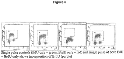
- FIG. 5 presents a series of graphs showing that when cells are treated with EdU and BrdU simultaneously, only a population of DNA labeled with BrdU is detected by flow cytometry.
- the first graph on the left shows a plot of EdU vs. DNA cell cycle only treated with a single pulse label of EdU.
- the second graph shows a plot of BrdU vs. DNA cell cycle only treated with a single pulse label of BrdU.
- the third and fourth graphs show the dual parameter plot of fluorescent nucleotide label vs. DNA cell cycle treated with EdU and BrdU simultaneously. Only BrdU was incorporated into cells treated simultaneously with EdU and BrdU, as shown in the third and fourth graphs.
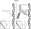
- FIG.6 presents a depiction of incorporation of nucleoside analogs into DNA.
- EdU is incorporated into the DNA double helix.
- the analog is easily accessible for labeling with the Alexa Fluor® azide without requiring a denaturation step.
- the right side of the depiction shows that denaturation is required for standard antibody-based labeling of incorporated BrdU.
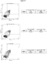
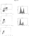
- FIG. 7 present a series of result graphs, labeled A through C, showing populations of cells (Ramos B-lymphocytes) treated with a first pulse label of EdU (20 ⁇ M) and a second pulse label of BrdU (10 ⁇ M) as detected by flow cytometry.
- FIG. 8 present a series of result graphs, labeled A through C, showing populations of cells (K562 human lymphoblast from chronic myelogenous leukemia cells) treated with a first pulse label of EdU (20 ⁇ M) and a second pulse label of BrdU (10 ⁇ M) as detected by flow cytometry.
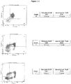
- FIGS. 9-1 through 9-3 present a series of result graphs, labeled A through I, showing populations of cells (TF-1a human erythroblast cells).
- the graphs A through G show the population of the cells treated with a first pulse label of EdU (20 ⁇ M) and a second pulse label of BrdU (10 ⁇ M) with the time of the pulses varied, as detected by flow cytometry.
- the graphs H and I show the population of the cells treated with one pulse only, with graph H showing the result of a pulse label of EdU (20 ⁇ M) only and graph I showing result of a pulse label of BrdU (10 ⁇ M) only, as detected by flow cytometry.
- FIG. 10 present a series of result graphs, labeled A through F, showing populations of cells (THP-1 monocyte cells) treated with a first pulse label of EdU (20 ⁇ M) and a second pulse label of BrdU (10 ⁇ M) as detected by flow cytometry.
- FIG. 11 shows the percentage of cells (Jurkat T-cell lymphocyte cells) which are EdU and BrdU co-positive (Q2), EdU and BrdU co-negative (Q3), BrdU positive and EdU negative (Q1), and BrdU negative and EdU positive (Q4) of the seven different treatment conditions.
- FIGS. 12-1 and 12-2 present a series of result graphs, labeled A through J, showing populations of cells (Jurkat T-cell lymphocyte cells) treated with a first pulse label of EdU (20 ⁇ M) and a second pulse label of BrdU (10 ⁇ M) as detected by flow cytometry.
- bio-orthoganal nucleoside or nucleotide analogs such that the newly synthesized cellular nucleic acid can be dual labeled without the need for a disruptive wash step.
- bio-orthoganal nucleoside or nucleotide analogs include, but are not limited to, halogenated (such as BrdU), an azido modified analog, an alkyne modified analog or a phosphine modified analog.
- the incorporation of these analogs is then detected by measurement of a fluorescent signal wherein the label is selectively attached to the nucleic acid analog due to the functional groups of the analog and label or through an antibody.
- a timed exposure to a nucleoside (or nucleotide) analog with the potential of incorporation of that analog into the actively synthesized cellular nucleic acid is defined as a pulse.
- the pulse may be measuring baseline proliferation, baseline gene expression, or a response to a specific treatment.
- an additional pulse with a different nucleoside analog which is selectively labeled, provides a mechanism to measure both baseline proliferation and a subsequent change in proliferation without the introduction of an artifact of washing or clearing the analog out of the cells or system being measured.
- a third pulse could be performed, for example, but not limited to, the ability to measure drug interaction on cell proliferation or gene expression.
- a baseline synthesis rate can be recorded by the first pulse labeling of the nucleic acid. Without interruption to remove the first pulse, the second pulse can be started. Normally, interruption to the cells by removing the first pulse label alters the rate of cell proliferation and makes assessment of changes in cell proliferations difficult.
- the no wash step makes the process compatible with high throughput screening (HTS). Having two compatible methods for pulse labeling nucleic acid without the use of radioactive nucleosides creates a very powerful tool which in one instance can be applied to the assessment of cancer therapy ex vivo or in vivo.
- alkyne reactive refers to a chemical moiety that selectively reacts with an alkyne modified group on the nucleoside analog to form a covalent chemical bond between the alkyne modified group and the alkyne reactive group.
- alkyne-reactive groups include azides.
- Alkyne-reactive can also refer to a molecule that contains a chemical moiety that selectively reacts with an alkyne group.
- azide reactive refers to a chemical moiety that selectively reacts with an azido modified group on another molecule to form a covalent chemical bond between the azido modified group and the azide reactive group.
- azide-reactive groups include alkynes and phosphines (e.g. triaryl phosphine).
- Azide-reactive can also refer to a molecule that contains a chemical moiety that selectively reacts with an azido group.
- the term "azide-selective phosphine dye” refers to a compound, molecule or reagent that comprises an engineered phosphine moiety with a dye label such that when reacted with an azide, provides for production of a covalent bond between the engineered phosphine moiety and the azide.
- engineered phosphine moiety refers to a moiety comprising a phosphine and an electrophilic moiety.
- an engineered phosphine moiety is 2-diphenylphosphanyl-benzoic acid methyl ester.
- Other engineered phosphine moieties are known in the art. See, e.g., Bertozzi et al., U.S. Pat. App. No. 20070037964 .
- bioorthogonal chemical reporter or “bioorthogonal labeling reagent” means a detectable label that comprises a chemical handle that will react selectively with the present nucleoside analog once incorporated into nucleic acid to form a covalent bond.
- cell in the context of the in vivo applications of the invention is meant to encompass eukaryotic and prokaryotic cells of any genus or species, with mammalian cells being of particular interest. "Cell” is also meant to encompass both normal cells and diseased cells, e.g., cancerous cells.
- cell proliferation and “cellular proliferation” are used herein interchangeably and refer to an expansion of a population of cells by the division of single cells into daughter cells, or to the division of a single cell to daughter cells.
- chemical handle or “bioorthogonal moiety” as used herein refers to a specific functional group, such as an azide, alkyne, activated alkyne, phosphite, phosphine, and the like.
- the chemical handle is distinct from biological reactive groups, defined below, in that the chemical handle are moieties that are rarely found in naturally-occurring biomolecules and are chemically inert towards biomolecules (e.g, native cellular components), but when reacted with an azide- or alkyne-reactive group the reaction can take place efficiently under biologically relevant conditions (e.g., cell culture conditions, such as in the absence of excess heat or harsh reactants).
- click chemistry refers to the copper-catalyzed version of a [3+2] cycloaddition reaction between a first reactive unsaturated group on the incorporated nucleoside analog (nucleotide analog) or labeling reagent and a second reactive unsaturated group present on the labeling regent or nucleoside analog (nucleotide analog).
- This click chemistry reaction is described by Sharpless et al. ( Sharpless et al., Angew Chem., Int. Ed. Engl., 2002, 41:1596-1599 ).
- the term "competitive nucleoside analog” refers to a nucleoside analog, which when added simultaneously to the first nucleoside analog in a cell or an organism, results in a population of cells that are preferentially labeled with the competitive nucleoside analog and not the first nucleoside analog.
- competitive nucleoside analog refers to a nucleoside analog, which when added simultaneously to the first nucleoside analog in a cell or an organism, results in a population of cells that are preferentially labeled with the competitive nucleoside analog and not the first nucleoside analog.
- BrdU competitive nucleoside analog
- EdU first nucleoside analog
- copper (I) catalyst refers to a compound, molecule or reagent that catalyzes the [3+2] cycloaddition reaction between a first reactive unsaturated group on the incorporated nucleoside analog (nucleotide analog) or labeling reagent and a second reactive unsaturated group present on the labeling reagent or nucleoside analog (nucleotide analog).
- copper (I) catalyst includes exogenous copper (I) as well as copper chelating moieties.
- copper chelating moieties refers to any compound, molecule or reagent characterized by the presence of two or more polar groups that can participate in the formation of a complex with copper (I) ions.
- the term "copperless click chemistry” refers to a strain-promoted [3+2] cycloaddition reaction that can be carried out under physiological conditions, as described by Bertozzi et al. US Publication No. 20060110782 ; Baskin et al. PNAS 2007 Oct 23;104(43):16793-7 ; Agard et al. J Am Chem Soc. 2004 Nov 24;126(46):15046-7 .
- the reaction is accomplished through use of a first molecule comprising a strained cycloalkyne moiety (typically the label), and second molecule comprising an azide moiety (typically the nucleoside analog).
- the azide moiety on the second molecule reacts, in the absence of a catalyst, with the strained cycloalkyne moiety on the first molecule, forming a final conjugate product comprising fused azide/cycloalkyne ring.
- the term "detectable response” refers to an occurrence of or a change in, a signal that is directly or indirectly detectable either by observation or by instrumentation.
- the detectable response is an optical response resulting in a change in the wavelength distribution patterns or intensity of absorbance or fluorescence or a change in light scatter, fluorescence lifetime, fluorescence polarization, or a combination of the above parameters.
- die refers to a compound that emits light to produce an observable detectable signal.
- the terms "dye-labeled azide” and "azide-dye molecule” refer to a compound or molecule with a reactive azide group that is also labeled with a dye. Examples include, but are not limited to: rhodamine-azide, Alexa Fluor® 350-azide (Molecular ProbesTM/InvitrogenTM, Carlsbad, CA), Alexa Fluor® 488-azide (Molecular ProbesTM/InvitrogenTM, Carlsbad, CA), Alexa Fluor® 555-azide (Molecular ProbesTM/InvitrogenTM, Carlsbad, CA), Alexa Fluor® 568-azide (Molecular ProbesTM/InvitrogenTM, Carlsbad, CA), Alexa Fluor® 568-azide (Molecular ProbesTM/InvitrogenTM, Carlsbad, CA), Alexa Fluor® 594-azide, Alexa Fluor® 633-azide (Molecular ProbesTM/InvitrogenTM,
- cycloalkyne refers to a cycloalkyne that has been further modified to include a dye label.
- cycloalkyne refers to compounds or molecules which may be used in strained [3+2] cycloaddition reactions in order to pulse label DNA.
- examples of cycloalkynes include, but are not limited to: cyclooctynes, difluorocyclooctynes, heterocycloalkynes, dichlorocyclooctynes, dibromocyclooctynes, or diiodocyclooctynes.
- die-labeled alkyne refers to an alkyne that has been further modified to include a dye label.
- the term "dual labeling” refers to a labeling process in which a nucleic acid polymer is labeled with two detectable agents that produce distinguishable signals. The nucleic acid polymer resulting from such a labeling process is said to be dually labeled.
- an effective amount refers to the amount of a substance, compound, molecule, agent or composition that elicits the relevant response in a cell, a tissue, or an organism.
- an effective amount is an amount of nucleoside that is incorporated into the DNA of the cells.
- fluorophore or “fluorogenic” refers to a composition that demonstrates a change in fluorescence upon binding to a biological compound or analyte interest.
- Preferred fluorophores of the present invention include fluorescent dyes having a high quantum yield in aqueous media.
- Exemplary fluorophores include xanthene, indole, borapolyazaindacene, furan, and benzofuran, cyanine among others.
- the fluorophores of the present invention may be substituted to alter the solubility, spectral properties or physical properties of the fluorophore.
- label refers to a chemical moiety or protein that retains it's native properties (e.g. spectral properties, conformation and activity) when part of a labeling reagent of the present invention and used in the present methods.
- Illustrative reporter molecules can be directly detectable (fluorophore) or indirectly detectable (hapten or enzyme).
- reporter molecules include, but are not limited to, radio reporter molecules that can be measured with radiation-counting devices; pigments, dyes or other chromogens that can be visually observed or measured with a spectrophotometer; spin labels that can be measured with a spin label analyzer; and fluorescent moieties, where the output signal is generated by the excitation of a suitable molecular adduct and that can be visualized by excitation with light that is absorbed by the dye or can be measured with standard fluorometers or imaging systems, for example.
- the reporter molecule can be a luminescent substance such as a phosphor or fluorogen; a bioluminescent substance; a chemiluminescent substance, where the output signal is generated by chemical modification of the signal compound; a metal-containing substance; or an enzyme, where there occurs an enzyme-dependent secondary generation of signal, such as the formation of a colored product from a colorless substrate.
- the reporter molecule may also take the form of a chemical or biochemical, or an inert particle, including but not limited to colloidal gold, microspheres, quantum dots, or inorganic crystals such as nanocrystals or phosphors (see, e.g., Beverloo, et al., Anal. Biochem. 203, 326-34 (1992 )).
- reporter molecule can also refer to a "tag" or hapten that can bind selectively to a labeled molecule such that the labeled molecule, when added subsequently, is used to generate a detectable signal.
- a tag or hapten that can bind selectively to a labeled molecule such that the labeled molecule, when added subsequently, is used to generate a detectable signal.
- HRP horseradish peroxidase
- the tag can be a hapten or antigen (e.g., digoxigenin), and an enzymatically, fluorescently, or radioactively labeled antibody can be used to bind to the tag.
- reporter molecules include, but are not limited to, particles, fluorescent dyes, haptens, enzymes and their chromogenic, fluorogenic, and chemiluminescent substrates, and other reporter molecules that are described in the MOLECULAR PROBES HANDBOOK OF FLUORESCENT PROBES AND RESEARCH CHEMICALS by Richard P. Haugland, 10th Ed., (2005 ), the contents of which are incorporated by reference, and in other published sources.
- a reporter molecule is not an amino acid.
- the term "Labeling Reagent” refers to a reagent used to label and detect the incorporated nucleotide analog.
- the labeling reagent comprises a label and a chemical handle.
- the labeling reagent comprises an antibody and a label, wherein the antibody binds the nucleoside analog.
- nucleoside analog and “nucleotide analog” are used interchangeably and refers to a molecule or compound that is structurally similar to a natural nucleoside or nucleotide that is incorporated into newly synthesized nucleic acid.
- nucleosides once inside the cells, they are phosphorylated into nucleotides and then incorporated into nascent nucleic acid polymers. Nucleotides are difficult to get across the cell membrane due to their charges and are more labile than nucleosides, thus their use typically requires and additional step and reagents for transfection to transport the nucleotides across the lipid bilayer.
- nucleoside analogs are incorporated into nucleic acid (DNA or RNA) in a similar manner as a natural nucleotide wherein the correct polymerase enzyme recognizes the analogs as natural nucleotides and there is no disruption in synthesis.
- These analogs comprise a number of different moieties which are ultimately used for detection, such as halogenated analogs (bromo, chloro, iodo, etc.) and those that comprise a bioorthogonal moiety such as azido, alkyne or phosphine.
- reactive group refers to a group that is capable of reacting with another chemical group to form a covalent bond, i.e. is covalently reactive under suitable reaction conditions, and generally represents a point of attachment for another substance.
- reactive groups refer to chemical moieties generally found in biological systems and that react under normal biological conditions, these are herein distinguished from the chemical handle or bioorthogonal functional moiety, defined above, such as the azido and activated alkyne moieties of the present invention.
- the reactive group is a moiety, such as carboxylic acid or succinimidyl ester, that is capable of chemically reacting with a functional group on a different compound to form a covalent linkage.
- Reactive groups generally include nucleophiles, electrophiles and photoactivatable groups.
- Staudinger ligation refers to a chemical reaction developed by Saxon and Bertozzi ( E. Saxon and C. Bertozzi, Science, 2000, 287: 2007-2010 ) that is a modification of the classical Staudinger reaction.
- the classical Staudinger reaction is a chemical reaction in which the combination of an azide with a phosphine or phosphite produces an aza-ylide intermediate, which upon hydrolysis yields a phosphine oxide and an amine.
- a Staudinger reaction is a mild method of reducing an azide to an amine; and triphenylphosphine is commonly used as the reducing agent.
- Staudinger ligation an electrophilic trap (usually a methyl ester) is appropriately placed on a triarylphosphine aryl group (usually ortho to the phosphorus atom) and reacted with the azide, to yield an aza-ylide intermediate, which rearranges in aqueous media to produce a compound with amide group and a phosphine oxide function.
- the Staudinger ligation is so named because it ligates (attaches/covalently links) the two starting molecules together, whereas in the classical Staudinger reaction, the two products are not covalently linked after hydrolysis.
- test compound and “test treatment” refer to any substance, compound, molecule, agent, composition, or treatment, which is tested during the claimed methods for its affect on cellular proliferation or the cell cycle.
- the affect on cellular proliferation of these "test compounds” and “test treatments” is not limited by outcome, that is, they may increase, decrease or not affect cellular proliferation or the cell cycle.
- reactive partner refers to a molecule or molecular moiety that specifically reacts with another reactive partner, such as the present nucleoside analog and the reporter molecule.
- nucleoside analogs that are "fed” to cells and incorporated into a growing nucleic acid polymer, wherein at least one of the nucleoside analogs comprises a bioorthogonal functional moiety.
- a method for screening test compounds for their effect on cellular proliferation is provided.
- a method for dual labeling wherein the baseline proliferation is measured against a change in proliferation from a treatment of the cells, either in vivo or ex vivo. This is accomplished by using a combination of a halogenated analogs (such as BrdU) or nucleoside analogs comprising a chemical handle in combination with nucleoside analogs comprising a chemical handle, which can be selectively labeled using a labeling reagent of the present invention.
- the present invention uses at least one nucleoside (or nucleotide) analog that comprises a chemical handle, herein also referred to as a bioorthogonal functional moiety.
- this dual labeling method can also be used to measure cellular gene expression (RNA synthesis) in response to an administered treatment.
- the nucleic acid analogs comprise a ribose sugar and are RNA nucleoside or nucleotide analogs.
- the advantage of this dual labeling method is that it does not require the removal of the first pulse of nucleoside analog from the culture medium, or animal prior to the administering of the second pulse of nucleoside analog.
- the distinct advantage of this 'no wash' treatment for the additional pulse of the second label is that baseline cell proliferation, or gene expression, measurements can be made using the first pulsed nucleoside analog, followed by the administration of a specific course of treatment or testing which may cause an intended alteration of cell proliferation, or gene expression. For example, as in the case of screening cancer therapeutic drugs by the addition of the drug to the culture medium system, or to the animal being tested. This treatment would be simultaneous to or followed by a pulse from the second nucleoside analog, without an interruption in the course of the treatment for the removal of the first analog. At the end of the test, the cells are prepared for detection of the two pulses of labeling of the nucleic acid.
- a nucleoside analog comprising a bioorthogonal functional moiety is used in the first pulse followed by a second pulse with a halogenated nucleoside analog.
- a nucleoside analog comprising a first bioorthogonal functional moiety is used in the first pulse followed by a second pulse with a nucleoside analog comprising a second bioorthogonal functional moiety.
- nucleoside analogs that comprise a bioorthogonal functional moiety are detected using reagents comprising a complimentary bioorthogonal functional moiety and a label, resulting in covalent attachment of the labeling reagent to the nucleoside analog.
- This dual labeling method for measuring cell proliferation is distinguished from known methods because 1) no wash treatment is required between the two pulses and 2) the compatibility of the two pulse labeling conditions for detection by two color, a first and second label, fluorescence measurement, wherein one of the nucleoside analogs comprises a bioorthogonal functional moiety.
- the first and second labels are preferably selected such that they produce distinguishable detectable signals, in other words the labels can be excited at the same or at different wavelengths, but the emission is at different wavelengths.
- the first pulsed nucleoside, 5-ethynyl 2'-deoxyuridine also termed herein ethynyluracil or EdU
- EdU ethynyluracil
- nucleoside and nucleotide analogs can be used in the present methods for measuring nascent nucleic acid synthesis.
- Nucleosides are typically used in experiments wherein the analogs are added to cell culture or administered to animals because the nucleoside analogs are easily taken up by live cells, wherein they are phosphorylated into a nucleotide and then incorporated into a growing nucleic acid polymer.
- nucleotides are labile and prone to enzyme cleave, either before or after incorporation into cells, and are generally less stable than nucleosides.
- nucleosides As the analog that is added to cells or animals, however this in no way is intended to be limiting, wherein nucleotides are equally as important.
- the nucleoside analogs can be an analog for any of the four DNA bases (adenine (A), cytosine (C), guanine (G) or thymine (T)) or any of the four RNA bases (adenine (A), cytosine (C), guanine (G) or uracil (U)) and include their triphosphate and phosphoramidite forms, wherein these analogs are incorporated into newly synthesized nucleic acid by polymerase present in the cells.
- the nucleosides are modified into analogs wherein they comprise a moiety that is ultimately used for detection of that nucleoside and the resulting nascent nucleic acid polymer synthesized in the presence of the nucleoside analogs.
- nucleoside analog is a halogenated analog, including but not limited to a bromo, chloro, and iodo moiety.
- nucleoside analog comprises a chemical handle or bioorthogonal functional moiety, including but not limited to an azido, alkyne and phosphine moiety.
- Halogenated analogs are well known in the art, e.g.
- BrdU, IdU, CldU and BrUTP can be purchased from a number of vendors (Sigma, Saint Louis, MO; Millipore, Billerica, MA; Anaspec, San Jose, CA; Invitrogen, Carlsbad, CA) Similarly the antibodies used to detect these analogs are also commercially available (Dako, Carpinteria, CA; BD Bioscience, San Diego, CA; EMD Biosciences, Madison, WI).
- nucleoside analogs that comprise a bioorthogonal functional moiety which are suitable for use in the methods described herein include any nucleoside analogue, as defined herein, that contains a reactive bioorthogonal moiety, or chemical handle, that can undergo a [3+2] cycloaddition or Staudinger ligation.
- the reactive bioorthogonal moiety is carried by the base of the nucleoside.
- the base carrying the reactive bioorthogonal moiety can be a purine (e.g., adenine or guanine) or a pyrimidine (e.g., cytosine, uracil or thymine).
- the base is uracil; in some such embodiments, uracil carries the reactive bioorthogonal moiety on the 5-position.
- the base is adenine; in some such embodiments, adenine carries the reactive bioorthogonal moiety.
- the bioorthogonal moiety is indirectly attached to the base, while in other embodiments the bioorthogonal moiety is directly covalently attached to the base.
- Non-limiting examples of the nucleoside analogues that may be used in the methods described herein include ethynyluracil or EdU and 5-azido-2'-deoxyuracil (also termed herein azidouracil or AzdU) as well as their triphosphate and phosphoramidite forms.
- EdU can be synthesized essentially as described by C.-S. Yu and F. Oberdorfer, Synlett, 2000, 1: 86-88 ; and AzdU can be synthesized using a method similar to that described in P. Sunthankar et al., Anal. Biochem., 1998, 258: 195-201 to synthesize azido-dUMP.
- EdU is also commercially available from Berry and Associates, Inc.
- the reactive bioorthogonal moiety is carried by the sugar (ribose and deoxyribose) of the nucleoside.
- the bioorthogonal moiety is indirectly attached to the sugar, while in other embodiments the bioorthogonal moiety is directly and covalently attached to the sugar.
- the sugar carrying the reactive bioorthogonal moiety can be covalently attached to a purine (e.g ., adenine or guanine) or a pyrimidine (e.g ., cytosine, uracil or thymine).
- the base is uracil, while in other embodiments the base is adenine.
- nucleoside analogues that may be used in the methods described herein include EdU, AzdU, or chain terminating dideoxy compounds such as AZT; 3'-Azido-2',3'-dideoxyadenosine, 3'-Azido-3'-deoxythymidine (AZT), 5'-Azido-5'-deoxythymidine, 5-(1-ethynyl)-2'-O-methyluridine, 5-(1-propynyl)-2'-deoxyuridine, 5-(propargyloxy)-2'-deoxyuridine, 8-Azido-2'-deoxyadenosine, 3'-Azido-2',3'-dideoxyadenosine.
- the reactive bioorthogonal moiety or chemical handle can be a 1,3-dipole such as a nitrile oxide, an azide, a diazomethane, a nitrone or a nitrile imine.
- the 1,3-dipole is an azide.
- the reactive bioorthogonal functional moiety can be a dipolarophile such as an alkene ( e.g ., vinyl, propylenyl, and the like) or an alkyne ( e.g ., ethynyl, propynyl, and the like).
- the dipolarophile is an alkyne, such as, for example, an ethynyl group.
- bioorthogonal functional moieties described above are non-native, non-perturbing bioorthogonal chemical moieties that possess unique chemical functionality that can be modified through highly selective reactions.
- these incorporated nucleosides are labeled using labeling reagents which comprise a chemical handle that will selectively form a covalent bond with the nucleoside in the presence of the cellular milieu.
- the labeling reagent is a first antibody, which may be conjugated to a label or bound by a second antibody that is covalently attached to a label, wherein the first antibody binds to the incorporated nucleoside.
- this is an anti-BrdU antibody.
- the antibody is an anti-IdU or anti-CldU antibody.
- other antibodies which could selectively bind to incorporated nucleoside analogs are also envisioned.
- the labeling reagent comprises a label and a chemical handle.
- a chemical handle as defined above, is a bioorthogonal functional moiety that selectively reacts with a functional moiety to form a covalent bond.
- a label As already mentioned above, the role of a label is to allow visualization or detection of a nucleic acid polymer, e.g., DNA in a cell, following labeling.
- a label or detectable agent or moiety is selected such that it generates a signal which can be measured and whose intensity is related (e.g., proportional) to the amount of labeled nucleic acid polymer, e.g., in a sample being analyzed.
- a label used in a labeling reagent in the methods and compositions described herein is any chemical moiety, organic or inorganic, that exhibits an absorption maximum at wavelengths greater than 280 nm, and retains its spectral properties when covalently attached to a modified nucleoside such as, by way of example only, an azide, and alkyne or a phosphine.
- Fluorophores used in the labeling reagent in the methods and compositions described herein include, without limitation; a pyrene (including any of the corresponding derivative compounds disclosed in US Patent 5,132,432 ), an anthracene, a naphthalene, an acridine, a stilbene, an indole or benzindole, an oxazole or benzoxazole, a thiazole or benzothiazole, a 4-amino-7-nitrobenz-2-oxa-1, 3-diazole (NBD), a cyanine (including any corresponding compounds in US Serial Nos.
- oxazines include resorufins (including any corresponding compounds disclosed in 5,242,805 ), aminooxazinones, diaminooxazines, and their benzo-substituted analogs.
- Xanthene type fluorophores used in labeling reagents in the methods and compositions described herein include, but are not limited to, a fluorescein, a rhodol (including any corresponding compounds disclosed in US Patent Nos. 5,227,487 and 5,442,045 ), or a rhodamine (including any corresponding compounds in US Patent Nos. 5,798,276 ; 5,846,737 ; US serial no. 09/129,015 ).
- fluorescein includes benzo- or dibenzofluoresceins, seminaphthofluoresceins, or naphthofluoresceins.
- rhodol includes seminaphthorhodafluors (including any corresponding compounds disclosed in U.S. Patent No. 4,945,171 ).
- the fluorophore is a xanthene that is bound via a linkage that is a single covalent bond at the 9-position of the xanthene.
- the xanthenes include derivatives of 3 H- xanthen-6-ol-3-one attached at the 9-position, derivatives of 6-amino-3 H -xanthen-3-one attached at the 9-position, or derivatives of 6-amino-3 H -xanthen-3-imine attached at the 9-position.
- the fluorophores used in the labeling reagent in the methods and compositions described herein include xanthene (rhodol, rhodamine, fluorescein and derivatives thereof) coumarin, cyanine, pyrene, oxazine and borapolyazaindacene.
- fluorophores are sulfonated xanthenes, fluorinated xanthenes, sulfonated coumarins, fluorinated coumarins and sulfonated cyanines.
- the choice of the fluorophore attached for the labeling reagent will determine the absorption and fluorescence emission properties of the modified nucleic acid.
- Physical properties of a fluorophore label that can be used for detection of modified nucleic acids include, but are not limited to, spectral characteristics (absorption, emission and stokes shift), fluorescence intensity, lifetime, polarization and photo-bleaching rate, or combination thereof. All of these physical properties can be used to distinguish one fluorophore from another, and thereby allow for multiplexed analysis.
- the fluorophore has an absorption maximum at wavelengths greater than 480 nm.
- the fluorophore absorbs at or near 488 nm to 514 nm (particularly suitable for excitation by the output of the argon-ion laser excitation source) or near 546 nm (particularly suitable for excitation by a mercury arc lamp).
- a fluorophore can emit in the NIR (near infra red region) for tissue or whole organism applications.
- Other desirable properties of the fluorescent labeling reagent may include cell permeability and low toxicity, for example if labeling of the nucleic acid polymer is to be performed in a cell or an organism (e.g., a living animal).
- the labeling reagent comprises a tandem dye or FRET dye pair. This is particularly useful to obtain a set of labeling reagents that are excited at the same wavelength but which have distinct emission spectra. Two dyes function as a FRET pair when they are within close proximity, usually covalently attached, so that the energy from the excited first dye (the donor) is transferred the second dye (the acceptor) where the energy is then emitted at a longer wavelength than would have been possible by excitation only of the first dye.
- a particularly useful combination is the FRET dye pairs disclosed in US Patent Nos. 7169939 ; 6849745 ; 5945526 ; 5863727 ; 5800996 ; and 6967250 .
- the labeling reagent comprises a fluorescent nanocrystal.
- a matched set of labeling reagents For each dual labeling experiment a matched set of labeling reagents must be selected based on the instrument being used and so that the dyes can be appropriately excited and measured at the correct wavelengths to distinguish between the baseline nucleic acid synthesis and the subsequent change in nucleic acid synthesis due to treatment.
- Matched pairs of fluorescent labeling dyes typically produce signals that are spectrally distinguishable.
- the fluorescent dyes in a matched pair do not significantly absorb light in the same spectral range (i.e., they exhibit different absorption maxima wavelengths) and can be excited (for example, sequentially) using two different wavelengths.
- the fluorescent dyes in a matched pair may emit light in different spectral ranges (i.e., they produce a dual-color fluorescence upon excitation). Pairs of fluorescent labels are known in the art (see, for example, R. P. Haugland, " Molecular Probes: Handbook of Fluorescent Probes and Research Chemicals, supra).
- a particular set of labels will depend on the purpose of the labeling to be performed and will be governed by several factors, such as the ease and cost of the labeling method, the quality of sample labeling desired, the effects of the detectable moiety on the cell or organism, the nature of the detection system, the nature and intensity of the signal generated by the detectable moiety, and the like.
- one label is selected for Reagent A and the second label is selected for Reagent B.
- the above dual labeling components are used in the present methods to measure cellular nascent nucleic acid synthesis by dual pulse labeling of the cellular nucleic acid.
- the first pulse labeling of nucleic acid with a nucleoside analog allows establishment of a baseline rate of nucleic acid synthesis.
- Pulse labeling of nucleic acid with an additional second nucleoside analog then allows measurement of any changes to the nucleic acid synthesis. This method does not require a potentially artifact-inducing intermediary wash step between pulse labels.
- the nucleic acid synthesis can be measured as cell proliferation, in the case of DNA, or in gene expression, in the case of RNA.
- this method may be used to screen compounds for their effect on cellular proliferation or gene expression by treating cells or an organism with the test compound simultaneous to or before treatment with a second nucleoside analog.
- a method for measuring a change in cellular nucleic acid synthesis comprising:
- a method for measuring a change in cellular DNA synthesis which can be measured as cell proliferation.
- a method for measuring a change in cellular RNA synthesis which can be measure as gene expression.
- a method for screening test compounds for their effect on cellular proliferation may include measuring cellular proliferation changes in a patient during the course of treatment for a disease with a specific compound.
- Cancer cells could be removed from a patient and grown in culture.
- a baseline DNA synthesis rate can be determined with the first pulse, then a drug is added along with the second nucleoside analog and the change of DNA synthesis rate determined, which in the case of drug resistance/sensitivity in cancer cells would easily be determined.
- Screens for compounds which either stimulate or block DNA synthesis at various places in the cell cycle could be greatly improved by the addition having an accurate baseline synthesis measurement which does not alter the state of the cell proliferation.
- breast cancer cells may be removed from a patient and grown in culture.
- the baseline cellular proliferation rate may be established by adding a first pulse of EdU.
- the cells may be treated with a chemotherapy drug, for example tamoxifen, and treated with a second pulse label of BrdU.
- the cellular proliferation rate in response to tamoxifen is then measured by comparing incorporation of EdU to BrdU.
- This process may be repeated over the course of the breast cancer patient's treatment to ensure that the patient's cancer cells remain responsive to the chosen chemotherapeutic agent, in this case, tamoxifen.
- the clinician would be looking for a decrease in cellular proliferation upon treatment with the chemotherapy drug.
- neural diseases including neurodegenerative diseases, for example, Huntington's disease ( Curtis et al., Increased Cell Proliferation and Neurogenesis in the Adult Human Huntington's disease brain, 100 (15) PNAS 9023-9027 (July 22, 2003 )), or HIV-associated dementia ( Okamoto et al., HIV/gp120 Decreases Adult Neural Progenitor Cell Proliferation via Checkpoint Kinase-Mediated Cell-Cycle Withdrawal and G1 Arrest, 1 Cell Stem Cell 230-236 (Aug.
- hyperplasia diseases including, psoriasis, seborrehea, eczema, benign prostate hyperplasia, congenital adrenal hyperplasia, endometrial hyperplasia, squamous cell hyperplasia, sebaceous hyperplasia, Crohn's disease, carcinoma, sarcoma, glioma and lymphoma (see U.S. Pat. No. 7,256,034 (filed January 5, 2004 )).
- the present invention concerns a method for identifying new compounds, which may be termed as "test compounds", which have a desired effect on cellular proliferation. Depending on the application, this desired effect may be to stimulate, to inhibit, or to not affect cellular proliferation.
- test compounds isolated from natural sources such as plants, animals or even sources such as marine, forest or soil samples, may be assayed for the presence of potentially useful pharmaceutical agents.
- test compounds to be screened could also be derived from chemical compositions or man-made compounds.
- the test compounds may also include proteins and peptides, such as those derived from recombinant DNA technology or by other means, including peptide synthesis.
- the test compounds may also be antibodies, including polyclonal and monoclonal antibodies.
- the test compounds may also include fragments or parts of naturally-occurring compounds or may be only found as active combinations of known compounds which are otherwise inactive.
- test compounds may also include nucleic acids, including, but not limited to: DNA; ribonucleic acid (RNA); small interfering RNAs (siRNA); and single-stranded nucleic acids, more particularly, those designed to form in vivo triplexes.
- nucleic acids including, but not limited to: DNA; ribonucleic acid (RNA); small interfering RNAs (siRNA); and single-stranded nucleic acids, more particularly, those designed to form in vivo triplexes.
- the present invention may be used to measure cellular proliferation following treatment with compounds known to affect cellular proliferation. See, e.g., Nicholas R. Cozzarelli, The Mechanism of Action of Inhibitors of DNA Synthesis, 46 ANN. REV. BIOCHEM. 641-648 (1977 ).
- the test compound is administered to an organism.
- Organisms to which the claimed methods may be applied include, but are not limited to: humans, mice, rats, horses, cows, sheep, rabbits, dogs, or cats.
- Test compounds and nucleoside analogs may be administered to organisms by a variety of methods known to persons of skill in the art. These include oral administration (e.g. ingestion of a pill, food or liquid containing the test compound and/or nucleoside analogs), or by subcutaneous, intravenous or intraperitoneal administration (e.g. by injection or topical application).
- the test compound may also be administered parenterally, intraspinally, or intracerebrally.
- the present invention provides for measuring cellular proliferation in cells, rather than an organism.
- the nucleoside analogs may be added to the medium in which the cells are grown.
- the method includes screening test compounds for their effect on cellular proliferation, the test compound may be added to the medium in which the cells are grown.
- test compounds effect on cell proliferation is also intended to relate to their effect on gene expression wherein the change in RNA synthesis is measure.
- cellular proliferation is measured in a cell by treating the cell with an effective amount of EdU, followed by treatment with an effective amount of BrdU.
- EdU pulse label is detected using click chemistry reagents, including copper sulfate (CuSO 4 ) and a dye-labeled azide (e.g. Alexa Fluor® 488-azide (Molecular ProbesTM/InvitrogenTM, Carlsbad, CA)).
- the BrdU pulse label is detected using standard antibody-based methods. See, e.g., Jonathon Pines et al., Assays for CDK Activity and DNA Replication in the Cell Cycle, CURRENT PROTOCOLS IN CELL BIOLOGY (Juan S. Bonifacino et al. eds., John Wiley & Sons, Inc. 2003) (1998 ). Both labels are then measured using flow cytometry.
- this dual pulse labeling method may be applied to the assessment of cancer therapy ex vivo or in vivo.
- Cancer cells may be removed from a patient and grown in culture.
- a baseline DNA synthesis rate may be determined with the first pulse, then a drug is added along with the second nucleoside analog and the change of DNA synthesis rate determined. Having an accurate baseline synthesis measurement which does not alter the state of the cellular proliferation will greatly improve screening methods for compounds that either stimulate or block DNA synthesis.
- the method for measuring cellular proliferation is performed on a neural cell.
- This dual pulse labeling method may be applied to the assessment of treatment for nervous system diseases. For instance, changes to cellular proliferation rates of neural cells upon treatment with a test compound may be measured by contacting neural cells with a first pulse of a nucleoside analog; treating with a test compound; contacting the neural cells with a competitive nucleoside analog simultaneous to or after treatment with the test compound; detecting incorporation of the first nucleoside analog and detecting incorporation of the competitive nucleoside analog.
- this dual pulse labeling method may be used to identify compounds that either stimulate or inhibit neural cell proliferation.
- compounds are screened for their effects on cellular proliferation using a method comprising the steps of: treating a cell with a first nucleoside analog; treating the cell with a test compound; treating the cell with at least one competitive nucleoside analog simultaneous to or after treating the cell with the test compound; detecting incorporation of the first nucleoside analog; and detecting incorporation of the second nucleoside analog.
- cellular proliferation in an organism is measured by: treating an organism with a first nucleoside analog; treating the organism with at least one second nucleoside analog; detecting incorporation of the first nucleoside analog; and detecting incorporation of the second nucleoside analog.
- cellular proliferation in an organism is measured by: treating an organism with a first nucleoside analog; treating the organism with a test compound; treating the organism with at least one competitive nucleoside analog simultaneous to or after treating the cells with the test compound; detecting incorporation of the first nucleoside analog; and detecting incorporation of the second nucleoside analog.
- the compounds of the invention may, at any time after or during an assay, be illuminated with a wavelength of light that results in a detectable optical response, and observed with a means for detecting the optical response.
- a wavelength of light that results in a detectable optical response
- the fluorescent compounds Upon illumination, such as by an violet or visible wavelength emission lamp, an arc lamp, a laser, or even sunlight or ordinary room light, the fluorescent compounds display intense visible absorption as well as fluorescence emission.
- Selected equipment that is useful for illuminating the fluorescent compounds of the invention includes, but is not limited to, hand-held ultraviolet lamps, mercury arc lamps, xenon lamps, argon lasers, laser diodes, and YAG lasers.
- illumination sources are optionally integrated into laser scanners, flow cytometer, fluorescence microplate readers, standard or mini fluorometers, or chromatographic detectors.
- This fluorescence emission is optionally detected by visual inspection, or by use of any of the following devices: CCD cameras, video cameras, photographic film, laser scanning devices, fluorometers, photodiodes, quantum counters, epifluorescence microscopes, scanning microscopes, flow cytometers, fluorescence microplate readers, or by means for amplifying the signal such as photomultiplier tubes.
- the instrument is optionally used to distinguish and discriminate between the first pulse labeling reagent of the invention and a second labeling reagent detectably different optical properties, typically by distinguishing the fluorescence response of the first fluorescent compounds of the invention from that of the second fluorophore.
- examination of the sample optionally includes isolation of particles within the sample based on the fluorescence response by using a sorting device.
- kits that includes at least one nucleoside analog and labeling reagent of the invention.
- the kit will generally also include instructions for using the nucleoside analog and labeling reagent in one or more methods, typically for measuring a change in cellular nucleic acid synthesis.
- the kit includes a first nucleoside or nucleotide analog, at least one second nucleoside or nucleotide analog, wherein in at least the first analog or the at least one second nucleoside or nucleotide analog contains a bioorthogonal functional moiety, a first labeling reagent and a second labeling reagent.
- Additional kit components include buffers, other detection reagents and standards.
- a standard method of preparing cultured cells for the measurement of newly synthesized DNA is set up according to known conditions for cells to be actively growing by providing the proper media and nutrient requirements.
- Jurkat cell cultures were diluted one to four to a density of 2 x 10 5 cells/ml. After these cells had been growing for two or three days, the first nucleoside analog, EdU, was added at 20 ⁇ M, a concentration appropriate for incorporation in the DNA of cells undergoing DNA synthesis. The cells were grown in the presence of EdU for thirty minutes.
- labeling of the EdU was performed by adding click chemistry based reagents, including a solution comprised of CuSO 4 in Tris-buffered saline and Alexa Fluor® 488-azide (495 nm excitation maxima/ 519 nm emission maxima) (Molecular Probes®/InvitrogenTM, Carlsbad, CA). The cells were then washed with 0.1% Triton®X-100/1%BSA/PBS. After this, labeling of the BrdU was performed using the anti-BrdU antibody Alexa Fluor® 647 conjugate (650 nm excitation maxima/ 670 nm emission maxima) (Molecular Probes®/InvitrogenTM, Carlsbad, CA).
- a nucleic acid dye SYTOX® Blue stain (444 nm excitation maxima/ 480 nm emission maxima) (Molecular Probes®/InvitrogenTM, Carlsbad, CA) was added with RNase (InvitrogenTM, Carlsbad, CA). Detection of the three labels was performed by flow cytometry.
- SYTOX® Blue stain 444 nm excitation maxima/ 480 nm emission maxima
- RNase InvitrogenTM, Carlsbad, CA
- Detection of the three labels was performed by flow cytometry.
- To detect the EdU label 488 nm excitation was used, with a 530/30 nm bandpass.
- To detect the BrdU label 633 nm excitation was used, with a 660/20 nm bandpass.
- DNA content 405 nm excitation was used, with a 450/50 nm bandpass.
- a standard method of preparing cultured cells for the measurement of newly synthesized DNA is set up according to known conditions for cells to be actively growing by providing the proper media and nutrient requirements.
- Jurkat cell cultures were diluted one to four to a density of 2 x 10 5 cells/ml. After these cells have been growing for two or three days the first nucleoside analog, EdU, was added at 20 ⁇ M, a concentration appropriate for incorporation in the DNA of cells undergoing DNA synthesis. The cells were grown in the presence of EdU for one hour.
- EdU labeling of the EdU was performed by adding click chemistry based reagents, including a solution comprised of CuSO 4 in Tris-buffered saline and Alexa Fluor® 488-azide (495 nm excitation maxima/ 519 nm emission maxima) (Molecular Probes®/InvitrogenTM, Carlsbad, CA). The cells were then washed with 0.1% Triton®X-100/1%BSA/PBS.
- FIGS. 3 & 4 There was also a population of cells which only had the label from the second pulse ( FIGS. 3 & 4 ). These cells were not actively replicating their DNA during the time of the first pulse but entered into the DNA synthesis phase during the time of the second pulse. Treatments that cause increases or decreases in the rate of synthesis can easily be seen and quantitated from the patterns of the graphs of the cell populations collected by flow cytometry.
- EdU labeling of the EdU was performed by adding click chemistry based reagents, including a solution comprised of CuSO 4 in Tris-buffered saline and Alexa Fluor® 488 conjugate (495 nm excitation maxima/ 519 nm emission maxima) (Molecular Probes®/InvitrogenTM, Carlsbad, CA). The cells were then washed with 0.1%Triton® X-100/1%BSA/PBS.
- click chemistry based reagents including a solution comprised of CuSO 4 in Tris-buffered saline and Alexa Fluor® 488 conjugate (495 nm excitation maxima/ 519 nm emission maxima) (Molecular Probes®/InvitrogenTM, Carlsbad, CA).
- Alexa Fluor® 488 conjugate 495 nm excitation maxima/ 519 nm emission maxima
- a standard method of preparing cultured cells for the measurement of newly synthesized DNA is set up according to known conditions for cells to be actively growing by providing the proper media and nutrient requirements.
- Jurkat cell cultures were diluted one to four to a density of 2 x 10 5 cells/ml. After these cells have been growing for two or three days the first nucleoside analog, EdU, was added at 20 ⁇ M, a concentration appropriate for incorporation in the DNA of cells undergoing DNA synthesis. The cells were grown in the presence of EdU for one hour.
- Labeling of the BrdU was performed using the anti-BrdU antibody Alexa Fluor® 647 conjugate (650 nm excitation maxima/ 670 nm emission maxima) (Molecular ProbesTM/InvitrogenTM, Carlsbad, CA). The cells were then washed with 0.1%TritonX/1%BSA/PBS. After this labeling of the EdU was performed by adding click chemistry based reagents, including a solution comprised of CuSO 4 in Tris-buffered saline and Alexa Fluor® 488-azide (495 nm excitation maxima/ 519 nm emission maxima) (Molecular ProbesTM/InvitrogenTM, Carlsbad, CA).
- a nucleic acid dye SYTOX® Blue stain (444 nm excitation maxima/ 480 nm emission maxima) (Molecular ProbesTM/InvitrogenTM, Carlsbad, CA) was added with RNase (InvitrogenTM, Carlsbad, CA). Detection of the three labels was performed by flow cytometry.
- SYTOX® Blue stain 444 nm excitation maxima/ 480 nm emission maxima
- RNase InvitrogenTM, Carlsbad, CA
- Detection of the three labels was performed by flow cytometry.
- To detect the EdU label 488 nm excitation was used, with a 530/30 nm bandpass.
- To detect the BrdU label 633 nm excitation was used, with a 660/20 nm bandpass.
- DNA content 405 nm excitation was used, with a 450/50 nm bandpass.
- a standard method of preparing cultured cells for the measurement of newly synthesized RNA is set up according to known conditions for cells to be actively growing by providing the proper media and nutrient requirements.
- HeLa cell cultures are diluted one to four to a density of 2 x 10 5 cells onto coverslips within a 6 well plate. After these cells have been growing for two days the first nucleoside analog, EU, is added at a concentration appropriate for incorporation in the RNA of cells actively transcribing message. The cells are grown in the presence of EU for one hour.
- an appropriate amount of the competitive nucleoside analog, BrU is added at a 10 ⁇ M concentration, and the cells are grown for thirty minutes.
- the cells are then harvested, washed, fixed with 3.7% formaldehyde/PBS for 30 minutes at 4°C.
- the cells are then washed in 3%BSA/PBS, permeabilized in 1.0% Triton® X-100/PBS and denatured first with 1 M HCl at 4°C for 10 minutes followed by-2 M HCl for 30 minutes room temperature.
- a borate buffer is then used to neutralize the cells.
- Labeling of the EU is performed by adding click chemistry based reagents, including a solution comprised of CuSO 4 in Tris-buffered saline and Alexa Fluor® 488 conjugate (495 nm excitation maxima/ 519 nm emission maxima) (Molecular Probes®/InvitrogenTM, Carlsbad, CA). The cells are then washed and blocked with 1.0% Triton® X-100/3%BSA/PBS. After this, labeling of the BrU is performed using the anti-BrdU antibody and a secondary detection antibody Alexa Fluor® 647 conjugate (650 nm excitation maxima/ 670 nm emission maxima) (Molecular Probes®/InvitrogenTM, Carlsbad, CA.
- a standard method of preparing cultured cells for the measurement of newly synthesized RNA is set up according to known conditions for cells to be actively growing by providing the proper media and nutrient requirements.
- HeLa cell cultures are diluted one to four to a density of 2 x 10 5 cells onto coverslips within a 6 well plate. After these cells have been growing for two days the first nucleoside analog, EU, is added at a concentration appropriate for incorporation in the RNA of cells actively transcribing message. The cells are grown in the presence of EU for one hour.
- ⁇ -amanitin 100 ⁇ g/mL
- BrU competitive nucleoside analog
- a borate buffer is then used to neutralize the cells.
- Labeling of the EU is performed by adding click chemistry based reagents, including a solution comprised of CuSO 4 in Tris-buffered saline and Alexa Fluor® 488 conjugate (495 nm excitation maxima/ 519 nm emission maxima) (Molecular Probes®/InvitrogenTM, Carlsbad, CA). The cells are then washed and blocked with 1.0% Triton® X-100/3%BSA/PBS.
- labeling of the BrU is performed using the anti-BrdU antibody and a secondary detection antibody Alexa Fluor® 647 conjugate (650 nm excitation maxima/ 670 nm emission maxima) (Molecular Probes®/InvitrogenTM, Carlsbad, CA). Detection of the two labels is performed by fluorescence microscopy using standard techniques and appropriate filters.
- a secondary detection antibody Alexa Fluor® 647 conjugate 650 nm excitation maxima/ 670 nm emission maxima
- Detection of the two labels is performed by fluorescence microscopy using standard techniques and appropriate filters.
- 470/50 nm excitation is used, with a 545/75 nm bandpass.
- BrdU label 630/50 nm excitation is used, with a 695/55 nm bandpass.
- a standard method of preparing cultured cells for the measurement of newly synthesized RNA is set up according to known conditions for cells to be actively growing by providing the proper media and nutrient requirements.
- HeLa cell cultures are diluted one to four to a density of 2 x 10 5 cells/ml. After these cells have been growing for two or three days to achieve 70% confluency they are lipofected with the first nucleoside analog, EUTP to deliver the compound to the nucleus at a concentration appropriate for incorporation in the RNA of cells actively transcribing message. The cells were grown in the presence of EUTP for 15 minutes.
- the cells are lipofected with the competitive nucleoside analog, BrUTP, added at appropriate concentration and the cells are grown an additional fifteen minutes.
- the cells are then allowed to grow for various times before harvesting.
- the cells are then harvested, washed, fixed with 3.7% formaldehyde/PBS for 30 minutes at 4°C.
- the cells are then washed in 3%BSA/PBS, permeabilized in 1.0% Triton® X-100/PBS and denatured first with 1 M HCl at 4°C for 10 minutes followed by-2 M HCl for 30 minutes room temperature.
- a borate buffer is then used to neutralize the cells.
- Labeling of the EU is performed by adding click chemistry based reagents, including a solution comprised of CuSO 4 in Tris-buffered saline and Alexa Fluor® 488 conjugate (495 nm excitation maxima/ 519 nm emission maxima) (Molecular Probes®/InvitrogenTM, Carlsbad, CA). The cells are then washed and blocked with 1.0% Triton® X-100/3%BSA/PBS.
- labeling of the BrU is performed using the anti-BrdU antibody and a secondary detection antibody Alexa Fluor® 647 conjugate (650 nm excitation maxima/ 670 nm emission maxima) (Molecular Probes®/InvitrogenTM, Carlsbad, CA) Detection of the two labels is performed by fluorescence microscopy using standard techniques and appropriate filters.
- a secondary detection antibody Alexa Fluor® 647 conjugate 650 nm excitation maxima/ 670 nm emission maxima
- Detection of the two labels is performed by fluorescence microscopy using standard techniques and appropriate filters.
- 470/50 nm excitation is used, with a 545/75 nm bandpass.
- BrdU label 630/50 nm excitation is used, with a 695/55 nm bandpass.
- a standard method of preparing cultured cells for the measurement of newly synthesized DNA is set up according to known conditions for cells to be actively growing by providing the proper media and nutrient requirements.
- HeLa cell cultures are diluted one to four to a density of 2 x 10 5 cells onto coverslips within a 6 well plate. After these cells have been growing for two days the first nucleoside analog, EdU, is added at 10 ⁇ M, a concentration appropriate for incorporation in the DNA of cells undergoing DNA synthesis. The cells are grown in the presence of EdU for one hour.
- a second nucleoside analog for the labeling of newly synthesized DNA AzdU is added at 10 mM along with a drug which alters cell proliferation rate and are grown for an additional 30 minutes.
- a third competitive analog, BrdU is added at a 10 ⁇ M concentration along with a second treatment which alters cell proliferation rate, and the cells are grown for an additional thirty minutes. The cells are then harvested, washed, fixed with 3.7% formaldehyde/PBS for 30 minutes at 4 °C.
- the cells are then washed in 3%BSA/PBS, permeabilized in 1.0% Triton® X-100/PBS and denatured first with 1 M HCl at 4°C for 10 minutes followed by-2 M HCl for 20 minutes room temperature. A borate buffer is then used to neutralize the cells. Then, labeling of the AzU is performed by washing the cells two times with 3% BSA in PBS, and adding the copper-less click chemistry based reagents, including a solution comprised of Alexa Fluor® 488-constrained cycloalkyne (495 nm excitation maxima/ 519 nm emission maxima) (Molecular ProbesTM/InvitrogenTM, Carlsbad, CA) in Tris-buffered saline.
- Alexa Fluor® 488-constrained cycloalkyne 495 nm excitation maxima/ 519 nm emission maxima
- labeling of the EdU is performed by adding click chemistry based reagents, including a solution comprised of CuSO 4 in Tris-buffered saline and Alexa Fluor® 594 conjugate (590 nm excitation maxima/ 615 nm emission maxima) (Molecular Probes®/InvitrogenTM, Carlsbad, CA). The cells are then washed and blocked with 1.0% Triton® X-100/3%BSA/PBS.
- labeling of the BrdU is performed using the anti-BrdU antibody and a secondary detection antibody Alexa Fluor® 647 conjugate (650 nm excitation maxima/ 670 nm emission maxima) (Molecular Probes®/InvitrogenTM, Carlsbad, CA) Detection of the three labels is performed by fluorescence microscopy using standard techniques.
- a secondary detection antibody Alexa Fluor® 647 conjugate 650 nm excitation maxima/ 670 nm emission maxima
- Detection of the three labels is performed by fluorescence microscopy using standard techniques.
- To detect the AzdU label 470/50 nm excitation is used, with a 545/75 nm bandpass.
- EdU label 560/55 nm excitation is used, with a 645/75 nm bandpass.
- 630/50 nm excitation is used, with a 695/55 nm bandpass.
- a standard method of preparing cultured cells for the measurement of newly synthesized DNA is set up according to known conditions for cells to be actively growing by providing the proper media and nutrient requirements.
- Ramos human B-lymphocyte cell cultures were diluted one to four to a density of 2 x 10 5 cells/ml. After these cells had been growing for one or two days, the first nucleoside analog, EdU, was added at 20 ⁇ M, a concentration appropriate for incorporation in the DNA of cells undergoing DNA synthesis. The cells were grown in the presence of EdU for two hours.
- EdU labeling of the EdU was performed by adding click chemistry based reagents, including a solution comprised of CuSO 4 in Tris-buffered saline and Alexa Fluor® 647-azide (650 nm excitation maxima/ 670 nm emission maxima) (Molecular ProbesTM/InvitrogenTM, Carlsbad, CA). The cells were then washed with 0.1%TritonX/1%BSA/PBS.
- Figure 7 presents a series of result graphs, labeled A through C, showing populations of the cells treated with a first pulse label of EdU (20 ⁇ M) and a second pulse label of BrdU (10 ⁇ M) as detected by flow cytometry.
- Graph A is divided into four quadrants with the first quadrant (Q1) located in the upper left hand corner, the second quadrant (Q2) located in the upper right hand corner, the third quadrant (Q3) located in the lower left hand corner, and the fourth quadrant (Q4) located in the lower right hand corner.
- Populations of cells in quadrant Q3 (lower left, colored light blue) are negative for both EdU (first pulse) and BrdU (second pulse).
- Populations of cells in quadrant Q2 are positive for both EdU (first pulse) and BrdU (second pulse).
- Populations of cells in quadrant Q1 (upper left, colored light green) are positive for BrdU and negative for EdU, a sub-population of BrdU-positive cells which are EdU-negative, this sub-population being the population of cells entering S-phase after the Edu only incorporation.
- Populations of cells in Q4 (lower right, colored red) are positive for EdU and negative for BrdU, a sub-population of EdU-positive cells (late-stage S) which are BrdU-negative, this sub-population being the population of cells leaving S-phase before the BrdU-incorp.
- Graph B is a graph of BrdU vs. DNA content showing these same colored populations form graph A.
- Graph C is a graph of EdU vs DNA content showing these same colored populations form graph A.
- a standard method of preparing cultured cells for the measurement of newly synthesized DNA is set up according to known conditions for cells to be actively growing by providing the proper media and nutrient requirements.
- K562 human lymphoblast from chronic myelogenous leukemia cell cultures were diluted one to four to a density of 2 x 10 5 cells/ml. After these cells had been growing for one or two days, the first nucleoside analog, EdU, was added at 20 ⁇ M, a concentration appropriate for incorporation in the DNA of cells undergoing DNA synthesis. The cells were grown in the presence of EdU for two hours.
- EdU labeling of the EdU was performed by adding click chemistry based reagents, including a solution comprised of CuSO 4 in Tris-buffered saline and Alexa Fluor® 647-azide (650 nm excitation maxima/ 670 nm emission maxima) (Molecular ProbesTM/InvitrogenTM, Carlsbad, CA). The cells were then washed with 0.1%TritonX/1%BSA/PBS.
- Figure 8 presents a series of result graphs, labeled A through C, showing populations of the cells treated with a first pulse label of EdU (20 ⁇ M) and a second pulse label of BrdU (10 ⁇ M) as detected by flow cytometry.
- Graph A is divided into four quadrants with the first quadrant (Q1) located in the upper left hand corner, the second quadrant (Q2) located in the upper right hand corner, the third quadrant (Q3) located in the lower left hand corner, and the fourth quadrant (Q4) located in the lower right hand corner.
- Populations of cells in quadrant Q3 (lower left, colored light blue) are negative for both EdU (first pulse) and BrdU (second pulse).
- Populations of cells in quadrant Q2 are positive for both EdU (first pulse) and BrdU (second pulse).
- Populations of cells in quadrant Q1 (upper left, colored light green) are positive for BrdU and negative for EdU, a sub-population of BrdU-positive cells which are EdU-negative, this sub-population being the population of cells entering S-phase after the Edu only incorporation.
- Populations of cells in Q4 (lower right, colored red) are positive for EdU and negative for BrdU, a sub-population of EdU-positive cells (late-stage S) which are BrdU-negative, this sub-population being the population of cells leaving S-phase before the BrdU-incorp.
- Graph B is a graph of BrdU vs. DNA content showing these same colored populations form graph A.
- Graph C is a graph of EdU vs DNA content showing these same colored populations form graph A.
- a standard method of preparing cultured cells for the measurement of newly synthesized DNA is set up according to known conditions for cells to be actively growing by providing the proper media and nutrient requirements.
- TF-1a human erythroblast cell cultures were diluted one to four to a density of 2 x 10 5 cells/ml.
- Culture media contains GM-CSF, or granulocyte-macrophage colony-stimulating factor, for growth.
- EdU the first nucleoside analog
- EdU labeling of the EdU was performed by adding click chemistry based reagents, including a solution comprised of CuSO 4 in Tris-buffered saline and Alexa Fluor® 647-azide (650 nm excitation maxima/ 670 nm emission maxima) (Molecular ProbesTM/InvitrogenTM, Carlsbad, CA). The cells were then washed with 0.1%TritonX/1%BSA/PBS.
- labeling of the BrdU was performed using the anti-BrdU antibody FITC conjugate (494 nm excitation maxima/ 518 nm emission maxima) (Exalpha Biologicals, Inc., Maynard California)
- FITC conjugate 494 nm excitation maxima/ 518 nm emission maxima
- DAPI 358 nm excitation maxima/ 461 nm emission maxima
- Detection of the three labels was performed by flow cytometry.
- 488 nm excitation was used, with a 530/30 nm bandpass.
- Chart 1 first pulse-20uM EdU second pulse-10uM BrdU A 2 hours 2 hours B 2 hours 4 hours C 2 hours 8 hours D 2 hours 16 hours E 4 hours 2 hours F 8 hours 2 hours G 16 hours 2 hours H 2 hours none I none 2 hours
- Figures 9-1 through 9-3 present a series of result graphs, labeled as A through I, showing populations of cells (TF-1a human erythroblast cells).
- the graphs A through G show the population of the cells treated with a first pulse label of EdU (20 ⁇ M) and a second pulse label of BrdU (10 ⁇ M) with the time of the pulses varied, as detected by flow cytometry.
- the graphs H and I show the population of the cells treated with one pulse only, with graph H showing the result of a pulse label of EdU (20 ⁇ M) only and graph I showing result of a pulse label of BrdU (10 ⁇ M) only, as detected by flow cytometry.
- the dual parameter graph is divided into four quadrants with the first quadrant (Q1) located in the upper left hand corner, the second quadrant (Q2) located in the upper right hand corner, the third quadrant (Q3) located in the lower left hand corner, and the fourth quadrant (Q4) located in the lower right hand corner.
- Populations of cells in quadrant Q3 are negative for both EdU (first pulse) and BrdU (second pulse).
- Populations of cells in quadrant Q2 are positive for both EdU (first pulse) and BrdU (second pulse).
- Populations of cells in quadrant Q1 are positive for BrdU and negative for EdU, a sub-population of BrdU-positive cells which are EdU-negative, this sub-population being the population of cells entering S-phase after the Edu only incorporation.
- Populations of cells in Q4 are positive for EdU and negative for BrdU, a sub-population of EdU-positive cells (late-stage S) which are BrdU-negative, this sub-population being the population of cells leaving S-phase before the BrdU-incorporation.
- a standard method of preparing cultured cells for the measurement of newly synthesized DNA is set up according to known conditions for cells to be actively growing by providing the proper media and nutrient requirements.
- TF-1a human erythroblast cell cultures were diluted one to four to a density of 2 x 10 5 cells/ml.
- Culture media contains varying amounts of GM-CSF, or granulocyte-macrophage colony-stimulating factor, at amounts equal to full (2ng/ml) or 1 ⁇ 4 (0.5ng/ml) recommended concentration of GM-CSF, and no GM-CSF.
- the first nucleoside analog, EdU was added at 20 ⁇ M, a concentration appropriate for incorporation in the DNA of cells undergoing DNA synthesis.
- the cells were grown in the presence of EdU for three hours.
- an appropriate amount of the competitive nucleoside analog, BrdU was added at a 10 ⁇ M concentration, and GM-CSF was added at the same time as the BrdU in amounts to equal the full amount recommended for GM-CSF concentration of 2ng/ml or kept at the same initial concentration of GM-CSF.
- the cells were grown for six hours.
- the cells were then harvested, washed, fixed with 70% ice-cold ETOH and stored at 4°C until use. The cells were then washed and resuspended in 4M HCL for 20 minutes at room temperature. A phosphate/critric acid buffer was added, and the cells washed twice and resuspended in 0.1%TritonX/1%BSA/PBS at 1x10 7 /ml.
- labeling of the EdU was performed by adding click chemistry based reagents, including a solution comprised of CuSO 4 in Tris-buffered saline and Alexa Fluor® 647-azide (650 nm excitation maxima/ 670 nm emission maxima) (Molecular ProbesTM/InvitrogenTM, Carlsbad, CA). The cells were then washed with 0.1%TritonX/1%BSA/PBS. After this, labeling of the BrdU was performed using the anti-BrdU antibody FITC conjugate (494 nm excitation maxima/ 518 nm emission maxima) (Exalpha Biologicals, Inc., Maynard California).
- DAPI 358 nm excitation maxima/ 461 nm emission maxima
- Molecular ProbesTM/InvitrogenTM Carlsbad, CA
- Detection of the three labels was performed by flow cytometry.
- 488 nm excitation was used, with a 530/30 nm bandpass.
- 633 nm excitation was used, with a 660/20 nm bandpass.
- DNA content 355 nm excitation was used, with a 450/50 nm bandpass.
- Chart 2 shows the percentage of cells that are EdU positive and BrdU positive during the various CM-CSF treatments.
- the dual pulse with the full amount of 2ng/ml GM-CSF is the control and shows the percentage of EdU and BrdU positive cell that are expected.
- the dual pulse with no GM-CSF shows decreased proliferation with both analogs, and addition of GM-CSF with just the BrdU pulse shows some proliferative recovery of cells with an increase of BrdU positivity.
- the dual pulse with 0.5ng/ml GM-CSF shows only a slight decrease of proliferation over control, and does not show any proliferative recovery with addition of GM-CSF to 2ng/ml with the BrdU pulse.
- a standard method of preparing cultured cells for the measurement of newly synthesized DNA is set up according to known conditions for cells to be actively growing by providing the proper media and nutrient requirements.
- THP-1 monocyte cell cultures were diluted one to four to a density of 2 x 10 5 cells/ml. After these cells had been growing for 1-2 days, the first nucleoside analog, EdU, was added at 20 ⁇ M, a concentration appropriate for incorporation in the DNA of cells undergoing DNA synthesis. The cells were grown in the presence of EdU for one hour.
- EdU labeling of the EdU was performed by adding click chemistry based reagents, including a solution comprised of CuSO 4 in Tris-buffered saline and Alexa Fluor® 647-azide (650 nm excitation maxima/ 670 nm emission maxima) (Molecular Probes®/InvitrogenTM, Carlsbad, CA). The cells were then washed with 0.1%TritonX/1%BSA/PBS.
- Figure 10 presents a series of result graphs, labeled A through F, showing populations of cells (THP-1 monocyte cells) treated with a first pulse label of EdU (20 ⁇ M) and a second pulse label of BrdU (10 ⁇ M) as detected by flow cytometry.
- Graph A is divided into four quadrants with the first quadrant (Q1) located in the upper left hand corner, the second quadrant (Q2) located in the upper right hand corner, the third quadrant (Q3) located in the lower left hand corner, and the fourth quadrant (Q4) located in the lower right hand corner.
- Populations of cells in quadrant Q3 (lower left, colored dark green) are negative for both EdU (first pulse) and BrdU (second pulse).
- Populations of cells in quadrant Q2 are positive for both EdU (first pulse) and BrdU (second pulse).
- Populations of cells in quadrant Q1 (upper left, colored light green) are positive for BrdU and negative for EdU, a sub-population of BrdU-positive cells which are EdU-negative, this sub-population being the population of cells entering S-phase after the Edu only incorporation.
- Populations of cells in Q4 (lower right, colored red) are positive for EdU and negative for BrdU, a sub-population of EdU-positive cells (late-stage S) which are BrdU-negative, this sub-population being the population of cells leaving S-phase before the BrdU-incorp.
- Graph B is a graph of BrdU vs. DNA content showing these same colored populations form graph A.
- Graph C is a graph of EdU vs DNA content showing these same colored populations form graph A.
- Graphs D and E show single parameter histograms for each EdU (graph D) with the P3 marker showing the positive events for EdU; and BrdU (graph E) with the P2 marker showing the positive events for BrdU.
- a standard method of preparing cultured cells for the measurement of newly synthesized DNA is set up according to known conditions for cells to be actively growing by providing the proper media and nutrient requirements.
- Jurkat T-cell lymphocyte cell cultures were diluted one to four to a density of 2 x 10 5 cells/ml.
- the cultures were treated with a cell cycle blocking agent, colchicine, which stops proliferation at the G2M stage of the cell cycle, at concentrations of 0, 32nM, 75nM, 125nM, 250nM, 500nM and 1 ⁇ M for 18 hours.
- the first nucleoside analog, EdU was then added at 10 ⁇ M, a concentration appropriate for incorporation in the DNA of cells undergoing DNA synthesis.
- the cells were grown in the presence of EdU for one hour. The cells were then centrifuged to pellet the cells and remove the media with blocking agent, and cells had replacement of fresh media. The cell cultures were allowed to recover for two hours before an appropriate amount of the second nucleoside analog, BrdU, was added at a 10 ⁇ M concentration, and the cells were grown for one hour. The cells were then harvested, washed, fixed with 70% ice-cold ETOH and stored at 4°C until use. The cells were then washed and resuspended in 4M HCL for 20 minutes at room temperature.
- a phosphate/critric acid buffer was added, and the cells washed twice and resuspended in 0.1%TritonX/1%BSA/PBS at 1x10 7 /ml. Then, labeling of the EdU was performed by adding click chemistry based reagents, including a solution comprised of CuSO 4 in Tris-buffered saline and Alexa Fluor® 647-azide (650 nm excitation maxima/ 670 nm emission maxima) (Molecular ProbesTM/InvitrogenTM, Carlsbad, CA). The cells were then washed with 0.1%TritonX/1%BSA/PBS.
- Figure 11 shows the percentage of cells which are EdU and BrdU co-positive (Q2), EdU and BrdU co-negative (Q3), BrdU positive and EdU negative (Q1), and BrdU negative and EdU positive (Q4) of the seven different treatment conditions.
- the control which has no colchicine blocker added shows proliferation as detected by both EdU and BrdU pulses. All of the concentrations of colchicine treated cells shows decreased proliferation with both pulses of EdU and BrdU, as expected. If there were some cell recovery during the two hours after the removal of the colchicine, the BrdU percentage is expected to increase. However, there is no recovery seen at any colchicine concentration after the first EdU pluse from the removal of the colchicine treatment, as seen by the second pulse of BrdU.
- Figures 12-1 and 12-2 present a series of result graphs, labeled A through J, showing populations of cells treated with a first pulse label of EdU (20 ⁇ M) and a second pulse label of BrdU (10 ⁇ M) as detected by flow cytometry.
- Graphs A through E represent the control cells untreated with colchicine
- Plots F through J represent cells treated with 32nM colchicine for 18 hours before the first pulse.
- Dual parameter graphs A and F are divided into four quadrants with the first quadrant (Q1) located in the upper left hand corner, the second quadrant (Q2) located in the upper right hand corner, the third quadrant (Q3) located in the lower left hand corner, and the fourth quadrant (Q4) located in the lower right hand corner.
- Populations of cells in quadrant Q3 are negative for both EdU (first pulse) and BrdU (second pulse).
- Populations of cells in quadrant Q2 (upper right, colored dark blue) are positive for both EdU (first pulse) and BrdU (second pulse).
- Populations of cells in quadrant Q1 (upper left, colored light green) are positive for BrdU and negative for EdU, a sub-population of BrdU-positive cells which are EdU-negative, this sub-population being the population of cells entering S-phase after the Edu only incorporation.
- Graphs B and G are graphs of BrdU vs. DNA
- C and H are graphs of EdU vs DNA content showing these same colored populations form the first plots A and F.
- Graphs D and I show single parameter histograms for BrdU with two populations of cells distinguishable, those cells which are positive for BrdU and those cells which are negative for BrdU.
- Graphs E and J show single parameter histograms for EdU with two populations of cells distinguishable, those cells which are positive for EdU and those cells which are negative for EdU.
- the control graphs (A through E) show higher EdU and BrdU positive events, the expected proliferation for the cells, than the graphs from the cells treated with 32nM colchicine (F through J), which show decreased proliferation as expected with the blocker treatment.
Landscapes
- Chemical & Material Sciences (AREA)
- Life Sciences & Earth Sciences (AREA)
- Organic Chemistry (AREA)
- Health & Medical Sciences (AREA)
- Engineering & Computer Science (AREA)
- Zoology (AREA)
- Wood Science & Technology (AREA)
- Proteomics, Peptides & Aminoacids (AREA)
- Analytical Chemistry (AREA)
- Immunology (AREA)
- Molecular Biology (AREA)
- Biochemistry (AREA)
- Biotechnology (AREA)
- Physics & Mathematics (AREA)
- Microbiology (AREA)
- Bioinformatics & Cheminformatics (AREA)
- General Health & Medical Sciences (AREA)
- Biophysics (AREA)
- General Engineering & Computer Science (AREA)
- Genetics & Genomics (AREA)
- Chemical Kinetics & Catalysis (AREA)
- Biomedical Technology (AREA)
- Pathology (AREA)
- Hematology (AREA)
- Urology & Nephrology (AREA)
- Toxicology (AREA)
- Tropical Medicine & Parasitology (AREA)
- Cell Biology (AREA)
- Food Science & Technology (AREA)
- Medicinal Chemistry (AREA)
- General Physics & Mathematics (AREA)
- Measuring Or Testing Involving Enzymes Or Micro-Organisms (AREA)
Claims (12)
- Méthode visant à mesurer un changement dans la synthèse d'acide nucléique cellulaire, la synthèse d'ADN cellulaire ou la synthèse d'ARN cellulaire comprenant :a) l'incubation d'un échantillon avec une quantité suffisante d'un premier analogue de nucléoside ou nucléotide pour former un échantillon incubé principal ;b) l'incubation de l'échantillon incubé principal avec au moins un second analogue de nucléoside ou nucléotide pour former un échantillon incubé secondaire, le premier analogue de nucléoside ou nucléotide n'étant pas retiré de l'échantillon avant l'incubation avec l'au moins un second analogue de nucléoside ou nucléotide ;c) l'incubation de l'échantillon incubé secondaire avec un premier réactif de marquage et au moins un second réactif de marquage pour former un échantillon marqué ;d) la détection de l'échantillon marqué dans lequel un niveau d'incorporation du premier et d'au moins un second analogue de nucléoside ou nucléotide est mesuré, dans lequel une différence de niveau d'incorporation de l'au moins un second analogue de nucléoside ou nucléotide par rapport au niveau d'incorporation du premier analogue de nucléoside ou nucléotide est mesurée respectivement sous la forme d'un changement dans la synthèse d'acide nucléique cellulaire, d'un changement dans la synthèse d'ADN cellulaire ou d'un changement dans la synthèse d'ARN cellulaire ;à condition que le premier analogue de nucléoside ou nucléotide contienne une fraction bioorthogonale.
- Méthode selon la revendication 1, dans laquelle l'échantillon est traité avec un composé test simultanément ou avant le traitement effectué avec l'au moins un second analogue de nucléoside ou nucléotide.
- Méthode selon la revendication 1, dans laquelle au moins un second analogue contient une fraction halogène ; éventuellement dans laquelle la fraction halogène est une fraction bromo, chloro ou iodo.
- Méthode selon la revendication 1, dans laquelle(a) le premier analogue de nucléoside ou nucléotide est l'éthynyl-désoxyuracil (EdU) ; ou(b) le premier analogue de nucléoside ou nucléotide est le 5-azido-2' désoxyuracil (AzdU) ; ou(c) au moins un second analogue de nucléoside ou nucléotide est BrdU.
- Méthode selon la revendication 1, dans laquelle le premier réactif de marquage est un marqueur qui contient une fraction bioorthogonale qui forme un échantillon marqué par(a) une réaction de cycloaddition [3+2] ; ou(b) une réaction de ligature de Staudinger ; ou(c) une réaction de sa fraction azido, alcyne ou phosphine, et le second réactif de marquage est un anticorps.
- Méthode selon la revendication 5, dans laquelle(a) le marqueur est un colorant fluorescent ; ou(b) l'anticorps est un anticorps anti-BrdU.
- Méthode selon la revendication 6, dans laquelle le premier réactif de marquage est un azoture marqué par un colorant ; éventuellement dans laquelle l'azoture marqué par un colorant est choisi dans le groupe constitué par : rhodamine-azoture, Alexa Fluor® 350-azoture, Alexa Fluor® 488-azoture, Alexa Fluor® 555-azoture, Alexa Fluor® 568-azoture, Alexa Fluor® 568-azoture, Alexa Fluor® 594-azoture, Alexa Fluor® 633-azoture, Alexa Fluor® 647-azoture, Alexa Fluor® 680-azoture, azoture Pacific Blue(TM), azoture Cascade Blue®, fluorescéine-azoture, cyanine-azoture, dapoxyl-azoture et tétraméthylrhodamine (TMR)-azoture.
- Méthode selon la revendication 1, dans laquelle l'incubation du premier réactif de marquage avec l'échantillon incubé secondaire est réalisée de sorte qu'une liaison covalente se forme entre le premier analogue de nucléoside ou nucléotide et le réactif de marquage.
- Méthode selon la revendication 1, dans laquelle l'incorporation dudit premier analogue de nucléoside et dudit au moins second analogue de nucléoside est détectée par cytométrie en flux, par microscopie en fluorescence, par dosage sur plaque multi-puits, par imagerie ou par criblage à haut débit.
- Méthode de criblage de composés pour obtenir des effets sur la prolifération cellulaire ou l'expression génique comprenant :a) l'incubation d'un échantillon avec une quantité suffisante d'un premier analogue de nucléoside ou nucléotide pour former un échantillon incubé principal ;b) le traitement de l'échantillon incubé principal avec un composé test pour former un échantillon traité ;c) l'incubation de l'échantillon traité avec au moins un second analogue de nucléoside ou nucléotide simultanément ou après le traitement de l'échantillon incubé principal avec le composé test pour former un échantillon incubé secondaire, le premier analogue de nucléoside ou nucléotide n'étant pas retiré de l'échantillon avant l'incubation avec l'au moins un second analogue de nucléoside ou nucléotide ;d) l'incubation de l'échantillon incubé secondaire avec un premier réactif de marquage et au moins un second réactif de marquage pour former un échantillon marqué ;e) la détection de l'échantillon marqué dans lequel un niveau d'incorporation du premier et d'au moins un second analogue de nucléoside ou nucléotide est mesuré, dans lequel une différence du niveau d'incorporation d'au moins un second analogue de nucléoside ou nucléotide par rapport au niveau d'incorporation du premier analogue de nucléoside ou nucléotide est mesurée sous la forme d'un effet des composés de criblage sur la prolifération cellulaire ou l'expression génique, à condition que le premier nucléoside contienne une fraction bioorthogonale
- Méthode selon la revendication 1 ou la revendication 10, dans laquelle l'échantillon est un organisme ou des cellules présentes dans la culture cellulaire.
- Méthode visant à mesurer un changement dans la synthèse d'acide nucléique cellulaire selon la revendication 1, dans laquelle la fraction bioorthogonale est marquée par(a) une réaction de cycloaddition [3+2] ; ou(b) une réaction de ligature de Staudinger ; ou(c) une réaction de sa fraction azido, alcyne ou phosphine avec le premier réactif de marquage.
Applications Claiming Priority (2)
| Application Number | Priority Date | Filing Date | Title |
|---|---|---|---|
| US5410208P | 2008-05-16 | 2008-05-16 | |
| PCT/US2009/044024 WO2009140541A2 (fr) | 2008-05-16 | 2009-05-14 | Procédé de marquage double pour mesurer la prolifération cellulaire |
Publications (2)
| Publication Number | Publication Date |
|---|---|
| EP2297344A2 EP2297344A2 (fr) | 2011-03-23 |
| EP2297344B1 true EP2297344B1 (fr) | 2018-03-14 |
Family
ID=41100505
Family Applications (1)
| Application Number | Title | Priority Date | Filing Date |
|---|---|---|---|
| EP09747622.0A Active EP2297344B1 (fr) | 2008-05-16 | 2009-05-14 | Procédé de marquage double pour mesurer la prolifération cellulaire |
Country Status (4)
| Country | Link |
|---|---|
| US (3) | US20110118142A1 (fr) |
| EP (1) | EP2297344B1 (fr) |
| CN (2) | CN102119223A (fr) |
| WO (1) | WO2009140541A2 (fr) |
Families Citing this family (7)
| Publication number | Priority date | Publication date | Assignee | Title |
|---|---|---|---|---|
| US20110118142A1 (en) | 2008-05-16 | 2011-05-19 | Life Technologies Corporation | Dual labeling methods for measuring cellular proliferation |
| WO2012054784A1 (fr) | 2010-10-20 | 2012-04-26 | Li-Cor, Inc. | Imagerie de fluorescence à colorants à base de cyanine substituée |
| EP2551354A1 (fr) * | 2011-07-25 | 2013-01-30 | Universität Heidelberg | Fonctionnalisation d'oligonucléotides ARN |
| CN107703292B (zh) * | 2016-08-09 | 2019-07-12 | 杨琴 | BrdU标记免疫荧光检测细胞增殖的改良方法 |
| WO2018129214A1 (fr) | 2017-01-04 | 2018-07-12 | Complete Genomics, Inc. | Séquençage par étapes par des terminateurs réversibles non marqués ou des nucléotides naturels |
| CN112888791A (zh) * | 2019-04-29 | 2021-06-01 | 伊鲁米那股份有限公司 | 通过快速温育和核酸富集来鉴定和分析微生物样本 |
| CN112057634A (zh) * | 2020-09-17 | 2020-12-11 | 深圳先进技术研究院 | 新生动物神经元新生日期的检测方法和5-乙炔基-2’脱氧尿嘧啶核苷标记胚胎的方法 |
Family Cites Families (68)
| Publication number | Priority date | Publication date | Assignee | Title |
|---|---|---|---|---|
| US3796407A (en) | 1970-08-19 | 1974-03-12 | H Brown | Apparatus for molding curved pipe |
| US4603209A (en) * | 1984-09-07 | 1986-07-29 | The Regents Of The University Of California | Fluorescent indicator dyes for calcium ions |
| US4714763A (en) * | 1985-07-11 | 1987-12-22 | Viomedics Inc. | Novel oxazine-ureas and thiazine urea chromophors as fluorescent labels |
| US5268486A (en) * | 1986-04-18 | 1993-12-07 | Carnegie-Mellon Unversity | Method for labeling and detecting materials employing arylsulfonate cyanine dyes |
| US5627027A (en) * | 1986-04-18 | 1997-05-06 | Carnegie Mellon University | Cyanine dyes as labeling reagents for detection of biological and other materials by luminescence methods |
| US5569587A (en) * | 1986-04-18 | 1996-10-29 | Carnegie Mellon University | Method for labeling and detecting materials employing luminescent arysulfonate cyanine dyes |
| US4810636A (en) * | 1986-12-09 | 1989-03-07 | Miles Inc. | Chromogenic acridinone enzyme substrates |
| US4945171A (en) * | 1987-08-10 | 1990-07-31 | Molecular Probes, Inc. | Xanthene dyes having a fused (C) benzo ring |
| US4774339A (en) * | 1987-08-10 | 1988-09-27 | Molecular Probes, Inc. | Chemically reactive dipyrrometheneboron difluoride dyes |
| GB2209754A (en) | 1987-09-17 | 1989-05-24 | Thomas Lee Mattson | Polyaldehydic polynucleotides in use as probes,their preparation and use |
| US4849362A (en) * | 1988-05-19 | 1989-07-18 | Smithkline Beckman Corporation | Fluorescent intracellular calcium indicators |
| WO1989012232A1 (fr) | 1988-06-08 | 1989-12-14 | London Diagnostics, Inc. | Analyses mettant en oeuvre la production de signaux detectables induite par activateur |
| US4981977A (en) * | 1989-06-09 | 1991-01-01 | Carnegie-Mellon University | Intermediate for and fluorescent cyanine dyes containing carboxylic acid groups |
| US5274113A (en) * | 1991-11-01 | 1993-12-28 | Molecular Probes, Inc. | Long wavelength chemically reactive dipyrrometheneboron difluoride dyes and conjugates |
| US5501980A (en) * | 1994-05-20 | 1996-03-26 | Molecular Probes, Inc. | Benzazolylcoumarin-based ion indicators |
| US5433896A (en) * | 1994-05-20 | 1995-07-18 | Molecular Probes, Inc. | Dibenzopyrrometheneboron difluoride dyes |
| US5227487A (en) * | 1990-04-16 | 1993-07-13 | Molecular Probes, Inc. | Certain tricyclic and pentacyclic-hetero nitrogen rhodol dyes |
| US5459276A (en) * | 1994-05-20 | 1995-10-17 | Molecular Probes, Inc. | Benzazolylcoumarin-based ion indicators for heavy metals |
| US5723289A (en) | 1990-06-11 | 1998-03-03 | Nexstar Pharmaceuticals, Inc. | Parallel selex |
| US5248782A (en) * | 1990-12-18 | 1993-09-28 | Molecular Probes, Inc. | Long wavelength heteroaryl-substituted dipyrrometheneboron difluoride dyes |
| US5451343A (en) * | 1991-05-20 | 1995-09-19 | Spectra Group Limited, Inc. | Fluorone and pyronin y derivatives |
| US5187288A (en) * | 1991-05-22 | 1993-02-16 | Molecular Probes, Inc. | Ethenyl-substituted dipyrrometheneboron difluoride dyes and their synthesis |
| US5808044A (en) * | 1993-01-22 | 1998-09-15 | Pharmacia Biotech Inc. | Indocarbocyanine and benzindocarbocyanine phosphoramidites |
| US5767259A (en) | 1994-12-27 | 1998-06-16 | Naxcor | Oligonucleotides containing base-free linking groups with photoactivatable side chains |
| SE9500342D0 (sv) * | 1995-01-31 | 1995-01-31 | Marek Kwiatkowski | Novel chain terminators, the use thereof for nucleic acid sequencing and synthesis and a method of their preparation |
| US5698276A (en) * | 1995-03-31 | 1997-12-16 | Ford Global Technologies, Inc. | Oval ornament having a locked-in urethane lens |
| US6127134A (en) * | 1995-04-20 | 2000-10-03 | Carnegie Mellon University | Difference gel electrophoresis using matched multiple dyes |
| US5843650A (en) | 1995-05-01 | 1998-12-01 | Segev; David | Nucleic acid detection and amplification by chemical linkage of oligonucleotides |
| US6008373A (en) * | 1995-06-07 | 1999-12-28 | Carnegie Mellon University | Fluorescent labeling complexes with large stokes shift formed by coupling together cyanine and other fluorochromes capable of resonance energy transfer |
| US5798276A (en) | 1995-06-07 | 1998-08-25 | Molecular Probes, Inc. | Reactive derivatives of sulforhodamine 101 with enhanced hydrolytic stability |
| US5874532A (en) * | 1997-01-08 | 1999-02-23 | Nexstar Pharmaceuticals, Inc. | Method for solution phase synthesis of oligonucleotides and peptides |
| US6004536A (en) * | 1995-11-14 | 1999-12-21 | Molecular Probes, Inc. | Lipophilic cyanine dyes with enchanced aqueous solubilty |
| US6162931A (en) * | 1996-04-12 | 2000-12-19 | Molecular Probes, Inc. | Fluorinated xanthene derivatives |
| US6140494A (en) | 1996-04-19 | 2000-10-31 | Amersham Pharmacia Biotech Uk Limited | Squarate dyes and their use in fluorescent sequencing method |
| US5800996A (en) * | 1996-05-03 | 1998-09-01 | The Perkin Elmer Corporation | Energy transfer dyes with enchanced fluorescence |
| US5863727A (en) * | 1996-05-03 | 1999-01-26 | The Perkin-Elmer Corporation | Energy transfer dyes with enhanced fluorescence |
| US5945526A (en) * | 1996-05-03 | 1999-08-31 | Perkin-Elmer Corporation | Energy transfer dyes with enhanced fluorescence |
| US5846737A (en) * | 1996-07-26 | 1998-12-08 | Molecular Probes, Inc. | Conjugates of sulforhodamine fluorophores with enhanced fluorescence |
| US5830912A (en) * | 1996-11-15 | 1998-11-03 | Molecular Probes, Inc. | Derivatives of 6,8-difluoro-7-hydroxycoumarin |
| US5696157A (en) | 1996-11-15 | 1997-12-09 | Molecular Probes, Inc. | Sulfonated derivatives of 7-aminocoumarin |
| CA2277159A1 (fr) * | 1997-01-08 | 1998-07-16 | Wolfgang Pieken | Bioconjugaison de macromolecules |
| US5877310A (en) * | 1997-04-25 | 1999-03-02 | Carnegie Mellon University | Glycoconjugated fluorescent labeling reagents |
| US6130101A (en) * | 1997-09-23 | 2000-10-10 | Molecular Probes, Inc. | Sulfonated xanthene derivatives |
| US6133445A (en) * | 1997-12-17 | 2000-10-17 | Carnegie Mellon University | Rigidized trimethine cyanine dyes |
| US7427678B2 (en) * | 1998-01-08 | 2008-09-23 | Sigma-Aldrich Co. | Method for immobilizing oligonucleotides employing the cycloaddition bioconjugation method |
| AU3386399A (en) | 1998-04-08 | 1999-10-25 | Ewald A. Terpetschnig | Luminescent compounds |
| US6002003A (en) * | 1998-04-14 | 1999-12-14 | Beckman Instruments, Inc. | Cyanine dye activating group with improved coupling selectivity |
| US6967250B1 (en) * | 1998-08-31 | 2005-11-22 | Amersham Biosciences Corp | Energy transfer dyes |
| JP3983404B2 (ja) * | 1999-01-13 | 2007-09-26 | 本田技研工業株式会社 | レーダ搭載車両用ゲート |
| EP1491591B1 (fr) | 1999-07-02 | 2009-07-01 | Visen Medical, Inc. | Marqueurs fluorescents de type cyanine contenant un groupe liant sulfamide |
| AU7316900A (en) | 1999-09-20 | 2001-04-24 | Fuji Photo Film Co., Ltd. | Compounds for fluorescence labeling |
| US7205105B2 (en) * | 1999-12-08 | 2007-04-17 | Epoch Biosciences, Inc. | Real-time linear detection probes: sensitive 5′-minor groove binder-containing probes for PCR analysis |
| US6570040B2 (en) * | 2000-03-16 | 2003-05-27 | The Regents Of The University Of California | Chemoselective ligation |
| DE10013600A1 (de) * | 2000-03-18 | 2002-01-10 | Aventis Res & Tech Gmbh & Co | Reaktive Monomere für die Oligonucleotid- und Polynucleotidsynthese, modifizierte Oligonucleotide und Polynucleotiden und ein Verfahren zu deren Herstellung |
| ATE415399T1 (de) * | 2000-08-04 | 2008-12-15 | Molecular Probes Inc | Kondensierte ringe enthaltende 1,2-dihydro-7- hydroxychinolinderivate |
| EP2325263B1 (fr) | 2000-09-29 | 2013-01-23 | Life Technologies Corporation | Colorants carbocyanines modifiés et leurs conjugués |
| EP3034627B1 (fr) | 2000-10-06 | 2019-01-30 | The Trustees of Columbia University in the City of New York | Procédé massivement parallèle pour décoder l'adn et l'arn |
| US6673894B1 (en) * | 2001-02-27 | 2004-01-06 | Nucleus Remodeling, Incorporated | Inhibitor of cell proliferation and methods of use thereof |
| ES2566761T3 (es) | 2002-05-30 | 2016-04-15 | The Scripps Research Institute | Ligación de azidas y acetilenos catalizada por cobre |
| DK3002289T3 (en) | 2002-08-23 | 2018-04-23 | Illumina Cambridge Ltd | MODIFIED NUCLEOTIDES FOR POLYNUCLEOTIDE SEQUENCE |
| EP1799273A2 (fr) | 2004-10-07 | 2007-06-27 | Koninklijke Philips Electronics N.V. | Uilization de la reaction de staudinger en imagerie et en therapie et kit pour l'imagerie et la therapie |
| US7807619B2 (en) * | 2004-11-01 | 2010-10-05 | The Regents Of The University Of California | Compositions and methods for modification of biomolecules |
| US20060147963A1 (en) * | 2004-12-30 | 2006-07-06 | Affymetrix, Inc. | Detection of polynucleotides on nucleic acid arrays using azido-modified triphosphate nucleotide analogs |
| CA2606094A1 (fr) | 2005-04-27 | 2006-11-02 | Siemens Medical Solutions Usa, Inc. | Procede de chimie a haute affinite pour la synthese de sondes d'imagerie moleculaire |
| PT1877415E (pt) | 2005-05-02 | 2010-12-09 | Baseclick Gmbh | Novas estratégias de marcação para a detecção sensível de analitos |
| EP3591069A3 (fr) * | 2005-10-27 | 2020-03-04 | The President and Fellows of Harvard College | Méthodes et compositions de marquage d'acides nucléiques |
| US20110118142A1 (en) | 2008-05-16 | 2011-05-19 | Life Technologies Corporation | Dual labeling methods for measuring cellular proliferation |
| KR102577241B1 (ko) | 2017-12-28 | 2023-09-11 | 주식회사 대웅제약 | 카이네이즈 저해제로서의 아미노-플루오로피페리딘 유도체 |
-
2009
- 2009-05-14 US US12/993,079 patent/US20110118142A1/en not_active Abandoned
- 2009-05-14 EP EP09747622.0A patent/EP2297344B1/fr active Active
- 2009-05-14 WO PCT/US2009/044024 patent/WO2009140541A2/fr active Application Filing
- 2009-05-14 CN CN2009801177097A patent/CN102119223A/zh active Pending
- 2009-05-14 CN CN201410429769.5A patent/CN104263816B/zh active Active
-
2014
- 2014-06-10 US US14/300,728 patent/US10138510B2/en active Active
-
2018
- 2018-10-05 US US16/153,194 patent/US20190078144A1/en not_active Abandoned
Non-Patent Citations (1)
| Title |
|---|
| ELLEN M. SLETTEN ET AL: "From Mechanism to Mouse: A Tale of Two Bioorthogonal Reactions", ACCOUNTS OF CHEMICAL RESEARCH, 1 January 2011 (2011-01-01), pages 110815072703056, XP055014258, ISSN: 0001-4842, DOI: 10.1021/ar200148z * |
Also Published As
| Publication number | Publication date |
|---|---|
| US20190078144A1 (en) | 2019-03-14 |
| EP2297344A2 (fr) | 2011-03-23 |
| WO2009140541A2 (fr) | 2009-11-19 |
| CN102119223A (zh) | 2011-07-06 |
| WO2009140541A3 (fr) | 2010-01-07 |
| US20160130637A1 (en) | 2016-05-12 |
| CN104263816A (zh) | 2015-01-07 |
| US20110118142A1 (en) | 2011-05-19 |
| CN104263816B (zh) | 2018-10-19 |
| US10138510B2 (en) | 2018-11-27 |
Similar Documents
| Publication | Publication Date | Title |
|---|---|---|
| US20190078144A1 (en) | Dual labeling methods for measuring cellular proliferation | |
| US11021737B2 (en) | Compositions and methods for analyte detection | |
| US11293054B2 (en) | Compositions and methods for analyte detection | |
| Söderberg et al. | Characterizing proteins and their interactions in cells and tissues using the in situ proximity ligation assay | |
| US10550422B2 (en) | Methods and compositions for labeling nucleic acids | |
| JP2619866B2 (ja) | 競合的均質検定法 | |
| Kang et al. | Diversity-driven chemical probe development for biomolecules: beyond hypothesis-driven approach | |
| JP6367307B2 (ja) | 遺伝的同一性における使用のための新規ケイ素およびゲルマニウム色素 | |
| CN107723338A (zh) | 一种在单分子水平同时检测多种dna糖基化酶的荧光化学传感器及其检测方法和应用 | |
| DE69932447T2 (de) | Auf sporenauskeimung basierendes analytisches system | |
| Adamec et al. | Multiple-label immunocytochemistry for the evaluation of nature of cell death in experimental models of neurodegeneration | |
| US20230059203A1 (en) | RNA Detection | |
| WO2017040510A1 (fr) | Procédés et compositions de marquage et de détection colorimétrique | |
| CN108384786A (zh) | 一种适配子结合蛋白的检测方法 | |
| CN114555125A (zh) | 多核苷酸连接的生物缀合物及其制备和使用方法 | |
| JP2010511385A (ja) | タンパク質産生のための至適化された宿主細胞 | |
| Westerns et al. | Cell Biology |
Legal Events
| Date | Code | Title | Description |
|---|---|---|---|
| PUAI | Public reference made under article 153(3) epc to a published international application that has entered the european phase |
Free format text: ORIGINAL CODE: 0009012 |
|
| 17P | Request for examination filed |
Effective date: 20101214 |
|
| AK | Designated contracting states |
Kind code of ref document: A2 Designated state(s): AT BE BG CH CY CZ DE DK EE ES FI FR GB GR HR HU IE IS IT LI LT LU LV MC MK MT NL NO PL PT RO SE SI SK TR |
|
| AX | Request for extension of the european patent |
Extension state: AL BA RS |
|
| DAX | Request for extension of the european patent (deleted) | ||
| 17Q | First examination report despatched |
Effective date: 20120913 |
|
| RAP1 | Party data changed (applicant data changed or rights of an application transferred) |
Owner name: LIFE TECHNOLOGIES CORPORATION |
|
| GRAP | Despatch of communication of intention to grant a patent |
Free format text: ORIGINAL CODE: EPIDOSNIGR1 |
|
| INTG | Intention to grant announced |
Effective date: 20170410 |
|
| GRAJ | Information related to disapproval of communication of intention to grant by the applicant or resumption of examination proceedings by the epo deleted |
Free format text: ORIGINAL CODE: EPIDOSDIGR1 |
|
| GRAP | Despatch of communication of intention to grant a patent |
Free format text: ORIGINAL CODE: EPIDOSNIGR1 |
|
| INTC | Intention to grant announced (deleted) | ||
| INTG | Intention to grant announced |
Effective date: 20170810 |
|
| GRAJ | Information related to disapproval of communication of intention to grant by the applicant or resumption of examination proceedings by the epo deleted |
Free format text: ORIGINAL CODE: EPIDOSDIGR1 |
|
| GRAR | Information related to intention to grant a patent recorded |
Free format text: ORIGINAL CODE: EPIDOSNIGR71 |
|
| GRAS | Grant fee paid |
Free format text: ORIGINAL CODE: EPIDOSNIGR3 |
|
| INTC | Intention to grant announced (deleted) | ||
| INTG | Intention to grant announced |
Effective date: 20180101 |
|
| GRAA | (expected) grant |
Free format text: ORIGINAL CODE: 0009210 |
|
| AK | Designated contracting states |
Kind code of ref document: B1 Designated state(s): AT BE BG CH CY CZ DE DK EE ES FI FR GB GR HR HU IE IS IT LI LT LU LV MC MK MT NL NO PL PT RO SE SI SK TR |
|
| REG | Reference to a national code |
Ref country code: GB Ref legal event code: FG4D |
|
| REG | Reference to a national code |
Ref country code: CH Ref legal event code: EP Ref country code: AT Ref legal event code: REF Ref document number: 978913 Country of ref document: AT Kind code of ref document: T Effective date: 20180315 |
|
| REG | Reference to a national code |
Ref country code: IE Ref legal event code: FG4D |
|
| REG | Reference to a national code |
Ref country code: FR Ref legal event code: PLFP Year of fee payment: 10 |
|
| REG | Reference to a national code |
Ref country code: DE Ref legal event code: R096 Ref document number: 602009051231 Country of ref document: DE |
|
| REG | Reference to a national code |
Ref country code: CH Ref legal event code: NV Representative=s name: REUTELER AND CIE S.A., CH |
|
| REG | Reference to a national code |
Ref country code: NL Ref legal event code: MP Effective date: 20180314 |
|
| REG | Reference to a national code |
Ref country code: LT Ref legal event code: MG4D |
|
| PG25 | Lapsed in a contracting state [announced via postgrant information from national office to epo] |
Ref country code: ES Free format text: LAPSE BECAUSE OF FAILURE TO SUBMIT A TRANSLATION OF THE DESCRIPTION OR TO PAY THE FEE WITHIN THE PRESCRIBED TIME-LIMIT Effective date: 20180314 Ref country code: HR Free format text: LAPSE BECAUSE OF FAILURE TO SUBMIT A TRANSLATION OF THE DESCRIPTION OR TO PAY THE FEE WITHIN THE PRESCRIBED TIME-LIMIT Effective date: 20180314 Ref country code: FI Free format text: LAPSE BECAUSE OF FAILURE TO SUBMIT A TRANSLATION OF THE DESCRIPTION OR TO PAY THE FEE WITHIN THE PRESCRIBED TIME-LIMIT Effective date: 20180314 Ref country code: NO Free format text: LAPSE BECAUSE OF FAILURE TO SUBMIT A TRANSLATION OF THE DESCRIPTION OR TO PAY THE FEE WITHIN THE PRESCRIBED TIME-LIMIT Effective date: 20180614 Ref country code: LT Free format text: LAPSE BECAUSE OF FAILURE TO SUBMIT A TRANSLATION OF THE DESCRIPTION OR TO PAY THE FEE WITHIN THE PRESCRIBED TIME-LIMIT Effective date: 20180314 Ref country code: CY Free format text: LAPSE BECAUSE OF FAILURE TO SUBMIT A TRANSLATION OF THE DESCRIPTION OR TO PAY THE FEE WITHIN THE PRESCRIBED TIME-LIMIT Effective date: 20180314 |
|
| PGFP | Annual fee paid to national office [announced via postgrant information from national office to epo] |
Ref country code: CH Payment date: 20180516 Year of fee payment: 10 |
|
| REG | Reference to a national code |
Ref country code: AT Ref legal event code: MK05 Ref document number: 978913 Country of ref document: AT Kind code of ref document: T Effective date: 20180314 |
|
| PG25 | Lapsed in a contracting state [announced via postgrant information from national office to epo] |
Ref country code: SE Free format text: LAPSE BECAUSE OF FAILURE TO SUBMIT A TRANSLATION OF THE DESCRIPTION OR TO PAY THE FEE WITHIN THE PRESCRIBED TIME-LIMIT Effective date: 20180314 Ref country code: LV Free format text: LAPSE BECAUSE OF FAILURE TO SUBMIT A TRANSLATION OF THE DESCRIPTION OR TO PAY THE FEE WITHIN THE PRESCRIBED TIME-LIMIT Effective date: 20180314 Ref country code: GR Free format text: LAPSE BECAUSE OF FAILURE TO SUBMIT A TRANSLATION OF THE DESCRIPTION OR TO PAY THE FEE WITHIN THE PRESCRIBED TIME-LIMIT Effective date: 20180615 Ref country code: BG Free format text: LAPSE BECAUSE OF FAILURE TO SUBMIT A TRANSLATION OF THE DESCRIPTION OR TO PAY THE FEE WITHIN THE PRESCRIBED TIME-LIMIT Effective date: 20180614 |
|
| PG25 | Lapsed in a contracting state [announced via postgrant information from national office to epo] |
Ref country code: IT Free format text: LAPSE BECAUSE OF FAILURE TO SUBMIT A TRANSLATION OF THE DESCRIPTION OR TO PAY THE FEE WITHIN THE PRESCRIBED TIME-LIMIT Effective date: 20180314 Ref country code: RO Free format text: LAPSE BECAUSE OF FAILURE TO SUBMIT A TRANSLATION OF THE DESCRIPTION OR TO PAY THE FEE WITHIN THE PRESCRIBED TIME-LIMIT Effective date: 20180314 Ref country code: NL Free format text: LAPSE BECAUSE OF FAILURE TO SUBMIT A TRANSLATION OF THE DESCRIPTION OR TO PAY THE FEE WITHIN THE PRESCRIBED TIME-LIMIT Effective date: 20180314 Ref country code: PL Free format text: LAPSE BECAUSE OF FAILURE TO SUBMIT A TRANSLATION OF THE DESCRIPTION OR TO PAY THE FEE WITHIN THE PRESCRIBED TIME-LIMIT Effective date: 20180314 Ref country code: EE Free format text: LAPSE BECAUSE OF FAILURE TO SUBMIT A TRANSLATION OF THE DESCRIPTION OR TO PAY THE FEE WITHIN THE PRESCRIBED TIME-LIMIT Effective date: 20180314 |
|
| PG25 | Lapsed in a contracting state [announced via postgrant information from national office to epo] |
Ref country code: AT Free format text: LAPSE BECAUSE OF FAILURE TO SUBMIT A TRANSLATION OF THE DESCRIPTION OR TO PAY THE FEE WITHIN THE PRESCRIBED TIME-LIMIT Effective date: 20180314 Ref country code: CZ Free format text: LAPSE BECAUSE OF FAILURE TO SUBMIT A TRANSLATION OF THE DESCRIPTION OR TO PAY THE FEE WITHIN THE PRESCRIBED TIME-LIMIT Effective date: 20180314 Ref country code: SK Free format text: LAPSE BECAUSE OF FAILURE TO SUBMIT A TRANSLATION OF THE DESCRIPTION OR TO PAY THE FEE WITHIN THE PRESCRIBED TIME-LIMIT Effective date: 20180314 |
|
| REG | Reference to a national code |
Ref country code: DE Ref legal event code: R097 Ref document number: 602009051231 Country of ref document: DE |
|
| PG25 | Lapsed in a contracting state [announced via postgrant information from national office to epo] |
Ref country code: PT Free format text: LAPSE BECAUSE OF FAILURE TO SUBMIT A TRANSLATION OF THE DESCRIPTION OR TO PAY THE FEE WITHIN THE PRESCRIBED TIME-LIMIT Effective date: 20180716 |
|
| PLBE | No opposition filed within time limit |
Free format text: ORIGINAL CODE: 0009261 |
|
| STAA | Information on the status of an ep patent application or granted ep patent |
Free format text: STATUS: NO OPPOSITION FILED WITHIN TIME LIMIT |
|
| REG | Reference to a national code |
Ref country code: BE Ref legal event code: MM Effective date: 20180531 |
|
| PG25 | Lapsed in a contracting state [announced via postgrant information from national office to epo] |
Ref country code: MC Free format text: LAPSE BECAUSE OF FAILURE TO SUBMIT A TRANSLATION OF THE DESCRIPTION OR TO PAY THE FEE WITHIN THE PRESCRIBED TIME-LIMIT Effective date: 20180314 Ref country code: DK Free format text: LAPSE BECAUSE OF FAILURE TO SUBMIT A TRANSLATION OF THE DESCRIPTION OR TO PAY THE FEE WITHIN THE PRESCRIBED TIME-LIMIT Effective date: 20180314 |
|
| 26N | No opposition filed |
Effective date: 20181217 |
|
| REG | Reference to a national code |
Ref country code: IE Ref legal event code: MM4A |
|
| PG25 | Lapsed in a contracting state [announced via postgrant information from national office to epo] |
Ref country code: SI Free format text: LAPSE BECAUSE OF FAILURE TO SUBMIT A TRANSLATION OF THE DESCRIPTION OR TO PAY THE FEE WITHIN THE PRESCRIBED TIME-LIMIT Effective date: 20180314 |
|
| PG25 | Lapsed in a contracting state [announced via postgrant information from national office to epo] |
Ref country code: LU Free format text: LAPSE BECAUSE OF NON-PAYMENT OF DUE FEES Effective date: 20180514 |
|
| PG25 | Lapsed in a contracting state [announced via postgrant information from national office to epo] |
Ref country code: IE Free format text: LAPSE BECAUSE OF NON-PAYMENT OF DUE FEES Effective date: 20180514 |
|
| PG25 | Lapsed in a contracting state [announced via postgrant information from national office to epo] |
Ref country code: BE Free format text: LAPSE BECAUSE OF NON-PAYMENT OF DUE FEES Effective date: 20180531 |
|
| REG | Reference to a national code |
Ref country code: CH Ref legal event code: PL |
|
| PG25 | Lapsed in a contracting state [announced via postgrant information from national office to epo] |
Ref country code: CH Free format text: LAPSE BECAUSE OF NON-PAYMENT OF DUE FEES Effective date: 20190531 Ref country code: LI Free format text: LAPSE BECAUSE OF NON-PAYMENT OF DUE FEES Effective date: 20190531 Ref country code: MT Free format text: LAPSE BECAUSE OF NON-PAYMENT OF DUE FEES Effective date: 20180514 |
|
| PG25 | Lapsed in a contracting state [announced via postgrant information from national office to epo] |
Ref country code: TR Free format text: LAPSE BECAUSE OF FAILURE TO SUBMIT A TRANSLATION OF THE DESCRIPTION OR TO PAY THE FEE WITHIN THE PRESCRIBED TIME-LIMIT Effective date: 20180314 |
|
| PG25 | Lapsed in a contracting state [announced via postgrant information from national office to epo] |
Ref country code: HU Free format text: LAPSE BECAUSE OF FAILURE TO SUBMIT A TRANSLATION OF THE DESCRIPTION OR TO PAY THE FEE WITHIN THE PRESCRIBED TIME-LIMIT; INVALID AB INITIO Effective date: 20090514 |
|
| PG25 | Lapsed in a contracting state [announced via postgrant information from national office to epo] |
Ref country code: MK Free format text: LAPSE BECAUSE OF NON-PAYMENT OF DUE FEES Effective date: 20180314 |
|
| PG25 | Lapsed in a contracting state [announced via postgrant information from national office to epo] |
Ref country code: IS Free format text: LAPSE BECAUSE OF FAILURE TO SUBMIT A TRANSLATION OF THE DESCRIPTION OR TO PAY THE FEE WITHIN THE PRESCRIBED TIME-LIMIT Effective date: 20180714 |
|
| PGFP | Annual fee paid to national office [announced via postgrant information from national office to epo] |
Ref country code: FR Payment date: 20230529 Year of fee payment: 15 Ref country code: DE Payment date: 20230515 Year of fee payment: 15 |
|
| PGFP | Annual fee paid to national office [announced via postgrant information from national office to epo] |
Ref country code: GB Payment date: 20230518 Year of fee payment: 15 |